pharmacological evaluation of leaves extract from
148
PHARMACOLOGICAL EVALUATION OF LEAVES EXTRACT FROM RIVEA ORNATA ROXB. A Dissertation submitted to THE TAMILNADU DR. M.G.R MEDICAL UNIVERSITY CHENNAI - 600 032 In partial fulfillment of the requirements for the award of the degree of MASTER OF PHARMACY IN BRANCH – IX PHARMACOLOGY Submitted by M. NAHOOR MEERAN Reg. No: 261725154 Under the guidance of Mr. K. A. S. Mohammed Shafeeq, M. Pharm., Associate Professor, Department of Pharmacology PERIYAR COLLEGE OF PHARMACEUTICAL SCIENCES TIRUCHIRAPPALLI - 620 021 (An ISO 9001: 2015 Certified Institution) NOVEMBER – 2019
-
Upload
khangminh22 -
Category
Documents
-
view
1 -
download
0
Transcript of pharmacological evaluation of leaves extract from

PHARMACOLOGICAL EVALUATION OF LEAVES EXTRACT FROM
RIVEA ORNATA ROXB.
A Dissertation submitted to
THE TAMILNADU DR. M.G.R MEDICAL UNIVERSITY
CHENNAI - 600 032
In partial fulfillment of the requirements for the award of the degree of
MASTER OF PHARMACY
IN
BRANCH – IX PHARMACOLOGY
Submitted by
M. NAHOOR MEERAN
Reg. No: 261725154
Under the guidance of
Mr. K. A. S. Mohammed Shafeeq, M. Pharm.,
Associate Professor, Department of Pharmacology
PERIYAR COLLEGE OF PHARMACEUTICAL SCIENCES
TIRUCHIRAPPALLI - 620 021
(An ISO 9001: 2015 Certified Institution)
NOVEMBER – 2019

Mr. K. A. S. Mohammed Shafeeq, M. Pharm.,
Associate Professor
Department of Pharmacology
Periyar College of Pharmaceutical Sciences
Tiruchirappalli – 620 021.
CERTIFICATE
This is to certify that the dissertation entitled “PHARMACOLOGICAL
EVALUATION OF LEAVES EXTRACT FROM RIVEA ORNATA ROXB.” Submitted
by Mr. M. NAHOOR MEERAN [Reg. No: 261725154] for the award of the degree of
“MASTER OF PHARMACY” is a bonafide research work done by him in the Department
of Pharmacology, Periyar College of Pharmaceutical Sciences, Tiruchirappalli during the
academic year 2018 - 2019 under my direct guidance and supervision.
Place: Tiruchirappalli
Date: (K. A. S. Mohammed Shafeeq)

Prof. Dr. R. Senthamarai, M. Pharm., Ph.D.,
Principal
Periyar College of Pharmaceutical Sciences
Tiruchirappalli – 620 021.
CERTIFICATE
This is to certify that the dissertation entitled “PHARMACOLOGICAL
EVALUATION OF LEAVES EXTRACT FROM RIVEA ORNATA ROXB.” done by
Mr. M. NAHOOR MEERAN [Reg. No: 261725154] for the award of the degree of
“MASTER OF PHARMACY” under The Tamilnadu Dr. M.G.R. Medical University,
Chennai is a bonafide research work performed by him in the Department of Pharmacology,
Periyar College of Pharmaceutical Sciences, Tiruchirappalli. The work was performed under
the guidance and supervision of Mr. K. A. S. Mohammed Shafeeq, M.Pharm., Associate
Professor, Department of Pharmacology, Periyar College of Pharmaceutical Sciences,
Trichirappalli during the academic year 2018 – 2019.
This dissertation is submitted for acceptance as project for partial fulfillment of the
degree of “MASTER OF PHARMACY” in Pharmacology, of The Tamilnadu Dr. M.G.R.
Medical University, during November 2019.
Place : Tiruchirappalli
Date : (Dr. R. Senthamarai)

ACKNOWELEDGEMENT
Though words are seldom sufficient to express gratitude and feelings, it somehow
give us an opportunity to thank those who helped us during the tenure of my study.
It is my greater privilege to express my ardent thanks and ineffable sense of gratitude
to my guide Mr. K. A. S. Mohammed shafeeq, M. Pharm., Associate Professor, Department
of Pharmacology, Periyar College of Pharmaceutical Sciences, Tiruchirappalli. It is my
foremost duty to express my sincere independents to his constant help, innovative ideas,
effort, moral support and valuable guidance during the course of my investigation.
I feel to honor to owe my profound sense of gratitude and heartfelt thanks to
Prof. Dr. R. Senthamarai, M. Pharm., Ph.D., Principal, Periyar College of pharmaceutical
Sciences, Trichirappalli for her whole hearted co-operation in rendering facilities to proceed
with this study.
My heartfelt and deep sense of gratitude to most respected and honourable
Dr. K. Veeramani, M.A., B.L., Chairperson, Periyar College of Pharmaceutical sciences,
Tiruchirappalli for providing all infrastructural facilities and ample opportunity to carry out
this work.
I express my profund thanks to Dr. A.M. Ismail, M. Pharm., Ph.D., Distinguished
professor and Dr. G. Krishnamorthy, B.Sc., M.Pharm., Ph.D., Vice Principal, Periyar
College of Pharmaceutical sciences, Tiruchirappalli for their moral support to complete my
project work.
I express my warmest acknowledgement, Thanks and gratitude to
Dr. S. Karpagam Kumara Sundari, M.Pharm., Ph.D., Head, Department of Pharmacology,
Periyar College of Pharmaceutical Sciences, Tiruchirappalli for her moral support in
completing my project work and course of study.
I express our gratitude to Dr. K. Reeta Vijaya Rani, M. Pharm., Ph.D., Head,
Department of Pharmaceutics, Periyar College of Pharmaceutical Sciences, Tiruchirappalli
for her earnest support and guidance on ointment preparation for wound healing activity
works

I convey my thankfulness to Dr. T. Shri Vijaya Kirubha, M. Pharm., Ph.D., Head,
Department of Pharmacognosgy, Periyar College of Pharmaceutical Sciences, Tiruchirappalli
for providing workplace and guidance to do the extraction and Phytochemical screening of
the dissertation.
I express my earnest thanks to Dr. V. Nandagopalan, M.Sc., M.Phil., Ph.D., SLST.,
Controler of Examination, Associate Professor, Department of Botany, National College,
Tiruchirappalli for his valuable help in authentication of plant.
I convey my gratefulness to Dr. A. Raja, M.Sc., Ph.D., Executive Director, Helixium
Research Academy, Tiruchirappalli for his valuable guidance in hispothological studies and
biochemical evaluation.
I extend my heartfelt thanks to all the Staff members of Periyar College of
Pharmaceutical Sciences, Tiruchirappalli for their valuable support.
I thank sincerely the Librarian and Assistant Librarian for the reference to the
resource of knowledge and wisdom.
Not as words but from the depth I thank my parents for giving me unconditional
support and motivation to pursue my interest even it went beyond the boundaries.
Finally I convey my thanks to everyone for this help in the completion of this research
work successfully.
M. NAHOOR MEERAN

PERIYAR COLLEGE OF PHARMACEUTICAL SCIENCES
DEPARTMENT OF PHARMACOLOGY
INSTITUTIONAL ANIMAL ETHICAL COMMITTEE (IAEC)
CENTRAL ANIMAL HOUSE REGISTRATION NUMBER: 265/PO/ReBi/S/2000/CPCSEA
Title of the project : Pharmacological Evaluation of Leaves Extract
from Rivea ornata Roxb.
Authors : M. Nahoor Meeran &
Mr. K. A. S. Mohammed Shafeeq
Proposal number : PCP/IAEC/005/2019
Date first received : 21.01.2019
Date received after
Modification (If any) : 18.02.2019
Date received after
Second modification (If any) : Nil
Approval date : 27.04.2019
Expiry date : 27.04.2020
Name of IAEC/CPCSEA
Chairperson : The HoD
Department of Pharmacology
Periyar College of Pharmaceutical Sciences
Trichy – 21
Date: 27.04.2019 CHAIRMAN
INSTITUTIONAL ANIMAL ETHICS COMMITTEE
PERIYAR COLLEGE OF PHARMACEUTICAL SCIENCES

CONTENTS
S. NO. CHAPTERS PAGE
NO.
1 INTRODUCTION 1
2 LITERATURE REVIEW 42
3 AIM AND OBJECTIVES 48
4 PLAN OF THE WORK 49
5 PLANT PROFILE 50
6 METHODOLOGY 55
7 RESULTS AND DISCUSSION 73
8 CONCLUSION 109
9 BIBILIOGRAPHY 111

LIST OF TABLES
Tab. No. Particulars Page No.
1 Regulators involved in the Lipoprotein Pathway 8
2 Lipoprotein classes 9
3 Lipoprotein Patterns Resulting from Elevation of Different Plasma Lipid
Fractions 13
4 Causes and Clinical features of Hyperlipidaemia 17
5 Specifications of FTIR Spectrophotometer 59
6 Preliminary phytochemical analysis 73
7 FTIR Interpretation of the Methanolic Extract of Rivea ornata 75
8 Rf values from HPTLC Chromatogram of MERO 79
9 Behavioral Changes in Acute Oral Toxicity in Albino rats 80
10 Effect of Test compound on Body Weight in Acute oral toxicity in Albino rats 81
11 Effect of Test compound on Biochemical parameters in Acute oral toxicity in
Albino rats 81
12 Behavioral Changes in Subacute Oral Toxicity in Albino rats 84
13 Effect of Test compound on Body Weight in Subacute oral toxicity in Albino
rats 85
14 Effect of Test compound on Biochemical parameters in Subacute oral toxicity
in Albino rats 85
15 Effect of Test compound on Haematological parameters in Subacute oral
toxicity in Albino rats 86
16 Body weight changes in Antihyperlipidemic activity of MERO 89
17 Antihyperlipidemic activity of MERO 89
18 Histopathological study of Antihyperlipidemic activity of MERO 92
19 Antidiabetic activity of Methanolic Extract of Rivea ornata 93
20 Histopathological study of Antidiabetic activity of MERO 95
21 Analgesic activity of MERO - Tail Immersion Method 96
22 Analgesic activity of MERO – Hot plate Method 96
23 Antipyretic activity of MERO – Brewer’s Yeast Induced Hyperpyrexia 98
24 Wound Healing activity of Methanolic Extract of Rivea ornate 99
25 Period of epithelialization in wound healing activity of MERO 99
26 Wound Contraction percentage in Wound Healing activity of MERO 100

LIST OF FIGURES
Fig. No. Particulars Page No.
1 Hyperlipidaemia Condition 7
2 Types of Diabetes Mellitus 26
3 Secretion and Release of Insulin 29
4 Pathway of Pain 33
5 Leaves of Rivea ornate 51
6 Flower of Rivea ornate 51
7 Soxhlet Apparatus 55
8 FTIR Spectrum of Methanolic Extract of Rivea ornate 74
9 HPTLC Chromatogram of MERO 76
10 HPTLC chromatogram of MERO (3D) 76
11 HPTLC Chromatogram of MERO at 5µl Concentration 77
12 HPTLC Chromatogram of MERO at 10µl Concentration 77
13 HPTLC Chromatogram of MERO at 15µl Concentration 78
14 HPTLC Chromatogram of MERO at 20µl Concentration 78
15 HPTLC peak at System suitability test 79
16 Effect of Test compound on Body Weight in Acute oral toxicity in Albino rats 82
17 Effect of Test compound on Biochemical parameters in Acute oral toxicity in
Albino rats 82
18 T.S of Heart – Control 83
19 T.S of Kidney – Control 83
20 T.S of Liver – Control 83
21 T.S of Pancreas – Control 83
22 T.S of Heart – Test 83
23 T.S of Kidney – Test 83
24 T.S of Liver – Test 83
25 T.S of Pancreas – Test 83
26 Effect of Test compound on Body Weight in Subacute oral toxicity in Albino
rats 86

Fig. No. Particulars Page No.
27 Effect of Test compound on Biochemical parameters in Subacute oral toxicity
in Albino rats 87
28 Effect of Test compound on Biochemical parameters in Subacute oral toxicity
in Albino rats 87
29 T.S of Heart – Control 88
30 T.S of Kidney – Control 88
31 T.S of Liver – Control 88
32 T.S of Pancreas – Control 88
33 T.S of Heart – Test 88
34 T.S of Kidney – Test 88
35 T.S of Liver – Test 88
36 T.S of Pancreas – Test 88
37 Body weight changes in Antihyperlipidemic activity of MERO 90
38 Serum Lipid parameters in Antihyperlipidemic activity of MERO 90
39 Normal Control 91
40 Toxic control 91
41 Standard 91
42 Test 91
43 Antidiabetic activity of Methanolic Extract of Rivea ornate 93
44 Normal Control 94
45 Toxic control 94
46 Standard 94
47 Test 94
48 Analgesic activity of MERO - Tail Immersion Method 97
49 Analgesic activity of MERO – Hot plate Method 97
50 Antipyretic activity of MERO – Brewer’s Yeast Induced Hyperpyrexia 98
51 Wound Healing activity of Methanolic Extract of Rivea ornate 100
52 Period of Epithelialization in Wound Healing activity of MERO 101
53 Wound Contraction percentage in Wound Healing activity of MERO 101
54 Control - Day 0 102
55 Standard - Day 0 102

Fig. No. Particulars Page No.
56 Test - Day 0 102
57 Control - Day 3 102
58 Standard - Day 3 102
59 Test - Day 3 102
60 Control - Day 7 102
61 Standard - Day 7 102
62 Test - Day 7 102
63 Control - Day 12 102
64 Standard - Day 12 102
65 Test - Day 12 102

LIST OF ABBREVIATIONS
WHO World Health Organization
ISM Indian Systems of Medicine
FA Fatty Acids
TAG Triacylglycerols
MAG Monoacylglycerols
CD Cluster Determinant
FABPs Fatty Acids Binding Proteins
MTP Microsomal Triglyceride Transfer Protein
PCTV Prechylomicron Transport Vesicle
ER Endoplasmic Reticulum
HMG CoA 3-Hydroxy 3 Methyl Glutaryl Co-enzyme
LPL Lipoprotein Lipase
PPAR-α Proliferator Protein Activated Receptor Alpha
VLDL Very Low Density Lipoprotein
IDL Intermediate Density Lipoprotein
LDL Low Density Lipoprotein
HDL High Density Lipoprotein
PC Phosphatidylcholine
ATP Adenosine Triphosphate
CETP Cholesterol Ester Transfer Protein
RCT Reverse Cholesterol Transport
APO Apolipoprotein
LCAT Lecithin Cholesterol Acyltransferase
AVD Atherosclerotic Vascular Disease
eNOs Endothelial Nitric oxide synthetase
APC Activated Protein C

CHD Congestive Heart Disease
FH Familial Hypercholesterolemia
TC Total Cholesterol
TG Triglycerides
CAD Coronary Artery Disease
SREBPs Sterol Regulatory Element Binding Proteins
NAD Nicotinamide Adenine Dinucleotide
NADP Nicotinamide Adenine Dinucleotide Phosphate
DM Diabetes Mellitus
IHD Ischemic Heart Disease
IDDM Insulin Dependent Diabetes Mellitus
NIDDM Non Insulin Dependent Diabetes Mellitus
HNF Hepatocyte Nuclear Transcription Factor
JOD Juvenile Onset Diabetes
GDM Gestational Diabetes Mellitus
NPH Neutral Protamine Hagedorn
DPP Dipeptide Peptidase Inhibitor
NSAIDs Non Steroidal Anti-Inflammatory Drugs
PGE2 Prostaglandin E2
TNF Tumour Necrosis Factor
IL Interleukin
PDGF Platelet Derived Growth Factor
BFGF Basic Fibroblast Growth Factor
TGFß Transformin Growth Factor Beta
MERO Methanolic Extract of Rivea ornata
VDCC Voltage Dependent Calcium Channel
DRG Dorsal Root Ganglion

PNs Peripheral Nerves
FTIR Fourier Transform Infrared Spectroscopy
HPTLC High Performance Thin Layer Chromatography
SEM Standard Error Mean
SGOT Serum Glutamic Oxaloacetic Transaminase
SGPT Serum Glutamic Pyruvic Transaminase
TS Transverse Section
NS Normal Saline
3D 3 Dimentional
OECD Organization for Economic Co-operation and
Development
IAEC Institutional Animal Ethical Committee
CPCSEA Committee for the Purpose of Control and Supervision of Experiments on
Animals

LIST OF SYMBOLS
p.o. Per Oral
i.p. Intra Peritoneal
s.c. Subcutaneous
G Gram
% Percentage
µl Microlitre
Α Alpha
Β Beta
C Celsius
Hrs Hours
Min Minute
Nm Nanometer
˚ Degree
w/v Weight by volume
w/w Weight by weight
mMol Millimole
Mm Millimeter
M Meter
Sec Seconds
M Meter
µg Microgram
L Litre
Mg Milligram
Kg Kilogram
Dl Decilitre
Ml Millilitre

INTRODUCTION

1. INTRODUCTION
1.1 Natural Products
Natural products signify large and diverse secondary metabolites with a
comprehensive choice of biological activities those have established with their numerous
practices, particularly in humans, veterinary and also in agriculture. The plant-derived
Natural products are the products of secondary metabolism; the compounds which are not
essential for existence in laboratory conditions but are certainly responsible for self-defense
coordination in natural conditions.[1]
Herbal Medicine
The use of herbal medicines continues to expand rapidly across the world. Many
people now take herbal medicines or herbal products for their health care in different national
health-care settings. Herbal medicines include herbs, herbal materials, herbal preparations
and finished herbal products. In some countries natural medicines may contain, by tradition,
natural organic or inorganic active ingredients that are not of plant origin (e.g. animal and
mineral materials).
Herbs include crude plant material, such as leaves, flowers, fruit, seeds, stems, wood,
bark, roots, rhizomes or other plant parts, which may be entire, fragmented or powdered.
Herbal materials include, in addition to herbs, fresh juices, gums, fixed oils, essential oils,
resins and dry powders of herbs. In some countries, these materials may be processed by
various local procedures, such as steaming, roasting or stir-baking with honey, alcoholic
beverages or other materials.
Herbal preparations are the basis for finished herbal products and may include
comminuted or powdered herbal materials, or extracts, tinctures and fatty oils of herbal
materials. They are produced by extraction, fractionation, purification, concentration, or other
physical or biological processes. They also include preparations made by steeping or heating
herbal materials in alcoholic beverages and/or honey, or in other materials.
Finished herbal products consist of herbal preparations made from one or more herbs.
If more than one herb is used, the term “mixture herbal product” can also be used. Finished
herbal products and mixture herbal products may contain excipients in addition to the active
ingredients. However, finished products or mixture herbal products to which chemically

defined active substances have been added, including synthetic compounds and/or isolated
constituents from herbal materials, are not considered to be herbal.[2]
Pharmaceutical, insecticidal, and herbicidal importance have been driven form natural
products discovery and been taken a significant role after the discovery of penicillin more
than 85 years ago. Since then, numerous natural products have been isolated and
characterized. However, throughout the ages, humans have relied on Mother Nature for the
practice of herbal and phytonutrients treatment to fight against numerous diseases which are
expanding across the world and about 80–85% or about 6 billion people worldwide trust
herbal medication for the treatment of various diseases.[1]
Traditional Medicine
Traditional use of herbal medicines refers to the long historical use of these
medicines. Their use is well established and widely acknowledged to be safe and effective,
and may be accepted by national authorities.[2]
Traditional medicine is the sum total of the knowledge, skills and practices based on
the theories, beliefs and experiences indigenous to different cultures, whether explicable or
not, used in the maintenance of health and in the prevention, diagnosis, improvement or
treatment of physical and mental illness. The terms “complementary medicine”, “alternative
medicine” and “nonconventional medicine” are used interchangeably with “traditional
medicine” in some countries.[3]
Over the past 100 years, the development and mass production of chemically
synthesized drugs have revolutionized health care in most parts of the word. However, large
sections of the population in developing countries still rely on traditional practitioners and
herbal medicines for their primary care. The World Health Organization (WHO) has also
recognized the important role of traditional medicine in developing countries. WHO accepts
that traditional systems will continue to play an important part in providing services to very
large numbers of people, particularly in rural areas.[4] In India 70% and in Africa up to 90%
of the population depend on traditional medicine to help meet their health care needs. In
China, traditional medicine accounts for around 40% of all health care delivered and more
than 90% of general hospitals in China have units for traditional medicine.[5]
The most common reasons for using traditional medicine are that it is more
affordable, more closely corresponds to the patient’s ideology, allays concerns about the

adverse effects of chemical (synthetic) medicines, satisfies a desire for more personalized
health care, and allows greater public access to health information. The major use of herbal
medicines is for health promotion and therapy for chronic, as opposed to life-threatening,
conditions. However, usage of traditional remedies increases when conventional medicine is
ineffective in the treatment of disease, such as in advanced cancer and in the face of new
infectious diseases. Furthermore, traditional medicines are widely perceived as natural and
safe, that is, not toxic. This is not necessarily true, especially when herbs are taken with
prescription drugs, over-the-counter medications, or other herbs, as is very common. In India
herbal medicine is a common practice, and about 960 plant species are used by the Indian
herbal industry, of which 178 are of a high volume, exceeding 100 metric tons per year.[6]
Modern medical doctors are too few in numbers in certain areas and are not always
ready to live with the poor peoples in the slums, the high mountains, the desert areas, or the
remote forests. Both Prime Ministers Jawaharlal Nehru and Indira Ghandi advocated the
integration of the best of indigenous medicine with modern medicine in the regular practice.
The government established a Central Council of Indian Medicine, a statutory body with a
mandate to ensure conformity of standards of education and regulation of practice in respect
to the traditional systems. To extend modern medical services to all sections of the
population, particularly those living in backward and rural areas, would take a long time and
require a large amount of funds. Because of the local availability and accessibility of herbs
and other traditional medicines, treatment according to traditional medical systems is often
cheaper[7].
Concepts and practices of different traditional medicinal systems in India are about
several thousand years old. A large proportion of the Indian population still believes in and
receives traditional medical care, which is based on the principles of three ancient codified
Indian systems of medicine (ISMs): Ayurveda, Siddha, Unani and Homeopathy and therapies
such as Yoga and Naturopathy. Though different chemicals, minerals, and animal products
are also used in such system to prepare curative agents, but use of plants have been the basis
of treatment in these system.[8, 9] Indian medical systems are found mentioned even in the
ancient Vedas and other scriptures. The Ayurvedic concept appeared and developed between
2500 and 500 BC in India. The literal meaning of Ayurveda is “science of life,” because
ancient Indian system of health care focused on views of man and his illness.[10]

Ayurveda
Ayurveda deals with the physical, mental, and spiritual world of mankind. It identifies
man as an integral part of nature and stresses the necessity of maintaining harmony with all
living and nonliving components of the surroundings (such as air, soil, and water). It is a
prevention-oriented holistic science of natural healing developed by the great masters of
India.[11]
The word ‘Ayurveda’ has derived out of fusion of two separate words- Áyu’ i.e. life
and ‘veda’ i.e. knowledge. Thus in literal meaning Ayurveda is the science of life. Ayurveda
is a classical system of preventive, promotive and curative healthcare originating from the
Vedas documented around 5000 years ago and currently recognized and practiced in India
and many countries in the world. It is one of the most ancient healthcare systems having
equal scientific relevance in the modern world, that take a holistic view of the physical,
mental, spiritual and social aspects of human life, health and disease.
According to Ayurveda, health is considered as a basic pre-requisite for achieving the
goals of life - Dharma (duties), Arth (finance), Kama (materialistic desires) and Moksha
(salvation). As per the fundamental basis of Ayurveda, all objects and living bodies are
composed of five basic elements, called the Pancha Mahabhootas, namely: Prithvi (earth), Jal
(water), Agni (fire), Vayu (air) and Akash (ether). Ayurveda imbibes the humoral theory of
Tridosha- the Vata (ether + air), Pitta (fire) and Kapha (earth + water), which are considered
as the three physiological entities in living beings responsible for all metabolic functions. The
mental characters of human beings are attributable to Satva, Rajas and Tamas, which are the
psychological properties of life collectively terms as ‘Triguna’. Ayurveda aims to keep
structural and functional entities in a state of equilibrium, which signifies good health
(Swasthya). Any imbalance due to internal or external factors leads to disease and the
treatment consists of restoring the equilibrium through various procedures, regimen, diet,
medicines and behavior change. Understanding of ‘Functional Anatomy’ i.e. Sharir is the
unique contribution of Ayurveda to the modern science which has great potential for new
discoveries in System Biology.[12]
Siddha
Siddha system of medicine is practiced in some parts of South India especially in the
state of Tamilnadu. It has close affinity to Ayurveda yet it maintains a distinctive identity of
its own. This system has come to be closely identified with Tamil civilization. The term
'Siddha' has come from 'Siddhi'- which means achievement. Siddhars were the men who
achieved supreme knowledge in the field of medicine, yoga or tapa (meditation).[13]
It is a well-known fact that before the advent of the Aryans in India a well-developed
civilization flourished in South India especially on the banks of rivers Cauvery, Vaigai,
Tamiraparani etc. The system of medicine in vogue in this civilization seems to be the
precursor of the present day Siddha system of medicine. During the passage of time it
interacted with the other streams of medicines complementing and enriching them and in turn
getting enriched. The materia medica of Siddha system of medicine depends to large extent
on drugs of metal and mineral origin in contrast to Ayurveda of earlier period, which was
mainly dependent upon drugs of vegetable origin.
According to the tradition eighteen Siddhars were supposed to have contributed to the
development of Siddha medicine, yoga and philosophy. However, literature generated by
them is not available in entirety. In accordance with the well-known self-effacing nature of
ancient Indian Acharyas (preceptors) authorship of many literary work of great merit remains
to be determined. There was also a tradition of ascribing the authorship of one’s work to his
teacher, patron even to a great scholar of the time. This has made it extremely difficult to
clearly identify the real author of many classics.[14]
Homeopathy
Homeopathy is a distinct medical specialty being practiced across the world. It is a
recognized medical system in India through the Homeopathy Central Council Act,
1973. The system has blended well into the ethos and traditions of the country that it has been
recognized as one of the national systems of medicine.[15]
Homeopathic medicine, is a medical system that was developed in Germany more
than 200 years ago. It’s based on two unconventional theories:
• “Like cures like”—the notion that a disease can be cured by a substance that produces
similar symptoms in healthy people

• “Law of minimum dose”—the notion that the lower the dose of the medication,
the greater its effectiveness. Many homeopathic products are so diluted that no
molecules of the original substance remain.[16]
Unani
The Unani medicine system was introduced to India about a thousand years ago by
the Muslims and became indigenous to the country. It is now practiced in the Indo-Pakistan
subcontinent. The Unani physicians who settled in India have added new drugs to the system
and therefore the Unani system practiced in India is somewhat different from the original
Greek form.[11]
Unani System of Medicine considers human body as a single unit, made by seven
components known as Umoor-e-Tabiya. Based on Unani philosophy, the human body is
made up of the four basic elements i.e. Earth, Air, water and fire which have different
temperaments i.e. cold, hot, wet and dry respectively. After mixing and interaction of four
elements a new compound having new Mizaj (temperament) comes into existence i.e. hot
wet, hot dry, cold wet, and cold dry.[17, 18]
The body has the simple and compound organs, which receive their nourishment
through four Akhlaat (Humors) i.e. Dam (Blood), Baigham (Phlegm), Safra (Yellow Bile)
and Sauda (Black Bile). Each humor has its own temperament blood is hot and moist, phlegm
is cold and moist, yellow bile is hot and dry and black bile is cold and dry[18,19]. Every person
attains a temperament according to the preponderance of the humors in them body and it
represents the person’s healthy state. The temperament of a person may be sanguine,
phlegmatic, choleric or melancholic.[19]
1.2 HYPERLIPIDAEMIA
Hyperlipidaemia is an increase in one or more of the plasma lipids, including
triglycerides, cholesterol, cholesterol esters and phospholipids and or plasma lipoproteins
including very lowdensity lipoprotein and low-density lipoprotein, and reduced high-density
lipoprotein levels.[20]
Intestinal Lipid Absorption
Growing bodies of evidences indicate, both in humans and animal models, that the
small intestine is not only involved in the absorption of dietary lipids but actively regulates
the production and secretion of CMs. The process of dietary lipid absorption is traditionally

divided into three components: (a) uptake into the enterocyte, (b) intracellular processing, and
(c) transport into the circulation.[21]
Pancreatic lipase makes the first step possible through the hydrolysis of dietary fats,
mostly triacylglycerols (TAG), within the lumen of the small intestine. Fatty acids (FA) and
sn-2-monoacylglycerol (MAG) are the results of this enzymatic breakdown.[22] Hydrolysis
products are then transported across the apical brush border membrane of the enterocyte by
cluster determinant 36 (CD 36).[23]
Fig. No. 1: Hyperlipidaemia Condition
The FA are then bound by FA binding proteins (FABPs) and targeted to microsomal
compartments for re-esterification to triglycerides. De-novo lipogenesis represents another
valid source of triglycerides useful for lipidation and this process is hormone-dependent.[21]
CM assembly is a complex process that needs the activity of microsomal triglyceride transfer
protein (MTP) to cotranslationally incorporate apoB-48 into a phospholipids-rich, dense,
primordial chylomicron particle (prechylomicron).[24]
Then, prechylomicrons are included in a unique transport vesicle, the prechylomicron
transport vesicle (PCTV), which is budded off the endoplasmic reticulum (ER) membrane
and transported to the Golgi. Once into the Golgi compartment several chylomicrons fuse
into another transport vesicle and are transported to the basolateral membrane for secretion in
the circulation. Two different models have been proposed for CMs assembly. According to
Hussain, the assembly of small nascent lipid poor CM particles and buoyant triglyceride-rich
chylomicrons progress through independent pathways.[25] On the other hand the so called
“core expansion” model, proposes that primordial chylomicrons and triglyceride-rich lipid
droplets of various sizes join together to form lipoproteins of different size.[26]

Tab. No. 1: Regulators involved in the Lipoprotein Pathway
Enzymes Function
HMG-CoA reductase 3-Hydroxy-3-methylglutaryl-coenzyme A reductase; the enzyme
that catalyzes the rate-limiting step in cholesterol biosynthesis
Lipoprotein lipase
(LPL)
An enzyme found primarily on the surface of endothelial cells that
releases free fatty acids from triglycerides in lipoproteins; the free
fatty acids are taken up into cells
Proliferator-activated
receptor-alpha
(PPAR-α)
Member of a family of nuclear transcription regulators that
participate in the regulation of metabolic processes; target of the
fibrate drugs and omega-3 fatty acids
Cholesterol
Cholesterol is a waxy fat molecule that the liver produces.[27] It is a major sterol in
animal tissues, has a significant function in the human body. Cholesterol is a structural
component of cell membranes and plays an integral role in membrane fluidity. Cholesterol is
also important in the synthesis of lipid rafts which are needed for protein sorting, cellular
signaling, and apoptosis.[28]
Cholesterol is derived both from the diet and by endogenous synthesis in the liver and
it is a component of all cell membranes, a precursor of steroid hormones including estrogen,
progesterone, testosterone, as well as vitamin D and bile salts, and of glycoproteins and
quinones. The biochemistry and metabolism of cholesterol is complex. Cholesterol and other
lipid fractions are transported in blood via lipoproteins of different densities.[29,30]
Triglycerides
Triacylglycerols (also called as triglycerides) are the most abundant lipids comprising
85-90% of body lipids. Most of the triglycerides (TG; also called neutral fat or depot fat) are
stored in the adipose tissue and serve as energy reserve of the body. This is in contrast to
carbohydrates and proteins which cannot be stored to a significant extent for energy purposes.
Fat also acts as an insulating material for maintaining the body temperature of animals.[31]
Triglycerides are the most predominant storage form of energy. There are two main
reasons for fat being the fuel reserve of the body
• Triglycerides (TG) are highly concentrated form of energy, yielding 9 Cal/g, in
contrast to carbohydrates and proteins that produce only 4 Cal/g. This is because fatty
acids found in TG are in the reduced form.

• The triglycerides are non-polar and hydrophobic in nature, hence stored in pure form
without any association with water (anhydrous form).[32]
Lipoproteins
Lipoproteins are macro molecules aggregate composed of lipids and proteins; this
structure facilitates lipids compatibility with the aqueous body fluids.[20] While in circulation,
cholesterol, being a lipid, requires a transport vesicle to shield it from the aqueous nature of
plasma. Complex, micelle-like amalgamations of various proteins and lipids achieve
cholesterol transport through the vascular system. These particles, intuitively known as
lipoproteins, are heterogeneous in size, shape, composition, function.[33]
Lipoproteins deliver the lipid components (cholesterol, triglycerides etc.) to various
tissues for utilization.[34] Homeostasis of cholesterol is centered on the metabolism of
lipoproteins, which mediate transport of the lipid to and from tissues.[33] Plasma lipoproteins
are separated by hydrated density; electrophretic mobility; size; and their relative content of
cholesterol, triglycerides, and protein into five major classes: chylomicrons, very-low-density
lipoproteins (VLDL), intermediate-density lipoproteins (IDL), low-density lipoproteins
(LDL), and high-density lipoproteins (HDL).[35]
Tab. No. 2: Lipoprotein classes
Lipoprotein Density
(g/ml) Size (nm)
Major
Lipids Major Apoproteins
Chylomicrons <0.930 75-1200 Triglycerides Apo B-48, Apo C, Apo E,
Apo A-I, A-II, A-IV
VLDL 0.930- 1.006 30-80 Triglycerides Apo B-100, Apo E, Apo
C
IDL 1.006- 1.019 25-35 Triglycerides
Cholesterol
Apo B-100, Apo E, Apo
C
LDL 1.019- 1.063 18- 25 Cholesterol Apo B-100
HDL 1.063- 1.210 5-12 Cholesterol
Phospholipids
Apo A-I, Apo A-II, Apo
C, Apo E

Chylomicrons
A small fat globule composed of protein and lipid. The chylomicrons are synthesized
in the mucosa (the lining) of the intestine and are found in the blood and lymphatic fluid
where they serve to transport fat from its port of entry in the intestine to the liver and to
adipose tissue. After a fatty meal, the blood is so full of chylomicrons that it looks milky.[36]
Very Low Density Lipoproteins (VLDLs)
VLDLs are produced by the liver and are triglyceride rich. They contain
apolipoprotein B-100, C-I, C-II, C-III, and E. Apo B-100 is the core structural protein and
each VLDL particle contains one Apo B-100 molecule. Similar to chylomicrons the size of
the VLDL particles can vary depending on the quantity of triglyceride carried in the
particle,[47] but their triglyceride content is lower and cholesterol content higher than that of
chylomicrons. Like chylomicrons, VLDLs are substrates for lipoprotein lipase-mediated
triglyceride removal. Their function is to carry triglycerides synthesized in the liver and
intestines to capillary beds in adipose tissue and muscle, where they are hydrolyzed to
provide fatty acids that can be oxidized to produce adenosine triphosphate (ATP) for energy
production. Alternatively, if not needed for energy production, they can be re-esterified to
glycerol and stored as fat. After removal of their triglyceride, VLDL remnants (called IDLs)
can be further metabolized to LDL. VLDLs serve as acceptors of cholesterol transferred from
HDL. This transfer process is mediated by an enzyme called cholesterol ester transfer protein
(CETP).[38]
Intermediate Density Lipoprotein (IDL)
Intermediate density lipoproteins (IDL) are also called as the VLDL remnants. These
lipoproteins are less dense than LDL molecules but denser than VLDL particles. As the
triglycerides on VLDL are broken down by the cells that need it, the particle becomes denser
due to the change in the lipid to protein ratio. This results in VLDL being converted into IDL.
Each native IDL particle consists of protein that encircles various fatty acids, enabling, as a
water-soluble particle, these fatty acids to travel in the aqueous blood environment as part of
the fat transport system within the body. IDL enable fats and cholesterol to move within the
water-based solution of the bloodstream. Their size is, in general, 25 to 35 nm in diameter,
and they contain primarily a range of triacylglycerols and cholesterol esters. They are cleared
from the plasma into the liver by receptor-mediated endocytosis, or further degraded to form
LDL particles.[39,40,41]

Low Density Lipoprotein (LDL)
These particles are derived from VLDL and IDL particles and they are even further
enriched in cholesterol. LDL carries the majority of the cholesterol that is in the circulation.
The predominant apolipoprotein is B-100 and each LDL particle contains one Apo B-100
molecule. LDL consists of a spectrum of particles varying in size and density. An abundance
of small dense LDL particles are seen in association with hypertriglyceridemia, low HDL
levels, obesity, type 2 diabetes (i.e. patients with the metabolic syndrome) and infectious and
inflammatory states. These small dense LDL particles are considered to be more pro-
atherogenic than large LDL particles for a number of reasons. Small dense LDL particles
have a decreased affinity for the LDL receptor resulting in a prolonged retention time in the
circulation. Additionally, they more easily enter the arterial wall and bind more avidly to
intra-arterial proteoglycans, which traps them in the arterial wall. Finally, small dense LDL
particles are more susceptible to oxidation, which could result in an enhanced uptake by
macrophages.[37]
High Density Lipoprotein (HDL)
HDLs are heterogeneous particles regarding their size and composition. Compared
with other lipoproteins, they have the highest relative density while being smallest in size.
HDL have an important role in carrier in reverse cholesterol transport (RCT) and act as a
carrier of cholesterol back to the liver. They effectively function in homeostasis and lipid
metabolism.
HDL is mainly secreted by the liver and small intestines. The liver, which secretes
~70-80% of the total HDL in plasma, is the main source of HDL in the circulation.
Apolipoprotein (apo)AI is the major structural protein and constitutes the framework of HDL
to bear phospholipids and cholesterol. In addition to apoAI, several other apolipoproteins (for
example, apoAII, apoAIV, apoB, apoCI and apoCII) contribute to the composition of HDL
(1-3). HDL particles are highly uniform and can be divided into several sub-types based on
their composition proteins or bulk density.[42]
Classification of HDL
Classification based on apoAII content:
In HDL, the content of apoAII content is lower than that of apoAI. HDL particles can
be divided into two sub-types according to whether they contain apoAII. HDL of the LPAI
category contain apoAI but not apoAII, while HDL of the LPAI: AII category contain apoAI
as well as apoAII.

The difference between the two HDL subtypes regarding their function has remained
to be fully elucidated. In human HDL, the small and dense apoAII-enriched HDL can
stimulate paraoxonase1, platelet-activating factor acetylhyokolase, lipoprotein-associated
phospholipase A2 and lecithin cholesterol acyltransferase (LCAT) activity and exert a higher
anti-LDL-oxidative effect, as compared with HDL that does not contain apoAII.[43]
Classification based on buoyant density:[44]
Mature HDL can be divided into two subtypes, based on their buoyant density:
• HDL2 (1.063 g/ml<d<1.125 g/ml)
• HDL3 (1.125 g/ml<d<1.210 g/ml)
Using the method of gradient gel electrophoresis, they can be divided into five sub‑types:
• HDL2a (8.8-9.7 nm)
• HDL2b (9.7-12.9 nm)
• HDL3a (8.2-8.8 nm)
• HDL3b (7.8-8.2 nm)
• HDL3c (7.2-7.8 nm).
They can also be classified using non-denaturing two-dimensional gel electrophoresis:
• pre-β HDL/pre-β1HDL (d=5.6 nm)
• pre-β2HDL (d=12.0-14.0 nm)
• αHDL/α1HDL (d=11.0 nm)
• α2HDL (d=9.2 nm)
• α3HDL (d=8.0 nm)
• α4HDL (d=7.4 nm).
Functions of HDL
HDL acts as a carrier in Reverse Cholesterol Transport (RCT). A large number of
epidemiological studies have found that low levels of high-density lipoprotein cholesterol
(HDL-C) are an independent risk factor for atherosclerotic vascular disease (CVD). It is also
having an anti-atherosclerotic effects.[45] According to the traditional view, HDL carries free
cholesterol from peripheral cells, including macrophages and endothelial cells. Free
cholesterol from HDL can be esterified into CE in the blood.[46]
HDL is also involved in the transport process of micro RNAs (miRNAs) in the cell.
Biological studies have shown that HDL can combine with miRNAs by divalent cation
binding.[47] HDL also has an anti-inflammatory role in macrophages and endothelial cells by
inhibiting the expression of adhesion molecules.[48]

HDL exerts vascular protective effects by upregulating endothelial nitric oxide
synthase (eNOs) expression and maintaining the caveolae lipid environment. HDL can boost
the blood flow to resist thrombosis and inhibit platelet activation by inhibiting platelet-
activating factor/cyclooxygenase A2. HDL can also lower anticoagulant activated protein C
(APC) and thrombomodulin to reduce the formation of thrombin in endothelial cells and
exerts an anti-thrombotic effect by inhibiting endothelial cell apoptosis and activities of tissue
factors and endothelial cells.[49]
Classification and Hyperlipidaemia
Hyperlipoproteinemia[50,51]
Increased or decreased level of plasma lipoproteins is usually occurs due to
abnormalities in the synthesis, degradation, and transport of their associated lipoprotein
particles. Increased concentration of plasma lipids is etiologically related mainly to genetic
disorders, dietary factors (such as ingestion of excessive calories, saturated fatty acids and
cholesterol), or ingestion of drugs, or it may occur as a secondary phenomenon in a large
variety of diseases. In any of these instances the elevation of the different plasma lipoproteins
usually occurs in a number of combinations that have led to their classification into six
different patterns or phenotypes.
Tab. No. 3: Lipoprotein Patterns Resulting from Elevation of Different Plasma Lipid
Fractions
Lipoprotein
pattern
Increased lipid
fraction
Predominant lipoprotein
Type I Triglycerides Chylomicrons
Type 2a Cholesterol LDL
Type 2b Cholesterol and
triglycerides
LDL and VLDL
Type 3 Triglycerides and
cholesterol
Remnants
Type 4 Triglycerides VLDL
Type 5 Triglycerides and
cholesterol
VLDL and chylomicrons
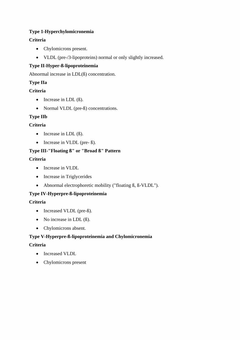
Type 1-Hyperchylomicronemia
Criteria
• Chylomicrons present.
• VLDL (pre-/3-lipoproteins) normal or only slightly increased.
Type II-Hyper-ß-lipoproteinemia
Abnormal increase in LDL(ß) concentration.
Type IIa
Criteria
• Increase in LDL (ß).
• Normal VLDL (pre-ß) concentrations.
Type IIb
Criteria
• Increase in LDL (ß).
• Increase in VLDL (pre- ß).
Type III-"Floating ß" or "Broad ß" Pattern
Criteria
• Increase in VLDL
• Increase in Triglycerides
• Abnormal electrophoretic mobility ("floating ß, ß-VLDL").
Type IV-Hyperpre-ß-lipoproteinemia
Criteria
• Increased VLDL (pre-ß).
• No increase in LDL (ß).
• Chylomicrons absent.
Type V-Hyperpre-ß-lipoproteinemia and Chylomicronemia
Criteria
• Increased VLDL
• Chylomicrons present

Hypercholesterolemia[51]
Three primary disorders causing hypercholesterolemia have been identified. They are:
Polygenic hypercholesterolemia
Polygenic hypercholesterolemia is the term utilized to describe the most common
primary disorder causing an increase in plasma cholesterol. It includes a group of related
disorders in which multiple genes apparently interact to cause an elevation in LDL above the
95th percentile in the general population. Increased rate of formation of LDL, defective
clearance of LDL, or both could be responsible for this elevation.
Familial hypercholesterolemia
Familial hypercholesterolemia is a common autosomal dominant disorder that affects
approximately 1:500 persons in the general population. Its principal defect lies in the gene for
the LDL receptor on the surface of cells so that clearance of LDL from plasma is delayed.
Homozygotes are rare and usually attain a six- to eightfold increase in total plasma
cholesterol due to an elevation in LDL; heterozygotes may have a two - to threefold elevation
and can be diagnosed at birth with analysis of umbilical cord blood. The most important
clinical characteristic of this disorder is the presence of premature and accelerated coronary
artery disease.
A variant of familial hypercholesterolemia
Familial combined hyperlipidaemia is another common disorder that has an autosomal
dominant inheritance. It can present clinically as hypercholesterolemia (type 2a),
hypertriglyceridemia (type 4) or both (type 2b) and has also been called multiple-type
hyperlipoproteinemia. It is characterized clinically by the absence of hyperlipoproteinemia
during childhood, and its development occurs around puberty in association with variable and
mild elevation in plasma lipid levels. There is no specific clinical or laboratory test to
determine if an individual has this disorder, and family screening is needed in order to make
the diagnosis.
Hypertriglyceridemia[51]
The primary disorders predominantly causing hypertriglyceridemia are:
Familial hypertriglyceridemia
Familial hypertriglyceridemia is a common autosomal dominant disorder
characterized by increased plasma concentration of VLDL (type 4 lipoprotein pattern).
Moderate elevations of triglycerides usually occur during early adulthood, and a triad of
obesity, hyperglycemia, and hyperinsulinemia can be seen in affected individuals. In

individuals with moderate elevation in plasma triglycerides associated with a normal
cholesterol level, the possibility of familial hypertriglyceridemia should be suspected.
Congenital deficiency of lipoprotein lipase
Congenital lipoprotein lipase deficiency is a rare autosomal recessive disorder
secondary to absence or severe diminution in the activity of lipoprotein lipase. Affected
individuals are homozygous for a mutation that prevents normal expression of lipoprotein
lipase activity. The parents, although clinically normal, are obligate heterozygotes. This
enzymatic disorder is reflected in a massive accumulation of chylomicrons in the plasma
without elevation of VLDL (type 1 lipoprotein pattern). Triglycerides may reach levels of
2000 to 10,000 mg/dl. This disorder usually appears in childhood with recurrent bouts of
abdominal pain secondary to pancreatitis.
Deficiency of apoprotein CII
Apoprotein CII deficiency is a rare autosomal recessive disorder caused by absence of
apoprotein CII, a required cofactor for the activity of lipoprotein lipase. The ensuing
functional deficiency in this enzyme leads to a clinical picture similar to that described above
for congenital lipoprotein lipase deficiency.
Familial dysbetalipoproteinemias.
Familial dysbetalipoproteinemia, also called familial type 3 hyperlipoproteinemia, is a
condition inherited through a single gene mechanism whose clinical presentation requires the
presence of other genetic or environmental factors . Elevation of both plasma cholesterol and
triglycerides occurs because of accumulation of remnant VLDL particles in the plasma. The
metabolic defect in most patients occurs in apolipoprotein E. This has three common alleles,
designated E2, E3, and E4. Patients with this disorder have only apolipoprotein E2 in VLDL,
which is less effective in facilitating clearance of remnants than E3 or E4.
Causes of Hyperlipidaemia
Abnormal lipid profiles are generally a combination of abnormalities of the
lipoprotein fractions. Hyperlipidaemia can broadly be classified as isolated elevation of
cholesterol, isolated elevated TG and elevations of both. The cause may be genetic,
environmental or both.[52]

Tab. No. 4: Causes and Clinical features of Hyperlipidaemia
CAUSES CLINICAL FEATURES
Isolated cholesterol elevation
Genetic Familial
Hypercholesterolemia
relatively common (1 in 500 heterozygote); TC exceeds 300
mg/dL, family history of elevated TC common, associated with
tendon xanthomas, premature (20 – 40 years old) CVD is common
Homozygotes are rare, but have TC > 600 and if not treated
usually die of MI prior to age 20.
Familial Defective
Apolipoprotein B100
increases LDL and has a phenotype that is indistinguishable from
that of FH, including increased susceptibility to CHD
Mutations Associated with
Elevated LDL Levels
Rare and isolated; suspect if elevated LDL unresponsive to
treatment
Elevated Plasma
Lipoprotein(a)
Relationship to CVD unclear, studies contradictory.
Polygenic
Hypercholesterolemia
No family history, no physical manifestations such as xanthomas,
exact cause is unknown
Lp(X) Associate with obstructive hepatic disease, CVD risk unclear
Sitosterolemia rare; plant sterols absorbed in large amounts, tendon xanthomas
develop in childhood, LDL levels normal to high
Cerebrotendinous
Xanthomatosis
rare; associated with neurologic disease, tendon xanthomas, and
cataracts in young adults
Elevated cholesterol and triglycerides
Combined (Familial)
Hyperlipidaemia
May occur randomly or with strong family history of
hyperlipidaemia; type 2 diabetes and metabolic syndrome are
associated and can make diagnosis more difficult
Familial Dysßlipoproteinemia
(Type III
Hyperlipoproteinemia)
severe hypertriglyceridemia and hypercholesterolemia (both often
> 300mg/dL), associated with premature diffuse vascular disease,
male predominance, Palmar xanthomas are pathognomonic
Hepatic Lipase Deficiency Rare disorder with very high cholesterol and triglyceride
concentrations, phenotypically similar to familial
dysbetalipoproteinemia. Isolated triglyceride elevations

LPL deficiency Results in elevated chylomicrons, which carry dietary fat;
chylomicrons are generally not present after an overnight fast, so a
creamy looking plasma in a fasting specimen should be a clue to
the diagnosis, especially if seen in young children; extremely high
triglycerides can lead to pancreatitis
ApoCII deficiency This apolipoprotein is an activator of LPL; its absence causes a
clinical picture identical to LPL deficiency
Familial hypertriglyceridemia Autosomal dominant inheritance; Main defect is overproduction of
VLDL triglycerides by the liver;
Secondary cause for Hyperlipidaemia:
Secondary causes of hyperlipidaemia are important to recognize. Some times
hyperlipidaemia will be a clue to diagnose the underlying systemic disorders. It may greatly
result in the risk of atherosclerosis with raised LDL concentration, triglyceride rich
lipoprotein excess and also decrease in HDL concentration. Diagnosis of secondary causes is
clue as to why the patient with suddenly developed worsening in lipid profiles.[53]
Diet
Foods which contain cholesterol, saturated fat, and Trans fats can raise your blood
cholesterol level. These include: Cheese, Egg yolk, Fried and processed food, Ice cream,
Pastries, Red meat.[54] Fish oil can also elevate LDL concentration when it is given to lower
triglycerides in diabetics and patients with familial combined hyperlipidaemia.[55] Dietary
factors that lower cholesterol include soluble fiber as well as substituting unsaturated fats or
complex carbohydrates for saturated fats. Diets rich in unsaturated fats can lower HDL
concentration slightly in men but not in women.[56] Alcohol can raise triglycerides as well as
HDL concentration and can markedly aggravate hyperlipidaemia in patients with preexisting
hypertriglyceridemia.[57]
Drugs
Drug-induced lipid and lipoprotein changes can clearly improve or aggravate
atherogenic risk or heighten the risk of pancreatitis when they promote severe
hypertriglyceridemia.[58]
Steroids

Steroid hormones can have a significant impact on lipid and lipoprotein
concentrations. Cholesterol is the precursor of adrenocorticosteroids, androgens, estrogens,
and progestins. Improper usage of these classes can convert a mild primary lipid abnormality
into a clinically life-threatening situation.[59] A study of normal men and women showed that
prednisone caused total cholesterol levels to be increased by 17.3%, triglyceride levels to be
increased in women only, LDL-c to be increased by 10.9% (not significant), and HDL-c to be
increased by 68%.[60]
Female Hormone Preparations
Estrogens raise triglycerides and HDL-c. These are elevated 1.5-fold to 2.5-fold in
proportion to the potency.[61] LDL-c tends to be elevated with increasing estrogen potency in
those on oral contraceptives. Progestins tend to lower triglycerides and HDL-c and in general
have effects that are in the opposite direction of the estrogens. Medroxyprogesterone acetate
is similar to progesterone and is used in combination with estrogen for postmenopausal
estrogen replacement in women with an intact uterus. Norgestrel and norethindrone are
derived from 19-nortestosterone and are used in birth control formulations with norgestrel
more likely to raise LDL-c and lower HDL-c than norethindrone.[62]
Diuretics
In short-term studies, diuretics raise total cholesterol 5% to 8%, triglycerides 15% to
25%, and LDL concentration 8%.[63]
Alpha & Beta Blockers
These antihypertensives are associated with no change in LDL concentration and may
cause increased HDL concentration. The mechanism is thought to be diminished clearance of
apo Al HDL concentration.[64] Beta blockers raise triglycerides and lower HDL
concentration.[65]
Hypothyroidism
Thyroid deficiency is also implicated in hypertriglyceridemia. Because thyroid
deficiency can lead to a decrease in LPL activity, the hypertriglyceridemia of an underlying
genetic triglyceride disorder can be exacerbated, and chylomicronemia can occur.[66]
Obesity
Obese subjects often have increased triglycerides and low HDL concentration. Obese
subjects have increased synthetic rates for cholesterol and bile acids. They have increased
turnover of apo LDL, but this is not necessarily associated with high LDL concentration
levels.[67] Another way to look at lipid and lipoprotein changes in obesity is to consider what

happens when the obese undergo weight loss. With weight loss, triglycerides decrease early
with a delayed effect on rise of HDL concentration.[68]
Diabetes Mellitus
In noninsulin-dependent diabetics, mild hypertriglyceridemia and low HDL-c are
often seen and are due to both overproduction and removal defects. When a familial form of
hypertriglyceridemia that causes enhanced production of triglyceride-rich VLDL and
noninsulin-dependent diabetes coexist, removal mechanisms for dietary glyceride become
saturated.[69]
Risks of Hyperlipidaemia
High cholesterol is associated with an elevated risk of cardiovascular disease. That
can include coronary heart disease, stroke, and peripheral vascular disease. High cholesterol
has also been linked to diabetes and high blood pressure.[70]
Strokes
Strokes (cerebrovascular accidents) are considered to be one of the most common
causes of mortality and long term severe disability. There is a positive correlation between
serum total cholesterol (TC) concentrations and ischaemic (thrombotic) stroke, and very low
TC concentrations have been associated with an increased risk of haemorrhagic stroke.
Raised low density lipoprotein cholesterol (LDL) or triglyceride (TG) concentrations,
reduced high density lipoprotein cholesterol (HDL) concentrations, and a high TC to HDL
ratio are associated with an increased risk of non-haemorrhagic stroke. There is evidence that
lipoprotein (a) is a predictor of many forms of vascular disease, including premature coronary
artery disease (CAD).[71,72]
Cardiovascular Disease (CVD)
According to the WHO, CVDs are the number 1 cause of death globally: more people
die annually from CVDs than from any other cause. An estimated 17.9 million people died
from CVDs in 2016, representing 31% of all global deaths. Of these deaths, 85% are due to
heart attack and stroke. In low and middle income countries 37% of premature death was
caused by the CVDs.[73] Coronary and peripheral artery diseases are caused by the
hyperlipidaemic condition. The importance of total cholesterol for coronary artery disease
(CAD) risk has been demonstrated in observational epidemiologic studies carried out over the
last three decades.[74] The most common reason for the CAD is that build-up of fatty deposits
on the inner walls of the blood vessels that supplies blood to the heart. Approximately 70% of
cholesterol is transported in blood as low density lipoprotein (LDL) cholesterol. Much of the
remaining cholesterol is transported from non-hepatic cells to the liver for synthesis into

lipoproteins, bile acids and steroids by high density lipoprotein (HDL), by a process known
as reverse cholesterol transport. Disturbances in reverse cholesterol transport have been
shown to enhance the deposition of LDL-cholesterol into the artery wall, resulting in
atherosclerotic lesions.[75]
Hypertension
Raised blood pressure attributes to the leading risk factor for morbidity and mortality
in India. Hypertension is attributable to 10.8% of all deaths in India.[76] Increased level of
cholesterol in blood circulation may increase the risk of hypertension. The excess oily stuffs
in cholesterol stick in to the walls of the arteries create a fatty build up, that eventually
hardens and forming an inflexible plaque that damages the arteries and they become stiff and
narrowed. The blood cannot able to flow easily through the blood vessels that lead to the
hypertension.[77]
Antihyperlipidaemic drugs
These are drugs which lower the levels of lipids and lipoproteins in blood and have
attracted considerable attention because of their potential to prevent cardiovascular disease by
retarding the accelerated atherosclerosis in hyperlipidaemic individuals.[78]
Classification of Antihyperlipidaemic Drugs[78]
a) HMG-CoA reductase inhibitors (Statins)
Lovastatin, Simvastatin, Pravastatin, Atorvastatin, Rosuvastatin, Pitavastatin
b) Bile acid sequestrants (Resins)
Cholestyramine, Colestipol
c) Lipoprotein lipase activator/ PPARα activators (Fibrates)
Fenofibrate, Bezafibrate, Clofibrate, Gemfibrozil
d) Lipolysis and Triglyceride synthesis inhibitors
Nicotinic acid
e) Sterol absorption inhibitor
Ezetimibe
HMG-CoA Inhibitors (Statins)
Statins were isolated from a mold, Penicillium citrinum, and identified as inhibitors of
cholesterol biosynthesis. Subsequent studies established that statins act by inhibiting HMG-
CoA reductase, which catalyzes an early, rate-limiting step in cholesterol biosynthesis. The
first statin studied in humans was compactin, renamed mevastatin, which demonstrated the
therapeutic potential of this class of drugs. The statins are the most effective and best-
tolerated agents for treating dyslipidemia. Higher doses of the more potent statins (e.g.,

atorvastatin, simvastatin, and rosuvastatin) also can reduce triglyceride levels caused by
elevated VLDL levels.[79]
Mechanism of action
Statins exert their major effect (reduction of LDL levels) through a mevalonic acid–
like moiety that competitively inhibits HMG-CoA reductase. By reducing the conversion of
HMG-CoA to mevalonate, statins inhibit an early and rate-limiting step in cholesterol
biosynthesis, which results in increased expression of the LDL receptor gene. In response to
the reduced free cholesterol content within hepatocytes, membrane-bound sterol regulatory
element binding proteins (SREBPs) are cleaved by a protease and translocated to the nucleus.
The transcription factors then bind the sterol-responsive element of the LDL receptor gene,
enhancing transcription and increasing the synthesis of LDL receptors. Degradation of LDL
receptors also is reduced. The greater number of LDL receptors on the surface of hepatocytes
results in increased removal of LDL from the blood, thereby lowering LDL-C levels.[79]
Adverse effect
The major adverse effect of statin use is Myopathy. Hepatotoxicity is rarely observed.
Gastrointestinal complaints and headache are usually mild.[78]
Bile acid sequestrants (Resins)
The bile-acid sequestrants or resins are among the oldest of the hypolipidemic drugs,
and they are probably the safest, because they are not absorbed from the intestine. These
resins also are recommended for patients 11-20 years of age. Because statins are more
effective as monotherapy, the resins are most often used as second agents if statin therapy
does not lower LDL-C levels sufficiently. When used with a statin, cholestyramine and
colestipol usually are prescribed at submaximal doses. Maximal doses can reduce LDL-C by
up to 25% but are associated with unacceptable gastrointestinal side effects. Colesevelam is a
newer bile-acid sequestrant that is prepared as an anhydrous gel and taken as a tablet or as a
powder that is mixed with water and taken as an oral suspension. It lowers LDL-C by 18% at
its maximum dose.[79]
Mechanism of action
The bile-acid sequestrants are highly positively charged and bind negatively charged
bile acids. Because of their large size, the resins are not absorbed, and the bound bile acids
are excreted in the stool. Because more than 95% of bile acids are normally reabsorbed,
interruption of this process depletes the pool of bile acids, and hepatic bile-acid synthesis
increases. As a result, hepatic cholesterol content declines, stimulating the production of LDL
receptors, an effect similar to that of statins. The increase in hepatic LDL receptors increases

LDL clearance and lowers LDL-C levels, but this effect is partially offset by the enhanced
cholesterol synthesis caused by upregulation of HMG-CoA reductase. Inhibition of reductase
activity by a statin substantially increases the effectiveness of the resins.[79]
Adverse effect
They are unpalatable in nature and inconvenient; have to be taken in large doses it
causes flatulence and other gastrointestinal symptoms, interfere with absorption of many
other drugs.
Lipoprotein lipase activators (Fibrates)
The fibrates are mainly used to treat two hyperlipidemias, familial
hypertriglyceridemia (type IV) and dysbetalipoproteinemia (type III). They are also useful in
the treatment of hypertriglyceridemia associated with type II diabetes (secondary
hyperlipidemia). The fibrates are the drugs of choice in treating hypertriglyceridemias,
particularly those associated with low levels of HDL cholesterol.The fibrates additionally
appear to shift LDL particles to larger, hence less atherogenic species.[80]
Mechanism of action
The fibrates typically lower VLDL triglyceride by 40% or more and elevate plasma
HDL cholesterol by 10 to 15%. The reduction of plasma triglycerides in humans appears due
to increased lipoprotein lipase (LPL) activity. The fibrates activate a nuclear receptor
(transcription factor) termed peroxisomal proliferation activated receptor (PPAR) that is a
member of the steroid hormone receptor superfamily. PPAR increases transcription of the
LPL gene and decreases transcription of the apolipoprotein CIII gene (apo CIII). Since LPL
is responsible for catabolism of VLDL triglyceride and apo CIII is an inhibitor of LPL
activity, the combined consequences of these changes are increased LPL activity and
enhanced removal of triglyceride from the circulation.[80]
Adverse effect
Fibric acid compounds usually are well tolerated. Side effects may occur in 5-10% of
patients but most often are not sufficient to cause discontinuation of the drug. Gastrointestinal
side effects occur in up to 5% of patients. Other side effects are reported infrequently and
include rash, urticaria, hair loss, myalgias, fatigue, headache, impotence, and anemia. Minor
increases in liver transaminases and alkaline phosphatase have been reported.[79]

Lipolysis and Triglycerides synthesis inhibitors[79]
Niacin (nicotinic acid) is one of the oldest drugs used to treat dyslipidemia, favorably
affects virtually all lipid parameters. Niacin is a water-soluble B-complex vitamin that
functions as a vitamin only after its conversion to NAD or NADP, in which it occurs as an
amide. Both niacin and its amide may be given orally as a source of niacin for its functions as
a vitamin, but only niacin affects lipid levels. The hypolipidemic effects of niacin require
larger doses than are required for its vitamin effects. Niacin is the best agent available for
increasing HDL-C (30-40%); it also lowers triglycerides by 35-45% (as effectively as fibrates
and the more effective statins) and reduces LDL-C levels by 20-30%. Niacin also is the only
lipid-lowering drug that reduces Lp(a) levels significantly.
Mechanism of action
In adipose tissue, niacin inhibits the lipolysis of triglycerides by hormone-sensitive
lipase, which reduces transport of free fatty acids to the liver and decreases hepatic
triglyceride synthesis.
Adverse effect
Dyspepsia, Flushing and Hepatotoxicity. In patients with diabetes mellitus, niacin
should be used cautiously because niacin-induced insulin resistance can cause severe
hyperglycemia. Reversible side effects include toxic amblyopia and toxic maculopathy. Atrial
tachyarrhythmias and atrial fibrillation have been reported, more commonly in elderly
patients.[79]
Sterol absorption inhibitor[81]
Ezetimibe is the first compound approved for lowering total and LDL-C levels that
inhibits cholesterol absorption by enterocytes in the small intestine. It lowers LDLC levels by
20% and is used primarily as adjunctive therapy with statins.
Mechanism of action
Ezetimibe inhibits luminal cholesterol uptake by jejunal enterocytes, by inhibiting the
transport protein NPC1L.
Adverse effects
Other than rare allergic reactions, specific adverse effects have not been observed in
patients taking ezetimibe.
1.3 Diabetes
As per the WHO, diabetes mellitus (DM) is defined as a heterogeneous metabolic
disorder characterised by common feature of chronic hyperglycaemia with disturbance of
carbohydrate, fat and protein metabolism. The number of individuals with diabetes is rising
rapidly throughout the world. Both genetic and environmental factors contribute to its
pathogenesis, which involves insufficient insulin secretion, reduced responsiveness to
endogenous or exogenous insulin, increased glucose production, and/or abnormalities in fat
and protein metabolism. The resulting hyperglycemia may lead to both acute symptoms and
metabolic abnormalities.
DM is a leading cause of morbidity and mortality world over. It is estimated that
approximately 1% of population suffers from DM. The incidence is rising in the developed
countries of the world at the rate of about 10% per year, especially of type 2 DM, due to
rising incidence of obesity and reduced activity levels. DM is expected to continue as a major
health problem owing to its serious complications, especially end-stage renal disease, IHD,
gangrene of the lower extremities, and blindness in the adults. It is anticipated that the
number of diabetics will exceed 250 million by the year 2010. [82, 83]
Diabetes mellitus is a metabolic disorder of multiple etiologies. It is characterized by
chronic hyperglycemia together with disturbances of carbohydrate, fat and protein
metabolism resulting from defects of insulin secretion, insulin action or both. The relative
contribution of these varies between different types of diabetes. These are associated with the
development of the specific microvascular complications of retinopathy, which can lead to
blindness, nephropathy with potential renal failure, and neuropathy. The latter carries the risk
of foot ulcers and amputation and also autonomic nerve dysfunction. Diabetes is also
associated with an increased risk of macrovascular disease. [84]
Classification
The older classification systems dividing DM into primary (idiopathic) and secondary
types, juvenile-onset and maturity onset types, and insulin-dependent (IDDM) and non-
insulin dependent (NIDDM) types, have become obsolete and undergone major revision due
to extensive understanding of etiology and pathogenesis of DM in recent times. [81, 85]

Fig. No. 2: Types of Diabetes Mellitus
I. TYPE 1 DIABETES MELLITUS (10%) (earlier called Insulin-dependent, or
juvenile-onset diabetes)
Type IA DM: Immune-mediated
Type IB DM: Idiopathic
II. TYPE 2 DIABETES MELLITUS (80%) (earlier called non-insulin-dependent, or
maturity-onset diabetes)
III. OTHER SPECIFIC TYPES OF DIABETES (10%)
a. Genetic defect of β-cell function due to mutations in various enzymes (earlier
called maturity-onset diabetes of the young or MODY) (e.g. hepatocyte nuclear
transcription factor—HNF, glucokinase)
b. Genetic defect in insulin action (e.g. type A insulin resistance)
c. Diseases of exocrine pancreas (e.g. chronic pancreatitis, pancreatic tumours, post-
pancreatectomy)
d. Endocrinopathies (e.g. acromegaly, Cushing's syndrome, pheochromocytoma)
e. Drug- or chemical-induced (e.g. steroids, thyroid hormone, thiazides, β-blockers
etc)
f. Infections (e.g. congenital rubella, cytomegalovirus)
g. Uncommon forms of immune-mediated DM (stiff man syndrome, anti-insulin
receptor antibodies)
h. Other genetic syndromes (e.g. Down's syndrome, Klinefelter's syndrome, Turner's
syndrome)
IV. GESTATIONAL DIABETES MELLITUS

TYPE 1 DM
It constitutes about 10% cases of DM. It was previously termed as juvenile-onset
diabetes (JOD) due to its occurrence in younger age, and was called insulindependent DM
(IDDM) because it was known that these patients have absolute requirement for insulin
replacement as treatment. However, in the new classification, neither age nor insulin-
dependence are considered as absolute criteria. Instead, based on underlying etiology, type 1
DM is further divided into 2 subtypes: [81]
Subtype 1A (immune-mediated) DM: This forms diabetes, which accounts for only 5–10%
of those with diabetes, previously encompassed by the terms insulindependent diabetes or
juvenile-onset diabetes, results from a cellular-mediated autoimmune destruction of the ß-
cells of the pancreas. Markers of the immune destruction of the ß-cell include islet cell
autoantibodies, autoantibodies to insulin, autoantibodies to GAD (GAD65), and
autoantibodies to the tyrosine phosphatases IA-2 and IA-2ß. [86]
Subtype 1B (idiopathic) DM: Some forms of type 1 diabetes have no known etiologies.
Some of these patients have permanent insulinopenia and are prone to ketoacidosis, but have
no evidence of autoimmunity. Only a minority of patients with type 1 diabetes fall into this
category. Individuals with this form of diabetes suffer from episodic ketoacidosis and exhibit
varying degrees of insulin deficiency between episodes. This form of diabetes is strongly
inherited, lacks immunological evidence for b-cell autoimmunity, and is not HLA associated.
Though type 1 DM occurs commonly in patients under 30 years of age, autoimmune
destruction of β-cells can occur at any age. In fact, 5-10% patients who develop DM above 30
years of age are of type 1A DM and hence the term JOD has become obsolete. [86]
Gestational Diabetes
Gestational diabetes mellitus (GDM) is hyperglycemia first detected during
pregnancy. This is distinct from women with diabetes undergoing pregnancy, who have
diabetes in pregnancy rather than gestational diabetes. Plasma glucose levels, both fasting and
post - prandial, are lower than normal in early pregnancy so that raised levels at this stage are
almost certainly caused by previously undetected T2DM. Screening for GDM is generally
undertaken at around 28 weeks. There is signifi cant morbidity associated with GDM
including intrauterine fetal death, congenital malformations, neonatal hypoglycemia,
jaundice, prematurity and macrosomia. Risk factors for GDM include certain ethnic groups,

those with previous GDM or abnormalities of glucose tolerance, age, obesity and previous
large babies. [85]
Diagnosis [87]
Glycated hemoglobin (A1C) test.
This blood test, which doesn't require fasting, indicates your average blood sugar
level for the past two to three months. It measures the percentage of blood sugar attached to
hemoglobin, the oxygen-carrying protein in red blood cells.
Fasting blood sugar test.
A blood sample will be taken after an overnight fast. A fasting blood sugar level less
than 100 mg/dL (5.6 mmol/L) is normal. A fasting blood sugar level from 100 to 125 mg/dL
(5.6 to 6.9 mmol/L) is considered prediabetes. If it's 126 mg/dL (7 mmol/L) or higher on two
separate tests, you have diabetes.
Oral glucose tolerance test
For this test, you fast overnight, and the fasting blood sugar level is measured. Then
you drink a sugary liquid, and blood sugar levels are tested periodically for the next two
hours. A blood sugar level less than 140 mg/dL (7.8 mmol/L) is normal. A reading of more
than 200 mg/dL (11.1 mmol/L) after two hours indicates diabetes. A reading between 140
and 199 mg/dL (7.8 mmol/L and 11.0 mmol/L) indicates prediabetes.
Management of Diabetes [87]
Depending on the type of diabetes blood sugar monitoring, insulin and oral
hypoglycemic therapy, healthy diet and maintaining the healthy body weight in regular life
play a major role in the management of diabetes.
Healthy diet
There is no specific diet for diabetes but adding more fruits, vegetables, lean proteins
and whole grains in regular diet, foods which are higher in nutrition and fiber and low in fat
and calories helps to control the blood sugar level. Patient with diabetes have to avoid
saturated fatty food, refined carbohydrates and sweets.

Physical activity
Every one need regular aerobic exercise, in patient with diabetes mellitus helps to
promote the transport of sugar into the cells where it is used for energy. Exercise increases
the sensitivity of the insulin, which means body needs less insulin to transport sugars into the
cells.
Monitoring of blood sugar
Careful monitoring is the only way to make sure that the blood sugar level remains
within the normal range. Depend on the treatment, periodic monitoring of blood glucose level
in essential to assure the safety of the patients. Treatment with oral hypoglycemic drugs and
insulin some time leads to the hypoglycemic level, periodic measure of blood sugar level
helps to avoid such a conditions.
Insulin Therapy
Insulin is a peptide hormone produced by beta cells of the pancreatic islets; it is
considered to be the main anabolic hormone of the body. [88] Insulin is responsible for
allowing glucose in the blood to enter cells, providing them with the energy to function. The
pancreas is an organ behind the stomach that is the main source of insulin in the body.
Clusters of cells in the pancreas called islets produce the hormone and determine the amount
based on blood glucose levels in the body. The higher the level of glucose, the more insulin
goes into production to balance sugar levels in the blood. Insulin also assists in breaking
down fats or proteins for energy. [89]
Fig. No. 3: Secretion and Release of Insulin

Types
Rapid Acting
The body absorbs this type into the bloodstream from the subcutaneous tissue
extremely quickly.
• Insulin Lispro, Insulin Aspart, Insulin Glulisine
Short acting
It starts working approximately 30 minutes after injection and peaks at approximately
2 to 3 hours but will continue to work for three to six hours.
• Regular (soluble) insulin
Intermediate Acting
This type insulin enters the bloodstream at a slower rate but has a longer-lasting effect. It
is most effective at managing blood sugar overnight, as well as between meals.
• Insulin Zinc Suspension or Lente, Neutral Protamine Hagedorn (NPH)
Long-acting insulin
Long-acting insulin is slow to reach the bloodstream and has a relatively low peak, it
has a stabilizing "plateau" effect on blood sugar that can last for most of the day.
• Protamine zinc insulin, Insulin glargine
Oral hypoglycemic therapy [90]
Oral hypoglycemic drugs are chemical agents which lowers the blood glucose level.
Classification
Sulfonylurea
• 1st Generation
Tolbutamide, Tolazamide, Chlorpropamide, Acetohexamide
• 2nd generation
Glipizide, Glyburide, Glimepride, Gliclazide

Biguanides
• Metformin
Meglitinides
• Repaglinde, Nataglinide
Thiazolidiones or Glitazones
• Pioglitazones, Troglitazone, Rosiglitazone
Alpha-glycosidase inhibitor
• Acarbose, Miglitazone
Dipeptide peptidase inhibitor (DPP-G)
• Vidagliptin, Sitagliptin
Amylin analogue
• Pramlintide
Incretin mimetic (Glucagon like peptide analogue)
• Exenatide, Liraglutide
1.4 Analgesia (Pain)
An unpleasant sensory and emotional experience associated with actual or potential
tissue damage, or described in terms of such damage. Pain is a conscious experience that
results from brain activity in response to a noxious stimulus and engages the sensory,
emotional and cognitive processes of the brain. In general terms we can distinguish two
dimensions or components of pain: (a) sensory-discriminative and (b) affective-emotional.
Nociception is the process by which information about a noxious stimulus is
conveyed to the brain. It is the total sum of neural activity that occurs prior to the cognitive
processes that enable humans to identify a sensation as pain. Nociception is necessary but not
sufficient for the experience of pain. [91]

Nociceptors
Nociceptors are special receptors which are activated through various noxious stimuli.
They present at the free nerve endings of the primary afferent neuron. Each nociceptor has its
own receptive field. This means one nociceptor will transduce the signal of pain when a
particular region is skin is stimulated. The size of receptive fields are vary throughout the
body and there is often overlap with neighbouring fields. [92]
Nociceptors can be found in the skin, muscle, joints, bone and organs (other than the
brain) and can fire in response to a number of different stimuli. Three types of nociceptors
exist:
• Mechanical nociceptors – detects sharp, pricking pain
• Thermal and mechano-thermal nociceptors – detects sensations which elicit pain
which is slow and burning, or cold and sharp in nature
• Polymodal nociceptors – detects mechanical, thermal and chemical stimuli
Pathway of pain
Signals from mechanical, thermal and mechano-thermal nociceptors are transmitted to
the dorsal horn of the spinal cord predominantly by Aδ fibres. These myelinated fibres have a
low threshold for firing and the fast conduction speed means they are responsible for
transmitting the first pain felt.
In addition, Aδ fibres permit for the localisation of pain and form the afferent pathway
for the reflexes elicited by pain. Aδ fibres predominantly terminate in Rexed laminae I where
they mainly release the neurotransmitter glutamate.
Polymodal nociceptors transmit their signals into the dorsal horn through C fibres. C
fibres are unmyelinated and a slow conduction speed. For this reason, C fibres are responsible
for the secondary pain we feel which is often dull, deep and throbbing in nature. These fibres
typically have large receptive fields and therefore lead to poor localisation of pain. [92]

Fig. No. 4: Pathway of Pain
TYPES OF PAIN
Somatic Pain
Pain arising from the skin and integumental structures, muscles, bones and joints is
known s Somatic pain. It is usually caused by inflammation and is well-defined or sharp pain.
Visceral Pain
Pain arising from the viscera is vague, dull- aching type, difficult to pinpoint to a site
and is known as visceral pain. It may be accompanied by autonomic responses like sweating,
nausea and hypotension. It may be due to spasm, ischaemia or inflammation.

Referred Pain
When pain is referred to a cutaneous area which receives nerve supply from the same
spinal segment as that of the affected viscera, it is known as referred pain, e.g. cardiac pain
referred to the left arm. [93]
Analgesics
Analgesics are drugs which relieve pain without causing loss of consciousness.
Classification
• Opioid analgesics
• Non opioid analgesics
Opioid analgesics
Within the central nervous system there are three types of opioid receptors which
regulate neurotransmission of pain signals. These receptors are called mu, delta and kappa
opioid receptors. The drugs which bind to this receptors and relieves the pain are called as
opioid receptors. They are all G protein coupled receptors and their activation leads to a
reduction in neurotransmitter release and cell hyperpolarisation, reducing cell excitability.
Mechanism of action
Opioid analgesics bind with opioid receptors and reduce the neurotransmitter release
from the first order neuron, and causes hyperpolarisation of the second order neuron.
Together, this reduces the firing of action potentials in the second order neuron, blocking the
transmission of pain signals. [92]
Classification of opioid analgesics
Agonists
• Morphine, codeine and its derivatives such as pethidine, methadone, levorphanol and
tramadol.
Partial agonists
• Buprenorphine and meptazinol.

Mixed agonist
• Nalbuphine, pentazoxine and butorphanol
Non opioid analgesics
Non-opioid analgesics can be classified due to their chemical characteristics as acid
(NSAIDs = Non-Steroidal Anti-Inflammatory Drugs) and non-acid. The common mechanism
of action of these substances is their effect on prostaglandin synthesis. [94]
Classification
Acid/NSAIDs
i. Salicylates
• Aspirin (Acetyl salicylic acid)
ii. Acetic acids
• Diclofenac, ketorolac, indomethacin
iii. Propionic acids
• Ibuprofen, naproxen, ketoprofen
iv. Oxicams
• Piraxicam, tenoxicam, meloxicam
v. Coxibs
• Celecoxib, rofecoxib, etoricoxib
Non acids
i. Anilines
• Paracetamol
ii. Pyrazolones
• Mitamizole
1.5 Pyrexia (Fever)
Fever is a complex physiologic response triggered by infectious or aseptic stimuli.
Elevations in body temperature occur when concentrations of prostaglandin E2 (PGE2)
increase within certain areas of the brain. These elevations alter the firing rate of neurons that
control thermoregulation in the hypothalamus. Although fever benefits the nonspecific
immune response to invading microorganisms, it is also viewed as a source of discomfort and
is commonly suppressed with antipyretic medication. [95]

Pathogenesis of fever
The critical “endogenous pyrogens” involved in producing a highly regulated
inflammatory response to tissue injury and infections are polypeptide cytokines. Pyrogenic
cytokines, such as interleukin-1b (IL-1b), tumor necrosis factor (TNF), and interleukin-6 (IL-
6), are those that act directly on the hypothalamus to produce a fever response. [96] Exogenous
pyrogens, such as microbial surface components, evoke pyrexia most commonly through the
stimulation of pyrogenic cytokines. The gram-negative bacteria’s outer membrane
lipopolysaccharide (endotoxin), however, is capable of functioning at the level of the
hypothalamus, in much the same way as IL-1b. [97]
Antipyretics
The drugs which reduce the elevated body temperature are called as the antipyretic
agents.
Classification
Salicylates
• Aspirin (acetylsalicylic acid), choline salicylate, magnesium salicylate and sodium
salicylate
Acetaminophen
• Paracetamol
NSAIDs
• Ibuprofen, naproxen and ketoprofen
1.6 Wound [98]
Wound is an injury involving an external or internal break in body tissue, usually
involving the skin.
Types
Abrasion
An abrasion occurs when your skin rubs or scrapes against a rough or hard surface.
Road rash is an example of an abrasion. There’s usually not a lot of bleeding, but the wound
needs to be scrubbed and cleaned to avoid infection.

Laceration
A laceration is a deep cut or tearing of your skin. Accidents with knives, tools, and
machinery are frequent causes of lacerations. In the case of deep lacerations, bleeding can be
rapid and extensive.
Puncture
A puncture is a small hole caused by a long, pointy object, such as a nail or needle.
Sometimes, a bullet can cause a puncture wound. Punctures may not bleed much, but these
wounds can be deep enough to damage internal organs. If you have even a small puncture
wound, visit your doctor to get a tetanus shot and prevent infection.
Avulsion
An avulsion is a partial or complete tearing away of skin and the tissue beneath.
Avulsions usually occur during violent accidents, such as body-crushing accidents,
explosions, and gunshots. They bleed heavily and rapidly.
Wound Healing [99]
Wound is an injury to living tissue caused by a cut, blow, or other impact, typically
one in which the skin is cut or broken. Healing is the body response to injury in an attempt to
restore normal structure and function. Healing involves 2 distinct processes:
• Regeneration when healing takes place by proliferation of parenchymal cells and
usually results in complete restoration of the original tissues.
• Repair when healing takes place by proliferation of connective tissue elements
resulting in fibrosis and scarring. At times, both the processes take place
simultaneously.
Regeneration
Some parenchymal cells are short-lived while others have a longer lifespan. In order
to maintain proper structure of tissues, these cells are under the constant regulatory control of
their cell cycle. Regeneration of any type of parenchymal cells involves the following two
processes:
i. Proliferation of original cells from the margin of injury with migration so as to cover
the gap.

ii. Proliferation of migrated cells with subsequent differentiation and maturation so as to
reconstitute the original tissue.
Repair
Repair is the replacement of injured tissue by fibrous tissue.
Two processes are involved in repair:
• Granulation tissue formation
• Contraction of wounds.
Repair response takes place by participation of mesenchymal cells (consisting of
connective tissue stem cells, fibrocytes and histiocytes), endothelial cells, macrophages,
platelets, and the parenchymal cells of the injured organ.
Granulation Tissue Formation
The term granulation tissue derives its name from slightly granular and pink
appearance of the tissue. Each granule corresponds histologically to proliferation of new
small blood vessels which are slightly lifted on the surface by thin covering of fibroblasts and
young collagen. The following three phases are observed in the formation of granulation
tissue.
Phase of inflammation
Following trauma, blood clots at the site of injury. There is acute inflammatory
response with exudation of plasma, neutrophils and some monocytes within 24 hours.
Phase of clearance
Combination of proteolytic enzymes liberated from neutrophils, autolytic enzymes
from dead tissues cells, and phagocytic activity of macrophages clear off the necrotic tissue,
debris and red blood cells.
Phase of ingrowth of granulation tissue
This phase consists of two main processes
1. angiogenesis or neovascularisation,
2. fibrogenesis.

• Angiogenisis
Formation of new blood vessels at the site of injury takes place by proliferation of
endothelial cells from the margins of severed blood vessels. Initially, the proliferated
endothelial cells are solid buds but within a few hours develop a lumen and start
carrying blood. The newly formed blood vessels are more leaky, accounting for the
oedematous appearance of new granulation tissue. Soon, these blood vessels
differentiate into muscular arterioles, thin-walled venules and true capillaries. The
process of angiogenesis is stimulated with proteolytic destruction of basement
membrane. Angiogenesis takes place under the influence of following factors:
a) Vascular endothelial growth factor (VEGF) elaborated by mesenchymal cells
while its receptors are present in endothelial cells only.
b) Platelet-derived growth factor (PDGF), transforming growth factor-β (TGF-β),
basic fibroblast growth factor (BFGF) and surface integrins are all associated
with cellular proliferation.
• Fibrogenesis
The newly formed blood vessels are present in an amorphous ground substance or
matrix. The new fibroblasts originate from fibrocytes as well as by mitotic division of
fibroblasts. Some of these fibroblasts have combination of morphologic and
functional characteristics of smooth muscle cells (myofibroblasts). Collagen fibrils
begin to appear by about 6th day. As maturation proceeds, more and more of collagen
is formed while the number of active fibroblasts and new blood vessels decreases.
This results in formation of inactive looking scar known as cicatrisation.
Contraction of Wounds
The wound starts contracting after 2-3 days and the process is completed by the 14th
day. During this period, the wound is reduced by approximately 80% of its original size.
Contracted wound results in rapid healing since lesser surface area of the injured tissue has to
be replaced.
Treatment
Wound healing can be accomplished in one of the following two ways: [99]
• Healing by first intention (primary union)
• Healing by second intention (secondary union).

Healing by First Intention (Primary Union)
This is defined as healing of a wound which has the following characteristics:
✓ Clean and uninfected
✓ Surgically incised
✓ Without much loss of cells and tissue; and
✓ Edges of wound are approximated by surgical sutures.
Healing by Second Intention (Secondary Union)
This is defined as healing of a wound having the following characteristics:
✓ Open with a large tissue defect, at times infected
✓ Having extensive loss of cells and tissues; and
✓ The wound is not approximated by surgical sutures but is left open.
Drugs used on wound healing [100]
NSAIDs
• Ibuprofen
Effect: Affects inflammatory phase by inhibiting cyclo-oxygenase production; reduces
tensile strength of wound.
• Colchicine
Effect: Affects inflammatory phase; affects proliferative phase by decreasing fibroblast
proliferation; affects remodelling phase by degrading newly formed extracellular matrix.
Corticosteroids
• Prednisolone
Effect: Affects haemostatic phase by decreasing platelet adhesion; affects inflammatory
phase by affecting phagocytosis; affects remodelling phase by reducing fibroblasts
activity and inhibiting collagen synthesis.
Antiplatelets
• Aspirin
Effect: Affects haemostatic phase by inhibiting platelet aggregation; inhibits
inflammation mediated by arachidonic acid metabolites.
Anticoagulants
• Heparin
Effect: Affects haemostatic phase by its effect on fibrin formation; can lead to thrombus
formation by causing thrombocytopaenia (white clot syndrome).
• Warfarin

Effect: Affects haemostatic phase by its effect on fibrin formation; can cause tissue
necrosis and gangrene by release of atheromatous plaque emboli in form of
microcholesterol crystals (blue toe syndrome)
Vasoconstrictors
• Nicotine, cocaine, adrenaline
Effect: Affects proliferative phase by inhibiting neovascularisation and decreasing
granulation tissue formation; impairs microcirculation and increases graft rejection and
ulcer necrosis.

LITERATURE REVIEW

LITERATURE REVIEW
1. Hammeso, W. W. et. al., (2019) reported that the leaf latex of A. megalacantha
possessed significant antihyperglycemic and antihyperlipidemic potential which may
prove the claimed use of the plant in amelioration of diabetes and associated
complications in Ethiopian folk medicine.[101]
2. NT Abdullah et. al., (2019) evaluated the antidiabetic activity of methanolic extract
of Simarouba glauca in STZ induced diabetes in rats for type I diabetic mellitus.[102]
3. Sushma Gour et. al., (2019) evaluated that the antipyretic activity of hydroalcoholic leaf
extract of Morus Alba Linn using Brewer’s yeast induced pyrexia model in wistar strain
albino rats.[103]
4. Kannika Inchab et. al., (2019) evaluated the anti-inflammatory, analgesic, and
antipyretic activities of water extract from fresh leaves of Pseuderanthemum palatiferum
(WEPP). Antipyretic activity was evaluated by yeast-induced hyperthermia model.[104]
5. Shanti Vasudevan CN et. al., (2019) investigated that the antipyretic activity of its heart
wood aqueous extract of Pterocarpus santalinus L. using brewer’s yeast induced pyrexia
method.[105]
6. Neha Kaila et. al., (2019) ascertained the analgesic and antipyretic activities of rhizome
extracts of Picrorhiza kurrooa. antipyretic activity was evaluated in Wister rats by
brewer’s yeast induced pyrexia method.[106]
7. Sabina Evan Prince et. al., (2019) analyzed the activity of Bacopa monnieri as an
antipyretic, analgesic and anti-ulcerogenic agent. The antipyretic activity was evaluated
by a yeast solution which was used to induce fever before administering the drugs.[107]
8. Mansour Sobeh et. al., (2019) investigated the antioxidant, anti-inflammatory, analgesic,
and antipyretic activities of the extract of Albizia anthelmintica. Pyrexia was induced by
intraperitoneal injection of Brewer’s yeast.[108]
9. M. G. Minoué Kuum et. al., (2019) investigated that the anti-inflammatory, analgesic
and antipyretic potential of aqueous extract of Albizia ferruginea stem bark. The
antipyretic activity of A. ferruginea extract was assessed on brewer’s yeast induced
pyrexia.[109]
10. Farjana Akhter et. al., (2019) studied that the phytochemical constituents and to
evaluate the antibacterial, antioxidant, antidiarrheal and antipyretic activities of different
extracts of Duabanga grandiflora.[110]

11. Swapnil Y. Chaudhari et. al., (2018) studied that an Ayurvedic herbo-metalo-mineral
formulation of Hridayarnava Rasa possessed significatnt Antihyperlipidemic activity
against high fat diet induced hyperlipidemia in Charles Foster albino rats.[111]
12. B. H. M. R. K. Guptha et. al., (2018) evaluated the antihyperlipidemic activity of
Chloroxylon swietenia in triton WR1339 induced hyperlipidemia in rats.[112]
13. Venkateswarlu G et. al., (2018) studied the antipyretic activity of methanolic whole
plant extracts of Limnophila repens (L. repens) and Argyreia cymosa (A. cymosa) using
Brewer’s yeast-induced pyrexia model.[113]
14. Rajendra Kumar Swain et. al., (2018) evaluated the analgesic activity of hydroalcoholic
extract of Cinnamomum zeylanicum (HAECZ) bark in albino rats.[114]
15. Doan, H. V. et. al., (2018) evaluated that the antidiabetic activity of the extract from C.
cainito stem bark and the possible mechanisms underlying this activity against alloxan
induced diabetic mice.[115]
16. Belayneh, Y. M. et. al., (2018) studied the hydromethanolic extract of Calpurnia aurea
leaves possesses signifcant antihyperglycemic activity against streptozotocin induced
hyperglycemia model in mice and justifying the traditional use of the plant for
diabetes.[116]
17. Hussain, S. N. et al., (2018) studied C. sophera extract possesses antidiabetic activity
against alloxan induced rats using glibenclamide as reference standard. This lends some
support for the traditional use of this herb in the management of diabetes.[117]
18. Dr. Sam Pavan Kumar Gandamalla et. al., (2017) investigated the analgesic and anti-
inflammatory effects of the leaves of Toona ciliata in laboratory animals. The ethanol
extract of the leaves of Toona ciliata was used to investigate the acute effect on analgesia
by Hot-plate test in mice and on inflammation in rats using carrageenan-induced rat paw
edema model.[118]
19. Charles Chatterjee et. al., (2017) evaluated the analgesic activity of methanolic
extraction of leaves of Bougainvillea spectabilis by tail immersion method in Mice.[119]
20. G. Vijay Kumar et. al., (2016) evaluated that the Leaf Extracts of Leucas aspera Linn.
Having significant inhibition against dexamethasone-induced hyperlipidemia in rats by
maintaining the serum levels of cholesterol, TGs and near to the normal levels.[120]
21. Nagar, H. K. et. al., (2016) the wound healing effect of ethanolic extract of Cestrum
nocturnum (L.) leaves (EECN) using excision and incision wound model. Methods.[121]

22. Sneha D. Borkar et. al., (2015) studied the leaves of R. hypocrateriformis contain
secondary metabolites such as tannin and possess mild antioxidant properties. Nutritional
analysis indicates the presence of energy in highest amount, carbohydrates, proteins, fats,
calcium, phosphorous, zinc, and magnesium.[122]
23. Asie Shojaii et. al., (2015) evaluated the anti-inflammatory and analgesic activities of the
hydro-alcoholic extract of the pods of Astragalushamosus (HAAH), a plant used in
Iranian traditional medicine, and antinociceptive effects of different fractions in animal
models.[123]
24. S.S. Godipurge et. al., (2015) investigated the potential pharmacological activities of the
ethanolic extract of aerial parts of Rivea hypocrateriformis (EERH) and also measured
their phenolic and flavonoid contents.[124]
25. Pal Chandrashekhar R et. al., (2015) evaluated the antihyperlipidemic activity of
methanolic extract of Gloriosa superb (MEGS) in two different experimental models of
hyperlipidemia namely Poloxamer-407 induced hyperlipidemia and Cholesterol and
cholic suspension induced hyperlipidemia.[125]
26. Mukesh Singh Sikarwar et. al., (2015) demonstrated ethanolic extract fraction of
Hibiscus rosa-sinensis Linn. possessed significant antihyperlipidemic activity.[126]
27. Syed Safiullah Ghori et. al., (2015) studied that the antihyperlipidemic activity of
Ethanolic Extact Of Glycosmis pentaphylla against hyperlipidemia induced by Triton X
100 and also High Fat Diet induced experimental models.[127]
28. Jain P and Surana (2015) Studied the hypolipidemic activity of traditional medicines
from different regions of India.[128]
29. Mahesh Anand Goudar et. al., (2014) evaluated the antidiabetic activity of petroleum
ether (PETP) and ethanolic (EtTP) extracts of bark of Terminalia paniculata Roth
(Family: Combretaceae) in normal and Streptozotocin induced diabetic rats. The extracts
of Terminalia paniculata was investigated for antidiabetic activity in Streptozotocin
induced diabetic rats by oral administration of extract 100 mg.kg body weight for single-
dose one day and multiple-dose seven days.[129]
30. Dinesh Dhingra et. al., (2014) evaluated the antihyperlipidemic activity of dried pulp
of Aloe succotrina leaves in Wistar albino rats.[130]
31. M S. Sikarwar et. al., (2014) demonstrated chloroform extract of P. pinnata leaves
possessed significant antihyperlipidemic activity hence it could be a potential herbal
medicine as adjuvant with existing therapy for the treatment of hyperlipidemia.[131]
32. Hardik Mori et. al., (2014) evaluated the antihyperlipidemic activity of rhizome
of Tectaria coadunata against high fat diet induced hyperlipidemia in wistar albino
rats.[132]
33. Ritu Mishra et. al., (2014) studied that methanolic extracts of Trichosanthes anguina
as significant anti-hyperlipidemic effects on Triton induced hyperlipidemia in rats.[133]
34. Panneer selvam Vijayaraj et. al., (2013) evaluated that Ethanolic extract of Cassia
auriculata has a beneficial effect in treating hyperlipidemia and ROS without any side
effects at the dosage and duration.[134]
35. Kittana Mäkynen et. al., (2013) evaluated that antioxidant and antihyperlipidemic
activity of Citrus grandis Linn. In experimental animals.[135]
36. VJ Sharma et. al., (2013) studied that pharmacognostical characters and anti-oxidant
effect of methanolic extract obtained from the plant Rivea ornata. Anti-oxidant activity
was studied by DPPH scavenging assay method. The extract was administered orally in
dose of 200 and 400 mg/kg/body weight to healthy animals. These extract showed a
greater anti anti-oxidant effect comparative to standard drug vitamin C.[136]
37. Najat T Hwisa et. al., (2013) studied that Rivea ornata (Roxb.) is a potential medicinal
plant used topically in haemorrhagic diseases and piles. It also possess to contain
antiinflammatory activity. The leaves are given after parturition. Owing to its medicinal
and industrial importance, the macroscopic and microscopic characters of the leaves were
also studied.[137]
38. Pawar, R. et. al., (2013) evaluated the ethanolic extract ointment of SCL effectively
stimulates wound contraction; increases tensile strength of excision, incision and burn
wounds.[138]
39. Murthy, S. et. al., (2013) studied the wound healing effects of 50% ethanol extract of
dried whole plant of Bacopa monniera (BME) on wound models in rats. BME (25 mg/kg)
was administered orally, once daily for 10 days (incision and dead space wound models)
or for 21 days or more (excision wound model) in rats.[139]
40. Mukesh S. Sikarwar et. al., (2012) studied that the alcoholic extract of Salacia
chinensis roots possessed significant antihyperlipidemic activity and hence it could be a
potential herbal medicine as adjuvant with existing therapy for the treatment of
hyperlipidemia.[140]
41. Bhaskar, A. et. al., (2012) studied that the wound-healing activity of H. rosa sinensis (5
and 10% w/w) on wistar albino rats was studied using three different models viz.,
excision, incision and dead space wound.[141]

42. Kiessoun Konaté et. al., (2012) investigated that the toxicological effects of aqueous
acetone extracts of Sidaacuta Burn f. and Sidacordifolia L. Furthermore, their analgesic
capacity was assessed, in order to assess the efficiency of the traditional use of these two
medicinal plants from Burkina Faso.[142]
43. Naveed Muhammad et. al., (2012) evaluated that methanolic extract of Viola
betonicifolia has marked Antipyretic, Analgesic and Anti-inflammatory activity.[143]
44. Kumbhar ST et. al., (2012) evaluated that the analgesic activity of a Ficusglo merata
leaf extract in mice by tail immersion method.[144]
45. Amberkar mohanbabu vittalrao et. al., (2011) Studied that alcoholic extract of
Kaempferia glanga in rats by tail flick and hot plate method.[145]
46. Yashwant Kumar, A. et. al., (2011) investigated that the alcoholic (AlcE) and aqueous
(AqsE) extracts of stem bark of Erythrina indica (Papilionaceae) for hypoglycaemic
effects in normal and diabetic rats. Diabetes was induced in rats by a single dose
administration of alloxan (120 mg/kg, i.p.) or by injecting dexamethasone (10 mg/kg, i.p.)
for 10 days.[146]
47. Garg, V. et. al., (2011) evaluated that the the ethanolic and aqueous extracts of F.
benghalensis have properties that render them capable of promoting accelerated wound-
healing activity compared with placebo control.[147]
48. P.R. Rachh et. al., (2010) studied that Antihyperlipidemic activity of Gymenma
sylvestre R. Br. Leaf Extract on high cholesterol diet induced hyperlipidemia in rats.[148]
49. AHM Zulfiker et. al., (2010) evaluated that centrally acting analgesic potential using hot
plate and peripheral pharmacological actions using acetic acid induced writhing test in
mice.[149]
50. Bassey S. Antia et. al., (2010) evaluated that the P. maximum leaves extract possesses the
antidiabetic activity against alloxan induced hyperglycemia model in rats and confirms
the folkloric use of this plant as antidiabetic agent.[150]
51. M. A. Khayum et. al., (2008) assessd that the influence of lisinopril on the glipizide
action in healthy and streptozotocin induced diabetic albino rats using single and repeated
treatments of selected doses (2.5mg/kg, 5mg/kg, 10mg/kg and 12mg/kg) of lisinopril on
the glucose lowering effect of glipizide.[151]
52. G. W. Staples et. al., (2007) studied that a synopsis of Rivea Choisy (Convolvulaceae)
accounts for all names published in the genus in a concise nomenclatural review. Three
species are recognized; a key is provided to identify them. Excluded species are referred

to the currently accepted names and an index of numbered specimens examined will
allow curators to correctly name material in herbaria.[152]
53. KN Reddy et. al., (2007) documented that the traditional wild food plants used by tribal
people in Andhra Pradesh. A Total of 156 species were documented as wild plants used
for food purposes. Among these species, 56 species are herbs followed by 55 trees, 27
shrubs and 18 climbers. Mostly, herbs are used as leafy vegetables. It has been observed
that the traditional knowledge on wild food plants is on sharp decline.[153]
54. S. S. Jalalpure et. al., (2006) evaluated that the antidiabetic potential of the bark of
Holarrhena antidysenterica Linn after single dose (acute study) and prolonged treatment
(chronic study) in alloxan induced diabetic albino rats.[154]
55. James et. al., (2004) investigated the anti-pyretic and analgesic activity of ethanol (EEO)
and aqueous (AEO) extracts of Emblica officinalis fruits in several experimental
models.[155]
56. Kallappa M et. al., (2000) found that Rivea ornata seed oil was contain 12,13-epoxy-
octadec-cis-9-enoic acid (vernolic acid, 22.0%) along with the other normal fatty acids
like palmitic acid(24.2%), stearic acid (8.9%), oleic acid (17.1%) and linoleic
acid (27.8%).[156]
57. A.Hattaa et. al., (1998) studied that the anti-nociceptive effect of ethanolic extract of 11
traditionally used Jordanian plants by using the acetic acid-induced writhing and hot-plate
test in mice.[157]
58. Elaine Elisabetsky et. al., (1995) evaluated that analgesic activity of extracts of P.
colorata, using the formalin, writhing and tail-flick methods.[158]

AIM & OBJECTIVE

3. AIM AND OBJECTIVE
The universal role of plants in the treatment of disease is exemplified by their
employment in all the major systems of medicine irrespective of the underlying philosophical
premise. As examples, we have Western medicine with origins in Mesopotamia and Egypt,
the Unani and Ayurvedic systems centred in western Asia and the Indian subcontinent and
those of the Orient (China, Japan, Tibet, etc.). How and when such medicinal plants were first
used is, in many cases, lost in pre-history, indeed animals, other than man, appear to have
their own materia medica.
One of these plants which is used to treat various disease is Rivea ornata choisy.
Traditionally they were used to treat hemorrhage and piles.
Rivea ornata are the most widely used medicinal plant of Ayurveda and siddha and
has well descripted pharmacological activities such as antimicrobial activity, antioxidant
activity, anti inflammatory activity.
Based on the literature review, it was planned to carry out the biological screening as
outlined below
➢ To validate acute oral toxicity
➢ To study the sub acute oral toxicity
➢ To study the antilipidemic activity
➢ To study the anti-diabetic activity
➢ To study the analgesic activity
➢ To study the antipyretic activity
➢ To study the wound healing activity

PLAN OF THE WORK

4. PLAN OF THE WORK
• Collection of the Rivea ornata leaves
• Authentication of the plant
• Extraction with suitable solvent
• Phytochemical screening of the extract
• Analytical studies
✓ FTIR spectral analysis
✓ HPTLC method
• Pharmacological Studies
✓ Acute oral toxicity test – Validation
o OECD guidline 423
✓ Sub acute toxicity test
o OECD guideline 407
✓ Antihyperlipidemic activity
o Triton X 100 induced hyperlipidemia in rats
✓ Anti-diabetic activity
o Alloxan induced diabetes in rats
✓ To study the analgesic activity
o Tail immersion test in rats
o Hot plate method
✓ To study the antipyretic activity
o Brewer’s yeast-induced hyperpyrexia in rats
✓ To study the wound healing activity
o Excision wound model in rats
PLANT PROFILE

5. PLANT PROFILE
Authentication Certificate
Description of the plant[137,159,160]

A climbing or straggling shrub, Leaves orbicular, glabrous above, white silky
beneath, white flowers, fragrant, 3 or more flowered axillary cyme, fruit capsule, subglobose,
brown.
Fig. No. 5: Leaves of Rivea ornata
Fig. No. 6: Flower of Rivea ornata
Taxonomical Classification of Rivea ornata

Kingdom : Plantae
Subkingdom : Trachebionta
Infrakingdom : Tracheophyta
Superdivision : Spermatophyte
Division : Magnoliophyta
Class : Magnolipsida
Subclass : Asteridae
Order : Solanales
Family : Convolvulaceae
Subfamily : Rivea
Synonyms
Argyreia ornate (Roxb.) Sweet (synonym)
Convolvulus ornatus (Roxb.) Wall. (ambiguous synonym)
Convolvulus tarita Buch.-Ham. ex Wall. (synonym)
Lettsomia ornata Roxb. (synonym)
Rivea clarkeana Craib (synonym)
Rivea laotica Ooststr. (synonym)
Rivea ornata var. griifithii Clarke (synonym)
Other common names

Tamil : Muchuttai, Musnttai
Gujarati : Phangya
English : Midnapore Creeper
Hasada : Baisjamburunari, Bhaisjamburunari
Marathi : Phand, Phanji, Sanjwel
Naguri : Tonangutuara
Sanskrit : Aparajitil, Ajanlsi, Ajara, Jirna, Jirnadaru, Padma,
Phanji, Phanjika, Sukshmapatra, Supushpika
Telugu : Bodditige
Habit
Erect shrub or scan dent from woody rootstock; stems 1–2.5 m tall, whitish hairy.
Leaves
Leaves orbicular to reniform, 10–15 by 6–20 cm, base cordate, apex rounded or
emarginate, upper side glabrous, underside densely whitish tomentose, glabrescent; lateral
veins 5 or 6 per side; petiole 3–12.5 cm long, hairy.
Macroscopy
Leaves are simple, alternate, petiolate and exstipulate and reniform-ovate, cordate in
shape with a shortly acuminate, or caudate to 4x5 cm, orbicular with a long petiole. Leaves
are sparsely grey in color and usually silky beneath.
Flowers

Flower sepals subequal, oblong to elliptic-oblong, 14–17 by 8–9 mm, apex obtuse,
rounded, or emarginate, outer 3 sepals appressed hairy outside, margins glabrous, inner sepals
glabrous; corolla tube c. 5 cm long, limb c. 7–8 cm diam., subentire, white with 5 greenish
bands hairy outside; stamens included, filaments inserted at middle of tube or below, anthers
c. 7 mm long; pistil included, disc cupular, ovary glabrous, style c. 25 mm long, stigma lobes
c. 4 mm long.
Fruits
Fruit subglobose, c. 20–30 mm diam., glossy brown, glabrous; fruiting calyx woody,
eventually reflexed.
Chemical Constituent
Rivea ornata seed oil was found to contain 12, 13- epoxy-octadec-cis-9-enoic acid
(vernolic acid, 22.0%) along with the other normal fatty acids like palmitic acid (24.2%),
stearic acid (8.9%), oleic acid (17.1%) and linoleic acid (27.8%).
Leaves contains carbohydrates, phytosterols, phenolic compounds, alkaloid,
triterpenoids, fixed oil and tannins.
Seeds
Seeds c. 7–9 mm by 4–6 mm, brown, embedded in crumbly crust.
Distribution
India, Nepal, Myanmar, Thailand, Laos.
Ecology
Open jungle, deciduous dipterocarp forest, mixed forests, on sandy loam; altitude: c. 10–500
meter
Phenology
Flowering: September, October; fruiting: September, November.

METHODOLOGY
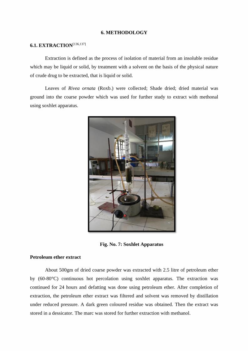
6. METHODOLOGY
6.1. EXTRACTION[136,137]
Extraction is defined as the process of isolation of material from an insoluble residue
which may be liquid or solid, by treatment with a solvent on the basis of the physical nature
of crude drug to be extracted, that is liquid or solid.
Leaves of Rivea ornata (Roxb.) were collected; Shade dried; dried material was
ground into the coarse powder which was used for further study to extract with methonal
using soxhlet apparatus.
Fig. No. 7: Soxhlet Apparatus
Petroleum ether extract
About 500gm of dried coarse powder was extracted with 2.5 litre of petroleum ether
by (60-80°C) continuous hot percolation using soxhlet apparatus. The extraction was
continued for 24 hours and defatting was done using petroleum ether. After completion of
extraction, the petroleum ether extract was filtered and solvent was removed by distillation
under reduced pressure. A dark green coloured residue was obtained. Then the extract was
stored in a dessicator. The marc was stored for further extraction with methanol.

Methonolic Extract
Marc obtained from the petroleum ether extract was dried and finally extracted with
2.5 litre of ethanol. The extraction was continued for 24 hours. After completion of
extraction, the extract was filtered and solvent was removed by distillation under reduced
pressure. A brown coloured residue was obtained. Then it was stored in dessicator.
The extracts were used for the identification of constituents by phytochemical
screening and for pharmacological studies.
6.2. PHYTOCHEMICAL SCREENING[136,137]
Preliminary phytochemical screening is done before resorting. Qualitative analysis is
thus a part of preliminary standardization. Phytochemical evaluation is used to determine the
nature of phyto constituents present in the plant by using suitable chemical tests. It is
essential to study the pharmacological activities of the plant.
Detection of Flavonoids
• Shinoda test
To the solution of extract, few piece of magnesium turnings and concentrated HCl
was added drop wise, pink to crimson red, occasionally green to blue color appears
after few minutes indicates the presence of flavonoids.
• Alkaline reagent test
To the test solution, few drops of sodium hydroxide solution was added, intense
yellow color is formed which turns to colorless on addition of few drops of dilute HCl
indicate presence of flavonoids.
Detection of Alkaloids
• Dragendorff’s reagent
The substance was dissolved in 5ml of distilled water, to this 5ml of 2M HCl was
added until an acid reactions occurs, then 1ml of Dragendorff’s reagent was added
and examined for an immediate formation of an orange red precipitate.
• Mayer’s reagent
The substance was mixed with little amount of Dilute Hydrochloric acid and Mayer’s
reagent and examined for the formation of white precipitate.
• Wagner’s reagent
The test solution was mixed with Wagner’s reagent and examined for the formation of
reddish brown precipitate.

• Hagne’s reagent
The test solution was mixed with few drops of Hydrochloric acid and filtered and then
Hagne’s reagent was added and examined for the formation of yellow precipitate.
Detection of Carbohydrates
• Molisch’s test
To the test solution, few drops of alcoholic alpha naphthol and few drops of Conc.
Sulphuric acid were added through the sides of test tube, purple to violet color ring
appears at junction.
• Fehling’s test
The test solution was mixed with Fehling’s I and II and heated and examined for the
appearance of red coloration for the presence of sugar.
Tannins
• Small quantities of test substances was treated with Ammonium Hydroxide then few
drops of Potassium ferricyanide solution was added and examined for the formation
of transited red colour.
Detection of Proteins
• Biuret test
The sample was treated with sodium hydroxide and 5-8 drops of 10% w/v Copper
sulphate solution, violet color was formed.
• Millon’s test
The sample was treated with Millon’s reagent, a white precipitate was formed.
Detection of fats and fixed oils
• Small quantities of extracts were pressed between two filter papers. An oily stain on
filter paper indicates the presence of fixed oils and fats.
Detection of Steroids
• Libermann Burchards Test
The powdered drug was treated with few drops of acetic anhydride, boiled and
cooled. Conc.sulphuric acid was added on the sides of the test tube, brown ring was
formed at the junction of two layers and upper layer turns green which shows
presence of steroids.
Detection of Terpenoids
• The extract was treated with caustic soda, a deep red color was formed, indicates
presence of tri terpenoids.

Detection of Glycosides
• Borntrager’s test
The extract was boiled with 1ml of sulphuric acid in a test tube for five minutes.
Filtered while hot, cooled and shaken with equal volume of chloroform. The lower
layer of solvent was separated and shaken with half of its volume of dilute ammonia.
A rose pink to red colour was produced in the ammonical layer.
Detection of Phenols
• Ferric chloride test
A small quantity of substance were dissolved with 2ml distilled water and a few drops
of 10% aqueous ferric chloride solution was added and observed for the appearance of
blue or green color.
Detection of Saponins
• A drop of sodium bicarbonate solution was added to the sample and the mixture was
shaken vigorously and left for 3 minutes. Development of any honey comb like froth
was examined.
6.3. INFRARED SPECTROSCOPY[137,162]
Fourier Transform –Infra Red Spectrophotometer
Fourier transform infrared (FTIR) spectroscopy is a measurement technique that
allows one to record infrared spectra. Infrared light is guided through an interferometer and
then through the sample (or vice versa). A moving mirror inside the apparatus alters the
distribution of infrared light that passes through the interferometer. The signal directly
recorded, called an "interferogram", represents light output as a function of mirror position.
• A data-processing technique called Fourier transform turns this raw data into the
desired result (the sample's spectrum): Light output as a function of infrared
wavelength (or equivalently, wavenumber). As described, the sample's spectrum is
always compared to a reference.

Tab. No. 5: Specifications of FTIR Spectrophotometer
Model Spectrum RX I
Make Perkin Elmer
Range 4000cm-1 to 400cm-1
Specimen Solids, Liquids
6.4. HPTLC ANALYSIS[136,137,162]
CAMAG HPTLC system equipped with Linomat 5 applicator, TLC scanner 3, repro
star 3 with 12 bit CCD camera for photo documentation, controlled by WinCATS-4 software
were used. All the solvents used for HPTLC analysis were obtained from MERCK. A total of
100 mg extract was dissolved in 5 mL of ethanol and used for HPTLC analysis as test
solution.
Test Solution : Methanolic Extract of Rivea ornata (MERO)
Stationary Phase : Merck, TLC Plates Silica gel 60 F-254
Mobile Phase : Toluene : Ethyl acetate : Acetone (2:4:4 v/v/v)
Tank : TTC 10×10
Saturation time : 20 Minutes
Procedure
The samples (5μl, 10μl, 15μl, 20μl) were spotted in the bands of width 8 mm with a
Camag microlitre syringe on pre-coated silica gel glass plate 60 F- 254. The sample loaded
plate was kept in TLC twin trough developing chamber (after saturated with Solvent vapor)
with respective mobile phase and the plate was developed up to 83 mm in the respective
mobile phase.
Linear ascending development was carried out in 20cm × 10cm twin trough glass
chamber saturated with the mobile phase and the chromatoplate development with the same
mobile phase to get good resolution of phytochemical contents. The optimized chamber
saturation time for mobile phase was 30 min at room temperature. The developed plate was
dried by hot air to evaporate solvents from the plate.

The plate was photo-documented at UV 254 nm and white light using photo
documentation chamber. Finally, the plate was fixed in scanner stage and scanning was done
at 254 nm. The plate was kept in photo-documentation chamber and captured the images
under white light, UV light at 254 nm. Densitometric scanning was performed on Camag
TLC scanner III and operated by Server Win software (Version 2.5.18053.1).
6.5. ANIMAL EXPERIMENTATION
Pharmacological evaluation of the methanolic extract of Rivea ornata (Leaves) was
carried out in the Department of Pharmacology, Periyar college of Pharmaceutical Sciences,
Tiruchirappalli, Tamilnadu, India. Animal facility of this institute is approved by CPCSEA.
The experimental protocols for the Pharmacological activities have been approved by the
Insititutional Animal Ethics Committee and conducted according to the guidelines of Indian
National Sciences Academy for the use and care of experimental animals. IAEC approved
this proposal with approval number PCP/IAEC/005/2019. The animals were maintained at a
well ventilated, temperature controlled 30°C ± 1°C animal room for 7days prior to the
experimental period and provided with food and water ad libitum. The animals were
acclimatized to laboratory conditions before the test. Each animal was used only once.

6.5.1. Validation of Acute toxicity[136]
Acute Toxic Class method: Guideline number 423
Principle
The test substance will be administered orally to a group of experimental animals at
one of the defined doses. The substance will test using a stepwise procedure, each step using
three animals of a single sex (normally female). Absence or presence of compound related
mortality of the animals dosed at one step will determine the next step
i.e
• No further testing is needed
• Dosing of three additional animals, with the same dose
• Dosing of three additional animals at the next higher or the next lower dose level
Healthy young rats were used. The test substance was administered in a single dose
(2000 mg/kg, b.w., p.o.) by gavage using a stomach tube.
Animals were observed individually after dosing at least once during the first 30
minutes, periodically during the first 24 hours, with special attention given during the first 4
hours and daily thereafter, for a total of 14 days.
Observed changes in skin, fur, eyes, mucous membranes, respiratory, circulatory,
autonomic, central nervous systems, somatomotar activity and behavior pattern. Attention
was directed to observations or tremors, convulsions, salivation diarrhea, lethargy, sleep and
mortality. In addition, behavioural changes body weight, histopathological studies and
biochemical parameters were also observed.

6.5.2. Sub acute toxicity[163]
Repeated dose oral toxicity method: Guideline number 407
The methanolic extract of Rivea ornata was orally administered daily in dose of
200 mg/kg to group of experimental animals for a period of 28 days. During the period of
administration the animals are observed closely, each day for signs of toxicity. Animals that
die or are euthanised during the test are necropsied and at the conclusion of the test surviving
animals are euthanised and necropsied. A 28 day study provides information on the effects of
repeated oral exposure and can indicate the need for further longer term studies. It can also
provide information on the selection of concentrations for longer term studies. The data
derived from using the Test Guideline (TG) should allow for the characterization of the test
substance toxicity, for an indication of the dose response relationship and the determination
of the No-Observed Adverse Effect Level (NOAEL).
Wistar albino rats of 6 animals from each sex selected with the weight variation of
±20% of the mean weight of each sex divided into the two groups. The experimental room
was maintained at the temperature of 22°C (±3°C) and the relative humidity of 50-60% with
12 hours artificial dark and light cycle. Animals were fed with conventional laboratory diet
with an unlimited supply of water.
Group I : Normal saline (5mL/kg, b.w., p.o.)
Group II : Methanolic Extract of Rivea ornata (MERO) (200mg/kg, bw.,
p.o.)
Experimental design
The animals from group I were administered with normal saline (5ml/kg, bw., p.o.)
and group II with test substance (MERO 200mg/kg, bw., p.o.) by gavage, daily, for 28
consecutive days. At the end of the experiment the animals were sacrificed to collect their
blood and organs (Heart, liver, kidney and pancreas) for biochemical, hematological and
histological analyzes.

Blood analysis
Biochemical analyzes included liver function markers (AST, ALT), total cholesterol,
and nephrotic marker urea. Hematologic parameters included red and white blood cells,
hemoglobin, platelets, Erythrocite sedimentation rate.
Histopatholigical examinations
The organs were fixed in 10% concentrated formalin, underwent several liquor baths,
before passing into toluene and being included in the liquid paraffin. The paraffin blocks are
mounted on a microtome to make the cuts. Soaked in aqueous dyes (Hematoxylin and Eosin)
and observed under microscopes.
Statistical analysis
The results are expressed are Mean ± SEM (n=6) two way ANOVA using a Graph
pad and PRISM software version 8.2.1 (441). *** P<0.001, ** P<0.01 and *P<0.05 were
considered as statistically significant.

6.5.3. Antihyperlipidemic activity[20,111,112]
Triton X 100 Induced Hyperlipidemia
Male wistar albino rats weighing 200-250g, used for the study were fed on pellet diet
and water ad libitum. Hyperlipidemia was induced by intraperitoneal administration of Triton
X 100 in 100mg/kg.
Principle
Triton X-100 acts as surfactant & suppresses the action of lipases to block the uptake
of lipoproteins from circulation by extra-hepatic tissue resulting in to increased blood lipid
concentration.
Treatment
Rats were divided into the following 4 groups of 6 each:
Group I : Normal diet water ad libitum
Group II : Normal diet and Triton X 100 (100mg/kg) (positive control)
Group III : Normal diet and Triton X 100 (100mg/kg) + Methanolic extract of
Rivea ornata (200mg/kg, p.o.)
Group IV : Normal diet & Triton X 100 (100mg/kg) +Atrovastatin (10mg/kg, p.o.)
Experimental Design
Hyperlipidemia was induced by single intraperitoneal injection of freshly prepared
solution of Triton X-100 (100 mg/ kg) in physiological solution after overnight fasting for 18
hrs. After 72 hours drugs were administered to the respective groups in predetermined dose
for 7 days.
Biochemical analysis
On the 8th day, blood was collected by cardiac puncture, under mild ether
anaesthesia. The collected samples were centrifuged for 10 minutes at 3000rpm. Then serum
samples were collected and used for various biochemical experiments. The serum was
assayed for total cholesterol, triglycerides, high density lipoprotein (HDL), low-density
lipoprotein (LDL) and very low-density lipoprotein (VLDL).

Histopathological studies
At the end of the treatment period, the animals were sacrificed for histopathology
studies .The livers of each animal from all four groups were isolated for histopathology
studies. The isolated livers were carefully kept in 10 % formalin solution in order to prevent
the damage.
Statistical analysis
The results are expressed are Mean ± SEM (n=6) two way ANOVA using a Graph
pad and PRISM software version 8.2.1 (441). *** P<0.001, ** P<0.01 and *P<0.05 were
considered as statistically significant.

6.5.4. Antidiabetic activity[101,102]
Principle
Alloxan is an acidic compound obtained by the oxidation of uric acid and isolated as
an efflorescent crystalline hydrate. It is a toxic glucose analogue, which selectively
destroys insulin-producing cells (beta cells) in the pancreas when administered to rodents and
many other animal species. This causes an insulin-dependent diabetes mellitus in these
animals, with characteristics similar to type I diabetes in humans.
Wistar Albino Rats weighing 150-200g were divided into 4 groups and kept under the
temperature of 25°C and the relative humidity of 50-60% with 12 hours dark and light cycle.
Animals were fasted for 12 hours before and after injection of alloxan.
Group I : Normal saline (5mL/kg, b.w., p.o.)
Group II : Alloxan (150mg/kg, b.w., p.o.)
Group III : Alloxan (150mg/kg, b.w., p.o.) + Glibenclamide (10mg/kg,
b.w., p.o.)
Group IV : Alloxan (150mg/kg, b.w., p.o.) + Methanolic Extract of Rivea
ornata (200mg/kg, bw., p.o.)
Experimental Design
Alloxan is used to induce diabetes. The dose of alloxan for diabetes control group is
150mg/kg of body weight. It was prepared by dissolving in normal saline and administered
intraperitonially. Rats were fasted 12 hours before and after injection of alloxan, unfed
animals are more susceptible for alloxan induction diabetes. One week after treatment,
animals with moderate diabetes (blood glucose level above of 150 mg /dl) were used for the
experiment. Test and standard drugs were administered to the respective group in
predetermined dose for 14 days.

Evaluation
Blood Glucose level
After 0 minutes, 1 hour, 2 hours, 4 hours, 7th day and 14th day from the administration
of standard and test drug, the blood glucose level was estimated by glucometer.
Histopathology of Pancreas
After the Blood glucose estimation was over the animals were sacrificed for
histopathology studies. The pancreas of each animal from all four groups were isolated for
histopathology studies. The isolated pancreas were carefully kept in 10% formalin solution in
order to prevent the damage.
Statistical analysis
The results are expressed are Mean ± SEM (n=6) two way ANOVA using a Graph pad and
PRISM software version 8.2.1 (441). *** P<0.001, ** P<0.01 and *P<0.05 were considered
as statistically significant.

6.5.5. Analgesic Activity[114,119,123]
a) Tail Immersion Test in Rats
Wistar albino rats of either sex weighing 18-22g were divided into three groups of six
animals each. The animals were fasted overnight for 18h. The dosage of the drugs
administered to the different groups was as follows
Group I : Control - Normal Saline (5ml/kg, b.w., p.o.)
Group II : Standard (Diclofenac sodium 10mg/kg in 10mL of normal
saline, p.o.)
Group III : Test (Methanolic extract of Rivea ornata 200mg/kg,
p.o.)
Experimental Design
The procedure is based on the observation that NSAID drugs are selectively capable
of prolonging the reaction time of the typical tail-withdrawal reflex in rats induced by
immersing the end of the tail in warm water of 55°C.
The lower 5 cm portion of the tail is marked. This part of the tail is immersed in to the
water bath of exactly 55 °C. Within a few seconds the rat reacts by withdrawing the tail.
After each determination the tail is carefully dried. The reaction time was recorded before
and at 30, 45, 60 and 90 minutes after the administration of the treatments. The cut off time is
15 seconds.
Evaluation
Tail withdrawal reflex
Statistical analysis
The results are expressed are Mean ± SEM (n=10) two way ANOVA using a Graph pad and
PRISM software version 8.2.1 (441). *** P<0.001, ** P<0.01 and *P<0.05 were considered
as statistically significant.
b) Hot plate method

Wistar albino mice of either sex weighing 20-25g were divided into three groups of
six animals each. The animals were fasted overnight for 18h. The dosage of the drugs
administered to the different groups was as follows
Group I : Control - Normal Saline (5ml/kg, b.w., p.o.)
Group II : Standard (Diclofenac sodium 10mg/kg in 10mL of normal saline, p.o.)
Group III : Test (Methanolic extract of Rivea ornata 200mg/kg, p.o.)
The mice were placed on a hot plate maintained at 55°C within the restrainer. The reaction
time (in seconds) or latency period was determined as the time taken for the mice to react to
the thermal pain by licking their paws or jumping. The reaction time was recorded before and
at 30, 45, 60 and 90 minutes after the administration of the treatments. The maximum
reaction time was fixed at 45 sec to prevent any injury to the tissues of the paws. If the
reading exceeds 45 sec, it would be considered as maximum analgesia.
Statistical analysis
The results were expressed as Mean ± SEM (n=6) two way ANOVA using a Graph
pad and PRISM software version 8.2.1. (441). *** P<0.001, ** P<0.01 and *P<0.05 were
considered as statistically significant.
6.5.6. Antipyretic activity[95,103,143]
Yeast-induced hyperpyrexia in rats

Wistar albino rats of either sex weighing 150-200g were divided into three groups of
six animals each. They were housed individually in standard laboratory environment for 7
days of period, fed with common laboratory diet and water ad libitum. The dosage of the
drugs administered to the different groups as follows
Group I : 12.5% w/v yeast suspended in a 0.5% w/v normal saline
(10ml/kg, b.w., sc.) + Normal saline at dose of
(5ml/kg, b.w., sc.)
Group II : 12.5% w/v yeast suspended in a 0.5% w/v normal saline
(10ml/kg, b.w., sc.) + Methanolic extract of Rivea ornata
(MERO) (200mg/kg, b.w., p.o.)
Group III : 12.5% w/v yeast suspended in a 0.5% w/v normal saline
(10ml/kg, b.w., sc.) + Paracetamol (150mg/kg, b.w., p.o.)
Experimental Design
The body temperature of each albino Wistar albino rats was recorded by
measuring rectal temperature at predetermined intervals. The rats are trained to remain quiet
in a restraint cage. A thermister probe is inserted 3 to 4 cm into the rectum and fastened to the
tail by adhesive tape. Temperature is recorded on a thermometer. After measuring the basal
rectal temperature, animals are given subcutaneous injections of 10 ml/kg of 12.5% w/v yeast
suspended in a 0.5% w/v normal saline (10ml/kg, b.w., sc.). At the 19th hr after yeast
injection the rectal temperature of the rats were recorded then Normal saline at dose of
5ml/kg was injected to the group I. Methanolic extract of Rivea ornata was administered with
dose 200mg/kg to group II animals. The group III rats received the standard drug paracetamol
at the dose of 150 mg/kg at the 19th hr after yeast injection. Subcutaneous injection of yeast
suspension markedly increased the rectal temperature 19th hr after its administration. Rectal
temperature of all the rats was recorded again on the 20th, 21st and 22nd hr after yeast
injection.
Statistical analysis

The results are expressed are Mean ± SEM (n=10) two way ANOVA using a Graph pad and
PRISM software version 8.2.1 (441). *** P<0.001, ** P<0.01 and *P<0.05 were considered
as statistically significant.
6.5.7. Wound Healing Activity[121,138,139]
Excision Wound Model
Wistar albino rats of either sex weighing 200-250g were divided into three groups of
six animals each. They were housed individually in standard laboratory environment for 7
days of period, fed with common laboratory diet and water ad libitum. The dosage of the
drugs administered to the different groups as follows
Group I : Control – Simple ointment base
Group II : 10% methanolic extract ointment of Rivea ornata (MERO)
Group III : Povidone Iodine cream (10%)
Experimental Design
Three groups of animals containing six rats in each group were anesthetized by bottle
method with anesthetic ether. The rats were depilated on the back and a predetermined area
of 300 mm2 full thickness skins was excised in the dorsal interscapular region. Rats were left
undressed to the open environment. The formulation ointment and standard drug were
applied daily until the complete healing. In this model, wound contraction and
epithelialization period was monitored. Wound contraction was measured as percent
contraction in each 3 days after wound formation.

Evaluation
Measurement of Wound Contraction
An excision wound margin was traced after wound creation by using transparent
paper and area measured by graph paper. Wound contraction was measured in each 5 days
interval, until complete wound healing and expressed in percentage of healed wound area.
The evaluated surface area was then employed to calculate the percentage of wound
contraction, taking initial size of wound, 300 mm2, as 100%, by using the following formula
as:
% of wound contraction = (Initial wound size – Specific day wound size) × 100
Initial wound size
Statistical analysis
The results are expressed are Mean ± SEM (n=10) two way ANOVA using a Graph pad and
PRISM software version 8.2.1 (441). *** P<0.001, ** P<0.01 and *P<0.05 were considered
as statistically significant.

RESULTS & DISCUSSION

7. RESULTS AND DISCUSSION
7.1 Results
Tab. No. 6: Preliminary Phytochemical Analysis
S.No Plant Constituents Test / reagent Methanolic extract of Rivea
ornata
1 Flavonoids
Shinoda test
--
Alkaline reagent test
2 Alkaloids
Dragendorff’s reagent
++
Mayer’s reagent
Wagner’s reagent
Hagne’s reagent
3 Carbohydrates
Molisch’s test
++
Fehling’s test
4 Tannins Ammonium Hydroxide ++
5 Protein
Biuret test
--
Millon’s test
6 Fat and Fixed oils Filter paper test ++
7 Steroids Libermann Burchards Test ++
8 Terpenoids Caustic soda --
9 Glycosides Borntrager’s test ++
10 Phenols Ferric chloride test ++
11 Saponins Form test ++

Analytical Determination
1. FTIR Analysis
Fig. No. 8: FTIR Spectrum of Methanolic Extract of Rivea ornata
PCP-Rivea Ornata-
Name Description
4000 4003500 3000 2500 2000 1500 1000 500
100
0
10
20
30
40
50
60
70
80
90
cm-1
%T
3370.39cm-1
1058.08cm-1
1629.01cm-1
1400.74cm-1
1384.98cm-1
1273.26cm-12931.59cm-1
1155.24cm-1
1449.63cm-1
1336.84cm-1
613.51cm-11518.05cm-1
779.51cm-1
818.04cm-1
863.27cm-1
932.04cm-1
899.62cm-1

Tab. No. 7: FTIR Interpretation of the Methanolic Extract of Rivea ornata
S. No Wavenumber (cm-1) Functional Group
1 3370.39 O-H stretching
2 2931.59 C-H stretching
3 1629.01 C=C stretching
4 1518.05 N-O stretching
5 1449.63 C-H bending
6 1400.63 O-H bending
7 1384.98 Phenolic O-H bending
8 1273.26 C-N stretching
9 932.04 C=C bending
10 899.62 C-H bending

HPTLC Analysis
Fig. No. 9: HPTLC Chromatogram of MERO
Fig No. 10: HPTLC chromatogram of MERO (3D)
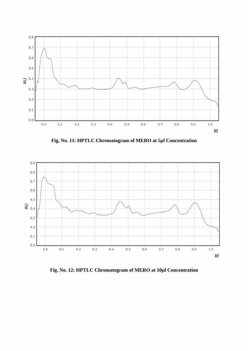
Fig. No. 11: HPTLC Chromatogram of MERO at 5µl Concentration
Fig. No. 12: HPTLC Chromatogram of MERO at 10µl Concentration

Fig. No. 13: HPTLC Chromatogram of MERO at 15µl Concentration
Fig. No. 14: HPTLC Chromatogram of MERO at 20µl Concentration

Fig. No. 15: HPTLC peak at System suitability test
Tab. No. 8: Rf values from HPTLC Chromatogram of MERO
S. No. Track No. Rf Value
1. Tr. 1 0.050
2. Tr. 2 0.450
3. Tr. 3 0.490
4. Tr. 4 0.780
5. Tr. 5 0.190
6. Tr. 6 0.910

PHARMACOLOGICAL EVALUATION
Toxicity Studies - Acute Oral Toxicity (Validation)
Table No. 9: Behavioral Changes in Acute Oral Toxicity in Albino rats
S.No. Symptoms
Control
(Normal Saline
5ml/kg, p.o.)
Methanolic Extract of
Rivea ornata
(2000mg/kg, p.o.,)
1 Death -- --
Autonomic Nervous System
2 Head movements -- --
3 Scratching -- --
4 Altered reactivity to touch -- ++
5 Loss of righting reflex -- --
6 Loss of corneal reflex -- --
7 Defecation/Diarrhea -- --
8 Salivation -- --
9 Lacrimation -- --
10 Myosis/ Mydriasis -- --
11 Loss of traction -- --
Central Nervous system
12 Convulsions -- --
13 Tremor -- --
14 Straub tail -- --
15 Sedation -- --
16 Excitation -- --
17 Jumping -- --
18 Abnormal gait -- --
19 Motor in Co ordination -- --
20 Akinesia -- --
21 Catelepsy -- --
22 Loss of Balance -- --
23 Fore-paw treading -- --
24 Writhing -- --
25 Stereotypy -- --
26 Altered fear -- --
27 Altered respiration -- --
28 Aggression -- --
29 Analgesia -- ++
30 Body temperature -- --

Tab. No. 10: Effect of Test compound on Body Weight in Acute oral toxicity in Albino
rats
S. No. Group
Body weight
Initial At the end of the study
1 Control
(Normal Saline 5ml/kg) 212.045 ± 2.209 212.738 ± 1.775
2 Test Drug
(MERO 2000mg/kg) 213.100 ± 2.154 205.686 ± 2.170
n = 6 Values are expressed as ± S.E.M.
Tab. No. 11: Effect of Test compound on Biochemical parameters in Acute oral toxicity
in Albino rats
S. No. Biochemical Parameters
Control
(Normal Saline
5ml/kg)
Test Drug
(MERO
2000mg/kg)
1 Glucose (mg/dl) 149.795 ± 0.547 139.213 ± 0.687
2 Blood Urea (mg/dl) 15.401 ± 0.173 15.06 ± 0.089
3 Total cholesterol (mg/dl) 81.867 ± 0.425 76.108 ± 0.723
4 SGOT (U/I) 113.035 ± 0.424 112.672 ± 0.226
5 SGPT (U/I) 75.092 ± 0.338 74.928 ± 0.360
6 Albumin (gm/dl) 3.397 ± 0.0723 3.555 ± 0.090
n = 6 Values are expressed as ± S.E.M.

Fig. No. 16: Effect of Test compound on Body Weight in Acute oral toxicity in Albino
rats
Fig. No. 17: Effect of Test compound on Biochemical parameters in Acute oral toxicity
in Albino rats
200
202
204
206
208
210
212
214
Initial At the end of the study
Body weight
1 Control
(Normal Saline 5ml/kg)
2 Test Drug
(MERO 2000mg/kg)
0
20
40
60
80
100
120
140
160
Glu
cose
(m
g/d
l)
Blo
od
Ure
a (m
g/d
l)
To
tal
cho
lest
erol
(mg
/dl)
SG
OT
(U
/I)
SG
PT
(U
/I)
Alb
um
in (
gm
/dl)
1 2 3 4 5 6
Control
(Normal Saline 5ml/kg)
Test Drug
(MERO 2000mg/kg)

Histopathological Studies of Acute Oral Toxicity
Control – Normal Saline (5ml/kg, p.o)
Fig. No. 18: T.S of Heart Fig. No. 19: T.S of Kidney
Fig. No. 20: T.S of Liver Fig. No. 21: T.S of Pancreas
Test – Methanolic Extract of Rivea ornata (2000 mg/kg, p.o)
Fig. No. 22: T.S of Heart Fig. No. 23: T.S of Kidney
Fig. No. 24: T.S of Liver Fig. No. 25: T.S of Pancreas
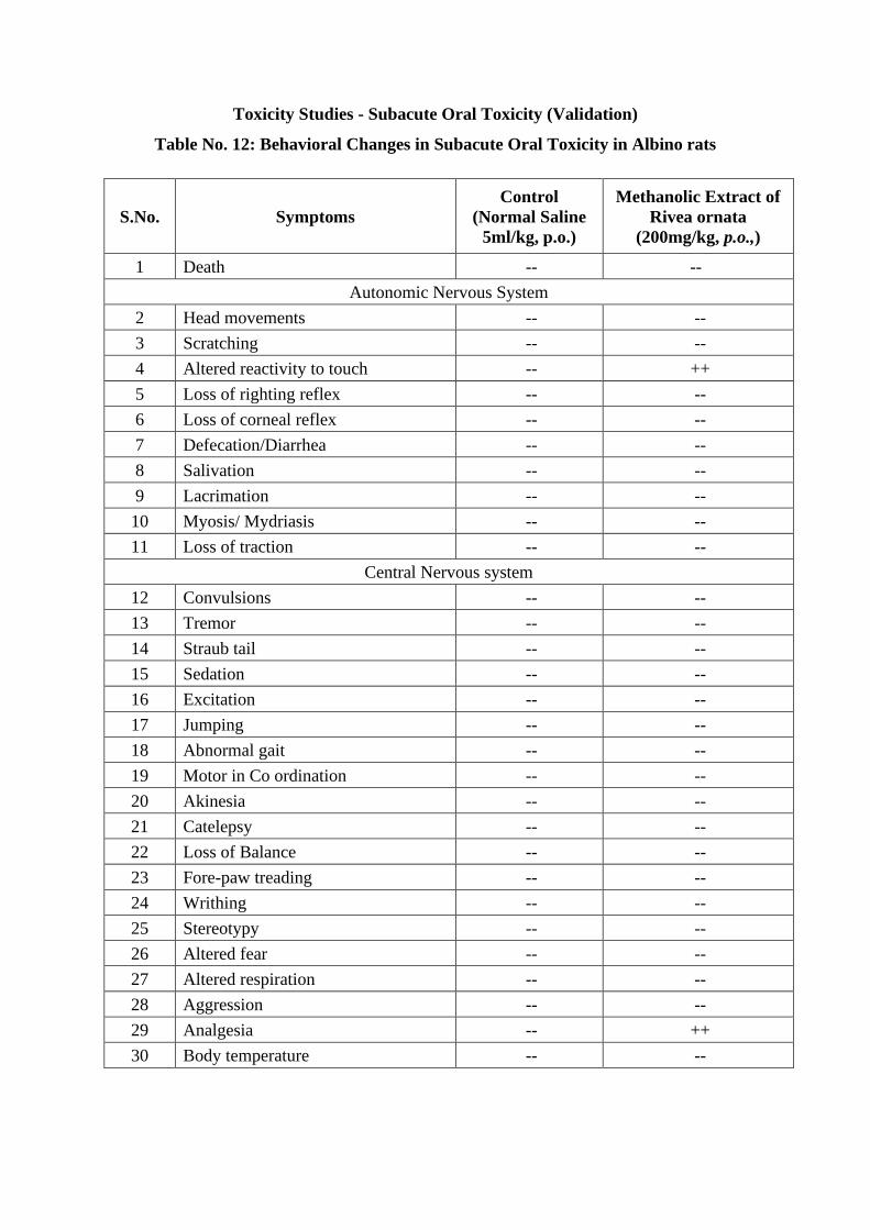
Toxicity Studies - Subacute Oral Toxicity (Validation)
Table No. 12: Behavioral Changes in Subacute Oral Toxicity in Albino rats
S.No. Symptoms
Control
(Normal Saline
5ml/kg, p.o.)
Methanolic Extract of
Rivea ornata
(200mg/kg, p.o.,)
1 Death -- --
Autonomic Nervous System
2 Head movements -- --
3 Scratching -- --
4 Altered reactivity to touch -- ++
5 Loss of righting reflex -- --
6 Loss of corneal reflex -- --
7 Defecation/Diarrhea -- --
8 Salivation -- --
9 Lacrimation -- --
10 Myosis/ Mydriasis -- --
11 Loss of traction -- --
Central Nervous system
12 Convulsions -- --
13 Tremor -- --
14 Straub tail -- --
15 Sedation -- --
16 Excitation -- --
17 Jumping -- --
18 Abnormal gait -- --
19 Motor in Co ordination -- --
20 Akinesia -- --
21 Catelepsy -- --
22 Loss of Balance -- --
23 Fore-paw treading -- --
24 Writhing -- --
25 Stereotypy -- --
26 Altered fear -- --
27 Altered respiration -- --
28 Aggression -- --
29 Analgesia -- ++
30 Body temperature -- --

Tab. No. 13: Effect of Test compound on Body Weight in Subacute oral toxicity in
Albino rats
S. No. Group
Body weight
Initial At the end of the study
1 Control
(Normal Saline 5ml/kg) 201.29 ± 0.94 203.57 ± 0.69
2 Test Drug
(MERO 200mg/kg) 201.22 ± 0.55 190.08 ± 1.17
n = 6 Values are expressed as ± S.E.M.
Tab. No. 14: Effect of Test compound on Biochemical parameters in Subacute oral
toxicity in Albino rats
S. No. Biochemical Parameters
Control
(Normal Saline
5ml/kg)
Test Drug
(MERO 200
mg/kg)
1 Glucose (mg/dl) 148.48 ± 0.41 138.27 ± 0.33
2 Blood Urea (mg/dl) 16.19 ± 0.13 13.72 0.11
3 Total cholesterol (mg/dl) 82.28 ± 0.11 73.77 ± 0.09
4 SGOT (U/I) 113.61 ± 1.28 112.15 ± 0.25
5 SGPT (U/I) 75.28 ± 0.07 74.41 ± 0.32
6 Albumin (gm/dl) 3.26 ± 0.03 3.52 ± 0.02
n=6, Values are expressed as ± S.E.M

Tab. No. 15: Effect of Test compound on Haematological parameters in Subacute oral
toxicity in Albino rats
S. No. Group Hb
(gm%)
RBC
(106/cu.mm)
Total WBC
(103/cu.mm)
ESR
(mm/1st hr) Platelets
1
Control
(Normal
Saline
5ml/kg)
14.43 ± 0.13 7.32 ± 0.03 7.22 ± 0.02 3.18 ± 0.02 400 ± 9.26
2
Test Drug
(MERO
200mg/kg)
14.13 ± 0.06 6.88 ± 0.03 7.25 ± 0.02 3.19 ± 0.02 393 ± 12.76
n = 6 Values are expressed as ± S.E.M.
Fig. No. 26: Effect of Test compound on Body Weight in Subacute oral toxicity in
Albino rats
180
185
190
195
200
205
Initial At the end of the study
Body weight
Control
(Normal Saline 5ml/kg)
Test Drug
(MERO 200mg/kg)

Fig. No. 27: Effect of Test compound on Biochemical parameters in Subacute oral
toxicity in Albino rats
Fig. No. 28: Effect of Test compound on Biochemical parameters in Subacute oral
toxicity in Albino rats
0.00
20.00
40.00
60.00
80.00
100.00
120.00
140.00
160.00
Glucose (mg/dl) Blood Urea
(mg/dl)
Total
Cholesterol
(mg/dl)
SGOT (U/I) SGPT (U/I) Albumin
(gm/dl)
Control
(Normal Saline 5ml/kg)
Test Drug
(MERO 200mg/kg)
0.00
50.00
100.00
150.00
200.00
250.00
300.00
350.00
400.00
450.00
Control
(Normal Saline 5ml/kg)
Test Drug
(MERO 200mg/kg)

Histopathological Studies of Subacute Oral Toxicity
Control – Normal Saline (5ml/kg, p.o)
Fig. No. 29: T.S of Heart Fig. No. 30: T.S of Kidney
Fig. No. 31: T.S of Liver Fig. No. 32: T.S of Pancreas
Test – Methanolic Extract of Rivea ornata (200 mg/kg, p.o)
Fig. No. 33: T.S of Heart Fig. No. 34: T.S of Kidney
Fig. No. 35: T.S of Liver Fig. No. 36: T.S of Pancreas

Antihyperlipidemic activity – Triton X 100 induced hyperlipidemia
Tab. No. 16: Body weight changes in Antihyperlipidemic activity of MERO
S.No. Group Body Weight (g)
1st Day 4th Day 10th Day
1 Normal
Control
209.56
±
0.39
210.55 ± 0.39 211.68 ± 0.26
2 Toxic
Control
210.72
±
0.43***
220.2
±
0.30***
226.22
±
0.38***
3 Standard
209.00
±
0.51***
221.59
±
0.61***
212.66
±
0.56***
4 Test
210.98
±
0.32***
221.40
±
0.57***
216.78
±
0.52***
n = 6 Values are expressed as ± S.E.M. Values are Mean ± SEM (n=6) two way ANOVA.
Where, *** P<0.001, ** P<0.01 and * P<0.05
Tab. No. 17: Antihyperlipidemic activity of MERO
S. No. Groups
Serum Lipid Parameter (mg/dl)
Total
Cholesterol
Total
Triglycerides HDL - C LDL - C VLDL – C
1
Normal Control
(Normal Saline
5ml/kg)
75.54
± 0.91
83.40
±
0.57
35.89
±
0.53
24.22
±
0.24
15.69
±
0.40
2
Toxic Control
(Triton X 100
100mg/kg)
121.52
±
0.62
148.73
±
0.57
28.41
±
0.55
88.94
±
0.47
33.01
±
0.30
3
Standard
(Atorvastatin
10mg/kg)
86.80
±
0.62***
73.64
±
0.60***
37.09
±
0.51***
44.13
±
0.70***
22.11
±
0.44***
4
Test
(MERO
200mg/kg)
93.20
±
0.58***
85.28
±
0.57***
34.55
±
0.35***
51.10
±
0.64***
26.70
±
0.26***
n = 6 Values are expressed as ± S.E.M. Values are Mean ± SEM (n=6) two way ANOVA.
Where, *** P<0.001, ** P<0.01 and * P<0.05

Fig. No. 37: Body weight changes in Antihyperlipidemic activity of MERO
Fig. No. 38: Serum Lipid parameters in Antihyperlipidemic activity of MERO
200.00
205.00
210.00
215.00
220.00
225.00
230.00
Normal Control Toxic Control Standard Test
Body Weight
(g)
1st Day
4th Day
10th Day
0.00
20.00
40.00
60.00
80.00
100.00
120.00
140.00
160.00
Total
Cholesterol
Total
Triglycerides
HDL - C LDL - C VLDL - C
Serum Lipid Parameter (mg/dl)
Normal Control
(Normal Saline 5ml/kg)
Toxic Control
(Triton X 100 100mg/kg)
Standard
(Atorvastatin 10mg/kg)
Test
(MERO 200mg/kg)

Histopathological study in Antihyperlipidemic Activity of Rivea ornata
Histopathology of Liver
Fig. No. 39: Normal Control Fig. No. 40: Toxic Control
Fig. No. 41: Standard Fig. No. 42: Test
Normal Control - Normal Saline (5ml/kg)
Toxic Control - Triton X 100 (100mg/kg)
Standard - Atorvastatin (10mg/kg)
Test - Methanolic Extract of Rivea ornata (200mg/kg)

Tab. No. 18: Histopathological study in Antihyperlipidemic Activity of Rivea ornata
T.S of Liver
S. No Group Description
1 Normal Control Normal architecture
2 Toxic Control Presence of the fatty changes in the liver cells and the
presence of necrosis and fatty liver was found
3 Standard Mild necrosis was found and fatty deposition got reduced
4 Test Focal necrosis and slight hepatic toxicity

Antidiabetic activity – Alloxan Induced Diabetes
Tab. No. 19: Antidiabetic activity of Methanolic Extract of Rivea ornata
S.No. Drug/Dose
Blood Glucose Level
0 Hour 1 Hour 2 Hours 3 Hours 4 Hours 7th day 14th
day
1
Normal
Control
Normal saline
(5ml/kg)
108.33
± 1.58
105.67
±
2.29
106.00
±
1.29
107.50
±
2.14
108.33
±
2.20
108.33
±
1.89
107.17
±
1.54
2
Diabetic
Control
Alloxan
(150mg/kg)
191.17
±
1.54
193.50
±
1.91
192.00
±
2.17
192.17
±
1.56
192.33
±
1.41
191.83
±
1.54
191.83
±
1.92
3
Standard
Glibenclamide
(10mg/kg)
192.5
±
1.26***
147.17
±
1.01***
138.17
±
1.08***
125.5
±
0.85***
119.00
±
0.73***
120.33
±
0.88***
117.33
±
0.49***
4
Test
MERO
(200mg/kg)
192.50
±
2.04***
150.17
±
0.70***
149.17
±
0.95***
132.00
±
0.73***
164.50
±
1.28***
150.33
±
0.99***
139.50
±
0.67***
n = 6 Values are expressed as ± S.E.M. Values are Mean ± SEM (n=6) two way ANOVA.
Where, *** P<0.001, ** P<0.01 and * P<0.05
Fig. No. 43: Antidiabetic activity of Methanolic Extract of Rivea ornata
0.00
50.00
100.00
150.00
200.00
250.00
0 Hour 1 Hour 2 Hours 3 Hours 4 Hours 7th day 14th day
Blood Glucose Level
Normal Control
Normal saline
(5ml/kg)
Diabetic Control
Alloxan
(150mg/kg)
Standard
Glibenclamide
(10mg/kg)
Test
MERO
(200mg/kg)

Histopathological Study in Antidiabetic activity of MERO
Histopathology of Pancreas
Fig. No. 44: Normal Control Fig. No. 45: Toxic Control
Fig. No. 46: Standard Fig. No. 47: Test drug
Normal Control - Normal Saline (5ml/kg)
Toxic Control - Alloxan (150mg/kg)
Standard - Alloxan (150mg/kg) + Glibenclamide (10mg/kg)
Test - Alloxan (150mg/kg) + Methanolic Extract of Rivea ornata
(200mg/kg)
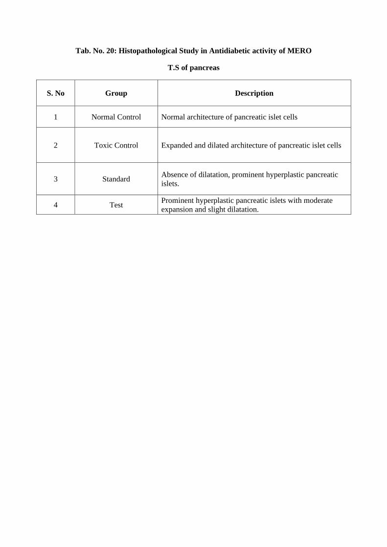
Tab. No. 20: Histopathological Study in Antidiabetic activity of MERO
T.S of pancreas
S. No Group Description
1 Normal Control Normal architecture of pancreatic islet cells
2 Toxic Control Expanded and dilated architecture of pancreatic islet cells
3 Standard Absence of dilatation, prominent hyperplastic pancreatic
islets.
4 Test Prominent hyperplastic pancreatic islets with moderate
expansion and slight dilatation.

Analgesic activity
Tab. No. 21: Analgesic activity of MERO - Tail Immersion Method
S.No. Drug/Dose
Reaction time in seconds
0
minute
30
minutes
45
minutes
60
minutes
90
minutes
1
Control
Normal saline
(5ml/kg)
1.72
±
0.06
1.81
±
0.04
1.8
±
0.02
1.85
±
0.04
1.81
±
0.02
2
Standard
Diclofenac
(10mg/kg)
1.74
±
0.02***
3.1
±
0.02***
5.7
±
0.02***
7.6
±
0.01***
9.49
±
0.02***
3
Test
MERO
(200mg/kg)
1.81
±
0.04***
2.93
±
0.02***
4.61
±
0.02***
6.95
±
0.02***
8.37
±
0.02***
n = 6 Values are expressed as ± S.E.M. Values are Mean ± SEM (n=6) two way ANOVA.
Where, *** P<0.001, ** P<0.01 and * P<0.05
Tab. No. 22: Analgesic activity of MERO – Hot plate Method
S.No. Drug/Dose
Reaction time in seconds
0 minute 30 minutes 45 minutes 60 minutes 90 minutes
1
Control
Normal
saline
(5ml/kg)
4.18 ± 0.06 4.1 ± 0.05 4.16 ± 0.04 4.15 ± 0.02 4.12 ± 0.04
2
Standard
Diclofenac
(10mg/kg)
4.22
±
0.06***
10.19
±
0.04***
11.55
±
0.02***
12.15
±
0.05***
12.21
±
0.03***
3
Test
MERO
(200mg/kg)
4.04
±
0.04***
8.77
±
0.02***
10.01
±
0.02***
10.14
±
0.02***
10.27
±
0.02***
n = 6 Values are expressed as ± S.E.M. Values are Mean ± SEM (n=6) two way ANOVA.
Where, *** P<0.001, ** P<0.01 and * P<0.05

Fig. No. 48: Analgesic activity of MERO - Tail immersion method
Fig. No. 49: Analgesic activity of MERO – Hot plate Method
0.00
1.00
2.00
3.00
4.00
5.00
6.00
7.00
8.00
9.00
10.00
0
minute
30 minutes 45 minutes 60 minutes 90 minutes
Reaction Time in Seconds
1 Control
Normal saline
(5ml/kg)
2 Standard
Diclofenac
(10mg/kg)
3 Test
MERO
(200mg/kg)
0.00
2.00
4.00
6.00
8.00
10.00
12.00
14.00
0 minute 30 minutes 45 minutes 60 minutes 90 minutes
Reaction Time in Seconds
1 Control
Normal saline
(5ml/kg)
2 Standard
Diclofenac
(10mg/kg)
3 Test
MERO
(200mg/kg)

Antipyretic activity
Tab. No. 23: Antipyretic activity of MERO – Brewer’s Yeast Induced Hyperpyrexia
Group
Basal
Temperature
(before yeast
administration)
(F)
Pre-Drug
control
(before drug
administration)
(F)
Rectal temperature after drug administration (F)
1
Hour
2
Hours
3
Hours
4
Hours
5
Hours
Control
Normal
Saline
(5ml/kg)
97.54
±
0.19
102.44
±
0.05
102.42
±
0.06
102.42
±
0.10
102.39
±
0.09
102.37
±
0.08
102.34
±
0.10
Standard
Paracetamol
(150mg/kg)
97.49
±
0.14***
102.21
±
0.08***
99.66
±
0.04***
99.15
±
0.03***
98.57
±
0.03***
97.82
±
0.03***
97.48
±
0.03***
Test
MERO
(200mg/kg)
97.56
±
0.13***
102.06
±
0.19***
99.81
±
0.03***
99.27
±
0.05***
98.8
±
0.04***
98.19
±
0.03***
97.77
±
0.03***
n = 6 Values are expressed as ± S.E.M. Values are Mean ± SEM (n=6) two way ANOVA.
Where, *** P<0.001, ** P<0.01 and * P<0.05
Fig. No. 50: Antipyretic activity of MERO – Brewer’s Yeast Induced Hyperpyrexia
94.00
95.00
96.00
97.00
98.00
99.00
100.00
101.00
102.00
103.00
1 Hour 2 Hours 3 Hours 4 Hours 5 Hours
Basal
Temperature
(F)
Pre-Drug
control
(F)
Rectal temperature after drug administration (F)
Control
Normal Saline
(5ml/kg)
Standard
Paracetamol
(150mg/kg)
Test
MERO
(200mg/kg)

Wound Healing Activity – Excision Wound Model
Tab. No. 24: Wound Healing activity of Methanolic Extract of Rivea ornata
S. No Wound Area (mm2)
Day 0 Day 3 Day 7 Day 12
Control
(Normal Saline)
310.9
±
2.68
242.26
±
1.70
123.34
±
1.65
32.5
±
0.62
Standard
(Povidone iodine
Cream)
311.98
±
4.18***
222.47
±
2.53***
99.18
±
3.09***
15.25
±
0.08***
Test
(MERO Cream)
317.2
±
3.55***
200.16
±
2.39***
72.93
±
2.14***
0.02
±
0.01***
n = 6 Values are expressed as ± S.E.M. Values are Mean ± SEM (n=6) two way ANOVA.
Where, *** P<0.001, ** P<0.01 and * P<0.05
Tab. No. 25: Period of Epithelialization in Wound Healing activity of MERO
S. No Period of Epithelialization
Control
(Normal Saline) 14.5 ± 0.43
Standard
(Povidone iodine) 13.5 ± 0.22
Test (MERO) 11.83 ± 0.31

Tab. No. 26: Wound Contraction percentage in Wound Healing activity of MERO
S.
No. Group
Wound Contraction (%)
0 Day 3rd Day 7th Day 12th Day
1 Control
(Normal Saline) 0 22.05 ± 0.91 49.09 ± 0.62 73.59 ± 0.85
2
Standard
(Povidone Iodine
Cream)
0 28.59 ± 1.64 55.45 ± 1.03 84.47 ± 1.17
3 Test
(MERO Cream) 0 36.83 ± 1.35 63.51 ± 1.34 99.98 ± 0.01
n = 6 Values are expressed as ± S.E.M.
Fig. No. 51: Wound Healing activity of Methanolic Extract of Rivea ornata
0
50
100
150
200
250
300
350
Day 0 Day 3 Day 7 Day 12
Wound Area (millimeter square)
Control
(Normal Saline)
Standard
(Povidone iodine)
Test (MERO)

Fig. No. 52: Period of Epithelialization in Wound Healing activity of MERO
0
2
4
6
8
10
12
14
16
Control
(Normal
Saline)
Standard
(Povidone
iodine)
Test (MERO)
Period of Epithelialization
0
20
40
60
80
100
120
0 Day 3rd Day 7th Day 12th Day
Wound Contraction (%)
1 Control
(Normal Saline)
2 Standard
(Povidone Iodine Cream)
3 Test
(MERO Cream)

Fig. No. 53: Wound Contraction Percentage in Wound Healing activity of MERO
Wound Healing activity of Methanolic Extract of Rivea ornata
O Day
Fig. No. 54: Control Fig. No. 55: Standard Fig. No. 56: Test
3rd Day
Fig. No. 57: Control Fig. No. 58: Standard Fig. No. 59: Test
7th Day
Fig. No. 60: Control Fig. No. 61: Standard Fig. No. 62: Test
12th Day

Fig. No. 63: Control Fig. No. 64: Standard Fig. No. 65: Test
7.2. Discussion
7.2.1. Extraction of Plant material
The leaves of Rivea ornata Roxb. was collected, dried and extracted using methanol
and prior to the ethanolinic extraction the plant was coarsely milled and defatted using
petroleum ether. The yield of the methanolic extract was 60%.
7.2.2. Preliminary Phytochemical Analysis
As a part of the preclinical study, the methanolic extract of leaves of Rivea ornata
Roxb. was subjected to qualitative chemical test and confirmed the presence of
carbohydrates, alkaloids, steroids, glycosides, tannins, saponins and phenolic compounds
shown in Table No. 6.
7.2.3. IR Spectral Data of the Ethanolic extract of Rivea ornata
The methanolic extract of leaves of Rivea ornata Roxb. was subjected to the Infra
Red Spectroscopy for their possible functional group (Table No. 7). The spectrum was
obtained and photo documentation was done (Fig. No. 8). The methanolic extract of leaves of
Rivea ornata roxb. showed the possible stretching and bending shows the presence of the
functional group.
7.2.4. HPTLC
The HPTLC fingerprinting of Methanolic extract of leaves of Rivea ornata Roxb. was
studied. The HPTLC fingerprinting was done. The 3D display of the chromatogram was
obtained and photo documentation was done (Fig. No. 10) and four different chromatograms
were obtained and photo documentation was done (Fig. Nos. 11, 12, 13 and 14). The Peaks
are viwed at system suitability was shown (Fig. No. 15).

7.2.5. Pharmacological Screening
7.2.5.1. Toxicity Studies – Validation of Acute Toxicity.
In acute oral toxicity studies, methanolic extract of leaves of Rivea ornata Roxb. did
not produce mortality at a dose of 2000mg/kg body weight in rats. Hence 1/10th of LD50
(i.e.) 200mg/kg was considered as the dose level for further pharmacological screening. The
parameters observed were behavioral changes, hiochemical parameters, body weight,
histopathological studies and mortality
Behavioural Changes
Behavioral changes observed were death, abnormal gait, aggression, akinesia, altered
fear, altered muscle tone, altered respiration, analgesia, body temperature, catalepsy,
convulsions, excitation, fore paw treading, jumping, loss of balance, motor in- coordination,
sedation, stereotypy, straub tail, tremor, writhing, altered reactivity to touch,
defecation/diarrhoea, head movements, lacrimation, loss of corneal reflux, loss of righting
reflux, loss of traction, miosis/mydriasis, salivation and scratching. Among the behavioral
parameters observed the animal showed positive response for altered reactivity to touch and
analgesia. (Table No. 9).
Biochemical Parameters and Body Weight
The biochemical parameters observed in animals treated with 2000 mg/kg of
methanolic extract of leaves of Rivea ornata Roxb. were albumin, blood urea, glucose,
SGOT, SGPT, total cholesterol. All the biochemical parameters were in normal range. The
increase in level of SGOT, SGPT, blood urea and decrease in level of Uric acid were
observed (Table No. 11) (Fig. No. 17). The body weights of the groups are studied on the
first day of the administration of the drugs and at the end of the 14 days of study There is no
change in the body weight of the rats as shown (Table No. 10 & Fig. No. 16).
Histopathological Studies (Fig. Nos. 18 - 25) represents the transverse sections of Heart,
Kidney, Liver and pancreas showing normal histology upon administration of 2000mg/kg of
Test Compound and the histopathology of the test group was compared with the control
group. It shows the normal architecture and there is no change in the cells indicates the
absence of toxicity.
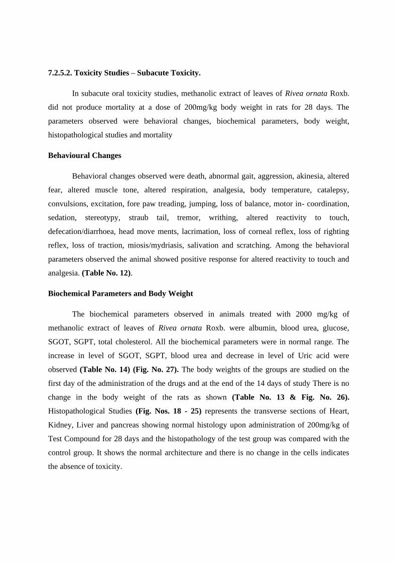
7.2.5.2. Toxicity Studies – Subacute Toxicity.
In subacute oral toxicity studies, methanolic extract of leaves of Rivea ornata Roxb.
did not produce mortality at a dose of 200mg/kg body weight in rats for 28 days. The
parameters observed were behavioral changes, biochemical parameters, body weight,
histopathological studies and mortality
Behavioural Changes
Behavioral changes observed were death, abnormal gait, aggression, akinesia, altered
fear, altered muscle tone, altered respiration, analgesia, body temperature, catalepsy,
convulsions, excitation, fore paw treading, jumping, loss of balance, motor in- coordination,
sedation, stereotypy, straub tail, tremor, writhing, altered reactivity to touch,
defecation/diarrhoea, head move ments, lacrimation, loss of corneal reflex, loss of righting
reflex, loss of traction, miosis/mydriasis, salivation and scratching. Among the behavioral
parameters observed the animal showed positive response for altered reactivity to touch and
analgesia. (Table No. 12).
Biochemical Parameters and Body Weight
The biochemical parameters observed in animals treated with 2000 mg/kg of
methanolic extract of leaves of Rivea ornata Roxb. were albumin, blood urea, glucose,
SGOT, SGPT, total cholesterol. All the biochemical parameters were in normal range. The
increase in level of SGOT, SGPT, blood urea and decrease in level of Uric acid were
observed (Table No. 14) (Fig. No. 27). The body weights of the groups are studied on the
first day of the administration of the drugs and at the end of the 14 days of study There is no
change in the body weight of the rats as shown (Table No. 13 & Fig. No. 26).
Histopathological Studies (Fig. Nos. 18 - 25) represents the transverse sections of Heart,
Kidney, Liver and pancreas showing normal histology upon administration of 200mg/kg of
Test Compound for 28 days and the histopathology of the test group was compared with the
control group. It shows the normal architecture and there is no change in the cells indicates
the absence of toxicity.

7.2.5.3. Antihyperlipidemic activity
Triton X 100 Induced Hyperlipidemia
Intraperitoneal administration of Triton X 100 (100mg/kg, b.w.) increased the level of
serum total cholesterol, triglycerides, LDL, VLDL and decrease in the level of good
cholesterol carrier HDL by inhibiting the lipase activity. Elevated level of blood cholesterol
especially LDL was the major risk factor for the coronary heart disease and HDL as cardio
protective protein. Treatment with methanolic extract of leaves of Rivea ornata (200 mg/kg)
significantly decreased the level of cholesterol, triglycerides, VLDL and LDL as compared to
hyperlipidemic control. There was significant increase in HDL as compared to control.
The body weight of the animal was measured at 1st, 4th and 10th day during the study
(Tab.No. 16) (Fig. No. 37)
The blood was collected on 10th day by cardiac puncture under mild anesthesia, the
serum was assayed for total cholesterol, total triglycerides, HDL-C, LDL-C and VLDL-C.
(Tab. No. 17) (Fig. No. 38)
The liver was collected on 10th day and transverse sections were obtained from
various groups such as normal control, toxic control, standard and test (Fig. Nos. 39 – 42)
Triton X 100 induced hyperlipidemia is associated with alteration in the
activities of enzyme responsible for cholesterol transport and metabolism. Triton X 100 cause
significant increases in triglycerides level by inhibiting the capillary lipoprotein lipase which
is responsible for plasma triglycerides hydrolysis and produce significant increase in serum
cholesterol by the stimulation of 3-hydroxy-3-rmethylglutaryl-Co-enzyme A (HMG-CoA)
reductase activity in the liver.
Treatment with methanolic extract of Rivea ornata (200 mg/kg) showed the significant
antihyperlipidemic activity. Reduction in the triglycerides level may be due to increased
lipoprotein lipase activity and reduction in total cholesterol level may be due to inhibition of
HMG CoA reductase activity.

7.2.5.4. Antidiabetic Activity
Alloxan Induced Hyperglycemia
Intraperitoneal administration of alloxan (150mg/kg, b.w.) increased the blood
glucose level by degrading the beta cells of pancreas. Treatment with methanolic extract of
leaves of Rivea ornata (200mg/kg) was decrease the blood glucose level and shows
antidiabetic index upto 3 hours. The blood glucose level was measured at 0 hour, 1 hour, 2
hour, 3 hour, 4 hour, 7th day and 14th day (Tab. No. 18) (Fig. No. 43).
The pancreas was collected on 14th day and transverse sections were obtained from
various groups such as normal control, toxic control, standard and test (Fig. No. 44 – 47)
Alloxan has two distinct pathological effects: it selectively inhibits glucose-induced
insulin secretion through specific inhibition of glucokinase (The glucose sensor of the beta
cell), and it causes a state of insulin-dependent diabetes. Alloxan also induce ROS (Reactive
Oxygen Species) formation, resulting in the selective necrosis of beta cells.
The preliminary phytochemical analysis showed that the methanolic extract of Rivea
ornata contains well known antioxidant phytochemicals such as alkaloids, glycosides and
polyphenols which act as free radical scavengers. The presumed mechanism of action of
these antioxidants was because of insulin mimetic effect on the peripheral tissues by either
stimulation of regeneration process or release of pancreatic secretion of insulin from existing
beta cells.
7.2.5.5. Analgesic Activity
The analgesic activity of methanolic extract of leaves of Rivea ornata was evaluated
by tail immersion and hot plate method. The result of analgesic activity of methanolic extract
of leaves of Rivea ornata was shown in Tab. No. 19, Fig. No. 48 and Tab. No. 20,
Fig. No. 49 respectively.
Tests of analgesic drugs commonly measure the nociception and involve the reaction
of animals to pain stimuli. The stimulus may be Thermal (Tail flick and Hot plate), Chemical
(Acetic acid induced writhing) or mechanical (Tail or paw pressure tests). Tail immersion
and Hot plate model induce pain by thermal stimuli method. The analgesic effect of Rivea

ornata may be mediated through peripheral pain mechanism or through suppression of
prostaglandin pathway.
7.2.5.6. Antipyretic activity
Brewer’s yeast induced hyperpyrexia
Subcutaneous injection of 12.5% w/v Brewer’s yeast solution increased the body
temperature in rats. Treatment with methanolic extract of leaves of Rivea ornata (200mg/kg)
was decrease the body temperature and that was compared with the reference standard
paracetamol (150mg/kg, b.w.) and shown in Tab. No. 21 and Fig. No. 50.
Normally, the infected or damaged tissue initiates the enhanced formation of pro-
inflammatory mediators (cytokines, such as interleukin-1ß, interleukin-α, interleukin-ß, and
TNF-α), which increase the synthesis of prostaglandin E2 (PgE2) near hypothalamic area and
thereby trigger the hypothalamus to elevate the body temperature. Brewer’s yeast induces
both TNF-α and prostaglandin synthesis.
The mechanism of action of antipyretic effect of methanolic extract of Rivea ornata
may be due to the inhibition of prostaglandin synthesis or by inhibiting the formation of
inflammatory mediators.
7.2.5.7. Wound Healing activity
The wound healing activity of methanolic extract of leaves of Rivea ornata Roxb. was
evaluated by the excision wound model. About area of wound with 300mm2 was produced by
excision made at the neck part of the rat. Application of ointment of methanolic extract of
leaves of Rivea ornata greatly reduce the area of wound was compared with control and
standard group (Tab. No. 22) (Fig. No. 53 – 64). The wound contraction percentage was
calculated and shown in Tab. No. 23 and Fig. No. 52.
Wound healing is a complex process that can be divided into at least 3 continuous and
overlapping processes: an inflammatory reaction, a proliferative process leading to tissue
restoration, and, eventually, tissue remodeling. Wound healing processes are regulated by
multiple growth factors and cytokines released at the wound site. The shorter period of
epithelialization and faster wound area contraction could be due to the ability of Rivea ornata
leaf extract to enhance collagen synthesis, induction of cell proliferation, and antimicrobial
activities of bioactive constituents such as alkaloids, steroids and glycosides.


CONCLUSION 8. CONCLUSION
From the study entitled "Pharmacological Evaluation of Leaves Extract from Rivea
ornata Roxb., the following conclusion could be drawn
❖ The present study has thus duly supported the traditional use of methanolic Extract of
Rivea ornata Roxb. have scientifically proved the antihyperlipidemic, antidiabetic,
analgesic, antipyretic and wound healing activity
❖ Apart from the suggested actions listed in discussion part absence of acute toxicity
and sub acute toxicity may also offer a new hope for safe treatment in future
❖ Preliminary phytochemical study of methanolic extract of Rivea ornata Roxb. was
found to contain Carbohydrates, Steroids, Alkaloids, Glycosides, Tannins, Phenolic
compounds, Gums and mucilage are present
❖ Presence of Alkaloids, Tannins and Glycosides in the methanolic extract of Rivea
ornata Roxb. was observed in IR & HPTLC analysis. Though present in small
quantities, it was found to produce considerable effects
❖ The results of acute toxicity study indicate that the methanolic extract of Rivea ornata
was non-toxic up to dose level of 2000mg/kg body weight in albino rats as per acute
oral toxicity studies. 1/10th of the LD50 Dose is 200mg/kg is used for
Pharmacological screening
❖ The results of subacute toxicity study indicate that the methanolic extract of Rivea
ornata was non-toxic in chronic administration
❖ The methanolic extract of Rivea ornata at a dose of 200mg/kg exhibited significant
hypolipidemic activity in Triton X 100 induced hyperlipidemic rats. This is showed
by the reduction of serum lipid parameters such as triglycerides, total cholesterol,
LDL, VLDL with an increase in HDL concentration in the group treated with
200mg/kg of methanolic Extract of Rivea ornata
❖ The methanolic extract of Rivea ornata at a dose of 200mg/kg showed significant
antidiabetic activity in Alloxan induced hyperglycemic rats. This is showed by the
reduction of blood glucose level in the group treated with 200mg/kg of methanolic
extract of Rivea ornata

❖ The methanolic extract of Rivea ornata was subjected to the Tail immersion and hot
plate method Analgesic test, showed a significant analgesic effect
❖ The methanolic extract of Rivea ornata was subjected to the Antipyretic activity
against Brewer’s yeast induced hyperpyrexia showed a significant reduce in body
temperature of the animals treated with 200mg/kg of methanolic extract of Rivea
ornata
❖ The methanolic extract of Rivea ornata was subjected to the wound healing activity,
showed a significant wound healing effect on excision wound in rats
❖ In future, further investigation might provide an insight to identify the functional
groups in the methanolic extract of Rivea ornata responsible for the
antihyperlipidemic, antidiabetic, analgesic, antipyretic and wound healing activity and
to elucidate the exact mechanism of action, which is responsible for the observed
significant activity with low toxicity and better therapeutic index

BIBLIOGRAPHY 9. BIBLIOGRAPHY
1. Partha Pradip Adhikari, Satya Bhusan Paul. History of indian traditional medicine: a
medical inheritance. Asian Journal of pharmaceutical and clinical research 2018; Vol 11,
Issue 1.
2. World Health Organization. WHO Guidelines on Safety Monitoring of Herbal Medicines
in Pharmacovigilance Systems. WHO; 2004.
3. World Health Organization. WHO Tradition medicine strategy. WHO; 2014-2023.
4. Regional Committee to World Health Organization for South East Asia. Meeting,
September, 1976.
5. World Health Organization. National policy on traditional medicine and regulation of
herbal medicines. Report of WHO global survey; 2005.
6. Sissi Wachtel-Galor, Iris F. F. Benzie. Herbal Medicine: Biomolecular and Clinical
Aspects. 2nd edition. CRC press; 2011. P. No. 2 & 3.
7. Mel Borins. Traditional medicine of India. Canadian fam. physician vol. 33:1061-5 April
1987.
8. Saikat Sen and Raja Chakraborty. Toward the integration and advancement of herbal
medicine: a focus on traditional Indian medicine. Botanics: Targets and Therapy 2015:5
33–44. 2015.
9. Planning commission. National plans. 10th five year plan 2002-2007 volume 2, chapter
2.9 Page. No. 153.
10. M. M. Pandey, Subha Rastogi, and A. K. S. Rawat. Review Article: Indian Traditional
Ayurvedic System of Medicine and Nutritional Supplementation. Hindawi Publishing
Corporation. Volume 2013, Article ID 376327; 2013.

11. Simone Badal and Rupika Delgoda. Pharmacognosy: Fundamentals, Applications and
Strategy. Elseiver publications. Chapter No. 2, Page No. 21
12. http://ayush.gov.in/sites/default/files/Introduction_2.html
13. Narayanaswamy V. (1975). Introduction to the Siddha System of Medicine. Director,
Pandit S.S. Anandam Research Institute of Siddha Medicine, T. Nagar, Madras
(Chennai).
14. Ravishankar B and Shukla V.J. Indian Systems Of Medicine: A Brief Profile. Afr. J.
Trad. CAM (2007) 4 (3): 319 – 337.
15. https://www.nhp.gov.in/homeopathy_mty National Health Portal
16. https://nccih.nih.gov/health/homeopathy National Centre for Complementary and
Integrative Health
17. Syed Ziaur Rahman, Khan Rahat Ali, Latif Abdul. Importance of pharmacovigilance
in Unani system of medicine. Indian J Pharmacol. 2008; 40(1):S17–S20.
18. MA Qasmi. Introduction to Unani Medicine. Available at:
www.similima.com/ppt/general/unani-introduction. [Cited on 2/8/13]
19. Rahman SZ. Impact of human medicines on environment - a new emerging
problem. Popul ENVIS. 2006; 3: 3–4.
20. Shattat G. F. A Review Article on Hyperlipidemia: Types, Treatments and New Drug
Targets. Biomed Pharmacol J., 2014; 7(2): 399-409.
21. Adeli, K., and Lewis, G. F. Intestinal lipoprotein overproduction in insulin-resistant
states. Current Opinion in Lipidology, 2008; 19(3): 221–228.
22. Ma, X. (2004). A common haplotype at the CD36 locus is associated with high free fatty
acid levels and increased cardiovascular risk in Caucasians. Human Molecular Genetics,
13(19), 2197–2205.
23. Nada A. Abumrad S B, M. Raafat El-Maghrabi S, Ez-Zoubir Amrill, Ellen Lopez S,
and Paul A. Grimaldill. Cloning of a rat adipocyte membrane protein implicated in
binding or transport of long-chain fatty acids that is induced during preadipocyte
differentiation. Homology with human CD 36. J. Biol. Chem. 1993; 268, 17665–17668.
24. Levy, E., Stan, S., Delvin, E., Menard, D., Shoulders, C., Garofalo, C., Bendayan, M.
Localization of Microsomal Triglyceride Transfer Protein in the Golgi: Possible role in

the Assembly of Chylomicrons. Journal of Biological Chemistry, 2002; 277(19), 16470–
16477.
25. Mahmood Hussain, M. A proposed model for the assembly of chylomicrons.
Atherosclerosis, 2000; 148(1): 1–15.
26. Ian J. Cartwright, Dietmar Plonné and Joan A. Higgins. Intracellular events in the
assembly of chylomicrons in rabbit enterocytes, The Journal of Lipid Research, 2000; 41:
1728-1739.
27. Your guide to lowering your Cholesterol with Therapeutic Life Style. U.S. Department
Of Health And Human Services, NIH Publication, No. 06-5235, pp 1-2
28. Jacobson, K.; Mouritsen, O.G.; Anderson, R.G. Lipid rafts: At a crossroad between
cell biology and physics. Nat. Cell Biol. 2007; 9, 7–14.
29. Gerard A. McKay, John L. Reid and Matthew R. Walters. Lecture Notes – Clinical
Pharmacology and Therapeutics, 8th Edition, 2010; Chapter 5, 71-77.
30. Harvey, R.A.; Ferrier, D. (Eds.) Biochemistry, Lippincott Illustrated Reviews, 6th ed.;
Lippincott Williams and Wilkins: Philadelphia, PA, USA, 2014; Chapter 18: 397-406.
31. David L. Nelson and Michael M. Cox. Lehninger Principles of Biochemistry, 7th
Edition, 2017; 10.1: 931-933.
32. Choi, S. S., & Diehl, A. M. (2008). Hepatic triglyceride synthesis and nonalcoholic fatty
liver disease. Current Opinion in Lipidology, 19(3), 295–300.
33. Daniels TF, Killinger KM, Michal JJ, Wright RW Jr, Jiang Z. Lipoproteins,
cholesterol homeostasis and cardiac health. Int J Biol Sci., 2009; 29, 5(5): 474-88.
34. Satyanarayana U and Chakrapani U. Biochemistry, 3rd Edition, Books and Allied (P)
Ltd., 2006; Chapter 14, 285-328.
35. Cox RA, García-Palmieri MR. Cholesterol, Triglycerides, and Associated Lipoproteins.
In: Walker HK, Hall WD, Hurst JW, editors. Clinical Methods: The History, Physical,
and Laboratory Examinations. 3rd edition. Boston: Butterworths, 1990; Chapter 31: 153-
160.
36. Berg JM, Tymoczko JL, Stryer L. Biochemistry. 5th edition. New York: W H Freeman;
2002. Section 26.2: 1072-1073
37. Feingold KR, Grunfeld C. Introduction to Lipids and Lipoproteins. Endotext [Internet].
South Dartmouth (MA): MDText.com, last update February 2 2018, Available at:
https://www.ncbi.nlm.nih.gov/books/NBK305896/
38. Freeman, M. W., & Walford, G. A. Lipoprotein Metabolism and the Treatment of Lipid
Disorders. Endocrinology: Adult and Pediatric, 2016; (41): 715–736.

39. Jennifer moll. Function of lipoproteins in the body, very well health website. available at
https://www.verywellhealth.com/lipoproteins-facts-and-info-697495
40. Shoji T, Nishizawa Y, Kawagishi T, Kawasaki K, Taniwaki H, Tabata T, Inoue T,
Morii H. Intermediate-density lipoprotein as an independent risk factor for aortic
atherosclerosis in hemodialysis patients. J Am Soc Nephrol. 1998; 9(7): 1277-84.
41. William Stillwell, in An Introduction to Biological Membranes (Second Edition),
Chapter 16, Membrane biogenesis (Protein), 2016; 349-367. ISBN: 978-0-444-63772-7.
42. Zhou, L., Li, C., Gao, L., & Wang, A. High-density lipoprotein synthesis and
metabolism (Review). Molecular Medicine Reports, 2015; 12(3), 4015–4021.
43. Boisfer E, Stengel D, Pastier D, Laplaud PM, Dousset N, Ninio E and Kalopissis AD:
Antioxidant properties of HDL in transgenic mice overexpressing human apolipoprotein
A-II. J Lipid Res 2002; 43: 732-741.
44. Rosenson RS: Brewer HB Jr, Chapman MJ. HDL measures, particle heterogeneity,
proposed nomenclature, and relation toatherosclerotic cardiovascular events. Clin Chem
2007; 57: 392-410.
45. Cappel DA, Palmisano BT, Emfinger CH, Martinez MN, McGuinness OP and
Stafford JM: Cholesteryl ester transfer protein protects against insulin resistance in
obese female mice. Mol Metab 2013; 2: 457-467.
46. Fisher EA, Feig JE, Hewing B, Hazen SL and Smith JD: High-density lipoprotein
function, dysfunction, and reverse cholesterol transport. Arterioscler Thromb Vasc Biol
2012; 32: 2813-2820.
47. Gromelski S and Brezesinski G: DNA condensation and interaction with zwitter ionic
phospholipids mediated by divalent cations. Langmuir 2006; 22: 6293-6301.
48. Barter PJ, Puranik R and Rye KA: New insights into the role of HDL as an anti-
inflammatory agent in the prevention of cardiovascular disease. Curr Cardiol Rep 2007;
9: 493-498.
49. Mineo C, Deguchi H, Griffin JH and Shaul PW: Endothelial and antithrombotic
actions of HDL. Circ Res 2006; 98: 1352-1364.
50. Rafael A, Cox and mario R, Garcia-palmieri. Cholesterol, Triglycerides, and
Associated Lipoproteins. National Center for Biotechnology Information Bookshelf,
Available at https://www.ncbi.nlm.nih.gov/books/NBK351/pdf/Bookshelf_NBK351.pdf
51. Nelson, R. H. Hyperlipidemia as a Risk Factor for Cardiovascular Disease. Primary Care:
Clinics in Office Practice, 2013; 40(1), 195–211.
52. WHO memorandum, Circulation, Volume XLV, February; 1972: 501- 508.

53. Stone, N. J. (1994). Secondary causes of hyperlipidemia. Medical Clinics of North
America, 78(1), 117–141.
54. Webmd.com, National Institute of Health, Americal academy of Family physicians,
available at https://www.webmd.com/cholesterol-management/hyperlipidemia-
overview#2-7
55. Harris WS: Fish oils and plasma lipid and lipoprotein metabolism in humans: A critical
review. J Lipid Res, 1989; 30: 785-807.
56. Mensink RP, Katan MB: Effect of a diet enriched with monounsaturated or
polyunsaturated fatty acids on levels of low-density and high-density lipoprotein
cholesterol in healthy men and women. N Engl J Med, 1989; 321: 436-441.
57. Chait A, Brunzell JD: Acquired hyperlipidemia (secondary dyslipoproteinemias).
Endocrinol Meta Clin North Am, 1990; 19: 259-278.
58. Henkin Y, Como JA, Oberman A: Secondary dyslipidemia. Inadvertent effects of drugs
in clinical practice. JAMA, 1992; 267: 961-968.
59. Glueck q, Scheel 0, Fishback I, Steiner P: Estrogen-induced pancreatitis in patients
with previously covert familial type V hyperlipoproteinemia. Metabolism, 1972; 21: 657-
666.
60. Zimmerman), Fainaru M, Eisenberg S: The effects of prednisone therapy on plasma
lipoproteins and apoproteins: A prospective study. Metabolism, 1984; 33: 521-527.
61. Knopp RH, Walden CE and Wahl PW. Oral contraceptive and postmenopausal
estrogen effects on lipoprotein triglyceride and cholesterol in an adult female population:
Relationships to estrogen and progestin potency, J Clin Endocrinol Metab. 1981; 53:
1123.
62. Wahl P, Walden C and Knopp R. Effect of estrogen progestin potency on lipid/
lipoprotein cholesterol. N Engl J Med, 1983; 308: 862-867.
63. Grimm RH and Hunninghake DB: Lipids and hypertension. Implications of new
guidelines for cholesterol management in the treatment of hypertension. Am J Med, 1986,
80 (suppl 2A):56-63.
64. Sheu WH, Swislocki AL and Hoffman BB. Effect of prazosin treatment on HDL
kinetics in patients with hypertension. Am J Hypertension, 1990; 3: 761-768.
65. Day JL, Simpson N, Metcalfe L Page RL: Metabolic consequences of atenolol and
propranolol in treatment of essential hypertension. BMJ, 1979; 1: 77-80.
66. Hazzard WR and Bierman EL: Aggravation of broad-[3disease (type 3
hyperlipoproteinemia) by hypothyroidism. Arch Intern Med 1972; 130: 822-828.

67. Kesaniemi YA and Grundy SM: Increased low density lipoprotein production
associated with obesity. Arteriosclerosis, 1983; 3: 170-177.
68. Wolf RN and Grundy SM: Influence of weight reduction on plasma lipoproteins in
obese patients. Arteriosclerosis, 1983; 3: 160-169.
69. Brunzell JD, Hazzard WR and Porte D Jr. Evidence for a common saturable
triglyceride removal mechanism for chylomicrons and very low density lipoproteins in
man. J Clin Invest, 1973; 15: 78-85.
70. Webmd.com, National Institute of Health, Americal academy of Family physicians,
available at https://www.webmd.com/cholesterol-management/guide/diseases-linked-
high-cholesterol#1-2
71. Papadakis JA, Mikhailidis DP and Winder AF. Lipids and stroke: neglect of a useful
preventive measure? Cardiovasc Res, 1998; 40: 265–71.
72. Milionis, H. J. Lipoprotein (a) and stroke. Journal of Clinical Pathology, 2000; 53(7):
487–496.
73. World Health Organization, Details, Cardiovascular disease, available at
https://www.who.int/news-room/fact-sheets/detail/cardiovascular-diseases-(cvds)
74. Huxley, R., Lewington, S., & Clarke, R. Cholesterol, Coronary Heart Disease and
Stroke: A Review of Published Evidence from Observational Studies and Randomized
Controlled Trials. Seminars in Vascular Medicine, 2002; 02(3), 315–324.
75. Ross R. The pathogenesis of atherosclerosis an update. N Engl J Med 1986; 314: 488–
500
76. Indian Council of Medical Research, Department of Health Research - Ministry of Health
& Family Welfare, Government of India, Hypertension available at
https://www.icmr.nic.in/sites/default/files/press_realease_files/Hypertension.pdf
77. Healthline.com, Hypertension available at https://www.healthline.com/health/high-
cholesterol/treating-with-statins/hypertension#1
78. K.D Tripathi. Essentials of Medical Pharmacology, Jaypee publications, 7th Edition,
Chapter 45, 2013; pp: 634-645. ISBN: 978-93-5025-937-5.
79. Goodman and Gillman’s. The Pharmacological Basics of Therapeutics, McGraw Hills
companies, 12th Edition, Chapter 31, pp: 877- 904. ISBN: 978-0-07-176939-6.
80. Charles R. Craig and Robert E. Stitzel. Modern Pharmacology with Clinical
Applications, 5th Edition, Lippincot Williams and wilkins, Chapter 23, 2003; pp: 268-277.
ISBN: 0-7817-3762-1.

81. Padmaja udaykumar. Medical Pharmacology, 3rd Edition, CBS publishers and
distributers pvt. Ltd., 2011, 39: 348-355.
82. Harsh Mohan, Text Book of Pathalogy, Jaypee Brothers Medical Publishers (P) Ltd. 6th
Edition, Chapter 27, Page No. 818.
83. Madalageri NK, Nagaraj L and Nidamarthi SB. Evaluation and comparative study of
hypoglycaemic activity of morus alba with oral hypoglycaemic drug (glibenclamide) in
alloxan induced diabetic rats. J. Evolution Med. Dent. Sci. 2016; 5(48): 3062-5.
84. World Health Organization (WHO). Report of a WHO Consultation. Definition,
diagnosis and classification of diabetes mellitus and its complications. 1. Diagnosis and
classification of diabetes mellitus. WHO/ NCD/NCS/99.2. Geneva: WHO, 1999.
85. Richard I.G. Holt, Clive S. Cockram, Allan Flyvbjerg and Barry J. Goldstein.
Textbook of Diabetes, 4th Edition, Willy-Blackwell, 2010; 1(3): 24-30. ISBN:
9781405191814.
86. Diagnosis and Classification of Diabetes Mellitus. Diabetes Care, 36(Supplement_1);
2012: S67–S74.
87. Mayoclinic.com, Patient Care & Health Information, Diabetes. Available at
https://www.mayoclinic.org/diseases-conditions/diabetes/diagnosis-treatment/drc-
20371451
88. Endocrineweb.com/ Information/ Diabetes mellitus/ What is insulin? Available at
https://www.endocrineweb.com/conditions/type-1-diabetes/what-insulin
89. Medicalnewstoday.com/ information/ An overview of insulin available at
https://www.medicalnewstoday.com/articles/323760.php
90. Tanabe M, Motonaga R, Terawaki Y, Nomiyama T and Yanase T. Prescription of
oral hypoglycemic agents for patients with type 2 diabetes mellitus: A retrospective
cohort study using a Japanese hospital database. J Diabetes Investig. 2017; 8 (2): 227-
234.
91. Pain management, Basic definitions and Clinical implications. Available at
http://projects.hsl.wisc.edu/GME/PainManagement/session2.1.html
92. Teach me Physiology, Pain pathways. Available at
https://teachmephysiology.com/nervous-system/sensory-system/pain-pathways/
93. Padmaja udaykumar. Medical Pharmacology, 3rd Edition. 2011; 5(27): 237-250. ISBN:
978-81-239-1966-9.

94. Change pain-Taking care of pain/ Insight/ Non opioid treatment available at
https://www.change-pain.com/grt-change-pain portal/change_pain_home/chronicpain/
insight/pain_treatment/non-opioids/en_EN/298801006.jsp
95. Aronoff, D. M., & Neilson, E. G. Antipyretics: mechanisms of action and clinical use in
fever suppression. The American Journal of Medicine, 2001; 111(4): 304–315.
96. Luheshi GN. Cytokines and fever: mechanisms and sites of action. Ann NY Acad Sci.
1998; 856: 83– 89.
97. Dinarello CA, Gatti S and Bartfai T. Fever: links with an ancient receptor. Curr Biol.
1999; 9: R147–150.
98. Healthline, Open wound available at https://www.healthline.com/health/open-wound
99. Harsh Mohan, Text Book of Pathalogy, Jaypee Brothers Medical Publishers (P) Ltd. 6th
Edition, Chapter 6, Page No. 165-170.
100. Enoch, S., Grey, J. E., & Harding, K. G. (2006). Non-surgical and drug treatments.
BMJ, 332(7546), 900–903.
101. Hammeso, W. W., Emiru, Y. K., Ayalew Getahun, K., & Kahaliw, W. (2019).
Antidiabetic and Antihyperlipidemic Activities of the Leaf Latex Extract of Aloe
megalacantha Baker (Aloaceae) in Streptozotocin-Induced Diabetic Model. Evidence-
Based Complementary and Alternative Medicine, 2019, 1–9
102. NT Abdullah and Raju Koneri. Screening of Simarouba Gluaca for Antidiabetic
and Antioxidant Activities, Int J Pharm Sci & Res 2019; 10(1): 294-02.
103. Sushma Gour, C.K. Tyagi, Hemant Kumar Sharma, Prabhakar Budholiya.
Phytochemical Screening And In Vivo Antipyretic Activity Of The Hydroalcoholic
Leaves Extract Of Morus Alba Linn. International Journal of Pharmaceutics & Drug
Research; 2019; 7 (1), 23-32.
104. Kannika Inchab, Parirat Khonsung, Natthakarn Chiranthanut, Puongtip
Kunanusorn, Ampai Panthong, and Seewaboon Sireeratawong. Evaluation of Anti-
inflammatory, Analgesic, and Antipyretic Activities of Pseuderanthemum palatiferum.
CMU J. Nat. Sci. (2019) Vol. 18(3) 393.
105. Shanti Vasudevan CN, Dr. Bibu John Kariyil and Dr. I’ma Neerakkal.
Antipyretic activity of aqueous extract of heart wood of Pterocarpus santalinus L. in
yeast induced pyrexia. Journal of Pharmacognosy and Phytochemistry 2019; 8(3): 244-
246.

106. Kaila N, Dhir S and Vashist N: Antipyretic and analgesic activity of PICRORHIZA
KURROOA rhizomes. Int J Pharm Sci & Res 2019; 10(5): 2240-43. doi:
10.13040/IJPSR.0975-8232.10(5).2240-43.
107. Prince SE, Das AP and Arumugam G: In-vivo appraisal of pharmacological
activity of Bacopa monnieri and its HPLC analysis for bacoside A. Int J Pharm Sci & Res
2019; 10(5): 2244-49. doi: 10.13040/IJPSR.0975-8232.10(5).2244-49.
108. Mansour Sobehah, Samar Rezq, Omar M. Sabry, Mohamed A.O.Abdelfattah,
Mohamed A.El Raey, Walaa A.El-Kashak, Assem M.El-Shazly, Mona F.Mahmoud
and Michael Wink. Albizia anthelmintica: HPLC-MS/MS profiling and in vivo anti-
inflammatory, pain killing and antipyretic activities of its leaf extract. Biomedicine &
Pharmacotherapy. Volume 115, July 2019, 108882.
109. Kuum, M. G. M., Lambou, A. F., Kiki, G. A. A., Ndzana, M. T. B., Keugni, B. A.,
Moukette, B. M., Mezui, C., Djomeni, P. D. D., & Dimo, T. (2019). Evaluation of Anti-
inflammatory, Analgesic and Antipyretic Potential of the Stem Barks Aqueous Extract of
Albizia ferruginea (Guill. & Perr.) Benth. (Mimosaceae) in Rats and Mice. Journal of
Advances in Biology & Biotechnology, 20(3), 1-15.
110. Akhter, F., Islam, Q., Adib, M., Faruq, A., & Ibrahim, M. (2019). Phytochemical
Screenings and Bioactivities of Duabanga grandiflora (Roxb. ex DC.) Walp. Bangladesh
Pharmaceutical Journal, 22(1), 68-72.
111. Swapnil Y. Chaudhari, Mukesh B. Nariya, Galib Ruknuddin, Pradeep Kumar
Prajapati and Jayram Hazra. Antihyperlipidemic activity of Hridayarnava Rasa (an
Ayurvedic herbo-metalo-mineral formulation) in Charles Foster albino rats. Journal of
Current Research in Scientific Medicine | Volume 4 | Issue 1 | January-June 2018
112. B. H. M. R. K. Guptha, S. L. D. V. Ramana Murty Kadali, Vijay Kumar M and
Revanasiddappa B. C. Antihyperlipidemic activity of Chloroxylon swietenia in triton
WR1339 induced hyperlipidemia. International Journal of Basic & Clinical
Pharmacology. 2018 March; 7(3):518-523.
113. Venkateswarlu G and Ganapathy S. Antipyretic effect of methanolic extracts of
Limnophila repens and Argyreia cymosa whole plant on albino rats. MOJ Bioequiv
Availab. 2018; 5(6):303‒306.
114. Rajendra Kumar Swain, Bimlendu Chowdhury and Bhanoji Rao. Evaluation of
Analgesic Activity of Hydroalcoholic Extract of Cinnamomum zeylanicum Bark in
Albino Rats. Der Pharmacia Lettre, 2018, 10 [3]: 84-90
115. Doan, H. V., Riyajan, S., Iyara, R., & Chudapongse, N. Antidiabetic activity,
glucose uptake stimulation and α-glucosidase inhibitory effect of Chrysophyllum cainito
L. stem bark extract. BMC Complementary and Alternative Medicine, 2018; 18(1).
116. Belayneh, Y. M., & Birru, E. M. (2018). Antidiabetic Activities of Hydromethanolic
Leaf Extract of Calpurnia aurea (Ait.) Benth. Subspecies aurea (Fabaceae) in Mice.
Evidence-Based Complementary and Alternative Medicine, 2018, 1–9.
117. Hussain, S. N., Uzair, M., Qaisar, M. N., Abbas, K., Ashfaq, K., & Chaudhari,
B. A. (2018). Assessment of anti-diabetic activity of Cassia sophera (Caesalpiniaceae).
Tropical Journal of Pharmaceutical Research, 17(3), 443.
118. Dr. Sam Pavan Kumar Gandamalla, Dr. Anil C, Dr.PushpalathaChinnam,
D.Sathish Kumar. Analgesic and anti-inflammatory effects of Toona Ciliata in
laboratory animals. Int. J. of Res. in Pharmacology & Pharmacotherapeutics Vol-6(4)
2017 [389-395].
119. Charles Chatterjee, Gautam K. and Mandal. Evaluation of Analgesic Activity of
Methanolic Extract of Bougainvillea spectabilis Leaves in Experimental Animal Models.
Annals of International Medical and Dental Research, 2 (5). PC01-PC04. ISSN 2395-
2814
120. G. Vijay Kumar and N. Devanna. Antihyperlipidemic Activity of Leaf Extracts of
Leucas aspera Linn. against Dexamethasone-induced Hyperlipidemia in Rats. Asian
Journal of Pharmaceutics, Jul-Sep 2016 (Suppl), 10 (3) | S408.
121. Nagar, H. K., Srivastava, A. K., Srivastava, R., Kurmi, M. L., Chandel, H. S., &
Ranawat, M. S. (2016). Pharmacological Investigation of the Wound Healing Activity of
Cestrum nocturnum (L.) Ointment in Wistar Albino Rats. Journal of Pharmaceutics,
2016, 1–8.
122. Sneha D. Borkar, Raghavendra Naik, Vinay J. Shukla, and Rabinarayan
Acharya. Evaluation of phytochemical content, nutritional value and antioxidant activity
of Phanji - Rivea hypocrateriformis (Desr.) Choisy leaf. Ayu. 2015 Jul-Sep; 36(3): 298–
302.
123. Asie Shojaii, Majid Motaghinejad, Sima Norouzi, and Manijeh Motevalian.
Evaluation of Anti-inflammatory and Analgesic Activity of the Extract and Fractions
of ASTRA GALUSHAMOSUS in Animal Models. Iranian Journal of Pharmaceutical
Research. 2015 Winter; 14(1): 263–269.
124. S.S. Godipurge, S.Rahber, J.S. Biradar and N. Mahurkar. Evaluation of
pharmacological activities of Rivea hypocrateriformis in experimental animal models.

International Journal of Toxicological and Pharmacological Research 7(2):65-73 · April
2015.
125. Pal Chandrashekhar R., GargeVaibhavi N. and KadamVilasrao J.
Antihyperlipidemic activity of Gloriosa superba. International journal of pharmaceutical
sciences and research. 0975-8232.6(11).4835-42.
126. Mukesh Singh Sikarwar and MB Patil. Antihyperlipidemic activity of Hibiscus
rosa-sinensis Linn. ethanolic extract fractions. International Journal of Health and Allied
Sciences. Vol. 4, Issue 2, Apr-Jun 2015.
127. Syed Safiullah Ghori, M.A Rizwan Khan, Kaab e Alam and Abid Hussain
Abrar. Evaluation of antihyperlipidemic activity of ethanolic extact of Glycosmis
pentaphylla in hyperlipidemic wistar rats. International Journal of Pharma Sciences and
Research. Vol. 6 No.2 Feb 2015.
128. Jain P and Surana SJ (2015) A review of Indian medicinal plants with
hypolipidemic activity and their medicinal importance. World Journal of Pharmacy and
Pharmaceutical Sciences 4(3): 1477-1493.
129. Mahesh Anand Goudar, H.A.Sayeswara and S.G.Manjunatha. Evaluation of
Antidiabetic activity of crude extracts of bark of Terminalia paniculata Roth in
Streptozotocin induced Diabetes in Wister Rats, Int. J. Drug Dev. & Res., October-
December 2014, 6 (4): 181-187.
130. Dinesh Dhingra, Deepak Lamba, Ramesh Kumar, Pashupati Nath, and
Satyaprakash Gauttam. Antihyperlipidemic Activity of Aloe succotrina in Rats:
Possibly Mediated by Inhibition of HMG-CoA Reductase. Volume 2014, Article ID
243575, 9 pages.
131. M S. Sikarwar and Mrityunjaya B. Patil. Antihyperlipidemic Activity of Pongamia
pinnata Leaf Extracts. Turkish Journal of Pharmaceutical Sciences, 11(3), 329-338, 2014.
132. Hardik Mori, K. Nishteswar, B. R. Patel, and Mukesh Nariya. Acute toxicity and
antihyperlipidemic activity of rhizome of Tectaria coadunata (Kukkutnakhi): A folklore
herb. An International Quarterly Journal of Research in Ayurvedha 2016 Jul-Dec; 37(3-
4): 238–243.
133. Mishra R, Pandit K, Bhagwat AM and Brijesh S: Antihyperlipidemic activity of
alcoholic extracts of TRICHOSANTHES ANGUINA in Triton Wr 1339 induced
hyperlipidemic rats. Int J Pharm Sci & Res 2014; 5(10): 4269-74. doi:
10.13040/IJPSR.0975-8232.5(10).4269-74.

134. Panneer selvam Vijayaraj, Kannan Muthukumar, Jayaraja Sabarirajan,
Vasanthi Nachiappan. Antihyperlipidemic activity of Cassia auriculata flowers in triton
WR 1339 induced hyperlipidemic rats. Volume 65, Issues 1–2, January 2013, Pages 135-
141
135. Kittana Mäkynen, Sritanaporn Jitsaardkul, Pansiree Tachasamran, Nathaporn
Sakai and Supitcha Puranachoti. Cultivar variations in antioxidant and
antihyperlipidemic properties of pomelo pulp (Citrus grandis [L.] Osbeck) in Thailand,
Science direct, Food Chemistry Volume 139, Issues 1–4, 15 August 2013, Pages 735-743.
136. Sharma VJ & Patel P. Evaluation of anti-inflammatory activity of plant Rivea
ornate. Journal of Drug Delivery & Therapeutics; 2013, 3(1), 59-60.
137. Najat T Hwisa, Babu Rao Chandu, Prakash Katakam and Ramalingam
Peraman. Pharmacognostical studies on Rivea ornata (roxb.) Leaves. Journal of
Chemical and Pharmaceutical Research, 2013, 5(2):342-344.
138. Pawar, R., Chaurasiya, P., Rajak, H., Singour, P., Toppo, F., & Jain, A. (2013).
Wound healing activity of Sida cordifolia Linn. in rats. Indian Journal of Pharmacology,
45(5), 474.
139. Murthy, S., Gautam, M. K., Goel, S., Purohit, V., Sharma, H., & Goel, R. K.
(2013). Evaluation ofIn VivoWound Healing Activity of Bacopa monnieraon Different
Wound Model in Rats. BioMed Research International, 2013, 1–9.
140. Mukesh S. Sikarwar and M. B. Patil. Antihyperlipidemic activity of Salacia
chinensis root extracts in triton-induced and atherogenic diet-induced hyperlipidemic rats.
Indian J Pharmacol. 2012 Jan-Feb; 44(1): 88–92.
141. Bhaskar, A., & Nithya, V. (2012). Evaluation of the wound-healing activity of
Hibiscus rosasinensis L (Malvaceae) in Wistar albino rats. Indian Journal of
Pharmacology, 44(6), 694.
142. Kiessoun Konaté, Imaël Henri Nestor Bassolé and Adama Hilou. Toxicity
assessment and analgesic activity investigation of aqueous acetone extracts of Sida
acuta Burn f. and Sida cordifolia L. (Malvaceae), medicinal plants of Burkina Faso. BMC
Complementary and Alternative Medicine 2012, 12:120.
143. Naveed Muhammad, Muhammad Saeed and Haroon Khan. Antipyretic, analgesic
and anti-inflammatory activity of Viola betonicifolia whole plant. BMC Complementary
and Alternative Medicine 2012 12:59.

144. Kumbhar ST, Ghaisas MM, Chavan DL, Pawar YD and Shaikh AA. Analgesic
Activity of Ethanolic Extract of Ficus Glomerata Leaves: In Vivo Evaluation.
International Journal of Research in Ayurveda and Pharmacy. 2012; 3(5):706-709.
145. Amberkar mohanbabu vittalrao, Tara shanbhag, Meena kumari k, k. L. Bairy
and smita shenoy. Evaluation Of Antiinflammatory And Analgesic Activities Of
Alcoholic Extract Of Kaempferia Galanga In Rats. Indian J Physiol Pharmacol 2011; 55
(1) : 13–24
146. Yashwant Kumar, A., Nandakumar, K., Handral, M., Talwar, S., &
Dhayabaran, D. Hypoglycaemic and anti-diabetic activity of stem bark extracts
Erythrina indica in normal and alloxan-induced diabetic rats. Saudi Pharmaceutical
Journal, 2011; 19(1), 35–42.
147. Garg, V., & Paliwal, S. (2011). Wound-healing activity of ethanolic and aqueous
extracts of Ficus benghalensis. Journal of Advanced Pharmaceutical Technology &
Research, 2(2), 110.
148. P.R. Rachh, M.R. Rachh, N.R. Ghadiya, D.C. Modi, K.P. Modi, N.M. Patel and
M.T. Rupareliya, 2010. Antihyperlipidemic Activity of Gymenma sylvestre R. Br. Leaf
Extract on Rats Fed with High Cholesterol Diet. International Journal of Pharmacology,
6: 138-141.
149. AHM Zulfiker, M Mahbubur Rahman, M Kamal Hossain. In vivo analgesic
activity of ethanolic extracts of two medicinal plants – Scoparia dulcis L. and Ficus
racemosa Linn. Biology and Medicine, 2 (2): 42-48, 2010
150. Bassey S. Antia, Jude E. Okokon, Emem E. Umoh and John A. Udobang (2010).
Antidiabetic Activity of Ethanolic Leaf Extract of Panicum maximum, Int.J.Drug Dev. &
Res., July-September 2010; 2(3): 488-492.
151. M. A. Khayum, T.S. Gouda, N. Venkata Rao, Md. Shalam, S.M. Shantakumar,
Richa Verma and S. Vijay Kumar. Influence of Lisinopril on Glucose Lowering Effect
of Glipizide in Normal and Diabetic Rats. Pharmacologyonline 2008; 1. 148-158.
152. G. W. Staples. A Synopsis of Rivea (Convolvulaceae). Edinburgh journal of botany.
Volume 64, Issue 2 July 2007, pp. 213-223
153. KN Reddy, Chiranjibipattanaik, CS Reddy and VS Raju. Traditional Knowledge
on wild food plants in Andhra Pradesh. Indian Journal of Traditional Knowledge
Vol 6 (1)-January 2007- pp 223-229
154. S. S. Jalalpure, Shivaji Bamne, M. B. Patil, Biren Shah and Md. Salahuddin.
Anti-Diabetic Activity of Holarrhena antidysenterica (Linn.) Wall, Bark on Alloxan
Induced Diabetic Rats, Journal of Natural Remedies, 2006; 6(1): 26-30.
155. James, B.Perianayagam and S.K.Sharma. Evaluation of anti-pyretic and analgesic
activity of Emblicaofficinalis Gaertn. Journal of Ethnopharmacology Volume 95, Issue
1, November 2004, Pages 83-85.
156. Kallappa M, Hosamani Raghavendra M and Sattigeri. Industrial utilization
of Rivea ornata seed oil: a moderate source of vernolic acid. Industrial Crops and
Products, Volume 12, Issue 2, August 2000, Pages 93-96.
157. A.H Attaa and A. Alkofahib. Anti-nociceptive and anti-inflammatory effects of
some Jordanian medicinal plant extracts. Journal of Ethnopharmacology Volume 60,
Issue 2, March 1998, Pages 117-124.
158. Elaine Elisabetsky, Tania A.Amador and Ruti R.Albuquerque. Analgesic activity
of Psychotria colorata (Willd. ex R. & S.) Muell. Arg. Alkaloids. Journal of
Ethnopharmacology Volume 48, Issue 2, October 1995, Pages 77-83.
159. India biodiversity portal available at
https://indiabiodiversity.org/species/show/243323
160. Eflora online. Available at https://sites.google.com/site/efloraofindia/species/a---
l/cl/convolvulaceae/rivea/rivea-laotica
161. Sharma VJ and Patel PM. Bergenin - An Active Constituent of Rivea ornata roxb.
And its antioxidant property, International Journal Pharmacognosy, 2017; 4(9): 309-319.
162. Affy Mataphouet Emmanuel, Kouakou Koffi Roger, Douhoure Gnaore
Toussaint and Kouakou Koffi. Acute and subacute toxicity of the aqueous extract of
Amaranthus viridis (Amaranthaceae) leaves in rats. The Journal of Phytopharmacology
2018; 7(4): 366-372.