Protective effect of Cnidoscolus aconitifolius leaves against ...
7
doi.org/10.36721/PJPS.2020.33.2.REG.651-657.1 Pak. J. Pharm. Sci., Vol.33, No.2, March 2020, pp.651-657 651 Protective effect of Cnidoscolus aconitifolius leaves against diclofenac- induced gastric mucosal damage Njoku Ugochi Olivia, Uzoagulu Chinedu Happiness and Ogugofor Martins Obinna* Pharmacology and Toxicology Unit, Department of Biochemistry, University of Nigeria, Nsukka, Nigeria Abstract: Cnidoscolus aconitifolius is one of the plants used in folk medicine for the treatment of peptic ulcer. However, the present study was designed to validate the gastro-protective effect of Cnidoscolus aconitifolius leaves using diclofenac-induced ulcer model. The gastro-protective effect of Cnidoscolus aconitifolius leaves was evaluated by employing diclofenac–induced gastric ulcer in rats. Gastric mucosal lesions were induced by oral administration of diclofenac in rats. The pH of gastric juice from pylorus-ligated rats was measured. Histological examinations of the gastric tissues were also carried out. The present findings showed that Cnidoscolus aconitifolius leaf methanol extract exerted gastro-protective activity, as demonstrated by its consistent significant and dose dependent increase in mean ulcer index (MUI) inhibition against gastric mucosa damage induced by diclofenac treatment. Histological investigation of the gastric lesions showed that the plant stimulates the scarring cicatrizing process and thereby maintaining the architecture of the mucosal surfaces. The gastro-protective effect observed could be related to the therapeutic properties of the phytoconstituents of Cnidoscolus aconitifolius extract. Cnidoscolus aconitifolius leaves possess gastro-protective property in a dose dependent manner. Keywords: Cnidoscolus aconitifolius, phytochemicals, ulcer, diclofenac-induced ulcer model, ulcer index, histology. INTRODUCTION According to Rezq and Elmalh (2010), peptic ulcer disease is a problem of the gastrointestinal tract characterized by mucosal damage secondary to pepsin and gastric acid secretion. The event of gastric ulcer mostly involves an imbalance between defensive (protective) factors such as (Mucus production, bicarbonate, prostaglandins, nitric oxide and blood flow to mucosa) and aggressive (hostile) factors such as (gastric acid, pepsin, Helicobacter pylori and non- steroidal anti-inflammatory drugs (NSAIDs) in the mucosal membrane of stomach and duodenum (Bigheti et al., 2005). Ulcerative lesions of gastrointestinal tract is a major side effect associated with the use of NSAIDs, stress, alcohol and ischemic reperfusion and secretion of gastric acid is central component in the pathogenesis (Luiz-Ferrerra et al., 2010). It has been reported that along with cyclooxygenase- dependent and independent mechanisms, various factors including stimulation of gastric acid production, inflammatory cells infiltration, cytokines, mucosal blood flow, and free radicals contribute to the development of NSAID-induced gastric mucosal damage. However, gastrointestinal toxicity associated with NSAIDs is an important medical problem and thus, the ability of NSAIDs to inhibit cyclooxygenase (COX) activities contributes to the cytotoxicity of these drugs in the gastrointestinal tract. Conventional anti-ulcer drugs such as histamine antagonists (cimetidine and ranitidine etc), proton pump inhibitors (omeprazol and benzimidazole etc), prostaglandin analogues and drugs affecting mucosal barrier are currently in use. Despite the therapeutic effects of these drugs, they are usually expensive and occasionally associated with relapse and adverse effects during ulcer therapy (Dharmani and Palit, 2006). This led to renewed interest in the search for new anti-ulcer drugs from natural sources. Natural products, such as plants extract, either as pure compounds or as standardized extracts, provide limitless opportunities for new drug discoveries because of the unmatched availability of chemical diversity (Cos et al., 2006; Sasidharan et al., 2011). According to Moghadamtousi et al. (2015), natural products from medicinal plants have been the basis of many traditional medicine systems throughout the world. Cnidoscolus aconitifolius is a perennial shrub which belongs to the family Euphorbiaceae. It is commonly found in the tropic and sub-tropical regions worldwide, including Africa, South of Sahara, North and South America, India, etc. It is commonly eaten as vegetable in soup in south western Nigeria where it is called “Iyana Ipaja”, “hospital is too far” (Oyagbemi and Otedola, 2013). Hepatoprotective, anti-inflammatory, and analgesic properties of Cnidoscolus aconitifolius leaves have been reported as well as its effect against multi-drug resistant microorganism (Adaramoye et al., 2011). In addition, it has antidiabetic property and antibacterial activities (Azeez et al., 2010; Awoyinka et al., 2007). Its use in the treatment of obesity, acne, kidney stones and eye problems has also been reported (Kuti and Torres, 1996). Despite the ethnopharmacological importance of Cnidoscolius aconitifolius, no study has been carried out *Corresponding author: e-mail: [email protected]
-
Upload
khangminh22 -
Category
Documents
-
view
3 -
download
0
Transcript of Protective effect of Cnidoscolus aconitifolius leaves against ...

doi.org/10.36721/PJPS.2020.33.2.REG.651-657.1
Pak. J. Pharm. Sci., Vol.33, No.2, March 2020, pp.651-657 651
Protective effect of Cnidoscolus aconitifolius leaves against diclofenac-induced gastric mucosal damage
Njoku Ugochi Olivia, Uzoagulu Chinedu Happiness and Ogugofor Martins Obinna* Pharmacology and Toxicology Unit, Department of Biochemistry, University of Nigeria, Nsukka, Nigeria
Abstract: Cnidoscolus aconitifolius is one of the plants used in folk medicine for the treatment of peptic ulcer. However, the present study was designed to validate the gastro-protective effect of Cnidoscolus aconitifolius leaves using diclofenac-induced ulcer model. The gastro-protective effect of Cnidoscolus aconitifolius leaves was evaluated by employing diclofenac–induced gastric ulcer in rats. Gastric mucosal lesions were induced by oral administration of diclofenac in rats. The pH of gastric juice from pylorus-ligated rats was measured. Histological examinations of the gastric tissues were also carried out. The present findings showed that Cnidoscolus aconitifolius leaf methanol extract exerted gastro-protective activity, as demonstrated by its consistent significant and dose dependent increase in mean ulcer index (MUI) inhibition against gastric mucosa damage induced by diclofenac treatment. Histological investigation of the gastric lesions showed that the plant stimulates the scarring cicatrizing process and thereby maintaining the architecture of the mucosal surfaces. The gastro-protective effect observed could be related to the therapeutic properties of the phytoconstituents of Cnidoscolus aconitifolius extract. Cnidoscolus aconitifolius leaves possess gastro-protective property in a dose dependent manner. Keywords: Cnidoscolus aconitifolius, phytochemicals, ulcer, diclofenac-induced ulcer model, ulcer index, histology. INTRODUCTION According to Rezq and Elmalh (2010), peptic ulcer disease is a problem of the gastrointestinal tract characterized by mucosal damage secondary to pepsin and gastric acid secretion. The event of gastric ulcer mostly involves an imbalance between defensive (protective) factors such as (Mucus production, bicarbonate, prostaglandins, nitric oxide and blood flow to mucosa) and aggressive (hostile) factors such as (gastric acid, pepsin, Helicobacter pylori and non-steroidal anti-inflammatory drugs (NSAIDs) in the mucosal membrane of stomach and duodenum (Bigheti et al., 2005). Ulcerative lesions of gastrointestinal tract is a major side effect associated with the use of NSAIDs, stress, alcohol and ischemic reperfusion and secretion of gastric acid is central component in the pathogenesis (Luiz-Ferrerra et al., 2010). It has been reported that along with cyclooxygenase-dependent and independent mechanisms, various factors including stimulation of gastric acid production, inflammatory cells infiltration, cytokines, mucosal blood flow, and free radicals contribute to the development of NSAID-induced gastric mucosal damage. However, gastrointestinal toxicity associated with NSAIDs is an important medical problem and thus, the ability of NSAIDs to inhibit cyclooxygenase (COX) activities contributes to the cytotoxicity of these drugs in the gastrointestinal tract. Conventional anti-ulcer drugs such as histamine antagonists (cimetidine and ranitidine etc), proton pump inhibitors (omeprazol and benzimidazole
etc), prostaglandin analogues and drugs affecting mucosal barrier are currently in use. Despite the therapeutic effects of these drugs, they are usually expensive and occasionally associated with relapse and adverse effects during ulcer therapy (Dharmani and Palit, 2006). This led to renewed interest in the search for new anti-ulcer drugs from natural sources. Natural products, such as plants extract, either as pure compounds or as standardized extracts, provide limitless opportunities for new drug discoveries because of the unmatched availability of chemical diversity (Cos et al., 2006; Sasidharan et al., 2011). According to Moghadamtousi et al. (2015), natural products from medicinal plants have been the basis of many traditional medicine systems throughout the world. Cnidoscolus aconitifolius is a perennial shrub which belongs to the family Euphorbiaceae. It is commonly found in the tropic and sub-tropical regions worldwide, including Africa, South of Sahara, North and South America, India, etc. It is commonly eaten as vegetable in soup in south western Nigeria where it is called “Iyana Ipaja”, “hospital is too far” (Oyagbemi and Otedola, 2013). Hepatoprotective, anti-inflammatory, and analgesic properties of Cnidoscolus aconitifolius leaves have been reported as well as its effect against multi-drug resistant microorganism (Adaramoye et al., 2011). In addition, it has antidiabetic property and antibacterial activities (Azeez et al., 2010; Awoyinka et al., 2007). Its use in the treatment of obesity, acne, kidney stones and eye problems has also been reported (Kuti and Torres, 1996). Despite the ethnopharmacological importance of Cnidoscolius aconitifolius, no study has been carried out *Corresponding author: e-mail: [email protected]

Protective effect of Cnidoscolus aconitifolius leaves against diclofenac-induced gastric mucosal damage
Pak. J. Pharm. Sci., Vol.33, No.2, March 2020, pp.651-657 652
to validate the usefulness of Cnidoscolius aconitifolius in treatment of peptic ulcer.
Table 1: Phytochemical Compositon of the Methanol Extract of Cnidoscolusaconitifolius Leaves
Phytochemical Bioavailability Carbohydrates +++ Flavonoids +++ Phenols +++ Terpenoids +++ Alkaloids +++ Tannins +++ Saponins ++ Steroids + Glycosides ND Reducing sugar ND
Key: + = Present in low concentration; ++ = Present in moderate concentration; +++ = Present in high concentration; ND = Not detected
MATERIALS AND METHODS
Chemicals and reagents/drug All chemicals and reagents used for this study were of analytical grade, and were products of Sigma Aldrich, Merck laboratories and Evans Medical plc, Nigeria. Plant materials The plant material, Cnidoscolus aconitifolius leaves were procured from Ihe-Achi in Oji-River L.G.A. of Enugu State of Nigeria. The identification of the plant material was done by the Alfred Ozioko of the Bioresource Development and Conservation Programme (BDCP) Research Centre in Nsukka, Enugu State of Nigeria. Preparation of plant materials Cnidoscolus aconitifolius leaves were washed with tap water and air-dried at room temperature for 21 days. Plant leaves were pulverized using mechanical grinder into coarse form and weighed using electronic weighing balance. A quantity of 500g of the pulverised leaves of Cnidoscolus aconitifolius was extracted by maceration in 3 litres of methanol 98% and allowed to stand for 48 hours. The mixture was filtered with mesh followed by Whatman no 1 filter paper. The filtrate was concentrated using rotary evaporator at 40ºC to give methanol extract. The extract was stored in the refrigerator at 4ºC until needed. Phytochemical evaluation The screening for the presence of some biologically active constituents of the plant leaf extract was carried out according to the method of Harbone (1973) and Trease and Evans (2012). Quantitative analysis was carried out as described by Harbone (1998), and Soni and Sosa (2013).
Animal Adult male Wistar rats with average weight of 150±11g maintained under suitable conditions (temp 23±2oC, relative humidity 55±10 % and 12 hours light dark cycle) were used for the experiment. The animals were given standard rat pellets and clean water ad libitum during the study period. The laboratory animals were used in accordance with laboratory practice regulation and principle of humane laboratory animal care.
Experimental design The rats were randomly divided into 6 groups of 5 rats each. Prior to the experiment, the rats were fasted for 24 hours but allowed free access to water. Group 1 served as normal control, received normal saline (2ml/100g per oral), group 2 received Diclofenac (2ml/100g, p.o) only, group 3 received ranitidine (100mg/kg b.w, p.o) while groups 4 to 6 received methanol extract of Cnidoscolus aconitifolius leaves at doses of 100, 200 and 400mg/kg b.w, p.o as a pretreatment. Thirty (30) minutes after pretreatment, diclofenac (150mg/kg b.w) was orally administered to all the rats to induce gastric ulcer. After 6 hours of induction, the rats were sacrificed by inhalation of chloroform in an air tight plastic container and their stomachs were immediately excised. The gastric content was evacuated into test tubes by cutting along the greater curvature of the stomach and centrifuged at 3000 rpm for 10 min for the determination of gastric juice volume, gastric juice pH and the stomach tissues removed and washed with normal saline for determination of gastric ulcer index. Thereafter, a portion of the stomach tissues was taken for histopathological studies.
Determination of gastric parameters Collection of gastric juice: Gastric juice volume was determined using the method described by Kiranmai et al. (2012). The evacuated gastric content was centrifuged at 3000 rpm for 10 min, then separated and the volume measured using a graduated cylinder.
Determination of pH of the gastric juice pH of gastric juice: pH of the gastric juices was determined using a digital pH meter.
Estimation of gastric ulcerative index changes The lesion index was determined by measuring each lesion in mm along its greater length. The stomach tissues opened along greater curvature were washed with normal saline, pinned flat on a cork board and were examined for gastric ulcers with the aid of magnifying lens (×10) and each given a severity rating according to the method of Main and Whittle (1975) as follows: <1mm = 1; > 1mm ≤ 2mm = 2 and >2mm ≤ 3mm = 3. The overall total score divided by 10 was designated as the ulcer index (UI). The percentage ulcer inhibition (percentage ulcer protection) was calculated for each of the groups according to method described by Vinothapooshan and Sundar (2010), using the following equation:
Njoku Ugochi Olivia et al
Pak. J. Pharm. Sci., Vol.33, No.2, March 2020, pp.651-657 653
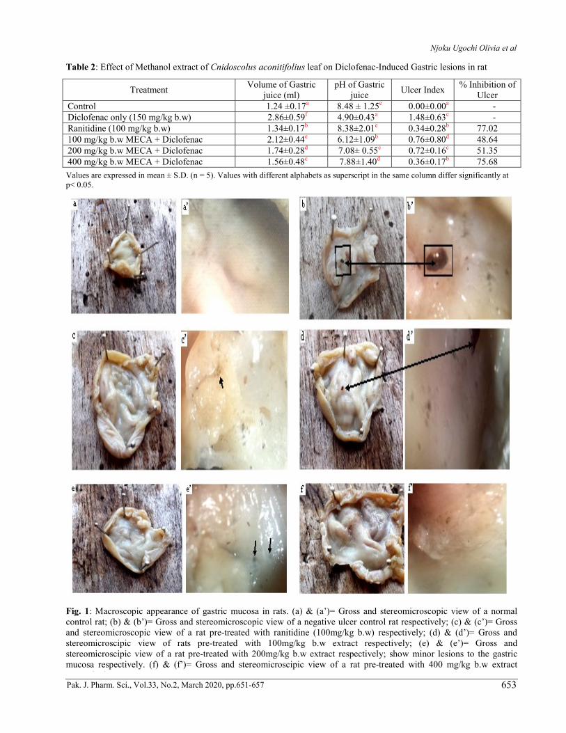
Table 2: Effect of Methanol extract of Cnidoscolus aconitifolius leaf on Diclofenac-Induced Gastric lesions in rat
Treatment Volume of Gastric
juice (ml) pH of Gastric
juice Ulcer Index
% Inhibition of Ulcer
Control 1.24 ±0.17a 8.48 ± 1.25e 0.00±0.00a - Diclofenac only (150 mg/kg b.w) 2.86±0.59f 4.90±0.43a 1.48±0.63e - Ranitidine (100 mg/kg b.w) 1.34±0.17b 8.38±2.01e 0.34±0.28b 77.02 100 mg/kg b.w MECA + Diclofenac 2.12±0.44e 6.12±1.09b 0.76±0.80d 48.64 200 mg/kg b.w MECA + Diclofenac 1.74±0.28d 7.08± 0.55c 0.72±0.16c 51.35 400 mg/kg b.w MECA + Diclofenac 1.56±0.48c 7.88±1.40d 0.36±0.17b 75.68
Values are expressed in mean ± S.D. (n = 5). Values with different alphabets as superscript in the same column differ significantly at p< 0.05.
Fig. 1: Macroscopic appearance of gastric mucosa in rats. (a) & (a’)= Gross and stereomicroscopic view of a normal control rat; (b) & (b’)= Gross and stereomicroscopic view of a negative ulcer control rat respectively; (c) & (c’)= Gross and stereomicroscopic view of a rat pre-treated with ranitidine (100mg/kg b.w) respectively; (d) & (d’)= Gross and stereomicroscipic view of rats pre-treated with 100mg/kg b.w extract respectively; (e) & (e’)= Gross and stereomicroscipic view of a rat pre-treated with 200mg/kg b.w extract respectively; show minor lesions to the gastric mucosa respectively. (f) & (f’)= Gross and stereomicroscipic view of a rat pre-treated with 400 mg/kg b.w extract respectively.

Protective effect of Cnidoscolus aconitifolius leaves against diclofenac-induced gastric mucosal damage
Pak. J. Pharm. Sci., Vol.33, No.2, March 2020, pp.651-657 654
Percentage protection =
Uc:Ulcer index in control Ut: Ulcer index in test Histopatological studies The stomachs of the scarified rats were immersed in 10% formalin solution. The fixed specimens were trimmed, washed and dehydrated in ascending grades of alcohol. The Specimens were then cleared in xylol, embedded in paraffin, sectioned at 4-6 microns thickness and stained
with Heamtoxylin and Eosin for examination as described by Drury et al. (1967). STATISTICAL ANALYSIS The results obtained were expressed as mean ± SD and were analysed using statistical product and service solutions (SPSS) version 16. Tests of statistical significance were carried out using one-way Analysis of Variance (ANOVA). P values < 0.05 were considered statistically significant.
Fig. 2: Haematoxylin and eosin stained histological section of gastric ulcer
A: Histological section of a normal control rat: intact gastric pits (black arrow), normal gastric walls and no lesion to the gastric mucosa are observed. B: Histological section of ulcer negative control rat: extensive mucosal damage, severe loss of surface epithelium and spotty haemorrhagic lesions are seen. C: Histological section of rat pre-treated with Ranitidine (100 mg/kg b.w): mild mucosal damage, with gastric wall appearance similar to normal. D: Histological section of a rat pre-treated with 100 mg/kg b.w of extract: mild gastric mucosa lesions, mild loss of epithelium and inflammatory infiltrate, as well as chief cells are seen. There is absence of cellular debris in the lumina. E: Histological section of a rat pre-treated with 200 mg/kg b.w. of extract showing: typical stomach region with mild granulation of tissue (blue arrow) characterized by the appearance of bumpy thickened structure in the mucosa are visible. The gastric pits are intact with good formation; cellular debris is also found around the superficial zone with numerous degenerate polymorphs (black arrow). F: Histological section of a rat pre-treated with 400 mg/kg b.w of extract: the intact muscularis mucosa is seen, similar to normal (star).
Njoku Ugochi Olivia et al
Pak. J. Pharm. Sci., Vol.33, No.2, March 2020, pp.651-657 655
RESULT Percentage yield Cnidoscolus aconitifolius leaves yielded 29.94g of the extract which represents 5.99% of the starting plant material. Phytochemical composition of the methanol extract of cnidoscolus aconitifolius leaf Preliminary phytochemical screening for some chemical constituents of the extract revealed the presence of soluble carbohydrate, flavonoids, phenols, terpenoids, alkaloids, tannins, steroids and saponins. Reducing sugar and glycosides were not detected as shown in table 1. Effect of methanol extract of cnidoscolus aconitifolius leaves on gastric juice ph, gastric juice volume gastric ulcer index and percentage ulcer inhibition (percentage ulcer protection) on diclofenac-induced ulcerogenic rats The gastro protective activity of Cnidoscolus aconitifolius leaf extract on diclofenac-induced gastric ulcer in malewistar rats is shown in table 2. Treatment of rats with diclofenac caused an increase in gastric juice volume and extensive gastric ulcers in the glandular mucosa of stomach, whereas marked decreases in pH of the gastric juice were observed when compared to normal control. Pretreatment of rats with Cnidoscolus aconitifolius leaves extract in the doses of 100, 200 and 400mg/kg b.w significantly (p<0.05) suppressed diclofenac induced increases in gastric juice secretion, gastric acidity and formation of gastric lesions in a dose dependent manner when compared to the ulcer control. In rats pre-treated with Cnidoscolus aconitifolius leaves extract, the gastro protection of the stomach mucosa were in the range of 48.64 to 75.68% as doses increased from 100 to 400mg/kg b.w. The efficacy of highest tested dose (400mg/kg b.w.) of Cnidoscolus aconitifolius leaves extract was similar to ranitidine (100mg/kg b.w.) Macroscopic examination of diclofenac-induced gastric ulcer in rats Essentially similar to normal gastric mucosa is presented in pre-treatment groups as shown in fig. 1. Treatment with diclofenac produced gross ulceration and visible black hemorrhagic lesions of gastric mucosa as revealed in the gross (b) and stereomicroscopic (b’) view of ulcer control rats. Gross and stereomicroscopic view of gastric mucosa of rats in pre-treatment groups show remarkable reduction of lesions relative to the ulcer control group while increasing doses of extract, decreased severity of the injuries. Histopatological evaluation of gastric lesion The histological study of diclofenac induced gastric mucosa damage in rats is shown in fig. 2.
Administration of diclofenac caused extensive ulceration of gastric mucosa and destruction of the epithelial lining in the group that received diclofenac only. Pretreatment with graded doses of Cnidoscolus aconitifolius leaf extract, demonstrated remarkable inhibition of histopathological alterations induced by diclofenac treatment when compared with the ulcer negative control group. At the dose of 400mg/kg b.w., extract and Ranitidine (100mg/kg b.w.) grossly inhibited formation of gastric ulcer ascertained by the appearance of gastric mucosa similar to normal. DISCUSSION Peptic ulcer disease is a chronic inflammatory disease characterized by the ulceration of upper gastrointestinal tract where parietal cells where secrete hydrochloric acid and pepsin (Al-Attar, 2011). However, secretion of gastric acid is central component in the pathogenesis of peptic ulcers (Luiz-Ferreira et al., 2010) by interfering with haemostasis (Green, 1978). More so, gastric acid can inhibit several growth factors that are important for the maintenance of mucosal integrity and for the repair of superficial injury, since these growth factors (e.g. fibroblast growth factor) are acid-labile (Szabo et al., 1994). In general, the properties of NSAIDs that contribute to ulcerogenesis are categorized into topical irritancy (such as increase in gastrointestinal permeability, decrease in gel hydrophobicity, uncoupling of oxidative phosphorylation in mitochondria etc.) and inhibition of prostaglandin synthesis. In this study, administration of diclofenac produced marked increases in gastric juice volume and gastric acid secretion (indicated by low pH) in the ulcer control group when compared to normal control as shown in table 2. Also, histological examination of gastric wall of ulcer control group revealed extensive gastric mucosa damage when compared to normal control. This may be attributed to topical irritant properties of diclofenac as well as its ability to inhibit the synthesis of prostaglandin with overall effect of gastro-duodenal ulcers and impairment of mucosal defence, consequently, indicating gastrotoxicity of diclofenac. Pre-treatment of rats with ranitidine and graded doses of methanol extract of Cnidoscolus actinifolius leaves (100, 200 and 400mg/kg b.w. respectively) significantly (p<0.05) suppressed the diclofenac-induced increases in gastric volume and gastric acid in a dose related manner. Although recent advances have highlighted the multi-factorial pathogenesis of peptic ulcer, secretions of gastric acid is still recognized as a central component of the disease (Luiz-Ferreira et al., 2010). The antiulcer effect of Cnidoscolus aconitifolius could be due to its phytochemical endowment. The presence of phenols and saponins in this plant extract agrees with the findings of Azeez et al. (2010). Flavonoids are a group of

Protective effect of Cnidoscolus aconitifolius leaves against diclofenac-induced gastric mucosal damage
Pak. J. Pharm. Sci., Vol.33, No.2, March 2020, pp.651-657 656
polyphenolic compounds, which exhibit several pharmacological effects in the human body such as anti-inflammatory, anti-hepatotoxic, anti-ulcer, anti-allergic, anti-viral and anti-cancer activities (Umamaheswari and Sangeetha, 2015). The presence of tannins in the Cnidoscolus aconitifolius indicates the astringent properties which facilitate wound healing, good blood circulation and protects the kidney from inflammation (Ross-Ibara and Molina, 2002). The leaf of Cnidoscolus aconitifolius contains appreciable amount of phenolics along with other biologically active constituents. The gastro protection observed in this study could be related to the presence of phenolics and flavonoids in the extract which demonstrated antioxidant activities against cellular damage. Barros et al. (2008) reported that phenolic compounds have an antiulcerogenic effect related to cytoprotective activity. Moreover, Kahraman et al. (2003) suggested that flavonoid quercetin promotes a decrease in ulcerative lesions due to its antioxidant effect. In addition, Nwafor et al. (2003) reported that tannins may precipitate micro proteins at the side of the ulcer which forms an impervious protective pellicle over the lining to render it less permeable to toxic substances and more resistant to attack of proteolytic enzymes. Also, Lewis and Hansan (1991) and Aguwa and Okunji (1986) reported that triterpenes and saponins possess antiulcer activity through the formation of mucus and inhibitory action on PGF2α. The antioxidant properties of phenols prevent oxidative damage to biomolecules like DNA, lipids, and protein with a specific role in the prevention of chronic diseases such as cancer and cardiovascular diseases (Oyedemi et al., 2012). CONCLUSION The result of the present study indicates that oral administration of Cnidoscolus aconitifolius leaf extract had a significant and dose dependent inhibition of ulceration. The anti-ulcerogenic effects of the extract could be attributed to its phytoconstituents. REFERENCES Adaramoye AO, Aluko A and Oyagbemi AA (2011).
Cnidoscolus aconitifolius leaf extract protects against hepatic damage induced by chronic ethanol administration in Wistar rats. Alcohol Alcohol, 46: 451-8.
Aguwa CN and Okonji CO (1986). Gastrointestinal studies of Pyrenacanthystaudii leaf extracts. J. Ethnopharmacol, 15: 45-55.
Al-Attar MA (2011). Protective effect of Avicennia alba leaves extract on gastric mucosal damage induced by ethanol. Res. J. Med. Plants, 5: 477-490.
Awoyinka AO, Balogun IO and Ogunnowo AA (2007). Phytochemical screening and in vitro bioactivity of
Cnidoscolus aconitifolius (Euphorbiaceae) J. Med. Plant Res., 1: 063-65.
Azeez IO, Oyagbemi AA, Oyeyemi MO and Odetola AA (2010). Ameliorative effects of Cnidoscolus aconitifolius on alloxan toxicity in wistar rats. Afr. Health Sci., 10: 283-291.
Barros PB, Lemos M, Maistro EL, Leite MF, Sousa JPB, Bastos JK and Andrade SF SF (2008). Evaluation of antiulcer activity of the main phenolic acids found in Brazillian green Propolis. J. Ethnopharmacol., 120: 372-377.
Bigheti AE, Antonio MA, Kohn IK, Rehder VIG, Foglio MA, Posserti A, Vilela I and Carvalho JE (2005). Anti-ulcerogenic activity of a crude hydroalcoholic extract and coumarin isolated from Mikaniala evigataschutz Bip. Phytomedicine, 12: 72-77.
Cos P, Vlietinck AR, Berghe DV and Maes L (2006). Anti-infective potential of natural products: How to develop a stronger in vitro proof-of-concept. J. Ethnopharmacol., 106: 290-302.
Dharmani P and Paiit G (2006). Exploring Indian medicinal plants for antiulcer activity. Ind. J. Pharmacol., 38(2): 95-99.
Drury RA, Wallington A and Cameroun SR (1967). In: Carlleton’s Histological Techniques. Oxford University Press, New York, USA, pp.1-420.
Green FW Jr, Kaplan MM, Curtis LE and Levine PH (1978). Effect of acid and pepsin on blood coagulation and platelet aggregation. A possible contributor prolonged gastroduodenal mucosal hemorrhage. Gastroenterology, 74(1): 38-43.
Harborne JB (1973). Phytochemical Methods: A Guide to Modern Techniques of Plant Analysis. Chapman and Hall Ltd, London. p.279.
Harborne JB (1998). Phytochemical methods: A guide to modern technique of plant analysis. Champman and Hall, London, UK.
Kahraman A, Erkasap N, Koken T, Serteser M, Aktepe F and Erkasa P (2003). The antioxidative and antihistaminic properties of quercetin in ethanol induced gastric lesions. Toxicology, 183: 133-42.
Kiranmai M, Sri BU, Reddy DS, Kumar CBM and Ibrahim M (2012). Evaluation of total phenolic contents and antiulcerogenic activity of root bark of Azadirachta indica. Glob. J. Med. Res., 12(6): 23-29.
Lewis DA and Hanson D (1991). Anti-ulcer drugs of plant origin. Prog. Med. Chem., 28: 208-210.
Luiz-Ferreira A, De Ameida ACA and Cola M (2010). Mechanisms of the gastric antiulcerogenic activity of Anarcadiumhumile on ethanol-induced acute gastric mucosal injury in rats. Molecules, 15(10): 7153-66.
Main IH and Whittle BJ (1975). Investigation of the vasodilator and antisecretory role of prostaglandins in the rat gastric mucosa by use of non-steroidal anti-inflammatory drugs. Br. J. Pharmacol. 53(2): 217-224.
Moghadamtousi SZ, Fadaeinasab M, Nikza S, Mohan G, Ali HM and Kadir HA (2015). Annon muricata
Njoku Ugochi Olivia et al
Pak. J. Pharm. Sci., Vol.33, No.2, March 2020, pp.651-657 657
(Annonaceae): A review of its traditional uses, isolated acetogenins and biological activities. Int. J. Mol. Sci., 16: 15625-15658.
Nwafor SV and Akah PA (2003). Effect of methanolic leaf extract of Cissampelos mucionata A rich against indomethacin-induced ulcer in rats. Indian J. Exp. Biol., 41: 181-183.
Oyagbemi AA and Odetola AA (2013). Hepatoprotective and nephroprotective effects of Cnidoscolus aconitifolius in protein energy malnutrition induced liver and kidney damage. Pharmacogn. Res., 5(4): 260-264.
Oyedemi SO, Oyedemi BO, Arowosegbe S and Afolayan AJ (2012). Phytochemical analysis and medicinal potentials of hydroalcoholic extract from Curstisia dentata (Burm.f) C.A. Sm stem bark. Int. J. Mol. Sci., 13: 6189-6203.
Rezq AA and Elmallh MM (2010). Antiulcer effect of Cinnamon and Chamomile Aqueous extract in rats models. J. Am. Sci., 6(12): 209-216.
Ross-Ibarra J and Molina-Cruz A (2002). The ethnobotany of Chaya (Cnidoscolus aconitifolius SSP. Aconitifolius breckon): A nutritious maya vegetable. Econ. Bot., 56(4): 350-365.
Sasidharan S, Chen Y, Saravanan D, Sundram KM and Latha LY (2011). Extraction, isolation and characterization of bioactive compounds from plants'
extracts. Afr. J. Tradit Complement altren Med., 8(1): 1-10.
Seo PJ, Kim N, Kim JH, Lee BH, Nam RH, Lee HS,
Park JH, Lee MK, Chang H, Jung HC and Song IS (2012). Comparison of indomethacin, diclofenac and aspirin-induced gastric damage according to Age in rats. Gut. Liver, 6(2): 210-217.
Soni A and Sosa S (2013). Phytochemical analysis and free radical scavenging potential of herbal and medicinal plant extrats. J. Pharmacogn. Phytochem., 2(4): 22-24.
Szabo S, Folkman J, Vattay P, Morales RE, Pinkus GS and Kato K (1994). Accelerated healing of duodenal ulcers by oral administration of a basic fibroblast growth factor in rats. Gastroenterology, 106: 1106-1111.
Trease GE and Evans WC (2002). Pharmacognosy. 15th Ed. Saunder Publishers, London pp. 42 - 44, 221 -229, 246-249, 404 -306, 331-332, 391-393.
Umamaheswari S and Sangeetha KSS (2015). Anti-Inflammatory Effect of Selected Dihydroxyflavones. J. Clin. Diagn. Res., 9(5): FF05-FF07.
Vinothapooshan G and Sundar K (2010). Antiulcer activity of Mimosa pudica leaves against gastric ulcer in rats. Res. J. Pharm. Biol. Chem. Sci, 1(4):606-14.
Wallace JL (2000). How do NSAIDs cause ulcer disease? Baillieres Best Pract. Res. Clin. Gastroenterology, 14(1): 147-159.