Protective effect of Aquilegia vulgaris (L.) against lead acetate-induced oxidative stress in rats
7
Protective effect of Aquilegia vulgaris (L.) against lead acetate-induced oxidative stress in rats Aziza A. El-Nekeety a , Ahmed A. El-Kady a , Mahmoud S. Soliman b , Nabila S. Hassan c , Mosaad A. Abdel-Wahhab a, * a Food Toxicology and Contaminants Dept., National Research Center, Dokki, Cairo, Egypt b Medicinal Chemistry Dept., National Research Center, Dokki, Cairo, Egypt c Pathology Dept. National Research Center, Dokki, Cairo, Egypt article info Article history: Received 31 March 2009 Accepted 3 June 2009 Keywords: Aquilegia vulgaris Lead acetate Oxidative stress Liver Kidney abstract Oxidative stress has been proposed as a possible mechanism involved in lead toxicity. The current study was carried out to evaluate the antioxidant activity of the ethanol extract of Aquilegia vulgaris (L.) against lead acetate (LA)-induced oxidative stress in male rats. Tested animals were treated orally with A. vulgaris extract (100 ppm) in combination with, before, or after LA treatment (20 ppm). The results indicated that the extract alone did not induce any significant changes in body weight gain, food intake, serum bio- chemical chemistry or the histological picture of the liver and kidney. However, it increased significantly the level of Glutathione (GSH). On the other hand, LA decreased food intake, body weight gain and induced oxidative stress as indicated by the significant changes in serum biochemical parameters and histological picture of liver and kidney and increased lipid peroxide and reduces GSH levels in liver tis- sues. The extract succeeded to improve the histological pictures of liver and kidney and the biochemical parameters towards the normal values of the control. Moreover, this improvement was pronounced in the animals treated with the extract after LA intoxication. Crown Copyright Ó 2009 Published by Elsevier Ltd. All rights reserved. 1. Introduction Lead is a pervasive and persistent environmental pollutant that can be detected in almost all phases of environment and biological systems. Lead is widely used in industry and life for its malleabil- ity, resistance to corrosion, and low melting point. Lead constitutes most abundant non essential element in the human organism, due to its dispersion in ambient air, in many foods, in drinking water, and in dust. Humans have used lead since ancient times. However, the quantity of lead used in the 20th century far surpasses the total consumption in all previous eras. This is mainly because of the industrial applications especially the consumption of vast quanti- ties of lead as an anti-knock agent in gasoline (Landrigan et al., 2000). Although lead is one of the most useful metals, it is also one of the most toxic (Shotyk and Le Roux, 2005). Several re- searches indicated that lead can cause neurological, hematological, gastrointestinal, reproductive, circulatory, and immunological pathologies (Patrick, 2006). Moreover, lead acetate and lead phos- phate are listed as reasonably anticipated human carcinogens (NIE- HS, 1994), and inorganic lead compounds were classified as 2B chemicals by IARC (IARC, 1987; Pulido and Parrish, 2003). It is well documented that lead has many undesired effects, including neu- rological (Royce et al., 1990), behavioral (Shafiq-ur-Rehman, 1991), respiratory (Hillam and Ozkan, 1986), visual (Winneke et al., 1988), growth retardation (Shukla et al., 1991), hematologi- cal (Falke and Xwennis, 1990), immunological (Sroczynski et al., 1987), renal (Vyskocil et al., 1989, 1991), hepatic (Honchel et al.,1991; Hao et al., 2002) and reproductive disfunctions (March- lewicz et al., 1993; Winder, 1993). It was reported that lead increased the level of lipid peroxidation (Upasani et al., 2001). Moreover, recent studies showed that lead inhibit the activities of antioxidant enzymes, including glutathione peroxidase, catalase and superoxide dismutase (Bolin et al., 2006; Ercal et al., 2001). Furthermore, generation of reactive oxygen species (ROS), stimula- tion of lipid peroxidation and depletion of antioxidant reserves have been postulated to be major contributors to lead-exposure re- lated diseases (Patrick, 2006; Silbergeld et al., 2000). Aquilegia vulgaris (L.) (Ranunculaceae) is a perennial herb indig- enous in central and southern Europe, Asia and Africa. Decoction from leaves and stems of A. vulgaris has been used in folk medicine against liver and bile duct disorders, especially for the treatment of jaundice, and chronic skin inflammation. Some researchers were isolated and identified several flavonoids (Bylka and Matławska, 1997a,b; Bylka, 2001; Bylka et al., 2002) and phenolic acids (Drost-Karbowska et al., 1996) in aerial parts of the plant as well 0278-6915/$ - see front matter Crown Copyright Ó 2009 Published by Elsevier Ltd. All rights reserved. doi:10.1016/j.fct.2009.06.019 * Corresponding author. Address: Food Toxicology and Contaminants Dept., National Research Center, El-Tahrir St., Dokki, Cairo 12622, Egypt. Tel.: +202 2283 1943; fax: +202 337 0931. E-mail address: [email protected] (M.A. Abdel-Wahhab). Food and Chemical Toxicology 47 (2009) 2209–2215 Contents lists available at ScienceDirect Food and Chemical Toxicology journal homepage: www.elsevier.com/locate/foodchemtox
-
Upload
independent -
Category
Documents
-
view
2 -
download
0
Transcript of Protective effect of Aquilegia vulgaris (L.) against lead acetate-induced oxidative stress in rats
Food and Chemical Toxicology 47 (2009) 2209–2215
Contents lists available at ScienceDirect
Food and Chemical Toxicology
journal homepage: www.elsevier .com/locate / foodchemtox
Protective effect of Aquilegia vulgaris (L.) against lead acetate-inducedoxidative stress in rats
Aziza A. El-Nekeety a, Ahmed A. El-Kady a, Mahmoud S. Soliman b, Nabila S. Hassan c,Mosaad A. Abdel-Wahhab a,*
a Food Toxicology and Contaminants Dept., National Research Center, Dokki, Cairo, Egyptb Medicinal Chemistry Dept., National Research Center, Dokki, Cairo, Egyptc Pathology Dept. National Research Center, Dokki, Cairo, Egypt
a r t i c l e i n f o a b s t r a c t
Article history:Received 31 March 2009Accepted 3 June 2009
Keywords:Aquilegia vulgarisLead acetateOxidative stressLiverKidney
0278-6915/$ - see front matter Crown Copyright � 2doi:10.1016/j.fct.2009.06.019
* Corresponding author. Address: Food ToxicologNational Research Center, El-Tahrir St., Dokki, Cairo 11943; fax: +202 337 0931.
E-mail address: [email protected]
Oxidative stress has been proposed as a possible mechanism involved in lead toxicity. The current studywas carried out to evaluate the antioxidant activity of the ethanol extract of Aquilegia vulgaris (L.) againstlead acetate (LA)-induced oxidative stress in male rats. Tested animals were treated orally with A. vulgarisextract (100 ppm) in combination with, before, or after LA treatment (20 ppm). The results indicated thatthe extract alone did not induce any significant changes in body weight gain, food intake, serum bio-chemical chemistry or the histological picture of the liver and kidney. However, it increased significantlythe level of Glutathione (GSH). On the other hand, LA decreased food intake, body weight gain andinduced oxidative stress as indicated by the significant changes in serum biochemical parameters andhistological picture of liver and kidney and increased lipid peroxide and reduces GSH levels in liver tis-sues. The extract succeeded to improve the histological pictures of liver and kidney and the biochemicalparameters towards the normal values of the control. Moreover, this improvement was pronounced inthe animals treated with the extract after LA intoxication.
Crown Copyright � 2009 Published by Elsevier Ltd. All rights reserved.
1. Introduction
Lead is a pervasive and persistent environmental pollutant thatcan be detected in almost all phases of environment and biologicalsystems. Lead is widely used in industry and life for its malleabil-ity, resistance to corrosion, and low melting point. Lead constitutesmost abundant non essential element in the human organism, dueto its dispersion in ambient air, in many foods, in drinking water,and in dust. Humans have used lead since ancient times. However,the quantity of lead used in the 20th century far surpasses the totalconsumption in all previous eras. This is mainly because of theindustrial applications especially the consumption of vast quanti-ties of lead as an anti-knock agent in gasoline (Landrigan et al.,2000). Although lead is one of the most useful metals, it is alsoone of the most toxic (Shotyk and Le Roux, 2005). Several re-searches indicated that lead can cause neurological, hematological,gastrointestinal, reproductive, circulatory, and immunologicalpathologies (Patrick, 2006). Moreover, lead acetate and lead phos-phate are listed as reasonably anticipated human carcinogens (NIE-HS, 1994), and inorganic lead compounds were classified as 2B
009 Published by Elsevier Ltd. All r
y and Contaminants Dept.,2622, Egypt. Tel.: +202 2283
(M.A. Abdel-Wahhab).
chemicals by IARC (IARC, 1987; Pulido and Parrish, 2003). It is welldocumented that lead has many undesired effects, including neu-rological (Royce et al., 1990), behavioral (Shafiq-ur-Rehman,1991), respiratory (Hillam and Ozkan, 1986), visual (Winnekeet al., 1988), growth retardation (Shukla et al., 1991), hematologi-cal (Falke and Xwennis, 1990), immunological (Sroczynski et al.,1987), renal (Vyskocil et al., 1989, 1991), hepatic (Honchelet al.,1991; Hao et al., 2002) and reproductive disfunctions (March-lewicz et al., 1993; Winder, 1993). It was reported that leadincreased the level of lipid peroxidation (Upasani et al., 2001).Moreover, recent studies showed that lead inhibit the activitiesof antioxidant enzymes, including glutathione peroxidase, catalaseand superoxide dismutase (Bolin et al., 2006; Ercal et al., 2001).Furthermore, generation of reactive oxygen species (ROS), stimula-tion of lipid peroxidation and depletion of antioxidant reserveshave been postulated to be major contributors to lead-exposure re-lated diseases (Patrick, 2006; Silbergeld et al., 2000).
Aquilegia vulgaris (L.) (Ranunculaceae) is a perennial herb indig-enous in central and southern Europe, Asia and Africa. Decoctionfrom leaves and stems of A. vulgaris has been used in folk medicineagainst liver and bile duct disorders, especially for the treatment ofjaundice, and chronic skin inflammation. Some researchers wereisolated and identified several flavonoids (Bylka and Matławska,1997a,b; Bylka, 2001; Bylka et al., 2002) and phenolic acids(Drost-Karbowska et al., 1996) in aerial parts of the plant as well
ights reserved.
2210 A.A. El-Nekeety et al. / Food and Chemical Toxicology 47 (2009) 2209–2215
as alkaloids in roots (Szaufer-Hajdrych et al., 1998). The ethanolextract (EE) of A. vulgaris and isocytisoside was found to protectagainst hepatotoxicity induced by carbon tetrachloride in rats(Adamska et al., 2003). The aim of the current study was to evalu-ate the protective effect of the ethanol extract of A. vulgaris againstlead toxicity in rats.
aaa
b
a a
0
5
10
15
20
25
Control Ext LA Ext +LA Ext thenLA
LA thenExtTreatments
g/ra
t
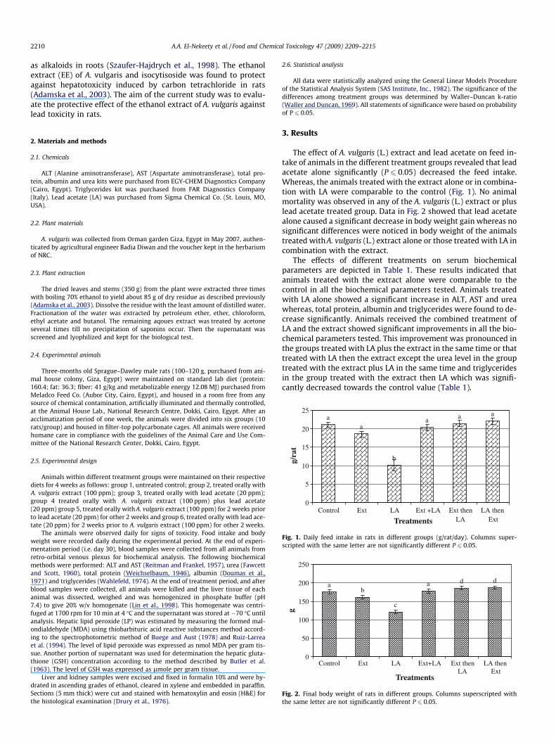
Fig. 1. Daily feed intake in rats in different groups (g/rat/day). Columns super-scripted with the same letter are not significantly different P 6 0.05.
dda
c
ba
0
50
100
150
200
250
Control Ext LA Ext+LA Ext thenLA
LA thenExt
Treatments
g
Fig. 2. Final body weight of rats in different groups. Columns superscripted withthe same letter are not significantly different P 6 0.05.
2. Materials and methods
2.1. Chemicals
ALT (Alanine aminotransferase), AST (Aspartate aminotransferase), total pro-tein, albumin and urea kits were purchased from EGY-CHEM Diagnostics Company(Cairo, Egypt). Triglycerides kit was purchased from FAR Diagnostics Company(Italy). Lead acetate (LA) was purchased from Sigma Chemical Co. (St. Louis, MO,USA).
2.2. Plant materials
A. vulgaris was collected from Orman garden Giza, Egypt in May 2007, authen-ticated by agricultural engineer Badia Diwan and the voucher kept in the herbariumof NRC.
2.3. Plant extraction
The dried leaves and stems (350 g) from the plant were extracted three timeswith boiling 70% ethanol to yield about 85 g of dry residue as described previously(Adamska et al., 2003). Dissolve the residue with the least amount of distilled water.Fractionation of the water was extracted by petroleum ether, ether, chloroform,ethyl acetate and butanol. The remaining aqoues extract was treated by acetoneseveral times till no precipitation of saponins occur. Then the supernatant wasscreened and lyophilized and kept for the biological test.
2.4. Experimental animals
Three-months old Sprague–Dawley male rats (100–120 g, purchased from ani-mal house colony, Giza, Egypt) were maintained on standard lab diet (protein:160.4; fat: 36.3; fiber: 41 g/kg and metabolizable energy 12.08 MJ) purchased fromMeladco Feed Co. (Aubor City, Cairo, Egypt), and housed in a room free from anysource of chemical contamination, artificially illuminated and thermally controlled,at the Animal House Lab., National Research Centre, Dokki, Cairo, Egypt. After anacclimatization period of one week, the animals were divided into six groups (10rats/group) and housed in filter-top polycarbonate cages. All animals were receivedhumane care in compliance with the guidelines of the Animal Care and Use Com-mittee of the National Research Center, Dokki, Cairo, Egypt.
2.5. Experimental design
Animals within different treatment groups were maintained on their respectivediets for 4 weeks as follows: group 1, untreated control; group 2, treated orally withA. vulgaris extract (100 ppm); group 3, treated orally with lead acetate (20 ppm);group 4 treated orally with A. vulgaris extract (100 ppm) plus lead acetate(20 ppm) group 5, treated orally with A. vulgaris extract (100 ppm) for 2 weeks priorto lead acetate (20 ppm) for other 2 weeks and group 6, treated orally with lead ace-tate (20 ppm) for 2 weeks prior to A. vulgaris extract (100 ppm) for other 2 weeks.
The animals were observed daily for signs of toxicity. Food intake and bodyweight were recorded daily during the experimental period. At the end of experi-mentation period (i.e. day 30), blood samples were collected from all animals fromretro-orbital venous plexus for biochemical analysis. The following biochemicalmethods were performed: ALT and AST (Reitman and Frankel, 1957), urea (Fawcettand Scott, 1960), total protein (Weichselbaum, 1946), albumin (Doumas et al.,1971) and triglycerides (Wahlefeld, 1974). At the end of treatment period, and afterblood samples were collected, all animals were killed and the liver tissue of eachanimal was dissected, weighed and was homogenized in phosphate buffer (pH7.4) to give 20% w/v homogenate (Lin et al., 1998). This homogenate was centri-fuged at 1700 rpm for 10 min at 4 �C and the supernatant was stored at �70 �C untilanalysis. Hepatic lipid peroxide (LP) was estimated by measuring the formed mal-ondialdehyde (MDA) using thiobarbituric acid reactive substances method accord-ing to the spectrophotometric method of Buege and Aust (1978) and Ruiz-Larreaet al. (1994). The level of lipid peroxide was expressed as nmol MDA per gram tis-sue. Another portion of supernatant was used for determination the hepatic gluta-thione (GSH) concentration according to the method described by Butler et al.(1963). The level of GSH was expressed as lmole per gram tissue.
Liver and kidney samples were excised and fixed in formalin 10% and were hy-drated in ascending grades of ethanol, cleared in xylene and embedded in paraffin.Sections (5 mm thick) were cut and stained with hematoxylin and eosin (H&E) forthe histological examination (Drury et al., 1976).
2.6. Statistical analysis
All data were statistically analyzed using the General Linear Models Procedureof the Statistical Analysis System (SAS Institute, Inc., 1982). The significance of thedifferences among treatment groups was determined by Waller–Duncan k-ratio(Waller and Duncan, 1969). All statements of significance were based on probabilityof P 6 0.05.
3. Results
The effect of A. vulgaris (L.) extract and lead acetate on feed in-take of animals in the different treatment groups revealed that leadacetate alone significantly (P 6 0.05) decreased the feed intake.Whereas, the animals treated with the extract alone or in combina-tion with LA were comparable to the control (Fig. 1). No animalmortality was observed in any of the A. vulgaris (L.) extract or pluslead acetate treated group. Data in Fig. 2 showed that lead acetatealone caused a significant decrease in body weight gain whereas nosignificant differences were noticed in body weight of the animalstreated with A. vulgaris (L.) extract alone or those treated with LA incombination with the extract.
The effects of different treatments on serum biochemicalparameters are depicted in Table 1. These results indicated thatanimals treated with the extract alone were comparable to thecontrol in all the biochemical parameters tested. Animals treatedwith LA alone showed a significant increase in ALT, AST and ureawhereas, total protein, albumin and triglycerides were found to de-crease significantly. Animals received the combined treatment ofLA and the extract showed significant improvements in all the bio-chemical parameters tested. This improvement was pronounced inthe groups treated with LA plus the extract in the same time or thattreated with LA then the extract except the urea level in the grouptreated with the extract plus LA in the same time and triglyceridesin the group treated with the extract then LA which was signifi-cantly decreased towards the control value (Table 1).
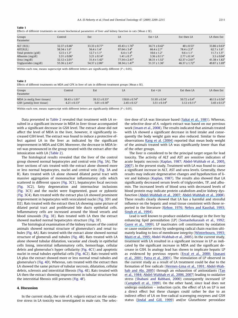
Table 1Effects of different treatments on serum biochemical parameters of liver and kidney function in rats (Mean ± SE).
Groups Control Ext LA Ext + LA Ext then LA LA then ExtParameter
ALT (IU/L) 32.37 ± 0.46a 33.33 ± 0.77a 45.43 ± 1.76b 34.71 ± 0.42a 40 ± 0.53c 33.86 ± 0.63a
AST(IU/L) 58.34 ± 1.6a 56.4 ± 1.4a 97.64 ± 3.4b 66.4 ± 2.7c 78.4 ± 2.5d 62.7 ± 1.6a
Total protein (g/dl) 12.5 ± 1.3a 12.7 ± 1.1a 6.4 ± 1.4b 10.6 ± 1.2c 9.6 ± 1.1c 11.7 ± 1.5a
Albumin (mg/dl) 3.31 ± 0.06a 3.21 ± 0.14a 1.41 ± 0.1b 3.36 ± 0.11a 2.77 ± 0.14c 3.5 ± 0.04a
Urea (mg/dl) 32.12 ± 2.01a 31.4 ± 1.42a 77.54 ± 2.41b 38.31 ± 1.52c 62.37 ± 2.61d 41.38 ± 1.82e
Triglycerides (mg/dl) 55.36 ± 2.41a 54.37 ± 2.06a 38.34 ± 1.47b 51.37 ± 1.36c 46.37 ± 1.72d 49.87 ± 1.69d
Within each row, means superscript with different letters are significantly different (P 6 0.05).
Table 2Effects of different treatments on MDA and GPX in liver of rats in different treatment groups (Mean ± SE).
Groups Control Ext LA Ext + LA Ext then LA LA then ExtParameter
MDA (n mol/g liver tissues) 38.43 ± 1.01a 39.12 ± 0.33a 150.8 ± 0.74b 51.95 ± 0.34c 59.72 ± 0.4d 40.13 ± 0.56a
GSH (lmol/g liver tissue) 4.21 ± 0.13a 5.01 ± 0.18b 2.45 ± 0.12c 3.51 ± 0.14d 3.12 ± 0.11d 3.45 ± 0.12d
Within each row, means superscript with different letters are significantly different (P 6 0.05).
A.A. El-Nekeety et al. / Food and Chemical Toxicology 47 (2009) 2209–2215 2211
Data presented in Table 2 revealed that treatment with LA re-sulted in a significant increase in MDA in liver tissue accompaniedwith a significant decrease in GSH level. The extract alone did notaffect the level of MDA in the liver, however, it significantly in-creased GSH level. The extract was found to induce a protective ef-fect against LA in the liver as indicated by the significantimprovement in MDA and GSH. Moreover, the decrease in MDA le-vel was pronounced in the group treated with the extract after theintoxication with LA (Table 2).
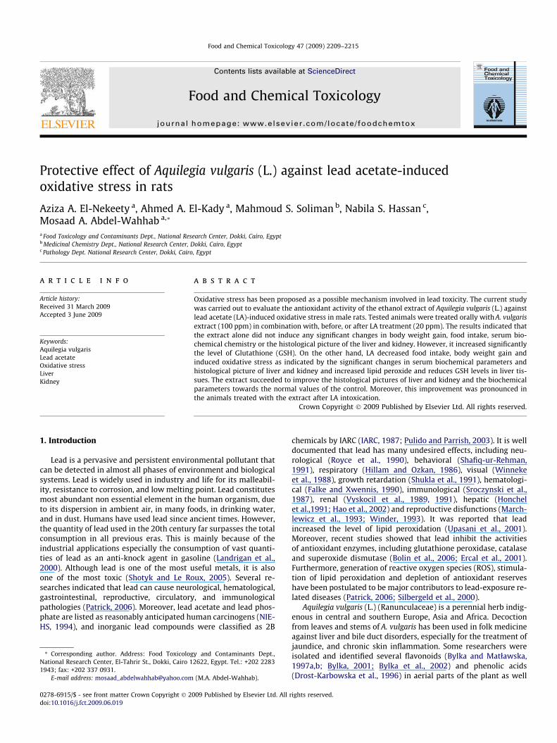
The histological results revealed that the liver of the controlgroup showed normal hepatocytes and central vein (Fig. 3A). Theliver sections of rats treated with the extract alone showed moreor less normal hepatocytes, nuclei and central vein (Fig. 3A andB). Rats treated with LA alone showed dilated portal tract withmassive aggregation of mononuclear inflammatory cells whichscattered in different spaces (Fig. 3C1), hepatocytes focal necrosis(Fig. 3C2), fatty degeneration and internuclear inclusions(Fig. 3C3) and the nuclei were fragmented, goast or pyknotic(Fig. 3C4). Rats treated with the extract plus LA showed prominentimprovement in hepatocytes with vesiculated nuclei (Fig. 3D1 andD2). Rats treated with the extract then LA showing same picture ofdilated portal tract and proliferated bile ducts epithelial cells.Inflammatory cells are increased around the blood vessels andblood sinusoids (Fig. 3E). Rats treated with LA then the extractshowed marked normal hepatocytes structure (Fig. 3F).
The histological examination of the kidney tissues of the controlanimals showed normal structure of glomerulus’s and renal tu-bules (Fig. 4A). Rats treated with the extract alone showed normalstructure of glomeruli and tubules (Fig. 4B). Rats treated with LAalone showed tubular dilatation, vacuolar and cloudy in epithelialcells lining, interstitial inflammatory cells, hemorrhage, cellulardebris and glomerulus’s hyper cellularity (Fig. 4C1) and apoptoticnuclei in renal tubules epithelial cells (Fig. 4C2). Rats treated withLA plus the extract showed more or less normal renal tubules andglomerulus’s (Fig. 4D). Whereas, rats treated with the extract thenLA showed the same picture of tubular necrosis, dilatation, cellulardebris, sclerosis and interstitial fibrosis (Fig. 4E). Rats treated withLA then the extract showing improvement in tubular structure butthe interstitial fibrosis still presents (Fig. 4F).
4. Discussion
In the current study, the role of A. vulgaris extract on the oxida-tive stress in LA toxicity was investigated in male rats. The selec-
tive dose of LA was literature based (Sakai et al., 1981). Whereas,the selective dose of A. vulgaris extract was based on our previouswork (Imam et al., 2008). The results indicated that animals treatedwith LA showed a significant decrease in feed intake and conse-quently the body weight gain was also reduced. Similar to theseobservations Kang et al. (2004) reported that mean body weightof the animals treated with LA was significantly lower than thatof the other groups.
The liver is considered to be the principal target organ for leadtoxicity. The activity of ALT and AST are sensitive indicators ofacute hepatic necrosis (Kaplan, 1987; Abdel-Wahhab et al., 2005,2007). In the present study, Treatment with LA was found to causea significant increase in ALT, AST and urea levels. Generally, theseresults may indicate degenerative changes and hypofunction of li-ver and kidneys (Kaplan, 1987). The results also showed that LAsignificantly decreased serum levels of triglycerides, TP, and albu-min. The increased levels of blood urea with decreased levels ofblood protein may indicate protein catabolism and/or kidney dys-function (Abdel-Wahhab et al., 2007; Abdel-Wahhab et al., 2008).These results clearly showed that LA has a harmful and stressfulinfluence on the hepatic and renal tissue consistent with those re-ported in the literature (Kehoe, 1976; Nehru and Kaushal, 1993;Singh et al., 1994).
Lead is well known to produce oxidative damage in the liver byenhancing lipid peroxidation (LP) (Somashekaraiah et al., 1992;Gurer et al., 1999). LP inactivates cell constituents by oxidationor cause oxidative stress by undergoing radical chain reaction ulti-mately leading to loss of membrane integrity (Winterbourn, 1993;Maiti et al., 1995; Abdel-Wahhab et al., 2005). In the current study,treatment with LA resulted in a significant increase in LP as indi-cated by the significant increase in MDA and the significant de-crease in GSH. In analogy lead has shown to implicate hepatic LPas evidenced by previous reports (Ercal et al., 2000; Upasaniet al., 2001; Patra et al., 2001). The stimulation of LP observed inthe current study as a result of LA treatment could be due to theformation of free radicals (Hermes-Lima et al., 1991; Abdel-Wah-hab and Aly, 2005) through an exhaustion of antioxidants (Tanet al., 1984; Abdel-Wahhab et al., 2006, 2007) leading to oxidativestress (Shabani and Rabbani, 2000) consequently increased LP(Campbell et al., 1999). On the other hand, since lead does notundergo oxidation – reduction cycle, the effect of LA on LP is nota direct effect but these changes could rather be due to anindirect effect of LA on free-radical scavenging enzymes and GSHstatus (Jindal and Gill, 1999) and/or Glutathione peroxidase
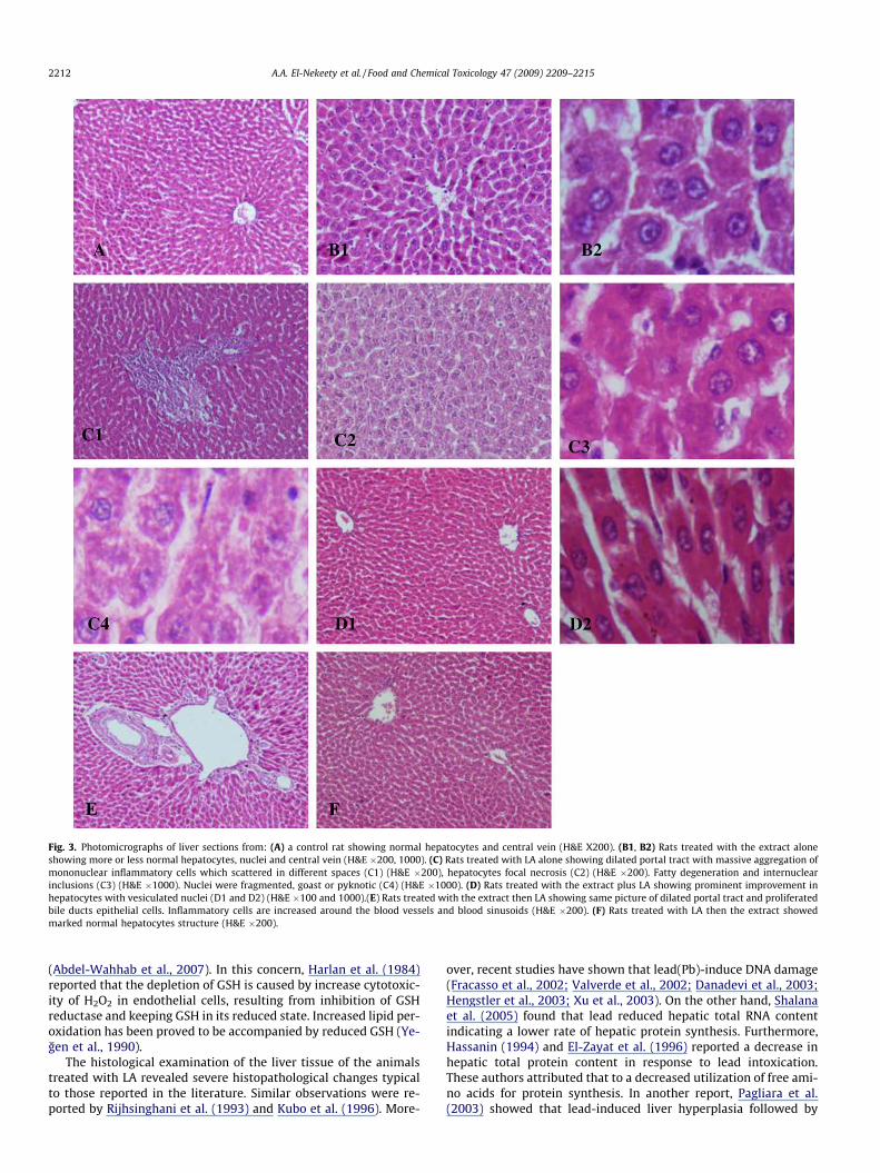
Fig. 3. Photomicrographs of liver sections from: (A) a control rat showing normal hepatocytes and central vein (H&E X200). (B1, B2) Rats treated with the extract aloneshowing more or less normal hepatocytes, nuclei and central vein (H&E �200, 1000). (C) Rats treated with LA alone showing dilated portal tract with massive aggregation ofmononuclear inflammatory cells which scattered in different spaces (C1) (H&E �200), hepatocytes focal necrosis (C2) (H&E �200). Fatty degeneration and internuclearinclusions (C3) (H&E �1000). Nuclei were fragmented, goast or pyknotic (C4) (H&E �1000). (D) Rats treated with the extract plus LA showing prominent improvement inhepatocytes with vesiculated nuclei (D1 and D2) (H&E �100 and 1000).(E) Rats treated with the extract then LA showing same picture of dilated portal tract and proliferatedbile ducts epithelial cells. Inflammatory cells are increased around the blood vessels and blood sinusoids (H&E �200). (F) Rats treated with LA then the extract showedmarked normal hepatocytes structure (H&E �200).
2212 A.A. El-Nekeety et al. / Food and Chemical Toxicology 47 (2009) 2209–2215
(Abdel-Wahhab et al., 2007). In this concern, Harlan et al. (1984)reported that the depletion of GSH is caused by increase cytotoxic-ity of H2O2 in endothelial cells, resulting from inhibition of GSHreductase and keeping GSH in its reduced state. Increased lipid per-oxidation has been proved to be accompanied by reduced GSH (Ye-gen et al., 1990).
The histological examination of the liver tissue of the animalstreated with LA revealed severe histopathological changes typicalto those reported in the literature. Similar observations were re-ported by Rijhsinghani et al. (1993) and Kubo et al. (1996). More-
over, recent studies have shown that lead(Pb)-induce DNA damage(Fracasso et al., 2002; Valverde et al., 2002; Danadevi et al., 2003;Hengstler et al., 2003; Xu et al., 2003). On the other hand, Shalanaet al. (2005) found that lead reduced hepatic total RNA contentindicating a lower rate of hepatic protein synthesis. Furthermore,Hassanin (1994) and El-Zayat et al. (1996) reported a decrease inhepatic total protein content in response to lead intoxication.These authors attributed that to a decreased utilization of free ami-no acids for protein synthesis. In another report, Pagliara et al.(2003) showed that lead-induced liver hyperplasia followed by
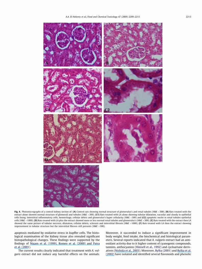
Fig. 4. Photomicrographs of a control kidney section of: (A) Control rats showing normal structure of glomerulus’s and renal tubules (H&E �300). (B) Rats treated with theextract alone showed normal structure of glomeruli and tubules (H&E �300). (C1) Rats treated with LA alone showing tubular dilatation, vacuolar and cloudy in epithelialcells lining. Interstitial inflammatory cells, hemorrhage, cellular debris and glomerulus’s hyper cellularity (H&E �300) and (C2) apoptotic nuclei in renal tubules epithelialcells (H&E �1000). (D) Rats treated with LA plus the extract showed more or less normal renal tubules and glomerulus’s (H&E �300). (E) Rats treated with the extract then LAshowed the same picture of tubular necrosis, dilatation, cellular debris, sclerosis and interstitial fibrosis (H&E �1000). (F) Rats treated with LA then the extract showingimprovement in tubular structure but the interstitial fibrosis still presents (H&E �300).
A.A. El-Nekeety et al. / Food and Chemical Toxicology 47 (2009) 2209–2215 2213
apoptosis mediated by oxidative stress in kupffer cells. The histo-logical examination of the kidney tissue also revealed significanthistopathological changes. These findings were supported by thefindings of Nigam et al. (1999), Romeo et al. (2000) and Patraet al. (2001).
The current results clearly indicated that treatment with A. vul-garis extract did not induce any harmful effects on the animals.
Moreover, it succeeded to induce a significant improvement inbody weight, feed intake, the biochemical and histological param-eters. Several reports indicated that A. vulgaris extract had an anti-oxidant activity due to it higher content of cyanogenic compounds,tannins, anthocyanins (Hänsell et al., 1992) and cycloartane deriv-atives (Nishida et al., 2003). Moreover, Bylka (2001) and Bylka et al.(2002) have isolated and identified several flavonoids and phenolic
2214 A.A. El-Nekeety et al. / Food and Chemical Toxicology 47 (2009) 2209–2215
acids (Drost-Karbowska et al., 1996) in the aerial parts of A. vulga-ris as well as alkaloids in roots (Szaufer-Hajdrych et al., 1998). Thepredominant compound was 40-methoxy-5,7-dihydroxyflavone, 6-C-glucopyranoside (isocytisoside) (Bylka and Matlawska, 1997a).
The ethanol extract of A. vulgaris and isocytisoside was found toprotect against hepatotoxicity induced by carbon tetrachloride inrats as assessed by inhibition of transaminases and sorbitol dehy-drogenase leakage to serum and by histopathological examination(Adamska et al., 2003). Bylka and Matlawska (1997a,b) reportedthat A. vulgaris is rich in compounds known to be strong antioxi-dants and it ameliorated liver damage induced by aflatoxin B1.Moreover, the TLC analysis of the ethyl acetate and the ethanol ex-tract revealed the presence of the following compounds: isocytiso-side (predominated in both extracts), isocytisoside 7-O-glucoside,isoorientin, orientin, isovitexin 40-O-glucoside, apigenin 70-Oruti-noside, apigenin 70-O-glucoside and apigenin (Bylka and Mat-lawska, 1997a,b). Additionally the ethanol extract containedphenolic acids: caffeic, ferulic, p-coumaric, resorcylic, p-hydroxy-benzoic, vanilic, sinapic and chlorogenic (Drost-Karbowska et al.,1996).
In the current study, co-treatment of LA and A. vulgaris extractresulted in a significant improvement in all biochemical parame-ters tested and the histological picture of the liver and kidney. Inthis regard, Jodynis-Liebert et al. (2006) hypothesized that someconstituents of A. vulgaris extract inhibited lipid peroxidation, pre-venting reduced glutathione depletion and the decrease in trans-aminases leakage to serum, therefore this extract may play aprotective role against LA-mediated liver injury. These results de-noted that A. vulgaris extract has been proved to have oxygen rad-ical scavenging and antioxidant properties.
5. Conclusion
The current study indicated that exposure to lead acetate couldgenerate free radicals which resulted in the elevation of hepatic li-pid peroxidation and in a reduction in the antioxidant enzyme Glu-tathione. A. vulgaris extract resulted in the restoration of thedifferent parameters tested. The protective effects of A. vulgaris ex-tract may be due to the radical scavenging activity of its compo-nents. Moreover, the protective role was more pronounced whenthe extract administrated after LA intoxication. Consequently, A.vulgaris extract is quite useful and reasonable in the treatment oflead toxicity.
Conflict of interest statement
The authors declare that there are no conflicts of interest.
References
Abdel-Wahhab, M.A., Aly, S.E., 2005. Antioxidant property of Nagilia Sativa (blackcumin) and Syzygium Aromaticum (clove) in rats during aflatoxicosis. J. Appl.Toxicol. 25, 218–223.
Abdel-Wahhab, M.A., Saeed, A., Hufner, A., 2005. NMR and radical scavengingactivities of patuletin from Urtica urens L. against aflatoxin B1. Pharm. Biol. 43(6), 515–525.
Abdel-Wahhab, M.A., Ahmed, H.H., Hagazi, M.M., 2006. Prevention of aflatoxin B1-initiated hepatotoxicity in rat by marine algae extracts. J. Appl. Toxicol. 26 (3),229–238.
Abdel-Wahhab, M.A., Abdel-Galil, M.M., Hassan, A.M., Hassan, N.H., Nada, S.A.,Saeed, A., El-Sayed, M.M., 2007. Zizyphus spina-christi extract protects againstaflatoxin B1-intitiated hepatic carcinogenicity. Afr. J. Trad. CAM 4 (3), 248–256.
Abdel-Wahhab, M.A., Abdel-Azim, S.H., El-Nekeety, A.A., 2008. Inulacrithmoides extract protect against ochratoxin A-induced oxidative stress,clastogenic and mutagenic alterations in male rats. Toxicon 52 (4), 566–573.
Adamska, T., Młynarczyk, W., Jodynis-Liebert, J., Bylka, W., Matławska, I., 2003.Hepatoprotective effect of the extract and isocytisoside from Aquilegia vulgaris.Phytother. Res. 17, 691–696.
Bolin, C.M., Basha, R., Cox, D., Zawia, N.H., Maloney, B., Lahiri, D.K., Cardozo-Pelaez,F., 2006. Exposure to lead and the developmental origin of oxidative DNAdamage in the aging brain. FASEB J. 20, 788–790.
Buege, J.A., Aust, S.D., 1978. Microsomal lipid peroxidation. Methods Enzymol. 52,302–315.
Butler, E., Duran, O., Mikus, K.B., 1963. Improved method for determination of bloodglutathione. J. Lab. Clin. Med. 61, 882–888.
Bylka, W., 2001. Isovitexin O-glucosides from Aquilegia vulgaris (L.). Acta Pol. Pharm.58, 273–275.
Bylka, W., Matławska, I., 1997a. Flavonoids from Aquilegia vulgaris L. Part I.Isocytisoside and its derivatives. Acta Pol. Pharm. 54 (4), 331–333.
Bylka, W., Matławska, I., 1997b. Flavonoids from Aquilegia vulgaris L. Part II.Derivatives of apigenin and luteolin. Acta Pol. Pharm. 54 (4), 335–337.
Bylka, W., Franski, R., Stobiecki, M., 2002. Differentiation between isomericacacetin-6-C-(60-O-malonyl) glucoside and acacetin-8-C-(60-Omalonyl)glucoside by using lower energy CID mass spectra. J. Mass Spectrom. 37, 648–650.
Campbell, A., Prassad, K.N., Bondy, S.C., 1999. Aluminum-induced oxidative eventsin cell lines: glioma are more responsive than neuroblastoma. Free Radic. Biol.Med. 26, 1166–1171.
Danadevi, K., Rozati, R., Saleha-Banu, B., Hanumanth-Rao, P., Grover, P., 2003. DNAdamage in workers exposed to lead using comet assay. Toxicology 187 (2–3),183–193.
Doumas, B.T., Watson, W.A., Bigga, H.G., 1971. Albumin standards and themeasurement of serum albumin with bromocresol green. Clin. Chem. Acta 31,87.
Drost-Karbowska, K., Szaufer-Hajdrych, M., Kowalewski, Z., Zgórka, G., 1996.Phenolic acids in Aquilegia vulgaris (Ranuculaceae). Herba Pol. 42, 21–24.
Drury, R.A., Wallington, E.A., Cancerson, R., 1976. Carlton’s HistopathologicalTechniques, fourth ed. Oxford University Press, Oxford, London, NewYork.
El-Zayat, E.M., El-Ymany, N.A., Kamel, Z.H., 1996. Combined supplementation ofzinc and vitamin C as protective agents against lead toxicity in growingmale albino rats. 1. Liver functions. J. Egypt Ger. Soc. Zool. 20 (A), 115–139.
Ercal, N., Neal, R., Treeratphan, P., Lutz, P.M., Hammond, T.C., Dennery, P.A., Spitz,D.R., 2000. A role for oxidative stress in suppressing serum immunoglobulinlevels in lead-exposed Fischer 344 rats. Arch. Environ. Contam. Toxicol. 39, 251–256.
Ercal, N., Gurer-Orhan, H., Aykin-Burns, N., 2001. Toxic metals and oxidative stresspart I: mechanisms involved in metal-induced oxidative damage. Curr. Top.Med. Chem. 1, 529–539.
Falke, H.E., Xwennis, W.C.M., 1990. Toxicity of lead acetate to female rabbits afterchronic subcutaneous administration. 1. Biochemical and clinical effects. Arch.Toxicol. 64, 322–329.
Fawcett, J.K., Scott, J.E., 1960. Determination of urea. J. Clin. Path. 13, 156–159.Fracasso, M.E., Perbellini, L., Solda, S., Talamini, G., Franceschetti, P., 2002. Lead
induced DNA strand breaks in lymphocytes of exposed workers: role of reactiveoxygen species and protein kinase C. Mutat. Res. 515 (1–2), 159–169.
Gurer, H., Neal, R., Yang, P., Oztezcan, S., Ercal, N., 1999. Captopril as an antioxidantin lead-exposed Fischer 344 rats. Hum. Exp. Toxicol. 18, 27–32.
Hänsell, R., Keller, K., Rimpler, H., Schneider, G., 1992. Handbuch derPharmaceutische Praxis, Bd. 4 Drogen A–D. Springer Verlag, Berlin. pp. 312–316.
Hao, S., Tian, P., Tang, W., Ru, B., 2002. Protective effect of extra metallothioninsfrom rabbit liver induced by zinc on toxicity of lead in rat primary hepatocyteculture. Wei Sheng Yan Jiu 31 (4), 229–231.
Harlan, J.M., Levine, J.D., Cullahan, K.S., Schwartz, B.R., Harker, L.A., 1984.Glutathione redox cycle protects cultured endothelial cells againstlysis by extracellularly generated by hydrogen peroxide. J. Clin. Invest. 73,706–713.
Hassanin, L.A.M., 1994. The effect of lead pollution on the susceptibility of rats toanticoagulants rodenticides. M.Sc. Thesis. Zoology Department, Faculty ofScience, Cairo University, Giza, Egypt.
Hengstler, J.G., Bolm-Audroff, U., Faldum, A., Janssen, K., Reifenrath, M., Gotte, W.,Jung, D., Mayer-Popken, O., Fuchs, J., Gebhard, S., Bienfait, H.G., Schlink, K.,Dietrich, C., Faust, D., Epe, B., Oesch, F., 2003. Occupational exposure to heavymetals: DNA damage induction and DNA repair inhibition prove co-exposuresto cadmium, cobalt and lead as more dangerous than hitherto expected.Carcinogenesis 24 (1), 63–73.
Hermes-Lima, M., Pereira, B., Bechara, E.J., 1991. Are free radicals involved in leadpoisoning? Xenobiotica 21, 1085–1090.
Hillam, R.P., Ozkan, A.N., 1986. Comparison of local and systemic immunity afterintratracheal intraperitoneal, and intravenous immunization of mice exposed toeither aerosolized or ingested lead. Environ. Res. 39, 265–277.
Honchel, R., Marsano, L., Cohen, D., Shedlofsky, S., McClain, C.J., 1991. Lead enhanceslipopolysaccharide and tumor necrosis factor liver injury. J. Lab. Clin. Med. 117(3), 202–208.
IARC, 1987. Monographs on the Evaluation of the Carcinogenic Risk to Human-Genetic and Related Effects: An Updating of Selected International Agency forResearch on Cancer Monographs, vol. 1–42. (Suppl. 6).
Imam, A., Abdel-Rahim, F., Hassan, N.S., Shallan, M., Abdel-Wahhab, M.A., 2008.Biochemical studies on the protective role of Aquilegia vulgaris L. duringfumonisin toxicity. Egypt J. Appl. Sci. 23, 502–519.
Jindal, V., Gill, K.P., 1999. Ethanol potentiates lead-induced inhibition of rat brainantioxidant defense systems. Pharmacol. Toxicol. 85, 16–21.
A.A. El-Nekeety et al. / Food and Chemical Toxicology 47 (2009) 2209–2215 2215
Jodynis-Liebert, J., Matławska, I., Bylka, W., Murias, M., 2006. Protective effect ofAquilegia vulgaris L. on aflatoxin B1-induced hepatic damage in rats. Environ.Toxicol. Pharmacol. 22, 58–63.
Kang, J.K., Sul, D., Kang, J.K., Nam, S., Kim, H., Lee, E., 2004. Effects of lead exposureon the expression of phospholipid hydroperoxidase glutathione peroxidasemRNA in the rat brain. Toxicol. Sci. 82, 228–236.
Kaplan, M.M., 1987. Laboratory tests. In: Schiff, L., Schiff, E.R. (Eds.). Diseases of theliver. J.B. Lippincott Co., Philadelphia, pp. 219–237.
Kehoe, R.A., 1976. Pharmacology and toxicology of heavy metals: lead. Pharmacol.Ther. 1, 161–188.
Kubo, Y., Yosunaga, M., Masuttara, M., Terai, S., Nakamura, T., Okita, K., 1996.Hepatocyte proliferation induced in rats by lead nitrate is suppressed by severaltumor necrosis factor inhibitors. Hepatology 23, 104–114.
Landrigan, P.J., Boffetta, P., Apostoli, P., 2000. The reproductive toxicity andcarcinogenicity of lead: a critical review. Am. J. Ind. Med. 38, 231–243.
Lin, C.C., Hsu, Y.F., Lin, T.C., Hsu, F.L., Hsu, H.Y., 1998. Antioxidant andhepatoprotective activity of punicalagin and punicalin on carbontetrachloride-induced liver damage in rats. J. Pharm. Pharmacol. 50, 789–794.
Maiti, P.K., Kar, A., Gupta, P., Chaurasia, S.S., 1995. Loss of membrane integrity andinhibition of type-I iodothyronine 50-mondoiodinase activity by fenvalerate infemale mouse. Biochem. Biophys. Res. Commun. 214, 905–909.
Marchlewicz, M., Protasouicki, M., Rozewicka, L., Piasecka, M., Laszczynska, M.,1993. Effect of long-term exposure to lead on testis and epididymis in rats.Folia-Histochem. Cytobiol. 31 (2), 55–62.
National Institute of Environmental Health Sciences (NIEHS), 1994. Annual Reporton Carcinogens. National Toxicology Program. Research Triangle Park, NC.
Nehru, B., Kaushal, S., 1993. Alterations in the hepatic enzymes followingexperimental lead poisoning. Biol. Trace Elem. Res. 38, 27–34.
Nigam, D., Shukla, G.S., Agarwal, A.K., 1999. Glutathione depletion and oxidativedamage in mitochondria following exposure to cadmium in rat liver and kidney.Toxicol. Lett. 106, 151–157.
Nishida, M., Yoshimitsu, H., Okawa, M., Nohara, T., 2003. Four new cycloartaneglycosides from Aquilegia vulgaris and their immunosuppressive activities inmouse allergenic mixed lymphocyte reaction. Chem. Pharm. Bull. 51, 683–687.
Pagliara, P., Chionna, A., Carla, E.C., Caforiao, S., Dini, L., 2003. Lead nitrate andgadolinium chloride administration modify hepatocytes cell surfaces. Cell Tiss.Res. 312 (1), 41–48.
Patra, R.C., Swarup, D., Dwivedi, S.K., 2001. Antioxidant effects of a-tocopherol,ascorbic acid and L-methionine on lead induced oxidative stress to the liver,kidney and brain in rats. Toxicology 162, 81–88.
Patrick, L., 2006. Lead toxicity part II: the role of free radical damage and the use ofantioxidants in the pathology and treatment of lead toxicity. Altern. Med. Rev.11, 114–127.
Pulido, M.D., Parrish, A.R., 2003. Metal-induced apoptosis: mechanisms. Mutat. Res.533, 227–241.
Reitman, S., Frankel, S., 1957. Colorimetric method for aspartate and alaninetransferases. Am. J. Clin. Pathol. 28, 56–63.
Rijhsinghani, K., Choi, H.H., Burton, L.A., Paronetto, F., Tavoloni, V., 1993.Immunoelectron microscopy identification of early proliferating cells in ratliver tissue during hyperplasia induced by lead nitrate. Hepatology 17, 685–692.
Romeo, M., Bennani, N., Gnassia-Borelli, M., Lafaurie, M., Girard, J.P., 2000. Cadmiumand copper display different responses towards oxidative stress in the kidney ofthe sea bass Dicentrarchus labrax. Aquat. Toxicol. 48, 185–194.
Royce, E.S., Herdert, L., Needleman, E., 1990. Case studies in environmentalmedicine. Lead toxicity. ATSDR, pp. 2–8.
Ruiz-Larrea, M.B., Leal, A.N., Liza, M., La Cort, M., De Groot, H., 1994. Antioxidanteffect of estradiol and 2 hydroxyestradiol on iron induced lipid peroxidation ofrat liver microsome. Steroids 59, 383–388.
Sakai, K., Murata, N., Chiba, K., Yamane, Y., 1981. Effect of copper administration onthe incorporation of [3H]-thymidine into the liver DNA of rats stimulated bydimethylnitrosamine and diethylnitrosamine. Carcinogenesis 2 (12), 1261–1266.
SAS Institute, Inc., 1982. SAS User’s Guide: Statistics. SAS Institute, Cary, NC.Shabani, A., Rabbani, A., 2000. Lead nitrate induced apoptosis in alveolar
macrophages from rat lung. Toxicology 149, 109–114.Shafiq-ur-Rehman, S., 1991. Effects of lead on the behavioral complex stereotypes
and regional brain dopamine levels in rats. Arch. Environ. Contam. Toxicol. 20,527–530.
Shalana, M.G., Mostafa, M.S., Hassouna, M.M., Hassab El-Nabi, S.E., El-Refaie, A.,2005. Amelioration of lead toxicity on rat liver with vitamin C and silymarinsupplements. Toxicology 206, 1–15.
Shotyk, W., Le Roux, G., 2005. Biogeochemistry and cycling of lead. Met. Ions Biol.Syst. 43, 239–275.
Shukla, R., Dietrich, K.N., Bornschein, R.L., Berger, O., Hammond, P.B., 1991. Leadexposure and growth in the early preschool child: a follow up report from theCincinnati lead study. Pediatrics 88, 886–892.
Silbergeld, E.K., Waalkes, M., Rice, J.M., 2000. Lead as a carcinogen: experimentalevidence and mechanisms of action. Am. J. Ind. Med. 38, 316–323.
Singh, B., Dhawan, D., Nehru, B., Garg, M.L., Mangal, P.C., Chand, B., Trehan, P.N.,1994. Impact of lead pollution on the status of other trace metals in blood andalterations in hepatic functions. Biol. Trace Elem. Res. 40, 21–29.
Somashekaraiah, B., Padmaja, K., Prasad, A.R., 1992. Lead-induced lipid peroxidationand antioxidant defense components of developing chick embryos. Free Radic.Biol. Med. 13, 107–114.
Sroczynski, J., Urbanska-Bonenberg, L., Twardowska-Saucha, K., Bonkowska, M.,1987. Biochemical studies in the evaluation of the health status of workerschronically exposed to lead. Med. Pr. 38 (6), 429–436.
Szaufer-Hajdrych, M., Drost-Karbowska, K., Kowalewski, Z., 1998. Phenolic acidsand alkaloids in the roots of Aquilegia vulgaris. Herba Pol. 44, 195–199.
Tan, K.H., Mayer, D.J., Belin, J., Ketterer, B., 1984. Inhibition of microsomal lipidperoxidation by glutathione and glutathione transferases B and A. A role ofendogenous phospholipase A2. Biochem. J. 220, 243–254.
Upasani, C.D., Khera, A., Balaraman, R., 2001. Effect of lead with Vitamins E, C, orSpirulina on malondialdehyde: conjugated dienes and hydroperoxides in rats.Ind. J. Exp. Biol. 39 (1), 70–74.
Valverde, M., Fortoul, T.I., Diaz-Barriga, F., Mejia, J., del Castillo, E.R., 2002.Genotoxicity induced in Cd-1 mice by inhaled lead: differential organresponse. Mutagenesis 17 (1), 55–61.
Vyskocil, A., Pancl, J., Tusl, M., Ettlerova, E., Semecky, V., Kasparova, L., Lauwerys, R.,Bernard, A., 1989. Dose-related proximal tubular dysfunction in male ratschronically exposed to lead. J. Appl. Toxicol. 9 (6), 395–400.
Vyskocil, A., Fiala, Z., Salandova, J., Popler, A., Ettlerova, E., Emminger, S., 1991. Theurinary excretion of specific proteins in workers exposed to lead. Arch. Toxicol.14, 218–221.
Wahlefeld, A.W., 1974. In: Bergmeyer, H.U. (Ed.), Methods of Enzymatic Analysis,vol. 5. Academic Press, New York, pp. 1831–1835.
Waller, R.A., Duncan, D.B., 1969. A Bayes rule for the symmetric multiplecomparison problems. J. Am. Stat. Assoc. 64, 1484–1503.
Weichselbaum, C.T.E., 1946. An accurate and rapid method for the determination ofproteins in small amounts of blood serum and plasma. Am. J. Clin. Path. 7, 40–45.
Winder, C., 1993. Lead, reproduction and development. Neurotoxicology 14 (2–3),303–318.
Winneke, G., Collet, W., Lilienthal, H., 1988. The effects of lead in laboratory animalsand environmentally exposed children. Toxicology 49, 291–298.
Winterbourn, C.C., 1993. Superoxide as an intracellular sink. Free Radic. Biol. Med.14, 85–90.
Xu, D.X., Shen, H.M., Ahu, Q.X., Chua, C.N., 2003. The associations among semenquality, oxidative DNA damage in human spermatozoa and concentrations ofcadmium, lead and selenium in seminal plasma. Mutat. Res. 534 (1–2), 155–163.
Yegen, B., Dedeoglu, A., Aykaç, I., Oktay, S., Yalçin, A.S., 1990. Effect of cold-restraintstress on glutathione and lipid peroxide levels in the liver and glandularstomach of rats. Pharm. Res. 221, 45–48.