Cyclosporine nephrotoxicity and long-term renal transplantation
Upload
independentCategory
view
0download
0
Gentamicin (GM) is an aminoglycoside antibiotic that isvery effective in treating life-threatening gram-negative in-fections.1) Unfortunately, 30% of patients treated with GMfor more than 7 d show some signs of nephrotoxicity.2,3) Ithas been reported that GM-induced nephrotoxicity is charac-terized by direct tubular necrosis, which is localized mainlyin the proximal tubule.4) The specificity of gentamicin forrenal toxicity is apparently related to its preferential accumu-lation in the renal proximal convoluted tubules (50 to 100times greater than serum).5) The exact mechanism of GM-in-duced nephrotoxicity is unknown. However, GM has beenshown to enhance the generation of reactive oxygen species(ROS)6,7) causing deficiency in intrinsic antioxidant en-zymes.8) ROS have been suggested as a cause of death formany cells in different pathological states including variousmodels of renal and cardiac diseases.9—11) Accordingly, theuse of several compounds with antioxidant activity has beensuccessfully used to prevent or ameliorate GM-inducednephrotoxicity.7)
Flavonoids comprise a large group of secondary metabo-lites occurring widely throughout the plant kingdom includ-ing food plants.12) The daily flavonoid intake (mainly fromonions, apples, grapes, wine, tea, berries, herbs and spices)in the human diet is highly variable. It is ranged from23 mg/d (only flavonols plus flavones)13) to more than500 mg/d (total flavonoids).14) Among dietary flavonols,quercetin is by far the most abundant. Quercetin, (3,5,7,3�,4�-pentahydroxy flavone)12) is a natural polyphenolic flavonoidwidely found in edible plants (i.e. fruits, vegetables, herbs,grains) and beverages (i.e. tea, red wine).15) It has been re-ported that quercetin inhibited thrombocyte aggregation16)
and had an anti-hypertensive effect.17) Interest in dietary phe-nolics has increased greatly recently, owing to their antioxi-dant properties (free radical scavenging and metal chelating)and their possible beneficial implications in human health,such as in the treatment and prevention of cancer and cardio-vascular diseases.18) Quercetin has already been shown to reduce the oxidative stress in streptozotocin-induced dia-betic rats.19,20) However, until now, the protective effect ofquercetin against GM-induced nephrotoxicity has not beeninvestigated.
Based on the above mentioned data, the hypothesis wasmade that quercetin could ameliorate GM-induced oxidativestress and renal damage. Therefore, the major objective ofthis study was to investigate the possible protective effect ofquercetin against GM-induced renal damage, which wasevaluated by measuring kidney biochemical parameters suchas urinary excretion of total protein, blood urea nitrogen(BUN) and serum creatinine as well as histological analysisof the renal tissues. Moreover, ROS scavenging properties ofquercetin were investigated by measuring oxidative stressbiomarkers such as glutathione (GSH), thiobarbituric acid re-active substauces (TBARS) levels, superoxide dismutase(SOD) and catalase (CAT) activities in rats treated with GMalone or combination of GM plus quercetin.
MATERIALS AND METHODS
Drugs and Chemicals Gentamicin sulfate was obtainedfrom Memphis Co., for Pharm. and Chem. Ind. (Cairo,Egypt). Quercetin was supplied from Sigma Chemical Co.(St. Louis, MO, U.S.A.). All other chemicals used were of
January 2009 61
Protective Effect of Quercetin against Gentamicin-Induced Nephrotoxicityin Rats
Ihab Talat ABDEL-RAHEEM,*,a Ahmed Ali ABDEL-GHANY,b and Gamal Abdallah MOHAMEDc
a Department of Pharmacology & Toxicology, Faculty of Pharmacy, Al-Azhar University; b Department of Biochemistry,Faculty of Pharmacy, Al-Azhar University; and c Department of Pharmacognosy, Faculty of Pharmacy, Al-AzharUniversity; Assiut-71511, Egypt.Received August 1, 2008; accepted October 30, 2008; published online November 4, 2008
Gentamicin (GM) is an antibiotic widely used in treating severe gram-negative infections. However, its clini-cal use is limited by its nephrotoxicity. Several lines of evidence indicate that free radicals are important media-tors of gentamicin nephrotoxicity. Therefore, the aim of this work was to investigate the possible protective effectof the flavonoid quercetin, an antioxidant, on gentamicin-induced nephrotoxicity. For this purpose, rats were di-vided into four groups. First group served as a control and injected with the normal saline, second group was in-jected with quercetin (50 mg/kg/d, per os) for 7 d, third group was injected with gentamicin (80 mg/kg/d, intraperi-toneally) for 7 d and the fourth group of animals was injected with quercetin plus gentamicin simultaneously for7 d. Total protein levels were estimated in 24-h urine samples to assess kidney dysfunction. The rats were sacri-ficed on the seventh day and kidneys were collected for histopathological studies. Blood urea nitrogen (BUN) andcreatinine levels were measured in the blood. Moreover, glutathione (GSH), lipid peroxide (TBARS) levels, super-oxide dismutase (SOD) and catalase (CAT) activities were determined in renal tissues. GM-treated rats showedearly kidney dysfunction as urinary total protein, BUN and serum creatinine levels were significantly increased.The significant decrease in GSH levels, SOD, CAT activities and increase in TBARS levels, indicated that GM-induced nephrotoxicity was mediated through oxidative stress reactions. Histopathological examination of GM-treated rats revealed degenerative changes in glomeruli and tubules. On the other hand, simultaneous ad-ministration of quercetin plus gentamicin protected kidney tissues against nephrotoxic effects of gentamicin asevidenced from amelioration of histopathological changes and normalization of kidney biochemical parameters.
Key words quercetin; gentamicin; nephrotoxicity; antioxidant; oxidative stress reaction
Biol. Pharm. Bull. 32(1) 61—67 (2009)
© 2009 Pharmaceutical Society of Japan∗ To whom correspondence should be addressed. e-mail: [email protected]

good quality and analytical grade.Animals Forty adult female Wistar albino rats weighing
150—200 g were selected for this study. The animals wereobtained from Animal House, Faculty of Medicine, AssiutUniversity (Assiut, Egypt), which were fed standard diet andwater ad-libitum. During the study, rats were maintained witha 12 h light/dark cycle in stainless steel metabolic cages tocollect urine. Experimental protocols were approved by sci-entific research practice committee at Al-Azhar University,Assiut, Egypt.
Experimental Protocol The animals were divided into 4groups each of 10 rats: Group 1: Rats in this group were in-jected with normal saline, intraperitoneally and served as acontrol. Group 2: Rats in this group were orally treated with50 mg/kg/d of quercetin for seven consecutive days.21,12)
Quercetin was dissolved in ethanol and then diluted with tapwater to the required concentration for oral administration.Group 3: Rats in this group were injected intraperitoneallywith 80 mg/kg/d of gentamicin sulfate for 7 d.22,23) Group 4:Rats in this group were simultaneously treated with the sameprevious doses of quercetin and gentamicin for 7 d. Totalprotein levels were estimated in 24-h urine samples everyother day. The rats were sacrificed on the seventh day; bloodsamples were collected into tubes and allowed to clot at roomtemperature. Thereafter, serum was separated by centrifuga-tion at 1200�g for 15 min at 4 °C for determination of BUNand serum creatinine. Both of the kidneys were collected andfixed with 10% buffered formalin solution in the room tem-perature for histopathological evaluation. Tissue samplesfrom the kidney were stored at �70 °C liquid nitrogen for en-zymatic analysis. Kidney samples were thawed and homoge-nized (10% w/v) in 0.15 M KCl at 4 °C then centrifuged at10000�g for 90 min. The supernatant was used as the sourceof experimental product for determination of oxidative stressbiomarkers.
Biochemical Analysis. Determination of Total ProteinContents in 24-h Urine Samples Rats were placed inmetabolic cages; urine was collected for 24 h every other day.Protein concentrations were measured using rat urinary pro-tein assay kit (Chondrex, U.S.A.) by precipitation with 3%sulfosalicylic acid, and the resultant turbidity24) was deter-mined by measurement of absorbance at 450 nm.
Determination of BUN BUN level was measured usingUrea Enzymatic Colorimetric Kit (Linear Chemicals, S.L.,Spain) according to the method of Fawecett and Scott.25)
Principle: Urea is hydrolyzed in the presence of water andurease to produce ammonia and carbon dioxide. The ammo-nia ions react with hypochlorite and salicylate to give greendye (2,2 dicarboxyl-indophenol). The color of the dye wasmeasured at 580 nm.
Determination of Serum Creatinine Level Serum crea-tinine level was determined using Creatinine ColorimetricKit (BioMérieux, France) according to the method ofHenry.26) Principle: Creatinine in alkaline solution reactswith picrate to form a colored complex. The color of thecomplex was measured at 492 nm.
Determination of Oxidative Stress Biomarkers in RenalTissues Glutathione (GSH) content of kidney tissues ho-mogenate was determined using Ellman’s reagent accord-ing to the method described by Ellman.27) Rat kidney ho-mogenate lipid peroxide levels were measured by colorimet-
ric determination of TBARS is based on the reaction of onemolecule of malondialdehyde (MDA) with 2 mol of thiobar-bituric acid (TBA) at low pH (2—3) according to the methodof Uchiyama and Mihara.28) The enzymatic activity of renalsuperoxide dismutase SOD was assessed according to themethod of Marklund.29) In brief, SOD activity was deter-mined by computing the difference between auto-oxidationof pyrogallol alone and in presence of SOD enzyme. Thecatalase activity was estimated in the rat kidney tissue de-pending on the decrease in absorbance at 240 nm due to thedecomposition of hydrogen peroxide by catalase according tothe method of Clairborne.30) All the measurements were car-ried out by using a spectrophotometer (Shimadzu spectro-photometer, UV, 1201, Japan).
Histopathological Evaluations The kidneys of each an-imal were dissected out then fixed in buffered formalin for12 h and processed for histopathological examination. Fourmicrometer-thick paraffin sections were stained with hema-toxylin and eosin for light microscope examination usingconventional protocol.31) Other paraffin sections were stainedwith periodic acid-Schiff (PAS)32) to detect other pathologi-cal alterations clearly. A minimum of 8 fields for each kidneysection were examined and assigned for severity of changesby an observer blinded to the treatments of the animals.
Statistical Analysis Results were expressed as themeans�S.E.M. Statistical significant difference was deter-mined by one-way analysis of variance (ANOVA) followedby Dunnett’s post hoc test for multiple comparison. Probabil-ity values (p) less than 0.05 were considered to be statisti-cally significant.
RESULTS
Kidney Function Tests GM caused an elevation in uri-nary excretion of total protein levels (mg/24 h) from12�1.73 to 70�5.7 after 7 d of its injection. In the animalgroup simultaneously treated with quercetin, the elevated uri-nary content of total protein was significantly reduced to25�2.9 (mg/24 h) (Fig. 1a). Moreover, GM produced an ele-vation in blood urea nitrogen levels (BUN, mg/dl) from26.6�2.3 to 53�2.8 (Fig. 1b) and serum creatinine levels(mg/dl) from 0.24�0.023 to 2.07�0.28 (Fig. 1c) after 7 d ofits injection when compared with untreated control rats. Onthe other hand, simultaneous administration of quercetin plusGM significantly reduced the elevated BUN and serum crea-tinine levels to 32�2.29 mg/dl and 0.62�0.21 mg/dl respec-tively (Figs. 1b, c). Administration of quercetin alone had noeffect on urinary excretion of total protein, BUN or serumcreatinine (Figs. 1a—c).
Effects of Quercetin on Renal Oxidative Stress Bio-markers: GSH, TBARS Levels, CAT and SOD ActivitiesGlutathione (GSH) has a very important role in protectingagainst oxygen free radical damage by providing reducingequivalents for several enzymes; GSH is also a scavenger ofhydroxyl radicals and singlet oxygen.33) In this study, GMproduced a decrease in GSH levels (nmol/mg protein) from20.9�1.15 to 9.4�1.7. Quercetin prevented the GM-induceddecline in GSH content and restored its normal level to18.24�1.16 (Fig. 2a). Free oxygen radicals can induce lipidperoxidation in cells, MDA is formed during oxidative de-generation and accepted as an indicator of lipid per-
62 Vol. 32, No. 1

oxidation.34) In this study, GM caused an elevation in lipidperoxide (TBARS) levels (nmol/mg protein) in renal tissuesfrom 0.024�0.0023 to 0.073�0.004. Quercetin was able tonormalize the elevated TBARS levels to 0.03�0.0028 (Fig.2b). SOD (U/mg protein) and CAT activities (k/mg protein)were decreased in renal tissues of GM-treated rats from18�1.73 to 8.66�1.45 (for SOD) and from 0.45�0.034 to0.23�0.017 (for CAT). However, the reduced SOD and CATactivities were increased to 16�1.15 and 0.36�0.023 respec-tively after quercetin administration (Figs. 2c, d). Adminis-tration of quercetin did not show any significant effects onGSH, TBARS levels or SOD and CAT activities (Figs. 2a—d).
Histopathological Analysis Sections from control groupshowed normal histological structure of the glomeruli andrenal tubules in the cortex (Fig. 3a) and normal tubules in themedulla (Fig. 3b). In renal sections from GM-treated rats, theglomeruli showed atrophy in some of them and hypertrophyin others (Fig. 3c). There were degeneration and necrobiosisin the epithelial cells lining the renal tubules with cystic lu-minal dilatation in others at the cortex (Fig. 3e). The en-dothelial cells lining the glomerular tufts showed swelling
and there was intarcytoplasmic vacuolation as detected byPAS (Fig. 3f).
Mononuclear leucocytes inflammatory cells infiltrationwas observed in focal manner between the tubules in the cor-ticomedullary junction as well as in the perivascular area ofthe dilated blood vessels associated with edema (Fig. 3d).Quercetin reversed most of the histopathological alterationsinduced by gentamicin as seen from sections from GM-quercetin treated rats (Table 1). Photomicrographs from GM-quercetin group revealed normal glomeruli with absence ofglomerular atrophy and hypertrophy (Fig. 3g) and (Table 1).Quercetin also alleviated the swelling of endothelial cells lin-ing the glomerular tufts usually seen with GM (Fig. 3g) and(Table 1). In addition, quercetin reduced the mononuclearleucocytes inflammatory cells infiltration and alleviated the congestion in the adjacent blood vessels in the cor-ticomedullary junction (Fig. 3h) and (Table 1). The rest ofhistopathological changes produced by GM were completelyprevented by quercetin treatment (Table 1). In addition to itsprotective effects, quercetin alone was found to be safe anddid not induce any histopathological changes in the kidney(Table 1). The histological alterations produced by GM thatare ameliorated by quercetin are summarized in Table 1.
DISCUSSION
The aminoglycosides antibiotics including gentamicin arecontinuously being used in clinical practice because of theirbactericidal efficacy against gram-negative bacterial infec-
January 2009 63
Fig. 1. Effects of Gentamicin (GM), Quercetin (QR) and Their Combina-tion (GM�QR) on Urinary Excretion of Total Protein (a), Blood Urea Ni-trogen (BUN) (b) and Serum Creatinine (c), Compared to Control Group(CO)
Data expressed as means�S.E.M., (n�10/group). The significant difference betweentwo groups was determined by ANOVA followed by Dunntt’s multiple comparison test.∗∗p�0.01, statistically significant difference from control group; † p�0.01, statisticallysignificant difference from GM�QR group.
Fig. 2. Effects of Gentamicin (GM), Quercetin (QR) and Their Combina-tion (GM�QR) on Renal Tissue Contents of Glutathione (GSH) (a), LipidPeroxide (TBARS) (b), Superoxide Dismutase (SOD) (c) and Catalase(CAT) (d) Activities Compared to Control Group (CO)
Data expressed as means�S.E.M., (n�10/group). The significant difference betweentwo groups was determined by ANOVA followed by Dunntt’s multiple comparison test.∗∗ p�0.01, statistically significant difference from control group; † p�0.01, statisticallysignificant difference from GM�QR group; ∗ p�0.05, statistically significant differ-ence from GM�QR group.

tions, synergism with b-lactam agents, low cost and limitedbacterial resistance.35) However, the incidence of nephrotoxi-city from aminoglycosides has increased from 3% in 1969 to20% in the past decade.36) In recent report, about 30% of pa-tients treated with GM for more than 7 d show some signs ofnephrotoxicity.23) The serious complications resulting fromGM-induced nephropathy are limiting factors for its clinicalusage. It has been shown that GM exerts its adverse renal ef-fect by generation of ROS37) which results in sever tissuedamage.38) Under normal conditions, ROS, which are gener-ated during cellular functions, are eliminated by intrinsic an-tioxidant enzyme systems like superoxide dismutase, catalaseand glutathione peroxidase.38) Therefore, reactive oxygenspecies scavengers and antioxidant molecules have the ca-pacity to partially reduce or eliminate the deleterious effects
induced by GM.Quercetin, an important member of the flavonoid family, is
strong antioxidant agent found in vegetables and fruits,mainly in grape and red wine.12) It has been found that peo-ple living in southern parts of France are identified to havelow incidence of coronary heart diseases and this was relatedto frequent consumption of Mediterranean diet containingquercetin and similar antioxidant flavonoids.17) Suzuki etal.21) have reported that oral quercetin has both gastric cyto-protective and gastric ulcer healing actions, at least in part,by scavenging free radicals generated in the injured or ulcer-ated area. Moreover, it has been found that quercetin has thecapacity to protect the myocardial tissue against global is-chemia and reperfusion injury due to its antioxidant and cy-toprotective actions.12) In addition, Duarte et al.39) have re-
64 Vol. 32, No. 1
Fig. 3. Light Microscope Photomicrographs Showing the Protective Effects of Quercetin against Histopathological Alterations Induced by Gentamicin(GM) in the Kidney Tissues of Different Experimental Groups
Sections from control group showed normal histological structure of the glomeruli (g) in the cortex [photo (a)] and normal tubules (R) in the medulla [photo (b)], (H&E �64).Renal sections from GM-treated rats, the glomeruli showed atrophy (arrow) in some of them and hypertrophy in others (G) [photo (c)], (H&E �100). Leucocytes cells infiltration(m) was observed in focal manner between the tubules in the corticomedullary junction as well as in the perivascular area of the dilated blood vessels (V) associated with edema(O) [photo (d)], (H&E �160). Degeneration and necrobiosis (D) in the epithelial cells lining the renal tubules with cystic luminal dilatation (C) in others at the cortex [photo (e)](H&E �64). The endothelial cells lining the glomerular tufts showed swelling and intarcytoplasmic vacuolation (vc) as detected by PAS [photo (f)] (PAS �140). Sections fromGM-quercetin treated animals revealed normal glomeruli (arrow) [photo (g)], (H&E �40). Few mononuclear leucocytes inflammatory cells infiltration (arrow) and congestion inthe adjacent blood vessels (v) were observed in the corticomedullary junction [photo (h)] (H&E �64).
ported that quercetin reduces the elevated blood pressure, thecardiac and renal hypertrophy in spontaneously hypertensiverats and have attributed these effects to the reduced oxidantsstatus due to the antioxidant properties of the drug. From theabove data, the potential protective effect of quercetin againstGM-induced nephrotoxicity merits study.
Results from many studies22,23,40) have shown that GM pro-duced an elevation in the concentrations of biochemical indi-cators of kidney function such as BUN, serum creatinine andtotal protein excretion in urine. Consistent with the data fromthe study of Pedraza-Chaverrí et al.40) we observed in ourstudy that urinary excretion of total protein was increasedafter GM injection indicating tubular damage. On the otherhand, BUN and serum creatinine levels were augmented indi-cating glomerular damage. However, the combined adminis-tration of quercetin plus GM to rats resulted in significant re-duction in the elevated levels of urinary total protein concen-trations, BUN and serum creatinine. These results could bein accord with several other researches, which reported that,compounds with antioxidant properties like S-allylmercapto-cysteine40) and diallyl sulfide41) inhibited the increased uri-nary excretion of total protein induced by GM in rats. Othercompounds like resveratrol,22) carnosine23) or garlic extract,8)
partially prevented the increase in BUN and serum creatininelevels induced by GM.
In the present study, the role of ROS in GM-inducednephrotoxicity was assessed by the usage of antioxidantagent, quercetin, and further evaluation of alterations in thebiochemical indicators of oxidative stress mainly GSH,TBARS levels, SOD and CAT activities beside histologicalchanges. It has been reported that, quercetin exerts its antiox-idant effects by scavenging free superoxide and hydroxyl rad-icals on one hand and by inhibiting xanthine oxidase activityand lipid peroxidation on the other.42) Glutathione (GSH) hasa very important role in protecting against oxygen free radi-cal damage by providing reducing equivalents for several en-zymes; GSH is also a scavenger of hydroxyl radicals and sin-glet oxygen.33) In the present study, the levels of GSH in ratkidney tissues were significantly reduced after GM injectioncompared with control group. This result is confirmed byother studies, which have pointed to reduction of GSH levels
after GM administration.22,23) An explanation to GSH deple-tion after GM treatment is increased consumption of GSH innon-enzymatic removal of oxygen-radicals. In addition, oxi-dation of GSH to GSSG by the oxidant stress, with efflux ofGSSG being the major factor responsible for maintenance ofthe redox ratio. GSSG is of great biological importance,since it allows fine-tuning for the cellular redox environmentunder normal conditions and upon the onset of stress, andprovides the basis for GSH stress signaling.43) Sinha et al.44)
have reported that galactosamine, an established experimen-tal toxin, decreases the reduced glutathione (GSH) and en-hances the renal tissue content of the oxidized form (GSSG).Pretreatment with antioxidant significantly increases theGSH and normalizes the GSSG levels in renal tissues. Theconversion of GSSG to GSH is mediated through the enzymeglutathione reductase. Therefore, GM may act like S-(1,2-dichlorovinyl)-L-cysteine a known nephrotoxicant and inter-feres with the recycling of GSSG into GSH by inhibition ofthe enzyme glutathione reductase.45) Quercetin was reportedto potentiate the activity of glutathione reductase under stresscondition,12) an effect that may lead to enhancement of recy-cling of GSSG back to GSH. As a result, under oxidativestress reaction, the levels of GSH are higher in quercetintreated group than in other groups without quercetin treat-ment. Our results were consistent with this finding as simul-taneous administration of quercetin plus GM significantly in-creased the GSH levels compared to that of GM-treatedgroup only.
Moreover, GM causes rapid changes in membrane lipidcomposition. These changes of membrane lipid compositionmay be induced by free radical-initiated lipid peroxidation.46)
This view is supported by increased MDA levels, one of theproducts of lipid peroxidation, in GM treated rats kidney.47,48)
We have found elevated lipid peroxide levels (TBARS) in theGM treated group, consistent with previous studies men-tioned. On the other hand, the activities of SOD and CAT en-zymes were greatly reduced in GM-treated rats comparedwith control group. The scavenging of superoxide radicals isachieved through an upstream enzyme, SOD, which catalysesthe dismutation of superoxide to H2O2. This reaction has a 10000-fold faster rate than spontaneous dismutation.49)
This reduction in SOD and CAT activities after GM injectionhas been previously recorded50,51) respectively, suggestingthat oxidative stress is one of the causes of GM-induced renal damage. Interestingly, the combined administration ofquercetin plus GM to rats reversed all of these alterations.Quercetin markedly, enhanced the activities of SOD and CATenzymes and reduced the elevated levels of TBARS indi-cating that quercetin treatment decreases oxidative stressthrough its antioxidant properties.
Previous studies have shown that agents including gentam-icin that enhance the generation of hydrogen peroxide andsuperoxide anion by mitochondria also enhances the genera-tion of hydroxyl radical.52) Walker and Shah53) have exam-ined the biological processes that may be affected by the hy-droxyl radical generated from GM-treatment leading to acuterenal failure. One of the mechanisms by which hydroxyl rad-ical has been postulated to induce renal tissues damage bycausing peroxidation of membrane lipids. This gives rise tomembrane lipid damage and initiation of autocatalytic reac-tions. The damage in plasma membrane results in loss of os-
January 2009 65
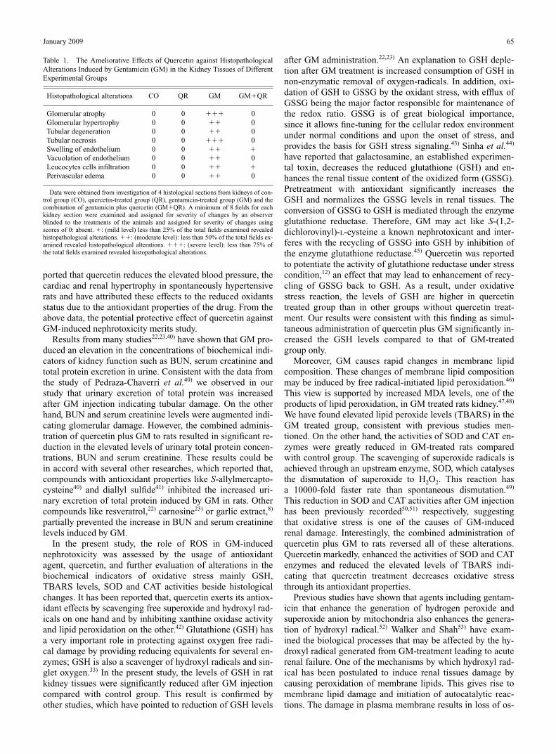
Table 1. The Ameliorative Effects of Quercetin against HistopathologicalAlterations Induced by Gentamicin (GM) in the Kidney Tissues of DifferentExperimental Groups
Histopathological alterations CO QR GM GM�QR
Glomerular atrophy 0 0 ��� 0Glomerular hypertrophy 0 0 �� 0Tubular degeneration 0 0 �� 0Tubular necrosis 0 0 ��� 0Swelling of endothelium 0 0 �� �Vacuolation of endothelium 0 0 �� 0Leucocytes cells infiltration 0 0 �� �Perivascular edema 0 0 �� 0
Data were obtained from investigation of 4 histological sections from kidneys of con-trol group (CO), quercetin-treated group (QR), gentamicin-treated group (GM) and thecombination of gentamicin plus quercetin (GM�QR). A minimum of 8 fields for eachkidney section were examined and assigned for severity of changes by an observerblinded to the treatments of the animals and assigned for severity of changes usingscores of 0: absent. �: (mild level) less than 25% of the total fields examined revealedhistopathological alterations. ��: (moderate level): less than 50% of the total fields ex-amined revealed histopathological alterations. ���: (severe level): less than 75% ofthe total fields examined revealed histopathological alterations.

motic balance and intracellular calcium levels increase. Cel-lular swelling is the first manifestation of these reversiblechanges which can be detected under light microscope.22)
Swelling of endothelium lining the glomerular tufts and tu-bular vacuolization is the reflection of these reversiblechanges in kidneys.54) In gentamicin group, we have ob-served swelling of endothelium lining the glomerular tuftsand vacuolization in all samples. In GM-quercetin group,most of the glomeruli are normal in size and shape.Quercetin blocked cellular inflammatory process as indicatedfrom reduction in mononuclear leucocytes inflammatorycells infiltration and alleviation of swelling of endotheliumlining the glomerular tufts. Biochemical results were concor-dant with pathological findings since quercetin was able tonormalize the elevated lipid peroxide (TBARS) levels andcompletely block lipid peroxidation. If intracellular free oxy-gen radicals increase, irreversible cellular injury process be-gins.22) Lysosomal enzymes activated and irreversible cell in-jury microscopically observed as tubular necrosis and tubulardegeneration of kidney occurs.54) Scavenging of free oxygenradicals prevents irreversible renal cell injury and necrosis.22)
We have observed tubular necrosis as a sign of irreversibleinjury in most sections examined from gentamicin group.Quercetin as an antioxidant inhibits lipid peroxidation andprevents renal cell injury manifested as tubular necrosis anirreversible cell damage. We have observed that quercetintreatment affected biochemical values and pathological find-ings, in accordance. Quercetin prevented the decrease inGSH levels, SOD and CAT activities and the increase inTBARS levels. In the same way, quercetin prevented re-versible cell damage such as swelling of endothelium andvacuolization, and irreversible cell damage such as tubularnecrosis. According to the pathological results, it can bestated that quercetin can be protective for degenerative injurycaused by gentamicin treatment. Another mechanism under-lying the antioxidative properties of flavonoids includingquercetin is the ability of flavonoids to alter peroxidation ki-netics by modification of the lipid packing order and to de-crease fluidity of the membrane.55) These changes couldstrictly hinder the diffusion of free radicals and restrict theirperoxidative reactions.
In conclusion, the present study revealed the nephrotoxiceffects of GM. The use of quercetin in combination with GMminimized its toxicity as revealed from decreasing urinaryexcretion of total protein, BUN and serum creatinine levels.Oxidative stress reactions and ROS may be one of the mech-anisms of GM-induced nephrotoxicity as indicated from al-terations in oxidative stress biomarkers. The correction ofoxidative stress biomarkers by quercetin was consistent withamelioration of the histopathological changes induced byGM. The ameliorative effect of quercetin against GM-in-duced renal damage may be at least in part due to its antioxi-dant and free radicals scavenger properties of quercetin.
REFERENCES
1) Ho J. L., Barza M., Antimicrob. Agents Chemother., 31, 485—491(1987).
2) Lerner S. A., Schmitt B. A., Seligsohn R., Matz G. J., Am. J. Med., 80,98—104 (1986).
3) Cuzzocrea S., Mazzon E., Dugo L., Serraino I., Di Paola R., Britti D.,De Sarro A., Pierpaoli S., Caputi A., Masini E., Salvemini D., Eur. J.Pharmacol., 450, 67—76 (2002).
4) Pedraza-Chaverrí J., González-Orozco A. E., Maldonado P. D., BarreraD., Medina-Campos O. N., Hernández-Pando R., Eur. J. Pharmacol.,473, 71—78 (2003).
5) Humes H. D., Weinberg J. M., “Toxic Nephropathies: The Kidney,” ed.by Rector F. C., Brenner B. M., W. B. Saunders Co., Philadelphia,1986, pp. 1491—1532.
6) Yanagida C., Ito K., Komiya I., Horie T., Chem. Biol. Interact., 148,139—147 (2004).
7) Morales A. I., Buitrago J. M., Santiago J. M., Fernández-Tagarro M.,López-Novoa J. M., Pérez-Barriocanal F., Antioxid. Redox Signal., 4,893—898 (2002).
8) Maldonado P. D., Barrera D., Medina-Campos O. N., Hernández-Pando R., Ibarra-Rubio M. E., Pedraza-Chaverrí J., Life Sci., 73,2543—2556 (2003).
9) Shah S. V., J. Clin. Invest., 74, 393—401 (1984).10) McCord J. M., Roy R. S, Schaffer S. W., Adv. Myocardiol., 5, 183—
189 (1985).11) Baliga R., Ueda N., Walker P. D., Shah S. V., Drug Metab. Rev., 31,
971—997 (1999).12) Ikizler M., Erkasap N., Dernek S., Kural T., Kaygisiz Z., Anadolu
Kardiyol. Derg., 7, 404—410 (2007).13) Hertog M. G., Hollman P. C., Katan M. B., Kromhout D., Nutr.
Cancer, 20, 21—29 (1993).14) Manach C., Regerat F., Texier O., Agullo G., Demigne C., Remesy C.,
Nutr. Res., 16, 517—544 (1996).15) Saponara S., Sgaragli G., Fusi F., Br. J. Pharmacol., 135, 1819—1827
(2002).16) Guglielmone H. A., Agnese A. M., Montoya S. C., José L., Thromb.
Res., 105, 183—188 (2002).17) Formica J. V., Regelson W., Food Chem. Toxicol., 33, 1061—1080
(1995).18) Bravo L., Nutr. Rev., 56, 317—333 (1998).19) Sanders R. A., Rauscher F. M., Watkins J. B., J. Biochem. Mol.
Toxicol., 15, 143—149 (2001).20) Mahesh T., Menon V. P., Phytother. Res., 18, 123—127 (2004).21) Suzuki Y., Ishihara M., Segami T., Ito M., Jpn. J. Pharmacol., 78,
435—441 (1998).22) Silan C., Uzun O., Comunoglu N. U., Gokçen S., Bedirhan S., Cengiz
M., Biol. Pharm. Bull., 30, 79—83 (2007).23) Soliman K. M., Abdul-Hamid M., Othman A. I., Med. Sci. Monit., 13,
73—83 (2007).24) Nishi H. H., Elin R. J., Clin. Chem., 31, 1377—1380 (1985).25) Fawcett J. K., Scott J. E., J. Clin. Pathol., 13, 156—159 (1960).26) Henry R. J., “Clinical Chemistry: Principles and Technics,” 2nd ed.,
Harper and Row, New York, 1974, pp. 548—551.27) Ellman G. L., Arch. Biochem. Biophys., 82, 70—77 (1959).28) Uchiyama M., Mihara M., Anal. Biochem., 86, 271—278 (1978).29) Marklund S. L., Mutat. Res., 148, 129—134 (1985).30) Claiborne A., “Catalase Activity: Handbook of Methods for Oxygen
Radical Research,” ed. by Greenwald R. A., Boca Raton, Florida,1985, pp. 283—284.
31) Allen C. T., “Laboratory Methods in Histochemistry,” 1st ed., ed. byProphet E. B., Mills B., Arrington J. B., Sobin L. H., American Reg-istry of Pathology, Washington, D.C., 1992, p. 53.
32) Bancroft J., Stevens A., “Theory and Practice of Histological Tech-niques,” 2nd ed., ed. by Stevens A., Churchill Livingstone, New York,1982, pp. 188—190.
33) Diplock A. T., “Antioxidants and Free Radicals Scavengers: Free Radi-cal Damage and Its Control,” ed. by Rice-Evans C. A., Burdon R. H.,Elsevier Science, Amsterdam, 1994, pp. 43—49.
34) Neilsen F., Mikkelsen B. B., Neilsen J. B., Andersen H. R., GrandjeanP., Clin. Chemist, 47, 1209—1214 (1997).
35) Sandhu J. S., Sehegal A., Singh A., JIACM, 8, 331—333 (2007).36) Leehey D. J., Braun B. I., Tholl D. A., Chung L. S., Gross C. A.,
Roback J. A., Lentino J. R., J. Am. Soc. Nephrol., 4, 81—90 (1993).37) Kadkhodaee M., Khastar H., Faghihi M., Ghaznavi R., Zahmatkesh
M., Exp. Physiol., 90, 571—576 (2005).38) Kaul N., Siveski-Iliskovic N., Hill M., Slezak J., Singal P. K., J. Phar-
macol. Toxicol. Methods, 30, 55—67 (1993).39) Duarte J., Pérez-Palencia R., Vargas F., Ocete M. A., Pérez-Vizcaino
F., Zarzuelo A., Tamargo J., Br. J. Pharmacol., 133, 117—124 (2001).40) Pedraza-Chaverrí J., Barrera D., Maldonado P. D., Chirino Y. I.,
Macías-Ruvalcaba N. A., Medina-Campos O. N., Castro L., SalcedoM. I., Hernández-Pando R., BMC Clin. Pharmacol., 4, 5—18 (2004).
66 Vol. 32, No. 1

41) Pedraza-Chaverrí J., Maldonado P. D., Barrera D., Cerón A., Medina-Campos O. N., Hernández-Pando R., Mol. Cell. Biochem., 254, 125—130 (2003).
42) Frankel E. N., Kanner J., German J. B., Parks E., Kinsella J. E., Lancet,341, 454—457 (1993).
43) Eberle D., Clarke R., Kaplowitz N., J. Biol. Chem., 256, 2115—2117(1981).
44) Sinha M., Manna P., Sil P. C., BMC Complement. Altern. Med., 7, 1—18 (2007).
45) Van de Water B., Zoeteweij J. P., Nagelkerke J. F., Arch. Biochem. Bio-phys., 327, 71—80 (1996).
46) Sandhya P., Varalakshmi P., J. Appl. Toxicol., 17, 405—408 (1997).47) Sener G., Sehirli A. O., Altunbas H. Z., Ersoy Y., Paskaloglu K.,
Arbak S., Ayanoglu-Dulger G., J. Pineal. Res., 32, 231—236 (2002).
48) Parlakpinar H., Tasdemir S., Polat A., Bay-Karabulut A., Vardi N.,Ucar M., Acet A., Toxicology, 207, 169—177 (2005).
49) Bowler C., Van Montagu M., Inze D., Ann. Rev. Plant Physiol. PlantMol. Biol., 43, 83—116 (1992).
50) Nitha B., Janardhanan K. K., Food Chem. Toxicol., 46, 3193—3199(2008).
51) Farombi E. O., Ekor M., Food Chem. Toxicol., 44, 1443—1448 (2006).52) Doroshow J. H., Davies K. J., J. Biol. Chem., 261, 3068—3074 (1986).53) Walker P. D., Shah S. V., J. Clin. Invest., 81, 334—341 (1988).54) Kumar V., Kotran R. S., Robbins S. L., “Pathologic Basis of Disease,”
6th ed., Chap. 1, ed. by Kumar V., Cotran R. S., Saunders W. B.,Philadelphia, 1999, pp. 15—18.
55) Arora A., Byrem T. M., Nair M. G., Strasburg G. M., Arch. Biochem.Biophys., 373, 102—109 (2000).
January 2009 67
Copyright © 2022 FDOKUMEN