Quercetin-6-C-β-d-glucopyranoside isolated from Ulmus wallichiana planchon is more potent than...
10
Menopause: The Journal of The North American Menopause Society Vol. 18, No. 2, pp. 198/207 DOI: 10.1097/gme.0b013e3181e84e67 * 2011 by The North American Menopause Society Quercetin-6-C-A-D-glucopyranoside isolated from Ulmus wallichiana planchon is more potent than quercetin in inhibiting osteoclastogenesis and mitigating ovariectomy-induced bone loss in rats Jawed A. Siddiqui, MSc, 1 Kunal Sharan, MSc, 1 Gaurav Swarnkar, MSc, 1 Preeti Rawat, MSc, 2 Manmeet Kumar, MSc, 2 Lakshmi Manickavasagam, MPharm, 3 Rakesh Maurya, PhD, 2 Dominique Pierroz, PhD, 4 and Naibedya Chattopadhyay, PhD 1 Abstract Objective: The aim of this study was to determine the skeletal effect of quercetin-6-C-A-D-glucopyranoside (QCG) isolated from the extract of Ulmus wallichiana and compare this effect with quercetin (Q) in a rat model of postmenopausal bone loss. Methods: Murine bone marrow cells were used to study the effect of QCG or Q on osteoclast differentiation. QCG or Q (1.0 and 5.0 mg kg j1 d j1 doses) was administered orally to ovarietomized (OVx) rats for 12 weeks. Sham-operated + vehicle and OVx + vehicle groups served as positive and negative controls, respectively. Bone mineral density, bone microarchitecture, biomechanical strength, bone turnover markers, and uterotrophic effect were studied. One-way analysis of variance was used to test significance of effects. Results: QCG at 1.0 nM significantly inhibited differentiation of multinucleated osteoclasts and expression of osteoclastogenic genes from bone marrow cells, whereas Q at 10.0 KM had comparable results. OVx rats treated with QCG exhibited significantly higher bone mass and better microarchitecture in trabecular and cortical bones compared with OVx + vehicle. QCG treatment of OVx rats had better functional impact than did Q-treated OVx rats, evident from increased bone biomechanical strength. Serum osteocalcin and urinary fragments of type 1 collagen were significantly lower in QCG-treated OVx rats compared with OVx + vehicle group. The protective effect of QCG under ovariectomy-induced bone loss setting was found to be significantly better than Q. Uterine histo- morphometry parameters of OVx rats did not change with QCG treatment. Conclusions: QCG improves bone biomechanical quality more effectively than Q through positive modifications of bone mineral density and bone microarchitecture without a hyperplastic effect on the uterus. Key Words: Flavonoid C-glucoside Y Antiresorptive Y Bone strength Y Estrogenicity. P ostmenopausal osteoporosis is characterized by in- creased resorption of bone, which, in turn, leads to increased bone fragility and fractures. 1 Being phytoes- trogens, flavonoids have gained preference as prophylactic agents against postmenopausal bone loss due to their lack of adverse effects that are commonly associated with hormone therapy. 2<9 Quercetin (Q), a major dietary flavonoid occurring in foods of plant origin, has recently been shown to reduce bone loss in ovarietomized (OVx) mice. 10 In an osteoclast precursor cell line, RAW 264.7 (mouse monocyte/macro- phage) cells, Q inhibits osteoclast differentiation and expres- sion of osteoclastogenic marker genes. 11<13 Q does not have a uterine estrogenic response and fails to activate estrogen receptors in reporter assay systems, suggesting its possible therapeutic use in postmenopausal osteoporosis. 10,14 A butanolic fraction of Ulmus wallichiana (Himalayan Elm, known for traditional medicinal use in India) has recently been shown by us to promote peak bone achievement and prevent OVx-induced bone loss in rats. 15 Furthermore, the active fraction was devoid of uterine hyperplastic effect, which is one of the prerequisites for human use for post- menopausal symptoms. 15 We sought to identify the putative active molecule(s) that contributed to the bone-sparing effect of the butanolic fraction of U. wallichiana. One of the abun- dant compounds isolated from the butanolic fraction of Received March 18, 2010; revised and accepted May 13, 2010. From the Divisions of 1 Endocrinology, 2 Medicinal & Process Chemistry, and 3 Pharmacokinetics and Metabolism, Central Drug Research Institute, Council of Scientific and Industrial Research, Lucknow, India; and 4 Department of Rehabilitation and Geriatrics Service of Bone Diseases, University Hospitals and Faculty of Medicine of Geneva, Geneva, Switzerland. Funding/support: This work was supported by the Ministry of Health and Family Welfare, Council of Scientific and Industrial Research, and Uni- versity Grants Commission, Government of India. Funding from the Indian Council of Medical Research (N.C.) and Department of Bio- technology (N.C.), Government of India, is acknowledged. Research fellowship grants from University Grants Commission (J.A.S.), Depart- ment of Biotechnology (K.S), and Council of Scientific and Industrial Research (G.S, P.R, M.K.), Government of India, are also acknowledged. Financial disclosure/conflicts of interest: None reported. Address correspondence to: Naibedya Chattopadhyay, PhD, Central Drug Research Institute, Council of Scientific and Industrial Research, Chattar Manzil, P.O. Box 173, Lucknow, India. E-mail: [email protected] 198 Menopause, Vol. 18, No. 2, 2011 Copyright © 2011 The North American Menopause Society. Unauthorized reproduction of this article is prohibited.
Transcript of Quercetin-6-C-β-d-glucopyranoside isolated from Ulmus wallichiana planchon is more potent than...

Menopause: The Journal of The North American Menopause SocietyVol. 18, No. 2, pp. 198/207DOI: 10.1097/gme.0b013e3181e84e67* 2011 by The North American Menopause Society
Quercetin-6-C-A-D-glucopyranoside isolated from Ulmus wallichianaplanchon is more potent than quercetin in inhibiting osteoclastogenesisand mitigating ovariectomy-induced bone loss in rats
Jawed A. Siddiqui, MSc,1 Kunal Sharan, MSc,1 Gaurav Swarnkar, MSc,1 Preeti Rawat, MSc,2
Manmeet Kumar, MSc,2 Lakshmi Manickavasagam, MPharm,3 Rakesh Maurya, PhD,2
Dominique Pierroz, PhD,4 and Naibedya Chattopadhyay, PhD1
AbstractObjective: The aim of this study was to determine the skeletal effect of quercetin-6-C-A-D-glucopyranoside
(QCG) isolated from the extract of Ulmus wallichiana and compare this effect with quercetin (Q) in a rat model ofpostmenopausal bone loss.
Methods: Murine bone marrow cells were used to study the effect of QCG or Q on osteoclast differentiation.QCG or Q (1.0 and 5.0 mg kgj1 dj1 doses) was administered orally to ovarietomized (OVx) rats for 12 weeks.Sham-operated + vehicle and OVx + vehicle groups served as positive and negative controls, respectively. Bonemineral density, bone microarchitecture, biomechanical strength, bone turnover markers, and uterotrophic effectwere studied. One-way analysis of variance was used to test significance of effects.
Results: QCG at 1.0 nM significantly inhibited differentiation of multinucleated osteoclasts and expression ofosteoclastogenic genes from bone marrow cells, whereas Q at 10.0 KM had comparable results. OVx rats treatedwith QCG exhibited significantly higher bone mass and better microarchitecture in trabecular and cortical bonescompared with OVx + vehicle. QCG treatment of OVx rats had better functional impact than did Q-treated OVx rats,evident from increased bone biomechanical strength. Serum osteocalcin and urinary fragments of type 1 collagenwere significantly lower in QCG-treated OVx rats compared with OVx + vehicle group. The protective effect ofQCG under ovariectomy-induced bone loss setting was found to be significantly better than Q. Uterine histo-morphometry parameters of OVx rats did not change with QCG treatment.
Conclusions: QCG improves bone biomechanical quality more effectively than Q through positive modificationsof bone mineral density and bone microarchitecture without a hyperplastic effect on the uterus.
Key Words: Flavonoid C-glucoside Y Antiresorptive Y Bone strength Y Estrogenicity.
Postmenopausal osteoporosis is characterized by in-creased resorption of bone, which, in turn, leads toincreased bone fragility and fractures.1 Being phytoes-
trogens, flavonoids have gained preference as prophylactic
agents against postmenopausal bone loss due to their lack ofadverse effects that are commonly associated with hormonetherapy.2<9 Quercetin (Q), a major dietary flavonoid occurringin foods of plant origin, has recently been shown to reducebone loss in ovarietomized (OVx) mice.10 In an osteoclastprecursor cell line, RAW 264.7 (mouse monocyte/macro-phage) cells, Q inhibits osteoclast differentiation and expres-sion of osteoclastogenic marker genes.11<13 Q does not have auterine estrogenic response and fails to activate estrogenreceptors in reporter assay systems, suggesting its possibletherapeutic use in postmenopausal osteoporosis.10,14
A butanolic fraction of Ulmus wallichiana (HimalayanElm, known for traditional medicinal use in India) has recentlybeen shown by us to promote peak bone achievement andprevent OVx-induced bone loss in rats.15 Furthermore, theactive fraction was devoid of uterine hyperplastic effect,which is one of the prerequisites for human use for post-menopausal symptoms.15 We sought to identify the putativeactive molecule(s) that contributed to the bone-sparing effectof the butanolic fraction of U. wallichiana. One of the abun-dant compounds isolated from the butanolic fraction of
Received March 18, 2010; revised and accepted May 13, 2010.
From the Divisions of 1Endocrinology, 2Medicinal & Process Chemistry,and 3Pharmacokinetics and Metabolism, Central Drug Research Institute,Council of Scientific and Industrial Research, Lucknow, India; and4Department of Rehabilitation and Geriatrics Service of Bone Diseases,University Hospitals and Faculty of Medicine of Geneva, Geneva,Switzerland.
Funding/support: This work was supported by the Ministry of Health andFamily Welfare, Council of Scientific and Industrial Research, and Uni-versity Grants Commission, Government of India. Funding from theIndian Council of Medical Research (N.C.) and Department of Bio-technology (N.C.), Government of India, is acknowledged. Researchfellowship grants from University Grants Commission (J.A.S.), Depart-ment of Biotechnology (K.S), and Council of Scientific and IndustrialResearch (G.S, P.R, M.K.), Government of India, are also acknowledged.
Financial disclosure/conflicts of interest: None reported.
Address correspondence to: Naibedya Chattopadhyay, PhD, Central DrugResearch Institute, Council of Scientific and Industrial Research, ChattarManzil, P.O. Box 173, Lucknow, India. E-mail: [email protected]
198 Menopause, Vol. 18, No. 2, 2011
Copyright © 2011 The North American Menopause Society. Unauthorized reproduction of this article is prohibited.

U. wallichiana was 3,3¶,4¶,5,7-pentahydroxyflavone-6-C-A-D-glucopyranoside or quercetin-6-C-A-D-glucopyranoside (QCG).C-glucoside forms of flavonoids have a more restricted dis-tribution than do aglycone or O-glucoside forms. For exam-ple, Q is abundantly available in apples and onions,16,17
whereas QCG is not known to be present in any food ornutriments. Furthermore, in QCG, sugar is directly linked to Qvia an acid-resistant and largely enzyme-resistant CYC bond,suggesting that QCG may be metabolically more stablethan Q. We therefore hypothesized that QCG could be oneof the bioactive compounds present in the butanolic fractionof U. wallichiana, in addition to 6-C-A-D-glucopyranosyl-(2S,3S)-(+)-3¶,4¶,5,7-tetrahydroxyflavanone/Ulmoside A,18 whichis responsible for the observed osteoprotective effect of thefraction.15
Because Q and QCG are structurally related, we studiedantiosteoclastogenic and bone-conserving effects of QCG andcompared these effects with Q. We first compared the effectsof Q and QCG in osteoclastic differentiation with bone mar-row precursor cells. In addition, because OVx rats representthe World Health OrganizationYrecommended animal modelfor postmenopausal osteoporosis, we compared the skeletaleffects of Q and QCG in adult OVx Sprague-Dawley rats.Because the so-called uterotrophy assay is recommended bythe Organization for Economic Cooperation and Developmentto test for the estrogenicity of any given compound/agent to beused in postmenopausal women, we investigated the uterineestrogenicity of QCG. Overall, the objective of this studywas to determine the osteoprotective effect of QCG underestrogen-deficient conditions and assess uterine safety.
METHODS
Reagents and chemicalsCell culture media and supplements were purchased from
Invitrogen (Carlsbad, CA). All fine chemicals were purchasedfrom Sigma-Aldrich (St. Louis, MO). Osteocalcin (midportion)and fragments of type 1 collagen (CTx) enzyme-linked immu-nosorbent AQ4 assay kits were purchased from Immunodiag-nostic Systems Ltd. (Tyne & Wear, UK).
Purification and characterization of QCG fromU. wallichiana
Stem-bark of U. wallichiana was collected from Pithor-agarh and Bageshwar districts in Uttarakhand Province, India.The powdered stem-bark (5.0 kg) was extracted with ethanol(20 L) and yielded an extract of 660.0 g. The ethnolic extract(100 g) was triturated with hexane (200 mL � 5), and thehexane-insoluble portion was suspended in water (500 mL)and then extracted with n-butanol (500 mL). The n-butanolYsoluble fraction (69.0 g) was subjected to silica gel columnchromatography eluted with EtOAc-MeOH and 10 fractionswere obtained (1-10). Fraction 3 was repeatedly chromato-graphed over the silica gel column, using a CHCl3-MeOHsolvent system, yielding pure QCG (2.0 g, yield 0.26%) as ayellow amorphous solid, [>]D
20: +9.79- (ca. 0.6 dimethylsulfoxide). The electrospray ionization mass spectrometry
exhibited [M + H]+ at m/z 465, corresponding to molecularformula C21H20O12. The infrared spectrum (in KBr) showedbands vmax at 3,428, 1,642, 1,446, and 1,349 cmj1. The UVabsorption showed bands Lmax (MeOH) at 352 and 255 nm.
The proton nuclear magnetic resonance spectrum of QCGdisplayed the characteristic signals for a flavone moiety; twosharp singlets at CH 6.45 (1H, s) and CH 13.06 (s) wereassignable to H-8 and chelated OH group (C-5), respectively;and signals at CH 7.65 (1H, d, J = 2.2 Hz), 6.88 (1H, d,J = 8.6 Hz) and 7.54 (1H, dd, J = 8.6, 2.2 Hz) resolved as oneABX system. Proton nuclear magnetic resonance data also ex-hibited signal at CH 4.60 (1H, d, J = 9.6 Hz) assigned toanomeric proton, and its large coupling constant (J = 9.6 Hz)revealed the existence of a C-A-glycoside. The remaining sugarprotons were resolved in the range of CH 4.05 to 3.20 (6H, m).Twenty-one carbon resonances were resolved in the carbon-13nuclear magnetic resonance spectrum and were further clas-sified by distortion-less enchancement by polarization trans-fer experiments as 4 aromatic methines, 5 aliphatic methines,1 oxygenated methylene, and 11 quaternary carbons. The mostdownfield quaternary carbon at CC 175.2 was assigned to con-jugated carbonyl, whereas other quaternary carbons were res-onated at CC 144.2, 134.7, 159.0, 107.2, 162.0, 154.2, 101.8,121.1, 145.7, and 146.8. Sugar carbons were observed at CC72.5, 69.8, 78.1, 69.3, 80.7, and 60.0. The anomeric protons(CH 4.60) gave heteronuclear multiple band correlation withcarbons resonated at CC 159.0 and 162.0, which indicated theattachment of sugar to the aromatic ring at the C-6 position. Onthe basis of above-described spectral evidence, the structureof the compound (QCG) was elucidated as quercetin-6-C-A-glucopyranoside and further confirmed by the comparison of itsspectral data with those reported in the literature.19
Differentiation of osteoclasts from bone marrowprecursor cells
Primary osteoclasts were generated according to our pre-viously described protocol.20 Briefly, bone marrow macrophageswere isolated from the whole marrow of 4- to 6-week-oldBALB/c mice and cultured overnight in >-minimal essentialmedium (plus penicillin and streptomycin) supplementedwith 10% heat-inactivated fetal bovine serum and 10 ng/mLmacrophage colony-stimulating factor (M-CSF). The non-adherent cells were collected and, after centrifugation, cul-tured on 48-well tissue culture plates in >-minimal essentialmedium with heat-inactivated fetal bovine serum (10%),receptor activator for nuclear factor JB ligand (RANKL;50 ng/mL), and M-CSF (10 ng/mL).
Tartrate-resistant acid phosphatase stainingTartrate-resistant acid phosphatase (TRAP) staining was
done according to our previously described protocol.20 Briefly,cells were exposed to different concentration of QCG and Q inculture media and indicated that supplement(s) were changedevery 2 days, for a total of 7 days. At the end of culture,the cells were fixed with ethanol-acetone (50:50, vol/vol) andwere incubated for 20 minutes in acetate buffer (0.1 M sodiumacetate, pH 5.0) containing naphthol AS-MX phosphate, fast
Menopause, Vol. 18, No. 2, 2011 199
C-GLUCOSIDE QUERCETIN HAS BONE-SPARING EFFECT
Copyright © 2011 The North American Menopause Society. Unauthorized reproduction of this article is prohibited.

red violet LB salt, and 20 mM sodium tartrate. TRAP-positivemononuclear cells and TRAP-positive multinucleated cells(three or more nuclei) were counted under a microscope.21,22
Two researchers blinded to the identities of the samples coun-ted the TRAP-positive cells as well as the total number of cellsin six different fields from three independent experiments.Taking the mean of control values, we derived an individualcontrol value as a ratio of the mean � 100, and data are ex-pressed as percent change of the mean value of control forTRAP-positive cells.
Quantitative real-time polymerase chain reactionQuantitative real-time polymerase chain reaction was per-
formed for assessing the expression of osteoclast-specificgenes (RANK and c-fos) from bone marrow cells (BMCs)treated with RANKL + M-CSF (control), QCG (1 and100 nM) + RANKL + M-CSF, and Q (10 and 20 KM) +RANKL + M-CSF for the period of 7 days, following ouroptimized protocol.20,23 The housekeeping gene GAPDH wasused as the internal control in this study. The design of senseand antisense oligonucleotide primers was based on publishedcDNA sequences using the Universal Probe Library (RocheDiagnostics). Primer sequences for RANK were 5¶- agaggcattatgagcatctcg-3¶ (sense), 5¶-ggagtgcacttagaggacaggt-3¶ (anti-sense); for c-fos, 5¶-cagcctttcctactaccattcc-3¶ (sense), 5¶-acagatctgcgcaaaagtcc-3¶ (antisense); and for GAPDH, 5¶- tttgatgttagtggggtctcg-3¶ (sense), 5¶- agcttgtcatcaacgggaag-3¶ (antisense).For real-time polymerase chain reaction, cDNA was synthe-sized with a RevertAid cDNA synthesis kit (Fermentas, Austin,TX) using 2.0 Kg total RNA. SYBR Green chemistry was usedto perform quantitative determination of relative expression oftranscripts for all genes. All genes were analyzed using theLight Cycler 480 (Roche Molecular Biochemicals, Indiana-polis, IN).
Experimental design for in vivo studyAll animal studies were conducted in accordance with
current legislation on animal experiments (Institutional Ani-mal Ethical Committee) at Central Drug Research Institute,India. Adult virgin OVx rats (180-200 g) were used as a well-established model of postmenopausal bone loss.24,25 Ovar-iectomy potently stimulates an estrogen deficiency condition,and age-matched sham-operated rats served as controls rep-resenting the estrogen-sufficient condition.
Animals and QCG treatmentBoth QCG and Q at 1.0 and 5.0 mg kgj1 dayj1 doses were
given by oral gavage to Sprague-Dawley rats after bilateralovariectomy. Each group consisted of 10 rats, and the groupswere as follows: sham operated (ovary intact) + vehicle (gumacacia), OVx + vehicle, OVx + 1.0 and 5.0 mg kgj1 dayj1 Q,and OVx + 1.0 and 5.0 mg kgj1 dayj1 QCG. Treatmentstarted 24 hours after the surgery and continued for 12 weeks.After 12 weeks of various treatments described above, all ratswere first acclimatized and then kept in metabolic cages withno food but only water for 24 hours to collect urine samples.
Rats were then killed and autopsied to collect blood and bones(tibia, femur, lumber vertebrae).
Measurements of bone mineral density (BMD) of excisedbones were performed according to our previously publishedprotocols.15,23 Briefly; lumbar vertebrae, femur, and tibiawere dissected and separated from adjacent tissue, cleaned,fixed in 70% ethanol, and stored at 4-C until BMD analysis.BMD measurements of regions of interest were performedusing a bone densitometer (model 4500 Elite, Hologic) fittedwith commercially available software for small animals (QDR4500 ACCLAIM series).23,26,27 After BMD measurement,femurs were subjected to three-point bending with bonestrength tester model TK 252C (Muromachi Kikai Co. Ltd.,Tokyo, Japan), according to our previously published proto-cols.15,23,26,27 Biomechanical parameters of femur includingmaximal power, stiffness, and energy were determined. Tak-ing the mean of control values, we derived an individualcontrol value as a ratio of the mean � 100, and data were ex-pressed as percent change over the mean value of the shamgroup for the various biomechanical parameters.
Uteri were carefully dissected out, gently blotted, weighed,and fixed in 4% paraformaldehyde for histology, followingour previously published articles.15,23,26,27 Approximately5.0-mm pieces from the middle segment of each uterus weredehydrated in ascending grades of ethanol, cleared in xylene,and embedded in paraffin wax using standard procedures.Representative transverse sections (5.0 Km) were stained withhematoxylin and eosin. Photomicrographs of sections wereobtained using a Leica DC 300 camera and Leica IM50 ImageAcquisition software fitted to a Leica DMLB microscope. Theluminal epithelial height, luminal area, and total uterine areawere determined using the program Leica Qwin software(Leica Microsystems, Bannockburn, IL).
Bone microarchitecture assessment by micro-CTMicro-CT (KCT) determination of excised bones was car-
ried out using the SkyScan 1076 KCT scanner (Aartselaar,Belgium) as described in our previously published proto-cols.23 Femurs and tibias were dissected from the animals afterthey were killed, cleaned of soft tissue, and fixed beforestorage in alcohol. The samples were scanned in batches ofthree at a nominal resolution (pixels) of 18 Km. Recon-struction was carried out using a modified Feldkamp algo-rithm using the SkyScan Nrecon software, which facilitatesnetwork distributed reconstruction carried out on four per-sonal computers running simultaneously. The x-ray sourcewas set at 70 kV and 100 mA, with a pixel size of 18 Km.A hundred projections were acquired over an angular rangeof 180-. The image slices were reconstructed using cone-beam reconstruction software version 2.6 based on the Feld-kamp algorithm (SkyScan). Trabecular bone was extracted bydrawing ellipsoid contours with the CT analyzer software.Trabecular bone volume (in percentage), trabecular number,and trabecular separation (in micrometers) were calculatedby the mean intercept length method. Trabecular thickness(in micrometers) was calculated according to the method of
200 Menopause, Vol. 18, No. 2, 2011 * 2011 The North American Menopause Society
SIDDIQUI ET AL
Copyright © 2011 The North American Menopause Society. Unauthorized reproduction of this article is prohibited.

Hildebrand and Ruegsegger.28 Three-dimensional parameterswere based on analysis of a marching cubes-type model with arendered surface. Cortical thickness, cortical area, corticalperimeter, periosteal area, periosteal perimeter, and endostealperimeter were calculated by two-dimensional analysis of cor-tical bones of femur (mid-diaphysis) and tibia-fibula separationpoint (TFSP).
Analysis of bone turnover markersBased on our previously published protocols,15,23 serum
osteocalcin (midportion) and urinary CTx levels were deter-mined by enzyme-linked immunosorbent assay kits purchasedfrom Immunodiagnostic Systems Inc. by following the man-ufacturer_s protocols.
StatisticsData are expressed as mean T SEM. The data obtained in
experiments with multiple treatments were subjected to one-way analysis of variance followed by the post hoc Newman-Keuls multiple comparison test of significance using GraphPadPrism 3.02 software. Qualitative observations have been rep-resented after assessments made by three individuals blindedto the experimental designs.
RESULTS
QCG more potently than Q inhibits differentiationof osteoclasts from BMCs
QCG was purified from the stem-bark of U. wallichianaas described in BMethods.[ Figure 1 shows the structuresof QCG (molecular weight 464) and Q (molecular weight338). The yield of QCG was at the rate of approximately133 mg/100 g butanolic extract (ie, 0.133%) of the stem-barkof U. wallichiana.
RANKL is secreted by osteoblasts and stimulates osteoclas-togenesis with accompanying increases in the expression ofosteoclast differentiation genes such as RANK and TRAP.26
Here we found that the presence of QCG (1 and 100 nM)dependently inhibited the formation of multinucleated andTRAP-positive cells from BMCs induced by RANKL and
FIG. 1. Structures of quercetin (Q) and 6-C-quercetin glucoside (QCG).
FIG. 2. QCG more potently than Q attenuates osteoclastogenesis from BMCs. A: QCG and Q inhibit formation of TRAP-positive cells (mononucleatedand multinucleated) of murine BMCs induced by RANKL (50 ng/mL) + M-CSF (10 ng/mL). B: Murine BMCs were cultured as described in A toinduce osteoclast differentiation. Quantitative real-time polymerase chain reaction determination of mRNA levels of osteoclastogenic genes, RANK andc-fos, in BMCs was done as described in BMethods.[ Each assay was performed in triplicate, and results from three independent experiments arepresented as mean T SEM of percent/fold change over control. *P G 0.05; **P G 0.01; ***P G 0.001. QCG, quercetin-6-C-A-D-glucopyranoside; Q,quercetin; TRAP + ve, tartrate-resistant acid phosphataseYpositive; BMC, bone marrow cell; M-CSF, macrophage colony-stimulating factor.
Menopause, Vol. 18, No. 2, 2011 201
C-GLUCOSIDE QUERCETIN HAS BONE-SPARING EFFECT
Copyright © 2011 The North American Menopause Society. Unauthorized reproduction of this article is prohibited.

M-CSF (Fig. 2A). Q, on the other hand, significantly inhibitedosteoclastogenesis from BMCs at 10.0 and 20.0 KM (Fig. 2A).Consistent with the inhibition of osteoclastogenesis, weobserved that QCG inhibited the expression of osteoclastmarkers including RANK and c-fos in BMCs cultured in thepresence of RANKL and M-CSF (Fig. 2B). Again, nanomolarconcentrations of QCG inhibited the expression of osteoclas-togenic genes, whereas comparable inhibition was achievedwith micromolar concentrations of Q (Fig. 2B). Together, thesedata suggested that QCG was at least 100-fold more potent ininhibiting bone marrow osteoclastogenesis than Q.
QCG treatment is more effective than Q in improvingbone mass and biomechanical strength in OVx rats
QCG treatment to OVx rats had no significant effect on gainof bodyweight due to OVxwhen compared with OVx + vehiclegroup (data not shown). Figure 3A shows results of BMDanalysis of trabecular regions including femur epiphysis,proximal tibia, and lumber vertebrae-4 (LV-4). As expected,OVx + vehicle group presented with significantly lower BMDlevels in all of the trabecular regions of the bones studiedcompared with sham + vehicle group. Treating OVx rats witheither QCG or Q produced comparable BMD levels in femurepiphysis and proximal tibia, and these values were signifi-cantly higher than OVx + vehicle group. OVx rats treatedwith QCG had significantly higher BMD in LV-4 than OVx +
vehicle group, whereas Q treatment had no effect (Fig. 3A). Incortical bones, that is, femur diaphysis and TFSP, QCG but notQ treatment to OVx rats resulted in significantly higher BMDlevels than OVx + vehicle group (Fig. 3B). Taken together, ourdata suggest that QCG was more effective than Q in main-taining both trabecular and cortical BMD levels in OVx rats.
QCG is better than Q in maintaining bonemicroarchitecture in OVx rats
Three-dimensional analysis of femoral epiphysis showeddifferences in trabecular microarchitecture among the varioustreatment groups as presented in Fig. 4A. Analysis of therepresentative sample data in trabecular sites (femur epiphysisand proximal tibia) indicated that ovariectomy significantlydecreased bone volume fraction (bone volume/trabecular vol-ume), trabecular number, and trabecular thickness comparedwith sham + vehicle group (Fig. 4B). In contrast, trabecularseparation was significantly increased in response to ovariec-tomy compared with sham + vehicle group (Fig. 4B). Com-pared with OVx + vehicle group, OVx rats treated with QCGmaintained all of the trabecular microarchitectural parame-ters studied (Fig. 4B). Q treatment of OVx rats was partiallyeffective in maintaining trabecular microarchitecture and henceless potent than QCG (Fig. 4B).
Next, two-dimensional analysis of cortical bones of femur(mid-diaphysis) and tibia (TFSP) was performed. As expected,
FIG. 3. QCG treatment was better than Q in preventing OVx-induced loss of BMD. A: Rats in various groups were maintained for 12 weeks. BMDlevels of trabecular regions of excised bones were determined. B: BMD levels of cortical regions of excised bones were determined. Data representmean T SEM. *P G 0.05, **P G 0.01, and ***P G 0.001 compared with vehicle-treated sham group. aP G 0.05, bP G 0.01, and cP G 0.001 compared withvehicle-treated OVx group (n = 10 rats/group). QCG, quercetin-6-C-A-D-glucopyranoside; Q, quercetin; OVx, ovarietomized; BMD, bone mineraldensity; TFSP, tibia-fibula separation point; LV-4, lumber vertebrae-4.
202 Menopause, Vol. 18, No. 2, 2011 * 2011 The North American Menopause Society
SIDDIQUI ET AL
Copyright © 2011 The North American Menopause Society. Unauthorized reproduction of this article is prohibited.

OVx for 12 weeks resulted in a reduction in cortical thickness,cortical area, cortical perimeter, periosteal area, periosteal pe-rimeter, and endosteal perimeter (Tables 1 and 2). Treatmentof OVx rats with QCG resulted in significantly better main-tenance of all the parameters (in both femur and tibia) studiedwhen compared with OVx + vehicle group. Again, Q treatmentof OVx rats resulted in partial maintenance of cortical param-eters of femur and tibia (Tables 1 and 2). These data suggestthat QCG is more effective than Q in maintaining histo-
morphometric measurements in cortical bones of femur andtibia shaft in OVx rats.
QCG but not Q maintained bone biomechanical strengthunder ovariectomy
Because QCG had protective effects in bone mass andmicroarchitecture under ovariectomy-induced bone loss con-ditions, we examined whether QCG maintained bone strengthin OVx rats. We assessed femoral bone biomechanical strength
FIG. 4. QCG treatment preserved trabecular microarchitecture better than Q treatment of OVx rats, assessed by three-dimensional KCT. A: Repre-sentative HCT images of the entire cross-section of the femur. B: Measurements of bone volume/trabecular volume (BV/TV), trabecular thickness(Tb.Th), trabecular spacing (Tb.Sp.), and trabecular number (Tb.N) of femur epiphysis. C: Proximal tibia are shown. Data represent mean T SEM.*P G 0.05, **P G 0.01, and ***P G 0.001 compared with vehicle-treated sham group. aP G 0.05, bP G 0.01, and cP G 0.001 compared with vehicle-treated OVx group (n = 10 rats/group). QCG, quercetin-6-C-A-D-glucopyranoside; Q, quercetin; OVx, ovarietomized; KCT, micro-CT.
TABLE 1. Cortical parameters of femoral bone using micro-CT
TreatmentCortical mean
cross-section area (B.Ar.)Cortical thickness
(Cs.Th.)Periosteal area
(T.Ar.)Periosteal
perimeter (T.Pm.)Cortical bone
perimeter (B.Pm.)Endosteal perimeter(B.Pm. j T.Ar.)
Sham + vehicle 4.76 T 0.120 0.389 T 0.005 11.8 T 0.551 14.1 T 0.908 26.6 T 0.881 14.8 T 0.661OVx + vehicle 3.99** T 0.20 0.325* T 0.005 9.69** T 0.66 12.2* T 0.405 22.1* T 0.91 12.3** T 0.261OVx + QCG (1 mg/kg BW) 4.44a T 0.191 0.371a T 0.126 11.1a T 0.167 13.1 T 0.126 25.2a T 0.366 14.1a T 0.781OVx + QCG (5 mg/kg BW) 5.31c T 0.261 0.380b T 0.005 12.40c T 0.331 15.5c T 0.874 31.2c T 1.035 18.8c T 0.714OVx + Q (1 mg/kg BW) 4.21 T 0.361 0.337 T 0.052 10.2 T 0.48 12.7 T 0.764 22.2 T 0.913 12.0 T 0.525OVx + Q (5 mg/kg BW) 4.33a T 0.402 0.328 T 0.019 11.0a T 0.36 13.2 T 0.805 24.1 T 1.79 13.1 T 1.89
Each parameter represents pooled data from 10 rats/group, and values are expressed as mean T SEM.OVx, ovariectomized; QCG, quercetin-6-C-A-D-glucopyranoside; BW, body weight; Q, quercetin.*P G 0.05, **P G 0.01, and ***P G 0.001 compared with vehicle-treated sham group.aP G 0.05, bP G 0.01, and cP G 0.001 compared with vehicle-treated OVx group.
Menopause, Vol. 18, No. 2, 2011 203
C-GLUCOSIDE QUERCETIN HAS BONE-SPARING EFFECT
Copyright © 2011 The North American Menopause Society. Unauthorized reproduction of this article is prohibited.
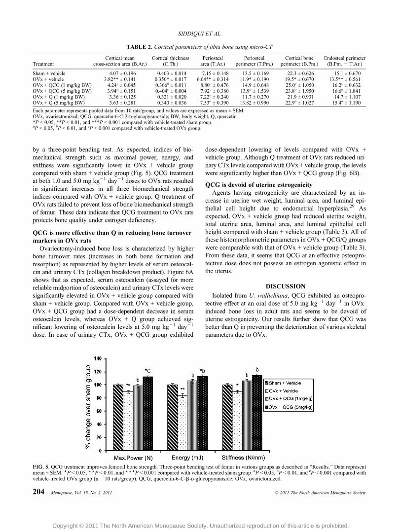
by a three-point bending test. As expected, indices of bio-mechanical strength such as maximal power, energy, andstiffness were significantly lower in OVx + vehicle groupcompared with sham + vehicle group (Fig. 5). QCG treatmentat both 1.0 and 5.0 mg kgj1 dayj1 doses to OVx rats resultedin significant increases in all three biomechanical strengthindices compared with OVx + vehicle group. Q treatment ofOVx rats failed to prevent loss of bone biomechanical strengthof femur. These data indicate that QCG treatment to OVx ratsprotects bone quality under estrogen deficiency.
QCG is more effective than Q in reducing bone turnovermarkers in OVx rats
Ovariectomy-induced bone loss is characterized by higherbone turnover rates (increases in both bone formation andresorption) as represented by higher levels of serum osteocal-cin and urinary CTx (collagen breakdown product). Figure 6Ashows that as expected, serum osteocalcin (assayed for morereliable midportion of osteocalcin) and urinary CTx levels weresignificantly elevated in OVx + vehicle group compared withsham + vehicle group. Compared with OVx + vehicle group,OVx + QCG group had a dose-dependent decrease in serumosteocalcin levels, whereas OVx + Q group achieved sig-nificant lowering of osteocalcin levels at 5.0 mg kgj1 dayj1
dose. In case of urinary CTx, OVx + QCG group exhibited
dose-dependent lowering of levels compared with OVx +vehicle group. Although Q treatment of OVx rats reduced uri-nary CTx levels compared with OVx + vehicle group, the levelswere significantly higher than OVx + QCG group (Fig. 6B).
QCG is devoid of uterine estrogenicityAgents having estrogenicity are characterized by an in-
crease in uterine wet weight, luminal area, and luminal epi-thelial cell height due to endometrial hyperplasia.29 Asexpected, OVx + vehicle group had reduced uterine weight,total uterine area, luminal area, and luminal epithelial cellheight compared with sham + vehicle group (Table 3). All ofthese histomorphometric parameters in OVx + QCG/Q groupswere comparable with that of OVx + vehicle group (Table 3).From these data, it seems that QCG at an effective osteopro-tective dose does not possess an estrogen agonistic effect inthe uterus.
DISCUSSION
Isolated from U. wallichiana, QCG exhibited an osteopro-tective effect at an oral dose of 5.0 mg kgj1 dayj1 in OVx-induced bone loss in adult rats and seems to be devoid ofuterine estrogenicity. Our results further show that QCG wasbetter than Q in preventing the deterioration of various skeletalparameters due to OVx.
TABLE 2. Cortical parameters of tibia bone using micro-CT
TreatmentCortical mean
cross-section area (B.Ar.)Cortical thickness
(C.Th.)Periostealarea (T.Ar.)
Periostealperimeter (T.Pm.)
Cortical boneperimeter (B.Pm.)
Endosteal perimeter(B.Pm. j T.Ar.)
Sham + vehicle 4.07 T 0.196 0.403 T 0.014 7.15 T 0.148 13.5 T 0.169 22.3 T 0.626 15.1 T 0.670OVx + vehicle 3.82** T 0.141 0.350* T 0.017 6.04** T 0.314 11.9* T 0.190 19.5* T 0.670 13.5** T 0.561OVx + QCG (1 mg/kg BW) 4.24c T 0.045 0.366a T 0.011 8.80c T 0.476 14.9 T 0.648 25.0c T 1.050 16.2b T 0.632OVx + QCG (5 mg/kg BW) 3.94a T 0.151 0.404b T 0.004 7.92c T 0.380 13.9a T 1.539 23.8a T 1.950 16.6a T 1.841OVx + Q (1 mg/kg BW) 3.36 T 0.125 0.321 T 0.020 7.22a T 0.240 11.7 T 0.270 21.9 T 0.931 14.7 T 1.107OVx + Q (5 mg/kg BW) 3.63 T 0.281 0.340 T 0.036 7.53a T 0.390 13.82 T 0.990 22.9a T 1.027 15.4a T 1.190
Each parameter represents pooled data from 10 rats/group, and values are expressed as mean T SEM.OVx, ovariectomized; QCG, quercetin-6-C-A-D-glucopyranoside; BW, body weight; Q, quercetin.*P G 0.05, **P G 0.01, and ***P G 0.001 compared with vehicle-treated sham group.aP G 0.05, bP G 0.01, and cP G 0.001 compared with vehicle-treated OVx group.
FIG. 5. QCG treatment improves femoral bone strength. Three-point bending test of femur in various groups as described in BResults.[ Data representmean T SEM. *P G 0.05, **P G 0.01, and ***P G 0.001 compared with vehicle-treated sham group. aP G 0.05, bP G 0.01, and cP G 0.001 compared withvehicle-treated OVx group (n = 10 rats/group). QCG, quercetin-6-C-A-D-glucopyranoside; OVx, ovarietomized.
204 Menopause, Vol. 18, No. 2, 2011 * 2011 The North American Menopause Society
SIDDIQUI ET AL
Copyright © 2011 The North American Menopause Society. Unauthorized reproduction of this article is prohibited.

Increased formation of osteoclasts at the onset of meno-pause is well established.30 We have reported that Q at 10and 20 KM inhibits the differentiation of murine osteoclastprecursor (RAW 264.7) cells to osteoclasts.31 In the presentstudy, our data show that 1.0 nM QCG inhibited osteoclastdifferentiation and expression of osteoclast-specific genes,whereas 20 KMQwas required to achieve a comparable effect,suggesting thereby that QCG was on the order of 104-fold morepotent than Q in inhibiting osteoclast formation from bonemarrow precursor cells.
One of the likely outcomes of inhibition of osteoclastic dif-ferentiation of BMCs is prevention of bone loss under estrogendeficiency.32 Post-ovariectomy bone loss in rats parallels skel-etal changes observed in postmenopausal women.33 BecauseQCG was found to be more potent than Q in inhibiting osteo-clastic differentiation of BMCs, we investigated skeletal effectsof Q and QCG in OVx rats. Data show that QCG treatment ofOVx rats preserved trabecular BMD at both axial and appen-
dicular bones at 1.0 mg kgj1 dayj1 dose. In comparison, ahigher dose (5.0 mg kgj1 dj1) of Q was effective in preventingloss of trabecular BMD in OVx rats at appendicular sites butnot axial bone (LV-4). The more potent effect of QCG over Qwas further apparent in cortical bones. Whereas QCG treatmentto OVx rats preserved BMD of both femur diaphysis and TFSP,Q treatment had no effect at either site. Our data suggest thatQCGwas more effective than Q in conserving bone mass underestrogen deficiency in rats.
The biomechanical strength of bone depends not only onbone mass but also on its architectural spatial organization.34
Many architectural parameters in cancellous bone can serve assurrogates for fracture risk prediction.34,35 Analysis of variousthree-dimensional KCT parameters revealed that QCG treat-ment to OVx rats preserved trabecular microarchitecture overOVx + vehicle group in femurs and tibia. In this regard, QCGwas again found to be significantly better than Q treatmentof OVx rats. Because trabecular bone is more likely to be
FIG. 6. QCG is more effective than Q in reducing bone turnover markers in OVx rats. Serum osteocalcin (A) and (B) urinary CTx levels weresignificantly reduced in OVx rats treated with either of the two (QCG and Q) compared with OVx + vehicle group. n = 10 rats/group for which serum orurine samples from each rat was assayed in duplicate. Data represent mean T SEM. aP G 0.05, bP G 0.01, and cP G 0.001 compared with vehicle-treatedOVx group. QCG, quercetin-6-C-A-D-glucopyranoside; Q, quercetin; OVx, ovarietomized; CTx, fragments of type 1 collagen.
TABLE 3. Uterine parameters in rats of various groups
Sham + vehicle OVx + vehicleOVx + QCG(1 mg/kg)
OVx + QCG(5 mg/kg)
OVx + Q(1 mg/kg)
OVx + Q(5 mg/kg)
Uterine weight, g 483.5 T 34a 84.3 T 2.2 84.5 T 4.3 86.8 T 5.8 88.3 T 5.4 88.1 T 6.3Total uterine area, mm2 264.4 T 10.4a 195.2 T 7.0 203.82 T 3.3 196.29 T 3.01 207.5 T 7.3 202.15 T 5.2Luminal area, mm2 8.6 T 0.34a 4.1 T 0.3 4.36 T 0.24 4.17 T 0.22 4.50 T 0.24 4.56 T 0.32Luminal epithelial height, Km 0.25 T 0.04a 0.14 T 0.01 0.12 T 0.01 0.12 T 0.0070 138 T 0.01 0.133 T 0.009
Each parameter represents pooled data from 10 rats/group, and values are expressed as mean T SEM.OVx, ovariectomized; QCG, quercetin-6-C-A-D-glucopyranoside; Q, quercetin.aP G 0.001 compared with OVx + vehicle group.
Menopause, Vol. 18, No. 2, 2011 205
C-GLUCOSIDE QUERCETIN HAS BONE-SPARING EFFECT
Copyright © 2011 The North American Menopause Society. Unauthorized reproduction of this article is prohibited.

affected due to ovariectomy, it is rational to assume that thetrabeculae would be more responsive to QCG treatment thanQ. The benefits of QCG were also more pronounced inhistomorphometric indices of cortical bones than in Q-treatedOVx rats. A dose-dependent QCG effect was observed inperiosteal mineral deposition, cortical thickness, and area offemur at mid-diaphysis and TFSP (as determined by two-dimensional KCT). Q treatment of OVx rats failed to maintainmost of the cortical indices in femur mid-diaphysis. In TFSP,Q treatment had a partial effect in preserving cortical histo-morphometric parameters at a dose of 5.0 mg kgj1 dayj1,whereas QCG maintained all the parameters studied. Preser-vation of cortical bone mass, for example, in femur diaphysis,by QCG treatment to OVx rats had favorable functional im-pact on bone biomechanical strength. A femur three-point bend-ing test showed that QCG treatment prevented a decrease inpower, energy, and stiffness of femoral diaphysis in ovariectomy-induced bone loss condition wherein Q was ineffective, therebyproviding additional evidence of the better osteoprotective ef-fect of QCG than Q.
Further evidence in favor of the bone-sparing action ofQCG included lowering of bone turnover markers in OVxrats. Bone turnover markers (urinary CTx and serum osteo-calcin) are surrogates of the bone remodeling rate. Becausethe resorption phase is short and the period required forosteoblastic replacement of the bone is long, increased levelsof osteocalcin and CTx levels in OVx rats reflect an increasein the rate of bone remodeling. Menopause is characterizedby accelerated bone remodeling due to uncoupling betweenresorption and formation, whereby more bone is resorbed bythe osteoclasts than is replaced by osteoblasts, resulting in netbone loss.30 Reduced levels of CTx and osteocalcin levels inQCG-treated OVx rats compared with OVx + vehicle ratssuggested that QCG reduced the rate of bone turnover underOVx. Q treatment of OVx rats also reduced the rate of boneturnover, albeit to a lesser extent than QCG.
A secondary concern with the use of phytoestrogens forthe treatment of postmenopausal osteoporosis is their poten-tial for endometrial hyperplasia, or excessive cell growth inthe uterus. Those cells may occasionally become precancer-ous. The osteoporosis study, therefore, monitored participantsfor endometrial hyperplasia as well.36,37 In rats, assessmentof wet weight, luminal area, and luminal epithelial cell heightof the uterus revealed the absence of estrogen-Blike[ action ofQCG and Q. Based on these data, we propose that QCG issafe (in terms of undesirable uterine effects) for use in post-menopausal osteoporosis.
Recently, we have shown that 6-C-A-D-glucopyranosyl-(2S,3S)-(+)-3¶,4¶,5,7-tetrahydroxyflavanone/Ulmoside A, iso-lated from the extracts of U. wallichiana, maintained bonebiomechanical quality through positive modifications of BMDand trabecular microarchitecture in OVx rats at a minimaleffective dose of 5.0 mg kgj1 dayj1.18 In this study, we foundthat QCG is another compound isolated from the same extractof U. wallichiana that mitigated ovariectomy-induced bone lossin rats, having a minimal effective dose of 5.0 mg kgj1 dayj1.
A human equivalent dose (HED) of 5.0 mg kgj1 of QCG inrats translates to approximately 810 Kg kgj1 by followingthe equation [HED (mg kgj1) = rat dose (mg kgj1), ie, 5.0 �rat Km factor, ie, 6/human Km factor, ie, 37].38 For a 60-kgperson, the daily requirement of QCG is calculated to be ap-proximately 4.80 mg. Any therapy for postmenopausal osteo-porosis is given long term, and a low HED such as that foundwith QCG could be considered desirable.
CONCLUSIONS
QCG at a favorable oral dose of 5.0 mg kgj1 dayj1 miti-gates estrogen deficiencyYinduced loss of bone mass, bonestrength, and microarchitecture of bones without a uterine hy-perplastic effect. The bone-conserving effect of QCG is sig-nificantly better than Q. An alternative therapeutic applicationof QCG could be suggested in the setting of postmenopausalbone loss.
REFERENCES
1. Prelevic GM, Jacobs HS. Menopause and post-menopause. BaillieresClin Endocrinol Metab 1997;11:311-340.
2. Ishimi Y. Prevention of osteoporosis by foods and dietary supple-ments. Soybean isoflavone and bone metabolism. Clin Calcium 2006;16:1661-1667.
3. Hillard TC, Whitcroft S, Ellerington MC, Whitehead MI. The long-termrisks and benefits of hormone replacement therapy. J Clin Pharm Ther1991;16:231-245.
4. Coxam V. Phyto-oestrogens and bone health. Proc Nutr Soc 2008;67:184-195.
5. Delmas PD. HRT in the prevention and treatment of osteoporosis. JEpidemiol Biostat 1999;4:155-160; discussion 160-163.
6. Lien LL, Lien EJ. Hormone therapy and phytoestrogens. J Clin Phar-macol Ther 1996;21:101-111.
7. Carusi D. Phytoestrogens as hormone replacement therapy: an evidence-based approach. Prim Care Update Ob Gyns 2000;7:253-259.
8. Arjmandi BH. The role of phytoestrogens in the prevention and treatmentof osteoporosis in ovarian hormone deficiency. J Am Coll Nutr 2001;20:398S-402S; discussion 417S-420S.
9. Sharan K, Siddiqui JA, Swarnkar G, Maurya R, Chattopadhyay N. Roleof phytochemicals in the prevention of menopausal bone loss: evidencefrom in vitro and in vivo, human interventional and pharmacokineticstudies. Curr Med Chem 2009;16:1138-1157.
10. Tsuji M, Yamamoto H, Sato T, et al. Dietary quercetin inhibits bone losswithout effect on the uterus in ovariectomized mice. J Bone Miner Metab2009;27:673-681.
11. Wattel A, Kamel S, Prouillet C, et al. Flavonoid quercetin decreasesosteoclastic differentiation induced by RANKL via a mechanism in-volving NF JB and AP-1. J Cell Biochem 2004;92:285-295.
12. Woo JT, Nakagawa H, Notoya M, et al. Quercetin suppresses boneresorption by inhibiting the differentiation and activation of osteoclasts.Biol Pharm Bull 2004;27:504-509.
13. Wattel A, Kamel S, Mentaverri R, et al. Potent inhibitory effect of nat-urally occurring flavonoids quercetin and kaempferol on in vitro osteo-clastic bone resorption. Biochem Pharmacol 2003;65:35-42.
14. Rachon D, Vortherms T, Seidlova-Wuttke D, Jarry H, Wuttke W. Dietaryquercetin does not affect pituitary luteinizing hormone (LH) expressionand has no uterotropic effects in ovariectomized Sprague-Dawley rats.Food Chem Toxicol 2008;46:513-518.
15. Sharan K, Siddiqui JA, Swarnkar G, et al. Extract and fraction fromUlmus wallichiana Planchon promote peak bone achievement and have anonestrogenic osteoprotective effect. Menopause 2010;17:393-402.
16. Boyer J, Brown D, Liu RH. Uptake of quercetin and quercetin 3-glucosidefrom whole onion and apple peel extracts by Caco-2 cell monolayers.J Agric Food Chem 2004;52:7172-7179.
206 Menopause, Vol. 18, No. 2, 2011 * 2011 The North American Menopause Society
SIDDIQUI ET AL
Copyright © 2011 The North American Menopause Society. Unauthorized reproduction of this article is prohibited.

17. Wang L, Lee IM, Zhang SM, Blumberg JB, Buring JE, Sesso HD.Dietary intake of selected flavonols, flavones, and flavonoid-rich foodsand risk of cancer in middle-aged and older women. Am J Clin Nutr2009;89:905-912.
18. Sharan K, Swarnkar G, Siddiqui JA, et al. A novel flavonoid, 6-C-A-D-glucopyranosyl-(2S,3S)-(+)-3¶,4¶,5,7-tetrahydroxyflavanone, isolated fromUlmus wallichiana Planchon mitigates ovariectomy-induced osteoporosisin rats. Menopause 2010;17:577-586.
19. Fang N, Yu S, Mabry TJ. Flavonoids from Ageratina calophylla. Phy-tochemistry 1986;25, 2684-2686.
20. Bandyopadhyay S, Lion JM, Mentaverri R, et al. Attenuation of osteo-clastogenesis and osteoclast function by apigenin. Biochem Pharmacol2006;72:184-197.
21. Ishikawa T, Kamiyama M, Tani-Ishii N, et al. Inhibition of osteoclastdifferentiation and bone resorption by cathepsin K antisense oligonu-cleotides. Mol Carcinog 2001;32:84-91.
22. Siddiqui JA, Swarnkar G, Sharan K, et al. 8,8µ-Biapigeninyl stimulatesosteoblast functions and inhibits osteoclast and adipocyte functions:osteoprotective action of 8,8µ-biapigeninyl in ovariectomized mice. MolCell Endocrinol 2010;323:256-267.
23. Trivedi R, Kumar A, Gupta V, et al. Effects of Egb 761 on bonemineral density, bone microstructure, and osteoblast function: possi-ble roles of quercetin and kaempferol. Mol Cell Endocrinol 2009;302:86-91.
24. Frost HM, Jee WS. On the rat model of human osteopenias and osteo-poroses. Bone Miner 1992;18:227-236.
25. Kalu DN. The ovariectomized rat model of postmenopausal bone loss.Bone Miner 1991;15:175-191.
26. Trivedi R, Kumar S, Kumar A, et al. Kaempferol has osteogenic effectin ovariectomized adult Sprague-Dawley rats. Mol Cell Endocrinol 2008;289:85-93.
27. Bhargavan B, Gautam AK, Singh D, et al. Methoxylated isoflavones,
cajanin and isoformononetin, have non-estrogenic bone forming effectvia differential mitogen activated protein kinase (MAPK) signaling. JCell Biochem 2009;108:388-399.
28. Hildebrand T, Ruegsegger P. Quantification of bone microarchitecturewith the structure model index. Comput Methods Biomech Biomed Eng1997;1:15-23.
29. Moller FJ, Diel P, Zierau O, Hertrampf T, Maass J, Vollmer G. Long-term dietary isoflavone exposure enhances estrogen sensitivity of ratuterine responsiveness mediated through estrogen receptor >. ToxicolLett 2010;196:142-153.
30. de Villiers TJ. Bone health and osteoporosis in postmenopausal women.Best Pract Res Clin Obstet Gynaecol 2009;23:73-85.
31. Pang JL, Ricupero DA, Huang S, et al. Differential activity of kaempferoland quercetin in attenuating tumor necrosis factor receptor family sig-naling in bone cells. Biochem Pharmacol 2006;71:818-826.
32. Taxel P, Kaneko H, Lee SK, Aguila HL, Raisz LG, Lorenzo JA. Estradiolrapidly inhibits osteoclastogenesis and RANKL expression in bonemarrow cultures in postmenopausal women: a pilot study. Osteoporos Int2008;19:193-199.
33. Jelinsky SA, Choe SE, Crabtree JS, et al. Molecular analysis of the vag-inal response to estrogens in the ovariectomized rat and postmenopausalwoman. BMC Med Genomics 2008;1:27.
34. Turner CH. Biomechanics of bone: determinants of skeletal fragility andbone quality. Osteoporos Int 2002;13:97-104.
35. Parfitt AM. Implications of architecture for the pathogenesis and pre-vention of vertebral fracture. Bone 1992;13:S41-S47.
36. MacGregor JI, Jordan VC. Basic guide to the mechanisms of antiestrogenaction. Pharmacol Rev 1998;50:151-196.
37. Neves-E-Castro M. Association of ovarian and uterine cancers with post-menopausal hormonal treatments. Clin Obstet Gynecol 2008;51:607-617.
38. Reagan-Shaw S, Nihal M, Ahmad N. Dose translation from animal tohuman studies revisited. FASEB J 2008;22:659-661.
Menopause, Vol. 18, No. 2, 2011 207
C-GLUCOSIDE QUERCETIN HAS BONE-SPARING EFFECT
Copyright © 2011 The North American Menopause Society. Unauthorized reproduction of this article is prohibited.