Protective Effect of Quercetin against Gentamicin-Induced Nephrotoxicity in Rats
Upload
mgcgvchitrakootCategory
view
2download
0
This article appeared in a journal published by Elsevier. The attachedcopy is furnished to the author for internal non-commercial researchand education use, including for instruction at the authors institution
and sharing with colleagues.
Other uses, including reproduction and distribution, or selling orlicensing copies, or posting to personal, institutional or third party
websites are prohibited.
In most cases authors are permitted to post their version of thearticle (e.g. in Word or Tex form) to their personal website orinstitutional repository. Authors requiring further information
regarding Elsevier’s archiving and manuscript policies areencouraged to visit:
http://www.elsevier.com/copyright
Author's personal copy
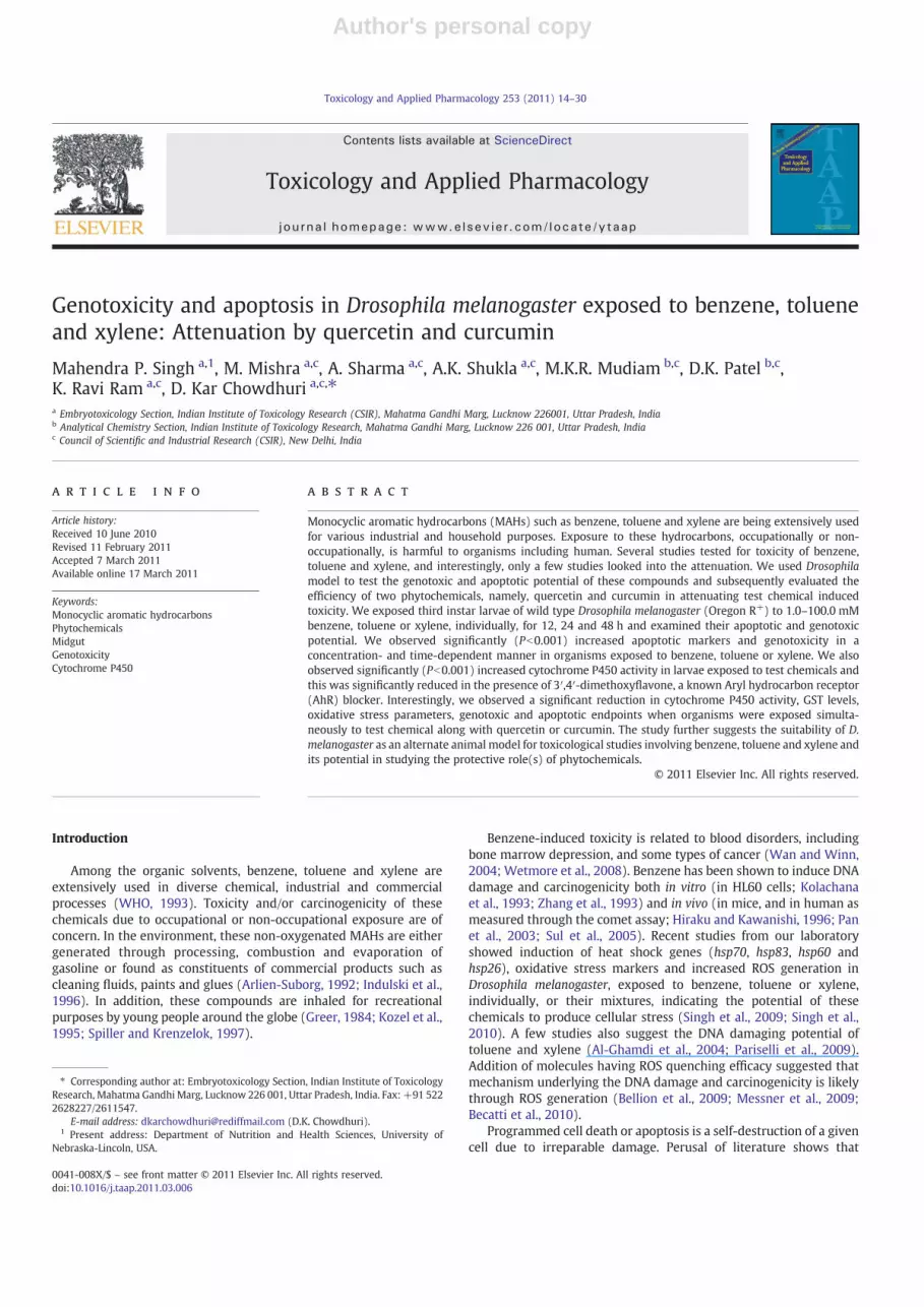
Genotoxicity and apoptosis in Drosophila melanogaster exposed to benzene, tolueneand xylene: Attenuation by quercetin and curcumin
Mahendra P. Singh a,1, M. Mishra a,c, A. Sharma a,c, A.K. Shukla a,c, M.K.R. Mudiam b,c, D.K. Patel b,c,K. Ravi Ram a,c, D. Kar Chowdhuri a,c,⁎a Embryotoxicology Section, Indian Institute of Toxicology Research (CSIR), Mahatma Gandhi Marg, Lucknow 226001, Uttar Pradesh, Indiab Analytical Chemistry Section, Indian Institute of Toxicology Research, Mahatma Gandhi Marg, Lucknow 226 001, Uttar Pradesh, Indiac Council of Scientific and Industrial Research (CSIR), New Delhi, India
a b s t r a c ta r t i c l e i n f o
Article history:Received 10 June 2010Revised 11 February 2011Accepted 7 March 2011Available online 17 March 2011
Keywords:Monocyclic aromatic hydrocarbonsPhytochemicalsMidgutGenotoxicityCytochrome P450
Monocyclic aromatic hydrocarbons (MAHs) such as benzene, toluene and xylene are being extensively usedfor various industrial and household purposes. Exposure to these hydrocarbons, occupationally or non-occupationally, is harmful to organisms including human. Several studies tested for toxicity of benzene,toluene and xylene, and interestingly, only a few studies looked into the attenuation. We used Drosophilamodel to test the genotoxic and apoptotic potential of these compounds and subsequently evaluated theefficiency of two phytochemicals, namely, quercetin and curcumin in attenuating test chemical inducedtoxicity. We exposed third instar larvae of wild type Drosophila melanogaster (Oregon R+) to 1.0–100.0 mMbenzene, toluene or xylene, individually, for 12, 24 and 48 h and examined their apoptotic and genotoxicpotential. We observed significantly (Pb0.001) increased apoptotic markers and genotoxicity in aconcentration- and time-dependent manner in organisms exposed to benzene, toluene or xylene. We alsoobserved significantly (Pb0.001) increased cytochrome P450 activity in larvae exposed to test chemicals andthis was significantly reduced in the presence of 3′,4′-dimethoxyflavone, a known Aryl hydrocarbon receptor(AhR) blocker. Interestingly, we observed a significant reduction in cytochrome P450 activity, GST levels,oxidative stress parameters, genotoxic and apoptotic endpoints when organisms were exposed simulta-neously to test chemical along with quercetin or curcumin. The study further suggests the suitability of D.melanogaster as an alternate animal model for toxicological studies involving benzene, toluene and xylene andits potential in studying the protective role(s) of phytochemicals.
© 2011 Elsevier Inc. All rights reserved.
Introduction
Among the organic solvents, benzene, toluene and xylene areextensively used in diverse chemical, industrial and commercialprocesses (WHO, 1993). Toxicity and/or carcinogenicity of thesechemicals due to occupational or non-occupational exposure are ofconcern. In the environment, these non-oxygenated MAHs are eithergenerated through processing, combustion and evaporation ofgasoline or found as constituents of commercial products such ascleaning fluids, paints and glues (Arlien-Suborg, 1992; Indulski et al.,1996). In addition, these compounds are inhaled for recreationalpurposes by young people around the globe (Greer, 1984; Kozel et al.,1995; Spiller and Krenzelok, 1997).
Benzene-induced toxicity is related to blood disorders, includingbone marrow depression, and some types of cancer (Wan and Winn,2004; Wetmore et al., 2008). Benzene has been shown to induce DNAdamage and carcinogenicity both in vitro (in HL60 cells; Kolachanaet al., 1993; Zhang et al., 1993) and in vivo (in mice, and in human asmeasured through the comet assay; Hiraku and Kawanishi, 1996; Panet al., 2003; Sul et al., 2005). Recent studies from our laboratoryshowed induction of heat shock genes (hsp70, hsp83, hsp60 andhsp26), oxidative stress markers and increased ROS generation inDrosophila melanogaster, exposed to benzene, toluene or xylene,individually, or their mixtures, indicating the potential of thesechemicals to produce cellular stress (Singh et al., 2009; Singh et al.,2010). A few studies also suggest the DNA damaging potential oftoluene and xylene (Al-Ghamdi et al., 2004; Pariselli et al., 2009).Addition of molecules having ROS quenching efficacy suggested thatmechanism underlying the DNA damage and carcinogenicity is likelythrough ROS generation (Bellion et al., 2009; Messner et al., 2009;Becatti et al., 2010).
Programmed cell death or apoptosis is a self-destruction of a givencell due to irreparable damage. Perusal of literature shows that
Toxicology and Applied Pharmacology 253 (2011) 14–30
⁎ Corresponding author at: Embryotoxicology Section, Indian Institute of ToxicologyResearch, Mahatma Gandhi Marg, Lucknow 226 001, Uttar Pradesh, India. Fax: +91 5222628227/2611547.
E-mail address: [email protected] (D.K. Chowdhuri).1 Present address: Department of Nutrition and Health Sciences, University of
Nebraska-Lincoln, USA.
0041-008X/$ – see front matter © 2011 Elsevier Inc. All rights reserved.doi:10.1016/j.taap.2011.03.006
Contents lists available at ScienceDirect
Toxicology and Applied Pharmacology
j ourna l homepage: www.e lsev ie r.com/ locate /ytaap
Author's personal copy
benzene, toluene and xylene induced apoptosis and genotoxicity inin vitro models and also in vivo (Smith, 1996; Ross, 2000; Snyder,2000; Nakai et al., 2003; Al-Ghamdi et al., 2004;Wan andWinn, 2004;Wetmore et al., 2008). Although several studies have been carried outto test the toxicity of benzene, toluene and xylene, only limitedinformation is available on the attenuation of toxic insults of these testchemicals in the exposed organisms (Emara and El-Bahrawy, 2008).To address this, we used D. melanogaster, as an in vivo model, as itoffers as an excellent alternative animal model [in accordance toEuropean Centre for the Validation of Alternative Methods (ECVAM)](Festing et al., 1998). Post genomic sequencing, Caenorhabditis elegansand Drosophila generatedmuch interest to toxicologists as these showfunctional conservation of majority of genes present in highermammals. Further, the availability of state-of-the-art moleculartools coupled with well-defined genetics and developmental biologyplaces this organism better for obtaining mechanistic insights.Moreover, from a toxicological/pharmacological perspective, fly andhigher mammals were shown to have similar dose–responserelationship with four monofunctional alkylating agents (Siddiqueet al., 2005a). Thus, we believe that laboratory-based experimentalevidences using this model is useful in generating information thatcould be of value for their efficient extrapolation to higher organisms.Therefore, we first studied apoptosis and genotoxicity in Drosophila,induced by benzene, toluene and xylene. Subsequently, we used thismodel to study the cytoprotective effect(s) of two phytochemicalshaving anti-oxidant properties, quercetin (QC) and curcumin (CUR)(Kottke, 1998; Lodha and Bagga, 2000), against benzene-, toluene-and xylene-induced cellular toxicity in exposed organisms. We showhere that QC and CUR do have the potential to protect against thebenzene-, toluene- and xylene-induced toxicity in vivo.
Material and methods
Flies and maintenance
All experiments were performed using wild type (Oregon R+)strain of D. melanogaster. The flies and larvae were reared at 23±1 °Con standard Drosophila food containing agar–agar, maize powder,sugar, yeast, nepagin (methyl-p-hydroxy benzoate salt; HiMedia,India) and propionic acid. Additional yeast suspension was providedfor their healthy growth.
Chemicals used for treatment
All chemicals, reagents and kits were procured from Sigma, MO,USA except otherwise stated. Analytical grade benzene (99.7% fromRanbaxy Pvt. Ltd, India), toluene (99.5%, SRL Pvt Ltd, India), xylene(mixture of o, m and p, 99.8%, SRL Pvt Ltd, India) and twophytochemicals namely, quercetin (3,5,7,3′,4′-pentahydroxyflavone,5,7,3′,4′-tetrahydroxyflavonol; QC) and curcumin [active ingredientof the rhizome of the plant turmeric (Curcuma longa Linn); CUR] wereused in the study. Dimethyl sulfoxide (DMSO) (SRL Pvt Ltd, India) wasused as a solvent and final concentration of DMSO in food wasrestricted to 0.3% (based on a previous study from this laboratoryNazir et al., 2003).
Treatment schedule
Four different concentrations (1.0, 10.0, 50.0 and 100.0 mM), eachcorresponding to different fractions of the LC50 (48 h) of benzene,toluene or xylene were used. Third instar larvae were grown onstandard Drosophila diet, contaminated with or without differentconcentrations of the three test chemicals in triplicates for 2–48 h. Forgenotoxicity studies by alkaline and neutral Comet assay, we used100.0 mM of benzene, toluene and xylene. Ethyl methanesulfonate(EMS) (1.0 mM) and γ-radiation (40.0 Gy) using Co60 source were as
used as positive controls for alkaline and neutral Comet assay,respectively. In another set of experiments, 100.0 μM QC or CUR(based on previous studies: Balasubramanyam et al., 2003; Guptaet al., 2007) was added to the food individually, or along with testchemicals for exposing larvae (24 and 48 h).
Quantitative determination of benzene, toluene and xylene
The chemical burden in the larvae exposed to test chemicals wasquantified as described previously (Singh et al., 2009). Briefly, thirdinstar larvae were exposed to 100.0 mM benzene, toluene or xylene infood with/without phytochemicals for 48 h. These larvae werehomogenized in deionized water in a headspace vial to obtain2.0 ml homogenate (10% w/v). The vials were heated at 65 °C for30 min with magnetic agitation. After the equilibrium, septa werepierced with SPME needle and the SPME polydimethyl siloxane(PDMS) fiber was exposed to headspace for 15 min to affect theadsorption of the test chemicals in the sample. SPME fiber wascollected and inserted directly into the injection port of Perkin Elmergas chromatography (USA) at 200 °C. Total time of this chromato-graphic analysis was 27 min. Blank analysis was carried out to avoidany carry over phenomena and/or external contamination betweenanalyses of samples (Alegretti et al., 2004).
Quantitative estimation of quercetin and curcumin levels
Wehave used 125 mg of larvae exposed to QC or CUR for extraction.Larvae were homogenized in 1.0 ml of 1× PBS. QC was extracted using500.0 μl of DMSO–methanol (1:4 v/v) following themethod of Schiborret al. (2010), CUR was extracted using 95% ethyl acetate and 5%methanol (v/v) and the extract was dried at 40 °C and resuspended in200.0 μl mobile phase (49% acetonitrile, 20% methanol, and 1% aceticacid (Jones et al., 1998). Subsequently, these extracts were analyzed byHPLC system (WaterMilford,MA,USA.) containing a reversephase C-18ODS analytical column (5 μM particle size). The levels of QC wereestimated by injecting 20.0 μl of sample (mobile phase containing 0.5%aqueous solutionof orthophosphoric acidandmethanol) and separatingwith aflowrate of 1 mlmin−1, for a run timeof 15 min,with photoarraydetector (PDA) at 375 nm. For CUR estimation, 20.0 μl of the extract wasseparatedwith a flow rate of 1.5 mlmin−1, for a run time of 5 min,withPDAdetector at 420 nm. A blank or control (extract from larvae exposedto control food) injection and the QC or CUR standards were runimmediately before each group of samples.
Measurement of oxidative stress parameters
In this study, we measured different oxidative stress parameterssuch as Reactive Oxygen Species (ROS), superoxide dismutase (SOD),catalase (CAT) and lipid peroxidation (LPO) measured as Malondial-dehyde content (MDA) essentially following methods describedpreviously (Gupta et al., 2007; Gupta et al., 2010).
Preparation of microsomes
The method for preparation of microsomes described previously byJohri et al. (2006) was followed with minor modifications. Control andtreated larvae were homogenized in ice-cold homogenization buffer(0.25 M potassium phosphate buffer containing 0.15 M KCl, 0.25 mMphenylmethanesulfonylfluoride (PMSF), 0.01 M ethylenediaminete-traacetic acid (EDTA) and 0.1 mM Dithiothreitol (DTT); pH 7.25) toobtain 10% homogenate. The homogenate was centrifuged at 9000×gfor 30 min (at 4 °C) to obtain the supernatant, which in turn wascentrifuged at 105,000×g for 60 min to sediment microsomes. Thepellets were resuspended in microsome dilution buffer (0.1 M potas-sium phosphate buffer, pH 7.25, 20% (v/v) glycerol, 0.25 mM PMSF,0.01 M EDTA and 0.1 M DTT) and stored at −80 °C until further use.
15M.P. Singh et al. / Toxicology and Applied Pharmacology 253 (2011) 14–30

Author's personal copy
Ethoxyresorufin-O-deethylase (EROD) and methoxyresorfin-O-deethylase(MROD) assay for estimation of cytochrome P450 activity. The activitiesof EROD and MROD in the isolated Drosophila larval microsomes weredetermined following Johri et al. (2006), with minor modifications.The reaction mixture consisted of 1.0 ml of 0.1 M PBS pH 7.8, 5.0 μl1.0 mM ethoxy or methoxy resorufin and 25.0 μl microsomal fraction.The reaction was initiated by the addition of 1.0 ml 1.0 mM NADPHand the mixture was incubated at 37 °C for 10 min. Reaction wasterminated by adding 2.0 ml of methanol and the mixtures werecentrifuged at 2000×g for 7 min. Levels of resorufin in the supernatantweremeasured using a Perkin Elmer LS 55 Luminescence Spectrometerat excitation wavelength of 550 nm and emission wavelength of585 nm.
Inhibition of aryl hydrocarbon receptor (AhR). The role of AhR orits homolog was examined using a potent AhR inhibitor 3′,4′-dimethoxyflavone (DMF) (Lin et al., 2006). Larvae were exposed tofood containing 100.0 μM DMF together with 100.0 mM of benzene,toluene or xylene as mentioned in Treatment schedule section andsubsequently, EROD activity was measured as in Ethoxyresorufin-O-deethylase (EROD) and methoxyresorfin-O-deethylase (MROD) assayfor estimation of cytochrome P450 activity section. We have usedlarvae exposed to 200.0 ng/ml Tetrachlorodibenzo-p-dioxin (TCDD)(Cespedes et al., 2010) together with 100.0 μM DMF as positivecontrol. TCDD was used as a positive control because this is also ahalogenated aromatic hydrocarbon and is known to induce cyto-chrome P450 1AI (CYP1A1) through AhR (Cespedes et al., 2010).Larvae exposed to 100.0 μM DMF alone was used as an additionalcontrol.
Glutathione S-transferase (GST, EC 2.5.1.18)
Glutathione S-transferase (GST) activity was determined followingHabig et al. (1974) with minor modifications. The reaction mixtureconsisted of 0.2 M sodium phosphate buffer, 75.0 μl larval homoge-nate, reduced glutathione (1.0 mM) and 1-chloro 2, 4 dinitrobenzene(CDNB) (5.0 mM). An increase in absorbance (340 nm)wasmeasuredfor 3 min at 30-s intervals and the enzyme activity was calculated asnmol CDNB reduced/min/mg larval protein using molar extinctioncoefficient of 6.25×10³ M−1 cm−1.
Trypan blue dye exclusion assay
Tissue damage in the test chemical exposed organisms wasexamined by trypan blue exclusion assay as described earlier (Krebsand Feder, 1997). In brief, internal tissues of control and treated larvae(50–60/group), explanted in PSS were washed once in 50.0 mMphosphate buffered saline (PBS), pH 7.4 (Dulbecco's, HiMedia Pvt Ltd.,Mumbai, India). They were then immersed in trypan blue stain(0.2 mg/ml in 50.0 mM PBS, pH 7.4) and shaken gently for 30 min at24±1 °C. After staining, tissues were washed thrice in wash buffer(0.1 M PBS pH 7.4) and immediately visualized and scored larvae fortrypan blue staining.
Single cell preparation
Midgut tissues of 15 larvae from control and treated groups wereincubated in collagenase (0.5 mg/ml of 1.0 M PBS pH 7.4) for 15 minat 24±1 °C. The dissociated cells were then passed through 80 μmnylon mesh to get rid of clumping. Collagenase was removed bywashing the cell suspension with 0.1 M PBS (pH 7.4) for at least threetimes with gentle shaking. Viability of cells was checked by trypanblue staining before the start of the experiment (Phillips, 1973). Thesecells were processed for different end point measurements asdescribed below.
Assay of apoptosis
All apoptotic end points were measured in single cells preparedfrom midgut tissues of control and treated organisms except forthose of terminal deoxynucleotidyl transferase mediated dUTP nickend labeling (TUNEL) assay. For TUNEL assay, we used whole midguttissues and for measuring caspase activity, we used 10% tissuehomogenate, to match the published standard protocols.
Flow cytometric determination of cellular Rpr, Hid and Grim (initiator ofapoptosis) levels. Previously described flow cytometric detectionmethod was followed with minor modification (Wechsler-Reyaet al., 1998). Cells after thorough washing in 0.1 M PBS (pH 7.4)were fixed in PBS containing 0.25% paraformaldehyde for 1 h at 4 °Cand then permeabilized by washing twice in PBS with 0.1% Triton X-100 for 10 min at 24±1 °C. Subsequently, cells were rinsed with PBSand incubated with primary antibodies against Rpr, Hid or Grim (anti-goat polyclonal IgG, 1:50 in PBS containing 2% BSA; Santa Cruz, CA,USA) for 1 h at 4 °C. This was followed by incubation of cells with FITCconjugated rabbit anti-goat IgG secondary antibody (1:100 in 0.1 MPBS, pH 7.4 containing 2% BSA) for 1 h at 4 °C. The stained cells wereanalyzed on Becton Dickinson flow cytometer (BD, NJ, USA) using Cellquest software (Mac OS 8.6). For each sample, 10,000 events werecounted and results were expressed in terms of the percentage cellsexpressing particular protein.
Phosphatidylserine (PS) externalization assay (Annexin V-FITC staining).Apoptotic cells were detected using Annexin V-FITC apoptosisdetection kit essentially following the manufacturer's instructions.Briefly, media binding reagent and Annexin V-FITC were mixed withapproximately 5×10⁵ cells and incubated at 24±1 °C for 15 min.After removing the media, the cells were re-suspended in cold 1×binding buffer and stained with PI. Ten thousand events wereacquired per treatment group using Becton Dickinson flow cytometerand data were analyzed with Cell Quest software (Mac OS 8.6). TheFITC signal was detected by FL1 (FITC detector) at 518 nm and Pl wasdetected by FL2 (phycoerythrin fluorescence detector) at 620 nm. Thelog of Annexin V-FITC and Pl fluorescence was displayed on the X- andY-axis of the data report respectively.
Determination of mitochondrial membrane potential (ΔΨm). Depolari-zation of mitochondrial membrane was analyzed following a previouslydescribed method with minor modification (Vayssier-Taussat et al.,2001). A fluorochrome 5,5′,6,6′-tetrachloro-1,1′,3,3′-tetraethylbenzimidazolyl carbocyanine iodide (JC-1) was used for this purpose.Themitochondrial membrane depolarization is associatedwith a shift inJC-1 fluorescence emission from red to green. Cells of control and treatedorganisms were suspended in Schneider's Drosophila medium con-taining 10.0 μM JC-1 (prepared in DMSO) for 30 min at 24 °C. Cells werethen washed with 0.1 M PBS (pH 7.4) twice and finally re-suspended in500.0 μl PBS (pH 7.4) for FACScan analysis. Ten thousand events werecounted per sample in acquisition and analysis was performed usingcell quest software (Mac OS 8.6). The results were expressed as thepercentage of cells with disrupted mitochondrial membranes.
Assay of DEVD- and IETD-ase activities. The assay is based on spec-trophotometric detection of the chromophore p-nitroanilide (pNA)obtained after specific action of different cysteine proteases involvedin apoptotic pathways on tetrapeptide substrates, respectively. Theassay was performed essentially following the manufacturer's protocol(Bio Vision, Inc., CA, USA). Supernatant from the 10% tissue homogenatewas mixed with chilled cell lysis buffer, 2× reaction buffer (containing10.0 mM dithiothreitol) and 200 μM substrates. The reaction mixturewas incubated at 37 °C for 1.5 h and absorbance of the colored productwas measured at 405 nm on a Cintra 20 ultraviolet spectrophotometer(GBC Scientific Equipment, Melbourne, Australia).
16 M.P. Singh et al. / Toxicology and Applied Pharmacology 253 (2011) 14–30

Author's personal copy
Terminal deoxynucleotidyl transferase mediated dUTP nick end labeling(TUNEL) assay. TUNEL assay was performed using “In situ cell deathdetection kit” essentially following the manufacturer's protocol(Roche Molecular Biochemicals, Mannheim, Germany). Midguttissues of control and treated larvae were fixed in freshly prepared2.5% glutaraldehyde and permeabilized in PBST followed by washingin PBS. Theywere incubated in primary TUNELmixture for 1 h at 37 °Cand then washed with PBS for 30 min at 24 °C (6 changes of 5 mineach) followed by incubation in converter alkaline phosphate (AP)solution at 37 °C. After washing, tissues were incubated in thesubstrate solution containing nitro blue tetrazolium (NBT) and 5-bromo-4-chloro-3-indolyl phosphate (BCIP), specific for AP, for15 min in dark at 24 °C. The tissues were then washed thoroughlywith PBS, mounted on clean glass slides using 50% glycerol andcoverglass was placed. The edges of the coverglass were sealed withDPX and the tissues were scored for TUNEL positive cells under Leitzorthoplan light microscope (Wetzlar, Germany). One hundred fiftycells were examined from each group by randomized manualcounting (25 cells/gut tissue and 2 gut tissues/experiment and 3experiments/group/time point).
Poly (ADP-ribose) polymerase (PARP) cleavage. We performed PARPantibody staining in cells of control and treated organisms using “Anti-PARP Cleavage Site Specific Antibody (CSSA) Assay kit” (Invitrogen,USA) essentially following themanufacturer's protocol. The FITC stainedcells were analyzed on a Becton Dickinson flow cytometer using Cellquest software (Mac OS 8.6). For each sample, 10,000 events werecounted and results were expressed in terms of percent cells expressingPARP.
Evaluation of DNA damage by Comet assay
Single cell preparation and viability test were performed asdescribed in Single cell preparation section. Care was taken toperform all the steps (from single cell preparation to staining)under dim light to avoid any light-induced DNA damage.
Slides preparation. A previously described method for preparation ofslides was followed (Tice et al., 2000) wherein frosted slides (with1.5 cm frosted end) were used. The slides were rinsed in methanoland flame dried. The slides were then coated on non-frosted end with1.0% normal melting agarose (NMA), prepared in milliQ water andkept at 60 °C) up to two-third of their length by dipping into moltenNMA and were allowed to dry for 1 h at room temperature (24 °C).
Evaluation of DNA damage. For the entire group, slides were preparedin duplicate according to themethod described earlier (Siddique et al.,2008). All experimentswere repeated three times. The cell suspension(80.0 μl) was mixed with 80.0 μl of 1.5% low melting point agarose(LMA; prepared in Ca2+Mg2+ free PBS; final concentration 0.75%, 35–40 °C). For each slide, 75.0 μl of the above mixture was immediatelylayered on a base slide. Cover slip was immediately placed over thesecond layer. The slide was then placed on a chilled plate for 10 min toallow solidification of agarose. This step was repeated again to placeanother layer of LMA 0.75%. Finally, the cover slip was removed andthe slide was immersed for 2 h in freshly prepared, chilled lysingsolution (2.5 M NaCl, 100.0 mM EDTA, 10.0 mM Tris and 1.0% TritonX-100, pH 10). After lysis, the slides were subjected to neutral andalkaline gel electrophoresis as follows.
Electrophoresis for both alkaline and neutral Comet assay. For alkalinecomet assay, slides were placed in chilled electrophoresis buffer(1.0 mM Na2EDTA and 300.0 mM NaOH, pHN13) for 10 min for DNAunwinding. Subsequently, electrophoresis was conducted in chilledelectrophoresis buffer (1.0 mM Na2EDTA and 300.0 mM NaOH,pHN13) for 15 min at 0.7 V/cm (300 mA/25 V) at 4 °C. The slides
were then washed three times with 0.4 M Tris buffer (pH 7.5) at 4 °Cto neutralize excess alkali and then for staining as described inStaining section.
We followed a previously described method for neutral Cometassay for the detection of double-strand breaks (Fracasso et al., 2009).The slides after lysis were kept in electrophoresis buffer (300.0 mMCH3COONa, 100.0 mM Tris–HCl, pH 8.5) for 1 h and then transferredto horizontal electrophoresis unit (Life Technologies, Gaithersburg,MD, USA) containing fresh buffer. Electrophoresis was carried out atconstant current of 60 mA for 1 h at 4 °C and slides were stainedsubsequently as described in the following section.
Staining. The slides were stained with ethidium bromide (20.0 μg/ml;75 μl per slide) for 10 min in dark. After staining, the slides were dippedonce in chilled distilledwater to remove the excess stain and cover slipswere placed over the slides.
Slide scoring. The slides were examined on a Leica DMLB microscopewith fluorescence attachment (Leica Germany). The images weretransferred to a computer through a charge coupled device (CCD)camera and analyzed using Komet 5.0 software (Kinetic Imaging,Liverpool, UK). One hundred fifty cells from each group (25 cells perslidewith two slides/experimental group in triplicates)were examined.The tail length (TL) (μm), tail DNA (%) (TD) and tail moment (TM)(arbitrary units) were used as indicators of DNA damage as describedearlier (Olive et al., 1992).
Statistical analysis
Different parameters were analyzed in control and exposed thirdinstar larvae of D. melanogaster (Oregon R+). Analysis of variance(ANOVA) was carried out to find out the significant differences inmeans considering each end point as dependent variable (with orwithout QC and CUR) and treatment (benzene, toluene and xylene);concentration (1.0, 10.0, 50.0 and 100.0 mM) and duration ofexposure (2, 4, 6, 12, 24 and 48 h) as independent variables. Prior toapplying the ANOVA, homogeneity of variance was ascertained usingLevene's test of equality error variance. Two-way analysis of variancewas carried out for ROS generation, apoptotic and genotoxic markersas dependent variables (Zar, 1984). SPSS 14.0 (SPSS, Mapinfo Max,USA) was used to analysis the data. To identify the degree ofrelationship between cytochrome P450 activity and oxidative stressparameters analyzed in the present study with test chemical alone orin combination with QC or CUR, simple linear correlation (Pearson r)statistics followed significance analysis of correlation coefficient wereperformed.
Results
We did not observe any significantly altered activities or levels ofall the tested parameters at 1.0 mM concentration of the testchemicals throughout the treatment period and till 6 h at 10.0 mMconcentration of these test chemicals (data for 1.0 mM test chemicalsnot shown). Hence, we only included data of 6, 12, 24 and 48 hexposure of 10.0–100.0 mM benzene, toluene and xylene. Unlikeapoptotic markers, Comet assay parameters did not show anyremarkable damage till 12 h and hence, we presented only 24 and48 h data in the study. Further, data obtained from parallel control(DMSO) were similar to those of controls and hence, we used the datafrom controls for comparison.
Detection of chemicals in larvae of D. melanogaster
To ensure the larval uptake of the test chemical and its uptake isnot altered in the presence of QC or CUR or vice versa, we estimatedthe chemical load in exposed organisms. We detected benzene,
17M.P. Singh et al. / Toxicology and Applied Pharmacology 253 (2011) 14–30
Author's personal copy
toluene or xylene in larvae exposed to test chemical alone at levelssimilar (PN0.05) to those in combination with QC or CUR. We alsodetected QC or CUR in organisms exposed to QC or CUR alone at levelssimilar (PN0.05) to those exposed to QC or CUR along with testchemical (please see Supplemental Figs. S1 and S2).
Moderate trypan blue staining in tissues of Oregon R+ larvae exposed tobenzene, toluene or xylene
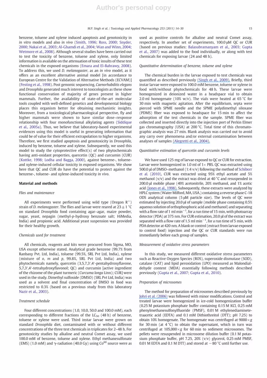
To determine if exposure to benzene, toluene or xylene results intissue damage, we analyzed trypan blue staining in tissues ofD. melanogaster larvae exposed to 100.0 mM benzene (Fig. 1B),toluene (Fig. 1C) and xylene (Fig. 1D) for 48 h (Fig. 1). Of the larvaeexposed to benzene, 94% of them showed blue staining in theirmidgut, salivary gland, gastric caeca and brain ganglia while 86% or84% of the larvae exposed to toluene or xylene exhibited a pale tomoderate blue staining in the above mentioned tissues respectively.
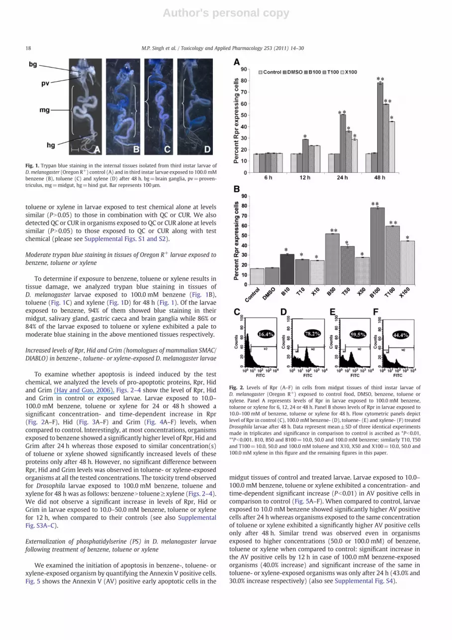
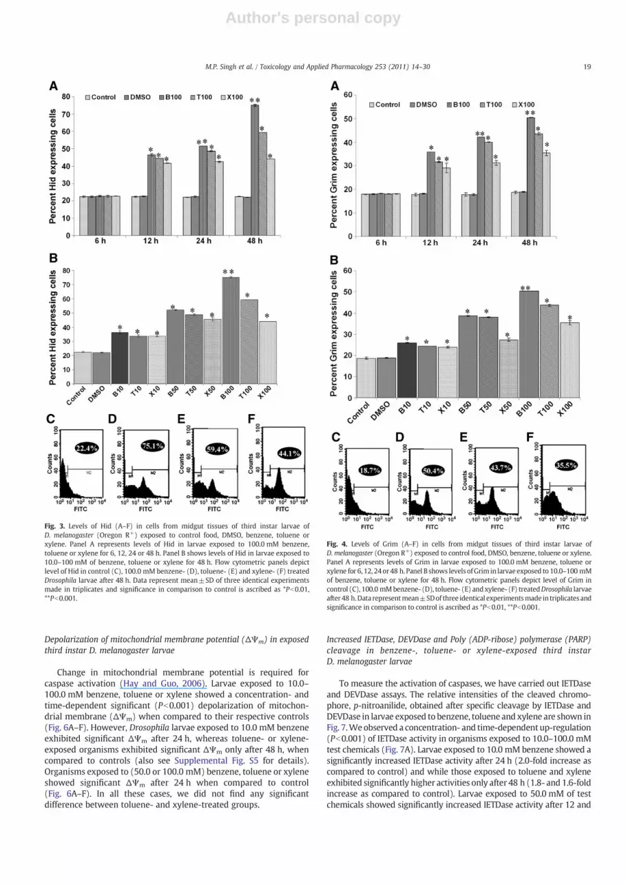
Increased levels of Rpr, Hid and Grim (homologues of mammalian SMAC/DIABLO) in benzene-, toluene- or xylene-exposed D. melanogaster larvae
To examine whether apoptosis is indeed induced by the testchemical, we analyzed the levels of pro-apoptotic proteins, Rpr, Hidand Grim (Hay and Guo, 2006). Figs. 2–4 show the level of Rpr, Hidand Grim in control or exposed larvae. Larvae exposed to 10.0–100.0 mM benzene, toluene or xylene for 24 or 48 h showed asignificant concentration- and time-dependent increase in Rpr(Fig. 2A–F), Hid (Fig. 3A–F) and Grim (Fig. 4A–F) levels, whencompared to control. Interestingly, at most concentrations, organismsexposed to benzene showed a significantly higher level of Rpr, Hid andGrim after 24 h whereas those exposed to similar concentration(s)of toluene or xylene showed significantly increased levels of theseproteins only after 48 h. However, no significant difference betweenRpr, Hid and Grim levels was observed in toluene- or xylene-exposedorganisms at all the tested concentrations. The toxicity trend observedfor Drosophila larvae exposed to 100.0 mM benzene, toluene andxylene for 48 h was as follows: benzeneN toluene≥xylene (Figs. 2–4).We did not observe a significant increase in levels of Rpr, Hid orGrim in larvae exposed to 10.0–50.0 mM benzene, toluene or xylenefor 12 h, when compared to their controls (see also SupplementalFig. S3A–C).
Externalization of phosphatidylserine (PS) in D. melanogaster larvaefollowing treatment of benzene, toluene or xylene
We examined the initiation of apoptosis in benzene-, toluene- orxylene-exposed organism by quantifying the Annexin V positive cells.Fig. 5 shows the Annexin V (AV) positive early apoptotic cells in the
midgut tissues of control and treated larvae. Larvae exposed to 10.0–100.0 mM benzene, toluene or xylene exhibited a concentration- andtime-dependent significant increase (Pb0.01) in AV positive cells incomparison to control (Fig. 5A–F). When compared to control, larvaeexposed to 10.0 mM benzene showed significantly higher AV positivecells after 24 h whereas organisms exposed to the same concentrationof toluene or xylene exhibited a significantly higher AV positive cellsonly after 48 h. Similar trend was observed even in organismsexposed to higher concentrations (50.0 or 100.0 mM) of benzene,toluene or xylene when compared to control: significant increase inthe AV positive cells by 12 h in case of 100.0 mM benzene-exposedorganisms (40.0% increase) and significant increase of the same intoluene- or xylene-exposed organisms was only after 24 h (43.0% and30.0% increase respectively) (also see Supplemental Fig. S4).
Fig. 1. Trypan blue staining in the internal tissues isolated from third instar larvae ofD. melanogaster (Oregon R+) control (A) and in third instar larvae exposed to 100.0 mMbenzene (B), toluene (C) and xylene (D) after 48 h. bg=brain ganglia, pv=proven-triculus, mg=midgut, hg=hind gut. Bar represents 100 μm.
Fig. 2. Levels of Rpr (A–F) in cells from midgut tissues of third instar larvae ofD. melanogaster (Oregon R+) exposed to control food, DMSO, benzene, toluene orxylene. Panel A represents levels of Rpr in larvae exposed to 100.0 mM benzene,toluene or xylene for 6, 12, 24 or 48 h. Panel B shows levels of Rpr in larvae exposed to10.0–100 mM of benzene, toluene or xylene for 48 h. Flow cytometric panels depictlevel of Rpr in control (C), 100.0 mM benzene- (D), toluene- (E) and xylene- (F) treatedDrosophila larvae after 48 h. Data represent mean±SD of three identical experimentsmade in triplicates and significance in comparison to control is ascribed as *Pb0.01,**Pb0.001. B10, B50 and B100=10.0, 50.0 and 100.0 mM benzene; similarly T10, T50and T100=10.0, 50.0 and 100.0 mM toluene and X10, X50 and X100=10.0, 50.0 and100.0 mM xylene in this figure and the remaining figures in this paper.
18 M.P. Singh et al. / Toxicology and Applied Pharmacology 253 (2011) 14–30
Author's personal copy
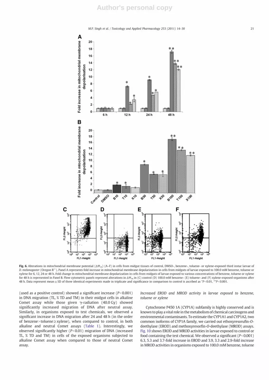
Depolarization of mitochondrial membrane potential (ΔΨm) in exposedthird instar D. melanogaster larvae
Change in mitochondrial membrane potential is required forcaspase activation (Hay and Guo, 2006). Larvae exposed to 10.0–100.0 mM benzene, toluene or xylene showed a concentration- andtime-dependent significant (Pb0.001) depolarization of mitochon-drial membrane (ΔΨm) when compared to their respective controls(Fig. 6A–F). However, Drosophila larvae exposed to 10.0 mM benzeneexhibited significant ΔΨm after 24 h, whereas toluene- or xylene-exposed organisms exhibited significant ΔΨm only after 48 h, whencompared to controls (also see Supplemental Fig. S5 for details).Organisms exposed to (50.0 or 100.0 mM) benzene, toluene or xyleneshowed significant ΔΨm after 24 h when compared to control(Fig. 6A–F). In all these cases, we did not find any significantdifference between toluene- and xylene-treated groups.
Increased IETDase, DEVDase and Poly (ADP-ribose) polymerase (PARP)cleavage in benzene-, toluene- or xylene-exposed third instarD. melanogaster larvae
To measure the activation of caspases, we have carried out IETDaseand DEVDase assays. The relative intensities of the cleaved chromo-phore, p-nitroanilide, obtained after specific cleavage by IETDase andDEVDase in larvae exposed to benzene, toluene and xylene are shown inFig. 7.Weobserved a concentration- and time-dependent up-regulation(Pb0.001) of IETDase activity in organisms exposed to 10.0–100.0 mMtest chemicals (Fig. 7A). Larvae exposed to 10.0 mM benzene showed asignificantly increased IETDase activity after 24 h (2.0-fold increase ascompared to control) and while those exposed to toluene and xyleneexhibited significantly higher activities only after 48 h (1.8- and 1.6-foldincrease as compared to control). Larvae exposed to 50.0 mM of testchemicals showed significantly increased IETDase activity after 12 and
Fig. 3. Levels of Hid (A–F) in cells from midgut tissues of third instar larvae ofD. melanogaster (Oregon R+) exposed to control food, DMSO, benzene, toluene orxylene. Panel A represents levels of Hid in larvae exposed to 100.0 mM benzene,toluene or xylene for 6, 12, 24 or 48 h. Panel B shows levels of Hid in larvae exposed to10.0–100 mM of benzene, toluene or xylene for 48 h. Flow cytometric panels depictlevel of Hid in control (C), 100.0 mM benzene- (D), toluene- (E) and xylene- (F) treatedDrosophila larvae after 48 h. Data represent mean±SD of three identical experimentsmade in triplicates and significance in comparison to control is ascribed as *Pb0.01,**Pb0.001.
Fig. 4. Levels of Grim (A–F) in cells from midgut tissues of third instar larvae ofD. melanogaster (Oregon R+) exposed to control food, DMSO, benzene, toluene or xylene.Panel A represents levels of Grim in larvae exposed to 100.0 mM benzene, toluene orxylene for 6, 12, 24or48 h. Panel B shows levels ofGrim in larvaeexposed to 10.0–100 mMof benzene, toluene or xylene for 48 h. Flow cytometric panels depict level of Grim incontrol (C), 100.0 mMbenzene- (D), toluene- (E) andxylene- (F) treatedDrosophila larvaeafter 48 h.Data representmean±SDof three identical experimentsmade in triplicates andsignificance in comparison to control is ascribed as *Pb0.01, **Pb0.001.
19M.P. Singh et al. / Toxicology and Applied Pharmacology 253 (2011) 14–30

Author's personal copy
24 h, respectively, in benzene-, toluene- or xylene-exposed groups ascompared to their respective controls. A maximum (Pb0.001) 4.0-, 3.3-or 3.1-fold increase in IETDase activity was observed in 100.0 mMbenzene-, toluene- or xylene-exposed larvae after 48 h, respectively(Fig. 7A). The DEVDase activity in the test chemicals exposed organismswas similar to that of IETDase activity (Fig. 7B). We observedsignificantly increased number of PARP-FITC positive cells in organismsexposed to 100.0 mM benzene, toluene or xylene for 24 and 48 h (4.3,3.9- and 3.6-fold increase after 48 h respectively), when compared totheir respective controls (Fig. 8A–E). The differences in the number ofPARP-FITC positive cells observed among benzene-, toluene- or xylene-exposed organisms for 24 and 48 h were found to be non-significant.
Increased DNA nicking in test chemicals exposed third instar larvae ofD. melanogaster
To examine test chemical mediated DNA nicking, a key feature ofapoptosis, we performed TUNEL assay in midgut cells of control and
100.0 mM test chemicals treated larvae after 24 and 48 h (Fig. 9). After24 h, we observed significant increase in TUNEL positive cells only inDrosophila larvae exposed to benzene (100.0 mM; 3.3-fold) whencompared to control. Organisms exposed to similar concentrations oftoluene or xylene showed significantly more TUNEL positive cellsthan controls only after 48 h (7.7- and 7.9-fold increase). However,even at this time point, the number of TUNEL positive cells inorganisms exposed to 100.0 mM of toluene or xylene for 24 and 48 hwas significantly lower than that in organisms exposed to benzene(Fig. 9A–C).
Increased DNA damage in the midgut cells of D. melanogaster larvaeexposed to benzene, toluene or xylene
To determine the extent of DNA damage, we analyzed Cometparameters in organisms exposed to the test chemical. We observed95–98% cell viability in controls, and also in 100.0 mM benzene-,toluene- or xylene-exposed groups. Larvae exposed to 1.0 mM EMS
Fig. 5. Annexin V positive cells (A–F) in cells frommidgut tissues of control, DMSO-, benzene-, toluene- or xylene-exposed third instar larvae of D. melanogaster (Oregon R+). Panel Arepresents percentage of Annexin positive cells from midguts of larvae exposed to 100.0 mM benzene, toluene or xylene for 6, 12, 24 or 48 h. Percent Annexin positive cells frommidguts of larvae exposed to various concentrations of benzene, toluene or xylene for 48 h is represented in Panel B. Flow cytometric panels depict cells in early apoptotic stage(Annexin V positive cells, FITC quadrant), late apoptotic stage (Annexin V and propidium iodide (PI) positive cells, FITC-PI quadrant) and necrotic state (PI positive, PI quadrant) frommidgut tissues of control (C), 100.0 mM benzene- (D), toluene- and (E) xylene- (F) exposed organisms after 48 h. Data represent mean±SD of three identical experiments made intriplicate and significance in comparison to control is ascribed as *Pb0.01, **Pb0.001.
20 M.P. Singh et al. / Toxicology and Applied Pharmacology 253 (2011) 14–30
Author's personal copy
(used as a positive control) showed a significant increase (Pb0.001)in DNA migration (TL, % TD and TM) in their midgut cells in alkalineComet assay while those given γ-radiation (40.0 Gy) showedsignificantly increased migration of DNA after neutral assay.Similarly, in organisms exposed to test chemicals, we observed asignificant increase in DNA migration after 24 and 48 h (in the orderof benzeneN toluene≥xylene), when compared to control, in bothalkaline and neutral Comet assays (Table 1). Interestingly, weobserved significantly higher (Pb0.01) migration of DNA (increasedTL, % TD and TM) in cells of the exposed organisms subjected toalkaline Comet assay when compared to those of neutral Cometassay.
Increased EROD and MROD activity in larvae exposed to benzene,toluene or xylene
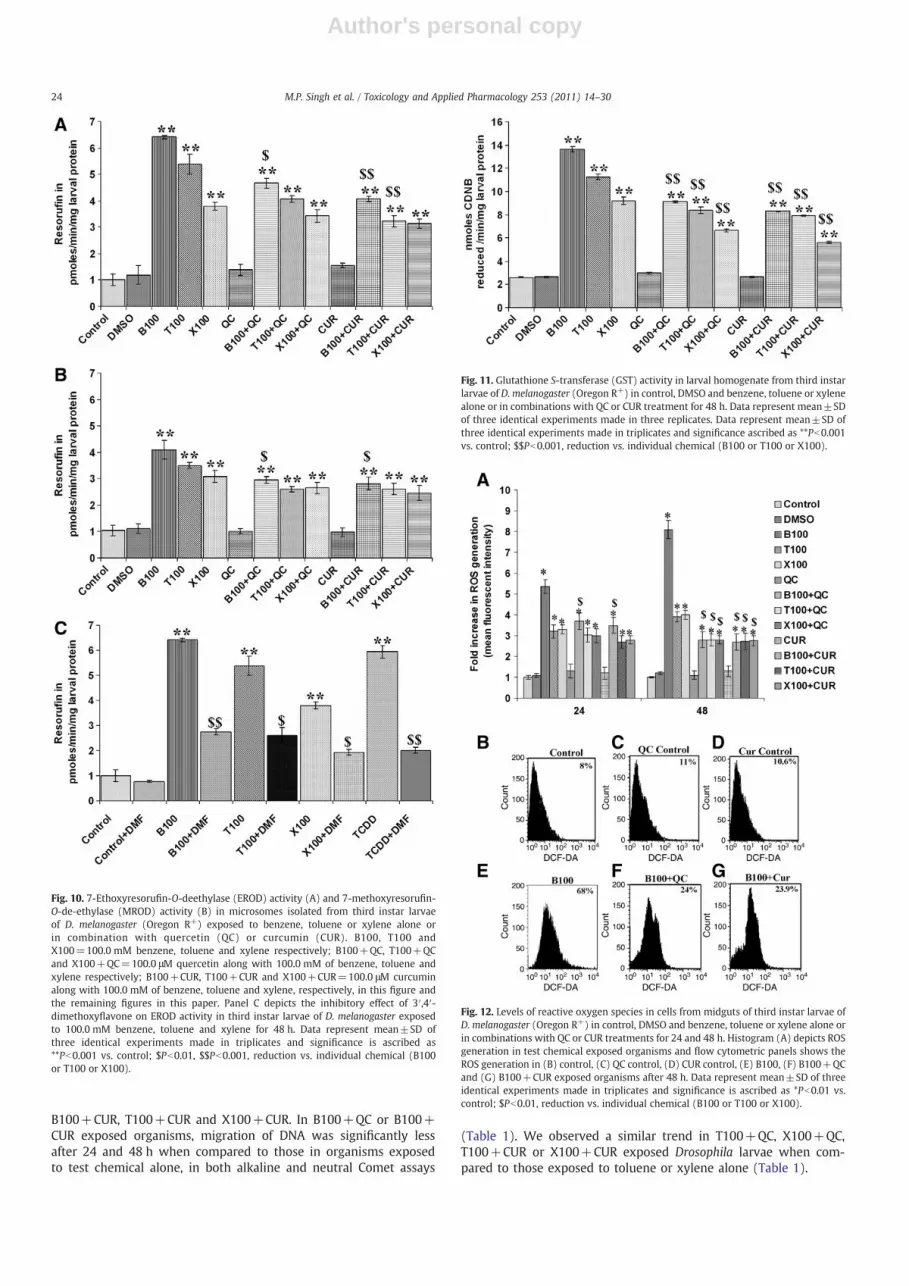
Cytochrome P450 1A (CYP1A) subfamily is highly conserved and isknown toplaya vital role in themetabolismof chemical carcinogens andenvironmental contaminants. To estimate the CYP1A1 and CYP1A2, twocommon isoforms of CYP1A family, we carried out ethoxyresorufin-O-deethylase (EROD) and methoxyresorfin-O-deethylase (MROD) assays.Fig. 10 shows EROD andMROD activities in larvae exposed to control orfood containing the test chemical. We observed a significant (Pb0.001)6.3, 5.3 and 3.7-fold increase in EROD and 3.9, 3.3 and 2.9-fold increaseinMROD activities in organisms exposed to 100.0 mMbenzene, toluene
Fig. 6. Alterations in mitochondrial membrane potential (ΔΨm) (A–F) in cells from midgut tissues of control, DMSO-, benzene-, toluene- or xylene-exposed third instar larvae ofD. melanogaster (Oregon R+). Panel A represents fold increase in mitochondrial membrane depolarization in cells from midguts of larvae exposed to 100.0 mM benzene, toluene orxylene for 6, 12, 24 or 48 h. Fold change in mitochondrial membrane depolarization in cells from midguts of larvae exposed to various concentrations of benzene, toluene or xylenefor 48 h is represented in Panel B. Flow cytometric panels represent alterations in ΔΨm in (C) control (D) 100.0 mM benzene- (E) toluene- and (F) xylene-exposed organisms after48 h. Data represent mean±SD of three identical experiments made in triplicate and significance in comparison to control is ascribed as *Pb0.01, **Pb0.001.
21M.P. Singh et al. / Toxicology and Applied Pharmacology 253 (2011) 14–30

Author's personal copy
or xylene for 48 h, respectively, when compared to their controls(Fig. 10A–B).
Significantly decreased EROD activity in larvae exposed to benzene,toluene or xylene together with DMF
To determine if benzene-, toluene- or xylene-mediated inductionof cytochrome P450 enzymes requires Aryl hydrocarbon Receptor(AhR) or its homolog, we exposed larvae to DMF, a potent inhibitor of
AhR, alone or in combination with test chemical. Exposure to DMF(100 μM) alone did not significantly decrease the EROD activitycompared to that in control Drosophila larvae. However, we observedsignificantly decreased EROD activity (Pb0.001) in organisms ex-posed to food containing B100, T100, X100 (100.0 mM benzene,toluene or xylene), or TCDD (positive control) along with DMF(Fig. 10C), in comparison to that observed in test chemical alonetreated group.
Effects of QC and CUR on the adverse effects of benzene, toluene or xylenein exposed Drosophila
As we have observed increase in EROD, MROD activities, oxidativestress parameters and genotoxicity in response to test chemicals, weexamined the effect of co-treatment with QC or CUR on the levels ofthese various parameters.
Fig. 7. IETDase (A) and DEVDase (B) activities in midgut tissue homogenate from control,DMSO-, benzene-, toluene- or xylene-exposed third instar larvae ofD.melanogaster (OregonR+) after 12, 24 and 48 h. Data representmean±SD of three identical experimentsmade intriplicates and significance in comparison to control is ascribed as *Pb0.01, **Pb0.001.
Fig. 8. PARP cleavage in cells frommidgut tissues of control and DMSO-, benzene-, toluene- or xylene-treated organisms for 24 and 48 h. Histogram (A) depicts fold increase in PARP-FITC positive cells. Flow cytometric panels represent PARP cleavage in (B) control, (C) 100.0 mM benzene- (D) toluene- and (E) xylene-exposed groups after 48 h. Data representmean±SD of three identical experiments made in triplicates and significance in comparison to control is ascribed as *Pb0.01.
Fig. 9. TUNEL positive cells in midgut tissues of third instar larvae of D. melanogaster(Oregon R+) of control and benzene-, toluene- or xylene-exposed third instar larvae ofD. melanogaster (Oregon R+) for 24 and 48 h. Histogram (A) depicts percent TUNELpositive cells in control and benzene-, toluene- or xylene-exposed organisms. Panels Band C represent microscopic images showing TUNEL positive cells in control and100.0 mM benzene-exposed organisms, respectively after 48 h. Arrows indicate TUNELpositive cells. Bar represents 200 μm. Data represent mean±SD of three identicalexperiments made in triplicates and significance in comparison to control is ascribed as*Pb0.01.
22 M.P. Singh et al. / Toxicology and Applied Pharmacology 253 (2011) 14–30
Author's personal copy
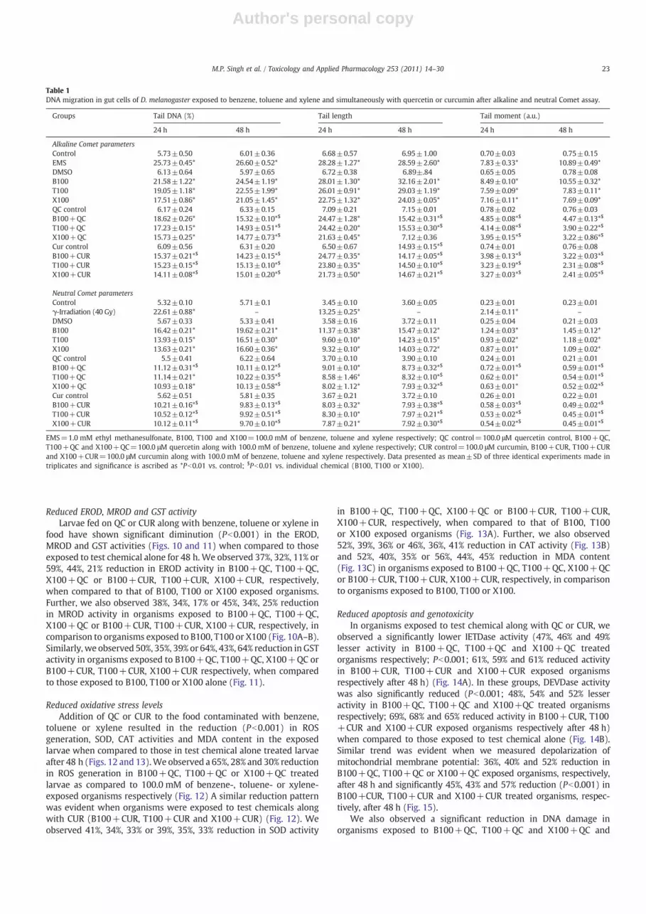
Reduced EROD, MROD and GST activityLarvae fed on QC or CUR along with benzene, toluene or xylene in
food have shown significant diminution (Pb0.001) in the EROD,MROD and GST activities (Figs. 10 and 11) when compared to thoseexposed to test chemical alone for 48 h. We observed 37%, 32%, 11% or59%, 44%, 21% reduction in EROD activity in B100+QC, T100+QC,X100+QC or B100+CUR, T100+CUR, X100+CUR, respectively,when compared to that of B100, T100 or X100 exposed organisms.Further, we also observed 38%, 34%, 17% or 45%, 34%, 25% reductionin MROD activity in organisms exposed to B100+QC, T100+QC,X100+QC or B100+CUR, T100+CUR, X100+CUR, respectively, incomparison to organisms exposed to B100, T100 or X100 (Fig. 10A–B).Similarly, we observed 50%, 35%, 39% or 64%, 43%, 64% reduction in GSTactivity in organisms exposed to B100+QC, T100+QC, X100+QC orB100+CUR, T100+CUR, X100+CUR respectively, when comparedto those exposed to B100, T100 or X100 alone (Fig. 11).
Reduced oxidative stress levelsAddition of QC or CUR to the food contaminated with benzene,
toluene or xylene resulted in the reduction (Pb0.001) in ROSgeneration, SOD, CAT activities and MDA content in the exposedlarvae when compared to those in test chemical alone treated larvaeafter 48 h (Figs. 12 and 13).We observed a 65%, 28% and 30% reductionin ROS generation in B100+QC, T100+QC or X100+QC treatedlarvae as compared to 100.0 mM of benzene-, toluene- or xylene-exposed organisms respectively (Fig. 12) A similar reduction patternwas evident when organisms were exposed to test chemicals alongwith CUR (B100+CUR, T100+CUR and X100+CUR) (Fig. 12). Weobserved 41%, 34%, 33% or 39%, 35%, 33% reduction in SOD activity
in B100+QC, T100+QC, X100+QC or B100+CUR, T100+CUR,X100+CUR, respectively, when compared to that of B100, T100or X100 exposed organisms (Fig. 13A). Further, we also observed52%, 39%, 36% or 46%, 36%, 41% reduction in CAT activity (Fig. 13B)and 52%, 40%, 35% or 56%, 44%, 45% reduction in MDA content(Fig. 13C) in organisms exposed to B100+QC, T100+QC, X100+QCor B100+CUR, T100+CUR, X100+CUR, respectively, in comparisonto organisms exposed to B100, T100 or X100.
Reduced apoptosis and genotoxicityIn organisms exposed to test chemical along with QC or CUR, we
observed a significantly lower IETDase activity (47%, 46% and 49%lesser activity in B100+QC, T100+QC and X100+QC treatedorganisms respectively; Pb0.001; 61%, 59% and 61% reduced activityin B100+CUR, T100+CUR and X100+CUR exposed organismsrespectively after 48 h) (Fig. 14A). In these groups, DEVDase activitywas also significantly reduced (Pb0.001; 48%, 54% and 52% lesseractivity in B100+QC, T100+QC and X100+QC treated organismsrespectively; 69%, 68% and 65% reduced activity in B100+CUR, T100+CUR and X100+CUR exposed organisms respectively after 48 h)when compared to those exposed to test chemical alone (Fig. 14B).Similar trend was evident when we measured depolarization ofmitochondrial membrane potential: 36%, 40% and 52% reduction inB100+QC, T100+QC or X100+QC exposed organisms, respectively,after 48 h and significantly 45%, 43% and 57% reduction (Pb0.001) inB100+CUR, T100+CUR and X100+CUR treated organisms, respec-tively, after 48 h (Fig. 15).
We also observed a significant reduction in DNA damage inorganisms exposed to B100+QC, T100+QC and X100+QC and
Table 1DNA migration in gut cells of D. melanogaster exposed to benzene, toluene and xylene and simultaneously with quercetin or curcumin after alkaline and neutral Comet assay.
Groups Tail DNA (%) Tail length Tail moment (a.u.)
24 h 48 h 24 h 48 h 24 h 48 h
Alkaline Comet parametersControl 5.73±0.50 6.01±0.36 6.68±0.57 6.95±1.00 0.70±0.03 0.75±0.15EMS 25.73±0.45* 26.60±0.52* 28.28±1.27* 28.59±2.60* 7.83±0.33* 10.89±0.49*DMSO 6.13±0.64 5.97±0.65 6.72±0.38 6.89±.84 0.65±0.05 0.78±0.08B100 21.58±1.22* 24.54±1.19* 28.01±1.30* 32.16±2.01* 8.49±0.10* 10.55±0.32*T100 19.05±1.18* 22.55±1.99* 26.01±0.91* 29.03±1.19* 7.59±0.09* 7.83±0.11*X100 17.51±0.86* 21.05±1.45* 22.75±1.32* 24.03±0.05* 7.16±0.11* 7.69±0.09*QC control 6.17±0.24 6.33±0.15 7.09±0.21 7.15±0.01 0.78±0.02 0.76±0.03B100+QC 18.62±0.26* 15.32±0.10*$ 24.47±1.28* 15.42±0.31*$ 4.85±0.08*$ 4.47±0.13*$
T100+QC 17.23±0.15* 14.93±0.51*$ 24.42±0.20* 15.53±0.30*$ 4.14±0.08*$ 3.90±0.22*$
X100+QC 15.73±0.25* 14.77±0.73*$ 21.63±0.45* 7.12±0.36 3.95±0.15*$ 3.22±0.86*$
Cur control 6.09±0.56 6.31±0.20 6.50±0.67 14.93±0.15*$ 0.74±0.01 0.76±0.08B100+CUR 15.37±0.21*$ 14.23±0.15*$ 24.77±0.35* 14.17±0.05*$ 3.98±0.13*$ 3.22±0.03*$
T100+CUR 15.23±0.15*$ 15.13±0.10*$ 23.80±0.35* 14.50±0.10*$ 3.23±0.19*$ 2.31±0.08*$
X100+CUR 14.11±0.08*$ 15.01±0.20*$ 21.73±0.50* 14.67±0.21*$ 3.27±0.03*$ 2.41±0.05*$
Neutral Comet parametersControl 5.32±0.10 5.71±0.1 3.45±0.10 3.60±0.05 0.23±0.01 0.23±0.01γ-Irradiation (40 Gy) 22.61±0.88* – 13.25±0.25* – 2.14±0.11* –
DMSO 5.67±0.33 5.33±0.41 3.58±0.16 3.72±0.11 0.25±0.04 0.21±0.03B100 16.42±0.21* 19.62±0.21* 11.37±0.38* 15.47±0.12* 1.24±0.03* 1.45±0.12*T100 13.93±0.15* 16.51±0.30* 9.60±0.10* 14.23±0.15* 0.93±0.02* 1.18±0.02*X100 13.63±0.21* 16.60±0.36* 9.32±0.10* 14.03±0.72* 0.87±0.01* 1.09±0.02*QC control 5.5±0.41 6.22±0.64 3.70±0.10 3.90±0.10 0.24±0.01 0.21±0.01B100+QC 11.12±0.31*$ 10.11±0.12*$ 9.01±0.10* 8.73±0.32*$ 0.72±0.01*$ 0.59±0.01*$
T100+QC 11.14±0.21* 10.22±0.35*$ 8.58±1.46* 8.32±0.10*$ 0.62±0.01* 0.54±0.01*$
X100+QC 10.93±0.18* 10.13±0.58*$ 8.02±1.12* 7.93±0.32*$ 0.63±0.01* 0.52±0.02*$
Cur control 5.62±0.51 5.81±0.35 3.67±0.21 3.72±0.10 0.26±0.01 0.22±0.01B100+CUR 10.21±0.16*$ 9.83±0.13*$ 8.03±0.32* 7.93±0.38*$ 0.58±0.03*$ 0.49±0.02*$
T100+CUR 10.52±0.12*$ 9.92±0.51*$ 8.30±0.10* 7.97±0.21*$ 0.53±0.02*$ 0.45±0.01*$
X100+CUR 10.12±0.11*$ 9.70±0.10*$ 7.87±0.21* 7.92±0.30*$ 0.54±0.02*$ 0.45±0.01*$
EMS=1.0 mM ethyl methanesulfonate, B100, T100 and X100=100.0 mM of benzene, toluene and xylene respectively; QC control=100.0 μM quercetin control, B100+QC,T100+QC and X100+QC=100.0 μM quercetin along with 100.0 mM of benzene, toluene and xylene respectively; CUR control=100.0 μM curcumin, B100+CUR, T100+CURand X100+CUR=100.0 μM curcumin along with 100.0 mM of benzene, toluene and xylene respectively. Data presented as mean±SD of three identical experiments made intriplicates and significance is ascribed as *Pb0.01 vs. control; $Pb0.01 vs. individual chemical (B100, T100 or X100).
23M.P. Singh et al. / Toxicology and Applied Pharmacology 253 (2011) 14–30
Author's personal copy
B100+CUR, T100+CUR and X100+CUR. In B100+QC or B100+CUR exposed organisms, migration of DNA was significantly lessafter 24 and 48 h when compared to those in organisms exposedto test chemical alone, in both alkaline and neutral Comet assays
(Table 1). We observed a similar trend in T100+QC, X100+QC,T100+CUR or X100+CUR exposed Drosophila larvae when com-pared to those exposed to toluene or xylene alone (Table 1).
Fig. 11. Glutathione S-transferase (GST) activity in larval homogenate from third instarlarvae of D. melanogaster (Oregon R+) in control, DMSO and benzene, toluene or xylenealone or in combinations with QC or CUR treatment for 48 h. Data represent mean±SDof three identical experiments made in three replicates. Data represent mean±SD ofthree identical experiments made in triplicates and significance ascribed as **Pb0.001vs. control; $$Pb0.001, reduction vs. individual chemical (B100 or T100 or X100).
Fig. 12. Levels of reactive oxygen species in cells from midguts of third instar larvae ofD. melanogaster (Oregon R+) in control, DMSO and benzene, toluene or xylene alone orin combinations with QC or CUR treatments for 24 and 48 h. Histogram (A) depicts ROSgeneration in test chemical exposed organisms and flow cytometric panels shows theROS generation in (B) control, (C) QC control, (D) CUR control, (E) B100, (F) B100+QCand (G) B100+CUR exposed organisms after 48 h. Data represent mean±SD of threeidentical experiments made in triplicates and significance is ascribed as *Pb0.01 vs.control; $Pb0.01, reduction vs. individual chemical (B100 or T100 or X100).
Fig. 10. 7-Ethoxyresorufin-O-deethylase (EROD) activity (A) and 7-methoxyresorufin-O-de-ethylase (MROD) activity (B) in microsomes isolated from third instar larvaeof D. melanogaster (Oregon R+) exposed to benzene, toluene or xylene alone orin combination with quercetin (QC) or curcumin (CUR). B100, T100 andX100=100.0 mM benzene, toluene and xylene respectively; B100+QC, T100+QCand X100+QC=100.0 μM quercetin along with 100.0 mM of benzene, toluene andxylene respectively; B100+CUR, T100+CUR and X100+CUR=100.0 μM curcuminalong with 100.0 mM of benzene, toluene and xylene, respectively, in this figure andthe remaining figures in this paper. Panel C depicts the inhibitory effect of 3′,4′-dimethoxyflavone on EROD activity in third instar larvae of D. melanogaster exposedto 100.0 mM benzene, toluene and xylene for 48 h. Data represent mean±SD ofthree identical experiments made in triplicates and significance is ascribed as**Pb0.001 vs. control; $Pb0.01, $$Pb0.001, reduction vs. individual chemical (B100or T100 or X100).
24 M.P. Singh et al. / Toxicology and Applied Pharmacology 253 (2011) 14–30

Author's personal copy
Discussion
Continuously high levels of exposure of benzene, toluene andxylene to the organisms from occupational/non-occupational sourcescause major health hazards. These contaminants are well known todisrupt various cellular processes (Gerin et al., 1998; Chambers et al.,2006). Previous studies showed that benzene, toluene and xylenehave potential to induce oxidative stress, an imbalance of the anti-oxidant system, such as SOD, glutathione peroxidase (GP), glutathi-one (GSH) and MDA content in mammalian cell line or petrochemicalworkers (Croute et al., 2002; Georgieva et al., 2002). Weaver et al.(2007) and Weaver and Liu (2008) demonstrated benzene-inducedapoptosis in epithelial cells of respiratory tracts as evidenced bynicking of DNA, endonucleolytic degradation of genomic DNA as wellas increased caspase activity. Similar observations were made in vivousingmurinemodels (Smith, 1996; Ross, 2000; Snyder, 2000;Weaver
et al., 2007). Cell death is the ultimate fate of the cell with irreparabledamage. DNA damage can also occur by chemicals, which if notrepaired, can have serious consequences leading to carcinogenesis.Benzene, being classified as class I carcinogen, has been shownpreviously to cause DNA damage both in vitro and in vivo (Sul et al.,2005; Weaver et al., 2007; Weaver and Liu, 2008). Singh and Winn(2008) and Faiola et al. (2004) showed significant increase inchromosomal breaks in K-562 cells and hematopoietic stem cells(HSC) exposed to benzene and its metabolites. Similarly, in vivostudies usingmouse bonemarrow cells showed significant increase insister chromatid exchanges and clastogenicity (Erexson et al., 1986;Zhang et al., 2002). Using alkaline Comet assay, Chen et al. (2008) andGalvan et al. (2008) showed increased Comet parameters in thehuman lymphocytes and HeLa cells respectively, exposed to benzeneand its metabolites. Limited available information suggests tolueneand xylene can also induce apoptosis (Nakai et al., 2003; Al-Ghamdiet al., 2004). Despite this vast knowledge on the adverse effects ofbenzene, toluene and xylene, only a few compounds have been testedfor their protective roles against benzene, toluene and xylene inducedtoxicity. In this study, we first analyzed the apoptotic and genotoxicpotential of these threemonocyclic hydrocarbonsusingD.melanogaster,an alternate to animal model system. Further, we examined the role ofpotential candidates/receptors involved in mediating the benzene-,toluene- or xylene-mediated toxicity. Subsequently, we utilized thismodel to evaluate the protective effects of two well-known phyto-chemicals namely, QC and CUR [quercetin (a well-known antioxidant(Chen and Kang, 2005; Kanupriya et al., 2006) and curcumin (a known
Fig. 13. Cu-Zn SOD (A) and CAT (B) activities and MDA content (C) in the larvalhomogenate from third instar larvae of D. melanogaster (Oregon R+) in control, DMSOand benzene, toluene or xylene alone or in combinations with QC or CUR treatment for48 h. Data represent mean±SD of three identical experiments made in triplicates andsignificance is ascribed as **Pb0.001 vs. control; $$Pb0.001, reduction vs. individualchemical (B100 or T100 or X100).
Fig. 14. IETDase (A) and DEVDase (B) activities inmicrosomes isolated from third instarlarvae of D. melanogaster (Oregon R+) in control, DMSO and benzene, toluene or xylenealone or in combinations with QC or CUR treatments for 24 and 48 h. Data representmean±SD of three identical experiments made in triplicates and significance isascribed as *Pb0.01 vs. control; $Pb0.01, reduction vs. individual chemical (B100 orT100 or X100).
25M.P. Singh et al. / Toxicology and Applied Pharmacology 253 (2011) 14–30

Author's personal copy
antioxidant and anti-cancer agent (Becatti et al., 2010; Biswas et al.,2010)] against benzene-, toluene- and xylene-induced toxicity.
Apoptosis is a physiological mode of cellular suicide that plays avital role during embryogenesis, development, and normal tissuehomeostasis (Brill et al., 1999; Vaux and Korsmeyer, 1999; Fulda andDebatin, 2006). The hallmarks of apoptosis include depolarization ofthe plasma membrane, cell shrinkage, alterations in intracellular ionconcentrations, mitochondrial membrane depolarization, chromatincondensation, and DNA fragmentation. We, therefore, examinedseveral candidates involved in the apoptotic pathway in Drosophilaexposed to benzene, xylene or toluene.
During early apoptosis, a cell loses its membrane asymmetry (Chenet al., 2008). Phosphatidylserine (PS), normally present on the innercytoplasmic leaflet of the plasma membrane of healthy cells, istranslocated and exposed on the outer leaflet (Seigneuret and Devaux,1984; Connor and Schroit, 1987). We used AV, a Ca2+-dependentphospholipid-binding protein that has a high affinity for PS (Comfuriuset al., 1996). The significantly increased (Pb0.001) AV positive cells inthe present study suggested the initiation of apoptosis in benzene-,toluene- or xylene-exposed organisms.
The process of apoptosis is controlled by a diverse range of cellsignals, which may originate either at extra-cellular or intra-cellularlevel (Chang et al., 1998; Budihardjo et al., 1999; Arya et al., 2007). Toidentify the apoptotic pathwaybeing followed in the cells of theexposedorganism, we analyzed mitochondrial membrane potential, DEVDaseactivity (caspase-3), IETDase activity, and poly ADP ribose polymerase(PARP) cleavage (Chiarugi and Moskowitz, 2002). We observedsignificant increase in DEVDase and IETDase activities, PARP cleavageand depolarization of mitochondrial membrane in organisms exposedto test chemicals, indicating the induction of mitochondria-mediatedcaspase-dependent cell death pathway in Drosophila larvae exposed tobenzene/toluene/xylene. Similarly, a number of earlier studies alsodocumented increased IETDase and DEVDase activities in organisms/cells exposed to organic solvents containing benzene, toluene or xylene
[in Jurkat cells by chlorinated biphenol (Inayat-Hussain et al., 2001;Inayat-Hussain and Ross, 2005), HL60 and CD34+ cells by benzenemetabolites (Hiraku andKawanishi, 1996;Moranet al., 1996)]. Caspasesare normally rendered inactive by Inhibitor of Apoptosis Proteins (IAPs)and this inhibition is overcome by p53-mediated transcription leadingto increased levels of Smac/DIABLO orthologs (Hid, Rpr and Grim inDrosophila) (Brenner and Kroemer, 2000; Brodsky et al., 2000; Schulerand Green, 2001; Brodsky et al., 2004; Kornbluth and White, 2005;Wichmann et al., 2006). The increased levels of Hid, Rpr, Grim andTUNEL positive cells observed in benzene-, toluene- and xylene-exposed groups suggest the activation of pro-apoptotic genes inorganisms leading to removal of IAP-mediated inhibition of caspasesand also activation of apoptotic pathway.
Increased PARP cleavage is usually associated with oxidative DNAdamage (Satoh and Lindahl, 1994; Miller et al., 2004; Babich et al.,2009; Deng et al., 2009). Since we also observed increased PARPcleavage in benzene-, toluene- and xylene-exposed organisms, weanalyzed DNA damage through Comet assay, which has been adaptedfor genotoxicity assessment (Miloshev et al., 2002; Rajaguru et al.,2003; Mukhopadhyay et al., 2004; Siddique et al., 2005b; Deguchiet al., 2008). Of the two variants of the Comet assay, the neutralversion detects double-strand DNA breaks (Yasuhara et al., 2003)whereas the alkaline version allows to reveal single- and double-strand breaks as well as alkali labile sites (Moller, 2006). In our study,we found significantly increased migration of DNA (TL, % TD and TM)in the order of benzeneN toluene≥xylene in the exposed organisms inalkaline as well as neutral Comet assays. Interestingly, alkaline Cometparameters showed statistically significant higher levels (Pb0.01) ascompared to neutral Comet parameters in the test chemical exposedorganisms. These results suggest benzene, toluene and xylene arepotentially genotoxic causing double-strand breaks to the exposedlarvae. These observations parallel the earlier studies on humanpopulation (epidemiological data) and experimental models (both invivo and in vitro) exposed to benzene, toluene or xylene (Carere et al.,
Fig. 15. Alterations in mitochondrial membrane potential (ΔΨm) in cells from midgut tissues of third instar larvae of D. melanogaster (Oregon R+) in control, DMSO and benzene,toluene or xylene alone or in combinations with QC or CUR treatments for 24 and 48 h. Histogram (A) depicts fold increase in mitochondrial membrane depolarization. Flowcytometry panels represent ΔΨm in (B) control, (C) B100, (D) B100+QC and (E) B100+Cur exposed organisms after 48 h. Data represent mean±SD of three identical experimentsmade in triplicates and significance is ascribed as *Pb0.01. $Pb0.01, reduction vs. individual chemical (B100 or T100 or X100).
26 M.P. Singh et al. / Toxicology and Applied Pharmacology 253 (2011) 14–30

Author's personal copy
1995a; Carere et al., 1995b; Chen et al., 2008; Pandey et al., 2009).Interestingly, the observed genotoxicity (benzeneN tolueneNxylene)is inversely related to lipophilicity (benzeneb toluenebxylene;Snyder et al., 1993; ATSDR, 1997) and this is consistent with ourprevious observations in mixture toxicity studies that the higher thelipophilicity, the lower is the toxicity (Singh et al., 2010).
Reactive oxygen species (ROS) are generated in biological systemseither by normal metabolic pathways or as a consequence of exposureto chemical (Iqbal et al., 2003; Sun, 1990). Recent studies from ourlaboratory showed induction of heat shock genes (hsp70, hsp83, hsp60and hsp26), oxidative stress markers and increased ROS generation inD. melanogaster exposed to benzene, toluene or xylene, individually,or their mixtures indicating the potential of these chemicals toproduce cellular stress (Singh et al., 2009; Singh et al., 2010). Inaddition, we observed significant induction of cytochrome P450enzymes in benzene-, toluene-, or xylene-exposed organisms whencompared to controls. Induction of cytochrome P450 enzymes inresponse to benzene, toluene or xylene exposure is well documented(Day et al., 1992; Nakajima and Wang, 1994; Seaton et al., 1994).Further, AhR has been shown to be the key component in themetabolic response to aromatic hydrocarbons, including benzene(Schmidt and Bradfield, 1996; Yoon et al., 2002; Nishiumi et al., 2007).AhR, a cytoplasmic bHLH-PAS transcription factor, upon binding to anaromatic hydrocarbon (TCDD for example), translocates to thenucleus. Within the nucleus, Ahr forms a complex with the Arylhydrocarbon receptor nuclear translocator (ARNT), another bHLH-PAS protein (Hoffman et al., 1991) and together bind to xenobioticresponse element to control the expression of specific target genesincluding certain cytochrome P450s (Swanson et al., 1995). Based onthis, we hypothesized a similar role for spineless (Drosophila homologof Aryl hydrocarbon receptor Cespedes et al., 2010) in the benzene-,xylene- and toluene-mediated toxicity in Drosophila. To test if theobserved induction of cytochrome P450s is mediated by AhRpathway, we exposed Drosophila larvae to test chemical togetherwith the blocker for AhR (DMF, Li, 2007) and measured the activity ofcytochrome P450s. The observed reduction of cytochrome P450s (inparticular EROD) in exposed organisms in the presence of blocker,clearly indicated that like in mammals, toxicity of benzene, in part, ismediated by AhR in Drosophila. In addition, our study provides anevidence for the involvement of AhR also in toluene- and xylene-mediated toxicity.
The other objective of our study was to look for the agents, whichcan attenuate the toxicity conferred by benzene, toluene or xylene. Ascytochrome P450 enzymes, AhR and oxidative stress underlie theobserved benzene-, toluene- or xylene-mediated toxicity in thepresent study, we looked for nutraceuticals that are known to acton the above. We examined the efficiency of two phytochemicals,namely, QC and CUR for their protective roles against benzene-,toluene- or xylene-induced toxicity. QC is a polyphenolic flavonoidand has protective effects on different adverse cellular events likecarcinogenesis through unknown mechanism of action yet reducingLPO and ROS generation or having anti-proliferative activity in cells/organisms (Dihal et al., 2006; Dihal et al., 2008; Larson et al., 2010)indicating its anti-oxidant and anti-cancer properties. Similarly, CUR(diferuloyl methane) has been shown to possess anti-oxidative andfree radical scavenging properties and anti-neoplastic properties(Reddy and Lokesh, 1994; Motterlini et al., 2000; Messner et al.,2009). In addition, both CUR and QC were found to degrade both AhRand ARNT to inhibit cytochrome P450 enzymes. Based on thesereports (on anti-oxidative, free radical scavenging, inhibiting proper-ties, anti-cancer properties of QC and CUR), we hypothesized thataddition of QC or CUR in the exposure regimen will reduce the toxiceffects of the test chemicals to the exposed organisms. To test this, weanalyzed their efficacy in modulating the cytochrome P450 and GSTactivities, ROS generation, apoptosis and genotoxicity in the testchemical exposed organism. We observed a significantly reduced
cytochrome P450 activities, oxidative stress, GST activity, ROSgeneration, lower intensity of apoptosis and DNA damage inorganisms exposed to QC or CUR along with test chemicals incomparison to those observed in test chemical alone group. Presenceof chemicals in organisms exposed to benzene, toluene or xylenealone at levels similar to those in combination with QC or CURsuggested that this reduction in assayed parameters is not due toreduced intake of test chemical by the organisms in the presence of QCor CUR but indeed due to the protective roles of QC or CUR. Theseresults demonstrated the potential of CUR and QC to attenuate thebenzene-, toluene- or xylene-mediated toxicity. Intriguingly, aprevious study from our group had shown that the co-treatment ofQC with dichlorvos (DV) enhanced the oxidative stress and apoptosiscompared to that of DV alone (Gupta et al., 2007). This discrepancymay be attributed to the test chemical used and QC may conferprotection in a compound specific manner. To confirm, if this is thecase, we analyzed the ROS levels in organisms exposed to DV, DV+QC, benzene, benzene+QC or QC alone. Interestingly, we observedthat ROS levels are increased in organisms exposed to DV+QC [as inagreementwith Gupta et al., 2007] and decreased in benzene+QC (asin the present study), when compared to their respective chemicalcontrols (please see Supplemental Fig. S6). These results indicate thatQC may play protective roles in a compound specific manner.
Several molecular mechanisms by which CUR and QC may playprotective roles are envisaged. Given their documented roles inmodulating cytochrome P450 activities (Firozi et al., 1996; Thapliyaland Maru, 2001) and their anti-oxidant properties (Chan et al., 2005;Messner et al., 2009) they may act either at the level of metabolism oralternatively they may act on the anti-oxidant defence system. DuringPhase I xenobiotic metabolism, CUR and QC may either 1) inhibitcytochrome P450 enzymes directly by acting as a competitivesubstrate (Appiah-Opong et al., 2007), 2) interfere with AhRmediatedinduction of cytochrome P450 enzymes (Rinaldi et al., 2002) or 3) actat the level of GSTs, which play an important role in the phase IIxenobiotic metabolism (Oetari et al., 1996). The reduction incytochrome P450 activities in organisms exposed to test chemicalalong with QC or CUR and the observed involvement of Ahr in thepresent study point to the second possibility. In addition, we observedsignificant (Pb0.05) positive correlation (please see SupplementaryTable S1 for details) between activity of cytochrome P450s and GSTs,ROS and other oxidative stress parameters analyzed. Based on these,we believe that the reduced levels of GSTs, ROS and other oxidativestress parameters might be a consequence of reduced activity ofcytochrome P450 in response to CUR or QC in exposed organisms.These ultimately might have resulted in reduced apoptosis andgenotoxicity in organisms exposed to test chemical along with CURor QC.
Taken together, the present study suggests that MAHs particularlybenzene, toluene and xylene can cause genotoxicity and apoptosis inthe exposed Drosophila larvae in vivo, through mitochondriamediated caspase dependent pathway of cell death. The study alsoshowed that AhR homologue plays a key role in the induction ofcytochrome P450 in response to benzene, toluene or xylene inDrosophila. Among the three tested chemicals, benzene is highlygenotoxic (possibly due to higher uptake) while toluene and xyleneare less genotoxic than benzene. The study further suggests thatquercetin and curcumin inhibit cytochrome P450, probably influenc-ing AhR mediated pathway, which eventually may result in thereduction of the genotoxicity and apoptosis in test chemicals exposedD. melanogaster.
Supplementarymaterials related to this article can be found onlineat doi:10.1016/j.taap.2011.03.006.
Conflicts of interest
None.
27M.P. Singh et al. / Toxicology and Applied Pharmacology 253 (2011) 14–30

Author's personal copy
Acknowledgment
The authors are thankful to the Director, Indian Institute ofToxicology Research (IITR) for facilities. We thank Mr. N. Mathur,Epidemiology Section, IITR for statistical analysis, Dr. A. Dhawan,Developmental Toxicology Division, IITR for Comet assay facility,Dr. M. Dixit and Mr. A.L. Viswakarma, Central Drug Research Institute,Lucknow for flow cytometry facility. Financial assistance to MPS fromUGC-SRF, New Delhi, MM from SRF-DBT, New Delhi, AS from ICMR-SRF, New Delhi and DKC from Council of Scientific and IndustrialResearch (SIP-08 and NWP-34) is thankfully acknowledged.
References
Al-Ghamdi, S.S., Raftery, M.J., Yaqoob, M.M., 2004. Toluene and p-xylene induced LLC-PK1 apoptosis. Drug Chem. Toxicol. 27, 425–432.
Alegretti, A.P., Thiesen, F.V., Maciel, G.P., 2004. Analytical method for evaluation ofexposure to benzene, toluene, xylene in blood by gas chromatography preceded bysolid phase microextraction. J. Chromatogr. 809, 183–187.
Appiah-Opong, R., Commandeur, J.N., van Vugt-Lussenburg, B., Vermeulen, N.P., 2007.Inhibition of human recombinant cytochrome P450s by curcumin and curcumindecomposition products. Toxicology 235, 83–91.
Arlien-Suborg, P., 1992. Solvent Neurotoxicity. CRC Press, Inc., Boca Raton, Florida.Arya, R., Mallik, M., Lakhotia, S.C., 2007. Heat shock genes—integrating cell survival and
death. J. Biosci. 32, 595–610.ATSDR, 1997. Toxicological profile for benzene. Public Health Service, Agency for Toxic
Substances and Disease Registry, Atlanta, GA, US Department of Health and HumanServices.
Babich, H., Ackerman, N.J., Burekhovich, F., Zuckerbraun, H.L., Schuck, A.G., 2009.Gingko biloba leaf extract induces oxidative stress in carcinoma HSC-2 cells.Toxicol. In Vitro 23, 992–999.
Balasubramanyam, M., Koteswari, A.A., Kumar, R.S., Monickaraj, S.F., Maheswari, J.U.,Mohan, V., 2003. Curcumin-induced inhibition of cellular reactive oxygen speciesgeneration: novel therapeutic implications. J. Biosci. 28, 715–721.
Becatti, M., Prignano, F., Fiorillo, C., Pescitelli, L., Nassi, P., Lotti, T., Taddei, N., 2010. Theinvolvement of Smac/DIABLO, p53, NF-kB, and MAPK pathways in apoptosis ofkeratinocytes from perilesional vitiligo skin: protective effects of curcumin andcapsaicin. Antioxid. Redox Signal.
Bellion, P., Olk, M., Will, F., Dietrich, H., Baum, M., Eisenbrand, G., Janzowski, C., 2009.Formation of hydrogen peroxide in cell culture media by apple polyphenols and itseffect on antioxidant biomarkers in the colon cell line HT-29. Mol. Nutr. Food Res.53, 1226–1236.
Biswas, J., Sinha, D., Mukherjee, S., Roy, S., Siddiqi, M., Roy, M., 2010. Curcumin protectsDNA damage in a chronically arsenic-exposed population of West Bengal. Hum.Exp. Toxicol. 29, 513–524.
Brenner, C., Kroemer, G., 2000. Apoptosis. Mitochondria—the death signal integrators.Sci. NY N. Y 289, 1150–1151.
Brill, A., Torchinsky, A., Carp, H., Toder, V., 1999. The role of apoptosis in normal andabnormal embryonic development. J. Assist. Reprod. Genet. 16, 512–519.
Brodsky, M.H., Nordstrom, W., Tsang, G., Kwan, E., Rubin, G.M., Abrams, J.M., 2000.Drosophilap53bindsadamage response elementat thereaper locus. Cell 101, 103–113.
Brodsky, M.H., Weinert, B.T., Tsang, G., Rong, Y.S., McGinnis, N.M., Golic, K.G., Rio, D.C.,Rubin, G.M., 2004.DrosophilamelanogasterMNK/Chk2 andp53 regulatemultipleDNArepair and apoptotic pathways following DNA damage. Mol. Cell. Biol. 24, 1219–1231.
Budihardjo, I., Oliver, H., Lutter, M., Luo, X., Wang, X., 1999. Biochemical pathways ofcaspase activation during apoptosis. Annu. Rev. Cell Dev. Biol. 15, 269–290.
Carere, A., Antoccia, A., Crebelli, R., Degrassi, F., Fiore, M., Iavarone, I., Isacchi, G., Lagorio,S., Leopardi, P., Marcon, F., et al., 1995a. Genetic effects of petroleum fuels:cytogenetic monitoring of gasoline station attendants. Mutat. Res. 332, 17–26.
Carere, A., Antoccia, A., Crebelli, R., Di Chiara, D., Fuselli, S., Iavarone, I., Isacchi, G.,Lagorio, S., Leopardi, P., Marcon, F., 1995b. Exposure to benzene and genotoxiceffects among filling station attendants. Epidemiol. Prev. 19, 105–119.
Cespedes, M.A., Galindo, M.I., Couso, J.P., 2010. Dioxin toxicity in vivo results from anincrease in the dioxin-independent transcriptional activity of the aryl hydrocarbonreceptor. PLoS ONE 5, e15382.
Chambers, D.M., McElprang, D.O., Waterhouse, M.G., Blount, B.C., 2006. An improvedapproach for accurate quantitation of benzene, toluene, ethylbenzene, xylene, andstyrene in blood. Anal. Chem. 78, 5375–5383.
Chan, W.H., Wu, H.J., Hsuuw, Y.D., 2005. Curcumin inhibits ROS formation andapoptosis in methylglyoxal-treated human hepatoma G2 cells. Ann. NY Acad. Sci.1042, 372–378.
Chang, H.Y., Nishitoh, H., Yang, X., Ichijo, H., Baltimore, D., 1998. Activation of apoptosissignal-regulating kinase 1 (ASK1) by the adapter protein Daxx. Sci. NY N. Y 281,1860–1863.
Chen, J., Kang, J.H., 2005. Quercetin and trichostatin A cooperatively kill humanleukemia cells. Pharmazie 60, 856–860.
Chen, Y., McMillan-Ward, E., Kong, J., Israels, S.J., Gibson, S.B., 2008. Oxidative stressinduces autophagic cell death independent of apoptosis in transformed and cancercells. Cell Death Differ. 15, 171–182.
Chiarugi, A., Moskowitz, M.A., 2002. Cell biology. PARP-1—a perpetrator of apoptoticcell death? Sci. NY N. Y 297, 200–201.
Comfurius, P., Williamson, P., Smeets, E.F., Schlegel, R.A., Bevers, E.M., Zwaal, R.F., 1996.Reconstitution of phospholipid scramblase activity from human blood platelets.Biochemistry 35, 7631–7634.
Connor, J., Schroit, A.J., 1987. Determination of lipid asymmetry in human red cells byresonance energy transfer. Biochemistry 26, 5099–5105.
Croute, F., Poinsot, J., Gaubin, Y., Beau, B., Simon, V., Murat, J.C., Soleilhavoup, J.P., 2002.Volatile organic compounds cytotoxicity and expression of HSP72, HSP90 andGRP78 stress proteins in cultured human cells. Biochim. Biophys. Acta 1591,147–155.
Day, B.J., DeNicola, D.B., Marcus, C.B., Carlson, G.P., 1992. Effect of p-xylene inhalation onthe bioactivation of bromobenzene in rat lung and liver. Fundam. Appl. Toxicol. 19,50–56.
Deguchi, K., Hayashi, T., Nagotani, S., Sehara, Y., Zhang, H., Tsuchiya, A., Ohta, Y.,Tomiyama, K., Morimoto, N., Miyazaki, M., Huh, N.H., Nakao, A., Kamiya, T., Abe, K.,2008. Reduction of cerebral infarction in rats by biliverdin associated withamelioration of oxidative stress. Brain Res. 1188, 1–8.
Deng, Y., Guo, X., Ferguson, D.O., Chang, S., 2009. Multiple roles for MRE11 at uncappedtelomeres. Nature 460, 914–918.
Dihal, A.A., de Boer, V.C., van der Woude, H., Tilburgs, C., Bruijntjes, J.P., Alink, G.M.,Rietjens, I.M., Woutersen, R.A., Stierum, R.H., 2006. Quercetin, but not itsglycosidated conjugate rutin, inhibits azoxymethane-induced colorectal carcino-genesis in F344 rats. J. Nutr. 136, 2862–2867.
Dihal, A.A., van der Woude, H., Hendriksen, P.J., Charif, H., Dekker, L.J., Ijsselstijn, L., deBoer, V.C., Alink, G.M., Burgers, P.C., Rietjens, I.M., Woutersen, R.A., Stierum, R.H.,2008. Transcriptome and proteome profiling of colon mucosa from quercetin fedF344 rats point to tumor preventive mechanisms, increased mitochondrial fattyacid degradation and decreased glycolysis. Proteomics 8, 45–61.
Emara, A.M., El-Bahrawy, H., 2008. Green tea attenuates benzene-induced oxidativestress in pump workers. J. Immunotoxicol. 5, 69–80.
Erexson, G.L., Wilmer, J.L., Steinhagen, W.H., Kligerman, A.D., 1986. Induction ofcytogenetic damage in rodents after short-term inhalation of benzene. Environ.Mutagen. 8, 29–40.
Faiola, B., Fuller, E.S., Wong, V.A., Pluta, L., Abernethy, D.J., Rose, J., Recio, L., 2004.Exposure of hematopoietic stem cells to benzene or 1,4-benzoquinone inducesgender-specific gene expression. Stem Cells 22, 750–758.
Festing, M.F.W., Baumans, V., Combes, D.R., Halder, M., Hendriksen, F.M., Howard, B.R.,Lovell, D.P., Moore, G.J., Overend, P., Wilson, M.S., 1998. Reducing the use oflaboratory animals in biomedical research: problems and possible solutions. Altern.Lab. Anim. 26, 283–301.
Firozi, P.F., Aboobaker, V.S., Bhattacharya, R.K., 1996. Action of curcumin on thecytochrome P450-system catalyzing the activation of aflatoxin B1. Chem.-Biol.Interact. 100, 41–51.
Fracasso, M.E., Doria, D., Carrieri, M., Bartolucci, G.B., Quintavalle, S., De Rosa, E., 2009.DNA single- and double-strand breaks by alkaline- and immuno-comet assay inlymphocytes of workers exposed to styrene. Toxicol. Lett. 185, 9–15.
Fulda, S., Debatin, K.M., 2006. Resveratrol modulation of signal transduction inapoptosis and cell survival: a mini-review. Cancer Detect. Prev. 30, 217–223.
Galvan, N., Lim, S., Zmugg, S., Smith, M.T., Zhang, L., 2008. Depletion of WRN enhancesDNA damage in HeLa cells exposed to the benzene metabolite, hydroquinone.Mutat. Res. 649, 54–61.
Georgieva, T., Michailova, A., Panev, T., Popov, T., 2002. Possibilities to control the healthrisk of petrochemical workers. Int. Arch. Occup. Environ. Health 75 (Suppl),S21–S26.
Gerin, M., Siemiatycki, J., Desy, M., Krewski, D., 1998. Associations between several sitesof cancer and occupational exposure to benzene, toluene, xylene, and styrene:results of a case–control study in Montreal. Am. J. Ind. Med. 34, 144–156.
Greer, J.E., 1984. Adolescent abuse of typewriter correction fluid. South. Med. J. 77 (804),297–298.
Gupta, S.C., Mishra, M., Sharma, A., Deepak Balaji, T.G., Kumar, R., Mishra, R.K.,Chowdhuri, D.K., 2010. Chlorpyrifos induces apoptosis and DNA damage inDrosophila through generation of reactive oxygen species. Ecotoxicol. Environ.Saf. 73, 1415–1423.
Gupta, S.C., Siddique, H.R., Mathur, N., Vishwakarma, A.L., Mishra, R.K., Saxena, D.K.,Chowdhuri, D.K., 2007. Induction of hsp70, alterations in oxidative stressmarkers and apoptosis against dichlorvos exposure in transgenic Drosophilamelanogaster: modulation by reactive oxygen species. Biochim. Biophys. Acta 1770,1382–1394.
Habig, W.H., Pabst, M.J., Jakoby, W.B., 1974. Glutathione S-transferases: the firstenzymatic step in mercapturic acid formation. J. Biol. Chem. 249, 7130–7139.
Hay, B.A., Guo, M., 2006. Caspase-dependent cell death in Drosophila. Annu. Rev. CellDev. Biol. 22, 623–650.
Hiraku, Y., Kawanishi, S., 1996. Oxidative DNA damage and apoptosis induced bybenzene metabolites. Cancer Res. 56, 5172–5178.
Hoffman, E.C., Reyes, H., Chu, F.F., Sander, F., Conley, L.H., Brooks, B.A., Hankinson, O.,1991. Cloning of a factor required for activity of the Ah (dioxin) receptor. Sci. NY N.Y 252, 954–958.
Inayat-Hussain, S.H., Ross, D., 2005. Intrinsic pathway of hydroquinone inducedapoptosis occurs via both caspase-dependent and caspase-independent mecha-nisms. Chem. Res. Toxicol. 18, 420–427.
Inayat-Hussain, S.H., Winski, S.L., Ross, D., 2001. Differential involvement of caspases inhydroquinone-induced apoptosis in human leukemic hl-60 and jurkat cells.Toxicol. Appl. Pharmacol. 175, 95–103.
Indulski, J.A., Sinczuk-Walczak, H., Szymczak, M., Wesolowski, W., 1996. Neurologicaland neurophysiological examinations of workers occupationally exposed toorganic solvent mixtures used in the paint and varnish production. Int. J. Occup.Med. Environ. Health 9, 235–244.
28 M.P. Singh et al. / Toxicology and Applied Pharmacology 253 (2011) 14–30

Author's personal copy
Iqbal, M., Sharma, S.D., Okazaki, Y., Fujisawa, M., Okada, S., 2003. Dietary Supplemen-tation of Curcumin Enhances Antioxidant and Phase IIMetabolizing Enzymes in ddYMaleMice: Possible Role in Protection against Chemical Carcinogenesis andToxicity. Pharmacology & Toxicology 92, 33–38.
Johri, A., Yadav, S., Singh, R.L., Dhawan, A., Ali, M., Parmar, D., 2006. Long lasting effectsof prenatal exposure to deltamethrin on cerebral and hepatic cytochrome P450sand behavioral activity in rat offspring. Eur. J. Pharmacol. 544, 58–68.
Jones, D.J., Lim, C.K., Ferry, D.R., Gescher, A., 1998. Determination of quercetin in humanplasma by HPLC with spectrophotometric or electrochemical detection. Biomed.Chromatogr. 12, 232–235.
Kanupriya, Dipti, P., Sharma, S.K., Sairam, M., Ilavazhagan, G., Sawhney, R.C., Banerjee, P.K., 2006. Flavonoids protect U-937 macrophages against tert-butylhydroperoxideinduced oxidative injury. Food Chem. Toxicol. 44, 1024–1030.
Kolachana, P., Subrahmanyam, V.V., Meyer, K.B., Zhang, L., Smith, M.T., 1993. Benzeneand its phenolic metabolites produce oxidative DNA damage in HL60 cells in vitroand in the bone marrow in vivo. Cancer Res. 53, 1023–1026.
Kornbluth, S., White, K., 2005. Apoptosis in Drosophila: neither fish nor fowl (nor man,nor worm). J. Cell Sci. 118, 1779–1787.
Kottke, M.K., 1998. Scientific and regulatory aspects of nutraceutical products in theUnited States. Drug Dev. Ind. Pharm. 24, 1177–1195.
Kozel, N., Sloboda, Z., De La Rosa, M., 1995. Epidemiology of Inhalant Abuse: AnInternational Perspective. US Department of Health and Human Services,Washington, DC. NIDA Res. Mon. 14.
Krebs, R.A., Feder, M.E., 1997. Tissue-specific variation in Hsp70 expression and thermaldamage in Drosophila melanogaster larvae. J. Exp. Biol. 200, 2007–2015.
Larson, A.J., Symons, J.D., Jalili, T., 2010. Quercetin: a treatment for hypertension?—areview of efficacy and mechanisms. Pharmaceuticals 3 (1), 237–250.
Li, L.A., 2007. Polychlorinated biphenyl exposure and CYP19 gene regulation intesticular and adrenocortical cell lines. Toxicol. In Vitro 21, 1087–1094.
Lodha, R., Bagga, A., 2000. Traditional Indian systems of medicine. Ann. Acad. Med.Singapore 29, 37–41.
Messner, D.J., Sivam, G., Kowdley, K.V., 2009. Curcumin reduces the toxic effects of ironloading in rat liver epithelial cells. Liver Int. 29, 63–72.
Miller, H., Fernandes, A.S., Zaika, E., McTigue, M.M., Torres, M.C., Wente, M., Iden, C.R.,Grollman, A.P., 2004. Stereoselective excision of thymine glycol from oxidativelydamaged DNA. Nucleic Acids Res. 32, 338–345.
Miloshev, G., Mihaylov, I., Anachkova, B., 2002. Application of the single cell gelelectrophoresis on yeast cells. Mutat. Res. 513, 69–74.
Moller, P., 2006. The alkaline comet assay: towards validation in biomonitoring of DNAdamaging exposures. Basic Clin. Pharmacol. Toxicol. 98, 336–345.
Moran, J.L., Siegel, D., Sun, X.M., Ross, D., 1996. Induction of apoptosis by benzenemetabolites in HL60 and CD34+ human bone marrow progenitor cells. Mol.Pharmacol. 50, 610–615.
Motterlini, R., Foresti, R., Bassi, R., Green, C.J., 2000. Curcumin, an antioxidant and anti-inflammatory agent, induces heme oxygenase-1 and protects endothelial cellsagainst oxidative stress. Free Radic. Biol. Med. 28, 1303–1312.
Mukhopadhyay, I., Chowdhuri, D.K., Bajpayee, M., Dhawan, A., 2004. Evaluation of invivo genotoxicity of cypermethrin in Drosophila melanogaster using the alkalineComet assay. Mutagenesis 19, 85–90.
Nakai, N., Murata, M., Nagahama, M., Hirase, T., Tanaka, M., Fujikawa, T., Nakao, N.,Nakashima, K., Kawanishi, S., 2003. Oxidative DNA damage induced by toluene isinvolved in its male reproductive toxicity. Free Radic. Res. 37, 69–76.
Nakajima, T., Wang, R.S., 1994. Induction of cytochrome P450 by toluene. Int. J.Biochem. 26, 1333–1340.
Nazir, A., Mukhopadhyay, I., Saxena, D.K., Chowdhuri, D.K., 2003. Evaluation of the noobserved adverse effect level of solvent dimethyl sulfoxide in Drosophilamelanogaster. Toxicol. Mech. Methods 13, 147–152.
Nishiumi, S., Yoshida, K., Ashida, H., 2007. Curcumin suppresses the transformation ofan aryl hydrocarbon receptor through its phosphorylation. Arch. Biochem. Biophys.466, 267–273.
Oetari, S., Sudibyo, M., Commandeur, J.N., Samhoedi, R., Vermeulen, N.P., 1996. Effects ofcurcumin on cytochrome P450 and glutathione S-transferase activities in rat liver.Biochem. Pharmacol. 51, 39–45.
Olive, P.L., Wlodek, D., Durand, R.E., Banath, J.P., 1992. Factors influencing DNAmigration from individual cells subjected to gel electrophoresis. Exp. Cell Res. 198,259–267.
Pan, H.Z., Na, L.X., Tao, L., 2003. DNA damage and changes of antioxidative enzymes inchronic benzene poisoning mice. Zhonghua Lao DongWei Sheng Zhi Ye Bing Za Zhi21, 423–425.
Pandey, A.K., Gurbani, D., Bajpayee, M., Parmar, D., Ajmani, S., Dhawan, A., 2009. In silicostudies with human DNA topoisomerase-II alpha to unravel the mechanism of invitro genotoxicity of benzene and its metabolites. Mutat. Res. 661, 57–70.
Pariselli, F., Sacco, M.G., Ponti, J., Rembges, D., 2009. Effects of toluene and benzene airmixtures on human lung cells (A549). Exp. Toxicol. Pathol. 61, 381–386.
Phillips, H.J., 1973. Evaluation of culture dynamics. Dye exclusion test for cell viability.In: Kruse, P.K., Patterson, M.K. (Eds.), Tissue Culture Methods and Applications.Academic Press, New York, NY, USA, pp. 406–408.
Rajaguru, P., Suba, S., Palanivel, M., Kalaiselvi, K., 2003. Genotoxicity of a polluted riversystem measured using the alkaline comet assay on fish and earthworm tissues.Environ. Mol. Mutagen. 41, 85–91.
Reddy, A.C., Lokesh, B.R., 1994. Effect of dietary turmeric (Curcuma longa) on iron-induced lipid peroxidation in the rat liver. Food Chem. Toxicol. 32, 279–283.
Rinaldi, A.L., Morse, M.A., Fields, H.W., Rothas, D.A., Pei, P., Rodrigo, K.A., Renner, R.J.,Mallery, S.R., 2002. Curcumin activates the aryl hydrocarbon receptor yetsignificantly inhibits (−)-benzo(a)pyrene-7R-trans-7,8-dihydrodiol bioactivationin oral squamous cell carcinoma cells and oral mucosa. Cancer Res. 62, 5451–5456.
Ross, D., 2000. The role of metabolism and specific metabolites in benzene-inducedtoxicity: evidence and issues. J. Toxicol. Environ. Health A 61, 357–372.
Satoh, M.S., Lindahl, T., 1994. Enzymatic repair of oxidative DNA damage. Cancer Res.54, 1899s–1901s.
Schiborr, C., Eckert, G.P., Rimbach, G., Frank, J., 2010. A validated method for thequantification of curcumin in plasma and brain tissue by fast narrow-bore high-performance liquid chromatography with fluorescence detection. Anal. Bioanal.Chem. 397, 1917–1925.
Schmidt, J.V., Bradfield, C.A., 1996. Ah receptor signaling pathways. Annu. Rev. Cell Dev.Biol. 12, 55–89.
Schuler, M., Green, D.R., 2001. Mechanisms of p53-dependent apoptosis. Biochem. Soc.Trans. 29, 684–688.
Seaton, M.J., Schlosser, P.M., Bond, J.A., Medinsky, M.A., 1994. Benzene metabolism byhuman liver microsomes in relation to cytochrome P450 2E1 activity. Carcinogen-esis 15, 1799–1806.
Seigneuret, M., Devaux, P.F., 1984. ATP-dependent asymmetric distribution of spin-labeled phospholipids in the erythrocyte membrane: relation to shape changes.Proc. Natl Acad. Sci. U.S.A. 81, 3751–3755.
Siddique, H.R., Chowdhuri, D.K., Saxena, D.K., Dhawan, A., 2005a. Validation ofDrosophila melanogaster as an in vivo model for genotoxicity assessment usingmodified alkaline Comet assay. Mutagenesis 20, 285–290.
Siddique, H.R., Gupta, S.C., Dhawan, A., Murthy, R.C., Saxena, D.K., Chowdhuri, D.K.,2005b. Genotoxicity of industrial solid waste leachates in Drosophila melanogaster.Environ. Mol. Mutagen. 46, 189–197.
Siddique, H.R., Sharma, A., Gupta, S.C., Murthy, R.C., Dhawan, A., Saxena, D.K.,Chowdhuri, D.K., 2008. DNA damage induced by industrial solid waste leachatesin Drosophila melanogaster: a mechanistic approach. Environ. Mol. Mutagen. 49,206–216.
Singh, M.P., Ram, K.R., Mishra, M., Shrivastava, M., Saxena, D.K., Chowdhuri, D.K., 2010.Effects of co-exposure of benzene, toluene and xylene to Drosophila melanogaster:alteration in hsp70, hsp60, hsp83, hsp26, ROS generation and oxidative stressmarkers. Chemosphere 79, 577–587.
Singh, M.P., Reddy, M.M., Mathur, N., Saxena, D.K., Chowdhuri, D.K., 2009. Induction ofhsp70, hsp60, hsp83 and hsp26 and oxidative stress markers in benzene, tolueneand xylene exposed Drosophila melanogaster: role of ROS generation. Toxicol. Appl.Pharmacol. 235, 226–243.
Singh, R., Winn, L.M., 2008. The effects of 1,4-benzoquinone on c-Myb andtopoisomerase II in K-562 cells. Mutat. Res. 645, 33–38.
Smith, M.T., 1996. Mechanistic studies of benzene toxicity—implications for riskassessment. Adv. Exp. Med. Biol. 387, 259–266.
Snyder, R., 2000. Overview of the toxicology of benzene. J. Toxicol. Environ. Health A 61,339–346.
Snyder, R., Witz, G., Goldstein, B.D., 1993. The toxicology of benzene. Environ. HealthPerspect. 100, 293–306.
Spiller, H.A., Krenzelok, E.P., 1997. Epidemiology of inhalant abuse reported to tworegional poison centers. J. Toxicol. Clin. Toxicol. 35, 167–173.
Sul, D., Lee, E., Lee, M.Y., Oh, E., Im, H., Lee, J., Jung, W.W., Won, N., Kang, H.S., Kim, E.M.,Kang, S.K., 2005. DNA damage in lymphocytes of benzene exposed workerscorrelates with trans, trans-muconic acids and breath benzene levels. Mutat. Res.582, 61–70.
Swanson, H.I., Chan, W.K., Bradfield, C.A., 1995. DNA binding specificities and pairingrules of the Ah receptor, ARNT, and SIM proteins. J. Biol. Chem. 270, 26292–26302.
Thapliyal, R., Maru, G.B., 2001. Inhibition of cytochrome P450 isozymes by curcumins invitro and in vivo. Food Chem. Toxicol. 39, 541–547.
Tice, R.R., Agurell, E., Anderson, D., Burlinson, B., Hartmann, A., Kobayashi, H., Miyamae,Y., Rojas, E., Ryu, J.C., Sasaki, Y.F., 2000. Single cell gel/comet assay: guidelines for invitro and in vivo genetic toxicology testing. Environ. Mol. Mutagen. 35, 206–221.
Lin, Tsu-Chun Emma, Shu-Chien, Chien, Ping-Chi, Hsu, Lih-Ann, Li, 2006. MechanisticStudy of Polychlorinated Biphenyl 126-Induced CYP11B1 and CYP11B2 Up-Regulation. Endocrinology 147 (3), 1536–1544.
Vaux, D.L., Korsmeyer, S.J., 1999. Cell death in development. Cell 96, 245–254.Vayssier-Taussat, M., Camilli, T., Aron, Y., Meplan, C., Hainaut, P., Polla, B.S., Weksler, B.,
2001. Effects of tobacco smoke and benzo[a]pyrene on human endothelial cell andmonocyte stress responses. Am. J. Physiol. Heart Circ. Physiol. 280, H1293–H1300.
Wan, J., Winn, L.M., 2004. The effects of benzene and themetabolites phenol and catecholon c-Myb and Pim-1 signaling in HD3 cells. Toxicol. Appl. Pharmacol. 201, 194–201.
Weaver, C.V., Liu, S.P., 2008. Differentially expressed pro- and anti-apoptogenic genesin response to benzene exposure: immunohistochemical localization of p53, Bag,Bad, Bax, Bcl-2, and Bcl-w in lung epithelia. Exp. Toxicol. Pathol. 59, 265–272.
Weaver,C.V., Liu, S.P., Lu, J.F., Lin, B.S., 2007. Theeffects ofbenzeneexposureonapoptosis inepithelial lung cells: localization by terminal deoxynucleotidyl transferase-mediateddUTP-biotin nick end labeling (TUNEL) and the immunocytochemical localization ofapoptosis-related gene products. Cell Biol. Toxicol. 23, 201–220.
Wechsler-Reya, R.J., Elliott, K.J., Prendergast, G.C., 1998. A role for the putative tumorsuppressor Bin1 in muscle cell differentiation. Mol. Cell. Biol. 18, 566–575.
Wetmore, B.A., Struve, M.F., Gao, P., Sharma, S., Allison, N., Roberts, K.C., Letinski, D.J.,Nicolich,M.J., Bird,M.G., Dorman, D.C., 2008. Genotoxicity of intermittent co-exposureto benzene and toluene in male CD-1 mice. Chem.-Biol. Interact. 173, 166–178.
WHO, 1993. Sources of human and environmental exposure. WHO. Benzene.Environmental Health Criteria, 150. WHO, Geneva, pp. 28–31.
Wichmann, A., Jaklevic, B., Su, T.T., 2006. Ionizing radiation induces caspase-dependentbut Chk2- and p53-independent cell death in Drosophila melanogaster. Proc. NatlAcad. Sci. U.S.A. 103, 9952–9957.
Yasuhara, S., Zhu, Y., Matsui, T., Tipirneni, N., Yasuhara, Y., Kaneki, M., Rosenzweig, A.,Martyn, J.A., 2003. Comparison of comet assay, electron microscopy, and flowcytometry for detection of apoptosis. J. Histochem. Cytochem. 51, 873–885.
29M.P. Singh et al. / Toxicology and Applied Pharmacology 253 (2011) 14–30

Author's personal copy
Yoon, B.I., Hirabayashi, Y., Kawasaki, Y., Kodama, Y., Kaneko, T., Kanno, J., Kim, D.Y.,Fujii-Kuriyama, Y., Inoue, T., 2002. Aryl hydrocarbon receptor mediates benzene-induced hematotoxicity. Toxicol. Sci. 70, 150–156.
Zar, H.J., 1984. Biostatistical Analysis, second ed. Prentice Hall Inc, Englewood Cliffs, NJ,pp. 194–195.
Zhang, L., Eastmond, D.A., Smith, M.T., 2002. The nature of chromosomal aberrationsdetected in humans exposed to benzene. Crit. Rev. Toxicol. 32, 1–42.
Zhang, Y.H., Takahashi, K., Jiang, G.Z., Kawai, M., Fukada, M., Yokochi, T., 1993. In vivoinduction of apoptosis (programmed cell death) in mouse thymus by administra-tion of lipopolysaccharide. Infect. Immun. 61, 5044–5048.
30 M.P. Singh et al. / Toxicology and Applied Pharmacology 253 (2011) 14–30
Copyright © 2022 FDOKUMEN