Pore-Forming Activity of Morning Glory Resin Glycosides in Model Membranes
Upload
independentCategory
view
0download
0
PLEASE SCROLL DOWN FOR ARTICLE
This article was downloaded by: [Pyrzynska, Krystyna]On: 27 April 2009Access details: Access Details: [subscription number 910674412]Publisher Taylor & FrancisInforma Ltd Registered in England and Wales Registered Number: 1072954 Registered office: Mortimer House,37-41 Mortimer Street, London W1T 3JH, UK
Critical Reviews in Analytical ChemistryPublication details, including instructions for authors and subscription information:http://www.informaworld.com/smpp/title~content=t713400837
Analytical Procedures for Determination of Quercetin and its Glycosides inPlant MaterialMagdalena Biesaga a; Krystyna Pyrzynska a
a Department of Chemistry, Warsaw University, Warsaw, Poland
Online Publication Date: 01 April 2009
To cite this Article Biesaga, Magdalena and Pyrzynska, Krystyna(2009)'Analytical Procedures for Determination of Quercetin and itsGlycosides in Plant Material',Critical Reviews in Analytical Chemistry,39:2,95 — 107
To link to this Article: DOI: 10.1080/10408340902820718
URL: http://dx.doi.org/10.1080/10408340902820718
Full terms and conditions of use: http://www.informaworld.com/terms-and-conditions-of-access.pdf
This article may be used for research, teaching and private study purposes. Any substantial orsystematic reproduction, re-distribution, re-selling, loan or sub-licensing, systematic supply ordistribution in any form to anyone is expressly forbidden.
The publisher does not give any warranty express or implied or make any representation that the contentswill be complete or accurate or up to date. The accuracy of any instructions, formulae and drug dosesshould be independently verified with primary sources. The publisher shall not be liable for any loss,actions, claims, proceedings, demand or costs or damages whatsoever or howsoever caused arising directlyor indirectly in connection with or arising out of the use of this material.

Critical Reviews in Analytical Chemistry, 39:95–107, 2009Copyright c© Taylor and Francis Group, LLCISSN: 1040-8347 print / 1547-6510 onlineDOI: 10.1080/10408340902820718
Analytical Procedures for Determination of Quercetinand its Glycosides in Plant Material
Magdalena Biesaga and Krystyna PyrzynskaDepartment of Chemistry, Warsaw University, Warsaw, Poland
Quercetin and its glycosides are widely distributed in the plant kingdom and belong to themost abundant of the flavonoid molecules. Besides their important biological roles in plantpigmentation, these flavonols possess anti-cancer and anti-inflammatory properties, which arethe consequence of their affinity for proteins and their anti-oxidant properties. The contentof quercetin and its glycosides in different plants have been summarized in various papers,although quantitative data are not so reliable due to the wide diversity of extraction anddetermination procedures. The general analytical strategy involves the isolation from a samplematrix followed by separation, identification and measurement. The recovery step usuallyinvolves solvent extraction using a range of solvents. Separation is commonly achieved by high-performance liquid chromatography (HPLC) and capillary electrophoresis. This paper intendsto present and discuss the analytical procedures for determination of quercetin—aglycone aswell as its conjugates—in different plant materials by means of routine and more recentlydeveloped separation techniques. Selected applications are included to illustrate the scope andlimitations of the various approaches.
INTRODUCTIONFlavonoids, particularly quercetin and its derivatives, have
received special attention as dietary constituents during the lastfew years. The epidemiological studies point out to their possi-ble role in preventing cardiovascular diseases and cancer (1–3).Flavonoids behave as anti-oxidants by a variety of ways in-cluding direct trapping of reactive oxygen species, inhibition ofenzymes responsible for superoxide anion production, chelationof transition metals involved in processes forming radicals andprevention of the peroxidation process by reducing alkoxyl andperoxyl radicals (1, 4). The flavonoids of dietary significance arewidely distributed in the plant kingdom and can be categorizedas flavonols, flavanols, flavanones, flavones, anthocyanidins andisoflavones.
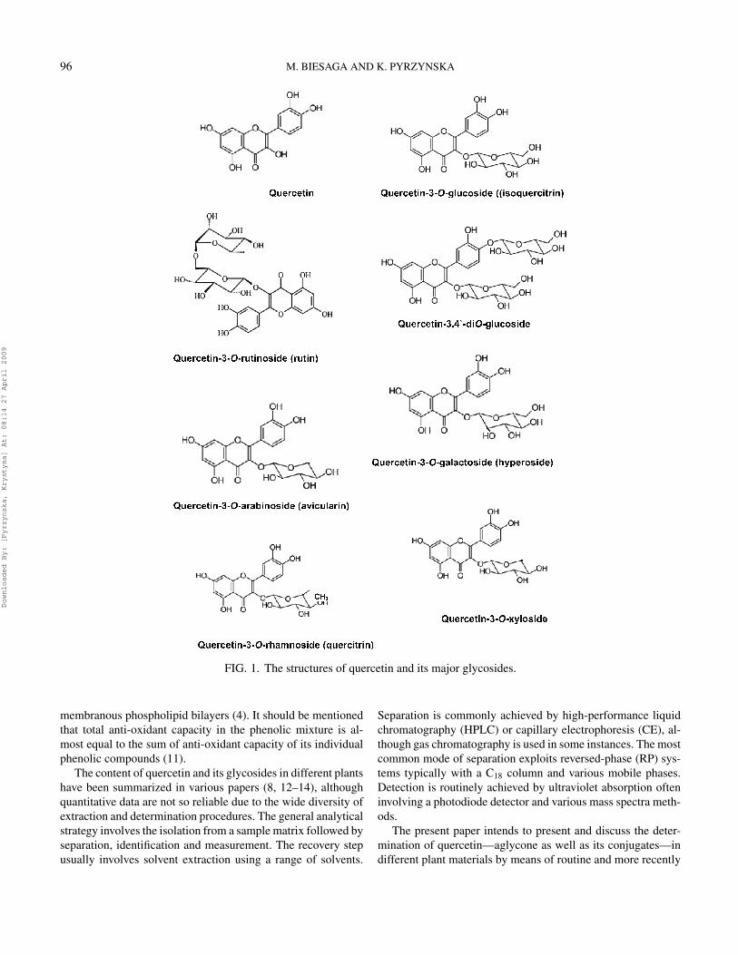
Quercetin, 3,3′,4′,5,7-pentahydroxylflavone, presented inFig. 1, is one of the most abundant flavonoids present in fruitsand vegetables. In plants, it occurs mainly in leaves and inthe outher parts of the plants as aglycones and glycosides, inwhich one or more sugar groups is bound to phenolic groupsby glycosidic bonds. Glucose is the most common sugar, withgalactose and rhamnose also frequently found in compositionwith flavonoids. In general, quercetin glycosides contain a sugar
Address correspondence to Krystyna Pyrzynska, Department ofChemistry, Warsaw University, Pasteura 1, 02-093 Warsaw, Poland.E-mail: [email protected]
group at the 3-position. Glycosylation increases the polarityof the flavonoid molecule, which is necessary for storage inplant cell vacuoles (5). A considerable amount of isoquercetrin(quercetin-3-O-β-glucoside) has been found in apple and pearpeels (6) as well as in Hypericum perfloratum leaves or flow-ers (7). Almost 180 different glycosides of quercetin have beendescribed in nature, with rutin (quercetin-3-O-β-rutinoside) be-ing one of the most common (8, 9). In onion, one of the com-mon quercetin sources, however, the phenolic group at the 4′-position is necessarily bound by a sugar group and thus its majorglycosides are quercetin 4′-O-β-glucoside and quercetin 3,4′-O-β-diglucoside (Fig. 1). Their content is about 80% of thetotal content of flavonoids. Vegetables, fruits and red wine arethe main dietary sources of quercetin. Onion (Allium cepa L.)ranked highest in quercetin content in a survey of 28 vegeta-bles and nine fruits (10). In fruits and vegetables, flavonols andtheir glycosides are found predominantly in the skin where theyserve, among others, as ultra-violet protection.
Nevertheless, we should take into account the fact that food-derived quercetin could be presented in its glysocides form andthus the effectiveness of its anti-oxidant activity is greatly mod-ified by the position of the sugar group attached to the ba-sic diphenylpropane structure. Quercetin aglycone seems tobe a more active chain-breaking anti-oxidant than, its gly-coside counterparts because of its higher accessibility to thesite of chain-initiating and chain-propagating free radicals in
95
Downloaded By: [Pyrzynska, Krystyna] At: 08:14 27 April 2009
96 M. BIESAGA AND K. PYRZYNSKA
FIG. 1. The structures of quercetin and its major glycosides.
membranous phospholipid bilayers (4). It should be mentionedthat total anti-oxidant capacity in the phenolic mixture is al-most equal to the sum of anti-oxidant capacity of its individualphenolic compounds (11).
The content of quercetin and its glycosides in different plantshave been summarized in various papers (8, 12–14), althoughquantitative data are not so reliable due to the wide diversity ofextraction and determination procedures. The general analyticalstrategy involves the isolation from a sample matrix followed byseparation, identification and measurement. The recovery stepusually involves solvent extraction using a range of solvents.
Separation is commonly achieved by high-performance liquidchromatography (HPLC) or capillary electrophoresis (CE), al-though gas chromatography is used in some instances. The mostcommon mode of separation exploits reversed-phase (RP) sys-tems typically with a C18 column and various mobile phases.Detection is routinely achieved by ultraviolet absorption ofteninvolving a photodiode detector and various mass spectra meth-ods.
The present paper intends to present and discuss the deter-mination of quercetin—aglycone as well as its conjugates—indifferent plant materials by means of routine and more recently
Downloaded By: [Pyrzynska, Krystyna] At: 08:14 27 April 2009

DETERMINATION OF QUERCETIN AND ITS GLYCOSIDES 97
developed analytical techniques. In all instances, selected appli-cations will be included to illustrate the scope and limitationsof the various approaches.
SAMPLE COLLECTION, STORAGE AND PREPARATIONDue to the vast reservoir of plants, and the variation of their
different parts, it is impossible to have any definitive procedureor protocol for the collection and storage of all plant materials.However, sample collection and storage conditions are essen-tial and should be treated carefully, because plant variety, aswell as growing location and season, may significantly affectthe quantity and quality of our analysis. Most plants used in tra-ditional medicine are harvested and dried for storage, while atother times, fresh or frozen plant material have been used. Theprocessing and storage conditions, such as drying temperatureand duration, storage length and humidity, therefore may affectthe outcome.
Flavonoids are generally found at higher concentrations inouter layers of fruits and vegetables, therefore peeling results intheir great loss. After home-like peeling, red onions contained79% of the original total content of quercetin-4′-glucoside,while this compound was unaffected by chopping of onions(15). DuPont et al. (16) found that shredding of lettuce leavesfollowed to exposure to light produced significant losses ofquercetin glycosides ranging 6–94% depending on the lettucevariety.
Storage experiments with commercial cultivars of onion wereperformed at a low constant temperature (1◦C) and at highervariable temperatures (similar to 8◦C) (17). Cultivar differencesin quercetin glucoside content were significant but neither nitro-gen fertilizer level nor lifting time had more than minor effectsat start of storage or after 3 or 5 months of storage. The role ofonion size was inconsistent but seemed to be of minor impor-tance.
The changes in the content of some major flavonoids, suchas quercetin and kaempferol, in onions, green beans and peasbefore and after different heat treatment (blanching, water-cooking, cooking in a microwave oven, frying and warm-holdingof the water-boiled sample at 60◦C for 1–2 hours) were studiedby Ewald et al. (18). The significant losses of quercetin tookplace during the peeling and trimming process of onions (39%).The onions were of different size and they were peeled veryunequally which means that several layers were peeled off fromsome onions and only one layer from others. Consequently, thelosses of flavonoids were very high in the onions where severallayers were peeled off since the most of quercetin is present inthe first and second layer of this vegetable. The small changesof flavonoid content at further processing of all onion sampleswere within the observed biological variation among samples.
The pH value had an important influence on the degradationprocess of quercetin during heating in aqueous solution (19). AtpH 5 the amount of quercetin decreased to approximately 75%after 300 minutes and under weak basic conditions (pH 8) wasundetectable by HPLC/DAD after 240 minutes (Fig. 2). The
0 50 100 150 200 250 3000
20
40
60
80
100
Pea
k ar
ea, %
Reaction time, min
Quercitin pH 5 Rutin pH 5 Quercetin pH 8 Rutin pH 5
FIG. 2. Degradation of quercetin and rutin (1 mM) in aqueoussolution at 100◦C at different pH (19). (Copyright John Wiley& Sons Limited. Reproduced with permission)
presence of oxygen accelerates the degradation process. Rutin,the quercetin glycoside, showed a higher stability towards ox-idation (Fig. 2). Lombard et al. (20) reported that baking andsauteing of onion produced a 7–25% gain in quercetin concen-tration, while boiling produced an 18% decrease in the contentof this flavonol.
Rhodes and Price (21) investigated the compositionalchanges of flavonols in onions (skinned, chopped and immedi-ately frozen in liquid nitrogen) during autolysis at 25◦C over the24-hours period. Their results showed that there was only a smallloss (11–18%) in total 4-O-flavonoids content. However, in allexamined varieties of onion there were significant quantitativechanges in flavonoid composition. For brown-skinned onion, forexample, within 2 hours 19% of quercetin-3,4-diglucose waslost with the appearance of an equimolar amount of quercetin-4-glucose, whilst during the first 5 hours there was a 50% de-crease of the diglucosides accompanied by increases in both themonoglucoside and the free aglycone. All of the diglucosideshad disappeared by the end of 24 hours.
Carrots retained more flavonoids after 30 days storage when,before drying, were treated with the solution containing ascorbicacid (0.1%) and glucose (1%). (22). Over this period about 10%of flavonoids content was lost when carrots were then dried in ahot-air oven at 80◦C in comparison to freeze-drying at –50◦C.
EXTRACTIONSimple filtration for some liquid samples, such as fruit juices
and wines, is ineffective for recovering quercetin and its gly-cosides and alternative strategies are necessary. The advan-tage of liquid extraction in comparison with direct injectionhave been demonstrated for HPLC analysis of a wine sample(23). Quercetin and its glycosides from fresh, freeze-dried andair-dried samples are generally extracted with pure methanol,
Downloaded By: [Pyrzynska, Krystyna] At: 08:14 27 April 2009

98 M. BIESAGA AND K. PYRZYNSKA
Water MeOH EtOH DMF EtOAc0,0
1,6
3,2
4,8
6,4
Con
tent
of q
uerc
etin
, mg/
g
Onion skinHypericum perforatum
Water MeOH EtOH DMF EtOAc0
2
4
6
8
10
12
14
16
18
Con
tent
of r
utin
, mg/
g
Apple peelHypericum perforatum
(A)
(B)
FIG. 3. The efficiency of extraction of (A) quercetin and (B)rutin using different solvents (26). (Copyright Elsevier Limited.Reproduced with permission).
ethanol or their combination with water (10, 24–27), but in somecases ethyl acetate (28) or acetone (6, 29) have been used.
The efficiencies of different solvents for extraction ofquercetin and rutin from onion skin, apple peel and Hypericumperforatum leaves (expressed in mg per gram of the dry plantmaterial) are presented in Fig. 3 (26). The extract compositionwas the same in each case, but the flavonoids yields were dif-ferent. The highest efficiency from onion samples was obtainedwhen water or DMF, the most polar solvents, were used. At thesub-cellular level, phenolic compounds are located mainly in thevacuoles. Their occurrence in soluble or suspended forms, and incombination with cell wall components, may have a significantimpact on their extraction (30). With dried plant materials, suchas Hypericum perforatum, low polarity solvents, such as ethylacetate, simply leach the sample whereas alcoholic solventsor water presumably rupture cell membranes and enhance the
extraction of endocellular materials (25). Aqueous methanol asan extracting solvent proved to be a useful compromise that en-sures the extraction of both aglycones and flavonoid glycosides,depending on time and temperature (24). Aqueous methanol,due to its polarity, is more effective for extraction of polyphe-nols linked to polar fibrous matrices, while acetone-water mix-tures are more useful for extracting polyphenols from proteinmatrices, since they appear to degradate the polyphenol-proteincomplexes (29).
Extraction of tea polyphenols was usually done by steepingtea leaves in boiling water for 5–30 minutes (31–33). The in-fusion was then filtered and subjected to HPLC analysis. Onthe other hand, tea polyphenol extracts were obtained usingpure methanol (34) or by employing a multiple extraction of teasamples with 80% (v/v) methanol and subsequently with 80%methanol containing 0.15% HCl (35).
The possibility of quercetin degradation under different sol-vent and temperature conditions was tested by Pinelo et al. (36).An initial increase and a following decrease in its anti-radicalactivity was observed in ethanol and methanol solutions whenstorage time was prolonged. By contrast, a progressive decreasein anti-oxidant activity was determined in 10% (v/v) ethanolwater solution due to oxidative cleavage which is favored underthese conditions.
The alternative extraction methods such as sonication (37–39), supercritical fluid extraction (40, 41) and pressurized liq-uid extraction (42), due to shorter extraction time and reducedsolvent consumption, have gained increasingly popularity forisolation of quercetin and its glycosides. A review on the ul-trasonically assisted extraction of active compounds from plantmaterials was published (43). Due to apolar properties of CO2 insupercritical fluid extraction the significant amount of polar or-ganic modifiers has to be added to obtain high extraction yield;however, it leads to reduced selectivity (44, 45). For example, inextracting polyphenols from green tea, the best extraction yieldwas found in the system using 95% ethanol and 5% CO2 (44).Comparison of the effectiveness of Soxlet extraction, sonica-tion and pressurized-fluid extraction of several major phenoliccompounds in St. John’s wort herb is presented in Fig. 4 (39).
Many extracts contain significant portions of carbohydrates,lipids and other unwanted compounds that potentially interferewith subsequent quantification. Simultaneous sample clean-upand pre-concentration of the analytes could be achieved by solid-phase extraction (SPE) using a range of sorbents (13, 24, 25,46). In most cases, C18-bonded silica or polymeric sorbents areused and the sample solution and solvents are usually slightlyacidified to prevent ionization of the flavonoids, which wouldreduce their retention. Methanolic extract of the homogenizedolives was evaporated to dryness, redissolved in water con-taining hydrochloric acid (pH 2) and loaded on a C18 sorbent(47). After washing with hexane to remove lipids, the flavonoidswere eluted with pure methanol. C18 cartridges were also usedfor isolation and pre-concentration of quercetin from the ex-tracts of vegetables (48) and cranberry juice (49). The use of
Downloaded By: [Pyrzynska, Krystyna] At: 08:14 27 April 2009

DETERMINATION OF QUERCETIN AND ITS GLYCOSIDES 99
FIG. 4. Comparison of the effectiveness of Soxlet extraction,sonication and pressurized-fluid extraction (PFE) of several ma-jor phenolic compounds in St. John’s wort herb (39). (CopyrightElsevier Limited. Reproduced with permission).
polymeric sorbent Oasis HLB (Waters, Milford, MA, USA) ef-fectively eliminated the interfering constituents from biologicalsamples with efficient extraction of quercetin (50). AmberliteXAD-2 (Aldrich, Deisenhofen, Germany) particles (51, 52) andBond Elut C18 (Varian, Middelburg, The Netherlands) cartridges(53) have been utilized for purification of acidified solutions ofhoney. Molecularly imprinted polymers (MIPs) are novel sor-bents used for SPE of phenolic compounds in food samples(54–56). Analyte is used as a template during formation ofthe polymer following appropriate extraction, which leaves re-ceptors for a molecular recognition of targed analyte. Figure 5
FIG. 5. Chromatographic analysis of Merlot wine (1) with and(2) without molecular imprinted polymer. Elution was donewith acetonitrile (54). (Copyright American Chemical Society.Reproduced with permission).
shows that the use of MIPs greatly reduced the complexity ofchromatographic analysis of red wine and enhanced the intensityof quercetin peak (54). Recently, similar MIPs for rutin isolationfrom wine, tea and juice samples has been presented (56).
Matrix solid-phase dispersion (MSPD) has been also testedfor the analysis of naturally occurring constituents from differentplant materials providing, in many cases, equivalent or superiorresults to more classical solvent and SPE techniques (57, 58).The entire sample is homogeneously dispersed into particles ofvery small size, usually a C18 or C8-bonded silica, providing anenhanced surface for subsequent extraction of the components.However, this technique does not give sufficient results for allphenolic compounds. Xio et al. (59) compared the efficiencyof MSPD with ultrasonic and soxlet extraction for isolation ofisoflavonoids from Radix astragali, the dried root of a medicinalChinese plant. While the amount of the aglycones were higherby MSPD, the extraction efficiency of the glycosides were betterusing the conventional techniques, particularly solvent extrac-tion. Extraction of quercetion-3-O-glucoside from white grapeswith MSPD in just one step gave similar results in comparisonto combined isolation procedure with solvent extraction (aque-ous HCl, pH 2, containing 5% methanol, 40◦C, in an ultrasonicbath) followed by a clean-up step with C18 non-endcapped sor-bent and ethanol elution (60). The best recoveries of aglyconequercetin in all the assays (different sample to sorbent ratios)carried out by MSPD were around only 30%.
HYDROLYSISOften due to difficulties in later separation procedures, hy-
drolysis of quercetin glycosides before or during extraction isperformed to break the glysocidic bonds and to obtain the agly-cone form. The extraction recovery is dependent on molarityof HCl, hydrolysis time and temperature and the compositionof the extraction solvent. In most publications the hydrolysis offlavonoid glycosides from vegetables and fruits is carried out in1.2 M HCl at 90◦C for 2 hours according to a procedure pre-sented by Hertog et al. (10). However, the extended exposuretime to HCl could cause degradation of quercetin (10, 61). Gen-erally, for the hydrolysis process, the optimum compromise is toachieve complete release of aglycones and to minimize degra-dation reactions of compounds involved. For this purpose, acentral composite experimental design was described (62). Ap-plying a multiple regression analysis on the data set, it was pos-sible to obtain a mathematical model that took linear, quadraticand cross-product terms into account. According to this math-ematical approach, optimum conditions for rutin hydrolysis inorange juice corresponded to HCl concentration of 1.5 M and ahydrolysis time of 1 hour.
Identical optimum hydrolysis conditions for flavonol glu-cosides in different vegetables or fruits could not be achievedas they are dependent on the binding site of the sugar on theflavonoid nucleus (10). The study presented by Lombard et al.(20) indicated that the main quercetin glucosides present inonion were relatively heat stable, thus, higher temperature andshorter time for their hydrolysis could be employed. However,
Downloaded By: [Pyrzynska, Krystyna] At: 08:14 27 April 2009

100 M. BIESAGA AND K. PYRZYNSKA
it should be mentioned that the conditions resulting in optimalbreakdown of glycosides could be too harsh for some of theother phenolic compounds present in the same plant material.For example, myricetin as well as catechins could be degradatedat high temperatures (20, 63). The method of choice is alwaysa compromise between efficiency production of aglycone fromthe plant material and degradation of aglycones.
Nuutila et al. (63) compared the efficiency of hydrolysis ofquercetin glycosides from red onion and spinach in the pres-ence of tert-butylhydroquinone (TBQH) and ascorbic acid asthe anti-oxidants. Although TBQH gave a slightly higher con-centration of quercetin than ascorbic acid, it caused some in-terferences with the detection. Moreover, the higher addition ofascorbic acid than 2 mg acted as a pro-oxidant rather than as ananti-oxidant. Thus, the proposed method (2 hours refluxing at80◦C with 1.2 M HCl in the presence of ascorbic acid) couldbe applied for screening vegetables and leafy vegetables butwould probably not be suitable for samples, which have higherconcentrations of myricetin and phenolic acids.
SEPARATIONIn general, separations of quercetin and its glycosides have
been mainly carried out by HPLC equipped with RP columns,generally packed with spherical particles of silica bonded withoctadecyl (C18) chains (13, 24, 25, 46). HPLC columns packedwith monolithic supports, consisting of a single piece of porousmaterial, are alternative means of performing fast separations.The main advantage of this type of support is its excellent hydro-dynamic property, which allows the reduction of backpressureand increases the flow rate. Monolithic columns are increas-ingly being applied in phytochemical analysis (64). However,in the field of food analysis they were only used for deter-mination of phenolics compounds in wine (65) and phenolicacids in fruits (66). A highly hydrophilic poly(7-oxonorbornene-5,6-dicarboxylic acid-block-norbornene)-coated silica was alsoinvestigated for the liquid chromatographic determination offlavonoids in plant extracts (67). Compared to the most com-monly used octadecyl-derivatized silica this sorbent allowed fastseparation even at extreme pH values.
Gradient elution is usually used in recognition of the com-plexity of the phenolic profile of plant samples (68–76). Nu-merous mobile phases have been employed but binary systemscomprising an aqueous component and a less polar organic sol-vent, such as acetonitrile or methanol, remain common. Acid,such as acetic, formic or trifluoroacetic (TFA), is usually addedto maintain constant acid concentration during gradient runs(Table 1). Phosphate buffers are less popular, mainly becauseof the dreaded contamination of ion sources when mass spec-trometry detection is used. In some cases, isocratic elution hasprovided adequate resolution due to selectivity effects of one ormore components of the mobile phase (77). The elution patternfor flavonoids is flavanone glycoside followed by flavonol andflavone glycosides and then the free aglycones in the same order.Most of these methods have been developed to measure differ-
ent groups of polyphenolics in a single plant material or a fewgroups in multiple plant sources, but obtaining good resolutionis considered to be the main difficulty in this last approach. Theselected examples of application of HPLC to the determinationof quercetin and its glycosides in plant materials are presentedin Table 1.
Ultra performance liquid chromatography (UPLC) takes ad-vantage of technological strides made in particle chemistryperformance (78). Using 1.5–2 µm particles, a more narrowanalytical column and instrumentation that operates at higherpressures than those used in HPLC, drastic increases in reso-lution, sensitivity and speed of analysis can be obtained. Thesame separation on RP-HPLC that takes over 20 minutes can beaccomplished in under 3 minutes by UPLC. This new chromato-graphic methodology has been applied in plant material analysisso far for separation and quantification of the major chocolatepolyphenols (79).
Although HPLC stays as the most dominating separationtechnique for polyphenolic compounds, CE is gaining pop-ularity and represents an alternative method for the analysisof plant materials (12, 24). The CE modes primarily used forthese purposes are capillary zone electrophoresis (CZE) (80–88) and micellar electrokinetic chromatography (MECK) (89,90).To achieve ionization of hydroxy compounds (as relativelyweak acids) enabling their separation by CZE, background elec-trolytes based on borate buffer of pH 9–11 are widely used andin many cases simultaneously the complex formation ability ofB(III) is utilized for manipulating or enhancing the selectivity ofelectrophoretic separation. Molybdenate was also examined as acomplex-forming additive (91). It forms more stable complexeswith aromatic o-dihydroxy compounds and hence the complex-formation effect is observed at a considerably lower pH equal to5.4. CZE is only applicable to charged analytes and the charge-to-size ratios determine the electrophoretic migration times. InMECK for neutral analytes in the presence of surfactants suchas sodium dodecyl sulfate, separation is based on hydrophobic-ity, which affects the analyte partitioning between the aqueousphase (moving with the electro-osmotic flow) and the micel-lar phases (charged and migrating with a different velocity). Forionic analytes separation in MECK is based on both the degree ofionization and the hydrophobicity. Wang et al. (92) assessed andcompared the electrophoretic behavior of 13 flavonoids com-monly found in medicinal plants using these two modes of CE.The separation selectivity of MECK was shown to be better thanthat of CZE, because electrophoretic behavior in the latter is af-fected by more factors, such as the degree of saturation and thestereochemistry of the C-ring, alkyl substitution and the numberand position of phenolic hydroxy groups, methylation and gly-cosylation of the hydroxy groups, as well as the complexationof flavonoids with a borate buffer. Unfortunately, the authorsdid not study any natural samples. Non-aqueous capillary elec-trophoretic separation of a group of flavonoids was investigatedin methanol at high. pH to alter selectivity of the separation (93).Additionally, tests of untreated capillaries and capillaries coated
Downloaded By: [Pyrzynska, Krystyna] At: 08:14 27 April 2009

TAB
LE
1Se
lect
edex
ampl
esof
appl
icat
ion
ofH
PLC
toth
ede
term
inat
ion
ofqu
erce
tinan
dits
glyc
osid
esin
plan
tmat
eria
ls
Sam
ple
Det
ecte
dco
mpo
unds
Stat
iona
ryph
ase
Mob
ileph
ase
Det
ectio
nR
ef.
Oni
on,s
pina
chqu
erce
tin,Q
-3-r
ut,Q
-3-g
luSy
mm
etry
C18
,5µ
m(1
50×
3.9
mm
)G
radi
ent:
2–60
%(v
/v)
met
hano
lw
ithaq
ueou
sT
FA(3
00µ
L/L
)D
AD
63
Stra
wbe
rrie
squ
erce
tinPu
rosp
her
RP-
18e,
5µ
m(1
25×
3m
mID
)G
radi
ent:
A-1
%fo
rmic
acid
,B
-ace
toni
trile
DA
D68
Oni
on,a
pple
,bl
ubbe
ry,
quer
cetin
,Q-3
-rut
,Q-3
-rha
LiC
hrom
osor
bR
P18,
7µ
m(2
5×
4m
m)
Gra
dien
t:A
-0.0
5%aq
ueou
sT
FA,
B-0
.05%
TFA
inac
eton
itrile
DA
D69
Her
bsqu
erce
tin,Q
-3-r
ut,Q
-3-g
al,Q
-3-g
lu,
Q-3
-rha
YM
CO
DS-
AQ
RP-
18,
5µ
m(2
50×
4.6
mm
)G
radi
ent:
A-0
.5%
TFA
+20
%(v
/v)
met
hano
l,B
-ace
toni
trile
DA
D70
Art
icho
kequ
erce
tin,Q
-3-r
ut,Q
-3-g
al,Q
-3-r
ha,
Q-3
-glu
Lun
aC
18,3
.5µ
m(5
0×
2.1
mm
)G
radi
ent:
A-
0.1%
form
icac
id,
B-a
ceto
nitr
ilew
ith0.
1%fo
rmic
acid
DA
D,M
S71
App
lepo
mac
eQ
-3-r
ut,Q
-gal
,Q-3
-glu
,Q-3
-xyl
,Q
-3-a
rap
,Q-3
-ara
f,Q
-3-r
haA
qua
C18
,5µ
m(2
5×
4.6
mm
)G
radi
ent:
A-2
%(v
/v)
acet
icac
id,
B-0
.5%
acet
icac
idin
wat
eran
dac
eton
itrile
(50:
50,v
/v)
MS,
NM
R72
Man
goQ
-gal
,Q-3
-glu
,Q-3
-ara
,Q-3
-rha
LiC
hrom
osph
erR
P18
e,5
µm
(250
×4
mm
)G
radi
ent:
2%(v
/v)
acet
icac
idin
wat
erB
-0.5
%ac
etic
acid
and
acet
onitr
ile(5
0:50
,v/v
)
DA
D,M
S73
Cas
hew
appl
eQ
-3-g
al,Q
-3-g
lu,Q
-3-x
ylp
,Q-3
-ara
p,
Q-3
-ara
f,Q
-3-r
haW
ater
sC
18,5
µm
(250
×4.
6m
m)
Gra
dien
t:A
-0.
05%
acet
icac
id,
B-a
ceto
nitr
ileD
AD
,MS
74
App
le,p
ear
Que
rcet
in,Q
-3-r
ut,Q
-3-g
al,Q
-3-g
lu,
Q-3
-xyl
,Q-3
-ara
,Q-3
-rha
Aqu
aC
18,5
µm
(250
×4.
6m
m)
Gra
dien
t:A
-2%
acet
icac
id,
B-a
ceto
nitr
ileM
S75
Oni
onQ
-4′ -g
lu,Q
-3,4
′ -dig
lu,Q
-7,4
′ -dig
lu,
Q-3
-glu
,Q-3
,7,4
′ -tri
glu
Dis
cove
ryC
18,5
µm
(250
×4.
6m
m)
Gra
dien
t:ac
eton
itrile
inw
ater
DA
D76
Q-3
-gal
—qu
erce
tin-3
-laa
ctos
ide
(hyp
eros
ide)
;Q
-3-g
lu—
quer
cetin
-3-g
luco
side
(iso
quer
cetr
in);
Q-3
-ara
—qu
erce
tin-3
-ara
bino
side
(avi
cula
rin)
;Q
-3-r
ha—
quer
cetin
-3-
rham
nosi
de(q
uerc
etri
n);
Q-3
-xyl
osid
e;Q
-3-r
ut—
quer
cetin
-3-r
utin
osid
e[r
ham
nosy
l(α
1→6)
-glu
cosi
de]
(rut
in);
Q-3
-xyl
p—
quer
cetin
-3-x
ylop
yran
osid
e;Q
-3-a
rap
—qu
ertc
etin
-3-
arab
inop
yran
osid
e(g
ujav
erin
);Q
-3-a
raf
—qu
erce
tin-3
-ara
bino
fura
nosi
de.
101
Downloaded By: [Pyrzynska, Krystyna] At: 08:14 27 April 2009

102 M. BIESAGA AND K. PYRZYNSKA
TABLE 2Application of capillary electrophoresis to the determination of quercetin and its glycosides
Sample Detected compounds Background electrolyte Detection Ref.
Medicinal plants Q-3,4′-diglu, Q-4′-glu 25 mM sodium tetraborate with 20% (v/v)methanol, pH 9.3
UV (270 nm) 80
Onion Q-3-rut, Q-3-rha 10 mM boric acid, 10 mM sodium tetraborate,15 mM EDTA in 15% (v/v) methanol, pH 10.2
UV (280 nm) 81
Red wine Quercetin, Q-3-rut,Q-3-rha
25 mM β-hydroxy-4-morpholinoprpanesulfonicacid, 50 mM Tris, 15 mM boric acid and 5 mMβ-cyclodextrin, pH 8.5
UV (254 nm) 82
Mulberry leaves Quercetin, Q-3-rut,Q-3-rha
150 mM boric acid, pH 10 UV (270 nm) 83
Medicinal plant Quercetin, Q-3-rut 50 mM sodium tetraborate in 18% (v/v) methanol,pH 9.7
UV (210 nm) 84
Propolis Quercetin, Q-3-rut 30 mM sodium tetraborate, pH 9 UV (214 nm) 85Standard mixture Quercetin, Q-3-rut,
Q-3-glu0.1 M boric acid – NaOH, pH 10 UV (254 nm) 86
Medicinal plants Quercetin 30 mM sodium tetraborate in 8% (v/v) acetonitrile,pH 9
UV (210 nm) 87
Grapes Quercetin 35 mM sodium tetraborate, pH 8.9 UV (250 nm) 88Wines Quercetin 40 mM sodium tetraborate, 40 mM sodium
dodecyl sulfate, pH 9UV ( 214 nm) 89
Tea Q-3-glu 50 mM NaH2PO4, 50 mM sodium tetraborate,20 mM sodium dodecyl sulfate in 10% (v/v)acetonitrile, pH 6
UV (278 nm) 90
Abbreviations as in Table 1
with poly(glycidylmethacrylate-co-N -vinylpyrrolidine showedthe analysis to be faster (6.5 minutes vs. 25 minutes) and therepeatability better in the coated capillaries.
CE as a separation technique is still evolving and a new modeof separation, called capillary electrochromatography (CEC),has been developed. This hybrid method combined CZE andµ-HPLC (94). It bridges the advantages of both these tech-niques, offering a unique separation mode and exploiting acombination of chromatographic retention and electrophoreticmobility. Application of CEC was also explored for analy-sis of quercetin (95) and biologically relevant herbal flavonols(96) (Table 2).
DETECTIONRoutine detection in HPLC and CE is typically based on mea-
surement of UV absorption, often using photodiode array detec-tion (DAD). A match of both ultraviolet-visible (UV-VIS) spec-trum and retention time can lead to highly positive identificationof the separated analytes. DAD detector is capable for simultane-ously detection of chromatograms at different wavelengths. Thisfeature significantly enhances the performance of the separationsystem, particularly when different groups of polyphenols aremixed in one sample. When proper wavelengths are chosen, e.g.,at the maximum absorptions, all groups can be detected with
the highest sensitivity. An appropriate selection of the detectionwavelength can also allow the quantification of an unresolved orpoorly resolved peak (46). However, the use of conventional ap-proaches based on spectra is often limited when samples containvery similar compounds.
Detection based on fluorescence is generally more sensitivethan UV absorption. Quercetin and its glycosides do not shownative fluorescence, although Rodriguez-Delago et al. (23) re-ported optimum excitation (260 and 264 nm) and emissionwavelengths (426 and 420 nm) for quercitrin and quercetin,respectively, but they could be used only for high concentra-tion of these analytes. Many flavonoids, such as quercetin, canform fluorescent chelates with several cations such as Mg(II),Be(II), Zn(II), Sc(III), Ga(III), In(III) and Al(III), which couldbe used as the post-column derivatization reagents for HPLCwith fluorescence detection (97–100). The limit of detection fordetermination of quercetin, based on the formation of its fluo-rescent complex with Al(III) (97) and Ga(III) (98), was foundto be 0.15 and 16.2 µg/L, respectively. The study shows that the3-hydroxyl-4-keto oxygen site is essential for fluorescence asrutin—containing a sugar bound to the 3-hydroxyl group—doesnot form a fluorescent chelate (97).
HPLC or CE with electrochemical detection can be a use-ful completion technique for determination of quercetin and its
Downloaded By: [Pyrzynska, Krystyna] At: 08:14 27 April 2009

DETERMINATION OF QUERCETIN AND ITS GLYCOSIDES 103
glycosides because they are, as most flavonoids, electroactiveat modest oxidation potentials (62, 101–104). Romani et al.(101) compared HPLC procedures with DAD and electrochem-ical detection (differential pulse voltammetry and amperomet-ric biosensor with bare graphite screen-electrodes) for analysisof phenolic compounds in natural extracts derived from grape,olives and green tea. The most accurate data were obtained fromHPLC-DAD analysis, while differential pulse voltammetry wasconsidered a good and quick method for screening. Recently,multi-walled carbon nanotubes have been found to be a excel-lent electrode material for the determination of quercetin andrutin at trace levels due to their strong surface adsorption (105,106).
For complete structural identification of the analytes, otherdetection techniques such as mass spectrometry (MS) with dif-ferent ionization modes is often necessary (13, 24, 25, 46, 74,107, 108). Soft electrospray ionization (ESI) or atmosphericpressure chemical ionization (APCI) provides the molecularmasses of chromatographically separated molecules and tan-dem MS-MS provides extra structure details of thermally la-bile, non-volatile polar phenolic compounds. Usually APCIis used more extensively with small molecule compounds(MW<1200), while ESI works better with more polar and highermolecular weight compounds. It was experimentally confirmedthat quercetin sensitivity with the ESI mode is better than thatwith the APCI mode (109). The possible reasons for this arethat APCI can cause high chemical background noise for lowmass compounds and thermal degradation can occur with labilecompounds, such as quercetin, which would decrease the APCIsensitivity.
Bonaccorsi et al. (76) demonstrated that the on-line HPLC-DAD electron spray mass spectrometry (ESI-MS-MS) tech-nique constitutes an accurate and easy methodology for the qual-itative and quantitative analysis of quercetin glycosides in south-ern Italian red onions, quercetin-4′glucoside and quercetin-3-4′-diglucoside being the most abundant components. Thepresence of quercetin-3-7-4′-triglucoside was identified andconfirmed by UV spectra and product ion scan experiments(Fig. 6). This compound exhibited an ESI-MS spectrum in thepositive ion mode with four significant peaks, one centred atm/z 789 [M + H]+, the second at m/z 627 [M + H—162]+,the third at m/z 465 [M + H—162—162]+ and the last oneat m/z 303 [M + H—162—162—162]+ (Fig. 6A). Four anal-ogous peaks were produced in the negative ESI-MS analysis(Fig. 6B). The loss of three glycosil units shown in the ESI-MSspectra and the fragmentation pattern in the negative ESI-MS-MS analysis on the ion at m/z 301 (Fig. 6C) were indicativeof a quercetin triglucoside. The hypsochromic shift observedin the UV spectrum with respect to aglycone quercetin value(252→265 nm) excluded substitution onto the 5-position of theA-ring and confirmed this conclusion. The presented method-ology allowed them to obtain a characteristic profile of severalquercetin glycosides also in other vegetables that are rich in thesecompounds.
FIG. 6. ESI-MS spectra in (A) positive and (B) negativemode and (C) negative ESI-MS-MS spectrum of chromato-graphic peak corresponding to quercetin-3,7,4′-triglucoside(76). (Copyright American Chemical Society. Reproduced withpermission).
Downloaded By: [Pyrzynska, Krystyna] At: 08:14 27 April 2009

104 M. BIESAGA AND K. PYRZYNSKA
CONCLUSIONFlavonoids are the main factors in the hypothesis that the
consumption of a diet rich in vegetables and fruits may be pro-tective for human health. The anti-oxidative properties of thesecompounds demonstrated in vitro become a key issue. Epi-demiological studies have also shown an inverse effect betweenthe intake of flavonoids and the risk of coronary heart disease.Quercetin belongs to the flavonol subgroup of flavonoids andis widely distributed as the main bioactive plant component. Inplant food, quercetin exists in a multiplicity of complex con-jugates, mainly glycosides. Important questions remain on thefate of such conjugates during processing and digestion, on thenature of the nutritionally important form of quercetin and onthe extent of their uptake and metabolism in the gut (4, 9, 110).An important prerequisite for tacking such problems is the de-velopment of convenient and reliable analytical procedures witha high-resolution separation system and appropriate quantifica-tion.
The preliminary steps, such as isolation and extraction fromplant materials, are of primary importance influencing the re-liability and reproducibility of the analysis. For this purpose,several classical techniques, like solvent extraction, as well re-cent techniques (pressurized-fluid extraction and matrix solid-phase dispersion) have been tested. The important aspect ofquercetin analysis is whether to determine the target analytein its various conjugated forms or as the aglycone. Chromato-graphic or electrophoretic separation of the obtained extract,without hydrolysis is expected to result in the identification andquantification of the quercetin aglycone and its glycosides, si-multaneously, in the presence of each other. Separation of thetarget analyte from a hydrolyzed extract provides a decreasednumber of compounds to be determined and the free aglyconeform of quercetin, as well as quercetin, initially in glycosidiclinkages cannot be distinguished. The combination of HPLC-DAD-MS or CE-DAD-MS is the best choice for separation,quantification and identification of the analytes. In order to de-fine fine structural differences, homo- and heteronuclear NMRspectroscopy is also very helpful (111).
REFERENCES1. S. Fiorucci, J. Golebiowski, D. Cabrol-Bass, and S. Antonczak,
DFT study of quercetin activated forms involved in antiradical,antioxidant and prooxidant biological processes. Journal of Agri-cultural and Food Chemistry 55 (2007):903–911.
2. M. G. L. Hertog, E. J. M. Feskens, P. C. H. Hollmann, M. B.Katan, and D. Kronthout, Dietary antioxidant flavonoids and riskof coronary heart disease: The Zutphen elderly study. Lancet 342(1993):1007–1011.
3. Y.-H. Chu, C.-L. Chang, and H.-F. Hsu, Flavonoid content ofseveral vegetables and their antioxidant activity. Journal of theScience of Food and Agriculture 80 (2000):561–566.
4. K. Murota and J. Terao, Antioxidative flavonoid quercetin: Im-plication of its intestinal absorption and metabolism. Archives ofBiochemistry and Biophysics 417 (2003):12–17.
5. S. A. Aherne and N. M. O’Brien, Dietary flavonols: Chemistry,food content, and metabolism. Nutrition 18 (2002):75–81.
6. A. Schieber, P. Hilt, J. Conrad, U. Beifuss, and R. Carle, Elu-tion order of quercetin glycosides from apple pomace extractson a new HPLC stationary phase with hydrophilic endcapping.Journal of Separation Science 25 (2002):361–364.
7. M. Urbanek, L. Blechtova, M. Pospisilova, and M. Polasek, On-line coupling of capillary isotachophoresis and capillary zoneelectrophoresis for the determination of flavonoids in methanolicextracts of Hypericum perforatum leaves or flowers. Journal ofChromatography A 958 (2002):261–271.
8. P. C. H. Hollman and I. C. W. Arts, Flavonoids, flavones andflavanols – nature, occurrence and dietary burden. Journal of theScience of Food and Agriculture 80 (2000):1081–1093.
9. K. Nemeth and M. K. Piskula, Food content, processing, ab-sorption and metabolism of onion flavonoids. Critical Reviews inFood Science and Nutrition 47 (2007):397–409.
10. M. G. L Hertog, P. C. H. Hollman, and M. B. Katan, Contentof potentially anticarcinogenic flavonoids of 28 vegetables and9 fruits commonly consumed in The Netherlands. Journal ofAgricultural and Food Chemistry 40 (1992):2379–2383.
11. H. J. Heo, J. J. Kim, D. Chung, and D. O. Kim, Antioxidantcapacities of individual and combined phenolic in a model system.Food Chemistry 104 (2007):87–92.
12. M. Naczk and F. Shahidi, Extraction and analysis of phenolic infood. Journal of Chromatography A 1952 (2004):95–111.
13. W. de Rijke, P. Out, W. M. A. Niessen, F. Ariese, C. Gooijer, andU. A. Brinkman, Analytical separation and detection methodsfor flavonoids. Journal of Chromatography A 1112 (2006):31–63.
14. H. Chen and Y. Zuo, Identification of flavonol glycosides in Amer-ican cranberry fruit. Food Chemistry 101 (2007):1357–1364.
15. L. Gennaro, C. Leonardi, F. Esposito, M. Salucci, G. Maiani,G. Quaglia, and V. Fogliano, Flavonoid and carbohydrate con-tents in Tropea onions: Effects of homelike peeling and storage.Journal of Agricultural and Food Chemistry 50 (2002):1904–1910.
16. M. S. DuPont, Z. Mondin, G. Williamson, and K. R. Price, Jour-nal of Agricultural and Food Chemistry 48 (2000):3957–3964.
17. L. M. Morgen, M. E. Olsson, and U. E. Gertsson, Quercetincontent in stored onion (Allium cepa L.): Effects of storageconditions, cultivar, lifting time and nitrogen fertilizer level.Journal of the Science Food and Agriculture 87 (2007):1595–1602.
18. C. Ewald, S. Fjelkner-Moding, K. Johansson, I. Sjoholm, and B.Akesson, Effect of processing on major flavonoids in processedonions, green beans and peas. Food Chemistry 64 (1999):231–235.
19. N. Buchner, A. Krunbein, S. Rohn, and L. W. Kroh, Effectof thermal processing on the flavonols rutin and quercetin.Rapid Communications in Mass Spectrometry 20 (2006):3229–3235.
20. K. Lombard, E. Peffley, E. Geoffrisu, L. Thompson, and A. Her-ring, Quercetin in onion (Allium cepa L.) after heat-treatmentsimulating home preparation. Journal of Food Composition andAnalysis 18 (2005):571–581.
21. M. J. C. Rhodes and K. R. Price, Analytical problems in thestudy of flavonoid compounds in onions. Food Chemistry 57(1996):113–117.
Downloaded By: [Pyrzynska, Krystyna] At: 08:14 27 April 2009
DETERMINATION OF QUERCETIN AND ITS GLYCOSIDES 105
22. Y. H. Yen, C. H. Shih, and C. H. Chang, Effect of adding ascorbicacid and glucose on the antioxidative properties during storageof dried carrots. Food Chemistry 107 (2008):265–272.
23. M. A. Rodriguez-Delago, S. Malovana, J. P. Perez, T. Borges,and E. J. Garcia Montelongo, Separation of phenolic compoundsby high-performance liquid chromatography with absorbanceand fluorimetric detection. Journal of Chromatography A 912(2001):249.
24. I. Molnar and Zs. Fuzfai, Chromatographic, capillary elec-trophoretic and capillary electrochromatographic techniques inthe analysis of flavonoids. Journal of Chromatography A 1073(2005):201–227.
25. K. Robards, Strategies for the determination of bioactive phenolsin plants, fruit and vegetables. Journal of Chromatography A1000 (2003):657–691.
26. A. Wach, K. Pyrzynska, and M. Biesaga, Quercetin content insome food and herbal samples. Food Chemistry 100 (2007):699–704.
27. M. Biesaga, A. Stafiej, and K. Pyrzynska, Extraction and hy-drolysis parameters for determination of quercetin in Hypericumperforatum. Chromatographia 65 (2007):701–706.
28. G. A. Spanos, R. E. Wrolstand, and D. A. Heatherbell, Influ-ence of processing and storage on the phenolic compositionof apple juice. Journal of Agricultural and Food Chemistry 38(1990):1572–1579.
29. J. Tabart, C. Kevers, A. Sipel, J. Pincemail, J. O. Defraigne,and J. Dommes, Optimisation of extraction of phenolic and anti-oxidants from black currant leaves and buds and of stability duringstorage. Food Chemistry 105 (2007):1268–1275.
30. K. Robards, P. D. Prenzler, G. Tucker, P. Swatsitang, andW. Glover, Phenolic compounds and their role in oxidative pro-cesses in fruits. Food Chemistry 66 (1999):401–436.
31. B. L. Lee and C. N. Ong, Comparative analysis of tea cate-chins and theaflavins by high-performance liquid chromatogra-phy and capillary electrophoresis. Journal of Chromatography A881 (2000):439–447.
32. M. H. Zhang, J. Luypaert, J. A. Fernandez Pierna, Q. S. Xu, andD. L. Massart, Determination of total anti-oxidant capacity ingreen tea by near-infrared spectroscopy and multivariate calibra-tion. Talanta 62 (2004):25–35.
33. Y. S. Lin, Y. J. Tsai, J. S. Tsay, and J. K. Lin, Factors affectingthe levels of tea polyphenols and caffeine in tea leaves. Journalof Agricultural and Food Chemistry 51 (2003):1864–1873.
34. P. J. Larger, A. D. Jones, and C. Dacombe, Separation oftea polyphenols using micellar electrokinetic chromatographywith diode array detection. Journal of Chromatography A 799(1998):309–320.
35. C. Cabbrera, R. Gimnez, and M. C. Lopez, Determination of teacomponents with antioxidant activity. Journal of Agricultural andFood Chemistry 51 (2003):4427–4435.
36. M. Pinelo, L. Manzocco, M. J. Nunez, and M. C. Nicoli, Sol-vent effect on quercetin antioxidant capacity. Food Chemistry 88(2004):201–207.
37. F. F. Liu, C. Y. W. Ang, T. M. Heinze, J. D. Rankin, R. D.Beger, J. P. Freeman, and J. O. Lay Jr., Evaluation of majorcomponents in St. John’s Wort dietary supplements by HPLC withphotodiode array detection and electrospray mass spectrometricconfirmation. Journal of Chromatography A 888 (2000):85–92.
38. P. Avato and G. Guglielmi, Determination of major constituentsin St. John’s Wort under different extraction conditions. Pharma-ceutical Biology 42 (2004):83–89.
39. F. B. Williams, L. C. Sander, S. A. Wise, and J. Girard, Devel-opment and evaluation of methods for determination of naphtho-dianthroned and flavonoids in St. John’s wort. Journal of Chro-matography A 1115 (2006):93–102.
40. A.Chafer, M. C. Pascual-Marti, A. Salvador, and A. Berna, Su-percritical fluids extraction and HPLC determination of relevantpolyphenolic compounds in grape skin. Journal of SeparationSciences 28 (2005):2050–2056.
41. B. J. Wang, Y. H. Lien, and Z. R. Yu, Supercritical fluidextraction—study of the anti-oxidant activities of propolis. FoodChemistry 86 (2004):237–243.
42. A. Smelcherovic, M. Spiteller, and S. Zuehlke, Comparison ofmethods for the exhaustive extraction of hyperins, flavonoids andhyperflorin from Hypericum perforatum L. Journal of Agricul-tural and Food Chemistry 54 (2006):2750–2753.
43. M. Vinatoru, An overview of the ultrasonically assisted extractionof bioactive principles from herbs. Ultrasonics Sonochemistry 8(2001):303–313.
44. C. J. Chang, K. L. Chiu, Y. L. Chen, and P. W. Yang, Effect ofethanol content on carbon dioxide extraction of polyphenols fromtea. Journal of Food Composition and Analysis 14 (2001):75–82.
45. R. Murga, R. Ruiz, S. Beltram, and J. L. Cabezas, Extraction ofnatural complex phenols and tannins from grape seeds by usingsupercritical mixtures of carbon dioxide and alcohol. Journal ofAgricultural and Food Chemistry 48 (2000):3408–3412.
46. T. Tsao and Z. Deng, Separation procedures for naturally occur-ring anti-oxidant phytochemicals. Journal of Chromatography B812 (2004):85–99.
47. A. F. Vinha, F. Ferreres, B. M. Silva, P. Valentao,A. Goncalves, J. A. Pereira, M. B. Oliveira, R. M. Seabra, andP. A. Andrade, Phenolic profiles of Portuguese olive fruits. In-fluences of cultivar and geographical origin. Food Chemistry 89(2005):561–568.
48. E. Dadakova, E. Prochazkova, and M. Krizek, Application ofmicellar electrokinetic capillary chromatography for quantita-tive analysis of plant materials. Electrophoresis 22 (2001):1573–1578.
49. H. Chen, Y. Zao, and Y. Deng, Separation and determination offlavonoids and other phenolic compounds in cranberry juice byhigh-performance liquid chromatography. Journal of Chromatog-raphy A 913 (2001):387–395.
50. K. Ishii, T. Furuta, and Y. Kasuya, High-performance liquid chro-matographic determination of quercetin in human plasma andurine utilizing solid-phase extraction and ultraviolet detection.Journal of Chromatography B 794 (2003):49–51.
51. L. Yao, Y. Jiang, B. D’Arcy, R. Singanusong, N. Datta, N. Caf-fin, and K. Raymont, Quantitative high-performance liquid chro-matography analyses of flavonoids in Australian Eucalyptus hon-eys. Journal of Agricultural and Food Chemistry 52 (2004):210–214.
52. L. Yao, Y. Jiang, R. Singansong, N. Datta, and K. Raymont,Phenolic acids and abscisic acid in Australian Eucalyptus honeysand their potential for floral authentication. Food Chemistry 86(2004):169–177.
Downloaded By: [Pyrzynska, Krystyna] At: 08:14 27 April 2009

106 M. BIESAGA AND K. PYRZYNSKA
53. B. Dimitrova, R. Gevrenova, and E. Anklam, Analysis of phe-nolic acids in honeys of differential floral origin by solid-phaseextraction and high-performance liquid chromatography. Phyto-chemical Analysis 18 (2007):24–32.
54. A. Molinelli, R. Weiss, and B. Mizaikoff, Advanced solid phaseextraction using molecularly imprinted polymers for the determi-nation of quercetin in red wine. Journal of Agricultural and FoodChemistry 50 (2002):1804–1808.
55. Y. G. Xia, T. Y. Guo, M. D. Song, B. H. Zhang, and B. L. Zhang,Selective separation of quercetin by molecularly imprinting usingchitosan as functional matrix. Reactive & Functional Polymers66 (2006):1734–1740.
56. G. Thedoridis, M. Lepsakova, V. Skerikova, A. Tegou, N. Gi-antsiou, and P. Jandrea, Molecular imprinting of natural flavonoidanti-oxidants: Application in solid-phase extraction for the sam-ple treatment of natural products prior to HPLC analysis. Journalof Separation Sciences 29 (2006):2310–2321.
57. S. A. Barker, Applications of matrix solid-phase dispersion infood analysis. Journal of Chromatography A 885 (2000):115–127.
58. S. A. Barker, Matrix solid dispersion (MSPD). Journalof Biochemical and Biophysical Methods 70 (2007):151–162.
59. H. B. Xiao, M. Krucker, K. Albert, and X. M. Liang, Determina-tion and identification of isoflavonoids in Radix astragali by ma-trix solid-phase dispersion extraction and high-performance liq-uid chromatography with photodiode array and mass spectromet-ric detection. Journal of Chromatography A 1032 (2004):117–124.
60. M. S. Dopico-Garcia, P. Valentao, A. Jagodziñska, J.Klepczyñska, L. Guerra, P. B. Andrade, and R. M. Seabra, Solid-phase extraction versus matrix solid-phase dispersion: Applica-tion to white grapes. Talanta 74 (2007):20–31.
61. H. M. Merken and G. R. Beecher, Measurement of foodflavonoids by high-performance liquid chromatography. Journalof Agricultural and Food Chemistry 48 (2000):577–599.
62. M. Careri, L. Elviri, A. Mangia, and M. Musci, Spectrometricand coulometric detection in the high-performance liquid chro-matography of flavonoids and optimization of sample treatmentfor the determination of quercetin in orange juice. Journal ofChromatography A 881 (2000):449–460.
63. A. M. Nuutila, K. Kammiovirta, and K. M. Oksman-Caldentey,Comparison of methods for the hydrolysis of flavonoids and phe-nolic acids from onion and spinach for HPLC analysis. FoodChemistry 76 (2002):519–525.
64. A. Maruska and O. Kornysova, Application of monolithic (contin-uous bed) chromatographic columns in phytochemical analysis.Journal of Chromatography A 1112 (2006):319–330.
65. M. Castellari, E. Sartini, A. Fabiani, G. Arfelli, and A. Amanti,Analysis of wine phenolics by high-performance liquid chro-matography using a monolithic type column. Journal of Chro-matography A 973 (2002):221–227.
66. M. Biesaga, U. Ochnik, and K. Pyrzynska, Analysis of phenolicacids in fruits by HPLC with monolithic column. Journal ofSeparation Sciences 30 (2007):2929–2934.
67. C. W. Huck, M. R. Buchmeiser, and G. K. Bonn, Fast anal-ysis of flavonoids in plant extracts by liquid chromatography–ultraviolet absorbance detection on poly(carboxylic acid)-coated
silica and electrospray ionization tandem mass spectrometric de-tection. Journal of Chromatography A 943 (2001):33–38.
68. S. H. Hakkinen and A. R. Torronen, Content of flavonols andselected phenolic acids in strawberries and Vaccinium species:Influence of cultivar, cultivation site and technique. Food Re-search International 33 (2000):517–524.
69. R. Baranowski, J. Kabut, and I. Baranowska, Analysis of mixtureof catechins, flavones, flavanones, flavonols and anthocyanidesby RP-HPLC. Analytical Letters 37 (2004):157–165.
70. W. Li and J. F. Fitzloff, High performance liquid chromatographicanalysis of St. John’s Wort with photodiode array detection. Jour-nal of Chromatography B 765 (2001):99–105.
71. F. Sanchez-Rabaneda, Ol. Jauregui, R. M. Lamuela-Raventos, J.Bastida, F. Viladomat, and C. Codina, Identification of pheno-lic compounds in artichoke waste by high-performance liquidchromatography-tandem mass spectrometry. Journal of Chro-matography A 1008 (2003):57–72.
72. A. Schieber, P. Hilt, J. Conrad, U. Beifuss, and R. Carle, Elu-tion order of quercetin glycosides from apple pomace extractson a new HPLC stationary phase with hydrophilic endcapping.Journal of Separation Science 25 (2002):361–364.
73. A. Schieber, W. Ullrich, and R. Carle, Characterization ofpolyphenols in mango puree concentrate by HPLC with diodearray and mass spectrometric detection. Innovative Food Science& Emerging Technologies 1 (2000):161–166.
74. E. S. de Brito, M. C. P. de Araujo, L. Z. Lin, and J. Harnly,Determination of the flavonoid components of cashew apple byLC-DAD-ESI/MS. Food Chemistry 105 (2007):1112–1118.
75. A. Schieber, P. Keller, and R. Carle, Determination of phenolicacids and flavonoids of apple and pear by high-performance liquidchromatography. Journal of Chromatography A 910 (2001):265–273.
76. P. Bonaccorsi, C. Caristi, C. Gargiulli, and U. Leuzzi, Flavonolglucoside profile of southern Italian red onion (Allium cepa L.).Journal of Agricultural and Food Chemistry 53 (2005):2733–2740.
77. Q. Zhang and H. Cai, Simultaneous determination of quercetin,kaempferol and isorhamnetin in phytopharmaceuticals of Hip-pophae rhamnoides L. by HPLC with chemiluminescence detec-tion. Journal of Separation Sciences 28 (2005):1171–1178.
78. M. E. Swartz, UPLC TM:An introduction and review. Jour-nal of Liquid Chromatography & Related Technologies 28(2005):1253–1263.
79. K. A. Cooper, E. Campos-Gimenez, D. J. Alvarez, K. Nagy,J. L. Donovan, and G. Williamson, Rapid reverse phase ultra-performance liquid chromatography analysis of the major co-coa polyphenols and inter-relationships of their concentrationsin chocolate. Journal of Agricultural and Food Chemistry 55(2007):2841–2847.
80. E. Marchart and B. Knopp, Capillary electrophoretic separationand quantification of flavone-O- and C-glycosides in Achillessetcea W. et K. Journal of Chromatography B 792 (2003):363–368.
81. D. Caridi, V. C. Trenerry, S. Rochfort, S. Duong, D. Laugher, andR. Jones, Profiling and quantifying quercetin glucosides in onionvarieties using capillary zone electrophoresis and high perfor-mance liquid chromatography. Food Chemistry 105 (2007):691–699.
Downloaded By: [Pyrzynska, Krystyna] At: 08:14 27 April 2009

DETERMINATION OF QUERCETIN AND ITS GLYCOSIDES 107
82. R. Harmoudova, M. Urbanek, M. Pospisilova, and M. Polasek,Assay of phenolic compounds in red wine by on-line combinationof capillary isotachophoresis with capillary zone electrophoresis.Journal of Chromatography A 1032 (2004):281–287.
83. L. Sutontornsuk, S. Kasemsook, and S. Wongyai, Quantitativeanalysis of aglycone quercetin in mulberry leaves by capillaryzone electrophoresis. Electrophoresis 24 (2003):1238–1241.
84. Y. Sun, T. Guo, Y. Sui, and F. Li, Quantitative determinationof rutin, quercetin and adenosine in Flos Carthami y capillaryelectrophoresis. Journal of Separation Sciences 26 (2003):1203–1206.
85. N. Volpi, Separation of flavonoids and phenolic acids frompropolis by capillary zone electrophoresis. Electrophoresis 25(2004):1872–1878.
86. T. Akiyama, T. Yamada, and T. Maitani, Analysis of enzymati-cally glucosylated flavonoids by capillary electrophoresis. Jour-nal of Chromatography A 895 (2000):279–283.
87. M. Luo, H. Lu, H. Ma, L. Zhao, X. Liu, and S. Jiang, Separa-tion and determination of flavonoids in Lamiophlomis rotata bycapillary electrophoresis using borate as electrolyte. Journal ofPharmaceutical and Biomedical Analysis 44 (2007):881–886.
88. S. P. Wang and K. J. Huang, Determination of flavonoids by high-performance liquid chromatography and capillary electrophore-sis. Journal of Chromatography A 1032 (2004):273–279.
89. Y. Su, N. Fang, D. D. Y. Chen, and K. K. Donkor, Determinationof potentially anti-carcinogenic flavonoids in wines by micellarelectrokinetic chromatography. Food Chemistry 106 (2008): 415–420.
90. P. J. Lager, A. D. Jones, and C. Dacombe, Separation oftea polyphenols using micellar electrokinetic chromatographywith diode array detection. Journal of Chromatography A 799(1998):309–320.
91. M. Polasek, I. Petriska, M. Pospisilova, and L. Jahodar, Use ofmolybdate as novel complex-forming selector in the analysis ofpolyhydric phenols in capillary zone electrophoresis. Talanta 69(2006):192–198.
92. S. F. Wang, J. Y. Zhang, X. G. Chen, and Z. D. Hu, Study ofthe electrophoretic behavior of flavonoids. Chromatographia 59(2004):507–511.
93. Z. Demianova, H. Siren, R. R. Kuldvee, and M. L. Riekkola,Nonaqueous capillary electrophoretic separation of polypheno-lic compounds in wine using coated capillaries at high pH inmethanol. Electrophoresis 24 (2004):4264–4271.
94. H. Schwerz, C. W. Huck, and G. K. Bonn, CEC and EKC ofnatural compounds. Electrophoresis 28 (2007):1645–1657.
95. F. N. Fonseca, M. F. M. Tavares, and C. Horvath, Capillaryelectrochromatography of selected phenolic compounds of Cha-monilla recutita. Journal of Chromatography A 1154 (2007):390–399.
96. W. M. Stoggl, C. W. Huck, G. Stecher, and G. K. Bonn, Cap-illary electrochromatography of biologically relevant flavonoids.Electrophoresis 27 (2006):787–792.
97. P. C. H. Holiman, J. M. P. van Trijp, and N. C. P. Buys-man, Fluorescence detection of flavonols in HPLC by postcol-umn chelation with aluminium. Analytical Chemistry 68 (1996):3511–3515.
98. I. Surowiec, J. Orska-Gawry, M. Biesaga, M. Trojanowicz, M.Hutta, R. Halko, and K. Urbaniak-Walczak, Identification of nat-ural dyestuff in archeological Coptic textiles by HPLC with flu-orimetric detection. Analytical Letters 36 (2003):1211–1229.
99. J. P. Cornard and J. C. Merlin, Spectroscopic and structural studyof complexes of quercetin with Al(III). Journal of Inorganic Bio-chemistry 91 (2002): 19–27.
100. G. Le Nest, O. Caille, M. Woudstra, S. Roche, F. Guerlesquin, andD. Lexa, Zn-polyphenol chelation: Complexes with quercetin,(+)-catechin and derivatives: Optical and NMR studies. Inorgan-ica Chimica Acta 357 (2004):775–784.
101. A. Romani, M. Minunni, N. Mulinacci, P. Pinelli, and F. F.Vinecieri, Comparison among differential pulse voltammetry,amperometric biosensor and HPLC/DAD analysis for polyphe-nol determination. Journal of Agricultural and Food Chemistry48 (2000):1197–1203.
102. G. Chen, H. Zhang, and J. Ye, Determination of rutin andquercetin in plants by capillary electrophoresis with electrochem-ical detection. Analytica Chimica Acta 423 (2000):69–76.
103. Y. Q. Guan, T. Wu, M. Lin, and J. N. Ye, Determination of pharma-cologically active ingredients in sweet potato (Ipomoea batatasL.) by capillary electrophoresis with electrochemical detection.Journal of Agricultural and Food Chemistry 54 (2006):24–28.
104. T. Wu, Y. Q. Guan, and J. N. Ye, Determination of flavonoidsand ascorbic acid in grapefruit peel and juice by capillary elec-trophoresis with electrochemical detection. Food Chemistry 100(2007):1573–1579.
105. X. Q. Lin, J. B. He, and Z. G. Zha, Simultaneous determinationof quercetin and rutin ay a multiwall carbon-nanotube paste elec-trodes by reversing differential pulse voltammetry. Sensors andActuators B 109 (2006):608–614.
106. J. Xu, H. Zhang, and G. Chen, Carbon nanotube/polystyrenecomposite electrode for microchip electrophoretic determinationof rutin and quercetin in Flos Sophorae Immaturus. Talanta 73(2007):932–937.
107. Z. Charrouf, M. Hilali, O. Jauregui, M. Saufiaoui, and D. Guil-laume, Separation and characterization of phenolic compoundsin argan fruit pulp using liquid chromatography-negative electro-spray ionization tandem mass spectroscopy. Food Chemistry 100(2007): 1398–1401.
108. C. S. Harris, A. J. Burt, A. Saleem, P. M. Li, L. C. Martineau,P. S. Haddad, S. A. L. Bennett, and J. T. Arnason, A single HPLC-APCI/MS method for the quantitative comparison of phenoliccompounds found in leaf, stem, root and fruit extracts of Vac-cinium angustifolium. Phytochemical Analysis 18 (2007):161–169.
109. L. Wang and M. E. Morris, Liquid chromatography – tandemmass spectroscopy assay for quercetin and conjugated quercetinmetabolites in human plasma and urine. Journal of Chromatog-raphy B 821 (2005):194–201.
110. M. S. Olivella, L. Lhez, N. B. Pappano, and N. B. Debattista,Effects of dimethylformamide and L-menthol permeation en-hancers on Transdermal delivery of quercetin. PharmaceuticalDevelopment and Technology 12 (2007):481–484.
111. T. Fossen, A. T. Pedersen, and Ø. M. Andersen, Flavonoids fromred onion (Allium cepa). Phytochemistry 47 (1998):281–285.
Downloaded By: [Pyrzynska, Krystyna] At: 08:14 27 April 2009
Copyright © 2022 FDOKUMEN