Procedures for rectal ProlaPse
23
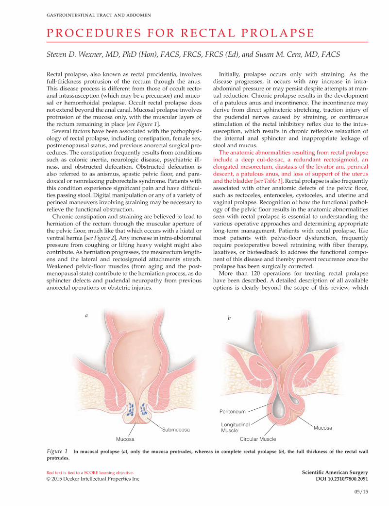
Scientific American Surgery DOI 10.2310/7800.2091 05/15 © 2015 Decker Intellectual Properties Inc gastrointestinal tract and abdomen PROCEDURES FOR RECTAL PROLAPSE Steven D. Wexner, MD, PhD (Hon), FACS, FRCS, FRCS (Ed), and Susan M. Cera, MD, FACS Rectal prolapse, also known as rectal procidentia, involves full-thickness protrusion of the rectum through the anus. This disease process is different from those of occult recto- anal intussusception (which may be a precursor) and muco- sal or hemorrhoidal prolapse. Occult rectal prolapse does not extend beyond the anal canal. Mucosal prolapse involves protrusion of the mucosa only, with the muscular layers of the rectum remaining in place [see Figure 1]. Several factors have been associated with the pathophysi- ology of rectal prolapse, including constipation, female sex, postmenopausal status, and previous anorectal surgical pro- cedures. The constipation frequently results from conditions such as colonic inertia, neurologic disease, psychiatric ill- ness, and obstructed defecation. Obstructed defecation is also referred to as anismus, spastic pelvic floor, and para- doxical or nonrelaxing puborectalis syndrome. Patients with this condition experience significant pain and have difficul- ties passing stool. Digital manipulation or any of a variety of perineal maneuvers involving straining may be necessary to relieve the functional obstruction. Chronic constipation and straining are believed to lead to herniation of the rectum through the muscular aperture of the pelvic floor, much like that which occurs with a hiatal or ventral hernia [see Figure 2]. Any increase in intra-abdominal pressure from coughing or lifting heavy weight might also contribute. As herniation progresses, the mesorectum length- ens and the lateral and rectosigmoid attachments stretch. Weakened pelvic-floor muscles (from aging and the post- menopausal state) contribute to the herniation process, as do sphincter defects and pudendal neuropathy from previous anorectal operations or obstetric injuries. Initially, prolapse occurs only with straining. As the disease progresses, it occurs with any increase in intra- abdominal pressure or may persist despite attempts at man- ual reduction. Chronic prolapse results in the development of a patulous anus and incontinence. The incontinence may derive from direct sphincteric stretching, traction injury of the pudendal nerves caused by straining, or continuous stimulation of the rectal inhibitory reflex due to the intus- susception, which results in chronic reflexive relaxation of the internal anal sphincter and inappropriate leakage of stool and mucus. The anatomic abnormalities resulting from rectal prolapse include a deep cul-de-sac, a redundant rectosigmoid, an elongated mesorectum, diastasis of the levator ani, perineal descent, a patulous anus, and loss of support of the uterus and the bladder [see Table 1]. Rectal prolapse is also frequently associated with other anatomic defects of the pelvic floor, such as rectoceles, enteroceles, cystoceles, and uterine and vaginal prolapse. Recognition of how the functional pathol- ogy of the pelvic floor results in the anatomic abnormalities seen with rectal prolapse is essential to understanding the various operative approaches and determining appropriate long-term management. Patients with rectal prolapse, like most patients with pelvic-floor dysfunction, frequently require postoperative bowel retraining with fiber therapy, laxatives, or biofeedback to address the functional compo- nent of this disease and thereby prevent recurrence once the prolapse has been surgically corrected. More than 120 operations for treating rectal prolapse have been described. A detailed description of all available options is clearly beyond the scope of this review, which Red text is tied to a SCORE learning objective. Mucosa Submucosa a Mucosa Circular Muscle Longitudinal Muscle Peritoneum b Figure 1 In mucosal prolapse (a), only the mucosa protrudes, whereas in complete rectal prolapse (b), the full thickness of the rectal wall protrudes.
-
Upload
khangminh22 -
Category
Documents
-
view
4 -
download
0
Transcript of Procedures for rectal ProlaPse
Scientific American Surgery DOI 10.2310/7800.2091
05/15
© 2015 Decker Intellectual Properties Inc
gastrointestinal tract and abdomen
P r o c e d u r e s f o r r e c ta l P r o l a P s e
Steven D. Wexner, MD, PhD (Hon), FACS, FRCS, FRCS (Ed), and Susan M. Cera, MD, FACS
rectal prolapse, also known as rectal procidentia, involves full-thickness protrusion of the rectum through the anus. this disease process is different from those of occult recto-anal intussusception (which may be a precursor) and muco-sal or hemorrhoidal prolapse. occult rectal prolapse does not extend beyond the anal canal. Mucosal prolapse involves protrusion of the mucosa only, with the muscular layers of the rectum remaining in place [see Figure 1].
several factors have been associated with the pathophysi-ology of rectal prolapse, including constipation, female sex, postmenopausal status, and previous anorectal surgical pro-cedures. the constipation frequently results from conditions such as colonic inertia, neurologic disease, psychiatric ill-ness, and obstructed defecation. obstructed defecation is also referred to as anismus, spastic pelvic floor, and para-doxical or nonrelaxing puborectalis syndrome. Patients with this condition experience significant pain and have difficul-ties passing stool. digital manipulation or any of a variety of perineal maneuvers involving straining may be necessary to relieve the functional obstruction.
chronic constipation and straining are believed to lead to herniation of the rectum through the muscular aperture of the pelvic floor, much like that which occurs with a hiatal or ventral hernia [see Figure 2]. any increase in intra-abdominal pressure from coughing or lifting heavy weight might also contribute. as herniation progresses, the mesorectum length-ens and the lateral and rectosigmoid attachments stretch. Weakened pelvic-floor muscles (from aging and the post-menopausal state) contribute to the herniation process, as do sphincter defects and pudendal neuropathy from previous anorectal operations or obstetric injuries.
Initially, prolapse occurs only with straining. as the disease progresses, it occurs with any increase in intra- abdominal pressure or may persist despite attempts at man-ual reduction. chronic prolapse results in the development of a patulous anus and incontinence. the incontinence may derive from direct sphincteric stretching, traction injury of the pudendal nerves caused by straining, or continuous stimulation of the rectal inhibitory reflex due to the intus-susception, which results in chronic reflexive relaxation of the internal anal sphincter and inappropriate leakage of stool and mucus.
the anatomic abnormalities resulting from rectal prolapse include a deep cul-de-sac, a redundant rectosigmoid, an elongated mesorectum, diastasis of the levator ani, perineal descent, a patulous anus, and loss of support of the uterus and the bladder [see Table 1]. rectal prolapse is also frequentl y associated with other anatomic defects of the pelvic floor, such as rectoceles, enteroceles, cystoceles, and uterine and vaginal prolapse. recognition of how the functional pathol-ogy of the pelvic floor results in the anatomic abnormalities seen with rectal prolapse is essential to understanding the various operative approaches and determining appropriate long-term management. Patients with rectal prolapse, like most patients with pelvic-floor dysfunction, frequently require postoperative bowel retraining with fiber therapy, laxatives, or biofeedback to address the functional compo-nent of this disease and thereby prevent recurrence once the prolapse has been surgically corrected.
More than 120 operations for treating rectal prolapse have been described. a detailed description of all available options is clearly beyond the scope of this review, which
red text is tied to a score learning objective.
Mucosa
Submucosa
a
Mucosa
Circular Muscle
LongitudinalMuscle
Peritoneum
b
Figure 1 In mucosal prolapse (a), only the mucosa protrudes, whereas in complete rectal prolapse (b), the full thickness of the rectal wall protrudes.

Scientific American Surgery
05/15
gastro procedures for rectal prolapse — 2
Preoperative Evaluation
Because of the protrusion of tissue through the anus and the frequent bloody discharge, pain, and pressure, and peri-anal skin excoriation, most patients initially mistake rectal prolapse for hemorrhoids [see Table 2]. these patients often present with a complaint of so-called “persistent hemor-rhoids” after a recent hemorrhoid operation or hemorrhoids that only reduce after sitting on a hard surface. similarly, many patients mistake incarcerated rectal prolapse for thrombosed hemorrhoids. consequently, a high level of suspicion and careful physical examination are required to differentiate hemorrhoids from rectal procidentia [see Table 3].
the diagnosis is most easily made with the patient strain-ing while seated on the toilet. Having the patient perform the Valsalva maneuver on the toilet in the clinical setting
will outline a few key widely accepted procedures. a number of regionally popular but not globally accepted procedures are excluded.
a b
c d
Figure 2 Rectal prolapse as a sliding hernia. (a) Invagination of the rectal wall during evacuation. (b) Progression of the invagination to recto-anal intususception. (c) The intussusception leads to sliding hernia of the rectum through the anal opening. (d) Full-thickness rectal prolapse with enterocele occurs when the small bowel prolapses along with the deepening cul-de-sac.
Table 1 anatomic abnormalities associated with rectal Prolapse
deep cul-de-sacredundant rectosigmoid colonelongated mesorectumdiastasis of levator aniPerineal descentHerniation of pelvic organs through pelvic funnelPatulous anusloss of support of uterus and bladder
Scientific American Surgery
05/15
gastro procedures for rectal prolapse — 3
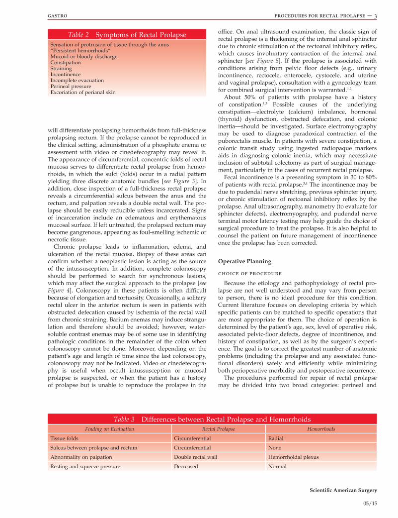
will differentiate prolapsing hemorrhoids from full-thickness prolapsing rectum. If the prolapse cannot be reproduced in the clinical setting, administration of a phosphate enema or assessment with video or cinedefecography may reveal it. the appearance of circumferential, concentric folds of rectal mucosa serves to differentiate rectal prolapse from hemor-rhoids, in which the sulci (folds) occur in a radial pattern yielding three discrete anatomic bundles [see Figure 3]. In addition, close inspection of a full-thickness rectal prolapse reveals a circumferential sulcus between the anus and the rectum, and palpation reveals a double rectal wall. the pro-lapse should be easily reducible unless incarcerated. signs of incarceration include an edematous and erythematous mucosal surface. If left untreated, the prolapsed rectum may become gangrenous, appearing as foul-smelling ischemic or necrotic tissue.
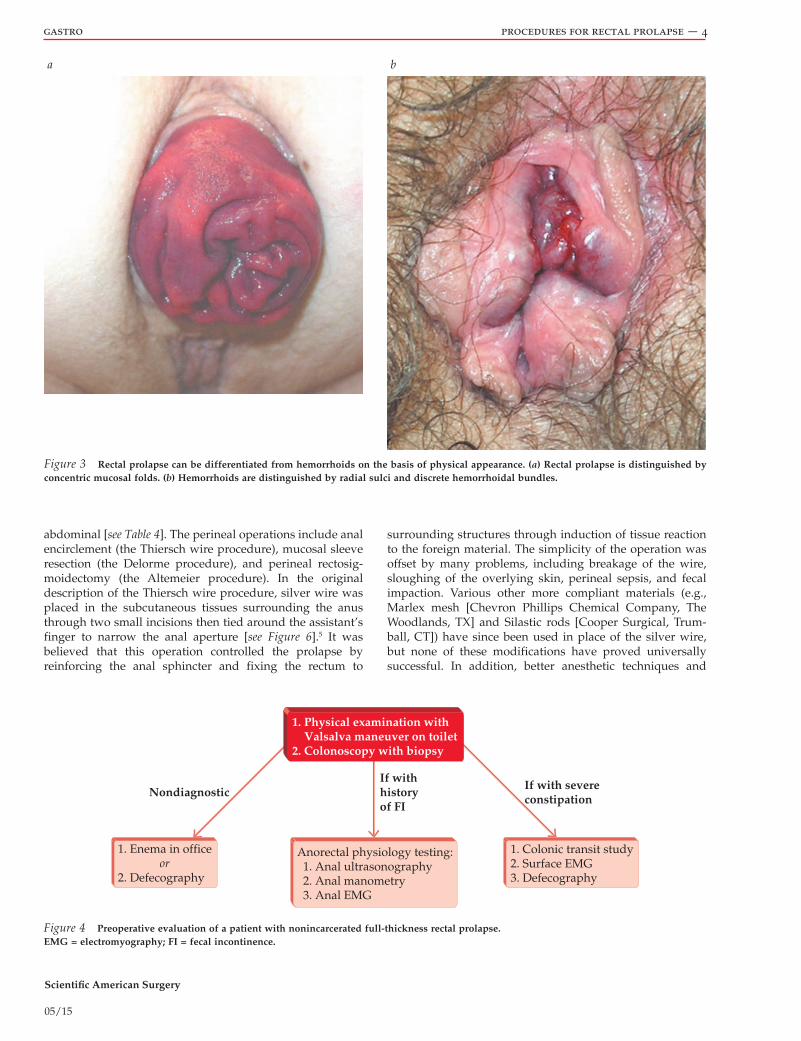
chronic prolapse leads to inflammation, edema, and ulceration of the rectal mucosa. Biopsy of these areas can confirm whether a neoplastic lesion is acting as the source of the intussusception. In addition, complete colonoscopy should be performed to search for synchronous lesions, which may affect the surgical approach to the prolapse [see Figure 4]. colonoscopy in these patients is often difficult because of elongation and tortuosity. occasionally, a solitary rectal ulcer in the anterior rectum is seen in patients with obstructed defecation caused by ischemia of the rectal wall from chronic straining. Barium enemas may induce strangu-lation and therefore should be avoided; however, water-soluble contrast enemas may be of some use in identifying pathologic conditions in the remainder of the colon when colonoscopy cannot be done. Moreover, depending on the patient’s age and length of time since the last colonoscopy, colonoscopy may not be indicated. Video or cinedefecogra-phy is useful when occult intussusception or mucosal prolapse is suspected, or when the patient has a history of prolapse but is unable to reproduce the prolapse in the
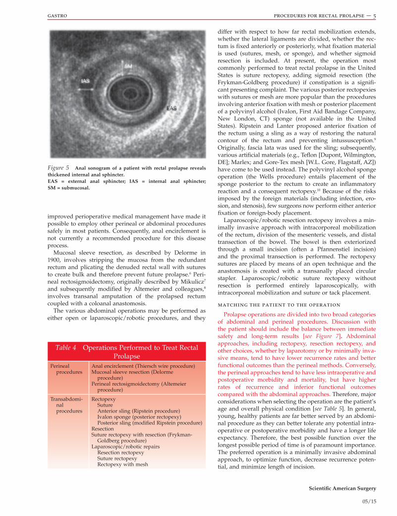
office. on anal ultrasound examination, the classic sign of rectal prolapse is a thickening of the internal anal sphincter due to chronic stimulation of the rectoanal inhibitory reflex, which causes involuntary contraction of the internal anal sphincter [see Figure 5]. If the prolapse is associated with conditions arising from pelvic floor defects (e.g., urinary incontinence, rectocele, enterocele, cystocele, and uterine and vaginal prolapse), consultation with a gynecology team for combined surgical intervention is warranted.1,2
about 50% of patients with prolapse have a history of constipation.1,3 Possible causes of the underlying constipation—electrolyte (calcium) imbalance, hormonal (thyroid) dysfunction, obstructed defecation, and colonic inertia—should be investigated. surface electromyography may be used to diagnose paradoxical contraction of the puborectalis muscle. In patients with severe constipation, a colonic transit study using ingested radiopaque markers aids in diagnosing colonic inertia, which may necessitate inclusion of subtotal colectomy as part of surgical manage-ment, particularly in the cases of recurrent rectal prolapse.
fecal incontinence is a presenting symptom in 30 to 80% of patients with rectal prolapse.3,4 the incontinence may be due to pudendal nerve stretching, previous sphincter injury, or chronic stimulation of rectoanal inhibitory reflex by the prolapse. anal ultrasonography, manometry (to evaluate for sphincter defects), electromyography, and pudendal nerve terminal motor latency testing may help guide the choice of surgical procedure to treat the prolapse. It is also helpful to counsel the patient on future management of incontinence once the prolapse has been corrected.
Operative Planning
choice of procedure
Because the etiology and pathophysiology of rectal pro-lapse are not well understood and may vary from person to person, there is no ideal procedure for this condition. current literature focuses on developing criteria by which specific patients can be matched to specific operations that are most appropriate for them. the choice of operation is determined by the patient’s age, sex, level of operative risk, associated pelvic-floor defects, degree of incontinence, and history of constipation, as well as by the surgeon’s experi-ence. the goal is to correct the greatest number of anatomic problems (including the prolapse and any associated func-tional disorders) safely and efficiently while minimizing both perioperative morbidity and postoperative recurrence.
the procedures performed for repair of rectal prolapse may be divided into two broad categories: perineal and
Table 2 symptoms of rectal Prolapsesensation of protrusion of tissue through the anus“Persistent hemorrhoids”Mucoid or bloody dischargeconstipationstrainingIncontinenceIncomplete evacuationPerineal pressureexcoriation of perianal skin
Table 3 differences between rectal Prolapse and HemorrhoidsFinding on Evaluation Rectal Prolapse Hemorrhoids
tissue folds circumferential radial
sulcus between prolapse and rectum circumferential None
abnormality on palpation double rectal wall Hemorrhoidal plexus
resting and squeeze pressure decreased Normal
Scientific American Surgery
05/15
gastro procedures for rectal prolapse — 4
abdominal [see Table 4]. the perineal operations include anal encirclement (the thiersch wire procedure), mucosal sleeve resection (the delorme procedure), and perineal rectosig-moidectomy (the altemeier procedure). In the original description of the thiersch wire procedure, silver wire was placed in the subcutaneous tissues surrounding the anus through two small incisions then tied around the assistant’s finger to narrow the anal aperture [see Figure 6].5 It was believed that this operation controlled the prolapse by reinforcing the anal sphincter and fixing the rectum to
surrounding structures through induction of tissue reaction to the foreign material. the simplicity of the operation was offset by many problems, including breakage of the wire, sloughing of the overlying skin, perineal sepsis, and fecal impaction. Various other more compliant materials (e.g., Marlex mesh [chevron Phillips chemical company, the Woodlands, tX] and silastic rods [cooper surgical, trum-ball, ct]) have since been used in place of the silver wire, but none of these modifications have proved universally successful. In addition, better anesthetic techniques and
a b
1. Physical examination with Valsalva maneuver on toilet2. Colonoscopy with biopsy
NondiagnosticIf withhistoryof FI
If with severeconstipation
1. Enema in office or2. Defecography
Anorectal physiology testing: 1. Anal ultrasonography 2. Anal manometry 3. Anal EMG
1. Colonic transit study2. Surface EMG3. Defecography
Figure 3 Rectal prolapse can be differentiated from hemorrhoids on the basis of physical appearance. (a) Rectal prolapse is distinguished by concentric mucosal folds. (b) Hemorrhoids are distinguished by radial sulci and discrete hemorrhoidal bundles.
Figure 4 Preoperative evaluation of a patient with nonincarcerated full-thickness rectal prolapse. EMG = electromyography; FI = fecal incontinence.
Scientific American Surgery
05/15
gastro procedures for rectal prolapse — 5
improved perioperative medical management have made it possible to employ other perineal or abdominal procedures safely in most patients. con sequently, anal encirclement is not currently a recommended procedure for this disease process.
Mucosal sleeve resection, as described by delorme in 1900, involves stripping the mucosa from the redundant rectum and plicating the denuded rectal wall with sutures to create bulk and therefore prevent future prolapse.6 Peri-neal rectosigmoidectomy, originally described by Mikulicz7 and subsequently modified by altemeier and colleagues,8 involves transanal amputation of the prolapsed rectum coupled with a coloanal anastomosis.
the various abdominal operations may be performed as either open or laparoscopic/robotic procedures, and they
differ with respect to how far rectal mobilization extends, whether the lateral ligaments are divided, whether the rec-tum is fixed anteriorly or posteriorly, what fixation material is used (sutures, mesh, or sponge), and whether sigmoid resection is included. at present, the operation most commonly performed to treat rectal prolapse in the united states is suture rectopexy, adding sigmoid resection (the frykman-Goldberg procedure) if constipation is a signifi-cant presenting complaint. the various posterior rectopexies with sutures or mesh are more popular than the procedures involving anterior fixation with mesh or posterior placement of a polyvinyl alcohol (Ivalon, first aid Bandage company, New london, ct) sponge (not available in the united states). ripstein and lanter proposed anterior fixation of the rectum using a sling as a way of restoring the natural contour of the rectum and preventing intussusception.9 originally, fascia lata was used for the sling; subsequently, various artificial materials (e.g., teflon [dupont, Wilmington, de]; Marlex; and Gore-tex mesh [W.l. Gore, flagstaff, aZ]) have come to be used instead. the polyvinyl alcohol sponge operation (the Wells procedure) entails placement of the sponge posterior to the rectum to create an inflammatory reaction and a consequent rectopexy.10 Because of the risks imposed by the foreign materials (including infection, ero-sion, and stenosis), few surgeons now perform either anterior fixation or foreign-body placement.
laparoscopic/robotic resection rectopexy involves a min-imally invasive approach with intracorporeal mobilization of the rectum, division of the mesenteric vessels, and distal transection of the bowel. the bowel is then exteriorized through a small incision (often a Pfannenstiel incision) and the proximal transection is performed. the rectopexy sutures are placed by means of an open technique and the anastomosis is created with a transanally placed circular stapler. laparoscopic/robotic suture rectopexy without resection is performed entirely laparoscopically, with intracorporeal mobilization and suture or tack placement.
matching the patient to the operation
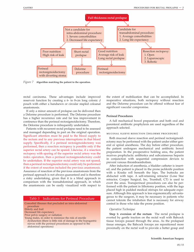
Prolapse operations are divided into two broad categories of abdominal and perineal procedures. discussion with the patient should include the balance between immediate safety and long-term results [see Figure 7]. abdominal approaches, including rectopexy, resection rectopexy, and other choices, whether by laparotomy or by minimally inva-sive means, tend to have lower recurrence rates and better functional outcomes than the perineal methods. conversely, the perineal approaches tend to have less intraoperative and postoperative morbidity and mortality, but have higher rates of recurrence and inferior functional outcomes compared with the abdominal approaches. therefore, major considerations when selecting the operation are the patient’s age and overall physical condition [see Table 5]. In general, young, healthy patients are far better served by an abdomi-nal procedure as they can better tolerate any potential intra-operative or postoperative morbidity and have a longer life expectancy. therefore, the best possible function over the longest possible period of time is of paramount importance. the preferred operation is a minimally invasive abdominal approach, to optimize function, decrease recurrence poten-tial, and minimize length of incision.
SM
EAS
IAS
Figure 5 Anal sonogram of a patient with rectal prolapse reveals thickened internal anal sphincter. EAS = external anal sphincter; IAS = internal anal sphincter; SM = submucosal.
Table 4 operations Performed to treat rectal Prolapse
Perineal procedures
anal encirclement (thiersch wire procedure)Mucosal sleeve resection (delorme
procedure)Perineal rectosigmoidectomy (altemeier
procedure)
transabdomi-nal procedures
rectopexysutureanterior sling (ripstein procedure)Ivalon sponge (posterior rectopexy)Posterior sling (modified ripstein procedure)
resectionsuture rectopexy with resection (frykman-
Goldberg procedure)laparoscopic/robotic repairs
resection rectopexysuture rectopexyrectopexy with mesh

Scientific American Surgery
05/15
gastro procedures for rectal prolapse — 6
levatorplasty is added, with the anticipation of improving continence. second, if sufficient redundancy exists, then a transperineal colonic J pouch with a coloanal anastomosis is created, with the aim of conferring the same advantages to the patient as those of transabdominal restorative proctec-tomy, a procedure that is typically reserved for patients with
elderly, frail patients, with their extensive comorbidities and decreased life expectancy, may best be served by a perineal approach to limit intra-abdominal trauma and facilitate recovery. the preferred perineal procedure is the perineal rectosigmoidectomy, with several possible adjuncts. first, if the levator muscles are identifiable, then a
Incisions Anus
a
b
c
d
Figure 6 Thiersch procedure for rectal prolapse. (inset) Patient placed in the prone jackknife position. (a and b) Wire is threaded around the anal sphincter muscle via two small incisions. (c) The wire is tightened, preventing the rectum from prolapsing. (d) Final procedure.
Scientific American Surgery
05/15
gastro procedures for rectal prolapse — 7
the extent of mobilization that can be accomplished. In reoperative situations, both rectopexy without resection and the delorme procedure can be offered without fear of significant vascular compromise.
Perineal Procedures
a full mechanical bowel preparation and both oral and parenteral antibiotic prophylaxis are used regardless of the approach selected.
mucosal sleeve resection (delorme procedure)
Both mucosal sleeve resection and perineal rectosigmoid-ectomy may be performed with the patient under either gen-eral or spinal anesthesia. the day before either procedure, the patient undergoes mechanical and antibiotic bowel preparation. In the preoperative holding area, the patient receives prophylactic antibiotics and subcutaneous heparin in conjunction with sequential compression devices to prevent venous thromboembolism.
after induction of anesthesia, a bladder catheter is insert-ed, and the patient is placed in the prone jackknife position with a Kraske roll beneath the hips. the buttocks are abducted with tape. a self-retaining retractor (lone star retractor, cooper surgical, Inc, trumball, ct) is used to evert the anus. transperineal procedures can also be per-formed with the patient in lithotomy position, with the legs placed high in padded medical stirrups for adequate expo-sure. although this approach is less ergonomically advanta-geous to the surgeon, it may be necessary in patients who cannot tolerate the intubation that is necessary for airway control in those who take the prone position.
Operative Technique
Step 1: eversion of the rectum the rectal prolapse is everted by gentle traction on the rectal wall with Babcock tissue forceps passed through the anus. as the prolapsed tissue emerges, the Babcock forceps are repositioned more proximally on the rectal wall to provide a better grasp and
rectal carcinoma. these advantages include improved reservoir function by creating a 6- to 8-cm long colonic J pouch with either a handsewn or circular stapled coloanal anastomosis.
If only a minor amount of prolapse can be delivered then a delorme procedure is performed. the delorme procedure has a higher recurrence rate and far less improvement in continence than the perineal rectosigmoidectomy. therefore, the delorme procedure is infrequently undertaken.
Patients with recurrent rectal prolapse need to be assessed and managed depending in part on the original operation. significant attention must be paid to the blood supply to the rectum and to any previous interruptions in that blood supply. specifically, if a perineal rectosigmoidectomy was performed, then a resection rectopexy is possible only if the superior rectal artery can be spared. likewise, if a resection rectopexy with sparing of the superior rectal artery was the index operation, then a perineal rectosigmoidectomy could be undertaken. If the superior rectal artery was not spared, then a perineal rectosigmoidectomy could only be performed if the extent of resection included the previous anastomosis. assurance of resection of the previous anastomosis from the perineal approach is not always guaranteed and is therefore a risky undertaking, given that it is a blind approach in comparison with the transabdominal approach, in which the anastomosis can be easily visualized with respect to
Full thickness rectal prolapse
Candidate for transabdominal procedure: 1. Average comorbidities 2. Long life expectancy
Not a candidate for intra-abdominal procedure: 1. Severe comorbidities 2. Decreased life expectancy
Resection rectopexy: 1. Open 2. Laparoscopic 3. Robotic
Poor nutritionHigh risk of leak
Perineal rectosigmoidectomy with diverting stoma
Perineal rectosigmoidectomy
Delorme procedure
Short rectal prolapse
Good nutritionAverage risk of leakLong rectal prolapse
Figure 7 Algorithm matching the patient to the operation.
Table 5 Indications for Perineal Procedurecomorbid illnesses that precluded an intra-abdominal
procedureelderly and fraildecreased life expectancyPrior pelvic surgery or radiationYoung males, in order to minimize the risk of erectile
dysfunction (there is little risk of damage to the hypogastric nerves with the perineal procedures as opposed to the abdominal operations)

Scientific American Surgery
05/15
gastro procedures for rectal prolapse — 8
facilitate delivery of the prolapse through the anus and into the operative field.
Step 2: submucosal injection of anesthetic once the rectum has been everted, a local anesthetic solution contain-ing 0.25% bupivacaine, 0.5% lidocaine, and epinephrine (in a 1:400,000 dilution) is circumferentially injected 1 to 1.5 cm above the dentate line to minimize bleeding.
Step 3: circumferential incision of mucosa the rectal mucosa is incised at this level with electrocautery, and four clamps are placed on the proximal mucosal edge for traction [see Figure 8]. care is taken to ensure deep mucosal incision that does not extend into muscle layer.
Step 4: dissection of mucosa from muscle Gentle trac-tion is placed on the clamps, and the mucosa is dissected away from the muscle with electrocautery. a finger is placed inside the muscular tube to facilitate traction and help prevent full-thickness injury. resection of the mucosal sleeve
is continued until resistance prevents further dissection. a tube of redundant muscular tissue then remains.
Step 5: plication of rectal muscle the muscular tube is plicated by placing eight 2-0 polyglactin (Vicryl, ethicon, somerville, NJ) reefing sutures circumferentially in the wall.
Step 6: resection of mucosa and anastomosis the excess mucosa is transected in a superior-to-inferior direc-tion and the two cut edges of mucosa are approximated with 2-0 polyglactin sutures. transection is continued on one side for a quarter of the circumference, at which point a second suture is placed. With traction applied to these two sutures, two additional sutures are placed at 90° intervals to establish four quadrants. transection and anastomosis are serially performed in each of the four quadrants until the mucosa is completely excised and the mucosal anastomosis has been completed. With the removal of the retractor, the anastomo-sis should spontaneously reduce into its anatomic position.
a b
c d
Figure 8 Mucosal sleeve resection. (a) With the rectum everted, the mucosa is incised and dissected away from the muscular tube. (b) The muscular tube is plicated with sutures to form a muscular pessary. (c) A mucosa-to-mucosa anastomosis is fashioned. (d) The anastomosis spontaneously reduces into its anatomic position.
Scientific American Surgery
05/15
gastro procedures for rectal prolapse — 9
more of the prolapse into the operative field. division of the mesorectum is continued close to the bowel wall until no more bowel can be delivered. during this phase, the sliding hernia of peritoneum (cul-de-sac) anterior to the rectum (on the inferior aspect of the prolapse with the patient in a prone position) may be opened to allow palpation of the intraper-itoneal contents and determination of whether the colon is straight in the pelvis. to perform this maneuver, a finger or occasionally a laparoscopic camera is inserted into the pelvis alongside the rectum to assess the redundancy of or the ten-sion on the remaining rectum and sigmoid. redundancy is assessed by grasping the sigmoid colon above the current area of dissection and attempting to prolapse it through the anal opening. alternatively, a laparoscopic telescope and camera placed next to the rectum through the perineal incision can be used to evaluate redundancy. If redundancy is still encoun-tered, more mesorectum is divided to allow further mobiliza-tion of the bowel through the anus. careful control of the mesentery with ligatures is advised to prevent retraction of bleeding vessels into the pelvis. one of the newer energy sources (bipolar or ultrasonic) may facilitate the dissection.
Step 5: ligation of the hernia sac once the redundant rectum and sigmoid have been adequately mobilized, the hernia sac of peritoneum can be sutured closed. care is taken to resect any redundant hernia sac to prevent future anterior intussusception. this step is similar to the high ligation performed for other types of hernias.
Step 6: levatorplasty levatorplasty is performed to restore the appropriate angles of the pelvic floor muscles, to aid in treatment of incontinence, and to narrow the aperture through which herniation occurs and thereby prevent recur-rence. It can be performed anterior to the rectum, posterior to the rectum, or both. a narrow retractor is employed to expose the outer tube of rectum and uncover the levator muscles. Interrupted 2-0 polyproplyene (Prolene, ethicon, sommerville, NJ) sutures are placed through the levator muscles on each side and secured loosel y enough to allow a finger to be inserted alongside the rectum.
Step 7: proximal transection of the rectum and anasto-mosis the point at which the redundant rectum is to be transected is identified at the level at which the mesorectum is divided. the bowel wall is circumferentially cleared of appendages in preparation for transection and anastomosis. transection begins by dividing a small area of the bowel wall superiorly, and placing a 2-0 polyglactin suture through the edge of the outer tube of rectum and the newly tran-sected proximal edge of the bowel. transection is continued inferiorly on one side for a quarter of the circumference, and a second suture is placed. With traction on these two sutures, two additional sutures are placed around the remaining circumference to mark four quadrants. transec-tion and anastomosis are serially performed in each of the four quadrants until the rectum is completely transected. When the anastomosis is complete, it retracts into the pelvis, where it may be inspecte d with a bivalve retractor. When possible, a transperineal colonic J pouch can be created. In these instances, a sufficient length of well-vascularized redundant colon is left to fold back onto itself. a 6- to 8-cm colonic J pouch is created with a linear cutting stapler. the
Postoperative Care
Because the patients are often elderly, 1 to 3 days of observation with intravenous antibiotics may be indicated. the bladder catheter is removed on the following morning and the patient is advanced to a regular diet as soon as he or she can tolerate it. the patient is sent home on a regimen of fiber supplementation, 7 days of oral antibiotics, and sitz baths as soon as medical stability is ensured and appropriate social circumstances are arranged.
Troubleshooting
the delorme procedure prevents rectal intussusception by resecting redundant mucosa and by removing laxity in the rectal wall through plication of the muscular redun-dancy. the key to success is to continue the sleeve resection until some resistance is met and the dissection cannot proceed further. sometimes, the anterior wall is longer than the posterior wall or vice versa, but such discrepancies should not affect the repair. the anastomosis is performed one quadrant at a time to prevent retraction of the transected mucosa into the proximal bowel.
perineal rectosigmoidectomy
Operative Technique
anesthesia and positioning are the same for perineal rectosigmoidectomy as for mucosal sleeve resection [see Mucosal sleeve resection (delorme Procedure), above].
Steps 1 and 2 steps 1 and 2 of this procedure are the same as steps 1 and 2 of mucosal sleeve resection [see Muco-sal sleeve resection (delorme Procedure), operative tech-nique, above].
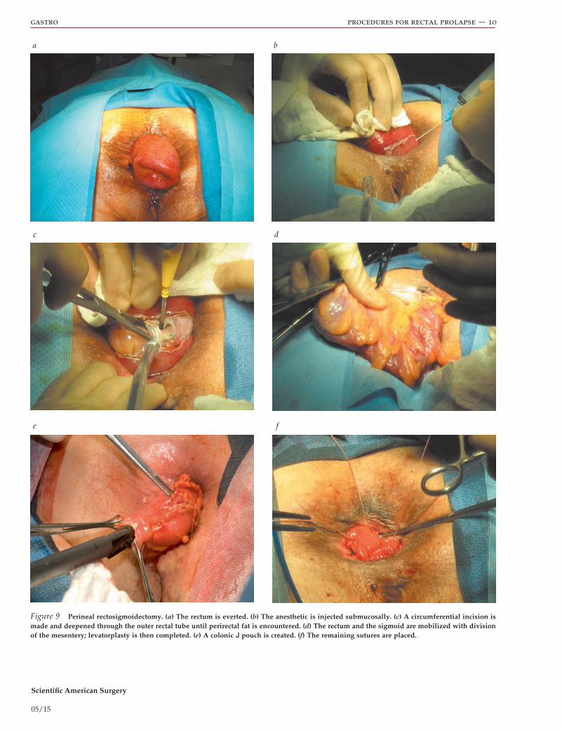
Step 3: circumferential incision through the rectal wall With the rectum everted, the prolapse consists of two tubes of rectal wall, with the inner tube consisting of rectum attached to the sigmoid and the outer tube consisting of rectum attached to the dentate line [see Table 6]. the mucosa is circumferentially scored with electrocautery 1 to 1.5 cm cephalad to the dentate line [see Figure 9]. the incision is deepened through all layers of the outer rectal tube until perirectal fat is encountered and the mesorectum is identi-fied on the dorsal aspect of the prolapse (at the superior por-tion of the prolapse when the patient is in a prone position).
Step 4: mobilization of the rectum and division of the mesentery rectal mobilization is accomplished by clamp-ing, ligating, and dividing the vessels of the mesorectum. as the mesorectum is divided, tension on the rectum delivers
Table 6 steps of the Perineal rectosigmoidectomy
1. Placement of the lone star retractor2. eversion of the rectum3. circumferential incision through the outer rectal wall4. ligation and division of mesenteric vessels5. Palpate sigmoid colon through open hernia sac to ensure no redundancy6. levatoroplasty7. transect second rectal wall one quadrant at a time8. Handsewn anastomosis
Scientific American Surgery
05/15
gastro procedures for rectal prolapse — 10
a b
c d
e f
Figure 9 Perineal rectosigmoidectomy. (a) The rectum is everted. (b) The anesthetic is injected submucosally. (c) A circumferential incision is made and deepened through the outer rectal tube until perirectal fat is encountered. (d) The rectum and the sigmoid are mobilized with division of the mesentery; levatorplasty is then completed. (e) A colonic J pouch is created. (f) The remaining sutures are placed.
Scientific American Surgery
05/15
gastro procedures for rectal prolapse — 11
physical examination and computed tomography (ct) imag-ing. In stable patients, it is treated with bowel rest, antibiotics, or drainage (either by ct guidance or in the operating room [or]). In septic and unstable patients, anastomotic dehiscence is treated intraoperatively, the patient in lithotomy position, with a combination of drain placement, debridement, and stoma formation. a laparoscopic loop ileostomy is the ideal approach in this setting, as simple diversion (as opposed to open pelvic exploration) in the setting of adequate drainage is all that is usually necessary in these situations.
early postoperative bleeding from the mucosal edges or, in the case of perineal rectosigmoidectomy, from the presa-cral space may be observed. late postoperative bleeding may result from tearing of the sutures through the mucosa or from separation of the anastomosis. stable patients can be conservatively managed with supportive care and transfusion. all significant bleeding should be evaluated and controlled in the or, with the source of the bleeding dictating the type of repair required. In particular, presacral bleeding may require open laparotomy to ensure adequate assessment and management of the bleeding source, as transperineal evalu-ation may limit identification of a mesenteric bleeding source.
long-term complications include anastomotic strictures, which may occur after both mucosal sleeve resection and perineal rectosigmoidectomy. they are treated with serial dilations either in the office or the or.
outcome evaluation
the advantages and disadvantages of the perineal proce-dures are listed here [see Table 7]. reported recurrence rates for the perineal approaches range from 5 to 21%11–13 and are higher than those for the abdominal approaches; however, the perineal repairs can be performed multiple times in the same patient as necessary.14 Because they are less invasive, perineal procedures generally carry a lower morbidity than abdominal procedures, with the majority of the complica-tions being medical in nature.11,12 Whereas constipation is neither exacerbated nor alleviated by the perineal proce-dures, continence is usually improved, although not as much as it is improved by the abdominal procedures.15,16 the improvement in continence seen after both abdominal and perineal procedures for rectal prolapse is related to cessation of rectoanal inhibition and recovery of sphincter function with reduction of the prolapse. the lesser improvement reported after perineal procedures may be related to sphinc-ter stretching or, in the case of perineal rectosigmoidectomy, to loss of the rectal reservoir. a comparison of various perineal procedures—including the delorme procedure, perineal rectosigmoidectomy, and rectosigmoidectomy with levatorplasty—found that the addition of a levatorplasty yielded the greatest improvement in continence, the least morbidity, and the lowest recurrence rate.16
afferent limb is sutured to the efferent limb with 3-0 polydioxanone (Pds, ethicon, sommerville, NJ) sutures. the anastomosis, whether straight, coloanal, or pouch anal, can be created with a circular stapler if the diameters of the two lumens are relatively similar. In this case, a # 0 polypropyl-ene suture is placed in the proximal bowel at the apex of the colonic J pouch. the anvil is inserted and the purse-string suture is secured. the purse-string suture is placed in the distal edge of mucosa, and after securing the anvil to the trocar of the circular stapler, a second purse-string suture is secured. the stapled anastomosis is completed. However, if significant discrepancy in the diameters of the two lumens exists, then a handsewn anastomosis is preferable.
Postoperative Care
same as that of the mucosal sleeve resection [see Mucosal sleeve resection (delorme Procedure), Postoperative care, above].
Troubleshooting
Perineal rectosigmoidectomy involves a combination of repairs of anatomic abnormalities associated with rectal pro-lapse. rectal mobilization yields a rectopexy from scarring; resection removes redundant bowel; ligation of the entero-cele obliterates the hernia sac; and levatorplasty provides reconstruction of the pelvic floor. each step requires that attention be paid to appropriate planes of dissection and that meticulous hemostasis be maintained. for example, transection of the outer rectal wall may lead to inadvertent division of the mesentery before vascular control is obtained; if this occurs, traction should be placed on the inner wall to expose the proximal mesentery and allow the surgeon to regain vascular control. In addition, transection of the outer rectal wall may lead to inadvertent simultaneous transection of both walls; if this occurs, clamps should be placed on the inner wall to prevent retraction into the pelvis and to facili-tate rectal mobilization and division of the mesorectum [see Perineal rectosigmoidectomy, operative technique, above].
to help prevent recurrence, all redundant bowel should be resected once mobilization of the rectum and division of the mesentery are complete. If, at the start of the procedure, only a short segment of prolapse is produced or the patient is found to have only a mucosal prolapse, a delorme operation may be performed instead.
Complications
Partial separation (dehiscence) of the mucosal anastomosis after mucosal sleeve resection is not uncommon and usually does not warrant intervention. after perineal rectosigmoid resection, anastomotic dehiscence may lead to leakage and pelvic sepsis, which is diagnosed using a combination of
Table 7 advantages and disadvantages of the Perineal ProceduresAdvantages Disadvantages
the option to use spinal anesthesiaavoidance of peritoneal adhesionsshort hospital stays (1 to 4 days)a lower risk of injury to the pelvic nerves, reduced painthe opportunity for concomitant repair of other anorectal problems (e.g.,
sphincter defects, hemorrhoids, rectoceles, cystoceles, and vaginal prolapse)
a higher recurrence rate reduced improvement of any fecal incontinence
because of the loss of reservoir function stemming from removal of the rectum.

Scientific American Surgery
05/15
gastro procedures for rectal prolapse — 12
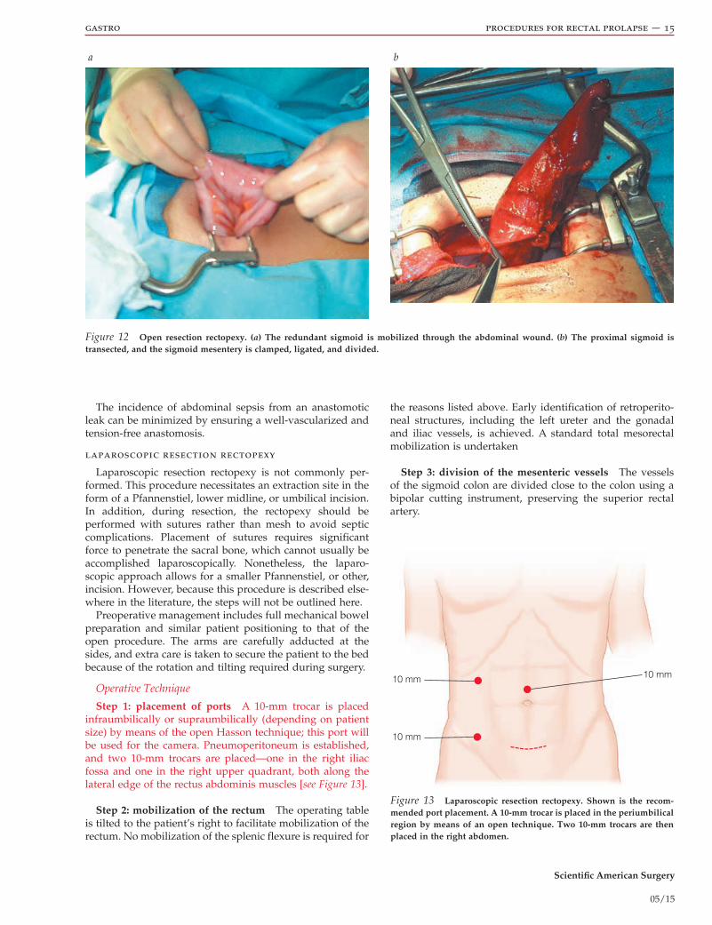
sigmoid colon is redundant in patients with rectal prolapse, no mobilization is performed in the sigmoid region, as acquir-ing length to perform a tension-free anastomosis is easily accomplished. for the same reasons, the splenic flexure is not mobilized. the gonadal vessels and the ureter are identified and swept posteriorly. the peritoneal incision is continued to the left of the rectum, curving anteriorly in the rectouterine or rectovesical sulcus. the peritoneum overlying the medial aspect of the upper rectum on the right is also incised, and this incision is continued to the right of the rectum to unite with the previous incision at the anterior rectum.
Step 3: proximal transection of the sigmoid colon and placement of a stapler anvil the proximal point of transection is chosen by finding an area of colon that easily falls into the pelvis and eliminates redundancy. this area of colon is circumferentially cleaned of surrounding tissue and divided using electrocautery. three Babcock forceps are placed to hold open the lumen. a # 0 polypropylene purse-string suture is put in place, and the head of a 33-mm circular stapler is secured in the lumen.
Step 4: division of the sigmoid mesentery the mesen-tery of the sigmoid colon is divided with an energy source close to the colon [see Figure 12b]. the inferior mesenteric and superior rectal vessels are carefully preserved.
Step 5: mobilization of the distal rectum and division of the lateral ligaments a Babcock clamp is placed on the sigmoid stump and lifted upward. the avascular plane of areolar tissue between the mesorectum and the presacral fascia is identified and divided with electrocautery. a st. Marks retractor is placed behind the rectum to provide trac-tion, then advanced with dissection distally along the rec-tum to the level of the coccyx. again the superior rectal ar-tery is preserved to avoid necrosis of the rectum.
dissection of the right side of the rectum is performed with the surgeon standing to the patient’s left. the left hand places traction on the rectum while the right hand uses electrocautery to divide the entire lateral stalk in a posterior-to-anterior direction down to the level of the levators. the st. Marks retractor is used to retract the tissues of the sidewall away from the rectum.
dissection of the left side of the rectum is performed with the surgeon on the patient’s right. again, the left hand plac-es traction on the rectum while the right hand performs the dissection in a posterior-to-anterior direction. dissection anterior to the rectum is also routinely performed.
the controversy related to this step is discussed later in this review [see troubleshooting, below].
Step 6: placement of sutures for rectopexy, with upward traction applied to the rectal stump Horizontal mattress 2-0 polypropylene sutures using a large heavy needle (ct-2) are placed in preparation for the rectopexy. starting on one side, a suture is placed through the peritoneum and the endopelvic fascia adjacent to the rectum, with care taken not to penetrate the rectal wall. the suture is guided through the presacral fascia and the periosteum to the side of the midline approximately 1 to 4 cm below the level of the sacral prom-ontory. a significant amount of force is usually needed to
as already mentioned in this review, a transperineal colonic J pouch is added whenever feasible. Because the incidence of rectal prolapse peaks in the sixth and seventh decades of life, patients undergoing these procedures are usually elderly and have significant comorbid conditions.17 the perineal procedures are economically and physiologi-cally advantageous in the short term and are therefore ideal for elderly patients or for any patients with multiple comor-bid conditions, as well as for those who are at high operative risk or who need combined intervention from various pelvic surgical specialists. In these patients, who have generally limited life expectancies, the high risk of recurrence associ-ated with perineal procedures may be irrelevant. Perineal operations may also be indicated for patients who have undergone multiple previous abdominal operations and are likely to have dense adhesions, as well as for young men who do not wish to risk impairment of sexual function.
Abdominal Procedures
resection rectopexy (frykman-goldberg procedure)
Just as with perineal procedures, patients undergo mechanical and antibiotic (oral) bowel preparation on the day before surgery. Just before the procedure, parenteral antibiotics and subcutaneous heparin are administered, and sequential compression devices are placed on the legs.
after induction of general anesthesia, the patient is placed in the modified lithotomy position, with the legs in padded stirrups. If there is a history of one or more pelvic opera-tions, or any other concern about potential ureteric injury, bilateral ureteral stents are placed by a urologist, and a urinary catheter is always inserted.
Operative Technique
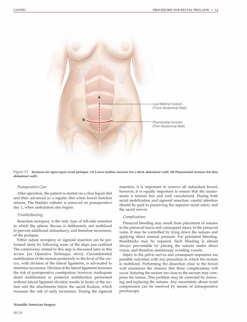
Step 1: initial incision and exploration a low midline (thick abdominal wall) or Pfannenstiel (thin abdominal wall) incision is made, the pelvis is explored, a Balfour or Buch-walter retractor is placed, and the small bowel is packed into the upper abdomen [see Table 8, Figure 10, and Figure 11].
Step 2: mobilization of the upper rectum the upper rec-tum is mobilized away from the left lateral wall by incising the lateral peritoneal reflection [see Figure 12a]. Because the
Table 8 steps of open resection rectopexy with Preservation of the superior rectal artery
1. Pfannenstiel incision 2. Mobilization of the rectum circumferentially to the level of
the levators 3. division of proximal sigmoid in an area that will easily
reach the pelvis 4. Place anvil into the proximal bowel 5. divide sigmoidal vessels close to the colon preserving the
superior rectal artery to the top of the rectum 6. retract the sigmoid colon upward to place rectopexy sutures
on either side of the rectum into the periosteum of the sacrum but do not tie them down
7. divide top of the rectum with a curved cutter stapler 8. create circular stapled anastomosis 9. air leak test10. secure rectopexy sutures
Scientific American Surgery
05/15
gastro procedures for rectal prolapse — 13
penetrate the sacral bone to ensure stability of the sutures placed. It is then completed by passing the needle back through both the peritoneum and the endopelvic fascia. one or two sutures are placed on each side; they are left untied and are tagged with hemostats.
Step 7: transection of bowel at the upper rectum the rectosigmoid junction is identified on the basis of the splay-ing of the taeniae coli, the absence of appendices epiploicae, and the proximity to the sacral promontory. Great care is taken at this point to ensure integrity of the superior rectal artery. this junction marks the site of distal transection. the distal end (proximal rectum) can be prepared with a hand-sewn # 0 polyproplyene suture, a purse-string clamp with a # 0 polypropylene or as a double stapled anastomosis.
Step 8: completion of colorectal anastomosis the circu-lar stapler is advanced through the anus to the rectal stump. under the manual and visual guidance of the abdominal and perineal operating surgeons, the trocar is advanced. the anvil is engaged on the trocar; the surgeon confirms that there is no inclusion of extraneous tissue and no rotation of the bowel or its mesentery, and the stapler is closed. the locations of the ureters and the vagina are reconfirmed to ensure that these structures are not incorporated into the
staple line. the stapler is then fired and gently removed, and the “doughnuts” are checked for integrity. the anastomosis is tested for leaks by filling the pelvis with an irrigating agent, clamping the colon proximal to the anastomosis, and insufflating air into the rectum. air bubbles from the anastomosis indicate a leak that requires suture reinforce-ment or anastomosis reconstruction. the rectopexy sutures are secured snugly to complete the procedure, after which flexible sigmoidoscopy is performed to exclude narrowing of the rectum from the rectopexy sutures. If any narrowing is noted one or more can be removed and replaced as neede d so as to prevent narrowing. ultimately, the abdomen is irrigated and closed in the usual fashion.
an alternative approach to this step is to perform endo-scopic visualization of the anastomosis while simultane-ously insufflating air through either the rigid proctoscope or the flexible sigmoidoscope. this method allows reliable confirmation of mucosal viability and anastomotic integrity. Moreover, if any supplemental anastomotic reinforcement sutures are needed, they can be placed much more easily at this time than after the rectopexy sutures have been secured. Sheets of sodium hyaluronate–based bioresorbable mem-brane are placed before closure of the fascia to help mini-mize postoperative adhesion formation.18 the abdominal incision is closed in the usual fashion.
Inferior Mesenteric Artery
Sigmoid Arteries DividedClose to Colon
Splaying of Teniae
Division for Proximal Transection
Redundant Sigmoid Colon
Stapler
Bladder
Uterus
Sutures
Sacral Promontory
Superior Rectal Artery(Spared)
D
E
A
B
C
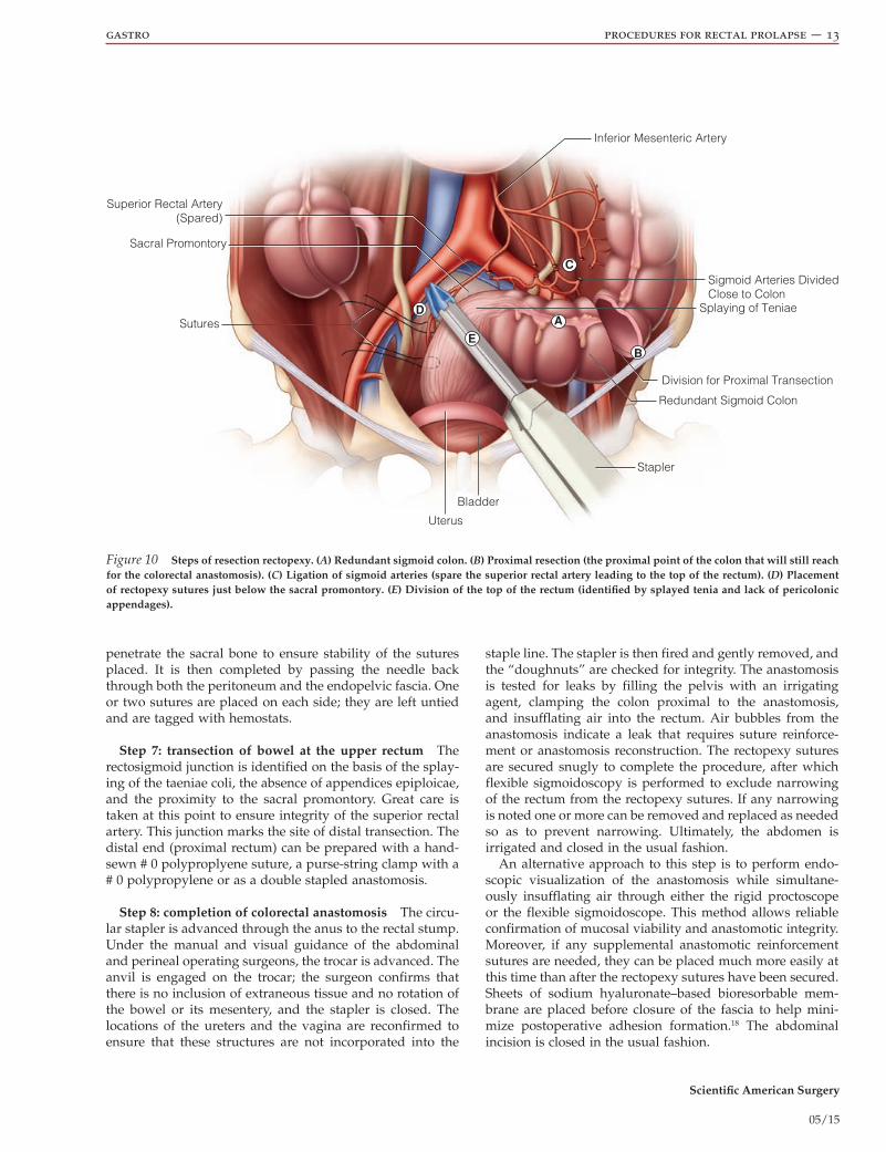
Figure 10 Steps of resection rectopexy. (a) Redundant sigmoid colon. (b) Proximal resection (the proximal point of the colon that will still reach for the colorectal anastomosis). (c) Ligation of sigmoid arteries (spare the superior rectal artery leading to the top of the rectum). (d) Placement of rectopexy sutures just below the sacral promontory. (e) Division of the top of the rectum (identified by splayed tenia and lack of pericolonic appendages).
Scientific American Surgery
05/15
gastro procedures for rectal prolapse — 14
Low Midline Incision(Thick Abdominal Wall)
Pfannenstiel Incision(Thin Abdominal Wall)
B
A
Figure 11 Incisions for open-repair rectal prolapse. (a) Lower midline incision (for a thick abdominal wall). (b) Pfannenstiel incision (for thin abdominal wall).
Postoperative Care
after operation, the patient is started on a clear liquid diet and then advanced to a regular diet when bowel function returns. the bladder catheter is removed on postoperative day 1, when ambulation also begins.
Troubleshooting
resection rectopexy is the only type of left-side resection in which the splenic flexure is deliberately not mobilized to prevent additional redundancy, and therefore recurrence, of the prolapse.
either suture rectopexy or sigmoid resection can be per-formed alone by following some of the steps just outlined the controversy related to this step is discussed later in this review [see operative technique, above]. circumferential mobilization of the rectum posteriorly to the level of the coc-cyx, with division of the lateral ligaments, is advocated to minimize recurrence. division of the lateral ligaments increases the risk of postoperative constipation; however, inadequate distal mobilization or posterior mobilization performed without lateral ligament division results in laxity of the rec-tum and the attachments below the sacral fixation, which increases the risk of early recurrence. during the sigmoid
resection, it is important to remove all redundant bowel; however, it is equally important to ensure that the anasto-mosis is tension free and well vascularized. during both rectal mobilization and sigmoid resection, careful attention should be paid to preserving the superior rectal artery and the sacral nerves.
Complications
Presacral bleeding may result from placement of sutures in the presacral fascia and consequent injury to the presacral veins. It may be controlled by tying down the sutures and applying direct manual pressure. for persistent bleeding, thumbtacks may be required. such bleeding is almost always preventable by placing the sutures under direct vision, and therefore assiduously avoiding vessels.
Injury to the pelvic nerves and consequent impotence are possible outcomes with any procedure in which the rectum is mobilized. Performing the dissection close to the bowel wall minimizes the chances that these complications will occur. suturing the rectum too close to the sacrum may com-press the lumen. this problem may be corrected by remov-ing and replacing the sutures. any uncertainty about rectal compression can be resolved by means of intraoperative proctoscopy.
Scientific American Surgery
05/15
gastro procedures for rectal prolapse — 15
a b
Figure 12 Open resection rectopexy. (a) The redundant sigmoid is mobilized through the abdominal wound. (b) The proximal sigmoid is transected, and the sigmoid mesentery is clamped, ligated, and divided.
the incidence of abdominal sepsis from an anastomotic leak can be minimized by ensuring a well-vascularized and tension-free anastomosis.
laparoscopic resection rectopexy
laparoscopic resection rectopexy is not commonly per-formed. this procedure necessitates an extraction site in the form of a Pfannenstiel, lower midline, or umbilical incision. In addition, during resection, the rectopexy should be performed with sutures rather than mesh to avoid septic complications. Placement of sutures requires significant force to penetrate the sacral bone, which cannot usually be accomplished laparoscopically. Nonetheless, the laparo-scopic approach allows for a smaller Pfannenstiel, or other, incision. However, because this procedure is described else-where in the literature, the steps will not be outlined here.
Preoperative management includes full mechanical bowel preparation and similar patient positioning to that of the open procedure. the arms are carefully adducted at the sides, and extra care is taken to secure the patient to the bed because of the rotation and tilting required during surgery.
Operative Technique
Step 1: placement of ports a 10-mm trocar is placed infraumbilically or supraumbilically (depending on patient size) by means of the open Hasson technique; this port will be used for the camera. Pneumoperitoneum is established, and two 10-mm trocars are placed—one in the right iliac fossa and one in the right upper quadrant, both along the lateral edge of the rectus abdominis muscles [see Figure 13].
Step 2: mobilization of the rectum the operating table is tilted to the patient’s right to facilitate mobilization of the rectum. No mobilization of the splenic flexure is required for
the reasons listed above. early identification of retroperito-neal structures, including the left ureter and the gonadal and iliac vessels, is achieved. a standard total mesorectal mobilization is undertaken
Step 3: division of the mesenteric vessels the vessels of the sigmoid colon are divided close to the colon using a bipolar cutting instrument, preserving the superior rectal artery.
10 mm10 mm
10 mm
Figure 13 Laparoscopic resection rectopexy. Shown is the recom-mended port placement. A 10-mm trocar is placed in the periumbilical region by means of an open technique. Two 10-mm trocars are then placed in the right abdomen.
Scientific American Surgery
05/15
gastro procedures for rectal prolapse — 16
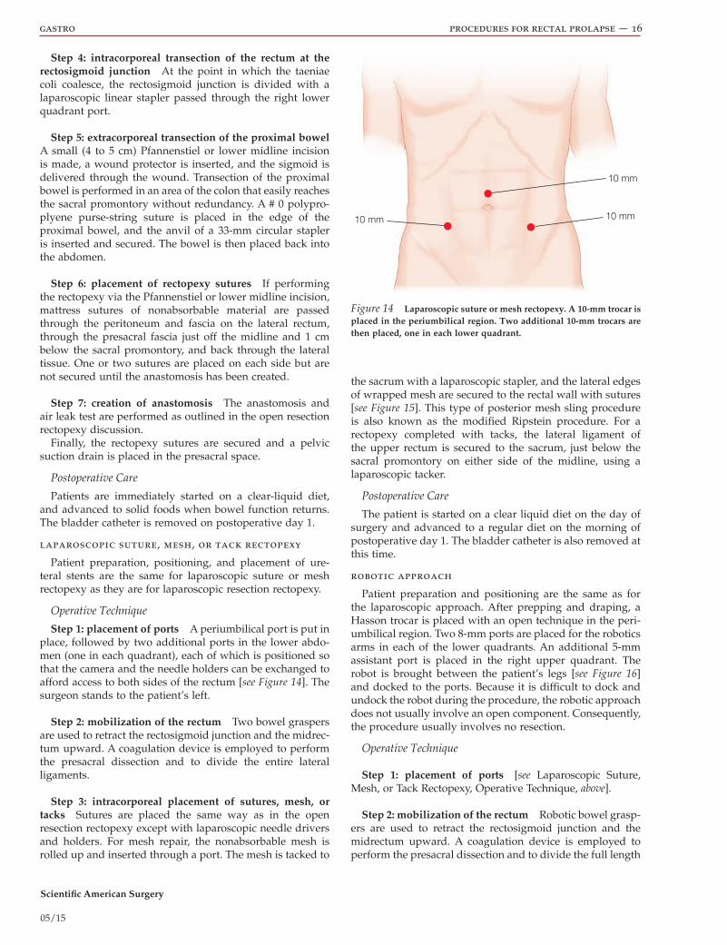
Step 4: intracorporeal transection of the rectum at the rectosigmoid junction at the point in which the taeniae coli coalesce, the rectosigmoid junction is divided with a laparoscopic linear stapler passed through the right lower quadrant port.
Step 5: extracorporeal transection of the proximal bowel a small (4 to 5 cm) Pfannenstiel or lower midline incision is made, a wound protector is inserted, and the sigmoid is delivered through the wound. transection of the proximal bowel is performed in an area of the colon that easily reache s the sacral promontory without redundancy. a # 0 polypro-plyene purse-string suture is placed in the edge of the proximal bowel, and the anvil of a 33-mm circular stapler is inserted and secured. the bowel is then placed back into the abdomen.
Step 6: placement of rectopexy sutures If performing the rectopexy via the Pfannenstiel or lower midline incision, mattress sutures of nonabsorbable material are passed through the peritoneum and fascia on the lateral rectum, through the presacral fascia just off the midline and 1 cm below the sacral promontory, and back through the lateral tissue. one or two sutures are placed on each side but are not secured until the anastomosis has been created.
Step 7: creation of anastomosis the anastomosis and air leak test are performed as outlined in the open resection rectopexy discussion.
finally, the rectopexy sutures are secured and a pelvic suction drain is placed in the presacral space.
Postoperative Care
Patients are immediately started on a clear-liquid diet, and advanced to solid foods when bowel function returns. the bladder catheter is removed on postoperative day 1.
laparoscopic suture, mesh, or tack rectopexy
Patient preparation, positioning, and placement of ure-teral stents are the same for laparoscopic suture or mesh rectopexy as they are for laparoscopic resection rectopexy.
Operative Technique
Step 1: placement of ports a periumbilical port is put in place, followed by two additional ports in the lower abdo-men (one in each quadrant), each of which is positioned so that the camera and the needle holders can be exchanged to afford access to both sides of the rectum [see Figure 14]. the surgeon stands to the patient’s left.
Step 2: mobilization of the rectum two bowel graspers are used to retract the rectosigmoid junction and the midrec-tum upward. a coagulation device is employed to perform the presacral dissection and to divide the entire lateral ligaments.
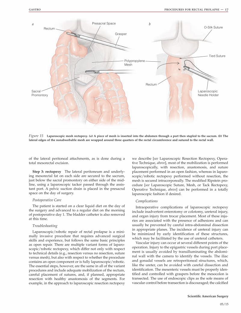
Step 3: intracorporeal placement of sutures, mesh, or tacks sutures are placed the same way as in the open resection rectopexy except with laparoscopic needle drivers and holders. for mesh repair, the nonabsorbable mesh is rolled up and inserted through a port. the mesh is tacked to
the sacrum with a laparoscopic stapler, and the lateral edges of wrapped mesh are secured to the rectal wall with sutures [see Figure 15]. this type of posterior mesh sling procedure is also known as the modified ripstein procedure. for a rectopexy completed with tacks, the lateral ligament of the upper rectum is secured to the sacrum, just below the sacral promontory on either side of the midline, using a laparoscopic tacker.
Postoperative Care
the patient is started on a clear liquid diet on the day of surgery and advanced to a regular diet on the morning of postoperative day 1. the bladder catheter is also removed at this time.
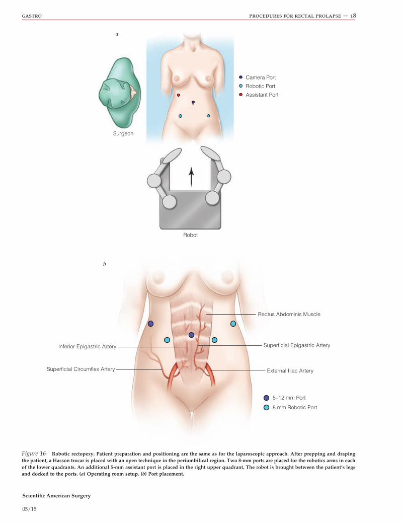
robotic approach
Patient preparation and positioning are the same as for the laparoscopic approach. after prepping and draping, a Hasson trocar is placed with an open technique in the peri-umbilical region. two 8-mm ports are placed for the robotics arms in each of the lower quadrants. an additional 5-mm assistant port is placed in the right upper quadrant. the robot is brought between the patient’s legs [see Figure 16] and docked to the ports. Because it is difficult to dock and undock the robot during the procedure, the robotic approach does not usually involve an open component. consequently, the procedure usually involves no resection.
Operative Technique
Step 1: placement of ports [see laparoscopic suture, Mesh, or tack rectopexy, operative technique, above].
Step 2: mobilization of the rectum robotic bowel grasp-ers are used to retract the rectosigmoid junction and the midrectum upward. a coagulation device is employed to perform the presacral dissection and to divide the full length
10 mm
10 mm10 mm
Figure 14 Laparoscopic suture or mesh rectopexy. A 10-mm trocar is placed in the periumbilical region. Two additional 10-mm trocars are then placed, one in each lower quadrant.
Scientific American Surgery
05/15
gastro procedures for rectal prolapse — 17
of the lateral peritoneal attachments, as is done during a total mesorectal excision.
Step 3: rectopexy the lateral peritoneum and underly-ing mesorectal fat on each side are secured to the sacrum, just below the sacral promontory on either side of the mid-line, using a laparoscopic tacker passed through the assis-tant port. a pelvic suction drain is placed in the presacral space on the day of surgery.
Postoperative Care
the patient is started on a clear liquid diet on the day of the surgery and advanced to a regular diet on the morning of postoperative day 1. the bladder catheter is also removed at this time.
Troubleshooting
laparoscopic/robotic repair of rectal prolapse is a mini-mally invasive procedure that requires advanced surgical skills and experience, but follows the same basic principles as open repair. there are multiple variant forms of laparo-scopic/robotic rectopexy, which differ not only with respect to technical details (e.g., resection versus no resection, suture versus mesh), but also with respect to whether the procedure contains an open component or is fully laparoscopic/robotic. the essential steps, however, are the same in all of the variant procedures and include adequate mobilization of the rectum, careful placement of sutures, and, if planned, appropriate resection with healthy anastomosis of the segments. for example, in the approach to laparoscopic resection rectopexy
we describe [see laparoscopic resection rectopexy, opera-tive technique, above], most of the mobilization is performed laparoscopically, with resection, anastomosis, and suture placement performed in an open fashion, whereas in laparo-scopic/robotic rectopexy performe d without resection, the mesh is secured intracorporeally. the modified ripstein pro-cedure [see laparoscopic suture, Mesh, or tack rectopexy, operative technique, above] can be performed in a totally laparoscopic fashion if desired.
Complications
Intraoperative complications of laparoscopic rectopexy include inadvertent enterotomy or colotomy, ureteral injury, and organ injury from trocar placement. Most of these inju-ries are associated with the presence of adhesions and can usually be prevented by careful intra-abdominal dissection in appropriate planes. the incidence of ureteral injury can be minimized by early identification of these structures, which may be facilitated by the use of ureteral catheters.
Vascular injury can occur at several different points of the operation. Injury to the epigastric vessels during port place-ment is usually avoided by transilluminating the abdomi-nal wall with the camera to identify the vessels. the iliac and gonadal vessels are retroperitoneal structures, which, like the ureter, can be avoided with careful dissection and identification. the mesenteric vessels must be properly iden-tified and controlled with graspers before the mesocolon is transected. the use of endoscopic clips as the sole means of vascular control before transection is discouraged; the calcified
Rectum
Grasper
Tied Suture
O-Silk Suture
Rectum
LaparoscopicNeedle Holder
Presacral Space
PolypropyleneMesh
SacralPromontory
a b
Figure 15 Laparoscopic mesh rectopexy. (a) A piece of mesh is inserted into the abdomen through a port then stapled to the sacrum. (b) The lateral edges of the nonabsorbable mesh are wrapped around three quarters of the rectal circumference and sutured to the rectal wall.
Scientific American Surgery
05/15
gastro procedures for rectal prolapse — 18
a
Camera Port
Robotic Port
Assistant Port
Surgeon
Robot
b
Inferior Epigastric Artery
Superficial Circumflex Artery
Rectus Abdominis Muscle
Superficial Epigastric Artery
External Iliac Artery
5–12 mm Port
8 mm Robotic Port
Figure 16 Robotic rectopexy. Patient preparation and positioning are the same as for the laparoscopic approach. After prepping and draping the patient, a Hasson trocar is placed with an open technique in the periumbilical region. Two 8-mm ports are placed for the robotics arms in each of the lower quadrants. An additional 5-mm assistant port is placed in the right upper quadrant. The robot is brought between the patient’s legs and docked to the ports. (a) Operating room setup. (b) Port placement.

Scientific American Surgery
05/15
gastro procedures for rectal prolapse — 19
vessels commonly encountered in this mainly elderly popu-lation lead to clip slippage and incomplete hemostasis.
the incidence of anastomotic leakage can be minimized by ensuring a tension-free, nonrotated, airtight connection with a good blood supply and by making sure not to incorporate diverticula into the suture line. surgical site infections are a common complication of all colorectal resections. their incidence can be minimized by performing appropriate bowel preparation, providing intravenous anti-biotic prophylaxis before surgery, and, possibly, employing a plastic wound protector at the site of colon extraction. copious wound irrigation before closure at the end of the procedure is also helpful.
mesh and sponge repairs (ripstein procedure)
Preoperative care and patient positioning are the same for the ripstein procedure as for open resection rectopexy [see resection rectopexy (frykman-Goldberg Procedure), above].
Operative Technique
Steps 1 and 2: initial incision and exploration, mobiliza-tion of the upper rectum [see resection rectopexy (frykman- Goldberg Procedure), operative technique, above].
Step 3: placement of mesh the original procedure was described in 1963 using Marlex (polypropylene) mesh. other types of synthetic mesh and fascia lata have since been described. a 5 cm rectangle of mesh is placed around the anterior rectum at the level of the peritoneal reflection and firmly sutured to the presacral fascia on one side [see Figure 17a]. the rectum is pulled taut with upward traction, partial-thickness nonabsorbable sutures are placed in rows along the anterior rectum to hold the mesh in place, and the mesh is sutured to the presacral fascia on the other side [see Figure 17b]. the sling is left loose enough to allow two fingers to pass between the bowel and the mesh so as not to cause luminal constriction with expected mesh shrinkage.
Because of the high risk of constipation and even obstipa-tion after anterior mesh encirclement, a modified version of the ripstein procedure was developed that involved
posterior fixation of mesh to the sacrum, leaving the anterior rectal wall free of any potential constriction. In this modified approach, the mesh is first tacked to the sacrum. the lateral edges of the mesh are then wrapped around three quarters of the circumference of the rectum and sutured anterolater-ally to the rectal wall. Intraoperative proctoscopy may be helpful for ensuring that the positioning of the mesh does not result in obstruction.
Step 4: closure of the peritoneum the peritoneal reflec-tion is closed so that the mesh is excluded from the perito-neal cavity and the small bowel is prevented from migrating into the pelvis on top of the mesh. the abdomen is irrigated and closed in the usual fashion.
Postoperative Care
the patient is started on a clear liquid diet and advanced to a solid diet when bowel function is resumed. the bladder catheter is removed 2 days after operation.
ivalon (polyvinyl alcohol) sponge repair (wells procedure)
Preoperative care and positioning are the same for the Wells procedure as for open resection rectopexy [see resec-tion rectopexy (frykman-Goldberg Procedure), above].
Operative Technique
Steps 1 and 2: initial incision and exploration, mobiliza-tion of the upper rectum [see resection rectopexy (fryk-man-Goldberg Procedure), operative technique, above].
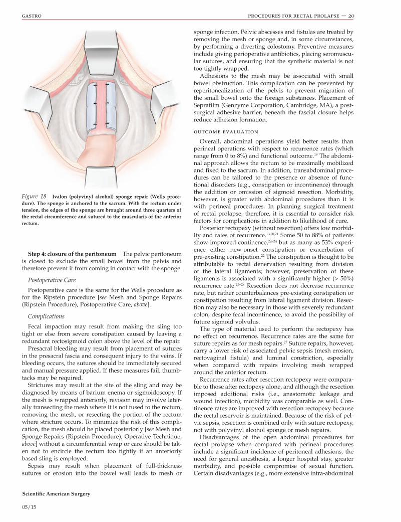
Step 3: placement of the sponge a rectangular piece of sterilized and moistened Ivalon (polyvinyl alcohol) sponge is secured in place with mattress sutures passed through the sponge, through the presacral fascia, and back through the sponge. careful hemostasis is ensured to prevent purulent collections in the area of the sponge. the rectum is retracted cephalad, around which the lateral edges of the sponge are folded for approximately three quarters of its circumference. the edges are secured to the anterior portion of the rectum with seromuscular sutures [see Figure 18].
a b
Figure 17 Ripstein procedure. (a) With the rectum under tension, a piece of mesh is sutured to the presacral fascia on one side then sutured to the muscularis of the anterior rectum. (b) The rectum is then secured to the presacral fascia on the other side to form a sling.
Scientific American Surgery
05/15
gastro procedures for rectal prolapse — 20
Step 4: closure of the peritoneum the pelvic peritoneum is closed to exclude the small bowel from the pelvis and therefore prevent it from coming in contact with the sponge.
Postoperative Care
Postoperative care is the same for the Wells procedure as for the ripstein procedure [see Mesh and sponge repairs (ripstein Procedure), Postoperative care, above].
Complications
fecal impaction may result from making the sling too tight or else from severe constipation caused by leaving a redundant rectosigmoid colon above the level of the repair.
Presacral bleeding may result from placement of sutures in the presacral fascia and consequent injury to the veins. If bleeding occurs, the sutures should be immediately secured and manual pressure applied. If these measures fail, thumb-tacks may be required.
strictures may result at the site of the sling and may be diagnosed by means of barium enema or sigmoidoscopy. If the mesh is wrapped anteriorly, revision may involve later-ally transecting the mesh where it is not fused to the rectum, removing the mesh, or resecting the portion of the rectum where stricture occurs. to minimize the risk of this compli-cation, the mesh should be placed posteriorly [see Mesh and sponge repairs (ripstein Procedure), operative technique, above] without a circumferential wrap or care should be tak-en not to encircle the rectum too tightly if an anteriorly based sling is employed.
sepsis may result when placement of full-thickness sutures or erosion into the bowel wall leads to mesh or
sponge infection. Pelvic abscesses and fistulas are treated by removing the mesh or sponge and, in some circumstances, by performing a diverting colostomy. Preventive measures include giving perioperative antibiotics, placing seromuscu-lar sutures, and ensuring that the synthetic material is not too tightly wrapped.
adhesions to the mesh may be associated with small bowel obstruction. this complication can be prevented by reperitonealization of the pelvis to prevent migration of the small bowel onto the foreign substances. Placement of seprafilm (Genzyme corporation, cambridge, Ma), a post-surgical adhesive barrier, beneath the fascial closure helps reduce adhesion formation.
outcome evaluation
overall, abdominal operations yield better results than perineal operations with respect to recurrence rates (which range from 0 to 8%) and functional outcome.19 the abdomi-nal approach allows the rectum to be maximally mobilized and fixed to the sacrum. In addition, transabdominal proce-dures can be tailored to the presence or absence of func-tional disorders (e.g., constipation or incontinence) through the addition or omission of sigmoid resection. Morbidity, however, is greater with abdominal procedures than it is with perineal procedures. In planning surgical treatment of rectal prolapse, therefore, it is essential to consider risk factors for complications in addition to likelihood of cure.
Posterior rectopexy (without resection) offers low morbid-ity and rates of recurrence.13,20,21 some 50 to 88% of patients show improved continence,22–24 but as many as 53% experi-ence either new-onset constipation or exacerbation of pre-existing constipation.22 the constipation is thought to be attributable to rectal denervation resulting from division of the lateral ligaments; however, preservation of these ligaments is associated with a significantly higher (> 50%) recurrence rate.25–29 resection does not decrease recurrence rate, but rather counterbalances pre-existing constipation or constipation resulting from lateral ligament division. resec-tion may also be necessary in those with severely redundant colon, despite fecal incontinence, to avoid the possibility of future sigmoid volvulus.
the type of material used to perform the rectopexy has no effect on recurrence. recurrence rates are the same for suture repairs as for mesh repairs.27 suture repairs, however, carry a lower risk of associated pelvic sepsis (mesh erosion, rectovaginal fistula) and luminal constriction, especially when compared with repairs involving mesh wrapped around the anterior rectum.
recurrence rates after resection rectopexy were compara-ble to those after rectopexy alone, and although the resection imposed additional risks (i.e., anastomotic leakage and wound infection), morbidity was comparable as well. con-tinence rates are improved with resection rectopexy because the rectal reservoir is maintained. Because of the risk of pel-vic sepsis, resection is combined only with suture rectopexy, not with polyvinyl alcohol sponge or mesh repairs.
disadvantages of the open abdominal procedures for rectal prolapse when compared with perineal procedures include a significant incidence of peritoneal adhesions, the need for general anesthesia, a longer hospital stay, greater morbidity, and possible compromise of sexual function. certain disadvantages (e.g., more extensive intra-abdominal
Figure 18 Ivalon (polyvinyl alcohol) sponge repair (Wells proce-dure). The sponge is anchored to the sacrum. With the rectum under tension, the edges of the sponge are brought around three quarters of the rectal circumference and sutured to the muscularis of the anterior rectum.

Scientific American Surgery
05/15
gastro procedures for rectal prolapse — 21
invasion, scarring, and longer recovery time) may be reduced when the laparoscopic approach is employed.
controlled studies of laparoscopic rectopexy revealed recurrence rates and morbidity comparable to those of open approaches.30–33 robotic-assisted rectal prolapse procedures are emerging as the technology evolves to various surgical specialties. data from randomized trials and meta-analyses are inconclusive in identifying the optimal technical approach when comparing open, laparoscopic, and robotic procedures.34,35 the disadvantages of robotic surgery include high cost, long intraoperative set up, and long procedure times.34 Numerous studies have failed to demonstrate any advantage.
an intra-abdominal approach should be performed for patients who can tolerate an abdominal operation, based on risk profile and physical status. our practice is to perform open resection rectopexy through a small Pfannenstiel incision, with minimal mobilization of the sigmoid and complete mobilization of the rectum down to the level of the levators posteriorly and anteriorly. the superior rectal artery is preserved while resecting redundant sigmoid, and the rectopexy is completed with sutures. Because this proce-dure can be performed with thoracic epidural, it is still a good option in the elderly patient. for those patients with serious comorbid conditions who are not candidates for an intra-abdominal procedure, the perineal approach offers a safe but less durable alternative.
Special Situations
incarceration, strangulation, and gangrene
on rare occasions, a rectal prolapse becomes incarcerated. If the bowel is viable, sedation and gentle manual reduction in the emergency department usually suffice. sprinkling table sugar on the prolapse helps reduce edema.36 If such measures are unsuccessful, paralyzing the perianal muscles with an anal block or general anesthesia in the or may help resolve the problem. If the incarceration is irreducible or the viability of the bowel is questionable (strangulation, with areas of gangrenous mucosa), emergency perineal rectosig-moidectomy should be performed, with or without fecal diversion.37 this procedure can be done in the supine or lithotomy position, although lithotomy position may be preferred if stoma is planned. Because preoperative bowel preparation is not possible, the rectum is irrigated with betadine. a circumferential incision is made above the dentate line in a viable area of mucosa, the perineal rectosig-moidectomy is performed, and a circular single-layer anas-tomosis is created. Poor nutrition, chronic steroid use, and severe comorbid conditions may warrant a diverting temporary stoma via open or laparoscopic approach. the new anastomosis is considered a coloanal anastomosis, and any adverse risk factors that may compromise a coloanal anastomosis apply when considering a diverting stoma.
ruptured prolapse
rupture of the prolapse usually involves opening of the hernia sac and exposure of the small bowel to the perineum. emergency transabdominal repair is indicated, with closure of the peritoneal hernia sac and suture rectopexy constitut-ing the safest and most efficient repair.
prolapse in male patients
Prolapse in male patients, although relatively rare, may occur at any age. Because obstetric injury is not a risk factor in this population, underlying disease processes that increase intra-abdominal pressure should be investigated and addressed both preoperatively and postoperatively to minimize risk of recurrence. these risk factors are the same as for hernia, and include coughing, constipation, and straining. Because of the deep and narrow pelvis, open approaches to rectal prolapse should proceed through a lower midline rather than Pfannenstiel incision. surgeons performing rectal mobilization should take care to preserve the presacral nerves, keep from damaging denonvilliers fascia, and avoid division of one of the lateral ligaments.38 lateral pararectal tissues carry parasympathetic fibers and are important in normal ejaculatory function.
recurrent prolapse
there are two types of recurrent rectal prolapse. recur-rent mucosal prolapse responds well to rubber banding or excision. recurrent full-thickness rectal prolapse occurring in the first year after surgery is likely the result of a specific technical failure, such as incomplete mobilization of the rectum, inadequate fixation of the rectum to the sacrum, incomplete resection of a redundant sigmoid, or vigorous physical activity or childbirth that disrupts the rectum from the sacrum. In addition, adequate mobilization from a trans-perineal approach is less certain because additional bowel can be mobilized even after dividing “the last attachment.” alternative concern is resecting too much bowel and placing tension on the anastomosis, which may be a risk factor for leakage.
rectal prolapse that recurs beyond 1 year of surgery results from persistence of the underlying pathophysiology (such as disordered defecation, abnormal intestinal motility, straining) that led to the original presentation of the disease. a meta-analysis of eight trials revealed recurrence for abdominal and perineal approaches in particular.27 the extent of rectal dissection was the single most important factor in decreasing recurrence, specifically bilateral collat-eral ligament division. the outcome of mesh and suture repairs was equivocal. a second multicentered study of more than 600 patients compared surgical technique (mobi-lization only, mobilization-resection rectopexy, mobilization rectopexy), means of access (open versus laparoscopic), and method of rectopexy (mesh versus sutures).39 None of these had an effect on recurrence rates. rectal mobilization alone is as effective as more complex procedures, such as rectopexy, with or without resection.
In approaching patients with recurrent rectal prolapse, extensive investigation should be undertaken to identify the risk factors that may have contributed to recurrence, includ-ing anorectal physiology testing and a colonic transit study. subtotal colectomy should be considered in patients with slow transit constipation without sphincter weakness.31 Preoperative and postoperative biofeedback therapy should be considered for those with outlet constipation.
currently, there is no standardized strategy for primary or recurrent rectal prolapse. the literature on recurrent rectal prolapse does not describe a treatment algorithm or rationale for decision making with respect to choice of the
Scientific American Surgery
05/15
gastro procedures for rectal prolapse — 22
second operation. However, most agree the approach should be tailored to the patient, taking into consideration the same factors that determined choice of the initial operation and keeping in mind blood supply to the remaining rectum. the optimal choice of operation should be minimally invasive, include low morbidity and mortality, provide optimal functional results, and minimal risk of recurrence. some authorities advocate an abdominal procedure for the second operation, regardless of what the initial operation was, because of the superior rates of success with such proce-dures40; however, care must be taken in performing repeat resectional procedures (e.g., anterior resection after perineal rectosigmoidectomy, or vice versa) because of the risk of bowel ischemia between the two anastomoses if the superior rectal artery is not preserved. some studies suggest that unless the previous anastomosis can be resected in the second procedure, repeat resectional procedures should be avoided.40,41 Perineal rectosigmoidectomies are an exception to this broad rule: they can be safely repeated as long as the recurrent prolapse contains the previous anastomosis.42 recurrence rates and morbidity after operative treatment of recurrent rectal prolapse are essentially the same as those after operative treatment of primary rectal prolapse.43 all types of secondary repairs are feasible, but because of the paucity of published studies, a defined standard of care is lacking.
rectal prolapse with solitary rectal ulcer syndrome
as many as 80% of patients with solitary rectal ulcer syndrome (srus) have an associated rectal prolapse,44,45 a finding that indicates a close relationship between these two entities. In cases of symptomatic srus associated with asymptomatic prolapse, a trial of nonoperative therapy including pelvic floor retraining is warranted46; if such therapy fails, surgical intervention with procedures used for rectal prolapse should be considered.47 In cases of symptom-atic prolapse associated with asymptomatic srus, healing of the ulcer can be demonstrated in one third of patients undergoing operation for the prolapse.47
Financial Disclosures: Steven D. Wexner, MD, PhD (Hon), FACS, FRCS, FRCS (Ed), and Susan M. Cera, MD, FACS, have no relevant financial relationships to disclose.
References
1. Madden MV, Kamm Ma, Nicholls rJ, et al. abdominal rec-topexy for rectal prolapse: prospective study evaluating changes in symptoms and anorectal function. dis colon Rectum 1992;35:48–55.
2. Boutsis c, ellis H. the Ivalon-sponge wrap operation for rectal prolapse: an experience with 26 patients. dis colon Rectum 1974;17:21–37.
3. Madoff rd, Williams JG, Wong Wd, et al. long-term func-tional results of colon resection and rectopexy for overt rectal prolapse. Am J Gastroenterol 1992;87:101–4.
4. Keighley Mr, shouler PJ. abnormalities of colonic function in patients with rectal prolapse and fecal incontinence. Br J Surg 1984;71:892–5.
5. Goldman J. concerning prolapse of the rectum with special emphasis on the operation by thiersch. dis colon rectum 1988;31:154–5.
6. classic articles in colonic and rectal surgery. edmond delo-rme 1847–1929. On the treatment of total prolapse of the rectum by excision of the rectal mucous membranes or recto-colic. Dis Colon Rectum 1985;28:544–53.
7. Mikulicz J. Zur operativen Behandlung des Prolapsus recti et coli invaginati. Verh dtsch Ges chirurgie 1888;17:294.
8. altemeier Wa, culbertson Wr, schwengerdt c, et al. Nine-teen years’ experience with the 1-stage perineal repair of rectal prolapse. Ann Surg 1971;173:993–1006.
9. ripstein cB, lanter B. etiology and surgical therapy of massive prolapse of the rectum. Ann Surg 1963;157:259–64.
10. Wells c. New operation for rectal prolapse. Proc r soc Med 1959;52:602.
11. Johanson oB, Wexner sd, daniel N, et al. Perineal rectosig-moidectomy in the elderly. dis colon rectum 1993;36: 767–72.
12. lechaux JP, lechaux d, Perez M. results of the delorme procedure for rectal prolapse: advantage of a modified technique. Dis Colon Rectum 1995;38:301–7.
13. ripstein cB. surgical treatment of rectal prolapse. Pac Med Surg 1967;75:329–32.
14. fengler sa, Pearl rK, Prasad Ml, et al. Management of recurrent rectal prolapse. Dis Colon Rectum 1997;40:832–4.
15. Williams JG, rothenberger da, Madoff rd, et al. treatment of rectal prolapse in the elderly by perineal rectosigmoidec-tomy. Dis Colon Rectum 1992;35:830–4.
16. agachan f, reissman P, Pfeifer J, et al. comparison of three perineal procedures for the treatment of rectal prolapse. South Med J 1992;90:925–32.
17. agachan f, daniel N, Wexner sd. the outcome of rectal prolapse surgery in the elderly. Is long-term follow up possible? coloproctology 1996;18:41.
18. Becker JM, dayton Mt, fazio VW, et al. Prevention of post-operative abdominal adhesions by a sodium hyaluronate–based bioresorbable membrane: a prospective, randomized, double-blind multicenter study. J am coll surg 1996;183: 297–306.
19. Beck de, Wexner sd. fundamentals of anorectal surgery. 2nd ed. london: WB saunders company; 1998.
20. duthie Gs, Bartolo G. abdominal rectopexy for rectal pro-lapse: a comparison of techniques. Br J Surg 1992;79:107–13.
21. Madoff rd, Mellgren a. one hundred years of rectal prolapse surgery. Dis Colon Rectum 1999;42:441–50.
22. aitola Pt, Hiltunen KM, Matikainen MJ. functional results of operative treatment of rectal prolapse over an 11 year period: emphasis of the transabdominal approach. dis Colon Rectum 1999;42:655–60.
23. Madden MV, Kamm Ma, Nicholls rJ. abdominal recto-pexy for complete rectal prolapse: prospective study evalu-ating changes in symptoms and anorectal function. dis Colon Rectum 1992;35:48–55.
24. Mccue Il, thompson JPs. clinical and functional results of abdominal rectopexy for complete rectal prolapse. Br J surg 1991;78:921–3.
25. scaglia M, fasth s, Hallgren t, et al. abdominal rectopexy for rectal prolapse: influence of surgical technique on functional outcome. Dis Colon Rectum 1994;37:805–13.
26. speakman ct, Madden MV, Nicholls rJ, et al. lateral liga-ment division during rectopexy causes constipation but
Scientific American Surgery
05/15
gastro procedures for rectal prolapse — 23
prevents recurrence: results of a prospective randomized study. Br J Surg 1991;78:1431–3.
27. Bachoo P, Brazelli M, Grant a. surgery for complete rectal prolapse in adults. cochrane database syst rev 2000;(2):cd001758.
28. McKee rf, lauder Jc, Poon fW, et al. a prospective randomized of abdominal rectopexy with and without sigmoidectomy in rectal prolapse. surg Gynecol obstet 1992;174:145–8.
29. lukkenon P, Mikkonen u, Jarvinen H. abdominal recto-pexy with sigmoidectomy versus rectopexy alone for rectal prolapse: a prospective, randomized study. Int J colorectal Dis 1992;7:219–22.
30. schlinkert rt, Beart rW Jr, Wolff BG, et al. anterior resec-tion for complete rectal prolapse. dis colon rectum 1985; 28:409–12.
31. Watts Jd, rothenberger da, Buls JG, et al. the manage-ment of procidentia: 30 years’ experience. dis colon rectum 1985;28:96–102.
32. solomon MJ, Young cJ, eyers aa, et al. randomized clini-cal trial of laparoscopic versus open abdominal rectopexy for rectal prolapse. Br J Surg 2002;89:35–9.
33. Boccasanta P, Venturi M, reitano Mc, et al. laparotomic versus laparoscopic rectopexy in complete rectal prolapse. Dig Surg 1999;16:415–9.
34. Heemskerk J, de Hoog de, van Gemert WG, et al. robot-assisted versus conventional laparoscopic rectopexy for rectal prolapse; a comparative study on costs and time. dis Colon Rectum 2007;40:1825–30.
35. tou s, Brown sr, Malik aI, Nelson rl. surgery for com-plete rectal prolapse in adults. cochrane database sys rev 2008;(4):cd001758.
36. Myers Jo, rothenberger da. sugar in the reduction of incarcerated prolapsed bowel: report of two cases. dis Colon Rectum 1991;34:416–8.
37. ramanujam Ps, Venkatesh Ks. Management of acute, incar-cerated rectal prolapse. dis colon rectum 1992;35:1154.
38. abou-enein a. Prolapse of the rectum in young men: treat-ment with a modified roscoe Graham operation. dis colon Rectum 1978;22:117–9.
39. raftopoulos Y, senagore aJ, di Giuro G, et al. recurrence rates after abdominal surgery for complete rectal prolapse: a multicenter pooled analysis of 643 individual patient data. Dis Colon Rectum 2005;48:1200–6.
40. Hool Ga, Hull tl, fazio VW. surgical treatment of recurrent complete rectal prolapse. Dis Colon Rectum 1997;40:270–2.
41. fengler sa, Pearl rK, Prasad Ml, et al. Management of recurrent rectal prolapse. Dis Colon Rectum 1997;40:832–4.
42. Williams JG, rothenberger da, Madoff rd, et al. treatment of recurrent rectal prolapse in the elderly by perineal rectosigmoidectomy. Dis Colon Rectum 1992;35:830–4.
43. Pikarsky aJ, Joo Js, Wexner sd, et al. recurrent rectal prolapse: what is the next good option? dis colon rectum 2000;43:1273–6.
44. Mackle eJ, Manton Mills Jo, Parks tG. the investigation of anorectal dysfunction in the solitary rectal ulcer syndrome. Int J Colorectal Dis 1990;5:21–4.
45. Mahieu PH. Barium enema and defecography in the diag-nosis and evaluation of the solitary rectal ulcer syndrome. Int J Colorectal Dis 1986;1:85–90.
46. van den Brandt-Gradel V, Huibregtse K, tytgat GNJ. treat-ment of the solitary rectal ulcer syndrome with high-fiber diet and abstention of straining at defecation. dig dis sci 1984;29:1005–8.
47. tjandra JJ, fazio VW, church JM, et al. ripstein procedure is an effective treatment for rectal prolapse without constipation. Dis Colon Rectum 1993;36:501–7.
Acknowledgments
figures 1, 8, 13, 14, 15, 17, and 18 tom Moorefigures 2, 6, 10, 11, and 16 christine Kenney