Research Article Dyeing Studies with Eucalyptus, Quercetin ...
Upload
khangminh22Category
view
3download
0
polymers
Article
Comparison of Polysaccharides as Coatings forQuercetin-Loaded Liposomes (QLL) and Their Effectas Antioxidants on Radical Scavenging Activity
Manuel Román-Aguirre 1, César Leyva-Porras 2 , Pedro Cruz-Alcantar 3 ,Alfredo Aguilar-Elguézabal 4 and María Zenaida Saavedra-Leos 3,*
1 Doctorado Institucional en Ingeniería y Ciencia de los Materiales, Universidad Autónoma de SanLuis Potosí, San Luis Potosí 78210, Mexico; [email protected]
2 Laboratorio Nacional de Nanotecnología (NanoTech), Centro de Investigación en MaterialesAvanzados S.C. (CIMAV), Chihuahua 31136, Mexico; [email protected]
3 Coordinación Académica Región Altiplano (COARA), Universidad Autónoma de San Luis Potosí,Matehuala 78700, Mexico; [email protected]
4 Departamento de Ingeniería y Química de Materiales, Centro de Investigación en MaterialesAvanzados S.C. (CIMAV), Chihuahua 31136, Mexico; [email protected]
* Correspondence: [email protected]; Tel.: +52-(488)-1250150
Received: 30 October 2020; Accepted: 23 November 2020; Published: 26 November 2020 �����������������
Abstract: Liposomes are microstructures containing lipid and aqueous phases employed in theencapsulation and delivery of bioactive agents. Quercetin-loaded liposomes (QLLs) were coatedwith three different polysaccharides and then tested as radical scavengers. Lactose (LCQLL),chitosan (CCQLL), and inulin (ICQLL) were employed as coating materials. Particle size determinedby light scattering, showed primary size of 200 nm for all samples, while a secondary particlesize of 600 nm was observed for CCQLL. Scanning electron microscopy (SEM) evidenced particleaggregation with the addition of the polysaccharide coating. Transmission electron microscopy(TEM) revealed the layered microstructure of liposomes composed of at least two layers, and primaryparticle size below 100 nm. QLL showed higher antioxidant activity than the coated liposomes.This behavior was attributed to the chemical interaction between quercetin and the correspondingcoating polysaccharide in the layered structure, which traps the quercetin and keeps it unavailable forradical scavenging. From the three polysaccharides, lactose showed a better performance as coatingmaterial in the antioxidant activity, which suggested that the smaller size of the disaccharide moleculeresulted in a faster releasing of the quercetin in the solution. Thus, LCQLL is an advantageous way todeliver quercetin for antioxidant purposes, where the low stability in delivered media of quercetinloaded liposomes is commonly compromised.
Keywords: loaded liposomes; antioxidant properties; polysaccharides coating; inulin; lactose; quercetin
1. Introduction
One of the biggest challenges facing in food industry is the rapid deterioration of foods,caused mainly by the growth of microorganisms, lipid oxidation, or enzymatic self-decomposition [1].Oxidation causes the loss of nutritional value, color, and texture in foods [2,3]. In meat products,lipid oxidation leads to the formation of hydroperoxides and volatile compounds like aldehydes andketones, provoking a characteristic rancidity smell [4–6]. In fruits and vegetables, the deteriorationcaused by enzymatic oxidation or oxidative stress, is extremely fast, being observed as darkening, loss oftexture, and alteration of taste, few hours after harvest or even faster once the peel is removed [7]. In orderto counteract this effect, the use of synthetic antioxidants such as butylated hydroxytoluene (BHT) has
Polymers 2020, 12, 2793; doi:10.3390/polym12122793 www.mdpi.com/journal/polymers

Polymers 2020, 12, 2793 2 of 15
been implemented, although its consumption has been linked to toxicological effects [8,9]. Consequently,the current trend is the replacement of synthetic antioxidants with naturally occurring ones.Natural antioxidants can be found in some essential oils of herbs and spices such as oregano, cinnamon,rosemary, marjoram or clove, all of them containing compounds such as terpenes (carvacrol, citral orthymol), or aldehydes (cinnamaldehyde) [10,11]. Another natural source rich in antioxidants are redfruits i.e., berries, grapes, and pomegranates, which are abundant in beneficial compounds such asterpenoids (resveratrol), and phenolics (anthocyanins, phenolic acids, flavan-3-oles and flavonoids) [12].Among the dietary flavonoids, quercetin (2-(3,4-dihydroxyphenyl)-3,5,7-trihydroxy-4H-chromen-4-one)is one of the most abundant [13,14]. However, because of the easy decomposition of quercetin uponexposure to environmental elements such as oxygen, temperature and ultraviolet light [15–17], it isnecessary to encapsulate it with protective vehicles or transport agents that may form a protective filmor coating [18]. This problem has also been studied for quercetin applications as anti-cancer agent,being its low oral availability and fast gastrointestinal digestion the main restrains for its use as directoral drug [19].
Liposomes are self-assembled spherical aggregates, composed of a phospholipid molecule shell(typically one or more bilayers of lecithin) enclosing an aqueous phase core containing a bioactivecompound. Since liposomes are prepared in liquid media, the experimental variables affecting thestability may include the pH, temperature of solution and melting point of the phospholipid, amongothers [20]. Recently, encapsulation in the form of liposomes has emerged as a potential solution to thedeterioration and preservation of bioactive compounds [15,21,22]. Due to the presence of both lipidand aqueous phases, these microstructures represent an emerging technology for the encapsulationand delivery of bioactive agents, either hydrophilic or hydrophobic materials, including antioxidants,antimicrobials and nutraceuticals [23–26]. Thus, in order to increase the stability and improve therelease of the active load, liposomes may be coated with other biocompatible compounds. Based onthe biocompatibility, low or non-toxicity, and neutral organoleptic properties, several materials havebeen employed as coatings for liposomes, including glycols, chitosan, and polysaccharides [23,27,28].
Chitosan, poly(D-glucosamine), is a biopolymer with excellent biocompatibility, obtained fromdeacetylated chitin, and found in the shell of crustaceans like shrimps and lobsters. The positivelycharged nitrogen of chitosan in acidic solution, prompts this biopolymer to undergo electrostaticattraction to negative charges, like the phosphate group in lecithin. Because of this, chitosan-coatedliposomes have been extensively studied [23,24,29,30]. For example, chitosan-coated liposomes loadedwith bioactive molecules such as quercetin or carotenoids have shown good stability and releaserates [23]. Pectin and polygalacturonic acid do not have a positive charge like chitosan, yet neverthelessthey have shown good stability, encapsulation efficiency, and release rate when studied as coatingmaterials for loaded liposomes [25]. Despite the good characteristics of chitosan, the preparation ofliposomes coated with this polymer has several limitations, such as the relatively high viscosity ofsolutions, the low pH required for its solubility and difficulty to eliminate excess of chitosan fromthe liposomal surface. Alternatively, the use of polysaccharides soluble at neutral pH and with lowviscosity can be alternatives to replace the use of chitosan. Polysaccharides’ interaction with theliposome wall affects the release rate of bioactive molecules. In turn, this is dependent on crystallinity,molecular weight, stability and intermolecular attractions of the polysaccharide [22,24,28,30–33].Lactose is a disaccharide formed by galactose and glucose subunits. Lactose molecules associated withceramide (lactosylceramide) have been used as liposome coatings mainly in transporting bioactivemolecules inside specific cells to prevent them from being identified as foreign bodies and beingdestroyed by the mechanisms of the immune system [34,35]. Inulin is a polysaccharide with probioticproperties, consisting of fructose units terminated by end glucose groups. Inulin has been mainlyused for encapsulation of antioxidants and other bioactive molecules by the spray drying process [36].Recently, inulin esterified with stearic acid was employed as a coating of liposomes by penetration ofa hydrophobic link of stearic acid inside the phospholipid bilayer [37]. Although in several works,inulin has been included in the hydrophilic phase of the lamellar structure of the liposomes, it is has

Polymers 2020, 12, 2793 3 of 15
not been employed as a coating, but as a material that is transported within the liposome. In theseworks, inulin containing 3H has been used as a molecular marker to determine the final location ofliposomes in vivo and in vitro tests [38,39].
To the best of our knowledge, the antioxidant properties of quercetin-loaded liposomes coated withpolysaccharides have not been reported so far. Three coatings were individually studied and compared,neutral low molecular weight-lactose, neutral high molecular weight-inulin, and positively-chargedhigh molecular weight-chitosan. This study is a first investigation towards the improvement of theantioxidant properties and set the basis for future work that may explain the release process of quercetinfrom the liposome. To the best of our knowledge, the antioxidant properties of quercetin-loadedliposomes coated with inulin or lactose have not been reported so far, and thus, the comparison ofpolysaccharide coated quercetin loaded liposomes may lead to new knowledge in this field. Therefore,in the present work, chitosan, lactose and inulin were separately employed for coating quercetin-loadedliposomes (QLL). The particle size, morphology and microstructure were characterized, the antioxidantproperty of the prepared QLL was tested for radical scavenging activity, and the effect of the differentcoatings compared.
2. Materials and Methods
2.1. Materials
Soybean phospholipid was purified from food grade lecithin. Methanol, ethanol, acetone,chloroform, acetic acid and sodium phosphate were ACS grade purchased from J.T. Baker(Radnor, PA, USA). Chitosan (80% of deacetylated chitin, and viscosity-average molecular weight of2.5 × 105 Da), 2,2-diphenyl-1-picrylhydrazyl (DPPH, purity 99.5%) and quercetin (purity 95%) werepurchased from Sigma-Aldrich (St. Louis, MO, USA).
2.2. Quercetin-Loaded Liposomes (QLL) Preparation
Prior to liposome preparation, the phospholipid was purified by adapting the method reportedby Hasan et al. [28]. Typically, 10 g of food grade commercial lecithin were dissolved in 20 mL ofchloroform and poured into 100 mL of acetone. The mixture was cooled to −14 ◦C and the precipitatedecanted by centrifugation. The supernatant liquid was discarded and the solid was washed twicewith acetone. Finally, the phospholipid was dried at 25 ◦C under vacuum for 8 h to eliminate all tracesof solvent. The buffer solution was prepared by dissolving 0.01 moles of monobasic sodium phosphatein 0.9 L of deionized water. The pH was adjusted to 7.4 by dropwise addition of 0.1 M NaOH solution.Distilled water was added to make up 1 L of phosphate buffer solution.
Liposomes were coated with polysaccharides according to the method reported by Hao et al. [23].The liposome suspension (loaded or unloaded) was mixed with the corresponding polysaccharidesolution, i.e., chitosan, lactose or inulin, and separated from the aqueous medium by centrifugation.
The QLL were prepared as follows. Usually, 60 mg of phospholipid and 2 mg of quercetin weredissolved in 15 mL of solution of methanol and chloroform (1:2). The content of quercetin in theliposomes was below the recommended daily intake dose for humans. The solution was evaporated at30 ◦C in a rotary evaporator under reduced pressure to obtain a solvent-free film. The flask containingthe film was immersed in an ice bath. The self-assembly of vesicles was done by hydrating the filmwith 15 mL of phosphate buffer solution under ultrasonic irradiation (3:1 s on/off pulses) during2 min. Control liposomes (CL), were prepared in a similar way but without adding quercetin to thephospholipid-methanol-chloroform solution. The suspensions obtained were stored at 4 ◦C and keptin darkness until utilization.
2.3. Chitosan Coated Liposomes
While maintaining magnetic stirring, 2 mL of QLL suspension were added dropwise into 5 mL ofchitosan solution (consisting of 0.5 mL of glacial acetic acid and 0.1 g of chitosan in 100 mL of deionized

Polymers 2020, 12, 2793 4 of 15
water). Then, the as-obtained coated material was centrifuged at 4 ◦C and 16,000 RPM during 30 min.The discarded liquid was substituted by a solution of acetic acid (0.5 mL of glacial acetic acid in 100mL of deionized water) in order to eliminate the excess of chitosan. The washing cycle was repeatedtwice and the settled solids were dried by lyophilization. Chitosan-coated liposomes were labeled asCCCL, while CCQLL when loaded with quercetin.
2.4. Polysaccharide Coated Liposomes
In order to coat QLL with lactose or inulin, the same procedure applied for chitosan coating wasfollowed, except that the acetic acid solution was replaced by deionized water. For example, 2 mL ofQLL suspension consisting of 0.1 g of lactose or inulin dissolved in 100 mL of deionized water wereadded dropwise into 5 mL of polysaccharide solution under magnetic stirring. Coated liposomes wereseparated by centrifugation at 4 ◦C and 16,000 RPM during 30 min, then washed twice with deionizedwater. Solids were dried by lyophilization. Inulin-coated liposomes were identified as ICQLL andICCL, while lactose-coated liposomes samples were identified as LCQLL and LCCL.
All these coated liposomes were stored at ambient temperature and maintained in darkness untiluse. Table 1 describes the identification of the samples prepared.
Table 1. Samples identification and description of liposomes. (CL) control liposomes, (QLL) quercetinloaded liposomes, (XCCL) polysaccharide coated control liposomes, and (XCQLL) polysaccharidecoated quercetin loaded liposomes. Where X stands for the corresponding polysaccharide (I) inulin,(L) lactose, and (C) chitosan.
Sample Name Coating Quercetin Loaded
CL Uncoated NoICCL Inulin NoLCCL Lactose NoCCCL Chitosan NoQLL Uncoated Yes
ICQLL Inulin YesLCQLL Lactose YesCCQLL Chitosan Yes
2.5. Liposomes Characterization
2.5.1. Particle Size Distribution
Prior to the centrifugation and washing, part of the liposome suspension was used to measurethe particle size distribution (PSD) of the coated liposomes. PSD of samples was determined by lightscattering using a Masterziser 2000 particle size analyzer Malvern Instruments (Malvern Panalytical,Malvern, UK) equipped with a 632 nm wavelength laser source. Typically, 12 mL of suspension or coatedliposomes suspension were directly poured into the sample port of the instrument. The measurementwas repeated three times for each sample.
2.5.2. Morphology and Microstructure
Morphology and microstructure of liposomes were characterized by scanning electron microscopy(SEM), and transmission electron microscopy (TEM), respectively.
The SEM analysis consisted in depositing a drop of liposomes solution on a substrate glass.The liquid was dried at room temperature in vacuum conditions by 12 h. In order to reduce chargingeffects from electron irradiation, dried samples were coated with a thin layer of gold deposited bysputtering. Coated samples were analyzed in a SU3500 SEM (Hitachi, Hitachi city, Japan) operated at15 kV, and spot size of 40 and low vacuum conditions of 60 Pa. Images were acquired with a secondaryelectron detector (SEI).

Polymers 2020, 12, 2793 5 of 15
For the TEM analysis, liposome suspensions were first diluted in order to decrease the particleconcentration. Commonly, 1 mL of uncoated liposomes suspension or 2 mL of coated liposomessuspension were diluted in 24 mL of deionized water. Then liposomes were stained by mixing 1 mL ofthe diluted suspension with 1 mL of 2% (wt%) aqueous solution of ammonium molybdate employedas a negative staining agent. Three minutes after mixing, a drop of stained liposome suspensionwas deposited on a carbon membrane copper grid and dried at room temperature under vacuumconditions. Deposited samples were analyzed on a HT7700 transmission electron microscope (Hitachi),operated at 100 kV.
2.6. Radical Scavenging Activity
DPPH radical scavenging activity was evaluated by mixing 1.7 mL of alcoholic DPPH solution(0.1 mmol of DPPH/L in ethanol) with 1.7 mL of lyophilized liposomes suspension. The concentrationof liposomes in suspensions varied as 5, 10 and 30 µg/mL. The mixture was left to stand in darknessfor 30 min. Then, absorbance was measured at 537 nm using an UV-Vis Evolution 220 spectrometer(Thermo Scientific, Waltham, MA. USA). The scavenging percentage was calculated according toEquation (1):
% DPPH∗ = 100×A0 −A30
A0(1)
where, A0 is the absorbance of the blank solution i.e., mixture of DPPH and ethanol without liposomes,and A30 is the absorbance of the of DPPH and ethanol solution containing the liposomes after 30 min.Scavenging activity was determined by triplicate for each sample.
2.7. Statistical Analysis
All experiments were performed in triplicate, reporting mean values and standard deviations.One-way analysis of variance (ANOVA) was performed to establish a significance level of 0.05, and theTukey’s honestly significant difference (HSD) post hoc test was used to determine the differencebetween means. The statistical analyses were conducted using the IBM SPSS Statistics version 21.0software (SPSS Inc., Chicago, IL, USA).
3. Results and Discussion
3.1. Particle Size Distribution
Figure 1 shows the PSD measured by light scattering determined for uncoated and coatedquercetin-loaded liposomes. In these figures, the particle size is plotted on the x-axis, while the volumefraction (%) occupied by these particles in the analyzed sample is shown on the y-axis. The averageparticle size and volume fraction of uncoated (QLL) and coated (ICQLL, and LCQLL) liposomes weresimilar with values of 185–200 nm, and 21–23%, respectively.
However, the coated liposomes showed a secondary volume fraction of about 2% with a sizedistribution on the order of 600–650 nm. Chitosan-coated liposomes (CCQLL) showed slight differencessuch as a larger primary particle size of 630 nm, lower volume fraction of 13%, and secondary particlesize distribution of 690 nm and 8%, respectively. From these results it is evident that inulin andlactose did not affect the primary particle size of the liposomes, while chitosan increased the size.Likewise, the addition of coating materials such as inulin and lactose resulted in the developmentof a secondary particle size distribution with larger size. Similar results were reported in literature,for example, Hao et al. obtained uncoated liposomes with size of 200–300 nm, while for chitosan-coatedliposomes the diameter increased to 700 nm [23]. Park and collaborators prepared chitosan-coatedliposomes loaded with resveratrol and observed that the size of uncoated liposomes increased from200 to 600 nm when using a 0.5% solution of chitosan as coating medium [40]. They explained thatseveral self-assembled layers of chitosan formed on the surface of liposomes increased the diameterof the vesicle. Vural et al. prepared chitosan-coated liposomes loaded with furosemide, and found

Polymers 2020, 12, 2793 6 of 15
that particle size increased from 50 to 350 nm when the chitosan concentration was varied from 0.1 to1% [41].
Figure 1. Particle size distribution by light scattering of coated liposomes. (a) QLL, (b) ICQLL,(c) LCQLL and (d) CCQLL.
3.2. Particle Morphology
Figure 2 shows representative SEM micrographs of uncoated and coated quercetin-loadedliposomes. Overall, spherically shaped particles with sizes in the range of 1–10 µm were observed,surrounded by a continuous material of irregular morphology. This suggested that the sphericalparticles corresponded to the agglomerated liposomes, while the continuous material correspondedto the excess lecithin that was not eliminated during the purification process. Several authors haveattributed the increase in particle size to the particle agglomeration caused by the addition of thecoating material. Li et al. observed that particle aggregation of uncoated samples showed a narrowpolydispersity with an agglomerate size of about 4 µm, while for chitosan-coated samples the particlesize was about 200 nm with wide polydispersity [42]. In contrast, Romero-Pérez et al. attributed thehigh polydispersity index to the diverse particle diameter [43]. Barea et al. prepared chitosan-coatedliposomes and explained that partial coating of liposomes might cause particle agglomeration due tothe interactions between opposite charges [44].

Polymers 2020, 12, 2793 7 of 15
Figure 2. Representative SEM micrographs of CCQLL showing agglomeration of liposomes. (a) CL,(b) QLL, (c) CCCL, and (d) CCQLL.
3.3. Particle Microstructure
Figure 3 shows bright field (BF) images acquired in the TEM of uncoated and coated quercetin-loaded liposomes. The first two columns show low and high magnification images, while the thirdcolumn presents the particle size distribution determined from measuring at least 100 particles in theimages. In general, the particles presented a distorted spherical morphology with sizes in the range of10–500 nm. The average particle size was in the order of nanoparticles (below 100 nm), with values of79.8, 107.6, 71.9, and 61.9 nm, for samples QLL, LCQLL, ICQLL, and CCQLL, respectively. With theexception of inulin, the other coating materials (lactose and chitosan) did not increase the primaryparticle size. High magnification images revealed the multilayer onion-like microstructure of theliposomes. This microstructure is well defined in the QLL showing about seven layers. With theaddition of the polysaccharides, i.e., samples ICQLL and LCQLL, the multilayer microstructure beginsto fade, observing the formation of a nucleus and the reduction in the number of layers (about 2–5 layers).The layered structure in the CCQLL is observed different to the onion microstructure of the othersamples. In this sample, the distance between the layers increased and the layers are distorted,observed as incomplete spiral-like liposomes or a jellyfish-like microstructure. Hasan et al. describedthe microstructure of liposomes as alternate concentric dark and bright fringes, and suggested thatquercetin is preferentially located in the bright fringes between two phospholipid layers in darkfringes [29]. In this sense, others works have reported that in the assembly of structures containingcarotenoids, curcumin and quercetin, these biomolecules are placed between the phospholipid layersin the form of layered liposomes [29,45,46]. Nonetheless, Hao et al., proposed that quercetin waslocated in the core of liposomes rather than in the layers [23]. In the present work, a negative stainingreagent was used to react preferentially with the unsaturated carbon-carbon bonds. From the molecules
Polymers 2020, 12, 2793 8 of 15
tested, i.e., quercetin, lecithin, inulin, lactose and chitosan, the first two presented unsaturation ofcarbon-carbon bonds, nevertheless quercetin has seven double bonds per molecule, while lecithin hasonly one. Therefore, in the unsaturated carbon-carbon bonds of quercetin the dyeing agent preferentiallyadsorbs, increasing the electronic scattering due to the high atomic number of molybdenum attachedto antioxidant molecule, which is observed in the BF TEM image as an increase in contrast. Thus,the results presented herein suggested that quercetin is preferentially located between the two layers oflecithin. Additionally, the chemical nature of the molecules also supports the evidence aforementioned.For example, since quercetin is hydrophilic and lecithin is hydrophobic, the insoluble nature on aqueousmedia of quercetin causes to be located between the multilayer spaces of two phospholipid regions.
Figure 3. BF TEM images of uncoated and coated quercetin loaded liposomes. (a) QLL, (b) ICQLL,(c) LCQLL, (d) CCQLL. Column I and II show low and high magnification micrographs, respectively,column III shows the particle size distribution measured from the TEM micrographs.

Polymers 2020, 12, 2793 9 of 15
Regarding the observations done on the microstructure of CCQLL, it is possible that the acidicmedia used to maintain chitosan in soluble form, leads to a rearrangement of the phospholipid lamellarstructure. This effect may be attributed to the high viscosity and stronger molecular interaction ofthis polymer with the liposome surface. Zhou et al. prepared liposomes loaded with vitamin C,obtaining irregularly shaped mono- and bilayer structured liposomes, and also noticed the increase inparticle agglomeration after coating liposomes with pectin [47]. In consequence, polysaccharide-coatedsamples showed a more regular microstructure because lactose and inulin present neutral charges thatlead to weaker attractions with the liposome walls [44].
3.4. Radical Scavenging Activity
Figure 4A,B shows the results of the DPPH radical scavenging activity (%) of uncoated andcoated quercetin-loaded liposomes. Table 2 summarizes the average values of these experiments.Among the tested quercetin-loaded liposomes, the QLL sample showed the highest scavenging activityvalue, followed by LCQLL, and CCQLL, while the lower value was observed in the ICQLL sample.As expected, unloaded liposomes did not show any antioxidant activity, and for this reason wereexcluded from Figure 4A,B. The relatively high value presented by the QLL sample may result fromthe availability of quercetin in the particle, where the antioxidant is rapidly released upon dissolvingin the aqueous ethanolic medium, promoting an immediate reaction between quercetin and theDPPH* radical. This was expected because of the ease of dissolution of liposomes in the alcoholicsolution [48]. On the other hand, although the coating with polysaccharides may increase the stabilityof the liposomes, the coating material holds the quercetin inside the liposome structure, thus decreasingthe antioxidant activity [49,50]. The susceptibility of liposomes to pH, enzymatic degradation, and lossof bio-activity during storage are some of the reasons for coating liposomes [45,51]. LCQLL showedlower antioxidant activity (27.5% less) than the QLL. This may result from the protective coating effectof lactose on the liposomal structure. Although lactose may prevent the easy dissolution of the lamellarliposomes structure, is partially permeable to release part of the quercetin in the alcoholic media.Additionally, because lactose is a molecule present on the surface of blood cells, its affinity avoids thatmacrophages capture the nanocarrier particles before they may achieve the target [34]. The Tukey’sHSD test parameters of the scavenging activity at 15 µg/mL (Figure 4B) showed statistically significant(p = 0.05) differences of CCQLL and ICQLL samples in comparison to LCQLL, indicating those samplespresented lower antioxidant activity.
However, there is not a clear trend in the different results reported in the literature. Zhao et al.prepared chitosan-loaded liposomes with coenzyme Q10 and α-lipoic acid. They found a hydroxylradical scavenging of 59% for the uncoated liposomes, and 64% for chitosan-coated liposomes,and explained that this increase may be attributed to the protecting effect of amino groups of chitosanagainst hydroxyl radical [52]. Caddeo et al., prepared quercetin-loaded liposomes coated withEudragit® S100. The antioxidant activity for quercetin solution was 90%, 81% for quercetin-loadedliposomes, and 78% for Eudragit® S100 coated quercetin-loaded liposomes [53]. Hao et al. foundslight differences in the antioxidant activity for quercetin-loaded liposomes coated and uncoated withchitosan [23]. Thus, it is evident that other factors such as microstructure, molecular weight distribution,and stearic hindrance of the coating material influence the chemical interactions within the antioxidantmolecule, thus affecting the antioxidant activity. From the microstructural results observed by TEM,the CCQLL particles showed a distorted microstructure with a spiral shape, while in the ICQLL theparticles were observed to be more ordered. These differences in the microstructure may help to explainthe antioxidant activity. In the CCQLL, the release of quercetin is less restricted by the disorderedmicrostructure, which acts as a weak barrier. While in the ICQLL, in addition to the microstructuralorder, the higher molecular weight may also reduce the release of quercetin. These features may act asa strong barrier avoiding that the radicals interact with the antioxidant. This observation also supportsthe results found in the LCQLL sample. Although the microstructural order in lactose-coated samples isgreater than that of inulin, the molecular weight of lactose is much lower. This suggests that molecular

Polymers 2020, 12, 2793 10 of 15
weight plays an important role in the antioxidant release of coated quercetin loaded liposomes, where ahigher molecular weight restricts the release of the antioxidant. Takeuchi et al. prepared coatedliposomes with modified polyvinyl alcohol (PVA-R) with different molecular weights [54]. They foundthat the thickness of the liposomes was related with the molecular weight of the PVA-R, where thehigher molecular weight resulted in thicker and more resistant liposomes, that last more time withinthe bloodstream of rats.
Figure 4. Effect of the coating material on the antioxidant activity of polysaccharide coated quercetinloaded liposomes. (A) Scavenging activity as a function of liposomes concentration. (B) Comparativescavenging activity at liposomes concentration of 15 µg/mL (a, b, c and d indicate the Tukey´s HSDtest parameters).
Polymers 2020, 12, 2793 11 of 15
Table 2. Summary of antioxidant activity of uncoated and polysaccharide coated quercetin loadedliposomes at different concentrations.
SampleScavenging Activity (%)
15 µg/mL 5 µg/mL 2.5 µg/mL
QLL 85.95 44.54 23.53ICQLL 9.06 1.78 0.71LCQLL 58.90 17.73 10.08CCQLL 27.83 10.93 4.85
CL —- —- —-ICCL —- —- —-LCCL —- —- —-CCCL —- —- —-
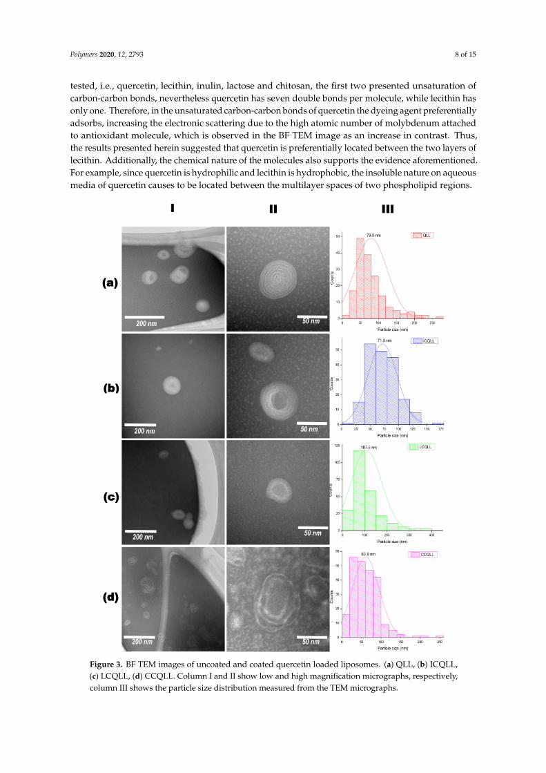
In addition, the chemical structure of quercetin also plays a major role in the chemical interactionswith the different polysaccharide. Figure 5 depicts the chemical structure of quercetin showing therelative position of hydroxyl groups (OH) were chemical interactions can occur. The hydroxyls fromcarbons C7, C14 and C15 are available to establish strong hydrogen bond-type interactions with thepolar groups of polysaccharides. Hydroxyls from carbon C3 and C5 also may establish hydrogenbond interactions, but the close position relative to the carbonyl group at C4 limits their availability tointeract with external polar groups [55]. From the three polysaccharides tested in the present work,lactose is a disaccharide, while chitosan and inulin are polysaccharides. Thus, the molecule sizei.e., molecular weight distribution, and the number of hydroxyls available for chemical interactions,also influences the release of quercetin in the alcoholic solution. Leyva-Porras et al. showed that inulinwith a higher degree of polymerization (DP) adsorbed more water than the low DP inulin in the wholerange of water activities (aw) [56]. Araujo-Díaz et al. reported a better performance of maltodextrin inthe conservation of resveratrol and quercetin when compared with inulin [36]. Besides the differencesin chemical structure between these two polysaccharides, the DP was 2-12 for inulin and 2-16 formaltodextrin. In this sense, low DP maltodextrins have shown a better performance in the content ofquercetin than high DP maltodextrins [57].
Figure 5. Quercetin molecule showing the relative positions of its hydroxyls.
Recently, Leyva-Porras et al., qualitatively compared the interaction of maltodextrins withquercetin and resveratrol, and concluded that the number of active sites for chemical interactions ismore important than chemical hindrance in these molecules [58]. From the above it is possible to inferthat polysaccharides with relatively high molecular weight distribution such as chitosan and inulintend to trap the quercetin within the liposome, while in lactose-coated liposomes, and due to the lowmolecular weight of the disaccharide, the antioxidant is more available for interactions with the DCCPmolecule, hence the higher antioxidant activity.
Agglomeration occurs when the material is dried i.e. for morphological studies. While thematerial was in solution, agglomeration was not noticed, indicating the suspension of liposomes in

Polymers 2020, 12, 2793 12 of 15
solution. According to Hao et al. [23], particle growth was observed (although it was not identified asagglomeration) and apparently did not affect the performance in the antioxidant properties. In thework reported by Li et al. [42] agglomeration was observed, but the agglomerated materials werediscarded prior the drug release tests. In the present work, there was not a noticeable trend betweenthe agglomerated particles and the performance as antioxidant. The liposomes coated with chitosanpresented the largest particle size (identified as agglomeration) and an intermediate antioxidant behavior.Conversely, the antioxidant behavior of lactose and inulin coated liposomes, which showed relativelysmaller particle size (i.e., less agglomerated than those coated with chitosan), presented the higher andlower antioxidant activities, respectively. Then, the differences in the antioxidant performance may beattributed to the chemical interactions of quercetin, and the microstructural disorder induced by thecoating polysaccharides, rather than to the agglomeration of liposomes in solution.
4. Conclusions
Three polysaccharides (lactose, chitosan and inulin) were employed as coating materials in thepreparation of quercetin-loaded liposomes (QLLs). Coated and uncoated liposomes were characterizedand tested in the scavenging of free radicals in solution. Particle size determined by light scatteringshowed large particle sizes in the range of 200 nm for QLL, LCQLL, and ICQLL, while it was 600 nm forCCQLL. This was corroborated by scanning electron microscopy (SEM) as due to the agglomeration ofpseudo-spherical particles. However, from transmission electron microscopy (TEM) the microstructurerevealed the primary particle size of the coated liposomes below 100 nm, and the layered structureformed of at least two layers. The antioxidant activity of QLL showed a better performance thatthe coated liposomes. This was explained in terms of quercetin availability since in the coatedliposomes, the polysaccharides trap the quercetin between the layers, reducing its availability forradical scavenging. Among the three coating materials, lactose presented the higher antioxidant activity.This may be caused by the relatively smaller size of the disaccharide when compared with the other twopolysaccharides (inulin and chitosan) which are considered as carbohydrate polymers. This suggestedthat the higher the molecular weight of the polysaccharide, the higher the chemical interactions withthe quercetin inside the liposome, observed as the retention of the antioxidant and consequently adecrease in the antioxidant activity. This study opens the possibility of using polysaccharides withdifferent potential properties for coating liposomes in the conservation and release of antioxidants.
Author Contributions: Conceptualization, M.Z.S.-L. and C.L.-P.; methodology, A.A.-E. and M.R.-A.; validation,C.L.-P. and M.Z.S.-L.; formal analysis, A.A.-E. and M.R.-A.; investigation, M.R.-A.; writing-original draftpreparation, M.Z.S.-L. and C.L.-P.; writing—review and editing, P.C.-A., C.L.-P. and M.Z.S.-L.; visualization,P.C.-A. All authors have read and agreed to the published version of the manuscript.
Funding: This work did not receive any funding.
Acknowledgments: Manuel Román-Aguirre is grateful to the Consejo Nacional de Ciencia y Tecnología(CONACYT) in Mexico for the financial support provided during his Ph.D. Studies through the scholarshipNo. 2018-000068-02NACF-24366. Also to Virgina Nevarez Morillón, and Diana Santos Gallegos from theUniversidad Autónoma de Chihuahua (UACH) for their kind support in the liofilization process of the samples.The technical support provided by Raúl Ochoa and Carlos Santillán during the TEM and light scattering analyzes,respectively. C.L.-P. thanks Andréa P. Balderrama-Aguilar for her support in the grammatical correction of therevised manuscript.
Conflicts of Interest: The authors declare no conflict of interest.
References
1. Eça, K.S.; Sartori, T.; Menegalli, F.C. Films and edible coatings containing antioxidants—A review. Braz. J.Food Technol. 2014, 17, 98–112.
2. Lorenzo, J.M.; Pateiro, M.; Fontán, M.C.G.; Carballo, J. Effect of fat content on physical, microbial, lipid andprotein changes during chill storage of foal liver pâté. Food Chem. 2014, 155, 57–63. [CrossRef] [PubMed]

Polymers 2020, 12, 2793 13 of 15
3. Sánchez-Ortega, I.; García-Almendárez, B.E.; Santos-López, E.M.; Amaro-Reyes, A.; Barboza-Corona, J.E.;Regalado, C. Antimicrobial Edible Films and Coatings for Meat and Meat Products Preservation. Sci. WorldJ. 2014, 2014, 248935.
4. Lobo, V.; Patil, A.; Phatak, A.; Chandra, N. Free radicals, antioxidants and functional foods: Impact onhuman health. Pharmacogn. Rev. 2010, 4, 118–126. [CrossRef]
5. Lorenzo, J.M.; Pateiro, M.; Domínguez, R.; Barba, F.J.; Putnik, P.; Kovacevic, D.B.; Shpigelman, A.; Granato, D.;Franco, D. Berries extracts as natural antioxidants in meat products: A review. Food Res. Int. 2018,106, 1095–1104. [CrossRef]
6. Przybylski, R.; Firdaous, L.; Châtaigné, G.; Dhulster, P.; Nedjar, N. Production of an antimicrobial peptidederived from slaughterhouse by-product and its potential application on meat as preservative. Food Chem.2016, 211, 306–313. [CrossRef]
7. Sanz, S.; Olarte, C.; Ayala, F.; Echávarri, J.F. Evolution of Quality Characteristics of Minimally ProcessedAsparagus During Storage in Different Lighting Conditions. J. Food Sci. 2009, 74, S296–S302. [CrossRef]
8. Takahashi, O. Haemorrhages due to defective blood coagulation do not occur in mice and guinea-pigsfed butylated hydroxytoluene, but nephrotoxicity is found in mice. Food Chem. Toxicol. 1992, 30, 89–97.[CrossRef]
9. Wang, W.; Kannan, P.; Xue, J.; Kannan, K. Synthetic phenolic antioxidants, including butylated hydroxytoluene(BHT), in resin-based dental sealants. Environ. Res. 2016, 151, 339–343. [CrossRef]
10. Özcan, M.M.; Arslan, D. Antioxidant effect of essential oils of rosemary, clove and cinnamon on hazelnutand poppy oils. Food Chem. 2011, 129, 171–174. [CrossRef]
11. Singh, G.; Maurya, S.; deLampasona, M.P.; Catalan, C.A.N. A comparison of chemical, antioxidant andantimicrobial studies of cinnamon leaf and bark volatile oils, oleoresins and their constituents. Food Chem.Toxicol. 2007, 45, 1650–1661. [CrossRef]
12. Samoticha, J.; Jara-Palacios, M.J.; Hernández-Hierro, J.M.; Heredia, F.J.; Wojdyło, A. Phenolic compounds andantioxidant activity of twelve grape cultivars measured by chemical and electrochemical methods. Eur. FoodRes. Technol. 2018, 244, 1933–1943. [CrossRef]
13. Oroian, M.; Escriche, I. Antioxidants: Characterization, natural sources, extraction and analysis. Food Res.Int. 2015, 74, 10–36. [CrossRef] [PubMed]
14. Russo, M.; Spagnuolo, C.; Tedesco, I.; Bilotto, S.; Russo, G.L. The flavonoid quercetin in disease preventionand therapy: Facts and fancies. Biochem. Pharmacol. 2012, 83, 6–15. [CrossRef] [PubMed]
15. Althans, D.; Schrader, P.; Enders, S. Solubilisation of quercetin: Comparison of hyperbranched polymer andhydrogel. J. Mol. Liq. 2014, 196, 86–93. [CrossRef]
16. Basu, A.; Kundu, S.; Sana, S.; Halder, A.; Abdullah, M.F.; Datta, S.; Mukherjee, A. Edible nano-bio-compositefilm cargo device for food packaging applications. Food Packag. Shelf Life 2017, 11, 98–105. [CrossRef]
17. Scalia, S.; Mezzena, M. Incorporation of quercetin in lipid microparticles: Effect on photo- andchemical-stability. J. Pharm. Biomed. Anal. 2009, 49, 90–94. [CrossRef] [PubMed]
18. Frenzel, M.; Steffen-Heins, A. Impact of quercetin and fish oil encapsulation on bilayer membrane andoxidation stability of liposomes. Food Chem. 2015, 185, 48–57. [CrossRef]
19. Das, S.S.; Hussain, A.; Verma, P.R.P.; Imam, S.S.; Altamimi, M.A.; Alshehri, S.; Singh, S.K. Recent Advancesin Liposomal Drug Delivery System of Quercetin for Cancer Targeting: A Mechanistic Approach. Curr. DrugDeliv. 2020, 17, 845–860. [CrossRef]
20. Grit, M.; Crommelin, D.J.A. Chemical stability of liposomes: Implications for their physical stability.Chem. Phys. Lipids 1993, 64, 3–18. [CrossRef]
21. Aditya, N.P.; Macedo, A.S.; Doktorovova, S.; Souto, E.B.; Kim, S.; Chang, P.-S.; Ko, S. Development andevaluation of lipid nanocarriers for quercetin delivery: A comparative study of solid lipid nanoparticles (SLN),nanostructured lipid carriers (NLC), and lipid nanoemulsions (LNE). LWT 2014, 59, 115–121. [CrossRef]
22. Frenzel, M.; Steffen-Heins, A. Whey protein coating increases bilayer rigidity and stability of liposomes infood-like matrices. Food Chem. 2015, 173, 1090–1099. [CrossRef] [PubMed]
23. Hao, J.; Guo, B.; Yu, S.; Zhang, W.; Zhang, D.; Wang, J.; Wang, Y. Encapsulation of the flavonoid quercetinwith chitosan-coated nano-liposomes. LWT 2017, 85, 37–44. [CrossRef]
24. Lopes, N.A.; Barreto Pinilla, C.M.; Brandelli, A. Antimicrobial activity of lysozyme-nisin co-encapsulated inliposomes coated with polysaccharides. Food Hydrocoll. 2019, 93, 1–9. [CrossRef]

Polymers 2020, 12, 2793 14 of 15
25. Lopes, N.A.; Pinilla, C.M.B.; Brandelli, A. Pectin and polygalacturonic acid-coated liposomes as noveldelivery system for nisin: Preparation, characterization and release behavior. Food Hydrocoll. 2017, 70, 1–7.[CrossRef]
26. Samadikhah, H.R.; Majidi, A.; Nikkhah, M.; Hosseinkhani, S. Preparation, characterization, and efficienttransfection of cationic liposomes and nanomagnetic cationic liposomes. Int. J. Nanomed. 2011, 6, 2275–2283.
27. Tai, K.; Rappolt, M.; Mao, L.; Gao, Y.; Li, X.; Yuan, F. The stabilization and release performances ofcurcumin-loaded liposomes coated by high and low molecular weight chitosan. Food Hydrocoll. 2020,99, 105355. [CrossRef]
28. Tan, C.; Feng, B.; Zhang, X.; Xia, W.; Xia, S. Biopolymer-coated liposomes by electrostatic adsorption ofchitosan (chitosomes) as novel delivery systems for carotenoids. Food Hydrocoll. 2016, 52, 774–784. [CrossRef]
29. Hasan, M.; Ben Messaoud, G.; Michaux, F.; Tamayol, A.; Kahn, C.J.F.; Belhaj, N.; Linder, M.; Arab-Tehrany, E.Chitosan-coated liposomes encapsulating curcumin: Study of lipid–polysaccharide interactions andnanovesicle behavior. RSC Adv. 2016, 6, 45290–45304. [CrossRef]
30. Ramezanzade, L.; Hosseini, S.F.; Nikkhah, M. Biopolymer-coated nanoliposomes as carriers of rainbow troutskin-derived antioxidant peptides. Food Chem. 2017, 234, 220–229. [CrossRef]
31. De Leo, V.; Milano, F.; Mancini, E.; Comparelli, R.; Giotta, L.; Nacci, A.; Longobardi, F.; Garbetta, A.;Agostiano, A.; Catucci, L. Encapsulation of Curcumin-Loaded Liposomes for Colonic Drug Delivery ina pH-Responsive Polymer Cluster Using a pH-Driven and Organic Solvent-Free Process. Molecules 2018,23, 739. [CrossRef] [PubMed]
32. Kang, E.-C.; Aklyoshi, K.; Sunamoto, J. Surface Coating of Liposomes with Hydrophobized Polysaccharides.J. Bioact. Compat. Polym. 1997, 12, 14–26. [CrossRef]
33. Sunamoto, J.; Iwamoto, K.; Takada, M.; Yuzuriha, T.; Katayama, K. Improved Drug Delivery to TargetSpecific Organs Using Liposomes as Coated with Polysaccharides. In Polymers in Medicine: Biomedical andPharmacological Applications; Chiellini, E., Giusti, P., Eds.; Springer US: Boston, MA, USA, 1983.
34. Betker, J.L.; Anchordoquy, T.J. The Use of Lactose as an Alternative Coating for Nanoparticles. J. Pharm. Sci.2020, 109, 1573–1580. [CrossRef] [PubMed]
35. Iwabuchi, K.; Masuda, H.; Kaga, N.; Nakayama, H.; Matsumoto, R.; Iwahara, C.; Yoshizaki, F.; Tamaki, Y.;Kobayashi, T.; Hayakawa, T.; et al. Properties and functions of lactosylceramide from mouse neutrophils.Glycobiology 2015, 25, 655–668. [CrossRef] [PubMed]
36. Araujo-Díaz, S.B.; Leyva-Porras, C.; Aguirre-Bañuelos, P.; Álvarez-Salas, C.; Saavedra-Leos, Z. Evaluation ofthe physical properties and conservation of the antioxidants content, employing inulin and maltodextrin inthe spray drying of blueberry juice. Carbohydr. Polym. 2017, 167, 317–325. [CrossRef]
37. Guldiken, B.; Linke, A.; Capanoglu, E.; Boyacioglu, D.; Kohlus, R.; Weiss, J.; Gibis, M. Formation andcharacterization of spray dried coated and uncoated liposomes with encapsulated black carrot extract.J. Food Eng. 2019, 246, 42–50. [CrossRef]
38. Derksen, J.T.P.; Morselt, H.W.M.; Scherphof, G.L. Processing of different liposome markers after in vitrouptake of immunoglobulin-coated liposomes by rat liver macrophages. Biochim. Biophys. Acta Mol. Cell Res.1987, 931, 33–40. [CrossRef]
39. Takada, M.; Yuzuriha, T.; Katayama, K.; Iwamoto, K.; Sunamoto, J. Increased lung uptake of liposomescoated with polysaccharides. Biochim. Biophys. Acta Gen. Subj. 1984, 802, 237–244. [CrossRef]
40. Park, S.N.; Jo, N.R.; Jeon, S.H. Chitosan-coated liposomes for enhanced skin permeation of resveratrol. J. Ind.Eng. Chem. 2014, 20, 1481–1485. [CrossRef]
41. Vural, I.; Sarisozen, C.; Olmez, S.S. Chitosan Coated Furosemide Liposomes for Improved Bioavailability.J. Biomed. Nanotechnol. 2011, 7, 426–430. [CrossRef]
42. Li, Z.; Paulson, A.T.; Gill, T.A. Encapsulation of bioactive salmon protein hydrolysates with chitosan-coatedliposomes. J. Funct. Foods 2015, 19, 733–743. [CrossRef]
43. Romero-Pérez, A.; García-García, E.; Zavaleta-Mancera, A.; Ramírez-Bribiesca, J.E.; Revilla-Vázquez, A.;Hernández-Calva, L.M.; López-Arellano, R.; Cruz-Monterrosa, R.G. Designing and evaluation of sodiumselenite nanoparticles in vitro to improve selenium absorption in ruminants. Vet. Res. Commun. 2010,34, 71–79. [CrossRef] [PubMed]
44. Barea, M.J.; Jenkins, M.J.; Lee, Y.S.; Johnson, P.; Bridson, R.H. Encapsulation of Liposomes within pHResponsive Microspheres for Oral Colonic Drug Delivery. Int. J. Biomater. 2012, 2012, 458712. [CrossRef][PubMed]

Polymers 2020, 12, 2793 15 of 15
45. Caddeo, C.; Díez-Sales, O.; Pons, R.; Carbone, C.; Ennas, G.; Puglisi, G.; Fadda, A.M.; Manconi, M.Cross-linked chitosan/liposome hybrid system for the intestinal delivery of quercetin. J. Colloid Interface Sci.2016, 461, 69–78. [CrossRef]
46. Tan, C.; Zhang, Y.; Abbas, S.; Feng, B.; Zhang, X.; Xia, S. Modulation of the carotenoid bioaccessibility throughliposomal encapsulation. Colloids Surf. B Biointerfaces 2014, 123, 692–700. [CrossRef]
47. Zhou, W.; Liu, W.; Zou, L.; Liu, W.; Liu, C.; Liang, R.; Chen, J. Storage stability and skin permeation ofvitamin C liposomes improved by pectin coating. Colloids Surf. B Biointerfaces 2014, 117, 330–337. [CrossRef]
48. Belhaj, N.; Arab-Tehrany, E.; Loing, E.; Bézivin, C. Skin delivery of hydrophilic molecules from liposomesand polysaccharide-coated liposomes. Int. J. Cosmet. Sci. 2017, 39, 435–441. [CrossRef]
49. Aguiar, J.; Costa, R.; Rocha, F.; Estevinho, B.N.; Santos, L. Design of microparticles containing naturalantioxidants: Preparation, characterization and controlled release studies. Powder Technol. 2017, 313, 287–292.[CrossRef]
50. Henriksen, I.; Smistad, G.; Karlsen, J. Interactions between liposomes and chitosan. Int. J. Pharm. 1994,101, 227–236. [CrossRef]
51. Celli, G.B.; Ghanem, A.; Brooks, M.S.-L. Bioactive Encapsulated Powders for Functional Foods—A Reviewof Methods and Current Limitations. Food Bioprocess Technol. 2015, 8, 1825–1837. [CrossRef]
52. Zhao, G.D.; Sun, R.; Ni, S.L.; Xia, Q. Development and characterisation of a novel chitosan-coated antioxidantliposome containing both coenzyme Q10 and alpha-lipoic acid. J. Microencapsul. 2015, 32, 157–165. [CrossRef][PubMed]
53. Caddeo, C.; Gabriele, M.; Fernàndez-Busquets, X.; Valenti, D.; Fadda, A.M.; Pucci, L.; Manconi, M. Antioxidantactivity of quercetin in Eudragit-coated liposomes for intestinal delivery. Int. J. Pharm. 2019, 565, 64–69.[CrossRef]
54. Takeuchi, H.; Kojima, H.; Yamamoto, H.; Kawashima, Y. Evaluation of circulation profiles of liposomes coatedwith hydrophilic polymers having different molecular weights in rats. J. Control. Release 2001, 75, 83–91.[CrossRef]
55. Desiraju, G.R.; Steiner, T. The Weak Hydrogen Bond: In Structural Chemistry and Biology; Oxford UniversityPress/International Union of Crystallography: Oxford, UK, 2001; Volume 9.
56. Leyva-Porras, C.; Saavedra–Leos, M.Z.; López-Pablos, A.L.; Soto-Guerrero, J.J.; Toxqui-Terán, A.;Fozado-Quiroz, R.E. Chemical, Thermal and Physical Characterization of Inulin for its TechnologicalApplication Based on the Degree of Polymerization. J. Food Process Eng. 2017, 40, e12333. [CrossRef]
57. Saavedra-Leos, M.Z.; Leyva-Porras, C.; López-Martínez, L.A.; González-García, R.; Martínez, J.O.; CompeánMartínez, I.; Toxqui-Terán, A. Evaluation of the Spray Drying Conditions of Blueberry Juice-Maltodextrin onthe Yield, Content, and Retention of Quercetin 3-d-Galactoside. Polymers 2019, 11, 312. [CrossRef] [PubMed]
58. Leyva-Porras, C.; Saavedra-Leos, M.Z.; Cervantes-González, E.; Aguirre-Bañuelos, P.; Silva-Cázarez, M.B.;Álvarez-Salas, C. Spray drying of blueberry juice-maltodextrin mixtures: Evaluation of processing conditionson content of resveratrol. Antioxidants 2019, 8, 437. [CrossRef] [PubMed]
Publisher’s Note: MDPI stays neutral with regard to jurisdictional claims in published maps and institutionalaffiliations.
© 2020 by the authors. Licensee MDPI, Basel, Switzerland. This article is an open accessarticle distributed under the terms and conditions of the Creative Commons Attribution(CC BY) license (http://creativecommons.org/licenses/by/4.0/).
Copyright © 2022 FDOKUMEN