Controlling Viral Immuno-Inflammatory Lesions by Modulating ...
Upload
independentCategory
view
0download
0
Aa
ALa
b
C
a
ARRA
KQAEO
1
taq
liM2
d4T
B
1d
Pharmacological Research 61 (2010) 288–297
Contents lists available at ScienceDirect
Pharmacological Research
journa l homepage: www.e lsev ier .com/ locate /yphrs
nti-inflammatory effect of quercetin-loaded microemulsion in the airwaysllergic inflammatory model in mice
lexandre P. Rogerioa,1, Cristiana L. Dorab, Edinéia L. Andradea, Juliana S. Chavesa,uis F.C. Silvab, Elenara Lemos-Sennab, João B. Calixtoa,∗
Departamento de Farmacologia, Universidade Federal de Santa Catarina, Campus Universitário Trindade, Centro de Ciências Biológicas, Florianópolis, SC, BrazilLaboratório de Farmacotécnica, Departamento de Ciências Farmacêuticas, Centro de Ciências da Saúde, Universidade Federal de Santa Catarina,ampus Trindade, Florianópolis, Brazil
r t i c l e i n f o
rticle history:eceived 9 September 2009eceived in revised form 24 October 2009ccepted 25 October 2009
eywords:uercetin-loaded microemulsionirways allergic inflammationosinophilsral bioavailability
a b s t r a c t
Quercetin is a plant-derived flavonoid widely known by its anti-oxidant and anti-inflammatory proper-ties, but its oral bioavailability is very poor and this becomes difficult to assess its therapeutic potential.Here we have compared the anti-inflammatory effect of quercetin-loaded microemulsion (QU-ME) andquercetin suspension (QU-SP) in an experimental model of airways allergic inflammation. Mice receiveddaily oral doses of QU-ME (3 or 10 mg/kg; in an oil-in-water microemulsion content 0.02:0.2:1 oflecithin:castor oil:Solutol HS15®), QU-SP [10 mg/kg, in carboxymethylcellulose (CMC) 0.5% in water]or vehicle from the 18th to the 22nd day after the first immunization with ovalbumin (OVA). Dexam-ethasone was used as positive control drug. Every parameter was evaluated in the 22nd day (24 h afterthe second OVA-challenge). We have also tried to assess by HPLC–MS a quercetin metabolite in the bloodof rats treated with QU-SP or QU-ME. QU-ME was better orally absorbed when compared with QU-SP.Furthermore, oral administration of QU-SP failed to interfere with leukocyte recruitment, while QU-ME
inhibited in a dose-dependent way, the eosinophil recruitment to the bronchoalveolar lavage fluid (BALF).QU-ME also significantly reduced both IL-5 and IL-4 levels, but failed to interfere with CCL11, IFN-� andLTB4 levels. In addition, QU-ME oral treatment inhibited the nuclear transcription factor kappa B (NF-�B)activation, P-selectin expression and the mucus production in the lung. The present results show thatQU-ME exhibits pronounced anti-inflammatory properties in a murine model of airways allergic inflam-mation and suggest that it might present therapeutic potential for the airways inflammatory diseases management.. Introduction
Allergic asthma is a complex inflammatory disorder charac-erized by airway hyperresponsiveness, eosinophilic inflammationnd hypersecretion of mucus by goblet cells. This disease is fre-uently accompanied by high serum levels of immunoglobulin
Abbreviations: BALF, bronchoalveolar lavage fluid; HPLC–MS, high performanceiquid chromatography coupled to mass spectrometry; Ig, immunoglobulin; IL,nterleukin; LTB4, leukotriene B4; NF-�B, nuclear factor �B; OVA, ovalbumin; QU-
E, quercetin-loaded microemulsion; QU-SP, quercetin suspension; Th2, T helper.∗ Corresponding author at: Departamento de Farmacologia, Universidade Federale Santa Catarina (UFSC), Campus Universitário, Trindade, Bloco D, CCB, Caixa Postal76, Florianópolis, Santa Catarina, CEP 88049-900, Brazil.el.: +55 48 3721 9491/9764; fax: +55 48 3337 5479.
E-mail addresses: [email protected], [email protected] (J.B. Calixto).1 Present address: Universidade Federal do Triângulo Mineiro, Uberaba, MG,
razil.
043-6618/$ – see front matter © 2009 Elsevier Ltd. All rights reserved.oi:10.1016/j.phrs.2009.10.005
© 2009 Elsevier Ltd. All rights reserved.
(Ig)E and associated with increasing intrapulmonary productionof certain interleukins (IL), especially IL-4, IL-5 and IL-13 byallergen-specific T helper 2 (Th2) cells [1]. Integrated signalingevents between IL-4 and IL-13 is believed to regulate pulmonaryeosinophilia by stimulating eosinophil-specific adhesion pathwaysand by modulating the local production of IL-5 and CCL11, whichin turn, selectively drive eosinophil recruitment. Furthermore, IL-5also plays a critical role in the regulation of bone marrow and bloodeosinophilia [2].
Although, a great progress has been obtained in the last decadesin our understanding of the cellular and molecular mechanismsunderlying the allergic asthma, few effective and safe drugs are cur-rently available for this disease management. Corticosteroids arethe most established therapy for controlling most type of inflam-
matory reaction, eosinophilic included, exerting a strong effecton leukocyte recruitment, when administered locally or system-ically [3]. Though these drugs have pronounced pharmacologicalactivity, they exhibit severe adverse effects that preclude theirlong-term use. Furthermore, pharmacologically active compounds
ogical
ctfdd
ddkcflavpbtaerwbccpstoacth
ha((tf
hiuctawb[
tmam
2
2
lBcadm
A.P. Rogerio et al. / Pharmacol
apable of inhibiting eosinophil function or inflammatory cells infil-ration into the lung, with less adverse effects, are still neededor the treatment of patients with atopy/allergic, parasitic disor-ers, hypereosinophilic syndromes, and other eosinophil-relatediseases.
Most plant-derived secondary metabolites are capable ofirectly affecting inflammatory mediators, as well as the pro-uction/activity of second messengers, transcription factors andey pro-inflammatory molecules expression [4,5]. Flavonoids areommon secondary metabolites found in the plant kingdom. Theavonoid quercetin is largely found in the diet and its main sourcesre tea, onions, apples and red wine [6]. Studies carried out initro have shown that quercetin exhibits, besides other activities,ronounced anti-inflammatory property, an effect that seems toe associated with its ability to block some inflammatory media-ors [7], adhesion molecules expression [8], inducible enzymes [9]nd nuclear transcription factor activation [10]. In asthma mod-ls, quercetin administered by oral [11,12] or intraperitoneal [13]outes or by aerosol [14,15] was able to reduce the allergic air-ay inflammation [11,13–15], hyperresponsiveness [13–15] and
ronchial hyperactivity [12] as well as to modulate the Th1/Th2ytokines [11,13] associated with the reduction of the histamineontents [14,15]. Despite its potential systemic anti-inflammatoryroperty, quercetin aglycone is well known to possess poor waterolubility. Previous findings have showed that it is less absorbedhan quercetin glycosides and that its absorption seems to dependn the type and position of the sugar moieties [16,17]. In thebove studies, DMSO and polietilenoglicol have been used asoadjuvants to improve the quercetin solubilization and absorp-ion. However, these substances are not approved to be used inumans.
When quercetin aglycone (100 mg) was administrated inumans by intravenous route it bound 98% to plasma proteinsnd it was mainly excreted in urine as a conjugated metabolite7.4%) as unchanged (0.65%). On the other hand, when quercetin4 g) was given orally to human it was detected neither inhe plasma nor in urine, either unchanged or in a metabolizedorm [18].
The colloidal drug delivery system, such as microemulsions,as been proposed to improve the absorption and therapeutic
ndex of several drugs [19]. Microemulsions are isotropic liq-id mixtures of oil, water and surfactant, frequently found inombination with a co-surfactant and present translucence andhermodynamic stability. The dispersed phase generally lipophilic,ct as a potential reservoir of lipophilic drugs, which in contactith semi permeable membranes, such as skin or mucous mem-
rane can facilitate the transport of drugs through these barriers20,21].
In this study, we sought to compare the quercetin oral absorp-ion in the quercetin suspension (QU-SP) and quercetin-loaded
icroemulsion (QU-ME). In addition, we have also assessed the oralnti-inflammatory effect of both formulations in an experimentalodel of airways allergic inflammation in mice.
. Material and methods
.1. Materials
Quercetin (purity 98.2%), castor oil and carboxymethylcellu-ose (CMC) were purchased from Natural Pharma (São Paulo,
razil). Hydrogenated soybean lecithin (Lipoid S75-3N) was pur-hased from Via Pharma (São Paulo, Brazil). 12-hydroxysteariccid–polyethylene glycol copolymer (Solutol HS15®) was kindlyonated by Basf (Trostberg, Germany). Dexamethasone, ovalbu-in, periodic acid-Schiff stain, ethanol, acetone, methanol wereResearch 61 (2010) 288–297 289
purchased from Sigma–Aldrich (Missouri, USA). The solventsemployed for HPLC–MS analysis were of HPLC grade (JBaker;Xalostoc, Mexico). HPLC grade water (18 m�) was prepared usinga Milli-Q system (Millipore; Massachusetts, USA).
2.2. Animals
Experiments were conducted by using female BALB/c mice (8weeks old and weighing 20–25 g) and male Wistar rat (12 weeksold weighing 200–250 g) kept in controlled room temperature(22 ± 2 ◦C) and humidity (60–80%) under a 12:12 h light–dark cycle(lights on 06:00 h). At appropriate time intervals, mice were killedby isoflurane overdose. All procedures used in the present studywere approved by UFSC Ethics Committee on the Use of Animals(protocol number 110), which follows the ‘Principles of LaboratoryAnimal Care’ from NIH Publication No. 85-23.
2.3. Preparation of quercetin suspension and quercetinmicroemulsion
The quercetin microemulsion (QU-ME) was prepared by a hotsolvent diffusion method [22]. Briefly, 10 mg of quercetin, 10 mgof lecithin and 100 mg of castor oil were completely dissolved intoa mixture of acetone:ethanol (60:40, v/v) at 60 ◦C. The resultingorganic solution was quickly poured into 50 ml of an aqueous solu-tion containing 1% of the surfactant (Solutol HS15®), maintainedunder magnetic stirring at 82 ◦C. Then, the sample was cooledto room temperature, the organic solvent was evaporated underreduced pressure, and the final volume was adjusted to 20 ml.Finally, the nanocarrier was filtered through an 8 �m filter paper.
The quercetin suspension (QU-SP) was prepared by addingquercetin in aqueous carboxymethylcellulose dispersion (0.5%) inorder to obtain a final quercetin concentration equal to microemul-sion.
2.4. Quercetin content determination and microemulsionrecovery
The quercetin content in the microemulsion was determinedby UV spectrophotometric method using a PerkinElmer Lambda 10UV/VIS spectrophotometer. The sample was diluted in methanoland the absorption was measured at 375 nm. The quercetin con-tent was estimated by using a quercetin standard solution analyzedin the same conditions and expressed in �g of quercetin/ml ofmicroemulsion. The quercetin recovery was calculated as beingthe percentage of the quercetin total concentration found in themicroemulsion in relation to the amount initially added.
2.5. Particle size analysis
The mean particle diameter of the microemulsion and thepolydispersity index were determined by photon correlation spec-troscopy using a Zetasizer Nanoseries (Marvern Instruments;Worcestershire, UK). Each size analysis lasted 300 s and was per-formed at 25 ◦C with an angle detection of 173◦.
2.6. HPLC–MS analysis
HPLC analysis was performed on a LCMS-2010EV apparatuswith a diode array detector (SPD M20A; Shimadzu, Kyoto, Japan)and mass spectrometer (2010EV, Shimadzu), coupled with an
auto-injector (SIL-20A, Shimadzu), both using the software LC-MS Solutions 3.0 (Shimadzu). A Shim-pack VP-ODS column (5 �m,150 mm × 2.0 mm; Shimadzu) coupled with a guard-column (5 �m,5 mm × 2.0 mm; Shimadzu) was used. The mobile phase consistedof a gradient solvent system of aqueous formic acid 0.2% (A) and
2 ogical
a41flnpba
2
tut(ccnc
2
aatno1H
2
ih(owiAO
2s
ppiBliwc
2
eo0tv
90 A.P. Rogerio et al. / Pharmacol
cetonitrile (B). The elution profile was 0–4 min: 9–20% B (linear);–9 min: 20–100% B (linear); 9–14 min: 100–20% B min (linear);4–18 min: 20–9% B (linear); and 18–23 min: 9% B (isocratic). Theow rate was 200 �l/min. The mass spectrometer operated in theegative and positive ionization mode. ESI-MS parameters were:otential of the ESI source 1.5 kV, CDL temperature heater 250 ◦C,lock heater 200 ◦C, nitrogen served as collision gas 1.5 ml/min. MSnalyses were recorded in the SCAN and SIM mode (m/z 301, 303).
.7. Quercetin oral absorption in rats
Animals were non-fasted by 12 h and then were orallyreated by gavage with QU-ME (10 mg/kg), QU-SP (10 mg/kg) ornloaded microemulsion. After 2 or 3 h, animals were anaes-hetized intraperitoneally with ketamine (70 mg/kg) and xylazine10 mg/kg), submitted to laparotomy and the blood samples wereollected by cardiac puncture. The samples were immediatelyentrifuged at 3700 × g for 15 min to separate the plasma. It isoteworthy to mention that each animal was used once for bloodollection and for each time point analyzed three animals was used.
.8. Sample preparation for chromatographic analysis
Samples were prepared by protein precipitation, evaporationnd re-dissolving steps. One milliliter of acetone and 10 �l of aceticcid were added to 100 �l of plasma. After mixed in vortex for 3 min,he mixture was centrifuged for 15 min at 20,800 × g. The super-atant was separated and evaporated to dryness by a gentle streamf nitrogen at 40 ◦C. The samples were reconstituted by addition of00 �l of acetonitrile: water (1:1), of which 20 �l was injected ontoPLC–MS system.
.9. Antigen immunization, booster, and airway challenge
Mice were immunized on days 0 and 7 by subcutaneousnjection of 4 �g of ovalbumin (OVA) plus 1.6 mg of aluminiumydroxide in 0.4 ml of saline followed by two intranasal challengeson post-immunization days 14 and 21) with 10 �g of OVA in 50 �lf saline, delivered into the nostrils under light ether anaesthesiaith the aid of a micropipette. The control group consisted of non-
mmunized mice that received two intranasal instillations of OVA.ll determinations were made at 24 h time point after the secondVA challenge (on post-immunization day 21) [23].
.10. Evaluation of leukocyte influx into the bronchoalveolarpace
Mice were killed by isoflurane overdose. Subsequently, aolyethylene cannula was introduced into the trachea, andhosphate-buffered saline (PBS) containing heparin (10 UI/ml) was
nstilled in three aliquots (0.3, 0.3 and 0.4 ml) in a total of 1 ml. TheALF was recovered and placed on ice. Total cell and differential
eukocyte count were made according Rogerio et al. [23]. Follow-ng centrifugation (400 × g, 5 min, 4 ◦C), supernatants of the BALF
ere collected and stored at −70 ◦C for subsequent cytokine andhemokine determination.
.11. Treatments
To investigate the possible therapeutic anti-inflammatory
ffects, QU-ME (at doses 3 or 10 mg/kg) or QU-SP (10 mg/kg)r unloaded microemulsion (an o/w microemulsion content.02:0.2:1 of lecithin:castor oil:Solutol HS15®) or aqueous solu-ion content CMC 0.5% were given orally by gavage with a volumearying between 140 and 580 �l according to the animals’ weightResearch 61 (2010) 288–297
and the dose, from day 18th to 22nd day after the first immu-nization with OVA. The positive control consisted of a one groupthat received dexamethasone (1 mg/kg, subcutaneous injection).The doses and period of treatment were carried out in according toprevious study [11].
2.12. Measurement of IL-4, IL-5, CCL11, IFN-� and LTB4 levels
The IL-4, IL-5, CLL11 and IFN-� levels were measured by specificELISA (RayBiotech; Georgia, USA). LTB4 were assayed according tothe manufacturer’s instructions (R&D Systems; Minnesota, USA).LTB4 in the BALF competed with a fixed amount of horseradishperoxidase (HRP)-labeled LTB4 for sites on a chicken polyclonalantibody. During the incubations, the chicken polyclonal antibodybecomes bound to the rabbit anti-chicken antibody coated ontothe microplate. Following a wash to remove excess conjugate andunbound LTB4 of BALF, a substrate solution was added to the wellsto determine the bound enzyme activity. Following color devel-opment, the assay was stopped, and the absorbance was read at450 nm. The intensity of the color was inversely proportional to theconcentration of LTB4 in the sample. Sensitivities were >10 pg/ml.
2.13. Analysis of mucus secretion in the lung histology
The lungs were removed, immersed in 4% phosphate-bufferedformalin, and embedded in paraffin. Tissues were cut into 5 �msections, which were then stained with periodic acid-Schiff stain toevaluate mucus production. Mucus hypersecretion by goblet cellsin the airway epithelium was analyzed using a five-point scoringsystem as previously described [24]. The scoring system was: 0, nogoblet cells; 1, less than 25%; 2, 25–50%; 3, 50–75%; and 4, morethan 75%. Goblet cell scoring was examined in three independentfields of lung section from each mouse.
2.14. Immunohistochemical studies
Immunohistochemical detections of NF-�B p65 subunit andP-selectin were carried out in the lung (5 �m slices), using poly-clonal rabbit anti-phospho-p65 NF-�B (#3037, 1:100) from CellSignaling Technology (Massachusetts, USA) and polyclonal goatanti-P-selectin (#6943, 1:2000), from Santa Cruz Biotechnology(California, USA) as described in a previous study [23].
2.15. Statistical analysis
The data are reported as mean ± S.E.M. The statistical signifi-cance among different treatments in each individual experimentwas compared by ANOVA. When significant differences wereidentified, individual comparisons were subsequently made withTukey’s test. Values of p < 0.05 were considered statistically signif-icant.
3. Results
3.1. Microemulsion characterization
A colloidal dispersion displaying mean particle size of approx-imately 20 nm and polydispersed index of 0.156 was obtainedby the hot solvent diffusion technique, as described in materialand methods section. The drug content and drug recovery were
430 �g/ml and 86%, respectively. Regarding the quercetin solubil-ity in water (0.33 �g/ml), the microemulsion allowed to increasethe drug concentration about 1.300 times in an aqueous disper-sion. We have prepared the quercetin suspension (QU-SP) with thesame quercetin final concentration of QU-ME (430 �g/ml).
A.P. Rogerio et al. / Pharmacological Research 61 (2010) 288–297 291
F le plac trile (B9 ). Them
3
tpsc(iv1q(mp
3B
ic2ai
ewwnaoosviB
ig. 1. HPLC–MS chromatograms (TIC × Tr): (A) quercetin standard and (B) rat samponsisted of a gradient solvent system of aqueous formic acid 0.2% (A) and acetoni–14 min: 100–20% B min (linear); 14–18: 20–9% B (linear); 18–23: 9% B (isocratic/z 301, positive ionization: m/z 303).
.2. Oral absorption of quercetin-loaded microemulsion in rats
Plasma samples obtained from rats were pre-treated with ace-one and acetic acid, followed by centrifugation to precipitatelasma proteins and give the supernatant. The HPLC–MS analy-is showed that retention time (tR) of quercetin under developedonditions was 10.25 min and the most abundant ion was m/z 301negative mode) both at 2 and 3 h (Fig. 1A). No peak of quercetinn plasma was observed in this region after the treatment withehicle (unloaded microemulsion) or QU-SP (Supplementary Fig.A and B). In the same conditions, it was possible to detect auercetin metabolite in plasma rats treated orally with QU-MEFig. 1B). This compound, so far not yet identified, represents a
inor polar metabolite (tR = 11.25 min), and it was detected in theositive mode and showed molecular ion m/z 303.
.3. Therapeutic effect of QU-ME on the leukocytes number in theALF
The volume usually recommended by oral route administrationn mice is 0.1 ml/10 g of weight. However, as the quercetin con-entration achieved was 430 �g/ml the administration volume was.32 times higher than the recommended to reach the dose to bedministrated. However, no important alterations were observedn the mice after the administration of the highest volume.
To assess the possible therapeutic anti-inflammatory prop-rty of QU-ME and QU-SP, OVA-immunized mice were treatedith QU-ME (at doses 3 or 10 mg/kg), with QU-SP (10 mg/kg) orith the respective vehicle from day 18th to 22nd day. No sig-ificant difference was observed between the OVA-immunizednd -challenged groups treated with unloaded microemulsion (anil-in–water microemulsion content 0.02:0.2:1 of lecithin:castor
il:Solutol HS15®) or aqueous solution content CMC 0.5% (data nothown). As can be seen in Fig. 2A–C mice that received only theehicle (unloaded microemulsion) exhibited a significant increasen neutrophil, eosinophil and mononuclear cells numbers in theALF when compared control animals. QU-ME given orally atsma (n = 3) after oral treatment with QU-ME (10 mg/kg of quercetin). Mobile phase). The elution profile was 0–4 min: 9–20% B (linear); 4–9 min: 20–100% B (linear);flow rate was 200 �l/min. ESI source operated in SIM mode (negative ionization:
10 mg/kg, like dexamethasone (1 mg/kg), significantly decreasedthe eosinophil recruitment (68 ± 4% and 84 ± 3%, respectively) tothe BALF, when compared with vehicle-treated group (Fig. 2B). Inaddition, dexamethasone completely inhibited the mononuclearcells numbers (Fig. 2C). No significant differences were observedin the groups treated orally with QU-ME at dose of 3 mg/kg, aswell as in the animals treated with QU-SP (Fig. 2A–C). Moreover, noalteration in the neutrophil numbers was observed in the treatedgroups with QU-SP, QU-ME and dexamethasone, when comparedto vehicle (Fig. 2A). QU-ME at dose 10 mg/kg reduced significantlythe eosinophil recruitment (62 ± 9%) to the BALF when comparedwith QU-SP (Fig. 2B).
Since that QU-SP did not show any anti-inflammatory effect,as evidenced by the lack of reduction in the number of neu-trophils, eosinophils and mononuclear cells when compared withOVA + vehicle group, all subsequent experiments were carried outonly with QU-ME.
3.4. Effects of QU-ME on the IL-4, IL-5, IFN-� , CCL11 and LTB4levels in the BALF
As only QU-ME (10 mg/kg) exhibited significant oral anti-inflammatory activity, we selected this dose to evaluate thecytokines (IL-4, IL-5 and IFN-�), the chemokine (CCL11) and thelipid (LTB4) levels in the BALF. In OVA-immunized and -challengedmice treated with vehicle (unloaded microemulsion), the IL-4, IL-5,CLL11 and LTB4 levels were significantly elevated when assessed24 h after the last OVA-challenge and compared with control group(Fig. 3A–D). The therapeutic treatment with QU-ME significantlyprevented the IL-5 and IL-4 levels (62 ± 2% and 71 ± 8%, respec-tively), when compared with vehicle-treated animals (Fig. 3A andB). However, no statistically significant differences were observed
in CCL11 and LTB4 levels (Fig. 3C and D, respectively). Dexametha-sone, used as the positive control, significantly reduced the IL-5(68 ± 3%), IL-4 (69 ± 6%), CCL11 (63 ± 15%) and LTB4 (61 ± 12%) lev-els (Fig. 3A–D). No alteration of IFN-� levels was observed amongthe experimental groups (data not shown).
292 A.P. Rogerio et al. / Pharmacological Research 61 (2010) 288–297
Fig. 2. The therapeutic oral treatment effect with QU-ME on the recruitment of neutrophils (A), eosinophils (B) and mononuclear cells (C) number in BALF of mice immunizeda g), QU2 OVA-e < 0.05a
3
th
Fvmw
nd then challenged with OVA. Mice were treated orally with QU-ME (3 or 10 mg/k2nd day after the first immunization. Samples were collected 24 h after the secondrrors are depicted (n = 6 per treatment). *p < 0.05 compared with control group; #pccording to Tukey’s test.
.5. Effects of QU-ME on the mucus secretion in the lung
In order to investigate the effects of QU-ME on mucus secre-ion, the lung tissues were stained with periodic acid-Schiff. Ayperproduction of mucus by globet cells was observed in the
ig. 3. The therapeutic oral treatment effect with QU-ME on IL-5 (A), IL-4 (B), CCL11 (C)ehicle or dexamethasone (1 mg/kg, s.c.) from the 18th day to the 22nd day after the firseans of at least three independent experiments and their standard errors are depictedith OVA + vehicle group according to Tukey’s test.
-SP (10 mg/kg), vehicle or dexamethasone (1 mg/kg, s.c.) from the 18th day to thechallenge. The means of at least three independent experiments and their standard
compared with OVA + vehicle group; +p < 0.05 compared with OVA + QU-SP group
bronchi of OVA-immunized and -challenged mice treated withvehicle (Fig. 4B and E) when compared with control group (Fig. 4Aand E). Therapeutic treatment with QU-ME (Fig. 4D and E) or withdexamethasone (Fig. 4C and E), significantly decreased the degreeof mucus secretion (62 ± 21% and 50 ± 12%, respectively) when
and LTB4 (D) levels in the BALF. Mice were treated orally with QU-ME (10 mg/kg),t immunization. Samples were collected 24 h after the second OVA challenge. The(n = 6 per treatment). *p < 0.05 compared with control group; #p < 0.05 compared

A.P. Rogerio et al. / Pharmacological Research 61 (2010) 288–297 293
F mucuc ith pi nts anc st.
cv
3e
ptn(o-poeit
ig. 4. The therapeutic oral treatment effect with QU-ME (10 mg/kg) on the airwayhallenged mice given vehicle (B), dexamethasone (C) or QU-ME (D) were stained wn the lung tissues was scored (E). The means of at least two independent experimeontrol group; #p < 0.05 compared with OVA + vehicle group according to Tukey’s te
ompared with OVA-immunized and -challenge mice treated withehicle.
.6. Effect of QU-ME on the NF-�B activation and P-selectinxpression in the lung
The effects of QU-ME treatment on P-selectin expression and thehosphorylation state of p65 NF-�B were evaluated by immunohis-ochemistry technique. Under homeostatic conditions, there wasot significant activation of p65 NF-�B was found in the lung cellsFig. 5A and E). However, a constitutive staining for P-selectin wasbserved (Fig. 6A and E). In the lung tissue of OVA-immunized andchallenged mice treated with vehicle it was detected the phos-
horylated p65 subunit of NF-�B (Fig. 5B and E) in the nucleusf endothelial and inflammatory cells around the bronchiolarpithelium. In addition, P-selectin staining was also significantlyncreased in these cells (Fig. 6B and E). Of note, QU-ME given orallyo mice, completely inhibited the p65 NF-�B activation (Fig. 5Ds. Representative lung tissue sections from control mice (A), immunized and OVA-eriodic acid-Schiff (magnification: 200×). Mucus production (indicated by arrows)d their standard errors are depicted (n = 4 per treatment). *p < 0.05 compared with
and E), and also the P-selectin overexpression (Fig. 6D and E) inthe lung, when compared to the vehicle-treated animals. Likewise,dexamethasone, abolished the activation of p65 NF-�B (Fig. 5C andE), but it failed to prevent the P-selectin expression in the lung(Fig. 6C and E).
4. Discussion
The main findings that emerge for the present study was thedemonstration that, in contrast to QU-SP, QU-ME like the steroidalanti-inflammatory drug dexamethasone, exhibits pronounced oralanti-inflammatory property when evaluated in OVA-induced air-
ways allergic inflammation. Also relevant, the HPLC–MS analysiscarried out in plasma of rats treated orally with QU-ME, differ-ently of QU-SP, revealed the presence of a substance detected asion [M+H]+ m/z 303, that probably represents an active metaboliteof quercetin. Together, these data strongly support the notion that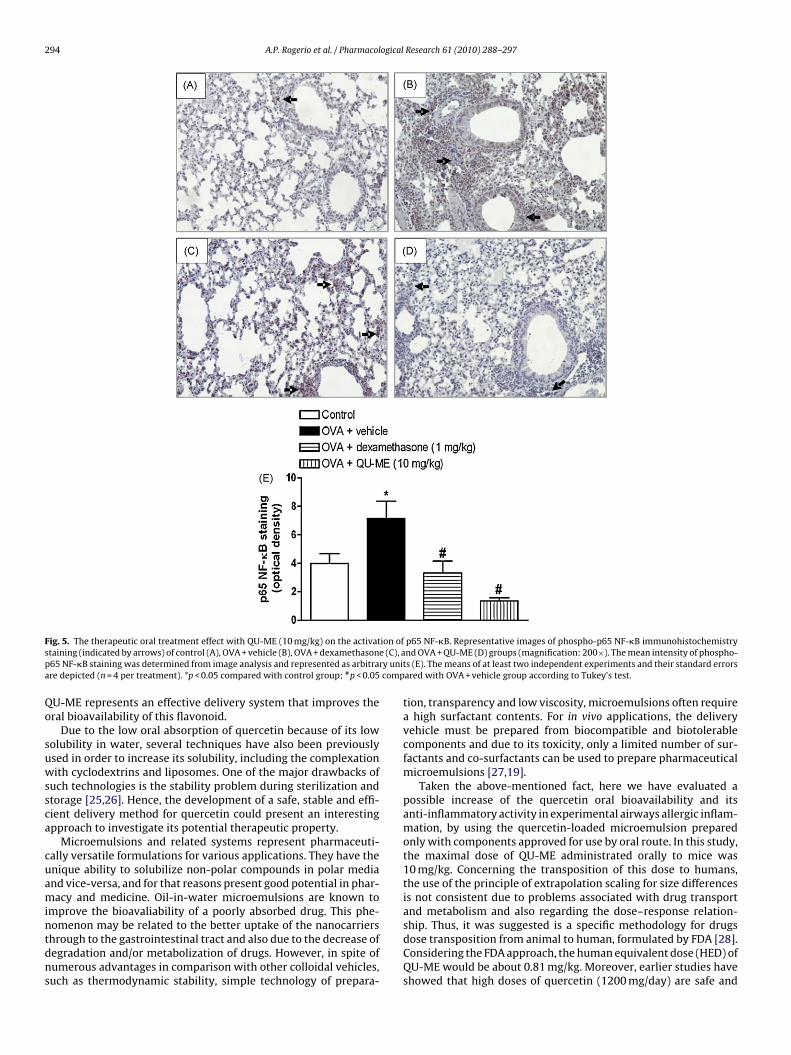
294 A.P. Rogerio et al. / Pharmacological Research 61 (2010) 288–297
F ion ofs e (C), ap ry unita comp
Qo
suwssca
cuamintdns
ig. 5. The therapeutic oral treatment effect with QU-ME (10 mg/kg) on the activattaining (indicated by arrows) of control (A), OVA + vehicle (B), OVA + dexamethason65 NF-�B staining was determined from image analysis and represented as arbitrare depicted (n = 4 per treatment). *p < 0.05 compared with control group; #p < 0.05
U-ME represents an effective delivery system that improves theral bioavailability of this flavonoid.
Due to the low oral absorption of quercetin because of its lowolubility in water, several techniques have also been previouslysed in order to increase its solubility, including the complexationith cyclodextrins and liposomes. One of the major drawbacks of
uch technologies is the stability problem during sterilization andtorage [25,26]. Hence, the development of a safe, stable and effi-ient delivery method for quercetin could present an interestingpproach to investigate its potential therapeutic property.
Microemulsions and related systems represent pharmaceuti-ally versatile formulations for various applications. They have thenique ability to solubilize non-polar compounds in polar mediand vice-versa, and for that reasons present good potential in phar-acy and medicine. Oil-in-water microemulsions are known to
mprove the bioavaliability of a poorly absorbed drug. This phe-
omenon may be related to the better uptake of the nanocarriershrough to the gastrointestinal tract and also due to the decrease ofegradation and/or metabolization of drugs. However, in spite ofumerous advantages in comparison with other colloidal vehicles,uch as thermodynamic stability, simple technology of prepara-p65 NF-�B. Representative images of phospho-p65 NF-�B immunohistochemistrynd OVA + QU-ME (D) groups (magnification: 200×). The mean intensity of phospho-s (E). The means of at least two independent experiments and their standard errorsared with OVA + vehicle group according to Tukey’s test.
tion, transparency and low viscosity, microemulsions often requirea high surfactant contents. For in vivo applications, the deliveryvehicle must be prepared from biocompatible and biotolerablecomponents and due to its toxicity, only a limited number of sur-factants and co-surfactants can be used to prepare pharmaceuticalmicroemulsions [27,19].
Taken the above-mentioned fact, here we have evaluated apossible increase of the quercetin oral bioavailability and itsanti-inflammatory activity in experimental airways allergic inflam-mation, by using the quercetin-loaded microemulsion preparedonly with components approved for use by oral route. In this study,the maximal dose of QU-ME administrated orally to mice was10 mg/kg. Concerning the transposition of this dose to humans,the use of the principle of extrapolation scaling for size differencesis not consistent due to problems associated with drug transportand metabolism and also regarding the dose–response relation-
ship. Thus, it was suggested is a specific methodology for drugsdose transposition from animal to human, formulated by FDA [28].Considering the FDA approach, the human equivalent dose (HED) ofQU-ME would be about 0.81 mg/kg. Moreover, earlier studies haveshowed that high doses of quercetin (1200 mg/day) are safe and
A.P. Rogerio et al. / Pharmacological Research 61 (2010) 288–297 295
F lectin( and Os (E). Thd pared
wo
[Mmmcpb
ioiIie[
ep
ig. 6. The therapeutic oral treatment effect with QU-ME (10 mg/kg) in the P-seindicated by arrows) of control (A), OVA + vehicle (B), OVA + dexamethasone (C),taining was determined from image analysis and represented as arbitrary unitsepicted (n = 4 per treatment). *p < 0.05 compared with control group; #p < 0.05 com
ell tolerable [29,30]. Thus, the HED of QU-ME seems to be withinf the tolerable limits.
By using HPLC–MS system it was possible to detect the ionM+H]+ m/z 303 only in the plasma of rat treated with QU-
E. Such ion might represent several fragments of quercetinetabolites which can be quercetin diglucoronide, quercetinonoglucoronide, quercetin sulfate monoglucoronide or monoglu-
uronide/gluthathionyl methyl quercetin [31]. It is not clear in theresent stage of our study which quercetin metabolite is responsi-le for the anti-inflammatory action of this flavonoid.
Eosinophilia is a hallmark of allergic and parasite diseases. Thenitial exposure to allergen or parasite antigen leads to activationf T helper 2 (Th2) cells which orchestrate the immune responsen these diseases through cytokine secretions such as interleukinsL-4 and IL-5 [32,33]. Additionally, the trafficking of eosinophilsnto inflammatory sites involves the chemokine interactions (e.g.
otaxin), lipids (e.g. LTB4) and adhesion molecules (e.g. P-selectin)34,35].We have evaluated in the present study the potential anti-osinophilic of QU-ME. Confirming and extending the HPLC–MSlasma analysis of QU-ME, the results show that the oral treatment
expression. Representative images of P-selectin immunohistochemistry stainingVA + QU-ME (D) groups (magnification: 200×). The mean intensity of P-selectine means of at least two independent experiments and their standard errors arewith OVA + vehicle group according to Tukey’s test.
with QU-ME decreased in a dose-dependent way, the eosinophilrecruitment to the BALF. In contrast, corroborating the HPLC–MSdata, no significant oral anti-inflammatory effect was observedin mice treated with QU-SP. Therefore, it become clear by usingboth HPLC–MS and in vivo studies that QU-ME when given orally(10 mg/kg) reach enough plasma concentration that rendered it aconsistent oral anti-inflammatory property.
We next investigate further some possible mechanisms under-lying the anti-inflammatory action of QU-ME in the allergic airwayinflammatory model. Accumulated evidences indicate that IL-5exerts a critical role in the eosinophil migration process frombone marrow to blood [36,37]. Furthermore, it is known that IL-5 also contributes for terminal differentiation and proliferation ofeosinophil precursors, as well as for activating mature eosinophils[38–41]. Concerning IL-4, this cytokine seems to be essential for thedifferentiation to a Th2 axis and consequently blocks the differen-
tiation towards Th1 axis by down-regulating interferon-� (IFN-�)gene transcription [42]. Of note, the present study shows that QU-ME given orally to mice, like dexamethasone, significantly reducedboth the IL-5 and IL-4 levels in the BALF. Interestingly, no signif-icant differences in the level of IFN-�, CCL11 and LTB4 levels in
2 ogical
tcce
wcmetiit
e5csdfittmfws
hgstn-ch
5
tprsifleifua
C
A
ddF(DEf
[
[
[
[
[
[
[
[
[
[
[
[
[
96 A.P. Rogerio et al. / Pharmacol
he BALF in QU-ME-treated animals have been noted. These resultsollectively indicate that QU-ME might selectively interfere withytokines Th2, a key mediator which exerts a critical role in airwayosinophilic inflammation in the asthma.
A new and also interesting data reported in the present studyas the demonstration that QU-ME, given orally to mice, in
ontrast to dexamethasone, prevented the increase in adhesionolecule P-selectin expression in the BALF. A great number of
vidence indicates the P-selectin modulates the eosinophil influxo the inflammatory tissues [35]. So, the P-selectin expressionnhibition caused by QU-ME is thought to contribute for the anti-nflammatory action of QU-ME, specially its ability in decreasinghe eosinophil recruitment observed in BALF.
Early studies suggest that in certain allergic diseases thexpressions of some relevant genes encoding cytokines (e.g. IL-), adhesion molecules (e.g. P-selectin) among others are criticallyontrolled by nuclear factor �B [43–45]. Previous studies havehown the in vitro [46,47] and in vivo [48] quercetin action inecreasing the NF-�B activation. The present study shows for therst time in an experimental model of airways allergic inflamma-ion, that QU-ME administered orally to mice was able to preventhe NF-�B activation in the lung of OVA-sensitized and -challenged
ice. It is tempt to suggest that the inhibition of NF-�B activationollowing mice oral treatment with QU-ME might be associatedith its ability to prevent the IL-4, IL-5 levels and P-selectin expres-
ion in the BALF and consequently the eosinophil number.Another reported important asthma feature is the mucus airway
ypersecretion. The mucus production is caused by the increase ofoblet cell numbers in the airway epithelia and by the increasingubmucosal gland sizes which leads to airflow obstruction [49]. Inhis study we have reported that QU-ME, like dexamethasone, sig-ificant reduced the mucus production in the OVA-immunized andchallenge mice. However, further studies are still needed to elu-idate the precise mechanism in which QU-ME reduces the mucusyperproduction.
. Conclusion
The results presented herein revealed for the first time that,he quercetin-loaded microemulsion reduced the most relevanthenotypes involved in the asthma process namely the eosinophilecruitment, IL-4 and IL-5 levels in the BALF, the P-selectin expres-ion and mucus secretion in the lung, probably associated withts ability to blockade the NF-�B activation. As the plant-derivedavonoid quercetin is part of many foods and seems to be safeven following long-term use in animals and humans [29], thets microemulsion would constitute an interesting and practicalormulation to increase its oral bioavailability and, in turn, to eval-ate its potential clinical interest to treat certain inflammatory andllergic diseases.
onflicts of interest
None.
cknowledgement
This work was supported by grants from the Conselho Nacionale Desenvolvimento Científico e Tecnológico (CNPq), Coordenacãoe Aperfeicoamento de Pessoal de Nível Superior (CAPES) and
undacão de Apoio a Pesquisa do Estado de Santa Catarina (FAPESC)Brazil). A.P. Rogerio held a postdoctoral fellowship from CNPq. C.L.ora is a PhD student in pharmacy receiving a grant from CAPES..L. Andrade is a PhD student in pharmacology receiving a grantrom CNPq. J.S. Chaves is postdoctoral research fellow receiving a[
[
Research 61 (2010) 288–297
grant from CNPq. F.C. Silva is a pharmacy student receiving a grantfrom CNPq.
Appendix A. Supplementary data
Supplementary data associated with this article can be found, inthe online version, at doi:10.1016/j.phrs.2009.10.005.
References
[1] Neurath MF, Finotto S, Glimcher LH. The role of Th1/Th2 polarization in mucosalimmunity. Nat Med 2002;8:567–73.
[2] Foster PS, Mould AW, Yang M, Mackenzie J, Mattes J, Hogan SP, et al. Ele-mental signals regulating eosinophil accumulation in the lung. Immunol Rev2001;179:173–81.
[3] Barnes PJ. Corticosteroids: the drugs to beat. Eur J Pharmacol 2006;533:2–14.[4] Calixto JB, Campos MM, Otuki MF, Santos AR. Anti-inflammatory compounds
of plant origin. Part II. Modulation of pro-inflammatory cytokines, chemokinesand adhesion molecules. Planta Med 2004;70:93–103.
[5] Calixto JB, Otuki MF, Santos AR. Anti-inflammatory compounds of plant origin.Part I. Action on arachidonic acid pathway, nitric oxide and nuclear factor kappaB (NF-kappaB). Planta Med 2003;69:973–83.
[6] Hertog MG, Feskens EJ, Hollman PC, Katan MB, Kromhout D. Dietary antioxidantflavonoids and risk of coronary heart disease: the Zutphen Elderly Study. Lancet1993;342:1007–11.
[7] Cho SY, Park SJ, Kwon MJ, Jeong TS, Bok SH, Choi WY, et al. Quercetin suppressesproinflammatory cytokines production through MAP kinases and NF-kappaBpathway in lipopolysaccharide-stimulated macrophage. Mol Cell Biochem2003;243:153–60.
[8] Ying B, Yang T, Song X, Hu X, Fan H, Lu X, et al. Quercetin inhibits IL-1 beta-induced ICAM-1 expression in pulmonary epithelial cell line A549 through theMAPK pathways. Mol Biol Rep 2009;36:1825–32.
[9] García-Mediavilla V, Crespo I, Collado PS, Esteller A, Sánchez-Campos S, TunónMJ, et al. The anti-inflammatory flavones quercetin and kaempferol causeinhibition of inducible nitric oxide synthase, cyclooxygenase-2 and reactive C-protein, and down-regulation of the nuclear factor kappaB pathway in ChangLiver cells. Eur J Pharmacol 2007;557:221–9.
10] Reiterer G, Toborek M, Hennig B. Quercetin protects against linoleic acid-induced porcine endothelial cell dysfunction. J Nutr 2004;134:771–5.
11] Rogerio AP, Kanashiro A, Fontanari C, da Silva EV, Lucisano-Valim YM, Soares EG,et al. Anti-inflammatory activity of quercetin and isoquercetrin in experimentalmurine allergic asthma. Inflamm Res 2007;56:402–8.
12] Dorsch W, Bittinger M, Kaas A, Müller A, Kreher B, Wagner H. Antiasth-matic effects of Galphimia glauca, gallic acid, and related compounds preventallergen- and platelet-activating factor-induced bronchial obstruction as wellas bronchial hyperreactivity in guinea pigs. Int Arch Allergy Appl Immunol1992;97:1–7.
13] Park HJ, Lee CM, Jung ID, Lee JS, Jeong YI, Chang JH, et al. Quercetin regu-lates Th1/Th2 balance in a murine model of asthma. Int Immunopharmacol2009;9:261–7.
14] Jung CH, Lee JY, Cho CH, Kim CJ. Anti-asthmatic action of quercetin and rutinin conscious guinea-pigs challenged with aerosolized ovalbumin. Arch PharmRes 2007;30:1599–607.
15] Moon H, Choi HH, Lee JY, Moon HJ, Sim SS, Kim CJ. Quercetin inhalation inhibitsthe asthmatic responses by exposure to aerosolized-ovalbumin in consciousguinea-pigs. Arch Pharm Res 2008;31:771–8.
16] Hollman PC, de Vries JH, van Leeuwen SD, Mengelers MJ, Katan MB. Absorptionof dietary quercetin glycosides and quercetin in healthy ileostomy volunteers.Am J Clin Nutr 1995;62:1276–82.
17] Hollman PC, Van Trijp JM, Buysman MN, van der Gaag MS, Mengelers MJ, deVries JH, et al. Relative bioavailability of the antioxidant flavonoid quercetinfrom various foods in man. FEBS Lett 1997;418:152–6.
18] Gugler R, Leschik M, Dengler HJ. Disposition of quercetin in man after singleoral and intravenous doses. Eur J Clin Pharmacol 1975;9:229–34.
19] Gupta S, Moulik SP, Lala S, Basu MK, Sanyal SK, Datta S. Designing and testingof an effective oil-in-water microemulsion drug delivery system for in vivoapplication. Drug Deliv 2005;12:267–73.
20] Formariz TP, Chiavacci lA, Sarmento VHV, Santilli CV, Tabosa do Egito ES,Oliveira AG. Relationship between structural features and in vitro release ofdoxorubicin from biocompatible anionic microemulsion. Colloids Surf B: Bioin-terfaces 2007;60:28–35.
21] Vicentini FTMC, Simi TR, Del Ciampo JO, Wolga NO, Pitol DL, Iyomasa MM,et al. Quercetin in w/o microemulsion: in vitro and in vivo skin penetrationand efficacy against UVB-induced skin damages evaluated in vivo. Eur J PharmBiopharm 2008;69:948–57.
22] Hu FQ, Jiang SP, Du YZ, Yuan H, Ye YQ, Zeng S. Preparation and characteristicsof monostearin nanostructured lipid carriers. Int J Pharm 2006;314:83–9.
23] Rogerio AP, Andrade EL, Leite DFP, Figueiredo CP, Calixto JB. Preventive andtherapeutic anti-inflammatory properties of the sesquiterpene �-humulene inexperimental airways allergic inflammatory model. Br J Pharmacol 2009.
24] Kuperman DA, Huang X, Koth LL, Chang GH, Dolganov GM, Zhu Z, et al. Directeffects of interleukin-13 on epithelial cells cause airway hyperreactivity andmucus overproduction in asthma. Nat Med 2002;8:885–9.

ogical
[
[
[
[
[
[
[
[
[
[
[[
[
[
[
[
[
[
[
[
[
[
[
A.P. Rogerio et al. / Pharmacol
25] Pralhad T, Rajendrakumar K. Study of freeze-dried quercetin–cyclodextrinbinary systems by DSC, FT-IR, X-ray diffraction and SEM analysis. J PharmBiomed Anal 2004;34:333–9.
26] Yuan ZP, Chen LJ, Fan LY, Tang MH, Yang GL, Yang HS, et al. Liposomal quercetinefficiently suppresses growth of solid tumors in murine models. Clin Cancer Res2006;12:3193–9.
27] Vicentini FT, Simi TR, Del Ciampo JO, Wolga NO, Pitol DL, Iyomasa MM, et al.Quercetin in w/o microemulsion: in vitro and in vivo skin penetration andefficacy against UVB-induced skin damages evaluated in vivo. Eur J PharmBiopharm 2008;69:948–57.
28] Sharma V, McNeill JH. To scale or not to scale: the principles of dose extrapo-lation. Br J Pharmacol 2009;157:907–21.
29] Harwood M, Danielewska-Nikiel B, Borzelleca JF, Flamm GW, Williams GM,Lines TC. A critical review of the data related to the safety of quercetin andlack of evidence of in vivo toxicity, including lack of genotoxic/carcinogenicproperties. Food Chem Toxicol 2007;45:2179–205.
30] Okamoto T. Safety of quercetin for clinical application. Int J Mol Med2005;16:275–8.
31] Hong YJ, Mitchell AE. Identification of glutathione-related quercetin metabo-lites in humans. Chem Res Toxicol 2006;19:1525–32.
32] Zheng W, Flavell RA. The transcription factor GATA-3 is necessary and sufficientfor the Th2 cytokine gene expression in CD4 T cells. Cell 1997;89:587–96.
33] Nakayama T, Yamashita M. Initiation and maintenance of Th2 cell identity. CurrOpin Immunol 2008;20:265–71.
34] Rothenberg ME, Hogan SP. The eosinophil. Annu Rev Immunol 2006;24:147–74.
35] Wardlaw AJ. Eosinophil trafficking in asthma. Clin Med 2001;1:214–8.36] Faccioli LH, Mokwa VF, Silva CL, Rocha GM, Araujo JI, Nahori MA, et al. IL-5 drives
eosinophils from bone marrow to blood and tissues in a guinea-pig model ofvisceral larva migrans syndrome. Mediators Inflamm 1996;5:24–31.
37] Rogerio AP, Sá-Nunes A, Albuquerque DA, Anibal FF, Medeiros AI, Machado ER,
et al. Lafoensia pacari extract inhibits IL-5 production in toxocariasis. ParasiteImmunol 2003;25:393–400.38] Sanderson CJ, Warren DJ, Strath M. Identification of a lymphokine that stim-ulates eosinophil differentiation in vitro. Its relationship to interleukin 3and functional properties of eosinophils produced in cultures. J Exp Med1985;162:60–74.
[
[
Research 61 (2010) 288–297 297
39] Yamaguchi Y, Suda T, Suda J, Eguchi M, Muira Y, Harada N, et al. Purifiedinterleukin-5 (IL-5) supports the terminal differentiation and proliferation ofmurine eosinophilic precursors. J Exp Med 1988;167:43–56.
40] Clutterbuck EJ, Sanderson CJ. Human eosinophil hematopoiesis studied invitro by means of murine eosinophil differentiation factor (IL-5): productionof functionally active eosinophils from normal human bone marrow. Blood1988;71:646–51.
41] Coeffier E, Joseph D, Vargaftig BB. Activation of guinea pig eosinophils by humanrecombinant IL-5. Selective priming to platelet-activating factor-acether andinterference of its antagonists. J Immunol 1991;147:2595–602.
42] Nakamura T, Kamogawa Y, Bottomly K, Flavell RA. Polarization of IL-4- and IFN-�-producing CD4+ T cells following activation of naive CD4+ T cells. J Immunol1997;158:1085–94.
43] Anrather J, Csizmadia V, Brostjan C, Soares MP, Bach FH, Winkler H. Inhibi-tion of bovine endothelial cell activation in vitro by regulated expression of atransdominant inhibitor of NF-kappa B. J Clin Invest 1997;99:763–72.
44] Atreya I, Atreya R, Neurath MF. NF-kappaB in inflammatory bowel disease. JIntern Med 2008;263:591–6.
45] Yang L, Cohn L, Zhang DH, Homer R, Ray A, Ray P. Essential role of nuclear factorkappaB in the induction of eosinophilia in allergic airway inflammation. J ExpMed 1998;188:1739–50.
46] Sato M, Miyazaki T, Kambe F, Maeda K, Seo H. Quercetin, a bioflavonoid,inhibits the induction of interleukin 8 and monocyte chemoattractant protein-1 expression by tumor necrosis factor-alpha in cultured human synovial cells.J Rheumatol 1997;24:1680–4.
47] Hämäläinen M, Nieminen R, Vuorela P, Heinonen M, Moilanen E. Anti-inflammatory effects of flavonoids: genistein, kaempferol, quercetin, anddaidzein inhibit STAT-1 and NF-kappaB activations, whereas flavone, isorham-netin, naringenin, and pelargonidin inhibit only NF-kappaB activation alongwith their inhibitory effect on iNOS expression and NO production in activatedmacrophages. Mediators Inflamm 2007;2007:45673.
48] Dias AS, Porawski M, Alonso M, Marroni N, Collado PS, González-GallegoJ. Quercetin decreases oxidative stress, NF-kappaB activation, and iNOSoverexpression in liver of streptozotocin-induced diabetic rats. J Nutr2005;135:2299–304.
49] Evans CM, Kim K, Tuvim MJ, Dickey BF. Mucus hypersecretion in asthma: causesand effects. Curr Opin Pulm Med 2009;15:4–11.
Copyright © 2022 FDOKUMEN