Inflammatory Bowel Disease Principles and Practice - Karger ...
44
Basel · Freiburg · Paris · London · New York · Bangalore · Bangkok · Singapore · Tokyo · Sydney Inflammatory Bowel Disease Principles and Practice July 9–12, 2003, Heidelberg, Germany Extended Abstracts Guest Editors Jan Schmidt, Heidelberg Helmut Friess, Heidelberg 13 figures and 6 tables, 2003 Dig Surg 2003;20:339–382 DOI:10.1159/000071869
-
Upload
khangminh22 -
Category
Documents
-
view
1 -
download
0
Transcript of Inflammatory Bowel Disease Principles and Practice - Karger ...

Basel · Freiburg · Paris · London · New York ·
Bangalore · Bangkok · Singapore · Tokyo · Sydney
Inflammatory Bowel DiseasePrinciples and PracticeJuly 9–12, 2003, Heidelberg, Germany
Extended Abstracts
Guest Editors
Jan Schmidt, HeidelbergHelmut Friess, Heidelberg
13 figures and 6 tables, 2003
Dig Surg 2003;20:339–382DOI:10.1159/000071869

State of the Art: Current Research Strategies in Crohn’s Disease
342 Current Research Strategies in Crohn’s Disease: Genetical AspectsFolwaczny, C. (München)
342 New Research Aspects of Crohn’s Disease: Animal ModelsHoffmann, J.C., Pawlowski, N.N., Kühl, A.A. (Berlin)
345 The Microenvironment of Intestinal Lamina Propria T Lymphocytes Regulates their Responsiveness by Redox ProcessesSido, B., Autschbach, F., Meuer, S.C. (Heidelberg)
346 Mucosal Immunity and Drug InteractionNeurath, M.F. (Mainz)
State of the Art: Current Research Strategies in Ulcerative Colitis
347 Basic Mechanisms of Inflammation in Ulcerative ColitisDuchmann, R., Maul, J., Heller, F., Zeitz, M. (Berlin)
349 Immune Mechanisms in Ulcerative ColitisVeltkamp, C. (Heidelberg)
350 Markers for Disease Activity in Ulcerative ColitisStallmach, A. (Saarland); Giese, T. (Heidelberg); Schmidt, C., Ludwig, B.,
Zeuzem, S., Meuer, S. (Saarland)
351 New Techniques and Methods in IBD ResearchGalandiuk, S. (Louisville, Ky.)
352 Carcinogenesis in Ulcerative Colitis: New Diagnostic Tools?Heuschen, G., German, A., Leowardi, C. (Heidelberg); Heuschen, U. (Limburg)
Pathophysiology, Diagnosis and Conservative Treatment of Crohn’s Disease
353 Current Concepts of Pathophysiology of Crohn’s DiseaseSchölmerich, J. (Regensburg)
355 Making the Diagnosis of Crohn’s DiseaseScribano, M.L., Prantera, C. (Rome)
357 Imaging Procedures in Crohn’s DiseaseHansmann, H.J., Flossdorf, P., Schmidt, J., Grüber-Hoffmann, B.,
Erb, G., Kauffmann, G.W. (Heidelberg)
359 Standards of Conservative Treatment in Crohn’s DiseaseStange, E.F. (Stuttgart)
360 Novel and Future Strategies in the Management of IBDSchreiber, S., Fölsch, U.R. (Kiel)
Atypical Mycobacteria and Crohn’s Disease
360 Mycobacterium avium Subspecies paratuberculosis as an Animaland Human Pathogen: Ecological, Environmental, Food Safety, andClinical PerspectivesHermon-Taylor, J. (London)
Fax �41 61 306 12 34
E-Mail [email protected]
www.karger.com
© 2003 S. Karger AG, Basel
Accessible online at:
www.karger.com/dsu
Contents
341Dig Surg 2003;20:339–382Contents
361 Mycobacterium avium Subspecies paratuberculosis as Causative Organism in Crohn’s Disease? (Contra)Thomsen, O.Ø. (Copenhagen)
Surgical Treatment
362 Crohn’s Disease – Surgical Treatment – Indications and Early OutcomeFazio, V.W. (Cleveland, Ohio)
363 Long-Term Effects of Surgical TherapyKienle, P., Uhl, W., Schmidt, J. (Heidelberg); Stern, J. (Dortmund)
365 Minimally Invasive Surgery for Crohn’s DiseaseYoung-Fadok, T.M. (Rochester, Minn.)
Fistula and Emergencies
367 Fistulas in Crohn’s Disease – Conservative versus SurgicalTreatment Pro-SurgicalStern, J. (Dortmund); Heuschen, G. (Heidelberg)
368 Fistula in Crohn’s Disease – Conservative versus SurgicalTreatment Pro-ConservativeReinshagen, M. (Ulm)
Pathophysiology, Diagnosis and Conservative Treatment of Ulcerative Colitis
369 Pathophysiology of Ulcerative Colitis – Implications for TherapyHodgson, H. (London)
370 Colorectal Cancer Risk in Ulcerative Colitis – For How Long isSurveillance Justified?Löfberg, R. (Stockholm)
371 Standards and Long-Term Effects of Therapy in Ulcerative ColitisFölsch, U.R., Schreiber, S. (Kiel)
372 Therapy-Refractory and Fulminant, Toxic ColitisHoltmann, M.H., Galle, P.R. (Mainz)
374 Dysplasia in Ulcerative Colitis – Biological Relevance and Clinical ConsequenceAutschbach, F. (Heidelberg)
Surgical Treatment of Ulcerative Colitis
375 Surgical Treatment – Indications and Early Outcome in UlcerativeColitisFazio, V.W. (Cleveland, Ohio)
376 Late Results of Surgical Therapy for Ulcerative ColitisLeowardi, C., Heuschen, G. (Heidelberg); Heuschen, U. (Limburg);
Schmidt, J. (Heidelberg)
378 Total Laparoscopic Ileoanal Pouch ProcedureZ’graggen, K., Kienle, P., Büchler, M.W., Schmidt, J. (Heidelberg)
378 Pouchitis and Late Pouch-Related ComplicationsHeuschen, G., Hinz, U. (Heidelberg); Heuschen, U. (Limburg); Stern, J. (Dortmund)
380 Quality of Life Assessment in Inflammatory Bowel DiseaseIrvine, E.J. (Toronto)
382 Author Index for Abstracts
Abstracts
Fax �41 61 306 12 34
E-Mail [email protected]
www.karger.com
© 2003 S. Karger AG, Basel
0253–4886/03/0204–0342$19.50/0
Accessible online at:
www.karger.com/dsu
State of the Art: Current ResearchStrategies in Crohn’s Disease
1
Current Research Strategies in Crohn’s Disease: Genetical AspectsC. Folwaczny
Medizinische Poliklinik and Chirurgische Klinik, Ludwig-Maximilians-Universität München, München, Germany
The current pathophysiologic model of Crohn’s disease comprises
a genetically mediated abrogation of the immunologic tolerance
towards luminal (e.g. bacterial) antigens resulting in an excessive
largely T-cell driven immunologic activity which leads to a chronic
inflammatory process within the bowel wall. Crohn’s disease has been
shown to cluster within families. As opposed to spouses of patients
first-degree relatives display an increased disease susceptibility. In the
latter the relative risk to develop inflammatory bowel disease is
increased 14–15 fold. Further support for the genetic background of
Crohn’s disease stems from studies in monozygotic twins with con-
cordance rates ranging between 42 and 58%.
Genetic linkage analyses, through genome-wide screens, have
identified a number of susceptibility loci, revealing the complexity of
inflammatory bowel disease. By using a candidate gene approach to
search for genetic influences on inflammatory bowel disease patho-
genesis, a variety of genes encoding various proteins involved in
immune regulation have been postulated as possible candidates for
disease susceptibility.
Most notable, about 30% of the CD patients are carrying one of
the three major mutations in the NOD2/CARD15 gene, L1007fsinsC
(3020insC), R702W and G908R. The frameshift mutation 3020insC
was described in about 20% of patients with Crohn’s disease and
subsequently associated with ileal involvement and the fistulizing or
fibrostenotic phenotype. NOD2 functions as an intracellular receptor
for bacterial components and activates the NF�B pathway following
inflammatory stimuli which results in increased transcription of
proinflammatory cytokines. The 3020insC mutation encodes for a
NOD2 protein with a truncation in its leucine rich region which
recognizes bacterial stimuli and induces pathogen specific responses.
As compared to wild type NOD2 the ability of the mutated protein to
confer responsiveness to lipopolysaccharides is significantly dimin-
ished in vitro. The contradiction between the in vivo situation which
is characterized by an excessive stimulation of the NF�B pathway in
Crohn’s disease and reduced NF�B activity in cells transfected with
the 3020insC mutation remains to be elucidated. Monocytes from
CD patients harbouring wildtype and mutant NOD2 proteins display
no significant difference with respect to cytokine release after stimu-
lation with LPS. However, LPS triggers a potent NF�B response in
monocytes through surface Toll-like receptors (TLRs). Recent studies
identified muramyl dipeptide (MDP) derived from peptidoglycan as
the essential bacterial component which is specifically bound by
NOD2 but not by TLRs. Therefore MDP might be a more feasible
stimulus for delineating the function of wild-type and mutated NOD2
in the pathophysiology of Crohn’s disease.
However, unless proven otherwise these observations are compat-
ible with an inappropriate response to bacterial components which
may alter signalling pathways of the innate immune system, leading
to the development and persistence of intestinal inflammation.
2
New Research Aspects of Crohn’s Disease:Animal ModelsJ.C. Hoffmann, N.N. Pawlowski, A.A. Kühl
Core Facility ‘IBD in vivo Models’ of the German Competence Network on IBD, Medizinische Klinik I,University Hospital Benjamin Franklin of the FreeUniversity Berlin, Berlin, Germany
Abstract
Both, human in vitro and animal in vivo studies have strongly
enhanced our understanding of the pathophysiology of Crohn’s
Disease (CD). Most animal models of inflammatory bowel disease
(overall 71) affect solely the colon while only a minority, i.e. 15, have
features resembling CD. These include one group with patchy inflam-
mation of the terminal ileum � perianal disease, one group with
macroscopic and microscopic characteristics of CD � arthritis, one
343Dig Surg 2003;20:339–382Abstracts
group with granulomatous colitis � arthritis, and finally one group
with only small intestinal disease. Depending on their development
animal models can be divided into 5 categories: 1. antigen-specific
colitis; 2. other inducible forms of colitis (e.g. chemical, immunolog-
ical); 3. genetic models (transgenic and knockout models); 4. adop-
tive transfer models; and 5. spontaneous models. In spite of the high
number of models, none of them is the ‘perfect’ model and therefore,
various aspects need to be considered when choosing one model for a
particular study. However, the increasing number of models now rep-
resents almost all clinical and pathological features of CD and should
therefore be useful for further research project focussing on different
aspects of CD.
Introduction
Our understanding of the etiology and the pathophysiology of
inflammatory bowel disease (IBD) has dramatically increased over the
last two decades. The discovery of intestinal inflammation in many
knockout or transgenic mice since 1990 has facilitated research on
IBD. During an international workshop on IBD animal models in
December 2001 we previously reviewed the increasing number of
models and presented a database on IBD animal models generated by
the German IBD competence network [1]. In order to characterize
each model, five different item groups were defined: (a) pathology
(localization, histology, macroscopy, and dysplasia), (b) pathophysiol-
ogy (cell types, inflammatory mediators, and environment), (c) clini-
cal aspects (clinical symptoms, extraintestinal manifestations, course,
and therapy), (d) technical aspects (category, name, species, and strain)
as well as (e) others (reference, hyperlink). Overall 63 different mod-
els were described by the end of 2001, only 8 of which were models
with one or more features resembling Crohn’s disease (CD). Since then
additional 7 new CD animal models were described. The present
review describes these 15 so far published CD animal models.
Classification of CD Animal Models
Various classification systems for IBD animal models have been
published (reviewed in [1]). This classification system which was
developed by the Core Facility ‘IBD animal models’ of the German
competence network IBD proposes 5 different groups of models: 1.
antigen-specific and bacterial models; 2. inducible models (chemical,
immunological, and physical); 3. genetic models (both transgenic and
knock out), 4. adoptive transfer models, and 5. spontaneous models.
Particularly, the number of genetic and adoptive transfer models has
been markedly increased since 1993 when the IL-10 ko, IL-2 ko, and
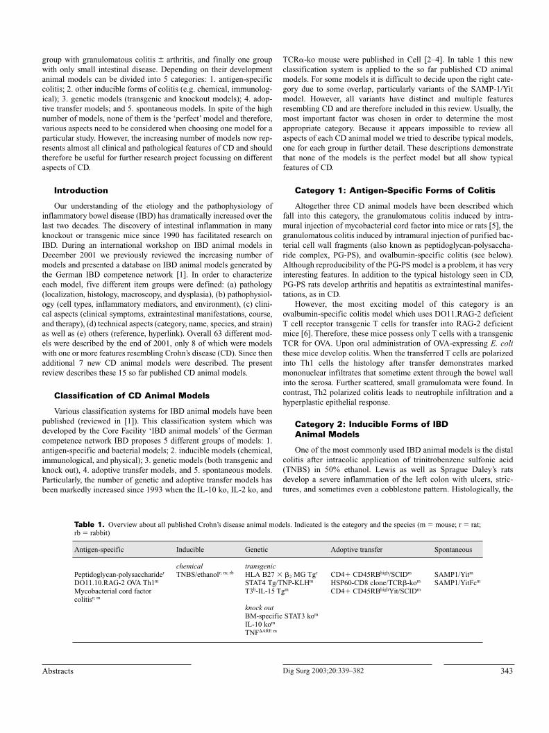
TCR�-ko mouse were published in Cell [2–4]. In table 1 this new
classification system is applied to the so far published CD animal
models. For some models it is difficult to decide upon the right cate-
gory due to some overlap, particularly variants of the SAMP-1/Yit
model. However, all variants have distinct and multiple features
resembling CD and are therefore included in this review. Usually, the
most important factor was chosen in order to determine the most
appropriate category. Because it appears impossible to review all
aspects of each CD animal model we tried to describe typical models,
one for each group in further detail. These descriptions demonstrate
that none of the models is the perfect model but all show typical
features of CD.
Category 1: Antigen-Specific Forms of Colitis
Altogether three CD animal models have been described which
fall into this category, the granulomatous colitis induced by intra-
mural injection of mycobacterial cord factor into mice or rats [5], the
granulomatous colitis induced by intramural injection of purified bac-
terial cell wall fragments (also known as peptidoglycan-polysaccha-
ride complex, PG-PS), and ovalbumin-specific colitis (see below).
Although reproducibility of the PG-PS model is a problem, it has very
interesting features. In addition to the typical histology seen in CD,
PG-PS rats develop arthritis and hepatitis as extraintestinal manifes-
tations, as in CD.
However, the most exciting model of this category is an
ovalbumin-specific colitis model which uses DO11.RAG-2 deficient
T cell receptor transgenic T cells for transfer into RAG-2 deficient
mice [6]. Therefore, these mice possess only T cells with a transgenic
TCR for OVA. Upon oral administration of OVA-expressing E. colithese mice develop colitis. When the transferred T cells are polarized
into Th1 cells the histology after transfer demonstrates marked
mononuclear infiltrates that sometime extent through the bowel wall
into the serosa. Further scattered, small gramulomata were found. In
contrast, Th2 polarized colitis leads to neutrophile infiltration and a
hyperplastic epithelial response.
Category 2: Inducible Forms of IBD Animal Models
One of the most commonly used IBD animal models is the distal
colitis after intracolic application of trinitrobenzene sulfonic acid
(TNBS) in 50% ethanol. Lewis as well as Sprague Daley’s rats
develop a severe inflammation of the left colon with ulcers, stric-
tures, and sometimes even a cobblestone pattern. Histologically, the
Table 1. Overview about all published Crohn’s disease animal models. Indicated is the category and the species (m � mouse; r � rat;
rb � rabbit)
Antigen-specific Inducible Genetic Adoptive transfer Spontaneous
chemical transgenicPeptidoglycan-polysaccharider TNBS/ethanolr; m; rb HLA B27 � �2 MG Tgr CD4� CD45RBhigh/SCIDm SAMP1/Yitm
DO11.10.RAG-2 OVA Th1m STAT4 Tg/TNP-KLHm HSP60-CD8 clone/TCR�-kom SAMP1/YitFcm
Mycobacterial cord factor T3b-IL-15 Tgm CD4� CD45RBhighYit/SCIDm
colitisr; m
knock outBM-specific STAT3 kom
IL-10 kom
TNF�ARE m

344 Dig Surg 2003;20:339–382 Inflammatory Bowel Disease
Principles and Practice
inflammation is often transmural and shows granulomata [7]. This
model has been used extensively also in mice with mixed repro-
ducibility. TNBS colitis in mice is generally believed to be Th1 medi-
ated. However, our own results suggested that at least rat TNBS
colitis is not induced by �� T cells and that T cells even have
protective functions in rat TNBS colitis [8, 9].
Category 3: Genetic Forms of IBD Animal Models
Among genetic CD models the TNF�ARE mice as described by
Kontoyiannis et al. has received a lot of attention because of typical
terminal ileitis combined with arthritis in heterozygous mice [10].
Homozygous mice only live for about 4 weeks and die because of
severe wasting disease together with ankylosing arthritis. In our own
hands this model lead to only mild clinical disease starting with week
10 and no weight loss until week 20. Therefore, this model seems very
interesting for looking at the pathophysiology but seems less suited
for treatment trials [8].
At least three additional models should be briefly mentioned in
this category: The HLA-B27 � �2 microglobulin Tg rat with right-
sided colitis, mild ileitis, and marked extraintestinal inflammation
(arthritis, uveitis, spondylarthropathy) [11]. Most recently two new
genetic models were described with jejunal inflammation mediated
by CD8� T cells in IL-15 Tg mice (T3b-IL-15 Tg) [12] and a typical
patchy terminal ileitis and typhlitis in bone marrow specific STAT3
knockouts [13].
Category 4: Models after Adoptive Transfer into Immunocompromised Mice
Apart from TNBS colitis the most common IBD animal model
is the CD45RBhigh transfer colitis model [14]. Numerous variants of
this model have been described with usually pancolitis [1]. Some his-
tological features resemble CD, particularly the transmural inflamma-
tion and occasional granulomata. Still, one should be cautious to
declare this model to be typical CD animal model. This is however
true for the SAMP-1/Yit transfer colitis using SCID mice [15].
One other adoptive transfer model with solely small intestinal
involvement is the jejunitis of TCR�-ko mice after transfer of CD8�HSP60-specific T cell clones [16]. At present it is unclear to what
extent this model resembles CD apart from inflammation in the small
intestine.
Category 5: Spontaneous Forms of IBD Animal Models
Among all IBD animal models the group of spontaneous models
is one of the smallest. Still, one of the most exciting models has
recently been published, is the SAMP-1/YitFc mouse model. These
mice spontaneously develop chronic ileitis and perianal fistulizing
disease. They were originally generated from senescence studies in
Japan and further breeding in Virginia led to this particular phenotype
with also histological and immunological features of CD [17].
Conclusion
IBD animal models have become a major focus of mucosal
immunology research because of their diversity and in vivo model
character. To this end, early processes as well as pathogenic and
protective factors can be investigated and, if necessary, manipulated.
Although only few good CD animal models are published, they
represent most aspects typical for CD, e.g. perianal disease or colitis
associated arthritis. Still, there is currently no perfect model available.
Because many models represent important aspects of CD research
such models can be used in order to improve our understanding of the
pathophysiology as well as to test new therapeutic strategies. With
these aims in mind it is encouraging that important clinical aspects
such as fistulae have recently been described in appropriate IBD
animal models. Although IBD animal models have markedly
improved our current understanding of IBD (e.g. the role of flora or
T helper cells) substantial improvement with regard to our under-
standing of IBD will only become available by combining studies
employing IBD animal models with human in vitro studies and even-
tually application in clinical trials.
References
1 Hoffmann JC, Pawlowski NN, Kuhl AA, Hohne W, Zeitz M: Animal
models of inflammatory bowel disease: An overview. Pathobiology 2002;
70:121–130.
2 Sadlack B, Merz H, Schorle H, Schimpl A, Feller AC, Horak I: Ulcerative
colitis-like disease in mice with a disrupted interleukin-2 gene. Cell
1993;75:253–261.
3 Mombaerts P, Mizoguchi E, Grusby MJ, Glimcher LH, Bhan AK,
Tonegawa S: Spontaneous development of inflammatory bowel disease in
T cell receptor mutant mice. Cell 1993;75:274–282.
4 Kuhn R, Lohler J, Rennick D, Rajewsky K, Muller W: Interleukin-10-
deficient mice develop chronic enterocolitis. Cell 1993;75:263–274.
5 Sogawa M, Matsumoto T, Yamagami H, Yamada T, Ozeki Y, Yano I,
Nakajima Y, Arakawa T, Kaneda K: A murine model of granulomatous
colitis with mesenteric lymphadenitis induced by mycobacterial cord fac-
tor. Virchows Arch 2003;442:151–158.
6 Iqbal N, Oliver JR, Wagner FH, Lazenby AS, Elson CO, Weaver CT:
T helper 1 and T helper 2 cells are pathogenic in an antigen-specific model
of colitis. J Exp Med 2002;195:71–84.
7 Morris GP, Beck PL, Herridge MS, Depew WT, Szewczuk MR, Wallace
JL: Hapten-induced model of chronic inflammation and ulceration in the
rat colon. Gastroenterology 1989;96:795–803.
8 Kuhl AA, Loddenkemper C, Westermann J, Hoffmann JC: Role of gamma
delta T cells in inflammatory bowel disease. Pathobiology 2002;70:
150–155.
9 Hoffmann JC, Peters K, Henschke S, Herrmann B, Pfister K, Westermann J,
Zeitz M: Role of T lymphocytes in rat 2,4,6-trinitrobenzene sulphonic acid
(TNBS) induced colitis: Increased mortality after gammadelta T cell
depletion and no effect of alphabeta T cell depletion. Gut 2001;48:
489–495.
10 Kontoyiannis D, Pasparakis M, Pizarro TT, Cominelli F, Kollias G:
Impaired on/off regulation of TNF biosynthesis in mice lacking TNF AU-
rich elements: Implications for joint and gut-associated immunopatholo-
gies. Immunity 1999;10:387–398.
11 Aiko S, Grisham MB: Spontaneous intestinal inflammation and nitric
oxide metabolism in HLA-B27 transgenic rats. Gastroenterology 1995;
109:142–150.
12 Ohta N, Hiroi T, Kweon MN, Kinoshita N, Jang MH, Mashimo T,
Miyazaki J, Kiyono H: IL-15-dependent activation-induced cell death-
resistant Th1 type CD8 alpha beta�NK1.1� T cells for the development
of small intestinal inflammation. J Immunol 2002;169:460–468.
13 Welte T, Zhang SS, Wang T, Zhang Z, Hesslein DG, Yin Z, Kano A,
Iwamoto Y, Li E, Craft JE, Bothwell AL, Fikrig E, Koni PA, Flavell RA,
Fu XY: STAT3 deletion during hematopoiesis causes Crohn’s disease-like
pathogenesis and lethality: A critical role of STAT3 in innate immunity.
Proc Natl Acad Sci USA 2003;100:1879–1884.
14 Leach MW, Bean AG, Mauze S, Coffman RL, Powrie F: Inflammatory
bowel disease in C.B-17 scid mice reconstituted with the CD45RB high
subset of CD4� T cells. Am J Pathol 1996;148:1503–1515.
15 Burns RC, Rivera-Nieves J, Moskaluk CA, Matsumoto S, Cominelli F,
Ley K: Antibody blockade of ICAM-1 and VCAM-1 ameliorates

345Dig Surg 2003;20:339–382Abstracts
inflammation in the SAMP-1/Yit adoptive transfer model of Crohn’s
disease in mice. Gastroenterology 2001;121:1428–1436.
16 Steinhoff U, Brinkmann V, Klemm U, Aichele P, Seiler P, Brandt U, Bland
PW, Prinz I, Zugel U, Kaufmann SH: Autoimmune intestinal pathology
induced by hsp60-specific CD8 T cells. Immunity 1999;11:349–358.
17 Rivera-Nieves J, Bamias G, Vidrich A, Marini M, Pizarro TT, McDuffie
MJ, Moskaluk CA, Cohn SM, Cominelli F: Emergence of perianal fis-
tulizing disease in the SAMP1/YitFc mouse, a spontaneous model of
chronic ileitis. Gastroenterology 2003;124:972–982.
We thank Wolfgang Höhne, Hacer Kakirman, Philipp Cremer, and
Annegret Schönberg for help with various aspects of the database on IBD ani-
mal models. The work of the authors is supported by the BMBF/DLR in the
medical competence network inflammatory bowel disease. We thank G.
Kollias and D. Kontoyiannis for kindly providing TNF�ARE mice.
3
The Microenvironment of Intestinal Lamina Propria T Lymphocytes Regulatestheir Responsiveness by Redox ProcessesB. Sido1, F. Autschbach2, S. Meuer3
1Department of Surgey, 2Institute of Pathology, and3Institute of Immunology, University of Heidelberg,Heidelberg, Germany
Summary: In lymphocytes, the availability of cysteine is
limiting for the synthesis of glutathione, which again is essential for
proliferation. Physiologic concentrations of the oxidized derivative
cystine cannot substitute for cysteine deficiency since uptake of
cystine is low in lymphocytes. Peripheral blood monocytes (PB-MO),
especially when stimulated, secrete cysteine, which can be easily
taken up by lymphocytes, thereby abolishing the hyporesponsiveness
of intestinal lamina propria T lymphocytes (LP-T). Because PBMO
are known to infiltrate the gut in Crohn’s disease and ulcerative coli-
tis, thiol-mediated costimulation contributes to increased glutathione
synthesis in hyperreactive LP-T in inflammatory bowel disease. In
contrast, resident intestinal macrophages are defective in cysteine
delivery and thus ensure the physiological hyporesponsiveness of
LP-T in the normal gut.
Introduction: Human intestinal lamina propria T lympho-
cytes (LP-T) usually do not establish systemic immune responses
although they are permanently exposed to luminal antigens. Similarly,
LP-T hardly proliferate after experimental stimulation through the
T cell receptor [1]. The low reactivity of LP-T is supposed to be due to
insufficient costimulation by resident antigen presenting cells: Lamina
propria macrophages (LP-MO), in contrast to peripheral blood mono-
cytes (PB-MO), express only minimal amounts of CD54 and CD58 is
hardly detectable [2]. Here, we provide evidence that the differential
capacity of PB-MO versus LP-MO to release cysteine is key in the
regulation of proliferation of LP-T. In lymphocytes, cysteine is the
limiting substrate for synthesis of the antioxidant glutathione, which
is a requisite for DNA synthesis [3].
Materials and Methods: Preparation of Cells. Normal
mucosa was dissected from surgical colon specimens. Lamina propria
mononuclear cells were isolated essentially as described earlier by
enzymatic digestion, Percoll and Ficoll-Hypaque density gradient
centrifugation [4]. LP-T were purified by E-rosette formation with
sheep red blood cells. LP-MO and PB-MO were isolated during a
3 h-adhesion step to plastic at 37� [4] and were irradiated before use
(50 Gy).
Proliferation Assay. LP-T (5 � 104/well) were cultured in 96-well
microtiter plates in RPMI 1640 (with 10% FCS, 2% glutamine and
antibiotics) at 37� and 7% CO2. Cells were stimulated via CD3 using
OKT3-coated beads [5]. Irradiated PB-MO or LP-MO were added to
LP-T at 30% of total cell number. After 4 days of culture, wells were
pulsed with 1 �Ci [3H]thymidine for another 16 h.
Determination of Cysteine in Tissue Culture Supernatant. Cysteine
was determined spectrophotometrically as acid-soluble thiol in cell-
free supernatants of PB-MO and LP-MO after 40 h of culture as
described earlier [5]. Cysteine was confirmed as the sole acid-soluble
thiol present by HPLC analysis [5].
Histochemical Staining of Glutathione. Native cryostat sections of,
respectively, normal colon and Crohn’s colitis were stained for glu-
tathione by short-term incubation (15 min) with Mercury Orange
(50 �M in toluene solution) at 4�C according to the method of Chieco
and Boor [6].
Results and Discussion: If autologous PB-MO were added
at 30% of total cell number, the otherwise abortive reactivity of LP-T
to CD3 stimulation was restored. In contrast, LP-MO were without
any effect. The antioxidants 2-ME (10 �M) and glutathione (3 mM)
could completely substitute for the PB-MO-mediated costimulation,
whereas various pro-oxidant culture conditions abolished prolifer-
ation. The proliferative effect of 2-ME is due to an increased avail-
ability of intracellular cysteine derived from extracellular cystine [7].
PB-MO release cysteine into the supernatant. Constitutive cysteine
release (6 �M at 5 � 105 PB-MO/well) was increased three- to four-
fold if PB-MO were stimulated by LPS from E. coli (1 �g/ml) or,
alternatively, if CD58 on PB-MO was crosslinked by an immobilized
anti-CD58 mAb [5]. Notably LP-MO, in contrast to PB-MO, did not
secrete any cysteine under these experimental conditions.
PB-MO, similar to mouse peritoneal macrophages [8], take up
cystine, reduce it intracellularly and secrete cysteine that can be easily
taken up by lymphocytes (but not cystine!). Correspondingly, costim-
ulation of LP-T by PB-MO was maximal above physiological cystine
concentrations (about 60 �M in serum) but was completely abolished
in cystine-deficient medium. Indeed, cysteine added to LP-T without
PB-MO in cystine-free cultures at 30 �M also completely restored the
proliferative response.
In inflammatory bowel disease (Crohn’s disease, ulcerative
colitis), a sustained recruitment of PB-MO to the inflamed gut is well
established [9, 10]. Through release of cysteine, recently recruited
PB-MO support glutathione synthesis in LP-T as documented by
histochemical staining and, thus, their hyperreactivity. In contrast, the
incapability of LP-MO to secrete cysteine initiates glutathione deple-
tion in LP-T and thereby ensures their physiological hyporesponsive-
ness in the normal gut.
References
1 Qiao L, Schürmann G, Betzler M, Meuer SC: Activation and signaling
status of human lamina propria T lymphocytes. Gastroenterology 1991;
101:1529–1536.
2 Qiao L, Braunstein J, Golling M, Schürmann G, Autschbach F, Möller P,
Meuer SC: Differential regulation of human T cell responsiveness by
mucosal versus blood monocytes. Eur J Immunol 1996;26:922–927.
3 Suthanthiran M, Anderson ME, Sharma VK, Meister A: Glutathione
regulates activation-dependent DNA synthesis in highly purified normal
human T lymphocytes stimulated via the CD2 and CD3 antigens. Proc
Natl Acad Sci USA 1990;87:3343–3347.
346 Dig Surg 2003;20:339–382 Inflammatory Bowel Disease
Principles and Practice
4 Sido B, Breitkreutz R, Seel C, Herfarth C, Meuer SC: Redox processes
regulate intestinal lamina propria T lymphocytes. Methods Enzymol 2002;
352:232–247.
5 Sido B, Braunstein J, Breitkreutz R, Herfarth C, Meuer SC: Thiol-mediated
redox regulation of intestinal lamina propria T lymphocytes. J Exp Med
2000;192:907–912.
6 Chieco P, Boor PJ: Use of low temperatures for glutathione histochemical
stain. J Histochem Cytochem 1983;31:975–976.
7 Ishii T, Bannai S, Sugita Y: Mechanism of growth stimulation of L1210
cells by 2-mercaptoethanol in vitro. Role of the mixed disulfide of
2-mercaptoethanol and cysteine. J Biol Chem 1981;256:12387–12392.
8 Gmünder H, Eck HP, Benninghoff B, Roth S, Dröge W: Macrophages
regulate intracellular glutathione levels of lymphocytes. Evidence for an
immunoregulatory role of cysteine. Cell Immunol 1990;129:32–46.
9 Burgio VL, Fais S, Boirivant M, Perrone A, Pallone F: Peripheral
monocyte and naive T-cell recruitment and activation in Crohn’s disease.
Gastroenterology 1995;109:1029–1038.
10 Rugtveit J, Brandtzaeg P, Halstensen TS, Fausa O, Scott H: Increased
macrophage subset in inflammatory bowel disease: Apparent recruitment
from peripheral blood monocytes. Gut 1994;35:669–674.
4
Mucosal Immunity and Drug InteractionM.F. Neurath
Laboratory of Immunology, I. Medical Clinic, University of Mainz, Mainz, Germany
Inflammatory bowel diseases (IBD: Crohn’s disease and ulcerative
colitis) are defined as chronic inflammatory diseases of the gut not
due to specific pathogens [1–4]. There is good recent evidence to sug-
gest that the activation of intestinal antigen presenting cells and T
lymphocytes by bacterial antigens plays a key role in the pathogenesis
of these disease. Antigen presenting cells, T lymphocytes and their
cytokines (e.g. TNF, IL-6, IL-12, IL-23) play an important role in reg-
ulating mucosal immune responses in IBD patients as well as in ani-
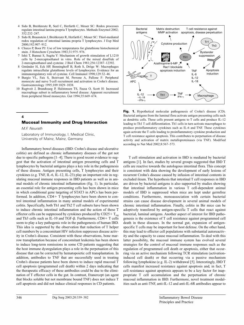
mal models of chronic intestinal inflammation (fig. 1). In particular,
an essential role for antigen presenting cells has been shown in mice
in which conditional gene targeting of STAT3 in APCs has been per-
formed. In addition, CD4� T lymphocytes have been shown to con-
trol intestinal inflammation in many animal models of experimental
colitis. Specifically, both Th1 and Th2 T cell subsets have been shown
to induce chronic intestinal inflammation and the action of these T
effector cells can be suppressed by cytokines produced by CD25� Treg
and Th3 cells such as IL-10 and TGF-�. Furthermore, CD4� T cells
seem to play a key pathogenic role in the pathogenesis of human IBD.
This idea is supported by the observation that reduction of T helper
cell numbers by a concomitant HIV infection suppresses disease activ-
ity in Crohn’s disease. Consistent with these observations, bone mar-
row transplantation because of concomitant leukemia has been shown
to induce long-term remissions in some CD patients suggesting that
the host immune dysregulation plays a role in the perpetuation of this
disease that can be corrected by hematopoietic cell transplantation. In
addition, antibodies to TNF that are successfully used in treating
Crohn’s disease patients have been shown to induce rapid mucosal T
cell apoptosis (programmed cell death) within 2 days indicating that
the therapeutic efficacy of these antibodies could be due to the elimi-
nation of T effector cells in the gut. In contrast, Etanercept (an agent
that blocks soluble but not membrane bound TNF) does not induce T
cell apoptosis and did not induce clinical responses in CD patients.
T cell stimulation and activation in IBD is mediated by bacterial
antigens [1]. In fact, studies by several groups suggested that IBD T
cells are reactive towards the autologous intestinal flora. This concept
is consistent with data showing the development of early lesions of
recurrent Crohn’s disease caused by infusion of intestinal contents in
excluded ileum. The hypothesis that intestinal T cell responses in IBD
are driven by bacterial antigens is also supported by studies showing
that intestinal inflammation in various T cell-dependent animal
models of IBD is suppressed when mice are kept under germfree
conditions. Furthermore, monoassociation with certain bacterial
strains can cause disease development in several animal models of
chronic intestinal inflammation. Finally, colitis in Bir mice can be
adoptively transfered by antigen-specific T cells that react against
bacterial, luminal antigens. Another aspect of interest for IBD patho-
genesis is the existence of T cell resistance against programmed cell
death in these diseases. In the normal gut, expansion of antigen-
specific T cells may be important for host defense. On the other hand,
this may lead to effector cell populations with substantial autoreactiv-
ity and the capacity to cause mucosal inflammation. To deal with this
latter possibility, the mucosal immune system has evolved several
strategies for the control of mucosal immune responses such as the
regulation of programmed cell death or apoptosis, either that occur-
ring via an active mechanism following TCR stimulation (activation-
induced cell death) or that occurring via a passive mechanism
following lymphokine (e.g. IL-2) withdrawal [5]. Interestingly, IBD T
cells manifest increased resistance against apoptosis and, in fact, T
cell resistance against apoptosis appears to be a key factor for inap-
propriate T cell accumulation and the perpetuation of chronic
mucosal inflammation in IBD. Furthermore, novel treatment modal-
ities such as anti-TNF, anti-IL-12 and anti-IL-6R antibodies appear to
Bacterialantigens
Matrix destructionMMP activation
T cell resistance againstapoptosis, disease perpetuation
Cytokine production
Bystandersuppression
Rac1 blockadeapoptosis induction
Azathioprine 6-MP
Apoptosis induction
Macrophage
T
Th1
Tr1
Th1
APC
IL-12�IL-18�IFN-
TNFIFN-
TNFIL-6IL-12IL-18
Th3
Fig. 1. Hypothetical molecular pathogenesis of Crohn’s disease (CD).
Bacterial antigens from the luminal flora activate antigen presenting cells such
as dendritic cells. These cells present antigens to T cells and produce IL-12
leading to Th1 T cell differentiation. Th1 cells in turn activate macrophages to
produce proinflammatory cytokines such as IL-6 and TNF. These cytokines
again activate the T cells leading to proinflammatory cytokine production and
T cell resistance against apoptosis. This contributes to perpetuation of disease
activity and activation of matrix metalloproteinases (via TNF). Modified
according to Nat Med 2002;8:567–573.
347Dig Surg 2003;20:339–382Abstracts
suppress colitis activity by the induction of T cell apoptosis. Finally,
azathioprine and 6-MP have been recently shown to induce T cell
apoptosis by specific blockade of vav1-mediated activation of the
small GTPase Rac1. These findings have important implications in
the design of more effective treatment regimens in IBD and suggest
that selective T cell targeting might be very helpful for IBD therapy.
References
1 Elson CO: Genes, microbes, and T cells – New therapeutic targets in
Crohn’s disease. N Engl J Med 2002;346(8):614–616.
2 Landers CJ, Cohavy O, Misra R, Yang H, Lin YC, Braun J, Targan SR:
Selected loss of tolerance evidenced by Crohn’s disease-associated
immune responses to auto- and microbial antigens. Gastroenterology
2002;123(3): 689–699.
3 Neurath MF, Finotto S, Glimcher LH: The role of Th1/Th2 polarization in
mucosal immunity. Nat Med 2002;8(6):567–573.
4 Podolsky DK: Inflammatory bowel disease. N Engl J Med 2002;347(6):
417–429.
5 Sturm A, Fiocchi C: Life and death in the gut: More killing, less Crohn’s.
Gut 2002;50(2):148–149.
State of the Art: Current ResearchStrategies in Ulcerative Colitis
5
Basic Mechanisms of Inflammation inUlcerative Colitis R. Duchmann, J. Maul, F. Heller, M. Zeitz
Medizinische Klinik I, Universitätsklinikum BenjaminFranklin, Freie Universität Berlin, Berlin, Germany
Ulcerative colitis and Crohn’s disease describe two common
forms of chronic inflammatory bowel diseases (IBD). Although the
pathogenetic mechanisms which cause the specific clinical presenta-
tions of ulcerative colitis and Crohn’s disease are not sufficiently
understood, major progress on the general pathogenesis of both dis-
eases provides an increasingly clearer picture of the essential
immunologic differences between them. Overall, basic clinical and
immunologic features suggest that ulcerative colitis is characterized
by a TH2-type cytokine response with a stronger epithelial and
autoimmune component and a stronger involvement of B cells,
whereas Crohn’s disease develops on the basis of a CD4� T cell
dysregulation characterized by a TH1 cytokine response and a strong
involvement of macrophages and antigen-presenting cells.
Ulcerative colitis and Crohn’s disease describe two common
forms of chronic inflammatory bowel diseases (IBD). They are both
complex clinical entities in which genetic, environmental and micro-
bial factors interact to generate chronic or chronic intermittent intes-
tinal inflammation. In addition, attesting to the systemic nature of
both diseases, a large number of patients with IBD suffer from
extraintestinal manifestations.
In contrast to Crohn’s disease, which is a transmural granuloma-
tous inflammation complicated by fibrostenotic lesions and formation
of fistula, inflammation in ulcerative colitis is limited to the mucosa.
This more superficial ulcerative colitis-type inflammation shows very
early changes characterized by widespread apoptosis of epithelial
cells and decreased complexity of the tight junctions between epithe-
lial cells [1]. On histology, crypt abscesses are typically observed.
Macroscopically, early disruption of the epithelial layer becomes
visible through small erosions which can develop to large ulcers as
the disease progresses. Ulcerative colitis and Crohn’s disease also
differ in disease distribution. Ulcerative colitis typically shows a
continuous inflammation that starts in the rectum, variably involves
more proximal colonic segments but spares the small bowel. In
contrast, inflammation in Crohn’s disease is discontinous and can
affect the mucosa from mouth to anus [2, 3].
Familial association and linkage studies clearly indicate a genetic
background for both ulcerative colitis and Crohn’s disease. However,
ulcerative colitis shows a lower concordance rate among monozygotic
twin pairs and overall genetic susceptibility seems to be of less impor-
tance than in Crohn’s disease, where disease susceptibility has even
been pinpointed to mutations in a single gene [4]. Investigations of
global gene expression in ulcerative colitis vs Crohn’s disease showed
that both diseases differ in gene expression profiles and support the
concept that both diseases are distinct molecular entities [5, 6].
Ulcerative colitis shows an increased association with autoimmune
disorders (e.g. primary sclerosing cholangitis, thyroid disease,
diabetes, pernicious anemia) and early studies of immunologic abnor-
malities centered on the possibility that autoantibodies mediate the
disease and that ulcerative colitis was an organ specific disease. There
is an increased synthesis in IgG1/IgG3 subclasses and autoantibodies
are found against a large number of self-antigens. Among these,
autoantibodies against colonic epithelial antigens, including antibodies
to epithelial cell associated components (ECACs) [7, 8] and a 40 kDa
epithelial antigen specifically present in patients with ulcerative colitis
but not inflammatory controls or healthy individuals [9, 10] are certainly
the most likely to be involved in disease pathogenesis.
Studies determining the cytokine production using lymphocytes
isolated from the intestinal lamina propria of patients with ulcerative
colitis, Crohn’s disease and healthy controls demonstrated that IL-5
production is clearly increased and that interferon (IFN)- produc-
tion is decreased in ulcerative colitis, which unmistakably differenti-
ates ulcerative colitis from TH1 inflammation. In addition, cells
produced less interleukin (IL)-4 than those from Crohn’s patients or
healthy controls [11, 12]. Although the responding T cell is less
defined in UC than the much clearer TH1 dysregulation seen in
Crohn’s disease, data indicate that T cells in ulcerative colitis at least
share some basic characteristics of TH2 but not TH1 responses. The
molecular mechanisms that contribute to the establishment and
maintenance of cytokine memory in T cell subpopulations is not yet
clear, but there is increasing evidence that TH2 type cells associated
with ulcerative colitis are controlled by GATA-3 whereas TH1 cells
associated with Crohn’s disease are controlled by transcription fac-
tors T-bet and Stat-4.
The majority of IBD animal models are TH1 models, associated
with an excessive IL-12/IFN-/TNF-� secretion and thus resemble
Crohn’s disease. Only a small number are TH2 models, associated
with an increased IL-4/IL-5 secretion and resemble ulcerative colitis
[13]. In oxazolone colitis, rectal application of the hapten oxazolone
subsequent to skin sensitization with the same hapten floods the
epithelium and the lamina propria with oxazolone and oxazolone-
modified bacterial antigens. This promotes the production of TH2-
type cytokines by colonic T cells and results in lesions characterized
348 Dig Surg 2003;20:339–382 Inflammatory Bowel Disease
Principles and Practice
by leukocyte infiltration that is limited to the superficial layer of the
mucosa. Administration of antibodies to IL-4 in that model amelio-
rates disease [14]. In the same animal model, it was recently shown
that oxazolone colitis is mediated by TH2 NK-T cells and that it is
dependent on the secretion of IL-13 by this T cell population [15].
NK-T cells in humans are activated by antigen in the context of
CD1d, a molecule widely expressed on colonic epithelial cells and
dendritic cells. Since IL-13 may increase CD1 antigen presentation
on epithelial cells, it has been suggested that IL-13 secreted by
activated NK-T cells in the rectum could spread inflammation into
neighbouring tissues. Upregulation of CD1 and NK-T cell mediated
injury to the epithelial cell barrier could thus be instrumental in
promoting the continuous inflammation typical for ulcerative colitis.
Recently, it has been proposed that animal models of IBD can be
distinguished into type 1 models, i.e. those where the defect lies with
the effector mechanisms of the mucosal immune response and type 2
models, i.e. those wherein the effector response is normal but the reg-
ulatory response is impaired [13]. Whether the balance in ulcerative
colitis is further tipped to inflammation through decreased presence
of regulatory (suppressor) cells is not well established. At least for
CD4CD25� regulatory T cells as yet unpublished data from our
laboratory for the first time suggest that this might be the case.
With regard to potential initiating antigens, animal models for
ulcerative colitis and Crohn’s disease are both dependent on the pres-
ence of a normal intestinal flora [13]. This endogenous immune
stimulus, equivalent to the presence of self-antigens in regular
autoimmune diseases, is thus required for the induction and perpet-
uation of the chronic intestinal inflammation in the vast majority of
IBD animal models known today. Similar to the situation in animal
models [13, 16], patients with ulcerative colitis and Crohn’s disease
show an abnormal immune response to antigens from the normal
intestinal flora, suggesting that abrogation of the normal state of
tolerance is involved in the pathogenesis of both diseases [17, 18]. An
important role of the intestinal flora in ulcerative colitis and pouchi-
tis is further indicated by the clinical effectiveness of probiotics for
remission maintenance [19–22].
The antigen(s) which drive the inflammatory response in animal
models of ulcerative colitis or in patients with ulcerative colitis unfor-
tunately still have not been identified. The observation that one hap-
tenizing agent, TNBS, elicits a TH1 respone in SJL/J mice [23, 24],
whereas another, oxazolone, elicits a TH2 response [14] in the same
animal, clearly demonstrates that the nature of the antigen can deter-
mine the type of immune response. However, this conclusion from
induced models relates to the situation were antigen contacts a healthy
immune system and cannot necessarily be transferred to a situation
where antigen contacts an immune system that is already abnormal.
With regard to the latter, an elegant study using TNBS as the inducing
antigen in a variety of mice genetically deficient in TH1 and TH2 type
cytokines showed, that both types of colitis, colitis resembling Crohn’s
disease and that resembling ulcerative colitis, are inducible in a single
strain of mice using the same antigen. This certainly supports the
notion, that the phenotype of intestinal inflammation that develops
in an individual with an already compromised immune system is
more dependent on the type of the underlying T cell dysregulation,
i.e., excess TH1 or TH2 type responses, than on the initiating
antigen(s) [25].
References
1 Gitter AH, Wullstein F, Fromm M, Schulzke JD: Epithelial barrier defects
in ulcerative colitis: Characterization and quantification by electrophysio-
logical imaging. Gastroenterology 2001;121(6):1320–1328.
2 Jewell DP: Ulcerative colitis; in Feldman M, Friedman LS, Sleisenger MH
(eds): Gastrointestinal and Liver Diseases, ed 7. Saunders Science, 2002,
vol 2, pp 2039–2067.
3 Duchmann R, Zeitz M: Crohn’s disease; in Orgra PL, Mestecky J, Lamm
ME, Strober W, Bienenstock J, McGhee J (eds): Mucosal Immunology,
ed 2. Academic Press, 1999, pp 1055–1080.
4 Bonen DK, Cho JH: The genetics of inflammatory bowel disease.
Gastroenterology 2003;124(2):521–536.
5 Lawrence IC, Fiocchi C, Chakravarti S: Ulcerative colitis and Crohn’s
disease: Distinctive gene expression profiles and novel susceptibility can-
didate genes. Hum Mol Genet 2001;10(5):445–456.
6 Dieckgraefe BK, Stenson WF, Korzenik JR, Swanson PE, Harrington CA:
Analysis of mucosal gene expression in inflammatory bowel disease by
parallel oligonucleotide arrays. Physiol Genomics 2000;4(1):1–11.
7 Fiocchi C, Roche JK, Michener WM: High prevalence of antibodies to
intestinal epithelial antigens in patients with inflammatory bowel disease
and their relatives. Ann Intern Med 1989;110(10):786–794.
8 Roche JK, Fiocchi C, Youngman K: Sensitization to epithelial antigens in
chronic mucosal inflammatory disease. Characterization of human intesti-
nal mucosa-derived mononuclear cells reactive with purified epithelial
cell-associated components in vitro. J Clin Invest 1985;75(2):522–530.
9 Das KM, Vecchi M, Sakamaki S: A shared and unique epitope(s) on
human colon, skin, and biliary epithelium detected by a monoclonal anti-
body. Gastroenterology 1990;98(2):464–469.
10 Halstensen TS, Das KM, Brandtzaeg P: Epithelial deposits of
immunoglobulin G1 and activated complement co-localize with the ‘M(r)
40kD’ putative auto-antigen in ulcerative colitis. Adv Exp Med Biol
1995;6:1273–1276.
11 Fuss IJ, Neurath M, Boirivant M, Klein JS, de la Motte C, Strong SA,
et al: Disparate CD4� lamina propria (LP) lymphokine secretion profiles
in inflammatory bowel disease. Crohn’s disease LP cells manifest
increased secretion of IFN-gamma, whereas ulcerative colitis LP cells
manifest increased secretion of IL-5. J Immunol 1996;157(3):1261–1270.
12 West GA, Matsuura T, Levine AD, Klein JS, Fiocchi C: Interleukin 4 in
inflammatory bowel disease and mucosal immune reactivity. Gastro-
enterology 1996;110(6):1683–1695.
13 Strober W, Fuss IJ, Blumberg RS: The immunology of mucosal models of
inflammation. Annu Rev Immunol 2002;20:495–549.
14 Boirivant M, Fuss IJ, Chu A, Strober W: Oxazolone colitis: A murine
model of T helper cell type 2 colitis treatable with antibodies to inter-
leukin 4. J Exp Med 1998;188(10):1929–1939.
15 Heller F, Fuss IJ, Nieuwenhuis EE, Blumberg RS, Strober W: Oxazolone
colitis, a Th2 colitis model resembling ulcerative colitis, is mediated by
IL-13-producing NK-T cells. Immunity 2002;17(5):629–638.
16 Duchmann R, Schmitt E, Knolle P, Meyer zum Buschenfelde KH, Neurath
M: Tolerance towards resident intestinal flora in mice is abrogated in
experimental colitis and restored by treatment with interleukin-10 or anti-
bodies to interleukin-12. Eur J Immunol 1996;26(4):934–938.
17 Duchmann R, Neurath MF, Meyer zum Buschenfelde KH: Responses to
self and non-self intestinal microflora in health and inflammatory bowel
disease. Res Immunol 1997;148(8–9):589–594.
18 Duchmann R, Kaiser I, Hermann E, Mayet W, Ewe K, Meyer zum
Buschenfelde KH: Tolerance exists towards resident intestinal flora but is
broken in active inflammatory bowel disease (IBD). Clin Exp Immunol
1995;102(3):448–455.
19 Gionchetti P, Rizzello F, Helwig U, Venturi A, Lammers KM, Brigidi P,
et al: Prophylaxis of pouchitis onset with probiotic therapy: a double-blind,
placebo-controlled trial. Gastroenterology 2003;124(5):1202–1209.
20 Gionchetti P, Rizzello F, Venturi A, Brigidi P, Matteuzzi D, Bazzocchi G,
et al: Oral bacteriotherapy as maintenance treatment in patients with
chronic pouchitis: A double-blind, placebo-controlled trial. Gastro-
enterology 2000;119(2):305–309.
21 Kruis W, Schutz E, Fric P, Fixa B, Judmaier G, Stolte M: Double-blind
comparison of an oral Escherichia coli preparation and mesalazine in
maintaining remission of ulcerative colitis. Aliment Pharmacol Ther 1997;
11(5):853–858.

349Dig Surg 2003;20:339–382Abstracts
22 Rembacken BJ, Snelling AM, Hawkey PM, Chalmers DM, Axon AT: Non-
pathogenic Escherichia coli versus mesalazine for the treatment of ulcer-
ative colitis: A randomised trial. Lancet 1999;354(9179):635–639.
23 Neurath MF, Fuss I, Kelsall BL, Presky DH, Waegell W, Strober W:
Experimental granulomatous colitis in mice is abrogated by induction of
TGF-beta-mediated oral tolerance. J Exp Med 1996;183(6):2605–2616.
24 Neurath MF, Fuss I, Kelsall BL, Stuber E, Strober W: Antibodies to inter-
leukin 12 abrogate established experimental colitis in mice. J Exp Med
1995;182(5):1281–1290.
25 Dohi T, Fujihashi K, Kiyono H, Elson CO, McGhee JR: Mice deficient in
Th1- and Th2-type cytokines develop distinct forms of hapten-induced
colitis. Gastroenterology 2000;119(3):724–733.
6
Immune Mechanisms in Ulcerative ColitisC. Veltkamp
Department of Gastroenterology, University of Heidelberg,Heidelberg, Germany
Recent evidence suggests that a pathologic activation of the
mucosal immune system in response to the normal intestinal flora is
a key factor in the pathogenesis of inflammatory bowel disease (IBD).
The main abnormality driving the inflammation is an exaggerated
T cell response driven by CD4� T cells which are increased in the
lamina propria both in animal models of chronic colitis and in
patients with IBD. In Crohn’s disease and in most animal models of
colitis, the intestinal inflammation is associated with a Th1 T cell
response with a predominant cytokine production of IFN-. In con-
trast, the pathogenesis of ulcerative colitis in patients is not driven by
Th1 T cells because IFN- is unchanged. While the cytokine pattern
has some features of a Th2 response with upregulation of interleukin-
5, the production of IL-4, one other signature cytokine associated
with a Th2 response is not increased. Adding to the complexity, in
some of the animal models used to study ulcerative colitis (TCR�knockout mice, oxazolone induced colitis), inflammation develops
due to excess IL-4 secretion and can be blocked by anti-IL-4 anti-
bodies. In others such as the Tg 26 mice, T cells secretion of high
amounts of IFN- by Th1 cells without IL-4 upregulation also results
in a histological pattern resembling ulcerative colitis. A possible
explanation for these conflicting results is that IL-4 is important in the
initial phase of ulcerative colitis but gradually decreases and other
cytokines increase during the course of inflammation. Evidence sup-
porting this concept was recently reported by Heller et al. for oxa-
zolone induced colitis. This study showed increasing levels of IL-13
secretion by NK-T which were shown to be the cellular mediators of
the colitis in this model. On the contrary, IL-4 was only detectable
shortly after oxazolone administration but rapidly superceded by
IL-13. Whether NK-T cells are the mediators of colitis in patients
with ulcerative colitis remains to be investigated.
Epstein-Barr virus-induced gene 3 (EBI3) is a novel IL-12p70
antagonising cytokine expressed by macrophage-like cells in normal
intestine. EBI3 shows enhanced expression in active ulcerative colitis
but not in active Crohn’s disease. Since IL-12p70 produced by
macrophages has a key function in the induction of a Th1 T cell dif-
ferentiation, the increased levels of EBI3 could explain why T cells
found in ulcerative colitis do not express a Th1 profile but rather a
Th2 profile as discussed above. Interestingly, T cells in the lamina
propria are more susceptible to Fas-mediated apoptosis compared to
circulating T cells. Fas ligand is strongly expressed by T cells in active
ulcerative colitis but not in Crohn’s disease, suggesting that Fas-FasL
induced apoptosis may play a part in mucosal damage of ulcerative
colitis.
Data from animal models and the fact that IBD occurs in parts of
highest bacterial concentration (colon and terminal ileum) implicate
that the antigens towards which the activated mucosal immune system
reacts are within the normal bacterial flora. A puzzling finding in this
context is that appendectomy at young age is a protective factor both
for ulcerative colitis in patients as well as for chronic colitis in
rodents. Animal data suggest that a reaction of the immune system in
the cecal region towards bacterial antigens might be important for the
manifestation but not the perpetuation of ulcerative colitis. Data from
our and other groups show that mouse models for ulcerative colitis
which develop intestinal inflammation are disease free in a germ-free
environment. Moreover, we have shown that in a T cell mediated
mouse model colitis develops when the animals are moved from the
germfree into a normal bacterial environment. In these experiments,
T cells produced proinflammatory cytokines exclusively upon stimu-
lation with normal bacterial antigens but not upon stimulation with
other antigens of the luminal content thus demonstrating that the
T cells responsible for the induction of colitis were bacteria-specific.
In vitro and in vivo data show that the commensal intestinal flora
affects the peroxisome poliferator-activated receptor (PPAR)
which is probably a key inhibitor of colitis by attenuating nuclear fac-
tor �B (NF-�B) activity. PPAR expression is considerably impaired
in patients with ulcerative colitis potentially contributing to the dis-
ease pathogenesis.
Recent research has focused on the immunological mechanisms
controlling the potentially aggressive bacteria-specific T cell popula-
tion. So called regulatory T cells exist in the normal immune system
which are in a balance with activated T cells. In animal models of
chronic colitis, a lack of regulatory T cells led to the induction of col-
itis. Experimental T cell-mediated colitis could not only be prevented,
but also successfully treated by injecting immunoregulatory T cells.
Whether this population of T cells is involved consistently in the
pathogenesis of IBD or in subgroups of patients is currently unknown.
A decrease in regulatory CD4�CD25� T cells has recently been
reported in Crohn’s patients. No data are currently available for ulcer-
ative colitis. In mice CD4�CD25� T cells have been able to inhibit
microbially induced colon cancer pointing towards a possible role of
these cells in the inflammatory process of ulcerative colitis.
References
1 Boirivant M, Fuss IJ, Chu A, Strober W: Oxazolone colitis: A murine
model of T helper cell type 2 colitis treatable with antibodies to inter-
leukin 4. J Exp Med 1998;188(10):1929–1939.
2 Heller F, Fuss IJ, Nieuwenhuis E, Blumber RS, Strober W: Oxazolone
colitis, a Th2 colitis model resembling ulcerative colitis, is mediated by
IL13-producing NK-T cells. Immunity 2002;17:629–638.
3 Omata F, Birkenbach M, Matsuzaki S, Christ AD, Blumerg RS: The
expression of IL-12p40 and its homologue, Epstein-Barr-virus-induced
gene 3, in inflammatory bowel disease. Inflamm Bowel Dis 2001;7(3):
215–220.
4 Suzuki A, Sugimura K, Ohtsuka K, Hasegaqa K, Suzuki K, Ishizuka K,
Mochizuki T, Honma T, Narisawa R, Askura H: Fas/Fas ligand expression
and characteristics of primed CD45RO� T cells in the inflamed mucosa
of ulcerative colitis. Scand J Gastroenterol 2000;35(12):1278–1283.
5 Dubuquoy L, Jansson EA, Deeb S, Rakotobe S, Karoui M, Colombel JF,
Auwerx J, Pettersson S, Desreumaux P: Impaired expression of peroxisome
proliferator-activated receptor in ulcerative colitis. Gastroenterology
2003;124:1265–1276.
350 Dig Surg 2003;20:339–382 Inflammatory Bowel Disease
Principles and Practice
6 Veltkamp C, Tonkonogy SL, De Jong YP, Albright C, Grenther WB,
Balish E, Terhorst C, Sartor RB: Continuous stimulation by normal luminal
bacteria is essential for the development and perpetuation of colitis in
Tg 26 mice. Gastroenterology 2001;120(4):900–913.
7 Erdman SE, Poutahidis T, Tomczak M, Rogers AB, Cormier K, Plank B,
Howitz BH, Fox JG: CD4�CD25� regulatory T lymphocytes inhibit
microbially induced colon cancer in Rag2-deficient mice. Am J Pathol
2003;162(2):691–702.
8 Mottet C, Uhlig HH, Powrie F: Cure of colitis by CD4�CD25� regula-
tory T cells. J Immunol 2003;170(8):3939–3943.
9 Neurath MF, Schuermann G: Immunopathogenesis of inflammatory bowel
diseases. Chirurg 2000;71:30–40.
10 Farrell RJ, Peppercorn MA: Ulcerative colitis. Lancet 2002;359:331–340.
7
Markers for Disease Activity in UlcerativeColitisA. Stallmach, T. Giese1, C. Schmidt, B. Ludwig, S. Zeuzem, S. Meuer1
Department of Internal Medicine II, Saarland University,Saarland, 1Institute of Immunology, Ruprecht-Karls-University Heidelberg, Heidelberg, Germany
Abstract: Cytokines contribute to pathological immune reac-
tions in inflammatory bowel disease (IBD). There is an urgent need
for a simple quantitation of inflammatory activity in mucosal biopsies
in patients with active ulcerative colitis (UC). The aim of this study
was to clarify the correlation between cytokine profile in colonic
mucosa and disease activity in patients with active UC using a reli-
able, reproducible quantitative method. Therefore, cytokine and
chemokine transcripts were quantified using real-time PCR in
mucosal biopsies of 25 patients with active UC and 16 patients with
specific colitis (ischemic colitis and infectious colitis). Compared to
non-inflamed mucosa of the same patient, the vast majority of active
UC tissue samples expressed significantly elevated transcript levels
for IL-1�, IL-8, MRP-14, MIP-2�, and MMP-1. Elevation of pro-
inflammatory cytokine transcripts in active UC may underlie disease
reactivation and chronicity. Real-time PCR quantification represents
a simple and objective method for grading inflammation of intestinal
mucosa.
Introduction: The clinical course of UC is characterized by
periods of clinical exacerbation and variable phases of remission. The
spectrum of inflammatory bowel disease ranges from mainly short
distal processes with minimal systemic reactions up to severe or
fulminant disease with extensive gastroenterological symptoms and
systemic manifestations e.g. fever and tachycardia. Effective manage-
ment of UC requires a reliable characterization of the disease activity
in individual patients [1]. Several studies have shown that the local
inflammatory process in the gut wall results from activation of lym-
phocytes, monocytes/macrophages, enterocytes and endothelial cells
which lead to the production of inflammatory mediators such as inter-
leukin (IL)-1, IL-6 or IL-8 [2, 3].
Recently, we performed real time RT-PCR analysis on frozen
mucosal samples from patients with IBD to identify genes differen-
tially expressed in inflammatory bowel disease [4]. Based on these
results, we selected genes related to inflammation such as cytokines
and chemokines to quantify the degree of inflammation in fresh
mucosal biopsies from patients with active UC.
Material and Methods: Patients. Mucosal biopsies were
obtained from ileo-colonic mucosa from 25 patients with active UC.
The diagnosis of UC was based on established endoscopic and histo-
logical criteria. In all patients paired samples from active and inactive
mucosa were obtained. A second group included 16 patients with spe-
cific inflammatory bowel disease (ischemic colitis or bacterial colitis).
All patients got an ileocoloscopy. In UC patients, macroscopically
signs of inflammation were assessed by a score ranging from 0–4 in
adaptation to the score of Baron et al: 0: no lesions; 1: granular
mucosa; 2: contact bleeding/spontaneous bleeding; 3: erosions and
small ulcers; 4: severe changes with ulcers, severely hemorrhagic
mucosa and pus present [5].
Sample Preparation and Real-time PCR. Tissue processing, prepa-
ration of RNA and real time PCR analysis were described in detail
recently [4, 6].
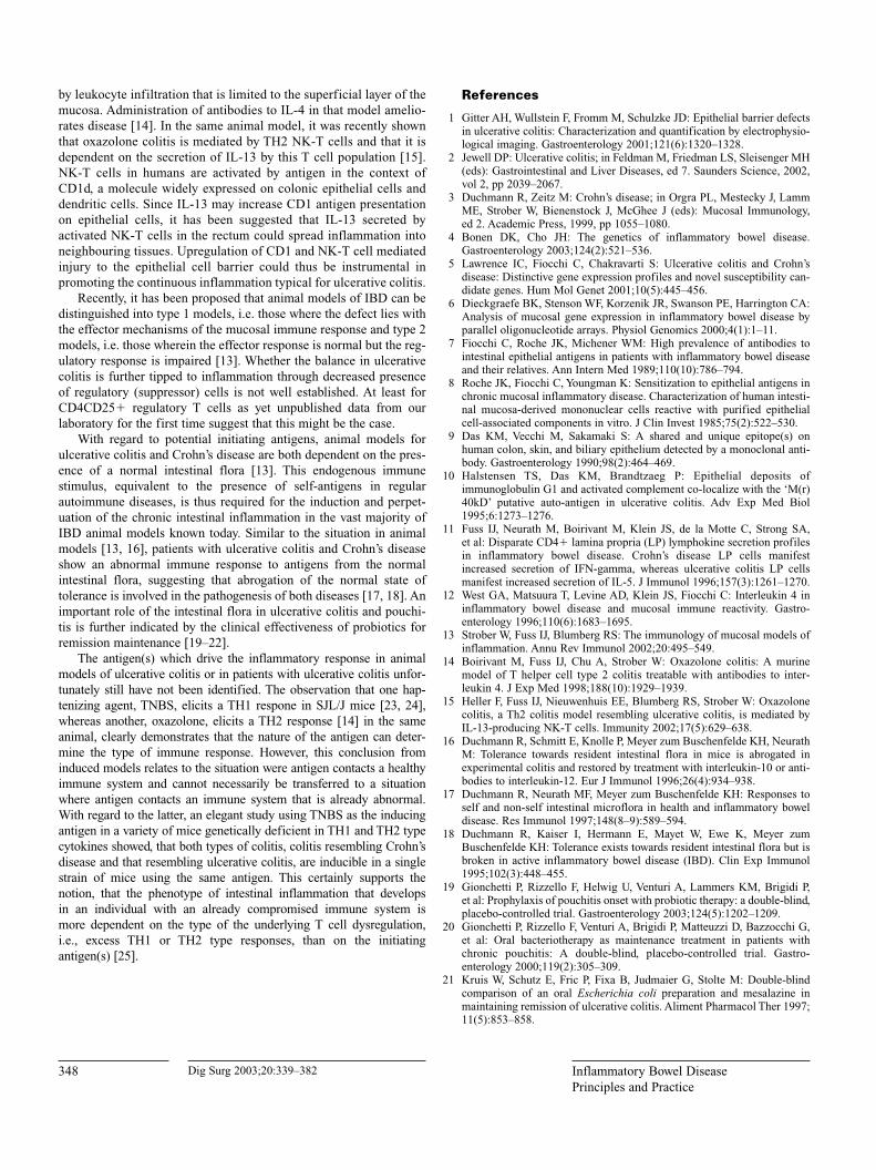
Results: Increased Cytokine/Chemokine Transcript Concentra-tion in Biopsies from Patients with Ulcerative Colitis. Compared to
non-inflamed mucosa tissue, samples from active UC expressed sig-
nificantly elevated transcripts of IL-1�, IL-8, MRP-14, MIP-2�, and
MMP-1 (see fig. 1). It should be noted, that in all patients paired sam-
ples obtained from inflamed and non-inflamed – so-called normal –
mucosa were analyzed. Intra-individual comparison supported the
overall finding of clear differences in cytokine/chemokine transcript
profiles depending on disease activity. Further, a loose correlation
exists between endoscopic scores, and histological scores and
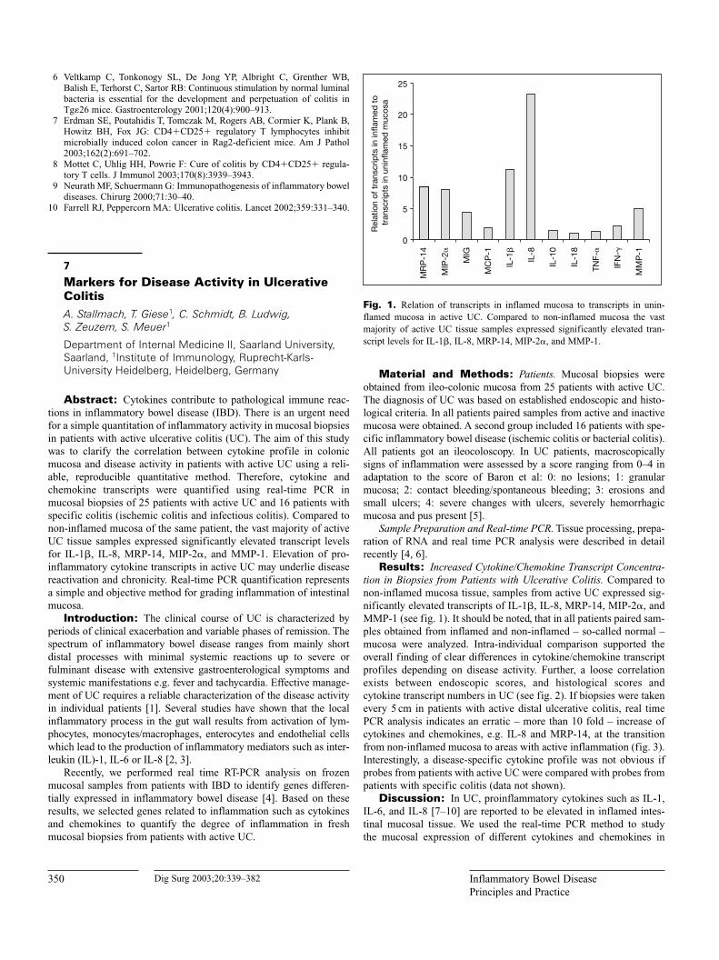
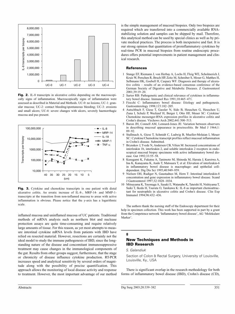
cytokine transcript numbers in UC (see fig. 2). If biopsies were taken
every 5 cm in patients with active distal ulcerative colitis, real time
PCR analysis indicates an erratic – more than 10 fold – increase of
cytokines and chemokines, e.g. IL-8 and MRP-14, at the transition
from non-inflamed mucosa to areas with active inflammation (fig. 3).
Interestingly, a disease-specific cytokine profile was not obvious if
probes from patients with active UC were compared with probes from
patients with specific colitis (data not shown).
Discussion: In UC, proinflammatory cytokines such as IL-1,
IL-6, and IL-8 [7–10] are reported to be elevated in inflamed intes-
tinal mucosal tissue. We used the real-time PCR method to study
the mucosal expression of different cytokines and chemokines in
0
5
10
15
20
25
Rel
atio
n of
tra
nscr
ipts
in in
flam
ed t
o tr
ansc
ripts
in u
ninf
lam
ed m
ucos
a
MR
P-1
4
MIP
-2�
MIG
MC
P-1
IL-1
�
IL-8
IL-1
0
IL-1
8
TNF-
�
IFN
-
MM
P-1
Fig. 1. Relation of transcripts in inflamed mucosa to transcripts in unin-
flamed mucosa in active UC. Compared to non-inflamed mucosa the vast
majority of active UC tissue samples expressed significantly elevated tran-
script levels for IL-1�, IL-8, MRP-14, MIP-2�, and MMP-1.
351Dig Surg 2003;20:339–382Abstracts
inflamed mucosa and uninflamed mucosa of UC patients. Traditional
methods of mRNA analysis such as northern blot and nuclease
protection assays are quite time-consuming and require relatively
large amounts of tissue. For this reason, as yet most attempts to meas-
ure intestinal cytokine mRNA levels from patients with IBD have
relied on resected material. However, resections are certainly not the
ideal model to study the immune pathogenesis of IBD, since the long-
standing nature of the disease and concomitant immunosuppressive
treatment may cause changes in the immunological components of
the gut. Results from other groups suggest, furthermore, that the stage
or chronicity of disease influence cytokine production. RT-PCR
increases speed and analytical sensitivity by several orders of magni-
tude along with the possibility of precise quantification. This
approach allows the monitoring of local disease activity and response
to treatment. However, the most important advantage of our method
is the simple management of mucosal biopsies. Only two biopsies are
required which are transferred into a commercially available RNA-
stabilizing solution and samples can be shipped by mail. Therefore,
this analytical method can be used by special clinics as well as by pri-
vate medical practices. The process is both inexpensive and fast. It is
our strong opinion that quantitation of proinflammatory cytokines by
real-time PCR in mucosal biopsies from routine endoscopic proce-
dures offers potential improvements in patient management and clin-
ical research.
References
1 Stange EF, Riemann J, von Herbay A, Lochs H, Fleig WE, Scholmerich J,
Kruis W, Porschen R, Bruch HP, Zeitz M, Schreiber S, Moser G, Matthes H,
Selbmann HK, Goebell H, Caspary WF: Diagnosis and therapy of ulcera-
tive colitis – results of an evidence-based consensus conference of the
German Society of Digestive and Metabolic Diseases. Z Gastroenterol
2001;39:19–20.
2 Sartor RB: Pathogenetic and clinical relevance of cytokines in inflamma-
tory bowel disease. Immunol Res 1991;10:465–471.
3 Fiocchi C: Inflammatory bowel disease: Etiology and pathogenesis.
Gastroenterology 1998;115:182–205.
4 Autschbach F, Giese T, Gassler N, Sido B, Heuschen G, Heuschen U,
Zuna I, Schulz P, Weckauf H, Berger I, Otto HF, Meuer SC: Cytokine/
Chemokine messenger-RNA expression profiles in ulcerative colitis and
Crohn’s disease. Virchows Arch 2002;441:500–513.
5 Baron JH, Connell AM, Lennard-Jones JE: Variation between observers
in describing mucosal appearance in proctocolitis. Br Med J 1964;1:
89–92.
6 Stallmach A, Giese T, Schmidt C, Ludwig B, Mueller-Molaian I, Meuer
SC: Cytokine/Chemokine transcript profiles reflect mucosal inflammation
in Crohn’s disease. Submitted.
7 Brynskov J, Tvede N, Andersen CB, Vilien M: Increased concentrations of
interleukin 1b, interleukin 2, and soluble interleukin 2 receptors in endo-
scopical mucosal biopsy specimens with active inflammatory bowel dis-
ease. Gut 1992;33:55–58.
8 Kusugami K, Fukatsu A, Tanimoto M, Shinoda M, Haruta J, Kuroiwa A,
Ina K, Kanayama K, Ando T, Matsuura T, et al: Elevation of interleukin-6
in inflammatory bowel disease is macrophage- and epithelial cell-
dependent. Dig Dis Sci 1995;40:949–959.
9 Nielsen OH, Rudiger N, Gaustadnes M, Horn T: Intestinal interleukin-8
concentration and gene expression in inflammatory bowel disease. Scand
J Gastroenterol 1997;32:1028–1034.
10 Mitsuyama K, Toyonaga A, Sasaki E, Watanabe K, Tateishi H, Nishiyama T,
Saiki T, Ikeda H, Tsuruta O, Tanikawa K: IL-8 as important chemoattrac-
tant for neutrophils in ulcerative colitis and Crohn’s disease. Clin Exp
Immunol 1994;96:432–436.
The authors thank the nursing staff of the Endoscopy department for their
help in specimen collection. This work has been supported in part by a grant
from the Competence network ‘Inflammatory bowel disease’, AG ‘Molekulare
Marker’.
8
New Techniques and Methods in IBD ResearchS. Galandiuk
Section of Colon & Rectal Surgery, University of Louisville,Louisville, Ky., USA
There is significant overlap in the research methodology for both
forms of inflammatory bowel disease (IBD), Crohn’s disease (CD),
0
1,000,000
2,000,000
3,000,000
4,000,000
5,000,000
6,000,000
7,000,000
8,000,000
UC-0 UC-1 UC-2 UC-3 UC-4
IL-8
tra
nsc
ripts
per
bio
psy
Fig. 2. IL-8 transcripts in ulcerative colitis depending on the macroscopi-
cally signs of inflammation. Macroscopically signs of inflammation were
assessed as described in Material and Methods. UC-0: no lesions; UC-1: gran-
ular mucosa; UC-2: contact bleeding/spontaneous bleeding; UC-3: erosions
and small ulcers; UC-4: severe changes with ulcers, severely haemorrhagic
mucosa and pus present.
10,000
100,000
1000,000
10,000,000
100,000,000
40 35 30 25 20 15 10 5(cm)
Tran
scrip
ts p
er b
iops
y
MMP-1TNF-�
IL-8
MRP-14IL-18
Fig. 3. Cytokine and chemokine transcripts in one patient with distal
ulcerative colitis. An erratic increase of IL-8-, MRP-14- and MMP-1-
transcripts at the transition from non-inflamed mucosa to areas with active
inflammation is obvious. Please notice that the y-axis has a logarithmic
scale.
352 Dig Surg 2003;20:339–382 Inflammatory Bowel Disease
Principles and Practice
and ulcerative colitis (UC). Research methods for UC can be divided
into two broad categories, animal models and human specimen-
based. Animal models include those in which colitis is induced by
agents such as Dextran sulfate and gene-knockout models with IBD-
like disorders, among others. With human samples, techniques range
from serology, to genotyping of genomic DNA, to studies of gene
expression. Several types of analyses can be performed using
genomic DNA derived from peripheral blood leukocytes. Linkages
analysis, family-based association studies, and population-based
association studies all have particular strengths and weaknesses.
Studies of genomic DNA and studies of gene expression will be
in the forefront over the next several years. Linkage studies measure
the degree of allele sharing among affected sibling pairs, with
increased allele sharing indicating the possible presence of a disease
locus [1]. Genome-wide scans are used to detect genomic regions
where there is a high degree of allele sharing between affected sibling
pairs. Such regions are said to be ‘susceptibility loci’ that confer
increased susceptibility to developing IBD. There are currently 9
genetic regions that have been associated with IBD [2–6]. Four of
these regions of linkage have been widely replicated: IBD1 on chro-
mosome 16q12 (implicated in CD), IBD2 (chromosome 12q13),
IBD3 (the major histocompatibility complex on chromosome 6p23),
and IBD4 (chromosome 14q11–12).
An alternate approach to performing genome-wide searches is to
specifically examine a gene or genes of interest. Such ‘candidate
genes’ are thought to be involved in IBD pathogenesis based on
demonstration of linkage or based on disease pathophysiology. Many
candidate genes have been proposed with respect to IBD. The only
CD candidate gene with broad consensus is NOD2 on chromosome
16 within the IBD1 locus [7, 8]. There is no consensus regarding
candidate genes for UC.
Transmission disequilibrium tests (TDTs) compare the difference
between the number of times an allele is transmitted from heterozy-
gous parents to affected children and the number of times it is not
transmitted [9]. A third type of study frequently performed in IBD
populations using allele and genotype data obtained from genomic
DNA is a case-control association study. In such studies, the fre-
quency of a given allele or genotype in IBD patients is compared to
that of non-affected controls. The possibility of false-positive associ-
ations in such population-based association studies is high.
Studies of gene expression based on analysis of colonic mucosa
affected with UC have many limitations, including the need for an
invasive procedure to obtain the specimen, i.e., either colonoscopy or
surgery. The possible effect of immunosuppressive therapy on gene
expression is another concern. In studies focusing on cancer risk, one
must also account for the effects of disease duration and extent of dis-
ease (i.e., pancolitis vs left-sided colitis). Oligonucleotide micro-
arrays can simultaneously analyze the expression of up to 20,000
genes. Such technology permits identification of potential pathways
or mechanisms of disease pathogenesis. Microarrays have also been
used to determine whether subsets of a particular disease can be iden-
tified based on gene expression. This approach has begun to reveal
novel molecular-based sub-classes of tumors in breast and colon can-
cer, as well as in lymphoma, leukemia, and melanoma [10]. This
could also find application in UC.
Over the next decade, these techniques and others will continue
to provide us with more insight into the pathogenesis of UC.
Knowledge gained from these studies will also promote the
development of targeted drug therapy and improve methods for
diagnosis.
References
1 Cordell HJ, Jacobs KB, Wedig GC, Elston RC: Improving the power for
disease locus detection in affected-sib-pair studies by using two-locus
analysis and multiple regression methods. Genet Epidemiol 1999;17
(Suppl 1):S521–S526.
2 Cho JH, Nicolae DL, Gold LH, et al: Identification of novel susceptibility
loci for inflammatory bowel disease on chromosomes 1p, 3q, and 4q:
Evidence for epistasis between 1p and IBD1. Proc Natl Acad Sci USA
1998;95:7502–7507.
3 Cavanaugh JA, Callen DF, Wilson SR, et al: Analysis of Australian Crohn’s
disease pedigrees refines the localization for susceptibility to inflamma-
tory bowel disease on chromosome 16. Ann Hum Genet 1998;62(Pt 4):
291–298.
4 Hampe J, Schreiber S, Shaw SH, et al: A genome wide analysis provides
evidence for novel linkages in inflammatory bowel disease in a large
European cohort. Am J Hum Genet 1999;64:808–816.
5 Satsangi J, Parkes M, Louis E, et al: Two-stage genome-wide search in
inflammatory bowel disease provides evidence for susceptibility loci on
chromosomes 3, 7 and 12. Nat Genet 1996;14:199–202.
6 Duerr RH, Barmada MM, Zhang L, et al: Linkage and association between
inflammatory bowel disease and a locus on chromosome 12. Am J Hum
Genet 1998;63:95–100.
7 Ogura Y, Bonen DK, Inohara N, et al: A frame shift mutation in NOD2
associated with susceptibility to Crohn’s disease. Nature 2001;411:
603–606.
8 Hugot JP, Chamaillard M, Zouali H, et al: Association of NOD2 leucine-
rich repeat variants with susceptibility to Crohn’s disease. Nature
2001;411:599–603.
9 Eichenbaum-Voline S, Baur MP, Knapp M: Genetic analysis of a complex
disease in the presence of an environmental risk factor. Genet Epidemiol
1999;17(Suppl 1):S545–S550.
10 Alizadeh AA, Ross DT, Perou CM, van de Rijn M: Towards a novel clas-
sification of human malignancies based on gene expression patterns.
J Pathol 2001;195:41–52.
9
Carcinogenesis in Ulcerative Colitis:New Diagnostic Tools?G. Heuschen1, A. German2, C. Leowardi1, U. Heuschen3
1Departments of Surgery and 2Pathology, University ofHeidelberg, Heidelberg, 3St. Vincenz Hospital, Limburg,Germany
Several studies indicated that patients with ulcerative colitis (UC)
face a higher risk to develop colorectal cancer (CRC) compared to
the normal population. Different clinical factors influencing the
individual risk in UC patients could have been identified.
Important clinical and biological differences exist between the
adenoma carcinoma sequence in sporadic CRC and UC-associated
inflammation dysplasia carcinoma sequence: Cancer in UC probably
evolves from microscopic dysplasia rather than from adenomas.
UC-associated CRC originate multifocally with a high percentage of
synchronous epithelial dysplasia and carcinomas in one proctocolec-
tomy specimen. UC patients with CRC are significantly younger than
patients with sporadic CRC, moreover there is a high percentage of
right-sided and mucinous CRC. The distinct differences between
UC-associated and sporadic CRC do not only concern the clinical
appearance, but also the molecular basis, especially incidence and
temporal occurrence of genetic lesions.

mainly associated with small bowel involvement and seems to be fre-
quently related to a stricturing phenotype [6–9].
The fact that this mutation is only responsible for a subgroup of
patients and in addition associated with a specific location and poten-
tially a specific phenotype suggests that there are different subgroups
of patients with Crohn’s disease. This indicates that there is no
Crohn’s disease but probably a Crohn’s syndrome [8]. This corre-
sponds to the daily clinical experience: comparing patients with peri-
anal Crohn’s disease with those having a circumscript stricture in the
ileocecal region and furthermore with patients with location in the
upper gastrointestinal tract or extensive colonic involvement demon-
strates that we have several different phenotypes. It is obvious that
these phenotypes will probably be related to different genotypes. First
data regarding surgical but also conservative treatment show that
these variable phenotypes probably need different treatment. The
recently published Vienna-classification for Crohn’s disease [8] pro-
vides one attempt for subgrouping of patients. It has to be further
developed in the light of new findings.
Epidemiological studies demonstrate that a north-south-gradi-
ent regarding the increase of the incidence of Crohn’s disease in
Europe exists. Meanwhile similar incidence increases have been
found in countries in Asia or South America. This suggests that
environmental factors have a role which obviously occurred earlier
in Northern Europe when compared to South Europe and again ear-
lier in Europe than in countries adapting western life style during
the last decades. No individual factor has been found yet.
Numerous papers provide evidence that a high hygienic standard in
early childhood is associated with a significantly increased risk of
a later manifestation of a Crohn’s disease (table 1) [10]. Studies
showing that the presence of elder siblings who introduce infections
from kindergarten or school to their younger siblings is protective
for the younger siblings regarding later occurrence of Crohn’s
353Dig Surg 2003;20:339–382Abstracts
Several studies have been performed concerning the molecular
characterisation of UC associated cancer, but mostly with a small
number of patients or specimens not well clinically characterised.
So far, systematic investigations of molecular genetic alterations in
the carcinogenesis of UC-associated CRC are missing. Moreover,
traditional methods of analysis have imposed a practical limit on the
number of investigated candidate genes, the expression of which can
be conveniently and simultaneously studied.
Analogous to clinicopathological differences, UC-associated
CRC seem to have distinct molecular genetic alterations which should
result in markedly differentiated transcriptional gene expression pro-
files. DNA-arrays capable of simultaneously measuring expression of
thousands of genes in fresh frozen specimens of patients, operated on
for UC have the potential to provide information about carcinogenesis
in UC not previously possible. Therefore, we used an oligonucleotide
array (GeneChip, Affymetrix) to monitor the expression profiles of
different clinicopathologic lesions in UC.
Pathophysiology, Diagnosis andConservative Treatment of Crohn’s Disease
10
Current Concepts of Pathophysiology ofCrohn’s DiseaseJ. Schölmerich
Klinik und Poliklinik für Innere Medizin I, Klinikum derUniversität Regensburg, Regensburg, Germany
In spite of the fact that the first official description of Crohn’s
disease took place more than 70 years ago its aetiology and patho-
genesis are still incompletely understood. During the last two decades
a number of findings regarding genetics, patient phenotypes, envi-
ronmental influences, abnormalities of the mucosal immune system
and disturbed interaction between the mucosa and its different cell
populations with luminal bacteria have brought significant progress.
The example of the cotton top tamarin which, while living in his
natural habitat, the forests of Central America, has rarely inflammatory
bowel disease, provides a paradigm for the human disorder. When these
primates are taken into captivity, many of them develop familiarly
accentuated chronic inflammatory bowel disease. This demonstrates
that genetic susceptibility and environmental factors play a role at least
in this monkey. As far as we know this will be similar in humans. An
important argument for the existence of genetic susceptibility comes
from twin studies, where monozygotic twins have a higher concordance
rate as compared to dizygotic twins [1]. Two years ago the first gene has
been found where mutation leads to manifestation of Crohn’s disease in
some patients: the NOD2/CARD15 gene. Its product is involved in the
construction of signals of bacterial peptidoglycans in monocytes and
probably also in other mucosal cells. This mutation seems to be respon-
sible for 10 to 20% of patients with Crohn’s disease (fig. 1) [2–5]. It is
Peptido- glycan CD14
TLR4
TRAF6
NOD2
I�B
NF�B
NucleusInflammation
Monocyte
C-insertion mutation Stop codon, abnormal activation of NF�B
NF�B
Fig. 1. The role of the gene product of NOD2/CARD15 and the interaction
between intestinal bacteria and the inflammatory cascade in intestinal immune
cells.
Table 1. Hygiene in childhood – ‘IBD’ [10]
RR
CD UC
Hot water tap 5.0 (1.4–17.3) 1.3 (0.7–2.2)
Water tap 1.8 (0.6–5.4) 0.9 (0.5–1.7)
Separate toilet 3.3 (1.3–8.3) 1.3 (0.7–2.4)
Canalisation 2.6 (0.9–7.3) 1.2 (0.7–2.1)
Appendectomy 1.4 (0.6–3.4) 0.3 (0.1–0.6)
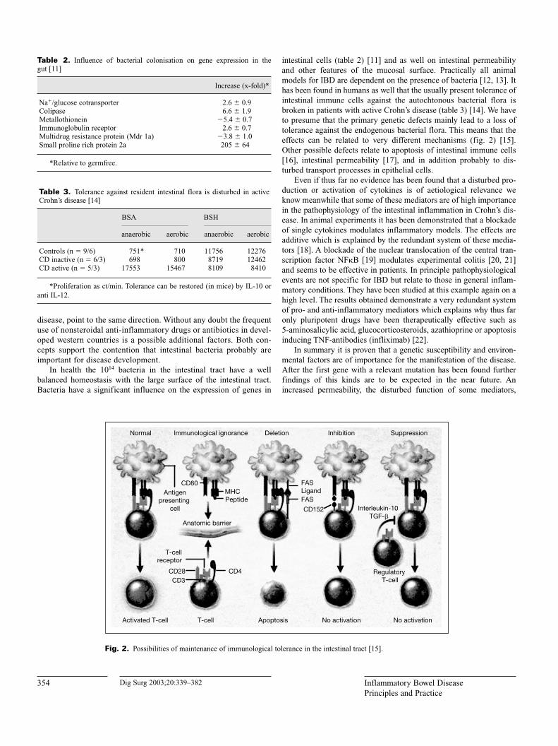
354 Dig Surg 2003;20:339–382 Inflammatory Bowel Disease
Principles and Practice
intestinal cells (table 2) [11] and as well on intestinal permeability
and other features of the mucosal surface. Practically all animal
models for IBD are dependent on the presence of bacteria [12, 13]. It
has been found in humans as well that the usually present tolerance of
intestinal immune cells against the autochtonous bacterial flora is
broken in patients with active Crohn’s disease (table 3) [14]. We have
to presume that the primary genetic defects mainly lead to a loss of
tolerance against the endogenous bacterial flora. This means that the
effects can be related to very different mechanisms (fig. 2) [15].
Other possible defects relate to apoptosis of intestinal immune cells
[16], intestinal permeability [17], and in addition probably to dis-
turbed transport processes in epithelial cells.
Even if thus far no evidence has been found that a disturbed pro-
duction or activation of cytokines is of aetiological relevance we
know meanwhile that some of these mediators are of high importance
in the pathophysiology of the intestinal inflammation in Crohn’s dis-
ease. In animal experiments it has been demonstrated that a blockade
of single cytokines modulates inflammatory models. The effects are
additive which is explained by the redundant system of these media-
tors [18]. A blockade of the nuclear translocation of the central tran-
scription factor NF�B [19] modulates experimental colitis [20, 21]
and seems to be effective in patients. In principle pathophysiological
events are not specific for IBD but relate to those in general inflam-
matory conditions. They have been studied at this example again on a
high level. The results obtained demonstrate a very redundant system
of pro- and anti-inflammatory mediators which explains why thus far
only pluripotent drugs have been therapeutically effective such as
5-aminosalicylic acid, glucocorticosteroids, azathioprine or apoptosis
inducing TNF-antibodies (infliximab) [22].
In summary it is proven that a genetic susceptibility and environ-
mental factors are of importance for the manifestation of the disease.
After the first gene with a relevant mutation has been found further
findings of this kinds are to be expected in the near future. An
increased permeability, the disturbed function of some mediators,
Normal Immunological ignorance Deletion Inhibition Suppression
Antigen presenting
cell
CD80MHCPeptide
Anatomic barrier
T-cell receptor
CD28CD3
CD4
Activated T-cell T-cell Apoptosis No activation No activation
CD152 Interleukin-10 TGF-�
Regulatory T-cell
FAS
FAS Ligand
Fig. 2. Possibilities of maintenance of immunological tolerance in the intestinal tract [15].
Table 2. Influence of bacterial colonisation on gene expression in the
gut [11]
Increase (x-fold)*
Na�/glucose cotransporter 2.6 � 0.9
Colipase 6.6 � 1.9
Metallothionein �5.4 � 0.7
Immunoglobulin receptor 2.6 � 0.7
Multidrug resistance protein (Mdr 1a) �3.8 � 1.0
Small proline rich protein 2a 205 � 64
*Relative to germfree.
Table 3. Tolerance against resident intestinal flora is disturbed in active
Crohn’s disease [14]
BSA BSH
anaerobic aerobic anaerobic aerobic
Controls (n � 9/6) 751* 710 11756 12276
CD inactive (n � 6/3) 698 800 8719 12462
CD active (n � 5/3) 17553 15467 8109 8410
*Proliferation as ct/min. Tolerance can be restored (in mice) by IL-10 or
anti IL-12.
disease, point to the same direction. Without any doubt the frequent
use of nonsteroidal anti-inflammatory drugs or antibiotics in devel-
oped western countries is a possible additional factors. Both con-
cepts support the contention that intestinal bacteria probably are
important for disease development.
In health the 1014 bacteria in the intestinal tract have a well
balanced homeostasis with the large surface of the intestinal tract.
Bacteria have a significant influence on the expression of genes in
355Dig Surg 2003;20:339–382Abstracts
alterations of bacterial adherence on epithelial cells and many other
principles are possible. The environmental factors comprise mainly the
bacterial flora, nutritional components, drugs, toxins and with some
certainty early childhood hygiene or factors associated with it. Most
probably several different genes and variable environmental conditions
are of importance for the different subgroups of Crohn’s syndrome.
Pathophysiology is characterised by a plethora of mediators which
influence in a redundant and difficult to modulate form the inflamma-
tory concert.
References
1 Tysk C, Lindberg E, Jarnerot G, Floderus-Myrhed B: Ulcerative colitis
and Crohn’s disease in an unselected population of monozygotic and dizy-
gotic twins. A study of heritability and the influence of smoking. Gut
1988;29:990–996.
2 Beutler B: Autoimmunity and apoptosis: The Crohn’s connection. Immunity
2001;15:5–14.
3 Hampe J, Cuthbert A, Croucher PJP, Mirza MM, Mascheretti S, Fisher S,
Frenzel H, King K, Hasselmeyer A, MacPherson AJS, Bridger S, van
Deventer S, Forbes A, Nikolaus S, Lennard-Jones JE, Foelsch UR,
Krawczak M, Lewis C, Schreiber S, Mathew CG: Association between
insertion mutation in NOD2 gene and Crohn’s disease in German and
British populations. Lancet 2001;357:1925–1928.
4 Hugot JP, Chamaillard M, Zouali H, Lesage S, Cezard JP, Belaiche J,
Almer S, Tysk C, O’Morain CA, Gassull M, Binder V, Finkel Y,
Cortot A, Modigliani R, Laurent-Puig P, Gower-Rousseau C, Macry J,
Colombel JF, Sahbatou M, Thomas G: Association of NOD2 leucine-rich
repeat variants with susceptibility to Crohn’s disease. Nature 2001;411:
599–603.
5 Ogura Y, Bonen DK, Inohara N, Nicolae DL, Chen FF, Ramos R, Britton H,
Moran T, Karaliuskas R, Duerr RH, Achkar JP, Brant SR, Bayless TM,
Kirschner BS, Hanauer SB, Nunez G, Cho JH: A frameshift mutation in
NOD2 associated with susceptibility to Crohn’s disease. Nature 2001;411:
603–606.
6 Ahmad T, Armuzzi A, Bunce M, Mulcahy-Hawes K, Marshall SE, Orchard
TR, Crawshaw J, Large O, de Silva A, Cook JT, Barnardo M, Cullen S,
Welsh KI, Jewell DP: The molecular classification of the clinical manifes-
tations of Crohn’s disease. Gastroenterology 2002;122:854–866.
7 Cuthbert AP, Fisher SA, Mirza MM, King K, Hampe J, Croucher PJ,
Mascheretti S, Sanderson J, Forbes A, Mansfield J, Schreiber S, Lewis
CM, Mathew CG: The contribution of NOD2 gene mutations to the risk
and site of disease in inflammation bowel disease. Gastroenterology 2002;
122:867–874.
8 Gasché C, Schölmerich J, Brynskow J, D’Haens G, Hanauer SB, Irvine EJ,
Jewell DP, Rachmilewitz D, Sachar DB, Sandborn WJ, Sutherland LR:
A simple classification of Crohn’s disease: Report of the Working Party for
the World Congresses of Gastroenterology, Vienna 1998. Inflamm Bowel
Dis 2000;6:8–15.
9 Lesage S, Zouali H, Cezard JP, Colombel JF, Belaiche J, Almer S, Tysk C,
O’Morain C, Gassull M, Binder V, Finkel Y, Modigliani R, Gower-
Rousseau C, Macry J, Merlin F, Chamaillard M, Jannot AS, Thomas G,
Hugot JP; EPWG-IBD Group; EPIMAD Group; GETAID Group:
CARD15/NOD2 mutational analysis and genotype-phenotype correlation
in 612 patients with inflammatory bowel disease. Am J Hum Genet 2002;
70:845–857.
10 Gent AE, Hellier MD, Grace RH, Swarbrick ET, Coggon D: Inflammatory
bowel disease and domestic hygiene in infancy. Lancet 1994;343:766–767.
11 Hooper LV, Wong MH, Thelin A, Hansson L, Falk PG, Gordon JI:
Molecular analysis of commensal host-microbial relationships in the intes-
tine. Science 2001;29:881–884.
12 Obermeier F, Dunger N, Deml L, Herfarth H, Schölmerich J, Falk W: CpG
motifs of bacterial DNA exacerbate colitis of dextran sulfate sodium-
treated mice. Eur J Immunol 2002;32:2084–2092.
13 Schultz M, Tonkonogy SL, Sellon RK, Veltkamp C, Godfrey VL, Kwon J,
Grenther WB, Balish E, Horak I, Sartor RB: IL-2-deficient mice raised
under germfree conditions develop delayed mild focal intestinal inflam-
mation. Am J Physiol 1999;276:G1461–G1472.
14 Duchmann R, Neurath MF, Meyer zum Büschenfelde KH: Responses to
self and non-self intestinal microflora in health and inflammatory bowel
disease. Res Immunol 1997;148:589–594.
15 Kamradt T, Mitchison NA: Tolerance and autoimmunity. N Engl J Med
2001;344:655–664.
16 Ina K, Itoh J, Fukushima K, Kusugami K, Yamaguchi T, Kyokane K,
Imada A, Binion DG, Musso A, West GA, Dobrea GM, McCormick TS,
Lapetina EG, Levine AD, Ottaway CA, Fiocchi C: Resistance of Crohn’s
disease T cells to multiple apoptotic signals is associated with a Bcl-2/Bax
mucosal imbalance. J Immunol 1999;163:1081–1090.
17 Hollander D, Vadheim CM, Brettholz E, Petersen GM, Delahunty T,
Rotter JI: Increased intestinal permeability in patients with Crohn’s
disease and their relatives. A possible etiologic factor. Ann Intern Med
1986;105:883–885.
18 Kojouharoff G, Hans W, Obermeier F, Männel DN, Andus T, Schölmerich J,
Gross V, Falk W: Neutralization of tumour necrosis factor (TNF) but not
of IL-1 reduces inflammation in chronic dextran sulphate sodium-induced
colitis in mice. Clin Exp Immunol 1997;107:353–358.
19 Rogler G, Brand K, Vogel D, Page S, Hofmeiseter R, Andus T, Knüchel R,
Bäuerle PA, Schölmerich J, Gross V: Nuclear factor �B is activated in
macrophages and epithelial cells of inflamed intestinal mucosa. Gastro-
enterology 1998;115:357–369.
20 Herfarth H, Brand K, Rath HC, Rogler G, Schölmerich J, Falk W: Nuclear
factor-kappa B activity and intestinal inflammation in dextran sulphate
sodium (DSS)-induced colitis in mice is suppressed by gliotoxin. Clin Exp
Immunol 2000;120:59–65.
21 Neurath MF, Pettersson S, Meyer zum Büschenfelde KH, Strober W:
Local administration of antisense phosporothioate oligonucleotides to the
p65 subunit of NF-�B abrogates established experimental colitis in mice.
Nat Med 1996;2:998–1004.
22 Schölmerich J: Therapeutische Innovationen bei chronisch entzündlichen
Darmerkrankungen durch ‘biologische Therapie’ – Anti-TNF und andere.
Internist 2002;43:1386–1399.
11
Making the Diagnosis of Crohn’s DiseaseM.L. Scribano, C. Prantera
Division of Gastroenterology, Azienda Ospedaliera S. Camillo-Forlanini, Rome, Italy
The aetiology of Crohn’s disease (CD) is unknown and, at the
moment, diagnosis is made on an initial clinical impression, by
evaluating risk factors, symptoms and physical findings of the sub-
ject. In addition, laboratory tests, radiological studies and
endoscopy with biopsies play an important role in the evaluation of
patients at the time of initial presentation and also for long term
assessment.
Risk Factors: Different environmental and lifestyle factors
have been associated with an increased risk of developing CD. These
factors have been investigated in epidemiological studies, but the
results are often conflicting. Based on twin studies, association and
linkage studies, there is evidence for familiar clustering, congruence
of disease type and pattern and the presence of specific genes which
increase the risk of CD. A family history of CD, especially, and of
ulcerative colitis (UC) is associated with an increased risk of CD [1].
A history of smoking is the most established environmental risk
factor for CD [2]. Except for smoking, no environmental factor has
consistently been shown to be associated with the disease. However,
perinatal infections, bacterial or viral infections, may also play a role,
in particular as promoters, in the development of CD [3].

356 Dig Surg 2003;20:339–382 Inflammatory Bowel Disease
Principles and Practice
Clinical Assessment: There is a variety of clinical presenta-
tions of the disease. Crohn’s disease can involve any location of the
gastrointestinal tract, from the mouth to the anus, and it is much more
common in the ileum and colon than in more proximal locations of
the digestive tract. The site of disease at the time of initial diagnosis
plays a significant role in the clinical features of patients with CD.
The spectrum of symptoms with diffuse involvement of the small
bowel is often different from that of symptoms with less extensive
disease. More typically, the patient with extensive disease of the distal
ileum has abdominal pain, diarrhoea, weight loss, malaise and, less
often, fever. Abdominal pain, a feature in about two-thirds of the
patients, is usually located in the right lower quadrant. On physical
examination, there may be a palpable inflammatory mass.
In Crohn’s colitis symptoms depend on the location, extent, and
severity of the disease. Abdominal pain is the most common symp-
tom. Patients with diffuse colonic disease may present with symptoms
that are indistinguishable from UC, such as diarrhoea, rectal bleeding,
abdominal pain, tenesmus and malaise.
The nature of the symptoms is influenced not only by the location,
but also by the type of lesions. Patients with fibrostenotic disease
have a different type of symptoms, that depend on the degree of
narrowing of the bowel lumen. Nevertheless, patients with marked
narrowing of the lumen may be asymptomatic because they have
restricted their fibre intake, sometimes unintentionally, and become
symptomatic after eating high fibre meals. With tight stenosis, there
may be obstructive symptoms, such as abdominal pain, bloating,
nausea and vomiting. Weight loss and growth failure are common;
they may be a result of the inflammation, but also of malabsorption
when the small bowel is involved, or a voluntary reduction in food
intake to minimize postprandial symptoms.
Sometimes, patients with CD have an insidious onset of symp-
toms. Extra intestinal manifestations, involving joints, skin or eyes
are symptoms at presentation in approximately 13% of patients [4]. It
is easy to suspect an IBD when there are associated intestinal symp-
toms, but more difficult when extra intestinal manifestations are
isolated. Perianal disease may be the first manifestation of CD in 10%
of patients. Some patients with ileal location present suddenly with
symptoms indistinguishable from acute appendicitis.
The average interval of 35 months from onset of symptoms to the
diagnosis of CD, reported from the National Cooperative Crohn’s
Disease Study, highlights the difficulties involved in its diagnosis [5].
Laboratory Evaluation: Laboratory tests are frequently
employed in clinical practice to aid in making the diagnosis of CD.
The acute-phase reactants, such as erythrocyte sedimentation rate
(ESR), C-reactive protein (CRP), seromucoids and alpha-2-globulin
are sensitive markers of an inflammation in some part of the body.
The depth involvement of the intestinal wall in the inflammatory
process of CD accounts for the early increase of these laboratory
tests, but occasionally they may be normal, for example in cases of
strictures. In fact, alterations of laboratory tests should be strictly
correlated to clinical symptoms, but that does not always happen.
C-reactive protein is markedly elevated in most patients with
active CD and the levels correlate well with the clinical assessment
[6]. Erythrocyte sedimentation rate is a good indicator of colonic
inflammation, but does not appear to be helpful in assessing small
bowel disease [6]. A low level of serum iron appears to be a very early
and sensitive marker of activity in CD and it is significantly corre-
lated with the extension of lesions in ileal CD [7]. The decline of
serum iron levels not only reflects a loss of iron via microhemorrhage
from erosions and ulcerations, but also a redistribution of the body
iron stores, related to inflammation. However, the limit of all of these
laboratory tests is, obviously, a lack of specificity.
Radiological Procedures: Diagnostic imaging studies play
an important role in the diagnosis of patients with CD. The role of radi-
ology in the initial presentation of the disease is to document suspected
CD in an undiagnosed patient, to detect the extent of the lesions, the
morphology of the mucosa and to identify any complications.
Plain abdominal film is often non specific, but it may be helpful
in patients with acute presentation. Contrast studies should be avoided
in these patients because of the possibility of precipitating toxic
megacolon. It is commonly performed to evaluate patients with
obstructive symptoms. The traditional method for demonstrating CD
involving the terminal ileum and other segments of small bowel is the
barium follow-through study; it remains the routine examination, in
spite of superiority of enteroclysis. Fistulae are also well studied
using these radiological methods. Ultrasonography is increasingly
used in diagnostic assessment of CD. It provides indications of any
thickening of the intestinal wall, it is an useful technique for studying
small bowel obstruction [8] and it has a good accuracy in differenti-
ating abdominal masses from abscesses. In addition, small bowel
sonography may be employed in case of clinical suspicion of CD
recurrence after surgery [9].
Endoscopic Procedures: The role of diagnostic
colonoscopy with mucosal biopsies for histological examination is to
establish a diagnosis and to differentiate Crohn’s colitis from UC,
indeterminate colitis and from other types of colitis. Biopsies must be
performed on the lesions, but also if the colon and ileum appear
normal. However, the diagnosis is sometimes difficult, especially in
the case of a first and severe form of colitis.
References
1 Orholm M, Munkholm P, Langholz E, Haagen Nielsen O, Sorensen TIA,
Binder V: Familiar occurrence of inflammatory bowel disease. N Engl J
Med 1991;324:84–88.
2 Lashner BA: Consideration of smoking in patients with Crohn’s disease;
in Prantera C, Korelitz BI (eds): Crohn’s Disease. New York, Dekker M,
1996, pp 81–91.
3 Russel MGVM, Stockbrugger RW: Epidemiology of inflammatory bowel
disease: An update. Scand J Gastroenterol 1996;31:260–266.
4 Both H, Torp-Pedersen S, Kreiner S: Clinical appearance at diagnosis of
ulcerative colitis and Crohn’s disease in a regional patient group. Scand J
Gastroenterol 1983;18:987–991.
5 Mekhjian HS, Switz DM, Melnyk CS, Rankin GB, Brooks RK: Clinical
features and natural history of Crohn’s disease. Gastroenterology 1979;77:
898–906.
6 Prantera C, Kohn A: Proposed measures of disease activity: How useful
are they? in Prantera C, Korelitz BI (eds): Crohn’s Disease. New York,
Dekker M, 1996, pp 187–197.
7 Prantera C, Baiocchi G, Levenstein S, Livi U, Luzi C, Fanucci A: Clinical
and laboratory parameters in Crohn’s disease: Relation to disease activity,
morphology and extent. Ital J Gastroenterol 1981;13:24–27.
8 Kohn A, Cerro P, Milite G, De Angelis E, Prantera C: Prospective evalua-
tion of transabdominal bowel sonography in the diagnosis of intestinal
obstruction in Crohn’s disease: Comparison with plain abdominal film and
small bowel enteroclysis. Inflam Bowel Dis 1999;5:153–157.
9 Andreoli A, Cerro P, Falasco G, Giglio LA, Prantera C: Role of ultra-
sonography in the diagnosis of postsurgical recurrence of Crohn’s disease.
Am J Gastroenterol 1998;93:1117–1121.

357Dig Surg 2003;20:339–382Abstracts
12
Imaging Procedures in Crohn’s Disease H.J. Hansmann, P. Flossdorf, J. Schmidt1, B. Grüber-Hoffmann, G. Erb, G.W. Kauffmann
Department of Diagnostic Radiology and 1General Surgery, University Hospital Heidelberg,Heidelberg, Germany
Crohn’s disease is diagnosed and monitored by the combination of
colonoscopy and small bowel enteroclysis. Biopsies taken during
endoscopy are limited to the mucosa and submucosa, thus do not
always cover the full extent of the diseased bowel wall in Crohn’s dis-
ease. Cross-sectional imaging (Sonography, CT, MRI) yields the
potential of non-invasive imaging of the complete bowel wall includ-
ing extraluminal pathologies. Sonography is limited by overlying
bowel gas and poor interobserver reproducibility. Multislice com-
puted tomography produces excellent depiction of the bowel and the
mesenteries, but radiation dose still limits its use in young patients
with the exception of emergency situations. Magnetic resonance
imaging is the gold standard in imaging of perirectal and pelvic
fistulas. Today dedicated bowel MRI with breath-hold imaging under
pharmacologic bowel paralysis is available. Imaging protocols
include fast T1-weighted gradient echo and T2-weighted TSE
sequences and oral or rectal bowel distension. High diagnostic accu-
racy can be achieved in Crohn’s disease with special reference to
patterns of disease, depth of inflammation, mesenteric reaction,
sinus tract depiction and formation of abscess. In ulcerative colitis,
the mucosa-related inflammation causes significantly minor bowel
wall thickening compared to Crohn’s disease. Hydro-MRI yields
the potential to replace enteroclysis in the diagnosis of chronic
inflammatory bowel disease and most of the follow-up colonoscopies
in Crohn’s disease. The included information about the surrounding
tissue replaces sonography and CT as well.
Although Crohn’s disease (CD) and ulcerative colitis (UC) have
several features in common, there are characteristic microscopic and
macroscopic features that allow a differentiation of CD from CU in
diagnostic imaging. CD is a transmural, often fistulating inflamma-
tion of the bowel wall with lymphocytic infiltrates and eosinophilic
granulomas. This transmural inflammation in CD results in a bowel
wall thickening, which regularly exceeds that in UC, being caused by
an predominantly mucosal inflammation.
Several imaging modalities are in use to diagnose IBD: plain
x-ray, fluoroscopy, sonography, endoscopy, computed tomography
(CT) and magnetic resonance tomography (MRI).
Plain x-ray of the abdomen is the best choice in the presence of
an acute abdomen as in cases of suspected bowel obstruction, ileus,
perforation or toxic megacolon.
Fluoroscopy uses either barium or iodinated enteral contrast media.
Barium is used in elective settings for the double contrast examination
of the colon and enteroclysis of the small bowel. Iodinated contrast
solutions are used preoperatively in the evaluation of enterocutaneous
fistulas, in the early postoperative phase in cases of suspected insuffi-
ciency of the anastomosis. Sonography plays an important role in the
evaluation of the acute abdomen. However, complete evaluation of the
abdomen is often hampered by overlying bowel gas. The correct
anatomical identification of a thickened bowel loop is difficult [1, 2].
Endoscopy has replaced traditional radiology of the intestinal tract
leaving only enteroclysis as gold standard in small bowel imaging.
Oesophagogastroduodenoscopy and colonoscopy including terminal
ileoscopy allow the direct visual inspection of mucosal alterations.
Additionally, biopsies are routinely taken for microscopic evaluation.
However, these biopsies do not represent the complete bowel wall,
making decisions in transmural processes as CD difficult. Push
enteroscopy of the small bowel has not gained wide acceptance.
CT, especially multislice CT offers excellent image quality, but
has the disadvantage of a considerable radiation dose [3]. Howewer,
in emergency examinations such as abscess formation with the need
for interventional drainage, CT still is the method of choice.
In routine abdominal MRI the bowel loops are not accessible,
sometimes even disturbing because of artifacts resulting from peri-
stalsis. With the development of MRI-techniques like ultrafast imag-
ing in breathhold in combination with enteral MR-contrast agents
dedicated MRI of the complete bowel has become available, which is
of special interest in the diagnosis of IBD with multiple repeated
examinations over years. In the majority of institutions MRI in IBD is
performed as MR-Enteroclysis [4–6]. The Heidelberg Hydro-MRI
protocol is designed for optimal image quality both of the small bowel
and the colon, thus depicting not only CD involvement of the jejunum
and ileum, but also of colonic disease (fig. 1–4). Patient preparation
consists in bowel cleansing using the same protocol as prior to
colonoscopy. Bowel distention is achieved by both oral water or tea
and a rectal enema (1.5–2 l). Imaging protocols include fast T1-
weighted gradient echo and T2-weighted TSE sequences in breath-
hold under pharmacologic bowel paralysis as well as ultrafast
T2-weighted sequences [7–9].
D
Fig. 1. Recurrent Crohn’s disese following ileoascendostomy with fistula to
the duodenum. T2-weighted coronal image, D � duodenum, arrow � fistula.
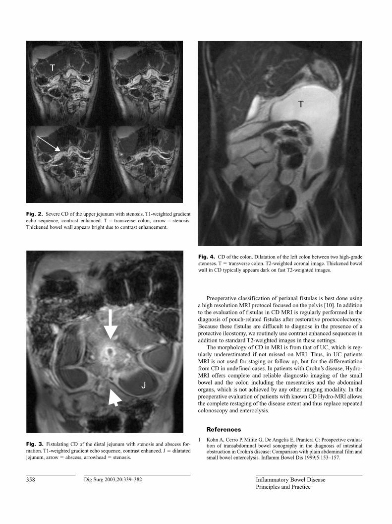
358 Dig Surg 2003;20:339–382 Inflammatory Bowel Disease
Principles and Practice
Preoperative classification of perianal fistulas is best done using
a high resolution MRI protocol focused on the pelvis [10]. In addition
to the evaluation of fistulas in CD MRI is regularly performed in the
diagnosis of pouch-related fistulas after restorative proctocolectomy.
Because these fistulas are diffucult to diagnose in the presence of a
protective ileostomy, we routinely use contrast enhanced sequences in
addition to standard T2-weighted images in these settings.
The morphology of CD in MRI is from that of UC, which is reg-
ularly underestimated if not missed on MRI. Thus, in UC patients
MRI is not used for staging or follow up, but for the differentiation
from CD in undefined cases. In patients with Crohn’s disease, Hydro-
MRI offers complete and reliable diagnostic imaging of the small
bowel and the colon including the mesenteries and the abdominal
organs, which is not achieved by any other imaging modality. In the
preoperative evaluation of patients with known CD Hydro-MRI allows
the complete restaging of the disease extent and thus replace repeated
colonoscopy and enteroclysis.
References
1 Kohn A, Cerro P, Milite G, De Angelis E, Prantera C: Prospective evalua-
tion of transabdominal bowel sonography in the diagnosis of intestinal
obstruction in Crohn’s disease: Comparison with plain abdominal film and
small bowel enteroclysis. Inflamm Bowel Dis 1999;5:153–157.
J
Fig. 3. Fistulating CD of the distal jejunum with stenosis and abscess for-
mation. T1-weighted gradient echo sequence, contrast enhanced. J � dilatated
jejunum, arrow � abscess, arrowhead � stenosis.
T
Fig. 4. CD of the colon. Dilatation of the left colon between two high-grade
stenoses. T � transverse colon. T2-weighted coronal image. Thickened bowel
wall in CD typically appears dark on fast T2-weighted images.
T
Fig. 2. Severe CD of the upper jejunum with stenosis. T1-weighted gradient
echo sequence, contrast enhanced. T � transverse colon, arrow � stenosis.
Thickened bowel wall appears bright due to contrast enhancement.
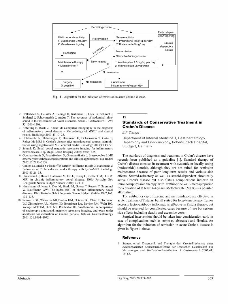
359Dig Surg 2003;20:339–382Abstracts
2 Hollerbach S, Geissler A, Schiegl H, Kullmann F, Lock G, Schmidt J,
Schlegel J, Schoelmerich J, Andus T: The accuracy of abdominal ultra-
sound in the assessment of bowel disorders. Scand J Gastroenterol 1998;
33:1201–1208.
3 Bitterling H, Rock C, Reiser M: Computed tomography in the diagnosis
of inflammatory bowel disease – Methodology of MSCT and clinical
results. Radiologe 2003;43:17–25.
4 Holzknecht N, Helmberger T, Herrmann K, Ochsenkuhn T, Goke B,
Reiser M: MRI in Crohn’s disease after transduodenal contrast adminis-
tration using negative oral MRI contrast media. Radiologe 2003;43:43–50.
5 Schunk K: Small bowel magnetic resonance imaging for inflammatory
bowel disease. Top Magn Reson Imaging 2002;13:409–425.
6 Gourtsoyiannis N, Papanikolaou N, Grammatikakis J, Prassopoulos P. MR
enteroclysis: technical considerations and clinical applications. Eur Radiol
2002;12:2651–2658.
7 Ganten M, Encke J, Flosdorff P, Gruber-Hoffmann B, Erb G, Hansmann J:
Follow up of Crohn’s disease under therapy with hydro-MRI. Radiologe
2003;43:26–33.
8 Hansmann HJ, Hess T, Hahmann M, Erb G, Elsing C, Richter GM, Dux M:
MRI in chronic inflammatory bowel disease. Röfo Fortschr Geb
Rontgenstr Neuen Bildgeb Verfahr 2001;173:4–11.
9 Hansmann HJ, Kosa R, Dux M, Brado M, Goeser T, Roeren T, Stremmel
W, Kauffmann GW: The hydro-MRT of chronic inflammatory bowel
diseases. Röfo Fortschr Geb Röntgenstr Neuen Bildgeb Verfahr 1997;167:
132–138.
10 Schwartz DA, Wiersema MJ, Dudiak KM, Fletcher JG, Clain JE, Tremaine
WJ, Zinsmeister AR, Norton ID, Boardman LA, Devine RM, Wolff BG,
Young-Fadok TM, Diehl NN, Pemberton JH, Sandborn WJ: A comparison
of endoscopic ultrasound, magnetic resonance imaging, and exam under
anesthesia for evaluation of Crohn’s perianal fistulas. Gastroenterology
2001;121:1064–1072.
13
Standards of Conservative Treatment inCrohn’s DiseaseE.F. Stange
Department of Internal Medicine 1, Gastroenterology,Hepatology and Endocrinology, Robert-Bosch Hospital,Stuttgart, Germany
The standards of diagnosis and treatment in Crohn’s disease have
recently been published as a guideline [1]. Standard therapy of
Crohn’s disease consists in treatment with systemic or locally acting
(budesonide) steroids, although they are not suited for remission
maintenance because of poor long-term results and various side
effects. Steroid-refractory as well as steroid-dependent chronically
active Crohn’s disease but also fistula complications indicate an
immunosuppressive therapy with azathioprine or 6-mercaptopurine
for a duration of at least 3–4 years. Methotrexate (MTX) is a possible
alternative.
The antibiotics ciprofloxacine and metronidazole are effective in
acute treatment of fistulas, but ill suited for long-term therapy. Tumor
necrosis factor-antibody infliximab is effective in fistula therapy, but
should be reserved for complicated cases because of rare but serious
side effects including deaths and excessive costs.
Surgical intervention should be taken into consideration early in
case of complications such as stenoses, abscesses and fistulas. An
algorithm for the induction of remission in acute Crohn’s disease is
given in figure 1 above.
Reference
1 Stange, et al: Diagnostik und Therapie des. Crohn-Ergebnisse einer
evidenzbasierten Konsensuskonferenz der Deutschen Gesellschaft Für
Verdauungs- und Stoffwechselkrankheiten. Z Gastroenterol 2003;41:
19–68.
Remitting course
Mild/moderate activity 1˚ Budesonide 9mg/day 2˚ Mesalamine 4g/day
Severe activity 1˚ Prednisone 1mg/kg per day 2˚ Budesonide 9mg/day
No remission
Remission
Maintenance therapy • Mesalamine (?)
No remission
1˚ Azathioprine 2.5mg/kg per day 2˚ Methotrexate 25mg/week
Steroid dependent
course
Steroid refractory course
No remission
• Additional Infliximab 5mg/kg per day
Early relapse
Remission
Surgery (if possible)
Early relapseupon tapering
No remission
Fig. 1. Algorithm for the induction of remission in acute Crohn’s disease.
360 Dig Surg 2003;20:339–382 Inflammatory Bowel Disease
Principles and Practice
14
Novel and Future Strategies in theManagement of IBDS. Schreiber, U.R. Fölsch
Department of General Internal Medicine, Christian-Albrechts-University, Kiel, Germany
The primary goal of therapy in inflammatory bowel disease is the
long-term maintenance of patients. A meaningful clinical benefit is
the induction of remission (as defined by a CDAI less than 100 points
accompanied by mucosal healing) and a reduction or, preferably, dis-
continuation of glucocorticoids. The introduction of novel agents has
not only led to impressive new therapeutic opportunities in Crohn’s
disease but also resulted in uncertainty regarding their optimal use
and possible side effects.
Although the exact nature of etiologic factors is still unclear the
pathophysiology of intestinal inflammation has been explored in great
detail: It appears that expression and secretion pro-inflammatory
mediators including the cytokines tumour-necrosis-factor � (TNF-�),
interleukin-1� (IL-1�), interleukin-6, interleukin-8 and interleukin-16
is upregulated in the intestinal lamina propria of patients with inflam-
matory bowel disease. Cytokines often activate transcription factors
as part of their signalling pathway.
It appears that the balance between pro-inflammatory and contra-
inflammatory cytokines (i.e. IL-10) is shifted towards a perpetuated
inflammatory reaction of the mucosa. Direct anti-inflammatory
approaches include an inhibition of pro-inflammatory cytokines (i.e.
monoclonal antibodies directed against TNF-� (infliximab, CDP571,
CDP870, D2E7 or IL-12). The therapy with infliximab is a powerful
and novel utility inducing remission in 30–50% of previously refrac-
tory cases with Crohn’s disease. A recent phase II study with CDP-
870, a PEGylated anti-TNF (Fab)2 fragment, indicated a similar
efficacy in acute Crohn’s disease as seen with infliximab. A long-term
maintenance scheme could be established by repeated dosing of
infliximab every 8 weeks. This has been approved by both the FDA
and the EMEA.
Several clinical questions regarding anti-TNF therapies are still
unanswered: An investigation of long-term outcome of surgical pro-
cedures after anti-TNF therapy in Crohn’s disease appears important.
The concomitant use of other biologicals or of antibiotics during ther-
apy with anti-TNF agents deserves formal evaluation, too. Future
studies should also evaluate step-up approaches (i.e. the current ther-
apeutic algorithm in inflammatory bowel disease) in comparison with
step-down strategies (i.e. that anti-TNF therapies are used as a first
line treatment soon after diagnosis), whether the long-term course of
the disease could be altered.
Attempts have been made to interrupt the inflammatory cascade
by systemic application of the contra-inflammatory cytokine IL-10.
�-Interferon has been also introduced as an anti-inflammatory agent
in the therapy of encephalomyelitis disseminate. It appears that
�-interferon has anti-inflammatory effects in vitro and in vivo which
include the induction of IL-10 as well as direct inhibition of immune
cell activation. Recently, large placebo controlled trials in steroid
refractory Crohn’s disease and in ulcerative colitis have started to con-
firm the initial, positive results from placebo controlled, small pilot
studies.
The development of clinical and molecular predictors (i.e. genetic
markers) to identify patients likely to respond to certain targeted
interventions or those having a reduced benefit/risk ratio, respectively
is urgently needed. The experimental use of these novel therapeutic
agents in trials does not only open a new avenues towards more effec-
tive therapeutic algorithms but also allows us to increase the under-
standing of the main pathophysiological processes which drive the
intestinal inflammation in IBD.
Atypical Mycobacteria and Crohn’s Disease
15
Mycobacterium avium Subspeciesparatuberculosis as an Animal and HumanPathogen: Ecological, Environmental, FoodSafety, and Clinical PerspectivesJ. Hermon-Taylor
Department of Surgery, St. George’s Hospital MedicalSchool, London, UK
Crohn’s disease (CD) is a systemic disorder whose principal clin-
icopathological manifestation is chronic inflammation of the intes-
tine. Although present in human populations at a low background
level for many years, CD really began to emerge in Western Europe
(WE) and North America (NA) in the late 1940s. Thereafter, with
some geographical variations and time plateaus, the trend in the inci-
dence has continued to climb. It is estimated that there are at least
600,000 CD sufferers in the US rising by about 25,000 new cases
each year. WE may be comparable. In some districts in Manitoba the
incidence of CD has reached 26.2/100,000/yr the highest reported in
the world. Chronic inflammation of the intestine of the CD type, is
therefore a major human healthcare problem in developed societies.
Mycobacterium avium subspecies paratuberculosis (MAP) is a spe-
cific cause of chronic inflammation of the intestine in many animals
including primates. The central question is, does MAP cause CD? It
is known that MAP infection is widespread in domestic livestock in
WE and NA and that there are wildlife reservoirs. It is known that
infected animals shed MAP into the environment where the organism
can persist for many months. Trafficking of MAP through environ-
mental protozoa may influence phenotype and pathogenicity. In some
areas, run-off contamination into surface waters used for domestic
abstraction may be a source of human exposure to MAP. So also may
aerosols from a heavily contaminated river which runs through a pop-
ulation centre. It is known for the UK (and by implication elsewhere),
that live MAP is transmitted to humans in retail pasteurized milk. So
human populations living in the same regions as their infected ani-
mals are unquestionably exposed to these pathogens. Is MAP present
in the inflamed gut in CD? MAP is very difficult to detect in pauci-
microbial disease unless appropriate methods are used. After uncer-
tainty generated by the conflicting results of clinical research over the
period 1994–99, the answer emerging from contemporary work in
several laboratories using appropriate critical methods, is an unequiv-
ocal yes; our own figure for MAP in CD gut is 92%. It is known that

361Dig Surg 2003;20:339–382Abstracts
there are ‘bovine’ and ‘ovine’ strains of MAP. Genotyping of MAP
from CD is consistent with the emergence of a ‘humanised’ strain.
What is MAP doing in CD gut? For the organism merely to have a
bystander role, it would have to be harmless to humans. That this is
unlikely is suggested by four independent open clinical studies avail-
able so far, which show that treatment with anti-MAP drugs can heal
CD. It is known that MAP in CD is present in a tough ZN-negative
form. Pathogenic mechanisms and drug susceptibilities are not like
tuberculosis. Intracellular MAP in CD minimises immune recogni-
tion. MAP in CD parasitises immunoregulatory cells in the gut of
people with an inherited or acquired susceptibility. MAP in animals
also causes an enteric neuritis. A MAP-induced immune dysregula-
tion and enteric neuritis would lead to a leaky mucosa, and result in
transmural inflammation occurring as a perturbed response to pene-
tration into the gut wall, of food residues and bacteria from the gut
lumen. Treatment by immune suppression, elemental diets, and
killing the invading enteric bacteria with ordinary antibiotics, can
improve CD, but these treatments do so by temporarily antagonising
disease mechanisms, not disease causation. With MAP still present
the disease returns. Therapeutic anti-MAP vaccines for CD to assist
in immune-mediated clearance of MAP-infected cells are needed.
The overwhelming balance of evidence favours causation of CD by
MAP, a Public Health problem of substantial proportions. The list of
remedial measures begins with the introduction of farm practices
which minimise the transmission of MAP infection, the certification
of MAP-free herds, and the incremental introduction of industrially
applicable procedures which ensure that retail milk does not contain
live MAP. Given the prevalence of MAP infection in domestic live-
stock together with wildlife reservoirs, and the increasing ability to
identify individuals with an inherited susceptibility to CD, we also
need preventative MAP vaccines for animals and for some humans,
before we can expect progress towards a global resolution of this
complex overall problem, to be achieved.
16
Mycobacterium avium Subspeciesparatuberculosis as Causative Organism in Crohn’s Disease? (Contra)O.Ø. Thomsen
Department of Gastroenterology C, Herlev Hospital,University of Copenhagen, Copenhagen, Denmark
A potential link between atypical tuberculosis in ruminants and
chronic granulomatous enteritis in humans was noted nearly a century
ago. The organism is now named M. avium subspecies paratubercu-losis (Map) which is the causative agent of Johne’s disease in rumi-
nants characterized by diarrhoea and weight loss. Great efforts have
been demonstrated in order to examine whether Map is transmissible
to humans and the microbiologic causation of Crohn’s disease [1–4].
Conventional scientific methods have failed to detect a specific
pathogen associated with chronic inflammatory bowel disease and
newer molecular biological guidelines have not given a final answer
to this problem [5–6]. Numerous studies have been performed and the
results are conflicting which may be due to differences in the tech-
niques and study design. However, the results of several studies indi-
cate that the intestinal fecal stream plays a major role in the
pathogenesis of Crohn’s disease [7]. The epidemiological findings of
family clustering of Crohn’s disease across genetic boundaries (cou-
ple) point towards a transmissible agent. Several factors speak against
Map as the causative agent of Crohn’s disease. Thus, no high risk
groups (farmers, slaughterers etc.) have been found and no documen-
tation exists for any zoonotic transmission of Map to humans as seen
with other microbiological agents (e.g. Brucella, Campylobacter,
Salmonella). Several papers have focused on epidemiology and
pathology for similarities between Johne’s disease and Crohn’s dis-
ease, however, there are major pathological differences between the
two diseases as well [1]. Studies with culture of Map from Crohn’s
disease tissue have not lent further support to the hypothesis, and
inoculation of Crohn’s disease tissue in animals have not been suc-
cessful. Koch’s postulates for an infectious disease are not applicable
to fulfil the paradigm of a cause-and-effect relationship between Mapand Crohn’s disease [8]. Using polymerase chain reaction methods
to detect DNA of Map have varied from 0% to 100% in Crohn’s
disease and ulcerative colitis and from 0% to 87.5% in healthy con-
trols [4, 5]. Methodological problems may explain such differences
including contamination from the intestinal luminal microflora [6].
Examinations with microscopy, immunocytochemistry and serology
have had problems with low specificity [1]. The studies with antitu-
berculous chemotherapy, including treatment for a significant period
of time and single to quadruple regimens, have not yielded convinc-
ing evidence for the hypothesis [9, 10]. However, it cannot be
excluded that Map plays a primary or secondary role in a subset of
patients with Crohn’s disease.
References
1 Van Kruiningen HJ: Lack of support for a common etiology in Johne’s dis-
ease of animals and Crohn’s disease in humans. Inflammatory Bowel
Diseases 1999;5:183–191.
2 Chamberlin W, Graham DY, Hulten K, El-Zimaity HMT, Schwartz MR,
Naser S, Shafran I, El-Zaatari FAK: Review article: Mycobacterium aviumsubsp. paratuberculosis as one cause of Crohn’s disease. Aliment
Pharmacol Ther 2001;15:337–346.
3 Hermon-Taylor J: Protagonist. Mycobacterium avium subspecies para-tuberculosis is a cause of Crohn’s disease. Gut 2001;49:755–756.
4 Quirke P: Antagonist. Mycobacterium avium subspecies paratuberculosisis a cause of Crohn’s disease. Gut 2001;49:757–760.
5 Collins MT, Lisby G, Moser C, Chicks D, Christensen S, Reichelderfer M,
Høiby N, Harms BA, Thomsen OØ, Skibsted U, Binder V: Results of mul-
tiple diagnostic tests for Mycobacterium avium subsp. paratuberculosis in
patiens with inflammatory bowel disease and in controls. J Clin Microbiol
2000;38:4373–4381.
6 Ryan P, Bennett MW, Aarons S, Lee G, Collins JK, O’Sollivan GC,
O’Connell J, Shanahan F: PCR detection of Mycobacterium paratuber-culosis in Crohn’s disease granulomas isolated by laser capture microdis-
section. Gut 2002;51:665–670.
7 Janowitz HD, Croen EC, Sachar DB: The role of the fecal stream in
Crohn’s disease: An historical and analytic review. Inflammatory Bowel
Diseases 1998;4:29–39.
8 Grimes DS: Mycobacterium avium subsp. paratuberculosis as a cause of
Crohn’s disease. Gut 2003;52:155.
9 Thomas GAO, Swift GL, Newcombe RG, Braniff-Mathews C, Rhodes J,
Wilkinson S, Strohmeyer G, Kreuzpainter G: Controlled trial of antituber-
culous chemotherapy in Crohn’s disease: A five year follow up study. Gut
1998;42:487–500.
10 Borgaonkar M, MacIntosh D, Fardy J, Simms L: Anti-tuberculous therapy
for maintenance of remission in Crohn’s disease (Cochrane Review). The
Cochrane Library, Oxford: Update Software, Issue 1, 2003.
362 Dig Surg 2003;20:339–382 Inflammatory Bowel Disease
Principles and Practice
Surgical Treatment
17
Crohn’s Disease – Surgical Treatment –Indications and Early Outcome V.W. Fazio
Department of Colorectal Surgery, Cleveland Clinic Foundation, Cleveland, Ohio, USA
Most patients with Crohn’s disease will require surgery at some
point in their disease course. Rates of operation at 10 years from diag-
nosis range up to 90% for the ileo colitis patient of disease and 75%
for the colonic and the small bowel patient. Indications for surgery
may be urgent or elective, and will vary depending upon the anatom-
ical clinical patient (or phenotype) present in a particular patient.
Surgery is mostly reserved for complications of the disease.
Indications
1. Ileal colitis – perforative disease (free perforation, contained per-
foration or abscess, fistula), obstruction.
2. Ileal disease – obstruction, perforation (see above).
3. Colorectal disease – indications for surgery include elective indi-
cations such as diarrhea, failure to thrive, incontinence, steroid
dependency, bleeding, cancer, stricture and colonic dysplasia.
Urgent indications include toxicity, toxic megacolon, free perfora-
tion and bleeding.
4. Anorectum, indications for surgery include active perianal dis-
ease, such as anal fissure, fistula – abscess, stricture, and anal
tags. Further indication may be fecal incontinence.
A range of procedures are used for patients with Crohn’s disease.
Small Bowel
Resection – This is the preferred operation. Anastomosis is usually
performed unless free perforation – peritonitis is present – or if fea-
tures adverse to healing are particularly evident e.g. major immuno-
suppression, or malnourishment. Surgery is usually deferred in the
patient presenting with acute obstruction until resolution occurs on
medical therapy. Then elective resection is done. Preservation of the
ileocecal valve is desirable if the terminal ileum is unaffected.
Anastomotic complications such as leak or abscess may occur along
with small bowel obstruction.
Bypass Procedures – Commonly used in previous years e.g. ileo-
transversostomy for ileo Crohn’s disease (Berg Procedure), this is
now used uncommonly for situations such as gastroduodenal
Crohn’s disease especially with obstruction, ileocecal fixation to
major vascular structures in the pelvis and then as temporizing
measures only. Subsequent operation to resect this segment is then
recommended.
Stricturoplasty – This operation has been developed as a bowel
conserving operation and as an alternative to resection or internal
bypass. Patients who are most suitable for its use are those who are
vulnerable to short bowel syndrome e.g. those with less than 250 cm
of small bowel remaining with an intact colon.
� Patients with multiple previous resections.
� Jejunoileitis with strictures.
� Recurrent stricturing disease after previous surgery.
Contraindications include: enteric fistulas; para-ileal abscess;
hypoproteinemia with albumin less than 2 g%; hemorrhagic areas at
the site of stricturoplasty; question regarding cancer; very thick wall
of the stricture greater than 1 cm; proximity of the stricture to the site
selected for resection; colonic stricture.
The ideal scenario is the patient with multiple fiberous short stric-
tures. These are best dealt with by longitudinal anti-mesenteric inci-
sions extending into the normal small bowel proximal and distal to the
stricture with lateral closure of defect (Heinecke-Mikulicz). Longer
strictures are managed by a latero-lateral stricturoplasty either Finney
or isoperistaltic or combination techniques. Post operative abdominal
sepsis is seen in 5–6% of patients about half of whom require surgery
for management of the sepsis. Post op hemorrhage from stricturo-
plasty sites is usually treated expectantly with transfusion or intra-
arterial pitressin. Recurrence rates are similar to those following
resection.
Colonic Disease
In the elective case, the choice of surgery depends on the presence
of rectal sparing, rectal compliance, presence of active perianal sep-
sis or fistula and extent of concomitant small bowel disease.
In the urgent setting, indications for surgery parallel those for ulcer-
ative colitis namely toxicity, perforation, megacolon, hemorrhage.
Ileocolic Resection
This is preferred for ileocecal disease or for extensive colonic dis-
ease with sigmoid and rectum sparing.
Sigmental Colon Resection
This is a method of preserving colon length and is indicated where
sigmental colonic disease, usually transverse colon or sigmoid colon
is affected with sparing or relative sparing of the remaining colon. It
is particular appropriate for patients vulnerable to dehydration e.g.
older aged patients and those with previous small bowel resection.
Some authors describe higher rates of recurrence compared with ileo-
rectal anastomosis in non-randomized studies.
Ileorectal Anastomosis
Where total colonic disease is present with rectal or relative rectal
sparing, this operation is a preferred procedure. The proviso is that
there is minimal rectal involvement, absence of active perianal sepsis,
good distensibility (i.e. adequate compliance) of rectum and lack of
extensive small bowel disease. Complications include anastomotic
leak (around 3–5%), incontinence, recurrence of active disease in the
rectum. Cancer of the rectum has been a rare complication but indi-
cates the need for rectal surveillance.
Subtotal Colectomy
This is used for patients with colonic hemorrhage, toxicity, toxic
megacolon, and colon perforation. In patients with extensive perianal
sepsis, this may allow for a safer second stage abdominoperineal
resection (APR). In high risk patients, subtotal colectomy may be a

363Dig Surg 2003;20:339–382Abstracts
definitive operation with a program of regular surveillance – endo-
scopic biopsy of the rectum remnant. A variation of this has been
resection of the cephalad portion of the rectum when the latter is
affected by ulcerating or stricturing disease. An ileal reservoir of
10–15 cm (J Pouch) is constructed and attached to the distal rectum.
Another variation has been total colectomy and partial proctectomy
with an extended proctocolectomy with preservation of the anorectum
to minimize the risk of the unhealed perineal wound. The anorectal
ring is stapled across. This is not an operation that is universally
endorsed, due to a relatively high rate of cancer in the rectal stump
and cancer in the fistula tracts have been reported.
Total Proctocolectomy (TPC) End Ileostomy
This is the preferred procedure for Crohn’s colitis or proctocolitis.
This may be done in one stage in good risk patients or completed with
APR after STC. Intersphincteric proctectomy is preferred to minimize
the risk of unhealed perineal wounds which occurs in about 25–30%
of patients. Complications also include those related to the ileostomy,
pelvic sepsis, retained rectal mucosa, and sexual dysfunction.
Anorectal Disease and Procedures
The variations here include acute and chronic fissures; perianal-
perirectal abscess; simple and complex fistulas; and anorectal steno-
sis. Procedures are directed towards the underlying diagnosis and
treatment algorithms depend upon several factors.
� Work up – evaluation and investigation of extent of the problem
with imaging e.g. high or low fistula.
� Presence of undrained sepsis.
� Presence or absence of rectal disease.
� Anal sphincter function.
� Activity of proximal Crohn’s disease.
� Response to conservative medical treatment (use of antibiotics,
immunosuppressives and Infliximab).
a. Setons. These are widely used to promote better drainage of
fistulas. As these preserve continence, this simple procedure is
attractive especially for complex fistulas or where the rectum
is diseased, provided the patient’s tolerance is acceptable.
Long term tolerance can be achieved in 40–50% of cases.
b. Fistulotomy. This is recommended for patients with low fis-
tulas (subcutaneous, intersphincteric) in the absence of rectal
disease. In patients with borderline sphincter function,
informed consent requires explanation of possible inconti-
nence. Staged fistulotomy is desirable in anterior fistulas in
women.
c. Advancement Rectal Flap Operations. This procedure avoids
sphincter division and is effective in 50–75% of cases where
rectal disease is absent and in the absence of associated stric-
ture. However, recurrence of fistulas is a common outcome,
which may lend itself to repeat advancement flap operation or
use of seton.
d. Fissures. In the acute stage, dilatation may suffice. Botulinus
toxin injection, and nitroglycerin paste have both been used
with variable long term success. Occasionally, intralesional
injection of Triamcinalone has been effective.
e. Stricture. Dilatation has been used for anorectal strictures.
Often there is underlying fistula-abscess, which has con-
tributed to the stricture formation.
In summary, Crohn’s disease has protean manifestations and many
factors have to be considered before choosing an operation specific
for the variable presentations seen in patients. Bowel conservation
and preservation of anorectal function are key elements to successful
outcomes. The natural history of Crohn’s disease is one of recurrence,
so it behooves the surgeon to keep a strategic view of the likely future
course of the patient, and tailor the operation to treating the immedi-
ate problem.
18
Long-Term Effects of Surgical TherapyP. Kienle1, W. Uhl1, J. Schmidt1, J. Stern2
1Surgical Department, University of Heidelberg,Heidelberg, 2St. Josephs Hospital, Dortmund, Germany
Abstract
The basis of treatment for Crohn’s disease remains medical ther-
apy, but the majority of patients will at some time require surgery
either to manage complications or for failure of medical treatment.
Although surgical therapy is very effective in ameliorating symptoms
and improving quality of life in the short term, the disease in itself
cannot be cured and about 50% of patients will again require surgery
in the long run. This review will summarize the literature on long-
term results of surgical treatment for Crohn’s disease focussing on the
re-operation rates for recurrent disease as the most objective determi-
nant of recurrence.
Introduction
Surgery, as well as conservative medical treatment, cannot cure
Crohn’s disease. The majority of patients will at some time require
surgical therapy and the postoperative recurrence rate accumulates to
about 50% within 10–15 years [1]. However, most of these patients
can then again be treated effectively with another operation. In the
following the long-term results of the standard operative procedures
for Crohn’s disease will be reviewed.
Stricturoplasty
Stricturoplasty is the ideal surgical procedure for patients with
short strictures of the small bowel in non-active Crohn’s disease. It is
also a good option in diffuse Crohn’s disease of the small bowel where
resective surgery may result in a short bowel syndrome. The literature
shows re-operation rates between 10 to 30% after 5 years and around
50% after 10 years of follow-up [2, 3]. However, it is somewhat diffi-
cult to interpret these results as most series include patients having
undergone concomitant bowel resection. Yamamoto et al. investigated
only patients undergoing stricturoplasty and found a 10-year cumula-
tive re-operation rate of 53% [4]. Overall the re-operation rates are
comparable to those after small bowel resection [3]. Interestingly
most studies have shown that most of the recurrences arise at new
sites and not at the original stricturoplasty site. The use of stricturo-
plasty is obviously confined to shorter strictures, we restrict this tech-
nique to strictures not exceeding 10 cm and we exclude all fistulating
lesions, which limits its application.
364 Dig Surg 2003;20:339–382 Inflammatory Bowel Disease
Principles and Practice
Segmental Small Bowel Resection and Ileocaecal Resection
Small bowel or ileocaecal involvement occur in about two thirds
of patients with Crohn’s disease. Data from the literature and our own
group have shown that there is no clear benefit in resecting the bowel
with wide margins [5, 6], therefore standard of care now is a limited
resection including only the macroscopically affected bowel.
Consequently short bowel syndrome after multiple or extended small
bowel resections has become very rare. The 10-year re-operation rates
after resection of small bowel segments or ileocaecal resection with
the limited resection technique range between 30 to over 60% [1, 7].
Most of the recurrences occur at the anastomosis.
Segmental Colonic Resection
Segmental colonic disease is an infrequent event occurring in
under 10% of patients with Crohn’s disease. Consequently there is
little data on long-term results after segmental colonic resection and
these are rather conflicting. Several groups found promising long-
term results after segmental resection with the majority of patients
(about 75%) not having to have another operation, whereas other
groups have found a recurrence rate of upto 62% within 6 years
[8–10].
Colectomy and Ileorectal Anastomosis
In about 20 to 50% of all patients with Crohn’s colitis the rectum
and distal sigmoid are spared, these patients are suited for ileorectal
anastomosis. However, the recurrence rate after this operation can be
as high as 80% within 10 to 20 years. Goligher for example found
his recurrence rate to be 71% after 15 years and the overwhelming
majority of these patients required a conversion to an ileostomy [11].
Chevalier on the other hand found that 63% of his patients had a
functioning ileorectal anastomosis after 10 years [12]. Patients with
perforating Crohn’s disease had a significantly increased risk of
failure.
Total Proctocolectomy and Ileostomy
Total proctocolectomy and ileostomy remains the ‘gold standard’
of operative treatment when the disease affects the entire colon. The
cumulative recurrence rate in the remaining small bowel equals about
5% at 1 year and adds upto roughly 20% at 12 years. Total procto-
colectomy with end ileostomy is obviously the operation with the
lowest recurrence rates for Crohn’s disease affecting the colon [11].
However, most patients will not accept an end ileostomy early in their
disease and therefore this operation is generally reserved for selected
cases.
Total Proctocolectomy and Ileal Pouch-Anal Anastomosis
We and other centers normally exclude patients with Crohn’s
disease from this operation because the ileoanal pouch failure rate has
been shown to be as high as 50% [own data, 13]. The long-term
results for ileal pouch-anal anastomosis in patients with Crohn’s
disease of Regimbeau et al. are somewhat in contrast to this. They
performed ileoanal pouch operations on 41 patients with Crohn’s
colitis and found a pouch excision rate of only 10% after 10 years
[14]. However, there is some concern that the majority of the included
patients in that study were actually patients with indeterminate coli-
tis, an entity which is known to yield much better results after this
procedure.
Procedures for Anorectal Fistulas
The incidence of fistula-in-ano varies between 9 to upto over 50%
depending on the series. Most of these can be managed conservatively
and the indications for surgery remain controversial. There is very lit-
tle data on long-term results (over 5 years) after surgery for complex
perianal fistulas in Crohn’s disease. The recurrence rate after rectal
advancement flap for closure of high anorectal or rectovaginal fistula
is between 50% to 60% [15]. Long-term results for the lay open tech-
nique for simple fistulas are more promising. In our experience it is
difficult to treat patients with Crohn’s disease with complex anorectal
fistulas if there is a significant amount of rectal inflammation,
because those fistulas have a very high persistence and recurrence
rate. These patients are often best treated by setons for long-term
drainage often allowing an acceptable continence to be preserved.
Conclusions
Surgery is very effective in treating complications and improving
quality of life in patients with Crohn’s disease. Long-term follow up
has shown that limited resection without wide resection margins or
stricturoplasty in selected cases are the gold standards of treatment in
small bowel affection. Due to the usage of limited resection tech-
niques short bowel syndrome nowadays is a rare event. The right
choice of treatment for colonic disease is somewhat more complex
and depends on the pattern of involvement. Segmental resection is the
preferred treatment in patients with very localized disease, however,
most patients will need more extensive resection. The ileal pouch-anal
anastomosis probably does not have a role in the treatment of Crohn’s
colitis. The demonstrated high long-term recurrence rates after all
operative procedures should always be considered in the light of
improved quality of life after surgical intervention. Most patients
would have agreed to an earlier surgical procedure in retrospect if
they had known the result of the procedure beforehand [16].
References
1 Post S, Herfarth C, Bohm E, Timmermanns G, et al: The impact of disease
pattern, surgical management, and individual surgeons on the risk for rela-
parotomy for recurrent Crohn’s disease. Ann Surg 1996;223:253–260.
2 Hurst R, Michelassi F: Stricturoplasty for Crohn’s disease: Techniques and
long-term results. World J Surg 1998;22:359–363.
3 Ozuner G, Fazi V, Lavery I, Milsom J, Strong S: Reoperative rates for
Crohn’s disease following stricturoplasty: Long-term analysis. Dis Colon
Rectum 1996;39:1199–1203.
4 Yamamoto T, Keighley MRB: Long-term results of stricturoplasty without
synchronous resection for jejunoileal Crohn’s disease. Scand J Gastroenterol
1999;34:180–184.
5 Fazio VW, Marchetti F, Church M, Goldblum JR, et al: Effect of resection
margins on the recurrence of Crohn’s disease in the small bowel. A ran-
domized controlled trial. Ann Surg 1996;224:563.
6 Ewe K, Herfarth C, Malchow H, et al: Postoperative recurrence of Crohn’s
disease in relation to radicality of operation and sulfasalazine prophylaxis:
A multicenter trial. Digestion 1989;42:224–232.
7 Chardavoyne R, Flint GW, Pollack S, Wise L: Factors affecting recurrence
following resection for Crohn’s disease. Dis Colon Rectum 1986;29:
495–502.
8 Goligher JC: Surgical treatment of Crohn’s disease, affecting mainly or
entirely the large bowel. World J Surg 1998;12:186–190.
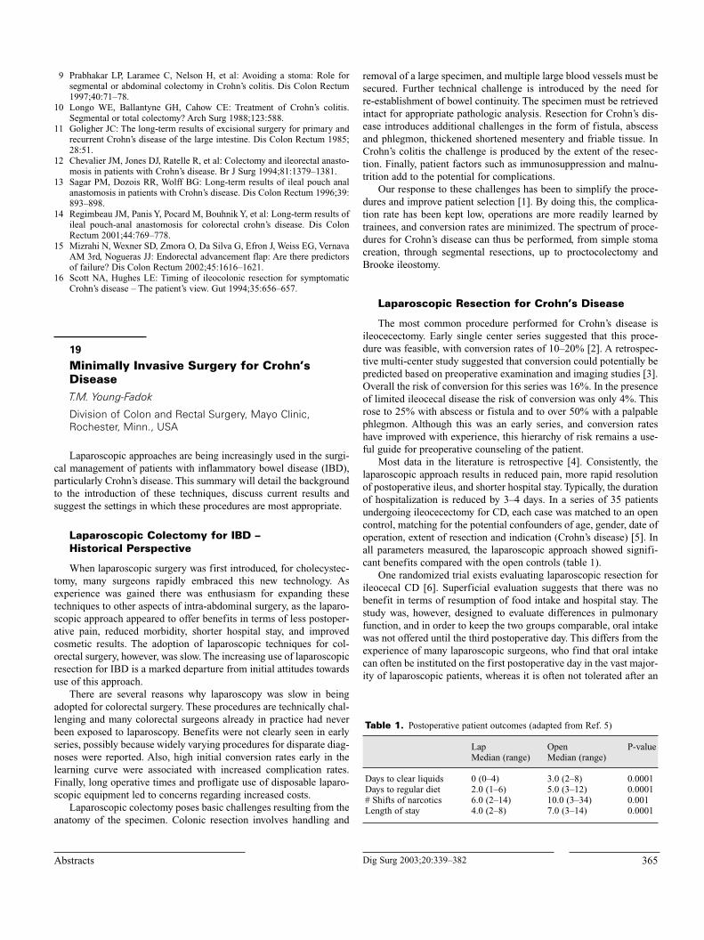
365Dig Surg 2003;20:339–382Abstracts
9 Prabhakar LP, Laramee C, Nelson H, et al: Avoiding a stoma: Role for
segmental or abdominal colectomy in Crohn’s colitis. Dis Colon Rectum
1997;40:71–78.
10 Longo WE, Ballantyne GH, Cahow CE: Treatment of Crohn’s colitis.
Segmental or total colectomy? Arch Surg 1988;123:588.
11 Goligher JC: The long-term results of excisional surgery for primary and
recurrent Crohn’s disease of the large intestine. Dis Colon Rectum 1985;
28:51.
12 Chevalier JM, Jones DJ, Ratelle R, et al: Colectomy and ileorectal anasto-
mosis in patients with Crohn’s disease. Br J Surg 1994;81:1379–1381.
13 Sagar PM, Dozois RR, Wolff BG: Long-term results of ileal pouch anal
anastomosis in patients with Crohn’s disease. Dis Colon Rectum 1996;39:
893–898.
14 Regimbeau JM, Panis Y, Pocard M, Bouhnik Y, et al: Long-term results of
ileal pouch-anal anastomosis for colorectal crohn’s disease. Dis Colon
Rectum 2001;44:769–778.
15 Mizrahi N, Wexner SD, Zmora O, Da Silva G, Efron J, Weiss EG, Vernava
AM 3rd, Nogueras JJ: Endorectal advancement flap: Are there predictors
of failure? Dis Colon Rectum 2002;45:1616–1621.
16 Scott NA, Hughes LE: Timing of ileocolonic resection for symptomatic
Crohn’s disease – The patient’s view. Gut 1994;35:656–657.
19
Minimally Invasive Surgery for Crohn’sDiseaseT.M. Young-Fadok
Division of Colon and Rectal Surgery, Mayo Clinic,Rochester, Minn., USA
Laparoscopic approaches are being increasingly used in the surgi-
cal management of patients with inflammatory bowel disease (IBD),
particularly Crohn’s disease. This summary will detail the background
to the introduction of these techniques, discuss current results and
suggest the settings in which these procedures are most appropriate.
Laparoscopic Colectomy for IBD – Historical Perspective
When laparoscopic surgery was first introduced, for cholecystec-
tomy, many surgeons rapidly embraced this new technology. As
experience was gained there was enthusiasm for expanding these
techniques to other aspects of intra-abdominal surgery, as the laparo-
scopic approach appeared to offer benefits in terms of less postoper-
ative pain, reduced morbidity, shorter hospital stay, and improved
cosmetic results. The adoption of laparoscopic techniques for col-
orectal surgery, however, was slow. The increasing use of laparoscopic
resection for IBD is a marked departure from initial attitudes towards
use of this approach.
There are several reasons why laparoscopy was slow in being
adopted for colorectal surgery. These procedures are technically chal-
lenging and many colorectal surgeons already in practice had never
been exposed to laparoscopy. Benefits were not clearly seen in early
series, possibly because widely varying procedures for disparate diag-
noses were reported. Also, high initial conversion rates early in the
learning curve were associated with increased complication rates.
Finally, long operative times and profligate use of disposable laparo-
scopic equipment led to concerns regarding increased costs.
Laparoscopic colectomy poses basic challenges resulting from the
anatomy of the specimen. Colonic resection involves handling and
Table 1. Postoperative patient outcomes (adapted from Ref. 5)
Lap Open P-value
Median (range) Median (range)
Days to clear liquids 0 (0–4) 3.0 (2–8) 0.0001
Days to regular diet 2.0 (1–6) 5.0 (3–12) 0.0001
# Shifts of narcotics 6.0 (2–14) 10.0 (3–34) 0.001
Length of stay 4.0 (2–8) 7.0 (3–14) 0.0001
removal of a large specimen, and multiple large blood vessels must be
secured. Further technical challenge is introduced by the need for
re-establishment of bowel continuity. The specimen must be retrieved
intact for appropriate pathologic analysis. Resection for Crohn’s dis-
ease introduces additional challenges in the form of fistula, abscess
and phlegmon, thickened shortened mesentery and friable tissue. In
Crohn’s colitis the challenge is produced by the extent of the resec-
tion. Finally, patient factors such as immunosuppression and malnu-
trition add to the potential for complications.
Our response to these challenges has been to simplify the proce-
dures and improve patient selection [1]. By doing this, the complica-
tion rate has been kept low, operations are more readily learned by
trainees, and conversion rates are minimized. The spectrum of proce-
dures for Crohn’s disease can thus be performed, from simple stoma
creation, through segmental resections, up to proctocolectomy and
Brooke ileostomy.
Laparoscopic Resection for Crohn’s Disease
The most common procedure performed for Crohn’s disease is
ileocecectomy. Early single center series suggested that this proce-
dure was feasible, with conversion rates of 10–20% [2]. A retrospec-
tive multi-center study suggested that conversion could potentially be
predicted based on preoperative examination and imaging studies [3].
Overall the risk of conversion for this series was 16%. In the presence
of limited ileocecal disease the risk of conversion was only 4%. This
rose to 25% with abscess or fistula and to over 50% with a palpable
phlegmon. Although this was an early series, and conversion rates
have improved with experience, this hierarchy of risk remains a use-
ful guide for preoperative counseling of the patient.
Most data in the literature is retrospective [4]. Consistently, the
laparoscopic approach results in reduced pain, more rapid resolution
of postoperative ileus, and shorter hospital stay. Typically, the duration
of hospitalization is reduced by 3–4 days. In a series of 35 patients
undergoing ileocecectomy for CD, each case was matched to an open
control, matching for the potential confounders of age, gender, date of
operation, extent of resection and indication (Crohn’s disease) [5]. In
all parameters measured, the laparoscopic approach showed signifi-
cant benefits compared with the open controls (table 1).
One randomized trial exists evaluating laparoscopic resection for
ileocecal CD [6]. Superficial evaluation suggests that there was no
benefit in terms of resumption of food intake and hospital stay. The
study was, however, designed to evaluate differences in pulmonary
function, and in order to keep the two groups comparable, oral intake
was not offered until the third postoperative day. This differs from the
experience of many laparoscopic surgeons, who find that oral intake
can often be instituted on the first postoperative day in the vast major-
ity of laparoscopic patients, whereas it is often not tolerated after an
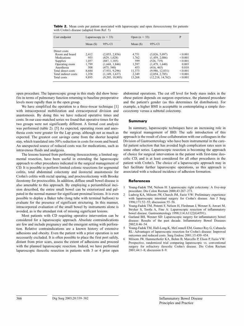
366 Dig Surg 2003;20:339–382 Inflammatory Bowel Disease
Principles and Practice
open procedure. The laparoscopic group in this study did show bene-
fits in terms of pulmonary function returning to baseline preoperative
levels more rapidly than in the open group.
We have simplified the operation to a three-trocar technique [1]
with intracorporeal mobilization and extracorporeal division and
anastomosis. By doing this we have reduced operative times and
costs. In our case-matched series we found that operative times for the
two groups were not significantly different. A formal cost analysis
was performed (table 2). [5] As expected, operating room and anes-
thesia costs were greater for the Lap group, although not as much as
expected. The greatest cost savings came from the shorter hospital
stay, which translated into 50% reduction in costs for room and board.
An unexpected source of reduced costs was for medications, such as
intravenous fluids and analgesia.
The lessons learned from performing ileocecectomy, a limited seg-
mental resection, have been useful in extending the laparoscopic
approach to other procedures indicated in the surgical management of
CD. It is possible to perform limited colonic resections for segmental
colitis, total abdominal colectomy and ileorectal anastomosis for
Crohn’s colitis with rectal sparing, and proctocolectomy with Brooke
ileostomy for proctocolitis. In addition, diffuse small bowel disease is
also amenable to this approach. By employing a periumbilical inci-
sion described, the entire small bowel can be exteriorized and pal-
pated in the normal manner for significant proximal lesions. It is even
possible to deploy a Baker tube (long tube with terminal balloon) to
evaluate for the presence of significant stricturing. In this manner,
intracorporeal evaluation of the small bowel by instruments alone is
avoided, as is the attendant risk of missing significant lesions.
Most patients with CD requiring operative intervention can be
considered for a laparoscopic approach. Absolute contraindications
are few and include pregnancy and the emergent setting with perfora-
tion. Relative contraindications are a known history of extensive
adhesions and obesity. Even the patient with a prior operation is not
necessarily excluded. It is often possible to place the first port safely,
distant from prior scars, assess the extent of adhesions and proceed
with the planned laparoscopic resection. Indeed, we have performed
laparoscopic ileocolic resection in patients with 3 or 4 prior open
abdominal operations. The cut off level for body mass index in the
obese patient depends on surgeon experience, the planned procedure
and the patient’s gender (as this determines fat distribution). For
example, a higher BMI is acceptable in contemplating a simple ileo-
cecectomy versus a subtotal colectomy.
Summary
In summary, laparoscopic techniques have an increasing role in
the surgical management of IBD. The safe introduction of this
approach is the result of close collaboration with our colleagues in the
Division of Gastroenterology who have been instrumental in the care-
ful patient selection that has avoided high complication rates seen in
some other series. Laparoscopic resection is becoming the approach
of choice for surgical intervention in the patient with first-time ileo-
colic CD, and is at least considered for all other procedures in the
patient with Crohn’s. The choice of a laparoscopic approach may in
fact facilitate further laparoscopic operations, as this approach is
associated with a reduced incidence of adhesion formation.
References
1 Young-Fadok TM, Nelson H: Laparoscopic right colectomy: A five-step
procedure. Dis Colon Rectum 2000;43:267–273.
2 Ludwig KA, Milsom JW, Church JM, Fazio VW: Preliminary experience
with laparoscopic intestinal surgery for Crohn’s disease. Am J Surg
1996;171:52–55; discussion 55–56.
3 Young-Fadok TM, Potenti F, Nelson H, Fleshman J, Wexner S, Anvari M,
Stryker S, Tootla A, Fine A: Laparoscopic resection of inflammatory
bowel disease. Gastroenterology 1998;114:A1122(G4591).
4 Gurland BH, Wexner SD: Laparoscopic surgery for inflammatory bowel
disease: Results of the past decade. Inflammatory Bowel Diseases
2002;8:46–54.
5 Young-Fadok TM, Hall-Long K, McConnell EM, Gomez Rey G, Cabanela
RL: Advantages of laparoscopic resection for Crohn’s disease: Improved
outcomes and reduced costs. Surg Endosc 2001;15:450–454.
6 Milsom JW, Hammerhofer KA, Bohm B, Marcello P, Elson P, Fazio VW:
Prospective, randomized trial comparing laparoscopic vs. conventional
surgery for refractory ileocolic Crohn’s disease. Dis Colon Rectum
2001;44:1–8; discussion 8–9.
Table 2. Mean costs per patient associated with laparoscopic and open ileocecectomy for patients
with Crohn’s disease (adapted from Ref. 5)
Cost endpoint Laparoscopy (n � 33) Open (n � 33) P
Mean ($) 95% CI Mean ($) 95% CI
Direct costs
Room and board 2,412 (2,055, 2,856) 4,751 (3,826, 5,097) �0.001
Medications 955 (829, 1,028) 1,762 (1,459, 2,086) �0.001
Supplies 1,057 (887, 1,185) 599 (526, 719) �0.001
Operating room 1,799 (1,668, 1,846) 1,597 (1,479, 1,640) 0.003
Anesthesia 508 (475, 540) 453 (416, 465) 0.010
Total direct costs 8,684 (7,931, 9,296) 11,373 (9,986, 12,031) �0.001
Total indirect costs 1,358 (1,149, 1,637) 2,349 (2,054, 2,785) �0.001
Total costs 9,895 (9,205, 10,905) 13,268 (12,218, 14,762) �0.001
367Dig Surg 2003;20:339–382Abstracts
Fistula and Emergencies
20
Fistulas in Crohn’s Disease – Conservativeversus Surgical Treatment Pro-SurgicalJ. Stern, G. Heuschen
St. Josephs Hospital, Dortmund, and Department ofSurgery, University of Heidelberg, Heidelberg, Germany
Basic treatment of Crohn’s disease (CD) is conservative. In con-
trast, for complications of this disease (for examples fistulas) surgical
treatment is recommended for most cases. Crohn’s disease is charac-
terized by a transmural inflammation, which may lead to formation of
fistulas. About one third of patients require a surgical procedure due
to a more or less serious fistula problem.
One can distinguish two main groups of patients with fistulating
Crohn’s disease:
– patients with abdominal fistulas and
– patients with perianal fistulas.
Both manifestations of fistulas may coexist in the same individ-
ual; however, the treatment strategy for these manifestations differs.
Abdominal Fistulas
Perforating Crohn’s disease in the abdomen may become sympto-
matic by abscess formation. The septic status of the patient may be
underestimated since the patients are often treated by steroids attenu-
ating the clinical symptoms. To prevent serious septicaemia, an
abscess should always be drained either by an interventional proce-
dure (ultrasound or CT-scan guided drainage) or open surgery. After
stabilization of the patient resection of the underlying intestinal dis-
ease should be performed.
Depending on the anatomical site of inflammation, fistulating
Crohn’s disease may lead to different secondary problems resulting in
different organs being at risk. The situation may then be more or less
tolerable for the patient, requiring more or less urgent surgery. A defin-
itive solution, however, can only be reached by a surgical approach.
Relative Indications for Surgery
Most often intestinal fistulas originate from a segment of
inflamed ileum, less frequently from the jejunum or colon. Typically,
the chronic inflammation leads to adhesions to adjacent intestinal
loops, for example the ascending or the sigmoid colon, with subse-
quent drainage of a developing fistula into these parts of the bowel.
Formation of a fistula will stabilize the patient’s status; therefore, def-
inite surgery may be postponed for some time.
If an affected segment of bowel adheres to the abdominal wall, an
enterocutaneous fistula may develop. This problem can be managed
by simply applying a stoma bag to the orifice of the fistula. Complex
fistulas with multiple cutaneous openings may cause considerable
difficulties for conservative management.
If an intestinal fistula drains into the genital system – usually the
fornix of the vagina – inflammatory secretions or intestinal contents
drain through the vagina. If the amount of drainage is limited, patients
will tolerate this situation for quite some time, often due to fear of
surgical intervention.
As a general rule, however, fistulating disease is always combined
with an inflammatory stricture of the bowel; therefore, it is unlikely
that the fistula will heal without surgical intervention [1].
Absolute Indications for Surgery
If fistulating Crohn’s disease affects the urinary tract (e.g.
enterovesical fistula), the kidneys are at high risk due to the possibil-
ity of an ascending infection. Conservative treatment using antibiotics
and anti-inflammatory drugs is not sufficient. These fistulas have to
be treated surgically.
A fistula arising from the duodenum or the proximal jejunum (a
rare situation because CD of the upper part of the gastrointestinal
tract is less common) may eventually drain into the transverse or even
sigmoid colon [2]. If the fistula is large enough, a substantial part of
the food will bypass digestion causing a more or less pronounced
short bowel syndrome, depending on fistula size.
Blind ending fistulas (e.g. retroperitoneal fistulas) may lead to
abscess formation or may affect adjacent organs. These fistulas
should also be treated surgically.
Perianal Fistulas
Perianal fistulas are regarded as a typical symptom of Crohn’s dis-
ease. About 20% of patients with Crohn’s disease present with this
problem. Perianal fistulas always affect anal continence in various
degrees; surgical treatment can even aggravate this situation.
Usually, perianal fistulas result from drainage of abscesses of the
proctodeal glands. In Crohn’s disease, however, perianal fistulas
might also originate from an inflamed rectum. Different types of peri-
anal fistulas may be found, with a spectrum from simple fistulas to
horseshoe or complex branching fistulas. If surgical treatment is
intended, intestinal inflammation should be controlled either by med-
ical or surgical treatment [3].
The surgical tactic depends on the status of the rectum (rectal
inflammation with or without stenosis) and on the extent of the fistu-
las [4]. A simple fistula may be adequately treated by an open-lay
technique or flap procedure. In case of a more complex situation, a
sleeve procedure may be successful in some patients [5, 6]. If exten-
sive perianal disease or recurrent fistulas are present, stool deviation
will be mandatory [7].
Surgical treatment of complex fistulas will often put the anal
sphincters at significant risk. Therefore, only palliative surgical treat-
ment with seton drainage is recommended in some instances [8]. In
most complex cases, only proctectomy with creation of a terminal
enterostomy will solve the perianal problem [9].
Conclusions
Fistulating Crohn’s disease will always cause discomfort and will
negatively impact social and sexual behaviour. Therefore, treatment of
this condition is always required. Medical treatment will only amelio-
rate the problems for a limited period of time [10]. If the medication is
stopped, the fistulas will recur almost inevitably [11]. Chronic inflam-
mation augments the collagenous fibers in the diseased area; the result-
ing stricture of the bowel will prevent any definite healing of the
fistulas by medical treatment alone [1]. The fibrosis per se can never be
368 Dig Surg 2003;20:339–382 Inflammatory Bowel Disease
Principles and Practice
reversed by any medical treatment. Therefore, surgery is the treatment
of choice in fistulating Crohn’s disease [12] and there are good argu-
ments for this approach. Surgery in CD is safe even if performed for a
second or third time [13, 14]. Surgery will provide the patients with
long periods of a good clinical status. It is important to perform only
limited resections of the inflamed bowel to prevent a short bowel syn-
drome. In case of complex perianal fistulas, only a palliative surgical
approach like seton drainage or stool deviation may be adequate [15].
In contrast, medical treatment of fistulating Crohn’s disease can
only provide the patient with temporary symptomatic relieve and
should be reserved for special circumstances. Examples are patients
at high risk for a short bowel syndrome or patients with complex fis-
tulas who already underwent multiple surgical interventions making
any further surgical procedure nearly impossible.
References
1 Kumar D, Alexander-Williams J: Crohn’s Disease and Ulcerative Colitis.
Surgical Management. London-Heidelberg-New York, Springer, 1993.
2 Schadde E, Schmidbauer S, Heinzlmann M, Hundegger K, Heldwein W,
Hallfeldt K: Duodenosigmoidal fistula in a Patient with Crohn’s disease.
Zentralbl Chir 2001;126:818–821.
3 Shinozaki M, Koganei K, Fukushima T: Simultaneous anus and bowel
operation is preferable for anal fistula in Crohn’s disease. J Gastroenterol
2002;37:611–616.
4 Winkler R: Chirurgische Aufgaben bei anorektalen Komplikationen des
Morbus Crohn. Zentralbl Chir 1998;123:362–367.
5 Hyman N: Endoanal advancement flap repair for complex anorectal
fistulas. Am J Surg 1999;178:337–340.
6 Marchesa P, Hull TL, Fazio VW: Advancement sleeve flaps for treatment
of severe perianal Crohn’s disease. Br J Surg 1998;85:1695–1698.
7 Yamamoto R, Allan RN, Keighley MR: Effect of fecal diversion alone on
perianal Crohn’s disease. World J Surg 2000;24:1258–1262.
8 Takesue Y, Ohge H, Yokoyama T, Murakami Y, Imamura Y, Sueda T: Long-
term results of seton drainage on complex anal fistulae in patients with
Crohn’s disease. J Gastroenterol 2002;37:912–915.
9 Keighley MRB, Williams NS: Surgery of the Anus Rectum and Colon,
ed 2. London-Edinburgh-New York, Saunders 1997.
10 Bemelman WA, Ivenski M, von Hogezand RA, Hermans J, Veenendaal RA,
Griffioen G: How effective is extensive nonsurgical treatment of patients
with clinically active Crohn’s disease of the terminal ileum in preventing
surgery? Dig Surg 2001;18:56–60.
11 Poritz LS, Rowe WA, Koltun WA: Remicade does not abolish the need for
surgery in fistulizing Crohn’s disease. Dis Col Rect 2002;6:771–775.
12 Lichtenstein GR: Treatment of fistulizing Crohn’s disease. Gastroenterol
2000;119:1132–1147.
13 Heimann TM, Greenstein AJ, Lewis B, Kaufman D, Heimann DM,
Aufses AH Jr: Comparison of primary and reoperative surgery in patients
with Crohn’s disease. Ann Surg 1998;227:492–495.
14 Yamamoto T, Allan RN, Keighley MRB: Long-term outcome of surgical
management for diffuse jejunoileal Crohn’s disease. Surgery 2001;129:
96–102.
15 Kosinski L, Welton ML: Surgical options in the management of perianal
Crohn’s disease. Seminars in Gastroint Dis 1998;9:15–20.
21
Fistula in Crohn’s Disease – Conservativeversus Surgical Treatment Pro-ConservativeM. Reinshagen
Department of Medicine I, University of Ulm, Ulm, Germany
Introduction: The cumulative incidence of fistulising Crohn’s
disease in a recent population-based study was 33% after 10 years and
50% after 20 years. The cumulative incidence for the subgroup of
patients with perianal fistula was 26% after 20 years [1]. Another
recent study reported the clinical course of fistulating Crohn’s disease
in 87 patients over 8 years [2]. 85% of patients had ileo-colonic or
colonic disease with only 14% of patients with only ileal disease. The
presence of rectal disease did not determine the complexity of peri-
anal fistulas. Of the 169 fistula in these 87 patients 110 (65%) were
perianal, 27 (16%) recto-vaginal, 20 (12%) were entero-enteric and 6
(3.5%) from other sites. In total 135/169 fistula were classified as
complex. A single fistula was present in one-third and multiple fistula
in two-thirds of these patients [2].
In respect to the final outcome after 8 years 68% showed healing
of all fistulas, 18% showed partial healing and 14% of patients did not
respond at all to therapy. In complex perianal fistula patients
responded to sole conservative treatment (antibiotics, azathioprine) in
13%. Combination of conservative treatment with simple surgery
(seton drainage, fistulotomy) was responsible for eventual healing in
47%. Complex surgery (proctectomy and stoma) was necessary in
38% of patients [2].
Conservative Therapy: Since 1975 metronidazole is used
to treat perianal fistulas in Crohn’s disease. 20 mg/kg/die metronida-
zole leads to decreased secretion and fistula closure in one-third of
patients after 6–8 weeks of therapy [3–5]. Because of adverse effects
like distal neuropathy and metallic taste ciprofloxacin(1,000–1,500 mg/die) is now widely used although officially it is doc-
umented only in small studies [6]. After discontinuation of antibiotic
therapy fistulas frequently recur. In daily practice antibiotic therapy is
combined with azathioprine (2.5 mg/kg) or 6-mercaptopurine(1–1.5 mg/kg) [7]. Fistula closure was reported in the available stud-
ies in up to 50% (see meta-analysis [8]). The combination of antibi-
otics/azathioprine with non-cutting setons controls or heals perianal
fistulas in more than 70% of patients [7]. Treatment with cyclosporine(4 mg/kg i.v.) [9] or tacrolimus (0.15–0.3 mg/kg) [10] leads to rapid
clinical improvement in 70–80% of patients. Since no longterm stud-
ies of oral cyclosporine or tacrolimus in fistulating Crohn’s disease
are available both therapies are used in eligible patients as a ‘bridge’
to maintenance therapy with azathioprine.
Infliximab (5 mg/kg i.v.) can be used in selected patients who are
resistant to at least 6 month of therapy with azathioprine/
6-mercaptopurine. In a large placebo-controlled study (94 patients)
reduction in the number of draining fistulas by 50% could be reached
in 62% of patients compared to 26% in the placebo group [11]. But
the median duration of response was 3 months. Whether continued
infliximab infusions are effective in maintaining fistula closure has
not been yet determined in a controlled study. Furthermore safety of
longterm therapy with infliximab is unclear since infections and sep-
sis [12], reactivation of tuberculosis [13] induction of autoimmunity
[14, 15] and malignancy [16, 17] occur. Until these safety issues are
not resolved long-term therapy with infliximab should only be
369Dig Surg 2003;20:339–382Abstracts
administered in carefully selected patients. Recent small studies
using MRI [18, 19] or endosonography [20] have shown that in
50–70% of patients despite external healing active fistulous tracts
remain after treatment with infliximab.
High entero-cutaneous and entero-vesico fistula should be treated
surgically after conservative control of the activity of the underlying
Crohn’s disease [7]. Similarily ano-vaginal fistula do not respond suf-
ficiently to conservative therapy and should be treated by an endorec-
tal advancement flap operation after sucessful treatment of local
inflammation [21]. In conclusion the majority of patients with fis-
tulising Crohn’s disease can be controlled or healed with conservative
therapy in combination with simple surgery. But eventually 20–30%
of patients will despite conservative treatment need major surgery
like proctectomy and stoma.
References
1 Schwartz DA, Loftus EVJ, Tremaine WJ, et al: The natural history of
fistulizing Crohn’s disease in Olmsted County, Minnesota. Gastroenterol
2002; 122(4):875–880.
2 Bell SJ, Williams AB, Wiesel P, Wilkinson K, Cohen RCG, Kamm MA:
The clinical course of fistulating Crohn’s disease. Aliment Pharmacol Ther
2003;17:1145–1151.
3 Jakobovits J, Schuster MM: Metronidazole therapy for Crohn’s disease
and associated fistulae. Am J Gastroenterol 1984;79(7):533–540.
4 Brandt LJ, Bernstein LH, Boley SJ, Frank MS: Metronidazole therapy for
perineal Crohn’s disease: A follow-up study. Gastro 1982;83(2):383–387.
5 Bernstein LH, Frank MS, Brandt LJ, Boley SJ: Healing of perineal
Crohn’s disease with metronidazole. Gastro 1980;79(2):357–365.
6 Turunen U, Farkkila M, Seppälä K: Long-term treatment of perianal or
fistulous Crohn’s disease with ciprofloxacin. Scand J Gastroenterol 1989;
24(Suppl):144–145.
7 Buhr HJ, Kroesen AJ, Stange EF: Chirurgie – Fisteln. Z Gastroenterol
2003;41(1):43–49.
8 Pearson DC, May GR, Fick GH, Sutherland LR: Azathioprine and
6-mercaptopurine in Crohn’s disease. A meta-analysis. Ann Intern Med
1995;123(2):132–142.
9 Present DH, Lichtiger S: Efficacy of cyclosporine in treatment of fistula
of Crohn’s disease. Dig Dis Sci 1994;39(2):374–380.
10 Lowry PW, Weaver AL, Tremaine WJ, Sandborn WJ: Combination therapy
with oral tacrolimus (FK506) and azathioprine or 6-mercaptopurine for
treatment-refractory Crohn’s disease perianal fistulae. Inflamm Bowel Dis
1999;5(4):239–245.
11 Present DH, Rutgeerts P, Targan S, et al: Infliximab for the treatment of
fistulas in patients with Crohn’s disease. N Engl J Med 1999;340(18):
1398–1405.
12 Ritz MA, Jost R: Severe pneumococcal pneumonia following treatment
with infliximab for Crohn’s disease. Inflamm Bowel Dis 2001;7(4):327.
13 Keane J, Gershon S, Wise RP, et al: Tuberculosis associated with
infliximab, a tumor necrosis factor alpha-neutralizing agent. N Engl J Med
2001;345(15):1098–1104.
14 Ali Y, Shah S: Infliximab-induced systemic lupus erythematosus. Ann
Intern Med 2002;137(7):625–626.
15 Kugathasan S, Levy MB, Saeian K, et al: Infliximab retreatment in adults
and children with Crohn’s disease: Risk factors for the development
of delayed severe systemic reaction. Am J Gastroenterol 2002;97(6):
1408–1414.
16 Cohen RB, Dittrich KA: Anti-TNF therapy and malignancy – A critical
review. Can J Gastroenterol 2001;15(6):376–384.
17 Brown SL, Greene, MH, Gershon SK, Edwards ET, Braun MM: Tumor
necrosis factor antagonist therapy and lymphoma development: twenty-six
cases reported to the Food and Drug Administration. Arthritis Rheum
2002;46(12):3151–3158.
18 Bell SJ, Halligan MS, Windsor ACJ, Williams AB, Wiesel P, Kamm MA:
Response of fistulating Crohn’s disease to infliximab therapy treatment
assesed by magnetic resonance imaging. Aliment Pharmacol Therap 2003;
17:387–393.
19 Van Assche G, Vanbeckevoort D, Bielen D, et al: Magnetic resonance
imaging of the effects of infliximab on perianal fistulizing Crohn’s
disease. Am J Gastroenterol 2003;98(2):332–339.
20 van Bodegraven AA, Sloots CEJ, Felt-Bersma RJF, Meuwissen SGM:
Endosonographic evidence of persistence of Crohn’s disease-associated
fistulas after infliximab treatment, irrespective of clinical response. Dis
Colon Rectum 2002;45(1):39–45.
21 Schwartz DA, Pemberton JA, Sandborn WJ: Diagnosis and treatment of
perianal fistulas in Crohn’s disease. Ann Intern Med 2001;135:906–918.
Pathophysiology, Diagnosis andConservative Treatment of Ulcerative Colitis
22
Pathophysiology of Ulcerative Colitis – Implications for TherapyH. Hodgson
Department of Medicine, Royal Free and University College, School of Medicine, London, UK
The symptom complex of active ulcerative colitis reflects a
combination of structural and functional abnormality. Some of these
are local and some systemic. In this presentation there will be con-
centration on those aspects of pathophysiology that constitute the
basis for therapy.
Colonic ChangesStructural abnormalities – oedema, ulceration, occasionally fibrosis
Functional abnormalities – disturbed motility, blood and protein
loss, electrolyte and water loss.
Systemic ChangesAnaemia, hypercoagulable state, propensity to infection, elec-
trolyte imbalance.
Colonic Changes
Mucosal inflammation, with oedema and ulceration, reflect the
end result of multiple pro-inflammatory processes – T-cell prolifera-
tion and cytotoxicity, B-cell proliferation and antibody production,
upregulation and over-expression of inflammatory mediators includ-
ing leukotrienes, platelet activating factor, histamine, kinins, cyto-
toxic and chemotactic cytokines and neuropeptides.
A significant element in an exacerbation of ulcerative colitis is the
recruitment of leukocytes from the systemic circulation, following the
upregulation of mucosal vascular adhesion molecules such as
MAdCAM-1, which attracts lymphocytes and leukocytes via integrin
binding.
The therapeutic strategies implied by this may be specific:
� Prevent up-regulation of adhesion molecules by inhibiting
specific molecular mechanisms.
� Prevent leukocyte-adhesion molecule interaction.
� Prevent generation/effect of pro-inflammatory agents either by
specific inhibitors/antagonists.
370 Dig Surg 2003;20:339–382 Inflammatory Bowel Disease
Principles and Practice
� On the other hand the mainstays of treatment for ulcerative colitis
are effective because they have multiple effects at different points
on a variety of pro-inflammatory pathways (corticosteroids,
5-aminosalicylates).
Motility disorders play an important and sometimes paradoxical
role. The symptom of diarrhoea and stool frequency in ulcerative
colitis reflects a combination of increased effluent volume (enhanced
secretion and impaired absorption, due to epithelial and luminal fac-
tors), hypermotility (often attributed to prostanoids), and the reflex
effects of rectal tenesmus. Pharmacological issues are important. In
severe acute colitis over-use of anti-motility agents may precipitate
megacolon. Some individuals are sensitive to amino-salicylates and
develop diarrhoea when they start them. In particular the ileal effects
of olsalazine in inducing secretion may be problematic. Additionally
in distal colitis the associated motility disorders may lead to proxi-
mal constipation, and under those circumstances laxatives may be
appropriate.
Strictures may occur as a consequence of previous acute episodes,
and an aetiology similar to that leading to post ischemic strictures
seems likely. Management importantly involves consideration of
possible malignancy.
Systemic Effects
Anaemia is generally normochromic or hypochromic, reflecting a
combination of acute bleeding and iron deficiency anaemia. The
back-wash ileitis associated with some severe colitis will not of itself
lead to B12 deficiency. Poor nutrition reflecting poor appetite may
however precipitate folate deficiency within fairly short periods of
illness.
Low protein levels – hypoalbuminaemia – reflect a combination
of both protein-losing enteropathy and decreased hepatic albumin
production as the liver turns to synthesis of acute phase reactants.
Albumin supplementation is rarely helpful and is contentious.
A hypercoagulable state may reflect increased platelet counts,
increased platelet activation and spontaneous aggregation, dehydra-
tion, increased production of fibrinogen and other positive acute
phase reactants and Factors V and VIII, decreased antithrombin III,
leading to deep venous thrombosis and pulmonary embolisation, and
thus prophylactic measures (compression stockings, low-molecular
weight or low-dose subcutaneous heparin) are required when patients
are hospitalised.
Hypokalaemia can develop rapidly particularly with the use of
systemic corticosteroids combined with potassium loss in the stool.
Stool losses range from 20–50 mmol K (and 10–170 mmol Na)
per day.
23
Colorectal Cancer Risk in Ulcerative Colitis – For How Long is SurveillanceJustified?R. Löfberg
IBD-unit at HMQ Sophia Hospital and Karolinska Institutet, Stockholm, Sweden
Rationale for Surveillance
There is a substantially increased risk for colorectal cancer (CRC)
in patients with longstanding, extensive/total ulcerative colitis (UC).
The absolute cumulative risk in population-based studies veers off
from the expected 8–10 years after the first attack of UC and after 25
years the risk for CRC is between 10–15% as assessed by life table
analysis [1, 2]. This is five to ten times the expected in the back-
ground population. A particularly increased risk for CRC has been
demonstrated for patients having concomitant primary sclerosing
cholangitis (PSC) [3], for those with a positive family history of CRC
[4] and for those with an early onset of UC (age � 15 yrs) [2]. Those
high-risk patients seem to run an absolute CRC-risk approaching 40%
after 25 years from onset. In comparison, patients having less exten-
sive UC (i.e. left-sided or distal UC/proctitis) at onset and who do not
experience a progression to more extensive disease over time, appear
to run no or only a marginally increased CRC-risk [1, 2]. The rationale
for endoscopic surveillance routines in patients with extensive/total
UC with more than eight years of disease duration seems to be
reasonably well grounded. In fact, the increased CRC-risk in pancol-
itis previously warranted surgeons to advocate prophylactic
proctocolectomy for all patients in this high-risk group.
Surveillance Strategy
To manage the increased CRC risk in UC colonoscopic surveil-
lance is now generally recommended in several countries. Patients
should undergo annual or biannual procedures with at least two biop-
sies sampled from every 10 cm of the colon, aiming at an early detec-
tion of mucosal dysplasia in order to select patients for prophylactic
colectomy [5]. Findings of definite dysplasia of low- or high-grade
(LGD/HGD) should be reviewed by two experienced pathologists for
consensus. Findings of significant dysplasia, i.e. multifocal LGD,
and/or HGD, and/or Dysplasia Associated Lesions or Masses
(DALMs), will imply that prophylactic colectomy need be performed
as the risk for invasive carcinoma is very high. Isolated finding of a
DALM, without adjacent or distant dysplasia in flat mucosa, may be
handled as an adenomatous polyp by snare polypectomy, but long-
term follow-up data using this approach is lacking. Patients display-
ing findings of isolated LGD, or of indefinite changes should have a
repeat colonoscopy within 6–12 months time. Using this approach, a
majority of patients with a clinically high CRC-risk (i.e. extensive
disease, PSC, CRC family history), but without findings of dysplasia,
will not have a colectomy recommended. Randomized, controlled
trials for evaluation of surveillance have not been performed.
Prospective series have demonstrated the feasibility in performing
colonoscopies and the capability to detect dysplasia, and also focused
on crucial issues in order to make surveillance worthwhile [6–9].
Prerequisites for successful surveillance comprise inclusion of all
known high-risk patients, high compliance, stringent protocol and
371Dig Surg 2003;20:339–382Abstracts
dedicated endoscopists and pathologists. The expected yield from
surveillance is rather low and is estimated to be around one signifi-
cant finding per year/100,000 inhabitants based on epidemiological
data from Northern Europe. Long-term results with respect to effi-
cacy have emerged from dedicated surveillance follow-up studies of
similar designs. A pooled analysis of four studies with a total of 612
patients with extensive UC of longstanding [6–9] it was found that
death from CRC was uncommon in the group of patients compliant
with surveillance, and that those patients developing dysplasia were
successfully selected for colectomy. Patients defaulting, or for some
other reason not adhering to the predetermined protocol, were more
likely to die from CRC. The few CRCs found within the frame of the
programs were detected at an early and thus curable stage. Besides
data from prospective surveillance programs, other data supporting
that surveillance is beneficial include case-control studies, analytical
approaches and population based studies.
For How Long is Surveillance Justified?
The ultimate goals of surveillance programs in UC should be to
decrease CRC morbidity and to eliminate CRC mortality. If you
accept the rationale for surveillance in the the high-risk group of UC-
patients, one inevitable question is how to optimise the scheduling of
surveillance procedures? Data from population based studies and now
also emerging data from long running surveillance programs clearly
indicate that the absolute risk of CRC and dysplasia increases over
time, and at more than 25 years of disease duration there is almost an
exponential rise. However, still only a minority of these patients will
ultimately develop CRC. Furthermore, the relative risk of CRC is
very high for patients with early UC onset (RR 100–400) and then
gradually decreases for patients with a later onset in life as the non-
colitis CRC incidence among the background population then
increases. Among those patients with pancolitis diagnosed between
the age of 30–70, a significantly increased relative risk of CRC
remains even after the age of 70 (RR 7–20). This finding indicates
that surveillance should be offered to high-risk patients as long as it
is biologically feasible, and there is no evidence-based support for a
cut-off level at the age of 70. However, for practical reasons, colono-
scopic procedures carried out beyond 80 years of age will probably
only be acceptable for a small number of UC-patients.
Flow-cytometry, for detection of DNA-aneuploidy and/or
increased proliferation rate by a higher S-phase fraction, is a robust
and objective method and the findings of aneuploidy appear to occur
before dysplasia [6]. It has been suggested that based on three con-
secutive dysplasia- and/or DNA-aneuploidy-free colonoscopies,
patients may be stratified to have a less intense surveillance schedule
with examinations in 5-year intervals without the risk of missing sig-
nificant dysplasia or cancer development [10]. However, this concept
has yet to be prospectively evaluated, and it does not necessarily make
sense in the 70� category of UC patients, since the risk of idiopathic
CRC then also increases substantially. In the older age group there
will instead be a mixed goal for surveillance in order to eliminate
mortality from both idiopathic and colitis associated CRC.
References
1 Gyde SN, Prior P, Allan RN, Stevens A, Jewell DP, Truelove SC, Löfberg R,
Broström O, Hellers G: Colorectal cancer in ulcerative colitis: A cohort
study of primary referrals from three centres. Gut 1988;29:206–217.
2 Ekbom A, Helmick C, Zack M, Adami HO: Ulcerative colitis and colorectal
cancer. A population-based study. N Engl J Med 1990;323:1228–1233.
3 Broomé U, Lindberg G, Löfberg R: Primary sclerosing cholangitis in
ulcerative colitis: A risk factor for the development of dysplasia and
DNA-aneuploidy? Gastroenterology 1992;102:1877–1880.
4 Askling J, Dickman PW, Karlén P, Broström O, Lapidus A, Löfberg R,
Ekbom A: Family history as a risk factor for cancer in inflammatory bowel
disease. Gastroenterology 2001;120:1356–1362.
5 Lennard-Jones J: Surveillance: A balanced view?; in Jewell DP, Warren BF,
Mortensen NJ (eds): Challenges in IBD. Oxford, Blackwell Science Ltd,
2001;223–236.
6 Löfberg R, Brostrom O, Karlen P, Tribukait B, Ost A: Colonoscopic
surveillance in long-standing ulcerative colitis – A 15-year follow-up
study. Gastroenterology 1990;99:1021–1031.
7 Lynch DAF, Lobo AJ, Sobala GM, Dixon MF, Axon ATR: Failure of
colonoscopic surveillance in ulcerative colitis. Gut 1993;34:1075–1080.
8 Connell WR, Lennard-Jones JE, Williams CD, Talbot IC, Price AB,
Wilkinson KH: Factors affecting the outcomes of endoscopic surveillance
for cancer in ulcerative colitis. Gastroenterology 1994;107:934–944.
9 Jonsson B, Ahsgren L, Andersson LO, Sterling R, Rutegard J: Colorectal
cancer survival in patients with ulcerative colitis. Br J Surg 1994;81:
689–691.
10 Karlen P: Ulcerative colitis and cancer with special reference to the
increased colorectal cancer risk. Dissertation, Karolinska Institutet,
Stockholm, 1998.
24
Standards and Long-Term Effects of Therapy in Ulcerative ColitisU.R. Fölsch, S. Schreiber
Department of General Internal Medicine I, Christian-Albrechts-University, Kiel, Germany
The etiology of ulcerative colitis is unclear. Disease pathophysiol-
ogy is characterized by chronic inflammation that differs little from
that described in Crohn’s disease. Mainstays of therapy are the use of
aminosalicylates for both remission induction and remission mainte-
nance. Salazosulfapyridine is composed from a 5-aminosalicylic acid
(5-ASA) residue and a sulfonamide group. While inactive in the
upper gastrointestinal tract, bacterial cleavage of the joining diazo-
bond in the terminal ileum releases the two components. This therapy
therefore delivers active drug from the intestinal lumen to the mucosa
with only a small fraction of the drug becoming systemically avail-
able. It was unclear for a long time whether 5-ASA or the sulfon-
amide are the therapeutically active parts of the molecule for this
‘topical therapy’. The work of Azad Khan and coworkers established
5-aminosalicylic acid as the active therapeutic moiety [1]. The avail-
able oral 5-ASA formulations appear to differ little in their clinical
efficacy. It takes typically between 3–6 weeks to induce remission in
mild-moderately active ulcerative colitis. Superiority to sulfasalazine
could not be demonstrated in a meta-analysis [2]. In addition to the
oral route, 5-ASA can be applied by rectal enema or suppository.
Typically, enemas deliver anti-inflammatory therapy up to 25–35 cm
from the anal verge. Glucocorticoids have their role in the rapid
induction of remission in more active cases, but provide no remission
maintenance. In severe ulcerative colitis requiring hospital admission,
systemic glucocorticoids should always be used. In severe cases with
parenteral nutrition high doses of glucocorticoids (i.e. 100 mg/d pred-
nisone equivalent) are administered intravenously [3]. Side-effects of
chronic glucocorticoid treatment can be dramatic even in young
patients. They range from cosmetic problems (moon face, acne) to
372 Dig Surg 2003;20:339–382 Inflammatory Bowel Disease
Principles and Practice
severe health problems (osteoporosis, psychiatric diseases). The ben-
efits of a therapy with glucocorticoids should therefore be carefully
balanced against the possible side-effects. The long-term use of
immunosuppressives is effective in chronic active disease for control
of inflammation, but is also effective to prolong remission.
Azathioprine and its main metabolite 6-mercaptopurine have been
used for decades in the therapy of inflammatory bowel disease.
Although most studies have addressed the use of these drugs in
Crohn’s disease, reasonable evidence has been shown that they are
equally effective in ulcerative colitis [4]. Both drugs are used to
reduce the necessity of long-term glucocorticoid treatment and for
remission maintenance in complicated or 5-ASA intolerant patients
[4]. Methotrexate is an alternative to azathioprine or 6-mercapto-
purine, respectively, in patients developing toxicity [5]. Methotrexate
is less well established through controlled clinical trials, but empiric
evidence and its utility in Crohn’s disease suggest that it would be
effective in ulcerative colitis to stabilize remission in chronically
active patients. However, methotrexate has several disadvantages in
comparison to azathioprine/6-mercaptopurine. These include its ter-
atogenicity and the occasional but severe liver toxicity. Cyclosporine
A is established as an intravenous therapy (4 mg/kg body weight as a
continuous infusion over 24 h) in cases of severe, refractory ulcerative
colitis [6]. While it can interrupt the inflammatory process, many
patients relapse within the next 180 days even if cyclosporine is con-
tinued per os. Tracrolimus, which followed cyclosporine as an
immunosuppressive in organ transplantation, has also been used in
ulcerative colitis [7]. While open label experiments suggest an effi-
cacy which is similar to cyclosporine, controlled trials have not
been conducted to establish its role in therapy. The use of probiotics
has been advocated in colonic inflammatory disease for a long
time. Only recently two controlled trials suggested that E.coli nissle is
as effective as 5-ASA for remission maintenance in ulcerative
colitis [8].
Conclusions: Therapy of ulcerative colitis is a long-term task
that has to balance efficacy against side-effects in a life-long disease.
Although effective therapies exist for both mild-moderately and mod-
erately-severely active disease, many patients still reach an end-stage
in their disease that is characterized by chronic activity and loss of
function of the large bowel.
References
1 Azad Khan AK, Piris J, Trulove SC: An experiment to determine the active
therapeutic moiety of sulphasalazine. Lancet 1977;29:892–895.
2 Sutherland L, Roth D, Beck P, May G, Makiyama K: Oral 5-aminosali-
cylic acid for inducing remission in ulcerative colitis. Cochrane Database
Syst Rev 2000;(2):CD000543.
3 Chun A, Chadi RM, Korelitz BI, Colonna T, Felder JB, Jackson MH,
Morgenstern EH, Rubin SD, Sacknoff AG, Gleim GM: Intravenous corti-
cotropin vs. hydrocortisone in the treatment of hospitalized patients with
Crohn’s disease: A randomized double-blind study and follow-up.
Inflamm Bowel Dis 1998;4:177–181.
4 Hawthorne AB, Logan RF, Hawkey CJ, Foster PN, Axon AT, Swarbrick
ET, Scott BB, Lennard-Jones JE: Randomised controlled trial of azathio-
prine withdrawal in ulcerative colitis. BMJ 1992;305:20–22.
5 Kozarek RA, Patterson DJ, Gelfand MD, Botoman VA, Ball TJ,
Wilske KR: Methotrexate induces clinical and histologic remission in
patients with refractory inflammatory bowel disease. Ann Intern Med
1989;110:353–356.
6 Lichtiger S, Present DH, Kornbloth A, Gelernt I, Bauer J, Galler G,
Michelassi F, Hanauer S: Cyclosporine in severe ulcerative colitis refrac-
tory to steroid therapy. N Engl J Med 1994;330:1841–1845.
7 Fellermann K, Ludwig D, Stahl M, David-Waledk T, Stange EF:
Steroid-unresponsive acute attacks of inflammatory bowel disease:
Immunomodulation by tacrolimus (FK506). Am J Gastroenterol
1998;93:1860–1866.
8 Rembacken BJ, Snelling AM, Hawkey PM, Chalmers DM, Axon AT:
Non-pathogenic Escherichia coli versus mesalazine for the treatment of
ulcerative colitis: A randomised trial. Lancet 1999;354:635–639.
25
Therapy-Refractory and Fulminant, Toxic ColitisM.H. Holtmann, P.R. Galle
Ist Department of Medicine, Johannes Gutenberg-University, Mainz, Germany
Etiology
Toxic megacolon is a rare, but potentially life threatening condi-
tion that is characterized by colonic dilatation in connection with
systemic toxicity [1]. Toxic megacolon is most often a complication
of severe flares of inflammatory bowel diseases, especially ulcerative
colitis, which makes up for about 75% [2] of all cases of toxic mega-
colon, but can also occur in infectious colitides of any type such as
C. difficile, Salmonella, Shigella, Campylobacter, amoebes or
cytomegalovirus (CMV) [3, 4]. Other etiologies of severe colitis that
can potentially lead to toxic megacolon include ischemic colitis,
volvulus, diverticulitis, and obstructive colon cancer.
Two decades ago approximately 1 to 5 percent of patients with
ulcerative colitis and Crohn’s disease developed toxic megacolon. Due
to improved management of severe colitis, these numbers have
decreased significantly. The association of toxic megacolon with CMV
has gained importance with the prevalence of HIV infection and AIDS.
CMV colitis leading to toxic megacolon is the most important cause of
emergency laparotomy in patients with AIDS. Interestingly, CMV
infection seems to be involved in development of toxic megacolon in
patients with IBD, too. In a survey of 46 resected colons in patients
with ulcerative colitis, 6 patients had CMV infection. 5 of these
6 patients had developed toxic megacolon, whereas only 2 out of the
40 patients without CMV infection developed this complication [4].
Pathogenesis
The association with systemic toxicity distinguishes toxic mega-
colon from colonic dilatation in Hirshsprung’s disease (congenital
megacolon), acquired megacolon secondary to chronic constipation
and intestinal pseudoobstruction. The pathogenesis of toxic mega-
colon is still unclear. Probably, there are various different contributing
pathomechanisms [1]. External factors that favour development of
toxic megacolon include hypokalemia, antimotility agents such as
opiates, anticholinergics and antidepressants. Barium enema and
colonoscopy physically distend the colon and may thus impair the
blood supply to the colon wall. When toxic megacolon arises in
patients with ulcerative colitis, in which inflammation is normally
restricted to the mucosa, the inflammation extends to the smooth
muscle layer leading to paralysis and consecutive dilatation.
Proinflammatory factors and bacterial products activate nitric oxide
synthase resulting in excessive nitric oxide, an inhibitor of smooth
373Dig Surg 2003;20:339–382Abstracts
muscle tone, which has been identified as a key mediator for colonic
dilatation [5]. Generation of nitric oxide occurs in macrophages and
smooth muscle cells and appears to be specific to dilatation of the
colonic wall in toxic megacolon. In the TNBS model of colitis in rats
the activity of inducible NO synthase could be decreased by treatment
with antibiotics, corticosteroids and NO synthase inhibitors leading to
an improvement of smooth muscle contractility, intracolonic pressure
and colon dilatation [6]. Of note, in one patient with toxic megacolon
due to ulcerative colitis, rectal application of a nitric oxide synthase
inhibitor by enema led to clinical improvement [7].
Pathology
Key macroscopic features of toxic megacolon are dilatation of the
colon with thinning of the bowel wall and deep ulcers. Histologically
all layers of the bowel wall show degeneration, necrosis and replace-
ment by granulation tissue with infiltrations of histiocytes, neu-
trophils, lymphocytes and plasma cells. Often muscle fibers are
shortened with eosinophilic cytoplasm. Interestingly, the colonic sub-
mucosal and myenteric plexi are usually intact suggesting that toxic
megacolon is not primarily neuropathic [8].
Clinical Manifestations
The likelihood for patients with IBD to develop toxic megacolon
is highest at an early stage of their disease. Up to 30 percent of
patients develop toxic dilatation within three months of diagnosis and
about 60 percent within the first three years. There are no differences
between the sexes and no preferred age.
Almost all patients report an episode of severe bloody diarrhea,
which might have improved with the onset of toxic megacolon, asso-
ciated with abdominal pain, peritonitis, distension and signs of gen-
eralized sepsis such as fever, tachycardia, hypotension and altered
sensorium. These signs of systemic toxicity accompanied by the find-
ing of an enlarged dilated colon are the basis for the diagnosis of toxic
megacolon. One should be alerted by IBD, especially by severe flares,
in the patient’s history. Other details such as recent travel, antibiotic
or chemotherapeutic treatment and HIV/AIDS status might point to
other causes of severe colitis leading to toxic megacolon. Formal cri-
teria for the diagnosis of toxic megacolon include [2]:
– radiographic evidence of colonic distension
– plus at least three of the following: fever �38�C, heart rate
�120 beats/min, leukocytosis �10,500/mm3, anemia
– plus at least one of the following: dehydration, altered sensorium,
electrolyte disturbances, hypotension.
Plain radiographies of the abdomen are sufficient for revealing
colonic distension. Typical features are:
– dilatation of the transverse or right colon of �6 cm, sometimes up
to 15 cm on supine films, the left colon is less frequently dilated
depending on the patient’s position
– severe disturbance of the normal haustral patterns of the colon
– multiple air-fluid levels
– deep mucosal ulcerations revealed by pseudopolypoid projections
into the colonic lumen.
Computed tomography provides no advantage to reveal alterations
of the colon, but might be useful to detect other underlying intraab-
dominal processes. Laboratory findings are nonspecific and include
anemia, leucocytosis, increased erythrocyte sedimentation rate and
serum C-reactive protein, electrolyte disturbances, metabolic alkalo-
sis and hypoalbuminemia.
It is generally felt that full colonoscopy should be avoided when
toxic megacolon is suspected because of the risk of perforation.
Partial colonoscopy may be necessary to gain specimen for microbi-
ological and virological testing, but should be performed cautiously
with minimal air insufflation.
Treatment
Management of toxic megacolon should occur at the intensive care
unit with frequent monitoring of the physical state, standard laboratory
parameters and plain abdominal films [1]. Patients are placed at com-
plete bowel rest and a nasogastric tube is inserted to decompress the
gastrointestinal tract. Enteral feeding is started as soon as possible to
support mucosal healing. Total parenteral nutrition (TPN) has shown
no benefit to avoid surgery or shorten the hospital stay [9]. Narcotics,
antimotility and anticholinergic drugs should be discontinued.
Standard medical treatment should include sufficient fluid and elec-
trolyte repletion, gastric stress ulcer prophylaxis with H2-blockers,
thrombosis prophylaxis by pneumatic compression stockings and
broad spectrum antibiotics such as metronidazole in combination with
ampicillin and gentamicin or a third-generation cephalosporin to meet
peritonitis and septis as frequent complications of toxic megacolon.
In addition to these general means specific treatment for the
underlying cause of toxic megacolon should be attempted. In case
of IBD intravenous corticosteroids such as hydrocortisone or pre-
dnisolone/methylprednisolone should be given provided that a con-
comitant infection has been ruled out as contributing etiology of toxic
megacolon. While the use of corticosteroids in toxic megacolon
is evidence based [8], little experience exists with other medical
regimens such as Cyclosporine [10], Tacrolimus [11] and
Cyclophosphamide [12] in severe ulcerative colitis or Azathioprine
and anti-TNF-treatment in Crohn’s disease [13]. Sulfasalazine and
5-ASA compounds should be discontinued.
Studies have shown a beneficial effect of physical maneuvers such
as intermitting rolling from the supine to the prone position or adopt-
ing the knee-elbow position [14]. These maneuvers are thought to
redistribute the intracolonic gas so that it can more easily be expelled
and thus decompress the colon.
50% of patients with toxic megacolon improve upon medical
treatment. The absolute indication for surgery is given in case of free
perforation, massive hemorrhage, increasing transfusion requirement,
worsening signs of toxicity and increasing colonic dilatation. While
some surgeons consider medical treatment as failed when colonic
distension still persists after 48 to 72 hours, others wait for up to
seven days if the patients appears to be clinically improving
otherwise [15].
Outcome
Improved medical treatment of IBD has led to a significant
decrease in the incidence of toxic megacolon. While toxic megacolon
was associated with a mortality rate of 27% in patients treated med-
ically and 19% in patients who underwent surgery 30 years ago, the
mortality rates dropped substantially to 0 to 2% in patients with IBD
[16, 17]. This improvement is due to earlier recognition of toxic
megacolon, better intensive medical treatment, earlier surgery and
improved postoperative care. The reported rates of patients, who were

374 Dig Surg 2003;20:339–382 Inflammatory Bowel Disease
Principles and Practice
treated medically initially and required colectomy later, vary between
25 and 50% [15, 18]. Overall, toxic megacolon has become a rare, but
still challenging complication in IBD, especially UC.
References
1 Sheth SG, LaMont JT: Toxic megacolon. Lancet 1998;351:509.
2 Jalan KN, et al: An experience with ulcerative colitis: Toxic megacolon in
55 cases. Gastroenterology 1969;57:68.
3 Rubin MS, Bodenstein LE and Kent KC: Severe Clostridium difficilecolitis. Dis Colon Rectum 1995;38:350.
4 Cooper HS, et al: Cytomegalovirus inclusion in patients with ulcerative
colitis and toxic dilatation requiring colonic resection. Gastroenterology
1977;72:755.
5 Mourelle M, et al: Induction of nitric oxide synthase in colonic smooth mus-
cle form patients with toxic megacolon. Gastroenterology 1995;109:1497.
6 Mourelle M, et al: Toxic dilatation of colon in a rat model of colitis is
linked to an inducible form of nitric oxide synthase. Am J Physiol 1996;
33:G425.
7 Schworer H, et al: Successful treatment of megacolon associated with colitis
with a nitric oxide synthase inhibitor. Am J Gastroenterol 2001;96: 2273.
8 Norland CC, Kirsner JB: Toxic dilatatio of the colon (toxic megacolon):
Etiology, treatment and prognosis in 42 patients. Medicine 1969;48:229.
9 Dickinson RJ, et al: Controlled trial of intravenous hyperalimentation and
total bowel rest as an adjunct to the routine therapy of acute colitis.
Gastroenterology 1980;79:1199.
10 D’Haens G, et al: Intravenous cyclosporine versus intravenous corticos-
teroids as single therapy for severe attacks of ulcerative colitis. Gastro-
enterology 2001;120:1323–1329.
11 Fellermann K, et al: Steroid-unresponsive acute attacks of inflammatory
bowel disease: Immunomodulation by tacrolimus (FK506). Am J Gastro-
enterol 1998;93:1860–1866.
12 Stallmach A, et al: Safety and efficacy of intravenous pulse cyclophos-
phamide in acute steroid refractory inflammatory bowel disease. Gut
2003;52:377–382.
13 van Dullemen HM, et al: Treatment of Crohn’s disease with anti-tumor
necrosis factor chimeric monoclonal antibody (cA2). Gastroenterology
1995;109(1):129–135.
14 Panos MZ, Wood MJ, Asquith P: Toxic, megacolon: The knee-elbow posi-
tion relieves bowel distension. Gut 1993;34:1726.
15 Caprilli R, et al: Early recognition of toxic megacolon. J Clin Gastro-
enterol 1987;9:136.
16 Strauss RJ, et al: The surgical management of toxic dilatation of the colon:
A report of 28 cases and a review of the literature. Ann Surg 1976;184:
682.
17 Danovitch SH: Fulminant colitis and toxic megacolon. Gastroenterol Clin
North Am 1989;18:73.
18 Fazio VW: Toxic megacolon in ulcerative colitis and Crohn’s disease. Clin
Gastroenterol 1980;9:389.
26
Dysplasia in Ulcerative Colitis – BiologicalRelevance and Clinical ConsequenceF. Autschbach
Institute of Pathology, Heidelberg University, Heidelberg, Germany
Abstract
Ulcerative colitis is a disease which is associated with an
increased risk of colorectal carcinoma. The occurrence of intraepithe-
lial neoplasia/dysplasia is regarded as a prominent indicator of an
increased cancer risk in these patients. This article summarizes some
aspects concerning the relevance of dysplasia in ulcerative colitis.
Introduction
Colorectal carcinoma (CRC) is an accepted complication of ulcer-
ative colitis (UC). The relative risk of CRC in UC is at least 2–3 times
higher than in the general population [1] and is dependent on the
duration and extent of inflammatory disease (about 2% after 10 years,
8–18% after 20–30 years in patients with pancolitis according to a
recent meta-analysis) [2]. A further known risk factor for carcinomas
in UC patients is concomitant primary sclerosing cholangitis. The
occurrence of dysplasia is regarded as an important indicator of an
increased cancer risk in a given patient. Detection of such lesions is a
major goal of current cancer surveillance strategies in patients with
inflammatory bowel disease (IBD) [3].
Intraepithelial Neoplasia/Dysplasia in UC
Dysplasia is defined as an unequivocally neoplastic change which
is confined to the intestinal epithelium and thus represents an intraep-
ithelial neoplasia (IEN) [4]. It is characterized by mucosal alterations
which to some extent resemble epithelial changes seen in a sporadic
adenoma in a non-colitic patient. This includes architectural changes
(crowding of glands, crypt branching, villiform changes) as well as
abnormal cytological features such as stratification of nuclei, loss of
nuclear polarity, nuclear hyperchromasia and pleomorphism. The
degree of cytologic and architectural abnormalities determines the
classification of IEN as low-grade (LGIEN) or high grade (HGIEN)
[5]. The histologic distinction of LGIEN from reactive epithelial
changes is challenging and can be even impossible, especially in areas
with active inflammation. Evidence of basal-luminal maturation of
crypt epithelia can be used as one criterion against the existence of
true IEN. Standardized criteria include a category indefinite for
IEN/dysplasia to classify problematic cases [5]. IEN in UC often
appears in grossly unsuspicious flat mucosa, can be very circumscript
or multifocal and shows a random pattern of distribution.
Macroscopically detectable IENs can have a villous or plaque-like
configuration or appear as polypoid structures resembling adenomas.
Such lesions have been referred to as dysplasia-associated lesion or
mass (DALM) [6]. Although IEN can precede the development of
invasive carcinoma, it is often found concomitant to an already exist-
ing colitis-associated carcinoma, both adjacent to and distant from the
invasive lesion [5]. In a substantial number of such cases, the carci-
noma is associated with ‘only’ LGIEN. Thus, increased cancer risk
must be considered in cases with both HGIEN and LGIEN [7].
DALMs are associated with an especially high rate of invasive cancer
(�40%) [7]. The distinction between colitis-associated IENs and
sporadic adenomas in UC patients is problematic and a matter of
recent debate. Under certain circumstances, a diagnosis of sporadic
adenoma appears to be possible [8].
Clinical Implications of IEN in UC
Current recommendations suggest regular colonoscopic surveil-
lance with biopsy in UC patients with a disease duration of longer
than 8 years (extensive colitis) respectively 12–15 years (left-sided
colitis) [9, 10]. In case of a histopathological diagnosis of IEN/
dysplasia, colectomy should be considered, especially in patients
with HGIEN and/or DALMs, but also in patients with LGIEN. Since
375Dig Surg 2003;20:339–382Abstracts
observer variability in classifying IEN is high, the diagnosis should be
independently confirmed by a second pathologist with special exper-
tise in this field. A special situation appears for the management of an
adenoma- like polypoid dysplastic lesion. In such a case, polypectomy
may be an adequate treatment [11], but should be planned in close col-
laboration between endoscopist and pathologist.
References
1 Bernstein CN, Blanchard JF, Kliewer E, Wajda A: Cancer risk in patients
with inflammatory bowel disease. Cancer 2001;91:854–862.
2 Eaden JA, Abrams KR, Mayberry JF: The risk of colorectal cancer in
ulcerative colitis: A meta-analysis. Gut 2001;48:526–535.
3 Loftus EV: Does monitoring prevent cancer in inflammatory bowel
disease? J Clin Gastroenterol 2003;36(Suppl 1):S79–S83.
4 Schlemper RJ, Riddell RH, Kato Y, Borchard F, Cooper HS, et al: The
Vienna classification of gastrointestinal epithelial neoplasia. Gut
2000;47:251–255.
5 Riddell RH, Goldman H, Ransohoff DF, Appelman HD, Fenoglio CM,
et al: Dysplasia in inflammatory bowel disease. Standardized classifica-
tion with provisional clinical applications. Hum Pathol 1983;14:931–968.
6 Blackstone MO, Riddell RH, Rogers BH, Levin B: Dysplasia-associated
lesion or mass (DALM) detected by colonoscopy in long-standing ulcerative
colitis: An indication for colectomy. Gastroenterology 1981;80:366–374.
7 Bernstein CN, Shanahan F, Weinstein WM: Are we telling patients the
truth about surveillance colonoscopy in ulcerative colitis? Lancet 1994;
343:71–74.
8 Schneider A, Stolte M: Differential diagnosis of adenomas and dysplastic
lesions in patients with ulcerative colitis. Z Gastroenterol 1993;31:653–656.
9 Winawer SJ, Fletcher RH, Miller L, Godlee F, Stolar MH, et al: Colorectal
cancer screening: Clinical guidelines and rationale. Gastroenterology
1997;112:594–642.
10 Stange EF, Riemann J, Von Herbay A, Lochs H, Fleig WE, et al: Diagnostik
und Therapie der Colitis ulcerosa – Ergebnisse einer evidenzbasierten
Konsensuskonferenz der Deutschen Gesellschaft für Verdauungs- und
Stoffwechselkrankheiten. Z Gastroenterol 2001;30:19–61.
11 Engelsgjerd M, Farraye FA, Odze RD: Polypectomy may be adequate
treatment for adenoma-like dysplastic lesions in chronic ulcerative colitis.
Gastroenterology 1999;117:1288–1294.
Surgical Treatment of UlcerativeColitis
27
Surgical Treatment – Indications and Early Outcome Ulcerative ColitisV.W. Fazio
Department of Colorectal Surgery, Cleveland Clinic Foundation, Cleveland, Ohio, USA
Indications for surgery, these may be grouped as elective or urgent.
Elective indications include the following:
� Failed medical therapy
� Complications/adverse effects of medical therapy
� Colonic dysplasia
� Colorectal carcinoma or suspicion of cancer (e.g. stricture)
� Extra intestinal manifestations of ulcerative colitis
� Growth retardation
� Disability due to other complications of ulcerative colitis such as
anemia, recurrent bleeding and fecal incontinence
Urgent – Emergency Indications include the following:
� Toxic colitis
� Failing to respond to medical treatment
� Toxic megacolon
� Colon perforation
� Hemorrhage
Surgical Procedures in Ulcerative Colitis
A number of procedures have been developed for the surgical
treatment of ulcerative colitis. While several of these have become
obsolete (e.g. ileostomy alone) with emergence of new techniques, a
range of options still remain as part of the armamentarium of the
complete IBD surgeon. Surgical procedures have evolved with several
tenets in mind.
� Tailor – the operation to suit the specific patient. For example,
definitive surgery is rarely performed in the acute or emergency
setting e.g. subtotal colectomy is used for toxic megacolon.
� Where feasible, the definitive operation involves removal of all –
most colonic – rectal epithelium at risk for continued inflamma-
tion or potential for neoplasty transformation.
� Where patients are highly motivated to avoid permanent fecal
diversion, to perform ileostomy sparing operations e.g. procto-
colectomy and ileopouch anal anastomosis (RP). Thus the goal of
surgery is to provide cure of ulcerative colitis as well as optimize
function.
The tactics of surgery include:
� Making the operation as safe as possible i.e. optimizing the phys-
ical condition of the patient
� Preoperative stoma site marking
� Antibiotic and mechanical bowel preparation
Indications for specific procedures:
1. Subtotal colectomy and ileostomy – This is indicated in patients
with toxic colitis with and without megacolon, colonic perfora-
tion, the immunosuppressed or malnourished patient and in cases
where diagnosis maybe Crohn’s disease. Complications of this
surgery specific for the operation include rectal-sigmoid stump
blow out with intra or extra peritoneal sepsis; rectal hemorrhage
and problems related to mucous fistula i.e. perfuse drainage,
muco-cutaneous separation.
2. Ileorectal anastomosis – This is rarely used in current times due to
alternatives of restorative proctocolectomy (RP). Indications
include, metastatic colon cancer where rectum has quiescent
ulcerative colitis. Some women may choose this alternative due to
perceived-probable higher rates of fertility compared with RP,
provided rectal disease activity is low.
3. Total proctocolectomy and ileostomy – Formerly this was the gold
‘Gold Standard’ of surgery for UC due to its curative nature,
usually performed in one stage only, and good durability.
Complications related to those of the ileostomy where up to 10%
of patients may require further surgery for this. As well, the
unhealed perineal wound occurs at about a similar rate and sexual
dysfunction with impotence, retrograde ejaculation, are occasional

376 Dig Surg 2003;20:339–382 Inflammatory Bowel Disease
Principles and Practice
but uncommon occurrences. Dyspareunia similarly has a low inci-
dence. The operation is indicated in patients with poor anal sphinc-
ter function, or anticipated poor function. For example, patients
over 70–80 years of age; as a salvage procedure namely ileal pouch
excision with a failed RP. About 4–6% of patients on whom RP is
planned, end up with TPC and ileostomy due to reach problems, or
concerns about Crohn’s disease. Finally, patients sometimes
choose TPC and ileostomy by preference over RP because of their
issues with safety and quality of life with RP.
4. Restorative Proctocolectomy (RP) with ileal pouch anal anasto-
mosis – This is now the elective procedure of choice for patients
with UC. Contraindications include:
� Poor anal sphincter function
� Technical major problem with tension free anastomosis
� Previous small bowel injury (radiation) or extensive resection
� Lower third and possibly mid 1/3 rectal cancer
� Crohn’s disease
� Patient prefers TPC and end ileostomy
Contraindications to preserving anal transitional zone (usually
stapled IPAA) include synchronous colorectal cancer, extra intestinal
manifestations of UC, primary sclerosing cholangitis, and active anal
canal or low rectal activity. Contraindications are mostly relativerequiring extensive discussion and informed consent with the patient.
Most operations are done in two stages, with protective loop
ileostomy or end ileostomy at the time of RP and ileostomy closure
three months later.
Complications may occur early and late. The early complications
include wound infection, small bowel obstruction, anastomotic leak,
pelvic abscess, fistula from the anastomosis to perineum, vagina, pre-
sacral space, anastomotic stricture, bleeding from the pouch, and sex-
ual dysfunction. Pouch ischemia is rarely seen. Later complications
include, pouchitis, acute and chronic, small bowel obstruction, affer-
ent limb syndrome, incontinence, late anastomotic fistulas to the
vagina and perineum, and exit conduit evacuation disorders, pouch
prolapse, ATZ cancer or dysplasia, and cuffitis. Pouch failure rates
range from poor to 10% and time and diagnosis dependent.
5. Continent Ileostomy – This procedure has been used uncom-
monly since the general acceptance of RP as the preferred conti-
nence preserving operation for UC. Indications are mainly for
patients seeking avoidance of wearing an external appliance and
include:
� Failure of ileoanal pouch
� Patient’s with poor anal sphincter
� Patient’s with an established end ileostomy
Complications include:
� Ileostomy or valve ischemia
� Enterical valve fistula
� Valve prolapse or procidentia with resultant incontinence
and/or difficulty with intubation
� Stricture of ileostomy
� Pouchitis
In summary, choice of operation is based upon the specific pre-
sentation of the individual patient. Informed consent will involve
extensive discussion with the patient and family regarding the optimal
approach and risks and benefits attended with the several alternative
operations.
28
Late Results of Surgical Therapy forUlcerative ColitisC. Leowardia, G. Heuschena, U. Heuschenb, J. Schmidta
aDepartment of Surgery, University of Heidelberg,Heidelberg, and bSt. Vincenz Hospital, Limburg, Germany
Introduction
Ulcerative colitis (UC) is a chronic, idiopathic inflammatory
disease of the rectal and colonic mucosa of unknown etiology. The
incidence in North- and Central Europe, as well as in North America
is 2 to 8 cases/100,000/year. Although the etiology of ulcerative coli-
tis remains unknown, several possible factors are presently being dis-
cussed, including environmental, microbial, genetic and immune
factors [1]. This contribution will focus on the late results of surgical
treatment of UC.
Principle of Surgical Treatment
The aim of surgery for UC is to remove the whole colon with a
proctomucosectomy. Surgical therapy for UC patients aims at remov-
ing the primary affected organ which can result in cure of this disease.
Side-effects of medical treatment may thus be avoided and malignant
transformation prevented or, if already occurred, adequately treated.
Quality of life may significantly be improved by surgical therapy.
Extraintestinal manifestations such as activity-related polyarthropa-
thy seem to be independent from the colonic affection, but will some-
times respond to surgical therapy.
Specific Indications for Surgical Treatment
Surgery for ulcerative colitis can either be indicated in the emer-
gency or the elective setting. Indications for urgent surgery include
toxic colitis (6.8%), perforation and severe bleeding (2.5%).
Elective Surgery
There are three indications for elective surgery: failed medical
treatment, premalignant or malignant changes and growth retardation
in children. Today, the golden standard in surgery for UC is total
restorative proctocolectomy with ileal J pouch-anal anastomosis
(IPAA) formation, which implies the removal of the complete colonic
mucosa including the rectum down to the dentate line. The anal sphinc-
ter is preserved and an ileoanal anastomosis is constructed after the
creation of an ileal reservoir. In Heidelberg a two stage procedure is
generally performed, the temporary protective loop ileostomy is usually
closed 3 months after the ileoanal pouch procedure. Our data demon-
strate a clear ‘learning curve’, in reducing specific complications. This
implies that this complex operation should only be performed by expe-
rienced surgeons in high volume centers. The same operation is also
used for treatment of patients with adenomatous familiar polyposis. In
selected patients a protective stoma may be omitted.
Postoperative Early Morbidity and Mortality
The most frequent early complications after ileal pouch-anal anas-
tomosis are pouch-related septic complications [2]. These issues have
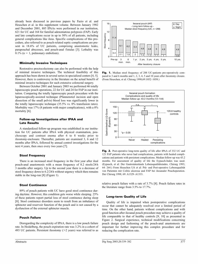
377Dig Surg 2003;20:339–382Abstracts
already been discussed in previous papers by Fazio et al. and
Heuschen et al. in this supplement volume. Between January 1982
and December 2001, 885 IPAAs were performed in our institution,
621 for UC and 164 for familial adenomatous polyposis (FAP). Early
and late complications occur in up to 50% of all patients, including
general complications like ileus. Specific complications of this pro-
cedure, also referred to as pouch-related septic complications are pre-
sent in 18.6% of UC patients, comprising anastomotic leaks,
parapouchal abscesses, and pouch-anal fistulas [3]. Lethality was
0.1% (n � 1, pulmonary embolism).
Minimally Invasive Techniques
Restorative proctocolectomy can also be performed with the help
of minimal invasive techniques. The technical feasibility of this
approach has been shown in several series in specialized centers [4, 5].
However, there is controversy in the literature on the actual benefit of
minimal invasive techniques for such extensive colorectal surgery.
Between October 2001 and January 2003 we performed 46 totally
laparoscopic pouch operations, 22 for UC and 24 for FAP in our insti-
tution. Comparing the totally laparoscopic pouch procedure with the
laparoscopically-assisted technique (Pfannenstiel incision and open
dissection of the small pelvis) blood loss was significantly lower in
the totally laparoscopic technique (35.5% vs. 0% transfusion rates).
Morbidity was 17% (8 patients with major complications), with a 0%
mortality [6].
Follow-up Investigations after IPAA and Late Results
A standardized follow-up program was established in our institu-
tion for UC patients after IPAA with physical examination, pou-
choscopy and contrast enema after 6 to 8 weeks prior to
ileostomy-enclosure. Thereafter, patients are examined 3, 6 and 12
months after IPAA, followed by annual control investigations for the
next 4 years, then once every two years [7].
Stool Frequency
There is an increased stool frequency in the first year after ileal
pouch-anal anastomosis with a mean frequency of 8.2 stools/24 h
3 months after surgery. Up to the second year there is a decrease of
stool frequency down to 6.2/24 h without urgency which then remains
stable in the long-run [8] (Figure 1).
Stool Continence
89% of pouch patients with UC have good stool continence dur-
ing daytime. However, this condition gets worse while sleeping. 25%
of these patients report partial loss of stool continence during sleep
[8]. Stool continence disorders seem to result from an imbalance of
sphincter and reservoir function of the pouch and is not caused by a
dysfunction of the external sphincter muscle.
Pouch Failure
Disregarding the complexity of IPAA, there is a low pouch failure
rate. In Heidelberg, the pouch expiration rate was 3.2% in a cohort of
493 UC patients. Persistent ileostomy (�2 years) was referred to as
relative pouch failure with a rate of 3.7% [8]. Pouch failure rates in
the literature range from 3.5% to 17.7%.
Long-term Quality of Life
Quality of life is impaired when postoperative complications
occur that cannot be adequately resolved over a limited period of
time. On the other hand, patients without complications and with
good function after ileoanal pouch procedure may achieve a quality of
life comparable to that of healthy controls [9, 10] as presented in
Figure 2. Surgical experience, technical modifications concerning
pouch design and fashioning of the pouch-anal anastomosis are
important for further improving this complex procedure and for
reducing the complication rate.
4.9
0.90
4
8
12
16
3.4
11.4
Pre-op
2.1
6.1
3 mon.
1.8
6.2
6 mon.
2
5.3
1 yr.
1.4
4.8
2 yrs.
4.8
1.2
3 yrs.
5
1.1
4 yrs.
4.9
1
5 yrs. 10 yrs.
After ileostomy closure
Sto
ol f
req
uenc
y
DayNight
Ileoanal pouch (IAP) - Long-term follow-up -
Median stool-frequency (UC, n�288)
Fig. 1. Median stool frequency of 288 UC-patients pre-operatively com-
pared to 3 and 6 months and 1, 2, 3, 4, 5 and 10 years after ileostomy closure.
(From Heuschen, et al: Chirurg 1998;69:1052–1058.)
Ileoanal pouch formation Complications and quality of life
Median follow-up: 43.2 months (12–144)
0
30
60
90
120
150
Never Healed Persistingcomplications
120.8 healthy controls
*p�0.05
Gas
troi
ntes
tinal
LQ
-Sco
re
(Eyp
asch
)
*n.s.
Fig. 2. Post-operative long-term quality of life after IPAA of 332 UC and
121 FAP patients who never had complications, patients with healed compli-
cations and patients with persistent complications. Median follow-up was 43.2
months. For assessment of quality of life the Eypasch-Index was used.
(Eypasch, et al: Der Gastrointestinale Lebensqualitätsindex. Chirurg 1993;
64: 264.) From Heuschen UA et al: Prä- und Post-operative Lebensqualität
von Patienten mit Colitis ulcerosa und FAP bei ileoanaler Pouchoperation.
Der Chirurg 1998; 69: A1329–A1333.
378 Dig Surg 2003;20:339–382 Inflammatory Bowel Disease
Principles and Practice
References
1 Farrell RJ, Peppercorn MA: Ulcerative colitis. The Lancet 2002;359:
331–340.
2 Heuschen UA, Hinz U, Allemeyer EH, Autschbach F, Stern J, Lucas M,
Herfarth C, Heuschen G: Risk factors for ileoanal J pouch-related septic
complications in ulcerative colitis and familial adenomatous polyposis.
Ann Surg 2002;235:207–216.
3 Heuschen UA, Allemeyer EH, Hinz U, Lucas M, Herfarth C, Heuschen G:
Outcome after septic complications in J pouch procedures. Br J Surg
2002;89:1–9.
4 Thibault C, Poulin EC: Total laparoscopic proctocolectomy and
laparoscopy-assisted proctocolectomy for inflammatory bowel disease:
Operative techniques and preliminary report. Surg Laparoscopy Endosc
1995;5:472–476.
5 Kienle P, Weitz J, Benner A, Herfarth Ch, Schmidt J: Laparoscopically
assisted colectomy and ileoanal pouch procedure with and without protec-
tive ileostomy. Surg Endosc 2003;17;716–720
6 Kienle P, Z’graggen K, Schmidt J, Benner A, Weitz J, Herfarth C,
Büchler MW: Totally laparoscopic ileoanal pouch procedure is superior to
the laparoscopically assisted technique. Ann Surg 2003 (submitted).
7 Heuschen UA, Autschbach F, Allemeyer EH, Zöllinger AM, Heuschen G,
Uehlein T, Herfarth C, Stern J: Long-term Follow-Up After Ileoanal Pouch
Procedure. Dis Col Rec 2000;44:487–499.
8 Heuschen UA, Schmidt J, Allemeyer E, Stern J, Heuschen G: Der
ileoanale Pouch. Zentralbl Chir 2001;1:1–8.
9 Heuschen UA, Heuschen G, Herfarth C: Lebensqualität nach
Proktocolektomie wegen Colitis ulcerosa. Der Chirurg 1998;69:1045–1051.
10 Heuschen UA, Heuschen G, Herfarth C: Der ileoanale Pouch als
Rectumersatz. Chirurg 1999;70:530–542.
29
Total Laparoscopic Ileoanal Pouch ProcedureK. Z’graggen, P. Kienle, M.W. Büchler, J. Schmidt
Department of Surgery, University of Heidelberg,Heidelberg, Germany
Background: Restorative proctocolectomy is the treatment of
choice for patients with ulcerative colitis and familial polyposis.
Minimal invasive techniques are increasingly being used for perform-
ing other complex abdominal procedures. We evaluated the use of
these techniques for performing restorative proctocolectomy within a
prospective series.
Patients and Methods: From June 1999 to March 2003,
107 patients underwent minimal invasive restorative proctocolectomy.
Patients with epithelial dysplasia, cancer or previous median laparo-
tomy were excluded. The first 59 patients had a laparoscopically
assisted procedure whereas the following 48 patients had a purely
laparoscopic procedure. In the laparoscopically assisted group the
colon was mobilized with a four trocar technique, facilitating vascu-
lar dissection, rectal resection and ileoanal pouch construction to be
done through a Pfannenstiel incision. All pouchanal anastomosis were
hand sewn after transanal mucosectomy. Just over half of the patients
(n � 32) had a one-stage procedure, all other patients received a pro-
tective ileostomy. In the purely laparoscopic group a five trocar tech-
nique was used to mobilize the ascending and descending colon
including both flexures, to perform complete rectal resection up to the
upper anal canal and to finally dissect the rectum at the anorectal
junction with an Endo-GIA. A small periumbilical incision of 4–5 cm,
incorporating the camera port, was then done and the large bowel
eviscerated and dissected. Ileoanal pouch construction was done
extracorporeally and the pouchanal anastomosis then fashioned in a
double stapling technique under camera vision. All patients with a
completely laparoscopic procedure had a covering ileostomy. Data
between the two groups was analyzed by logistic regression analysis,
Fisher’s exact and the Mann-Whitney test. To exclude protective
ileostomy creation as a potential bias, a separate analysis was done
excluding all one-stage procedures in the laparoscopically assisted
group.
Results: The conversion rate with both techniques was compa-
rable (8.5% in the laparoscopically assisted versus 8.7% in the totally
laparoscopic group). The operative times were significantly lower in
the laparoscopic group (median: 310 min vs 385 min; p � 0.001,
Mann-Whitney test). The difference in estimated blood loss was
250 ml (p � 0.001, Mann-Whitney test) in favor of the laparoscopic
group, when only including patients with protective ileostomy this
increased to 500 ml (p � 0.001, Mann-Whitney test). None of the
patients in the laparoscopic group required a blood transfusion,
whereas 21 patients (35.5%) in the laparoscopically assisted group
needed blood transfusions (p � 0.001, Fisher’s exact test). The over-
all complication rate was comparable (32.6% vs 35.5%); there was no
mortality. An increased BMI was significantly associated with the
occurrence of postoperative complications.
Conclusion: Both techniques are technically feasible and can
be performed with acceptable complication rates. In our hands
laparoscopic restorative proctocolectomy has advantages over the
laparoscopically assisted technique in regard to intraoperative blood
loss, the need for perioperative blood transfusions and the length of
postoperative hospital stay. We conclude that passing the learning
curve this procedure can be advantageous for the average patient
with familial polyposis and ulcerative colitis. The indication for per-
forming the operation minimal invasively should be seen critically in
obese patients. Main advantage of the minimal invasive procedures
over the conventional standard procedure is undoubtedly the better
cosmetic situation. However, randomized studies need to investigate
whether the complication rates and the long-term follow up are
comparable.
30
Pouchitis and Late Pouch-RelatedComplicationsG. Heuschen1, U. Hinz1, U. Heuschen2, J. Stern3
1Department of Surgery, University of Heidelberg,Heidelberg, Germany, 2St. Vincenz Hospital, Limburg, 3St. Josephs Hospital, Dortmund, Germany
Abstract
Inflammation of the ileoanal pouch (pouchitis) is one of the main
complications after restorative proctocolectomy with ileopouch-anal
anastomosis (IPAA) in patients with ulcerative colitis (UC) and famil-
ial adenomatous polyposis (FAP), yet its cause remains poorly under-
stood. A standardized definition and diagnostic procedures in
pouchitis are lacking. We established an algorithm for the classifica-
tion and management of pouchitis which enables the clinicians to

379Dig Surg 2003;20:339–382Abstracts
determine the severity of pouchitis, to differentiate between primary
and secondary pouchitis and to evaluate the course (acute versus
chronic). Early detection and precise classification of the inflamma-
tory process are required for adequate therapy.
Pouchitis
Definition
We designed a program for the long-term follow-up after IPAA,
focusing on the occurrence of pouchitis. As tools for diagnosis and
classification of pouchitis, we devised an algorithm [1] and a pouch-
itis activity score (Heidelberg Pouchitis Activity score, PAS) [2]. The
PAS consisted of 3 fields: clinical manifestations, endoscopic and his-
tologic examinations.
Between January 1989 and December 1997, 515 consecutive
patients received IPAA at the Surgical Department, University of
Heidelberg. 68% (n � 348) of all patients who received restorative
proctocolectomy during this time agreed to take part in the long-term
follow-up program focusing on pouchitis at our department. Forty of
348 patients were lost to the regular follow-up, so that 210 patients
operated on for UC and 98 patients operated on for FAP were
included in the study.
Incidence and Course
At least one episode of pouchitis (Grade II or III inflammation)
was diagnosed in 29% of UC patients and in 2% of FAP patients. For
UC patients the probability of pouchitis occurring within 8 years after
the initial operation was 50%. 80% of all cases of pouchitis occurred
within the first four years after IPAA in UC patients.
Of all cases of pouchitis in UC patients, 67% had an acute (dura-
tion �3 months) and 33% a chronic course (duration �3 months).
Primary and Secondary Pouchitis
Using a wide range of diagnostic means we identified a group of
patients in whom pouchitis was caused by surgical complications spe-
cific to the ileal reservoir and pouch-anal anastomosis. For these
cases, we chose the term ‘secondary’ pouchitis. They have to be
clearly differentiated from primary (idiopathic) pouchitis with an
unclear cause. Secondary pouchitis was found in 6% of all UC
patients. Among UC patients with pouchitis, 21% suffered from sec-
ondary pouchitis. Both FAP patients with pouchitis suffered from sec-
ondary pouchitis. In accordance to our definition, all cases of
pouchitis in which diagnostic instruments gave no hint at any sec-
ondary cause of the inflammation were considered as primary pouch-
itis. Primary was thus diagnosed in 79% of UC patients with pouchitis.
In FAP patients no case of primary pouchitis was diagnosed.
Chronic Primary Pouchitis and Secondary Pouchitis – Pouchitis of Special Interest
Management of pouchitis should give special consideration to two
forms of the inflammatory process: chronic primary pouchitis and
secondary pouchitis.
Chronic primary pouchitis occurred in 6% of all UC patients. The
mean time of onset was 2 months after IPAA, mean duration was 22
(range, 6–48 months). 69% of the cases were persistent and 31%
relapsing. Medical treatment led to a remission of the inflammatory
process in only 23% and to improvement in 38%. Another 38% did
not benefit from medical treatment. In two patients with chronic pri-
mary pouchitis the pouch had to be excised, in one case because of
severe symptoms of pouchitis and in one because of premalignant
high-grade dysplasia. In the latter, a carcinoma of the ileal pouch
mucosa was found in the resected specimen of a UC patient.
Therefore, for patients with chronic primary pouchitis long-term
surveillance seems mandatory because of the risk of malignant trans-
formation of the pouch mucosa.
6% of UC patients, and 2% of FAP patients developed secondary
pouchitis. Only 20% of the patients with secondary pouchitis devel-
oped severe pouchitis; 80% showed moderate pouchitis. Surgical
complications as cause for secondary pouchitis were pouch-anal
abscesses (13%), anastomotic leak (7%), pouch-anal fistula (60%),
and outlet obstruction (13%). Secondary pouchitis was cured by
surgical therapy in 87%. Treatment consisted of minor transanal
procedures (e.g. fistulotomy or dilation of outlet obstruction) in
47% and major transabdominal procedures in another 47%. One
pouch had to be excised because of multiple fistulas, and in another
case fistulotomy led to only a reduction of the inflammatory
process. Only one case of secondary pouchitis was treated success-
fully with conservative therapy alone. The algorithm is highly effi-
cient in identifying patients with a secondary pouchitis who require
surgical treatment.
Pouch-Related Septic Complications (PRSC)
Pouch-related septic complications (peripouchal fistulas and
abscesses) occur as late complications and should therefore be con-
sidered in regular, specific long-term follow-up examinations. We
identified significant risk factors for PRSC specific to patients with
UC and FAP [3]. In the univariate analysis, patients with UC younger
than 50 years, with severe proctitis, with preoperative hemoglobin
levels less than 10 g/L, or receiving corticoid medication had a sig-
nificantly higher risk for PRSC. Multivariate analysis showed that UC
patients receiving a systemic prednisolone-equivalent corticoid med-
ication of more than 40 mg/day had a significantly greater risk of
developing PRSC than UC patients receiving 1–40 mg/day and UC
patients who were not receiving corticoid medication. FAP patients
proved to have a significantly higher risk for PRSC in the univariate
and multivariate analysis if anastomotic tension had occurred. These
risk factors must be considered for each individual surgical strategy
to lower PRSC.
References
1 Heuschen UA, Autschbach F, Allemeyer EH, Zöllinger A, Heuschen G,
Uehlein T, Herfarth Ch, Stern J: Long-term follow-up after ileoanal pouch
procedure. Algorithm for diagnosis, classification, and management of
pouchitis. Dis Colon Rectum 2001;44:487–499.
2 Heuschen UA, Allemeyer EH, Hinz U, Autschbach F, Uehlein T,
Herfarth Ch, Heuschen G: Diagnosing pouchitis. Dis Colon Rectum 2002;
45:776–788.
3 Heuschen UA, Hinz U, Allemeyer EH, Autschbach F, Stern J, Lucas M,
Herfarth Ch, Heuschen G: Risk factors for ileoanal J pouch-related septic
complications in ulcerative colitis and familial adenomatous polyposis.
Ann Surg 2002;235:207–216.

380 Dig Surg 2003;20:339–382 Inflammatory Bowel Disease
Principles and Practice
31
Quality of Life Assessment in Inflammatory Bowel DiseaseE.J. Irvine
Department of Medicine, University of Toronto, Toronto, Canada
Evaluating the impact of inflammatory bowel disease (IBD) has
relied heavily upon indices assessing symptom severity that describe,
for example, stool frequency, abdominal pain and well being.
Endoscopy and radiology, are useful imaging techniques for describ-
ing disease severity together with laboratory markers, such as
hemoglobin, WBC or C-reactive protein. However, full colonoscopy
is not amenable to repeated frequent assessments and blood parame-
ters are often normal, even in the face of moderately severe disease.
Health-related quality of life (HRQOL) has, therefore, presented
as a clinically relevant measure of disease impact and treatment
response.
HRQOL has been defined as the functional effect of an illness and
its therapy upon an individual, as perceived by the individual, himself
or herself. The specific domains that determine HRQOL include
physical and occupational function, emotional state, social interac-
tions, and somatic sensation [1]. These features are often simply
described as disease-related (e.g. affected by severity, treatment effi-
cacy, adverse effects) or disease independent (e.g. age, gender, smok-
ing status, education and knowledge, personality and coping skills,
culture and beliefs [2]. However, both are important features of
this outcome. No doubt, the inherited predisposition of IBD that
may also be enhanced by environmental or psychosocial factors, can
affect motility, sensation and disease outcome. Experiences and
beliefs of the individual may also change these variables [2]. Thus, the
HRQOL of two individuals with similar disease could produce
one who is substantially disabled and another who functions
quite well.
Clinicians and researchers evaluate HRQOL by administering
questionnaires that assess important disease-related features, such as
diarrhea or abdominal pain, fatigue, or poor appetite, as well as other
determinants such as vitality and energy, or emotional well-being.
The major domains of HRQOL include physical, emotional and
social function and these may be evaluated using generic question-
naires such as the SF-36 [3] or the EuroQOL [4] or the Health Utility
Index [5]. These instruments are applicable in most populations, irre-
spective of the population demographics and independent of the par-
ticular medical condition and allow comparisons among different
conditions or populations. They may be somewhat insensitive regard-
ing important aspects of dysfunction but detect reliably unanticipated
problems. HRQOL assessment, thus, provides a good gauge to mea-
sure improved HRQOL for clinicians, researchers or policy makers,
by helping identify the needs of individual patients, assessing the
impact of therapy (in individuals or in clinical trials) and determining
health policy [6].
HRQOL assessment can also be applied in pharmacoeconomic
assessments in which costs to achieve a particular outcome (such as
remission, no symptoms), or costs per quality adjusted life year
gained are assessed. Utilities are patient generated preference
‘weights’, ranging from 0 (death) to 1.0 (full health), that may be
derived in groups of patients or in a reference population using utility
instruments. A quality adjusted life year (QALY) can then be calcu-
lated and represents one full year in perfect health. Different utility
instruments, like different HRQOL tools may elicit different values in
the same reference population and still produce similar results when
assessing change over time or after treatment.
Several disease specific instruments have been developed for par-
ticular diseases such as gastro-esophageal reflux disease, dyspepsia,
arthritis, cancer, HIV-AIDS and IBD [7, 8]. Items to be included in
these questionnaires are often generated by focus groups of patients
and using well established standardized methodologies are then
drafted into a HRQOL index. This is then validated and subjected to
psychometric testing before being applied in patient research and
management [7–9]. These disease-specific questionnaires differ from
generic instruments by including some of the particular features of
the condition at hand. For example, for patients with inflammatory
bowel disease, this might include abdominal pain, diarrhea, rectal
bleeding, weight loss or bloating, as well as fatigue, difficulty sleep-
ing and features of emotional (tearful, angry, embarrassed, sad, wor-
ried about cancer) and social (unable to go to social activities, avoid
sports, be absent from work or school) function.
For many patients with IBD, their disease severity determines
much of their HRQOL goals. However, there are also factors inde-
pendent of disease severity, such as education level, cognitive func-
tion, coping skills and social support [2]. Comorbid medical
conditions and depression and anxiety may also contribute to
impaired HRQOL. Most patients who have active disease, however,
desire effective medical or surgical treatment.
Many descriptive studies have now reported significant HRQOL
impairment, when comparing IBD patients to the general population.
Patients most similar to the background general population are
patients who have been cured surgically, of ulcerative colitis or have
had prolonged disease remission [9]. Those with the most severe
impairment are those with short bowel, who are on home parenteral
nutrition or patients that are awaiting surgery for severe disease or
complications [reviewed in 9]. Some studies suggest that there are
different types of problems experienced by adults and children (more
important to be seen to be just like peers) and that longer duration of
disease prompts fewer physician visits and absenteeism. Clinical
trials of effective therapies for active Crohn’s disease, including
corticosteroids (e.g. prednisone, budesonide), immunosuppressives
(methotrexate but not cyclosporine) or biological agents (infliximab,
natalizumab) have shown improved HRQOL using disease specific
instruments (such as the Inflammatory Bowel Disease Questionnaire-
IBDQ). Comparative and cross-sectional studies have shown signifi-
cant differences in HRQOL using disease specific instruments such
as the IBDQ or the Rating Form of IBD Patient Concerns. Different
instruments appear to be necessary to identify specific problems after
surgical therapy, such as sexual dysfunction and frequency of incon-
tinence [8, 9].
Selecting the best HRQOL instrument for a new study should be
based on the research questions. However, a priori psychometric
assessment should have been performed to be able to accurately inter-
pret results. Combining a generic and disease specific instrument has
become acceptable to ensure recognition of all clinically important
changes. When assessing HRQOL, all important outcomes must be
determined. Finally, in recent times, studies are undertaken in many
countries simultaneously. It is critical to ensure that proper cross-
cultural validation is performed when HRQOL questionnaires must
be translated or adapted for other languages [10, 11].
381Dig Surg 2003;20:339–382Abstracts
References
1 Spilker B: Quality of Life and Pharmacoeconomics in Clinical Trials,
ed 2. Philadelphia, Lippincott-Raven Publishers, 1996.
2 Garrett JW, Drossman DA: Health status in inflammatory bowel disease:
Biological and behavioral considerations. Gastroenterology 1990;99:90–96.
3 Ware JE, Sherbourne CD: The MOS 36 item Short Form Health Survey
(SF-36).Conceptual framework and item selection. Med Care 1992;30:
473–483.
4 The EuroQol Group: EuroQol – A new facility for the measurement of
health-related quality of life. Health Policy 1990;16:199–208.
5 Torrance GW, Furlong W, Feeny D, Boyle M: Multiattribute preference
functions; Health Utilities Index. PharmacoEconomics 1995;7L:503–520.
6 Dupuy HJ: The Psychological General Well Being Index; in Wenger NK,
Mattson ME, Furberg CF, Ellinson J (eds): Assessment of Quality of Life
in Clinical Trials of Cardiovascular Therapies. Le Jaq Publishing Inc.,
1984, pp 170–183.
7 Fitzpatrick R, Fletcher A, Gore S, et al: Quality of life measures in health
care. I: Applications and issues in assessment. BMJ 1992;305:1074–1077.
8 Borgaonkar M, Irvine EJ: Quality of life measurement in gastrointestinal
and liver disorders. Gut 2000;47:444–454.
9 Irvine EJ: Measuring quality of life in inflammatory bowel disease; in
Targan SR, Shanahan F, Karp LC (eds): Inflammatory Bowel Disease:
From Bench to Bedside. Dordrecht, the Netherlands, Kluwer Academic
Publishers, 2003, pp 481–494.
10 Scientific Advisory Committee of the Medical Outcomes Trust: Assessing
health status and quality-of-life instruments: Attributes and review crite-
ria. Quality of Life Research 2002;11:193–205.
11 Guillemin F, Bombardier C, Beaton D: Cross-cultural adaptation of
health-related quality of life measures: Literature review and proposed
guidelines. J Clin Epidemiol 1993;46:1417–1432.

Autschbach, F. 345, 374
Büchler, M.W. 378
Duchmann, R. 347
Erb, G. 357
Fazio, V.W. 362, 375
Flossdorf, P. 357
Fölsch, U.R. 360, 371
Folwaczny, C. 342
Galandiuk, S. 351
Galle, P.R. 372
German, A. 352
Giese, T. 350
Grüber-Hoffmann, B. 357
Hansmann, H.J. 357
Heller, F. 347
Hermon-Taylor, J. 360
Heuschen, G. 352, 367, 376, 378
Heuschen, U. 352, 376, 378
Hinz, U. 378
Hodgson, H. 369
Hoffmann, J.C. 342
Holtmann, M.H. 372
Irvine, E.J. 380
Kauffmann, G.W. 357
Kienle, P. 363, 378
Kühl, A.A. 342
Leowardi, C. 352, 376
Löfberg, R. 370
Ludwig, B. 350
Maul, J. 347
Meuer, S. 345, 350
Neurath, M.F. 346
Pawlowski, N.N. 342
Prantera, C. 355
Reinshagen, M. 368
Schmidt, C. 350
Schmidt, J. 357, 363, 376, 378
Schölmerich, J. 353
Schreiber, S. 360, 371
Scribano, M.L. 355
Sido, B. 345
Stallmach, A. 350
Stange, E.F. 359
Stern, J. 363, 367, 378
Thomsen, O.Ø. 361
Uhl, W. 363
Veltkamp, C. 349
Young-Fadok, T.M. 365
Zeitz, M. 347
Zeuzem, S. 350
Z’graggen, K. 378
Numbers refer to page number
Author Index for Abstracts
Fax �41 61 306 12 34
E-Mail [email protected]
www.karger.com
© 2003 S. Karger AG, Basel
Accessible online at:
www.karger.com/dsu
382