Inflammatory Bowel Disease and Mutations Affecting the Interleukin-10 Receptor
18
Inflammatory Bowel Disease and Mutations Affecting the Interleukin-10 Receptor Erik-Oliver Glocker, M.D., Daniel Kotlarz, M.D., Kaan Boztug, M.D., E. Michael Gertz, Ph.D., Alejandro A. Schäffer, Ph.D., Fatih Noyan, Ph.D., Mario Perro, M.Sc., Jana Diestelhorst, B.Sc., Anna Allroth, M.D., Dhaarini Murugan, M.Sc., Nadine Hätscher, B.Sc., Dietmar Pfeifer, M.D., Karl-Walter Sykora, M.D., Martin Sauer, M.D., Hans Kreipe, M.D., Martin Lacher, M.D., Rainer Nustede, M.D., Cristina Woellner, M.Sc., Ulrich Baumann, M.D., Ulrich Salzer, M.D., Sibylle Koletzko, M.D., Neil Shah, M.D., Anthony W. Segal, M.D., Axel Sauerbrey, M.D., Stephan Buderus, M.D., Scott B. Snapper, M.D., Ph.D., Bodo Grimbacher, M.D., and Christoph Klein, M.D., Ph.D. Abstract BACKGROUND—The molecular cause of inflammatory bowel disease is largely unknown. METHODS—We performed genetic-linkage analysis and candidate-gene sequencing on samples from two unrelated consanguineous families with children who were affected by early-onset inflammatory bowel disease. We screened six additional patients with early-onset colitis for mutations in two candidate genes and carried out functional assays in patients’ peripheral-blood mononuclear cells. We performed an allogeneic hematopoietic stem-cell transplantation in one patient. RESULTS—In four of nine patients with early-onset colitis, we identified three distinct homozygous mutations in genes IL10RA and IL10RB, encoding the IL10R1 and IL10R2 proteins, respectively, which form a heterotetramer to make up the interleukin-10 receptor. The mutations abrogate interleukin-10–induced signaling, as shown by deficient STAT3 (signal transducer and activator of transcription 3) phosphorylation on stimulation with interleukin-10. Consistent with this observation was the increased secretion of tumor necrosis factor α and other proinflammatory cytokines from peripheral-blood mononuclear cells from patients who were deficient in IL10R subunit proteins, suggesting that interleukin-10–dependent “negative feedback” regulation is disrupted in these cells. The allogeneic stem-cell transplantation performed in one patient was successful. CONCLUSIONS—Mutations in genes encoding the IL10R subunit proteins were found in patients with early-onset enterocolitis, involving hyperinflammatory immune responses in the intestine. Allogeneic stem-cell transplantation resulted in disease remission in one patient. Inflammatory bowel disease is a heterogeneous group of disorders, classified as Crohn’s disease, ulcerative colitis, and indeterminate colitis. 1,2 In most patients, these disorders are manifested in adolescence or adulthood; however, they may present in infancy and may be inherited as an autosomal recessive trait. 3–6 Address reprint requests to Dr. Klein at the Department of Pediatric Hematology/Oncology, Hannover Medical School, Carl-Neuberg- Straße, 1 D-30625, Hannover, Germany, or at [email protected]; or to Dr. Grimbacher at the Department of Immunology and Molecular Pathology, Royal Free Hospital and University College London, Pond St., London NW3 2QG, United Kingdom, or at [email protected]. Drs. Glocker, Kotlarz, Boztug, Grimbacher, and Klein contributed equally to this article. Dr. Snapper reports receiving consulting fees from Viamet and Cytokine PharmaSciences and lecture fees from UCB. No other potential conflict of interest relevant to this article was reported. NIH Public Access Author Manuscript N Engl J Med. Author manuscript; available in PMC 2010 May 19. Published in final edited form as: N Engl J Med. 2009 November 19; 361(21): 2033–2045. doi:10.1056/NEJMoa0907206. NIH-PA Author Manuscript NIH-PA Author Manuscript NIH-PA Author Manuscript
-
Upload
independent -
Category
Documents
-
view
1 -
download
0
Transcript of Inflammatory Bowel Disease and Mutations Affecting the Interleukin-10 Receptor
Inflammatory Bowel Disease and Mutations Affecting theInterleukin-10 Receptor
Erik-Oliver Glocker, M.D., Daniel Kotlarz, M.D., Kaan Boztug, M.D., E. Michael Gertz, Ph.D.,Alejandro A. Schäffer, Ph.D., Fatih Noyan, Ph.D., Mario Perro, M.Sc., Jana Diestelhorst,B.Sc., Anna Allroth, M.D., Dhaarini Murugan, M.Sc., Nadine Hätscher, B.Sc., Dietmar Pfeifer,M.D., Karl-Walter Sykora, M.D., Martin Sauer, M.D., Hans Kreipe, M.D., Martin Lacher, M.D.,Rainer Nustede, M.D., Cristina Woellner, M.Sc., Ulrich Baumann, M.D., Ulrich Salzer, M.D.,Sibylle Koletzko, M.D., Neil Shah, M.D., Anthony W. Segal, M.D., Axel Sauerbrey, M.D.,Stephan Buderus, M.D., Scott B. Snapper, M.D., Ph.D., Bodo Grimbacher, M.D., and ChristophKlein, M.D., Ph.D.
AbstractBACKGROUND—The molecular cause of inflammatory bowel disease is largely unknown.
METHODS—We performed genetic-linkage analysis and candidate-gene sequencing on samplesfrom two unrelated consanguineous families with children who were affected by early-onsetinflammatory bowel disease. We screened six additional patients with early-onset colitis formutations in two candidate genes and carried out functional assays in patients’ peripheral-bloodmononuclear cells. We performed an allogeneic hematopoietic stem-cell transplantation in onepatient.
RESULTS—In four of nine patients with early-onset colitis, we identified three distinct homozygousmutations in genes IL10RA and IL10RB, encoding the IL10R1 and IL10R2 proteins, respectively,which form a heterotetramer to make up the interleukin-10 receptor. The mutations abrogateinterleukin-10–induced signaling, as shown by deficient STAT3 (signal transducer and activator oftranscription 3) phosphorylation on stimulation with interleukin-10. Consistent with this observationwas the increased secretion of tumor necrosis factor α and other proinflammatory cytokines fromperipheral-blood mononuclear cells from patients who were deficient in IL10R subunit proteins,suggesting that interleukin-10–dependent “negative feedback” regulation is disrupted in these cells.The allogeneic stem-cell transplantation performed in one patient was successful.
CONCLUSIONS—Mutations in genes encoding the IL10R subunit proteins were found in patientswith early-onset enterocolitis, involving hyperinflammatory immune responses in the intestine.Allogeneic stem-cell transplantation resulted in disease remission in one patient.
Inflammatory bowel disease is a heterogeneous group of disorders, classified as Crohn’sdisease, ulcerative colitis, and indeterminate colitis.1,2 In most patients, these disorders aremanifested in adolescence or adulthood; however, they may present in infancy and may beinherited as an autosomal recessive trait.3–6
Address reprint requests to Dr. Klein at the Department of Pediatric Hematology/Oncology, Hannover Medical School, Carl-Neuberg-Straße, 1 D-30625, Hannover, Germany, or at [email protected]; or to Dr. Grimbacher at the Department of Immunologyand Molecular Pathology, Royal Free Hospital and University College London, Pond St., London NW3 2QG, United Kingdom, or [email protected]. Glocker, Kotlarz, Boztug, Grimbacher, and Klein contributed equally to this article.Dr. Snapper reports receiving consulting fees from Viamet and Cytokine PharmaSciences and lecture fees from UCB. No other potentialconflict of interest relevant to this article was reported.
NIH Public AccessAuthor ManuscriptN Engl J Med. Author manuscript; available in PMC 2010 May 19.
Published in final edited form as:N Engl J Med. 2009 November 19; 361(21): 2033–2045. doi:10.1056/NEJMoa0907206.
NIH
-PA Author Manuscript
NIH
-PA Author Manuscript
NIH
-PA Author Manuscript

The genetic causes of inflammatory bowel disease are only partly understood. Studies intransgenic murine models7 and genomewide genetic-linkage and association studies haveprovided insights into the genetic complexity underlying these inflammatory conditions.8Investigators using these approaches have implicated several genes in the pathogenesis ofinflammatory bowel disease; the identity of these genes suggests that disruption of the innateand adaptive arms of the immune system,9–11 the process of autophagy,12,13 epithelial barrierfunction,14 and activation of the endoplasmic reticulum stress response15 may causesusceptibility. However, the functional relevance of most of these susceptibility genes isunclear. An alternative approach to identifying disease-causing genes is to study families inwhich inflammatory bowel disease is inherited as a potentially monogenic trait and to identifythe relevant gene by positional cloning.
Interleukin-10 restricts excessive immune responses.16 Initially, it was described as a solublefactor that is released by type 2 helper T cells and that inhibits the secretion of type 1 helper Tcytokines, such as interleukin-2 and interferon-γ.17 Interleukin-10 is secreted by a wide varietyof cells and has pleiotropic effects on T cells, B cells, myeloid cells, and other cell types.16 Inparticular, interleukin-10 limits the secretion of proinflammatory cytokines, such as tumornecrosis factor α (TNF-α) and interleukin-12.18 The receptor for interleukin-10 consists of twoalpha molecules (IL10R1) and two beta molecules (IL10R2).19 Although IL10R1 is specificto the interleukin-10 receptor, IL10R2 is a subunit of the receptors for several additionalcytokines (e.g., interleukin-22 and interleukin-26).20 The assembly of this heterotetramericinterleukin-10 receptor results in the activation of the receptor-associated Janus tyrosinekinases, JAK1 and Tyk2, resulting in the phosphorylation of STAT3 (signal transducer andactivator of transcription 3) and the induction of STAT3-dependent genes.21–25
Mice that are deficient in either interleukin-10 or interleukin-10 receptor 2 have been shownto have severe enterocolitis,26–28 a finding that underscores the pivotal role of interleukin-10in the mediation of signaling that controls inflammation in the gut. These studies in mice havealso proffered genes that encode proteins in the interleukin-10–signaling pathway as candidatesfor association with inflammatory bowel disease.
METHODSPATIENTS
In Family A, the index patient (Patient II-3), who was of Turkish origin and born of parentswho were first-degree cousins, presented at the age of 3 months with proctitis and abscessesin the peri-anal region, which required multiple surgical interventions. Protective colostomywas performed because of impaired wound healing. Multiple enterocutaneous fistulasoriginating from the small intestine required further partial bowel resections and ileostomy. Inaddition, he had early-onset cutaneous folliculitis. Recurrent infections such as otitis media,bronchitis, and two episodes of purulent gonarthritis were potentially linked toimmunosuppressive therapy. He had normal numbers and functioning of T cells and B cells.Increased serum levels of IgG (21.8 g per liter; normal range, 5.2 to 12.9), IgA (7.66 g per liter;normal range, 0.47 to 2.1), and IgM (2.98 g per liter; normal range, 0.47 to 1.90) wereinterpreted as being associated with chronic ileocolitis. The neutrophil function was normalwith respect to NADPH oxidase activity (data not shown). Histologic analysis of ileal- andcolonic-biopsy samples that were obtained from the sites of inflammation showed multifocalulcerations and agglomerations of polymorphic infiltrates (Fig. 1A and 1B in theSupplementary Appendix, available with the full text of this article at NEJM.org). The patientwas treated with a wide spectrum of anti-inflammatory agents, including corticosteroids,methotrexate, thalidomide, and anti–TNF-α monoclonal antibodies. None of these therapiesinduced remission or long-term improvement.
Glocker et al. Page 2
N Engl J Med. Author manuscript; available in PMC 2010 May 19.
NIH
-PA Author Manuscript
NIH
-PA Author Manuscript
NIH
-PA Author Manuscript

In his affected sister, Patient II-4, proctitis and rectovaginal fistula developed in the first yearof life. She had folliculitis, pneumonia, and one episode of renal abscess caused by Escherichiacoli. She underwent several surgical interventions, including colonic resection andcolostomies. The results of the immune workup were normal. The clinical course of the colitiswas slightly milder in Patient II-4 than it was in her affected brother.
In Family B, the index patient (Patient II-5), who was of Lebanese descent, presented in herfirst year of life with severe enterocolitis (Fig. 1A) associated with enteric fistulas, perianalabscesses, and chronic folliculitis, consistent with a diagnosis of Crohn’s disease (Fig. 1B).She required multiple surgical interventions, including colectomy and ileostomy (Fig. 1C).The histopathological analysis of the specimen from the colon obtained during resectionrevealed circumscribed ulcerations of the mucosa, from which pear-shaped intramuralabscesses extended into the submucosa and muscularis propria (Fig. 1D, 1E, and 1F).Inflammatory infiltrates were also seen in the small intestine, albeit to a lesser extent.Immunologic analyses showed normal numbers and function of T cells, B cells, and naturalkiller cells (data not shown). Serum immunoglobulin levels were normal or increased.Neutrophil granulocytes showed regular oxidative burst capacity (data not shown). Despitemultiple therapies, including treatment with corticosteroids and azathioprine and totalcolectomy, a sustained, long-term remission could not be achieved.
We also performed studies in six patients with an onset of severe colitis during the first yearof life.
We obtained written informed consent from adult patients and from the parents of childrenwho participated in the study. In addition, the affected children provided their assent. The studywas approved by the institutional review board at each study center (see the SupplementaryAppendix).
GENETIC ANALYSES AND ASSAYS OF MOLECULAR FUNCTIONWe analyzed the genotypes of each family to look for markers or intervals that showed perfectsegregation with the disease phenotype. For each pedigree, we defined “perfect segregation”as meaning that the affected child or children in each family had the same homozygousgenotype or haplotype. In addition, if both parents were heterozygous, then the unaffectedchildren each needed to have at least one genotype or one haplotype that differed from thoseof the affected sibling or siblings.
Once we had identified perfectly segregating intervals, we chose genes therein to sequence formutations on the basis of previous studies of the genes. We then performed gene-specific andprotein-specific assays to determine the functional effects of the identified mutations oninterleukin-10 signaling. The functional experiments included the transduction of cell lines andmutant cells with retroviral vectors encoding wild-type IL10RA and IL10RB complementaryDNA, respectively, to show that the defect in interleukin-10 signaling was corrected byreplacing the mutant interleukin receptor with a normal receptor. For more details on the geneticanalyses and assays of molecular function, see the Supplementary Appendix.
RESULTSGENETIC ANALYSES
We genotyped 253 microsatellite markers that spanned all 22 autosomes in members of FamilyA (Fig. 2A). Five markers showed perfect segregation with phenotype (corresponding to alogarithm of the odds [LOD] score of approximately 2.0) and were located on chromosomes2, 7, 14, 19, and 21. Fine mapping with additional micro-satellites showed that three of thesemarkers were within intervals that perfectly segregated with disease phenotype and that
Glocker et al. Page 3
N Engl J Med. Author manuscript; available in PMC 2010 May 19.
NIH
-PA Author Manuscript
NIH
-PA Author Manuscript
NIH
-PA Author Manuscript

spanned multiple megabases. Table S1 in the Supplementary Appendix summarizes the extentof these intervals; the minimal interval was defined by the two most distant flanking, perfectlysegregating markers, and the maximal interval was defined by extending, in each direction, tothe first marker that did not perfectly segregate with disease phenotype. Genes that were locatedwithin these intervals and the genes we sequenced (one gene on chromosome 7 and four geneson chromosome 21) are listed in Table S2 in the Supplementary Appendix.
We focused on the chromosome 21 interval, since it contained more genes than the other twointervals combined and more functional candidate genes, including a family of interferon-related genes. We used all suitably positioned microsatellite markers on the Marshfield29 anddeCode30 maps to reduce the uncertainty of the region most distal to the upper telomere toapproximately 350 kb (Fig. 2A). The candidate gene, IL10RB, was the first gene (starting at33.56 Mb) that was below and close to the perfect marker D21S1898. IL10RB is the only oneof four consecutive immune-related genes (including the better- positioned IFNAR2) with anorthologous gene in the mouse that when “knocked out” results in colitis.28
We sequenced IL10RB and identified a homozygous point mutation in exon 4 in both affectedsiblings, resulting in a premature stop codon (c.G477A, p.Trp159X) (Fig. 2A in theSupplementary Appendix). Both healthy parents and the two healthy siblings carried a singleheterozygous mutation (W159X) in IL10RB (data not shown). This mutation was absent in 180unaffected German subjects of European descent, in 70 unaffected subjects of Turkish ancestry,and in 30 subjects of Iranian ancestry. We sequenced IL10RB in 90 patients with adult-onsetinflammatory bowel disease: 45 patients with Crohn’s disease and 45 with ulcerative colitis.None of the unaffected subjects or the patients with adult-onset inflammatory bowel diseasecarried the mutation or any other sequence variations.
We used single-nucleotide–polymorphism arrays (Affymetrix) to map homozygous regionssegregating with disease in Family B (in which the parents were second-degree cousins) (Fig.2B). We identified eight regions larger than 1 Mb that were homozygous (with respect tohaplotype) in the index patient and that “housed” in each unaffected family member a haplotypethat was not present in the patient (Table S3 in the Supplementary Appendix). These regionsyielded a peak LOD score of 2.5 (see the Supplementary Appendix). We observed thatIL10RA was located in one of these regions (on chromosome 11q) and identified a homozygousmissense mutation in exon 4 (c.G421A, p.Gly141Arg) in the index patient (Fig. 2B in theSupplementary Appendix). All the other members of Family B carried at least one IL10RAwild-type allele and did not have any inflammatory bowel disease. The mutation was absentin 100 unaffected Arabic subjects and 30 unaffected Iranian subjects.
We sequenced IL10RA and IL10RB in six additional patients who had an onset of severe colitisin the first year of life and identified a homozygous missense mutation in exon 3 of IL10RA(c.C325T, p.Thr84Ile) in one German patient of European ancestry (Fig. 2C in theSupplementary Appendix). This patient, who was 8 months of age, had received a diagnosisof severe pancolitis at the age of 3 months (Fig. 2D, 2E, and 2F in the SupplementaryAppendix). This mutation was absent in 100 healthy white subjects of European ancestry.Furthermore, we observed no mutations in either IL10RA or IL10RB in 32 children withinflammatory bowel disease who had an onset of symptoms at more than 12 months of age.
EFFECTS OF MUTATIONS ON INTERLEUKIN-10–RECEPTOR PATHWAY FUNCTIONSWe studied the functional effects of the implicated mutations. Fluorescence-activated cellsorting (FACS) analysis disclosed no expression of IL10R2 in Epstein–Barr virus (EBV)–transformed B cells obtained from Patient II-3 in Family A, who carried the homozygousW159X mutation in IL10RB, in contrast to robust IL10R2 expression in cells from an
Glocker et al. Page 4
N Engl J Med. Author manuscript; available in PMC 2010 May 19.
NIH
-PA Author Manuscript
NIH
-PA Author Manuscript
NIH
-PA Author Manuscript

unaffected subject (Fig. 3A).31 Figure 3B shows the localization of the W159X mutation inthe IL10R2 protein.
When interleukin-10 binds to its receptor, it signals predominantly through STAT3 to mediateits antiinflammatory effects.25,32 To examine the integrity of this pathway, we stimulatedperipheral-blood mononuclear cells (PBMCs) or EBV-transformed B cells from Patient II-3in Family A and those from an unaffected subject with interleukin-10 and assessed them forSTAT3 phosphorylation at tyrosine 705, using Western blot analysis. Interleukin-10 inducedSTAT3 phosphorylation at tyrosine 705 in wild-type IL10R2 cells but not in cells with theW159X mutation (Fig. 3C, and Fig. 1C in the Supplementary Appendix). To reconstitute thefunction of IL10R, which has both IL10R1 and IL10R2 subunits, we analyzed EBV-transformed B cells from Patient II-3 in Family A that were transduced either with a lentiviralvector encoding IL10R2 along with green fluorescent protein (GFP) as a marker gene or witha control vector encoding GFP only (Fig. 1D in the Supplementary Appendix). In contrast toIL10R2-negative cells, IL10R2-reconstituted cells showed intact STAT3 phosphorylation attyrosine 705 (Fig. 3D, and Fig. 1E in the Supplementary Appendix). We concluded that theW159X mutation abrogated the phosphorylation of STAT3 mediated by interleukin-10.
The mutations we observed in IL10RA were missense variants; analyses with the use of FACSand Western blot analysis indicated that the G141R mutation in the IL10R1 protein wasexpressed at levels similar to those in control cells (Fig. 3E, and Fig. 2G in the SupplementaryAppendix). When interleukin-10 binds to the cell surface, it first interacts with IL10R1 andsubsequently binds IL10R2. We used a structure of the intermediate interleukin-10–IL10R1state (identifier 1Y6K in the Protein Data Bank) to model possible effects of missense changes,33 using computer algorithms34,35 (for details, see the Supplementary Appendix). Weconcluded that the IL10R1 G141R mutation, which was localized in a region binding tointerleukin-10,36 probably has a substantial effect on the structure of the interleukin-10–receptor complex (Fig. 3F and 3G). The IL10R1 T84I variant was similarly predicted to bedeleterious on the basis of computer modeling and because threonine 84 is highly conservedin homologous proteins (Fig. 2H and 2I in the Supplementary Appendix).
To assess the functional capacity of the interleukin-10 receptor, we stimulated PBMCs fromPatient II-5 in Family B and those from an unaffected subject with recombinant humaninterleukin-10 and then measured the extent of phosphorylation of STAT3 at tyrosine 705 andserine 727. As expected, phosphorylation of STAT3 was abrogated at tyrosine 705 but wasunaffected at serine 727 in the cells from the patient, as compared with those from the controlsubject (Fig. 3H). To validate these findings, we coexpressed either wild-type IL10R1 proteinor that with the G141R mutation, along with wild-type IL10R2, in IL10R-negative HeLa cells.Coexpression of wild-type IL10R1 and wild-type IL10R2 resulted in STAT3 phosphorylationon interleukin-10 signaling. In contrast, coexpression of mutant IL10R1 G141R and wild-typeIL10R2 did not result in STAT3 phosphorylation on exposure to interleukin-10 (Fig. 3I).
EFFECTS ON INTERLEUKIN-10 SIGNALINGInterleukin-10 is a pleiotropic cytokine with effects on T cells, B cells, monocytes, and othercell types.16 We hypothesized that the pathophysiology of a deficiency in the interleukin-10receptor involves undue and prolonged activation of mononuclear cells on exposure to bacterialparticles, resulting in an augmented efflux of inflammatory cytokines (e.g., TNF- α) anddamage to the intestinal mucosa. To test this idea, we analyzed TNF- α secretion of monocytesand monocyte-derived macrophages on exposure to lipopolysaccharide (LPS) or LPS plusinterleukin-10 in samples from Patient II-3 in Family A, Patient II-5 in Family B, the unrelatedpatient who was homozygous for the T84I mutation, and five control subjects. Interleukin-10substantially reduced the release of TNF- α in cells from the control subjects. This inhibitoryeffect was absent in cells from Patient II-3, who carried the IL10RB W159X mutation (Fig.
Glocker et al. Page 5
N Engl J Med. Author manuscript; available in PMC 2010 May 19.
NIH
-PA Author Manuscript
NIH
-PA Author Manuscript
NIH
-PA Author Manuscript

4A); Patient II-5, who carried the IL10RA G141R mutation (Fig. 4B and 4C); and the unrelatedadditional patient, who carried the IL10RA T84I mutation (Fig. 2K in the SupplementaryAppendix).
To assess whether LPS induced sustained secretion of other proinflammatory cytokines inIL10R1-deficient PBMCs, we used protein array analysis to measure supernatants of LPS-stimulated PBMCs. As compared with control cells, the cells carrying the IL10RA G141Rmutation secreted increased levels of TNF- α; TGF-β1; interleukin-1 α, -1β, -2, and -6; solublereceptor of interleukin-6; RANTES (regulated on activation, normal T expressed and secretedprotein); MCP1 (monocyte chemoattractant protein 1); and MIP-1α and MIP-1β (macrophageinflammatory proteins 1α and 1β). None of these proteins were down-regulated byinterleukin-10 (Fig. 4D and 4E, and Fig. 3A and 3B in the Supplementary Appendix). Similarresults were seen in PBMCs from the patient with the IL10R1 T84I mutation (Fig. 3C to 3Fin the Supplementary Appendix).
Suppressor of cytokine signaling 3 (SOCS3) is a downstream target gene of STAT3 that isinduced by interleukin-10.25,31 We exposed PBMCs from Patient II-3 in Family A and froma healthy control subject to interleukin-10 and then assayed the messenger RNA (mRNA)expression of SOCS3, using a real-time polymerase-chain-reaction assay. PBMCs from thecontrol subject showed an increase in the up-regulation of SOCS3 mRNA by a factor of four,as compared with that in unstimulated control cells. In contrast, SOCS3 mRNA levels inPBMCs from Patient II-3 did not change after incubation with interleukin-10, indicating a lackof interleukin-10 signaling (Fig. 4F).
ALLOGENEIC STEM-CELL TRANSPLANTATIONBone marrow transplantation ameliorates disease in mice with colitis and interleukin-10deficiency.37 In view of our discovery that a nonsense mutation in the IL10RB gene is probablythe genetic cause of inflammatory bowel disease in the affected patients in Family A and giventhe severity of their disease, we considered allogeneic hematopoietic stem-cell transplantationas treatment. Patient II-3 had an unaffected, HLA-matched sibling who could serve as the donorfor such transplantation. After written informed consent was obtained, the patient underwentconditioning with the use of alemtuzumab (1 mg per kilogram of body weight), fludarabine(180 mg per square meter of body-surface area), treosulfan (42 mg per square meter), andthiotepa (10 mg per kilogram). Strict gut decolonization was performed with the use of colistinand total parenteral nutrition during the peritransplantation period. Engraftment of donor cellswas documented 13 days after transplantation. Grade III skin graft-versus-host diseasesubsequently developed, for which prednisone was administered. More than a year aftertransplantation, full chimerism without evidence of graft-versus-host disease was documented;no further adverse side effects were reported as of October 2009. Both cutaneous folliculitisand inflammatory anal fistulas resolved shortly after transplantation (Fig. 5A and 5B). Thepatient has remained in continuous remission from ileocolitis more than a year after stem-celltransplantation. He gained weight and had no further episodes of intestinal pseudo-obstruction.
DISCUSSIONWe have shown that loss-of-function mutations in either IL10RA or IL10RB can be found inchildren with severe, early-onset enterocolitis, findings that are consistent with the idea that alack of negative-feedback signaling mediated by interleukin-10 perturbs homeostasis of theintestinal immune system. Since IL10R1 is expressed on many cells of the innate and adaptiveimmune system, further studies are needed to determine which types of cells are primarilyresponsible for the altered intestinal immunity. In contrast, IL10R2 is expressed not only oncells of the immune system but also on a wide range of nonimmune cells, such as epithelialcells and keratinocytes.38 Because IL10R2 is a component of the receptors for interleukin-10,
Glocker et al. Page 6
N Engl J Med. Author manuscript; available in PMC 2010 May 19.
NIH
-PA Author Manuscript
NIH
-PA Author Manuscript
NIH
-PA Author Manuscript

-22, -26, -28A, -28B, and -29,38,39 defective signaling of any of these cytokines as a result ofIL10R2 deficiency may have additive or synergistic effects. The presence of severeinflammatory bowel disease is the most prominent phenotype in patients with IL10R1 orIL10R2 deficiency. We therefore infer that a lack of interleukin-10 signaling is the principalmalfunction and is a likely cause of inflammatory bowel disease in patients with IL10R2deficiency. Nevertheless, interleukin-22 and interleukin-26 regulate immunity in the skin,40,41 suggesting that chronic folliculitis in IL10R2-deficient patients may be caused by irregularor diminished signaling by either interleukin-22 or interleukin-26.
Our findings are consistent with those regarding severe colitis in mice lacking either Il10 orIl10rb.27,28 Expression of the murine gene encoding interleukin-22 in the appropriate cell typesprovides protection against colitis and is associated with the resolution of colitis in two distinctmurine models,42,43 suggesting that some IL10R2-related functions may be independent ofinterleukin-10. Moreover, interleukin-22 induces the antimicrobial proteins REGIIIβ andREGIIIγ and enhances mucus production in colonic epithelial cells, thereby maintaining theepithelial barrier function and preventing bacterial infections.42,44
We speculate that in the absence of an interleukin-10–mediated antiinflammatory response,the presence of intestinal commensal bacteria leads to activation of a fulminant immuneresponse, resulting in a hyperinflammatory response with associated tissue damage. This mayfacilitate increased transmigration of intestinal bacteria and result in chronic intestinallymphadenopathy or even organ-related abscesses.
Our limited search for mutations in IL10RA and IL10RB in patients with inflammatory boweldisease indicated that loss-of-function mutations may be confined to very severe cases with anonset in infancy. A polymorphism in IL10 has been associated with the risk of colitis in agenomewide association study,11 and this finding has been replicated,45 suggesting that mildergenetic variants affecting interleukin-10–dependent pathways may be involved in thepathophysiology of inflammatory bowel disease.
Our study provides an example of the power of molecular medicine to go from the bedside (fordiagnosis) to the bench (for the discovery of mutations) and back to the bedside (for treatment).Because no conventional therapeutic approach was successful in our patients and given therole of interleukin-10 signaling in cells of the hematopoietic system, we attempted a curativeapproach by means of allogeneic stem-cell transplantation, which would have been ethicallydifficult to justify without knowledge of the monogenic cause. The sustained remission afterstem-cell transplantation in the patient suggests that interleukin-10 signaling in hematopoieticcells rather than signaling through a pathway associated with interleukin-22, interleukin-26,or interferon-λ in nonhematopoietic cells was critical for the therapeutic effect.
In summary, mutations in the genes encoding the two polypeptide chains of the interleukin-10receptor abrogate interleukin-10–mediated immunomodulatory signaling and are stronglyassociated with hyperinflammation of the intestine.
Supplementary MaterialRefer to Web version on PubMed Central for supplementary material.
AcknowledgmentsSupported by grants from the European Commission Marie Curie Excellence program (MEXT-CT-2006-042316, toDr. Grimbacher), Deutsche Forschungsgemeinschaft (SFB621, to Dr. Klein), and the German Federal Ministry ofEducation and Research (PID-NET), to Drs. Klein and Grimbacher; by the Intramural Research Program of theNational Institutes of Health; and by fellowships from Deutsche José Carreras Leukämie-Stiftung (to Dr. Kotlarz) andElse-Kröner-Fresenius-Stiftung (to Dr. Boztug).
Glocker et al. Page 7
N Engl J Med. Author manuscript; available in PMC 2010 May 19.
NIH
-PA Author Manuscript
NIH
-PA Author Manuscript
NIH
-PA Author Manuscript

We thank the clinical and nursing staff at Hannover Medical School for their support, Jessica Pfannstiel for technicalassistance, David Escors for providing an experimental reagent and technical advice, and Mahdad Noursadeghi andLynn Williams for technical advice, suggestions, and discussions.
References1. Podolsky DK. Inflammatory bowel disease. N Engl J Med 2002;347:417–29. [PubMed: 12167685]2. Xavier RJ, Podolsky DK. Unravelling the pathogenesis of inflammatory bowel disease. Nature
2007;448:427–34. [PubMed: 17653185]3. Fried K, Vure E. A lethal autosomal recessive enterocolitis of early infancy. Clin Genet 1974;6:195–
6. [PubMed: 4426135]4. Mégarbané A, Sayad R. Early lethal autosomal recessive enterocolitis: report of a second family. Clin
Genet 2007;71:89–90. [PubMed: 17204052]5. Satsangi J, Silverberg MS, Vermeire S, Colombel J-F. The Montreal classification of inflammatory
bowel disease: controversies, consensus, and implications. Gut 2006;55:749–53. [PubMed: 16698746]6. Vernier-Massouille G, Balde M, Salleron J, et al. Natural history of pediatric Crohn’s disease: a
population-based cohort study. Gastroenterology 2008;135:1106–13. [PubMed: 18692056]7. Mizoguchi A, Mizoguchi E. Inflammatory bowel disease, past, present and future: lessons from animal
models. J Gastroenterol 2008;43:1–17. [PubMed: 18297430]8. Cho JH. The genetics and immunopathogenesis of inflammatory bowel disease. Nat Rev Immunol
2008;8:458–66. [PubMed: 18500230]9. Hugot J-P, Chamaillard M, Zouali H, et al. Association of NOD2 leucine-rich repeat variants with
susceptibility to Crohn’s disease. Nature 2001;411:599–603. [PubMed: 11385576]10. Duerr RH, Taylor KD, Brant SR, et al. A genome-wide association study identifies IL23R as an
inflammatory bowel disease gene. Science 2006;314:1461–3. [PubMed: 17068223]11. Franke A, Balschun T, Karlsen TH, et al. Sequence variants in IL10, ARPC2 and multiple other loci
contribute to ulcerative colitis susceptibility. Nat Genet 2008;40:1319–23. [PubMed: 18836448]12. Hampe J, Franke A, Rosenstiel P, et al. A genome-wide association scan of non-synonymous SNPs
identifies a susceptibility variant for Crohn disease in ATG16L1. Nat Genet 2007;39:207–11.[PubMed: 17200669]
13. Parkes M, Barrett JC, Prescott NJ, et al. Sequence variants in the autophagy gene IRGM and multipleother replicating loci contribute to Crohn’s disease susceptibility. Nat Genet 2007;39:830–2.[PubMed: 17554261]
14. Stoll M, Corneliussen B, Costello CM, et al. Genetic variation in DLG5 is associated withinflammatory bowel disease. Nat Genet 2004;36:476–80. [PubMed: 15107852]
15. Kaser A, Lee A-H, Franke A, et al. XBP1 links ER stress to intestinal inflammation and confersgenetic risk for human inflammatory bowel disease. Cell 2008;134:743–56. [PubMed: 18775308]
16. Moore KW, de Waal Malefyt R, Coffman RL, O’Garra A. Interleukin-10 and the interleukin-10receptor. Annu Rev Immunol 2001;19:683–765. [PubMed: 11244051]
17. Fiorentino DF, Bond MW, Mosmann TR. Two types of mouse T helper cell. IV. Th2 clones secretea factor that inhibits cytokine production by Th1 clones. J Exp Med 1989;170:2081–95. [PubMed:2531194]
18. Fiorentino DF, Zlotnik A, Vieira P, et al. IL-10 acts on the antigen-presenting cell to inhibit cytokineproduction by Th1 cells. J Immunol 1991;146:3444–51. [PubMed: 1827484]
19. Pestka S, Krause CD, Sarkar D, Walter MR, Shi Y, Fisher PB. Interleukin-10 and related cytokinesand receptors. Annu Rev Immunol 2004;22:929–79. [PubMed: 15032600]
20. Commins S, Steinke JW, Borish L. The extended IL-10 superfamily: IL-10, IL-19, IL-20, IL-22,IL-24, IL-26, IL-28, and IL-29. J Allergy Clin Immunol 2008;121:1108–11. [PubMed: 18405958]
21. Donnelly RP, Dickensheets H, Finbloom DS. The interleukin-10 signal transduction pathway andregulation of gene expression in mononuclear phagocytes. J Interferon Cytokine Res 1999;19:563–73. [PubMed: 10433356]
22. Finbloom DS, Winestock KD. IL-10 induces tyrosine phosphorylation of tyk2 and Jak1 and thedifferential assembly of STAT1 and STAT3 complexes in human T cells and monocytes. J Immunol1995;155:1079–90. [PubMed: 7543512]
Glocker et al. Page 8
N Engl J Med. Author manuscript; available in PMC 2010 May 19.
NIH
-PA Author Manuscript
NIH
-PA Author Manuscript
NIH
-PA Author Manuscript

23. Kotenko SV, Krause CD, Izotova LS, Pollack BP, Wu W, Pestka S. Identification and functionalcharacterization of a second chain of the interleukin-10 receptor complex. EMBO J 1997;16:5894–903. [PubMed: 9312047]
24. Weber-Nordt RM, Riley JK, Greenlund AC, Moore KW, Darnell JE, Schreiber RD. Stat3 recruitmentby two distinct ligand-induced, tyrosine-phosphorylated docking sites in the interleukin-10 receptorintracellular domain. J Biol Chem 1996;271:27954–61. [PubMed: 8910398]
25. Williams L, Bradley L, Smith A, Foxwell B. Signal transducer and activator of transcription 3 is thedominant mediator of the anti-inflammatory effects of IL-10 in human macrophages. J Immunol2004;172:567–76. [PubMed: 14688368]
26. Berg DJ, Kühn R, Rajewsky K, et al. Interleukin-10 is a central regulator of the response to LPS inmurine models of endotoxic shock and the Shwartzman reaction but not endotoxin tolerance. J ClinInvest 1995;96:2339–47. [PubMed: 7593621]
27. Kühn R, Lohler J, Rennick D, Rajewsky K, Müller W. Interleukin-10-deficient mice develop chronicenterocolitis. Cell 1993;75:263–74. [PubMed: 8402911]
28. Spencer SD, Di Marco F, Hooley J, et al. The orphan receptor CRF2-4 is an essential subunit of theinterleukin 10 receptor. J Exp Med 1998;187:571–8. [PubMed: 9463407]
29. Broman KW, Murray JC, Sheffield VC, White RL, Weber JL. Comprehensive human genetic maps:individual and sex-specific variation in recombination. Am J Hum Genet 1998;63:861–9. [PubMed:9718341]
30. Kong A, Gudbjartsson DF, Sainz J, et al. A high-resolution recombination map of the human genome.Nat Genet 2002;31:241–7. [PubMed: 12053178]
31. Shen B, Vihinen M. RankViaContact: ranking and visualization of amino acid contacts.Bioinformatics 2003;19:2161–2. [PubMed: 14594727]
32. O’Shea JJ, Murray PJ. Cytokine signaling modules in inflammatory responses. Immunity2008;28:477–87. [PubMed: 18400190]
33. Yoon SI, Jones BC, Logsdon NJ, Walter MR. Same structure, different function crystal structure ofthe Epstein-Barr virus IL-10 bound to the soluble IL-10R1 chain. Structure 2005;13:551–64.[PubMed: 15837194]
34. Salzer U, Bacchelli C, Buckridge S, et al. Relevance of biallelic versus mono-allelic TNFRSF13Bmutations in distinguishing disease-causing from risk increasing TNFRSF13B variants in antibodydeficiency syndromes. Blood 2009;113:1967–76. [PubMed: 18981294]
35. Thusberg J, Vihinen M. Bioinformatic analysis of protein structure–function relationships: case studyof leukocyte elastase (ELA2) missense mutations. Hum Mutat 2006;27:1230–43. [PubMed:16986121]
36. Josephson K, Logsdon NJ, Walter MR. Crystal structure of the IL-10/IL-10R1 complex reveals ashared receptor binding site. Immunity 2001;15:35–46. [PubMed: 11485736]
37. Bamba S, Lee C-Y, Brittan M, et al. Bone marrow transplantation ameliorates pathology ininterleukin-10 knockout colitic mice. J Pathol 2006;209:265–73. [PubMed: 16550633]
38. Wolk K, Sabat R. Interleukin-22: a novel T- and NK-cell derived cytokine that regulates the biologyof tissue cells. Cytokine Growth Factor Rev 2006;17:367–80. [PubMed: 17030002]
39. Donnelly RP, Sheikh F, Kotenko SV, Dickensheets H. The expanded family of class II cytokines thatshare the IL-10 receptor-2 (IL-10R2) chain. J Leukoc Biol 2004;76:314–21. [PubMed: 15123776]
40. Wolk K, Kunz S, Witte E, Friedrich M, Asadullah K, Sabat R. IL-22 increases the innate immunityof tissues. Immunity 2004;21:241–54. [PubMed: 15308104]
41. Wolk K, Witte E, Wallace E, et al. IL-22 regulates the expression of genes responsible forantimicrobial defense, cellular differentiation, and mobility in keratinocytes: a potential role inpsoriasis. Eur J Immunol 2006;36:1309–23. [PubMed: 16619290]
42. Sugimoto K, Ogawa A, Mizoguchi E, et al. IL-22 ameliorates intestinal inflammation in a mousemodel of ulcerative colitis. J Clin Invest 2008;118:534–44. [PubMed: 18172556]
43. Zenewicz LA, Yancopoulos GD, Valenzuela DM, Murphy AJ, Stevens S, Flavell RA. Innate andadaptive interleukin-22 protects mice from inflammatory bowel disease. Immunity 2008;29:947–57.[PubMed: 19100701]
44. Zheng Y, Valdez PA, Danilenko DM, et al. Interleukin-22 mediates early host defense againstattaching and effacing bacterial pathogens. Nat Med 2008;14:282–9. [PubMed: 18264109]
Glocker et al. Page 9
N Engl J Med. Author manuscript; available in PMC 2010 May 19.
NIH
-PA Author Manuscript
NIH
-PA Author Manuscript
NIH
-PA Author Manuscript
45. Amre DK, Mack DR, Morgan K, et al. Interleukin 10 (IL-10) gene variants and susceptibility forpediatric onset Crohn’s disease. Aliment Pharmacol Ther 2009;29:1025–31. [PubMed: 19210299]
APPENDIXThe authors’ affiliations are as follows: the Department of Immunology, Royal Free Hospitaland University College London (E.-O.G., M.P., C.W., B.G.), the Department of PaediatricGastroenterology, Great Ormond Street Hospital, University College London (N.S.), and theDepartment of Medicine, University College London (A.W.S.) — all in London; theDepartments of Pediatric Hematology/ Oncology (D.K., K.B., F.N., J.D., A.A., D.M., N.H.,K.-W.S., M.S., C.K.), Pathology (H.K.), Pediatric Surgery (R.N.), and Pediatric Pulmonology(U.B.), Hannover Medical School, Hannover, Germany; the National Center for BiotechnologyInformation, National Institutes of Health, Bethesda, MD (E.M.G., A.A.S.); the Departmentof Hematology/Oncology, Core Facility II Genomics, Freiburg University Medical Center(D.P.), and the Department of Rheumatology and Clinical Immunology, University HospitalFreiburg (U.S.) — both in Freiburg; Dr. von Hauner’sches Kinderspital, Ludwig-MaximilianUniversity, Munich (M.L., S.K.); the Department of Pediatrics, HELIOS Hospital Erfurt, Erfurt(A.S.); and the Department of Pediatrics, St.-Marien-Hospital Bonn, Bonn (S.B.) — all inGermany; and Massachusetts General Hospital and Harvard Medical School — both in Boston(S.B.S.).
Glocker et al. Page 10
N Engl J Med. Author manuscript; available in PMC 2010 May 19.
NIH
-PA Author Manuscript
NIH
-PA Author Manuscript
NIH
-PA Author Manuscript

Figure 1. Clinical Phenotype in a Patient with a Deficiency in the Interleukin-10 ReceptorThe index patient in Family B, Patient II-5, had evidence of erosive lesions on colonoscopy,shown in Panel A, and of skin folliculitis, shown in Panel B. Panel C shows the patient’s statusafter ileostomy. Panel D shows the histopathological findings in a specimen from the colonobtained during resection in the patient, revealing circumscribed ulcerations of the mucosafrom which pear-shaped intramural abscesses (arrows) extend into the submucosa andmuscularis propria. Panel E shows a higher magnification of inset 1 in Panel D, revealingintramural microabscesses. Panel F shows a higher magnification of inset 2 in Panel D,revealing neighboring mucosa with scarce, superficial lymphoplasmocytic infiltrates withoutglandular distortion. There is minimal intramural lymphocytic hyperplasia (as seen in the dark
Glocker et al. Page 11
N Engl J Med. Author manuscript; available in PMC 2010 May 19.
NIH
-PA Author Manuscript
NIH
-PA Author Manuscript
NIH
-PA Author Manuscript

area just to the left of inset 1 in Panel D) and no thickening of the intestinal wall as a result offibrosis or granulomas.
Glocker et al. Page 12
N Engl J Med. Author manuscript; available in PMC 2010 May 19.
NIH
-PA Author Manuscript
NIH
-PA Author Manuscript
NIH
-PA Author Manuscript

Figure 2. Haplotypes in Families A and BPanel A shows the pedigree of consanguineous Family A, which had two children with early-onset inflammatory bowel disease (Patients II-3 and II-4) and allelic segregation onchromosome 21q. Panel B shows the pedigree of a second consanguineous family, Family B,which had one affected child (Patient II-5) and allelic segregation on chromosome 11q. Squaresindicate male family members, and circles female family members. Shading indicates the seriesof homozygous markers in the affected patients.
Glocker et al. Page 13
N Engl J Med. Author manuscript; available in PMC 2010 May 19.
NIH
-PA Author Manuscript
NIH
-PA Author Manuscript
NIH
-PA Author Manuscript
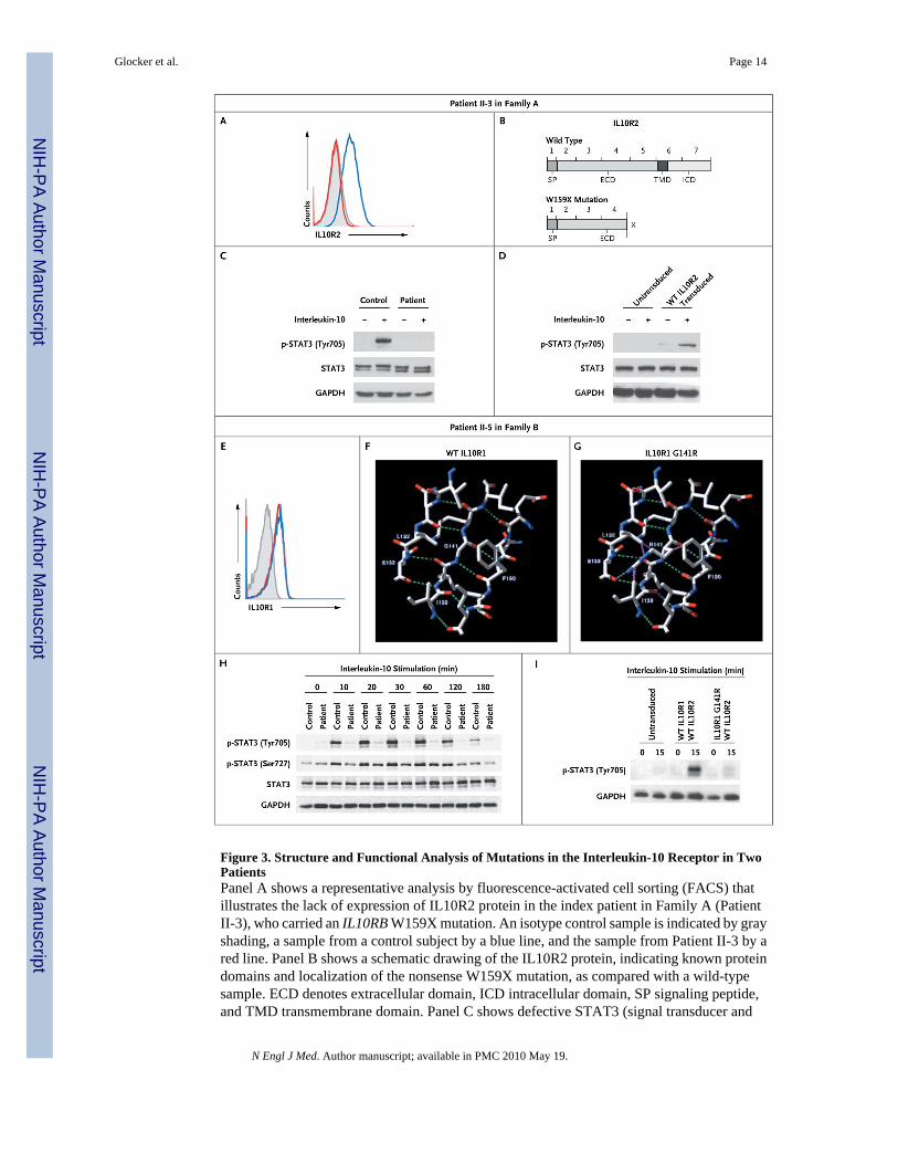
Figure 3. Structure and Functional Analysis of Mutations in the Interleukin-10 Receptor in TwoPatientsPanel A shows a representative analysis by fluorescence-activated cell sorting (FACS) thatillustrates the lack of expression of IL10R2 protein in the index patient in Family A (PatientII-3), who carried an IL10RB W159X mutation. An isotype control sample is indicated by grayshading, a sample from a control subject by a blue line, and the sample from Patient II-3 by ared line. Panel B shows a schematic drawing of the IL10R2 protein, indicating known proteindomains and localization of the nonsense W159X mutation, as compared with a wild-typesample. ECD denotes extracellular domain, ICD intracellular domain, SP signaling peptide,and TMD transmembrane domain. Panel C shows defective STAT3 (signal transducer and
Glocker et al. Page 14
N Engl J Med. Author manuscript; available in PMC 2010 May 19.
NIH
-PA Author Manuscript
NIH
-PA Author Manuscript
NIH
-PA Author Manuscript

activator of transcription 3) phosphorylation (p-STAT3) at the residue tyrosine 705 onstimulation with interleukin-10 in peripheral-blood mononuclear cells (PBMCs) from PatientII-3, as compared with intact phosphorylation in a control sample. Panel D shows thereconstitution of STAT3 phosphorylation on transduction of cell lines from Patient II-3, witha lentiviral vector encoding wild-type (WT) IL10R2. Panel E shows normal expression levelsof IL10R1 on FACS analysis in the index patient in Family B (Patient II-5), who carried anIL10RA G141R mutation. An isotype control sample is indicated by gray shading, a samplefrom a control subject by a blue line, and the sample from Patient II-5 by a red line. Panel Fshows a structural model of wild-type IL10R1. Residues Leu132, Glu133, Ile139, Gly141,Lys142, and Phe190 are labeled. Also shown are nearby residues that participate with Gly141or its neighbors in a beta-sheet conformation. The residues that are illustrated are those thatare reported to have contact with Gly141 in wild-type IL10R1 (according toRankViaContact31) or with Arg141 in the G141R mutation and that lie within 6 Å. Panel Gshows a structural model of the G141R mutation in IL10R1 (from Protein Data Bank structure1Y6K, chain R). Putative hydrogen bonds are shown as green dashed lines in Panels F and G,and steric clashes are shown as purple dashed lines in Panel G. Panel H shows a Western blotanalysis of STAT3 in PBMCs from Patient II-5 after stimulation for various periods withinterleukin-10, as compared with a control sample. Panel I shows a Western blot analysis ofphosphorylation of STAT3 at tyrosine 705 in interleukin-10–stimulated HeLa cells that wereretrovirally transduced with wild-type IL10R2 along with either wild-type IL10R1 or IL10R1with mutant G141R. Glyceraldehyde-3-phosphate dehydrogenase (GAPDH) was used as acontrol for the experiments in Panels C, D, H, and I.
Glocker et al. Page 15
N Engl J Med. Author manuscript; available in PMC 2010 May 19.
NIH
-PA Author Manuscript
NIH
-PA Author Manuscript
NIH
-PA Author Manuscript

Figure 4. Defective Down-Regulation of Proinflammatory Cytokines Mediated by Interleukin-10in Mononuclear Cells with a Mutation in the Interleukin-10 ReceptorPanel A shows defective inhibition of the release of tumor necrosis factor α (TNF- α) bycostimulation with interleukin-10 in lipopolysaccharide (LPS)–stimulated macrophages fromthe index patient in Family A (Patient II-3), as compared with the mean value from samplesobtained from five control subjects, as measured by enzyme-linked immunosorbent assay(ELISA). Panel B shows a similar ELISA measuring TNF- α secretion in LPS-stimulatedperipheral-blood mononuclear cells (PBMCs) from the index patient in Family B (Patient II-5)and three control subjects. Panel C shows defective interleukin-10–mediated suppression ofTNF- α secretion in LPS-stimulated cells from Patient II-5 in Family B. Panel D showsincreased secretion of proinflammatory cytokines upon stimulation of cells with LPS in PatientII-5, as compared with a healthy control subject. These cytokines include MIP-1α and
Glocker et al. Page 16
N Engl J Med. Author manuscript; available in PMC 2010 May 19.
NIH
-PA Author Manuscript
NIH
-PA Author Manuscript
NIH
-PA Author Manuscript

MIP-1β (macrophage inflammatory proteins 1α and 1β), MCP1 (monocyte chemoattractantprotein), and RANTES (regulated on activation, normal T expressed and secreted protein).Panel E shows the abrogated effect of interleukin-10 on the release of proinflammatorycytokines in cells from the patient that were costimulated with LPS and interleukin-10, ascompared with a healthy control subject. Cells from Patient II-5 show increased secretion ofinflammatory cytokines, which could not be counteracted by costimulation with exogenousinterleukin-10. Panel F shows defective interleukin-10–mediated induction of messenger RNAexpression in the suppressor of cytokine signaling 3 gene (SOCS3) in Patient II-3 in FamilyA; induction was more than four times as great in a control sample. Expression levels weremeasured relative to β-actin as a housekeeping gene. The I bars indicate standard errors.
Glocker et al. Page 17
N Engl J Med. Author manuscript; available in PMC 2010 May 19.
NIH
-PA Author Manuscript
NIH
-PA Author Manuscript
NIH
-PA Author Manuscript

Figure 5. Success of Allogeneic Hematopoietic Stem-Cell TransplantationIn Patient II-3 from Family A, who had severe anocutaneous fistulas that were resistant totherapy (Panel A), allogeneic stem-cell transplantation resulted in clinical amelioration of alleffects of disease (Panel B).
Glocker et al. Page 18
N Engl J Med. Author manuscript; available in PMC 2010 May 19.
NIH
-PA Author Manuscript
NIH
-PA Author Manuscript
NIH
-PA Author Manuscript