Politics of mobile Code - RePub, Erasmus University Repository
Upload
khangminh22Category
view
6download
0
INTERLEUKIN-3

CIP-DA TA KONINKLUKE BIBLIOTHEEK, DEN HAAG
Gils, Francisca Cornelia Johanna Maria van
Interleukin-3 I Francisca Cornelia Johanna Maria van Oils. -Amsterdam : Thesis Publishers Thesis Rotterdam. -With ref. -With summary in Dutch. ISBN 90-5170-240-X NUGI743 Subject headings: hemopoiesis I stem cells I interleukin.
Cover by Eddy V arekamp.

INTERLEUKIN -3
Interleukine-3
Proefschrift
ter verkrijging van de graad van doctor aan de Erasmus Universiteit Rotterdam
op gezag van de Rector Magnificus Prof. dr PWC Akkermans M. Lit. en volgens bes1uit van het college van dekanen
De openbare verdediging za1 plaatsvioden op woensdag 15 december 1993 om 11.45 uur
door
Francisca Cornelia Johanna Maria van Gils geboren te Breda

Promotor
Co-promotor
Referenten
: Prof. dr OW van Bekkum
:Dr G Wagemaker
: Prof. dr B Lowenberg Prof. dr UW Schaeffer
The work described in this thesis was performed in the Institute of Radiobiology of the Faculty of Medicine and Health Sciences, Erasmus University Rotterdam, in collaboration with the Radiobiological Institute TNO, Rijswijk and Gist-brocades NV, Delft, The Netherlands, and supported by project grants of the Dutch Cancer Society and by contracts of the Commission of the European Conununities. Printing costs were partly covered from generous contributions of the Dutch Cancer Society, CAM van Gils and Sandoz BV, Uden.

M ADCC AML ALL
BDA BFU-E BFU-MK BSA
CFU-C CFU-E CFU-GM CFU-MK CML
ELISA Epo
FACS FCS FITC
GAM-FITC GM-CSF G-CSF
HHBS HGF HSC
IL
MCA M-CSF MDS MGF MHC MNC
PHA PMA
RA RAEB RBC
SA-RPE SCF
TBI TGF TNF
List of abbrevations
aplastic anemia antibody dependent cellular cytotoxicity acute myeloid leukemia acute lymphoblastic leukemia
Blackfan-Diamond anemia burst-forming unit-erythroid burst-forming unit-megakaryocyte bovine serum albumin
colony-fanning unit in culture colony-forming unit-erythroid colony-forming unit stimulated by GM-CSF colony-forming unit-megakaryocyte chronic myeloid leukemia
enzyme-linked-immuno-absorbent assay erythropoietin
fluorescence activated cell separator fetal calf serum fluorescein isothiocynate
fluorescein-conjugated goat anti-mouse Ig granulocyte-monocyte colony-stimulating factor monocyte colony-stimulating factor
HEPES buffered Hanks balanced salt solution hemopoietic growth factor hemopoietic stem cells
interleukin
monoclonal antibody monocyte colony-stimulating factor myelo dysplastic syndrome mast cell growth factor (=SCF=KL=Steel factor=c-KIT ligand) major histocompatibility complex mononuclear cells
phytohaemagglutinin phorbol-myristate acetate
refractory anemia refractory anemia with excess of blasts red blood cells
R-phycoerythrin-conjugated streptavidin stem cell factor (=MGF=KL=Steel factor=c-KIT ligand)
total body irradiation transformation growth factor tumor necrosis factor
Contents
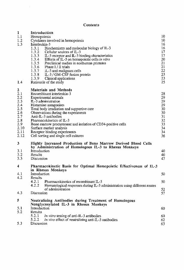
1 Introduction 1.1 Hemopoiesis 1.2 Cytokines involved in hemopoiesis 1.3 lnterleukin-3
1.3 .1 Biochemistry and molecular biology of IL-3 1.3 .2 Cellular sources of IL-3 1.3. 3 IL-3 receptor and IL-3 binding characteristics 1.3.4 Effects ofiL-3 on hemopoietic cells in vitro 1.3.5 Preclinical studies in nonhuman primates 1.3.6 Phasel/lltrials 1.3.7 IL-3 and malignant cells 1.3.8 IL-3 I GM-CSF fusion protein 1.3. 9 Clinical applications
1.4 Rationale of the study
2 Materials and Methods 2.1 Recombinant interleukin-3 2.2 Experimental animals 2.3 IL-3 administration 2.4 Histamine antagonists 2.5 Total body irradiation and supportive care 2.6 Observations during the experiments 2.7 Anti-IL-3 antibodies 2.8 Pharmacokinetics of!L-3 2. 9 Bone marrow procurement and isolation of CD34-positive cells 2.10 Surface marker analysis 2.11 Receptor binding experiments 2.12 Cell sorting and single cell cultures
3 Highly Increased Production of Bone Marrow Derived Blood by Administration of Homologous IL-3 to Rhesus Monkeys
3.1 Introduction 3.2 Results 3.3 Discussion
Cells
4 Pharmacokinetic Basis for Optimal Hemopoietic Effectiveness of IL-3 in Rhesus Monkeys
10 10 16 16 17 17 20 21 21 22 23 23 25
28 29 29 29 30 30 31 32 33 34 34 36
40 40 47
4.1 Introduction 50 4.2 Results
4.2 .1 Pharmacokinetics of recombinant IL-3 50 4.2.2 Hematological responses during IL-3 administration using different routes
of administration 52 4.3 Discussion 57
5 Neutralizing Antibodies during Treatment of Homologous Nonglycosylated IL-3 in Rhesus Monkeys
5.1 Introduction 60 5.2 Results
5.2.1 In vitro testing of anti-IL-3 antibodies 60 5.2.2 In vivo effect of neutralizing anti-IL-3 antibodies 62
5.3 Discussion 63

6
6.1 6.2
6.3
7
7.1 7.2
7.3
8 8.1 8.2 8.3
9
9.1 9.2
9.3
10
IL-3 Receptors on Rhesus Monkey Bone Marrow Cells: Species Specificity of Human IL-3, Binding Characteristics and Lack of Competition with GM-CSF Introduction Results 6.2.1 Species specificity of human IL-3 6.2.2 Binding characteristics ofiL-3 and competition with GM-CSF 6.2.3 Binding ofradiolaheled IL-3 to CD34-positive cells Discussion
Flow Cytometric Detection of IL-3 Receptors on Distinct Subsets of Peripheral Blood and Bone Marrow Cells in Normal and IL-3 Treated Rhesus Monkeys Introduction Results 7.2.1 7.2.2 7.2.3
Expression of IL-3 receptors during IL-3 treatment Disttibution of IL-3 receptors in IL-3 treated animals IL-3 receptor expression on immature cells in normal and IL-3 treated animals
Discussion
Acute Side Effects of IL-3 in Rhesus Monkeys Introduction Results Discussion
Mitigation of Radiation Induced Pancytopenia by IL-3 in Rhesus Monkeys Introduction Results 9.2.1 IL-3 administration before TBI 9.2.2 IL-3 after TBI 9.2.3 Progenitor cells in bone marrow after TB! Discussion
General Discussion
Summary & Samenvatting
References
Curriculum vitae
List of publications
Dankwoord
66
67 69 73 73
76
77 78
81 87
92 92
106
llO
ll1 liS 115 117
120
123
131
154
155


INTRODUCTION
9

1.1 Hemopoiesis
Bone marrow contains a small population of stem cells with extensive capacities to proliferate and mature into blood cells and a few specific tissue cells. Their direct descendants develop into lineage specific progenitor cells, while gradually losing their renewal and proliferative capacity in exchange for specialized functions, such as oxygen transport, phagocytosis, hemostasis and immune responses. Among the mature end cells, lymphocytes and monocytes retain the capacity to proliferate. Transplantation of immature hemopoietic cells and various culture systems allow for detailed studies in the regulatory mechanisms governing blood cell production. Those mechanisms include specific soluble proteins, termed growth factors, as well as signals provided by stromal elements of hemopoietic tissues such as bone marrow, spleen and thymus. Around twenty, in many cases pleiotropic, growth factors have been identified so far, which apparently control the different stages of blood cell production. The availability of monoclonal antibodies directed against surface antigens expressed by immature hemopoietic progenitor cells and the development of sophisticated cell separation methods (single cell sorting, immunomagnetic beads, streptavidin colomns) have led to the identification of specific subsets of immature progenitor cells, devoid of contaminating growth factor producing accessory cells, which offer the opportunity to evaluate the stimulatory capacity of each hemopoietic growth factor in detail (3,21,57,240). In vitro, hemopoietic progenitor cells can be studied in growth factor stimulated monocellular cultures, in which progenitor cells may proliferate to form a clone of mature cells, detectable as a colony, and therefore is called a colony-forming unit in culture (CFU-C). Progenitor cells can develop into lineage specific colonies depending on maturation stage and growth factors added: culture systems have been developed for blast cells (214) and progenitors of granulocytes, monocytes, erythrocytes (351), eosinophils, megakaryocytes (187,376) and lymphocytes (104,313). Transplantation of purified, defined subsets of immature cells is required to evaluate stem cell properties (19,20) and growth factor responses of these cells in vivo. Identification of growth factor receptor expressing cells is necessary to gain insight into the physiological role and to understand the hemopoietic and side effects of the corresponding growth factors, when applied in vivo.
1.2 Cytokines regulating hemopoiesis
Gene cloning and expression techniques have made growth factors, which may be distinguished into an early acting, pleiotropic and a more lineage restricted group, available as recombinant proteins (Table 1, Figure 1). This has initiated detailed studies on receptor signalling pathways, target cell specificity and possible clinical applications.
1 0

HSC committed progenitor cells mature end cells
IL1 IL·3 IL·6 SCF
I I
I
I I
I I
I r---. "S"'C"'F-c, I""L·-;;3Ec', ~i;'L--c-4:-, ""IL"""'·9")>"':
I IL-3, IL-6, GM·CSjl.l
SCF, IL-1, IL-11, : Epo? 1
I I
,- - - - - ---- - - ll-3 -- - - - )l--.1 1 ? I I
I I I
~/r ~!@) ~cs~lu
~~~ I
IL·3 __
GM-CSF IL·3, IL·5, GM·CS~:
I I I I I I
IL-3, SCF ---)>-1 I I
' I I \ ? \ I
\ ? I 1-------'--IL-4 IL-9 IL-10--~
' ' I
I I I I
IL·2, IL-10 )' 1 __./ I
I I
""'IL-3 IL·7 1 )>I
Figure 1 Schematic representation of blood cell formation
1 1
erythrocytes
thrombocytes
'
osteoclasts IL-1
monocytes IL-2, IL-3 M·CSF, GM-CSF
neutrophils IL·S, G·CSF, GM-CSF
eosinophils IL-3, IL-5, GM·CSF
basophils IL-1, IL-3, IL-5, GM·CSF
mast cells SCF
T lymphocytes IL·2, IL-4, IL-6, IL-7 IL-10, IL-12
B lymphocytes IL-2, IL-4, IL·S IL·6, IL-7, IL-11

Table 1 Human cytokines influencing hemopoiesis and function of peripheral blood cells
Factor
IL-l
IL-2
IL-3
IL-4
IL-5
Biological effects
Synergism with other HGF's Stimulation ofiL-l,IL-2,lL-3, IL-4,IL-5,IL-6, IL-7, IL-8, GCSF, GM-CSF, M-CSF, TNF and INF production Basophil histamine release B and T cell activation Enhancement of NK cell activity Platelet production Acute phase response Bone resorption
T cell proliferation Modulation function ofT and B cells, NK cells and monocytes Induction synthesis of other growth factors
Production of all bone marrow derived peripheral blood cells Osteoclast differentiation Mast cell production Enhancement of lgE stimulated histamine and IL-4 release by basophils
Activation of resting B cells lgE/lgG switch T cell proliferation Mast cell proliferation Synergism with IL-l and G-CSF in colony-formation Induction of inhibitory factors in stromal cells Inhibition of colony-formation by CFU-M
Proliferation of activated B cells Terminal maturation and activation of eosinophils Enhancement histamine release by basophils
1 2
cellular source
Monocytes Neutrophils BandT cells Endothelial cells Fibroblasts Synovial lining cells Dendritic cells
T cells
T cells Eosinophils Neutrophils Thyntic endothelial cells
T cells Mast cells Basophils
T cells
references
8,10,11,28,37 ,66, 75,112,174,204, 218,248,321,332, 337,338,347,355, 363,370,429
17,87,125,180, 181,234,256,260, 272,280,285,302, 312, 339, 340,361
9,13,14,26,41,42, 30-32,59,69,81, 94-97,103,113, 116, 161,179, 191, 192,194, 207,222, 231, 243-245, 247, 249, 286, 316,327, 341, 358, 360,377, 379, 387, 393, 412-414, 416
29,39,41,71,153, 154,173,176,264, 281,294,295, 297-300,306,343
23,59,60,188, 224, 227, 341

Factor Biolol.\!cal effects cellular source references
IL-6 Final B cell mattrration BandT cells 6,120,150,163, Induction of immunoglobulins Monocytes 169,193,213,230, Anti-viral activity Fibroblasts 241,337,346 Maturation of megakatyocytes Keratinocytes Platelet production Endothelial cells Production acute phase proteins Astrocytes Synergism with IL-3 and M-CSF BM stromal cells in colony-formation Mesangial cells Co-stimulator ofT cells
IL-7 Proliferation of early B cells, Stromal cells 127,128,139,262, immattrre and activated T cells 317,398,401
IL-8 Activation and chemoattractor of Macro phages 70,132,207,210, neutrophils Lymphocytes 220,23 8,252,301' Inhibition of IL-3 induced Endothelial cells 309,325,353,367' histamine release of basophils Hepatocytes 422,423,427
Fibroblasts Keratinocytes Mesangial cells
IL-9 Colony-formation by BI'U-E T lymphocytes 80,151,342,418 In synergy with IL-3 proliferation of mast cells
IL-10 T cell differentiation BandT cells 259,329,357,388, Co-growth factor of !L-2 in T cell 389 proliferation Mast cell proliferation
IL-11 Proliferation of mature B cells Stromal cells 185,293,365 Formation of megakatyocyte colonies in synergy with IL-3 (mutine)
IL-12 Proliferation of activated T cells B lymphocytes 118,138,352 and NK cells
Stem cell Synergism with Epo in colony- Bone marrow stromal l ,2,9,24,31 ,40,68, factor formation by BI'U-E cells 155,236,245,278,
Synergism with other HGFs in Fibroblasts 290,403,431,430 colony-formation Rat liver cells Mast cell proliferation Histamine release by mast cells
Erythro- Colony-formation by late BFU-E Kidney cells 68,74,90, 110,130, poietin and CFU-E Liver cells 165,201,202,266,
Survival CI'U-E 351,364 Megakatyocyte colony-formation
I 3

Factor Biolo!£cal effects cellular source references
G-CSF Neutrophil production Monocytes 46,73,97 ,141, 195, Neutrophil function Fibroblasts 199,226,263,270,
Mesothelial cells 273,274,345,354, Endothelial cells 399,424,429
GM-CSF Proliferation and maturation of BandT cells 16,37,52,58,73, myeloid progenitor cells Monocytes 79,98,106,142, Activation of mature effector cells Mast cells 149,172,182,226, Platelet production Endothelial cells 242,244,251,258, Induction of TNF and IL-l Fibroblasts 266,277,303,333,
Mesothelial cells 344,354,366,369, 375,405,429
M-CSF Monocyte production, Monocytes 15,184,287,308, differentiation and survival Fibroblasts 349,348,395,410 Stimulation cytokine production Endothelial cells by monocytes (IFN,TNF,CSF)
Cytokines that support stem cell survival in long term culture and stimulate the generation of CFU-C from immature cells include IL-l, IL-3, IL-6, GM-CSF and the lately cloned stem cell factor (SCF), also termed c-KIT ligand (KL), mast cell growth factor (MGF) or Steel factor (278,403). Each of these growth factors alone has only limited effects on colony-formation, but strong synergistic responses are found upon combination (26,27,162,163,200,213,244,245.344,347,413). Commitment of immature progenitor cells and final maturation is regulated by these early acting growth factors as well and by more lineage specific growth factors. Erythropoiesis is stimulated by SCF (68,278,290), IL-3 (316), IL-4 (300) and IL-9 (80.151,342) in combination with erythropoietin (Epo), which results in enhanced colony-formation by BFU-E. Erythropoietin alone exerts its action on more mature progenitor cells (CFU -E) which are strictly restricted to the erythroid lineage (351,364). Megakaryocytopoiesis is supported by IL-3 (32,365) and GM-CSF (32,42), whereas SCF (9,31), Epo (32), IL-l (32), IL-4 (32), IL-6 (32,42,169,271,307,346) and IL-11 (365) act in synergy with other growth factors on colony-formation by BFU-MK and CFU-MK. Pre B-cell proliferation is stimulated by IL-3 (360,412) and IL-7 (262.317). The activation and proliferation of B-cells is enhanced by IL-2 (256), IL-4 (71,297 .299,306), IL-5 (420), IL-7 (128) and IL-11 (293), whereas IL-6 provides the stimulus for final maturation into immunoglobulin secreting B-cells (150). T lymphocytes are triggered to proliferate by IL-2 (260), IL-4 (154,400), IL-6 (230),
I 4

IL-7 (262) and IL-12 (118). Differentiation ofT lymphocytes occurs under the influence of IL-2 (134,340) and IL-10 (54,232). The maturation of the granulocyte-monocyte progenitor cells into the neutrophilic direction is stimulated by GM-CSF (16,244,251) and G-CSF (16,97,158,244). IL-4 enhances the effects of G-CSF (300). The function of neutrophils is stimulated by IL-8 (309,423), G-CSF (195,270,283,424,425) and GMCSF (242,270,375,424). IL-3, IL-5 and GM-CSF (375) induce differentiation into the eosinophilic lineage (59,60,97) and all three enhance mature end cell function (113,224,227). The maintenance (414) and differentiation (191) of basophilic granulocytes is influenced by IL-3, whereas mature end cell function is stimulated by IL-l (355), IL-3 (224,231,247), IL-5 (23) and GM-CSF (224). Monocyte differentiation is regulated by M-CSF, IL-3 and GM-CSF (428), and monocyte function by IL-2 (234), M-CSF, IL-3 and GM-CSF (24,95,142,426). SCF potentiates mediator release by human lung mast cells. Osteoclast differentiation is influenced by IL-3 (14), whereas osteoclast function i.e., bone resorption is enhanced by IL-l (75). The regulation of the cytokine production and tuning of their effects are supposed to be essential in maintaining the balance of normal hemopoiesis. Several mechanisms may be involved: 1. Indirectly inhibitory or stimulatory cytokines: IL-10 inhibits cytokine production by activated monocytes (107,389) and IL-4 has been shown to stimulate stromal cells to produce a factor inhibiting colony-formation by CFUM (173,298). The production of IL-4 by basophils primed by IL-3 in response to IgE is enhanced (41). IL-l stimulates the production of several other cytokines (Table 1). 2. Naturally occurring receptor antagonists: the IL-l receptor antagonist (ILlRa) (92) can compete with IL-l for receptor binding, without transducing a signal (91). IL-lRa antagonizes the biological effects of IL-l in vivo and in vitro (4,101,143,282). 3. Enhancement or reduction of growth factor receptor expression: IL-l enhances the expression of the common~ subunit shared by IL-3, IL-5 and GMCSF on TF-1 cells (396). TGF~ (85) and IL-l (239) have been reported to reduce IL-lR expression. 4. Binding circulating growth factors to soluble receptor molecules: in serum, soluble IL-2R (315,328) and in urine, IL-6R (279) have been detected. Cloned IL-7R have been shown to compete with cell surface bound receptors for IL-7 binding (127). 5. Membrane-bound growth factors: M-CSF (350), SCF (1), and IL-l (208) are expressed membrane-bound, in this manner localizing the cytokine effect by direct cell-cell contact.
I 5

1.3 Interleukin-3
1.3.1 Biochemistry and molecular biology of IL-3
The gene encoding murine IL-3 has been isolated from the cell line WEHI-3B (114) and from T lymphocytes activated with concanavalin A (421). Comparison of sequences of the murine and rat (61) eDNA showed that the gene was not highly conserved between species. Therefore, the search for the human counterpart proved difficult. It was speculated that the gene encoding IL-3 had been lost during primate evolution and that its function in humans had been taken over by GM-CSF (333). Eventually, however, human IL-3 has been cloned, expressed and purified to homogeneity simultaneously by two different groups. Yang et al. (416) have used a gibbon IL-3 eDNA to probe a human fetal liver genomic library. Dorssers et al. (81) have used a eDNA library transcribed from mRNA of mitogen stimulated peripheral blood cells to isolate the human gene. The gene consists of 5 exons and 4 introns. The deduced mature protein consists of 133 amino acids, with 2 N-linked glycosylation sites and 2 highly conserved cysteine residues, which are important for protein folding and biological activity. There is 45% homology in the coding region and 25% protein homology between murine and human IL-3, which is low compared to other growth factors and iodicates an unusual rate of evolutionary divergence. Since human IL-3 exerted only limited effects on the production of peripheral blood cells when administered to rhesus monkeys (Macaca mulatta) (79,243) or cynomolgus monkeys (Macaca fascicularis) (205) the gene encoding rhesus monkey IL-3 was cloned using aM. mulatta genomic library and hybridization with a human eDNA IL-3 probe. The deduced mature protein, expressed in Bacillus licheniformis consists of 124 amino acids with 1 potential N-linked glycosylation site and 2 conserved cysteine residues and differs in 23 amino acids from the remaining mature human IL-3. Homologous IL-3 was shown to be 100-fold more active in stimulating hemopoietic colony-formation in vitro by purified rhesus monkey bone marrow cells than its human counterpart. Rhesus monkey IL-3 gene shares 93% nucleotide sequence homology with the complete human IL-3 sequence, whereas the mature protein homology was found to be 80%. Comparison of the coding sequences of rhesus monkey IL-3 to those of mouse, rat, gibbon and human revealed a high rate of nonsynonymous nucleotide substitutions. The high rate of amino acid substitutions during evolution explains the pronounced species specificity encountered for IL-3 (45). The gene encoding human IL-3 has been located on a small segment of the long arm of chromosome 5 (216): the cytokioe gene cluster, a region containiog the genes encoding IL-4, IL-5 (359), IL-9, GM-CSF (156) and the M-CSF receptor (276,314) as well (198,217,219,417). The gene encoding M-CSF was also though to be localized at chromosome 5, but recently it was shown that the M -CSF gene maps at chromosome 1 (211,261). Deletion of the long arm of chromosome 5 is found in various hematological disorders and has been associated with previous
1 6

exposure to chemotherapeutic, especially alkylating, agents and/or radiation. Characteristic for the so called 5q- syndrome are refractory anemia (RA), macrocytosis and poorly lobulated megakaryocytes. Furthermore, deletion of the long arm of the 5q chromosome has been found in refractory anemia with excess of blasts (RAEB), CML, AML, polycythemia vera, essential thrombocytaemia and myelodysplasia (review in 407).
1.3.2 Cellular sources of human IL-3
IL-3 mRNA is expressed and IL-3 is produced by T lymphocytes stimulated with PHA, PMA, IL-l or IL-2 (137,275,287,405). IL-3 mRNA could be detected in infiltrating cells in skin biopsies from atopic patients after allergen challenge (69). Cell cultures of thymic endothelial cells can produce growth factors, capable to stimulate colony-formation by bone marrow cells, which could be partially inhibited by anti-IL-3 antibodies (69). Eosinophilic granulocytes and to a lesser extent neutrophilic granulocytes from subjects suffering from hay fever have been reported to release IL-3 upon stimulation with PMA or ionomycin (186). IL-3 mRNA expression could not be detected in monocytes and hemopoietic stromal cells (fibroblasts and endothelial cells) (275). In serum of healthy individuals, patients suffering from MDS (432) or patients subjected to autologous peripheral blood stem cell transplantation (183), detectable levels of IL-3 could not be demonstrated.
1.3.3 IL-3 receptor and binding characteristics
Genes encoding hemopoietic growth factor receptors (R) are cloned and characterized and can be divided into two major families (figure 2). IL-l R (83,335), M-CSFR (314,330) and c-KIT (SCFR) (419) can be classified as members of the immunoglobulin superfamily on the basis of homologies in their extracellular ligand-binding domain, i.e., immunoglobulin-like loops. The intracellular part of the M-CSFR and c-KIT contains a tyrosine kinase sequence. The second group is called the hematopoietin or cytokine receptor superfamily; IL-2Rj3 (63,145), lL-3R (197), IL-4R (157), IL-5R (267.362), IL-7R (127), GMCSFR (119,147), and EpoR (175.406) belong to this family, which is characterized by a major region of homology in the extracellular ligand-binding domain; at the N-terminal side, cystein residues are conserved and, adjacent to the transmembrane part, a tryptophan-serine-x-tryptophan-serine motif. The intracellular part of the receptors does not contain any consensus sequence for known signal transducing molecules such as protein kinases. GM-CSF and IL-3 have been shown to induce rapid tyrosine phosphorylation of several substrates through activation of an unknown tyrosine kinase (102,105,126,166,167 ,177 ,203) and also rapidly activate several serine/threonine kinases involved in mitogenic signal transduction, such as Raf-1 (178) and mitogen-activated protein kinase-2 (140,284).
1 7

immune globulin superfamily
cytokine superfamily
ll-1 R M-CSFR c-kit
IL-6R a (gp 80)
Immune globulin-like domain
IL-6R ~ (gp130) G-CSFR
shared ~-subunit
IL-2R ~ EpoR IL-3a IL-4R ll-5R a
GM-CSFR a
ll-7R
• I lli5l g
intracellular domain; not homologous with any known protein kinase sequence
intracellular domain with tyrosine kinase sequence
modified fibronectine type Ill-like domain, with WSXWS-motif
1!:> cystein residue
D fibronectine type Ill-like domain
EJ modified fibronectine type Ill-like domain, with WS-motif
Figure 2 Structure of growth factor receptors
IL-6R (415) and G-CSFR (212,268) possess characteristics of both families (62,129); the N-terminal amino acids of the mature receptors align with the IgG superfamily C2 domain, followed by a domain characteristic for the hematopoietin receptor superfamily. IL-3 receptors are complex, heterogeneous (291) and are differentially expressed on different cell types (Table 2).
I 8

Table 2 Receptor binding characteristics and cross~competition of
IL-3, GM-CSF and IL-5*
IL-3 receEtor GM-CSF receEtor IL-5 receEtor
Kd (Emoi/L) sites/cell Kd(Emoi!L) sites/cell Kd(£IDOI/L) sites/cell
NEUTROPHILS no 70 200 no 363 2155 363 2720 405 1949 469 2343
Competition IL-5 no IL-3 no
EOS!NOPHILS 440 370 36 520 58 218 470 940 53 840 494 1950 636 1990
Competition GM-CSF ± ± IL-5 no,± no,± IL-3 ±± ±
BASOPHILS 80 710 39 190 present 160 2100 230 500
Competition GM-CSF no ± IL-5 ± ± IL-3 + ±±
MONOCY1ES 8 95 5 35 none 10 186 9 8 27 122 10 67 38 580 19 78 40 143 39 14 82 115 576 431
513 467 588 307 815 179 991 657 939 5274 1120 130
2800 5185 Competition GM-CSF ± IL-5 no ? IL-3 ±,+
* Each pair of Kd values and sites per cell representing individual observations. ±,±± and+ indicate partial, almost complete and complete competition with the radiolabeled ligand for receptor binding.( data substracted from ref.43,44,93,95,223,224,229,377)
I 9

A functional high-affinity heterodimer receptor structure has been identified, combining a ~-chain, shared by IL-5, GM-CSF and IL-3 receptors and a growth factor specific a-chain of low binding affinity (119,147,197,254,362). In addition, in mice, a second, closely homologous ~-chain has been identified (144,171,254). In contrast to the ~-chain, common to the IL-5, GM-CSF and IL-3 receptor, that by itself does not bind growth factors, the IL-3 specific ~-chain binds IL-3 with low affinity. Co-expression of the two f)-chains in hemopoietic cells of mice has been uniformly observed and so far does not account for differential expression of receptors on different cell types (255). Functional specificity, therefore, should be attributed to the expression of growth factor specific a-chains. Results obtained from IL-3 receptor binding studies using human cells, mostly cell lines, leukemic cells or mature blood cells and from binding studies using cell lines reconstituted with the cloned subunits (196), are in agreement with this model, that predicts a variety of expression and specificity patterns. Indeed, primary human monocytes were shown to have high-affinity receptors that bind specifically IL-3 or GM-CSF and a third type that binds both (43,93,95,291). Human CML basophils were found to bind IL-3 but not GM-CSF (377), or, alternatively, strongly bound IL-3 with weaker binding of GM-CSF and IL-5 (224). IL-3, GM-CSF and IL-5 appeared to cross-compete for binding to human eosinophils (229), whereas neutrophils bound GM-CSF but not IL-3 and IL-5 (43,95,229,377). On AML blasts, low-affinity GM-CSF receptors did not bind IL-3, but a dual high-affinity receptor type bound both GM-CSF and IL-3, while a high-affinity specific IL-3 receptor was also demonstrated (43). Little information is available for normal human bone marrow cells, except that a low number of receptors (55/cell) has been demonstrated (291). The receptor expression is consistent with the ability of the corresponding growth factors to modulate mature end cell function.
1.3.4 Effects of human IL-3 on hemopoietic cells in vitro
IL-3 stimulates colony-formation by immature progenitor cells. Using unsorted bone marrow cells, IL-3 is capable to induce colony-formation by immature progenitor cells of myeloid, erythroid and megakaryocytic lineages (2,9,13,26,31, 30,32,42,123,249,327,365,387). Colony-formation by sorted human CD34-positive cells in the presence of IL-3 alone showed a restricted stimulatory spectrum and the necessity of accessory cells or growth factors acting on more mature progenitor cells to restore the broad stimulatory spectrum of colony-formation (26). Single cell sorted human CD34-positive cells cultured in serum-depleted medium showed no positive wells in the presence of IL-3. Again, combination with other growth factors was needed to promote cluster growth (413). IL-3 is a differentiation factor for osteoclasis (14), pre B-cells (360,412), eosinophils (59,97,341), basophils (191,192,379) and mast-like cells (13,!79,191.192). In the neutrophilic myeloid series the responsiveness to IL-3 is lost with differentiation (222).
20
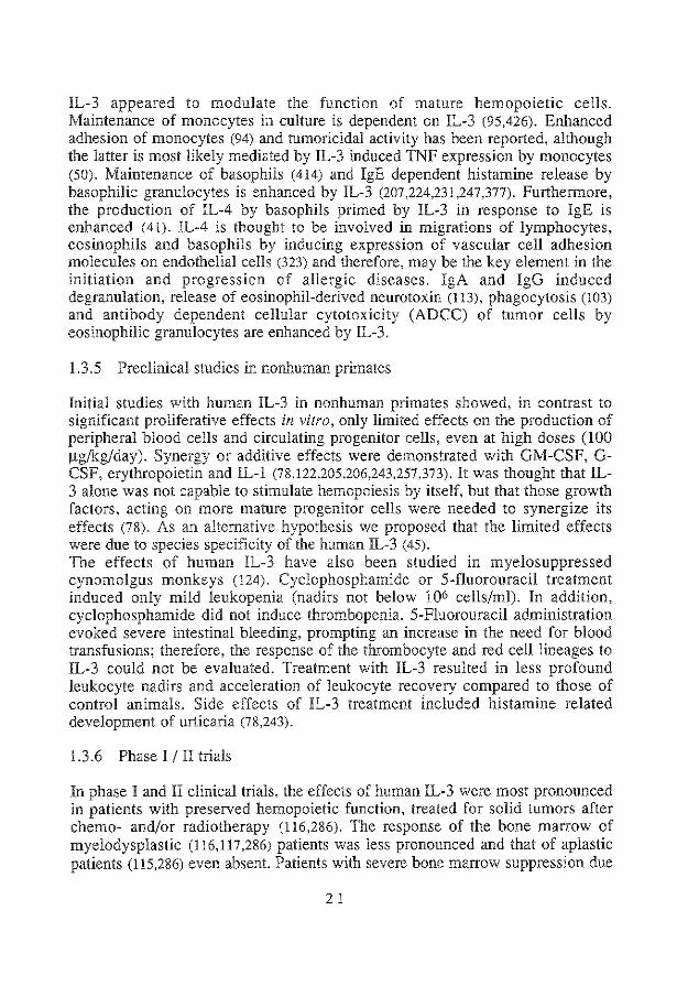
IL-3 appeared to modulate the function of mature hemopoietic cells. Maintenance of monocytes in culture is dependent on IL-3 (95,426). Enhanced adhesion of monocytes (94) and tumoricidal activity has been reported, although the latter is most likely mediated by IL-3 induced TNF expression by monocytes (50). Maintenance of basophils (414) and IgE dependent histamine release by basophilic granulocytes is enhanced by IL-3 (207,224,231,247,377). Furthermore, the production of IL-4 by basophils primed by IL-3 in response to IgE is enhanced (41). IL-4 is thought to be involved in migrations of lymphocytes, eosinophils and basophils by inducing expression of vascular cell adhesion molecules on endothelial cells (323) and therefore, may be the key element in the initiation and progression of allergic diseases. IgA and IgG induced degranulation, release of eosinophil-derived neurotoxin (113), phagocytosis (103) and antibody dependent cellular cytotoxicity (ADCC) of tumor cells by eosinophilic granulocytes are enhanced by IL-3.
1.3.5 Preclinical studies in nonhuman primates
Initial studies with human IL-3 in nonhuman primates showed, in contrast to significant proliferative effects in vitro, only limited effects on the production of peripheral blood cells and circulating progenitor cells, even at high doses (100 J.lg/kg/day). Synergy or additive effects were demonstrated with GM-CSF, GCSF, erythropoietin and IL-l (78,122,205,206,243,257,373). It was thought that IL-3 alone was not capable to stimulate hemopoiesis by itself, but that those growth factors, acting on more mature progenitor cells were needed to synergize its effects (78). As an alternative hypothesis we proposed that the limited effects were due to species specificity of the human IL-3 ( 45). The effects of human IL-3 have also been studied in myelosuppressed cynomolgus monkeys (124). Cyclophosphamide or 5-fluorouracil treatment induced only mild leukopenia (nadirs not below !06 cells/ml). In addition, cyclophosphamide did not induce thrombopenia. 5-Fluorouracil administration evoked severe intestinal bleeding, prompting an increase in the need for blood transfusions; therefore, the response of the thrombocyte and red cell lineages to IL-3 could not be evaluated. Treatment with IL-3 resulted in less profound leukocyte nadirs and acceleration of leukocyte recovery compared to those of control animals. Side effects of IL-3 treatment included histamine related development of urticaria (78,243).
1.3.6 Phase I I II trials
In phase I and II clinical trials, the effects of human IL-3 were most pronounced in patients with preserved hemopoietic function, treated for solid tumors after chemo- and/or radiotherapy (116,286). The response of the bone marrow of myelodysplastic (116,117,286) patients was less pronounced and that of aplastic patients (115,286) even absent. Patients with severe bone marrow suppression due
2 1

to chemo- and/or radiotherapy responded (116,286,304) in a similar way as patients with preserved hemopoietic function, although in a delayed manner. IL-3 treatment has been given to patients suffering from Blackfan-Diamond disease (congenital pure red cell aplasia) (86). All six patients responded with a rise in leukocyte and thrombocyte counts upon IL-3 administration of 60 J.lgfm2 for 4 consecutive weeks. Two patients required fewer transfusions or turned transfusion independent for several months. In the responsive patients, erythroid maturation beyond the proerythroblast stage and a close to normal percentage of erythroid precursors in bone marrow samples were observed during IL-3 treatment. Administration of IL-3 during five consecutive days followed by GM-CSF for ten consecutive days has been applied to fourteen patients with a malignancy that was refractory to standard anticancer therapy or for which no proven effective treatment had been established (286). All patients showed preserved hemopoietic function and had previously been treated with IL-3 alone. Combined treatment had equivalent effects on myelopoiesis as fifteen days of monotherapy with GMCSF or IL-3 and the effect of combined stimulation on thrombopoiesis was similar to that of IL-3 alone. The results obtained from these studies are difficult to interpret, since, as inherent to clinical studies, the patient populations were heterogeneous and most of the studies were designed to investigate toxicity of IL-3 treatment. Reported transient side effects of IL-3 in human patients included fever, chills, headache, neck stiffness, bone pain, rash, erythema at injection sites, urticaria, facial flushing, edema, dyspnea, nausea, vomiting and, at the highest dose levels used, thrombopenia.
1.3.7 IL-3 and malignant cells
In vitro, IL-3 has been show to stimulate proliferation and maturation of AMLblasts, although the responses were heterogeneous and did not always correlate with IL-3R expression (13,43,44,292,296,319). In some cases no response to IL-3 was seen, although IL-3 receptors were present. The underlying mechanism is unclear. The unresponsiveness of the IL-3R-positive AML cells may have been caused by defects in signal transduction. Responsiveness to IL-3 of AML cells, which virtually did not express IL-3 receptors, was observed as well and can be explained by assuming that these cells expressed IL-3 receptors below the detection level of the assay. Malignant B-cell precursors in ALL, but not T -cell ALL (292) have been shown to express IL-3R (372). Correlation of IL-3 receptor expression with proliferative response could be demonstrated in a proportion of the patients, whereas stimulation with GM-CSF could not be detected (237 ,372,412). IL-3R expression and proliferative response to IL-3 have been demonstrated in follicular B-cell lymphoma (18). Furthermore, IL-3 stimulated clonal growth of
22

cell lines derived from colon adenocarcinoma (18) and small cell lung carcinoma (386) were reported. In vivo, IL-3 induced a transient rise in circulating atypical B lymphocytes in a patient with follicular, mixed, small-cleaved large-cell lymphoma and in one with diffuse large-cell lymphoma. One patient with RA exhibited progression to RAEB with transformation to acute leukemia while on study (116,117).
1.3.8 IL-3 I GM-CSF fusion protein
Growth factors can be engineered by modification of the coding DNA to display specific binding domains and to develop more potent agonists or antagonists. To develop so called second generation proteins, genes encoding analogs (228) and fusion proteins were constructed (67,404). The coding regions of GM-CSF and IL-3 were connected by a synthetic linker sequence followed by subsequent expression in yeast, resulting in a fusion protein (PIXY-321), which showed to have similar binding characteristics on cell lines expressing GM-CSFR (HL-60) or IL-3R (JM-1) compared to GM-CSF or IL-3. Using cell lines expressing both growth factor receptors (AML-193 and KG-1) a 10 to 20 fold higher affinity could be demonstrated compared to each growth factor alone (404). Proliferation of the AML-193 cell line and colony-formation by BFU-E and CFU-GM of normal bone marrow cells were enhanced likewise by the fusion protein in comparison with each growth factor or the combination of individual growth factors (67). In vivo, administration of this protein might be useful as it combines the rapid response to GM-CSF and the sustained multi-lineage response to IL-3 and may be superior to administration of both factors simultaneously, since some binding studies demonstrated reciprocal competition between those factors.
1.3.8 Clinical applications
Only few of the many therapeutic possibilities of growth factors have been clinically explored. Erythropoietin has been proven useful in substitution therapy in end-stage renal diseases to correct anemia (99,100,374). G- and GMCSF have successfully been used to shorten the pancytopenic phase following chemotherapy and/or TBI (5,318). All three growth factors are presently registered for the above mentioned indications. The potential of IL-3 to stimulate hemopoiesis and enhance hemopoietic function makes it a useful tool for several clinical applications (Table 3). In chemotherapy and radiation induced pancytopenia IL-3 may be useful through two mechanisms. First, pretreatment with IL-3 may expand the stem cell pool in the bone marrow, resulting in a larger surviving fraction following mye!o-ablation and second, IL-3 treatment following myelo-ablation may lead to accelerated regeneration of stem cells (endogenous, autologous or allogeneic) and thereby in both cases
23

Possible clinical applications of IL-3
BONE MARROW TRANSPLANTATION
- PB progenitor cell harvesting - accelerated regeneration
HEMOPOIETIC DISORDERS
bone marrow failure: primary -congenital (DBA, cyclic neutropenia) -MDS,AA
secondary - radiation- or chemotherapy induced - BM infiltration
malignancies: -leukennia -lymphoma
ENHANCED IMMUNOLOGICAL FUNCTION
-infections -AIDS -tumors
IN VITRO multiplication of stem cells
shortening the pancytopenic phase. Expansion of circulating progenitor cells can facilitate bone marrow harvesting for transplantation purposes. IL-3 may be a differentiation and/or proliferation factor for leukemias and lymphomas: triggering malignant cells into S-phase renders them more susceptible for cytotoxic agents. As observed in phase I and II trials, IL-3 stimulates hemopoiesis in bone marrow failure (BDA, MDS, AA and secondary bone marrow failure). IL-3 may be useful in patients with decreased immunological function (AIDS) or with increased demand for immunocompetent cells (bums, abdominal surgery, solid tumors). In vitro, IL-3 can be used for multiplication of immature progenitor cells, e.g., to facilitate genetic modification of stem cells by retroviral gene transfer. The design of optimal dose schedules and combination of IL-3 with growth factors that act on lineage restricted progenitor cells need to be investigated in more detail for each application. Combination therapy allows selective expansion of desired cell types
24

and has the advantage of employing relatively low doses of both growth factors, thereby reducing the risks of side effects and antibody formation.
1.4 Rationale of the present study
IL-3 is thought to stimulate proliferation and differentiation of immature hemopoietic progenitor cells. In vitro studies using purified progenitor cells showed that IL-3 acts in conjunction with other growth factors, acting on more mature progenitor cells, to stimulate proliferation of the broad spectrum of colony-forming cells. This may indicate that IL-3 acts on immature progenitor cells and subsequent differentiation and proliferation requires the action of growth factors, acting on more mature progenitor cells. It may also be possible that IL-3 exerts its action through indirect mechanisms, such as stimulation of accessory cells to produce growth factors. IL-3 binding studies have shown the presence of IL-3 receptors on different hemopoietic end cells, leukemic cells and cell lines, whereas little infonnation is available for IL-3 receptor expression on nonnal bone marrow cells. The presence of IL-3 receptors on immature progenitor cells has not been established yet. Preclinical studies in which human IL-3 was administered to cynomolgus and rhesus monkeys showed only limited effects of IL-3 on the production of hemopoietic cells. This can be explained by the pronounced species specificity of IL-3 due to the unusual rate of evolutionary divergence of the IL-3 gene. Therefore, these studies are not fully predictive for the outcome of clinical studies with IL-3. The results obtained from phase I and II clinical trials give only an impression of the toxicity and the possibilities of the use of IL-3, but do not provide infonnation of its efficacy. Initial clinical trials showed that IL-3 was capable to stimulate hemopoiesis, prominently so in patients with preserved hemopoietic function after chemoand/or radiotherapy and less pronounced in patients suffering from myelodysplasia or aplasia. This indicates that a minimum number of functionally normal bone marrow precursor cells is required to respond to growth factor stimulation. This has been directly demonstrated in preclinical studies in which rhesus monkeys were subjected to graded doses of total body irradiation (TBI) in the range of 4 to 10 Gray (Gy). GM-CSF was administered to rhesus monkeys after TBI to investigate its efficacy in preventing or mitigating pancytopenia. The results showed that GM-CSF is fully capable of preventing leukopenia after 5 Gy TBI, which is equivalent to about a 2 log reduction in hemopoietic stem cells (HSC), but is ineffective after TBI doses higher than 8 Gy, which is equivalent to approximately a 3 log HSC reduction (402). The nature of the reported side effects in humans and rhesus monkeys suggests that IL-3 is involved in acute type hypersensitivity reactions, which is supported by the in vivo IL-3 induced production of basophilic and eosinophilic granulocytes and by the in vitro results demonstrating that IL-3 is capable to enhance the production of histamine and IL-4 by basophils triggered by IgE.
25

In this study, rhesus monkeys were used as a preclinical model to predict the biological effects and possible therapeutic applications of recombinant IL-3 in humans. Homologous IL-3 was administered to healthy animals to evaluate its dose-response relationships, pharmacokinetics, most effective route of administration, stimulatory spectrum and side effects. The side effects were also subjected to histopathological analyses. The efficacy of IL-3 in mitigation of pancytopenia induced by cytoreductive therapy was studied in animals subjected to 5 Gy total body irradiation. To gain more insight in the mechanisms by which IL-3 induces its hemopoietic and side effects IL-3 receptor binding studies, using both radiolabeled and biotin-labeled IL-3, were performed on bone marrow and peripheral blood cells of normal and IL-3 treated animals.
26

MATERIALS AND METHODS
27

2.1 Recombinant Interleukin-3
Genes encoding the human and rhesus monkey interleukin-3 (IL-3) were cloned in our laboratory (45,81) and expressed in B. licheniformis by Gist-Brocades, Delft, The Netherlands (384). The excreted, functional proteins were purified to homogeneity and used in the in vitro as well as in vivo experiments. Both species of IL-3 were nonglycosylated, with a molecular weight of 15 kDa (Figure.!); the specific activity was determined by 50% stimulation of AML-193 cells at l-3 ng/ml of lL-3 (45). The endotoxin levels as measured by the limulus assay were less than I U/ml.
M M
' ' ::! -'
c "' ~ ,_ ~ . "' • ~"" E :;; • c :;; , ,cO
.c ..J c E ..J
kDa kDa
- 94 - 94
- 67 - 67
- 43 - 43
- 30 - 30
- 20.1 - 20.1
- 14.4 - 14.4
Figure 1 Sodium dodecyl sulphate-polyacrylamide gel electrophoresis (SDS-PAGE) of human and rhesus monkey IL-3, demonstrating the presence of single Coomassie brilliant blue stained bands with a purity >99%.
28

2.2 Experimental animals
Rhesus monkeys (Macaca mulatta) were bred in the Primate Center, ITRI-TNO, Rijswijk, The Netherlands (385). Young adult animals, weighting 2.5-4 kg and between 2-4 years of age were used for the experiments. All animals were seronegative for SIV, herpes B and STLV as tested at the Virus Reference Laboratory, Southwest Foundation for Biomedical Research, San Antonio, Tx, USA. Animals were free of intestinal parasites, as analyzed in faeces in the bacteriological department of ITRI-TNO and were kept in reverse barier isolation throughout the experiments. All animals were typed for major histocompatibility (MHC) antigens, using standard serological tests (385).
2.3 IL-3 administration
Continuous intravenous (iv) administration of IL-3 was used in several experimental settings. A Port a Cath® system 427 CS (Pharmacia) was implanted subcutaneously and was entered into a jugular vein. To prevent destruction of the system a special jacket was designed in which an inside pocket could contain a miniature insulin pump (Dahedi Elektronics, Maarssen, The Netherlands), providing a continuous infusion throughout the experiments (393,402). IL-3 was renewed every day; biological activity was periodically tested and was stable for more than 24 hours as tested in an IL-3 dependent cell line (M07E (34) or AML-193 (45)). Doses of 1, 10 or 30 1-!g/kg/day IL-3 were tested. For subcutaneous (sc) administration, the daily dose of IL-3 was injected at the dorsal part of the trunk once daily (qd) or equally divided over 24 hours into two (bid) or three (tid) injections. Doses of 3, 10, 30 or 100 1-!g/kg/day IL-3 were tested. The first day of IL-3 administration in unirradiated animals was designed as day 0. In irradiated animals IL-3 administration was stopped at the day of TBI or, alternatively, was started the day after TBI.
2.4 Histamine antagonists
H 1 histamine antagonist (ceterizine, Zyrtec®, UCB Farma, Breda, The Netherlands) or H2 histamine antagonist (cimetidine, Tagamet®, Smith, Kline and French, Rijswijk, Holland) were given to nine IL-3 treated animals. Drugs were given orally, twice daily at a dose of 10, 20 or 30 mg and 400 or 800 mg, respectively.
29

2.5 Total body irradiation (TBI) and supportive care
TBI. At day 0, animals were irradiated with a single fraction of 5 or 4 Gy TBI, delivered by two opposing X-ray generators, operating at a tube voltage of 300 kV and a current of 10 rnA. The half-value layer thickoess was 3 mm Cu. The focus skin distance was 0.8 m and the average dose rate 0.20-0.22 Gy/min. During irradiation the animals were placed in a cylindric polycarbonate cage, which rotated slowly (3 times per minute) around its vertical axis. Supportive care. Two weeks prior to irradiation the monkeys were placed in reverse barier nursing rooms and the intestinal tract was decontaminated using oral administration of antimicrobial agents for 5 consecutive days to eradicate occult multiresistent Streptococceae and parasites. The agents used were ciprofloxacin (Ciproxin®, Bayer, Mijdrecht, The Netherlands), vancomycin (Vancocin®, Lilly, Nieuwegein, The Netherlands), metronidazol (Flagyl®, Rh6ne-Poulence/Specia/May & Baker, Amstelveen, The Netherlands), mebendazol (Vermox®, Janssen, Tilburg, The Netherlands), niclosamide (Yomesan®, Bayer, Mijdrecht, The Netherlands) and nystatin (Nystatine Labaz®, Sanofi BV, Maassluis, The Netherlands). Normal intestinal flora was restored by six portions of human-derived, pathogen-free faeces samples (148,380), which were administered through a gastric tube. At day one after irradiation, selective decontamination of the intestinal tract was achieved by oral administration of polymyxine B, ciprofloxacin and nystatin. Selective decontamination and reverse barier nursing was continued until leukocyte counts exceeded 109 /L. In case of fever (body temperature >39.5°C), a blood sample was taken for culture and systemic antibiotic treatment was started if leukocytes were below 109 /L ( cephamandol in combination with ticarcilline ). This treatment was adapted as soon as the antibiogram of a positive blood culture demonstrated the presence of resistant bacteria. Irradiated (15 Gy) platelet transfusions were given whenever thrombocyte counts reached values lower than 40.109/L and irradiated whole blood when hematocrits were lower than 20%. Dehydration and electrolyte disturbances were treated by appropriate parenteral fluid and electrolyte administration.
2.6 Observations during the experiments
Twice daily the animals were inspected for general condition, appetite, body weight, body temperature, hydration and production of urine and faeces. Hematological parameters. Peripheral red and white blood cell numbers were determined daily using a Baker Hematology Series 7000 automated cell counter (Baker Instruments, Amersfoort, The Netherlands). The differential of the white cells was determined by standard counting after May-Griinwald-Giemsa staining. The leukocyte counts were corrected for normoblasts. Thrombocyte counts were determined by an 810™ Platelet Analyzer (Baker Instruments). Reticulocyte
30

percentages were initially determined by standard counting after staining with brilliant cresyl blue and later on using a FACScan analyzer and Retic-count computer software after staining with thiazol orange (Becton Dickinson). Normal values were determined on blood samples of 180 age and sex matched control animals. Biochemistry. Biochemical parameters were analyzed in serum, twice weekly, using a Paramax analyzer (Baxter, Irvine, CA, USA) and included bilurubin, alkaline phosphatase (AP), aspartate aminotransferase (ASA T), alanine aminotransferase (ALAT), gamma-glutamyltranspeptidase (yGT), lactate dehydrogenase (LDH), total protein, albumin, sodium, potassium, chloride, bicarbonate, urea and creatinine. Normal values were determined on blood samples from simultaneous control animals during the experiments and from 70 age and sex matched control animals. Histamine determination. Histamine in serum and peripheral blood cells was measured using an automated fluorometric method according to Siriganian (336).
2.7 Anti-IL-3 antibodies
Detection of anti-IL-3 antibodies. During IL-3 administration and for a period of one month thereafter serum was collected twice weekly, and frozen. The serum samples were thawed and tested for antibody content by an enzymelinked-immuno-absorbent assay (ELISA). Micro ELISA plates (Biomedicals, Zoetermeer, The Netherlands) were incubated with IL-3 (50 Jll per well, 5 Jlg/ml) at 4 oc overnight, 50 Jll of test sera were tested in threefold steps dilutions, starting with a 1: 100 dilution and incubated for 60 minutes at room temperature (RT). Peroxidase labeled rabbit-anti-monkey immunoglobulin (Nordic, Tilburg, The Netherlands) was added and incubated for 60 minutes at room temperature. A solution of o-phenyldiamine (2 mg/ml) (Sigma, St Louis, Mo USA) and HzOz (0.015 %) in phosphate buffer (50 mmol/L, pH 6.0) was subsequently added and incubated in the dark for 5 minutes at R T. After stopping the reaction with sulpheric acid (2 N, 25 J.Ll/well), the absorbance at 492 nm was measured in a microtiter spectrophotometer (Titertek, Biomedicals, Zoetermeer, The Netherlands). The antibody response was defined as the serum dilution (titer), which gave an optical density (OD) of 0.1 after correction for the background absorbance. As negative controls, pooled serum of twenty untreated animals and serum at day zero were used. A positive signal was not obtained if the plates were coated with irrelevant growth factors (IL-2, GM-CSF or erythropoietin). Purification of antibodies. Antibodies in sera of rabbits or rhesus monkeys were purified using a cyanogen-bromide column (Pharmacia, Uppsala, Sweden) to which IL-3 was covalently bound. Starting conditions were 50 mmol/L
3 l

phosphate buffer, pH 7.4 and bound antibodies were eluted by a 0.1 mol/L glycine buffer, pH 2.9. Assay for antibodies neutralizing IL-3. An IL-3 dependent human cell line (M07E (34)) was used. Cells were grown the presence of human IL-3 (5 ng/ml), 10% FCS (Seromed, PHC Diagnostics, Haarlem The Netherlands), 10% supernatant of the 5637 cell line, collected, washed and resuspended Ln modified Dulbecco's medium. After culturing JOS cells/200 J.!l/well in microliter plates for 2 days with rhesus monkey IL-3 (20 ng/ml) in the presence of titrated purified antibodies cells were radiolabeled with 3H-thymidine (0.25 j.!Ci/well) for 4 hours. Cultures and incubations were performed at 37°C in a fully humidified atmosphere of 10% C02 in air. Cells were harvested and incorporation was measured with a liquid scintillation analyzer and expressed as counts per minute (cpm). At the highest antibody concentration, a 100 fold molar excess of IL-3 was added to evaluate toxicity and specificity of the antibodies. Human IgG did not reveal neutralizing properties in this assay.
2.8 Pharmacokinetics of IL-3
Assay for !L-3 in serum. A sandwich ELISA was developed 1o determine IL-3 levels in serum of animals given an iv or sc bolus of 50 j.!g/kg. Micro ELISA plates (Biomedicals, Zoetermeer, The Netherlands) were coated with rhesus monkey anti-IL-3 antibodies (1 j.!g/ml) at 4°C, overnight. Serum and standard IL-3 samples in pooled serum of twenty untreated animals were added in 3-fold dilutions and incubated for 60 minutes at RT. Thereafter, rabbit anli-IL-3 antibodies, biotin-labeled as prescribed by the manufacturer (NHS-biotin, Pierce, Rockford IL, USA), were added (1:1000, 60 minutes, RT). Peroxidase labeled streptavidin (1 :3000) was added and incubated for 60 min, RT. A solution of o-phenyldiamine (2 ng/ml) (Sigma, St Louis, MO, USA) a_rld H20 2 (0.015%) in phosphate buffer (50 mmol/L) was subsequently added and incubated for 5 minutes in the dark. After stopping the reaction with sulpheric acid (2N, 25 J.!l/well), the absorbance at 492 nm was measured in a microliter spectrophotometer (Titertek, Biomedicals, Zoetermeer, The Netherlands). OD was corrected for background absorbance. OD values remained below background levels, when irrelevant growth factors, i.e., GM-CSF (60 ng/ml) and erythropoietin (4 j.!g/ml) were tested in the sandwich ELISA. The curves of diluted IL-3 serum samples were parallel to the diluted IL-3 calibration curve. Each serum sample was tested in two or three independent ELISA's, in each case in duplicate titrations. The calculated serum concentrations did not differ significantly from ELISA to ELISA. The limit of quantitation of the assay was 0.25 ng/ml. Estimation of serum half life times. The tenninal half life was estimated by assuming that after 2 hours (iv administration) or after 5 hours (sc administration) the decrease in IL-3 sermn concentration was predominantly due
32

to an elimination process and the best curve was fitted using Cricket Graph software. To estimate initial half life, curves were fitted using a formula for double exponential decay (Sigmaplot software). The term apparent half life is used for the half life after sc administration, since some absorption can still influence the measured serum levels. Areas under the curves. IL-3 serum decay curves were plotted linearly on paper and the corresponding areas under the curve were weighed. The bioavailability was calculated by dividing the weight after sc administration by the weight after iv administration.
2.9 Bone marrow procurement and isolation of CD34-positive cells
Bone marrow procurement. Bone marrow punctures were done weekly to evaluate cellularity and progenitor cell content of the bone marrow and to perform IL-3 binding studies. Animals were anesthetized with 10 mg ketamin (Ketamin®, Tesink BV, Oudewater, The Netherlands) and 0.5 mg acepromazine (Vertanquil®, Sanofi sante animale, Paris, France). Bone marrow punctates were obtained by piercing the head of the humeral shaft for small samples (393) or by piercing the knee joint into the femoral shaft in case larger amounts of bone marrow were needed. The bone marrow was suspended in an equal volume HEPES buffered Hanks balanced salt solution (HHBS), containing 100 U/ml Heparin (Thromboliquin®, Organon Tecknika, Boxtel, The Netherlands) and 0.1 ).lg/ml Deoxyribonuclease I (Calbiogem, San Diego, CA, USA). Bone marrow cells were depleted of erythrocytes and mature granulocytes by density centrifugation over Ficoll (Organon Teknika, Durham, NC, USA) at a ratio of 2:1. The resulting interphase layer contained mononuclear bone marrow cells, which were enriched about two-fold for progenitor cells. In vitro colony-formation. Colony-formation by CFU-GM, BFU-E and CFU-E were detected in methylcellulose cultures (246) stimulated with recombinant GMCSF (Glaxo, Geneva, Switzerland), rhesus monkey IL-3 and recombinant human erythropoietin (Eprex, Cilag, Herentals, Belgium) and with erythropoietin alone in the presence of 7% hemine, respectively. Isolation of CD34-positive cells. Mononuclear bone marrow cells were subjected to a discontinuous BSA-density centrifugation (76) and T lymphocytes were removed by E-rosetting sedimentation (233.392). The resulting cell suspension was depleted of CDllb-positive cells and enriched for CD34-positive cells by sequential magnetic separation steps, using immunomagnetic beads (Dynabeads, Dynal AS, Oslo, Norway), that had been conjugated with Protein-A and coated with an anti-CDllb monoclonal antibody (MCA) or an anti-CD34 MCA (397), respectively. CD34-positive cells were recovered from the beads by competitive elution with excess bovine IgG.
33

2.10 Surface marker analysis
Surface antigens on peripheral blood cells were analyzed using a FACScan flow cytometer (Becton Dickinson). Direct labeled MCA were used: anti-CDllb (M0 1-FITC, Nordic, or CR3-PE, Becton Dickinson) determining myeloid derived blood cells, anti-CD4 and anti-CD8 (Leu2a-PE and Leu3a-PE, Becton Dickinson), together identifying the majority of T lymphocytes and anti-CD20 (Leu16-PE, Becton Dickinson), which identifies B-cells. Peripheral blood (100 j.t1) was incubated for half an hour at 4 °C with 5 j.tl MCA, after which red blood cells were lysed using lysing solution (Becton Dickinson) for 10 minutes. Cells were washed twice and analyzed the same day.
2.11 Receptor binding experiments
Radioiodination of growth factors. Rhesus monkey and human IL-3 and GMCSF (Genetics Institute,Cambridge, MA, USA) were radiolabeled with Bolton and Hunter reagent (Amersham Laboratories, Amersham, UK) (25) as described (44,43). Specific activity, as determined by self displacement analysis (49) was 1.4 x10⁴. 4 x10⁴, 5.6 x10⁴. 7.3 x10⁴ or 7.7 x10⁴ cpm/ng for different batches of rhesus monkey IL-3, 8 x 10⁴ cpm/ng for human IL-3 and 7.3 x 10⁴ cpm/ng for GM-CSF. Less than 5% of radioactivity was nonprecipitable in 10% (w/v) thrichloroacetic acid. The radiolabeled growth factors had retained biological activity as demonstrated in a 3H-thymidine uptake assay, using an IL-3/GM-CSF dependent cell line (M07E (34)). Binding of radiolabeled ligand to cells. Prior to use in binding experiments, Ficoll prepared mononuclear rhesus monkey cells were incubated for 1 hour at 3rC in 5% FCS, HHBS to allow dissociation of IL-3 from its receptor complex. AML blasts cells were obtained as previously described (43,44) in compliance with the "Recommendations guiding the physicians in biomedical research involving human subjects" adopted by the 18th World Assembly in Helsinki, June 1964, and subsequently amended (Tokyo, 1975; Verdee, 1983; Hong Kong, 1989). One to six million cells were incubated in 100-200 j.!l RPMI (Gibco,UK) with 1% BSA. All binding experiments were done as independent duplicates or, if possible, triplicates. Binding of radiolabeled ligands was assessed as described (43,44). One to 6 xl06 cells were incubated for 1 hour at 22°C in 150 j.!l RPMI (Gibco,UK) containing 1% BSA (w/v). All binding experiments were done as independent duplicates or, if possible, triplicates. To compare the relative affinities of IL-3 receptors for rhesus monkey and human IL-3, a fixed concentration of radiolabeled rhesus monkey (500 pmol/L) or human IL-3 (250 pmol/L) was incubated with normal rhesus monkey bone
34

marrow cells or AML cells, respectively. Binding was competed by increasing concentrations of nonlabeled human or rhesus monkey IL-3. To estimate IL-3 receptor numbers and affinity on rhesus monkey hemopoietic cells, complete binding assays with increasing concentrations of radiolabeled rhesus monkey IL-3 were performed. Nonspecific binding was determined in parallel incubations in the presence of excess nonlabeled IL-3. Competition with GM-CSF was evaluated by the addition of excess (500 nmoi/L) nonlabeled human GM-CSF. Also, GM-CSF receptor numbers and apparent Kd values were estimated after Scatchard analysis of equilibrium binding studies using increasing concentrations of radiolabeled human GM-CSF (30-3000 pmol/L) and Ficoll separated rhesus monkey mononuclear peripheral blood cells in the presence or absence of excess nonlabeled GM-CSF (100 nmoi/L). To determine specific binding, counts obtained in the presence of excess nonlabeled growth factor were subtracted from those in its absence. Receptor numbers and apparent Kd values were estimated after Scatchard plot analysis (320) of the binding data, using sums of simple Michaelis-Menten terms as described (84). The data were analyzed using the ENZFITTER computer program (SIGMA, Chemical Co., St Louis, MO, USA) or Sigmaplot curve fitting program (Jande! Co., USA). Standard deviations given reflect the variation of the regression line. CD34-positive isolated rhesus monkeys bone marrow cells were incubated with 500 pmol/L of radiolabeled rhesus monkey IL-3 in the presence or absence of 2500 nmoi/L of nonlabeled rhesus monkey IL-3. Cells were separated from free radiolabeled ligand by centrifugation through 500 IlL FCS or through a cushion of precooled oil (a mixture of dibutyl-phtalate and dioctyl-phtalate at a ratio of 3:2, Sigma). Microcentrifuge tubes were frozen in liquid nitrogen and tips were cut off for counting in a y-counter. Biotin-labeling of IL-3. IL-3 was incubated with a 100-fold molar excess of Nhydroxy-succinymidyl-biotin (Pierce, Rockford, IL, USA) for 2 hours at 20°C, after which IL-3-biotin was separated from unbound reagent by size exclusion chromatography on a Sephadex G-25 column (Pharrnacia, Uppsala, Sweden). Recovery of biotin-labeled IL-3, as tested in a 3H-Thymidine uptake assay, using M07E cells (34) was about 50%. Bioactive IL-3 was >99% biotin-labeled as tested by absorption to streptavidin agarose beads (Sigma). Binding of biotin-labeled IL-3 to cells. One million cells were incubated in 100 111 HHBS, 1% FCS, 0.05% sodium azide and biotin-labeled IL-3 (5 nmoi/L) for 20 hours on ice. Specific binding was evaluated in parallel incubations with the addition of 100-fold molar excess of nonlabeled IL-3. Cells were washed twice and incubated with streptavidinPE (SA-RPE) (Molecular probe, Eugene, OR, USA) for 1 hour on ice. For two-color analysis, cells were incubated with a MCA against CD34, (MCA 563, kindly provided by dr. T Egeland, University of Oslo, Norway), a MCA against c-KIT (MCA SR-1, kindly provided by VC Broudy, University of Washington, Seattle, W A (38)) or an isotype control antibody (anti-alkaline phosphatase), respectively, during the last 30 min. of biotin-labeled IL-3 incubation. Cells were then incubated with fluorescein-
35
conjugated anti-mouse Ig (GAM-FITC, Dako, Copenhagen, Denmark), simultaneously with SA-RPE. After washing, IL-3 receptor expressing cells were detected by flow cytometry, using a FACScan flow cytometer (Becton Dickinson, San Jose, CA). Data were analyzed using Consort-30 or Paint-a-Gate software (Becton Dickinson).
2.12 Cell sorting and single cell cultures
Cell sorting. Atypical basophilic cells of animals treated with human or rhesus monkey IL-3 were sorted for histamine content determination on the basis of light scatter properties, using a FACSII flow cytometer (Becton Dickinson). Purified CD34-positive cells, were sorted individually on the basis of IL-3 receptor expression into 96 well plates (Nunc,Gibco,UK) using a FACStar flow cytometer, equipped with a single cell deposition unit (Becton Dickinson). Single cell cultures. Cells were cultured in modified Dulbecco's medium (Gibco), containing 1.5% BSA, 5% FCS and single growth factors at optimal concentrations (IL-6: 10 ng/ml; SCF: 200 ng/ml; IL-3: 30 ng/ml; GM-CSF: 5 ng/ml; Epo: 4 U/ml).
36


Highly Increased Production of Bone Marrow Derived Blood Cells by Administration of Homologous IL-3 to Rhesus Monkeys
Recombinant rhesus monkey interleukin-3 was administered to normal rhesus monkeys in graded doses ranging from 3 to 30 fig/kg/day subcutaneously for 30 consecutive days or given as a continuous intravenous infusion at a dose of 30 fig/kg/day for 16 days. Following a lag phase of about one week, a highly increased, dose-dependent production of bone marrow derived blood cells was observed, preceded by amplification of bone marrow hemopoietic progenitor cells. Simultaneously, peripheral blood progenitor cells rose. The increases included basophilic, eosinophilic and neutrophilic granulocytes, monocytes, and the erythrocyte and platelet lineages. Characteristically, aT lymphocyte response was absent. It is concluded that interleukin-3 in vivo stimulates blood cell production from an immature, multipotent progenitor cell.
39

3 .I Introduction
IL-3, also termed multilineage colony-stimulating factor, is a cytokine involved in blood cell formation. IL-3 stimulates in vitro a bone marrow cell population ancestral to most, if not all, of the bone marrow derived blood cells (26,81,114,161,168,310,356), in addition to pre B-cells (289), mast cells (161), natural cytotoxic cells (77), the formation of osteoclasis (322), blast cells in acute myeloid leukemia (72), but not prothymocytes (265) or natural killer cells (77). Before the identification and naming of murine IL-3 (159-161), its biological activity has been apparent from stimulation of murine stem cells (51,82,390). Contrasting its broad range of action in vitro, recombinant human IL-3 (81,247) administered to rhesus monkeys (Macaca mulatta) and cynomolgus monkeys (Macacafasicularis), exerted limited and in part inconsistent effects on blood cell production (79,205,243). Somewhat larger effects of human IL-3 on peripheral blood cell numbers were noted by sequential administration of another hemopoietic growth factor, GM-CSF. Therefore, it is generally held, that IL-3 expands an early cell population that then requires the action of a later acting factor such as GM-CSF to complete its development (79). As an alternative hypothesis we proposed the limited effects of human IL-3 in Macaca species to be in part attributable to its species specificity. Hence, we isolated the gene encoding rhesus monkey IL-3 (45). The rhesus monkey IL-3 gene encodes a mature protein of 124 amino acids that lacks 9 C-terminal amino acids of human IL-3 and differs in 23 amino acids from the remaining mature human IL-3. Comparison of the coding DNA sequences of rhesus monkey IL-3 to those of mouse (114), rat (61), gibbon (416) and human (81,416) showed a high rate of nonsynonymous nucleotide substitutions, which provides an explanation for the species specificity encountered for IL-3 (45). Here we report the multilineage effects of homologous IL-3 in rhesus monkeys.
3.2 Results
Homologous IL-3 was administered to rhesus monkeys in doses ranging from 3 to 30 J.Lg/kg/day subcutaneously during 30 consecutive days to test its in vivo effects. One monkey received a continuous intravenous infusion at the highest dose used, as a pilot experiment for comparison of subcutaneous versus intravenous routes of administration, which was discontinued because of severity of side effects after 16 consecutive days. After a lag phase of one week, a strong dose-dependent effect on the numbers of nucleated blood cells, including normoblasts, was noted (Figure 1). Analysis of white blood cells showed increases in numbers of eosinophilic and neutrophilic granulocytes and the appearance of large numbers of cells designated as atypical basophilic granulocytes, reported earlier (79,243).
40

A
B
D
0 5 1 0 15 20 25 30 35 40 time (day)
from top to bottom
D normoblasts
\Ill eosinophils
!ill neutrophils
Ill bands
D atypical basophils
Ill monocytes
\Ill lymphocytes
FigW'e 1 Peripheral nuclealed blood cell connls during graded doses of homologous IL-3 From top to bottom: continuous iv infusion of 30 ~g!kg/day, during 16 consecutive days (A), and daily single sc injections of 30 (B), 10 (C), and 3 (D) ~g/kg/day, respectively, given for 30 consecutive days and a simultaneous control monkey (E), which did not receive IL-3, but was otherwise treated identically. Note scale difference between top panel and the other panels.
41

•
Figure 2 Example of atypical basophilic cells in peripheral blood of a rhesus monkey treated with homologous IL-3.
These cells morphologically resembled basophils (Figure 2), but did not stain with typical basophilic dyes such as toluidin blue and alcian blue. Intracellular histamine levels of peripheral blood cells rose directly proportional to the numbers of these atypical basophils (Table 1, Figure 1). Monocytes and lymphocytes increased in number. Highest white blood cell counts (up to 75 xl09fL) were observed in the monkey infused with 30 Jlg/kg/day. Neither absolute cell numbers nor the variety of cell types produced are precedented in studies with similar doses of human IL-3 in Macaca species (79,205,243). Peripheral blood cells were also monitored by measuring the frequency of cells with the myeloid differentiation antigen CDllb versus the number of T lymphocytes as characterized by CD4 and CD8 antigens (Table 1). Together these markers identify the vast majority of white blood cells. CDllb-positive cells, including atypical basophilic granulocytes, showed an IL-3 dose-dependent increase, whereas the T lymphocyte numbers were not appreciably influenced by IL-3. In two monkeys that received 3 or 10 Jlg/kg/day, respectively, the number of T lymphocytes was measured every three days during IL-3 treatment; peripheral blood T-cells were stable at a mean value of 3.3 ±1.5 xl09/L, not different from simultaneously determined normal values.
42

Table 1 Reticulocyte numbers, surface marker analysis and histamine levels during IL-3 treatment
UAN IL-3 reticulocytes peripheral blood cells histamine 109/L 109/L (d 24) ~giL (d 14)
dose* days dO 8 16 24 37 CD lib+ CD4+ ens+
WBC serum blood
IWM 3 30 II 47 73 50 100 6.7 2.9 9.7 34 nd 8627 72 131 121 153 76 nd 2.1 9.8 9 680 8718 10 30 47 78 239 215 !55 nd 2.6 18.4 7 nd 8670 20 93 364 420 121 14.1 5.3 19.8 38 1448 8632 30 30 44 84 293 539 193 10.7 3.1 14.9 77 nd 8623 30 iv 16 4 97 238 234 118 nd nd nd 120 nd
8606 control 0 49 72 61 103 4.0 3.0 7.0 25 18 nonnal ±sd 41 ±27 6±2 3 ±I 8±2 23 ±II 51 ±25
n= 70 30 30 30 10 10
UAN, unique animal number; *dose in ~g/kg/day; iv indicates intravenous; WBC, white blood cell counts; nd, not done.
The red blood cell lineage was strongly stimulated by IL-3. More than sixfold increases of reticulocyte numbers were observed in the monkeys that received 10 or 30 !J.g/kg/day (Table 1). Normoblast numbers rose to 109/L in the recipient of 30 !J.g/kg/day subcutaneously and up to 18 x109fL in the monkey that received a continuous infusion of 30 !J.g/kg/day (Figure 1). The reticulocytosis did not translate into a rise in red blood cell numbers, most likely due to enhanced peripheral erythrocyte turnover (chapter 8) and aggravated by the frequent blood and bone marrow punctures for analyses. In addition, vast numbers of circulating normoblasts may also point to ineffective erythropoiesis, suggesting a possible lack of erythropoietin in levels proportional to those of IL-3. Bone marrow was punctured weekly as specified in Table 2. Total punctate cellularity during treatment showed dose-dependent increases after one week of IL-3 administration. Dose dependence was lost after two weeks when values of 3.8 ±2.4 xl 08 (mean ±sd) nucleated cells per ml punctate were reached as opposed to 0.7 ±0.6 x10⁸ cells/ml for pretreatment punctates combined with those of the control monkey. IL-3-stimulated bone marrow cellularity was maintained during the third (1.6 ±0.5 x10⁸/ml) and fourth week (3.3 ±2.2 xl08/ml). It decreased to low numbers in the first week after cessation of IL-3 administration (0.2 ±0.1 x 1 08/ml), but returned to more normal numbers (0.4 ±0.2 xl08/ml) in the second week posttreatment. Prominent features of bone marrow morphology were dose-dependent increases of undifferentiated cells, atypical
43

basophilic granulocytes, megakaryocytes and eosinophilic precursors. Juvenile neutrophils as well as erythroid precursor cells in all stages of development were most numerous (Figure 3). The frequency of in vitro detected immature colonyfanning hemopoietic progenitor cells CFU-GM and BFU-E increased as well throughout the treatment with IL-3. An illustrative example was provided by the monkey which received homologous IL-3 intravenously. In this animal the progenitor cell numbers had increased on the seventh day of IL-3 treatment to 2.5 x10⁶ CFU-GM per ml punctate from a pretreatment number of 16 x103 CFU-GM/ml and to 2 x10⁵ BFU-E/ml from 8 x10² BFU-E/ml. Because peripheral blood counts during the first week of IL-3 administration did not show major changes (Figure 1), it is concluded that IL-3-initiated production of blood cells is preceded by amplification of immature bone marrow hemopoietic progenitor cells.
Figure 3 Bone marrow changes during IL-3 treatment Area of a bone marrow clot preparation of an untreated rhesus monkey (left) and of the rhesus monkey receiving a continuous iv infusion of 30 J..Lg/kg/day after 2 weeks of treatment (right). Note vastly increased cellularity, disappearance of fat cells, abundant juvenile granulocytes, RBC precursors, and megakaryocytes in the IL-3 treated animal. Clots were stained with hematoxylinphloxin-saffran.
44

Table 2 Bone marrow punctate cellularity and colony-formation by progenitor cells
IL-3 punctate cellularity CFU-GM I m1 bone marrow llFU-E I m1 bone marrow UAN J()6 I ml x!03 xJ03
dose* days dO 7 14 21 28 35 42 dO 7 14 21 28 35 42 dO 7 14 21 28 35 42
IWM 3 30 82 23 696 118 328 19 25 165 20 1559 247 171 I 21 14 6 480 86 154 3 14
8627 " " 38 27 232 217 73 19 86 46 37 480 463 112 12 107 5 2 23 28 12 I 10
8718 10 30 19 60 241 242 553 40 38 25 123 504 382 2660 44 26 2 18 39 29 448 7 7
8670 " " 222 41 702 172 104 20 16 284 21 1657 359 92 23 6 0 5 541 155 33 10 4
8632 30 30 95 190 163 112 292 38 10 143 532 396 194 447 68 6 24 139 127 Ill 204 57 4
8623 30 iv 16 20 692 245 120 640 17 16 16 2505 742 301 851 29 22 12 332 61 49 218 10 6
8606 control 31 17 16 45 42 16 38 25 2 14 70 52 4 38 I I I 17 17 2 13
normai±SEM 73 ±12 !57 ±11 51±7 n~78
UAN, unique animal number; * dose in J.lg/kg/day; nd indictates not done.

Table 3 Colony-formation by progenitor cells in peripheral blood
UAN IL-3 CFU -GM I ml blood x10
dose* days dO 7 14 21 28 35 42
IWM 3 30 5 3 6 I 2 0 I
8627 2 3 nd 1 0 nd 2
8718 10 30 5 17 39 15 20 nd 3
8620 5 11 29 24 34 0 1
8632 30 30 12 2 44 5 14 0 0
8623 30 iv 16 1 1 83 10 7 6
8606 control 2 1 nd 26 6 nd 0
normal± SEM 6 ±2 n=15
UAN, unique animal number; *dose in Jlg/kg/day; nd, not done
In the same animal, the peripheral blood CFU-GM rose from !0/ml pretreatment to 0.8 x10³fml after 14 days of IL-3 administration without an appreciable change during the first week (Table 3 ). The thrombocyte response to administration of IL-3 showed a peculiar dose dependence. At lower doses, a clear thrombocytosis was observed, which lasted for two weeks after discontinuation (Figure 4). The monkeys which received 3 J.!g/kg/day had mean peak thrombocyte counts of 618 x109JL, starting from a mean pretreatment value of 377 xl09JL, while those receiving 10 J.Lg/kg/day increased from a mean of 285 x 109 /L pretreatment to a maximum level of 580 x109JL.
700
:::? 600 ~
"' 500 0 ~
400 "' ii' 300 0
200 ..c E e 100 £
0
0 10 20 30 time (day)
40 50
46
Figure 4 IL-3 induced thrombocytosis Thrombocyte counts of two rhesus monkeys administered 10 Jlg/kg/day sc for 30 days. Shaded area represents the means ±sd of 78 untreated sex and agematched animals.

The monkeys that received IL-3 in a dose of 30 !lg/kg/day developed profound thrombopenia. Because all bone marrow preparations showed active megakaryocytopoiesis (e.g., Figure 3) and shift platelets were abundant (data not shown), it was concluded that the thrombopenia reflected an increased consumption rather than decreased production.
3.3 Discussion
The results demonstrate that in vivo administration of homologous IL-3 to rhesus monkeys resulted in a dose-dependent stimulation of the production of all major bone marrow derived cell lineages. In line with existing in vitro data and strongly supported by bone marrow analyses, this may be simply explained by increased production of progenitor cells from developmentally early, multipotential bone marrow cells resulting in augmented production of all bone marrow derived blood cell lineages. Characteristically, a peripheral blood T lymphocyte response was absent. The magnitude of the IL-3 responses described here are comparable to those seen after administration of murine recombinant IL-3 to mice (189,250) or to the myeloproliferative disorder observed after retrovirus-mediated transfer and expression of the mouse IL-3 gene in mouse hematopoietic cells (411). Among the hemopoietic growth factors, human IL-3 is the most rapid evolutionary divergent (45). Our present data with rhesus monkey IL-3 convincingly demonstrate that the results of in vivo studies on human IL-3 in nonhuman primates (79,205,243) have suffered from the considerable species specificity of IL-3 and are not representative of its full potential. We predict that many of the effects of IL-3 can be selectively amplified if combined with lineage specific growth factors, such as erythropoietin for red cells and G-CSF, M-CSF or GM-CSF for cells of the granulocyte/monocyte series. Selective amplification of the response by other hemopoietic growth factors might well provide a most versatile use of IL-3 in medicine, and may reduce effective single therapeutic doses of IL-3 and lineage-specific hemopoietic growth factors to doses that more approach physiological requirements, thus reducing side effects (chapter 8). The vast increase in intracellular peripheral blood histamine and stimulation of the production of eosinophilic granulocytes may demonstrate an important role of IL-3 in allergic and asthmatic reactions.
47


Pharmacokinetic Basis for Optimal Hemopoietic Effectiveness of Homologous IL-3 administered to Rhesus monkeys
To design an interleukin-3 administration schedule for optimal hemopoietic effectiveness, serum half life was determined after intravenous (iv) and subcutaneous (sc) bolus injections. The initial halflife in serum after iv injection was about ten minutes and the terminal half life close to two hours. Subcutaneous administration resulted in plateau levels after 2 to 4 hours, while the apparent terminal half life was similar to that after iv infusion. The bioavailability of IL-3 following subcutaneous administration was only about 40% of that following iv administration. Hemopoietic effects of continuous iv infusion of IL-3 was then compared to sc administration in either one, two, or three daily injections. Doses ranged from 1 to 30 !lg/kg/day. The results confirmed stimulation of granulopoiesis, erythropoiesis and thrombopoiesis, whereas peripheral blood T and B lymphocyte numbers were not influenced. In agreement with the more limited bioavailability of IL-3 following sc administration continuous iv infusion was much more effective in stimulating hemopoiesis than sc administration. Two or three daily sc injections did not improve the hemopoietic response compared to a single sc injection, which is in agreement with the apparent terminal half life of 10 l min. It is concluded that IL-3 is more effective by continuous iv infusion than by subcutaneous administration.
49

4.1 Introduction
IL-3 promotes survival, proliferation and differentiation of multipotential and committed hemopoietic progenitor cells of granulocyte, erythrocyte and thrombocyte lineages in vitro (26,161,168) and to stimulate production of these cell types in vivo (86,116). In addition, stimulation of osteoclasis (322), mast cells (53), monocytes (50,95), B-cell precursors (289,360,412) and eosinophilic (103) and basophilic granulocytes (224.231), but not neutrophilic granulocytes (222), has been demonstrated. Subcutaneous administration of homologous IL-3 (chapter 3) to rhesus monkeys in a dose-range of 3 to 30 ~g!kg/day resulted in a dosedependent stimulation of the major bone marrow derived hemopoietic cell lineages. Total nucleated cell counts in the peripheral blood began to rise after a lag phase of 1 week, which was preceded by an increase in progenitor cell content of the bone marrow. Peripheral blood differential counts of nucleated cells revealed the appearance of normoblasts and of juvenile and atypical, histamine containing, basophilic granulocytes. A rise in neutrophils, eosinophils, monocytes and reticulocytes was clearly demonstrated, while peripheral blood T lymphocyte numbers appeared to be unaffected. Thrombocytosis occurred at relatively low doses of IL-3, whereas a higher dose resulted in thrombopenia, which was ascribed to enhanced peripheral consumption due to sequestration in the spleen and hemophagocytosis (chapters 3,8). Since the initial study provided evidence for a greater effectiveness upon continuous intravenous administration than following daily subcutaneous injections, the present study was directed at a comparison of iv and sc administration, both by measuring serum levels of IL-3 and evaluation of hemopoietic effects, to develop an optimal dose scheduling strategy.
4.2 Results
4.2.1 Pharmacokinetics of recombinant IL-3
Two animals were given an iv and sc bolus IL-3 dose of 50 ~g/kg, separated by an interval of one week to compare serum half life (T 112) following both routes of administration. In the first animal serum samples were taken at 0, 2, 5, 10, 20, 30, 45, and 60 minutes and at 2, 4, 8, 12 and 24 hours. To determine the plateau phase after sc administration more accurately, in the second animal time points at 3, 5, 6 and 10 hours were included. Since the initial T1;2 in the first two animals showed some divergence (Table 1 ), two additional animals were given the same dose iv to determine the initial phase more accurately. Serum samples in these animals were taken at 0, 2, 5, 10, 15, 20, 30, 40, 50 and 60 minutes. The average T 112 of the initial phase was 11 ±7 min. and of the terminal phase 108 min. The apparent terminal T 112 after sc administration was 95 min. On the assumption that the absorption phase following sc administration did not
50

significantly influence the apparent T 1/2· the mean T 1/2 of all observations was 101 ±28 minutes. Peak values after sc administration were 30 and 35 ng/ml after 2 hours and declined after 4-6 hours (Figure 1 ). The area's under the curve after sc and iv bolus injections showed that the bioavailability of IL-3 after sc administration was 40% of that after iv administration (Table 1 ).
Table 1 Pharmacokinetics after bolus injection of 50 ~g/kg IL-3
VAN I XZ 8814 8956 8959
intravenous
T 1/2 initial phase (min) 19
T 1(2 terminal phase (min) 125
subcutaneous
T 1(2 terminal phase (min) 67
C max (ngiml) 35
bioavailability (%) 33
UAN: unique animal number.
10 4~-------------------.
0 120 240 360 480 600 720 840 time (min)
5 I
3 9 14
91
122
30
45
Figure 1 IL-3 Serum levels after bolus injection IL-3 serum levels of two different animals after 50 ~g/kg iv (solid symbols) or sc (open symbols) bolus injection, as determined by sandwich ELISA (limit of quantitation is indicated by the shaded area). Squares and circles represent animal IXZ and 8814, respectively. Dotted lines are extrapolated.

4.2.2 Hematological responses during IL-3 administration using different routes of administration
Doses used and termination of treatment. Three doses of IL-3 were tested: 1, 10 and 30 J.Lg/kg/day (Table 2). The daily dose of IL-3 was given sc as a single injection (qd) or divided equally over 24 hours into two (bid) or three injections (tid) or as a continuous iv infusion. Initially, eight animals were scheduled to receive IL-3 for 30 consecutive days. Cessation of IL-3 administration was inevitable in four animals, scheduled for 30 days of IL-3 administration, due to the severity of side effects. All side effects, which included urticaria, edema, diarrhoea, vomiting, acute arthritis, enlarged lymph nodes and spleen, anemia and thrombopenia, were reversible upon cessation of IL-3 administration. One animal, receiving 30 J.!g/kg/day as an iv infusion, was autopsied at day 17 to evaluate acute toxicity by histopathology (chapter 8). Seven animals were scheduled to receive IL-3 for 14 consecutive days, which in the earlier experiments was shown to be sufficient to discriminate between the hemopoietic effects of different doses without interference of serious side effects or antibody formation (chapters 3,5). Two animals received 1 J.!g/kg/day by continuous iv infusion for a prolonged period of time to assess long term side effects. In this report, the hemopoietic effects in these monkeys during the first 30 days of IL-3 administration were included for reasons of comparison. Three simultaneous control animals did not receive IL-3, but were otherwise treated similarly. Hematological parameters. A dose-dependent rise in leukocyte counts could be demonstrated in all animals (Figure 2). The effect of continuous iv infusion on WBC counts was superior to the sc route of administration; values of WBC counts reached 2 to 5 fold higher levels in the iv treated animals compared to the sc treated animals. The effect of administration of 1 J.Lg/kg/day iv was comparable to that of 10 !J.g/kg/day sc. At all doses, an increase of nucleated cells could be clearly demonstrated (Table 2, Figure 2). Differential counts at day 14 of IL-3 administration, showed that the rise in WBC counts could be partly ascribed to increased numbers of neutrophilic and juvenile granulocytes but was predominantly attributable to increases in eosinophilic granulocytes and/or the appearance of atypical basophilic granulocytes. One week after cessation of IL-3 treatment, WBC counts returned to pretreatment values. As described before, IL-3 did not increase the numbers ofT lymphocytes, defined as the sum of CD4 and CD8 positive cells (chapter 3) as was confirmed in the present series of monkeys. As shown in Table 3, IL-3 did neither influence the ratio of CD4+JCD8+ cells significantly nor the number of peripheral blood B lymphocytes, identified as CD20-positive cells, in the animals receiving 1 J.!g/kg/day iv. Erythropoiesis was stimulated in all animals (Table 2). Normoblastosis was most prominent in animals treated with 30 J.Lg/kg/day as an iv infusion and in general preceded reticulocytosis (Table 2, Figure 3). A 2-3 fold rise in reticulocyte counts in animals treated with 10 J.Lg/kg/day iv and a 2-5 fold rise in animals
52

treated with 30 J.Lg/kg/day iv compared with values of 3 simultaneous control animals could be demonstrated (Table 2).
"b
A
40~----~------~--~ 2~1 '.i?fi&¥t I
60 40
2~.~~~~~~~
~! l~~~efBd~----~---·~r \~. '.~~~. ~~·~d I
B
40~----------------, 2~1 ?!. ;5 ;££ PI 100,--------r~------, 80 60 40
2~~~~~~~~
140.-----~---------,
120 100 80 60 40
2~~~~~~~~ 0 10 20 30 40 0 1 0 20 30 40
lime (day)
Figure 2 Leukocyte counts during IL-3 treatment A: subcutaneous route. Upper three graphs depict values of animals treated with 10 J.Lg/kg/day and lower two those with 30 fig/kg/day. B: intravenous route. Leukocyte counts in peripheral blood of rhesus monkeys receiving IL-3 as a continuous iv infusion: 1, 10, 30 J.Lg/kg/day from top to bottom, respectively. Dotted and solid lines represent counts of animals receiving TI.,-3 for 14 consecutive or more days, respectively. Vertical lines indicate the day at which IL-3 was discontinued. Shaded area's depict the normal values ±sd of 180 untreated sex and age matched animals.
53

Table 2 Differential blood cell counts and hemoglobin levels, 14 days after initiation of IL-3 treatment
~ ;{? ;, ,p ·~ ~ ~~ ~~ ~-$' ~'lj ~ ~ ·:S"
&"' c" &"' · u;;; f 19 ,;,§!'. « "" i &"' ~ .<f 0 >': ;;J c IL-3 time iff ~ >:' &~ >:' ;:; !;' t' ;<§!"' r:;. ,§' ~ ~ ~ ~~ ~ ~ ~0
UAN )lg/kg/day days ~ ~ ~'lj 'lj ~
subcutaneous BB60 10 qd 14 14.2 7.2 0.1 LO 0.0 4.3 1.6 0.0 70 4.4 6.2 8718 " " 30 23.4 4.2 1.4 6.8 0.5 4.0 6.5 0.2 146 4.4 6.6 8670 " " 30 15.1 4.7 0.6 1.8 0.0 6.5 1.5 0.5 44 4.3 7.0 8641 " bid 14 26.0 10.9 0.0 0.0 0.0 5.2 9.9 0.0 72 5.7 7.3 BB63 " " 14 13.9 3.1 0.3 3.2 0.0 3.8 3.6 0.0 105 3.7 5.6 8790 " tid 14 13.5 3.9 0.1 1.9 0.3 6.7 0.5 0.9 78 3.5 5.2 8640 " " 14 23.6 8.3 L2 0.5 1.9 9.2 2.6 1.2 80 4.9 6.3 8632 30 qd 30 15.2 2.9 0.2 3.6 0.5 6.4 1.5 0.0 18 4.2 6.1 8841 " " 28 31.3 3.8 0.3 2.8 2.5 12.8 9.1 1.6 72 3.2 5.4 8808 " tid 14 41.2 5.4 0.0 26.8 0.4 4.5 4.1 1.2 41 4.6 5.1 8746 " " 28 21.0 7.3 0.8 0.8 0.2 3.4 8.4 2.1 42 3.3 5.8
intravenously 8836 I 30 21.4 6.6 0.4 4.7 0.6 7.1 1.9 0.4 90 4.9 7.0 8850 " 30 22.3 5.6 0.9 2.9 0.0 10.9 2.0 0.0 74 5.4 7.0 8811 10 15 17.7 6.7 0.4 2.7 0.5 6.0 1.4 0.4 90 4.0 5.5 8831 " 19 49.9 11.0 0.0 30.4 0.5 6.0 LO 0.0 58 2.9 5.3 8623 30 16 49.5 5.9 LO 15.3 1.5 9.9 15.8 5.4 68 3.2 5.4 8707 " 17 81.3 1.6 0.8 56.9 7.3 11.4 3.3 18.7 145 3.0 4.6
controls 30 9.3 3.8 0.2 0.0 0.1 5.1 0.1 0.0 14 4.4 7.0 30 6.9 3.5 0.1 0.0 0.0 3.2 0.2 0.0 24 4.9 7.0 30 7.3 5.3 0.0 0.0 0.0 1.8 0.2 0.0 33 4.9 6.5
normal± sd 9.0±4.6 3.4±1.3 0.2±0.2 0.0 0.0 5.1±3.5 0.3±0.4 0.0 23 ± 13 5.6±0.6 7.5±0.6 (n=180)
UAN indicates unique animal number. Normal values were obtained from sex and age matched normal animals. leukocytes and nonnoblasts: 109/L; reticulocytes: 0/00; erythrocytes: 1012/L; hemoglobin: mmoVL

Table 3 Serial measurements of T and B lymphocytes in
peripheral blood during IL-3 treatment
day of CD4+ CDS+ CD2o+ WBC treatment 10 fL
I* 0 2.8 1.8 0.7 II.!
8 3.5 2.4 0.7 16.8
15 4.0 2.7 0.9 22.3
21 2.5 2.5 0.8 20.9
29 3.1 2.5 0.7 30.9
II* 0 2.1 1.3 0.1 10.8
8 3.0 1.9 0.4 21.2
15 3.8 2.6 0.3 21.8
21 2.5 2.4 0.4 18.1
29 1.3 1.2 0.1 14.6
control ±sd 1.6 ±0.8 1.8 ±0.9 0.7 ±0.3 8.4 ±3.1
* 1 ).lg/kg/day by continuous iv infusion in two animals (1, IT).
300 20
(\ • ::::;-
0 250 r, -' ' "' '2 ' ' 15 0
0 ' ' -;;; 200 ' ~
Ol ' ' (fJ
~ 150 ' ' 1ii ' ' 10 "' Figure 3 0 ' ' :0
'S ' 0 Nonnob1astosis (solid line) prece-" 100 -, E ~ ' ding reticulocytosis (dotted line)
r'~ ' ~
5 0 in a representative rhesus monkey c 50 "" \: (8623) receiving 30 ).lg/kg/day .. , /) \
ll"\1\. IL-3 for 16 consecutive days as a 0
...... :.,::--A I
0 continuous iv infusion. 0 5 10 15 20 25 30
time (day)
55
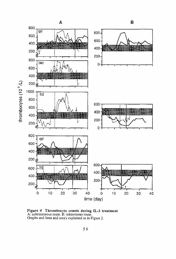
A
800-=~----~---.---,
600
400
200
800 ,-,-,..,----,..-;:--.,..------,
600
400
800
600
400
200
B
:::;- 200 m~ ~~--r-~-.--~-r~~
(/)
CD
~ .0 E e
.<:: -
800
600 600.----.-.----,
400 400
200 200
o+-~.-~~~~~~
600
400
200
600
400
200
~2::;:=:===;:t:::;:::;
0 10 20 30 40 0
time (day) 10
Figure 4 Thrombocyte counts during IL-3 treatment A: subcutaneous route. B: intravenous route. Graphs and lines and area's explained as in Figure 2.
56
20 30 40

Normoblastosis and reticulocytosis did not result in increased erythrocyte numbers and hemoglobin levels (Table 2). In contrast, a decrease was observed during the course of treatment, most prominently so in animals treated with 10 and 30 Jlg/kg/day iv. Two weeks after cessation of IL-3 administration, normal pretreatment values for hemoglobin, erythrocytes, reticulocytes and normoblasts in the peripheral blood of all animals were reached. In the lower dose range of IL-3, thrombocytosis was observed (Figure 4). Animals treated with I Jlg/kg/day iv showed 2-fold higher peak levels of thrombocyte counts, comparable to the animals which received 10 Jlg/kg/day once or twice daily sc. In one of the two monkeys receiving 10 Jlg/kg/day IL-3 tid sc, a decrease in thrombocyte counts was noted in the first week followed by an increase during the second week of treatment. The other showed unchanged values during IL-3 treatment. Both animals showed increases to supernormal levels (up to 900 xl09fL) after cessation of IL-3 administration. At higher dose levels thrombopenia occurred (Figure 4). IL-3 administered at 10 or 30 Jlg/kg/day iv induced a rapid decrease of thrombocyte counts to very low levels. At a dose of 30 Jlg/kg/day sc, a less rapid and less serious decrease was observed. Thrombopenic animals reached normal values within one week after discontinuation of IL-3 treatment, whereas in animals with thrombocytosis it took two weeks before pretreatment values were found. IL-3 levels were not detectable in serum during iv administration.
4.3 Discussion
Serum levels of recombinant IL-3 following iv as well as sc bolus injection, demonstrated that the bioavailability after sc administration is only 40 % of that after iv administration. It was assumed that the hemopoietic response to IL-3 is directly related to the total dose of IL-3 made available in the blood stream. Thus, if the iv bolus injection is representative for the pharmacokinetics of IL-3 following continuous iv infusion, the latter should stimulate hemopoiesis more effectively than sc administration. In view of the short terminal T112 of IL-3, it could also be predicted that dividing the daily dose over several injections would not lead to accumulation of IL-3 and, therefore, would not result in a more prominent hemopoietic effect. Results presented on the hemopoietic effects indeed demonstrated a more effective stimulation of hemopoiesis if IL-3 is given as a continuous iv infusion: the effects of 1 Jlg/kg/day iv were comparable to those of 10 Jlg/kg/day sc. Peripheral blood leukocyte counts resulting from multiple daily injections were not significantly different from those obtained by a single sc injection. Normoblastosis was most prominent in the iv treated animals receiving 10 or 30 Jlg/kg/day, moderate in animals receiving 1 Jlg/kg/day iv or 30 Jlg/kg/day sc and minimal in the other animals. Subsequent reticulocytosis was present for all doses tested, but a clear dose dependence could not be demonstrated. Despite
57
stimulation of erythropoiesis, increased values of erythrocyte counts or hemoglobin levels were not observed; at higher dose levels decreased values were measured. As shown earlier (chapter 3), relatively low dose IL-3 treatment resulted in thrombocytosis, but high dose level treatment induced thrombopenia. Both anemia and thrombopenia were most pronounced in animals treated with continuous iv infusion of IL-3 and can be attributed to enhanced peripheral turnover due to sequestration in the enlarged spleen and hemophagocytosis rather than to a decrease in production (chapter 8). Optimal doses were 10 jlg/kg/day sc or 1 jlg/kg/day by continuous iv infusion, resulting in a 2- to 3-fold rise in leukocyte counts, a 2-fold rise in thrombocytes and a mild stimulation of erythropoiesis, with acceptable side effects. We conclude that continuous iv infusion is the more effective route of administration.
58

Neutralizing Antibodies during Treatment of Homologous Nonglycosylated IL-3 in Rhesus Monkeys
During administration of homologous nonglycosylated IL-3 to rhesus monkeys, reversal of hematological effects and disappearance of side effects suggested a neutralizing anti-IL-3 antibody response. Among a total of twenty monkeys treated with IL-3, ELISA of serial serum samples revealed anti-IL-3 antibodies in ten animals. Antibody production tended to be dose-dependent. IL-3 provoked earlier appearance of antibodies when the daily dose was divided into three daily subcutaneous injections or was given as a continuous intravenous infusion than daily single subcutaneous injections. Prolonged continuous intravenous IL-3 administration (63 and 93 days) at a dose of 1 J.Lg/kg/day did not result in antibody production. Among a total of eight animals with sufficiently high titers to allow for antibody purification, seven appeared to have generated antibodies that neutralized the biological activity of IL-3 in vitro. In six monkeys, the response to IL-3 decreased while antibody titers rose, strongly suggesting neutralization of IL-3 in vivo. It is concluded that recombinant, nonglycosylated IL-3 as used in this study may elicit a neutralizing antibody response.
59
5.1 Introduction
IL-3 stimulates proliferation of hemopoietic progenitor cells and may modulate the functional activity of basophils (224,231,247,377), eosinophils (103,113) and monocytes (86,95,426). Earlier we reported the hemopoietic effects of homologous IL-3 in rhesus monkeys (chapter 3,4). Following a lag phase of about one week during which bone marrow cells were shown to multiply, a large increase in peripheral blood cells along white and red cell series as well as of platelets was observed. Side effects included hypersplenism at high dose levels, accompanied by slight anemia and thrombopenia, urticaria, edema, enlargement of lymphoid tissues and arthritis (chapters 3,4,8). Here we report that in ten animals among twenty studied, antibodies directed against IL-3 were demonstrated, which in the majority of cases neutralized its biological activity.
5.1 Results
5.2.1 In vitro testing of anti-IL-3 antibodies
An ELISA was developed to test sera of all IL-3 treated animals. Sera were sampled twice weekly during IL-3 treatment and one month thereafter. Of twenty animals treated, ten showed the presence of antibodies directed against IL-3 (Table 1). All animals which received 30 or 100 J.lg/kg/day sc developed antibodies. Two animals treated with a daily dose of 10 J.lg/kg/day as triplicate sc injections (tid) also showed an antibody response, whereas those treated with a daily dose of 10 J.lg/kg/day as a single (qd) sc or duplicate injections (bid) did not show anti-IL-3 antibody titers. Half of the animals treated with 10 or 30 J.lg/kg/day as a continuous iv infusion were positive. At 30 J.lg/kg/day, anti-IL-3 antibodies were detected earlier (day 16, 12 and 14) when the dose was administered as a continuous iv infusion or sc in three injections than when a single sc injection was used (day 21 and 28) (Table 2). In animals, treated with 1 J.lg/kg/day iv antibodies could not be detected, also not after a more prolonged period of IL-3 administration (63 and 93 days, respectively). To evaluate the neutralizing capacity of antibodies, sera were tested in the M07E assay. Since sera of untreated animals showed inhibition of the thymidine uptake as well, antibodies were purified and tested thereafter. Eight samples contained sufficient levels of antibodies to be evaluated in the M07E assay. Figure 1 shows two representative examples of the neutralizing capacity of antibodies. Table 2 further shows that seven among eight evaluated animals developed in vitro neutralizing IL-3 antibodies.
60

Table 1 Frequency of animals treated with homologous
which developed anti-IL-3 antibodies IL-3,
IL-3 antibodies* [!g/k[:'da~ n/total
3 qd 1/2 10 qd 0!3
bid 0/2
tid 2/2
30 qd 2/2
tid 2/2
100 qd ]/I
iv 0!2
10 iv l/2
30 iv 1/2
*as detected in vitro by ELISA. Antibody formation was more often found in animals treated with 30 and I 00 ~g/kg!day as compared to those receiving the lower doses (p=0.03, using Fisher's exact test).
~5000~-------------------, -2-5 4000
-~ 3000 e-8 2000 .s: g1 1000 15
I o -~t.,;..~~~~~-.....+-~.,.,.,J ,;:; 10 -2 10 -t 10 ° 10 1 10 2 10 3
ll-3 (ng/ml) 10
1 10
2 10
3 10
4 10
5
anti-ll-3 (ng/ml)
Figure 1 Neutralizing capacity of anti-IL-3 antibodies in vitro (M07E assay) Left panel: titration of rhesus monkey IL-3. Right panel: titration of purified anti-IL-3 antibodies of 2 individual monkeys, in the presence of 20 ng/ml IL-3. Lines with open and closed symbols represent the data of monkey 8841 and 8664, respectively. The single symbols represent stimulation in the presence of a 100-fold excess of IL-3, demonstrating the specificity of the anti-IL-3 antibodies.
6 1

Table 2 Summary of anti-IL-3 antibody responses in rhesus monkeys
UAN* IL-3 ANTIBODIES NEUTRALIZING
highest titer start+ in vitro in vivo ~g/kg/da~ da~s da~ MWE PB cells urticaria
IWM 3 qd 30 1:2700 25 +
8790 10 tid 14 1: 900 18 nd
8640 10 tid 14 1: 100 28 nd
8632 30 qd 30 1:8100 21 +
8841 30 qd 28 1: 300 28 +
8808 30 tid 14 1:8100 12
8746 30 tid 28 1:8100 14 + I, t +
8664 100 qd 30 1:8100 17 + I, t +
8831 10 iv 19 1: 900 18 + +
8707 30 iv 16 1:1500 16 + +
*Unique Animal Number; titer indicates the highest serum dilution giving a positive signal in ELISA; + day at which antibodies were first detected; +means neutralizing capacity; nd indicates not done; 1 and t indicate antibodies neutralized IL-3 effects on leukocyte and thrombocyte counts, respectively.
5.2.2 In vivo effect of neutralizing anti-IL3 antibodies
Six animals showed effects of the neutralizing capacity of the antibodies in vivo. In four animals, a reversal of the IL-3 induced effect on thrombocyte counts and/or leukocyte counts was seen and in four animals disappearance of urticaria during IL-3 treatment was noted (Table 2). The first animal in which a neutralizing antibody response was suspected received 100 J.lg/kg/day as a single sc injection during 30 consecutive days. Figure 2 depicts the reversal of the IL-3 effects on leukocyte and thrombocyte counts in this monkey: at day 20, leukocyte counts declined and the thrombocyte counts rose (thrombopenia was seen in most animals receiving high doses of IL-3 (chapters 3,4,8) and is attributed to hypersplenism and hemophagocytosis (chapter 8)). One animal with neutralizing antibodies did not display in vivo anti-IL-3 activity. This animal received a relatively low dose of 3 !lg/kg/day sc for 30 consecutive days, that resulted in moderate hemopoietic and only mild side effects, which may have obscured evaluation of the anti-IL-3 activity.
62

Figure 2 Reversal of IL-3 induced hemopoietic responses Thrombocyte counts (solid line) and white blood cell counts (dotted line) of monkey 8664, treated with 100 J.lg/kg/day, sc for 30 consecutive days. The arrow marks the time at which antibodies were first detected in serum.
The two animals of which the sera contained detectable antibodies in insufficient quantities to be purified and tested, did not show signs of neutralizing effects of anti-IL-3 antibodies. The single animal with non-neutralizing antibodies and the animals without antibodies did not show signs of reversal of IL-3 effects.
5.3 Discussion
The observations demonstrate that administration of high doses of homologous recombinant IL-3 may evoke an antibody response that neutralizes its effects. In the present study, the dose appeared to be important for the antibody response, in that antibody formation occurred more frequently in animals treated with high doses of IL-3 (30 and 100 f.lg/kg/day). IL-3 given three times a day sc tended to induce earlier antibody formation than a single injection (30 f.lg/kg/day). IL-3 given for a prolonged period of time at a dose of 1 ~-tg/kg/day as a continuous iv infusion did not elicit antibody formation. A significant difference in the frequency of antibody formation of animals treated with iv administration compared to sc treated animals was not found. As expected, homologous IL-3 was only weakly antigenic since large quantities of IL-3 were required to elicit an antibody response. Antigenicity of recombinant hemopoietic growth factors can be expected to result from lack of glycosylation or from differences in primary, secondary and/or tertiary protein structure compared to their native counterparts. Alternatively, IL-3 might be a locally acting cytokine not normally found in the circulation and, therefore, more likely to elicit an antibody response following injection. Recombinant IL-3 may for all these reasons display antigenic epitopes. Until now, antibody formation against homologous IL-3 has not been reported in humans. Formation of anti-GM-CSF antibodies has been described (133). It was shown that these antibodies did react with the unoccupied glycosylation sites of GM-CSF.
63

Anti-IL-3 antibodies neutralized the biological activity of IL-3 in vitro and evidence for neutralization in vivo was obtained in the majority of monkeys in which an in vitro antibody response was demonstrated. However, 10 to 20 !lg/ml immunoaffinity purified antibodies were required to achieve substantial neutralization of 20 ng/ml IL-3 in vitro. This most likely means that the majority of antibodies were nonneutralizing, since similar antibody titers in serum as measured by ELISA were either neutralizing or nonneutralizing. An additional explanation might be that the binding affinity of neutralizing antibodies for IL-3 binding sites is much lower compared to the affinity of the IL-3 receptor complex for its ligand. The observation emphasizes, that a recombinant homologous nonglycosylated hemopoietic growth factor may elicit an antibody response if given in sufficiently high doses to immunocompetent individuals. Since IL-3 stimulates the functional activity of monocytes (94,95,426) and acute type hypersensitivitylike reactions (209,243,chapter 8), it is not excluded that activation of immune reactivity makes IL-3 particularly prone to antibody formation.
64
IL-3 Receptors on Rhesus Monkey Bone Marrow Cells: Species Specificity of Human IL-3, Binding Characteristics,
and Lack of Competition with GM-CSF
The relative affinity of recombinant human IL-3 binding to normal rhesus monkey bone marrow cells was found to be 25- to 50-fold less than that of homologous IL-3, which explained the species specificity of human IL-3 observed when tested in Macaca species. In contrast, only a small difference was found between human and rhesus monkey IL-3 in relative binding affinity for receptors on human AML cells, which confirmed that the species specificity of IL-3 is largely unidirectional. The biological significance of the findings was demonstrated by in vivo comparison of the effects of high dose recombinant rhesus monkey IL-3 with human IL-3 in rhesus monkeys. The binding of IL-3 to receptors on rhesus monkey bone marrow and peripheral blood cells were further characterized using radiolabeled rhesus monkey IL-3. Scatchard analysis of two bone marrow samples demonstrated high-affinity IL-3 receptors (25 and 80 sites/cell, and equilibrium dissociation constants of 8 and 3 pmoi/L, respectively) as well as low-affinity receptors (1070 and 1290 sites/cell, and equilibrium dissociation constants of 2600 and 1200 pmol/L, respectively). In addition, IL-3 receptor expression was also detected on purified CD34-positive bone marrow cells. Competition by human GM-CSF of IL-3 binding to high- or low-affinity receptors on rhesus monkey peripheral blood and bone marrow cells could not be demonstrated, which may indicate that the growth factor specific a.-subunits of the GM-CSF and IL-3 receptors are expressed predominantly on different cell types.
65
6.1 Introduction
IL-3 stimulates immature bone marrow cells to produce progeny along multiple blood cell lineages, including all major nucleated blood cell types as well as platelet and red cell lineages (13,26,32,42,86,115-117,123,249,387). In addition, IL-3 has been reported to modulate the functional activity of monocytes (94,95,426) and of eosinophilic (103,113) and basophilic granulocytes (224,231,247,377). Stimulation of these cells may play a functional role in the development of acute type hypersensitivity reactions and allergic states, as is apparent from side effects such as urticaria and acute arthritis in rhesus monkeys treated with IL-3 (382, chapter 8). To avoid an apparent species specificity observed when human IL-3 was subjected to preclinical studies in Macaca species (78,122,205,206,243,257,373) the gene encoding rhesus monkey IL-3 (45) was cloned and expressed. In vitro, rhesus monkey IL-3 is about 2 logs more active than human IL-3 in stimulating colony-formation by rhesus monkey bone marrow cells, whereas human bone marrow cells are almost equally well stimulated by both species of IL-3 (45). Although evolutionary divergence of IL-3 in general is greater than that of any related hemopoietic growth factor such as GM-CSF (416,417), the evolution of rhesus monkey IL-3 was found to be unusual in that almost all nucleotide substitutions appeared to have resulted in an amino acid substitution in the protein and, thus, showed a more pronounced divergence than IL-3 of other primate species (45). Administration of homologous IL-3 to rhesus monkeys resulted in a dose-dependent expansion of bone marrow derived peripheral blood cells (383,chapters 3,4) and was shown to be much more effective than was reported for human IL-3 in Macaca species (78). To evaluate the apparent species specificity of human IL-3 in more detail, we investigated the relative binding affinity of human and rhesus monkey IL-3 receptors for their homologous and heterologous ligands and compared the effects of in vivo administration of the two species of IL-3 in rhesus monkeys. The structure of IL-3 receptors has recently been elucidated. A high-affinity heterodimer IL-3 receptor structure has been identified, consisting of a ~-chain shared by IL-5, GM-CSF and IL-3 receptors and an IL-3 specific a-chain of low binding affinity (119,147,197,254,362). In addition, a second, closely homologous ~-chain has been identified in mice (144, 171,254), but not in humans. In contrast to the common ~-chain, itself incapable of binding growth factors, the IL-3 specific ~-chain binds IL-3 with low affinity. Co-expression of the two ~-chains in hemopoietic cells of mice has been uniformly observed and so far does not account for differential expression of receptors on different cell types (255). Cellular specificity, therefore, should be attributed to differential expression of growth factor specific a-chains on different cell types as has been directly demonstrated in radiolabeled ligand competition experiments with cells transfected with the human genes encoding the various receptor subunits (197). These experiments also showed that the high-affinity IL-3 receptor by itself does not bind GM-CSF or any other growth factor. This feature predicts various
66
patterns of competitiOn on different cell types. Indeed, primary human monocytes were reported to have high-affinity receptors that bind specifically IL-3 or GM-CSF and a third type that binds both (43,93,95,291), whereas human CML basophils were found to bind IL-3 but not GM-CSF (377), or, alternatively, strongly bound IL-3 with weaker binding of GM-CSF and IL-5 (224). IL-3, GMCSF and IL-5 appeared to cross-compete for binding to human eosinophils (229), whereas neutrophils bound GM-CSF but not IL-3 and IL-5 (43,95,229,377). On AML blasts, low-affinity GM-CSF receptors did not bind IL-3, but a dual highaffinity receptor type bound both GM-CSF and IL-3, while a high-affinity specific IL-3 receptor was also demonstrated (43). Surprisingly little information is available for normal human bone marrow cells, except that a low number of receptors (55/cell) has been demonstrated (291). In this chapter, the binding characteristics of the rhesus monkey IL-3 receptors on rhesus monkey peripheral blood and bone marrow cells were evaluated, as well as co-expression of GM-CSF receptors on IL-3 receptor expressing cells. The strong hemopoietic effects of IL-3 in normal rhesus monkeys may have been induced by indirect mechanisms, since a variety of nonhemopoietic effects, including acute type hypersensitivity reactions with concomitant high levels of histamine, were seen. Histamine by itself may stimulate hemopoiesis as has been reported for the murine hemopoietic system (47,269,331). To investigate whether early progenitor cells express IL-3 receptors, IL-3 binding experiments were performed using CD34-positive cells, which include stem cells and progenitor cells (56) but are devoid of mature cells.
6.2 Results
6.2.1 Species specificity of human IL-3
To evaluate the mechanism of the limited effectiveness of human IL-3 on rhesus monkey bone marrow cells as observed in vivo (78,122,205,206,243,257,373) and in vitro (45), rhesus monkey bone marrow cells were incubated with a fixed amount of radiolabeled rhesus monkey IL-3 and specific binding was competed with increasing concentrations of nonlabeled rhesus monkey or human IL-3. Rhesus monkey IL-3 was 30-fold more effective than human IL-3 in displacing radiolabeled rhesus monkey IL-3 from rhesus monkey bone marrow cells (Figure 1, left panel). In two subsequent experiments a 25 and 50-fold difference in binding affinity was obtained, respectively. In a reciprocal experiment rhesus monkey IL-3 was only 5-fold less effective than human IL-3 in displacing radiolabeled human IL-3 from AML blasts (one experiment, Figure 1, right panel). Both human and rhesus monkey IL-3 displaced the radio labeled ligand to the same extent, indicating that both species of IL-3 competed for binding to the same receptors.
67

'E 2000 'E 2000 Q_ a. .9- Q .9-"0 "0 D c 1500 c 1500 ::l ::l 0 0
~ .c .c C') "' 1000 --" 1000 --" <I) c ::l "' (}) E 500 Q) ::l -E _c
'
"' "' 0 "' "' 10.310.2 10.1 10°10 1
102
103
104 1 a·' 10° 101 10
2
non labeled competitor (nmol/l)
Figure 1 Displacement of radiolabeled IL-3 by rhesus monkey or human IL-3 A fixed amount of radiolabeled IL-3 was incubated with 5 x10⁶ rhesus monkey bone marrow cells (left panel: 500 pmoi!L rhesus monkey JL-3, specific activity of 7.3 x!O" cpm/ng) or 3 x!06
human AML cells (right panel: 250 pmoi!L human JL-3, specific activity of 8 x10⁴ cpm/ng). Closed and open circles represent competition by nonlabeled rhesus monkey and human !L-3, respectively. The squares represent the binding of radio labeled 1!..-3 in the absence of competitor. Vertical bars represent standard errors.
To quantify the biological significance of the differences in binding affinity, two rhesus monkeys were administered human and rhesus monkey IL-3, respectively, at a dose of 30 )lg/kg/day by continuous iv infusion for seventeen consecutive days. Peripheral white blood cell counts reached a 5-fold higher peak level in the animal treated with rhesus monkey IL-3 as compared to the animal receiving human IL-3. The differentials of nucleated cells of both animals revealed a similar distribution (Table 1). The numbers of eosinophils and histamine containing atypical basophils strongly increased and in both animals juvenile band shaped granulocytes appeared in the peripheral blood. Neutrophil counts increased as well, whereas lymphocyte counts remained within normal ranges during treatment. Norrnoblastosis and reticulocytosis were more pronounced in the animal treated with rhesus monkey IL-3. Urticaria and edema were seen in both animals but diarrhea, vomiting, mild intestinal bleeding and hypersplenism accompanied by profound thrombopenia, characteristic for high dose IL-3 treatment, occurred only in the animal treated with rhesus monkey IL-3, indicating that human IL-3 is also less effective in inducing the characteristic nonhemopoietic effects in monkeys.
68

Table 1 Comparison of biological effects of human and rhesus monkey IL-3 infusion*
Effect human rhesus normal± sd lL-3 lL-3 (n=250)
White blood cells (1Q9fL)+ 31 143 9.0 ± 4.6 differential
bands 2.2 5.2 0 neutrophils 19.5 11.4 5.1 ± 3.5 eosinophils 4.0 7.1 0.3 ± 0.4 atypical basophils 13.3 114.2 0 monocytes 1.6 2.9 0.2 ± 0.2 lymphocytes 6.5 6.7 3.4 ± 1.5
normoblasts (IQ9fL)+ 0.9 24.3 0
reticulocytes (% )+ 8.2 33.6 2.3 ± 1.3
urticaria + + intestinal symptoms bleeding,
diarrhea, vomiting
lymph node enlargement + + spleen enlargement + anemia (1-!b, mmoi!L)i 5.3 3.4 7.5 ± 0.6 thrombopenia§ 231 38 394± 96 edema + +
* 30 1-lg/kg/day, continuous iv infusion;+ peak values during IL-3 treatment; § lowest value during lL-3 treatment
6.2.2 Binding characteristics of IL-3 and competition with GM-CSF
Since only little information is available on IL-3 receptors on normal bone marrow cells we characterized the IL-3 receptor complex by complete binding experiments using normal rhesus monkey bone marrow and peripheral blood cells (Figure 2). Bone marrow cells, in two independent experiments, and peripheral blood cells, in one experiment, were incubated with increasing concentrations of radiolabeled rhesus monkey IL-3 (2 to 3500 nmol/L) in the absence or presence of at least a 100-fold molar excess of nonlabeled IL-3. Figure 2, left panels, shows the equilibrium binding of radiolabeled IL-3 of these experiments in the high-affinity concentration range. Transformation of specific binding data to Scatchard plots (Figure 2, right panels) revealed bilinear curves, demonstrating the existence of high- as well as low-affinity binding sites. Affinity constants (Kd ±sd) for IL-3 receptors on bone marrow cells were estimated at 7.5 ±1.3 and 3.1 ±1.2 pmoi/L (corresponding average binding sites per cell ±sd were estimated 24 ±2 and 77 ±24) for high-affinity binding sites and
69

2600 ±100 and 1200 ±100 pmol/L (1070 ±28 and 1290 ±45 binding sites per cell) for low-affinity binding sites. Kd values for IL-3 receptors on peripheral blood mononuclear cells were estimated 25 ±12 pmol/L (47 ±18 sites binding per cell) for high-affinity binding sites and 630 ±480 pmoi/L (88 ±13 binding sites per cell) for low-affinity binding sites. In contrast to competition of IL-3 binding by GM-CSF on normal monocytes, eosinophils of hypereosinophilic patients and leukemic blasts ( 43,95,229,377), nonlabeled human GM-CSF did not compete with radiolabeled rhesus monkey IL-3 for binding to high-affinity IL-3 receptors on rhesus monkey peripheral blood or bone marrow cells (Figure 2). An at least 10.000-fold molar excess of human GM-CSF did not significantly inhibit binding of radiolabeled rhesus monkey IL-3 in the high-affinity concentration range (1-50 pmoi/L) (Figure 2, left panels). The slight reduction in counts in the presence of GM-CSF was within the range of nonspecific binding of IL-3 and therefore not significant. Transformation of binding data after GM-CSF competition to Scatchard plots showed bilinear curves similar to those found after competition with nonlabeled IL-3 (Figure 2, right panels).
Figure 2 Binding characteristics of the rhesus monkey IL-3 receptor complex and lack of competition by GM·CSF of IL-3 specific binding Left panels represent part of equilibrium binding data of increasing concentrations (0-500 pmol/L) of radiolabeled rhesus monkey IL-3 (closed circles) to peripheral blood cells (upper panel: 5 xJ06 cells, specific activity of 7.3 xl04 cpm/ng) or bone marrow cells (middle panel: 1.5 xl06 cells, specific activity of 5.6 xl04 cpm/ng, lower panel: 6 xl06 cells, specific activity of 7.7 xl04 cpm/ng) in the presence of nonlabeled IL-3 (200 nmol/L, open circles) or nonlabeled human GMCSF (500 nmol/L, triangles). Saturation of IL-3 binding was found at concentrations higher than 1500 pmol/L. The right panels show transformation to Scatchard plot of specific binding data (concentration range of radiolabeled IL-3 from 2 to 3500 pmol/L) after incubation with excess of nonlabeled IL-3 (closed circles). Kd values for IL-3 receptors on peripheral blood cells were estimated 25 ±12 pmol/L (47 ±18 sites per cell) for high-affinity binding sites and 630 ±48 pmol/L (88 ±13 sites per cell) for low-affinity binding sites. Bone marrow cells expressed 24 ±2 or 77 ±25 high-affinity binding sites per cell with an apparent Kd of 7.5 ±1.3 or 3.1 ±1.2 pmol/L and 1070 ±28 or 1290 ±45 low-affinity sites per cell with an apparent Kd of 2600 ±100 or 1200 ±100 pmol/L, respectively. Triangles represent transformation of specific binding data after incubation with excess of nonlabeled GM-CSF. Similar bilinear curves were found and estimated Kd values and average sites per cell were not significantly different from those obtained after incubation with nonlabeled IL-3.
70

400
300
200
100
0
2000
§.1500 .!e.
"' " 61000 .0
"' ...J ~-:;:. 500 N
0
0.04
0.03
1!/ 0.02
y /0 0.01
_....0 0.00
0 100 200 300 400 500 0.0 0.5 1.0 1.5 2.0 2.5 3.0 0.25
~ 0.20
~ cr
0.15 g ::> §:
~ 0.10 ~
o---~ ~ 0.05
r:~::::!:::::=~i 0.00 0 100 200 300 400 500 0 5 10 15 20 25 30
1000 or------------,
800
600 ./! 400 ;;t:. 200 ~
-0 0 0
0 100 200 300 400 500 0 5 10 15 20 1251-IL-3 input (pmoi/L) 1251-IL-3 bound (pmoi!L)
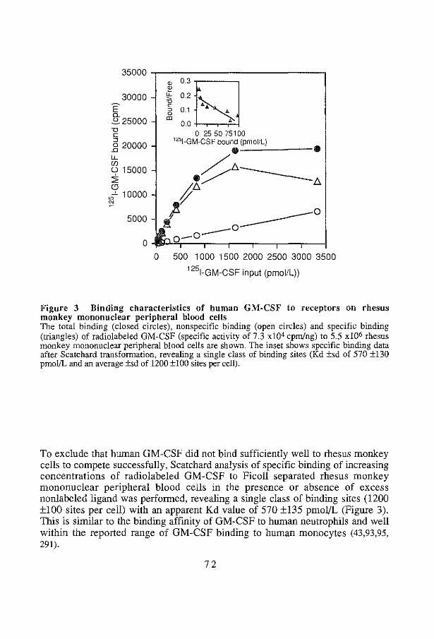
35000 -.--------------.,
30000 E' 0. ~25000 1:l c:
_§ 20000 lL CfJ
c.;> 15000 :;;; CD
~-'- 10000 "'
5000
0
" 0.3rs:J i" !:!::0.2~ ~ ....... s 0.1 •
0.0 ...
0 25 50 75100 1251-GM-CSF bound (pmoi/L) ._:___:__ ___ _ -~t;.----ft;./ /:;.
fl -----0 .! 0 -0--0 500 1 000 1500 2000 2500 3000 3500
1251-GM-CSF input (pmoi/L))
Figure 3 Binding characteristics of human GM-CSF to receptors on rhesus monkey mononuclear peripheral blood cells The total binding (closed circles), nonspecific binding (open circles) and specific binding (triangles) of radiolabeled GM-CSF (specific activity of 7.3 x10⁴ cpm/ng) to 5.5 xl06 rhesus monkey mononuclear peripheral blood cells are shown. The inset shows specific binding data after Scatchard transformation, revealing a single class of binding sites (Kd ±sd of 570 ±130 pmoi!L and an average ±sd of 1200 ±100 sites per cell).
To exclude that human GM-CSF did not bind sufficiently well to rhesus monkey cells to compete successfully, Scatchard analysis of specific binding of increasing concentrations of radiolabeled GM-CSF to Ficoll separated rhesus monkey mononuclear peripheral blood cells in the presence or absence of excess nonlabeled ligand was performed, revealing a single class of binding sites (1200 ±100 sites per cell) with an apparent Kd value of 570 ±135 pmo]/L (Figure 3). This is similar to the binding affmity of GM-CSF to human neutrophils and well within the reported range of GM-CSF binding to human monocytes (43,93,95, 291).
72

6.2.3 Binding of radiolabeled IL-3 to CD34-positive cells
To detect the presence of IL-3 receptors on early progenitor cells, CD34-positive bone marrow cells were isolated and receptor binding studies were performed. Since the percentage of CD34-positive cells in bone marrow is low (about 1% ), it was not feasible to obtain sufficient numbers of purified CD34-positive cells to perform complete Scatchard analysis. However, when purified CD34-positive cells were incubated with a single concentration of radiolabeled IL-3 (500 pmol/L), IL-3 receptors could clearly be demonstrated at a comparable level as observed for peripheral blood cells (Figure 4 ).
E' 700
0. 600 ~ -o c: 500 :> 0 .0
400 (')
~ -o 300 "' Cii 200 .0
"' 0 100 '6 !!'
0 PB
6.3 Discussion
CD34+
Figure 4 Expression of IL-3 receptors on CD34+ cells Radiolabeled rhesus monkey IL-3 (500 pmol/L, specific activity 7.3 x!04 cpm/ng) was incubated with 1.6 xiO' peripheral blood cells (PB) or 1.8 x!06 CD34-positive bone marrow cells in the absence (white bars) or presence of a 500-fold excess of nonlabeled rhesus monkey IL-3 (black bars).
The limited response of nonhuman primates belonging to Macaca species to administration of human IL-3 (78,122,205,206,243,257,373) is explained by the present finding of a 25- to 50-fold lower relative binding affinity of the rhesus monkey IL-3 receptor complex for human IL-3 than for homologous IL-3. In vivo comparison of both species of IL-3 confirmed the relatively weak capacity of human IL-3 to elicit a hemopoietic response in rhesus monkeys. The magnitude of the hemopoietic response of rhesus monkey bone marrow cells upon stimulation by human IL-3 corresponds to that found by others (78,122,205,206,243,257,373). The hemopoietic effects of iv administration of 30 jlg/kg/day of human IL-3 are fully comparable with those of rhesus monkey IL-3 at a dose of 1 jlg/kg/day iv or 3 to 10 jlg/kg/day sc (383,chapters 3,4), confirming the 25- to 50-fold difference in binding affinity. The species specificity appeared to be largely unidirectional, since a much smaller difference between human and rhesus monkey IL-3 stimulated colony-formation by human bone marrow cells was observed as compared with the 100-fold difference in stimulation of colony-formation by rhesus monkey bone marrow cells (45). In
73

this chapter, the conclusion of a unilateral species specificity of human IL-3 was corroborated by the finding of an equally small difference between human and rhesus monkey IL-3 in binding to human AML cells. These results may favor the hypothesis that the rhesus monkey IL-3 receptor complex has been subject to a major change in its IL-3 binding site(s) during Macaca speciation, on which basis rhesus monkey IL-3 followed a forced selective adaptation and diverged rapidly from that of other primate species. About 50 high-affinity IL-3 receptors and 1000 low-affinity receptors were demonstrated on normal rhesus monkey bone marrow cells. Among those, CD34-positive cells were found to express IL-3 receptors, which demonstrates that IL-3 may stimulate early hemopoiesis directly. However, the nature of the CD34-positive cells which express IL-3 receptors needs further elucidation. Evidence for competition of high-affinity IL-3 binding by GM-CSF on rhesus monkey hemopoietic cells was not obtained, although an at least 10,000 molar excess of nonlabeled GM-CSF was used in the high-affinity concentration range of IL-3. The Kd value for GM-CSF binding to rhesus monkey peripheral blood cells (570 pmol/L, 1200 binding sites per cell) was found to be in the range of Kd values found for human cells (43,93,95,291), excluding that the lack of competition is attributable to poor binding of human GM-CSF to rhesus monkey cells. Thus, the expression pattern of the IL-3/GM-CSF receptors on rhesus monkey hemopoietic cells results in independent binding of IL-3 and GM-CSF. This is most likely due to selective expression of the specific a-chains in different cell populations, although the existence of an IL-3 specific ~-chain similar to mice (144,171,254), may be an alternative explanation. We do not exclude that one or more minor subpopulations of bone marrow and/or blood cells express both IL-3 and GM-CSF a-chains, in which case cross-competition may be expected. However, in physiological circumstances as studied here, cross-competition of GM-CSF and IL-3 may very well be an exception rather than the rule.
74

Flow Cytometric Detection of IL-3 Receptors on Distinct Subsets of Peripheral Blood and Bone Marrow Cells in Normal and IL-3
Treated Rhesus Monkeys
IL-3 stimulates the proliferation and differentiation of immature hemopoietic cells. To understand the mechanism by which IL-3 stimulates hemopoiesis, IL-3 receptor expression was examined on bone marrow and peripheral blood cells of rhesus monkeys under normal conditions and during in vivo IL-3 treatment. Results of radiolabeled IL-3 binding studies demonstrated a low expression of IL-3 receptors on normal bone marrow and peripheral blood cells, whereas in vivo IL-3 treatment resulted in a time-dependent increase of specific binding. Examination of the cellular distribution of IL-3 receptors by flow cytometry after staining with biotin-labeled IL-3 and phycoerythrin-conjugated streptavidin demonstrated that most IL-3 receptor-positive cells in bone marrow and peripheral blood of IL-3 treated monkeys could be characterized as atypical, histamine containing basophilic granulocytes. In untreated monkeys, IL-3 receptors were exclusively detected on a distinct population of cells which displayed an immature blast-like phenotype and represented less than 1% of mononuclear bone marrow cells. IL-3 receptors were present on a small fraction (less than 3%) of CD34-positive bone marrow cells, which did not express the cKIT antigen. In contrast to the IL-3 receptor-expressing atypical basophils, the numbers of these immature cells remained within normal ranges after two weeks of in vivo IL-3 treatment. Purified CD34-positive cells, individually sorted on the basis of IL-3 receptor expression responded selectively to IL-3. The results indicate that IL-3 expression on immature bone marrow cells is restricted to a specific subset of blast-like, CD34-positive, c-KIT -negative cells and demonstrate that in vivo IL-3 treatment results in the production of IL-3 receptor-positive cells, characterized as belonging to the basophilic lineage. IL-3 mediated activation of basophils and the resulting generalized reactions may be involved in the broad hemopoietic effects of IL-3 by stimulating the release or production of mediators such as histamine, IL-l, IL-4, IL-6 and TNFa. It remains to be established to what extent the rare immature IL-3 receptorexpressing cells can sustain multilineage hemopoiesis.
75
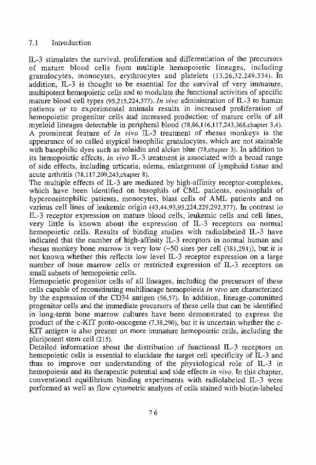
7 .I Introduction
IL-3 stimulates the survival, proliferation and differentiation of the precursors of mature blood cells from multiple hemopoietic lineages, including granulocytes, monocytes, erythrocytes and platelets (13,26,32,249,334). In addition, IL-3 is thought to be essential for the survival of very immature, multi potent hemopoietic cells and to modulate the functional activities of specific mature blood cell types (95,215,224,377)./n vivo administration of IL-3 to human patients or to experimental animals results in increased proliferation of hemopoietic progenitor cells and increased production of mature cells of all myeloid lineages detectable iu peripheral blood (78,86,116,117 ,243,368,chapter 3,4). A prominent feature of in vivo IL-3 treatment of rhesus monkeys is the appearance of so called atypical basophilic granulocytes, which are not stainable with basophilic dyes such as toluidin and alcian blue (78,chapter 3). In addition to its hemopoietic effects, in vivo IL-3 treatment is associated with a broad range of side effects, including urticaria, edema, enlargement of lymphoid tissue and acute arthritis (78,117,209,243,chapter 8). The multiple effects of IL-3 are mediated by high-affinity receptor-complexes, which have been identified on basophils of CML patients, eosinophils of hypereosinophilic patients, monocytes, blast cells of AML patients and on various cell lines of leukemic origin (43,44,93,95,224,229,292,377). In contrast to IL-3 receptor expression on mature blood cells, leukemic cells and cell lines, very little is known about the expression of IL-3 receptors on normal hemopoietic cells. Results of binding studies with radiolabeled IL-3 have indicated that the number of high-affioity IL-3 receptors in normal human and rhesus monkey bone marrow is very low (-50 sites per cell (381,291)), but it is not known whether this reflects low level IL-3 receptor expression on a large number of bone marrow cells or restricted expression of IL-3 receptors on small subsets of hemopoietic cells. Hemopoietic progenitor cells of all lineages, including the precursors of these cells capable of reconstitutiog multiliueage hemopoiesis in vivo are characterized by the expression of the CD34 antigen (56,57). In addition, lineage-committed progenitor cells and the immediate precursors of these cells that can be identified in long-term bone marrow cultures have been demonstrated to express the product of the c-KIT proto-oncogene (7,38,290), but it is uncertaio whether the cKIT antigen is also present on more immature hemopoietic cells, iocludiog the pluripotent stem cell (215). Detailed information about the distribution of functional IL-3 receptors on hemopoietic cells is essential to elucidate the target cell specificity of IL-3 and thus to improve our understanding of the physiological role of IL-3 in hemopoiesis and its therapeutic potential and side effects in vivo. In this chapter, conventional equilibrium binding experiments with radiolabeled IL-3 were performed as well as flow cytometric analyses of cells staioed with biotin-labeled
76

IL-3 and fluorescently-labeled streptavidin (SA-RPE) to investigate IL-3 receptor distribution patterns in rhesus monkeys. This dual approach enabled us to identify IL-3 receptor-expressing cells in normal rhesus monkeys and to study changes in IL-3 receptor expression levels and distribution patterns during in vivo IL-3 treatment.
7.2 Results
7.2.1 Expression of IL-3 receptors during IL-3 treatment
Specific binding of radiolabeled IL-3 to normal rhesus monkey bone marrow and peripheral blood cells was extremely low, whereas in vivo IL-3 treatment resulted in increased specific binding of radiolabeled IL-3 to bone marrow as well as peripheral blood cells (Figure 1 ). Equilibrium binding studies on bone marrow cells of an animal during IL-3 treatment demonstrated a time-dependent increase of specific binding (Figure 2A). Transformation of the data to Scatchard plots (Figure 2B) revealed that the increased radiolabeled IL-3 binding was due to increased numbers of high- as well as low-affinity binding sites, whereas the Kd values of low- and highaffinity binding sites remained virtually unchanged during IL-3 treatment.
25000 E Cl. .2. 20000 l:J c: :::> 0 15000 .c
" -= ·u 10000 Ol a. <f)
M ~ 5000 3340 _,_
"' ~ control IL-3 treated monkey
Figure 1 Specific binding of radiolabeled IL-3 to bone marrow and peripheral blood cells of rhesus monkeys Three million bone marrow cells (white bars) or peripheral blood cells (black bars) of an untreated (left 2 bars) and !L-3 treated rhesus monkey (right 2 bars, I J.Lg/kg/day. iv, day 30) were incubated with 500 pmol/L radiolabeled IL-3 (specific activity: 40,000 cpm/ng). Nonspecific binding was detemtined by parallel incubations with a 100-fold molar excess of nonlabeled IL-3, and was subtracted from total binding to obtain specific binding. Results are the means of duplicate detemtinations.
77

i 60 A
0.25 B •
:::J 0.20 'i)120 E Eo ~ O.i5 "' § 80 "" "0 0 .0
"' ...J
'"-~
c 6 O.iO .0
40 0.05
• 0 0.00
0 i 2 3 4 5 6 0 iOOO 2000 3000 4000 1251-IL-3 input (nmoi/L) sites/cell
Figure 2 Increased IL-3 receptor expression during IL-3 treatment A: Saturation binding curves of specific binding of radiolabeled !L-3 (specific activity:14,000 cpm/ng) to bone marrow cells (5 x10⁶ cells per point) of a rhesus monkey at day 2, 6 and 8 of !L-3 treatment (15 flglkglday,sc). Results represent the means of duplicate determinations. B: Scatchard transformation of the data in A, depicting data points and corresponding fitted curves, demonstrating an increase in high- as well as low-affmity binding sites per cell. Estimated Kd values: 63,45 and 54 pmoVL (56, 109 and 181 high-affinity binding sites/cell) and 1799, 1764 and 1782 pmoVL (422, 2678 and 39991ow-affinity binding sites/cell) for day 2, day 6 and day 8, respectively. Specific binding of radio labeled !L-3 at day 0 cells was too low for evaluation by Scatchard analysis.
7.2.2 Distribution of IL-3 receptors in IL-3 treated monkeys
The cellular distribution of IL-3 receptors was examined by staining cells with biotin-labeled IL-3 and SA-RPE followed by detection of receptor expressing cells by flow cytometry. IL-3 receptor expressing cells were readily detectable in bone marrow (Figure 3) as well as peripheral blood (Figure 4) of IL-3 treated rhesus monkeys. IL-3 receptor expression was prominent in a specific population of cells which displayed intermediate to high forward and intermediate right-angle light scatter properties, distinct from those of granulocytes, lymphocytes and blast cells (Figure 3C/D,4A). The frequency of these cells was similar to that of the atypical basophils identified previously by morphological analysis (chapter 3). The identity of these cells was confirmed by demonstrating that levels of histamine in cells sorted on the basis of the specific light scatter properties were comparable to those found in human basophils and mast cells (170,326), i.e., 700 and 1000 fg/cell in two separate experiments, whereas cells outside the sort window of atypical cells contained only 140 and 70 fg/cell.
78

A biotin-IL-3 c
B I excess unlabeled IL-3
I.··
green (auto-} fluorescence forward light scatter
Figure 3 Cellular distribution of IL-3 receptors in bone marrow of an IL-3 treated rhesus monkey Upper panels represent the dot plots of green background fluorescence versus red fluorescence of bone marrow cells of an IL-3 treated monkey (15 ~glkg/day scat day 10) stained with biotin-IL-3 and SA-RPE (A) and the corresponding light scatter proflle (C). The black dots indicate the fluorescence and light scatter properties of cells gated on basis of high red fluorescence, distinct from autofluorescence. Black dots in the lower panels represent the fluorescence (B) and light scatter properties (D) of nonspecifically stained cells as detennined in the presence of excess nonlabeled !L-3.
79

~
"' "5 (.)
'6 c Ol
e-O) a.
A
atypical cells
forward light scatter
B atypical cells
lymphocytes
biotin-IL-3/SARPE
Figure 4 IL-3 receptor distribution on peripheral blood cells of an IL-3 treated rhesus monkey A: Dot plot of forward versus right-angle light scatter properties of Ficoll-separated peripheral blood cells of an ~-3 treated monkey (15 ~g/kg/day scat day 10). The boxed areas indicate the electronic gates used for analysis of ~-3 receptor expression on atypical cells and lymphocytes, respectively. B and C: Fluorescence profiles of atypical cells (B) and lymphocytes (C) stained with biotin-IL-3 in the absence (solid lines) or the presence of a 100-fold molar excess of nonlabeled ~-3 (broken lines). Note that all atypical cells were specifically stained by biotin-~-3, whereas lymphocytes displayed background fluorescence only.
Table l Estimation of high-affinity IL-3 receptor numbers
on atypical basophilic cells produced during in vivo IL-3 treatment
day
2
6
8
high-affinity receptors
Kd
63
45
54
sites/cell
56
109
181
atypical cells
%ofBM
3
11
13
~-3Rs/cell*
250-1900
600-1000
1050-1400
*Range: the lower limit was calculated on the assumption that all non-atypical cells display 50 sites per cell i.e., the sensitivity of the flow cytometric method and the upper limit on the assumption that only atypical cells display IL-3 receptors.
80

By combining the average number of IL-3 receptors per cell, as estimated by Scatchard plot analysis (Figure 2) with the frequencies of IL-3 receptorexpressing bone marrow cells, determined by flow cytometry (Figure 3), the histamine-containing atypical basophils could be estimated to express some I 03 high-affinity IL-3 receptors per cell, a number which did not change significantly during IL-3 treatment within the error margins inherent to the calculation (Table 1).
7.2.3 IL-3 receptor expression on immature cells in normal and IL-3 treated monkeys
IL-3 receptor-expressing cells were not detectable in peripheral blood of untreated rhesus monkeys (data not shown). IL-3 receptor-expressing cells were also not readily detectable in bone marrow of untreated rhesus monkeys when Ficoll-separated cells were analyzed ungated. A small, but distinct population of IL-3 receptor-positive bone marrow cells could, however, be detected when IL-3 receptor expression was analyzed in an electronic gate for blast cells, defined as cells with intermediate to high forward and low right-angle light scatter properties (Figure 5). These IL-3 receptor-expressing cells represented 2.5 ±0.3% of cells within this gate (average ±standard deviation of 9 individual experiments), corresponding to 0.3 ±0.2% of total mononuclear bone marrow cells. Most IL-3 receptor-expressing cells formed a distinct subset within the blast gate and were characterized by intermediate forward and very low rightangle light scatter properties (Figure 5B/C insets). To further characterize the infrequent IL-3 receptor-expressing cells in normal bone marrow IL-3 receptor expression was compared with co-expression of the CD34 antigen, using twocolor flow cytometry. The results of four separate experiments, one of which is presented in Figure 6, demonstrated that only a minority of blast cells expressing intermediate to high levels of the CD34 antigen co-expressed detectable IL-3 receptors. To examine IL-3 receptor expression on CD34-positive cells in more detail, IL-3 receptor expression was also analyzed on immunomagnetically purified CD34-positive cells (Figure 7). Consistent with the results of the twocolor flow cytometry experiments, the frequency of purified CD34-positive cells that expressed detectable levels of IL-3 receptors was very low and ranged between 1.2 and 2.9% in 4 individual experiments (Figure 7 A, Table 2). The IL-3 receptor-expressing cells formed a homogeneous subset with intermediate forward and low right -angle light scatter properties as compared to the CD34-positive population as a whole, which is relatively heterogeneous with respect to light scatter properties (Figure 7C). Double-staining of purified CD34-positive cells for IL-3 receptor expression and c-KIT expression (as detected by MCA SR-1) revealed that IL-3 receptor expression was only detectable on c-KITnegative cells, although a large fraction of CD34-positive cells expressed the c-
8 1

KIT antigen, similar to published results on c-KIT expression on human CD34-positive cells (Figure 8) (7,38,290). To test whether the IL-3 receptor-expressing subset of CD34-positive cells was functionally distinct from the IL-3 receptor negative subset, purified CD34-positive cells were sorted individually on the basis of IL-3 receptor expression and cultured in the presence of growth factors. The IL-3 receptor-positive cells preferentially responded to IL-3, with 1/3 of the wells containing colonies after 6 days of culture, in contrast to the IL-3 receptor-negative population, which had a plating efficiency of only 6/96 in response to IL-3 stimulation (Table 3). In agreement with the finding that the IL-3 receptor-positive cells were c-KIT negative (Figure 8), the sorted IL-3 receptor positive cells did not respond to SCF (Table 3).
Table 2 IL~3 receptor expression on CD34-positive bone marrow cells of normal rhesus monkeys and one animal after IL-3 treatment
Experiment
I
2
3
4*
5*
CD34+ cells
% of mononuclear cells
0.8
1.2
1.0
1.4
0.2
!L-3 receptor+JCD34+ cells
% ofCD34+ % of mononuclear cells cells
1.6 0.01
1.2 0.01
2.3 0.02
2.9 0.04
7.9 0.02
*same rhesus monkey. before (#4) and after (#5) 14 days oflL-3 treatment (15 J.lg/kg/day sc)
82

A :•.
~
"' "' Cil " <f)
.E g "' 0, c:
"' :E "' ·c:
forward light scatter
B
w a.. a: C: 'i5 ·s;
"' a. !'! 1i! "' _:, c C: ~ :a
i i
FSC
control: excess unlabeled IL-3
0 (/) (/)
green (auto-) fluorescence
Figure 5 Cellular distribution of IL-3 receptors in normal bone marrow A: Light scatter dot plot of Ficoll-separated normal rhesus monkey bone marrow cells. The boxed area indicates the electronic window used to analyze IL-3 receptor expression on blast cells. B: Dot plot of green background fluorescence versus red fluorescence of blast cells stained with biotin-IL-3 and SA-RPE. Nonspecific binding was determined in the presence of a 100-fold excess of nonlabeled IL-3 (C). The boxed areas in B and C indicate the electronic gates used to calculate the frequencies of ll..-3 receptor -positive cells and to analyre their light scatter properties. Inset: Light-scatter properties of blast cells: the black dots indicate the light scatter properties of IL-3 receptor positive cells and nonspecifically stained cells, in B and C respectively. FSC: forward light scatter; SSC: right-angle light scatter.
83
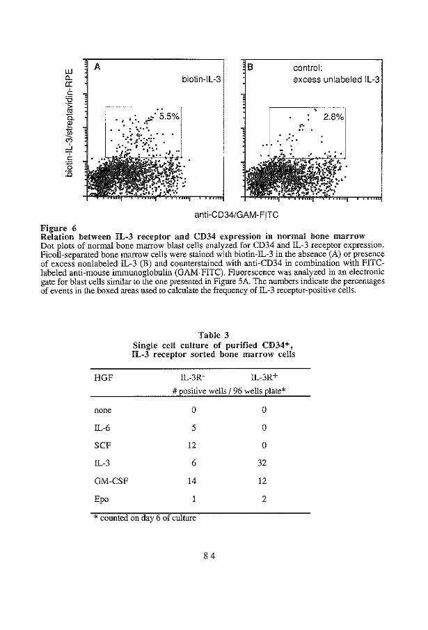
A B control: biotin-Jl-3 excess unlabeled IL-3
. ' '.· . . of:· 5.5% 2.8%
.: :. - .
anti-CD34/GAM-F!TC
Figure 6 Relation between IL-3 receptor and CD34 expression in normal bone marrow Dot plots of normal bone marrow blast cells analyzed for CD34 and IL-3 receptor expression. Ficoll-separated bone marrow cells were stained with biotin-IL-3 in the absence (A) or presence of excess nonlabeled !L-3 (B) and counterstained with anti-CD34 in combination with F!TClabeled anti-mouse immunoglobulin (GAM-FITC). Fluorescence was analyzed in an electronic gate for blast cells similar to the one presented in Figure 5A. The numbers indicate the percentages of events in the boxed areas used to calculate the frequency of IL-3 receptor-positive cells.
HGF
none
IL-6
SCF
IL-3
GM-CSF
Epo
Table 3 Single cell culture of purified CD34+, IL-3 receptor sorted bone marrow cells
IL-3R- !L-3R+
#positive wells I 96 wells plate*
0
5
12
6
14
0
0
0
32
12
2
* counted on day 6 of culture
84

A biotin-IL-3 c
El excess unlabeled D
green (auto-) fluorescence forward light scatter
Figure 7 Expression of IL-3 receptors on purified CD34-positive cells in normal bone marrow Upper panels represent the fluorescence profile (A) and light scatter profile (C) of irnmunomagnetically purified CD34-positive rhesus monkey bone marrow cells stained with biotin-IL-3 and SA-RPE. The black dots indicate the fluorescence and light scatter properties of cells, gated on basis of high red fluorescence, distinct from autofluorescence. Black dots in the lower panels represent the fluorescence (B) and light scatter properties (D) of nonspecifically stained cells as detennined in the presence of excess nonlabeled IL-3.
85

biotin-IL-3 excess unlabeled IL-3
. ' .~ .. . .
anti-KIT/GAM-FITC
Figure 8 Relation between IL-3 receptor and c-KIT expression on purified CD34-positive normal bone marrow cells Dot plots of immunomagnetically purified CD34-positive bone marrow cells stained for IL-3 receptor expression and counterstained with anti-c-KIT (MCA SR-I) in combination with GAMFITC (A). Nonspecific binding of biotin-IL-3 was determined in the presence of excess nonlabeled IL-3 (B). Quadrants were set using isotype-matched control antibody stained cells, such that >99% of events were located in the left bottom quadrants. The numbers indicate the frequencies of events in the 4 respective quadrants.
To examine the effect of IL-3 administration on the immature bone marrow compartment, IL-3 receptor expression on purified CD34-positive cells was analyzed before and after IL-3 treatment. As shown in Table 3 the frequency of CD34-positive cells, expressing IL-3 receptors increased from 2.9 to 7.9% after 14 days of IL-3 treatment, whereas the frequency of CD34-positive cells decreased sevenfold. Since absolute numbers of mononuclear cells increased from 8 xl06 to 18 x106 per ml aspirate, a significant change in absolute number of cells expressing both IL-3 receptors and CD34 antigens could not be demonstrated after IL-3 treatment (i.e., 3.2 xl03 and 2.8 xl03 per ml bone marrow aspirate, respectively), indicating that IL-3 administration did not lead to major changes in the numbers of CD34-positive hemopoietic cells expressing IL-3 receptors as compared to the large increases in atypical basophilic cells.
86

7.3 Discussion
In this chapter, the cellular distribution of IL-3 receptors was examined using a novel flow cytometric detection method involving staining of cells with biotinlabeled IL-3 and phycoerythrin-tagged streptavidin. In addition, conventional receptor binding experiments using radiolabeled IL-3 were performed to allow estimation of binding affinities and average binding sites per cell. Using this dual approach, increased IL-3 receptor expression on bone marrow and peripheral blood cells in response to in vivo IL-3 treatment was demonstrated and appeared to be predominantly attributable to the production of large numbers of atypical basophils which expressed high levels of IL-3 receptors. The numbers of highaffinity receptors on atypical basophils, as derived from Scatchard plot analysis, were similar to those reported previously for leukemic basophils isolated from CML patients (224,377). IL-3 receptor-positive, basophilic cells were not detectable in the peripheral blood of normal monkeys, probably due to the low frequency of circulating basophils in rhesus monkeys: in a series of 140 animals we observed 1 to 2% basophilic granulocytes in only 15 normal monkeys (unpublished observations). IL-3 receptors were not detected on monocytes in peripheral blood of either normal rhesus monkeys or IL-3 treated animals. This observation is at variance with results of radiolabeled IL-3 binding studies, in which IL-3 receptors were detected on partially purified human peripheral blood monocytes (43,93,95,291). The Kd values for IL-3 binding to rhesus monkey basophils (Figure 2) are very similar to those reported for binding of human IL-3 to monocytes, making it unlikely that differences in affinity form the basis of the lack of detectable IL-3 receptor expression on monocytes. The discrepancy could be caused by the presence of variable amounts of basophils in purified monocyte preparations used in radiolabeled IL-3 binding studies (43,93,95,291), which is the more likely as reported values for high-affinity IL-3 receptor numbers on human monocytes are 2- to 20-fold lower than those on either leukemic (224,377) or atypical basophils (this chapter). IL-3 stimulated colony-formation by purified CD34-positive cells has been demonstrated to be co-dependent on the presence of accessory cells (26), which suggests that part of IL-3 induced stimulation of hemopoiesis may be mediated by indirect mechanisms, e.g., by increased production of other stimulators. Possible candidates for these mediators are histamine and IL-4, produced by basophils upon IL-3 stimulation (41,224,377). Normal bone barrow has also been demonstrated to produce histamine in response to IL-3 stimulation (253). Histamine has previously been shown to stimulate proliferation of immature hemopoietic cells and to have a role in IL-3 mediated activities on these cells in vitro (47,48,324,331). Therefore, it is conceivable that continuous production and activation of atypical basophils during IL-3 treatment and resulting generalized reactions are not only involved in the broad range of side effects (78,116,117,209,243,chapters 3,8), but at least partially in the hemopoietic effects of
87

IL-3 as well. Hemopoietic growth factors, such as IL-l, IL-4 and IL-6 have also been shown to be released upon IL-3 stimulation of murine cells (88,109) and increased levels of TNFa and IL-6 during IL-3 administration was observed in patients, treated with chemotherapy for relapse small cell lung cancer (304), providing further evidence for possible indirect mechanisms being involved in hemopoietic stimulation by IL-3. In addition to the atypical basophils produced in response to IL-3 treatment, IL-3 receptors were also detected on a small, but distinct subset of CD34-positive blast cells of untreated animals. The number of these cells did not change significantly during IL-3 treatment, which indicates that the population size of these cells is not influenced by IL-3. This subset of CD34-positive cells expressed IL-3 receptors in the absence of c-KIT, whereas on the majority of CD34-positive progenitor cells, including all c-KIT-positive cells, IL-3 receptors were not detectable. It cannot be excluded that IL-3 receptors are expressed on a larger fraction of CD34-positive cells, in numbers lower than the detection limit of the flow cytometric method using biotin-labeled IL-3. Due to the large quantity of cells required per data point, it has not been feasible to purify sufficient CD34-positive cells for a direct estimate of IL-3 receptor numbers by means of equilibrium binding and Scatchard analysis. It has also not been possible to directly measure the detection limit of the flow cytometric method using biotin-labeled rhesus monkey IL-3, since characterized rhesus monkey cell lines that express IL-3 receptors in low numbers are not available. Human cell lines, such as M07E and AMLl93, are not suitable for this purpose since the sensitivity of these cells to rhesus monkey IL-3 has been demonstrated to be lower than to human IL-3, due to the species specificity of IL-3 (45). However, similar flow cytometric methods to detect receptors for IL-6 and erythropoietin (408,409) have sensitivities of about 50 receptor molecules per cell. Since IL-3 binds to its receptor with similar affinity as these growth factors, it is likely that the detection of IL-3 receptor-positive cells using biotin-labeled IL-3 and flow cytometry has a similarly high sensitivity. Park et a!. (291) reported an average of 50 high-affinity IL-3 receptors per cell on unfractionated human bone marrow, using radiolabeled IL-3. In the present study IL-3 receptor expression could be detected on fewer than 1% of normal bone marrow cells. This finding may indicate that the small subset of CD34-positive cells with detectable IL-3 receptors have receptor numbers in the order of at least 103/ cell. Analysis of the growth factor responses of CD34-positive cells, individually sorted on the basis of IL-3 receptor expression, demonstrated that the flow cytometric method did not only discriminate between IL-3 receptor-negative and -positive cells phenotypically, but functionally as well, because most IL-3 responsive progenitor cells were found in the IL-3 receptor positive population. Some responsive cells were also seen in the population expressing IL-3 receptors below detection level, which may indicate that cells require only low levels of IL-3 receptor expression to respond to IL-3 or,
88

alternatively, that IL-3 receptor expression is enhanced during culture, rendering previously receptor negative cells sensitive to IL-3 stimulation. The presence of IL-3 receptors on a small, but distinct subset of CD34-positive/c-KIT-negative bone marrow cells in both untreated and IL-3 treated rhesus monkeys may be taken as evidence that IL-3 can directly stimulate specific immature cell types. Although the IL-3 receptor-positive/CD34-positive/c-KIT -negative cells may represent relatively differentiated immature cells, e.g., those committed to the basophilic lineage, these cells could also represent developmentally earlier stages than the CD34-positive/c-KIT -positive progenitor cells. IL-3 has been demonstrated to be essential for the survival of immature, quiescent human CD34-positive/HLA-DR-negative cells that do not express lineage-specific differentiation antigens, whereas other growth factors, including the c-KIT ligand SCF, are thought to stimulate the proliferation and differentiation of these cells after activation (215). Activation of quiescent hemopoietic cells has also been shown to be associated with enhanced expression of the c-KIT antigen (131). The absence of the IL-3 receptors on c-KIT-positive, CD34-positive cells could therefore indicate that activation also results in reduced expression of IL-3 receptors on these cells. To establish the role of IL-3 in early stages of hemopoiesis, it will not only be essential to extend the phenotypic and functional characterization of IL-3 receptor-expressing bone marrow cells in vitro, but also to examine their ability to reconstitute long-term multilineage hemopoiesis in irradiated recipients. The flow cytometric method presented here in principle allows for transplantation of cells selected on the basis of their growth factor receptor phenotype.
89


Acute Side Effects of Homologous IL-3 in Rhesus Monkeys
IL-3 treatment of juvenile rhesus monkeys elicits a dose- and time-dependent syndrome, which includes urticaria, palpable lymph nodes, splenomegaly, thombopenia, anemia, vomiting, diarrhea, intestinal bleeding, edema and arthritis, apart from a strong stimulation of hemopoiesis. Arthritis was found to occur significantly more often in animals expressing the major histocompatibility complex alleles B9 and DrS. Histological analysis revealed an abundance of mast cells in urticaria and, to a lesser extent, in lungs and synovia of arthritic joints. Active osteoclasts were present in ribs and arthritic joints. Extramedullary hemopoiesis was encountered in liver, spleen and kidneys. The spleen showed deposits of hemosiderin and, in the liver, Kupffer cells were loaded with iron, indicating enhanced turnover of hemoglobin. Lymph nodes and bone marrow showed macrophages involved in hemophagocytosis which likely contributed to the development of anemia and thrombopenia. Biochemical parameters in sera were indicative for parenchymal liver damage with cholestasis and increased erythrocyte destruction. The side effects were strongly reduced in monkeys, subjected to total body irradiation just before IL-3 treatment. Histamine antagonists were not significantly effective in preventing side effects, which is explained from perpetual stimulation of basophilic granulocytes by exogenous IL-3. The nature of the side effects indicates that IL-3 may be involved in the pathogenesis of acute type hypersensitivity reactions and arthritis.
9 1

8.1 Introduction
IL-3 stimulates in vitro colony-formation by a wide range of hemopoietic progenitor cells (22,26,30-32,42,96,213,244) and the differentiation of eosinophilic (59,60,97,341) and basophilic (191,190,379) granulocytes, mast cells (191,192) and osteoclast (14, 146,305). In vivo administration of homologous IL-3 to rhesus monkeys resulted in highly increased hemopoiesis along multiple lineages (chapters 3,4), including eosinophilic, neutrophilic and atypical, histamine containing, basophilic granulocytes, monocytes, normoblasts, reticulocytes and thrombocytes. Characteristically, a peripheral blood T and B lymphocyte response was absent. Increases in peripheral blood cell values were preceded by an increase in progenitor cell content of the bone marrow (chapter 3). IL-3 receptors have been demonstrated on basophilic (224,225,377) and eosinophilic (223) granulocytes, monocytes (43,95), but not on neutrophilic granulocytes (223,377) and human lung mast cells (378). IL-3 supports survival (95), adhesion (94) and antimicrobial activity (394) of monocytes, degranulation (113) and phagocytosis (103) of eosinophils and the survival of basophilic granulocytes (414), in addition to enhancement of IgE-induced histamine release (224,231,377). Furthermore, the production of intracellular histamine in bone marrow cells was shown to be stimulated (243,253). Histamine by itself may stimulate hemopoietic cell cycle activity of immature cells (47,48,89) and is involved in the pathogenesis of urticaria (209,243,chapter 3). The pleiotropic effects of IL-3 have also been demonstrated in mice following hemopoietic reconstitution from bone marrow cells transfected with the murine IL-3 gene. Overexpression of the gene not only showed stimulation of hemopoiesis but a broad range of side effects in other organ systems as well (53,411). Neutrophilic and mast cells infiltration of liver, spleen, heart, lungs, intestine, muscles and skin were described, while hepatosplenomegaly and extramedullary hemopoiesis were common findings. This study was performed to evaluate hemopoietic effects of homologous IL-3 (chapter 3,4,9) and its side effects in a preclinical primate model. In this chapter we report the range of acute side effects and histopathological findings during IL-3 treatment. In addition, the IL-3 side effects in monkeys subjected to total body irradiation to induce pancytopenia are described.
8.2 Results
To test its in vivo effects, IL-3 was given to eighteen normal rhesus monkeys in doses ranging from 3 to 100 J.!g/kg/day and was scheduled for 14 to 30 consecutive days. In several animals administration of IL-3 was discontinued at an earlier time point, due to the severity of side effects. Two additional animals were treated with 1 J.!g/kg/day administered by continuous iv infusion for a
92

prolonged period of time (68 and 93 days, respectively). The effects of IL-3 during the first 30 days of treatment are included in this study. One monkey, which received a continuous iv infusion of 30 Jlg/kg/day for 17 consecutive days, was sacrificed to evaluate the acute effects by histopathological examination. The hemopoietic effects included stimulation of granulocyte, monocyte, erythrocyte as well as thrombocyte lineages (chapter 3,4). Characteristic for IL-3 treatment was the appearance of atypical basophilic granulocytes, abundantly expressing IL-3 receptors, which comprised the majority of peripheral blood cells at the end of IL-3 treatment (chapter 3,7). Concomitantly, high levels of serum histamine were measured (chapter 3). During the first and second week of treatment, eighteen animals developed skin lesions that were diagnosed as urticaria (Figure lA), located on the abdomen, limbs, face, head and external genitals, whereas the injection sites were not affected. The severity of the lesions was dependent on dose and duration of IL-3 treatment (Table 1). Mechanical irritation provoked more extensive urticaria (derrnatographism). Four animals (#4,10,14,16) showed signs of scratching, indicating the pruritic·nature of the lesions. Five affected animals (#11,13,14,16,17) showed hemorrhages in the skin lesions and animal 11 showed nonpurulent skin defects. The skin lesions vanished within several days upon cessation of IL-3 treatment in most animals and in four animals (#13,14,18,20) during IL-3 treatment. Histological examination of skin biopsies revealed subcutaneous edema, leukocytoclastic vasculitis and extravasation of erythrocytes and atypical basophilic granulocytes. To!uidin blue staining exhibited numerous perivascular mast cells (Figure lB). Diarrhea (2/20), vomiting (1/20) and intestinal bleeding (3/20) further showed involvement of epithelial tissue. Pathological changes were not observed in the gastrointestinal tract at autopsy. Histological examination showed small numbers of neutrophilic granulocytes in the mucosa, whereas active destruction of epithelial cells was not detected. During the second and third week, enlargement of lymphoid tissues was observed. Ioguinal as well as axillary lymph node enlargements were seen in seventeen animals (Table 1). Histological examination of lymph nodes revealed cortex hypoplasia (Figure lC), sinus histiocytosis and many macrophages which showed erythrophagocytosis (Figure 10). Spleen enlargement was detectable by physical examination in eight animals and more prominently present at higher dose levels of IL-3 (Table 1). The enlarged spleen (8 x5 x4 em, normal 2 xl x0.5 em) of the autopsied animal showed macroscopic hemorrhages. Histological examination revealed deposits of hemosiderin, which were extensive in hemorrhagic areas and were also present in congested areas. Extramedullary hemopoiesis, fibrosis and clusters of atypical basophilic granulocytes were observed, whereas the white pulp was severely depleted (Figure IE). At high dose levels (> 10 Jlg/kg/day) thrombopenia and anemia occurred. At a dose of 3 Jlg/kg/day thrombocytosis was seen and the mean lowest values of hemoglobin was 6.2 mmoi/L.
93

Table 1 Side effects affecting epithelial tissues, lymphoid tissue enlargement and edema
# UAN* !L-3 urticaria enlar~ement of edema
t!:~I!Lda~ da~s lymph nodes SEleen
I IWM 3 q~ 30 ++ + 2 8627 30 ++ +
3 8670 10 qd 30 ++ + 4 8718 " 30 +++ + + 5 BB60 14 + 6 8641 bid 14 7 BB63 14 8 8640 tid 14 + + 9 8790 14 ++++ ++ + ++++
10 8632 30 qd 30 +++ ++ +++ II 8841 " 28 ++++ ++ ++ ++++ 12 8808 3 tid 14 + + + + 13 8746+ " " 28 +++ +++ ++ +
14 8664+ 100 qd 30 ++ ++ +
15 8836 I iv 30 +++ + + + 16 8850 " 30 ++ + + +
17 8811 10 lV 15 ++++ + +++ 18 883!+ 19 ++++ +++ +++ +++
19 8623 30 lV 16 +++ + +++ 20 8707+ 17 ++ + +++ +
4 Gy TBI
21 8873 10 iv 14 ++ ++ ++ ++ 22 8830 30 tid 14 ++ + +
5 GyTBI
23 8705 10 qd 14 24 8782 qd 14 + 25 4055 iv 30 +
* UAN indicates unique animal number.+ animals showed early disappearance of urticaria. The daily dose was given subcutaneously once (qd), or divided into two (bid) or three (tid) injections or by continuous intravenous infusion (iv).
94

severity urticaria ± :sporadic + : head, trunk or limbs ++ : head, trunk and/or limbs +++ : generalized ++++ : +++ & confluent
severity lymph node enlargement + < 1 em + 1-2 em ++ 2-3 em +++ >3cm
95
edema :eyelids : face, scrotum or abdomen : face and scrotum, or limbs : ++ and limbs or abdomen : +++, limbs and abdomen
spleen enlargement : just palpable : < 1 em below costal margin : > 1 em below costal margin :++,extending the midline

Figure 1 Side effects affecting epithelial and lymphoid tissue A. Right arm of an animal during IL-3 treannent: typical examples of separate urticaria as well as coalescent ones, in which several hemorrhages (animall4, day 14). B. Cross section of a skin biopt of urticaria (toluidin blue staining, x200): mast cells (arrows) at perivascular sites as well as subcutaneous edema, extravasation of erythrocytes and polynuclear and atypical basophilic granulocytes (animal!O, day 16). CDE: animal20 after autopsy. C. Lymph node (HPS, x40): the cortex shows severe hypoplasia. D. Erythrophagocytosis (HPS, xlOOO): histiocytes in a lymph node involved in conswning erythrocytes (arrows). E. Enlarged spleen. Left (HPS, x40): areas of hemorrhages, depletion of white pulp, extramedullary hemopoiesis. Middle (HPS, x400): detail of extramedullary hemopoiesis (note the megakaryocyte) and hemosiderin deposits (arrows), illustrating the enhanced peripheral turnover of erythrocytes. Right (HPS, x400): Detail of fibrosis. Arrows point at neutrophilic and basophilic granulocytes.
96

B
97

Two animals (#8,9) in the group which received 10 jlg/kg/day sc developed thrombopenia (mean lowest value 218 xl09JL). The mean lowest hemoglobin value of all animals in tbis treatment group was 5.9 ±0,5 mmol/L. At 30 and 100 jlg/kg/day sc tbe mean lowest values of thrombocyte counts was 196 ±24 and 73 xl09JL and of hemoglobin 4.8 ±I and 5 mmol/L, respectively. Similar dose dependent effects were observed in the iv treated animals. Thrombopenia did not occur in animals treated with 1 jlg/kg/day of IL-3 iv. Lowest thrombocyte counts were 95 and 58 x109JL in animals treated witb 10 and 30 !J.g/kg/day iv, respectively. The lowest values of hemoglobin were 6.1, 4.5 and 3.9 mmol/L for animals treated with, respectively, 1, 10 and 30 jlg/kg/day iv. Thrombocyte counts and hemoglobin levels in normal rhesus monkeys were 395 ±95 xl09JL and 7.5 ±0.6 mmoi/L, respectively. Peripheral pitting edema was observed in thirteen animals and started during the second week of treatment (Table 1). Most edematous animals showed weight gain during IL-3 treatment, except for tbe autopsied animal which, due to loss of appetite, lost 9% of its body weight. Those animals with the most severe edema showed weight gains up to about 25% (animal 9). All animals showed a decrease in serum albumin levels, but those animals developing edema exhibited the lowest serum albumin levels. The mean lowest albumin levels were 39, 36 ±6, 24 ±2 and 22 g/L in animals receiving IL-3 sc at doses of 3, 10, 30 and 100 jlg/kg/day, respectively, and 37, 19 and 22 giL in animals receiving IL-3 iv at doses of 1, 10 and 30 jlg/kg/day, respectively (normal value: 49 ±7 g/L). Infusion of human albumin did not reduce the severity of edema. Serum albumin returned to normal levels several days after discontinuation of IL-3 administration. Of two affected animals, urine was collected over a period of 24 hours and mild albuminuremia was detected in one (4.4 mg/24 hrs). However, albumin excretion was not essentially different from that in one control animal (30 mg/24 hrs ). Four animals (#9,13,15,17) developed in the second week of treatment swollen joints of hand and feet, which were diagnosed as arthritis (Figure 2A). The proximal interphalangeal (PIP) joints were more often affected than the distal (DIP) joints (Table 2 and 3). Resistance to development of arthritis was not related to tbe MHC allele A26, as in the case in collagen-induced juvenile arthritis (12), whereas tbe MHC alleles B9 or Dr5 were significantly more frequent in animals developing arthritis (Table 2). The arthritic symptoms disappeared after cessation of IL-3 administration in animals 9 and 17, but in animal 13 the symptoms already regressed during IL-3 treatment. The animal treated for a prolonged period of time (#15) showed recurrent periods of arthritis (Table 3). During active arthritis, the most affected fingers of two animals were amputated for histological examination.
98

Table 2 Development of arthritis and MHC alleles
animal MHCl arthritis* A B Dr #l"!P #DIP
1 2,26 9, 19 1, 1
2 17, 17 5, 10 3,-
3 14,26 10, 19 3, 1
4 17, 17 5, 10 3, -
5 32, 34 -, - 8, 4
6 2, 32 6, 28 2, 4
7 26,32 10,- 8, 3
8 14,24 6, 10 3, 3
9 14,26 9,23 5,5 5 0
10 11,25 6,9 -, 1
11 17, 23 6, 10 3, 2
12 14,26 6, 10 5, 8
13 20,26 19, 23 3, 3 1 0
14 2,26 6, 10 7, 1
15 11, 14 23,23 5,4 12 6
16 14, 34 10, 23 3, 3
17 13, 24 9,9 -, 5 9 0
18 20, 24 3, 19 3, 7
19 17,29 6, 19 2, 15
20 11,25 6,22 -, 5
* number of arthritic proximal and dtstal interphalangeal JOints (PIP and DIP) of hands and feet during IL-3 treatment. ;Major Histocompatibility Complex: a relation of the resistance to development of arthritis with the MHC allele A26 was not found (p;0.34), whereas MHC alleles B9 and DrS occurred significantly more frequent in the group developing arthritis (p;0.03, using Fischer1s exact test).
99

Figure 2 IL-3 effects on joints, bone marrow and bone tissue A. Arthritis in animal 17: the hands display swelling and redness of PIP joints in the second week of IL-3 treatment, demonstrating an acute inflammation. B. Cross section through an affected joint of the same arthritic animal (HPS, x40): swollen synovium with mononuclear infiltration and intra-articular deposit of fibrin and debris. Note the slight irregularity of the cartilage surfaces. C. Bone marrow and bone tissue (HPS x200). Left: Detail of an affected joint, showing active osteoclasts (arrows) in the bone tissue. Right: cross section through a rib showing the absence of fat cells and abundant hemopoiesis. Arrows point at a megakaryocyte and osteoclast.
100

101

Table 3 Arthritis during IL-3 treatment: affected joints and duration
animal arthritis da:Ys* dig!t joint
1 2 3 4 5 9 12-17 lift hand + ~+ + PIP
right hand + + PIP
13 11-13 lift hand + PIP
15 25-30 lift hand + + + DIP + + PIP
40-43 + + PIP
25-30 right hand + + + DIP + + PIP
40-43 + + PIP
16-18 left foot + PIP 40-42 + + + PIP
15-18 right foot + + + PIP 40-41 + PIP
18-25 inner side 34/40-45 ankles
17 12-17 lift hand .+ + + PIP right hand + + + + PIP
left foot + + PIP
*day 0 is start ofiL-3 treatment
102

The synovia were mildly swollen with infiltrating mononuclear cells and neovascularization. At perivascular sites, mast cells were seen. The joint space contained fibrinous exudate and debris. The cartilage was only slightly irregular (Figure 2B). An abundance of osteoclasts in affected joints and ribs (Figure 2C) was observed. At this acute stage, there were no signs yet of the histological alterations characteristic for chronic rheumatoid arthritis, i.e., widening of the joint space and pannus formation with erosion of cartilage and subchrondral bone. Examination of the bone marrow revealed a virtually complete loss of fat cells. The marrow cavity was packed with nucleated cells, most of the cells being of the myeloid lineage, whereas mature cells were strikingly absent. Also erythroid precursors and megakaryocytes were evident (Figure 2C). As in lymph nodes, overt hemophagocytosis was present. Serum biochemical parameters, which were evaluated twice weekly, revealed elevated serum levels of ASAT (7/20), ALAT (4/20), yGT (5/9), bilirubin (9/20), AP (3/20), LDH (10/20) and were indicaiive for parenchymal liver damage and bile obstruction combined with increased erythrocyte turnover. The liver of the autopsied animal showed parenchymal damage, periportal mono- and polynuclear infiltration with extramedullary hemopoiesis and Kupffer cells loaded with iron (Figure 3A). Electrolytes, creatinine and urea levels remained within normal ranges throughout treatment, except for the autopsied animal, which at the end of treatment showed raised serum urea levels (up to 27 mmol/L; normally below 10 mmol/L). Histological examination of the kidneys revealed the presence of large numbers of immature hemopoietic cells along the collecting tubules, which were partly located in the blood vessels and partly in the interstitium. Pathological changes in glomeruli or tubules could not be demonstrated (Figure 3B). Abnormalities were further observed in the lungs, heart and thymus. The lungs were partly atelectatic and edematous. The alveolar walls were extensively infiltrated by neutrophils, eosinophils and to a lesser extent by atypical basophils and perivascular cells (Figure 3C). The heart showed recent ischemic changes, like hypereosinophilia and pykoosis of myocytes, as well as margination and migration of granulocytes. The thymus revealed cortical atrophy. The side effects were considerably reduced in radiation induced pancytopenic animals (Table 1), which were treated with IL-3 to study its possible efficacy in stimulating blood cell recovery. In two of three animals (#24,25) subjected to 5 Gy TBI, urticaria developed at the end of IL-3 treatment (day 15 and 30, respectively). In 4 Gy irradiated animals, urticaria were more pronounced and started in the second week after irradiation. In the latter animals, enlargement of lymph nodes and spleen were diagnosed in the second week of treatment, whereas the animal treated with 10 ).lg!kg/day iv (#21) developed edema at its lower extremities.
103

Figure 3 Extramedullary hemopoiesis in liver and kidney A. Liver. Left (HPS, x200): periportal cellular infiltration and extramedullary hemopoiesis (arrow points at a megakaryocyte). Right (HPS, x 400): Kupffer cells loaded with iron (arrows). B. Kidney (HPS, x200): immature hemopoietic cells along collecting tubules, partly located in the vessels and partly in the interstitium. C: Lung. Left (HPS, x200): heavily infiltrated alveolar walls with neutrophils, eosinophils and atypical basophilic cells. Edema and atelectasis. Right (toluidin blue staining, x400): detail showing mast cells, neutrophilic and basophilic granulocytes (arrows).
104

10 5

It has been reported that both H1 and H2 receptor antagonists may partially prevent histamine induced vasopermeability (132a,219a,235) and may be beneficial in rheumatoid arthritis (221,297a). H1 receptor antagonists are generally used in allergic diseases and urticaria and Hz histamine antagonists to treat gastrointestinal ulcers. Both histamine antagonists are used in mastocytosis to reduce pruritus, flushing and tachycardia (Ill). To evaluate whether the development of IL-3 induced, histamine related side effects could be prevented, H 1 histamine antagonist ( ceterizine) or Hz histamine antagonist ( cimetidine) were given to nine IL-3 treated animals treated with high or low dose IL-3 (3, 30 J.l.g/kg/day sc and 30 !J.g/kg/day iv, in two, five and·two animals, respectively). Drugs were given orally, twice daily at a dose of 10, 20 or 30 mg and 400 or 800 mg, respectively. In two animals treated with 30 J.l.g/kg/day IL-3 sc in combination with the H 1-antagonist, a reduction in the severity of urticaria was observed; the skin lesions were pale, small and less numerous in contrast to the the wheal-like, red skin lesions in other animals. The development of edema was delayed for several days in two animals treated with 30 J.l.g/kg/day IL-3 sc and H1 or H2 antagonists simultaneously, compared to all other animals. The time of onset and severity of enlargement of lymphoid tissues, thrombopenia and anemia were not influenced by histamine antagonists. None of the animals developed arthritis, which is however, not significantly different from four out of twenty in the group which was not treated with histamine antagonists.
8.3 Discussion
The stimulatory effects of IL-3 on progenitor cells of the hemopoietic system have been extensively described (22,26,30-32,42,96,213,244,chapters 3,4). In addition, this study revealed extramedullary hemopoiesis in liver, kidney and spleen. The nature of the IL-3 evoked side effects showed its involvement in the function of mature end cells in vivo, as was predicted from the demonstration of IL-3 receptors on monocytes, basophilic and eosinophilic granulocytes (43,95,223-225,377). Most of the observed side effects were dose- and time-dependent. These included urticaria, intestinal bleeding, vomiting, diarrhea, edema, arthritis of small joints, lymphoid tissue enlargement, thrombopenia, anemia, and parenchymal liver damage. Early disappearance of urticaria (four animals) during treatment could be attributed to neutralizing anti-IL-3 antibodies (chapter 5). Severe anemia and thrombopenia were encountered in animals exhibiting enlargement of lymphoid tissues. Hemophagocytosis in lymph nodes and bone marrow may have contributed to the development of anemia and probably thrombopenia as well. The symptoms resemble those found in the virus associated hemophagocytic syndrome in man (311), which is thought to result from altered function of T -cells (288) and uncontrolled production of large
106

amounts of lymphokines (164), in tum activating macrophages. IL-3 may mimic this stimulus, and indeed, IL-3 receptors have been demonstrated on monocytes (43,95), while monocyte function has been shown to be modulated by IL-3 (94,95,394), Bleeding, as observed in skin lesions and in the spleen and perhaps (occult) blood loss from the gastrointestinal tract may have contributed to the severity of anemia. In addition, daily blood samples and weekly bone marrow punctures for analytical purposes may have slightly worsened anemia; simultaneous control animals, which did not receive IL-3 but were similarly treated otherwise, showed a decrease in hemoglobin concentrations from 7.6 to 6.9 mmol/L (chapter 4). Enhanced peripheral turnover of erythrocytes was illustrated by hemosiderin deposits in the spleen, Kupffer cells loaded with iron in the liver and elevated serum levels of LDH and bilurubin. The histological examination of the bone marrow revealed expansion of the myeloid precursor cell population as well as the presence of normal numbers of erythroid precursor cells and megakaryocytes. The latter observation and the rapid restoration to pretreatment normal values of erythrocyte as well as thrombocyte counts upon discontinuation of IL-3 administration argue against inadequate production as the cause of anemia and thrombopenia. The development of arthritis of small joints of the extremities may suggest that activation of effector cells by IL-3 is involved in its. pathogenesis. In contrast to the collagen-induced disease in juvenile rhesus monkeys (12), the IL-3 induced arthritis did not relate to the occurrence of the MHC allele A26. Rather, the MHC alleles B9 and DrS were more frequent in the arthritic monkeys than in the nonarthritic group. The role of the latter alleles remains to be confirmed in a prospective randomized study using a standardized dose of IL-3. Mast cells were abundantly present at perivascular sites of skin lesions, synovia and lungs. In humans, IL-3 receptors could not be detected on mature lung mast cells (24,378). Neither could IL-3 induced activation of mast cells be demonstrated, although IL-3 induced differentiation of bone marrow cells into mast-like cells has been reported (191). Osteoclasis were abundantly present in bone tissue. IL-3 has been shown to stimulate osteoclast differentiation from primate (305) and murine (14,146) bone marrow cells. Involvement of mast cells (33,35,64,65,221) and osteoclasts (36) in arthritis has been suggested. Mastocytosis patients often suffer from myeloproliferative disorders, lymphoma, neutropenia, anemia, thrombopenia, urticaria, splenomegaly and gastrointestinal symptoms (duodenal ulcers, duodenitis, abdominal pain, mild malabsorption and diarrhea) (55,111,135,152,371). IL-3 probably stimulates the production of mast cells as well as osteoclasts, which explains the abundance of osteoclasis in arthritic joints and mast cells in tissues. Elevated histamine levels have been correlated with the appearance of atypical basophilic granulocytes found in the peripheral blood of monkeys treated with human IL-3 (78,243) and in our experiments (chapter 3,4). Basophilic granulocytes are triggered by IL-3 to release histamine (224,231,377). Histamine may induce
107

enhanced endothelial permeability, capillary leakage (235) and facilitate extravasation of poly- and mononuclear cells. In our experiments, severe edema as well as extravasation of mono- and polynuclear cells was seen in skin lesion, lungs, liver, hart, kidneys, spleen and in synovia of arthritic joints. Histamine antagonists have been applied with varying success in rheumatoid arthritis, mastocytosis and gastrointestinal ulcers and have been shown to reduce experimentally histamine induced capillary leakage. The addition of histamine antagonists to the IL-3 treatment protocol did not result in prevention of major histamine related side effects, although in some animals urticaria were less severe (2/9), while edema (2/9) developed later. None of the monkeys developed arthritis. We have shown the presence of high- as well as low-affinity binding sites on atypical basophils (chapter 7), characteristic for IL-3 treatment (377,243,78, chapter 3,4). It is thought that the perpetual activation of very large numbers of histamine containing, IL-3 receptor-positive basophilic granulocytes (chapter 7), results in such a high release of histamine (chapter 3) that treatment with histamine antagonists becomes ineffective. In contrast to the severity and broad range of side effects seen in normal rhesus monkeys, IL-3 administration to radiation induced pancytopenic animals did provoke only mild side effects, which depended on the dose of TBI given. The data are in accordance with those obtained in clinical trials, in which IL-3 was administered to patients suffering from solid tumors with functionally normal bone marrow after chemo- and/or radiotherapy, myelodysplasia or aplasia. The doses of IL-3 ranged from 60 to 250 (115-117) or 1000 ~g!m2fday (209). Mild side effects occurred at low dose levels and were more frequent at higher doses. Increases in basophilic granulocytes and histamine release could be detected. Our results indicate that serious side effects of IL-3 can be expected in patients with allergy, asthma or arthritis and in patients with functionally normal bone marrow whenever high doses are applied.
108

Mitigation of Radiation Induced Pancytopenia by Interleukin-3 in Rhesus Monkeys
The effectiveness of IL-3 to mitigate radiation induced pancytopenia was tested by treatment of rhesus monkeys with homologous IL-3, either before or after 5 Gy total body irradiation (TBI). In the pretreated group of three monkeys, earlier recovery from pancytopenia, which may have been expected on the basis of the reported large increase in bone marrow hemopoietic progenitor cells, could not be demonstrated. Rather, two animals showed a delayed onset of norrnoblastosis and a delayed recovery of thrombocytes and reticulocytes, indicating that stimulation with IL-3 to a certain extent had diminished the number of cells available to initiate regeneration. IL-3 administered after TBI resulted in an accelerated recovery from pancytopenia in all animals, although the hemopoietic effect was limited compared to that reported for IL-3 treatment of unirradiated animals. The IL-3 treated animals revealed less profound white blood cell nadirs, a reduced requirement of transfusions, higher normoblast counts and a reduced period of low reticulocyte counts as compared to control animals. The monkeys given IL-3 after TBI showed an accelerated recovery of bone marrow progenitor cells, in agreement with the accelerated peripheral blood cell reconstitution. It is concluded that IL-3 treatment before cytoreductive therapy should be avoided as it may delay peripheral blood cell recovery, whereas IL-3 given after cytoreduction stimulates bone marrow hemopoietic progenitor cell recovery and thereby enhances blood cell production.
109
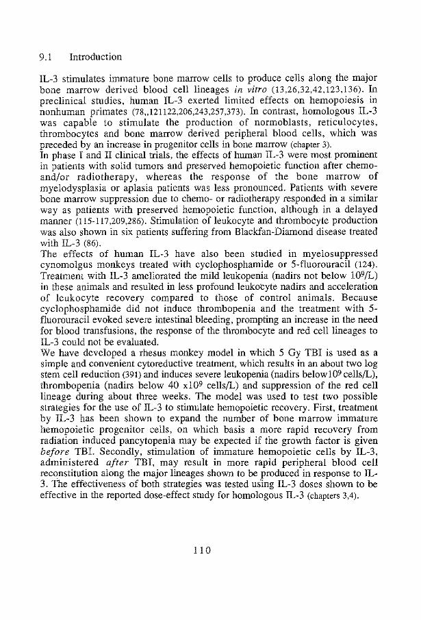
9.1 Introduction
IL-3 stimulates immature bone marrow cells to produce cells along the major bone marrow derived blood cell lineages in vitro (13,26,32,42,123,136). In preclinical studies, human IL-3 exerted limited effects on hemopoiesis in nonhuman primates (78,121122,206,243,257,373). In contrast, homologous IL-3 was capable to stimulate the production of normoblasts, reticulocytes, thrombocytes and bone marrow derived peripheral blood cells, which was preceded by an increase in progenitor cells in bone marrow (chapter 3). In phase I and II clinical trials, the effects of human IL-3 were most prominent in patients with solid tumors and preserved hemopoietic function after chemoand/or radiotherapy, whereas the response of the bone marrow of myelodysplasia or aplasia patients was less pronounced. Patients with severe bone marrow suppression due to chemo- or radiotherapy responded in a similar way as patients with preserved hemopoietic function, although in a delayed manner (115-117,209,286). Stimulation of leukocyte and thrombocyte production was also shown in six patients suffering from Blackfan-Diamond disease treated with IL-3 (86). The effects of human IL-3 have also been studied in myelosuppressed cynomolgus monkeys treated with cyclophosphamide or 5-fluorouracil (124). Treatment with IL-3 ameliorated the mild leukopenia (nadirs not below 109/L) in these animals and resulted in less profound leukocyte nadirs and acceleration of leukocyte recovery compared to those of control animals. Because cyclophosphamide did not induce thrombopenia and the treatment with 5-fluorouracil evoked severe intestinal bleeding, prompting an increase in the need for blood transfusions, the response of the thrombocyte and red cell lineages to IL-3 could not be evaluated. We have developed a rhesus monkey model in which 5 Gy TBI is used as a simple and convenient cytoreductive treatment, which results in an about two log stem cell reduction (391) and induces severe leukopenia (nadirs belowJ09 cells/L), thrombopenia (nadirs below 40 xl09 cells!L) and suppression of the red cell lineage during about three weeks. The model was used to test two possible strategies for the use of IL-3 to stimulate hemopoietic recovery. First, treatment by IL-3 has been shown to expand the number of bone marrow immature hemopoietic progenitor cells, on which basis a more rapid recovery from radiation induced pancytopenia may be expected if the growth factor is given before TBI. Secondly, stimulation of immature hemopoietic cells by IL-3, administered after TBI, may result in more rapid peripheral blood cell reconstitution along the major lineages shown to be produced in response to IL-3. The effectiveness of both strategies was tested us·ing IL-3 doses shown to be effective in the reported dose-effect study for homologous IL-3 (chapters 3,4).
11 0

9 .2. Results
9.2.1 Pretreatment with IL-3
Three animals were given a continuous iv infusion of IL-3 before TBL One animal received 1 !lg/kg/day for 14 consecutive days, one 10 J.Lg/kg/day and one 30 J.Lg/kg/day for 7 consecutive days. The onset of leukopenia (i.e., leukocyte counts <109/L), at 5, 6 and 6 days respectively, was not delayed as compared to control animals (Figure 1, left panels). Leukocyte counts of the animal given IL-3 in a dose of 10 J.Lg/kg/day did not reach levels as low as those of controls. In the other two animals, leukocytes regenerated at the same rate as control animals. An earlier recovery of leukopenia could not be demonstrated: leukocyte count values of more than 109JL were observed in the IL-3 treated animals at day 23, 19 and 22, respectively, which was not significantly different from the control animals (Figure 1, Table 1 ). The differential counts revealed that the regeneration of leukocytes was largely attributable to the production of neutrophilic granulocytes (Table 2). The onset of thrombopenia (i.e., <40 xl09JL) started at day 11, 10 and 12, respectively. The number of transfusions required were similar to those of controls. Thrombocyte counts >40 xJ09JL were reached after 20, 14 and 29 days. The animals treated with 1 and 30 J.Lg/kg/day showed delayed thrombocyte recovery, whereas the recovery of the animal treated with 10 !lglkg/day was similar to those of the controls (Figure 1, Table 1). The decrease in reticulocytes to counts lower than 1% was delayed in two animals (day 12 and 8 in the animals treated with 10 and 30 J.Lg/kg/day respectively). Reticulocytes remained lower than 1% until day 20, 20 and 29 days. The reticulocyte recovery of the animals pretreated with 1 and 30 J.Lg/kg/day was delayed, whereas reticulocyte recovery in the 10 J.Lg/kg/day treated animal was similar to those of controls (Figure 1, Table 1 ). The onset of nonnoblastosis was delayed in the same two animals (day 23 and 32). In the animal treated with 10 J.Lg/kg/day, nonnoblastosis was similar to that of control monkeys. Normoblast counts reached values of 0.6, 1.5 and 0.1 xl09JL respectively, not significantly different from those of controls (Table 1). The animals treated with 1 and 30 !lg/kg/day revealed a positive blood culture at day 28 and 17, respectively; septicemia may have delayed the recovery of leukocytes and thrombocytes in the latter animal.
l l I

100
~ 0 10
"' "' 0
Ul
0
it 100 (.) 0 .0 E e cS
1000.---------------,
0 Q 2..100 Ul
"' % 0 :; 10 .S! ~
0 5 10 15 20 25 30 0 time (day)
5 1 0 15
Figure l Regeneration curves of animals, subjected to 5 Gy Till
20 25 30
Shaded areas represent the mean ±sd of values of four (reticulocytes) or five control animals. Horizontal lines defme cytopenia. Left panels: Closed circles, open circles and triangles represent the regeneration curves of animals treated before TBl (day 0) with I, 10 or 30 ~g/kg/day !L-3 iv for 14,7 and 7 days, respectively. Right panels: Closed circles represent the regeneration curves of the animal treated qfter TBI with 10 ~g/kg/day !L-3 iv for 30 days, and open circles and triangles those of animals treated with 10 ~g/kg/day sc for I 4 days after TBl. !L-3 was started at day !.
!!2

Table 1 Regeneration parameters after 5 Gy TBI
UAN IL-3 leukocytes thrombocytes transfusions normoblasts reticulocytes < J09JL <40 xJ09JL <I%
onset duration onset duration WBT TI first highest value onset duration dose* days day days day days # # day 109/L day days
beforeTBl 8863 1 iv 14 5 ns !80.05 11 ns 911s 2 411s 230.05 0.6 ns 211S 240.05 BB64 10 iv 7 6 ns J30.05 11 ns 30.05 I 2 ns 18 liS 1.5 ns J20.01 80.05 8613 30iy 7 6 ns 16 ns J20.05 !70.01 2 5o.o5 320.001 0.1 ns 30.01 210.05
qfrerTBI 4055 10 iv 30 70.01 90.01 J20.05 sns 0 2 !IS 18 liS 3 _2o.o1 5 ns 60.01 8705 10 sc 14 6 ns 14 ns !70.001 J0.01 1 0 ns 20 ns 1.7 ns 211S 70.01 8782 10 sc 14 sns 14 11S !40.01 JO.O/ 0 1 flS 20 ns 3.10.01 3 ns 70.Dl
TBI controls 1 XS 5 16 11 5 2 1 19 1.1 + + 8831 6 15 10 9 1 5 18 0.2 3 17 8717 5 17 11 7 1 1 21 1.5 3 17 8715 5 14 11 7 3 1 19 0.9 4 15
mean ± sd 5.3±0.5 15.5 ±1.3 10.8 ±o.5 7 ±1.6 3.8 ± 1.71 19.3 ±1.3 0.9 ±0.5 3.3 ±o.6 16.3 ±1.2
95 % confidence limit 4.5- 6 13.5- 17.6 10- 11.6 4.4-9.6 1.9 -5.6 17.3- 21.3 0- 1.8 1.9 - 4.8 14- 19.7
UAN indicates unique animal number. At day 0, all animals were subjected to TBI. At day 0, IL-3 was stopped in the pretreated animals. At day 1 IL-3 was started in animals treated with IL-3 after TBI. * dose in )..!g/kg/day, iv and sc indicate continuous intravenous and subcutaneous administration. TT and WBT indicate thrombocyte and whole blood transfusion, respectively. :j: reticulocyte values were excluded, since a different method was used. Levels of significance (ns: not significant, p=O.Ol, p=0.05 and p=O.OOl) were obtained by comparing the individual values of IL-3 treated animals to the mean of control animals, using Student-t test. § the mean of total transfusions in controls was compared to the total transfusions of IL-3 treated animals.

Table 2 Differential white blood cell counts after 5 Gy TBI
UAN IL-3* WBC neutrophils basophils eosinophils monocytes lymphocytes J09/L ]09/L J09fL !09/L J09fL J09fL
dose da~s d 0 14 28 0 14 28 0 14 28 0 14 28 0 14 28 0 14 28
beforeTBI 8863 1 iv 14 17.0 0.2 7.6 7.0 6.1 4.8 0 0.3 0 0.2 0.5 4.8 1.0 BB64 10 iv 7 8.8 0.8 3.8 4.9 3.0 0 0 0.4 0.04 0.1 0.1 3.3 0.7 8613 30 iv 7 8.4 0.5 3.1 4.2 2.5 0.7 0 0 0 0.7 0.2 2.9 0.4
afterTBI 4055 10 iv 30 14.1 0.6 17.9 8.3 0.2 12 0 0 0.4 0.14 0 0.4 0.14 0 0.4 5.5 0.3 4.7 8705 10 sc 14 9.9 0.8 3.5 5.3 0.06 1.7 0 0.1 0 1.1 0 0 0.14 0.03 0.1 3.4 0.6 1.7 8782 10 sc 14 8.5 0.7 2.7 5.4 0.2 1.6 0 0.1 0 0.08 0 0.14 0.2 0.12 0 2.8 0.3 1.0
TBI controls !XS 13.8 0.2 4.4 8.4 1.3 0 0 0.6 0.04 0 0.1 4.8 2.9 8831 9.5 0.2 3.7 4.1 2.0 0.2 0 0.4 0 0.5 0.2 4.4 1.6 8717 5.9 0.4 5.9 4.2 4.9 0 0 0.3 0 0 0.2 1.4 0.7 8715 7.2 0.2 2.9 4.1 1.7 0 0 0.2 0 0 0.05 2.9 1.1
unirradiated controls mean± sd 9.0 ± 4.6 5.1 ±3.5 0 0.3 ± 0.4 0.2 ± 0.2 3.4 ± 1.5
*dose of!L-3 in J.lg/kg/day. WBC indicates white blood cells

9.2.2 Animals treated with IL-3 after TBI
Three animals were treated with 10 IJ.g/k:g/day. One monkey was given IL-3 intravenously for 30 days and two received IL-3 subcutaneously for 14 consecutive days. The onset of leukopenia started at !lay 7, 6 and 5, respectively, and leukopenia lasted until day 16, 20 and 19. The nadir of leukocyte counts of all three animals was not as profound as those of control animals and recovery was accelerated in the animal which received IL-3 iv, whereas in the other two monkeys leukocytes regenerated similar to the controls (Figure I, right panels, Table 1). Also in these monkeys, the regeneration of leukocytes was largely attributable to the production of neutrophils (Table 2). Transfusions were given at day 11, 17 and 13 for the first time. Transfusion requirements tended to be reduced compared to control animals (Table 1). Animals became transfusion independent at day 17, 18 and 15. Accelerated recovery of thrombocytes compared to control animals was observed in all three monkeys (Figure 1). The period that reticulocyte counts were lower than 1% was shortened ( 6, 7 and 7 days, respectively) and the recovery phase was accelerated in all three animals (Figure l, Table 1). Normoblasts appeared in the peripheral blood at day 18, 20 and 20 and reached values higher than those of control animals (3.2, 1.7 and 3.1 xl09(L, respectively) (Table 1). As in control animals, positive blood cultures were not detected. The occurrence of side effects was limited to the development of a few mild urticaria in animals 8782 and 4055 at the end of IL-3 treatment (day 15 and 30)(chapter 8).
9.2.3. Bone marrow cultures
Bone marrow aspirates were performed weekly whenever thrombocyte counts were sufficiently high and the clinical condition allowed for safe anesthesia. Therefore, the data (Table 3) do not cover the whole regeneration period of all control and IL-3 treated animals. Bone marrow cellularity returned to normal values after 4 weeks in animals treated with IL-3 after TBI, whereas bone marrow cellularity of animals in the other group remained low during this period, similar to bone marrow cellularity of TBI control animals. In the first week after TBI, colony-formation in GM-CSF stimulated cultures or in unstimulated cultures ("background colony-formation") could not be detected, except for one animal which was pretreated with 30 !J.g/k:g/day IL-3 and showed some persistent colony-formation upon stimulation with GM-CSF. In the second week, the bone marrow of animals treated with IL-3 after TBI showed some colony-formation upon stimulation with GM-CSF, whereas background Golony-formation remained undetectable. In the fourth week after TBI, CFU-GM approached normal numbers in the bone marrow of animals treated with IL-3 after TBI, whereas CFU-GM analyses in the other group still remained low.
115

Table 3 Bone marrow cellularity and colony-formation by progenitor cells after 5 Gy 'fBI
IL-3* TNCt CFU-C BFU-E CFU-E col xJ031 mlJ2unctate col x103 col xJ03
1Q6 1 mlJ2unctate 5ackground GM-CSF I ml punctate I ml punctate dose da~s wk 1 2 3 4 1 2 3 4 I 2 3 4 I 2 3 4 1 2 3 4
before 'fBI 10 iv 7 1.2 5.6 27 0 0.6 0 1.7 0 0.5 5.9 - 5.1 227 30 iv 7 1.2 1.6 6 0 0 0.05 0 0 0 6.2 - 0 3
qfterTBI 10 iv 30 1.2 0.8 7.0 121 0 4.8 44 0.2 6.2 97 0 0.07 11 0 38 1085 10 sc 14 1.4 8.2 78 0 0 15 0 1.2 84 0 0.4 16 466 10 sc 14 2.6 2.0 52 0 0 26 0 0.08 77 0 0 28 514
'fBI controls 2.6 0.4 1.1 5 0 0.08 0 0.7 0 0.02 0.3 1.8 0 0.2 1.8 0 32 1.6 1.4 1 0 0 0 0 0 0.2 0 0 0.07
13.0 10.4 2.2 1.7 0.3 2.2
unirradiated controls mean± SEM 73 ± 12 98±9 157 ± 11 51± 7 467 ± 19
*dose of IL-3 in ~g/kg/day.t total nucleated cells. Background colony-formation in the absence of any added growth factor

BFU-E and CFU-E recovery followed similar time courses as CFU-GM recovery and was most pronounced in all three animals treated with IL-3 after TBI. BFU-E could be detected in the third week after TBI except for one animal, which also had a delayed recovery of reticulocytes and a delayed onset of normoblastosis. The bone marrow of this animal also revealed low numbers of CFU-E four weeks after TBI. CFU-E could be detected in the first week after TBI in two IL-3 pretreated animals, in which the onset of the decrease in reticulocyte counts was delayed as well. In the fourth week after TBI, BFU-E as well as CFU-E in bone marrow of animals treated with IL-3 AFTER TBI were, similar to CFU-GM numbers, clearly increased in comparison to the other two groups of monkeys.
9.3 Discussion
Moderate hemopoietic stimulation was observed by homologous IL-3 administered to rhesus monkeys subjected to 5 Gy TBI. In the majority of the monkeys, a biphasic recovery pattern was observed (Figure 1) with a small, apparently abortive rise in the second week after TBI, and an exponential increase in the third. A similar biphasic pattern has been described in humans and is explained by a two-compartment model for hemopoiesis, consisting of progenitor cells with only a limited capacity of clonal expansion, which cause the transient early rise, and immature cells capable of exponential expansion and sustained reconstitution (108). The biphasic pattern was most prominent for reticulocytes, which take a couple of days to mature, but less so for leukocytes, which are predominantly short-lived granulocytes, and also less prominent for thrombocytes, which are presumably rapidly consumed in the initial phase of regeneration. In monkeys given IL-3 before TBI, an expected later onset of cytopenia could only be demonstrated for reticulocytes in the two animals which received IL-3 during 7 days before TBI. This finding may be explained by IL-3 stimulated production of erythroid progenitor cells (chapter 3), as is supported by the persistence of late erythroid progenitor cells (CFU-E) in the first week after TBI, whereas early erythroid progenitor cells (BFU-E) were absent. The reported IL-3 induced increase in inunature hemopoietic progenitor cells (chapter 3) apparently did not contribute to an earlier exponential peripheral blood cell recovery. Rather, the observed delay in exponential recovery of reticulocyte and thrombocyte production indicates that the IL-3 induced rise in progenitor cells occurs at the expense of immature cells available for the exponential regeneration phase. This may imply, that IL-3 treatment in vivo promotes differentiation rather than self renewal of stem cells. It is concluded that IL-3 treatment before cytoreductive therapy should be avoided as it may delay peripheral blood recovery.
11 7

Accelerated blood cell reconstitution could be demonstrated in rhesus monkeys treated with 10 Jlg/kg/day of IL-3 after TBI for leukocyte, thrombocyte as well as for reticulocyte recovery, but was less pronounced than could have been expected from the observed strong stimulatory effects of IL-3 on the hemopoietic system of unirradiated animals (chapters 3,4). The effect of IL-3 was more prominent in the thrombocyte and red cell lineages than in leukocyte series, which is also contrary to unirradiated monkeys. At the doses tested, which were selected for effectiveness in unirradiated monkeys, IL-3 shortened pancytopenia but failed to prevent it. The measurements of bone marrow hemopoietic progenitor cells are in line with the data on peripheral blood cell recovery. Bone marrow cellularity as well as granulocyte/macrophage and erythroid progenitor cells showed a considerable rise in the monkeys treated with IL-3 after TBI. The increases were most prominently observed in the fourth week when IL-3 treatment had been discontinued already in two monkeys and, thus·, were the result of IL-3 stimulation in the first two weeks after TBI. This suggests that IL-3 treatment supplemented with growth factors which act on more mature cells, such as erythropoietin or G-CSF, may result in mutually enhanced effectiveness. The limited response of the residual hemopoietic system of irradiated rhesus monkeys to IL-3 in comparison to the large effects in normal monkeys is as yet not fully understood. Several mechanisms may be involved to reduce the response. Endogenous IL-3 production may already be close to optimal after cytoreductive treatment, which is unlikely, or a relative shortage of growth factors acting on more mature progenitor cells may lead to underestimation of the IL-3 response. It has been noted that full expression of the stimulatory spectrum of IL-3 stimulated colony-formation of human bone marrow is dependent on the presence of CDllb-positive accessory cells (26). In bone marrow of normal, untreated monkeys, background colony-formation is usually rather high (i.e., numerically comparable to GM-CSF stimulated colonyformation), but it is strongly reduced after depletion of CDllb accessory cells (unpublished observation). The absence of background colony-formation during the initial phase of recovery from TBI may thus indicate a deficiency of accessory cells required for full expression of the effects of IL-3. In addition, the characteristic nonhemopoietic effects of IL-3 observed in normal monkeys, i.e., urticaria, edema and palpable lymph nodes, were absent in the irradiated monkeys (chapter 8), and so were the characteristic atypical basophilia and eosinophilia (Table 2). Rather, IL-3 stimulated the recovery of neutrophilic granulocytes. It is not excluded that the large hemopoietic effects of IL-3 in unirradiated monkeys are in part attributable to cascade reactions initiated in the development of nonhemopoietic effects.
I I 8

GENERAL DISCUSSION
11 9

10.1 Species specificity of IL-3
IL-3 is a pleiotropic hemopoietic growth factor generally thought to play a role in the regulatory mechanisms acting on immature hemopoietic cells. For preclinical studies in rhesus monkeys on its mechanism of action, efficacy in counteracting pancytopenia following cytoreductive treament and its side effects, the homologous IL-3 had to be cloned, since in vivo and in vitro data suggested a (unidirectional) species specificity of human IL-3. Although IL-3 in general has followed a more divergent course during evolution than related hemopoietic growth factors such as GM-CSF (416,417), the evolution of rhesus monkey IL-3 was found to be unusual in that most nucleotide substitutions appeared to have resulted in an amino acid substitution in the protein and, thus, shows a more pronounced divergence than IL-3 of other primate species (45). The unidirectional species specificity was confirmed by the in vivo difference in hemopoietic effects (chapter 3,4,6) between human and rhesus monkey IL-3 and the in vitro difference in binding affinity to rhesus monkey bone marrow cells and human AML cells (chapter 6). Therefore, the results described in this thesis more validly predict the biological effects and possibly therapeutic applications of recombinant IL-3 in humans than those obtained in studies performed with human IL-3 in nonhuman primates (78,122,124,205,206,243,257,373).
10.2 Stimulatory effects of IL-3 in normal rhesus monkeys
The stimulatory range of IL-3 treatment in vivo was shown to include cells of the myeloid, erythroid and thrombocyte lineages (chapter 3,4). Increases in peripheral blood cells, most prominently so in eosinophilic and atypical, IL-3 receptor expressing basophilic granulocytes (chapter 7), were preceded by an expansion of bone marrow progenitor cells, resulting in bone marrow hyperplasia (chapters 3,8). The numbers of peripheral blood T and B lymphocytes were not increased following IL-3 treatment (chapter 3,4). These data suggested that IL-3 may act on immature progenitor cells and does not result in commitment into the lymphoid lineage. Initial studies suggested that the iv route of administration was more effective in stimulating hemopoiesis than the sc route of administration (chapter 3). This was further investigated by pharmacokinetic studies and confirmed by a more extensive comparison of both routes of administration (chapter 4). Pharmacokinetic studies showed that the bioavailability of IL-3 after sc administration was about 40% of that after iv administration. The 2.5-fold difference in bioavailability does not fully explain the 10-fold difference in hematological effects of both routes of administration. Part of the difference may be explained by indirect effects, e.g., the perpetual activation of IL-3R expressing (chapter 7) basophilic granulocytes, resulting in high serum levels of histamine (chapter 3). It is conceivable that a continuous low level of IL-3 is more
120

effective in inducing indirect effects of IL-3 than repetitive higher levels after sc administration. The relatively short terminal half life (-2 hours) of IL-3 may explain why the magnitude of the hemopoietic effects of the sc administered IL-3 did not change significantly when the dose was divided in two or three daily injections (chapter 4). The IL-3 induced effects saturated at the highest doses used. In several animals a reversal of the effects was observed during IL-3 treatment, most probably due to a neutralizing antibody response, especially in animals treated with high doses of IL-3 (chapter 5). This demonstrated that a recombinant homologous protein is capable of eliciting an antibody response, possibly due to epitopes that are not exposed by glycosylated native IL-3. Alternatively, IL-3 may be a locally acting cytokine not normally found in the circulation and may therefore more easily elicit an antibody response following injection. Until now, antibody formation against homologous IL-3 has not been reported. This may be due to the relative low doses used in clinical studies or to an immunocompromised status of the patients treated with IL-3 (115-117.209.286). Dose-dependent side effects during IL-3 treatment included urticaria, edema, enlargement of lymph nodes and spleen, mild parenchymal liver damage, acute arthritis, and at high doses, thrombopenia and anemia (chapter 8). The continuous production and stimulation of atypical basophils, which expressed IL-3 receptors abundantly (chapter 7), and concomitant high levels of histamine (chapter 3) may explain the majority of the observed side effects and the ineffectiveness of antihistamine treatment in preventing the development or reducing the severity of the side effects (chapter 8). In phase I and II clinical trials, comparable side effects have been observed, albeit less frequent and less serious. In general, the patients treated with IL-3 did not receive the high doses used in monkeys, while most of the patients either suffered from bone marrow dysplasia/aplasia or had been treated with intensive cytoreductive therapy, both conditions resulting in much lower numbers of cells responding to IL-3 than are available in healthy monkeys (115-117 ,209.286).
10.3 The effects of IL-3 in myelosuppressed animals
In contrast to the broad range of hemopoietic and side effects in normal animals, the effects of IL-3 in animals subjected to TBI were less pronounced (chapter 8.9). The acceleration of recovery from pancytopenia was much less than expected in animals treated with IL-3 after TBI, whereas an earlier recovery from pancytopenia in animals treated with IL-3 before TBI was virtually absent and in one animal the hemopoietic recovery tended to be delayed. Both observations may indicate that IL-3 does not significantly expand the number of immature progenitor cells responsible for hemopoietic regeneration (stem cells). Since side effects were almost absent in animals subjected to TBI we presumed that the large hemopoietic effects in unirradiated animals were predominantly
121
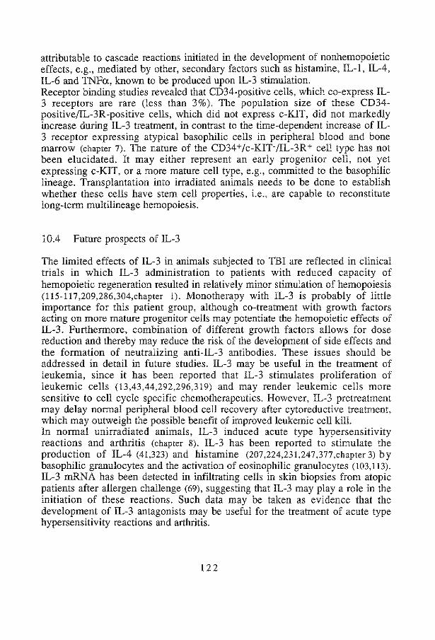
attributable to cascade reactions initiated in the development of nonhemopoietic effects, e.g., mediated by other, secondary factors such as histamine, IL-l, IL-4, IL-6 and TNFa, known to be produced upon IL-3 stimulation. Receptor binding studies revealed that CD34-positive cells, which co-express IL-3 receptors are rare (less than 3%). The population size of these CD34-positive/IL-3R-positive cells, which did not express c-KIT, did not markedly increase during IL-3 treatment, in contrast to the time-dependent increase of IL-3 receptor expressing atypical basophilic cells in peripheral blood and bone marrow (chapter 7). The nature of the CD34+jc-KIT-/IL-3R+ cell type has not been elucidated. It may either represent an early progenitor cell, not yet expressing c-KIT, or a more mature cell type, e.g., committed to the basophilic lineage. Transplantation into irradiated animals needs to be done to establish whether these cells have stem cell properties, i.e., are capable to reconstitute long-term multilineage hemopoiesis.
10.4 Future prospects of IL-3
The limited effects of IL-3 in animals subjected to TBI are reflected in clinical trials in which IL-3 administration to patients with reduced capacity of hemopoietic regeneration resulted in relatively minor stimulation of hemopoiesis (115-117,209,286,304,chapter 1). Monotherapy with IL-3 is probably of little importance for this patient group, although co-treatment with growth factors acting on more mature progenitor cells may potentiate the hemopoietic effects of IL-3. Furthermore, combination of different growth factors allows for dose reduction and thereby may reduce the risk of the development of side effects and the formation of neutralizing anti-IL-3 antibodies. These issues should be addressed in detail in future studies. IL-3 may be useful in the treatment of leukemia, since it has been reported that IL-3 stimulates proliferation of leukemic cells (13,43,44,292,296,319) and may render leukemic cells more sensitive to cell cycle specific chemotherapeutics. However, IL-3 pretreatment may delay normal peripheral blood cell recovery after cytoreductive treatment, which may outweigh the possible benefit of improved leukemic cell kill. In normal unirradiated animals, IL-3 induced acute type hypersensitivity reactions and arthritis (chapter 8). IL-3 has been reported to stimulate the production of IL-4 (41,323) and histamine (207,224,231,247,377,chapter 3) by basophilic granulocytes and the activation of eosinophilic granulocytes (103,113). IL-3 mRNA has been detected in infiltrating cells in skin biopsies from atopic patients after allergen challenge (69), suggesting that IL-3 may play a role in the initiation of these reactions. Such data may be taken as evidence that the development of IL-3 antagonists may be useful for the treatment of acute type hypersensitivity reactions and arthritis.
122

Summary en Samenvatting
123
Summary
Interleukins, colony-stimulating factors and erythropoietin, together classified as hemopoietic growth factors, regulate the production of peripheral blood cells. These growth factors, which may act on developmentally early, immature bone marrow cells, or on late stages, i.e., on the maturation of committed progenitor cells, have been produced and purified to homogeneity as recombinant proteins. Several of these have entered phase I, II and HI clinical studies, whereas other are still under investigation in preclinical studies. Interleukin-3 is thought to stimulate immature hemopoietic progenitor cells to differentiate and to modulate the function of specific mature end cell types. The full stimulatory spectrum of IL-3 was tested in a preclinical rhesus monkey model. Recombinant homologous IL-3 was used, as human IL-3 was shown to be insufficiently effective in stimulating the rhesus monkey hemopoietic system. Administration to normal rhesus monkeys resulted in a dose-dependent stimulation of the production of all major bone marrow derived peripheral blood cells, after a lag phase of one week, in which an increase of progenitor cells in the bone marrow was observed (chapters 3,4). Peripheral blood T and B lymphocyte numbers remained unaffected by IL-3 treatment. Characteristic for IL-3 treatment was the production of high numbers of atypical basophilic cells, which comprised the majority of nucleated cells at the end of IL-3 treatment, and a concomitant high level of histamine. At low dose levels, thrombocytosis was observed, whereas high dose IL-3 treatment resulted in severe thrombopenia. Normoblastosis preceded reticulocytosis, but did not result in a rise in erythrocytes or hemoglobin levels; at high doses, anemia was seen. On the basis of the pharmacokinetics of recombinant nonglycosy lated IL-3 it was predicted that the intravenous route of administration should be more effective than the subcutaneous route of administration, which was confirmed by a direct comparison of both routes (chapter 4). A neutralizing antibody response was elicited by high doses of IL-3. Prolonged iv administration of low dose IL-3 did not result in antibody formation (chapter 5). To evaluate the mechanisms involved in the species specificity of human IL-3, binding studies using radiolabeled IL-3 were performed. The rhesus monkey IL-3 receptor complex bound homologous IL-3 20- to 50-fold more effectively than human IL-3, whereas human IL-3 receptors bound both species of IL-3 with similar affinity, demonstrating the unidirectional species specificity of human IL-3. The species specificity of human IL-3 was confirmed by in vivo comparison of both species of IL-3; homologous IL-3 was about 5-fold more effective than its human counterpart in stimulating hemopoiesis (chapter 6). Since little information was available on IL-3 receptor expression of normal bone marrow cells, the binding characteristics of the IL-3 receptor were evaluated by conventional binding studies using radiolabeled IL-3. The distribution of IL-3 receptors on different cell types was investigated by more novel flow cytometric methods, using biotin-labeled IL-3 (chapters 6,7). An
124

average of 50 high- and 1100 low-affinity IL-3 receptors per cell were detected on normal rhesus monkey bone marrow. Competition of high-affinity IL-3 binding by GM-CSF could not be demonstrated (chapter 6), probably due to differential expression of growth factor specific a-chains. IL-3 receptors could only be demonstrated on some 3% or less of the isolated CD34-positive bone marrow cells, which did not express c-KIT. When rhesus monkeys were treated with IL-3, the number of high- as well as low- affinity IL-3 receptors did increase significantly, which could be attributed to the increase of atypical, histamine containing, basophilic cells. The percentage and absolute number of CD34-positive, c-KIT-negative cells, expressing IL-3 receptors did not increase during IL-3 treatment (chapter 7). Besides the hemopoietic effects, a variety of dose-dependent side effects were observed during IL-3 treatment, which included urticaria, edema, enlargement of spleen and lymph nodes and at high dose levels, anemia and thrombopenia (chapter 8). Irrespective of the dose, acute arthritis was observed in several animals. Resistance to the development of arthritits was not related to the MHC allele A26, as is the case in collagen-induced arthritis in juvenile rhesus monkeys. Rather, the MHC alleles B9 and Dr5 were significantly more frequently found in the animals which developed IL-3-induced arthritis. Side effects could not be prevented by the addition of histamine antagonists to the treatment protocol, most probably due to perpetual production and activation of IL-3R-expressing, atypical basophils, resulting in a continuous release and, subsequent, high levels of histamine. The effect of IL-3 on bone marrow after cytoreductive treatment was tested in animals subjected to 5 Gy TBI (chapter 9). IL-3 treatment before 5 Gy TBI resulted in a later onset of a reduction in reticulocytes, whereas no effect was seen on the onset of leukopenia or thrombopenia. Pretreatment with IL-3 did not result in a more rapid recovery from pancytopenia; a delay in accelerated expansion of reticulocytes and thrombocytes was observed, whereas the leukocyte regeneration was comparable to that of control irradiated animals. IL-3 treatment after TBI resulted in shortening of the pancytopenic phase, more prominent for the red cell and thrombocyte lineages than for granulocytes. Remarkably, IL-3 stimulated the recovery of neutrophilic granulocytes rather than that of basophils and eosinophils. A decrease in platelet and red cell transfusion requirements was observed. Serial measurements of colonyformation by bone marrow CFU-GM, BFU-E and CFU-E demonstrated accelerated bone marrow recovery, in agreement with the stimulated recovery of peripheral blood cells in animals treated with IL-3 after TBL Side effects were only mild in myelosuppressed animals (chapter 8). The effects of IL-3 in irradiated animals were more limited than predicted from the strong stimulatory effects of IL-3 in unirradiated animals. It was concluded that IL-3 may have a direct stimulatory effect on immature hemopoietic progenitor cells, albeit limited due to the low frequency of bone marrow cells expressing IL-3 receptors. The strong hemopoietic effects of IL-3 in normal
125

rhesus monkeys may be mediated by increased production of other stimulators. Histamine, and possibly IL-l, IL-4, IL-6 and TNFa are candidates for these mediators. It remains to be investigated whether the rare CD34+/c-KIT-/IL-3R+ bone marrow cell population has stem cell properties and whether the perpetual activation of IL-3 receptor expressing atypical basophilic cells has a bearing on the hemopoietic effects of IL-3. IL-3 given to patients with bone marrow of reduced proliferative capacity due to intensive cytoreductive treatment or myelodysplastic disorders may be of limited use to stimulate hemopoiesis. Further investigations and the pharmaceutical development of IL-3 and its derivatives may be directed at its possible role in acute type hypersensitivity reactions and in the initiation of auto-inunune diseases.
126

Samenvatting
lnterleukines, kolonie-stimulerende groeifactoren en erythropoietine, gezamelijk hemopoietische groeifactoren genoemd, reguleren de aanmaak van perifere bloedcellen. Deze groeifactoren, die zowel de groei en differrentiatie van onrijpe beenmergcellen als de rijping van meer gedifferentieerde jonge voorlopercellen beinv loeden, zijn als recombinant eiwitten geproduceerd en gezuiverd. Verscheidene zijn reeds getest in fase I, II en III klinische studies, terwijl de meerderheid nog onderzocht word! in preklinische studies. lnterleukine-3 (IL-3) is een van de groeifactoren waarvan word! aangenomen dat het onrijpe beenmergcellen aanzet tot proliferatie en differentiatie en tevens dat het de functie van specifieke rijpe bloedcellen moduleert. In een preklinisch model met rhesusapen is het spectrum van de stiniulerende werking van deze groeifactor onderzocht. Hiervoor was het nodig homoloog IL-3 te cloneren, omdat het menselijke IL-3 onvoldoende effectief bleek om het bloedvorroende systeem van de rhesusaap te stimuleren. De toediening van deze groeifactor aan gezonde rhesusapen resulteerde in een sterke dosis-afhankelijke produktie van de belangrijke differentiatielijnen die tot perifere bloedcellen leiden, voorafgegaan door een sterke toename van het aantal jonge voorlopercellen in het beenmerg (hoofdstuk 3,4). Het aantal Ten B lymfocyten werd door IL-3 niet beinvloed. De produktie van hoge aantallen atypische basofiele granulocyten en hoge histamine waarden in het serum bleken karakteristiek te zijn voor IL-3 behandeling. Aan het einde van de behandeling maakten deze basofiele granulocyten het grootste dee! van de kemhoudende cel!en in het bloed uit. Een thrombocytose werd gezien wanneer de dieren !age doses IL-3 kregen toegediend. Hoge doses IL-3 daarentegen resulteerden in een thrombopenie. Een normoblastose ging vooraf aan een reticulocytose. Dit leidde echter niet tot verhoging van hemoglobinewaarden in het bloed. Toediening van hoge doses IL-3 resulteerde zelfs in anemie. Op basis van farroacokinetische studies kon worden voorspeld dat eenzelfde dosis IL-3 na intraveneuze toediening het hemopoietische systeem effectiever zou stimuleren dan na subcutane toediening. Dit werd bevestigd door beide manieren van toediening in vivo te vergelijken (hoofdstuk 4 ). Neutraliserende antistoffen tegen IL-3 werden vaker gevormd naarroate de toegediende dosis IL-3 hoger was. Langdurige continue intraveneuze toediening van een !age dosis IL-3 had dit effect niet (hoofdstuk 5). Om inzicht te krijgen in de waargenomen soortspecificiteit van het menselijke IL-3 zijn studies verricht naar de binding van IL-3 aan zijn receptor, gebruikmakend van radioactief gemerkt IL-3. Het bleek dat homoloog IL-3 20 tot 50 maal beter bindt aan het IL-3-receptorcomplex van de rhesusaap dan het menselijke IL-3. Het receptorcomplex op menselijke cellen vertoonde min of meer dezelfde bindings-affiniteit voor beide soorten IL-3. Hierroee werd de soortspecificiteit van het menselijke IL-3 in een richting aangetoond. De soortspecificiteit werd bevestigd door beide soorten IL-3 in vivo te vergelijken;
127

het effect van homoloog IL-3 op de bloedvonning in rhesusapen was ongeveer 5 maal groter dan van het menselijke IL-3 (hoodstuk 6). Omdat er maar weinig gegevens beschikbaar waren over de IL-3-receptor op beenmergcellen zijn de bindingskarakteristieken van de IL-3-receptor onderzocht met behulp van conventionele bindingsstudies, gebruikmakend van radioactief gemerkt IL-3. De distributie van IL-3-receptoren over verschillende celsoorten werd onderzocht met meer modeme flow-cytometrische methoden, gebruikmakend van biotine gemerkt IL-3 (hoofdstuk 6,7). Een gemiddelde van 50 receptoren met hoge affiniteit en ongeveer 1100 receptoren met !age affiniteit werden gevonden op nonnaal beenmerg van rhesusapen. GM-CSF was niet in staat IL-3 van he! receptorcomplex te verdringen (hoofdstuk 6), waarschijnlijk omdat de groeifactorspecifieke a-ketens op verschillende celsoorten tot expressie komen. Op minder dan 3% van geisoleerde CD34-positieve beenmerg cellen werden IL-3-receptoren aangetoond. Expressie van c-KIT kon op deze zeldzame celpopulatie niet worden aangetoond. Ten gevolge van de behandeling van rhesusapen met IL-3 nam zowel het aantal receptoreh met hoge affiniteit als het aantal met !age affiniteit aanzienlijk toe. Dit kon worden toegeschreven aan de toename van het aantal atypische, histamine bevattende, basofiele granulocyten. Noch het percentage, noch het absolute aantal CD34-positieve, c-KIT-negatieve beenmergcellen, die IL-3-receptoren tot expressie brachten nam toe onder IL-3 behandeling (hoofdstuk 7). Naast de sterke hemopoietische effecten van IL-3 werd een scala van dosisafhankelijke bijwerkingen waargenomen, waaronder galbulten, oedeem, vergroting van de milt en lymfklieren en bij hoge doses anemie en thrombopenie (hoofdstuk 8). In enkele dieren werd, onafhankelijk van de toegediende dosis, artritis waargenomen. Resistentie tegen de ontwikkeling van artritis was niet gerelateerd aan de aanwezigheid van het MHC-allel A26, zoals in collageenge'induceerde artritis in juveniele rhesusapen. In dieren die artritis ontwikkelden gedurende IL-3-behandeling werd daarentegen vee! vaker de MHC-allelen B9 en DrS gevonden dan in dieren die vrij van artritis bleven. De bijwerkingen konden niet worden voorkomen door gelijktijdige behandeling met antihistaminica, waarschijniijk vanwege de pennanente aanmaak en activering van atypische, IL-3-receptor-positieve, basofiele granulocyten met als gevolg een voortdurend vrijkomen van histamine, leidend tot hoge serumspiegels. Het effect van IL-3 op beenmerg na cytoreductieve behandeling werd getest in dieren na 5 Gy totale lichaamsbestraling (TBI) (hoofdstuk 9). IL-3 behandeling voor TBI had een latere aanvang van een daling van het aantal reticulocyten tot gevolg, terwijl er geen effect op het begin van de leukopenie en thrombopenie werd waargenomen. Voorbehandeling met IL-3 zorgde niet voor een sneller herstel van de pancytopenie; een vertraging van de versnelde expansie van reticulocyten en thrombocyten werd waargenomen, terwijl de snelheid van de leukocyten regeneratie overeen kwam met die van de controledieren. IL-3 behandeling na TBI resulteerde in een verkorting van de pancytopene periode; meer uitgesproken voor rode cellen en thrombocyten dan voor granulocyten. Het
128
herstel van de neutrofiele granulocyten werd opvallend meer gestimuleerd door IL-3 dan dat van basofiele en eosinofiele granulocyten. De behoefte aan bloedtransfusies was enigszins verminderd bij deze dieren. Een versneld beenmergherstel werd aangetoond door serie!e metingen van kolonievorming door CFU-GM, BFU-E en CFU-E in het beenmerg, in overeensternming met het waargenomen gestimuleerde herstel in het perifere bloed van dieren die behandeld waren met IL-3 na TBI. De bijwerkingen van IL-3 waren in deze dieren uitermate mild (hoofdstuk 8). De stimulering van het hemopoietisch systeem door IL-3 in bestraalde dieren was vee! geringer dan op grond van de sterke stimulerende effecten in gezonde dieren kon worden voorspeld. Er is geconcludeerd dat IL-3 een direct effect op onrijpe hemopoietische voorlopercellen kan hebben, hoewel gering, gezien de !age frequentie van onrijpe beenmergcellen met aantoonbare IL-3-receptorexpressie. De sterke stimulering van de hemopoiese door IL-3 zoals waargenomen in gezonde dieren kunnen indirect zijn geweest door verhoogde produktie van mediatoren zoals histamine, IL-l, IL-4, IL-6 en TNFo:. Verder onderzoek is nodig om te evalueren of de zeldzame CD34-positieve/c-KITnegatieve/IL-3-receptor-positieve beenmergcelpopulatie stamceleigenschappen bezit, d.w.z. in staat is een nieuw hemopoietisch systeem te genereren, en of de voortdurende activering van de IL-3-receptor-positieve, atypische basofiele granulocyten een bijdrage !evert aan de hemopoietische effecten van IL-3. De toediening van IL-3 aan patienten met beenmerg met een verminderde proliferende capaciteit op basis van intensieve chemo- en/of radiotherapie of myelodysplastische afwijkingen zou van gering nut kunnen blijken te zijn om de hemopoiese te stimuleren. Verder onderzoek en de pharmaceutische ontwikkeling van IL-3 en IL-3-derivaten zou gericht kunnen worden op de mogelijke rol in het onstaan van acute overgevoeligheidsreacties en auto-irnmuun ziekten.
129


References
!.Anderson DM, Lyman SD, Baird A, Wignall JM, Eisenman J, Rauch C, March 0, Boswell HS, Gimpel SD, Cosman D and Williams DE. (1990) Molecular cloning of mast cell growth factor, a hematopoietin that is active in both membrane bound and soluble forms. Cell63: 235.
2.Andrews RG, Knitter GH, Bartelmez SH, Langley KE, Farrar D, Hendren RW, Appelbaum FR, Bernstein !D and Zsebo KM. (1991) Recombinant human stem cell factor, a c-kit ligand, stimulates hematopoiesis in primates. Blood 78: 1975.
3.Andrews RG, Singer JW and Berstein ID. (1989) Precursors of colony-forming cells in humans can be distinguished from colony-forming cells by expression of the cd33 and cd34 antigens and light scatter properties. J. Exp. Med. 169: 1721.
4.Arend WP, Welgus HG, Thompson RC and Eisenberg SP. (1990) Biological properties of recombinant monocyte-derived interleukin-1 receptor antagonist J. Clin.. Invest 85: 1694.
5.Asano S. (1991) Human G-CSF: its basis aspects and clinical applications. Am. J. Pediatr. Hematol. Oneal. 13: 400.
6.Asano S, Okano A, Ozawa K, Nakahata T, Ishibashi T, Koike K, Kimura H, Tanioka Y, Hirano T, Kishimoto T, Takaku F and Akiyama Y. (1990) In vivo effects of recombinant human interleukin-6 in primates: stimulated production of platelets. Blood 75: 1602.
?.Ashman L, Cambareri A, To LB, Levinsky Rand Juttner C. (1991) Expression of the YB5.B8 antigen (c-KIT proto-oncogene product) in normal human bone marrow. Blood 78: 30.
S.Atkins E. (1960) Pathogenesis of fever. Physiol. Rev. 40: 580. 9.Avraharn H, Vannier E, Cowley S, Jiang S, ChiS, Dinarello CA, Zsebo KM and Groopman
JE. (1992) Effects of the stem cell factor, c-kit ligand, on human megakaryocytic cells. Blood 79: 365.
lO.Bagby GC. (1987) Production of multilineage growth factors by hematopoietic stromal cells: an intercellular regulatory network involving mononuclear phagocytes and interleukin-1. Blood Cells 13: 147.
!!.Bagby GC, Dinarello CA, Wallace P, Wagner C, Hefeneider S and McCall E. (1986) Interleukin-1 stimulates granulocyte macrophage colony-stimulating activity release by vascular endothelial cells. J. Clin.Invest. 78: 1316.
12.Bakker NPM, Erck MGMv, Otting N, Lardy NM, Noort RCv, Hart BA't, Jonker M and Bontrop RE. (1991) Resistance to collagen-induced arthritis in a nonhuman primate species maps to the major histocompatibility complex class I region. J. Exp. Med. 175: 933.
13.Barber KE, Crosier PS, Purdie KJ, Buchanan JM, Cattermole JA and Watson JD. (1989) Human interleukin-3: effects on normal and leukemic cells. Growth factors 1: 101.
14.Barton BE and Mayer R. (1989) IL-3 induces differentiation of bone marrow precursor cells to osteoclast-like cells. J. lmmunol. 143: 3211.
15.Becker S, Warren MK and Haskill S. (1987) Colony-stimulating factor-induced monocyte survival and dllferentiation into macrophages in serum free-cultures. J. Immunol. 139: 3703.
16.Begley CG, Nicola NA and Metcalf D. (1988) Proliferation of normal human promyelocytes and myelocytes after a single pulse stimulation by purified GM-CSF or G-CSF. Blood 71: 640.
17.Beneviste EN and Merrill JE. (1986) Stimulation of oligodendroglia! proliferation and maturation by IL-2. Nature 321: 610.
18.Berdel WE, Danhauser-Riedl S, Steinhauser G and Winton E. (1989) Various hematopoietic growth factors (!L-3, GM-CSF, G-CSF) stimulate clonal growth of nonhematopoietic tumor cells. Blood 73: 80.
19.Berenson RJ, Andrews RG, Bensinger WI, Kalamasz D, Knitter G, Buckner CD and Bernstein lD. (1988) CD34+ marrow cells engraft lethally irridiated baboons. J. Clin. Invest. 81: 951.
20.Berenson RJ, Bensinger WI, Hill RS, Andrews RG, Garcia-Lopez J, Kalamasz DF, Still BJ, Spitzer G, Buckner CD, Bernstein lD and Thomas ED. (1991) Engraftment after infusion of CD34+ marrow cells in patients with breast cancer or neuroblastoma. Blood 77: 1717.
13 l

21.Berenson RJ, Bensinger Wl and Kalamasz D. (1986) Positive selection of viable cell populations using avidin-biotin immunoabsorption. J. Irnmunol. Methods 91: 11.
22.Bemstein ID, Andrews RG and Zsebo KM. (1991) Recombinant human stem cell factor enhances the formation of colonies by CD34+1in- cells, and the generation of colony-fanning cell progeny from CD34+lin- cells cultured with IL-3, G-CSF, or GM-CSF. Blood 77: 2316.
23.Bischoff SC, Brunner T, Week ALD and Dahinden CA. (1990) IL-5 modifies histamine release and leukotriene generation by human basophils in response to divers agonists. J. Exp. Med. 172: 1577.
24.Bischoff SC and Dahinden CA. (1992) c-kit ligand: a unique·potentiator of mediator release by human lung mast cells. J. Exp. Med. 175: 237.
25.Bolton AE and Hunter WM. (1973) The labelling of proteins to high specific radioactivities by conjugation to a 125-l-containing acylating agent. Biochem. J. 133: 529.
26.Bot FJ, Dorssers LCJ, Wagemaker G and Lowenberg B. (1988) Stimulating spectrum of human recombinant multi-CSF (ll..-3) on human marrow precursors: importance of accessory cells. Blood 67: 1609.
27.Bot FJ, Eijk Lv, Broeders L, Aarden LA and Lowenberg B. (1989) lnterleukin-6 synergizes with M-CSF in the formation of macrophage colonies from purified human marrow progenitor cells. Blood 73: 435.
28.Bot FJ, Schipper P, Broeders L, Delwel R, Kaushansky K and Lowenberg B. (1990) lnterleukin-1alpha also induces granulocyte-macrophage colony-stimulating factor in inunature normal bone marrow cells. Blood 76: 307.
29.Bradding P, Feather IH, Howarth PH, Mueller R, Robberts JA, Britten K, Bews JPA, Hunt TG, Okayama Y and Heusser CH. (1992) IL-4 is localized to and released by human mast cells. J. Exp. Med. 176: 1381.
30.Briddell RA, Brandt JE, Leemhuis TB and Hoffman R. (1992) Role of cytokines in sustaining long-term human megakaryocytopoiesis in vitro. Blood 79: 332.
3l.Briddell RA, Bruno E, Cooper Rl, Brandt JE and Hoffman R. (1991) Effect of c-kit ligand on in vitro hwnan megakaryocytopoiesis. Blood 78: 2854.
32.Briddell RA and Hoffman R. (1990) Cytokine regulation of the human burst-fanning unitmegakaryocyte. Blood 76: 516.
33.Bridges AJ, Malone DG, Jicinsky J, Chen M, Ory P, Engber W and Graziano FM. (1991) Human synovial mast cell involvement in rheumatoid arthritis and osteoarthritis. Arthritis Rheum. 34: 1116.
34.Brizzi M, Avanzi G, Veglia F, ClarkS and Pegoraro L. (1990) Expression and modulation of IL-3 and GM-CSF receptors in human growth factor dependent leukaemic cells. Brit. J. Haematol. 76: 203.
35.Bromley M, Fisher WD and Woolley DE. (1984) Mast cells at sites of cartilage erosion in the rheumatoid joint. Ann. Rheum. Dis. 43: 76.
36.Bromley M and Woolley DE. (1984) Chondroclasts and osteoclasts at subchondral sites of erosion in the rheumatoid joint. Arthritis Rheum. 27: 968.
37 .Broudy VC, Kaushansky K, Harlan JM and Adamson JW. (1987) lnterleukin-1 stimulates human endothelial cells to produce granulocyte macrophage colony-stimulating factor. J. lmmunol. 139: 464.
38.Broudy VC, Lin N, Zsebo K, Birkett N, Smith K, Bernstein I and Papayannopoulou T. (1992) Isolation and characterization of a monoclonal antibody that recognizes the human c-kit receptor. Blood 79: 338.
39.Brown MA, Pierce JH, Watson CJ, Falco J, Ihle JN and Paul WE. (1987) B cell stimulatory factor-1/interleukin-4 mRNA is expressed by normal and transformed mast cells. Cell 50: 809.
40.Broxmeyer HE, Cooper S, Lu L, Hangoc G, Anderson D, Cosman D, Lyman SD and Williams DE. (1991) Effect of murine mast cell growth factor(c-kit proto-oncogene ligand) on colony formation by hwnan marrow hematopoietic progenitor cells. Blood 77: 2142.
41.Brunner T, Heusser CHand Dahinden CA. (1993) Human peripheral blood basophils primed by IL-3 produce IL-4 in response to immunoglobulin E receptor stimulation. J. Exp. Med. 177: 605.
132

42.Bruno E, Cooper RJ, Briddell RA and Hoffman R. (1991) Further examination of the effects of recombinant cytokines on the proliferation of human megakaryocyte progenitor cell. Blood 77: 2339.
43.Budel LM, Elbaz 0, Hoogerbrugge H, Delwel R, Mahmoud LA, Lowenberg Band Touw IP. (1990) Common binding structure for GM-CSF and IL-3 on human acute myeloid leukemia cells and monocytes. Blood 75: 1439.
44.Budel LM, Touw IP, Delwel R, Clark SC and Lowenberg B. (1989) IL-3 and GM-CSF receptors on human acute myelocytic leukemia cells and relationship to the proliferative response. Blood 74: 565.
45.Burger H, van Leen RW, Dorssers LCJ, Persoon NLM and Wagemaker G. (1990) Species specificity of human interleukin-3 demonstrated by cloning and expression of the homologous rhesus monkey (Macaca mulatta) gene. Blood 76: 2229.
46.Burgess A and Metcalf D. (1980) Characterization of a serum factor stimulating the differentiation of myelomonocytic leukemia cells. Int. J. Cancer 26: 647.
47.Byron JW. (1977) Mechanism for histamine H2-receptor induced cell-cycle changes in the bone marrow stem cell. Agent Actions 7: 209.
48.Byron JW. (1980) Pharmacodynamic basis for the interaction of cimetidine with the bone marrow stem celi(CRJ-s). Exp. Hematol. 8: 256.
49.Calvo JC, Radicella JP and Charreau EH. (1983) Measurement of specific radioactivities in labelled hormones by self-displacement analysis. Biochem. J. 212: 259.
50.Cannistra SA, Vellenga E, Groshek P, Rambaldi A and Griffin JD. (1988) Human granulocyte-monocyte colony-stimulating factor and interleukin-3 stimulate monocyte cytotoxicity through a tumour necrosis factor-dependent mechanism. Blood 71: 672.
51. Cerny J. (197 4) Stimulation of bone marrow haemopoietic stem cells by a factor from activated T cells. Nature 240: 63.
52.Chan JY, Slamon DJ, Nimer SD, Golde DW and Gassen JC. (1986) Regulation of expression of human granulocyte/macrophage colony-stimulating factor. Proc. Nat!. Acad. Sci. USA 83: 8669.
53.Chang JM, Metcalf D, Lang RA, Gonda TJ and Johnson GR. (1989) Nonneoplastic hematopoietic myeloproliferetive syndrome induced by dysregulated multi-CSF (IL-3) expression. Blood 73: 1487.
54.Chen W and Zlotnik A. (1991) IL-10 a novel cytotoxic T cell differentiation factor. J. Immunol. 147: 528.
55.Cherner JA, Jensen RT, Dubois A, O'Dorsio TM, Gardner JD and Metcalfe DD. (1988) Gastrointestinal dysfunction in systemic mastiocytosis. Gastroenterology 95: 657.
56.Civin Cl, Banquerigo ML, Strauss LC and Loken MR. (1987) Antigenic analysis of hematopoiesis. VI. Flow cytometric characterization of My-10 positive progenitor cells in normal human bone marrow. Exp. Hematol. 15: 10.
57.Civin C!, Strauss LC, Brovall C, Fackler MJ, Schwartz JF and Shaper JH. (1984) Antigenic analysis of hematopoiesis.III. A hematopoietic progenitor cell surface antigen defined by a monoclonal antibody raised against KO-la cells. J. lmmunol. 133: 157.
58.Cline MJ and Golde DW. (1974) Production of colony-stimulating activity by human lymphocytes. Nature 248: 703.
59.Clutterbuck EJ, Hirst EMA and Sanderson CJ. (1989) Human IL-5 regulates the production of eosinophils in human bone marrow cultures: comparison and interaction with IL-1, IL-3, and GM-CSF. Blood 73: 1504.
60.Clutterbuck EJ and Sanderson CJ. (1990) Regulation of human eosinophil precursors by cytokines: a comparison of recombinant human interleukin-1 (rhiL-1), rh!L-3, rh!L-5, rh!L-6, and rhGranulocyte-Macrophage colony stimulating factor. Blood 75: 1774.
61.Cohen DR, Hapel A and Young !G. (1985) Cloning and expression of the rat IL-3 gene. Nucl. Acids. Res. 14: 3642.
62.Cosman D. (1990) A new cytokine receptor superfamily. Trends Biochem. Sci. 15: 265. 63.Cosman D, Cerretti DP, Larsen A, Park L, March C, DowerS, Gillis S and Urdal D. (1984)
Cloning, sequence and expression of human IL-2 receptor. Nature 312: 768.
133

64.Crisp AJ. (1984) Mast cells in rheumatoid arthritis. J. Royal Soc. Med. 77: 450. 65.Crisp AJ, Chapman CM, Kirkham SE, Schiller AL and Krane SM. (1984) Articular
mastocytosis in rheumatoid arthritis. Arthritis Rheum. 27: 845. 66.Crown J, Jakubowski A, Kemeny N, Gordon M, Gasparetto C, Wong G, Sheridan C, Toner
G, Meisenberg B, Botet J, Applewhite J, Sinha S, Moore M, Kelsen D, Buhles W and Gabrilove J. (1991) A phase I trial of recombinant human interleukin-Jbeta alone and in combination with myelosuppressive doses of fluorouracil in patients with gastrointestinal cancer. Blood 78: 1420.
67.Curtis BM, Williams DE, Broxmeyer HE, Dunn J, Farrah T, Jefferey E, Clevenger W, DeRoos P, Martin U, Friend D, Craig V, GayleR, Price V, Cosman D, March CJ and Park LS. (1991) Enhanced hematopoietic activity of a human GM-CSF/lL-3 fusion protein. Proc. Nat!. Acad. Sci. USA 88: 5809.
68.Dai CH, Krantz SB and Zsebo KM. (1991) Human burst-forming units-erythroid need direct interaction with stem cell factor for further development. Blood 78: 2493.
69.Dalloul AH, Arock M, Fourcade C, Hatzfeld A, Bertha J, Debre P and Mossalayi MD. (1991) Human thymic epithelial cells produce IL-3. Blood 77: 69.
70.Damme Jv, Beeumen Jv, Opdenakker G and Billiau A. (1988) A novel, NH2-terminal sequence-characterized human monokine possessing neutrophil chemotactic, skin-reactive, and granulocytosis-promoting activity. J. Exp. Med. 167: 1364.
?!.Defrance T, Vandervliet B, Aubry J, Takabe Y, Arai N, Miyajima A, Yokota T, Lee F, Arai K, Vries JEd and Banchereau J. (1987) B cell growth-promoting activity of recombinant human interleukin-4. J. Immunol. 139: 1135.
72.Delwel R, Dorssers L, Touw l, Wagemaker G and Lowenberg B. (1987) Human recombinant multilineage colony-stimulating factor (interleukin-3): stimulator of acute myelocutic leukemia progenitor cells in vitro. Blood 70: 333.
73.Demetri G, Zenzie B, Rheinwald J and Griffin J. (1989) Expression of colony-stimulating factor genes by normal human mesothelial cells and human malignant mesothelioma cell lines in vitro. Blood 74: 940.
74.Dessypris EN, Graber SE, Krantz SB and Stone WJ. (1988) Effects of recombinant erythropoietin on the concentration and cycling status of human marrow hematopoietic progenitor cells in vivo. Blood 72: 2060.
75.Dewhirst FE, Stashenko PP, Mole JE and Tsurumachi T. (1985) Purification and partial sequence of human osteoclast activating factor: identity with interleukin-1 beta. J. Immunol. 135: 2562.
76.Dicke KA and Bekkum DWv. (1970) Avoidance of acute secondary disease by purification of hemopoietic stem cells with density gradient centrifugation. Exp. Hematol. 20: 126.
77.Djeu JY, Lanza E, Pastore S and Hapel AJ. (1983) Selective growth of natural cytotoxic but not natural killer effector cells of IL-3. Nature 306: 788.
78.Donahue RE, Seehra J, Metzger M, Lefebvre D, Rock B, Carbone S, Hathan DG, Garnick M, Sehgal PK, Laston D, La Vile E, McCoy J, Schendel PF, Notron C, Turner K, Yang YC and Clark SC. (1988) Human lL-3 and GM-CSF act synergistically in stimulating hematopoiesis in primates. Science 241: 1820.
79.Donahue RE, Wang EA, Stone DK, Kamen R, Wong GG, Sehgal PK, Nathan DG and Clark SC. (1986) Stimulation of haematopoiesis in primates by continuous infusion of recombinant human GM-CSF. Nature 321: 872.
80.Donahue RE, Yang Y and Clark SC. (1990) Human P40 T-cell growth factor (JL-9) supports erythroid colony formation. Blood 75: 2271.
81.Dorssers LCJ, Burger H, Bot F, Delwel R, Geurts van Kessel AHM, Lowenberg B and Wagemaker G. (1987) Characterization of a human multilineage colony-stimulating factor eDNA clone identified by conserved noncoding sequence in mouse JL-3. Gene 55: 115.
82.Dorssers LCJ, Burger Hand Wagemaker G. (1984) Identity of murine stem cell activating factor and lL-3 and common specificity for pluripotent stem cells. Exp. hematol. 12: 357.
134

83.Dower SK, McMahan C, Flack J, Grubin C, Lupton S, Mosley B and Sims GE. (1990) Molecular chararterization of two types of IL-l receptor coding peptides on murine and human cells. J. Leukoc. Biol. l(suppl): 103.
84.Dower SK, Ozato K and Segal DM. (1984) The interaction of monoclonal antibodies with MHC class I antigens on mouse spleen cells. J lmmunol. 132: 7 41.
85.Dubois CM, Ruscetti FW, Palaszynski EW, Falk LA, Oppenheim JJ and Keller JR. (1990) Transforming growth factor beta is a potent inhibitor of interleukin-1 (IL-l) receptor expression: proposed mechanism of inhibition of!L-1 action. J. Exp. Med. 172:737.
86.Dunbar CE, Smith DA, Kimball J, Garrison L, Nienhuis AW and Young NS. (1991) Treatment of Diamond-Blackfan anaemia with haematopoietic growth factors, GM-CSF and IL-3: sustained remissions following IL-3. Br. J. Haematol. 79: 316.
87.Dvorak AM, Massey W, Warner J, Kissell S, Kagey-Sobotka A and Lichtenstein LM. (1991) IgE-mediated anaphylactic degranulation of isolated human skin mast cells. Blood 77: 569.
88.Dy M, Jankovic D, Ploemacher R, Theze J and Schneider E. (1991) Concomitant histamine, IL-4, and IL-6 production by hematopoietic progenitor subsets in response to IL-3. Exp. Hematol. 19: 934.
89.Dy M, Schneider E, Piquet-Pellorce C, Lebel B, Minkowski M, Kindler V and Luffau 0.(1989) Evidence for in vivo histamine-producing cell-stimulating activity (HCSA) in response to endogenous interleuk:in-3 (IL-3). A new role for histamine in hematopoiesis. in: Lymphokine Receptor Interactions, Fradelizi D and Bertoglio J (eds), Paris, Colloque INSERM/John Libbey Eurotext Ltd: 243
90.Eaves AC and Eaves CJ. (1984) Erythropoiesis in culture. Clin. Haematol. 13: 19. 9l.Eisenberg SP, Brewer MT, Verderber E, Heimdal P, Brandhuber BJ and Thompson RC.
(1991) lnterleukin-1 receptor antagonist is a member of the interleukin-1 gene family: evolution of a cytokine control mechanism. Proc. Nat!. Acad. Sci. USA 88: 5232.
92.Eisenberg SP, Evans RJ, Arend WP, Verderber E, Brewer MT, Hannum CHand Thompson RC. (1990) Primary structure and functional expression from complementary DNA of a human interleukin-1 receptor antagonist. Nature 343: 341.
93.EIIiott MJ, Moss J, Dettore M, Park LS, Vadas MA and Lopez AF. (1992) Differential binding of IL-3 and GM-CSF to human monocytes. Growth Factors 6: 15.
94.EIIiott MJ, Vadas MA, Cleland LG, Gamble JR and Lopez AF. (1990) IL-3 and GM-CSF stimulate two distinct phases of adhesion in human monocytes. J. lmmunol. 145: 167.
95.Eiliott MJ, Vadas MA, Eglinton JM, Park LS, To LB, Cleland LG, Clark SC and Lopez AF. (1989) Recombinant human IL-3 and GM-CSF show common biological effects and binding characteristics on human monocytes. Blood 74: 2349.
96.Ema H, Suda T, Miura Y and Nakauchi H. (1990) Colony formation of clone-sorted human hematopoietic progenitors. Blood 75: 1941.
97.Ema H, Suda T, Nagayoshi K, Miura Y, Civin Cl and Nakauchi H. (1990) Target cells for GCSF, IL-3, and IL-5 in differentiation pathways of neutrophils and eosinophils. Blood 76: 1956.
98.Emerson SG, Yang Y, Clark SC and Long MW. (1988) Human recombinant granulocytemacrophage colony-stimulating factor and interleukin-3 have overlapping but distinct hematopoietic activities. J. Clin. Invest. 82: 1282.
99.Eridani S. (1990) Erythropoietin. Biotherapy 2: 291. 100.Eschbach JH, Egrie JC, Downing MR, Browne JK and Adamson JW. (1987) Correction of
the anemia of end stage renal disease with recombinant erythropoietin: results of a combined phase I and I! clinical trial. N. Eng. J. Med. 316: 73.
10l.Estrov Z, Kurzrock R, Wetzler M, Kantarjian H, Blake M, Harris D, Gutterman JU and Talpaz M. (1991) Suppression of chronic myelogenous leukemia colony growth by interleukin-1 (IL-l) receptor antagonist and soluble IL-l receptors: a novel application for inhibitors of!L-1 activity. Blood 78: 1467.
102.Evans JPM, Mire-S luis AR, Hoffbrand A V and Wickremasinghe RG. (1990) Binding of GCSF, GM-CSF, TNFa andy-interferon to cell surface receptors on human myeloid leukemia cells trigger rapid tyrosine and serine phosphorylation of a 75-Kd protein. Blood 75: 88.
135
103.Fabian l, Kletter Y, Mor S, Geller-Bernstein C, Volovitz MB and Golde DW. (1992) Activation of human eosinophil and neutrophil functions by haematopoietic growth factors: comparisons oflL-1, IL-3, IL-5 and GM-CSF. Br. J. Haematol. 80: 137.
104.Fausser AA and Messner HH. (1979) Identification of megakaryocytes, macrophages, and eosinophils in colonies of human bone marrow containing neutrophilic granulocytes and erythroblasts. Blood 53: 1023.
105.Ferris DK and Leder P. (1990) !L-3 stimulation of tyrosine kinase activity in FDC-Pl cells. Biochem. Biophys. Res. Commun. 154: 991.
106.Fibbe WE, Kluck PMC, Duinkerken N, Voogt PJ, Willemze Rand Falkenburg JHF. (1988) Factors influencing release of granulocyte-macrophage colony-stimulating activity from human mononuclear phagocytes. Eur. J. Haematol. 41: 352.
107.Fiorentino DF, Zlotnik A, Mosmann TR, Howard M and O'Garra A. (1991) IL-10 inhibits cytokine production by activated macrophages. J.Immunol. 147: 3815.
108.Fiiedner TM, Maiwald M, Weinsheimer W and Szepesi T.(l990) Prediction of clinical outcome of radiation accident victims. in: The Biology of hematopoiesis, Dainiak N, Cronkite EP, McCaffrey Rand Shadduck RK (eds), Wiley-Liss inc.: 459
l09.Frendl G, Fenton MJ and Beller Dl. (1990) Regulation of macrophage activation by IL-3.ll. ll-3 and lipopolysaccharide act synergistically in the regulation of IL-l expression. J. Immunol. 144: 3400.
llO.Fried W, Barone-Varelas J and Morley C. (1984) Factors that regulate extrarenal erythropoietin production. Blood Cells 10: 287.
lll.Friedman BS and Metcalfe DD.(l989) Mastocytosis. in: Biochentistry of the acute allergic diseases, Tauber AI, Wintroub BU and Stolper Simon A (eds), New York, Liss inc., A.R: 163
112.Fryling C, Dombalagian M, Burgess M, Hollander N, Schreiber BB and Heimovich J. (1989) Purification and charaterization of tumor inhibitory factor-2: its identity to interleukin-1. Cancer Res. 49: 3333.
113.Fujisawa T, Abu-Ghazaleh R, Kita H, Sanderson CJ and Gleich GJ. (1990) Regulatory effect of cytokines on eosinophil degranulation. J. lmmunol. 144: 642.
114.Fung MC, Hapel AJ, Ymer S, Cohen DR, Johnson RM, Campbell HD and Young !G. (1984) Molecular cloning of eDNA for mouse IL-3. Nature 307: 233.
115.Ganser A, Lindemann A, Seipel! G, Ottmann OG, Eder M, Falk S, Herrmann F, Kaltwasser JP, Meusers P, Klausmann M, Frisch J, Schulz G, Mertelsmann Rand Hoelzer D. (1990) Effects of recombinant human IL-3 in aplastic anentia. Blood 76: 1287.
116.Ganser A, Lindemann A, Seipelt G, Ottrnann OG, Herrmann F, Eder M, Frisch J, Schulz G, Mertelsmann Rand Hoelzer D. (1990) Effects ofrecombinant human interlenkin-3 in patients with normal hematopoiesis and in patients with bone marrow failure. Blood 76: 666.
117.Ganser A, Seipel! G, Lindemann A, Ottrnann OG, Falk S, Eder M, Herrmann F, Becher R, HOffken K, BUchner T, Klausmann M, Frisch J, Schulz G, Mertelsmann Rand Hoelzer D. (1990) Effects of recombinant !L-3 in patients with myelodysplastic syndromes. Blood 76: 455.
118.Gately MK, Desai BB, Wolitzky AG, Quinn PM, Dwyer CM, Podlaski FJ, Fantilletti PC, Sinigaglia F, Chizonnite R, Gubler U and Stern AS. (1991) Regulation of human lymphocyte proliferation by a hetereodimeric cytokine, IL-12 (cytotoxic lymphocyte maturation factor). J. Immunol. 147: 874.
119.Gearing DP, King JA, Cough NM and Nicola NA. (1989) Expression of a receptor for human GM-CSF. EMBO J. 8: 3667.
120.Geiger T, Andus T, Klapproth J, Hirano T, Kishimoto T and Heinrich PC. (1988) Induction of rat acute phase proteins by IL-6 in vivo. Eur. J. Immunol. 18: 717.
12l.Geissler K, Valent P, Bettelheim P, Sillaber C, Wagner B, Kyrle P, Hinterberger W, Lechner K, Liehl E and Mayer P. (1992) In vivo synergism of recombinant human IL-3 and recombinant human IL-6 on thrombopoiesis in primates. Blood 79: 1155.
136

122.Geissler K, Valent P, Mayer P, Liebl E, Hinterberger W, Lechner K and Bettelheim P. (1990) Recombinant human interleukin-3 expands the pool of circulating hematopoietic progenitor cells in primates: synergism with recombinant human granulocyte/macrophage colony-stimulating factor. Blood 75: 2305.
123.Gibson FM, Bagnara M, lonnidou E and Gordon-Smith EC. (1992) Interaction of GM-CSF and !L-3 in human long-term bone marrow culture. Exp. Hematol. 20: 235.
124.Gillio AP, Gasparetto C, Laver J, Abboud M, Bonilla MA, Garnick MB and O'Reilly RJ. (1990) Effects of!L-3 on hemopoietic recovery after 5-FU or cyclophosphamide treattnent of cynomolgus monkeys. J Clin Invest 85: 1560.
125.Gillis S. (1983) lnterleukin-2: biology and biochemistry. J. Clin.lmmunol. 3: I. 126.Gomez-Carnbronero J, Yamazaki M, Metwally F, Molski TFP, Bonak V, Huang C, Becker
ELand Sha'afi Rl. (1989) GM-CSF and human neutrophils: role of guanine nucleotide regulatory proteins. Proc. Nat!. Acad. Sci. USA 86: 3569.
127.Goodwin RG, Friend D, Ziegler SF, Jerzy R, Falk BA, Gimpel S, Cosman D, Dower SK, March CJ, Narnen AE and Park LS. (1990) Cloning of the human and murine interleukin-7 receptors: demonstration of a soluble form and homology to a new receptor superfamily. Cell 60: 941.
128.Goodwin RG, Lupton S, Schmierer A, Hjerrild KJ, Jerzy R, Clevenger W, Gillis S, Cosman D and Narnen AE. (1989) Human interleukin-7: molecular cloning and growth factor activity on human and murine B-lineage cells. Proc. Nat!. Acad. Sci. USA 86: 302.
129.Gordon MY. (1991) Hemopoietic growth factors and receptors: bound and free. Cancer Cells 3: 127.
130.Graber SE and Krantz SB. (1989) Erythropoietin: biology and clinical use. Hematol. Oneal. C!in. North Am. 3: 369.
131.Graf L, Chabannon C and Torok-Storb B. (1992) Differential c-kit expression and growth factor response of quiescent versus cycling lineage negative CD34+ cells. Exp. Hematol. 20: 736.
132.Gregory H, Young J, SchrOder J, Mrowietz U and Christophers E. (1988) Structure determination of a human lymphocyte derived neutrophil activating peptide (L YNAP). Biochem. Biophys. Res. Commun. 151: 883.
132a. Grennan DM, Rooney PJ, Onge RAS, Brooks PM, Zeitlin IJ and Dick WC. (1975) Histamine receptors in the synovial microcirculation. Eur. J. Clin. Invest. 5: 75.
133.Gribben JG, Devereux S, Thomas NSB, Keirn M, Jones HM, Goldstone AH and Linch DC. (1990) Development of antibodies to unprotected glycosilation sites on recombinant human GM-CSF. Lancet 335: 434.
134.Grimm EA, Mazumder A, Zhang HZ and Rosenberg SA. (1982) Lymphokine-activated killer phenomenon. Lysis of natural killer-resistant fresh solid tumor cells by IL-2 activated autologous human peripheral blood lymphocytes. J. Exp. Med. 155: 1823.
135.Grundfest S, Cooperman AM, Ferguson R and Benjamin J. (1987) Portal hypertension associated with systemic mastocytosis and splenomegaly. Gastroenterology 78: 370.
136.Gryfe A, Sanders PM and Gardner DL. (1971) The mast cell in early rat adjuvant arthritis. Ann. Rheum. Dis. 30: 24.
137.Guba SC, Stella G, Turka LA, June CH, Thompson CB and Emerson SG. (1989) Regnlation of IL-3 gene induction in normal human T cells. J. Clin.lnvest. 84: 1701.
138.Gubler U, Chua AO, Schoenhaut DS, Dwyer CM, McComas W, Motyka R, Nabavi N, Wolitzky AG, Quin PM and Farnilletti PC. (1991) Coexpression of two distinct genes is required to generate secreted bioactive cytotoxic lymphocyte maturation factor. Proc. NatL Acad. Sci. USA 88: 4143.
!39.Gunji Y, Sudo T, Yamaguchi Y, Nakauchi H, Nishikawa S, Yanai N, Obinata M, Yanagisawa M, Miura Y and Suda T. (1991) Support of early B-cell differentiation in mouse fetal liver by stromal cells and interleukin-7. Blood 77: 2612.
140.Hallek M, Druker B, Lepisto EM, Wood KW, Ernst TJ and Griffin JD. (1992) GM-CSF and steel factor induce phosphorylation of both unique and overlapping signal transduction intermediates in a human factor-dependent hematopoietic cell line. J. Cell. Physiol. 153: 176.
137

14l.Hammond W, Csiba E, Canin A, Hockman H, Souza L, Layton J and Dale D. (1991) Chronic neutropenia: a new canine model induced by human granulocyte colony-stimulating factor. J. Clin. lnv. 87: 704.
142.Handman E and Burgess AW. (1979) Stimulation by granulocyte-macrophage colonystimulating factor of Leishmania tropica killing by macrophages. J. lmmunol. 122: 1134.
143.Hannum CH, Wilcox CJ, Arend WP, Joslin FG, Dripps DJ, Heimdal PL, Armes LG, Sommer A, Eisenberg SP and Thompson RC. (1990) lnterleukin-1 receptor antagonist activity of a human interleukin-1 inhibitor. Nature 343: 336.
144.Hara T and Miyajima A. (1992) Two distinct functional high affinity receptors for mouse !L-3. EMBO J. 11: 1875.
145.Hatakeyama M, Tsuda M, Minamoto S, Kono T, Doi T, Miyata T, Miyasaka M and Tanaguchi T. (1989) !L-2 receptor~ chain gene: generation of three receptor forms by cloned human a and~ chain cDNAs. Science 244: 551.
146.Hattersley G and Chambers TJ. (1990) Effects of interleukin-3 and of granulocytemacrophage and macrophage colony stimulating factors on osteoclast differentiation from mouse hemopoietic tissue. J Cell Physiol142: 201.
147.Hayashida K, Kitamura T, Gorman DM, Arai K, Yokota T and Miyajima A. (1990) Molecular cloning of a second subunit of the receptor for human GM-CSF: reconstitution of a high-affinity GM-CSF receptor. Proc. Nat!. Acad. Sci. USA 87: 9655.
148.Heidt PJ, van der Waaij D, Vossen JM and Hendriks WDH. (1983) The use of a human donor flora for recontamination following antibiotic decontamination. Pro g. Fd. Nutr. Sci. 7: 53.
149.Herrmann F, Oster W, Meuer SC, Lindemann A and Mertelsmann RH. (1988) lnterleukin-1 stimulates T lymphocytes to produce granulocyte-monocyte colony-stimulating factor. J. Clin. Invest. 81: 1415.
150.Hirano T, Yasukawa K, Harada H, Taga T, Watanabe Y, Matsuda T, Kashiwamura S, Nakajima K, Koyama K and lwarnatsu A. (1986) Complementary DNA for a novel human interleukin(BSF-2) that induces B lymphocytes to produce immunoglobulin. Nature 324: 73.
15l.Holbrook ST, Ohls RK, Schibler KR, Yang YC and Christensen RD. (1991) Effect of IL-9 on clonogeneic maturation and cell-cycle status of fetal and adult hematopoietic progenitors. Blood 77: 2129.
152.Homy H, Ruck M, Wehrmann M and Kaiserling E. (1990) Blood findings in generalized mastocytosis: evidence of frequent simultaneous occurrence of myeloproliferative disorders. Br. J. Haematol. 76: 186.
153.Howard M, Farrar J, Hilfiker M, Johnson B, Takatsu K, Hamaoka T and Paul WE. (1982) Identification of aT-cell derived B-cell growth factor distinct from interleukin-2. J. Exp. Med. 155: 914.
154.Hu-Li J, Shevach EM, Mizuguchi J, Ohara J, Mosmann T and Paul WE. (1987) B cell stimulatory factor-1 (interleukin-4) is a potent costimulant for normal resting T lymphocytes. J. Exp. Med. 165: 157.
155.Huang E, Nocka K, Beier DR, Chu T, Buck J, Lahm H, Wellner D, Leder P and Besmer P. (1990) The hematopoietic growth factor KL is encoded by S!locus and is the ligand of the ckit receptor, the gene product of theW locus. Cell 63: 225.
156.Huebner K, lsobe M, Croce CM, Golde DW, Kaufman SE and Gassen JC. (1985) The human gene encodong GM-CSF is at 5q21-q23, the chromosome region deleted in the 5qanomaly. Science 230: 1282.
157.ldzerda RL, March CJ, Mosley B, Lyman SD, VandenBos T, Gimpel SD, Din WS, Grabstein KH, Widmer MB, Park LS and Beckmann MP. (1990) Human IL-4 receptor confers biological responsiveness and defines a novel receptor superfamily. J. Exp. Med. 171: 861.
158.leki R, Kudoh Sand Kimura H. (1990) Granulocyte colony formation in serum-free cultures stimulated with purified recombinant G-CSF. Exp. Hematol. 18: 883.
159.lhle JN, Keller J, Greenberger JS, Henderson L, R.A Y and Morse HC. (1982) Phenotypic characteristics of cell lines requiring !L-3 for growth. J.lmmunol. 129: 1377.
138

160.lhle JN, Keller J, Henderson L, Klein F and Palaszynski E. (1982) Procedures for the purification of !L-3 to homogeneity. J. lmmunol. 129: 2431.
16l.lhle JN, Keller J, Orozslan S, Henderson LE, Copeland TD, Fitch F, Prystowski MB, Goldwasser E, Schrader JW, Palaszynski E, Dy M and Lebel B. (1984) Biological properties of homogeneous interleukin-3. J.Immunol. 131: 282.
162.1kebuchi K, !hie JN and Hirai Y. (1988) Synergistic factors for stem cell proliferation: further studies of the target stem cells and the mechanism of stimulation by interleukin-1, interleukin-6, and granulocyte colony-stimulating factor. Blood 72: 2007.
163.1kebuchi K, Wong GG, Clark SC, !hie JN, Hirai Y and Ogawa M. (1987) lnterleukin-6 enhancement of interleukin-3 dependent proliferation of multipotential hemopoietic progenitors. Proc. Nat!. Acad. Sci. USA 84: 0935.
164.Imashuku Sand Hibi S. (1991) Cytokines in haemophagocytic syndrome. Br. J. Haematol. 77: 438.
165.lscove NN. (1977) The role of erythropoietin in regulation of population size and cell cycling of e:rrly and late erythroid precursors in mouse bone marrow. Cell Tissue Kinet. 10: 323.
166.lsfort RJ, Huhn RD, Freckelton AR and !hie JN. (1988) Stimulation of factor dependent myeloid cell lines by !L-3 induces tyrosine phosphorylation of several cellul:rr substrates. J. Biol. Chern. 263: 19203.
167.lsfort RJ, Stevens D, May WS and lhle JN. (1988) JL-3 binds to a 140-kDa phosphotyrosine-containing cell surface protein. Proc. Natl. Acad. Sci. USA 85: 7982.
168.lshibashi T and Burstein SA. (1986) lnterleukin-3 promotes the differentiation of isolated single megak:rryocytes. Blood 67: 1512.
169.Ishibashi T, Kimura H, Uchida T, Kariyone S, Friese P and Burstein SA. (1989) Human interleukin-6 is a direct promotor of maturation of megakaryocytes in vitro. Pro. Natl. Acad. Sci. USA 86: 5953.
170.Jshizaka T, Debernardo R, Tomioka H, Lichtenstein LM and lshizaka K. (1972) Identification of basophil granulocytes as a site of allergic histamine release. J. Immunol. 108: 1000.
17l.ltoh H, Yonehara S, Scheurs J, Gorman DM, M:rruyama K, Ishii A, Yahara !, Arai K and Miyajirna M. (1990) Cloning of an !L-3 receptor: a member of a distinct receptor gene family. Science 247: 324.
172.Jaffe BD, Sabath DE, Johnson GD, Moscinski LC, Johnson KR, Rovera G, Nauseef WM and Prystowsky MB. (1988) Myeloperoxidase and oncogene expression in GM-CSF induced bone marrow differentiation. Oncogene 2: 167.
173.Jansen JH, Wientjens GJ, Fibbe WE, Willemze Rand Kluin-Nelemans HC. (1989) Inhibition of human macrophage colony formation by interleukin-4. J. Exp. Med. 170: 577.
174.Jelinek DF and Lipsky PE. (1987) Enhancement of human B cell proliferation and differentiation by tumor necrosis factor-alpha and interleukin-1. J. lmmunol. 139: 2970.
175.Jones SS, D'Andrea AD, Haines LL and Wong GG. (1990) Human erythropoietin receptor: cloning, expression and biological characterization. Blood 76: 31.
176.Kajitani H, Enokihara H, Tsunogake S, Takano N, Sajto K, Furusawa S, Shishido H, Noma T, Shimizu A and Honjo T. (1989) Effect of human recombinant interleukin-4 on in vitro granulopoiesis of human bone marrow cells. Growth Factors 1: 283.
177.Kanakura Y, Druker B, Cannistra SA, Furukawa Y, Torimoto Y and Griffin JD. (1990) Signal transduction of the human GM-CSF and IL-3 receptors involves tyrosine phosphorylation of a common set of cytoplasmic proteins. Blood 76: 706.
178.Kanakura Y, Druker B, Wood KW, Mamon HJ, Okuda K, Roberts TM and Griffin JD. (1991) GM-CSF and !L-3 induce rapid phosphorylation and activation of the proto-oncogene Raf-1 in a human factor-dependent myeloid cell line. Blood 77: 243.
179.Kannourakis G and Johnson GR. (1990) Proliferative properties of unfractionated, purified, and single cell human progenitor populations stimulated by recombinant human IL-3. Blood 75: 370.
139

180.Kasahara T, Hooks JJ, Dougerthy SF and Oppenheim JJ. (1983) lL-2 mediated immune interferon (INFy) production by human T cells and T cell subsets. J. lmmunol. 130: 1784.
18l.Kasid A, Director EP and Rosenberg SA. (1989) Induction of endogenous cytokine mRNA in circulating peripheral blood mononuclear cells by ll.-2 administration to cancer patients. J. lmrnunol. 143: 736.
182.Kaushansky K, Lin Nand Adamson JW. (1988) lnterleukin-1 stimulates fibroblasts to synthesize granulocyte-macrophage colony-stimulating factor. J. Clin.lnvest. 81: 1314.
183.Kawano Y, Takaue Y, Saito S, Sato J, Shimizu T, Suzue T, Hirao A, Okamoto Y, AbeT, Watanabe T, Kuroda Y, Kimura F, Motoyoshi K and Asano S. (1993) G-CSF, M-CSF, GM-CSF, lL-3 and IL-6 levels in sera from children undergoing blood stem cell autografts. Blood 81: 856.
184.Kawasaki ES, Ladner MB, Wang AM, Arsdell Jv, Warren MK, Coyne MY, Schweickart VL, Lee MT, Wilson KH, Boosman A, Stanley ER, Ralph P and Mark DF. (1985) Molecular cloning of a complementary c DNA encoding human macrofage specific colony stimulating factor (CSF-1). Science 230: 291.
185.Kawashima I, Ohsumi l, Mita-Honjo K, Shimoda-Takano K, Ishikawa H, Sakakibara S, Miyada K and Takiguchi Y. (1991) Molecular cloning of eDNA encoding adipogenesis inhibitory factor and identity with ll.-1 I. FEBS-Lett. 283: 199.
186.Kay AB, Ying S, Varney V, Gaga M, Durham SR, Moqbel R, Wardlaw AJ and Hantid Q. (1991) Messenger RNA expression of the cytokine gene cluster, IL-3, IL-4, ll.-5, and GMCSF, in allergen-induced late phase cutaneous reactions in atopic subjects. J. Exp. Med. 173: 775.
187.Kimura H, Burstein SA, Thorning SA, Powell JS, Harker LA, Fialkow PJ and Adamson JW. (1984) Human megakaryocytic progenitors (CFU-MK) assays in methylcellulose: physical characteristics and requirements for growth factors. J. Cell. Physiol. 118: 87.
188.Kinashi T, Harada N, Severinson E, Tanaba T, Sideras P, Konishi M, Azuma C, Tominaga A, Bergstedt-Linqvist S, Takahashi M, Matsuda F, Yaoita Y, Takatsu K and honjo T. (1986) Cloning of complementary DNA encoding T-cell replacing factor and identity with B-cell growth factor ll. Nature 324: 70.
!89.Kindler V, Thorens B, Kossodo SD, Allet B, Eliason JF, Thatcher D, Farber Nand Vassali P. (1989) Stimulation of hematopoiesis in vivo by recombinant murine inteleukin-3. Proc. Nat!. Acad. Sci. USA 83: 1001.
190.Kirshenbaum AS, Dreskin SC and Metcalfe DD. (1989) A staphylococcal protein A rosetting assay for the demonstration of high affinity lgE receptors on JL-3-dependent human basophillike cells grown in mixed cell cultures. J.lmrnunol. Methods 123: 55.
191.Kirshenbaum AS, Goff JP, Dreskin SC, Irani A, Schwartz LB and Metcalfe DD. (1989) JL-3 dependent growth of basophil-like cells and mastlike cells from human bone marrow. J. lmrnunol. 142: 2424.
192.Kirshenbaum AS, Kessler SW, Goff JP and Metcalfe DD. (1991) Demonstration of the origin of human mast cells from CD34+ bone marrow progenitor cells. J. Immunol. 146: 1410.
193.Kishimoto T. (1989) The biology ofinterleukin-6. Blood 74: I. 194.Kita H, Ohnishi T, Okubo Y, Weiler D, Abrams JS and Gleich GJ. (1991) GM-CSF and IL-
3 release from human peripheral blood eosinophils and neutrophils. J. Exp. Med. 174: 745. 195.Kitagawa S, Yuo A, Souza LM, Saito M, Miura Y and Takaku F. (1987) Recombinant
human granulocyte colony-stimulating factor enhances superoxide release in human granulocytes stimulated by chemotactic peptide. Biochem. Biophys. Res. Commun. 144: 1143.
196.Kitamura T and Miyajima A. (1992) Functional reconstitution of the human IL-3 receptor. Blood 80: 84.
197.Kitamura T, Sato N, Arai K and Miyajima A. (1991) Expression cloning of the human IL-3 receptor eDNA reveals a shared~ subunit for the human ll.-3 and GM-CSF receptors. Cell 66: 1165.
140

198.K1uck PMC, Wiegant J, Raap AK, vrolijk H, Tanke HJ; Willemze R and Landegent JE. (1993) Order of human hematopoietic growth factor and receptor genes on the long ann of chromosome 5, as determined by fluorescence in situ hybridization. Ann. Hematol. 66: 15.
l99.Koeffler HP, Gasson J, Ranyard J, Souza L, Shepard M and Munker R. (1987) Recombinant human TNF alpha stimulates production of granulocyte colony-stimulating factor. Blood 70: 55.
200.Koike K, Nakahata T, Takagi M, Kobayashi T, lshiguro A, Tsuji K, Naganuma K, Okano A, Akiyama Y and Akiyama T. (1988) Synergism ofBSF-2/interleukin-6 and interleukin-3 on development of multipotential hemopoietic progenitors in serum free culture. J. Exp. Med. 168: 879.
20l.Koury MJ and Bondurant MC. (1990) Erythropoietin retards DNA breakdown and prevents progammed cell death in erythroid progenitor cells. Science 248: 273.
202.Koury ST, Bondurant MC and Koury MJ. (1988) Localization of erythropoietin synthesizing cells in murine kidneys by in situ hybridyzation. Blood 71: 524.
203.Koyasu S, Toho A, Miyajima A, Akiyama T, Kasnga M, Urabe A, Schreurs J, Arai KL, Takata FF and Yahara !. (1987) IL-3 specific tyrosine phosphorylation of membrane glycoprotein ofMr 150,000 in multi-dependent myeloid cell. EMBO J. 13: 3979.
204.Krane SM, Conca W, Stephenson ML, Amen to EP and Goldring MB. (I 990) Mechanisms of matrix degradation in rheumatoid arthritis. Ann. NY Acad. Sci. 580: 340.
205.Krumwieh D and Seiler FR. (1989) In vivo effects of recombinant colony stimulating factors on hematopoiesis in cynomolgus monkeys. Transplant. Proc. 21: 2964.
206.Krumwieh D, Weinmann E and Seiler FR. (1990) Preclinical studies on synergistic effects of IL-l, IL-3, G-CSF and GM-CSF in cynomolgous monkeyB. Int. J. Cell Cloning 8(suppl1): 229.
207.Kuna P, Reddigari SR, Kornfeld D and Kaplan AP. (1991) IL-8 inhibits histamine release from human basophils induced by histamine-releasing factors, connective tissue activating peptide III, and IL-3. J. lmmunol. 147: 1920.
208.Kurt-Jones EA, Beller DI, Mizel SB and Unanue ER. (1985) Identification of a membraneassociated IL-l in macrophages. Proc. Nat!. Acad. Sci. USA 82: 1204.
209.Kurzrock A, Talpaz M, Estrov Z, Rosenblum MG and Gutterman JU. (1991) Phase I study of recombinant !L-3 in patients with bone marrow failure. J. Clin. Oncol. 9: 1241.
210.Kusner DJ, Luebbers EL, Nowinski RJ, Konieczkowski M, King CHand Sedor JR. (1991) Cytokine- and LPS-induced synthesis of !L-8 from human mesangial cells. Kidney Int. 39: 1240.
2li.Landegent JE, Kluck PMC, Bolk MWJ and Willemze R. (1992) The human M-CSF gene is localized at chromosome I band p21 and not as 5q 33.1. Ann. Hematol. 64: 110.
212.Larsen A, Davis T, Curtis BM, Gimpel S, Sims JE, Cosman D, Park L, Sorensen E, March CJ and Smith CA. (1990) Expression cloning of a human G-CSF receptor: a structural mosaic of hematopoietin receptor, immunoglobulin, and fibronectin domains. J. Exp. Med. 172: 1559.
213.Leary AG, lkebuchi K, Hirai Y, Wong GG, Yang Y, Clark SC and Ogawa M. (1988) Synergism between interleukin-6 and interlenkin-3 in supporting proliferation of human hematopoietic stem cells: comparison with interleukin-la. Blood 71: 1759.
214.Leary AG and Ogawa M. (1987) Blast cell colony asay for umbilical cord blood and adult bone marrow progenitors. Blood 69: 953. -
215.Leary AG, Zeng H, Clark SC and Ogawa M. (1992) Growth factor requirements for survival in GO and the entry into the cell cycle of primitive human hemopoietic progenitors. Proc. Nat!. Acad. Sci. USA 89: 4013.
216.LeBeau MM, Epstein ND, O'Brien SJ, Nienhuis AW, Yang YC, Clark SC and Rowley JD. (1987) The !L-3 gene is located on human chromosome 5 and is deleted in myeloid leukemias with a deletion of 5q. Proc. Nat!. Acad. Sci. USA 84: 5913.
217.LeBeau MM, Wesbrook CA, Diaz MO, Larson RA, Rowley JD, Gasson JC, Golde DW and Sherr 0. (1986) Evidence for the involvement of GM-CSF and FMS in the deletion (5q) in myeloid disorders. Science 231: 984.
141

218.Lee M, Segal GM and Bagby GC. (1987) Interleukin-1 induces human bone marrowderived fibroblasts to produce multilineage hematopoietic growth factors. Exp. Hematol. 15: 983.
219.Leeuwen BHv, Martinson ME, Webb GC and Young !G. (1989) Molecular organization of the cytokine gene cluster, involving the human IL-3, IL·4, IL-5 and GM-CSF genes, on human chromosome 5. Blood 73: 1141.
219a. Lemanske RF, Barr L, Guthman DA and Kaliner M. (1983) The biological activity of mast cell granules. V. The effects of antihistamine treatment on rat cutaneous early- and late-phase allergic reactions. J. Allergy and Clin. lmmunol. 72: 94.
220.Lindley I, Ashauer H, Seifert J, Lam C, Brunowsky W, Kownatzki E, Thelen M, Peveri P, Dewald B, Tscharner Vv, Walz A and Baggiolini M. (1988) Synthesis and expression in Escherichia coli of the gene encoding monocyte-derived neutrophil activating factor: biological equivalence between natural and recombinant neutrophil-activating factor. Proc. Nat!. Acac. Sci. USA 85: 9199.
22l.Lindsley CB and Miner PB. (1991) Seronegative juvenile rheumatoid arthritis and mast cellassociated gastritis. Arthritis Rheum. 34: 106.
222.Lopez AF, Dyson PG, To LB, Elliott M, Milton SE, Russell JA, Juttner CA, Yang YC, Clark SC and Vadas MA. (1988) Recombinant human interleukin-3 stimulation of hematopoiesis in humans: loss of responsiveness with differentiation in the neutrophilic myeloid series. Blood 72: 1797.
223.Lopez AF, Eglinton JM, Gillis D, Park LS, Clark S and Vadas MA. (1989) Reciprocal inhibition of binding between interleukin-3 and granulocyte-macrophage colony-stimulating factor to human eosinophils. Proc. Nat!. Acad. Sci. USA 86: 7022.
224.Lopez AF, Eglinton JM, Lyons AB, Tapley PM, TO LB, Park LS, Clark SC and Vadas MA. (1990) Human interleukin-3 inhibits the binding of granulocyte-macrophage colonystimulating factor and interleukin-5 to basophils and strongly enhances their functional activity. J. Cell. Physiol. 145: 69.
225.Lopez AF, Lyons AB, Eglinton JM, Park LS, To LB, Clark SC and Vadas MA. (1990) Specific binding of human interleukin-3 and granulocyte-macrophage colony-stimulating factor to human basophils. J. Allergy Clin. lmmunol. 85: 99.
226.Lopez AF, Nicola N, Burgess A, Metcalf D, Battye F, Sewell Wand Vadas M. (1983) Activation of granulocyte cytotoxic function by purified mouse colony-stimualting factors. J. lmmunol. 131: 2983.
227.Lopez AF, Sanderson CJ, Gamble JR, Campbell HD, Young IG and Vadas MT. (1988) Recombinant human interleukin-5 is a selective activator of human eosinophil function. 1. Exp. Med. 167: 219.
228.Lopez AF, Shannon MF, Barry S, Philips JA, Cambareri B, Dottore M, Simmons P and Vadas MA. (1992) A human lL-3 analog with increased biological and binding activities. Proc. Nat!. Acad. Sci. USA 89: 11842.
229.Lopez AF, Vadas MV, Woodcock JM, Milton SE, Lewis A, Elliott MJ, Gillis D, Ireland REO and Park LS. (1991) Interleukin-5, interleukin-3, and GM-CSF cross-compete for binding to cell surlace receptors on human eosinophils. J. Biol. Chern. 266: 24741.
230.Lotz M, Jirik F, Kabouridis R, Tsoukas C, Hirano T, Kishimoto T and Carson DA. (1988) BSF-2/IL-6 is costimulant for human thymocytes and T lymphocytes. J. Exp. Med. 167: 1253.
23l.MacDonald SM, Schleimer RP, Kagey-Sobotka A, Gillis S and Lichtenstein LM. (1989) Recombinant IL-3 induces histamine release from human basophils. J. lmmunol. 142: 3527.
232.MacNeil !A, Suda T, Moore KW, Mosmann TR and Zlotnik A. (1990) lL-10: a novel growth cofactor for mature and immature T cells. J. Irnmuriol. 145: 4167.
233.Madsen M, Johnson HE, Hansen PW and Christiansen SE. (1980) Isolation of human Tand B- lymphocytes by E-rosette centrifugation. Charaterization of the isolated subpopulations. J. lmmunol. Methods 33: 323.
234.Malkovsky M, Loveland B and North M. (1987) Recombinant !L-2 directly augments the cytoxicity of human monocytes. Nature 325: 262.
142

235.Malone DG, Vikingsson A, Seebruch JS, Verbsky JW and Dolan PW. (1991) In vivo effects of nonsteroidal antiinflammatory drugs on rat skin and synovial mast cell-induced vasopenneabilicy. Arthritis Rheum. 34: 164.
236.Martin FH, Suggs SV, Langley KE, Lu HS, Ting J, Okino KH, Morris F, McNiece IK, Jacobson FW, Mendiaz EA, Birkett NC, Smith KA, Johnson MJ, Parker VP, Flores JC, Patel AC, Fisher EF, Erjavec HO, Herrera CJ, Wypych J, Sachdev RK, Pope JA, Leslie I, Wen D, Lin CH, Cupples RL and Zsebo KM. (1990) Primary structure and functional expression ofrat and human stem cell factor DNAs. Cell63: 203.
237.Masuda M, Hoshino S, Motoji T, Oshimi K and Mizoguchi H. (1990) Effects of various cytokines on proliferation of acute lymphoblastic leukemia cells. Leukemia Research 14: 533.
238.Matsushima K, Morishita K, Yoshimura T, Lavu S, Kobayashi Y, Lew W, Appella E, Kung HF, Leonard EJ and Oppenheim JJ. (1988) Molecular clonig of a human monocyte-derived neutrophil-chemotactic factor(MDNCF) and the induction of MDNCF mRNA by interleukin 1 and tumor necrosis factor. J. Exp. Med. 167:
239.Matsushima K, Yodoi J, Tagaya Y and Oppenheim JJ. (1986) Down-regulation of IL-lR expression by IL-l and fate of internalized 125-1-labeled IL-l beta in a human large granular lymphocyte celline. J.lmmunol. 137: 3183.
240.Mayani H, Baines P, Jones A, HoyT and Jacobs A. (1989) Effects of recombinant human granulocyte-macrophage colony-stimulating factor (rhGM-CSF) on single cd34+ hemopoietic progenitors from human bone marrow.lnt. J. Cell Cloning 7: 30.
241.Mayer P, Geissler K, Valent P, Ceska M, Bettelheim P and Liehl E. (1991) Recombinant human interleukin-6 is a potent inducer of the acute phase response and elevates the blood platelets in nonhuman primates. Exp. Hematol. 19: 688.
242.Mayer P, Lam C, Obenhaus H, Liebl E and Bessemer J. (1987) Recombinant human GMCSF induces leukocytosis and activates peripheral blood polymorphonuclear neutrophils in nonhuman primates. Blood 70: 206.
243.Mayer P, Valent P, Schmidt G, Liebl E and Bettelheim P. (1989) The in vivo effects of recombinant human interleukin-3: demonstration of basophil differentiation factor, histamineproducing activity, and priming of GM-CSF responsive progenitors in nonhuman primates. Blood 74: 613.
244.McNiece !K, Andrews R, Steward M, Clark SC, Boone T and Quesenberry P. (1989) Action of interleukin-3, G-CSF and GM-CSF on highly enriched human hematopoietic progenitor cells: synergistic interaction of GM-CSF plus G-CSF. Blood 74: 110.
245.McNiece IK, Langley KE and Zsebo KM. (1991) Recombinant human stem cell factor synergizes with GM-CSF, G-CSF, IL-3 and Epo to stimulate human progenitor cells of the myeloid and erythroid lineages. Exp. Hematol. 19: 226.
246.Merchav S and Wagemaker G. (1984) Detection of murine bone marrow Granulocyte/Macrophage progenitor cells (GM-CFl.J) assay in serum-free cultures stimulated with purified M-CSF or GM-CSF. Int. J. Cell Cloning 2: 356.
247.Merget RD, Maurer AB, Koch U, Ganser A, Ottmann OG, Schultze-Weminghaus G, Seipel! G, Zachgo W, Hoelzel D and Meier-Sydow J. (1990) Histamine release from basophils after in vivo application of recombinant human IL-3 in man. Int. Arch. All. Appl. lmmunol. 92: 366.
248.Merriman CR, Pulliam LA and Karnpschmidt RF. (1977) Comparison of leukocytic pyrogen and leukocytic endogenous mediator. Proc. Soc. Exp. Bioi. Med. 154: 224.
249.Messner HA, Yamasaki K, Jamal N, Minden MM, Yang Y, Wong GG and Clark SC. (1987) Growth of human hemopoietic colonies in response to recombinant gibbon IL-3: comparison with human recombinant G- and GM-CSF. Proc. Nat!. Acad. Sci. USA 84: 6765.
250.Metcalf D, Begley CG, Johnson GR, Nicola NA, Lopez AF and Williamson DJ. (1986) Effects of purified bacterially synthesized murine multi-CSF (IL-3) on hematopoiesis in normal adult mice. Blood 68: 46.
25l.Metcalf D, Begley CG, Johnson GR, Nicola NA, Vadas MA, Lopez AF, Williamson DJ, Wong GC, Clark SC and Wang EA. (1986) Biological properties in vitro of a recombinant human granulocyte-macrophage colony-stimulating factor. Blood 67: 37.
143

252.Mielke V, Bauman JGL, Sticherling M, lbs T, Zomerschoe AG, Seligman K, Henneicke H, SchrOder J, Sterry Wand Christophers E. (1990) Detection of neutrophil-activating peptide NAP/IL-8 and mRNA in human recombinant ll..-la and human recombinant tumor necrosis factora stimulated human dermal fibroblasts. J.lmmunol. 144: 153.
253.Minkowski M, Lebel B, Arnauld A and Dy M. (1990) lnterleukin 3 induces histatrtine synthesis in the human hemopoietic system. Exp. Hematol. 18: 1158.
254.Miyajima A. (1992) Molecular structure of the IL-3, GM-CSF and lL-5 receptors. Int. J. Cell Cloning 10: 126.
255.Miyajima A, Gorman D and Hara T. (1992) Expression of the ~-subunits of the murine !L-3, GM-CSF and IL-5 receptors. Exp. Hematol. 20: 752.
256.Mond JJ, Thompson C, Fiukelman FD, Farrar J, Schaefer M and Robb RJ. (1985) Affinitypurified IL-2 induces proliferation of large but not small B cells. Proc. Nat!. Acad. Sci. USA 82: 1518.
257 .Monroy RL, Davis TA, Donahue RE and MacVittie TJ. (1991) In vivo stimulation of platelet production in a primate model using !L-1 and IL-3. Exp. Hematol. 19: 629.
258.Monroy RL, Skelly RR, McVittie TJ, Davis TA, Sauber JJ, Clark SC and Donahue RE. (1987) The effect of recombinant GM-CSF on the recovery of moukeys transplanted with autologous bone marrow. Blood 70: 1696.
259.Moore K, Vieira P, Fiorentino D, Trounstine T, Khan T and Mosmann T. (1990) Homology of cytokine synthesis inhibitory factor(IL-10) to the Ebstein-Barr virus geneBCRFl. Science 248: 1230.
260.Morgan DA, Ruscetti FW and Gallo RG. (1976) Selective in vitro growth ofT lymphocytes from normal human bone marrows. Science 193: 1007.
261.Morris SW, Valentine MB, Shapiro DN, Sublett JE, Deaven LL, Foust JT, Roberts WM, Cerretti DP and Look AT. (1991) Reasignment of the human CSF-1 gene to chromosome 1 pl3-p21. Blood 78: 2013.
262.Morrissey PJ, Goodwin RG and Nordan RP. (1989) Recombinant interleukin-7, pre-B cell growth factor, has costimulatory activity on purified ma~JTe T cells. J. Exp. Med. 169:
263.Morstyn G, Campbell L, Souza LM, Alton NK, Keech J, Green M, Sheridan W, Metcalf D and Fox R. (1988) Effect of granulocyte colony-stimulating factor on neutropenia induced by cytotoxic chemotherapy. Lancet 667.
264.Mosmann TR, Bond MW, Coffman RL, Ohara J and Paul WE. (1986) T cell and mast cell lines respond to B cell stimulatory factor-!. Proc. Nat!. A cad. Sci. USA 83: 5654.
265.Mulder AH, Visser JWM and van den Engh GJ. (1987) Thymus regeneration by bone marrow suspensions differing in the potential to form ear-ly and late spleen colonies. Exp. Hematol. 15: 99.
266.Munker R, Gasson J, Ogawa M and Koeffler HP. (1986) Recombinant human TNF induces production of granulocyte-monocyte colony-stimulating factor. Nature 323: 79.
267.Murata Y, TAkaki S, Migata M, Kikuchi Y, Totrtinaga A and Takatsu K. (1992) Molecular cloning and expression of the human JL-5 receptor. J. Exp. Med. 175: 341.
268.Nagata S, Tsuchiya M, Asano S, Kaziro Y, Yamazaki T, Yamamoto 0, Hirata Y, Kubota N, Nomura H and Ono M. (1986) Molecular cloning and expression of eDNA for human GCSF. Nature 319: 415.
269.Nakaya N and Tasaka K. (1988) The influence of hist.antine on precursors of granulocytic leukocytes in murine bone marrow. Life Science 42: 999.
270.Nathan CF. (1989) Respiratory burst in adherent human neutrophils: triggering by colonystimulating factors CSF-GM and CSF-G. Blood 73: 301.
271.Navarro S, Debili N, Couedic JL, Klein B, Breton-Gorius J, Doly J and Vaincheuker W. (1991) lnterleukin-6 and its receptor are expressed by human megakaryocytes: in vitro effects on proliferation and endoreplication. Blood: 461.
272.Nedwin GE, Svedersky LP, Bringman TS, Palladino MA and Goedde! DV. (1985) Effect of IL-2, lNFy, and trtitogens on the production of tumor necrosis factora and~- J. lmmunol. 135:2492.
144

273.Nicola NA, Begley CG and Metcalf D. (1985) Identification of the human analogue of a regulator that induces differentiation in murine leukemic cells. Nature 341: 625.
274.Nicola NA, Metcalf D, Matsumoto M and Johnson GR. (1983) Purification of a factor inducing differentiation in murine myelomonocytic leukemia cells. Identification as granulocyte colony-stimulating factor. J. Biol. Chern. 258: 9017.
275.Niemeyer CM, Sieff CA, Mathey-Prevot B, Wimperis JZ, Bierer BE, Clark SC and Nathan DG. (1989) Expression of human IL-3 is restricted to human lymphocytes and T-een tumor lines. Blood 73: 945.
276.Nienhuis AW, Bunn HF, Turner PH, Gopal TV, Nash WG, O'Brien SJ and Sherr CJ. (1985) Expression of the human c-fms proto-oncogene in hemopoietic cells and its deletion in the 5q- syndrome. Cell42: 42!.
277.Nienhuis AW, Donahue RE, Karlsson S, Clark SC, Agricola B, Antinoff N, Pierce JE, Turner P, Anderson WF and Nathan DG. (!987) Recombinant human granulocyte/macrophage colony-stimulating factor(GM-CSF) shortens the period of neutropenia after autologous bone marrow transplantation in a primate model. J. Clin. Invest 80: 573.
278.Nocka K, Buck J, Levi E and Besmer P. (1990) Candidate ligand for the c-kit transmembrane kinase receptor: KL, a fibroblast derived growth factor stimulates mast cells and erythroid progenitors. EMBO J. 9: 3287.
279.Novick D, Engelmann H, Wallach D and Rubinstein M. (1989) Soluble cytokine receptors are present in normal human urine. J. Exp. Med. 170: 14409.
280.Numerof R.P, Aronson FR and Mier JW. (1988) IL-2 stimulates the production of IL-1a and IL-l~ by human peripheral blood mononuclear cells. J. lmmunol. 141: 4250.
28l.Ohara J and Paul WE. (1988) Up-regulation of interleukin-4/B-cell stimulatory factor 1 receptor expression. Proc. Nat!. Acad. Sci. USA 85: 8221.
282.0hlsson K, Bjork P, Bergenfeldt M, Hageman Rand Thompson RC. (1990) lnterleukin-l receptor antagonist reduces mortaJity from endotoxin shock. Nature 348: 550.
283.0hsaka A, Kitagawa S and Sakamoto S. (1989) In vivo activation of human neutrophil functions by administration of recombinant human G-CSF in patients with malignant lymphoma. Blood 74: 2743.
284.0kuda K, Sanghera JS, Pelech SL, Kanakura Y, Hallek M, Griffin JD and Druker B. (1992) GM-CSF, !L-3 and steel factor induce rapid phosphorylation of p42 a~d p44 MAP kinase. Blood 79: 2880.
285.0rtaldo JR, Mason AT, Gerard JP, Henderson LE, Farrar W, Hopkins RF, Herbennan RB and Rabin H. (1984) Effects of natural and recombinant IL-2 on regulation of INFy production and natural killer cell activity: lack of involvementof the T AC antigen for these immunoregulatory effects. J. lmmunol. 133: 779.
286.0ster W, Frisch J, Nicolay U and Schulz G. (1991) lnterleukin-3. Biologic effects and clinical impact. Cancer 10: 2712.
287.0ster W, Lindemann A, Mertelsmann T and Herman F. (1989) Production of M-,G-, OMand multi-colony stimulating factor by peripheral blood cells. Eur. J. Immunol. 19: 534.
288.0yama Y, Amana T, Hirakawa S, Hironaka K, Suzuki S and Ota Z. (1989) Haemophagocytic syndrome treated with cyclosporin A: a T cell disorder? Br. J. Haematol. 73: 276.
289.Palacios R, Henson G, Steinmetz M and McKearn JP. (1984) lnterleukin-3 supports growth of mouse pre-B-cell clones in vitro. Nature 309: 126.
290.Papayannopoolou T, Brice M, Broudy VC and Zsebo KM. (1991) lsoloation of c-kit receptor-expressing cells from bone marrow, peripheral blood, and fetal liver: functional properties and composite antigenetic profile. Blood 78: 1403.
29l.Park LS, Friend D, Price V, Anderson D, Singer J, Prickett KS and Urdal DL. (1989) Heterogeneity in human IL-3 receptors. J. Biol. Chern. 264: 5420.
292.Park LS, Waldron PE, Friend D, Sassenfeld HM, Price V, Anderson D. Cosman D, Andrews RG, Bernstein ID and Urdal DL. (1989) IL-3, GM-SCF, and G-CSF receptor
145

expression on cell lines and primary leukemia cells: receptor heterogeneity and relationship to growth factor responsiveness. Blood 74: 56.
293.Paul SR, Bennett F, Calvetti JA, Kelleher K, Wood CR, O'Hara RM, Leary JAC, Sibley B, Clark SC, Williams DA and Yang Y. (1990) Molecular cloning of a eDNA encoding interleukin-11, a stromal cell-derived lymphopoietic and hematopoietic cytokine. Proc. Natl. Acad. Sci. USA 87: 7512.
294.Paul WE. (1984) Nomenclature of 1ymphokines which regulate B-1ymphocytes. Mol. lmmunol. 21: 343.
295.Paul WE. (1991) lnterleukin-4: a prototypic immunoregu1atory lymphokine. Blood 77: 1859. 296.Pebusque M, Fay C, Lafage M, Sempere C, Saeland S, Caux C and Mannoni P. (1989)
Recombinant human IL-3 and G-CSF act synergistically in stimulating the growth of acute myeloid leukemia cells. Leukemia 3: 200.
297.Pene J, Roussel F, Briere F, Chretien l, Bonnefoy JY, Spits H, Yokota T, Arai N, Banchereau J and Vries JEd. (1988) lgE production by normal human lymphocytes is induced by interleukin-4 and suppresses interferon rand u and prostaglandin E2. Proc. Natl. Acad. Sci. USA 85: 6880.
297a. Permin H, Skov PS, Norn S, Geisler A, Klyser R, Andersen V, Wijk A, Manthorpe, Nielsen Hand Petersen J. (1981) Possible role of histamine in rheumatoid arthritis. Allergy. 36: 435.
298.Peschel C, Green I and Paul WE. (1989) lnterleuk:in-4 induces a substance in bone marrow stromal cells which reversibly inhibits factor-dependent and factor-independent cell proliferation. Blood 73: 1130.
299.Peschel C, Greenland Paul WE. (1989) Preferential proliferation of immature B lineage cells in long-term stromal cell-dependent cultures with IL-4. J.lmmunol. 142: 1558.
300.Peschel C, Paul WE, Ohara J and Green I. (1987) Effects of B-cell stimulatory factor-1/interleukin-4 on hematopoietic progenitor cells. Blood 70: 254.
301.Peveri P, Walz A, Dewald Band Baggiolini M. (1988) A novel neutrophil-activating factor produced by human mononuclear phagocytes. J. Exp. Med. 167: 1547.
302.Phillips JH and Lanier LL. (!986) Dissection of the 1ymphokine-activated phenomenon. Relative contribution of peripheral blood natural killer cells on T lymphocytes to cytolysis. J. Exp. Med. 164: 814.
303.Pluznik DH, Bickel M and Mergenhagen SE. (1989) B lymphocyte derived hematopoietic growth factors.lmmunol.lnvest. 18: 103.
304.Postmus PE, Gietema JA, Damsma 0, Biesma B, Limburg PC, Vellenga E and de Vries EGE. (1992) Effects of recombinant human IL-3 in patients with relapsed small-cell lung cancer treated with chemotherapy: a dose finding study. J. Clin. Oncol. 10: 1131.
305.Povolny B, Lee M and HallS. (1990) Modulation of tartate-resistant acid phosphatase expression by calcitriol in CSF-induced macrophage colonies. Exp. Hematol. 18: 283.
306.Prete GD, MaggiE, Parronchi P, Chetrien l, Tiri A, Macchia D, Ricci M, Banchereau J, Vries Jd and Romagnani S. (1988) IL-4 is an essential factor in IgE synthesis induced in vitro by human T cell clones and their supernatants. J.lmrnunol. 140: 4193.
307.Quesenberry PJ, McGrath HE, Williams ME, Robinson BE, Deacon DH, ClarkS, Urdal D and McNiece !K. (1991) Multifactor stimulation of megakaryocytopoiesis: effects of JL-6. Exp. Hematol. 19: 35.
308.Rambaldi A, Young DC and Griffin JD. (1987) Expression of the CFS-1 gene by human monocytes. Blood 69: 1409.
309.Rarnpart M, Damme Jv, Zonnekeyn Land Herman AG. (1989) Granulocyte chemotactic protein/IL-8 induces plasma leakage and neutrophil accumulation in rabbit skin. Am. J. Pathol. 135: 21.
310.Rennick DM, Lee FD, Yokota T, Arai K, Cantor H and Nabel GJ. (1985) A cloned MCGF eDNA encodes a multilineage hematopoietic growth factor: multiple effects of interleuk:in-3. J. lmmunol. 134: 910.
146

311.Risdal1 RJ, McKenna RW, Nesbit ME, Krivit W, Balfour HH, R.L.Simmons and Brunning RD. (1979) Virus-associated hemophagocytic syndrome. Cancer 44: 993.
312.Robb RJ. (1984) lnterleukin-2: the molecule and its function.lmmunol. Today 5: 203. 313.Rosenzajn LA, Shoham D and Kalechman I. (1975) Clonal proliferation of PHA-stimulated
human lymphocytes in soft agar culture. Immunology 29: 1D41. 314.Roussel MF, Sherr CJ, Barker PE and Ruddle FH. (1983) Molecular cloning of the c-fms
locus and its assingnment to human chromosome 5. J. Virol. 48: 770. 315.Rubin LA, Jay G and Nelson DL. (1986) The released IL-2 receptor binds IL-2 efficiently. J.
lmmunol. 137: 3841.
316.Saeland S, Caux C, Favre C, Aubry JP, Mannoni P, Pebusque MJ, Gentilhome 0, Otsuka T, Yokota T, Arai N, Arai K, Banchereau J and Vries JEd. (1988) Effects of recombinant human IL-3 on CD34-enriched normal hematopoietic progenitors and on myeloblastic leukemia cells. Blood 72: 1580.
317.Saeland S, Duvert V, Pandrau D, Caux C, Durant I, Wrighton N, Wideman J, Lee F and Banchereau J. (1991) Interleukin-7 induces the proliferation of normal human B-cell precursors. Blood 78: 2229.
318.Sakarnoto KM and Gasson JC. (1991) Clinical applications of human GM-CSF.lnt. J. Cell Cloning 9: 531.
3!9.Salem M, Delwel R, Mahmoud LA, ClarkS, Elbasousy EM and Lowenberg B. (1989) Maturation of human acute myeloid leukemia in vitro: the response to five recombinant haematopoietic factors in a serum-free system. Br. J. Haematol. 71: 363.
320.Scatchard G. (1949) The attraction of proteins for small molecules and ions. Ann. N.Y. A cad. Sci. 51: 660.
32l.Schaafsma MR, Falkenburg JHF, Duinkerken N, Darnme N, Altrock BW, Willemze Rand Fibbe WE. (1989) lnterleukin-1 synergizes with granulocyte-macrophage colony-stimulating factor on granulocyte colony formation by intermediate production of granulocyte colonystimating factor. Blood 74: 2398.
322.Scheven BAA, Visser JWM and Nijweide PJ. (1986) In vitro osteoclast generation from different bone marrow fractions, including a highly enriched hematopoietic stem cell population. Nature 321:79.
323.Schleimer RP, Sterbinsky SA, Kaiser J, Bickel CA, Klunk DA, Tomioka K, Newman W, Luscinskas FW, Gimbrone MA, Mcintyre BW and Bochner BS. (1992) IL-4 induces adherence of human eosinophils and basophils but not neutrophils to endothelium. J. lmmunol. 148: 1086.
324.Schneider E, Piquet-Pellorce C and Dy M. (1990) A new role for histamine in interleukin 3-induced proliferation of hematopoietic stem cells. J. Cell. Physiol. 143: 337.
325.Schr5der J, Mrowietz U, Morita E and Christophers E. (1987) Purification and partial biochemical characterization of a monocyte-derived, neutrophil-activating peptide that lacks the interleukin 1 activity. J. Inrmunol. 139: 3474.
326.Schulman ES, Kagey-Sobotka A, MacGlashan Jr DW, Adkinson Jr NF, Peters SP, Schleimer RP and Lichtenstein LM. (1983) Heterogeneity of human mast cells. J. lmmunol. 131: 1936.
327.Schwartz RN, Emerson SG, Clarke MF and Palsson BO. (1991) In vitro myelopoiesis by rapid medium exchange and supplementation with hematopoietic growth factors. Blood 78: 3155.
328.Sethi KK and Naber H. (1986) Elevated titers of cell-free IL-2 receptor in serum and cerebrospinal fluid specimens of patients with aquired immunodeficiency syndrome. lmmunol. Lett. 13: 179.
329.Sher A, Fiorentino D, Caspar P, Pearce E and Mosmann T. (1991) Production of IL-10 by cd4+ T lymphocytes correlates with down regulation of Thl cytokine synthesis in helntinth infection. J. lmmunol. 147: 2713.
330.Sherr CJ, Rettenmier CW, Sacca R, Roussel MF, Look AT and Stanley ER. (1985) The cfms proto-oncogene product is related to the receptor for the mononuclear phagocyte growth factor. Cell41: 665.
147
33l.Shounan Y and You-Heng X. (1988) The influence of histamine at various concentrations on the cell cycle state of hematopoietic stem cells(CFU-s). Int. J. Cell Cloning 6: 290.
332.Sieff CA. (1987) Hematopoietic growth factors. J. Clin. Invest. 79: 1549. 333.Sieff CA, Emerson SG, Donahue RE, Nathan DG, Wang EA, Wong GG and Clark SC.
(1985) Human recombinant GM-CSF: a multi-lineage hematopoietin. Science 230: 1171. 334.Sieff CA, Niemeyer CM, Nathan DG, Ekern SC, Bieber FR, Yang YC, Wong G and Clark
SC. (1987) Stimulation of human hemopoietic colony formation by recombinant gibbon multi-CSF or interleukin-3. J. Clin. Invest. 80: 818.
335.Sims JE, March CJ, Cosman D, Widmer MB, MacDonald HR, McMahan CJ, Grubin CE, Wignall JM, Jackson JL, Call SM, Gillis Sand Dower SK. (1988) eDNA expression cloning of the IL-l receptor, a member of the immunoglobulin superfamily. Science 241: 585.
336.Siriganian RP. (1975) Refinements in the automated fluorometric histamine analysis system. J. lmmunol. Methods 7: 283.
337.Sironi M, Breviario F, Proserpio P, Biondi A, Vecchi A, Damme Jv, Dejana E and Mantovani A. (1989) IL-l stimulates IL-6 production in endothelial cells. J. lmmunol. 142: 549.
338.Smith J, Urba W, Steis R, Janik J, Fenton B, Sharfman W, Conlon K, Sznol M, Creekmore S, Wells N, Elwood L, Keller J, Hestdal K, Ewe! C, Rossio J, Kopp W, Shimuzut M, Oppenheim J and Longo D. (1990) lnterleukin-1 alpha: results of a phase l toxicity and immunomodulatory trial. Am. So. Clin. Oneal. 9: 717.
339.Smith KA. (1988) lnterleukin-2: inception, impact, and implications. Science 240: 1169. 340.Sondel PM, Hank JA and Kohler PC. (1986) Destruction of autologous human lymphocytes
by !L-2 activated cytotoxic cells. J.lmmunol. 137: 502. 34l.Sonoda Y, Arai Nand Ogawa M. (1989) Humoral regulation of eosinophilopoiesis in vitro:
analysis of the targets of interleukin-3, Granulocyte/Macrophage colony-stimulating factor (GM-CSF), and interleukin-5. Leukemia 3: 14.
342.Sonoda Y, Maekawa T, Kuzuyarna Y, Clark SC and AbeT. (1992) Human IL-9 supports formation of a subpopulation of erythroid busts that are respomsive to IL-3. Am. J. Hematol. 41: 84.
343.Sonoda Y, Okuda T, Yokota S, Maekawa T, Shizumi Y, Nishigaki H, Misawa S, Fuji Hand AbeT. (1990) Actions of human interleukin-4/B-cell stimulatory Factor-! on proliferation and differentiation of enriched hematopoietic progenitor cells in culture. Blood 75: 1615.
344.Sonoda Y, Yang Y, Wong GC, Clark SC and Ogawa M. (1988) Analysis in serum-free culture of the targets of recombinant growth factors: interleukin-3 and granulocyte/macrophage colony-stimulating factor are specific for early developmental stages. Proc. Nat!. Acad. Sci. USA 85: 4360.
345.Souza LM, Boone TC, Gabrilove J, Lai PH, Zsebo KM, Murdock DC, Chazin VR, Bruszewski J, Lu H, Chen KK, Barendt J, Platzer E, Moore MAS, Mertelsmann R and Welte K. (1986) Recombinant human granulocyte colony-stimulating factor: effects on normal and leukemic myeloid cells. Science 232: 61.
346.Stahl CP, Zucker-Franklin D, Evatt BLand Winton EF. (1991) Effects of interleukin-6 on megakaryocyte development and thrombopoiesis in primates. Blood 78: 1467.
347.Stanley ER, Bartocci A, Patiukin D, Rosendaal M and Bradley TR. (1986) Regulation of very primitive, mu!tipotent, hemopoietic cells by hemopoietin- I. Cell45: 667.
348.Stanley ER, Guilbert LJ, Tushinski RJ and Bartelemz SH. (1983) CSF-1. A mononuclear phagocyte lineage-specific hematopoietic growth factor. J. Cell. Biochern. 21: 151.
349.Stanley ER, Hansen G, Woodcock J and Metcalf D. (1975) Colony stimulating factor and the regulation of granulopoisis and macrophage production. Fed. Proc. 34:
350.Stein J, Borzillo GV and Rettenmier CW. (1990) Direct stimulation of cells expressing receptors for M-CSF by a plasma membrane-bound precursor of human M-CSF. Blood 76: 1308.
351.Stephenson JR, Axelrad AA, McLeod DL and Shreeve MM. (1971) Induction of colonies of hemoglobin synthesizing cells by erythropc;etin in vitro.- Proc. Nat!. Acad. Sci. USA 68: 1542.
148

352.Stern AS, Podlaski FJ, Hulmes JD, Pan YE, Quinn PM, Wolitzki AG, Familletti PC, Stremlo DL, Truitt T, Chizzonite Rand Gately MK. (1990) Purification to homogeneity and partial characterization of cytotoxic lymphocyte maturation factor from human B lymphoblastoid cells. Proc. Nat! .Acad. Sci. USA 87: 6808.
353.Strieter RM. (1990) Disparate gene expression of chemotactic cytokines by human mononuclear phagocytes. Biochem. Biophys. Res. Commun. 166: 886.
354.Strife A, Larnbek C, Wisniewski D, Gulati S, Gasson J, Golde D, Welte K, Gabrilove J and Clarkson B. (1987) Activities of four purified growth factors on highly enriched human hematopoietic progenitor cells. Blood 69: 1508.
355.Subrarnanian Nand Bray MA. (1987) lnterleukin-1 releases histamine from human basophils and mast cells in vitro. J.lmmunol. 138: 271.
356.Suda J, Suda T, Kubota K, !hie JN, Saito M and Miura Y. (1986) Purified IL-3 and erythropoietin support the terminal differentiation of hemopoietic progenitors in serum-free culture. Blood 67: 1002.
357.Suda T, O'Garra A, Macneill, Fischer M, Bond M and Zlotnik A. (1990) Identification of a novel thymocyte growth-promoting factor derived from B cell lymphomas. CelL Immunol. 129: 228.
358.Sunderland MC and Roodman GD. (1991) lnterleukin-3. Its biology and potential use in pediatric hematology/oncology. Am. J. Pediatr. Hematol. Oncol. 13: 414.
359.Sutherland GR, Baker E, Callen DF, Campbell HD, Young !G, Sanderson CJ, Garson OM, Lopez AF and Vadas MA. (1988) lL-5 is at 5q31 and is deleted in the 5q- syndrome. Blood 71: !!50.
360.Tadmori W, Feingersh D, Clark SC and Choi YS. (1989) Human recombinant IL-3 stimulates B cell differentiation. J. lmmunol. 142: 1950.
36i.Taniguchi T, Matsui H, Fujitta T, Takaoka C, Kashima N, Yoshimoto Rand Hamuro J. (1983) Structure and expression of a clonal eDNA for human interleukin-2. Nature 302: 305.
362.Tavenier J, Devos R, Camelis S, Tuypens T, VanderHeyden J, Fiers Wand Plaetinck G. (1991) A human high affinity lL-5 receptor is composed of an lL-5-specific a chain and a~ chain shared with the receptor for GM-CSF. Cell66: 1175.
363.Tawari A, Buhles WC and Starnes HF. (1990) Preliminary report: effects of interleukin-1 on platelet counts. Lancet 336: 712.
364.Tepperman AD, Curtis JE and McCulloch EA. (1974) Erythropoietic colonies in cultures of human bone marrow. Blood 44: 659.
365.Teramura M, Kobayashi S, Hoshino S, Oshimi K and Miz.oguchi H. (1992) lL-11 enhances human megakaryocytopoiesis in vitro. Blood 79: 327.
366.Thorens B, Mermod JJ and Vassalli P. (1987) Phagocytosis and inflammatory stimuli induce GM-CSF mRNA in macrophages through posttranscriptional regulation. Cell48: 671.
367.Thornton A, Strieter RM, Lindley I, Baggiolini M and Kunkel SL. (1990) Cytokine-induced gene expression of a neutrophil chemotactic factor/IL-8 in human hepatocytes. J. Immunol. 144: 2609.
368.Todd RF, Nadler LM and Schlossman SF. (1981) Antigens on human monocytes identified by monoclonal antibodies. J. Immunol. 126: 1435.
369.Tomonaga M, Golde DW and Gasson JC. (1986) Biosynthetic (recombinant) human granulocyte-macrophage colony-stimulating factor: effect on normal bone marrow and leukemia cell lines. Blood 67: 31.
370.Tosato G and Jones KD. (1990) lnterleukin-1 induces interleukin-6 production in peripheral blood monocytes. Blood 75: 1305.
37l.Travis WD, Li Y and W.P.D S. (1985) Adult-onset urticaria pigmentosa and systemic mas cell disease. Am. J. Clin. Pathol. 84: 710.
372.Uckun M, Gesner TG, Song CW, Myers DE and Mufson A. (1989) Leukemic B-cell precursors express functional receptors for human lL-3. Blood 73: 533.
!49

373.Umemura T, Al-Khatti A, Donahue RE, Papayannopoulou T and Stamatoyannopoulos G. (1989) Effects of IL-3 and erythropoietin on in vivo erythropoiesis and F-eel! formation in primates. Blood 74: 1571.
374.Urabe A, Tkaku F, Mizoguchi H, Kubo K, Ota K, Shimizu N, Tanaka K, Mimura N, Nihei H, Koshikawa S, Akizawa T, Akiyama N, Otsubo 0, Kawaguchi Y and Maeda T. (1988) Effect of recombinant erythropoietin on the anemia of chronic renal failure. Int. J. Cell Cloning 6: 179.
375.Vadas MA, Nicola NA and Metcalf D. (1983) Activation of antibody-dependent cell-mediated cytotoxicity of the human neutrophils and eosinophils by seperate colony-stimulating factors. J. lmmunol. 130: 795.
376.Vainchenker W, Bouget J, Guichard J and Breton-Gorius J. (1974) Megakaryocyte colony formation from human bone marrow precursors. Blood 54: 940.
377.Valent P, Besemer J, Muhm M, Majdic 0, Lechner K and Bettelheim P. (1989) lnterleukin-3 activates human blood basophils via high-affinity binding sites. Proc. Nat!. Acad. Sci. USA 86: 5542.
378.Valent P, Besemer J, Sillaber C, Butterfield JH, Eher R, Majclic 0, Kishi K, KJepetko W, Eckersberger F, Lechner K and Bettelheim P. (1990) Failure to detect IL-3 binding sites on human mast cells. J. lmmunol. 145: 3432.
379.Valent P, Schmidt G, Besemer J, Mayer P, Zenke G, Liebl E, Hinterberger W, Lechner K, Maurer D and Bettelheim P. (1989) IL-3 is a differentiation factor for human basophils. Blood 73: 1763.
380.van der Waaij D, Vossen JM, Korthals Altes C and Hartgrink C. (1977) Reconventionalization following antibiotic decontamination in man and animals. Am. J. Clin. Nutr. 30: 1887.
38l.van Gils FCJM, Budel L, Burger H, Leen RWv, Lowenberg Band Wagemaker G. (1993) !L-3 receptors on rhesus monkey bone marrow cells: species specificity of human IL-3, Binding characteristics and lack of competition with GM-CSF. submitted
382.van Gils FCJM, Mulder AH, Bos Cvd, Burger H, Leen RWv and Wagemaker G. (1993) Acute side effects of homologous IL-3 in rhesus monkeys. Am. J. Path. in press.
383.van Gils FCJM, Westerman Y, van den Bos C, Burger H, van Leen RW and Wagemaker G. (1993) Phannacokinetic basis for optimal hemopoietic effectiveness of homologous IL-3 administered to rhesus monkeys. Leukemia 7: 1602.
384.van Leen RW, Bakhuis JG, van Beckhoven RFWC, Burger H, Dorssers LCJ, Hommes RWJ, Lemson PJ, Noordam B, Persoon NLM and Wagemaker G. (1991) Production of human intedeukin-3 using industrial micro-organisms. Bio{fechnology 9: 47.
385.van Vreeswijk W, Roger JH, D'Amaro J and Bainer H. (1977) The major histocompatibility complexes of rhesus monkeys, RhL-A. VII. Identification of five new serologically defined antigens. Tissue Antigens 9: 17.
386.Vellenga E, Biesma Band Meyer C. (1991) The effects of five hematopoietic growth factors on human small cell lung carcinoma cell lines: IL-3 enhances the proliferation in one of the eleven cell lines. Cancer Res. 51: 73.
387.Vellenga E, Wolf JTMd, Beentjes JAM, Esselink MT, Smit JW and Halie MR. (1990) Divergent effects of IL-4 on the GM-SCF and !L-3 supported myeloid colony formation from normal and lenkemic bone marrow cells. Blood 75: 633.
388.Vieira P, Waal-Malefyt Rd, Dang M, Johnson KE, Kastelein R, Fiorentino DF, Vries JEd, Roncarolo M, Mosmann TR and Moore KW. (1991) Isolation and expression of human cytokine synthesis inhibitory factor(CSIF/IL!O) eDNA clones: homology to Epstein-Barr virus open reading frame BCRFI. Proc. Nat!. Acad. Sci. USA 88: 1172.
150

389.Waai-Malefyt Rd, Haanen J, Spits H, Roncarolo MG, Velde At, Fidgor C, Johnson K, Kastelein R, Yssel Hand Vries JEd. (1991) lL-10 and virallL-10 strongly reduce antigenspecific human T -cell proliferation by diminishing the antigen-presenting capacity of monocytes via downregulation of class II major histocompatibility complex expression. J. Exp. Med. 174: 915.
390.Wagemaker 0.(1980) Early erythropoietin-dependent stage of in vitro erythropoiesis: relevance to stem cell differentiation. in: Experimental Hematology Today, Baum SJ, Ledney GD and van Bekkum DW (eds), Basel, Karger: 47
39l.Wagemaker G, Burger H, van Oils FCJM, van Leen RW and Wielenga JJ. (1990) lnterleukin-3. Biotherapy 2: 337.
392.Wagemaker G, Heidt PJ, Merchav S and van Bekkum DW.(l982) Abrogation of histocompatibility bariers to bone marrow transplantation in rhesus monkeys. in: Experimental Hematology Today, Baum SJ (eds), Basel, Karger: 111
393.Wagemaker G, van Oils FCJM, Burger H, Dorssers LCJ, van Leen RW, Persoon NLM, Wielenga JJ, Heeney JL and Knol E. (1990) Highly increased production of bone marrow derived blood cells by administration of homologous interleukin-3 to rhesus monkeys. Blood 76: 2235.
394.Wang M, Friedman Hand Djeu JY. (1989) Enhancement of human monocyte function against candida albicans by the colony-stimulating factor(CSF): IL-3, GM-CSF, and M-CSF. J. Immunol. 143: 671.
395.Warren MK and Ralph P. (1896) Macrophage growth factor CSF-1 stimulates human monocyte production of interferon, tumour necrosis factor, and colony stimulating activity. J. lmmunol. 137: 2281.
396.Watanabe Y, Kitamura T, Hayashida K and Miyajima A. (1992) Monoclonal antibody against the common p subunit (~c) of the human !L-3, !L-5 and GM-CSF receptors shows upregulation of ~c by IL-l and TNF'a. Blood 80: 2215.
397.Watt SM, Karhi K, Gatter K, Furley AJW, Katz FE, Healey LE, Atass LJ, Bradley NJ, Sutherland DR, Levinski Rand Greaves MF. (1987) Distribution and epitope analysis of the cell membrane glycoprotein (HPCA-1) associated with human hemopoietic progenitor cells. Leukemia 1: 4!7.
398.Welch PA, Namen AE, Goodwin RG, Armitage R and Cooper MD. (1989) Human interleukin-7: a novel growth factor. J. lmmunol. 143: 3562.
399.Welte K, Bonilla MA, Gillio AP, Boone TC, Potter GK, Gabrilove JL, Moore MA, O'Reilly RJ and Souza LM. (1987) Recombinant human granulocyte colony-stimulating factor. Effects on hematopoiesis in normal and cyclophosphantide-treated primates. J. Exp. Med. 165: 941.
400.Widmer MB and Grabstein KH. (1987) Regulation of cytolytic T-lymphocyte generation by B-cel stimulatory factor. Nature 326: 795.
40l.Widmer MB, Morrissey PJ, Goodwin RG, Grabstein KH, Park LS, Watson JD, Kincade PW, Conlon PJ and NAmen AE. (1990) Lymphopoiesis and interleukin-7. Int. J. Cell Cloning 8: 168.
402.Wielenga JJ. (1990) Hemopoietic stem cells in rhesus monkeys. Thesis. Erasmus University of Rotterdam, Rotterdam, The Netherlands.
403.Williams DE, Eisenman J, Baird A, Rauch C, Ness KV, March CJ, Park LS, Martin U, Mochizuki DY, Boswell HS, Burgess GS and Lyman SD. (1990) Identification of a ligand for the c-kit proto-oncogene. Cell63: 167.
404.Williams DE and Park LS. (1991) Hematopoietic effects of a GM-CSF/ IL-3 fusion protein. Cancer 67: 2705.
405.Wimperis JZ, Niemeyer CM, Sieff CA, Mathey-Prevo! B, Nathan DG and Arceci RJ. (1989) Granulocyte-macrophage colony-stimulating factor and interleukin-3 mRNAs are produced by a small fraction of blood mononuclear cells. Blood 74: 1525.
406.Winkelman JC, Penny LA, L.L.Deavan, Forget BG and Jenkins RB. (1990) The gene for the human erythropoietin receptor:analysis of the coding sequence and assignment to chromosome 19p. Blood 76: 24.
407.Wisniewski LP and Hirschhorn K. (1983) Aquired pa11ial deletions of the long arm of chromosome 5 in hematologic disorders. Am. J. Hematol. 15: 295.
15 1

408.Wognum AW, Krystal G, Eaves CJ, Eaves AC and Landsdorp PM. (1992) Increased erythorpoitin receptor expression on CD34-positive bone marrow cells of patients with CML. Blood 79: 642.
409.Wognum AW, van Gils FCJM and Wagemaker G. (1993) Flow cytometric detection of receptors for IL-6 on bone marrow and peripheral blood cells of humans and rhesus monkeys. Blood 81: 2036.
410.Wong GG, Temple PA, Leary AC andale. (1987) Human CSF-1: molecular cloning and expression of 4 kb eDNA encoding the human urinary protein. Science 235: 1504.
4li.Wong PMC, Chung S, Dunbar CE, Bodine DM, Ruscetti Sand Nienhuis AW. (1989) Retrovirus-mediated transfer and expression of the IL-3 gene in mouse hematopoietic cells results in a myeloproliferative disorder. Mol. Cell. Biol. 9: 798.
412.Wormann B, Gesner TG, Mufson RA and LeBien TW. (1989) Proliferative effect of!L-3 on normal and leukemic human B cell precursors. Leukemia 3: 399.
413.Xiao M, Leemhuis T, Broxmeyer HE and Lu L. (1992) Influence of combinations cytokines on proliferation of isolated single cell-sorted human bone marrow hematopoietic progenitors in the absence and presence of serum. Exp. Hematol. 20: 276.
414.Yamaguchi M, Hirai K, Morita Y, Takaishi T, Ohta K, Suzuki S, Motoyoshi K, Kawanami 0 and Ito K. (1992) Hemopoietic Growth Factors Regulate the Survival of Human Basophils In vitro. Int. Arch. Allergy. lmmunol. 97: 322.
415.Yamasak:i K, Taga T, Hirata T, Yawata H, Kawanishi Y, Seed B, Taniguchi T, Hirano T and Kishimoto T. (1988) Cloning and expression of the human .IL-6 receptor. Science 241: 825.
416.Yang Y, Ciarletta AB, Temnle PA, Chung MP, Kovacic S, Witek-Gianotti JS, Leary A, Kriz R, Donahue RE, Wong GG and Clark SC. (1986) Human !L-3(multi-CSF): identification by expression cloning of a novel hemopoietic growth factor related to murine ll..,-3. Cell47: 3.
417.Yang Y, Kovacic S, Kriz R, WolfS, Clark SC, Wellems TE, Nienhuis A and Epstein N. (1988) The human genes for GM-CSF and IL-3 are closely linked in tandem on chromosome 5. Blood 71: 958.
418.Yang Y, Ricciardi S, Ciarletta A, Calvetti J, Kelleher K and Clark SC. (1989) Expression cloning of a eDNA encoding a novel human hematopoietic growth factor: human homologue of murine T-cell growth factorp40. Blood 74: 1880.
419.Yarden Y, Kuang WJ, Yang-Feng T, Coussens L, Munemitsu S, Dull TJ, Chen E, Schlessinger J, Francke U and Ullrich A. (1987) Human proto-oncogene c-kit: a new cell surface receptor tyrosine kinase for an unidentified ligand. EMBO J. 6: 3341.
420.Yokata T, Coffman RL, Hagiwara H, Rennick DM, Takebe Y, Yokota K, Gemmell L, Shrader B, Yang G, Meyerson P, Luh J, Hoy P, Pene J, Briere F, Spits H, Banchereau J, Vries JEd, Lee F, Arai Nand Arai K. (1987) Isolation and characterization of lymphokine eDNA clones encoding mouse and human IgA-enhancing factor and eosinophil colonystimulating factor activities: relationship to IL-5. Proc. Nat!. Acad. Sci. USA 84: 7388.
42l.Yokata T, Lee F, Rennick D, Hall C, Arai N, Mosmann T, Nabel G, Cantor Hand Arai K. (1984) Isolation and characterization of a mouse eDNA clone that expresses mast-cell growthfactor activity in monkey cells. Proc. Natl. Acad. Sci. USA 81: 1070.
422. Yoshimura T, Matsushima K, Oppenheim JJ and Leonard EJ. (1987) Neutrophil chemotactic factor produced by lipopolysaccharide(LPS)-stimulated human blood mononuclear leukocytes: partial charcterization and separation from interleukinl (IL-l). J.lmmunol. 139: 788.
423.Yuo A, Kitagawa S, Kasahara T, Matsushima K, Saito M and Takaku F. (1991) Stimulation and priming of human neutrophils by IL-8: cooperation with TNF and CSFs. Blood 78: 2708.
424.Yuo A, Kitagawa S, Ohsaka A, Saito M and Takaku F. (1990) Stimulation and priming of human neutrophils by granulocyte colony-stimulating factor and granulocyte-macrophage colony-stimulating factor; qualitative and quantitative differences. Biochem. Biophys. Res. Commun. 171: 491.
152
425.Yuo A, Kitagawa S and Okabe T. (1987) Recombinant human G-CSF repairs the abnormalities of neutrophilic patients with myelodysplastic syndromes and chronic myelogenous leukemia. Blood 70: 404.
426.Yuong DA, Lowe LD and Clark SC. (1990) Comparison of the effects of IL-3, GM-CSF and M-CSF in supporting monocyte differentiation in culture. Analysis of macrophage antibody-dependent mechanism. J. lmmunol. 145: 607.
427.Zee KJv, DeForge LA, Fischer E, Marano MA, Kenney JS, Remick DG, Lowry SF and Moldawer LL. (1990) IL-8 in septic shock, endotoxemia, and after IL-l administration. J. lmmunol. 146: 3478.
428.Zhou Y, Stanley ER, Clark SC, Hatzfeld JA, Levesque J, Frederici C, Watt SM and Hatzfeld A. (1988) !L-3 and IL-Ia allow earlier bone marrow progenitors to respond to human CSF-1. Blood 72: 1870.
429.Zsebo GC, Yuschenkoff N, Schiffer S, Chang D, McCall E, Dinarello CA, Brown MA, Altrock Band Bagby GCJ. (1988) Vascular endothelial cells and granulopoiesis: interleukin-1 stimulates release of G-CSF and GM-CSF. Blood 71: 99.
430.Zsebo KM, Williams DA, Geissler EN, Broudy VC, Martin FH, Atkins HL, Hsu R, Birkett NC, Okino KH, Murdock DC, Jacobsen FW, Langley KE, Smith KA, Takeishi T, Cattanach BM, Galli SJ and Suggs SV. (1990) Stem cell factor is encoded at the Sllocus of the mouse and is the ligand for the c-kit tyrosine kinase receptor. Cell63: 213.
431.Zsebo KM, Wypych J, McNiece !K, Lu HS, Smith KA, Karkare SB, Sachdev RK, Yuschenkoff VN, Birkett NC, Williams LR, Satyagal VN, Tung W, Bosselman RA, Mendiaz EA and Langley KE. (1990) Identification, purification, and biological characterization of hematopoietic stem eel factor from Buffalo rat lever-conditioned medium. Cell63: 195.
432.Zwieringa H, Schollenberger S, Herold M, Schmalz! F and Besemer J. (1992) Endogenous serum levels and surface receptor expression of GM-CSF and IL-3 in patiens with MDS. Leukemia Res. 16: 1181.
153

Curriculum Vitae
Francis van Gils was born on December 17, 1957. After completion of secondary education (Atheneum-b) and one year of biology at the Free University of Amsterdam she enrolled in the Faculty of Medicine of the University of Amsterdam in 1978. In 1986 she was registered MD, after which she was assistant-physician at the department of Radiotherapy of the Netherlands Cancer Institute/ Antoni van Leeuwenhoek Huis Amsterdam and at the department of Gynaecology of the St Lucas Hospital io Amsterdam. In the period of 1988 till 1992 she performed the studies presented in this thesis at the department of Radiobiology, Erasmus University, Rotterdam, located in the Radiobiological Institute TNO (Director: Prof. dr DW van Bekkum), in the research group of dr G Wagemaker. In September 1992 she started to be trained for radiotherapist at the Netherlands Cancer Institute/ Antoni van Leeuwenhoek Huis Amsterdam.
154

Publications
DW van Bekkum, JJ Wielenga, FCJM van Gils, G Wagemaker: Factors influencing reconstitution by bone marrow transplantation. In: Progress in Clinical and Biological Research (Wiley-Liss) 325: 479-491, 1990
G Wagemaker, FCJM van Gils, H Burger, LCJ Dorssers, RW van Leen, NLM Persoon, JJ Wielenga, JL Heeney, E Knol: Highly increased production of bone marrow derived blood cells by administration of homologous interleukin-3 to rhesus monkeys. Blood 76:2235-2241, 1990
G Wagemaker, H Burger, FCJM van Gils, RW van Leen, JJ Wielenga: lnterleukin-3. Bio/therapy 2: 337-345, 1990
C van den Bos, FCJM van Oils, G Wagemaker: Flow cytometric analysis of peripheral blood eryhrocyte chimerism in a-thalassemic mice. Flow cytometry 13: 659-662, 1992
AW Wognum, FCJM van Gils, G Wagemaker: Flow cytometric detection of receptors for IL-6 on bone marrow and peripheral blood cells of humans and rhesus monkeys. Blood 81: 2036-2043, 1993
FCJM van Gils, Y Westerman, C van den Bos, H Burger, RW van Leen, G Wagemaker: Pharmacokinetic basis for optimal hemopoietic effectiveness of IL-3 in rhesus monkeys. Leukemia 7: 1602-1607,1993
FCJM van Gils, AH Mulder, C van den Bos, H Burger, RW van Leen, G Wagemaker: Acute side effects of homologous IL-3 in rhesus monkeys. American Journal of Pathology: in press, 1993
FCJM van Gils, Y Westerman, TP Visser, H Burger, RW van Leen, G Wagemaker: Neutralizing antibodies during treatment of homologous non-glycosylated IL-3 in rhesus monkeys. Submitted
FCJM van Gils, L Budel, H Burger, RW van Leen, B Lowenberg, G Wagemaker: IL-3 receptors on rhesus monkey bone marrow cells: species specificity of human IL-3, binding characteristics and lack of competition with GM-CSF. Submitted
FCJMvan Gils, M van Wely, J Hendrikx, H Burger, RW van Leen, E Knol, G Wagemaker, AW Wognum: Flow cytometric detection of IL-3 receptors on distinct subsets of peripheral blood and bone marrow cells in normal and IL-3 treated rhesus monkeys. Submitted
FCJM van Gils, C van den Bos, H Burger, RW van Leen, G Wagemaker: Mitigation of radiation induced pancytopenia by IL-3 in rhesus monkeys. Submitted
!55

Dankwoord
Aan velen ben ik dank verschuldigd voor de totstandkoming van dit proefschrift. Prof. dr DW van Bekkum ben ik dank verschuldigd .voor de grondige kritiek op het manuscript. Mijn co-promotor, Gerard Wagemaker wil ik danken voor de inspirerende begeleiding en stimulatie bij het onderzoek en het schrijven van dit proefschrift. Tevens wil ik hem danken voor de nodige ruggesteun en diplomatieke diensten, die hij heeft verleend als mijn ontmoetingen binnen TNO tot problemen leidden. Zonder zijn hulp waren vele kiezen ongetrokken en vele hokken ongeopend gebleven. Jenne Wielenga, mijn voorganger in het apenwerk, heeft mij in de beginperiode op voortreffelijke wijze wegwijs gemaakt in het uitvoeren van de experimenten. Het was plezierig met hem samen te werken en te zijn. Uit deze beginperiode dateren oak goede herinneringen aan Marian Bovenhoff. Aan de discussies met Marc Einerhand, thuis, in het cafe of op de weg, denk ik met vee! genoegen terug. Bert Wognum wil ik danken voor de plezierige samenwerking op receptorgebied en voor de zorgvuldige kritiek die hij leverde op de manuscripten, ook al wilde hij vaak meer en ik vaak minder. Cor van den Bos wil ik danken voor de brommende discussies en samenwerking bij de uitvoering van de apenproeven. Tien op een rij zou zonder hem niet gehaald zijn. Madelon van Wely en Manuel van Teeffelen wil ik danken voor de incidentele, doch prettige samenwerking aan het einde van de periode. Herman Burger en Rob van Leen wil ik speciaal danken voor de clonering, respectievelijk de produktie van het rhesus IL-3; zonder dit zou het hele proefschrift niet eens bestaan. Yvonne Westerman heeft met enthousiaste inzet en vasthoudendheid de (sandwich) ELISA's ontwikkeld, waarvoor dank. De prettige, doch enigszins luidruchtige samenwerking met Trui Visser zal ik niet Iicht vergeten. Dorinde Pluimes (tegenwoordig Kieboom) en Wit Akkerman wil ik danken voor de experimenten die ze voor mij uitgevoerd hebben, ook al staan een groot aantal hiervan niet verrneld in dit proefschrift. Julia Bart-Baumeister wil ik danken voor de FACS-samenwerking in de beginperiode. Joke Soekarrnan heeft, tot aan haar VUT, de eerste jaren van het onderzoek zorg gedragen voor "de bloedjes", waarvoor dank. Van de mensen die buiten de afdeling een bijdrage geleverd hebben aan dit proefschrift wil ik danken Jan Hendrikx, voor zijn sorteerwerk, Leo Budel, voor zijn hulp bij het radioactieve IL-3-receptorwerk, Edward Knol, voor de histaminebepalingen en Dries Mulder, voor zijn pathologische blik. Het experimenteren met apen zou niet mogelijk geweest zijn zonder de zorg die de "apenjongens" aan onze dieren gegeven hebben. Speciaal Andre Arkesteijn wil ik hierbij danken, zonder wiens loyale opstelling vele experimenten in de soep zouden zijn gelopen. Zijn nauwe betrokkenheid met het welzijn van de dieren en met mijn onderzoek zal ik niet vergeten. Hilko Wiersema word! bedankt voor de samenwerking bij de implantatie van de port-a-cath systemen en zijn aldan niet willige medewerking aan de uitvoering van de experimenten. V erder wil ik met name danken Cees, Joop, Con, Pierre, Naud en niet te vergeten Dennis.
De mensen van Peter Heidt, afdeling bacteriologie, en van Johan Broerse, afdeling bestraling, zijn onmisbaar geweest bij het uitvoeren van de bestralings experimenten. Cor, Yvon, Cees, Ko, Chris en Peter, bij deze bedankt. Niet van minder groot belang waren Peter Kuipers en zijn mensen, afdeling inkoop, vanwege de snelle service die ze ver!eenden bij de bestellingen, vooral als we op vrijdagmiddag weer eens antibiotica nodig hadden voor een aap met sepsis. Uiteraard zijn er nog velen, die niet direct bijgedragen hebben aan de totstandkoming van dit proefschrift, maar die wei een positieve bijdrage hebben geleverd aan de aangename werksfeer binnen TNO; zowel de mensen die kortere of langere tijd op lab Wag hebben rondgelopen of met ons de ruimte gedeeld hebben als de mensen van andere afdelingen.
Amsterdam, oktober 1993 Francis van Gils
Copyright © 2022 FDOKUMEN