Inflammatory Bowel Disease and Mutations Affecting the Interleukin-10 Receptor
CADAVERIC SMALL BOWEL AND SMALL BOWEL–LIVERTRANSPLANTATION IN HUMANS,1,2
Satoru Todo, Andreas G. Tzakis, Kareem Abu-Elmagd, Jorge Reyes, John J. Fung, AdrianCasavilla, Kenjiro Nakamura, Atsuhito Yagihashi, Ashok Jain, Noriko Murase, YuichiIwaki3, Anthony J. Demetris3, David Van Thiel, and Thomas E. Starzl43The Departments of Surgery and Pathology, University of Pittsburgh School of Medicine; and TheVeterans Administration Medical Center, Pittsburgh, Pennsylvania
AbstractFive patients had complete cadaveric small bowel transplants under FK506 immunosuppression, oneas an isolated graft and the other 4 in continuity with a liver. Three were children and two were adults.The five patients are living 2–13 months posttransplantation with complete alimentation by theintestine. The typical postoperative course was stormy, with sluggish resumption of gastrointestinalfunction. The patient with small intestinal transplantation alone had the most difficult course of thefive, including two severe rejections, bacterial and fungal translocation with bacteremia, renal failurewith the rejections, and permanent consignment to renal dialysis. The first four patients (studies onthe fifth were incomplete) had replacement of the lymphoreticular cells in the graft lamina propriaby their own lymphoreticular cells. Although the surgical and aftercare of these patients was difficult,the eventual uniform success suggests that intestinal transplantation has moved toward becoming apractical clinical service.
Until recently, death or graft loss after clinical intestinal transplantation usually was caused byfailure to control rejection and/or the inability to prevent an attack on the host by graft lymphoidtissue (1). A stimulus for continued trials was provided in 1987 by the prolonged survivalwithout rejection or GVHD of a patient whose functioning multivisceral graft contained theentire small bowel and other intraabdominal hollow viscera (2). Using a variation of themultivisceral operation in which the liver and small intestine constituted the graft, Grant et al.(3) achieved complete rehabilitation of a 41-year-old woman who is still alive after 33 months(Personal communication, W. Wall, April 1991). These patients and a handful of others withisolated intestinal grafts (4-6) or short segments of duodenum and jejunum in cluster grafts(7,8) were treated with cyclosporine-based immunosuppression.
In several animal intestinal transplant models (9-12), FK506 has provided results superior tocyclosporine, prompting us to institute a clinical intestinal transplant trial. We report here 5consecutive cases—one with small intestinal transplantation alone and the other 4 with anintestine-liver combination. All 5 patients are alive after 2–13 months and nutritionallysupported entirely by their intestinal grafts.
1Presented at the 17th Annual Meeting of the American Society of Transplant Surgeons, May 29–31, 1991, Chicago, IL.2This work was supported by Research Grants from the Veterans Administration and by Project Grant DK 29961 from the NationalInstitutes of Health, Bethesda, Md.Copyright © 1992 by Williams & Wilkins4Address correspondence to: Thomas E. Starzl, M.D., Ph.D., Department of Surgery, 3601 Fifth Ave., Falk Clinic 5C, Pittsburgh, PA15213..Department of Pathology.
NIH Public AccessAuthor ManuscriptTransplantation. Author manuscript; available in PMC 2010 October 22.
Published in final edited form as:Transplantation. 1992 February ; 53(2): 369–376.
NIH
-PA Author Manuscript
NIH
-PA Author Manuscript
NIH
-PA Author Manuscript
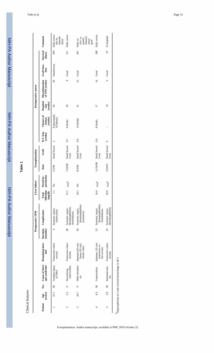
MATERIALS AND METHODSTwo of the recipients were young adults and 3 were children. Four of the 5 patients also hadliver failure after 30–52 months of parenteral hyperalimentation. The recipient of the isolatedintestine lost his small bowel 6 months before transplantation and still had good (although notnormal) liver function. These and other features of the 5 cases are summarized in Table 1.
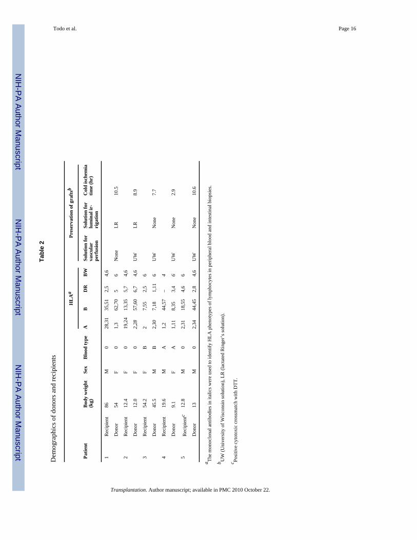
The donorsThe donors were ABO-identical with the recipient and were either size-matched (2 examples)or significantly smaller (Table 2). HLA matching was random and uniformly poor (Table 2).Donor and recipient were the opposite sex in 3 of the 5 cases. The principles of the donoroperations that allow flexibility of planning are described elsewhere (8). An attempt was madeat selective bacterial decontamination. Limited core cooling by aortic infusion of a limitedamount (1000 ml maximum) of UW solution was used in all but one donor whose uninfusedintestine was placed in an ice bath. No attempt was made to alter the graft lymphoreticulartissue with ALG or other modalities. In the last 3 cases, the contents of the intestine wereentrapped by stapling the proximal jejunum and terminal ileum and carried with the specimenthroughout the preservation and implantation; intraluminal washing was performed on thegrafts for the first 2 patients.
Recipient operationsVascularization: The technique for isolated intestinal transplantation was similar to thatoriginally used by Lillehei et al. (13) in dogs more than 30 years ago, except that arterializationwas with a free segment of donor iliac artery that was interposed between the superiormesenteric artery of the intestinal graft and the recipient aorta. In this case (patient 1), the distalstump of the recipient superior mesenteric vein was found after a tedious dissection andanastomosed to the graft SMV.
The 4 liver-intestine transplantations varied in detail but followed the principles describedelsewhere (8), which derived in turn from the experimental canine procedure of multivisceraltransplantation (14). A typical reconstruction is shown in Figure 1, including the use of aninterposition arterial graft that was used in all cases from the recipient aorta to the graft arterialsupply. Other specific details of revascularization included (1) The use of the piggy-backvenous outflow (15) in all 4 liver-intestine cases with preservation of the intrahepatic inferiorvena cava; and (2) portacaval shunting prior to the anhepatic phase of the liver-intestinetransplantations to prevent acute congestion of the residual splanchnic bed. The recipient portalvein was later detached and anastomosed to the portal vein or superior mesenteric vein of theallograft in 2 of the 4 patients, thereby assuring transhepatic delivery ofpancreaticoduodenosplenic affluent from the native organ. In the other 2 patients, theportacaval shunt was retained permanently. The spleen had been removed at an earlieroperation in one recipient. It was kept in 2 others and removed in the fourth in order to makemore room for the liver-intestinal graft.
Biliary drainage: No procedure was needed in the patient with intestinal transplantation only.Biliary drainage for the liver-intestine recipients was through an isolated bowel conduit takenfrom the proximal end of the jejunum and emptied at a lower point into the jejunum (Fig. 1).
Gastrointestinal reconstruction: Continuity was restored in stages in all 5 patients withconstruction of proximal (graft jejunostomy) and distal (ileostomy) vents (Fig. 1). Becauseend-to-side anastomoses to the duodenum (or jejunal stump) and colon (or, in one case, stumpof ileum) were performed primarily (Fig. 1), the vents allowed early alimentation—or,
Todo et al. Page 2
Transplantation. Author manuscript; available in PMC 2010 October 22.
NIH
-PA Author Manuscript
NIH
-PA Author Manuscript
NIH
-PA Author Manuscript
alternatively, decompression on a moment-to-moment basis. Then 8–16 weeks later whenintestinal motility was adequate, the vent chimneys were excised at a second operation.
ImmunosuppressionFK506 was given intravenously at first, at 0.10–0.15 mg/kg/day and enterally later at a startingdose of 0.3 mg/kg/day in divided doses, as described elsewhere for simple liver transplantation(16) (Figs. 2 and 3). Maintenance doses usually were lower. Prednisone was given at the outsetin all but case 5 and later stopped in each of the children (Fig. 3). Drug therapy was changedfrom intravenous to enteral (Table 1) when jejunostomy feeding and ultimately oral intakewere possible (Figs. 2 and 3). Patient 5 is still being fed and given oral medications through anasogastric tube with its tip advanced into the graft jejunum.
Management of immunosuppression was greatly facilitated by serial biopsies of both the upperand lower graft stomas, and of the liver in cases 2–5. These biopsies also were used for studyingthe graft lymphoreticular and epithelial phenotypes for their donor-recipient specificity asdescribed elsewhere (17).
Bacterial translocationFrequent blood and stool cultures were obtained and the results compared for similarity anddissimilarity of the flora. Selective decontamination was used for 4 weeks with a combinationof polymyxin E, gentamycin/tobramycin, and mycostatin/amphotericin B. Vancomycin wasadded at the time of positive blood cultures.
NutritionAdequacy of intestinal function was judged principally by the ability to hold or gain weight,and to maintain serum protein concentrations. D-xylose absorption tests (18) were performedsporadically.
RESULTSSurvival and hospitalization
The 5 patients are alive, and only the last one (2 months postoperatively) is in the hospital. Thefirst 4 recipients were hospitalized for 4–9 months. primarily because of difficulties in weaningfrom intravenous to enteral feeding. Restoration of reliable intestinal motility was slow,necessitating frequent switches from parenteral to enteral feeding and back before managementcould be stabilized. This required 8–36 weeks in patients 1–4. The longest interval of 36 weekswas in the patient who received intestine alone (Table 1). By the end of these times, all foodand medications were given by the enteral route. The enteral doses of FK506 and steroids werenot different from those in patients with liver transplantation alone (Figs. 2 and 3).
Bacterial translocationPatients 1, 2, and 4 had the same microorganisms (Candida albicans, Enterococcus fecium/Fecalis, cagula-negative Staphylococcus, and indifferent combinations) in the intestine andblood simultaneously for 3–7 days at 2. 2.5, and 0.6 months postoperatively, respectively. Onlyone of these incidents was associated with rejection.
Graft functionLiver: None of the recipients of the liver-intestinal grafts (cases 2–5) has been jaundiced formore than a few days. However, patient 2 had continuous hypoalbuminemia (<2 g%) until thethird postoperative month. The cause was suspected to be the portacaval shunt used to drainthe venous outflow of the recipient stomach, duodenum, and pancreas (Fig. 1, insert). However,
Todo et al. Page 3
Transplantation. Author manuscript; available in PMC 2010 October 22.
NIH
-PA Author Manuscript
NIH
-PA Author Manuscript
NIH
-PA Author Manuscript
the hypoalbuminemia (now >4g%) resolved without treatments and did not recur, despite anunequivocal hepatic rejection (confirmed by biopsy) at 176 days.
Patient 4 had transient jaundice at 3 months when rejection was diagnosed by liver biopsies.The jaundice resolved with increased immunosuppression.
Intestine: After resuming full alimentation, all 5 intestinal recipients have either maintained orgained weight. Patient 1 had a weight loss from 86 to 75 kg during the first 9 postoperativemonths, but after diet was started and hyperalimentation stopped, weight stabilized and slowlyincreased. All other patients gained weight (Fig. 4).
D-xylose absorption in the recipient of the isolated intestine (patient 1) was determinedfrequently and was normal until after a severe intestinal rejection at day 166. Three weeks later,absorption was severely depressed (Fig. 5, left). The latest D-xylose absorptions in the firstthree liver-intestine recipients (cases 2–4) ranged from normal to slightly depressed (Fig. 5,right).
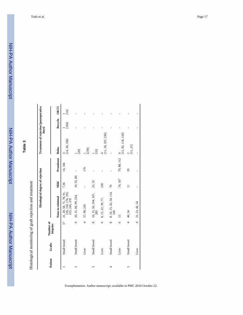
Incidence of rejectionThe diagnosis of rejection of either the intestine or liver was made histopathologically at thetimes indicated in Figures 2 and 3. Most of the biopsies (all summarized in Table 3) were freeof unequivocal rejection—and, even when present, the tissue diagnosis did not correlate wellwith clinical events unless the degree of rejection was classified as prominent.
The most serious intestinal rejection was in the patient who received an intestine only (case1). At 14 days, he became hypotensive and acidotic when both graft stomas turned cyanotic.Stomal biopsies showed severe rejection that was reversed with augmented FK506 andprednisone. At 166 days after transplantation after temporary discharge from the hospital, andconcomitant with drug noncompliance, he developed the same hypotensive syndrome andileus. Endoscopy showed mucosal sloughing, and biopsies revealed severe rejection andintramural microabscesses. The process was reversed with immunosuppression.
In the 4 recipients of a liver-intestine graft, both organs had relatively few diagnoses ofunequivocal rejection (Table 3). Neither the liver nor the intestine appeared to be more or lessfavored relative to the other.
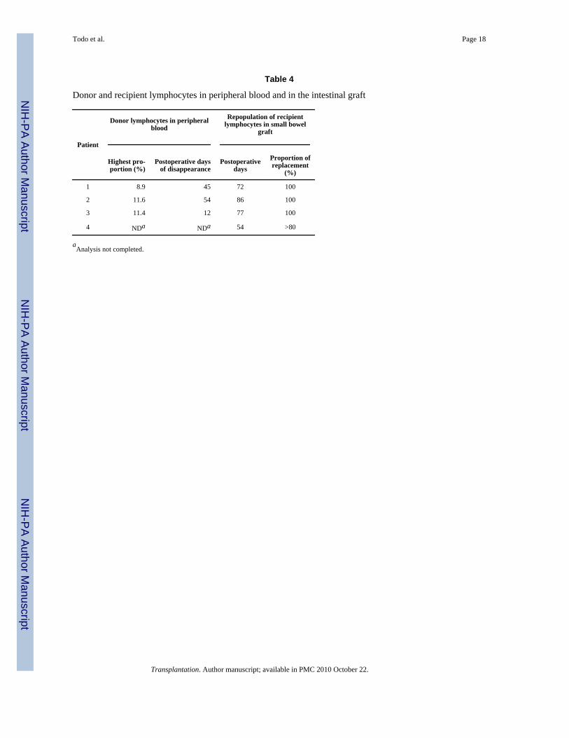
Graft lymphoreticular repopulationIn the first four cases the HLA phenotypes of the lymphoid tissue of the lamina propria becamethose of the recipient after 54–86 days. During the same time, donor mononuclear cells werefound in all of the recipients’ peripheral blood. The details of these studies have been publishedelsewhere (17). Studies in patient 5 are still in progress.
DISCUSSIONThis experience has demonstrated the inherent feasibility and practicality of small boweltransplantation in humans, a procedure which was first attempted in humans by Lillehei et a1.almost 25 years ago (19). As with the liver at an earlier time (20), the exploitation of intestinaltransplantation awaited better immunosuppression before emancipation from the “forbidden”organ category. Liver transplantation became a service with the advent of cyclosporine, andnow intestinal transplantation may come into wide use because of FK506 whose qualities havebeen shown by direct comparison to be superior to cyclosporine in rat intestinal transplantexperiments (10,11).
Todo et al. Page 4
Transplantation. Author manuscript; available in PMC 2010 October 22.
NIH
-PA Author Manuscript
NIH
-PA Author Manuscript
NIH
-PA Author Manuscript
Most of the other lessons from the early days of liver transplantation appear also to be applicableto the intestine including the infectious implications of rejection. With its rejection, the hepaticgraft becomes a sieve for bacteria translocated from the intestine (21,22). Paradoxically, suchhepatic graft based infections could be prevented or treated only by the heavierimmunosuppression that itself contributes to systemic susceptibility to infection. Thetherapeutic philosophy of preventing bacterial translocation in this way is even more applicableto the intestinal graft in which the foremost requirement is maintenance of an intact mucosalbarrier. Using cyclosporine-based immunosuppression in multivisceral recipients, it was notpossible to interdict bacterial leakage, with the consequence that there was repeated bacteremiaor fungemia with the same microorganisms found in the intestinal content (2). Of the 5 presentlyreported patients under FK506, 3 had this complication, but it could be controlled withadjustments of immunosuppression combined with efficient antibiotic treatment.
Prevention of GVHD is also dependent upon highly effective immunosuppression. ClinicalGVHD was not seen in any of our 5 recipients despite severe histoincompatibility and theappearance of donor mononuclear cells in the peripheral blood of all (17). At the same time,the lymphoreticular cells in the lamina propria of the graft were being replaced bylymphoreticular cells of the recipient. Rapid cell migration and repopulation of lymphoreticularcells in chronically tolerated piglet intestinal grafts under cyclosporine were noted by Arnaud-Battandier et a1. (23) in 1985, and by Jaffe et a1. (24) in the intestinal component of amultivisceral graft that also was the site of lymphoproliferative lesions of recipient origin. Clarket al. (25) and Lear et al. (26) showed striking cell migration in rat intestinal recipients.
In most of these reports, the cell repopulation was equated more with rejection or GVHD thanwith graft acceptance (24-26). The significance of the cell repopulation phenomenon and itsindispensability for graft acceptance was first described by Murase et a1. in rats (12) and byIwaki et al. in the first 3 patients of the present human series (17). By this process, thetransplanted intestine becomes a “composite” organ within a few weeks but with retention ofdonor-specific epithelium.
Studies are in progress to learn the fate of the donor lymphoreticular cells that leave the graftand enter the circulation. Lear et a1. (26) described the major movement of passenger(presumably T) cells into thymus-dependent host lymphoid tissue, and warned that GVHDcould actually be promoted by effective immunosuppression that would allow these cells tobecome established. The phenomenon of donor cell peripheralization and replacement withrecipient cells is probably generic, occurring in all kinds of grafts, although it is most easilystudied in the intestine because of the intestine’s rich lymphoid constituency. For example,macrophage repopulation of the liver has been known for almost two decades (27) and similarreplacement of the lymphoreticular cells has been described in heart-lung grafts (28).
What was made clear by Murase’s rat experiments (12) and by Iwaki’s study of the presentlyreported patients (17) is that the relocated cells can be functionally inert, causing neitherrejection nor GVHD, provided that the conditions of induction and maintenanceimmunosuppression are propitious. We have speculated that the cell exchange occurs mostreliably in the presence of a normal microenvironment. If so, we have pointed out that thecommon practices may be ill-conceived, damaging the graft lymphoid deposits with irradiation,antilymphoid globulins, chemotherapy, or other measures (8). No such graft pretreatment wasemployed in any of our 5 cases.
Even at a technical surgical level, other widely accepted assumptions about intestinaltransplantation come into question. For example, the preservation of the intestine was with anextremely simple technique in which the graft was cooled by immersion in an ice bath, or withjust enough intraarterial UW solution to cause blanching of the capillary bed. Instead of
Todo et al. Page 5
Transplantation. Author manuscript; available in PMC 2010 October 22.
NIH
-PA Author Manuscript
NIH
-PA Author Manuscript
NIH
-PA Author Manuscript
washing out the succus entericus of the intestine, this was kept with the specimen throughoutin 3 of the 5 cases by stapling shut the intestinal graft at the upper and lower ends. There wereno consequent infections from this practice.
In the recipient an attempt was made to restore normal anatomic relationships between theresidual recipient organs and the transplanted or native liver, and above all to direct the venousaffluent from the pancreas, intestine, and other splanchnic viscera through the retained orconcomitantly engrafted new liver. The complex metabolic and immunologicinterrelationships of the various intraabdominal organs are discussed elsewhere (8,29).
The function of the transplanted intestine alone or with accompanying liver has beensatisfactory in all five patients, with eventual complete independence from parenteral nutrition.Since resuming full alimentation, all 5 patients have maintained (1 case) or gained weight (4cases). Reliable absorption studies with D-xylose were difficult to obtain under these clinicalcircumstances, and the results did not correlate well with common-sense clinical assessmentsof the state of nutrition.
The achievement of alimentation was a slow process, requiring from 6 weeks to 9 months. Bythe time this was achieved, the doses of oral FK506, steroids, and other medications were inthe same range as with other kinds of transplantation, indicating the efficient absorption ofthese medications. Earlier in the postoperative course, the transplanted and denervated intestinewas prone to ileus, making it essential to have proximal and distal enteric vents either fordecompression or as an entry for graded feedings. There are theoretical reasons for earlyresumption of alimentation, but practical reasons for doing this with extreme caution.
The question remains whether intestinal transplantation alone is a more or less difficultprocedure than combined liver-intestine transplantation. The experimental work of Murase etal. (12,30) and others (8) has supported the concept that the liver may shield the intestine fromimmunologic attack, but this advantage is relative only. The long survival of patient 1 in ourseries and that of the recipients of DeItz et al. (6) and Ricour and Goulet et al. (5) havedemonstrated the feasibility of the technically less draconian procedure of isolated intestinaltransplantation.
In our patient 1, severe but reversible rejection after 2 weeks and again at 5½ months afterisolated small bowel transplantation caused a shock syndrome, with a third space fluidaccumulation in the graft, as well as bacteremia and candidemia that led to permanentsecondary loss of renal function. However, the intestinal changes were reversible, includinghealing and regeneration of ulcerative and sloughed intestinal epithelium. The power of thereparative process has been seen even more dramatically in a duodenal jejunal intestinalsegment that was part of an upper abdominal cluster graft that recovered to a normal state afterbeing almost completely denuded of epithelium as a consequence of rejection (8,31).
AcknowledgmentsWe thank Mrs. Terry Mangan, Mr. Steve Miller, and Miss Sally Wagner for their help in preparing the manuscript.
Appendix
ORAL DISCUSSIONDR. A. LANGNAS (Omaha, Nebraska): You indicated that there were a number of episodesof bowel rejection. How did you document and classify this? How did you adjust your therapy?
Todo et al. Page 6
Transplantation. Author manuscript; available in PMC 2010 October 22.
NIH
-PA Author Manuscript
NIH
-PA Author Manuscript
NIH
-PA Author Manuscript
DR. TODO: The most important diagnostic tool for the small bowel recipient is the inspectionof the stoma. Cyanotic change is the first sign of rejection. When this is noted, we performendoscopy through the stoma to obtain biopsy material for histologic confirmation. Later, afterthe stomas have been closed the situation might be more difficult—however, we have observedonly one episode of rejection in that setting. This patient had an episode at 166 days, the resultof drug-noncompliance. He had severe abdominal pain and diarrhea. At colonoscopy we sawsloughing of the mucosa, and biopsy showed the denuding of the mucosal layers.
DR. LANGNAS: Have you seen any findings in the submucosa such as the vasculitis describedin a number of small animal models?
DR. TODO: Yes, we observed slight arteritic changes in the intestinal wall in one patient whena biopsy was taken at the operation for stomal closure about 4 months after transplantation.
DR. ALSINA (Hartford, Connecticut): I would like to know if you are planning to use portalvenous drainage in the future. The issue as to whether this is beneficial or necessary has notbeen settled. I also have a comment. We have finished a small bowel transplant study in outbredpigs and have observed more graft-versus-host reaction in the native tissues when using verylow doses of cyclosporine in combination with azathioprine and prednisone.
DR. TODO: Responding to your comment first, in our laboratory we also saw the GVH diseasewhen the animals were treated with subtherapeutic immunosuppression, but we didn’t see anyGVH diseases in our five clinical small bowel recipients. Concerning the first question, webelieve that venous drainage from the graft should be with a portal anastomosis, since it isphysiological and may also have metabolic and immunological advantages.
DR. HARDY (New York, New York): I wonder whether you could comment on two issues:One is the question that you alluded to suggesting a certain exchange in migration oflymphocytes, so that the donor lymphocytes end up in the blood and recipient lymphocytesend up in the small bowel, an expected phenomenon. A recent article by your group suggestedthat there is some magic about this in terms of protection of the bowel. Perhaps you couldcomment on those observations in relation to other people’s experience with pretreating thedonor in clinical situations with massive doses of antilymphoid preparations. and inexperimental situations with various antilymphoid drugs, as well as low-dose radiation. Mysecond question concerns GVHD. We all know that a small amount of graft-versus-host diseasemay help the graft to survive. You have an excellent model for studying this in men. Does ithelp?
DR. TODO: Although exchange of donor and recipients lymphocytes could have beenexpected, this phenomenon was documented clinically only recently in hepatic and small bowelallografts by our group and the Canadian group. This suggests that pretreatment of the donoror small bowel graft to avoid GVH disease is probably unnecessary. However, we do not knowthe value of pretreatment as a strategy to reduce graft antigenicity, such as by passengerleukocyte depletion—this is our current laboratory interest.
DR. D. GRANT (London Ontario Canada): We have three patients who are well with liver–small bowel grafts at two and half years, one and a half years, and three months aftertransplantation. These patients were treated with cyclosporine. I’m wondering if you think thatyour success is due to the combined liver graft, or whether you think it’s due to the potency ofFK506. Are you prepared to proceed with isolated small bowel grafting using the FK506?
DR. TODO: Regarding successful combined liver and small bowel graft, I think both factorscontributed to the result. As you described in the Lancet, and has been described also by ourgroup and others, the liver seems to have a protective effect on other organs when transplanted
Todo et al. Page 7
Transplantation. Author manuscript; available in PMC 2010 October 22.
NIH
-PA Author Manuscript
NIH
-PA Author Manuscript
NIH
-PA Author Manuscript

together. The potency of FK506 should not be ignored, since 2 out of 5 recipients were treatedvirtually only with FK506. We plan more isolated small bowel transplantations using FK506;we additionally have several ongoing animal studies designed to support the clinical program.
DR. HARDY: Dr. Grant, do you still pretreat your donors with massive doses of OKT3 or asimilar preparation?
DR. GRANT: We have, but I’m not sure that it is necessary.
DR. FRANK GUTTMAN (Montreal, Quebec, Canada): I would like to comment on the liver–small bowel experience in rats. I was privileged a few weeks ago to see a manuscript fromRevillon’s group in Paris. They have the experience of nine clinical small boweltransplantations in children. They have completed a study in a rat combination where the liveris accepted between the two strains without any immunosuppression, but the bowel is not.However, if they do a liver transplant and 17 days later carry out a bowel transplant, againwithout immunosuppressive drugs, tolerance of the small bowel was observed.
DR. HARDY: Can you identify the specific rat combinations?
DR. GUTTMAN: It was Rt1A and C or D, something like that.
DR. HARDY: This phenomenon, protection by the liver, has in many instances been rat-specific. I would warn those who wish to repeat the experiment to be aware of the specificshown combinations.
REFERENCES1. Kirkman RT. Small bowel transplantation. Transplantation 1984;37:429. [PubMed: 6729949]2. Starzl TE, Rowe M, Todo S, et al. Transplantation of multiple abdominal viscera. JAMA
1989;261:1449. [PubMed: 2918640]3. Grant D, Wall W, Mimeault R, et al. Successful small-bowel/liver transplantation. Lancet
1990;335:181. [PubMed: 1967664]4. Schroeder P, Goulet O, Lear PA. Small bowel transplantation: European experience [Letter]. Lancet
1990;336:110. [PubMed: 1975289]5. Goulet O, Revillon Y, Jan D, et al. Small-bowel transplantation in children. Transplant Proc
1990;22:2499. [PubMed: 2264125]6. Deltz E, Schroeder p, Gebhardt H, et al. Successful clinical small bowel transplantation: report of a
case. Clin Transplant 1989;3:89.7. Starzl TE, Todo S, Tzakis A, et al. Abdominal organ cluster transplantation for the treatment of upper
abdominal malignancies. Ann Surg 1989;210:374. [PubMed: 2673085]8. Starzl TE, Todo S, Tzakis A, et al. The many faces of multivisceral transplantation. Surg Gynecol
Obstet 1991;172:335. [PubMed: 2028370]9. Murase N, Kim D, Todo S, Cramer DV, Fung J, Starzl TE. Induction of liver, heart, and multivisceral
graft acceptance with a short course of FK 506. Transplant Proc 1990;22:74. [PubMed: 1689906]10. Hoffman AL, Makowka L, Banner B, et al. The use of FK 506 for small intestine allotransplantation:
inhibition of acute rejection and prevention of fatal graft-versus-host disease. Transplantation1990;49:483. [PubMed: 1690469]
11. Lee K, Stangl MJ, Todo S, Langrehr JM, Starzl TE, Schraut WH. Successful orthotopic small boweltransplantation with short term FK 506 immunosuppressive therapy. Transplant Proc 1990;22:78.[PubMed: 1689908]
12. Murase N, Demetris AJ, Matsuzaki T, et al. Long survival in rats after multivisceral versus isolatedsmall bowel allotransplantation under FK 506. Surgery 1991;110:87. [PubMed: 1714104]
Todo et al. Page 8
Transplantation. Author manuscript; available in PMC 2010 October 22.
NIH
-PA Author Manuscript
NIH
-PA Author Manuscript
NIH
-PA Author Manuscript
13. Lillehei RC, Goott B, Miller FA. The physiologic response of the small bowel of the dog to ischemiaincluding prolonged in vitro preservation of the bowel with successful replacement and survival. AnnSurg 1959;150:543. [PubMed: 14416956]
14. Starzl TE, Kaupp HA Jr, Brock DR, Butz GW Jr, Linman JW. Homotransplantation of multiplevisceral organs. Am J Surg 1962;103:219. [PubMed: 13916395]
15. Tzakis A, Todo S, Starzl TE. Piggyback orthotopic liver transplantation with preservation of theinferior vena cava. Ann Surg 1989;210:649. [PubMed: 2818033]
16. Todo S, Fung JJ, Starzl TE, et al. Liver, kidney, and thoracic organ transplantation under FK 506.Ann Surg 1990;212:295. [PubMed: 1697743]
17. Iwaki Y, Starzl TE, Yagihashi A, et al. Replacement of donor lymphoid tissue in human small boweltransplants under FK 506 immunosuppression. Lancet 1991;337:818. [PubMed: 1707470]
18. Breiter HC, Craig RM, Levee G, Atkinson AJ. Use of kinetic methods to evaluate d-xylosemalabsorption in patients. J Lab Clin Med 1988;112:533. [PubMed: 3183487]
19. Lillehei RC, Idezuki Y, Feemster JA, et al. Transplantation of stomach, intestine, and pancreas:experimental and clinical observations. Surgery 1967;62:721. [PubMed: 4862242]
20. Starzl TE, Iwatsuki S, Van Thiel DH, et al. Evolution of liver transplantation. Hepatology 1982;2:614.[PubMed: 6749635]
21. Brettschneider L, Tong JL, Boose DS, et al. Specific bacteriologic problems after orthotopic livertransplantation in dogs and pigs. Arch Surg 1968;97:313. [PubMed: 4872484]
22. Starzl, TE. Infectious complications, excluding partial hepatic gangrene. In: Starzl, TE., editor.Experience in hepatic transplantation. Saunders; Philadelphia: 1969. p. 329
23. Arnaud-Battandier F, Salmon H, Vaiman M, et al. Small intestinal allotransplantation in swine withcyclosporine treatment: studies of the intestinal lymphoid populations. Transplant Proc1985;17:1440.
24. Jaffe R, Trager JDK, Zeevi A, Sonmez-Alpan E, Duquesnoy R. Multivisceral intestinaltransplantation: surgical pathology. Pediatr Pathol 1989;9:633. [PubMed: 2557597]
25. Clark CLI, Cunningham AJ, Crane PW, Wood RFM, Lear PA. Lymphocyte infiltration patterns inrat small-bowel transplants. Transplant Proc 1990;22:2460. [PubMed: 2264107]
26. Lear PA, Cunningham AJ, Clark CLI, Crane PW, Wood RFM. What role for passenger leucocytesin small-bowel allografts? Transplant Proc 1990;22:2463. [PubMed: 2264109]
27. Porter, KA. Pathology of the orthotopic homograft and heterograft. In: Starzl, TE., editor. Experiencein hepatic transplantation. Saunders; Philadelphia: 1969. p. 422
28. Fung JJ, Zeevi A, Kaufman C, et al. Interactions between bron-choalveolar lymphocytes andmacrophages in heart-lung transplant recipients. Hum Immunol 1985;14:287. [PubMed: 3932269]
29. Starzl TE, Porter KA, Francavilla A. The Eck fistula in animals and humans. Curr Probl Surg1983;20:687. [PubMed: 6357642]
30. Murase N, Demetris AJ, Kim DG, Todo S, Fung JJ, Starzl TE. Rejection of the multivisceral allograftsin rats: a sequential analysis with comparison to isolated orthotopic small bowel and liver grafts.Surgery 1990;108:880. [PubMed: 2237770]
31. Starzl TE, Todo S, Tzakis A, et al. Abdominal organ cluster transplantation for the treatment of upperabdominal malignancies. Ann Surg 1989;210:374. [PubMed: 2673085]
Todo et al. Page 9
Transplantation. Author manuscript; available in PMC 2010 October 22.
NIH
-PA Author Manuscript
NIH
-PA Author Manuscript
NIH
-PA Author Manuscript
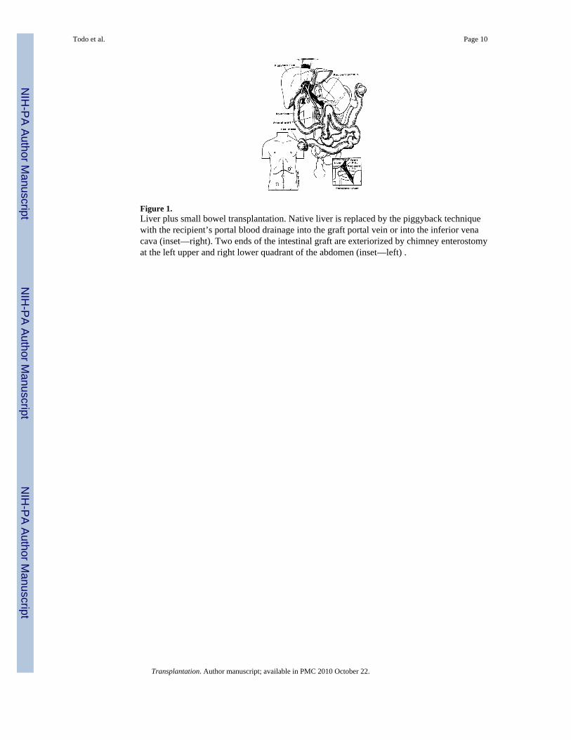
Figure 1.Liver plus small bowel transplantation. Native liver is replaced by the piggyback techniquewith the recipient’s portal blood drainage into the graft portal vein or into the inferior venacava (inset—right). Two ends of the intestinal graft are exteriorized by chimney enterostomyat the left upper and right lower quadrant of the abdomen (inset—left) .
Todo et al. Page 10
Transplantation. Author manuscript; available in PMC 2010 October 22.
NIH
-PA Author Manuscript
NIH
-PA Author Manuscript
NIH
-PA Author Manuscript
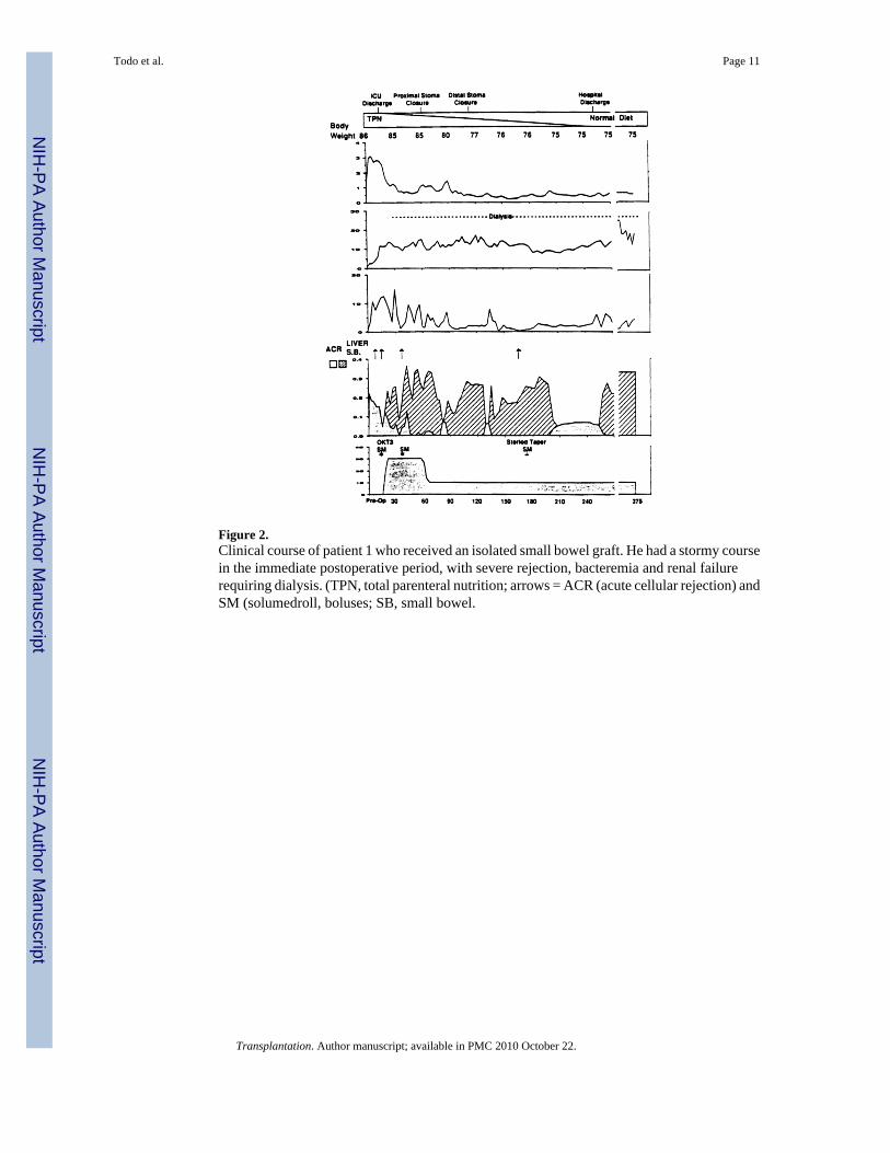
Figure 2.Clinical course of patient 1 who received an isolated small bowel graft. He had a stormy coursein the immediate postoperative period, with severe rejection, bacteremia and renal failurerequiring dialysis. (TPN, total parenteral nutrition; arrows = ACR (acute cellular rejection) andSM (solumedroll, boluses; SB, small bowel.
Todo et al. Page 11
Transplantation. Author manuscript; available in PMC 2010 October 22.
NIH
-PA Author Manuscript
NIH
-PA Author Manuscript
NIH
-PA Author Manuscript
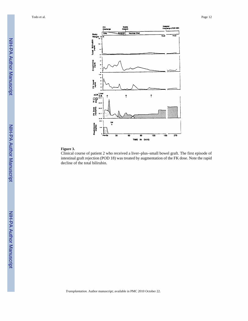
Figure 3.Clinical course of patient 2 who received a liver–plus–small bowel graft. The first episode ofintestinal graft rejection (POD 18) was treated by augmentation of the FK dose. Note the rapiddecline of the total bilirubin.
Todo et al. Page 12
Transplantation. Author manuscript; available in PMC 2010 October 22.
NIH
-PA Author Manuscript
NIH
-PA Author Manuscript
NIH
-PA Author Manuscript
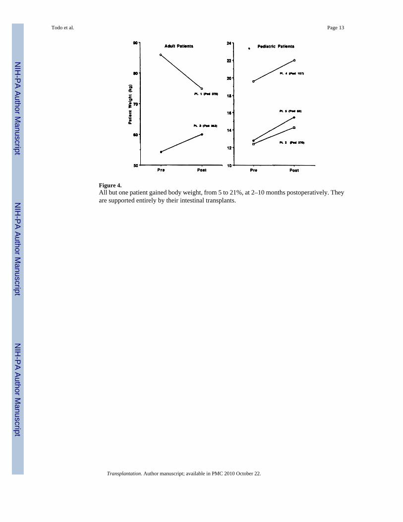
Figure 4.All but one patient gained body weight, from 5 to 21%, at 2–10 months postoperatively. Theyare supported entirely by their intestinal transplants.
Todo et al. Page 13
Transplantation. Author manuscript; available in PMC 2010 October 22.
NIH
-PA Author Manuscript
NIH
-PA Author Manuscript
NIH
-PA Author Manuscript
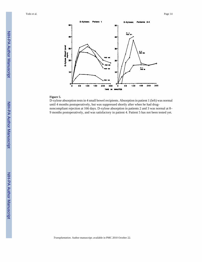
Figure 5.D-xylose absorption tests in 4 small bowel recipients. Absorption in patient 1 (left) was normaluntil 4 months postoperatively, but was suppressed shortly after when he had drug-noncompliant rejection at 166 days. D-xylose absorption in patients 2 and 3 was normal at 8–9 months postoperatively, and was satisfactory in patient 4. Patient 5 has not been tested yet.
Todo et al. Page 14
Transplantation. Author manuscript; available in PMC 2010 October 22.
NIH
-PA Author Manuscript
NIH
-PA Author Manuscript
NIH
-PA Author Manuscript
NIH
-PA Author Manuscript
NIH
-PA Author Manuscript
NIH
-PA Author Manuscript
Todo et al. Page 15
Tabl
e 1
Clin
ical
feat
ures
Preo
pera
tive
TPM
Liv
er fa
ilure
Tra
nspl
anta
tion
Post
oper
ativ
e co
urse
Patie
ntA
ge(y
ears
)Se
xC
ause
of s
hort
gut s
yndr
ome
Rem
aini
ng in
tes-
tine
Dur
atio
n(m
onth
s)C
ompl
icat
ion
Tot
albi
lirub
in(m
g/dl
)
Port
al h
y-pe
rten
sion
Dat
eG
raft
ICU
Sta
y(w
eeks
)C
losu
re o
fst
omas
(wee
ks)
Hos
pita
lst
ay(w
eeks
)
Dis
cont
inua
tion
of T
PN (w
eeks
)G
raft
func
-tio
nSu
rviv
al(d
ays)
Com
men
t
131
.1M
Gun
shot
inju
ry
of S
MA
Tran
sver
se c
olon
-
rect
um6
Syst
emic
seps
is,
ch
olec
ystit
is1.
1N
o5/
2/90
Smal
l bow
el2
8 (p
roxi
mal
),16
(dis
tal)
3636
Satis
fact
ory
394
Fully
act
ive
bu
t on
he
mod
i-
alys
is
22.
3F
Nec
rotiz
ing
en
tero
colit
isTr
ansv
erse
col
on-
re
ctum
38Sy
stem
ic se
psis
,th
rom
boph
lebi
tis,
chol
elith
iasi
s
15.1
Yes
a7/
24/9
0Sm
all b
owel
Live
r0.
78
(bot
h)29
8G
ood
311
Fully
act
ive
326
.7F
SMA
thro
mbo
-
sis
Jeju
num
(20
cm),
ile
um (1
0 cm
),
who
le c
olon
30Sy
stem
ic se
psis
,
thro
mbo
phle
bitis
(e
xten
sive
)
18.1
No
8/3/
90Sm
all b
owel
Live
r0.
610
(bot
h)21
15G
ood
301
Fully
ac-
tiv
e, re
-
quire
d
fem
oral
ar
tery
gr
aft
44.
3M
Gas
trosc
hisi
sJe
junu
m, (
10 c
m)
tra
nsve
rse
co-
lo
n-re
ctum
52Sy
stem
ic se
psis
,
thro
mbo
phle
bitis
,
chol
elith
iasi
s
16.4
Yes
a11
/24/
90Sm
all b
owel
Live
r0.
48
(bot
h)17
16G
ood
188
Fully
act
ive
52.
8M
Inte
stin
al a
tre-
si
aTr
ansv
erse
col
on-
re
ctum
33Sy
stem
ic se
psis
,th
rom
boph
lebi
tis42
.0Y
esa
3/24
/91
Smal
l bow
elLi
ver
6–
>86
Goo
d67
In h
ospi
tal
a Hyp
ersp
leni
sm in
2 a
nd v
aric
eal h
emor
rhag
e in
all
3.
Transplantation. Author manuscript; available in PMC 2010 October 22.
NIH
-PA Author Manuscript
NIH
-PA Author Manuscript
NIH
-PA Author Manuscript
Todo et al. Page 16
Tabl
e 2
Dem
ogra
phic
s of d
onor
s and
reci
pien
ts
HL
Aa
Pres
erva
tion
of g
rafts
b
Patie
ntB
ody
wei
ght
(kg)
Sex
Blo
od ty
peA
BD
RB
WSo
lutio
n fo
rva
scul
arpe
rfus
ion
Solu
tion
for
lum
inal
ir-
riga
tion
Col
d is
chem
iatim
e (h
r)
1R
ecip
ient
86M
028
,31
35,5
12,
54,
6
Don
or54
F0
1,3
62,7
05
6N
one
LR10
.5
2R
ecip
ient
12.4
F0
19,2
413
,35
5,7
4,6
Don
or12
.0F
02,
2857
,60
6,7
4,6
UW
LR8.
9
3R
ecip
ient
54.2
FB
27,
552,
56
Don
or45
.5M
B2,
307,
181,
116
UW
Non
e7.
7
4R
ecip
ient
19.6
MA
1,2
44,5
7–
4
Don
or9.
1F
A1,
118,
353,
46
UW
Non
e2.
9
5R
ecip
ient
c12
.8M
02,
3118
,55
4,6
6
Don
or13
M0
2,34
44,4
52,
84,
6U
WN
one
10.6
a The
mon
oclo
nal a
ntib
odie
s in
italic
s wer
e us
ed to
iden
tify
HLA
phe
noty
pes o
f lym
phoc
ytes
in p
erip
hera
l blo
od a
nd in
test
inal
bio
psie
s.
b UW
(Uni
vers
ity o
f Wis
cons
in so
lutio
n), L
R (l
acta
ted
Rin
ger’
s sol
utio
n).
c Posi
tive
cyto
toxi
c cr
ossm
atch
with
DTT
.
Transplantation. Author manuscript; available in PMC 2010 October 22.
NIH
-PA Author Manuscript
NIH
-PA Author Manuscript
NIH
-PA Author Manuscript
Todo et al. Page 17
Tabl
e 3
His
tolo
gica
l mon
itorin
g of
gra
ft re
ject
ion
and
treat
men
t
His
tolo
gica
l deg
ree
of r
ejec
tion
Tre
atm
ent o
f rej
ectio
n [p
osto
pera
tive
days
]
Patie
ntG
rafts
Num
ber
ofbi
opsi
es
Non
e-to
-min
imal
Mild
Prom
inen
tB
olus
Rec
ycle
OK
T3
1Sm
all b
owel
1721
, 28,
58,
71,
78,
91,
12
6, 1
50, 1
76, 1
95,
21
5, 2
45, 3
78
7,36
14, 1
663 [1
4, 3
6, 1
66]
1 [166
]1 [1
4]
2Sm
all b
owel
820
, 31,
66,
99,
224
,10
, 55,
ll6
–1 [2
0]–
–
Live
r4
57, 9
8, 2
26–
176
1 [230
]–
–
3Sm
all b
owel
810
, 15,
56,
104
, 107
,
285
25, 3
5–
1 [35]
––
Live
r6
8, 1
5, 2
2, 3
9, 7
7,23
6–
4 [11,
18,
107
, 236
]–
–
4Sm
all b
owel
88,
10,
15,
43,
54,
114
,
166
70–
––
–
Live
r6
1574
, 167
79, 8
8, 1
124 [1
1, 8
2, 1
18, 1
20]
––
5Sm
all b
owel
446
, 54
1120
2 [11,
21]
––
Live
r4
21, 2
3, 4
6, 5
4–
––
––
Transplantation. Author manuscript; available in PMC 2010 October 22.
NIH
-PA Author Manuscript
NIH
-PA Author Manuscript
NIH
-PA Author Manuscript
Todo et al. Page 18
Table 4
Donor and recipient lymphocytes in peripheral blood and in the intestinal graft
Donor lymphocytes in peripheralblood
Repopulation of recipientlymphocytes in small bowel
graft
Patient
Highest pro-portion (%)
Postoperative daysof disappearance
Postoperativedays
Proportion ofreplacement
(%)
1 8.9 45 72 100
2 11.6 54 86 100
3 11.4 12 77 100
4 NDa NDa 54 >80
aAnalysis not completed.
Transplantation. Author manuscript; available in PMC 2010 October 22.
Copyright © 2022 FDOKUMEN