S100B as a new fecal biomarker of inflammatory bowel diseases
10
323 Abstract. – OBJECTIVE: S100 proteins are demonstrated to exert a protective role in the gastrointestinal tract. In the present study, we investigated whether S100B protein, that is typ- ically expressed by enteroglial cells, is detect- able in feces and could be a useful noninvasive indicator of gut chronic inflammation. PATIENTS AND METHODS: This clinical pro- spective study included n=48 patients suffer- ing Crohn’s disease (CD) or ulcerative colitis (UC) and non IBD-controls. The clinical dis- ease activity was evaluated using Harvey-Brad- shaw or Mayo Score Index while the diagnosis of IBD was defined based on standard endoscop- ic and histological criteria. S100B and calprotec- tin were extracted and analyzed using commer- cial enzyme-linked immunosorbent assay (ELI - SA) kits. RESULTS: Unlike calprotectin, S100B was sig- nificantly decreased in both CD and UC com- pared to non IBD-patients. The strongest quan- titative alterations of S100B were detected con- comitantly with signs of active or quiescent dis- ease, including high/normal expression of fecal calprotectin, mucosal damage/cryptitis, mucin depletion and inflammatory infiltrate, as defined by endoscopic evaluation and histological anal- ysis. At the onset of disease and under no In- fliximab-based therapy, the lowest was detected suggesting that S100B in feces could have a po- tential diagnostic value for IBD. CONCLUSIONS: Testing for S100B and cal- protectin could be a useful screening tool to bet- ter predict IBD activity. Key Words: S100B, Feces, Calprotectin, IBD biomarkers, Gut in- flammation. Introduction Ulcerative colitis (UC) and Crohn’s disease (CD) represent the two major clinically defined forms of inflammatory bowel disease (IBD), and are associated with an increased risk of develop- ing colon cancer, being more common in devel- oped countries, with the highest incidence rates and prevalence in North America and Europe 1 . Although dysfunction of the mucosal immune system plays an undoubtable role in the patho- genesis of IBD 2 , in recent years it has become clear that the mucosal immune system alone may not account for all the aspects of IBD; thus, a role for the enteric nervous system (ENS), has been recognized in intestinal inflammation, also involving interactions with the intestinal microbiota 3 . The ENS is located within the wall of the gastrointestinal tract and, independently from the central nervous system, coordinates many aspects of digestive functions such as motility, blood flow, and immune/inflammatory processes 4 . Among different ENS cell types, en- teric glial cells (EGCs) appear to deserve special European Review for Medical and Pharmacological Sciences 2020; 24: 323-332 R. DI LIDDO 1,2 , M. PICCIONE 1 , S. SCHRENK 1 , C. DAL MAGRO 3 , C. COSMA 4 , A. PADOAN 4 , N. CONTRAN 4 , M.L. SCAPELLATO 5 , A. PAGETTA 1 , V. ROMANO SPICA 6 , M.T. CONCONI 1 , P.P. PARNIGOTTO 2 , R. D’INCÀ 3 , F. MICHETTI 7,8 1 Department of Pharmaceutical and Pharmacological Sciences, University of Padova, Padua, Italy 2 Foundation for Biology and Regenerative Medicine, Tissue Engineering and Signaling (T.E.S.) Onlus, Padua, Italy 3 Department of Surgery, Oncology and Gastroenterology, Gastroenterology Unit, University Hospital of Padova, Padua, Italy 4 Department of Laboratory Medicine, University-Hospital of Padova, Padua, Italy 5 Department of Cardiac Thoracic Vascular Sciences and Public Health, Preventive Medicine and Risk Assessment Unit, University of Padova, Padua, Italy 6 Public Health Unit, University of Rome “Foro Italico”, Rome, Italy 7 Istituto di Anatomia Umana e Biologia Cellulare, Università Cattolica del Sacro Cuore, Rome, Italy 8 Facoltà di Medicina e Chirurgia – IRCCS San Raffaele Scientific Institute, Università Vita-Salute San Raffaele, Milan, Italy Corresponding Author: Rosa Di Liddo, MD; e-mail: [email protected] S100B as a new fecal biomarker of inflammatory bowel diseases
-
Upload
khangminh22 -
Category
Documents
-
view
0 -
download
0
Transcript of S100B as a new fecal biomarker of inflammatory bowel diseases
323
Abstract. – OBJECTIVE: S100 proteins are demonstrated to exert a protective role in the gastrointestinal tract. In the present study, we investigated whether S100B protein, that is typ-ically expressed by enteroglial cells, is detect-able in feces and could be a useful noninvasive indicator of gut chronic inflammation.
PATIENTS AND METHODS: This clinical pro-spective study included n=48 patients suffer-ing Crohn’s disease (CD) or ulcerative colitis (UC) and non IBD-controls. The clinical dis-ease activity was evaluated using Harvey-Brad-shaw or Mayo Score Index while the diagnosis of IBD was defined based on standard endoscop-ic and histological criteria. S100B and calprotec-tin were extracted and analyzed using commer-cial enzyme-linked immunosorbent assay (ELI-SA) kits.
RESULTS: Unlike calprotectin, S100B was sig-nificantly decreased in both CD and UC com-pared to non IBD-patients. The strongest quan-titative alterations of S100B were detected con-comitantly with signs of active or quiescent dis-ease, including high/normal expression of fecal calprotectin, mucosal damage/cryptitis, mucin depletion and inflammatory infiltrate, as defined by endoscopic evaluation and histological anal-ysis. At the onset of disease and under no In-fliximab-based therapy, the lowest was detected suggesting that S100B in feces could have a po-tential diagnostic value for IBD.
CONCLUSIONS: Testing for S100B and cal-protectin could be a useful screening tool to bet-ter predict IBD activity.
Key Words:S100B, Feces, Calprotectin, IBD biomarkers, Gut in-
flammation.
Introduction
Ulcerative colitis (UC) and Crohn’s disease (CD) represent the two major clinically defined forms of inflammatory bowel disease (IBD), and are associated with an increased risk of develop-ing colon cancer, being more common in devel-oped countries, with the highest incidence rates and prevalence in North America and Europe1.
Although dysfunction of the mucosal immune system plays an undoubtable role in the patho-genesis of IBD2, in recent years it has become clear that the mucosal immune system alone may not account for all the aspects of IBD; thus, a role for the enteric nervous system (ENS), has been recognized in intestinal inflammation, also involving interactions with the intestinal microbiota3. The ENS is located within the wall of the gastrointestinal tract and, independently from the central nervous system, coordinates many aspects of digestive functions such as motility, blood flow, and immune/inflammatory processes4. Among different ENS cell types, en-teric glial cells (EGCs) appear to deserve special
European Review for Medical and Pharmacological Sciences 2020; 24: 323-332
R. DI LIDDO1,2, M. PICCIONE1, S. SCHRENK1, C. DAL MAGRO3, C. COSMA4, A. PADOAN4, N. CONTRAN4, M.L. SCAPELLATO5, A. PAGETTA1, V. ROMANO SPICA6, M.T. CONCONI1, P.P. PARNIGOTTO2, R. D’INCÀ3, F. MICHETTI7,8
1Department of Pharmaceutical and Pharmacological Sciences, University of Padova, Padua, Italy2Foundation for Biology and Regenerative Medicine, Tissue Engineering and Signaling (T.E.S.) Onlus, Padua, Italy 3Department of Surgery, Oncology and Gastroenterology, Gastroenterology Unit, University Hospital of Padova, Padua, Italy4Department of Laboratory Medicine, University-Hospital of Padova, Padua, Italy5Department of Cardiac Thoracic Vascular Sciences and Public Health, Preventive Medicine and Risk Assessment Unit, University of Padova, Padua, Italy 6Public Health Unit, University of Rome “Foro Italico”, Rome, Italy 7Istituto di Anatomia Umana e Biologia Cellulare, Università Cattolica del Sacro Cuore, Rome, Italy8Facoltà di Medicina e Chirurgia – IRCCS San Raffaele Scientific Institute, Università Vita-Salute San Raffaele, Milan, Italy
Corresponding Author: Rosa Di Liddo, MD; e-mail: [email protected]
S100B as a new fecal biomarker of inflammatory bowel diseases

R. Di Liddo, M. Piccione, S. Schrenk, C. Dal Magro, C. Cosma, et al.
324
interest for IBD pathogenic processes. EGCs re-semble astrocytes in the central nervous system, and form vast communication networks through a complex repertoire of calcium signals enabling EGCs to integrate information transmitted by neurons, glial cells, immune cells or other cells in the gut microenvironment; in disease states, inflammation can convert EGCs to a “reactive EGC phenotype”, resembling the behavior of astrocytes in the central nervous system5,6. In addition, EGCs express typical astrocytic iden-tification markers, such as, among others, the S100B protein7.
S100B is a calcium-binding protein which, in the nervous system, is concentrated in astrocytes, although it is expressed also in definite extra-neu-ral cell types, including adipocytes, which also constitute a site of concentration for the protein. When secreted, S100B is believed to have para-crine/autocrine trophic effects at physiological concentrations, but also toxic effects participat-ing in inflammatory processes at higher concen-trations, behaving as a damage/danger-associated molecular pattern molecule (DAMP). Elevated S100B levels in biological fluids (cerebrospinal fluid, blood, urine, saliva, amniotic fluid) are thus currently regarded as a reliable biomarker of pathological conditions in the nervous system4,8. Notably, enteric glial-derived S100B has been shown to be overexpressed, to correlate with the gut’s inflammatory status and to stimulate NO production in human ulcerative colitis9,10. In contrast, the S100B inhibitor pentamidine ame-liorates the severity of experimentally dextran sodium sulphate (DSS)-induced acute colitis in mice, as shown by macroscopic evaluation and histological/biochemical assays in colonic tissues and in plasma11. Interestingly, data have been re-ported suggesting a common pathway involving S100B upregulation and Toll-like receptor (TLR) stimulation in the colon inflammatory process12,13. Thus, enteroglial S100B protein, when over-ex-pressed and released, is regarded as a pivotal participant in the cascade of events able to induce chronic inflammatory changes in gut mucosa6,14.
Serum S100B levels have been tested as a bio-marker for enteroglial activation in patients with ulcerative colitis, having been reported to be sig-nificantly lower than in healthy controls. It should also be noted, in this respect, that the authors point out the limitations of their study, includ-ing the restricted number of untreated patients15. Reasonably, levels of S100B in feces reflect more directly the behavior of S100B in the gastroin-
testinal tract, thus offering a reliable biomarker for IBD, since the protein appears to be actively involved in IBD pathogenic processes. The pres-ent study investigates the levels of S100B in feces of IBD patients, also offering a correlation with clinical parameters.
Patients and Methods
PatientsThirty-three UC [mean age=49 years;
range=20-72] fifteen CD [mean age=43 years; range=21-65 years] and twenty-one control do-nors [mean age=50 years; range: 33-63] (Table I) were enrolled between June 2016 and October 2016 in this prospective study. Standard clinical, endoscopic and histological criteria were used for the diagnosis of CD and UC. Clinical disease activity was defined by Harvey-Bradshaw index (HBI) or MAYO score, considering a score low-er than 5 for CD or 2 for UC as indicative for remission. Subjects listed for routine preventive care and not affected by IBD organic diseases were enrolled as control donors. All procedures were approved by the Ethics Committee of the University Hospital of Padova (protocol No. 46093).
Samples They were collected at the Department of
Laboratory Medicine, University-Hospital of Pa-dova, and kept frozen at −80°C until use. For the analysis of S100B and calprotectin, 25 mg of stool were dissolved in 2 mL of extraction saline buffer (Immundiagnostik AG, Bensheim, Germany) for 15 min at room temperature (RT). After centrifugation at 10,000 rpm for 10 min, the supernatant was collected and frozen at −20°C. The preparation of fecal protein extracts and the quantification of calprotectin were performed at the Department of Laboratory Medicine, Uni-versity-Hospital of Padova, following a standard protocol.
S100B AssayThe enzyme-linked immunosorbent assay
(ELISA) of S100B was performed using Human S100B (Protein S100-B) ELISA Kit (Immuno-logical Sciences, Rome, Italy) according to the manufacturer’s protocol. Briefly, after dilution (1:2-1:10) in the provided buffer, fecal protein extracts and standards were incubated in mi-crotiter plates for 90 min at 37°C. In parallel,

S100B as a new biomarker of inflammatory bowel diseases
325
control (zero) wells were prepared for detect-ing background noise. When the plate content was discarded, the treatment with biotin-de-tection antibody solution was carried out for 60 min at 37°C. Plates were washed three times and horseradish peroxidase (HRP)-Streptavi-din conjugate was added for incubation for 30 min at 37°C. Finally, after five washings, the treatment with 3,3’,5,5’-tetramethylbenzi-dine substrate solution was performed for 10-20 min at RT. The colorimetric reaction was blocked by addition of a stop solution and the optical density (OD) was measured at 450 nm (OD450) using an EL311SK plate reader (Bio Tek Instruments Inc., Winooski, VT, USA). For calculation, the relative OD450 for each sample and standard solution was defined by subtracting the plate background. The concen-tration of S100B (µg/L) was interpolated from the standard curve plotted as the relative OD450 vs. the respective concentration of the standard solutions. In this study, calprotectin (mg/kg) of each sample was measured by standard pro-cedure and used as reference. All results were expressed as box and whisker plots, wherein box extends from the 25th percentile to the 75th percentile of values and whiskers display val-
ues from the highest and the lowest value of the total data set. The median (50th percentile) was drawn inside the box.
Histological AnalysisParaffin embedded biopsies from n=10 rep-
resentative donors (n=5 UC active, n=1 UC in-active, n=2 CD active, n=2 CD inactive) were sectioned to 5-μm thickness and mounted on standard glass slides (Thermo Fisher Scientif-ic, Waltham, MA, USA). After deparaffiniza-tion and rehydration, the specimens were stained with Hoechst 33342 (3.3 µg/mL) (Invitrogen Life Technology, Carlsbad, CA, USA) in Fluoro-Gel with Tris Buffer (Electron Microscopy Sciences, Hatfield, PA, USA) for the histological study. Im-ages were acquired using a Zeiss LSM800 Axio Observer Z1 inverted microscope (Zeiss, Oberko-chen, Germany).
Statistical AnalysisTo analyse data, the GraphPad Prism 5.0 soft-
ware package (GraphPad Software, La Jolla, CA, USA) was used. When CD or UC patients were compared to controls, Student’s t-test was ap-plied. In all analyses, p <0.05 was considered statistically significant.
*N=1 control sample presenting calprotectin = 324.28 mg/kg was excluded from the study; location of inflammation in Crohn’s disease: (L1) terminal ileum; (L2) colon; (L3) ileo-colon; (L4) upper gastrointestinal tract; location of inflammation in Ulcerative Colitis: (E1) proctitis); (E2) left-sided colitis; (E3) pancolitis. SD: Standard Deviation.
Table I. Classification of patients and control donors.
Indicators Crohn’s disease Ulcerative colitis Controls
No. 15 33 22*Gender (M/F) 11/4 21/12 15/7Age/years (mean ± SD) 43.13 ± 11.42 48.78 ± 15,22 49.55 ± 7.38Age at diagnosis/years (mean ± SD) 28.63 ± 13.36 36.02 ±13.23 –Disease activity (Mayo > 1 or HBI > 4) Remission 14 21 – Low 0 8 – Moderate 1 3 – Severe 0 1 –Therapy (immunosuppressive agents) None 12 25 22 Steroids 2 5 – Azathioprine 1 3 -Therapy (biologics) None 6 27 22 Infliximab/Humira 9 6 –Location of inflammation L1 L2 L3 L4 E1 E2 E3 (n=patients) 4 5 6 0 3 11 19CD phenotype B1-Non stricturing, non-penetrating 5 B2-Stricturing 8 B3-Penetranting 2
R. Di Liddo, M. Piccione, S. Schrenk, C. Dal Magro, C. Cosma, et al.
326
Results
PatientsThe analysis of demographic and laboratory
data of IBD patients and control subjects (Table I) evidenced no significant differences (p>0.05) in terms of age or sex or disease onset. UC pa-tients exhibited pancolitis (E2, E3: 57.58%) or proctitis (E1: 9.09%) with a remission rate of ~63%. In contrast, CD patients showed a prev-alent remission (93.33%), a disease location at ileum (L1: 26.67%), ileum-colon (L2: 33.33%), colon (L3: 40%), and a predominant stricturing phenotype (B2: 53.85%). Moreover, the patients enrolled in the study resulted being treated with Azathioprine (CD: n=1; UC: 3) or Corticosteroids (CD: n=2; UC: 5) or anti-TNFα agents (CD:60%; UC:18.16%).
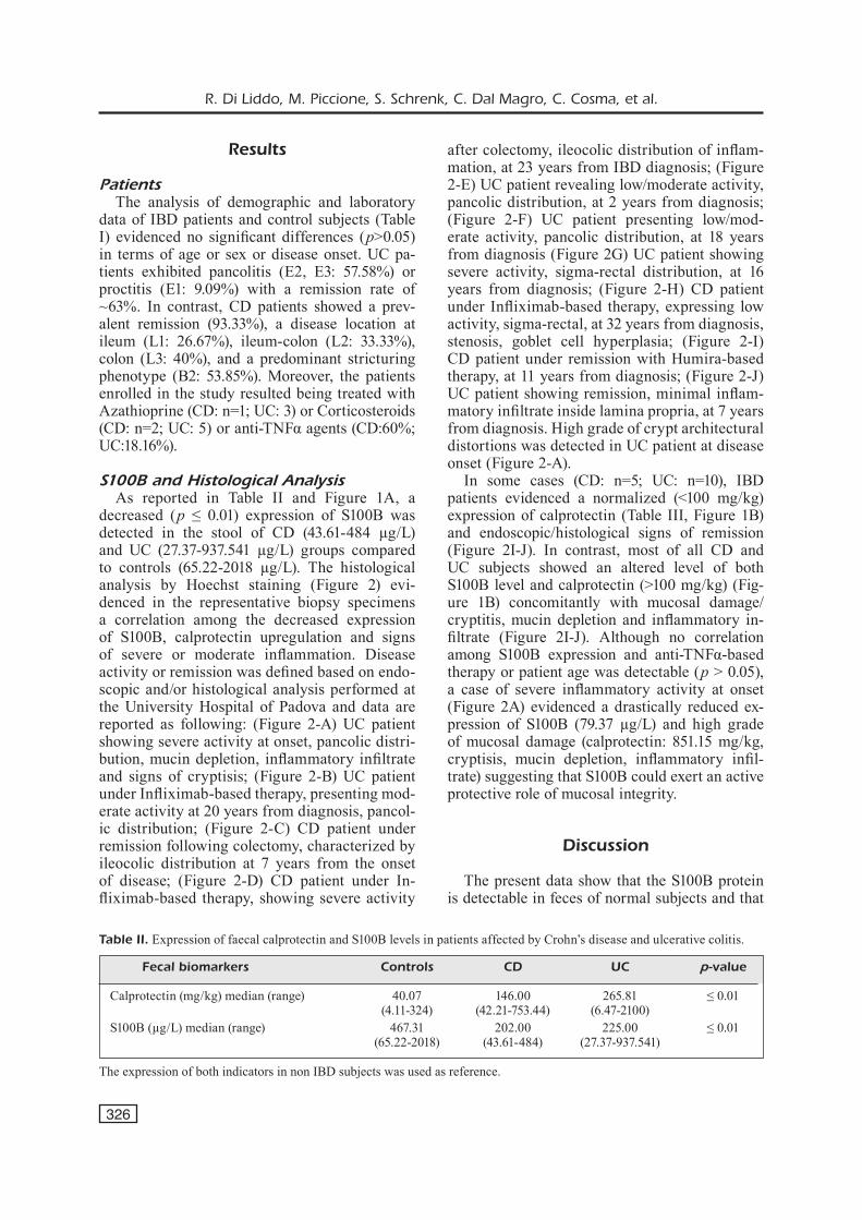
S100B and Histological AnalysisAs reported in Table II and Figure 1A, a
decreased (p ≤ 0.01) expression of S100B was detected in the stool of CD (43.61-484 µg/L) and UC (27.37-937.541 µg/L) groups compared to controls (65.22-2018 µg/L). The histological analysis by Hoechst staining (Figure 2) evi-denced in the representative biopsy specimens a correlation among the decreased expression of S100B, calprotectin upregulation and signs of severe or moderate inflammation. Disease activity or remission was defined based on endo-scopic and/or histological analysis performed at the University Hospital of Padova and data are reported as following: (Figure 2-A) UC patient showing severe activity at onset, pancolic distri-bution, mucin depletion, inflammatory infiltrate and signs of cryptisis; (Figure 2-B) UC patient under Infliximab-based therapy, presenting mod-erate activity at 20 years from diagnosis, pancol-ic distribution; (Figure 2-C) CD patient under remission following colectomy, characterized by ileocolic distribution at 7 years from the onset of disease; (Figure 2-D) CD patient under In-fliximab-based therapy, showing severe activity
after colectomy, ileocolic distribution of inflam-mation, at 23 years from IBD diagnosis; (Figure 2-E) UC patient revealing low/moderate activity, pancolic distribution, at 2 years from diagnosis; (Figure 2-F) UC patient presenting low/mod-erate activity, pancolic distribution, at 18 years from diagnosis (Figure 2G) UC patient showing severe activity, sigma-rectal distribution, at 16 years from diagnosis; (Figure 2-H) CD patient under Infliximab-based therapy, expressing low activity, sigma-rectal, at 32 years from diagnosis, stenosis, goblet cell hyperplasia; (Figure 2-I) CD patient under remission with Humira-based therapy, at 11 years from diagnosis; (Figure 2-J) UC patient showing remission, minimal inflam-matory infiltrate inside lamina propria, at 7 years from diagnosis. High grade of crypt architectural distortions was detected in UC patient at disease onset (Figure 2-A).
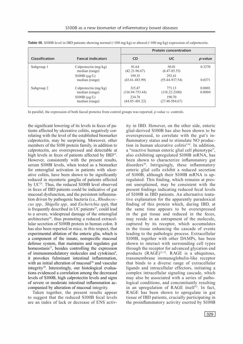
In some cases (CD: n=5; UC: n=10), IBD patients evidenced a normalized (<100 mg/kg) expression of calprotectin (Table III, Figure 1B) and endoscopic/histological signs of remission (Figure 2I-J). In contrast, most of all CD and UC subjects showed an altered level of both S100B level and calprotectin (>100 mg/kg) (Fig-ure 1B) concomitantly with mucosal damage/cryptitis, mucin depletion and inflammatory in-filtrate (Figure 2I-J). Although no correlation among S100B expression and anti-TNFα-based therapy or patient age was detectable (p > 0.05), a case of severe inflammatory activity at onset (Figure 2A) evidenced a drastically reduced ex-pression of S100B (79.37 µg/L) and high grade of mucosal damage (calprotectin: 851.15 mg/kg, cryptisis, mucin depletion, inflammatory infil-trate) suggesting that S100B could exert an active protective role of mucosal integrity.
Discussion
The present data show that the S100B protein is detectable in feces of normal subjects and that
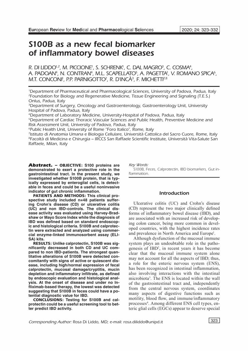
Table II. Expression of faecal calprotectin and S100B levels in patients affected by Crohn’s disease and ulcerative colitis.
Fecal biomarkers Controls CD UC p-value
Calprotectin (mg/kg) median (range) 40.07 146.00 265.81 ≤ 0.01 (4.11-324) (42.21-753.44) (6.47-2100) S100B (µg/L) median (range) 467.31 202.00 225.00 ≤ 0.01 (65.22-2018) (43.61-484) (27.37-937.541)
The expression of both indicators in non IBD subjects was used as reference.

S100B as a new biomarker of inflammatory bowel diseases
327
its concentration is significantly reduced in feces of untreated patients affected by ulcerative colitis.
After the earlier finding of S100B in cerebro-spinal fluid (CSF) of multiple sclerosis patients in the acute phase16, research on S100B as a biomarker of neural injury has been extended to other biological fluids besides CSF. Peripheral blood17, cord blood18, amniotic fluid19, urine20, and saliva21 have all been shown to contain detectable levels of S100B, which have been found to be altered in a variety of pathological conditions of the nervous system. These include acute brain
injury (cardiovascular disorders and traumatic injury), neurodegenerative diseases (Alzheimer’s disease, Parkinson’s disease, amyotrophic lateral sclerosis, multiple sclerosis), congenital/perinatal disorders (Trisomy 21, pre-term and full term as-phyxiated newborns, intrauterine growth retard-ed fetuses), psychiatric disorders (schizophrenia, mood disorders)8.
Feces have never been used, before the present study, in order to evaluate the presence of S100B and its possible alteration as a biomarker in patho-logical conditions. The contiguity of feces with the
Figure 1. A, Detection of faecal calprotectin (mg/kg) and S100B (µg/L) in patients affected by Crohn’s disease (CD) and Ulcerative Colitis (UC) by using enzyme-linked immunosorbent assay (ELISA) tests. In parallel, the analysis was performed on control samples obtained from donors listed for routine preventive care and not affected by IBD organic diseases. B, Discriminative analysis of S100B level compared to unchanged or increased calprotectin expression. **p-value < 0.01 vs. control.
R. Di Liddo, M. Piccione, S. Schrenk, C. Dal Magro, C. Cosma, et al.
328
wall of the intestinal tract proposes this material as particularly suitable to evaluate the gastroenter-ic apparatus and, as in the case of calprotectin, is in fact usefully employed as a source of this
accurate biomarker of disease activity in IBD22. Indeed, S100B is known to be overexpressed and to correlate with the gut’s inflammatory status in human ulcerative colitis9,10, so that the finding of
Figure 2. Confocal microscope images from some representative gut biopsies of patients included in the study. Paraffin sections were stained with Hoechst 33342 (nuclear stain) and images were acquired at 40× magnification. All pictures were optimized by brightness- and contrast-enhancement. The age and the disease activity of patients is reported in all pictures. UC: Ulcerative Colitis; CD: Crohn Disease; A, UC [severe activity at onset, pancolic distribution, signs of cryptisis]; B, UC [moderate activity, pancolic distribution, 20 years from diagnosis, Infliximab-based therapy]; C, CD [remission, ileocolic distribution, 7 years from diagnosis]; D, CD [severe activity, ileocolic distribution, 23 years from diagnosis, Infliximab-based therapy]; E, UC [low/moderate activity, pancolic distribution, 2 years from diagnosis]; F, UC [low/moderate activity, pancolic distribution, 18 years from diagnosis]; G, UC [severe activity, sigma-rectal distribution, 16 years from diagnosis]; H, CD [low activity, sigma-rectal, 32 years from diagnosis, stenosis, goblet cell hyperplasia, Infliximab-based therapy]; I, [remission, 11 years from onset, Humira-based therapy]; J, UC [remission, minimal inflammatory infiltrate in LP, 7 years from diagnosis]. Scale bar: 50 μm.
S100B as a new biomarker of inflammatory bowel diseases
329
the significant lowering of its levels in feces of pa-tients affected by ulcerative colitis, negatively cor-relating with the level of the established biomarker calprotectin, may be surprising. Moreover, other members of the S100 protein family, in addition to calprotectin, are overexpressed and detectable at high levels in feces of patients affected by IBD23. However, consistently with the present results, serum S100B levels, when tested as a biomarker for enteroglial activation in patients with ulcer-ative colitis, have been shown to be significantly reduced in myenteric ganglia of patients affected by UC24. Thus, the reduced S100B level observed in feces of IBD patients could be indicative of gut mucosal dysfunction, and the persistent inflamma-tion driven by pathogenic bacteria (i.e., Rhodococ-cus spp., Shigella spp., and Escherichia spp), that is frequently described in UC patients25, could lead to a severe, widespread damage of the enteroglial architecture26, thus promoting a reduced extracel-lular secretion of S100B protein in human colon. It has also been reported in mice, in this respect, that experimental ablation of the enteric glia, which is a component of the innate, nonspecific mucosal defense system, that maintains and regulates gut homeostasis27, besides controlling the expression of immunomodulatory molecules and cytokines9, it provokes fulminant intestinal inflammation, with an initial alteration of mucosal28 and vascular integrity29. Interestingly, our histological evalua-tions evidenced a correlation among the decreased levels of S100B, high calprotectin levels and signs of severe or moderate intestinal inflammation ac-companied by alteration of mucosal integrity.
Taken together, the present results appear to suggest that the reduced S100B fecal levels are an index of lack or decrease of ENS activ-
ity in IBD. However, on the other side, enteric glial-derived S100B has also been shown to be overexpressed, to correlate with the gut’s in-flammatory status and to stimulate NO produc-tion in human ulcerative colitis9,10. In addition, a “reactive human enteric glial cell phenotype”, also exhibiting upregulated S100B mRNA, has been shown to characterize inflammatory gut disorders30. Intriguingly, these inflammatory enteric glial cells exhibit a reduced secretion of S100B, although their S100B mRNA is up-regulated. This finding, which remains at pres-ent unexplained, may be consistent with the present findings indicating reduced fecal levels of S100B in IBD patients. An alternative tenta-tive explanation for the apparently paradoxical finding of this protein which, during IBD, at the same time appears to be overexpressed in the gut tissue and reduced in the feces, may reside in an entrapment of the molecule, captured by its receptor, which accumulates in the tissue enhancing the cascade of events leading to the pathologic process. Extracellular S100B, together with other DAMPs, has been shown to interact with surrounding cell types through the receptor for advanced glycation end products (RAGE)31,32. RAGE is a ubiquitous, transmembrane immunoglobulin-like receptor that binds to a diverse range of extracellular ligands and intracellular effectors, initiating a complex intracellular signaling cascade, which may also be associated with a series of patho-logical conditions, and concomitantly resulting in an upregulation of RAGE itself33. In fact, RAGE has been shown to upregulate in gut tissue of IBD patients, crucially participating in the proinflammatory activity exerted by S100B
Table III. S100B level in IBD patients showing normal (<100 mg/kg) or altered (>100 mg/kg) expression of calprotectin.
Protein concentration
Classification Faecal indicators CD UC p-value
Subgroup 1 Calprotectin (mg/kg) 81.64 50.01 0.3370 median (range) (42.21-96.67) (6.47-85.53) S100B (µg/L) 199.35 292.61 median (range) (43.61-483.99) (55.44-937.54) 0.0371
Subgroup 2 Calprotectin (mg/kg) 315.47 771.13 0.0001 median (range) (116.94-753.44) (118.22-2100) 0.0004 S100B (µg/L) 234.78 190.70 median (range) (44.85-481.22) (27.48-584.67)
In parallel, the expression of both faecal proteins from control groups was reported. p-value vs. controls.
R. Di Liddo, M. Piccione, S. Schrenk, C. Dal Magro, C. Cosma, et al.
330
protein10,34. Interestingly, multimeric S100B has been shown to exhibit a higher binding affini-ty to RAGE than dimeric S100B, which is the physiologically more diffuse form of the pro-tein35,36, so that a stronger binding of the protein with its receptor may result at high concentra-tion, possibly also involved in an inflammasome protein complex37 in the tissue, where DAMPs and RAGE are actually regarded as active par-ticipants38.
Finally, it should be noted that the fecal bio-markers of IBD currently regarded as the most reliable, such as calprotectin and lactoferrin, ex-hibit elevated levels during the active phase of the disease39,40 Indeed, they are proteins released during neutrophil degranulation, while S100B is a constituent of enteroglial tissue cells.
In any case, the question remains about the different findings of S100B in biological fluids as a biomarker of neural injury in other pathological conditions: in neurodegenerative diseases, acute brain injury and psychiatric disorders, the protein is constantly increased4, while, in feces of IBD patients, it is reduced as reported in the present study. In this respect, the structural/functional peculiarities of the enteric nervous system and its interaction with the intestinal epithelial barrier (IEB)41 may play a role in this discrepancy. Also the possi-ble interaction of the protein with the intestinal microbiota, which also differs in healthy and pathological conditions42 may interfere with S100B findings in feces. In this respect, in-triguingly, S100B has been reported indeed to be overexpressed in cultured enteric glial cells after exposure to pathogen but not probiotic bacteria13, but a degradation of the fecal protein operated by pathogen bacteria43 might explain the discrepancy.
To summarize, the present results show for the first time the presence of S100B in feces, at sig-nificantly different levels in healthy subjects and in patients with IBD, thus proposing S100B as a biomarker for this disease. Whether the reduced levels of fecal S100B in IBD reflect lack of ENS activity or a peculiarity of S100B dynamics in the gut compartment remains to be investigated. Studies on patients affected by irritable bowel syndrome, an intestinal disorder sharing some characteristics with IBD (for review, Spiller and Major, 2016)44, and/or on the interaction of S100B with microbiota, which constitutes an especially promising field of investigation, may be illumi-nating in this respect.
Conclusions
Compelling evidence indicates that intestinal barrier dysfunction exerts a pathogenic role in chronic inflammatory bowel diseases. Therefore, the identification of mediators associated with re-inforcing or re-establishing IEB functions could be of great clinical interest. In this study, the glial mediator S100B is found at lower level in feces of IBD patients compared to healthy subjects. While further studies are required to clarify the processes underlying this phenomenon, S100B is proposed as a new biomarker for the clinical evaluation of IBD.
Conflict of InterestThe Authors declare that they have no conflict of interests.
AcknowledgementsThe authors would like to thank University of Padova (PRID 2016/Di Liddo Rosa, DOR 2016-2018/Di Liddo) for economical support to this study, Carola Cenzi, Sonia Fac-chin and Enrico Rossi Sirena for technical assistance.
Authors’ ContributionConceptualization: R.D.L., R.D.I. and F.M.; execution of experiments: M.P., S.S., C.D.M., C.C., A.P., N.C., M.S. and A.P.; theoretical calculations: R.D.L., R.D.I. and F.M., writ-ing of the original draft: R.D.L., M.P. and F.M.; review and editing: R.D.L., R.S.V., M.T.C., P.P.P., F. M.
References
1) Nishida a, iNoue R, iNatomi o, BamBa s, Naito Y, aN-doh a. Gut microbiota in the pathogenesis of in-flammatory bowel disease. Clin J Gastroenterol 2018; 11: 1-10.
2) Kim dh, CheoN Jh. Pathogenesis of inflammato-ry bowel disease and recent advances in biologic therapies. Immune Netw 2017; 17: 25-40.
3) maRgolis Kg, geRshoN md. Enteric neuronal regu-lation of intestinal inflammation. Trends Neurosci 2016; 39: 614-624.
4) miChetti F, d’amBRosi N, toesCa a, Puglisi ma, seR-RaNo a, maRChese e, CoRviNo v, geloso mC. The S100B story: from biomarker to active factor in neural injury. J Neurochem 2019; 148: 168-187.
5) shaRKeY Ka. Emerging roles for enteric glia in gas-trointestinal disorders. J Clin Invest 2015; 125: 918-925.
6) oChoa-CoRtes F, tuRCo F, liNaN-RiCo a, soghomoN-YaN s, WhitaKeR e, WehNeR s, Cuomo R, ChRistoFi
S100B as a new biomarker of inflammatory bowel diseases
331
Fl. Enteric glial cells: a new frontier in neurogas-troenterology and clinical target for inflammato-ry bowel diseases. Inflamm Bowel Dis 2016; 22: 433-449.
7) FeRRi gl, PRoBeRt l, CoCChia d, miChetti F, maRaNgos PJ, PolaK JM. Evidence for the presence of S-100 protein in the glial component of the human enter-ic nervous system. Nature 1982; 297: 409-410.
8) miChetti F, CoRviNo v, geloso mC, lattaNzi W, BeR-NaRdiNi C, seRPeRo l, gazzolo d. The S100B protein in biological fluids: more than a lifelong biomark-er of brain distress. J Neurochem 2012; 120: 644-659.
9) esPosito g, CiRillo C, saRNelli g, de FiliPPis d, d’aRmieNto FP, RoCCo a, NaRdoNe g, PetRuzzelli R, gRosso m, izzo P, iuvoNe t, Cuomo R. Enteric gli-al-derived S100B protein stimulates nitric oxide production in celiac disease. Gastroenterology 2007; 133: 918-925.
10) CiRillo C, saRNelli g, esPosito g, gRosso m, PetRuz-zelli R, izzo P, Calì g, d’aRmieNto FP, RoCCo a, NaR-doNe g, iuvoNe t, steaRdo l, Cuomo R. increased mucosal nitric oxide production in ulcerative coli-tis is mediated in part by the enteroglial-derived S100B protein. Neurogastroenterol Motil 2009; 21: 1209-e112.
11) esPosito g, CaPoCCia e, saRNelli g, sCudeRi C, CiRil-lo C, Cuomo R, steaRdo l. The antiprotozoal drug pentamidine ameliorates experimentally induced acute colitis in mice. J Neuroinflammation 2012; 9: 277.
12) esPosito g, CaPoCCia e, tuRCo F, PalumBo i, lu J, steaRdo a, Cuomo R, saRNelli g, steaRdo l. Pal-mitoylethanolamide improves colon inflammation through an enteric glia/toll like receptor 4-depen-dent PPAR-α activation. Gut 2014; 63: 1300-1312.
13) tuRCo F, saRNelli g, CiRillo C, PalumBo i, de gioRgi F, d’alessaNdRo a, CammaRota m, giuliaNo m, Cuomo R. Enteroglial-derived S100B protein integrates bacteria-induced Toll-like receptor signalling in human enteric glial cells. Gut 2014; 63: 105-115.
14) CaPoCCia e, CiRillo C, gigli s, PesCe m, D’Alessan-dro A, Cuomo R, Sarnelli G, Steardo L, Esposito G. Enteric glia: a new player in inflammatory bow-el diseases. Int J Immunopathol Pharmacol 2015; 28: 443-451.
15) CeliKBileK a, CeliKBileK m, saBah s, taNK N, BoReKCi e, dogaN s, aKiN Y, BaldaNe s, deNiz K, Yilmaz N, oz-BaKiR o, YuCesoY m. The serum S100B level as a biomarker of enteroglial activation in patients with ulcerative colitis. Int J Inflam 2014; 2014: 986525.
16) miChetti F, massaRo a, muRazio m. The nervous system-specific S-100 antigen in cerebrospinal fluid of multiple sclerosis patients. Neurosci Lett 1979; 11: 171-175.
17) Kato K, KimuRa s, semBa R, suzuKi F, NaKaJima t. In-crease in S-100 protein levels in blood plasma by epinephrine. J Biochem 1983; 94: 1009-1011.
18) gazzolo d, viNesi P, maRiNoNi e, di ioRio R, maR-Ras m, lituaNia m, BRusChettiNi P, miChetti F. S100B protein concentrations in cord blood: correlations
with gestational age in term and preterm deliver-ies. Clin Chem 2000; 46: 998-1000.
19) gazzolo d, BRusChettiNi m, CoRviNo v, oliva R, saR-li R, lituaNia m, BRusChettiNi P, miChetti F. S100B protein concentrations in amniotic fluid correlate with gestational age and with cerebral ultrasound scanning results in healthy fetuses. Clin Chem 2001; 47: 954-956.
20) gazzolo d, BRusChettiNi m, lituaNia m, seRRa g, Bo-NaCCi W, miChetti F. Increased urinary S100B pro-tein as an early indicator of intraventricular hem-orrhage in preterm infants: correlation with the grade of hemorrhage. Clin Chem 2001; 47: 1836-1838.
21) gazzolo d, lituaNia m, BRusChettiNi m, Ciotti s, saC-Chi R, seRRa g, Calevo mg, CoRviNo v, BuoNoCoRe g, miChetti F. S100B protein levels in saliva: cor-relation with gestational age in normal term and preterm newborns. Clin Biochem 2005; 38: 229-233.
22) motagaNahalli s, BesWiCK l, CoN d, vaN laNgeNBeRg dR. Faecal calprotectin delivers on convenience, cost reduction and clinical decision-making in in-flammatory bowel disease: a real-world cohort study. Intern Med J 2019; 49: 94-100.
23) maNolaKis aC, KaPsoRitaKis aN, tiaKa eK, PotamiaNos sP. Calprotectin, calgranulin C, and other mem-bers of the s100 protein family in inflammatory bowel disease. Dig Dis Sci 2011; 56: 1601-1611.
24) BeRNaRdiNi N, segNaNi C, iPPolito C, de gioRgio R, ColuCCi R, FaussoNe-PellegRiNi ms, ChiaRugi m, Cam-PaNi d, CastagNa m, mattii l, BlaNdizzi C, dolFi a. Immunohistochemical analysis of myenteric gan-glia and interstitial cells of Cajal in ulcerative coli-tis. J Cell Mol Med 2012; 16: 318-327.
25) sasaKi m, KlaPPRoth Jm. the role of bacteria in the pathogenesis of ulcerative colitis. J Signal Trans-duct 2012; 2012: 704953.
26) CoRoN e, FlamaNt m, auBeRt P, Wedel t, PedRoN t, letessieR e, galmiChe JP, saNsoNetti PJ, NeuNlist m. Characterisation of early mucosal and neuronal lesions following Shigella flexneri infection in hu-man colon. PLoS One 2009; 4: e4713.
27) giaRoNi C, de PoNti F, CoseNtiNo m, leCChiNi s, FRigo g. Plasticity in the enteric nervous system. Gas-troenterology 1999; 117: 1438-1458.
28) Bush tg, savidge tC, FReemaN tC, Cox hJ, CamPBell ea, muCKe l, JohNsoN mh, soFRoNieW mv. Fulmi-nant jejuno-ileitis following ablation of enteric gila in adult transgenic mice. Cell 1998; 93: 189-201.
29) CoRNet a, savidge tC, CaBaRRoCas J, deNg Wl, Co-lomBel JF, lassmaNN h, desReumaux P, liBlau Rs. Enterocolitis induced by autoimmune targeting of enteric glial cells: a possible mechanism in Crohn’s disease? Proc Natl Acad Sci 2001; 98: 13306-13311.
30) liñáN-RiCo a, tuRCo F, oChoa-CoRtes F, haRzmaN a, NeedlemaN BJ, aRseNesCu R, aBdel- Rasoul m, Fadda P, gRaNts i, WhitaKeR e, Cuomo R, ChRistoFi FL. Mo-lecular signaling and dysfunction of the human reactive enteric glial cell phenotype: implications
R. Di Liddo, M. Piccione, S. Schrenk, C. Dal Magro, C. Cosma, et al.
332
for GI infection, IBD, POI, neurological, motility, and GI disorders. Inflamm Bowel Dis 2016; 22: 1812-1834.
31) hoFmaNN ma, dRuRY s, Fu C, Qu W, taguChi a, lu Y, avila C, KamBham N, BieRhaus a, NaWRoth P, NeuR-ath mF, slatteRY t, BeaCh d, mCClaRY J, Nagashima m, moRseR J, steRN d, sChmidt am. RAGE mediates a novel proinflammatory axis: a central cell sur-face receptor for S100/calgranulin polypeptides. Cell 1999; 97: 889-901.
32) huttuNeN h, KuJa-PaNula J, soRCi g, agNeletti a, doNato R, Rauvala h. Coregulation of neurite out-growth and cell survival by amphoterin and S100 proteins through RAGE activation. J Biol Chem 2000; 275: 40096-40105.
33) BoNgaRzoNe s, saviCKas v, luzi F, gee ad. targeting the receptor for advanced glycation endproducts (RAGE): a medicinal chemistry perspective. J Med Chem 2017; 60: 7213-7232.
34) aNdRassY m, igWe J, autsChBaCh F, volz C, RemPPis a, NeuRath mF, sChleiCheR e, humPeRt Pm, WeNdt t, lilieNsieK B, moRCos m, sChieKoFeR s, thiele K, CheN J, KieNtsCh-eNgel R, sChmidt am, stRemmel W, steRN dm, Katus ha, NaWRoth PP, BieRhaus a. Posttrans-lationally modified proteins as mediators of sus-tained intestinal inflammation. Am J Pathol 2006; 169: 1223-1237.
35) osteNdoRP t, heizmaNN CW, KRoNeCK Pmh, FRitz g. Purification, crystallization and preliminary X-ray diffraction studies on human Ca2+-binding pro-tein S100B. Acta Crystallogr Sect F Struct Biol Cryst Commun 2005; 61: 673-675.
36) osteNdoRP t, leCleRC e, galiChet a, KoCh m, demliNg N, Weigle B, heizmaNN CW, KRoNeCK Pmh, FRitz g. Structural and functional insights into RAGE ac-
tivation by multimeric S100B. EMBO J 2007; 26: 3868-3878.
37) maRtiNoN F, BuRNs K, JuRg t. The inflammasome: a molecular platform triggering activation of inflam-matory caspases and processing of proIL- β. Mol Cell 2002; 10: 417-426.
38) tuRNeR Na. Inflammatory and fibrotic responses of cardiac fibroblasts to myocardial damage associ-ated molecular patterns (DAMPs). J Mol Cell Car-diol 2016; 94: 189-200.
39) galgut BJ, lemBeRg da, daY as, leaCh st. The val-ue of fecal markers in predicting relapse in in-flammatory bowel diseases. Front Pediatr 2018; 5: 292.
40) dai C, JiaNg m, suN mJ. Fecal markers in the man-agement of inflammatory bowel disease. Post-grad Med 2018; 130: 597-606.
41) NeuNlist m, auBeRt P, BoNNaud s, vaN laNdeghem l, CoRoN e, Wedel t, NaveilhaN P, Ruhl a, laRdeux B, savidge t, PaRis F, galmiChe JP. Enteric glia inhibit in-testinal epithelial cell proliferation partly through a TGF-β1 -dependent pathway. Am J Physiol Liver Physiol 2007; 292: G231-241.
42) zuo t, Ng sC. The gut microbiota in the pathogen-esis and therapeutics of inflammatory bowel dis-ease. Front Microbiol 2018; 9: 2247.
43) WestReiCh st, aRdeshiR a, alKaN z, KaBle me, KoRF i, lemaY dg. Fecal metatranscriptomics of ma-caques with idiopathic chronic diarrhea reveals altered mucin degradation and fucose utilization. Microbiome 2019; 7: 41.
44) sPilleR R, maJoR g. IBS and IBD-separate entities or on a spectrum? Nat Rev Gastroenterol Hepa-tol 2016; 13: 613-621.