Management of malignant bowel obstruction
11
Management of malignant bowel obstruction Carla Ida Ripamonti a,b, * , Alexandra M. Easson c , Hans Gerdes d a Professor on Contract of Oncology (Teaching Palliative Medicine) at the School of Specialization in Oncology, University of Milan b Palliative Care Unit (Pain Therapy-Rehabilitation) IRCCS Foundation, National Cancer Institute, Milano Italy c Assistant Professor of Surgery, University of Toronto, Division of General Surgery, Mount Sinai Hospital, Department of Surgical Oncology, Princess Margaret Hospital Toronto, Ontario Canada d Attending Physician, Director of GI Endoscopy, Memorial Sloan-Kettering Cancer Center, and Professor of Clinical Medicine, Weill Medical College of Cornell University ARTICLE INFO Article history: Received 11 February 2008 Accepted 25 February 2008 Available online 21 March 2008 Keywords: Malignant bowel obstruction Advanced/end-stage cancer patients Palliative medical treatment Surgery Stents Nasogastric suction Symptom control ABSTRACT Malignant bowel obstruction (MBO) is a common and distressing outcome particularly in patients with bowel or gynaecological cancer. Radiological imaging, particularly with CT, is critical in determining the cause of obstruction and possible therapeutic interventions. Although surgery should be the primary treatment for selected patients with MBO, it should not be undertaken routinely in patients known to have poor prognostic criteria for surgical intervention such as intra-abdominal carcinomatosis, poor performance status and massive ascites. A number of treatment options are now available for patients unfit for surgery. Nasogastric drainage should generally only be a temporary measure. Self-expand- ing metallic stents are an option in malignant obstruction of the gastric outlet, proximal small bowel and colon. Medical measures such as analgesics according to the W.H.O. guide- lines provide adequate pain relief. Vomiting may be controlled using anti-secretory drugs or/and anti-emetics. Somatostatin analogues (e.g. octreotide) reduce gastrointestinal secre- tions very rapidly and have a particularly important role in patients with high obstruction if hyoscine butylbromide fails. A collaborative approach by surgeons and the oncologist and/or palliative care physician as well as an honest discourse between physicians and patients can offer an individualised and appropriate symptom management plan. Ó 2008 Elsevier Ltd. All rights reserved. 1. Introduction A recent consensus conference defined MBO using the follow- ing criteria: clinical evidence of bowel obstruction (history/ physical/ radiological examination); bowel obstruction be- yond the ligament of Treitz, in the setting of a diagnosis of in- tra-abdominal cancer with incurable disease, OR a diagnosis of non-intra-abdominal primary cancer with clear intraperi- toneal disease. 1 Retrospective reviews show that 10-28% of patients with colorectal cancer will develop a MBO in the course of their dis- ease whereas 20-50% of patients with ovarian cancer present with symptoms of bowel obstruction. 2 Intestinal involvement of metastatic cancer commonly presents as diffuse peritoneal carcinomatosis or more rarely as an isolated gastrointestinal metastasis in 10% of cases. 8 Breast cancer or melanoma are the most common non-gastrointestinal causes and can occur many years from primary presentation. 3 0959-8049/$ - see front matter Ó 2008 Elsevier Ltd. All rights reserved. doi:10.1016/j.ejca.2008.02.028 * Corresponding author: Address. Palliative Care Unit (Pain Therapy-Rehabilitation) IRCCS Foundation, National Cancer Institute, Milano Italy. Tel.: +39 02 23902243; fax. +39 02 23903656. E-mail addresses: [email protected] (C.I. Ripamonti), [email protected] (A.M. Easson), gerdesh@mskcc. org (H. Gerdes). EUROPEAN JOURNAL OF CANCER 44 (2008) 1105 – 1115 available at www.sciencedirect.com journal homepage: www.ejconline.com
-
Upload
independent -
Category
Documents
-
view
0 -
download
0
Transcript of Management of malignant bowel obstruction

E U R O P E A N J O U R N A L O F C A N C E R 4 4 ( 2 0 0 8 ) 1 1 0 5 – 1 1 1 5
. sc iencedi rec t . com
ava i lab le a t wwwjournal homepage: www.ejconl ine.com
Management of malignant bowel obstruction
Carla Ida Ripamontia,b,*, Alexandra M. Eassonc, Hans Gerdesd
aProfessor on Contract of Oncology (Teaching Palliative Medicine) at the School of Specialization in Oncology, University of MilanbPalliative Care Unit (Pain Therapy-Rehabilitation) IRCCS Foundation, National Cancer Institute, Milano ItalycAssistant Professor of Surgery, University of Toronto, Division of General Surgery, Mount Sinai Hospital, Department of Surgical Oncology,
Princess Margaret Hospital Toronto, Ontario CanadadAttending Physician, Director of GI Endoscopy, Memorial Sloan-Kettering Cancer Center, and Professor of Clinical Medicine,
Weill Medical College of Cornell University
A R T I C L E I N F O
Article history:
Received 11 February 2008
Accepted 25 February 2008
Available online 21 March 2008
Keywords:
Malignant bowel obstruction
Advanced/end-stage cancer patients
Palliative medical treatment
Surgery
Stents
Nasogastric suction
Symptom control
0959-8049/$ - see front matter � 2008 Elsevidoi:10.1016/j.ejca.2008.02.028
* Corresponding author: Address. Palliative CItaly. Tel.: +39 02 23902243; fax. +39 02 23903
E-mail addresses: carla.ripamonti@istitutoorg (H. Gerdes).
A B S T R A C T
Malignant bowel obstruction (MBO) is a common and distressing outcome particularly in
patients with bowel or gynaecological cancer. Radiological imaging, particularly with CT,
is critical in determining the cause of obstruction and possible therapeutic interventions.
Although surgery should be the primary treatment for selected patients with MBO, it
should not be undertaken routinely in patients known to have poor prognostic criteria
for surgical intervention such as intra-abdominal carcinomatosis, poor performance status
and massive ascites. A number of treatment options are now available for patients unfit for
surgery. Nasogastric drainage should generally only be a temporary measure. Self-expand-
ing metallic stents are an option in malignant obstruction of the gastric outlet, proximal
small bowel and colon. Medical measures such as analgesics according to the W.H.O. guide-
lines provide adequate pain relief. Vomiting may be controlled using anti-secretory drugs
or/and anti-emetics. Somatostatin analogues (e.g. octreotide) reduce gastrointestinal secre-
tions very rapidly and have a particularly important role in patients with high obstruction if
hyoscine butylbromide fails.
A collaborative approach by surgeons and the oncologist and/or palliative care physician
as well as an honest discourse between physicians and patients can offer an individualised
and appropriate symptom management plan.
� 2008 Elsevier Ltd. All rights reserved.
1. Introduction
A recent consensus conference defined MBO using the follow-
ing criteria: clinical evidence of bowel obstruction (history/
physical/ radiological examination); bowel obstruction be-
yond the ligament of Treitz, in the setting of a diagnosis of in-
tra-abdominal cancer with incurable disease, OR a diagnosis
of non-intra-abdominal primary cancer with clear intraperi-
toneal disease.1
er Ltd. All rights reserved
are Unit (Pain Therapy-Re656.tumori.mi.it (C.I. Ripamo
Retrospective reviews show that 10-28% of patients with
colorectal cancer will develop a MBO in the course of their dis-
ease whereas 20-50% of patients with ovarian cancer present
with symptoms of bowel obstruction.2 Intestinal involvement
of metastatic cancer commonly presents as diffuse peritoneal
carcinomatosis or more rarely as an isolated gastrointestinal
metastasis in 10% of cases.8 Breast cancer or melanoma are
the most common non-gastrointestinal causes and can occur
many years from primary presentation.3
.
habilitation) IRCCS Foundation, National Cancer Institute, Milano
nti), [email protected] (A.M. Easson), gerdesh@mskcc.

1106 E U R O P E A N J O U R N A L O F C A N C E R 4 4 ( 2 0 0 8 ) 1 1 0 5 – 1 1 1 5
Tumour causes of obstruction by many mechanisms are
shown in Table 1.
Patients with MBO usually describe a pattern of gradual
worsening of symptoms that include episodes of abdominal
cramps, nausea and vomiting and abdominal distension that
resolve with the passage of gas or loose stool (Fig. 1). Symp-
toms become more frequent and last longer until near to
complete obstruction results. Initial management includes a
clinical assessment to rule out acute causes of obstruction
and to ensure that the patient does not have a surgical emer-
gency. The patient is resuscitated with fluid to replace any
losses from vomiting and a nasogastric tube may be placed
Table 1 – Pathophysiology of bowel obstruction
Mechanical obstruction
Extrinsic occlusion of the lumen: enlargement of the primary tumour or
adhesions, postirradiation fibrosis that cause bowel compression
Intra-luminal occlusion of the lumen: results from tumour growth from
Intramural occlusion of the lumen: intestinal linitis plastica, tumour wi
Adynamic ileus or functional obstruction
Intestinal motility disorders: tumour infiltration of the mesentery or bow
plexus
Intestinal motility disorders: paraneoplastic neuropathy particularly in p
paraneoplastic pseudo-obstruction.
• *Mechanical obstruction only, ** Prostaglandins , # Vasoactive Intestinal Polypeptide
PARTIAL OR COMPLEBOWEL OBSTRUCTI
CONTINUOUSPAIN
ALSO DUE TO
TUMOUR MASS
REDUCTION OR STOP OF TH
MOVEMENTS OF INTESTINA
CONTENTS
↑ BOWEL DI
DAMAGE OF INTESTINAL EPITAELIUM
BOWEL INFLAMMATORY RESPONSE WITH OEDEMA AND HYPERAEMIA
AND PRODUCTION OF PG**, VIP#, NOCICEPTIVE MEDIATORS
Fig. 1 – Pathophysiology
to decompress the proximal bowel and alleviate the patient’s
acute symptoms. Table 2 shows the radiological examinations
required.4–9 Although the location of the obstruction can of-
ten be determined by the nature and presentations of symp-
toms (Table 3), it is recommended that further imaging,
primarily with CT scan, be performed in order to determine
the management plan.1 MRI may also be used if necessary.10
Clinically, radiological imaging with CT (and/or potentially
MRI) has become indispensable in the decision-making pro-
cess when deciding whether a surgical or medical manage-
ment plan would be most effective to relieve obstructive
symptoms in patients with a MBO.
recurrence, mesenteric and omental masses, abdominal or pelvic
within the bowel
thin the wall of the bowel resulting in poor motility
el wall muscle and nerves, or malignant involvement of the coeliac
atients with lung cancer, chronic intestinal pseudo-obstruction (CIP),
TE ON
ROUGH-L
STENSION ↑ OF LUMEN CONTENTS
↑ GUT EPITHELIAL SURFACE AREA
↑ BOWEL SECRETIONS OF H2O, NA, CL
NAUSEA AND/OR VOMITING
↑ BOWEL CONTRACTIONS TO
SURMOUNT THE OBSTACLE*
↑ COLICKY PAIN
of bowel obstruction.
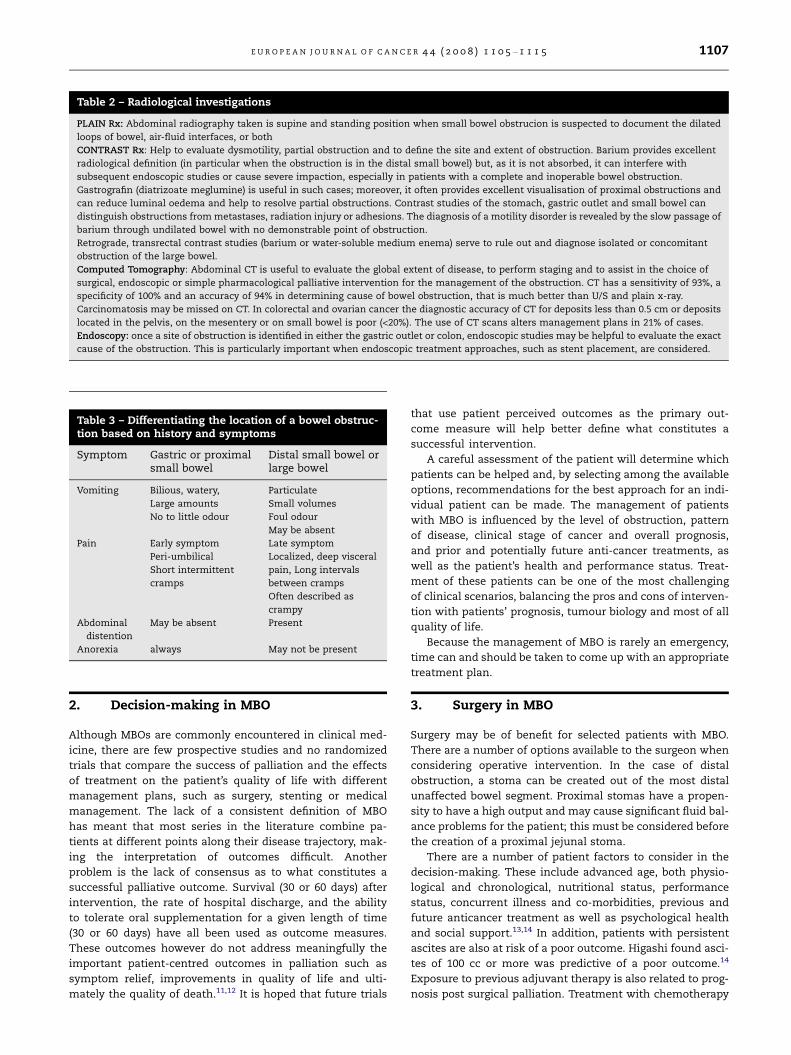
Table 2 – Radiological investigations
PLAIN Rx: Abdominal radiography taken is supine and standing position when small bowel obstrucion is suspected to document the dilated
loops of bowel, air-fluid interfaces, or both
CONTRAST Rx: Help to evaluate dysmotility, partial obstruction and to define the site and extent of obstruction. Barium provides excellent
radiological definition (in particular when the obstruction is in the distal small bowel) but, as it is not absorbed, it can interfere with
subsequent endoscopic studies or cause severe impaction, especially in patients with a complete and inoperable bowel obstruction.
Gastrografin (diatrizoate meglumine) is useful in such cases; moreover, it often provides excellent visualisation of proximal obstructions and
can reduce luminal oedema and help to resolve partial obstructions. Contrast studies of the stomach, gastric outlet and small bowel can
distinguish obstructions from metastases, radiation injury or adhesions. The diagnosis of a motility disorder is revealed by the slow passage of
barium through undilated bowel with no demonstrable point of obstruction.
Retrograde, transrectal contrast studies (barium or water-soluble medium enema) serve to rule out and diagnose isolated or concomitant
obstruction of the large bowel.
Computed Tomography: Abdominal CT is useful to evaluate the global extent of disease, to perform staging and to assist in the choice of
surgical, endoscopic or simple pharmacological palliative intervention for the management of the obstruction. CT has a sensitivity of 93%, a
specificity of 100% and an accuracy of 94% in determining cause of bowel obstruction, that is much better than U/S and plain x-ray.
Carcinomatosis may be missed on CT. In colorectal and ovarian cancer the diagnostic accuracy of CT for deposits less than 0.5 cm or deposits
located in the pelvis, on the mesentery or on small bowel is poor (<20%). The use of CT scans alters management plans in 21% of cases.
Endoscopy: once a site of obstruction is identified in either the gastric outlet or colon, endoscopic studies may be helpful to evaluate the exact
cause of the obstruction. This is particularly important when endoscopic treatment approaches, such as stent placement, are considered.
Table 3 – Differentiating the location of a bowel obstruc-tion based on history and symptoms
Symptom Gastric or proximalsmall bowel
Distal small bowel orlarge bowel
Vomiting Bilious, watery,
Large amounts
No to little odour
Particulate
Small volumes
Foul odour
May be absent
Pain Early symptom
Peri-umbilical
Short intermittent
cramps
Late symptom
Localized, deep visceral
pain, Long intervals
between cramps
Often described as
crampy
Abdominal
distention
May be absent Present
Anorexia always May not be present
E U R O P E A N J O U R N A L O F C A N C E R 4 4 ( 2 0 0 8 ) 1 1 0 5 – 1 1 1 5 1107
2. Decision-making in MBO
Although MBOs are commonly encountered in clinical med-
icine, there are few prospective studies and no randomized
trials that compare the success of palliation and the effects
of treatment on the patient’s quality of life with different
management plans, such as surgery, stenting or medical
management. The lack of a consistent definition of MBO
has meant that most series in the literature combine pa-
tients at different points along their disease trajectory, mak-
ing the interpretation of outcomes difficult. Another
problem is the lack of consensus as to what constitutes a
successful palliative outcome. Survival (30 or 60 days) after
intervention, the rate of hospital discharge, and the ability
to tolerate oral supplementation for a given length of time
(30 or 60 days) have all been used as outcome measures.
These outcomes however do not address meaningfully the
important patient-centred outcomes in palliation such as
symptom relief, improvements in quality of life and ulti-
mately the quality of death.11,12 It is hoped that future trials
that use patient perceived outcomes as the primary out-
come measure will help better define what constitutes a
successful intervention.
A careful assessment of the patient will determine which
patients can be helped and, by selecting among the available
options, recommendations for the best approach for an indi-
vidual patient can be made. The management of patients
with MBO is influenced by the level of obstruction, pattern
of disease, clinical stage of cancer and overall prognosis,
and prior and potentially future anti-cancer treatments, as
well as the patient’s health and performance status. Treat-
ment of these patients can be one of the most challenging
of clinical scenarios, balancing the pros and cons of interven-
tion with patients’ prognosis, tumour biology and most of all
quality of life.
Because the management of MBO is rarely an emergency,
time can and should be taken to come up with an appropriate
treatment plan.
3. Surgery in MBO
Surgery may be of benefit for selected patients with MBO.
There are a number of options available to the surgeon when
considering operative intervention. In the case of distal
obstruction, a stoma can be created out of the most distal
unaffected bowel segment. Proximal stomas have a propen-
sity to have a high output and may cause significant fluid bal-
ance problems for the patient; this must be considered before
the creation of a proximal jejunal stoma.
There are a number of patient factors to consider in the
decision-making. These include advanced age, both physio-
logical and chronological, nutritional status, performance
status, concurrent illness and co-morbidities, previous and
future anticancer treatment as well as psychological health
and social support.13,14 In addition, patients with persistent
ascites are also at risk of a poor outcome. Higashi found asci-
tes of 100 cc or more was predictive of a poor outcome.14
Exposure to previous adjuvant therapy is also related to prog-
nosis post surgical palliation. Treatment with chemotherapy

1108 E U R O P E A N J O U R N A L O F C A N C E R 4 4 ( 2 0 0 8 ) 1 1 0 5 – 1 1 1 5
does not impact on surgical complications, unless the patient
is malnourished or frail from this treatment. However, the
overall exposure to chemotherapy limits its successful use
after surgical intervention for a MBO and ultimately impacts
on patients’ overall survival.
There is much debate on the role of psychological health
and societal support on oncology patient’s survival, however
a decreased survival and an increased rate of mortality is
reported in patients who report feeling socially isolated or
who have severe recurrent depression.15 Randomized trials
in oncology patients evaluating the role of psychotherapy
on survival, report conflicting data, however there is a trend
showing an improved survival with participation in psycho-
therapy interventions and no evidence of a detrimental
effect.
Absolute and relative contraindications to proceeding with
palliative surgery have been identified from retrospective case
series examining characteristics associated with high rates of
mortality and morbidity and translated into prognostic crite-
Patient presenting with sobstruction and a hi
Clinical assessment − Patient acutely ill: surgical emergency Most
patients with MBO surgical emergency− History of symptoms
Patient factors − Age: biologic/physiologic − Performance status − Stage of cancer: previous
treatments, any anticancer treatment options
− Malnutrition/cachexia− Concurrent illnesses − Ascites
MBO decision m− Identify cause for obstr
mechanical vs. functionobstruction
− Assess the realistic abilintervention to alleviatesymptoms
− Formulate recommendathe intervention(s) that the best results for this this time
− NO obligation to recomtherapy
Decision-making with p− What do they understand about their disease a
trajectory?− Determine whether symptom alleviation fits t
Explain clearly the expected potential benefitsomething that would be worth it to them give
− Provide a commitment to continue to care forof the discussion
Fig. 2 – Algorithm for assessing and managing
ria.16 It is becoming increasingly clear that a MBO from gener-
alized carcinomatosis is a distinct entity that responds poorly,
or not at all, to surgical intervention. These obstructions are
usually partial, intermittent and do not involve strangulated
or twisted bowel at risk of perforation. They are caused by
blockage of the bowel at multiple levels of the small and/or
large bowel, possibly complicated by motility disorders sec-
ondary to bowel wall infiltration by tumour and/or compro-
mise of the parasympathetic and sympathetic nerves
responsible for peristalsis. Symptoms may resolve temporar-
ily with nasogastric decompression but will recur. When such
patients are taken to the operating room, the results are gen-
erally poor, with a high 30-day mortality (21-40%) and a high
complication rate (20-40%) and, even more discouraging,
most will re-obstruct within a short period of time.16
Although anaesthesia and surgical practices have evolved
over the last 50 years, recent reports of morbidities and mor-
talities from patients treated surgically for MBO remain high,
even with good patient selection.17,18
ymptoms of bowel story of cancer
Radiologic assessment CT and/MRI
− Diagnosis and cause of obstruction − Site: Single vs. multiple
− Large vs. small bowel − Partial (most MBO) vs. complete
aking uction: al
ity of any the
tions: Select will provide patient at
mend futile
Technical factors − Degree of invasiveness
− Interventional radiology − Endoscopy− Open laparotomy/laparoscopy
− Anaesthetic requirements − Risk of post-procedure complications
atient and family nd where they are on their disease
he goals of care of the patient. s of any intervention: is this n the risks?
the patient regardless of the outcome
a patient with malignant bowel obstruction.

E U R O P E A N J O U R N A L O F C A N C E R 4 4 ( 2 0 0 8 ) 1 1 0 5 – 1 1 1 5 1109
Patients facing incurable cancer are extremely vulnerable.
Because of this, the lines between informing, persuading and
manipulating can become blurred and obtaining truly in-
formed consent can be difficult.19 Involved well-meaning
family members and clinicians may cloud the decision-mak-
ing process and supersede the patient’s wishes. In the face of
an incurable, progressive illness the balance between honesty
and maintaining hope and optimism can be difficult to
achieve but it is necessary to avoid the use of futile treat-
ments and harm to the patient.20 A treatment is considered
futile if a response is physiologically impossible, if it is non-
beneficial, or if it is unlikely to produce the desired benefit.
However, there is little guidance on what should be consid-
ered a futile treatment as the definition may vary from pa-
tient-to-patient and/or clinician-to-clinician based on
previous personal experiences and expectations. Most clini-
cians agree, however, palliative surgery in oncology patients
should not be offered to meet emotional, existential and/or
psychological needs.21 The decision to consider palliative or
supportive care can be regarded by both clinicians and pa-
tients as a reflection of their personal failure and may signify
the absence of hope and loss of optimism about the disease
and the future.22 In this circumstance, the physician may feel
obliged to offer treatment even if it is futile and the patient
obliged to accept it or be considered a failure. To maintain
and achieve a better balance of honesty, hope and avoidance
of a futile procedure, the patient, the family and the clinicians
should first address the patient’s realistic goals of treatment.
These are usually directed towards relief of suffering, improv-
ing quality of life and may vary between similar patients as
they are based on the patients’ perceptions and life experi-
ences. Secondly, all treatment options including surgery,
interventional radiology and medications should be dis-
cussed, including the expected realistic benefits of each pro-
posed intervention and associated risks. We propose that
each patient should be assessed considering the above fac-
tors, the risks and benefits of proceeding with surgery, consid-
ering all non-surgical options as well as the patient’s goals
and expectations in order to come up with the optimal treat-
ment plan for each individual patient. (Fig. 2).
4. Endoscopic management ofgastro-duodenal obstruction
Gastric outlet obstruction (GOO) and proximal small bowel
obstruction are common complications in patients with pan-
creatic cancer, distal gastric cancer, gall bladder cancer and
cholangiocarcinoma, but may also result from metastases
from ovarian cancer and non-abdominal malignancies such
as lung cancer and breast cancer. Over the past decade ad-
vances in endoscopic technology have accomplished good re-
sults in the relief of obstruction and reduction of symptoms
with the endoscopic insertion of a self-expanding metal stent
(SEMS), or gastric venting via a percutaneously placed gas-
trostomy (drainage PEG). These approaches are particularly
useful for patients with poor short-term prognosis.
The technical success rates for placement of a stent have
been reported to be >90% and clinical success with resolution
of nausea and vomiting and improved ability to consume food
orally is reported over 75%.23–26 Late complications or delayed
stent failure can occur, including stent obstruction by food
impaction, and reobstruction caused by tumour ingrowth.
Stent migration also can occur, sometimes in association
with cancer treatment, if there is reduction in the size of
the tumour. In most cases, re-obstruction due to tumour in-
growth can be managed with placement of a second stent
or tumour ablation by Nd:YAG LASER or argon plasma
coagulator.27
In the limited comparative studies published, endoscopic
stent placement has been associated with shorter hospital
stay and lower peri-procedural mortality in patients with gas-
tric outlet obstruction secondary to pancreatic cancer,28,29
and with more rapid food intake compared to surgical by-
pass.28,30 Those managed with stent however have a greater
need for re-intervention compared with surgically-treated pa-
tients as a result of delayed stent occlusion.30,31
Assessments of the patient’s quality of life (QOL) after pal-
liative stenting for malignant GOO have been limited or ab-
sent from most studies.32,33 In one prospective study
examining QOL in patients with malignant GOO, Mehta and
coauthors randomized 27 patients to receive laparoscopic
gastrojejunostomy or endoscopic stent placement for malig-
nant GOO. Stent placement was associated with less pain
and shorter hospital stay, with a greater improvement in
physical health following stent placement relative to those
managed surgically.32
We consider surgical bypass the preferred option for pa-
tients with a good performance status, a slowly progressive
disease and a relatively longer life expectancy (>60 days). Fur-
thermore, if the site of obstruction is more distal in the jeju-
num or if there are multiple sites of obstruction, endoscopic
stenting is likely to have a lower rate of technical success,
so surgical intervention or drainage gastrostomy should be
considered. We estimate that the patients who are best suited
for endoscopic stenting are those with a short length of tu-
mour, a single site of obstruction that is located at the pylorus
or in the proximal duodenum, with an intermediate to high
performance status and an intermediate life expectancy of
greater than 30 days. Patients with a poor performance status,
rapidly progressive disease, carcinomatosis, malignant asci-
tes, a very short life expectancy of less than 30 days, or multi-
ple levels of obstruction are best served by medical palliation
of symptoms or the insertion of a drainage PEG.
5. Endoscopic management of malignantcolorectal obstruction
The technical success rates for insertion of metallic stents
range from 80% to 100%, and clinical improvement in symp-
toms reportedly occurs in more than 75% of patients.34–36
Many patients treated with stents have a durable relief of
symptoms until death from progression of disease, but reste-
nosis is relatively common, usually caused by tumour in-
growth through the interstices of the stent. This can usually
be managed with insertion of another stent, endoscopic dila-
tion or laser ablation.34,35,37,38
Two analyses of pooled data from the multiple reported
case series have been published.39,40 (Table 4). Both report
clinical success rates of 88% and 91% defined as resolution
of obstructive symptoms following the insertion of stents.

Table 4 – Results of systematic reviews of efficacy andsafety of colorectal stenting in the management of acutemalignant colorectal obstruction
Khot et al.39 Sebastian et al.40
Technical success 551 (92%) 1198 (94%)
Clinical success 525 (88%) 1198 (91%)
Palliative success 301/336 (90%) 791 (93%)
Deaths 3 (1%) 7 (0.6%)
Perforation 22 (4%) 45 (3.8%)
Stent migration 54 (10 %) 132 (11.8%)
Re-obstruction 53 (10%) 82 (7.3%)
1110 E U R O P E A N J O U R N A L O F C A N C E R 4 4 ( 2 0 0 8 ) 1 1 0 5 – 1 1 1 5
The limitations to success are a very proximal location of
obstruction with a higher rate of failure in the proximal colon
in some reported series, and the ability to traverse a tightly
obstructing tumour with the endoscope or a guide wire. A
greater success with stenting primary colorectal cancer has
been noted, with lesser success for obstruction caused by
extrinsic compression from metastatic or locally invasive pel-
vic tumours such as ovarian cancer. Limited data on cost
effectiveness of colorectal stenting are available in published
reports, with some calculations suggesting a potential reduc-
tion in the estimated cost of palliation for such patients of
approximately 50% compared to surgical patients. This is pre-
dominantly attributed to a reduced hospital stay with
stenting.39
ANALGESICS
- ACCORDING TO WHO GUIDELINES
- STRONG OPIOIDS
ANTI-CHOLINERGICS
- SCOPOLAMINE BUTYLBROMIDE
- SCOPOLAMINE HYDROBROMIDE
NAUSEAVOMITING
REDUCE THE GASTROINTESTINAL SECRETIONS
1. ANTI-CHOLINERGICS
- SCOPOLAMINE BUTYLBROMIDE (40-120 MG/DAY)- GLYCOPYRROLATE (0.1-0.2 MG T.I.D. SC OR IV)- SCOPOLAMINE HYDROBROMIDE (0.8-2.0 MG/DAY)
OR/AND2. SOMATOSTATIN ANALOGUE
- OCTREOTIDE 0.2 - 0.9 MG/DAY CIV OR CSI
-A
N
-
1 butyrophenones 2 phenothiazines * SKIN IRRITATION WHEN ADMINI
Fig. 3 – Symptomatic phar
The proper evaluation of the efficacy of palliative treat-
ments requires a careful assessment of the effect of the treat-
ment on symptoms and the quality of life, and less attention
on survival.
6. Drainage percutaneous endoscopicgastrostomy in bowel obstruction
Percutaneous endoscopic gastrostomy (PEG) tube placement
is an option for palliation of nausea and vomiting due to gas-
trointestinal obstruction in patients with abdominal malig-
nancies. The alternative is long-term venting with
nasogastric tubes, but patients managed with NG tubes be-
come more uncomfortable with time as the tubes interfere
with cough for clearing pulmonary secretions, can be cosmet-
ically unacceptable and confine patients at home.
PEG tube placement, provides a rapid and safe method of
achieving symptomatic relief without therisks of a surgical pro-
cedure or the discomfort of a nasogastric tube. Initial guidelines
suggested that patients with advanced abdominal malignan-
cies were contraindicated for PEG placement due to the pres-
ence of ascites and tumour infiltration of the stomach, but the
data have shown that PEGs can be safely inserted and can pro-
vide meaningful palliation of the severe nausea and vomiting
occurring with irreversible forms of bowel obstruction.
In an early series, Campagnutta et al.41 reported on 34
patients with bowel obstruction from gynaecological
malignancies that were palliated with drainage PEG. Using
CONTINUOUSPAIN
COLICKYPAIN
ROUTES OF ADMINISTRATION:
- CONTINUOUS
SUBCUTANEOUS INFUSION
(CSI)
- CONTINUOUS INTRAVENOUS
INFUSION (CIV)
- TRANSDERMAL
ANTI-EMETICS
METOCLOPRAMIDE (ONLY PATIENTS WITH PARTIAL OBSTRUCTION ND NO COLICKY PAIN)
HALOPERIDOL (5-15 MG/DAY CSI) 1
METHOTRIMEPRAZINE (50-150 MG/DAY CSI) 2 EUROLEPTICS
PROCHLORPERAZINE (25-75 MG/DAY RECTAL)* 2
CHLORPROMAZINE (50-100 MG/8 H RECTAL / SC)* 2
CYCLIZINE (100-150 MG/DAY SC OR RECTALLY)
ANTIHISTAMINIC DRUGS
STERED SUBCUTANEOUS (SC)
macological approach.

E U R O P E A N J O U R N A L O F C A N C E R 4 4 ( 2 0 0 8 ) 1 1 0 5 – 1 1 1 5 1111
15 and 20 Fr. Tubes, 94% had PEGs successfully placed and
84.4% had resolution of symptoms, with return of the ability
to consume liquids or soft food for a median of 74 days.
In a retrospective study,42 28 Fr. PEG tube placement was
feasible in 98% of patients with advanced recurrent ovarian
cancer, even in patients with tumour encasing the stomach,
diffuse carcinomatosis and ascites.
Pharmacological symptomatic treatment should be used
in inoperable patients with the following aims:
1. to relieve continuous abdominal pain and intestinal colic
2. to reduce vomiting to an acceptable level for the patient
(e.g. 1-2 times in 24 hours) without the use of the NGT,
3. to relieve nausea,
Table 5 – More suitable routes of opioid administration in MBO
Subcutaneous
route
Main factors determining the
subcutaneous absorption: drug
solubility, site of the injection,
surface exposed, blood pressure,
presence of cutaneous
vasoconstriction, oedema or
inflammatory processes.
Subcutaneous (S
can be performe
continuously.
Continuous subc
a PCA device or
recommended
CSI of drugs allo
administration o
combinations, p
discomfort for th
in a home settin
Intravenous-
route
Although morphine is the drug of
choice, clinical experience has
shown that other drugs, such as,
hydromorphone, oxycodone,
fentanyl and methadone can also
be used successfully
Intravenous (IV)
permits complet
produces rapid a
to lipidic solubil
morphine, 2-5 m
of short duration
to repeat infusio
Bolus administr
continuous intra
using a pump.
This is very freq
population durin
in those with ce
is also possible b
Transdermal
route
Among opioids, the potent
synthetic drugs are particularly
suitable for transdermal
administration and in stable,
chronic cancer pain TTS is an
alternative to oral strong opioids.
The patches should be applied to a
flat and hairless area of non-
inflamed skin, preferably on the
upper back, subclavicular region or
chest.
Transdermal fen
alternative to or
reserved for pat
requirements ar
particular advan
they are unable t
alternative to su
Transdermal fen
available in four
50,75, 100 lg/h,
continuously for
corresponding to
2.4 mg respectiv
In 11 to 43% of p
treatment, the p
every 48 h.
The permeabilit
affected by temp
temperature to 4
absorption rate
In patients with
avoided.
References.46–51
4. to achieve hospital discharge, and to allow for home/hos-
pice care.
Clinical practice recommendations for the management of
MBO in patients with end-stage cancer have been published
by the Working Group of the European Association for Pallia-
tive Care.16
Fig. 3 shows the pharmacological approach to symptom
control.
The administration of analgesics, mainly strong opioids,
according to the W.H.O guidelines43 allows adequate pain relief
in most patients.16,44,45 The dose of opioids should be titrated
against the effect and most usually be administered parenter-
ally (Table 5; Ref.46–51).
patients
C) opioid administration
d both intermittently and
utaneous infusion (CSI) via
via a syringe pump is
ws a parenteral
f different drug
roduces minimal
e patient and is easy to use
g.
The average relative potency ratio of
oral morphine to subcutaneous
morphine is between 1:2 and 1:3 (i.e.
20–30 mg of morphine by mouth is
equianalgesic to 10 mg by s.c.
injection).
administration of opioids
e systemic absorption, and
nalgesia that is correlated
ity (10-15 minutes for
inutes for methadone) but
. This makes it necessary
ns at least every 4 hours.
ation can be substituted by
venous infusion (CIVI)
uent in the cancer
g hospitalisation, above all
ntral venous catheters. PCA
y IV route.
IV infusion of morphine may be
preferred in patients:
a. who already have an indwelling
intravenous line;
b. with generalized oedema;
c. who develop erythema, soreness or
sterile abscesses with subcutaneous
administration;
d. with coagulation disorders;
e. with poor peripheral circulation.
The average relative potency ratio of
oral to intravenous morphine is
between 1:2 and 1:3.
tanyl is an effective
al morphine but is best
ients whose opioid
e stable. It may have
tages for such patients if
o take oral morphine, as an
bcutaneous infusion
tanyl systems (TTS) are
strengths: programmes 25,
and the drug is released
72 hours maximum
a daily dose of 0.6, 1.2, 1.8,
ely
atients during long term
atch had to be changed
y coefficient for fentanyl is
erature. A rise in body
0 �C may increase the
by about one-third.
fever the TTS should be
The partial agonist buprenorphine is
another candidate for delivery via a
transdermal patch which is available
in three dosage strengths.
The patches are loaded with 20, 30 or
40 mg of buprenorphine and are
designed to release the opioid at a
controlled rate of 35, 52.5 and 70 lg/h,
corresponding to a daily dose of 0.8,
1.2 and 1.6 mg, respectively.
Buprenorphine patches are designed
for a 72-hour application period.

1112 E U R O P E A N J O U R N A L O F C A N C E R 4 4 ( 2 0 0 8 ) 1 1 0 5 – 1 1 1 5
If colic persists despite the use of an opioid, hyoscine buty-
lbromide or hyoscine hydrobromide21,38 should also be admin-
istered in association.52–55
Nausea and vomiting can be managed using two different
pharmacological approaches:
1. administration of drugs that reduce gastrointestinal (GI)
secretions such as anticholinergics (hyoscine hydrobro-
mide, hyoscine butylbromide, glycopyrrolate) and/ or
somatostatin analogues (octreotide)53,55–57
2. administration of anti-emetics acting on the central ner-
vous system, alone or in association with drugs to reduce
GI secretions.
There are no comparative studies on the efficacy of these
different approaches. Generally, physicians are guided by
Table 6 – Role of Octreotide in Malignant Bowel Obstruction (s
Author No. ofpatients
Site of Cancers /Site of
Obstruction
Symptoms
Mercadante
et al.60
2 Intra-abdominal/
small
and/or large bowel
and
carcinomatosis
Abdominal pain and
vomiting (1�)Colic pain and
vomiting despite the
use of NGT and
haloperidol (2�)
Khoo
et al.62
5 Various intra-
abdominal
sites/small bowel
Intractable vomiting,
unresponsive to
conventional therapy
Steadman
et al.74
1 pancreas/small
bowel
Vomiting and
drowsiness with
diamorphine,
cyclizine, and hyoscin
Mercadante
et al.61
14 Various intra-
abdominal
sites/ small and/or
large bowel
Nausea, vomiting
unresponsive to
haloperidol or
chlorpromazine
Riley et al.63 24 Various intra-
abdominal
sites/ small and/or
large bowel
Intractable vomiting
not responsive to a
combination of anti-
emetics, steroids and/
or NGT drainage for 2
hours
Mangili
et al.65
13 Ovary/ small and/
or
large bowel
Vomiting not
responsive to
metoclopramide and
haloperidol
CSI = continuous subcutaneous infusion; NGT = nasogastric tube; SCB = s
drug availability and costs. Fig. 3 describes the drugs used to
control nausea and vomiting, their possible association and
the doses reported to be effective.52,55–66
Octreotide is a synthetic analogue of somatostatin with
greater specificity and a longer duration of action (12 hours).
Octreotide has been shown to inhibit the release and activity
of GI hormones, modulate GI function by reducing gastric acid
secretion, slow intestinal motility, decrease bile flow, increase
mucous production, and reduce splanchnic blood flow. It re-
duces GI contents and increases absorption of water and elec-
trolytes at intracellular level.67,68 These effects may be due to
the inhibition of vasoactive intestinal polypeptide (VIP).69–71
Octreotide also may be effective in relieving partial bowel
obstruction because it can reduce the hypertensive state in
the lumen that causes the distension-secretion-distention cy-
cle, which can lead to total obstruction if not treated.72,73
ingle cases and prospective series)
Octreotide dose/route
and other drugs
Outcomes
0.2–0.3 mg/day + 0.9
mg buprenorphine
via CSI
0.9 mg/day + 3 mg
haloperidol
Pain and vomiting disappeared within
24 hours. No adverse effects were
reported. NGT secretions decreased
from 2,600 mL/day to 350 mL/day and
vomiting disappeared within 24
hours. NGT was removed; no further
need for analgesics or intravenous
fluids.
No adverse effects were reported.
0.1–0.5 mg/day via SCB
to start, then CSI
Vomiting stopped within 1 hour of
start of treatment. The only patient
with a NGT presented a reduction in
aspirate from 2,000 mL/day to <300
mL/day.
No important toxicity was reported.
e
0.2 mg/
day + diamorphine
Switching to octreotide produced
good symptom relief without causing
unwanted uncomfortable drowsiness.
NGT was removed.
0.3–0.6 mg/day via SCB
or CSI + haloperidol
+ analgesics
Vomiting was controlled in 12
patients and reduced in 2 patients. In
2 of 3 patients NGT was removed and
symptoms were controlled.
No important toxicity was reported.
4
0.1–1.2 mg/day via SCB
or CSI
Fourteen patients had no further
vomiting, and 4 pts showed some
improvements on 0.1–0.6 mg/day of
octreotide. Aspirate was reduced in all
5 pts with a NGT.
Six patients did not respond, despite
dosages of 0.6–1.2 mg/day.
No adverse effects were reported,
even at higher doses.
0.3–0.6 mg/day via SCB
or CSI ± analgesics
Vomiting was controlled in all cases
within 3 days (range, 1–6 days). In
eight patients with an NGT there was
a significant reduction of secretions
and the NGTwas removed. No adverse
effects were reported.
ubcutaneous bolus.

E U R O P E A N J O U R N A L O F C A N C E R 4 4 ( 2 0 0 8 ) 1 1 0 5 – 1 1 1 5 1113
Table 6 summarizes the case reports60,62,74 and prospective
studies61,63,65 showing the efficacy of octreotide in the control
of GI symptoms due to bowel obstruction. Reported effective
doses range from 0.1 to 0.6 mg/day, given either as a continu-
ous parenteral infusion or as intermittent subcutaneous or
intravenous boluses.
Two randomized prospective studies were carried out to
compare the anti-secretory effects of octreotide (0.3 mg/day)
and scopolamine butylbromide (60 mg/day), administered by
continuous subcutaneous infusion in patients with inopera-
ble bowel obstruction.56,57
Octreotide was shown to reduce significantly the volume
of GI secretions and the number of daily episodes of vomiting
and alleviated nausea better than scopolamine butylbromide.
When one of these drugs is ineffective by itself, combining
the two may improve the GI secretions.
In one recent study of patients with advanced cancer,
octreotide combined with metoclopramide, dexamethasone
and an initial bolus of amidotrizoate allowed the recovery of
intestinal transit within 1–5 days and prevented bowel
obstruction until death in most of the patients studied.75
The perioperative use of octreotide in bowel obstruction is
indicated to improve the obstructed patient’s condition, along
with intravenous replacement of fluids and electrolytes,
placement of a nasogastric tube and use of antibiotics.72,76
Among the anti-emetic drugs, parenteral metoclopramide,
can be considered the drug of choice in patients with mainly
functional bowel obstruction but it is not recommended in
the presence of complete bowel obstruction because of its
prokinetic effect.16
If metoclopramide fails to relieve the vomiting or there is
associated colic, other anti-emetics to consider are the butyr-
ophenones, an antihistaminic anti-emetic or phenothia-
zine.16 Haloperidol, a dopamine antagonist and a potent
suppressor of the chemoreceptor trigger zone (CTZ), is con-
sidered to be the anti-emetic drug of first choice, in the pres-
ence of a complete obstruction. It can be administered
subcutaneously as a bolus or as a continuous infusion and
may be combined with scopolamine butylbromide and opioid
analgesic in the same syringe. Among the phenothiazines,
methotrimeprazine (levomepromazine), chlorpromazine and
prochlorperazine,58 are all used and can be effective.
A combination of anti-emetics with different sites of ac-
tion may be more effective than a single agent.77
The corticosteroids could reduce peritumoural inflamma-
tory oedema and increase water and salt absorption; however
their role in MBO is unclear.78
Octreotide, can be administered in association with either
morphine or hyoscine butylbromide or haloperidol in the
same syringe driver.16 Some authors report the results of sta-
bility and/or compatibility testing for interactions between
the recommended drugs.16
The use of total parenteral nutrition (TPN) in advanced
cancer patients with incurable cancer is still a controversial
issue in both oncology as well as in the palliative care setting.
The role of TPN in the management of patients with inopera-
ble bowel obstruction should be considered carefully on the
basis of several factors and the routine use should be avoided.
It is predicated on the expectation of demonstrable benefit for
the patients.79 TPN should only be used in selected patients.80
Most patients with bowel obstruction are dehydrated, due
to an accumulation of water and electrolytes at intestinal le-
vel and poor oral intake of fluids. The correction of this status
usually does not have any effect on dry mouth and thirst, as
the intensity of these symptoms seems to be independent
of the amounts of fluids administered either by the oral or
parenteral routes.16,56,81 A high level of hydration may result
in more bowel secretions.56,57 However, administration of 1-
1.5 litre/day of solution containing electrolytes and glucose
may be useful in preventing symptoms due to metabolic
derangement. Hypodermoclysis is a valid alternative to intra-
venous administration of fluids for patients with poor vein
availability of without a central venous catheter.82 Providing
sips of fluids orally, frequent mouth care, sucking ice cubes
are of paramount importance for relieving dry mouth, com-
monly associated with the use of anticholinergics.16,83
7. Conclusion
The management of MBO requires a very careful assessment
by an experienced multidisciplinary team. It rarely requires
an acute decision and time should be dedicated to the deci-
sion-making process, during which medical management
can be instituted as appropriate.
Conflict of interest statement
None declared.
R E F E R E N C E S
1. Anthony T, Baron T, Mercadante S, et al. Report of the clinicalprotocol committee: development of randomized trials formalignant bowel obstruction. J Pain Symptom Manage2007;34:S49–59.
2. Ripamonti C, Bruera E. Palliative management of malignantbowel obstruction. Int J Gynecol Cancer 2002;12:135–43.
3. Krouse RS. The international conference on malignant bowelobstruction: a meeting of the minds to advance palliative careresearch. J Pain Symptom Manage 2007;34:S1–6.
4. Legendre H, Vanhuyse F, Caroli-Bosc FX, et al. Survival andquality of life after palliative surgery for neoplasticgastrointestinal obstruction. Eur J Surg Oncol 2001;27:364–7.
5. Suri S, Gupta S, Sudhakar PJ, et al. Comparative evaluation ofplain films, ultrasound and CT in the diagnosis of intestinalobstruction. Acta Radiol 1999;40:422–8.
6. Thompson WM, Kilani RK, Smith BB, et al. Accuracy ofabdominal radiography in acute small-bowel obstruction:does reviewer experience matter? AJR Am J Roentgenol2007;188:W233–8.
7. De Bree E, Koops W, Kroger R, et al. Peritoneal carcinomatosisfrom colorectal or appendiceal origin: correlation ofpreoperative CT with intraoperative findings and evaluationof interobserver agreement. J Surg Oncol 2004;86:64–73.
8. Jacquet P, Jelinek JS, Steves MA, et al. Evaluation of computedtomography in patients with peritoneal carcinomatosis.Cancer 1993;72:1631–6.
9. Taourel PG, Fabre JM, Pradel JA, et al. Value of CT in thediagnosis and management of patients with suspected acutesmall-bowel obstruction. AJR Am J Roentgenol1995;165:1187–92.

1114 E U R O P E A N J O U R N A L O F C A N C E R 4 4 ( 2 0 0 8 ) 1 1 0 5 – 1 1 1 5
10. Beall DP, Fortman BJ, Lawler BC, et al. Imaging bowelobstruction: a comparison between fast magnetic resonanceimaging and helical computed tomography. Clin Radiol2002;57:719–24.
11. Easson AM, Lee KF, Brasel K, et al. Clinical research forsurgeons in palliative care: challenges and opportunities. J AmColl Surg 2003;196:141–51.
12. McCahill LE, Krouse RS, Chu DZ, et al. Decision making inpalliative surgery. J Am Coll Surg 2002;195:411–22.
13. Bryan D, Radbod R, Berek J. An analysis of surgical versuschemotherapeutic intervention for the management ofintestinal obstruction in advanced ovarian cancer. Int JGynecol Cancer 2004;16:125–34.
14. Higashi H, Shida H, Ban K, et al. Factors affecting successfulpalliative surgery for malignant bowel obstruction due toperitoneal dissemination from colorectal cancer. Jpn J ClinOncol 2003;33:357–9.
15. Spiegel D. Effects of psychotherapy on cancer survival. NatRev Cancer 2002;2:383–9.
16. Ripamonti C, Twycross R, Baines M, et al. Clinical-practicerecommendations for the management of bowel obstructionin patients with end-stage cancer. Support Care Cancer2001;9:223–33.
17. Helyer LK, Law CH, Butler M, et al. Surgery as a bridge topalliative chemotherapy in patients with malignant bowelobstruction from colorectal cancer. Ann Surg Oncol2007;14:1264–71.
18. Abbas SM, Merrie AE. Resection of peritoneal metastasescausing malignant small bowel obstruction. World J Surg Oncol2007;5:122.
19. Hofmann B, Heheim LL, Soreide JA. Ethics of palliative surgeryin patients with cancer. Br J Surg 2005;92:809.
20. Tattersall MH, Butow PN, Clayton JM. Insights from cancerpatient communication research. Hematology Oncology Clinicsof North America 2002;16:731–43.
21. Hofmann B, Haheim LL, Soreide JA. Ethics of palliative surgeryin patients with cancer. Br J Surg 2005;92:802–9.
22. Tattersall MH, Butow PN, Clayton JM. Insights from cancerpatient communication research. Hematol Oncol Clin North Am2002;16:731–43.
23. Lowe AS, Beckett CG, Jowett S, et al. Self-expandable metalstent placement for the palliation of malignantgastroduodenal obstruction: experience in a large, single, UKcentre. Clin Radiol 2007;62:738–44.
24. Telford JJ, Carr-Locke DL, Baron TH, et al. Palliation ofpatients with malignant gastric outlet obstruction with theenteral Wallstent: outcomes from a multicenter study.Gastrointest Endosc 2004;60:916–20.
25. Dormann A, Meisner S, Verin N, et al. Self-expanding metalstents for gastroduodenal malignancies: systematic review oftheir clinical effectiveness. Endoscopy 2004;36:543–50.
26. Nassif T, Prat F, Meduri B, et al. Endoscopic palliation ofmalignant gastric outlet obstruction using self-expandablemetallic stents: results of a multicenter study. Endoscopy2003;35:483–9.
27. Holt AP, Patel M, Ahmed MM. Palliation of patients withmalignant gastroduodenal obstruction with self-expandingmetallic stents: the treatment of choice? Gastrointest Endosc2004;60:1010–7.
28. Espinel J, Sanz O, Vivas S, et al. Malignant gastrointestinalobstruction: endoscopic stenting versus surgical palliation.Surg Endosc 2006;20:1083–7.
29. Lillemoe KD, Cameron JL, Hardacre JM, et al. Is prophylacticgastrojejunostomy indicated for unresectable periampullarycancer? A prospective randomized trial. Ann Surg1999;230:322–8. discussion 328-30.
30. Jeurnink SM, Steyerberg EW, Hof GV, et al. Gastrojejunostomyversus stent placement in patients with malignant gastric
outlet obstruction: a comparison in 95 patients. J Surg Oncol2007.
31. Wong YT, Brams DM, Munson L, et al. Gastric outletobstruction secondary to pancreatic cancer: surgical vsendoscopic palliation. Surg Endosc 2002;16:310–2.
32. Mehta S, Hindmarsh A, Cheong E, et al. Prospectiverandomized trial of laparoscopic gastrojejunostomy versusduodenal stenting for malignant gastric outflow obstruction.Surg Endosc 2006;20:239–42.
33. Schmidt C, Gerdes H, Hawkins W, et al. Palliation formalignant gastric outlet obstruction: A prospective quality oflife study. Manuscript in Preparation.
34. Camunez F, Echenagusia A, Simo G, et al. Malignantcolorectal obstruction treated by means of self-expandingmetallic stents: Effectiveness before surgery and in palliation.Radiology 2000;216:492–7.
35. Law WL, Chu KW, Ho JW, et al. Self-expanding metallic stentin the treatment of colonic obstruction caused by advancedmalignancies. Dis Colon Rectum 2000;43:1522–7.
36. Mainar A, De Gregorio MA, Tejero E, et al. Acute colorectalobstruction: Treatment with self-expandable metallic stentsbefore scheduled surgery—Results of a multicenter study.Radiology 1999;210:65–9.
37. Nash CL, Markowitz AJ, Schattner M, et al. Colorectal stentsfor the management of malignant large bowel obstruction.Gastrointest Endo 2002;55:AB216.
38. Pothuri B, Guiguis A, Gerdes H, et al. The use of colorectalstents for palliation of large bowel obstruction due torecurrent gynecologic cancer. Gynecol Oncol 2004;95:513–7.
39. Khot UP, Wenk Lang A, Murali K, et al. Systematic review ofthe efficacy and safety of colorectal stents. Br J Surg2002;89:1096–102.
40. Sebastian S, Johnston S, Geoghegan T, et al. Pooled analysis ofthe efficacy and safety of self-expanding metal stenting inmalignant colorectal obstruction. Am J Gastro 2004;99:2051–7.
41. Campagnutta E, Cannizzaro R, Gallo A, Zarrelli A, Valentini M,De Cicco M, et al. Palliative Treatment of Upper IntestinalObstruction by Gynecological Malignancy: The Usefulness ofPercutaneous Endoscopic Gastrostomy. Gynecologic Oncology1996;62:103–5.
42. Pothuri B, Montemarano M, Gerardi M, Shike M, Ben-Porat L,Sabbatinin P, et al. Percutaneous endoscopic gastrostomytube placement in patients with malignant bowel obstructiondue to ovarian carcinoma. Gynecologic Oncology 2005;96:330–4.
43. World Health Organization. Cancer Pain Relief.second ed. Geneve: WHO; 1996.
44. Mercadante S. What is the opioid of choice? Progress PalliatCare, in press.
45. Mercadante S, Sapio M, Serretta R. Treatment of pain inchronic bowel subobstruction with self-administration ofmethadone. Support Care Cancer 1997;5:327–9.
46. Hanks GW, De Conno F, Cherny N, et al. Expert WorkingGroup of the Research Network of the European Associationfor Palliative Care. Morphine and alternative opioids in cancerpain: the EAPC recommendations. British Journal of Cancer2001;84/5:587–93.
47. Anderson SL, Shreve ST. Continuous subcutaneous infusionof opiates at the end-life. Ann Pharmacother 2004;38:1015–23.
48. Ripamonti C, Bruera E. Current status of patient-controlledanalgesia in cancer patients. Oncology 1997;11:373–84.
49. Grond S, Radbruch L, Lehmann KA. Clinical pharmacokineticsof transdermal opioids: focus on transdermal fentanyl. ClinPharmacokin 2000;38:59–89.
50. Muijsers RBR, Wagstaff AJ. Transdermal fentanyl. An updatedreview of its pharmacological properties and therapeuticefficacy in chronic cancer pain control. Drugs 2001;61:2289–307.
51. Bohme K. Buprenorphine in a transdermal therapeuticsystem – a new option. Clin Rheumatol 2002;Suppl 1:S13–6.

E U R O P E A N J O U R N A L O F C A N C E R 4 4 ( 2 0 0 8 ) 1 1 0 5 – 1 1 1 5 1115
52. Fainsinger RL, Spachynski K, Hanson J, et al. Symptomcontrol in terminally ill patients with malignant bowelobstruction. J Pain Symptom Manage 1994;9:12–8.
53. Ventafridda V, Ripamonti C, Caraceni A, et al. Themanagement of inoperable gastrointestinal obstruction interminal cancer patients. Tumouri 1990;76:389–93.
54. Mercadante S. Pain in inoperable bowel obstruction. PainDigest 1995;5:9–13.
55. De Conno F, Caraceni A, Zecca E, Spoldi E, Ventafridda V.Continuous subcutaneous infusion of hyoscine butylbromidereduces secretions in patients with gastrointestinalobstruction. J Pain Sympt Manage 1991;6:484–6.
56. Ripamonti C, Mercadante S, Groff L, Zecca E, De Conno F,Casuccio A. Role of octreotide, scopolamine butylbromide andhydration in symptom control of patients with inoperablebowel obstruction having a nasogastric tube. A prospective,randomized clinical trial. J Pain Symptom Manage2000;19:23–34.
57. Mercadante S, Ripamonti C, Casuccio A, Zecca E, Groff L.Comparison of octreotide and hyoscine butylbromide incontrolling gastrointestinal symptoms due to malignantinoperable bowel obstruction. Supportive Care in Cancer2000;8:188–91.
58. Baines M, Oliver DJ, Carter RL. Medical management ofintestinal obstruction in patients with advanced malignantdisease: a clinical and pathological study. Lancet 1985;2:990–3.
59. Isbister WH, Elder P, Symons L. Non-operative managementof malignant intestinal obstruction. J R Coll Surg Edinb1990;35:369–72.
60. Mercadante S, Maddaloni S. Octreotide in the management ofinoperable gastrointestinal obstruction in terminal cancerpatients. J Pain Symptom Manage 1992;7(8):496–8.
61. Mercadante S, Spoldi E, Caraceni A, Maddaloni S, SimonettiMT. Octreotide in relieving gastrointestinal symptoms due tobowel obstruction. Palliative Medicine 1993;7:295–9.
62. Khoo D, Riley J, Waxman J. Control of emesis in bowelobstruction in terminally ill patients. The Lancet1992;339:375–6.
63. Riley J, Fallon MT. Octreotide in terminal malignantobstruction of the gastrointestinal tract. European J PalliativeCare 1994;1/1:20–2.
64. Stiefel F, Morant R. Vapreotide, a new somatostatin analoguein the palliative management of obstructive ileus in advancedcancer. Supportive Care Cancer 1993;1:57–8.
65. Mangili G, Franchi M, Mariani A, Zanaboni F, Rabaiotti E,Frigerio L, et al. Octreotide in the management of bowelobstruction in terminal ovarian cancer. Gynecologic Oncology1996;61:345–8.
66. Davis MP, Furste A. Glycopyrrolate: a useful drug in thePalliation of Mechanical Bowel Obstruction. J Pain SymptomManage 1999;18:153–4.
67. Ripamonti C, Panzeri C, Groff L, Galeazzi G, Boffi R. The role ofsomatostatin and octreotide in bowel obstruction: pre-clinicaland clinical results. Tumouri 2001;87:1–9.
68. Anthone GJ, Bastidas JA, Orlandle MS, Yeo CJ. Directproabsorptive effect of octreotide on ionic transport in thesmall intestine. Surgery 1990;108:1136–42.
69. Basson MD, Fielding LP, Bilchik AJ, et al. Does vasoactiveintestinal polypeptide mediate the pathophysiology of bowelobstruction? Am J Surg 1989;157:109–15.
70. Neville R, Fielding P, Cambria RP, Modlin I. Vascularresponsiveness in obstructed gut. Dis Colon Rectum1991;34:229–35.
71. Nellgard P, Bojo L, Cassuto J. Importance of vasoactiveintestinal peptide and somatostatin for fluid losses in small-bowel obstruction. Scand J Gastroenterol 1995;30:464–9.
72. Mercadante S, Kargar J, Nicolosi G. Octreotide may preventdefinitive intestinal obstruction. J Pain Symptom Manage1997;13:352–5.
73. Mercadante S, Ferrera P, Villari P, Marrazzo A. Aggressivepharmacological treatment for reversing malignant bowelobstruction. J Pain Symptom Manage 2004;28:412–6.
74. Steadman K, Franks A. A woman with malignant bowelobstruction who did not want to die with tubes. Lancet1996;347:944.
75. Mercadante S, Avola G, Maddaloni S, et al. Octreotideprevents the pathological alterations of bowel obstruction incancer patients. Support Care Cancer 1996;4:393–4.
76. Sun X, Li X, Li H. Management of intestinal obstruction inadvanced ovarian cancer: an analysis of 57 cases [in Chinese].Zhonghua Zhong Liu Za Zhi 1995;17:39–42.
77. Twycross R, Back I, et al. Nausea and vomiting in advancedcancer. European J of Palliative Care 1998;5(2):39–45.
78. Feuer DJ, Broadley KE. Systematic review and meta-analysisof corticosteroids for the resolution of malignant bowelobstruction in advanced gynaecological and gastrointestinalcancers. Systematic Review Steering Committee. Ann Oncol1999;10:1035–41.
79. Cozzaglio L et al. Outcome of cancer patients receiving homeparenteral nutrition. J Parenteral Enteral Nutrition1997;21:339–42.
80. Hoda D, Jatoi A, Burnes J, Loprinzi C, Kelly D. Should patientswith advanced, incurable cancers ever be sent home withTotal parenteral nutrition? Cancer 2005;103:863–8.
81. Burge FI. Dehydration symptoms of palliative care cancerpatients. J Pain Symptom Manage 1993;8:454–64.
82. Fainsinger RL, MacEachern T, Miller MJ, et al. The use ofhypodermoclysis for rehydration in terminally ill cancerpatients. J Pain Sympt Manage 1994;9:298–302.
83. Ventafridda et al. (2003) Mouth care. In: Doyle D, Hanks GWC,Cherny N, et al., editors. Oxford Textbook of palliativemedicine, 3rd ed. Oxford: Oxford University Press; 2005.