Portal obstruction in children. I. Clinical investigation and hemorrhage risk
Upload
independentCategory
view
4download
0
NATURE REVIEWS | UROLOGY ADVANCE ONLINE PUBLICATION | 1
Department of Urology, Guy’s Hospital, Great Maze Pond, London, SE1 9RT, UK (F.K., K.A., N.L., B.C., M.S.K., P.D.).
Correspondence to: K.A. kamran.ahmed@ kcl.ac.uk
Management of ureteropelvic junction obstruction in adultsFahd Khan, Kamran Ahmed, Nikiesha Lee, Ben Challacombe, Mohammed S. Khan and Prokar Dasgupta
Abstract | Ureteropelvic junction obstruction (UPJO) is characterized by impaired flow of urine from the renal pelvis to the ureter. Untreated disease can result in renal impairment making effective management crucial. A combination of CT imaging and diuretic renography is typically used for diagnosis. CT is the investigation of choice for obtaining anatomical information about UPJO and can help to identify potential causes. Diuretic renography is best for providing functional information about UPJO. A variety of open and minimally invasive surgical techniques are available for treatment of UPJO. Traditionally open pyeloplasty has been the standard of care but minimally invasive surgical techniques have become increasingly popular. Endopyelotomy has a lower success rate than other modalities (42–90% depending on the approach), but is associated with reduced pain and shorter convalescence. Laparoscopic pyeloplasty and robot-assisted pyeloplasty have similar success rates to open pyeloplasty (>90%), with the additional advantages of significantly reduced morbidity and shorter convalescence. More long-term outcome data for minimally invasive surgical techniques are awaited.
Khan, F. et al. Nat. Rev. Urol. advance online publication 7 October 2014; doi:10.1038/nrurol.2014.240
IntroductionUreteropelvic junction obstruction (UPJO) is characterized by impaired flow of urine from the renal pelvis to the ureter owing to blockage. There are multiple possible causes, which can be categorized as intrinsic versus extrinsic, or congenital versus acquired (Box 1). Congenital UPJO affects approximately 1:1,000–2,000 live births, with a male to female ratio of 1:1 and can be detected at any time, ranging from in utero (prenatal ultrasonography) to old age. Twothirds of congenital cases affect the left kidney, with 10–46% occurring bilaterally.1,2
The overall incidence of UPJO is 1 in 1,500; adults are more likely to present with UPJO secondary to acquired causes, such as kidney stones or previous surgery, with symptoms of acute renal colic and chronic back pain that can be exacerbated by increased fluid intake and diuretics.3 Other nonspecific presenting features include haema turia, UTI, pyelonephritis and causal h ypertension.4–6 Rarely, UPJO is detected incidentally on imaging.
If left untreated, UPJO can cause an increase in back pressure on the kidney. Hydronephrosis involves dilatation and distension of the renal pelvis and calyces, which leads to interstitial fibrosis, loss of nephrons and, ultimately, renal failure.7 Thus, effective management is essential, which requires early identification of signs and symptoms, accurate diagnosis and prompt treatment.
In this Review, we summarize the role of imaging in the diagnosis of UPJO (congenital or acquired) in adults, and provide an overview of the surgical approaches used for treatment. We provide a critical evaluation of the various surgical approaches, clearly describing the advantages and disadvantages of each. Our ultimate aim is to propose optimal management strategies for this common condition.
Diagnosis of UPJOUPJO hinders the normal flow of urine from the renal pelvis to the ureter and if left untreated can significantly impair renal function. Most patients present with symptoms of abdominal or back pain or recurrent UTI; signs include persistent loin pain, renal calculi, pyelonephritis, impaired renal function, haematuria and, rarely, hypertension.6,8 UPJO is chiefly diagnosed on radiological imaging, with blood tests to assess renal impairment of the affected and contralateral kidneys and pyelo nephritis. Ultrasonography can be used to evaluate hydronephrosis, the presence of renal calculi and the exact location of the obstruction.
Contrastenhanced CT of the abdomen and pelvis is the investigation of choice for diagnosis of UPJO because it has a sensitivity of 97% and specificity of 92% for renal pathology.9 Highresolution imaging modalities, such as CT and MRI, provide detailed anatomical information about the obstruction (including location, o rientation and presence of aberrant vessels), and can identify possible under lying causes, such as calculi or urothelial tumours, both of which can provide helpful information to aid treatment decisionmaking.10 In addition,
Competing interestsThe authors declare no competing interests.
REVIEWS
© 2014 Macmillan Publishers Limited. All rights reserved
2 | ADVANCE ONLINE PUBLICATION www.nature.com/nrurol
3D reconstruction can provide precise details of the vascular anatomy; detailed imaging of the renal arterial supply provides preoperative knowledge and adaptation of surgical approach to reduce the potential of complications and improve prognosis.11 Dilatation of the collecting system on CT does not always equate to functional obstruction and, therefore, a nuclear medicine scan with an added diuretic phase is mandatory to d etermine the significance of renal pelvic dilatation.
Diuretic renography is the most effective investigation for quantifying the degree of obstruction and assessment of renal function. Although CT provides detailed images of the urological tract anatomy and pathology of the lesion, diuretic renography also evaluates renal plasma clearance. Before undergoing diuretic renography, the patient should be well hydrated and their bladder should be completely empty. The patient is
injected with a radiolabelled pharmaceutical agent, most commonly 99mTcMAG3 (mercaptoacetyl triglycine), 99mTcDTPA (diethylene triamine pentaacetic acid), ethylcysteine or 131Ilabelled ortho iodo hippurate.12 As soon as the radioactive emission is maximized in the renal pelvis, a diuretic, such as furosemide, should be administered. The clearance of half the radioisotope from the renal pelvis (that is, halflife) is a measure of renal function. A halflife of >20 min is consistent with anatomical obstruction.13
MAG3 has an effective rate of renal clearance, whereby 40–50% of plasma MAG3 concentration is extracted by the proximal tubules and secreted into the tubular lumen. MAG3 diuretic renography is, therefore, considered safer than DTPA, which has a renal clearance of ~20%—less than half that of MAG3, in patients with renal impairment and can be used as a sole marker in measuring renal function. Thus, 99mTcMAG3 is the preferred pharmacological agent for diuretic renography in patients with suspected UPJO.
Treatment of UPJOThe indications for surgical intervention in adult patients with UPJO include symptoms associated with an obstructive system, impaired split renal function (the ratio of renal function between the ipsilateral and contra lateral kidneys) on diuretic renography, presence of renal calculi on CT and development of hypertension. Treatment aims to improve renal drainage, renal function and to resolve clinical symptoms. In the absence of these indications patients are closely monitored with CT and their management plan is dependent upon progression of clinical symptoms and/or impaired split renal function on diuretic renography.
Before evaluating the different procedural techniques used to treat UPJO, it is important to define a ‘successful’ procedure. Unfortunately, there is no universal consensus in the literature, which makes comparison of outcomes difficult. Mikkelsen et al.14 and Arun et al.15 describe the concepts of clinical and radiological success, whereby clinical success is the absence or relief of symptoms and radiological success is an improvement in drainage on imaging and renal function on nuclear scans.16 Procedure failure is defined as the need for a repeat surgical procedure owing to persistent pain or further obstruction on renal imaging.17
Since the first attempted pyeloplasty by Trendelenburg in 1886, and a subsequent successful procedure by Kuster in 1891, treatment strategies have continued to evolve.18 Open pyeloplasty has been the main standard of care but modern minimally invasive techniques are now considered to be viable alternative options with equal efficacy. Minimally invasive treatment options include antegrade or retrograde endopyelotomy, laparoscopic pyeloplasty and robotassisted pyeloplasty.
Open pyeloplastyOpen pyeloplasty techniquesOpen pyeloplasty has long been the standard treatment of UPJO. The most widely adopted technique
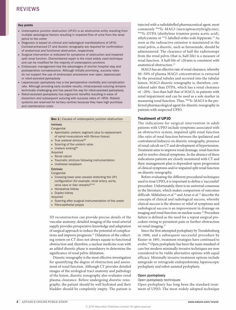
Key points
■ Ureteropelvic junction obstruction (UPJO) is an obstructive entity resulting from multiple aetiological factors resulting in impaired flow of urine from the renal pelvis to the ureter
■ Diagnosis is based on clinical and radiological manifestations of UPJO. Contrast-enhanced CT and diuretic renography are required for confirmation of anatomical and functional obstruction, respectively
■ Surgical intervention is indicated for symptoms of obstruction and impaired split renal function. Dismembered repair is the most widely used technique and can be modified for the majority of ureteropelvic junctions
■ Endoscopic management has the advantages of shorter hospital stay and postoperative convalescence. Although initially promising, success rates do not support the use of endoscopic procedures over open, laparoscopic or robot-assisted pyeloplasty
■ Laparoscopic pyeloplasty has a low perioperative morbidity and complication rate. Although providing early durable results, intracorporeal suturing remains technically challenging and has paved the way for robot-assisted pyeloplasty
■ Robot-assisted pyeloplasty has ergonomic benefits resulting in ease of dissection and subsequent suturing with success rates of >95%. Robotic systems are reserved for tertiary centres because they have high purchase and maintenance costs
Box 1 | Causes of ureteropelvic junction obstruction
IntrinsicCongenital ■ Aperistaltic ureteric segment (due to replacement
of spiral musculature with fibrous tissue) ■ True ureteral stricture88
■ Scarring of the ureteric valve ■ Ureteric kinking89
Acquired ■ Renal calculi ■ Traumatic stricture following instrumentation ■ Urothelial neoplasm
ExtrinsicCongenital ■ Crossing lower pole vessels distorting the UPJ
configuration (for example, renal artery, aorta, vena cava or iliac vessels)90,91
■ Horseshoe kidney ■ Duplex kidney
Acquired ■ Scarring after surgical instrumentation of the ureter ■ Fibro-epithelial polyps
REVIEWS
© 2014 Macmillan Publishers Limited. All rights reserved

NATURE REVIEWS | UROLOGY ADVANCE ONLINE PUBLICATION | 3
is dismembered repair, which was popularized by Anderson and Hynes in the middle of the 20th century (Figure 1). This procedure can be universally applied to a variety of UPJO scenarios—including obstruction complicated by loss of renal function and renal calculi —and involves complete excision of the abnormal u reteropelvic junction.19
For congenital UPJO caused by high insertion of the ureter, a nondismembered YV Foley pyeloplasty might be used (Figure 2), although indications for this pro cedure remain limited in the presence of proximal ureteral stricture, lower pole vessels requiring transposition and where reduction of a dilated renal pelvis is required.20,21 Spiral Culp–DeWeerd flap (Figure 3) and vertical (Scardino–Prince) flap (Figure 4) pyeloplasties have been recommended in cases of large redundant extrarenal pelvis and long narrow proximal ureters, respectively.22,23 Primary indications for spiral flap repair include dilated extrarenal pelvis and a long proximal ureteral obstruction. Vertical flap repairs are largely of historical interest and are applied in cases of obstruction with a dependent ureteropelvic junction located on the medial border of an extrarenal pelvis. Vertical flap repairs do not provide the length and v ersatility c ompared with the spiral flap.
Anderson–Hynes dismembered pyeloplastyThe ureteropelvic junction can be approached using a posterior extraperitoneal flank approach or a posterior lumbotomy approach. The posterior extraperitoneal flank approach is preferred for most patients under going a primary surgical UPJO repair owing to the excellent exposure without restrictions from body habitus. In addition, familiarity with the procedure makes this the approach of choice for many urologists. The posterior lumbotomy approach, which offers both minimal mobilization of surrounding structures and enables direct exposure to the UPJO, is best suited to children and patients with low BMI without previous ipsilateral surgery as no muscle is transected, leading to low morbidity. An anterior extraperitoneal flank approach enables surgical repair with minimal mobilization of the renal pelvis and proximal ureter, which is preferred for patients with horseshoe or pelvic kidneys.
Once the incision is made, the ureteropelvic junction is exposed by identifying the proximal ureter in the retro peritoneum and dissecting cephalically to the renal pelvis. A marking stitch is placed on the lateral aspect of the proximal ureter, below the level of the obstruction. The medial and lateral portions are marked in a similar fashion to assist in proper orientation of the repair (Figure 1a). The ureteropelvic junction tissue is then excised and an anastomosis is created with fine interrupted or running absorption sutures (Figure 1b). The sutures are placed in a watertight manner over an internal ureteral stent, which remains in situ (Figure 1c).
OutcomesNo comparative studies of open pyeloplasty techniques and their outcomes have been performed to date. A large case series of retroperitoneal Anderson–Hynes dismembered repair report a high success rate of 80–98%14,24,25 (Table 1). In the largest of these case series, Gogus et al.25 report outcomes of the Anderson–Hynes pyeloplasty in 180 adults with a 10year followup period. The success rate (91.1% overall) was inversely related to degree of hydronephrosis (62.5% in patients with grade IV hydronephrosis). In a study of 24 patients who underwent the Anderson–Hynes pyeloplasty with a mean followup period of 10.6 years, there was a 96% improvement in drainage, and 79% improvement in renal function postoperatively.17
Excellent exposure to the ureteropelvic junction, facilitating optimal repair with familiar anatomical planes, contributes to the excellent success rate of open pyeloplasty. However, this procedure is associated with a large flank incision, which invariably causes prolonged pain, recovery and poor cosmesis.26 Minimally invasive p rocedures might overcome these disadvantages.
EndopyelotomyEndopyelotomy became popular in the 1980s after the original work of Davis in 1943,27 who first described ‘intubated ureterotomy’. Endopyelotomy procedures are based on the principle of a fullthickness lateral incision through the obstructed proximal ureter, into
a cb
Figure 1 | Anderson–Hynes dismembered pyeloplasty. a | Marking stitches are placed on the renal pelvis superior to the obstruction and the lateral aspect of the proximal ureter below the level of the obstruction. b | The ureteropelvic junction tissue is then excised and an anastomosis is created with fine interrupted or running absorption sutures. c | The sutures are placed in a watertight manner over an internal ureteral stent, which remains in situ.
a cb
Figure 2 | Foley Y-V plasty. a | The ureter is pulled with a stitch while a Y-shaped incision is made in the renal pelvis and ureter. b | A V-shaped flap is opened in the ureteropelvic junction tissue. c | The V-shaped flap is sutured to the apex of the ureteral incision with fine interrupted or running absorption sutures.
REVIEWS
© 2014 Macmillan Publishers Limited. All rights reserved

4 | ADVANCE ONLINE PUBLICATION www.nature.com/nrurol
periureteral fat. A stent remains in situ following the procedure, to allow time for healing.27 Contraindications for endo pyelotomy include urosepsis and uncorrected coagulo pathy, which are contraindications for any surgical procedures, and long segment obstruction (>2 cm), as the tissue becomes ischaemic. Advantages of endopyelotomy include shorter operative time and hospital stay, decreased postoperative recovery time, reduced pain and improved cosmetic result.28
There are four main types of endopyelotomy: antegrade (percutaneous) endopyelotomy, retrograde (ureteroscopic) endopyelotomy, cautery wire balloon endopyelotomy and balloon endodilatation.29–31
Antegrade (percutaneous) endopyelotomyAntegrade (percutaneous) endopyelotomy with a cold knife was first reported by Wickham and Kellet in 1983,29 who termed the procedure ‘percutaneous pyelolysis’. This technique is the preferred endopyelotomy approach for patients with concomitant pelvicalyceal stones, and, many medical centres now offer it as a primary p rocedure even in the absence of calculi.
With the patient lying prone, access to the ureteropelvic junction can be achieved through an upper or midpole calyx under fluoroscopic guidance. An endopyelotomy cannot be performed safely without access
across the ureteropelvic junction with both ‘safety’ and ‘working’ guidewires. These guidewires can be inserted via an antegrade or retrograde approach. The percutaneous tract is then dilated and a nephroscope is placed. The endopyelotomy can be performed under direct vision using a coldknife technique (Figure 5). A fullthickness posterolateral coldknife incision from the ureteral lumen to the periureteral fat should be made to avoid inadvertent injury to crossing vessels,32 followed by a period of stenting to allow the incision to heal. Any upper tract calculi should be removed before endopyelotomy to prevent stone fragments from migrating to the periureteral tissues and causing localized o bstruction secondary to fibrosis or granuloma formation.33,34
Retrograde (ureteroscopic) endopyelotomyRetrograde endopyelotomy, which involves a combined percutaneous and flexible ureteroscopic approach, was first described by Bagley et al.35 in 1985. Retrograde endopyelotomy has evolved alongside successive technological developments of ureteroscopic instrumentation, and is now the most widely performed approach for endopyelotomy.
Retrograde pyelography is performed under fluoroscopic guidance. After the ureteropelvic junction is accessed with a small calibre semirigid ureteroscope, a 200 μm or 365 μm holmium laser fibre is used for the incision. At energy settings of 0.8–1.2 J and frequency of 10 Hz, the ureteropelvic junction is incised posterolaterally under direct vision via the ureteroscope to avoid any vessels and is gradually deepened to normal p eriureteral tissues.
Cautery wire endopyelotomy and dilatationCautery wire balloon endopyelotomy and high pressure balloon dilatation are performed under fluoroscopic guidance.36,37 Cautery wire balloon endopyelotomy using the Acucise device (Applied Medical, USA) was first reported by Chandhoke et al. in 1993.38 Although it was initially quite popular, particularly amongst urologists lacking skills in ureteroscopy, use has declined with time.
The incision for cautery wire balloon endopyelotomy depends on the location of obstruction. Posterolateral incisions are used for the proximal ureter, and anteromedial incisions are used for the distal ureter. The technique involves placing the Acucise catheter over a guidewire across the ureteropelvic junction. Under fluoro scopic guidance the balloon is inflated with contrast medium and the wire is positioned laterally to avoid injury to crossing vessels. As the balloon is reinflated, the ureteropelvic junction is incised at the same time.36
Balloon endodilatationBalloon endodilatation is also performed under fluoroscopic control. First reported in 1982, the catheter is placed across the ureteropelvic junction where the stricture is observed by the characteristic ‘waisting’. As the balloon is inflated, the waisting disappears. Advantages include minimal bleeding and a short learning curve.39
a cb
Figure 3 | Spiral (Culp–DeWeerd) flap pyeloplasty. a | A spiral incision is made in the enlarged renal pelvis and extended an equal distance into the ureter. b | The tissue flap is turned down and stitched into the adjacent ureter. c | The flap is closed with fine interrupted or running absorption sutures.
cba
Figure 4 | Vertical (Scardino–Prince) flap pyeloplasty. a | A vertical incision is made in the renal pelvis and ureter. b | The vertical flap of ureteropelvic junction tissue is turned down and sutured to the ureter. c | The renal pelvis is closed with fine interrupted or running absorption sutures.
REVIEWS
© 2014 Macmillan Publishers Limited. All rights reserved

NATURE REVIEWS | UROLOGY ADVANCE ONLINE PUBLICATION | 5
OutcomesSuccess rates for endopyelotomy are generally lower than for open pyeloplasty. Data from large series of patients who have undergone endopyelotomy, with long followup duration, show that the antegrade approach has a slightly superior success rate to other endopyelotomy techniques (Table 2). Reported success rates for antegrade endo pyelotomy range from 67% to 90% at highvolume centres and little difference in outcome is noted for primary versus secondary UPJO.40, 41 The wide range in success rates might represent a lack of uniformity in the definition of procedure success. In a case series of 64 patients, Vaarala et al.42 report success rates for primary UPJO repair of 84% for the antegrade coldknife approach, 77% for the retrograde (HoYAG laser) approach and 62% for the retrograde balloon approach. Studies show declining success rates with time; a 100% success rate was reported for retrograde endopyelotomy at 11.6 months and 94.6% at 60 months.43
Identification of factors associated with high success rates for endopyelotomy might enable careful patient
selection, to achieve comparable success to open pyeloplasty. Using inclusion criteria of stricture <2 cm, renal function >25% and the absence of severe hydronephrosis, Butani and Eshghi43 demonstrated a success rate of 94% immediately after retrograde endopyelotomy and 94.5% at 60 months. Reduced success rate is associated with the presence of crossing vessels, high insertion of the ureter into the renal pelvis, long stricture lengths (>2 cm), severe hydronephrosis, poor ipsilateral renal function and multiple previously failed endopyelotomies.28,44 Gupta et al.28 reported a success rate of 85% after antegrade endopyelotomy at 6 months and 92% at 12 months. They also found that the grade of hydronephrosis was a prognostic factor, with a success rate of 80% for grade III and 96% for grade II.
The effects of stent size and duration on success rate have also been studied. Mandhani et al.45 investigated the minimum possible duration of stenting to reduce stentrelated complications without compromising the success rate of endopyelotomy. They found that 2 weeks was a sufficient period for ureteropelvic junction function to be restored.45 Although it is commonly assumed that using a stent with a larger calibre results in a greater final ureteral diameter following endopyelotomy, there is no consensus in the literature regarding the optimal stent calibre.46,47
Complications of percutaneous endopyelotomy include haemorrhage secondary to vascular injury (the most common), infection, inadvertent incision of the renal pelvis and ureteral necrosis anaesthesia complications.48 Owing to the lack of direct vision, cautery wire balloon endopyelotomy is associated with significant haemorrhage requiring blood transfusion and embolization, as reported by Kim et al.49 in three of 77 patients (4%). If haemorrhage occurs, angiographic embolization is preferred over conservative management to avoid the potential requirement for blood transfusion and surgical exploration. Successful embolization, therefore, precludes surgical exploration and potential nephrectomy.50 Endourological modalities have serious limitations compared with open, laparoscopic and robotic techniques for their technical feasibility in all possible anatomical variations of UPJO, particu larly in the presence of crossing vessels. Although initially promising, the success rates of endopyelotomy do not support the use of this procedure over open, l aparoscopic or robotassisted pyeloplasty.42
Laparoscopic pyeloplastySchuessler and colleagues51 first performed laparoscopic pyeloplasty in 1993. When it was initially introduced, the use of early fibreoptic probe technology provided poor visualization. Furthermore, surgeons had limited experience of this new technique. These factors led to difficulties in intracorporeal suturing and lengthy operative times.52 These challenges are being overcome with the development of better instruments, greater surgical experience and increased use of simulation training for surgeons.53 Unlike endopyelotomy, laparoscopic pyeloplasty is not limited by anatomical
Table 1 | Selected case series of Anderson–Hynes open pyeloplasty
Study n LOS (days)
Complications (%)
Mean follow-up (months)
Success rate (%)
Mikkelsen et al. (1992)14 21 11 23.8 85.4 95.2
O’Reilly et al. (2001)24 56 NR NR 127.6 96.0
Gogus et al. (2004)25 180 NR NR 113.2 91.1
Abbreviations: LOS, length of stay; NR, not reported.
Guidewires
Knife
Nephroscope
Figure 5 | Antegrade percutaneous endopyelotomy. Access to the ureteropelvic junction is achieved through an upper or midpole calyx under fluoroscopic guidance. Access across the ureteropelvic junction is achieved with both ‘safety’ and ‘working’ guidewires, which can be inserted via an antegrade or retrograde approach. The percutaneous tract is then dilated and a nephroscope is placed. A full-thickness posterolateral cold-knife incision is made from the ureteral lumen to the periureteral fat followed by a period of stenting to allow the incision to heal.
REVIEWS
© 2014 Macmillan Publishers Limited. All rights reserved
6 | ADVANCE ONLINE PUBLICATION www.nature.com/nrurol
variation of the renal arteries associated with UPJO and is preferable in the presence of crossing vessels. In addition, patients with severe hydronephrosis, long stricture lengths (>2 cm) and split renal function of <30% have better success with laparoscopic pyeloplasty than endopyelotomy.54 Renal calculi can be concurrently treated with laparoscopic pyeloplasty using grasping i nstruments to achieve 90% stonefree rates.55
Surgical techniqueAfter the patient is positioned in a 45° lateral decubitus position, access to the ureteropelvic junction can be achieved by either a transperitoneal or retroperitoneal approach. The transperitoneal approach is more commonly used because it provides a larger operative field with familiar and identifiable anatomical landmarks.56 A retroperitoneal approach might be preferable for patients who have previously undergone t ransperitoneal or abdominal surgeries, or for morbidly obese patients.
Before surgery, confirmation of the UPJO is recommended by performing a rigid cystoscopy and retrograde pyelography. Thereafter, a guidewire is inserted at the renal pelvis and a 5 French gauge openended stent secured at the level of the obstruction. Once the UPJO has been identified the laparoscopic trocars are inserted into the abdominal cavity.
After accessing the renal hilum, techniques used for laparoscopic pyeloplasty are similar to those described for open pyeloplasty, with Anderson–Hynes dismembered repair the procedure of choice for most surgeons. In the absence of crossing vessels or a high ureteral insertion, a YV Foley pyeloplasty analogous to the open p rocedure can be performed.
OutcomesMultiple comparative case series have demonstrated laparo scopic pyeloplasty to have higher success rates than endopyelotomy; for example success rates of 98% versus 84%,44 95.3% versus 55.4%57 and 94.4% versus 72.6% have been reported.58
Large studies of laparoscopic pyeloplasty report high success rates (>90%), for both retroperitoneal and transperitoneal approaches (Table 3),59–65 which are comparable to those of open pyeloplasty.44,55,57,58,61 LopezPujals et al.66 and Maynes et al.67 used scintigraphic criteria for the assessment of success of laparoscopic pyeloplasty. LopezPujals et al.66 reported improvement in drainage in 94% of patients and clinical improvement in 95.7% over a mean f ollow up period of 19 months. Maynes et al.67 observed an overall success rate of 92% after a mean followup period of 13 months.
Laparoscopic pyeloplasty has a low conversion rate (4%) and perioperative morbidity; intraoperative and postoperative complications have been recorded in 11.5–12.7% of patients, with urinary leak and bleeding being the most common complications.16,68 Laparo scopic pyeloplasty also offers a better cosmetic result, shorter postoperative recovery time, reduced postoperative pain and no difference in procedure time compared with open pyeloplasty.26 Thus, laparoscopic pyeloplasty might be considered the new surgical standard of care for UPJO.69 However, longterm outcomes must also be considered. Although the initial success rates of laparoscopic pyeloplasty seem promising, Madi et al.70 found that late failures—occurring after 2 years of followup m onitoring—were more frequent than initially expected. Of 60 patients, seven (12%) failed in the first year, with an additional three patients presenting with recurrences
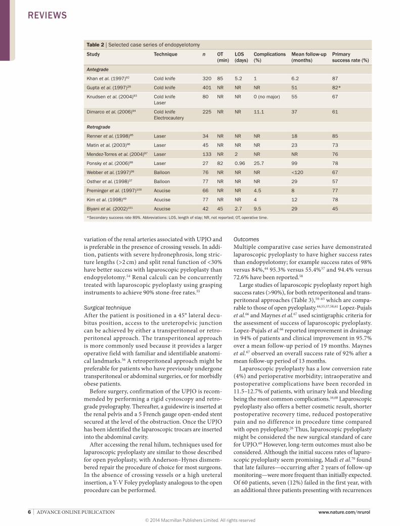
Table 2 | Selected case series of endopyelotomy
Study Technique n OT (min)
LOS (days)
Complications (%)
Mean follow-up (months)
Primary success rate (%)
Antegrade
Khan et al. (1997)92 Cold knife 320 85 5.2 1 6.2 87
Gupta et al. (1997)28 Cold knife 401 NR NR NR 51 82*
Knudsen et al. (2004)93 Cold knifeLaser
80 NR NR 0 (no major) 55 67
Dimarco et al. (2006)94 Cold knife Electrocautery
225 NR NR 11.1 37 61
Retrograde
Renner et al. (1998)95 Laser 34 NR NR NR 18 85
Matin et al. (2003)96 Laser 45 NR NR NR 23 73
Mendez-Torres et al. (2004)97 Laser 133 NR 2 NR NR 76
Ponsky et al. (2006)98 Laser 27 82 0.96 25.7 99 78
Webber et al. (1997)99 Balloon 76 NR NR NR <120 67
Osther et al. (1998)37 Balloon 77 NR NR NR 29 57
Preminger et al. (1997)100 Acucise 66 NR NR 4.5 8 77
Kim et al. (1998)49 Acucise 77 NR NR 4 12 78
Biyani et al. (2002)101 Acucise 42 45 2.7 9.5 29 45
*Secondary success rate 89%. Abbreviations: LOS, length of stay; NR, not reported; OT, operative time.
REVIEWS
© 2014 Macmillan Publishers Limited. All rights reserved

NATURE REVIEWS | UROLOGY ADVANCE ONLINE PUBLICATION | 7
after 2 years of followup monitoring. Tan et al.71 reported 18 failures after 102 procedures (17.6%); these patients were successfully managed with endoscopic approaches.
Furthermore, global acceptance of laparoscopic pyeloplasty is limited owing to the steep learning curve of precise intracorporeal suturing.52 Despite continuous advances in instrumentation and 3D stack systems, the procedure remains technically challenging and is largely performed by expert laparoscopic surgeons in select tertiary medical centres. These technical challenges have paved the way for the development of robotassisted pyeloplasty.
Robot-assisted pyeloplastyRobotassisted pyeloplasty, using the da Vinci® system (Intuitive Surgical, USA) was first introduced by Sung and colleagues72 in an experimental setting in 1999. Clinical feasibility of the procedure was confirmed by Palese et al.73 in 2005 and Mufarrij et al. in 2007.74 The da Vinci® robot offers enhanced 3D views and the robotic arms enable a range of movement up to seven degrees of freedom, motion scaling and reduced tremor interference.75
Surgical techniqueThe transperitoneal approach is the route of choice as it offers greater space for manoeuvrability and enables surgeon recognition of familiar anatomical and surgical landmarks. Feasibility of the retroperitoneal approach has been demonstrated.76 Regardless of the approach used, the da Vinci® robot enables the surgeon to operate the laparoscopic camera and robotic arms from a remote console. Four trocars are typically used for the pro cedure; three for the robotic arms and one 10 mm port for the scrubbed assistant to suction, irrigate, retract and introduce sutures at the bedside. Surgical steps are the same as those of the conventional open dismembered Anderson–Hynes pyeloplasty.
OutcomesA large number of studies have been published on the outcomes of robotassisted pyeloplasty (Table 4), which
suggest higher success rates than for laparoscopic pyeloplasty. Patel et al.11 published the first series of 50 cases in 2005 with a mean followup duration of 11.7 months. The study showed high shortterm efficacy with 48 of 50 patients demonstrating stable renal function and better drainage with no recurrent obstruction on diuretic renography, demonstrating a 96% success rate after a 31month followup period. Furthermore, perioperative morbidity was low with no intraoperative complications, and the learning curve was shorter than for laparoscopic pyeloplasty.11 In 2007, Schwentner et al.77 presented a 5year case series of 92 patients who underwent dismembered Anderson–Hynes robotassisted pyeloplasty, none of whom experienced any longterm complications, with a success rate of 96.7%; this study consolidated confidence in the precision of robotassisted suturing and the superior quality of a nastomosis achieved.77
Etafy et al.79 reported a lower success rate for robotassisted pyeloplasty (81%) than other studies (Table 4), owing to the use of stringent criteria to define success based on diuretic renography with a halflife of <10 min and symptomatic relief with a patientreported pain a nalogue score of ≤2 at 3 months, 6 months and 1 year after surgery. In 2010, Nayyar and colleagues79 assessed the feasibility and outcomes of robotassisted pyeloplasty for the treatment of 29 complicated UPJO cases, including malrotated, horseshoe and ectopic kidneys, multiple or calyceal calculi, secondary UPJO (after ureterorenoscopy or open surgery), giant hydro nephrosis, poorly functioning kidney (glomerular filtration rate <20 ml/min) and duplicated renal pelvis. Only one case required conversion to an open technique. The overall success rate was 96.6% with a mean followup period of 15 months; 80% of patients with renal calculi were stonefree after treatment.
One of the largest series published to date is by Sivaraman et al.,80 who reported 6year outcomes of 168 patients from multiple institutions, 87.5% (n = 147) of whom had a primary repair and 12.5% (n = 21) had a secondary repair. Overall success rate was 97.6% for both primary and secondary repairs, with a low complication rate of 6.6%.80
Table 3 | Large (>100 patients) case series of laparoscopic pyeloplasty
Study n (dism)*
OT (min)
LOS (days)
Complications (%)
Mean follow-up (months)
Success rate (%)
Conversion rate (%)
Transperitoneal approach
Szydelko et al. (2011)65 149 190 5.1 18.0 53.7 90.5 1.4
Symons et al. (2009)61 118 (94) 205‡ 4.7 11.9 12 94.5 NR
Romero et al. (2006)60 170 (124) 176 2.7 5.9 22 94.1 0
Srivastava et al. (2009)62 186 (143) 180‡ 4 9.6 39 94.3 4.8
Retroperitoneal approach
Moon et al. (2006)59 167 (167) 140§ 3 7.1 12 96.2 0.6
Rassweiler et al. (2008)63 189 (66) 123 5 7.4 NR 95.2 0.5
Shao et al. (2011)64 105 (105) 96 7 7.6 42 100 0
*Denotes dismembered repair. Other types of repair were not specified. ‡Mean operative time included cystoscopy, retrograde ureteropyelography and stent placement. §Mean operative time was from first incision to wound closure. Abbreviations: LOS, length of stay; NR, not reported; OT, operative time.
REVIEWS
© 2014 Macmillan Publishers Limited. All rights reserved

8 | ADVANCE ONLINE PUBLICATION www.nature.com/nrurol
To summarize, robotassisted pyeloplasty is associated with greater success rates and lower perioperative morbidity than laparoscopic pyeloplasty, and provides ergonomic benefits. Unsurprisingly, this procedure has generated great enthusiasm amongst urologists, but it is important to remember that robotic urology is a relatively new speciality; in many of the studies of robotassisted pyeloplasty, the followup periods are relatively short. In addition, robotic systems are not yet widely available, and have high purchase and maintenance costs.
Secondary UPJO repairRepeat pyeloplasty after failed primary intervention is a challenging procedure, mainly owing to fibrosis around the renal pelvis and proximal ureter as a result of previous surgery. Traditionally, open pyeloplasty is the reference standard for secondary UPJO repair demon strating a success rate of 95%.28 Minimally invasive techniques, such as endopyelotomy, have yielded disappointing success rates of 59–70% after failed primary intervention.81,82 Eden et al.83,84 assessed laparoscopic pyeloplasty for the treatment of s econdary UPJO repair and reported success rates of 75–91%.
The role of robotassisted pyeloplasty in repeat pyeloplasty was assessed in a singlecentre study of 117 cases performed over an 8year period; radiological success rates were 96.1% for primary repair and 94.1% for secon dary UPJO repair.85 These results are superior to the published success rates of laparoscopic pyeloplasty for secondary UPJO repair (84–88%).86,87 These data strongly suggest that robotassisted pyeloplasty is the optimal minimally invasive surgical technique and a durable option for secondary UPJO repair.85
ConclusionsA combination of contrast CT and diuretic reno graphy should be used to define the anatomy of UPJO obstruction, and provide a functional assessment of its effects. Both open and minimally invasive options are available for the treatment of UPJO.
Minimally invasive surgical techniques are associated with reduced morbidity, improved cosmetic result and better convalescence than open pyeloplasty. For endopyelotomy, these advantages are tempered by an inferior success rate to open pyeloplasty, laparoscopic pyeloplasty and robotassisted pyeloplasty. However, the success rate could potentially be improved by careful selection of patients, using the criteria of stricture <2 cm, renal function >25% and the absence of severe hydronephrosis. Laparoscopic pyeloplasty and robotassisted pyeloplasty have similar success rates to open pyeloplasty (>90%) and the best outcomes have been reported for robotassisted pyeloplasty, although this treatment option is less readily available than laparo scopic pyeloplasty. Thus, laparoscopic pyeloplasty and robotassisted pyeloplasty are viable alternative options to open pyeloplasty for primary UPJO repair. Management of secondary UPJO after failed primary repair remains challenging, whichever technique is used. Success rates are lower than those reported for primary repair, but robotassisted pyeloplasty is associated with the most favourable outcomes. To conclude, minimally invasive surgical techniques, namely laparoscopic and robotassisted pyeloplasty, are emerging as the new gold standards for UPJO treatment. However, longerterm data are still awaited to consolidate these findings. To date, no prospective randomized trials have been completed comparing open, l aparoscopic and robotic approaches.
Table 4 | Case series of robot-assisted pyeloplasty
Study n (dism)*
OT (min)
LOS (days)
Complications (%)
Mean follow-up (months)
Success rate (%)
Conversion rate (%)
Muffarij et al. (2008)102 140 217 2.1 10 29 95.7 0
Gupta et al. (2010)103 85 121 2.5 2.4 13.6 96.5 2.4
Schwentner et al. (2007)77 92 108.3 4.6 NR 39.1 96.7 0
Patel et al. (2005)11 50 122 1.1 2.0 11.7 96.0 0
Etafy et al. (2011)78 61 335 2 11.4 18 81.0 0
Sivaraman et al. (2012)80 168 134 1.5 6.6 39 97.6 0
Niver et al. (2012)85‡ 117 218 2.8 1.0 21.9 96.0 0
All case series used the transperitoneal approach unless otherwise stated. *Denotes dismembered repair. Other types of repair were not specified. ‡Used a combination of transperitoneal and retroperitoneal approaches. Abbreviations: LOS, length of stay; NR, not reported; OT, operative time.
Review criteria
A search of original articles published between 1940 and 2013, focusing on ureteropelvic junction obstruction and its management was performed in MEDLINE and PubMed. Search terms used were “pyeloplasty”, “ureteropelvic junction”, “laparoscopy”, “robotic”, “outcomes” and “endopyelotomy”, alone and in combination. All articles referenced were English-language and reference lists of selected articles were cross-searched for additional literature.
1. Cohen, B., Goldman, S. M., Kopilnick, M., Khurana, A. V. & Salik, J. O. Ureteropelvic junction obstruction: its occurrence in 3 members of a single family. J. Urol. 120, 361–364 (1978).
2. Thomas, A. K. & June, C. H. The promise of T-lymphocyte immunotherapy for the treatment of malignant disease. Cancer J. 7 (Suppl. 2), S67–S75 (2001).
3. Sepulveda, L. & Rodrigues, F. Giant hydronephrosis—a late diagnosis of ureteropelvic junction obstruction. World J. Nephrol. & Urol. 2, 33–35 (2013).
REVIEWS
© 2014 Macmillan Publishers Limited. All rights reserved

NATURE REVIEWS | UROLOGY ADVANCE ONLINE PUBLICATION | 9
4. Bauer, S. B. in Campbell’s Urology 7th edn (eds Walsh, P. W., Retik, A. B., Vaughan, E. D. Jr & Wein, A. J.) 1739–1749 (WB Saunders Company, 1998).
5. Park, J. M. & Bloom, D. A. The pathophysiology of UPJ obstruction. Current concepts. Urol. Clin. North Am. 25, 161–169 (1998).
6. Riehle, R. A. Jr & Vaughan, E. D. Jr. Renin participation in hypertension associated with unilateral hydronephrosis. J. Urol. 126, 243–246 (1981).
7. Sohn, B., Kim, M., Han, S. W., Im, Y. & Lee, M. Shear wave velocity measurements using acoustic radiation force impulse in young children with normal kidneys versus hydronephrotic kidneys. Ultrasonography 33, 116–121 (2014).
8. Jacobs, J. A., Berger, B. W., Goldman, S. M., Robbins, M. A. & Young, J. D. Jr. Ureteropelvic obstruction in adults with previously normal pyelograms: a report of 5 cases. J. Urol. 121, 242–244 (1979).
9. El-Nahas, A. R. et al. Role of multiphasic helical computed tomography in planning surgical treatment for pelvi-ureteric junction obstruction. BJU Int. 94, 582–587 (2004).
10. Lam, J. S., Breda, A. & Schulam, P. G. Ureteropelvic junction obstruction. J. Urol. 177, 1652–1658 (2007).
11. Patel, V. Robotic-assisted laparoscopic dismembered pyeloplasty. Urology 66, 45–49 (2005).
12. Roarke, M. C. & Sandler, C. M. Provocative imaging. Diuretic renography. Urol. Clin. North Am. 25, 227–249 (1998).
13. O’Reilly, P. H., Lawson, R. S., Shields, R. A. & Testa, H. J. Idiopathic hydronephrosis—the diuresis renogram: a new non-invasive method of assessing equivocal pelvioureteral junction obstruction. J. Urol. 121, 153–155 (1979).
14. Mikkelsen, S. S., Rasmussen, B. S., Jensen, T. M., Hanghoj-Petersen, W. & Christensen, P. O. Long-term follow-up of patients with hydronephrosis treated by Anderson-Hynes pyeloplasty. Br. J. Urol. 70, 121–124 (1992).
15. Arun, N., Kekre, N. S., Nath, V. & Gopalakrishnan, G. Is open pyeloplasty still justified? Br. J. Urol. 80, 379–381 (1997).
16. Jarrett, T. W., Chan, D. Y., Charambura, T. C., Fugita, O. & Kavoussi, L. R. Laparoscopic pyeloplasty: the first 100 cases. J. Urol. 167, 1253–1256 (2002).
17. Bratt, C. G., Aurell, M. & Nilsson, S. Renal function in patients with hydronephrosis. Br. J. Urol. 49, 249–255 (1977).
18. Poulakis, V., Witzsch, U., Schultheiss, D., Rathert, P. & Becht, E. History of ureteropelvic junction obstruction repair (pyeloplasty). From Trendelenburg (1886) to the present [German]. Urologe A 43, 1544–1559 (2004).
19. Wolf, J. S. Laparoscopic transperitoneal pyeloplasty. J. Endourol. 25, 173–178 (2011).
20. Grasso, M., Caruso, R. P. & Philipps, C. K. UPJ obstruction in the adult population: are crossing vessels significant? Rev. Urol. 3, 42–51 (2001).
21. Foley, F. E. A new plastic operation for stricture at the uretero-pelvic junction. Report of 20 operations. 1937. J. Urol. 167, 1075–1095 (2002).
22. Culp, O. S. & DeWeerd, J. H. A pelvic flap operation for certain types of UPJ obstruction: Preliminary report. Proc. Staff Meet. Mayo Clin. 26, 483–488 (1951).
23. Scardino, P. L. & Prince, C. L. Vertical flap ureteropelvioplasty: Prelimary report. South Med. J. 46, 325–331 (1953).
24. O’Reilly, P. H. et al. The long-term results of Anderson-Hynes pyeloplasty. BJU Int. 87, 287–289 (2001).
25. Gogus, C. et al. Long-term results of Anderson-Hynes pyeloplasty in 180 adults in the era of endourologic procedures. Urol. Int. 73, 11–14 (2004).
26. Simforoosh, N. et al. A comparision between laparoscopic and open pyeloplasty in patients with ureteropelvic junction obstruction. J. Endourol. 1, 165–169 (2004).
27. Davis, D. M. Intubated ureterotomy: a new operation for ureteral and ureteropelvis stricture. J. Surg. Gynecol. Obstet. 76, 513–514 (1943).
28. Gupta, M., Tuncay, O. L. & Smith, A. D. Open surgical exploration after failed endopyelotomy: a 12-year perspective. J. Urol. 157, 1613–1618 (1997).
29. Wickham, J. E. & Kellet, M. J. Percutaneous pyelolysis. Eur. Urol. 9, 122–124 (1983).
30. Badlani, G. H. & Smith, A. D. Percutaneous ureteral surgery. Semin. Urol. 4, 191–197 (1986).
31. Korth, K., Kuenkel, M. & Erschig, M. Percutaneous pyeloplasty. Urology 31, 503–509 (1988).
32. Sampaio, F. J. Vascular anatomy at the ureteropelvic junction. Urol. Clin. North Am. 25, 251–258 (1998).
33. Streem, S. B. Percutaneous endopyelotomy. Urol. Clin. North Am. 27, 685–693 (2000).
34. Giddens, J. L., Grotas, A. B. & Grasso, M. Stone granuloma causes ureteropelvic junction obstruction after percutaneous nephrolithotomy and antegrade endopyelotomy. J. Urol. 164, 118–119 (2000).
35. Bagley, D. H., Huffman, J., Lyon, E. & McNamara, T. Endoscopic ureteropyelostomy: opening the obliterated ureteropelvic junction with nephroscopy and flexible ureteropyeloscopy. J. Urol. 133, 462–464 (1985).
36. Nadler, R. B., Rao, G. S., Pearle, M. S., Nakada, S. Y. & Clayman, R. V. Acucise endopyelotomy: assessment of long-term durability. J. Urol. 156, 1094–1097 (1996).
37. Osther, P. J., Geertsen, U. & Nielsen, H. V. Ureteropelvic junction obstruction and ureteral strictures treated by simple high-pressure balloon dilation. J. Endourol. 12, 429–431 (1998).
38. Chandhoke, P. S. et al. Endopyelotomy and endoureterotomy with the acucise ureteral cutting balloon device: preliminary experience. J. Endourol. 7, 45–51 (1993).
39. Symons, S. J. et al. Minimally invasive surgical options for ureteropelvic junction obstruction: A significant step in the right direction. Indian J. Urol. 25, 27–33 (2009).
40. Kletscher, B. A., Segura, J. W., LeRoy, A. J. & Patterson, D. E. Percutaneous antegrade endopyelotomy: review of 50 consecutive cases. J. Urol. 153, 701–703 (1995).
41. Shalhav, A. L. et al. Adult endopyelotomy: impact of etiology and antegrade versus retrograde approach on outcome. J. Urol. 160, 685–689 (1998).
42. Vaarala, M. H., Marttila, T., Paananen, I. & Hellstrom, P. Retrospective analysis of long-term outcomes of 64 patients treated by endopyelotomy in two low-volume hospitals: good and durable results. J. Endourol. 22, 1659–1664 (2008).
43. Butani, R. P. & Eshghi, M. Cold-knife retrograde endopyelotomy: a long-term follow-up. J. Endourol. 22, 657–660 (2008).
44. Ost, M. C., Kaye, J. D., Guttman, M. J., Lee, B. R. & Smith, A. D. Laparoscopic pyeloplasty versus
antegrade endopyelotomy: comparison in 100 patients and a new algorithm for the minimally invasive treatment of ureteropelvic junction obstruction. Urology 66, 47–51 (2005).
45. Mandhani, A. et al. Is a 2-week duration sufficient for stenting in endopyelotomy? J. Urol. 169, 886–889 (2003).
46. Danuser, H., Hochreiter, W. W., Ackermann, D. K. & Studer, U. E. Influence of stent size on the success of antegrade endopyelotomy for primary ureteropelvic junction obstruction: results of 2 consecutive series. J. Urol. 166, 902–909 (2001).
47. Wolf, J. S. Jr, Elashry, O. M. & Clayman, R. V. Long-term results of endoureterotomy for benign ureteral and ureteroenteric strictures. J. Urol. 158, 759–764 (1997).
48. Gettman, M. T. & Segura, J. W. Technique of percutaneous endopyelotomy. Braz. J. Urol. 26, 64–70 (2000).
49. Kim, F. J., Herrell, S. D., Jahoda, A. E. & Albala, D. M. Complications of acucise endopyelotomy. J. Endourol. 12, 433–436 (1998).
50. Badlani, G., Karlin, G. & Smith, A. D. Complications of endopyelotomy: analysis in series of 64 patients. J. Urol. 140, 473–475 (1988).
51. Schuessler, W. W., Grune, M. T., Tecuanhuey, L. V. & Preminger, G. M. Laparoscopic dismembered pyeloplasty. J. Urol. 150, 1795–1799 (1993).
52. Turk, I. et al. Laparoscopic dismembered pyeloplasty—the method of choice in the presence of an enlarged renal pelvis and crossing vessels. Eur. Urol. 42, 268–275 (2002).
53. Ahmed, K. et al. Effectiveness of procedural simulation in urology: a systematic review. J. Urol. 186, 26–34 (2011).
54. Samarasekera, D. & Chew, B. Endopyelotomy still has an important role in the management of ureteropelvic junction obstruction. Can. Urol. Assoc. J. 5, 134–136 (2011).
55. Inagaki, T., Rha, K. H., Ong, A. M., Kavoussi, L. R. & Jarrett, T. W. Laparoscopic pyeloplasty: current status. BJU Int. 95 (Suppl. 2), 102–105 (2005).
56. El-Shazly, M. A., Moon, D. A. & Eden, C. Laparoscopic pyeloplasty: status and review of literature. J. Endourol. 21, 673–678 (2007).
57. Szydelko, T. et al. Antegrade endopyelotomy versus laparoscopic pyeloplasty for primary ureteropelvic junction obstruction. J. Laparoendosc. Adv. Surg. Tech. A 19, 45–51 (2009).
58. Rassweiler, J. J. et al. Minimally invasive treatment of ureteropelvic junction obstruction: long-term experience with an algorithm for laser endopyelotomy and laparoscopic retroperitoneal pyeloplasty. J. Urol. 177, 1000–1005 (2007).
59. Moon, D. A., El-Shazly, M. A., Chang, C. M., Gianduzzo, T. R. & Eden, C. G. Laparoscopic pyeloplasty: evolution of a new gold standard. Urology 67, 932–936 (2006).
60. Romero, F. R. et al. Transmesenteric laparoscopic pyeloplasty. J. Urol. 176, 2526–2529 (2006).
61. Symons, S. J., Bhirud, P. S., Jain, V., Shetty, A. S. & Desai, M. R. Laparoscopic pyeloplasty: our new gold standard. J. Endourol. 23, 463–467 (2009).
62. Srivastava, A. et al. Laparoscopic pyeloplasty: a versatile alternative to open pyeloplasty. Urol. Int. 83, 420–424 (2009).
63. Rassweiler, J. J., Teber, D. & Frede, T. Complications of laparoscopic pyeloplasty. World J. Urol. 26, 539–547 (2008).
64. Shao, P. et al. Comparison of two different suture methods in laparoscopic dismembered pyeloplasty. Urol. Int. 87, 304–308 (2011).
REVIEWS
© 2014 Macmillan Publishers Limited. All rights reserved
10 | ADVANCE ONLINE PUBLICATION www.nature.com/nrurol
65. Szydelko, T. et al. Clavien classification of complications after 150 laparoscopic pyeloplasties. Urology 77, 1359–1364 (2011).
66. Lopez-Pujals, A., Leveillee, R. J. & Wong, C. Application of strict radiologic criteria to define success in laparoscopic pyeloplasty. J. Endourol. 18, 756–760 (2004).
67. Maynes, L. J., Levin, B. M., Webster, T. M., Baldwin, D. & Herrell, S. D. Measuring the true success of laparoscopic pyeloplasty. J. Endourol. 22, 1193–1198 (2008).
68. Soulie, M. et al. Retroperitoneal laparoscopic versus open pyeloplasty with a minimal incision: comparison of two surgical approaches. Urology 57, 443–447 (2001).
69. Gettman, M. T., Peschel, R., Neururer, R. & Bartsch, G. A comparison of laparoscopic pyeloplasty performed with the daVinci robotic system versus standard laparoscopic techniques: initial clinical results. Eur. Urol. 42, 453–457 (2002).
70. Madi, R., Roberts, W. W. & Wolf, J. S. Jr. Late failures after laparoscopic pyeloplasty. Urology 71, 677–680 (2008).
71. Tan, H. J., Ye, Z., Roberts, W. W. & Wolf, J. S. Failure after laparoscopic pyeloplasty: prevention and management. J. Endourol. 25, 1457–1462 (2011).
72. Sung, G. T., Gill, I. S. & Hsu, T. H. Robotic-assisted laparoscopic pyeloplasty: a pilot study. Urology 53, 1099–1103 (1999).
73. Palese, M. A. et al. Robot-assisted laparoscopic dismembered pyeloplasty. JSLS 9, 252–257 (2005).
74. Mufarrij, P. W., Shah, O. D., Berger, A. D. & Stifelman, M. D. Robotic reconstruction of the upper urinary tract. J. Urol. 178, 2002–2005 (2007).
75. Bernie, J. E., Venkatesh, R., Brown, J., Gardner, T. A. & Sundaram, C. P. Comparison of laparoscopic pyeloplasty with and without robotic assistance. JSLS 9, 258–261 (2005).
76. Kaouk, J. H. et al. Is retroperitoneal approach feasible for robotic dismembered pyeloplasty: initial experience and long-term results. J. Endourol. 22, 2153–2159 (2008).
77. Schwentner, C. et al. Robotic Anderson–Hynes pyeloplasty: 5-year experience of one centre. BJU Int. 100, 880–885 (2007).
78. Etafy, M. et al. Robotic pyeloplasty: the University of California-Irvine experience. J. Urol. 185, 2196–2200 (2011).
79. Nayyar, R., Gupta, N. P. & Hemal, A. K. Robotic management of complicated ureteropelvic junction obstruction. World J. Urol. 28, 599–602 (2010).
80. Sivaraman, A. et al. Robot-assisted laparoscopic dismembered pyeloplasty for ureteropelvic junction obstruction: a multi-institutional experience. Urology 79, 351–355 (2012).
81. Park, J., Kim, W. S., Hong, B., Park, T. & Park, H. K. Long-term outcome of secondary endopyelotomy after failed primary intervention for ureteropelvic junction obstruction. Int. J. Urol. 15, 490–494 (2008).
82. Ng, C. S., Yost, A. J. & Streem, S. B. Management of failed primary intervention for ureteropelvic junction obstruction: 12-year, single-center experience. Urology 61, 291–296 (2003).
83. Eden, C. G., Cahill, D. & Allen, J. D. Laparoscopic dismembered pyeloplasty: 50 consecutive cases. BJU Int. 88, 526–531 (2001).
84. Eden, C., Gianduzzo, T., Chang, C., Thiruchelvam, N. & Jones, A. Extraperitoneal laparoscopic pyeloplasty for primary and secondary ureteropelvic junction obstruction. J. Urol. 172, 2308–2311 (2004).
85. Niver, B. E. et al. Analysis of robotic-assisted laparoscopic pyleloplasty for primary versus secondary repair in 119 consecutive cases. Urology 79, 689–694 (2012).
86. Scardino, P. S. & Scardino, P. L. in The Ureter 2nd edn (ed. Bergman, H.) 697–716 (Springer, 1981).
87. Murphy, D., Challacombe, B., Khan, M. S. & Dasgupta, P. Robotic technology in urology. Postgrad. Med. J. 82, 743–747 (2006).
88. Yang, Y., Zhou, X., Gao, H., Ji, S. J. & Wang, C. The expression of epidermal growth factor and transforming growth factor-beta1 in the stenotic tissue of congenital pelvi-ureteric junction obstruction in children. J. Pediatr. Surg. 38, 1656–1660 (2003).
89. Lowe, F. C. & Marshall, F. F. Ureteropelvic junction obstruction in adults. Urology 23, 331–335 (1984).
90. Lallas, C. D. et al. The minimally invasive management of ureteropelvic junction obstruction in horseshoe kidneys. World J. Urol. 29, 91–95 (2011).
91. Adey, G. S. et al. Fibroepithelial polyps causing ureteropelvic junction obstruction in children. J. Urol. 169, 1834–1836 (2003).
92. Khan, A. M., Holman, E., Pasztor, I. & Toth, C. Endopyelotomy: experience with 320 cases. J. Endourol. 11, 243–246 (1997).
93. Knudsen, B. E. et al. Percutaneous antegrade endopyelotomy: long-term results from one institution. Urology 63, 230–234 (2004).
94. Dimarco, D. S. et al. Long-term success of antegrade endopyelotomy compared with pyeloplasty at a single institution. J. Endourol. 20, 707–712 (2006).
95. Renner, C., Frede, T., Seemann, O. & Rassweiler, J. Laser endopyelotomy: minimally invasive therapy of ureteropelvic junction stenosis. J. Endourol. 12, 537–544 (1998).
96. Matin, S. F., Yost, A. & Streem, S. B. Ureteroscopic laser endopyelotomy: a single-center experience. J. Endourol. 17, 401–404 (2003).
97. Mendez-Torres, F. R., Urena, R. & Thomas, R. Retrograde ureteroscopic endopyelotomy. Urol. Clin. North Am. 31, 99–106 (2004).
98. Ponsky, L. E. & Streem, S. B. Retrograde endopyelotomy: a comparative study of hot-wire balloon and ureteroscopic laser. J. Endourol. 20, 823–826 (2006).
99. Webber, R. J., Pandian, S. S., McClinton, S. & Hussey, J. Retrograde balloon dilatation for pelviureteric junction obstruction: long-term follow-up. J. Endourol. 11, 239–242 (1997).
100. Preminger, G. M. et al. A multicenter clinical trial investigating the use of a fluoroscopically controlled cutting balloon catheter for the management of ureteral and ureteropelvic junction obstruction. J. Urol. 157, 1625–1629 (1997).
101. Biyani, C. S. et al. The role of Acucise endopyelotomy in the treatment of ureteropelvic junction obstruction. Eur. Urol. 41, 305–310 (2002).
102. Mufarrij, P. W. et al. Robotic dismembered pyeloplasty: a 6-year, multi-institutional experience. J. Urol. 180, 1391–1396 (2008).
103. Gupta, N. P. et al. Outcome analysis of robotic pyeloplasty: a large single-centre experience. BJU Int. 105, 980–983 (2010).
Author contributionsF.K. and N.L. researched data for the article. F.K. and M.S.K. contributed to the discussion of content. F.K. and K.A. wrote the article. F.K., K.A., B.C., M.S.K. and P.D. reviewed/edited the manuscript before submission.
REVIEWS
© 2014 Macmillan Publishers Limited. All rights reserved
Copyright © 2022 FDOKUMEN