Association of angiotensin type 2 receptor gene polymorphisms with ureteropelvic junction...
18
This article is protected by copyright. All rights reserved. 1 Association of Angiotensin Type 2 Receptor Gene Polymorphisms with Ureteropelvic Junction Obstruction in Brazilian Patients 1 Running title: AGTR2 gene and UPJO Debora M. Miranda 1,2 ; Augusto Cesar dos Santos Junior 1,2 ; Helena C. Sarubi 1 ; Luciana Bastos-Rodrigues 1 ; Daniela Valadão Rosa 1 ; Izabella S. Freitas 1 ; Luiz Armando De Marco 1 ; Eduardo A. Oliveira 1,2 ; Ana Cristina Simões e Silva 1,2 . 1 INCT / MM - Instituto Nacional de Ciência e Tecnologia em Medicina Molecular, Universidade Federal de Minas Gerais (UFMG), Belo Horizonte, MG, Brazil. 2 Unidade de Nefrologia Pediátrica, Departamento de Pediatria, Laboratório Interdisciplinar de Investigação Médica, Faculdade de Medicina, UFMG, Belo Horizonte, MG, Brazil Correspondence to Ana Cristina Simões e Silva, MD, PhD Laboratório Interdisciplinar de Investigação Médica Av. Alfredo Balena, 190, 2 o andar, sala 281, Belo Horizonte, MG, Brazil, 30130-100. This article has been accepted for publication and undergone full peer review but has not been through the copyediting, typesetting, pagination and proofreading process, which may lead to differences between this version and the Version of Record. Please cite this article as doi: 10.1111/nep.12308 Accepted Article
-
Upload
independent -
Category
Documents
-
view
3 -
download
0
Transcript of Association of angiotensin type 2 receptor gene polymorphisms with ureteropelvic junction...

This article is protected by copyright. All rights reserved. 1
Association of Angiotensin Type 2 Receptor Gene
Polymorphisms with Ureteropelvic Junction Obstruction in
Brazilian Patients1
Running title: AGTR2 gene and UPJO
Debora M. Miranda1,2; Augusto Cesar dos Santos Junior1,2; Helena C.
Sarubi1; Luciana Bastos-Rodrigues1; Daniela Valadão Rosa1; Izabella S.
Freitas1; Luiz Armando De Marco1; Eduardo A. Oliveira1,2; Ana Cristina
Simões e Silva1,2.
1INCT / MM - Instituto Nacional de Ciência e Tecnologia em Medicina
Molecular, Universidade Federal de Minas Gerais (UFMG), Belo Horizonte,
MG, Brazil.
2Unidade de Nefrologia Pediátrica, Departamento de Pediatria, Laboratório
Interdisciplinar de Investigação Médica, Faculdade de Medicina, UFMG, Belo
Horizonte, MG, Brazil
Correspondence to Ana Cristina Simões e Silva, MD, PhD
Laboratório Interdisciplinar de Investigação Médica
Av. Alfredo Balena, 190, 2o andar, sala 281, Belo Horizonte, MG, Brazil,
30130-100.
This article has been accepted for publication and undergone full peer review but has not been through the copyediting, typesetting, pagination and proofreading process, which may lead to differences between this version and the Version of Record. Please cite this article as doi: 10.1111/nep.12308
Acc
epte
d A
rticl
e

This article is protected by copyright. All rights reserved. 2
Phone: +55-31-34098073, FAX: + 55-31-34099750
E-mail: [email protected]
Abstract
Background and aims: The angiotensin type 2 (AT2) receptor takes part in
the process of ureteric bud during kidney development. Therefore, the gene
encoding AT2 receptor, the AGTR2 gene located in the X chromosome, is a
potential candidate for genetic association with Congenital Anomalies of the
Kidney and Urinary Tract (CAKUT). This study aimed to investigate if AGTR2
gene polymorphisms are associated with CAKUT in general or even with
common phenotypes of CAKUT in a Brazilian sample of paediatric patients.
Methods: We analysed 290 paediatric patients with CAKUT and 262 healthy
controls from the same geographic area. TaqMan single-nucleotide
polymorphism (SNP) genotyping assays for AGTR2 gene at rs1403543,
rs3736556, rs35474657, rs5193 and rs5194 were performed. The sample was
in Hardy Weinberg Equilibrium for all five SNPs. Results: The presence of
CAKUT in general was not significantly associated with the SNPs included in
this study. However, when patients were segregated according to major
phenotypes, the diagnosis of Ureteropelvic Junction Obstruction (UPJO) was
significantly associated with AGTR2 gene polymorphisms at rs3736556 and at
rs5194. On the other hand, the diagnoses of vesicoureteral reflux and of
multicystic dysplastic kidney were not associated with AGTR2 gene
polymorphisms. Conclusion: Our results support that the AGTR2 gene may
contribute to the pathogenesis of UPJO and the genetic origin of CAKUT
could vary according to phenotype expression. Acc
epte
d A
rticl
e

This article is protected by copyright. All rights reserved. 3
Keywords: Angiotensin type 2 receptor (AGTR2) gene, Ureteropelvic
Junction Obstruction (UPJO), Congenital Anomalies of the Kidney and Urinary
Tract (CAKUT), Renin Angiotensin System.
Introduction
The genetic origins of congenital anomalies of the kidney and urinary
tract (CAKUT) have not been well defined, but the concomitant presence of
diverse urinary tract alterations and the familial pattern of inheritance with
variable penetrance have been observed in many cases1,2. Therefore, a
common genetic background for these anomalies has been speculated and
genes responsible for kidney and urinary tract morphogenesis have been
frequently associated with CAKUT3,4.
The critical role of the renin-angiotensin system (RAS) in kidney and
renal collecting system morphogenesis is evident from occurrence of diverse
forms of CAKUT in animals or humans as a result of RAS gene mutations5. In
this regard, Yosypiv et al.6 showed that Angiotensin II (Ang II) induces the
expression of glial-derived neurotrophic factor (GDNF)/Ret/Wnt11 pathway
genes, promotes cell proliferation preferentially in ureteric bud tip cells and
stimulates branching. The same research group also showed that Ang II type
2 (AT2) receptor is highly expressed in the ureteric bud epithelia during
metanephric development and the pharmacological antagonism of this
receptor at early stages of kidney development down-regulates
GDNF/Ret/Wnt11 pathway gene expression, decreases proliferation, and
induces apoptosis of the ureteric bud cells7. Acc
epte
d A
rticl
e

This article is protected by copyright. All rights reserved. 4
The AT2 receptor is a 323-residue of the G-protein-coupled family
highly expressed in the fetal kidney, diminishing after the neonatal period8.
The AT2 receptor is encoded by the AGTR2 gene, which is located in X
chromosome. The AGTR2 gene has two short non-coding exons, two introns
and three exons, which codify the complete protein9. AGTR2 gene is actively
transcribed at the onset of, and throughout, the embryonic development of the
kidney and urinary tract system, and is mostly inactivated by the time of
birth10. Nishimura et al.11 reported that AGTR2 mutant mice have lower
apoptosis around the ureteric bud and higher incidence of renal and urinary
tract anomalies than wild type animals, and, in a concurrent human study,
showed that AGTR2 genotype in Caucasians exhibited a significant
association with CAKUT. The general hypothesis is that abnormalities in the
expression of AT2 receptors hinder interaction between the ureteric bud and
metanephric blastema, and hamper normal development, resulting in
CAKUT5,12. It has been reported that A to G transition at the 1332 position in
AGTR2 gene is associated with abnormal splicing, producing shorter length
mRNA and thereby affecting AT2 receptor activity11. This polymorphism has
been inconsistently shown to be associated with CAKUT in Caucasians13,
Korean14 and Japanese subjects15.
However, only the A to G transition at the 1332 position in the AGTR2
gene has been investigated in human CAKUT samples with controversial
results11,14,16,17. In this context, we used an alternative experimental strategy
to evaluate the association between AGTR2 gene and CAKUT. We selected
three tag and one functional single-nucleotide polymorphisms (SNPs)
covering the entire AGTR2 gene and tested if any of these SNPs are Acc
epte
d A
rticl
e

This article is protected by copyright. All rights reserved. 5
associated with the development of CAKUT or even with different phenotypes
of CAKUT in our sample of Brazilian paediatric patients.
Patients and methods
Case and control groups
Cases: This study included 290 patients with CAKUT, being 185 boys
and 85 girls from our Paediatric Nephrology Unit, which is consider a
reference centre in Brazil18,19. The estimated prevalence of CAKUT in the
Centre of Fetal Medicine of our institution is 17.7 per 1,000 live births18. At our
Centre of Fetal Medicine, all fetuses underwent a detailed ultrasonography
(US) aimed at detecting renal abnormalities and other malformations, as
previously detailed18. After birth, infants who present renal pelvic dilatation in
antenatal US or other renal alterations underwent systematic investigation for
urinary tract anomalies and have been prospectively followed up at the
Paediatric Nephrology Unit19. Renal pelvic dilatation (RPD) in antenatal US
was considered to be present if the maximum anteroposterior diameter of the
renal pelvis was equal to or greater than 5 mm at prenatal US performed after
28 weeks of gestation19. The systematic approach to infants with prenatally
detected RPD at our Unit included an US performed after the first week of
postnatal life (7 to 15 days) and a voiding cystourethrogram (VCUG) in a
selected subgroup of patients19. US scans, clinical examination (including
growth and blood pressure measurements), and laboratory reviews (including
urine culture and serum creatinine) were scheduled at 6-month intervals.
When the VCUG was normal but postnatal US showed RPD≥10 mm, renal
scintilography was performed after the first month19. Clinical and image Acc
epte
d A
rticl
e

This article is protected by copyright. All rights reserved. 6
findings were considered for the diagnosis of the phenotype expression of
CAKUT. The absence of any recognized malformation was classified as
idiopathic hydronephrosis. Ureteropelvic junction obstruction was defined as
the presence of moderate or severe hydronephrosis in US associated with an
obstructive pattern at renal dynamic scintigraphy19. Multicystic dysplastic
kidney (MCDK) was defined when cysts of various sizes without connection
between them were located in parenchyma of a structurally abnormal kidney
in which no renal pelvis could be demonstrated18. VUR was characterized by
the return of urine from bladder to renal pelvis and the diagnosis was
confirmed and graded by VCUG.
Controls: The control group included 262 healthy sex and age-matched
children from the same geographic area. All controls were normotensive and
had serum creatinine within normal range at the time of blood sampling.
Family history of any kind of renal disease, urinary tract malformation or
urinary tract infection automatically excluded the subject. Healthy status was
also determined through the medical history and either a parental report or
self-report to rule out the presence of chronic or acute diseases.
Ethical issues
The study followed the ethics guidelines of the Declaration of Helsinki
and was approved by the local Ethics Committee. Informed consent was
obtained from all subjects.
Sample collection and single-nucleotide genotyping
Peripheral blood was collected in EDTA vacuum tubes from all participating
individuals: cases and controls. Genomic DNA extraction was performed
following the high-salt method of Lahiri and Nurnberger20. Hapmap database Acc
epte
d A
rticl
e

This article is protected by copyright. All rights reserved. 7
and literature were used to select tag and functional (rs35474657, MAF =
0.002 in Caucasians) SNPs covering the AGTR2 gene. A sample of 50ng of
DNA was used for TaqMan SNP genotyping assays (rs1403543, rs3736556,
rs35474657, rs5193 and rs5194), according to standard protocol (Applied
Biosystem, Foster City, CA). Genotyping was performed by real-time
polymerase chain reaction (PCR) using the allelic discrimination mode (7500
Real Time PCR, Applied Biosystems). Retypes of 20% of the whole sample
were performed for quality control. The sample was in Hardy Weinberg
Equilibrium (HWE) for all five SNPs.
Genomic ancestry analysis or control for population stratification
Individual DNA was independently genotyped for a set of 40 bi-allelic short
insertion/deletion polymorphisms (indels) for genomic ancestry analysis21.
Amplicons were then sized using an ABI 3130 DNA sequencer (Applied
Biosystems) and analysed using the Gene Mapper Software (version 3.7).
European, African and Amerindian individuals of the HGPD-CEPH Human
Genome Diversity Cell Line Panel22 were used as parental populations. To
estimate the proportion of European, African and Amerindian bio-geographical
ancestry at each individual the STRUCTURE software version 2.3 was used
(available at http://pritch.bsd.uchicago.edu/structure.html). Graphic was
performed using GraphPad Prism version 4.00 for Windows (GraphPad
software, San Diego, California, USA).
Statistical Analysis
Allele and haplotype association analysis were performed using
UNPHASED v.3.1.2 software23. The inter-marker linkage disequilibrium (LD)
were expressed by D’ and the haplotype block structure were calculated and Acc
epte
d A
rticl
e

This article is protected by copyright. All rights reserved. 8
visualized using the HAPLOVIEW v.3.32 software
(http://www.broad.mit.edu/mpg/haploview) according to the algorithm
described by Gabriel et al.24. We performed 1,000 permutations in each test
as an estimative of the global significance of results for all haplotype analysis
and to validate the expectation-maximization values. All tests were two tailed,
and p level for significance was set at 0.05.
Results
This study consisted of a sample of 552 individuals. The case group
had 290 patients (185 boys and 105 girls) with mean age of 6.4 (interquartile
range, 2.7-8.5), recruited at our Paediatric Nephrology Unit. The control group
included 262 healthy individuals (169 boys and 93 girls) with mean age of 6.1
years old (interquartile range, 2.6-8.2). The phenotypes detected in case
group were idiopathic hydronephrosis (n=70, 24.1%), VUR (n=62, 21.4%),
UPJO (n=55, 19%), MCDK (n=49, 16.9%), posterior urethral valve (n=15,
5.2%), ureterocele (n=10, 3.5%), primary megaureter (n=9, 3.1%), unilateral
hypo-dysplasia (n=9, 3.1%), ureterovesical junction obstruction (n=5, 1.8%)
and others.
No association was found between the case group and the SNPs of the
AGTR2 gene in our sample (Table 1). The functional SNP rs35474657 was
not identified in our case group. The markers were in high linkage
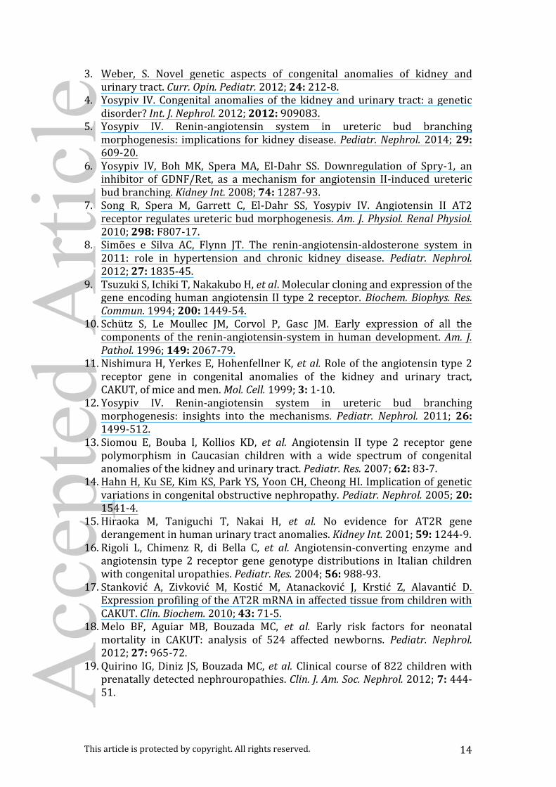
disequilibrium (LD) and pairwise LD as shown in Figure 1. However, when we
evaluated separately each of the most representative phenotypes of CAKUT
(VUR, UPJO and MCDK) in our sample, a significant association between
UPJO and this gene was identified at rs3736556 and at rs5194 (Table 2). On Acc
epte
d A
rticl
e

This article is protected by copyright. All rights reserved. 9
the other hand, associations between the AGTR2 gene and VUR or MCDK
were not found.
Since the frequency of the alleles for the AGTR2 gene seems to differ
between African Americans and Europeans, the possibility of ethnic
stratification was ruled out with ethnical study. For this purpose, we genotyped
all samples for 40 polymorphic indels loci, which form a powerful ancestry
informative test battery21. The gene location and minor allele frequencies
(MAF) in polymorphic sites of AGTR2 gene is shown in Table 3. The
differences in the proportions of genomic ancestry between the two groups
were not significant.
Discussion
Normal morphogenesis of the kidney, ureteral bud differentiation,
ureteropelvic junction formation, and bladder development are regulated by
complex epithelial-mesenchymal signalling events25. Failure of these
signalling events at specified times results in diverse phenotypes of CAKUT26.
Murine knockout models have examined the role of various signalling
molecules in genitourinary organogenesis6,11,27. In this regard, animal studies
have reinforced two possible roles for AGTR2 gene in ureteral development.
One is the regulation of apoptosis of the undifferentiated mesenchymal cells
surrounding the developing ureter27. A second role is the inhibition of ectopic
ureteral budding11. Indeed, the AT2 receptor activation has been required for
normal apoptosis of the mesenchymal cells that surrounded the developing
ureter11. When apoptosis of the undifferentiated mesenchymal cells was
halted or otherwise delayed as a result of failure to activate the AT2 receptor, Acc
epte
d A
rticl
e

This article is protected by copyright. All rights reserved. 10
diverse patterns of CAKUT were seen17.
Studies of AGTR2 gene in patients with CAKUT showed controversial
results. Nishimura et al.11 observed an increased A–G transition in AGTR2
gene in male Caucasian American and German patients with MCDK and
UPJO, while Hiraoka et al.15 reported that there is no evidence for this AGTR2
gene derangement in human urinary tract anomalies in Japanese patients.
The frequency of the G allele in AGTR2 gene was higher in CAKUT patients
than in the general population in Italian16 and Korean children14, while no
differences were found in a more recent study from Greece13. Nishimura’s
subjects were selected through strict inclusion criteria, namely male
UPJO/MCDK patients11. In contrast, the Japanese15, the Italian16, the
Korean14 and the Greece13 CAKUT samples included male and female
patients with various phenotypes. The Korean study reported that the A–G
transition in intron 1 of the AGTR2 gene is associated with UPJO/MCDK14,
whereas, in the Greece study, the same transition of the AGTR2 gene was
not associated with the development of the CAKUT13. However, the number
of CAKUT patients in these previous studies seemed to be low to reach
definitive conclusions and may not allow the detection of associations
between AGTR2 gene and specific CAKUT phenotypes. In addition, only the
A–G transition at 1332 position of the AGTR2 gene was tested in previous
studies11,13,14,16. In this context, our study extended previous findings by
increasing the sample size (290 cases and 262 controls) and by testing five
SNPs that cover the entire AGTR2 gene. We found a significant association of
AGTR2 gene at rs3736556 and at rs5194 with UPJO. No associations were
detected with MCDK and VUR. Acc
epte
d A
rticl
e

This article is protected by copyright. All rights reserved. 11
Findings obtained in animal studies corroborate the association between
AGTR2 gene and UPJO found in the present study. In general, the inhibition
of endogenous AT2 receptor signalling or the knockout of AGTR2 gene
resulted in impaired ureteric bud branching11. Indeed, AT2 receptor plays an
important role in the expansion of the ampulla, subsequent branching, and
directional bud elongation25. We might speculate that the alteration of these
morphogenetic pathways may contribute to the pathogenesis of UPJO. It
should be pointed, however, that the penetrance of AGTR2 gene in mice is
reported as 21% in males and 5% in females11, and the anatomical spectrum
of CAKUT in null mutants for the AGTR2 gene is impressively wide,
documenting that a common gene abnormality can result in different CAKUT
phenotypes5. It should be also mentioned that AGTR2 gene interacts with
other genetic pathways3. For example, given that bone morphogenic protein 4
(BMP4) inhibits UB branching27, decreased UB branching in AT2 receptor
knockout mice may be mediated, in part, by enhanced BMP4 signalling. In
this regard, Song and co-workers detected an increased expression of BMP4
gene in metanephroi of AT2 receptor knockout mice, suggesting that the
stimulatory effects of endogenous AT2 receptor on ureteric bud branching are
mediated, at least in part, via down-regulation of BMP47. Accordingly, our
research group studying the same CAKUT sample recently found an
association between BMP4 gene polymorphisms and UPJO29. Taken
together, these findings support that the expression of the ureteric bud
morphogenetic program genes and ureteric bud growth depend on complex
network of regulatory pathways and signalling effectors. Acc
epte
d A
rticl
e

This article is protected by copyright. All rights reserved. 12
The functional effect of the AGTR2 gene in the origin of UPJO is still
unknown. Stankovic and co-workers17 found mRNA expression for AT2
receptors in ureter samples of children affected by CAKUT and this
expression was not affected by -1332A/G polymorphism. The detection of AT2
receptor expression after birth in human ureter tissue suggests a prolonged
role in CAKUT origin. Indeed, the asymmetry of the CAKUT phenotype is well
described3,4, which suggests the existence of modifying genes and/or
environmental factors. Changes in post-transcriptional and post-translational
events and also in protein expression may not be ruled out. This is in line with
results provided by the animal model of unilateral ureteral obstruction, which
mimic CAKUT associated renal injuries after ureteral obstruction, where the
expression of AT2 receptor was reported30.
We are aware of the limitations of our study. The existence of different
phenotypes, incomplete penetrance, variable aggregation, and clinical
presentation makes uniform data collection difficult and introduces selection
bias. Uncertainty in the clinical classification resulted in the aggregation of
different entities under the acronym CAKUT. On the other hand, there is
genetic support for the use of the term CAKUT, since mutations in a single
gene can have pleiotropic effects on the development of the kidney and
urinary tract2. Therefore, the heterogeneity of our CAKUT sample might be
responsible for the absence of association between AGTR2 gene and the
entire case group. One might also consider the possibility that the
heterogeneity of Brazilian population interfered with molecular studies.
However, despite the fact that Brazilian population is a result of a distinctive
mixture, our ancestral genetic data is quite similar from all previously studied Acc
epte
d A
rticl
e

This article is protected by copyright. All rights reserved. 13
populations and seems perfectly comparable to HapMap studied populations.
Nevertheless, the large number of patients and controls of our study made
possible the evaluation of CAKUT phenotypes, as OJUP, VUR and MCKD,
separately. In addition, this study is the first one that selected five SNPs
covering the entire AGTR2 gene.
Finally, the data obtained in this study at least party contribute to the
understanding of CAKUT origins in Brazilian population. Data show that the
AGTR2 gene is associated UPJO, but not with MCKD and VUR. This gene
might have an essential role in the development of the ureter. However,
functional studies should be carried out to unravel the molecular pathways of
AGTR2 gene in CAKUT phenotypes.
Acknowledgments and Financial Support
This study was partially supported by CNPq (Conselho Nacional de
Desenvolvimento Científico e Tecnológico, Brazil), FAPEMIG (Fundação de
Amparo à Pesquisa do Estado de Minas Gerais, Brazil) and by the Grant
INCT-MM (Instituto Nacional de Ciência e Tecnologia – Medicina Molecular:
FAPEMIG: CBB-APQ-00075-09 / CNPq 573646/2008-2).
Statement of Interest – No conflict of interest
References
1. Bulum B, Ozçakar ZB, Ustüner E, et al. High frequency of kidney and urinary tract anomalies in asymptomatic first-degree relatives of patients with CAKUT. Pediatr. Nephrol. 2013; 28: 2143-7.
2. Vivante A, Kohl S, Hwang DY, Dworschak GC, Hildebrandt F. Single-gene causes of congenital anomalies of the kidney and urinary tract (CAKUT) in humans. Pediatr. Nephrol. 2014; 29: 695-704. Acc
epte
d A
rticl
e
This article is protected by copyright. All rights reserved. 14
3. Weber, S. Novel genetic aspects of congenital anomalies of kidney and urinary tract. Curr. Opin. Pediatr. 2012; 24: 212-8.
4. Yosypiv IV. Congenital anomalies of the kidney and urinary tract: a genetic disorder? Int. J. Nephrol. 2012; 2012: 909083.
5. Yosypiv IV. Renin-angiotensin system in ureteric bud branching morphogenesis: implications for kidney disease. Pediatr. Nephrol. 2014; 29: 609-20.
6. Yosypiv IV, Boh MK, Spera MA, El-Dahr SS. Downregulation of Spry-1, an inhibitor of GDNF/Ret, as a mechanism for angiotensin II-induced ureteric bud branching. Kidney Int. 2008; 74: 1287-93.
7. Song R, Spera M, Garrett C, El-Dahr SS, Yosypiv IV. Angiotensin II AT2 receptor regulates ureteric bud morphogenesis. Am. J. Physiol. Renal Physiol. 2010; 298: F807-17.
8. Simões e Silva AC, Flynn JT. The renin-angiotensin-aldosterone system in 2011: role in hypertension and chronic kidney disease. Pediatr. Nephrol. 2012; 27: 1835-45.
9. Tsuzuki S, Ichiki T, Nakakubo H, et al. Molecular cloning and expression of the gene encoding human angiotensin II type 2 receptor. Biochem. Biophys. Res. Commun. 1994; 200: 1449-54.
10. Schütz S, Le Moullec JM, Corvol P, Gasc JM. Early expression of all the components of the renin-angiotensin-system in human development. Am. J. Pathol. 1996; 149: 2067-79.
11. Nishimura H, Yerkes E, Hohenfellner K, et al. Role of the angiotensin type 2 receptor gene in congenital anomalies of the kidney and urinary tract, CAKUT, of mice and men. Mol. Cell. 1999; 3: 1-10.
12. Yosypiv IV. Renin-angiotensin system in ureteric bud branching morphogenesis: insights into the mechanisms. Pediatr. Nephrol. 2011; 26: 1499-512.
13. Siomou E, Bouba I, Kollios KD, et al. Angiotensin II type 2 receptor gene polymorphism in Caucasian children with a wide spectrum of congenital anomalies of the kidney and urinary tract. Pediatr. Res. 2007; 62: 83-7.
14. Hahn H, Ku SE, Kim KS, Park YS, Yoon CH, Cheong HI. Implication of genetic variations in congenital obstructive nephropathy. Pediatr. Nephrol. 2005; 20: 1541-4.
15. Hiraoka M, Taniguchi T, Nakai H, et al. No evidence for AT2R gene derangement in human urinary tract anomalies. Kidney Int. 2001; 59: 1244-9.
16. Rigoli L, Chimenz R, di Bella C, et al. Angiotensin-converting enzyme and angiotensin type 2 receptor gene genotype distributions in Italian children with congenital uropathies. Pediatr. Res. 2004; 56: 988-93.
17. Stanković A, Zivković M, Kostić M, Atanacković J, Krstić Z, Alavantić D. Expression profiling of the AT2R mRNA in affected tissue from children with CAKUT. Clin. Biochem. 2010; 43: 71-5.
18. Melo BF, Aguiar MB, Bouzada MC, et al. Early risk factors for neonatal mortality in CAKUT: analysis of 524 affected newborns. Pediatr. Nephrol. 2012; 27: 965-72.
19. Quirino IG, Diniz JS, Bouzada MC, et al. Clinical course of 822 children with prenatally detected nephrouropathies. Clin. J. Am. Soc. Nephrol. 2012; 7: 444-51. Acc
epte
d A
rticl
e

This article is protected by copyright. All rights reserved. 15
20. Lahiri DK, Nurnberger JI. A rapid non-enzymatic method for the preparation of HMW DNA from blood for RFLP studies. Nucleic Acids Res. 1991;19: 5444.
21. Bastos-Rodrigues L, Pimenta JR, Pena SD. The genetic structure of human populations studied through short insertion-deletion polymorphisms. Ann. Hum. Genet. 2006; 70: 658-65.
22. Cann HM, de Toma C, Cazes L, et al. A human genome diversity cell line panel. Science 2002; 296: 261-2.
23. Dudbridge F, Gusnanto A. Estimation of significance thresholds for genomewide association scans. Genet. Epidemiol. 2008; 32: 227-34.
24. Gabriel SB, Schaffner SF, Nguyen H, et al. The structure of haplotype blocks in the human genome. Science 2002; 296: 2225-9.
25. Kuwayama F, Miyazaki Y, Ichikawa I. Embryogenesis of the congenital anomalies of the kidney and the urinary tract. Nephrol. Dial. Transplant. 2002; 17 (Suppl 9): 45-7.
26. Ichikawa I, Kuwayama F, Pope JC, Stephens FD, Miyazaki Y. Paradigm shift from classic anatomic theories to contemporary cell biological views of CAKUT. Kidney Int. 2002; 61: 889-98.
27. Miyazaki Y, Oshima K, Fogo A, Hogan BL, Ichikawa I. Bone morphogenetic protein 4 regulates the budding site and elongation of the mouse ureter. J. Clin. Invest. 2000; 105: 863-73.
28. Hohenfellner K, Hunley TE, Schloemer C, et al. Angiotensin type 2 receptor is important in the normal development of the ureter. Pediatr. Nephrol. 1999; 13: 187-91.
29. Dos Reis GS, Simões e Silva AC, Freitas IS, et al. Study of the association between the BMP4 gene and congenital anomalies of the kidney and urinary tract. J. Pediatr. (Rio J) 2014; 90: 58-64.
30. Esteban V, Lorenzo O, Rupérez M, et al. Angiotensin II, via AT1 and AT2 receptors and NF-kappaB pathway, regulates the inflammatory response in unilateral ureteral obstruction. J. Am. Soc. Nephrol. 2004; 15: 1514-29.
Acc
epte
d A
rticl
e

This article is protected by copyright. All rights reserved. 16
Table 1- Genotype and allele frequency of AGTR2 polymorphisms in case
(CAKUT patients) and control group
SNP name Cases (290) Controls (262) p-value Odds-R 95%CI nº % nº % rs1403543 / c.-95-29G>A GG 117 0.4366 72 0.4138 0.2010 1 1 - 1 GA 50 0.1866 58 0.3333 0.9049 1.119 0.63 – 1.98 AA 101 0.3769 44 0.2529 0.1542 1.413 0.89 - 2.24 G 197 0.5368 162 0.5645 0.4803 1 1 - 1 A 170 0.4632 125 0.4355 1.1180 0.82 - 1.53 rs3736556 / c.-36+55A>T AA 162 0.7751 138 0.6603 0.4311 1 1 - 1 AT 23 0.1100 48 0.2397 0.4472 0.7802 0.43 - 1.43 TT 24 0.1148 23 0.1100 0.7910 0.8889 0.48 – 1.65 A 239 0.8328 271 0.7787 0.0884 1 1 - 1 T 48 0.1672 77 0.2213 0.7068 0.47 – 1.06 rs35474657 / c.971G>A GG 290 1 262 1 1 1 1 - 1 G 393 1 431 1 1 1 1 - 1 rs5193 / c.*199G>T
GG 101 0.8347 93 0.7686 0.6193 1 1 -1 GT 12 0.0992 19 0.1570 0.2806 1.5800 0.66 – 3.76 TT 8 0.0661 9 0.0744 0.6232 0.8185 0.30 – 2.21 G 137 0.8726 171 0.8407 0.5537 1 1 - 1 T 20 0.1274 30 0.1493 0.8321 0.45– 1.53 rs5194 / c.*205A>G AA 50 0.3704 22 0.3056 0.8704 1 1 - 1 AG 23 0.1704 22 0.3056 0.5083 1.2880 0.54 – 3.10 GG 62 0.4593 28 0.3889 0.7240 0.9743 0.50 – 1.91 A 81 0.4451 59 0.4758 0.5960 1 1 - 1 G 101 0.5549 65 0.5242 1.132 0.72 – 1.79 * After a 1000 permutation, corrected best p-value > 0.05
Acc
epte
d A
rticl
e

This article is protected by copyright. All rights reserved. 17
Table 2 - Genotype and allele frequency of AGTR2 polymorphisms in patients
with ureteropelvic junction obstruction (UPJO) and controls
SNP name Patients (55) Controls (262) p-value Odds-R 95%CI nº % nº % rs1403543 / c.-95-29G>A GG 28 0.56 72 0.41 0.62 1 1 - 1 GA 6 0.12 58 0.33 0.41 0.62 0.19 – 1.90 AA 16 0.32 44 0.25 098 0.94 0.45 - 1.92 G 41 0.63 162 0.56 0.33 1 1 - 1 A 24 0.37 125 0.44 0.76 0.43 - 1.32 rs3736556 / c.-36+55A>T AA 36 0.92 138 0.66 0.02 1 1 - 1 AT 2 0.05 48 0.24 0.21 0.33 0.07 - 1.59 TT 1 0.03 23 0.11 0.07 0.17 0.02 – 1.28 A 48 0.94 271 0.78 0.01 1 1 - 1 T 3 0.06 77 0.22 0.22 0.07 – 0.73 rs35474657 / c.971G>A GG 55 1 262 1 1 1 1 - 1 G 71 1 431 1 1 1 1 - 1 rs5193 / c.*199G>T
GG 20 0.87 93 0.77 0.84 1 1 -1 GT 1 0.04 19 0.16 0.69 0.64 0.07 – 5.88 TT 2 0.09 9 0.07 0.94 1.03 0.21 – 5.15 G 26 0.90 171 0.84 0.51 1 1 - 1 T 3 0.10 30 0.15 0.66 0.19– 2.31 rs5194 / c.*205A>G AA 5 0.20 22 0.31 0.09 1 1 - 1 AG 3 0.12 22 0.31 0.98 0.33 0.33 – 11.96 GG 17 0.68 28 0.39 0.11 0.85 0.85 – 8.38 A 8 0.25 59 0.48 0.02 1 1 - 1 G 24 0.75 65 0.52 2.72 1.14 – 6.53
* After a 1000 permutation, corrected best p-value > 0.05
Table 3- Gene location and Minor allele frequencies (MAF) in polymorphic
sites of AGTR2 gene
Gene location
Caucasian African American Chinese CAKUT Brazilian sample
rs1403543 Intron 0.39 (G) 0.43 (A) 0.33 (G) 0.45 (A) rs3736556 Intron 0.23 (T) 0.07 (T) 0.18 (T) 0.20 (T) rs35474657 Intergenic
/Unknown 0.0 (A) 0.03 (A) - 0.0 (A)
rs5193 UTR 3’ Intragenic
0.31 (T) 0.0 (T) 0.16 (T) 0.14 (T)
rs5194 UTR 3 0.42 (A) 0.37(G) 0.36 (A) 0.46 (A)
Acc
epte
d A
rticl
e

This article is protected by copyright. All rights reserved. 18
Figure legend Figure 1 : LD block structure across AGTR2 gene. The upper panel shows the location of five polymorphisms in AGTR2 gene and lower panel shows the output of Haploview (v.4.1.) LD plot, each square (with D´ values written within the box) represents a pairwise LD relationship between the two SNPs. Red squares indicate statistical significant LD between the pair of SNPs as measured by the D´ statistic. Darker colors of red indicate higher values of D´, up to a maximum of 1, and the white square indicate pairwise D´ values of < 1 with no statistically significant evidence of LD.
NEP_12308_F1
Acc
epte
d A
rticl
e