Evaluation and Surgical Planning for Craniovertebral Junction ...
14
554 www.e-neurospine.org Review Article Corresponding Author Jae Taek Hong https://orcid.org/0000-0001-6453-0439 Department of Neurosurgery, Eunpyeong St. Mary’s Hospital Catholic University of Korea, 1021 Tongil-ro, Eunpyeong-gu, Seoul 03312, Korea E-mail: [email protected] Received: August 16, 2020 Revised: August 25, 2020 Accepted: September 16, 2020 Evaluation and Surgical Planning for Craniovertebral Junction Deformity Jae Taek Hong 1 , Il Sup Kim 2 , Ho Jin Lee 2 , Jong Hyuk Park 3 , Jeong Woo Hur 1 , Jong Beom Lee 4 , Jeong Jae Lee 5 , Sang Hyo Lee 1 1 Department of Neurosurgery, Eunpyeong St. Mary’s Hospital, e Catholic University of Korea, Seoul, Korea 2 Department of Neurosurgery, St. Vincent’s Hospital, e Catholic University of Korea, Suwon, Korea 3 Department of Neurosurgery, Incheon St. Mary’s Hospital, e Catholic University of Korea, Incheon, Korea 4 Department of Neurosurgery, Chungbuk National University, Cheongju, Korea 5 Department of Neurosurgery, GangNeung Asan Hospital, University of Ulsan College of Medicine, Gangneung, Korea Craniovertebral junction (CVJ) deformity is a challenging pathology that can result in pro- gressive deformity, myelopathy, severe neck pain, and functional disability, such as diffi- culty swallowing. Surgical management of CVJ deformity is complex for anatomical rea- sons; given the discreet relationships involved in the surrounding neurovascular structures and intricate biochemical issues, access to this region is relatively difficult. Evaluation of the reducibility, CVJ alignment, and direction of the mechanical compression may determine surgical strategy. If CVJ deformity is reducible, posterior in situ fixation may be a viable so- lution. If the deformity is rigid and the C1–2 facet is fixed, osteotomy may be necessary to make the C1–2 facet joint reducible. C1–2 facet release with vertical reduction technique could be useful, especially when the C1–2 facet joint is the primary pathology of CVJ ky- photic deformity or basilar invagination. e indications for transoral surgery are becom- ing as narrow as a treatment for CVJ deformity. In this article, we will discuss CVJ align- ment and various strategies for the management of CVJ deformity and possible ways to pre- vent complications and improve surgical outcomes. Keywords: Craniovertebral junction, Alignment, Kyphosis, Basilar invagination, Defor- mity, Treatment INTRODUCTION Craniovertebral junction (CVJ) is the region around the skull base and the upper cervical spine (atlas and axis), and its align- ment is unique regarding the segmental motion and biomechani- cal perspective. 1-4 The stability of CVJ is dependent on a robust ligamentous complex and the shape of the bony structures, which are also responsible for much of the axial rotation (C1–2 joint) and flexion-extension movements (C0–1 and C1–2 joint). 5-7 Although deformity or malalignment around CVJ is a rare, CVJ deformity results in sagittal and coronal imbalances, which causes significant pain due to arthritis, instability, and C2 fo- raminal stenosis. Moreover, malalignment can be a potentially life-threatening due to swallowing difficulty and cervicomedul- lary compressive myelopathy. Patients presenting intractable pain, functional disability, or myelopathy caused by the CVJ deformity usually require surgi- cal treatment. Decompression of neural structures, sagittal and coronal spinal realignment, and stabilization are the primary goals of surgical intervention. Surgical management of CVJ pathology could be complicat- ed for various reasons; given the discreet relationships involved in the surrounding neurovascular structures and intricate bio- mechanical issues, access to this region is relatively tricky, and there are many risks involved. 8-14 Recently, there have been tremendous advances in CVJ sur- gery. Understanding normal alignment, segmental motion, com- pensatory reaction, and anatomical variation around CVJ is criti- cal not only to decide optimal surgical options but also to pre- vent complications and improve surgical outcomes. Neurospine eISSN 2586-6591 pISSN 2586-6583 This is an Open Access article distributed under the terms of the Creative Commons Attribution Non-Commercial License (https://creativecom- mons.org/licenses/by-nc/4.0/) which permits unrestricted non-commercial use, distribution, and reproduction in any medium, provided the original work is properly cited. Copyright © 2020 by the Korean Spinal Neurosurgery Society Neurospine 2020;17(3):554-567. https://doi.org/10.14245/ns.2040510.255
-
Upload
khangminh22 -
Category
Documents
-
view
0 -
download
0
Transcript of Evaluation and Surgical Planning for Craniovertebral Junction ...
554 www.e-neurospine.org
Review ArticleCorresponding AuthorJae Taek Hong
https://orcid.org/0000-0001-6453-0439
Department of Neurosurgery, Eunpyeong St. Mary’s Hospital Catholic University of Korea, 1021 Tongil-ro, Eunpyeong-gu, Seoul 03312, KoreaE-mail: [email protected]
Received: August 16, 2020 Revised: August 25, 2020 Accepted: September 16, 2020
Evaluation and Surgical Planning for Craniovertebral Junction DeformityJae Taek Hong1, Il Sup Kim2, Ho Jin Lee2, Jong Hyuk Park3, Jeong Woo Hur1, Jong Beom Lee4, Jeong Jae Lee5, Sang Hyo Lee1
1Department of Neurosurgery, Eunpyeong St. Mary’s Hospital, The Catholic University of Korea, Seoul, Korea 2Department of Neurosurgery, St. Vincent’s Hospital, The Catholic University of Korea, Suwon, Korea 3Department of Neurosurgery, Incheon St. Mary’s Hospital, The Catholic University of Korea, Incheon, Korea 4Department of Neurosurgery, Chungbuk National University, Cheongju, Korea 5 Department of Neurosurgery, GangNeung Asan Hospital, University of Ulsan College of Medicine, Gangneung, Korea
Craniovertebral junction (CVJ) deformity is a challenging pathology that can result in pro-gressive deformity, myelopathy, severe neck pain, and functional disability, such as diffi-culty swallowing. Surgical management of CVJ deformity is complex for anatomical rea-sons; given the discreet relationships involved in the surrounding neurovascular structures and intricate biochemical issues, access to this region is relatively difficult. Evaluation of the reducibility, CVJ alignment, and direction of the mechanical compression may determine surgical strategy. If CVJ deformity is reducible, posterior in situ fixation may be a viable so-lution. If the deformity is rigid and the C1–2 facet is fixed, osteotomy may be necessary to make the C1–2 facet joint reducible. C1–2 facet release with vertical reduction technique could be useful, especially when the C1–2 facet joint is the primary pathology of CVJ ky-photic deformity or basilar invagination. The indications for transoral surgery are becom-ing as narrow as a treatment for CVJ deformity. In this article, we will discuss CVJ align-ment and various strategies for the management of CVJ deformity and possible ways to pre-vent complications and improve surgical outcomes.
Keywords: Craniovertebral junction, Alignment, Kyphosis, Basilar invagination, Defor-mity, Treatment
INTRODUCTION
Craniovertebral junction (CVJ) is the region around the skull base and the upper cervical spine (atlas and axis), and its align-ment is unique regarding the segmental motion and biomechani-cal perspective.1-4 The stability of CVJ is dependent on a robust ligamentous complex and the shape of the bony structures, which are also responsible for much of the axial rotation (C1–2 joint) and flexion-extension movements (C0–1 and C1–2 joint).5-7
Although deformity or malalignment around CVJ is a rare, CVJ deformity results in sagittal and coronal imbalances, which causes significant pain due to arthritis, instability, and C2 fo-raminal stenosis. Moreover, malalignment can be a potentially life-threatening due to swallowing difficulty and cervicomedul-lary compressive myelopathy.
Patients presenting intractable pain, functional disability, or myelopathy caused by the CVJ deformity usually require surgi-cal treatment. Decompression of neural structures, sagittal and coronal spinal realignment, and stabilization are the primary goals of surgical intervention.
Surgical management of CVJ pathology could be complicat-ed for various reasons; given the discreet relationships involved in the surrounding neurovascular structures and intricate bio-mechanical issues, access to this region is relatively tricky, and there are many risks involved.8-14
Recently, there have been tremendous advances in CVJ sur-gery. Understanding normal alignment, segmental motion, com-pensatory reaction, and anatomical variation around CVJ is criti-cal not only to decide optimal surgical options but also to pre-vent complications and improve surgical outcomes.
NeurospineeISSN 2586-6591 pISSN 2586-6583
This is an Open Access article distributed under the terms of the Creative Commons Attribution Non-Commercial License (https://creativecom-mons.org/licenses/by-nc/4.0/) which permits unrestricted non-commercial use, distribution, and reproduction in any medium, provided the original work is properly cited.
Copyright © 2020 by the Korean Spinal Neurosurgery Society
Neurospine 2020;17(3):554-567.https://doi.org/10.14245/ns.2040510.255
Craniovertebral Junction DeformityHong JT, et al.
https://doi.org/10.14245/ns.2040510.255 www.e-neurospine.org 555
CVJ deformity has various etiologies, such as congenital anom-aly, tumor, infection, rheumatoid arthritis (RA), and trauma.15 One of the most common CVJ anomalies is basilar invagina-tion (BI), either congenital or degenerative, as represented by the upward migration of the odontoid process into an already limited space of the foramen magnum, causing compressive myelopathy around the cervicomedullary junction.16,17 Many bony anomalies are associated with BI, such as clivus and con-dyle hypoplasia, occipital assimilation, os odontoideum, bifid C1 arch, and Klippel-Feil syndrome.18,19
RA is the leading cause of secondary BI, which is also called basilar impression, atlantoaxial impaction, vertical settling, and cranial settling.20 Symmetrical rheumatoid destruction of the C0–1 and C1–2 joints allow for the cranium to settle on the cer-vical spine and for the dens to enter the foramen magnum. How-ever, only one lateral mass involvement may result in a fixed ro-tational tilt of the head. Secondary BI often results in a reduc-tion of atlantodental interval (ADI) due to the conical shape of dens and reduced motion, producing a false impression of ana-tomic improvement known as “pseudostabilization”.21
CRANIOVERTEBRAL JUNCTION ALIGNMENT
The human cervical spine is located inferior to the foramen
magnum. These characteristics ensure an energy-saving bal-ance of the head and neck when at rest, allowing humans to ef-fectively maintain horizontal vision without the significant ef-fort of the neck extensor muscle.22 However, quadrupeds with posteriorly positioned foramina magna require well-developed nuchal musculature and ligaments to bear the head’s weight to maintain horizontal gaze. When humans are at rest in standing position, the C0–1 articulation is held rather flexibly, whereas the C1–2 joints are held slightly in an extended position.
So, the C1 slope is backwardly slanted in the neutral position, and kyphotic angulation of the C0–1 segment allows some de-gree of freedom for neck extension as the space between the occiput and C1 posterior arch allows for upper cervical exten-sion (Fig. 1A).
When patients have a kyphotic deformity in the lower cervi-cal spine or thoracolumbar spine, the C0–1 segment can hold up the head to compensate for distal kyphosis, maintaining the sagittal balance and horizontal gaze. Therefore, a normal ky-photic angulation of the C0–1 segment could be referred to as a “tertiary curvature” to characterize human CVJ alignment, to differentiate between upper cervical spine alignment and pri-mary/secondary curvature of the human spine as well as differ-entiate between the cervical spine curvature of humans and that of quadrupeds.
Although the asymptomatic population may have different
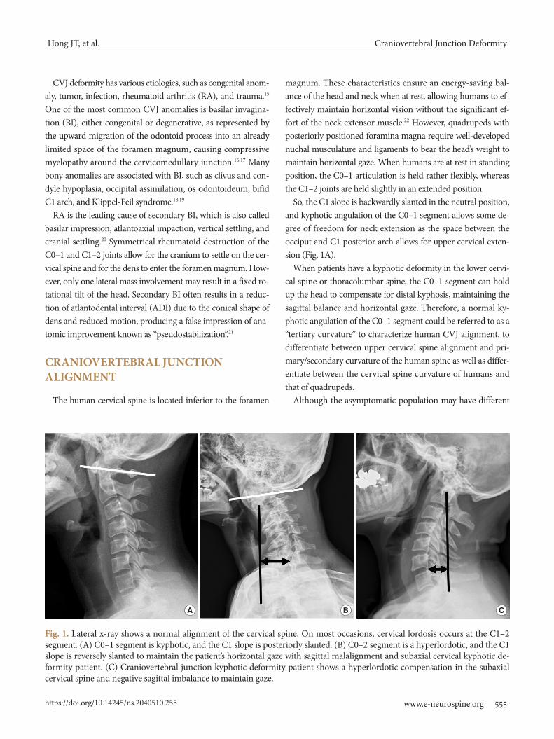
Fig. 1. Lateral x-ray shows a normal alignment of the cervical spine. On most occasions, cervical lordosis occurs at the C1–2 segment. (A) C0–1 segment is kyphotic, and the C1 slope is posteriorly slanted. (B) C0–2 segment is a hyperlordotic, and the C1 slope is reversely slanted to maintain the patient’s horizontal gaze with sagittal malalignment and subaxial cervical kyphotic de-formity patient. (C) Craniovertebral junction kyphotic deformity patient shows a hyperlordotic compensation in the subaxial cervical spine and negative sagittal imbalance to maintain gaze.
A B C

Craniovertebral Junction DeformityHong JT, et al.
https://doi.org/10.14245/ns.2040510.255556 www.e-neurospine.org
types of cervical alignment, such as “straight” or “kyphotic” cer-vical spines ranging from 7% to 40%, it has been well accepted that cervical lordosis is a physiological posture in asymptomatic subjects, and a large percentage (about 75%–80%) of cervical lordosis is localized to the C1–2 segment. Only a small percent-age (15%) of cervical lordosis exists in the lower cervical levels. On average, the C0-1 segment is kyphotic.23-28 The loss of sub-axial lordosis has been reported in CVJ fixation in which exces-sive hyperlordosis is created at the C0–2 segment (Fig. 1B).29 Moreover, craniometrics studies revealed that excessive CVJ kyphosis could cause the subaxial compensatory lordosis (Fig. 1C).30-32 The cervicothoracic junction has been shown to be the main determining factor for cervical lordosis. However, the CVJ angle has also been shown to be related to the subaxial cervical spine’s angle significantly.
The O–C2 angle was significantly greater in the patients with a straight or kyphotic subaxial cervical spine than those with lordotic lower cervical spine. When patients have a kyphotic deformity in the lower cervical spine, positive sagittal imbal-ance occurs in the cervical spine, and the hyperextended C0–2 segment holds up the head to compensate for distal kyphosis, sagittal imbalance, and maintaining horizontal gaze.
In the patients with CVJ kyphotic deformity, lateral radiographs show hyperlordotic compensation in the subaxial cervical spine and negative sagittal imbalance to maintain gaze (Fig. 1B, C).
These findings suggest that reciprocal interaction may likely affect not only global balance but also regional balance.33
Another interesting finding is that neck flexion, and exten-sion movement is initiated primarily by motions between the head and C1.34 As the head approaches full flexion and exten-sion, contributions from the upper cervical segments decrease, whereas the lower cervical segments’ contribution increases.
Understanding the normal CVJ alignment and segmental motion is paramount to understand CVJ deformity better and to decide on a more appropriate surgical strategy according to the pathologies of CVJ.
PREOPERATIVE RADIOLOGIC ASSESSMENT
Irreducibility was defined as nonalignment of C1–2 after neck extension (determined on lateral x-ray) or after cervical trac-tion. Lateral radiography with a dynamic study is necessary to evaluate ADI, degree of vertical subluxation, and reducibility. Furthermore, bony abnormalities, such as occipital assimila-tion, os odontoideum, bifid C1 arch, and C2–3 fusion, can be
evaluated on lateral radiography.Traction can be a useful tool in evaluating the reducibility and
predicting intraoperative neurological worsening by surgical position. Preoperative cervical traction may correct the atlanto-axial dislocation (AAD) and vertical subluxation in some re-ducible deformity cases. This could especially be important for managing fragile patients because they can be treated with only a stabilization procedure postreduction, reducing the level of fixation. Gardner cervical traction was applied, depending on age and weight, starting with 2–5 kg for one or 2 days. The head of the bed was elevated to provide counter traction. Serial ra-diographs were assessed for reduction. These patients who dem-onstrated a reduction were classified as reducible AAD or BI.
Magnetic resonance imaging (MRI) was studied to evaluate cord compression, T2 signal change, Chiari malformation, and syringomyelia. Computed tomography (CT) scans can confirm bony abnormalities, the dens’ exact location, C1–2 joint destruc-tion, and abnormal C1–2 facet angle. CT scans are also used to determine the extent of facet fusion and osteophytic bridging to assess the need for C1–2 osteotomy.
Computed tomography angiography (CTA) screening is a useful tool for getting 3-dimensional complex deformity infor-mation and identifying vertebral artery (VA) anomaly around CVJ.14,35 The incidence of V3 segment anomalies has been re-ported to be as high as 10%.9,14 In V3 segment anomalies, a more optimal entry point for C1 fixation should be selected to avoid significant morbidities associated with VA injury.36 Al-though there could be a racial difference in the incidence of V3 segment anomaly, existing literature indicates that the V3 seg-ment anomaly is more common in the group with the congeni-tal bony anomaly. Quadrupeds, including humans, are more likely to have other CVJ bony abnormalities, such as atlantooc-cipital assimilation or dens anomalies, if atlantoaxial instability is present.
Therefore, we suggest that preoperative CTA might be infor-mative for deciding on a surgical technique, especially in the high-risk group of the V3 segment anomaly, such as Asians and those with C1–2 instability with congenital bony anomaly.9
TREATMENT OF CRANIOVERTEBRAL JUNCTION DEFORMITY
Fig. 2 provides decision-making pathways to treat CVJ defor-mity. The treatment goal of CVJ deformity is to restore normal alignment, relieve neural compression, and stabilization with-out neurological complications. Factors that influence the spe-

Craniovertebral Junction DeformityHong JT, et al.
https://doi.org/10.14245/ns.2040510.255 www.e-neurospine.org 557
cific treatment of CVJ deformity are as follows: (1) the reduc-ibility of deformity (i.e., restoring anatomic alignment, thereby relieving neural compression); (2) the direction of mechanical compression; (3) the presence of abnormal craniocervical angle and alignment; and (4) the presence of hindbrain herniation, syrinx, and vascular abnormalities.
Irreducible lesions can be divided into 2 types: The ventral compressive lesion and the dorsal compressive lesion. Doral compression cases need either a posterior or posterolateral de-compression procedure, and if there is instability after decom-pression, posterior fixation is mandatory to gain stability. Irre-ducible CVJ deformity may require decompression at the site of compression or may require an osteotomy procedure to make the lesion reducible and relieve neural compression.33
Transoral surgery is a procedure carried out through the oral cavity to gain access to the ventral compressive lesion of CVJ from the lower rim of clivus cranially to C23 disc space caudal-ly. Transoral surgery could be necessary in case of irreducible CVJ deformity, where ventral compression to neutral structures is present.37 Other indications for transoral surgery include CVJ tumor and infection, in which a simple posterior reduction38 cannot decompress the anterior compression. Although the combined anteriorposterior approach may provide adequate neural decompression and fusion, it poses several disadvantag-es, such as the higher risk of surgical morbidities, prolonged
postoperative intubation/nasogastric feeding, phonation diffi-culty, and potential infection.39-41 The risk of infection is a big concern, mainly when the subarachnoid space had to be opened, which may cause catastrophic sequelae. Another issue is that transoral decompression can induce significant instability of CVJ, inevitably requiring a subsequent posterior stabilization procedure. These issues are why the initial enthusiasm vanished, and an indication of transoral decompression became less evi-dent in the recent literature.
A C1–2 joint distraction technique, as introduced by Goel, has been gaining popularity recently as a possible treatment modality for selected cases of AAD and BI.42-45 This technique seems to have several advantages over conventional transoral surgery and occipitocervical (OC) fixation. First, direct posteri-or reduction and fixation are possible for cervicomedullary de-compression. Hence, a transoral surgery and its related compli-cations could be avoided. Second, the fusion bed dimension could substantially be increased because C1–2 facet surface is rather large. Third, it can avoid occipital fixation and conserve C0–1 segmental motion. Fourth, C1–2 joint manipulation can make a fixed subluxation or deformity reducible, minimizing the need for head traction, before and during surgery.
1. Preoperative PlanningPreoperative cervical traction may correct AAD and vertical
Fig. 2. The algorithm shows decision-making pathways for the treatment of the craniovertebral junction (CVJ) deformity. OC, occipitocervical.
CVJ deformity
ReducibilityDynamic x-ray/traction
Rigid/semirigid
Facet pathology (Facet fusion/inclination)
Posterior in situ stabilization Ventral compression
Flexible
Bony deformity
Vertical reduction/posterior fixation
Transoral decompression/
posterior fixation
Pannus CVJ kyphosis
No or mild Severe
C1–2 osteotomy/vertical reduction/
C1–2 fusion
C1–2 osteotomy/vertical reduction/craniocervical realignment /OC fusion
Craniovertebral Junction DeformityHong JT, et al.
https://doi.org/10.14245/ns.2040510.255558 www.e-neurospine.org
subluxation in some patients with reducible deformity. This could especially be important for managing fragile patients be-cause they can be treated with only a stabilization procedure af-ter reduction and decreasing the numbers of fixation levels.
The direction of the mechanical compression was the critical factor in deciding a surgical approach. With recent advance-ments in surgical techniques and instruments, the results of posterior C1–2 release and reduction have been satisfactory. However, difficult or insufficient reduction and loss of reduc-tion have been observed after posterior reduction and fixation.37 Thus, anterior release reduction and posterior fixation could still be available options for irreducible BI with AAD, especially in patients with severely deformed bony mass anteriorly com-pressing the cervicomedullary junction.
CVJ alignment is another crucial factor to decide surgical strategy. Clivus-canal angle and cervicomedullary angle is tend-ed to be kyphotic in congenital CVJ anomaly, which is caused by the short clivus, platybasia, angulated odontoid process, or abnormal sagittal inclination of deformed C1–2 facet joints.30-32 Craniocervical realignment through screws and rod systems may be an efficacious surgical technique for treating congenital craniocervical anomaly with abnormal angulation of the clivus. However, craniocervical realignment procedure, including dis-traction, compression, and skull extension, need additional de-compression/fixation up to the cranium, and it should be cau-tiously performed, especially in cases with severe canal steno-sis.46
2. Surgical Techniques for CVJ Deformity1) Fixed atlantoaxial rotatory fixation
Reducible AAD can be defined as C1–2 alignment on exten-sion or cervical traction. If the dislocation cannot be reduced
on extension position and preoperative traction, it is labeled as an irreducible or fixed AAD and rotatory subluxation (Fig. 3). Management becomes difficult with this type of fixed deformi-ty. Since Goel introduced the C1–2 distraction method, the fo-cus in CVJ pathology treatment has shifted to the dislocated C1–2 facets rather than the C2 dens, and the C1–2 joint ma-nipulation has been the preferred method of treatment for irre-ducible AAD.21,43 It is because the cause of irreducibility is in the C1–2 facets. In cases of irreducible AAD or rotatory fixa-tion, C1–2 joint mutilation or osteophyte formation is so severe that dislocation cannot be reduced despite cervical traction. Therefore, C1–2 facet mobilization is necessary to make this fixed deformity reducible using facet osteotomies (Fig. 3C).
After releasing the C1–2 facet joint capsule and removing os-teophytes, the mobility between C1 and C2 is usually identifi-able intraoperatively. The cartilage endplate was curetted and drilled to increase mobility further and remodel the C1–2 facet joint plane. The rotated C1–2 facet joint could be mobilized and reduced in a neutral position after releasing the C1–2 facet joint (Supplementary video clip 1).
In the horizontal subluxation cases, the surgical assistant pressed down firmly on the C2 spinous process or pulled the C1 arch using cables to reduce horizontal subluxation. Adjusting the pa-tients’ neck to allow for a slight extension should help the pro-cess of reduction. Bone block or cage filled with cancellous bone can be placed inside the decorticated C1–2 joint.
2) Rheumatoid BIrheumatoid BI is caused by C1–2 joint destruction and sub-
sequent upward migration of the odontoid process. These find-ings suggest that the primary pathology of rheumatoid BI is the C1–2 joint, not the C0–1 joint. Hence, spine surgeons should
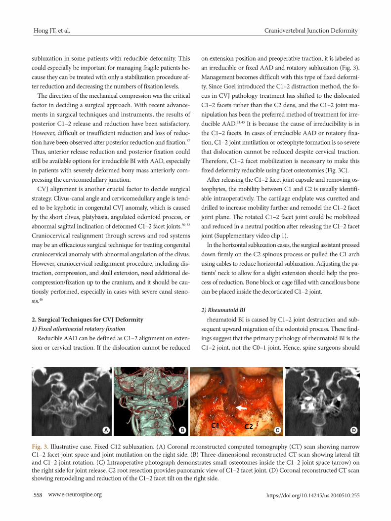
Fig. 3. Illustrative case. Fixed C12 subluxation. (A) Coronal reconstructed computed tomography (CT) scan showing narrow C1–2 facet joint space and joint mutilation on the right side. (B) Three-dimensional reconstructed CT scan showing lateral tilt and C1–2 joint rotation. (C) Intraoperative photograph demonstrates small osteotomes inside the C1–2 joint space (arrow) on the right side for joint release. C2 root resection provides panoramic view of C1–2 facet joint. (D) Coronal reconstructed CT scan showing remodeling and reduction of the C1–2 facet tilt on the right side.
A B C D
Craniovertebral Junction DeformityHong JT, et al.
https://doi.org/10.14245/ns.2040510.255 www.e-neurospine.org 559
focus on C1–2 joint reduction to treat rheumatoid BI to spare the innocent C0–1 segmental motion. Rheumatoid destruction of the C1–2 joint could easily be confused with spontaneous fu-sion mass on CT scan due to narrowing the joint space and de-struction of joint cartilage. However, the C1–2 joint of rheuma-toid BI is unstable and easily reducible in vertical distraction.
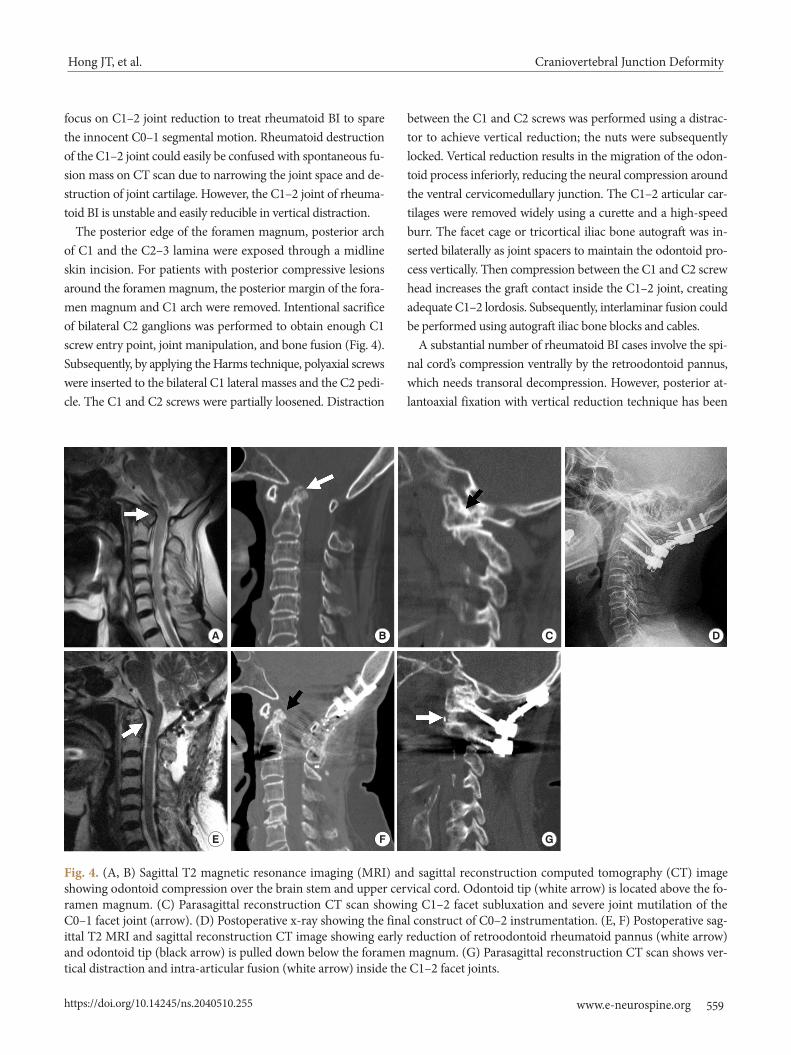
The posterior edge of the foramen magnum, posterior arch of C1 and the C2–3 lamina were exposed through a midline skin incision. For patients with posterior compressive lesions around the foramen magnum, the posterior margin of the fora-men magnum and C1 arch were removed. Intentional sacrifice of bilateral C2 ganglions was performed to obtain enough C1 screw entry point, joint manipulation, and bone fusion (Fig. 4). Subsequently, by applying the Harms technique, polyaxial screws were inserted to the bilateral C1 lateral masses and the C2 pedi-cle. The C1 and C2 screws were partially loosened. Distraction
between the C1 and C2 screws was performed using a distrac-tor to achieve vertical reduction; the nuts were subsequently locked. Vertical reduction results in the migration of the odon-toid process inferiorly, reducing the neural compression around the ventral cervicomedullary junction. The C1–2 articular car-tilages were removed widely using a curette and a high-speed burr. The facet cage or tricortical iliac bone autograft was in-serted bilaterally as joint spacers to maintain the odontoid pro-cess vertically. Then compression between the C1 and C2 screw head increases the graft contact inside the C1–2 joint, creating adequate C1–2 lordosis. Subsequently, interlaminar fusion could be performed using autograft iliac bone blocks and cables.
A substantial number of rheumatoid BI cases involve the spi-nal cord’s compression ventrally by the retroodontoid pannus, which needs transoral decompression. However, posterior at-lantoaxial fixation with vertical reduction technique has been
Fig. 4. (A, B) Sagittal T2 magnetic resonance imaging (MRI) and sagittal reconstruction computed tomography (CT) image showing odontoid compression over the brain stem and upper cervical cord. Odontoid tip (white arrow) is located above the fo-ramen magnum. (C) Parasagittal reconstruction CT scan showing C1–2 facet subluxation and severe joint mutilation of the C0–1 facet joint (arrow). (D) Postoperative x-ray showing the final construct of C0–2 instrumentation. (E, F) Postoperative sag-ittal T2 MRI and sagittal reconstruction CT image showing early reduction of retroodontoid rheumatoid pannus (white arrow) and odontoid tip (black arrow) is pulled down below the foramen magnum. (G) Parasagittal reconstruction CT scan shows ver-tical distraction and intra-articular fusion (white arrow) inside the C1–2 facet joints.
A B C D
E F G
Craniovertebral Junction DeformityHong JT, et al.
https://doi.org/10.14245/ns.2040510.255560 www.e-neurospine.org
reported to provide an early reduction of retroodontoid rheu-matoid pannus, as well as a reduction of BI and AAD.47-51 This finding suggests that rheumatoid pannus could be a buckling of the ligamentous complex around CVJ caused by BI rather than granulation tissue caused by the inflammation.
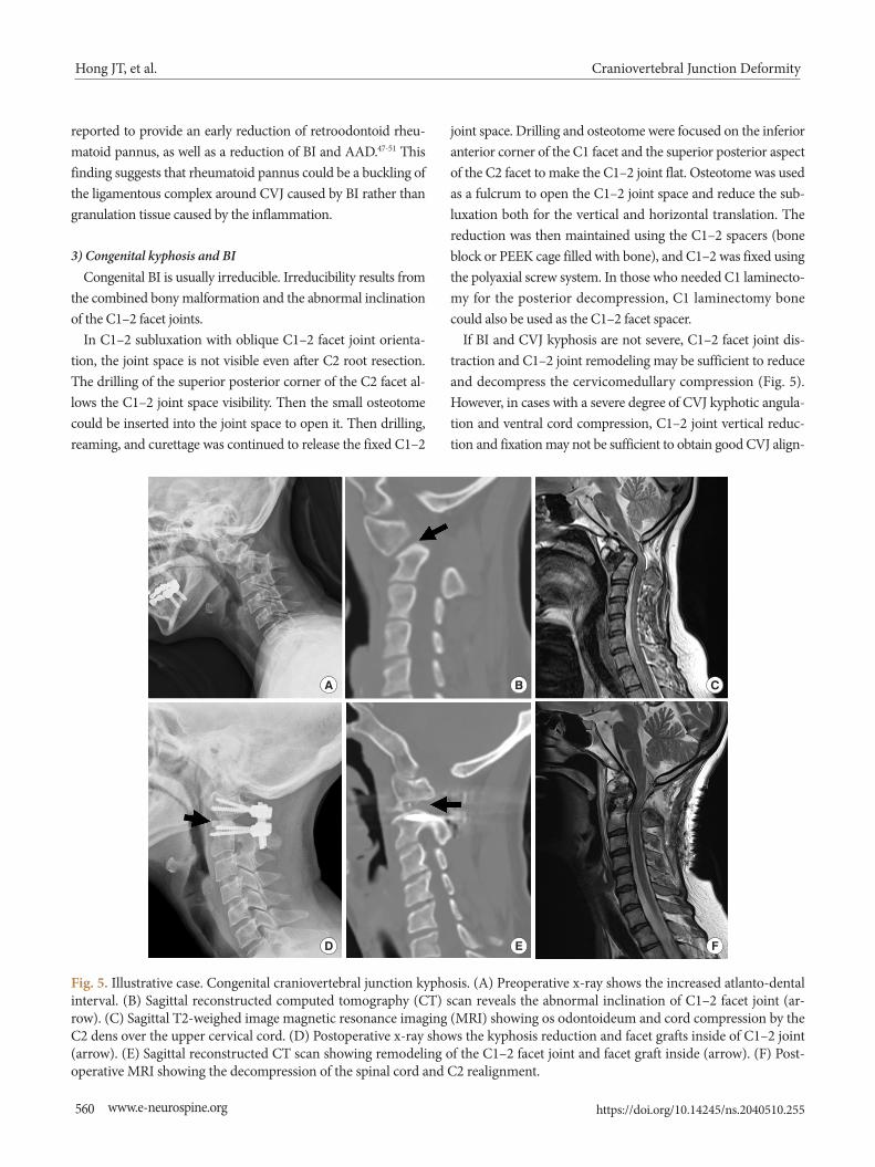
3) Congenital kyphosis and BICongenital BI is usually irreducible. Irreducibility results from
the combined bony malformation and the abnormal inclination of the C1–2 facet joints.
In C1–2 subluxation with oblique C1–2 facet joint orienta-tion, the joint space is not visible even after C2 root resection. The drilling of the superior posterior corner of the C2 facet al-lows the C1–2 joint space visibility. Then the small osteotome could be inserted into the joint space to open it. Then drilling, reaming, and curettage was continued to release the fixed C1–2
joint space. Drilling and osteotome were focused on the inferior anterior corner of the C1 facet and the superior posterior aspect of the C2 facet to make the C1–2 joint flat. Osteotome was used as a fulcrum to open the C1–2 joint space and reduce the sub-luxation both for the vertical and horizontal translation. The reduction was then maintained using the C1–2 spacers (bone block or PEEK cage filled with bone), and C1–2 was fixed using the polyaxial screw system. In those who needed C1 laminecto-my for the posterior decompression, C1 laminectomy bone could also be used as the C1–2 facet spacer.
If BI and CVJ kyphosis are not severe, C1–2 facet joint dis-traction and C1–2 joint remodeling may be sufficient to reduce and decompress the cervicomedullary compression (Fig. 5). However, in cases with a severe degree of CVJ kyphotic angula-tion and ventral cord compression, C1–2 joint vertical reduc-tion and fixation may not be sufficient to obtain good CVJ align-
A B C
D E F
Fig. 5. Illustrative case. Congenital craniovertebral junction kyphosis. (A) Preoperative x-ray shows the increased atlanto-dental interval. (B) Sagittal reconstructed computed tomography (CT) scan reveals the abnormal inclination of C1–2 facet joint (ar-row). (C) Sagittal T2-weighed image magnetic resonance imaging (MRI) showing os odontoideum and cord compression by the C2 dens over the upper cervical cord. (D) Postoperative x-ray shows the kyphosis reduction and facet grafts inside of C1–2 joint (arrow). (E) Sagittal reconstructed CT scan showing remodeling of the C1–2 facet joint and facet graft inside (arrow). (F) Post-operative MRI showing the decompression of the spinal cord and C2 realignment.
Craniovertebral Junction DeformityHong JT, et al.
https://doi.org/10.14245/ns.2040510.255 www.e-neurospine.org 561
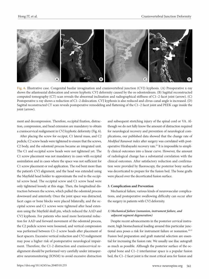
ment and decompression. Therefore, occipital fixation, distrac-tion, compression, and head extension are mandatory to obtain a craniocervical realignment in CVJ kyphotic deformity (Fig. 6).
After placing the screw for occiput, C1 lateral mass, and C2 pedicle, C2 screw heads were tightened to ensure that the screws, C2 body, and the odontoid process became an integrated unit. The C1 and occipital screw heads were not tightened yet. The C1 screw placement was not mandatory in cases with occipital assimilation and in cases where the space was not sufficient for C1 screw placement or rod application. The rod bent more than the patient’s CVJ alignment, and the head was extended using the Mayfield head holder to approximate the rod to the occipi-tal screw head. The occipital screw and C1 screw head were only tightened loosely at this stage. Then, the longitudinal dis-traction between the screws, which pulled the odontoid process downward and anteriorly. Once the joint space was distracted, facet cages or bone blocks were placed bilaterally, and the oc-cipital screws and C1 screws were tightened after head exten-sion using the Mayfield skull pin, which reduced the AAD and CVJ kyphosis. For patients who need more horizontal reduc-tion for AAD and forward movement of the odontoid process, the C2 pedicle screws were loosened, and vertical compression was performed between C1–2 screw heads after placement of facet spacers. Excessive vertical reduction and CVJ realignment may pose a higher risk of postoperative neurological impair-ment. Therefore, the C1-2 distraction and craniocervical re-alignment should be performed very carefully under intraoper-ative neuromonitoring (IONM) to avoid excessive distraction
and subsequent stretching injury of the spinal cord or VA. Al-though we do not fully know the amount of distraction required for neurological recovery and prevention of neurological com-plications, our published data showed that the change rate of Modified Ranawat index after surgery was correlated with post-operative Hirabayashi recovery rate.52 It is impossible to simpli-fy clinical outcomes into a linear curve. However, the amount of radiological change has a substantial correlation with the clinical outcomes. After satisfactory reduction and confirma-tion were provided by fluoroscopy, the posterior bony surface was decorticated to prepare for the fusion bed. The bone grafts were placed over the decorticated fusion surface.
3. Complication and PreventionMechanical failure, various kinds of neurovascular complica-
tions, and postoperative swallowing difficulty can occur after the surgery in patients with CVJ deformity.
1) Mechanical failure (nonunion, instrument failure, and adjacent segment degeneration)
Despite recent advancements in the posterior cervical instru-ment, high biomechanical loading around this particular junc-tional area poses a risk for instrument failure or nonunion.10,12 Fusion bed preparation and graft material selection are essen-tial for increasing the fusion rate. We usually use iliac autograft as much as possible. Although the posterior surface of the oc-cipital bone and C1–2 interlaminar space is a popular fusion bed, the C1–2 facet joint is the most critical area for fusion and
Fig. 6. Illustrative case. Congenital basilar invagination and craniovertebral junction (CVJ) kyphosis. (A) Preoperative x-ray shows the atlantoaxial dislocation and severe kyphotic CVJ deformity caused by the os odontoideum. (B) Sagittal reconstructed computed tomography (CT) scan reveals the abnormal inclination and radiographical stiffness of C1–2 facet joint (arrow). (C) Postoperative x-ray shows a reduction of C1–2 dislocation. CVJ kyphosis is also reduced and clivus-canal angle is increased. (D) Sagittal reconstructed CT scan reveals postoperative remodeling and flattening of the C1–2 facet joint and PEEK cage inside the joint (arrow).
A B C D
Craniovertebral Junction DeformityHong JT, et al.
https://doi.org/10.14245/ns.2040510.255562 www.e-neurospine.org
malalignment correction for CVJ kyphosis patients, especially when C1 laminectomy is inevitable.
Moreover, the long-term fate of the adjacent C0–1 segment has rarely been reported in patients after receiving C1–2 verti-cal distraction procedure with respect to the segmental motion and adjacent segmental degeneration. Yoshida et al.53 reported that C1–2 rigid fixation could provide prophylactic benefits against vertical subluxation and the subaxial subluxation in RA patients. Werle et al.54 reported that C0–1 degeneration is very low after C1–2 fusion in RA patients. Therefore, the C0–1 seg-ment sparing technique should be the preferred treatment op-tion of CVJ fusion, especially for RA patients.
2) Subaxial alignment change following CVJ fixationSeveral studies have investigated the relationship between
postoperative C1–2 angle and C2–7 sagittal alignment change after C1–2 fixation,55-58 and most have reported a reciprocal re-lationship between changes in the upper cervical spine and subaxial cervical spine.57,58 If the CVJ is fixed in the hyperlor-dotic posture, subaxial cervical kyphosis is inevitable, and up-per cervical kyphosis causes lordotic compensation in the sub-axial cervical spine. The most excellent care is necessary to de-termine the C1–2 fixation angle during surgery to reduce the risk of postoperative malalignment and subluxation of the sub-axial cervical spine. Subaxial alignment change is not uncom-mon after CVJ fixation. Muscle detachment at the C2 spinous process was not a risk factor of kyphotic change. Our findings suggest small range of motion (ROM) of the C0–1 segment with or without occipital fixation and combined subaxial lami-noplasty are risk factors of subaxial kyphotic change. Although the little study has sought to identify the risk factors of postop-erative subaxial malalignment following CVJ fixation, we re-cently reported subaxial cervical spine alignment might change significantly during the first year after CVJ posterior fixation. The subaxial cervical alignment is significantly related to the upper cervical angle after CVJ fixation. Furthermore, the study suggested a small ROM of the C0–1 segment with or without occipital fixation and combined subaxial laminoplasty inde-pendently predict subaxial kyphotic change after upper cervical fixation. Interestingly, extensor muscle detachment at C2 itself was not identified as a risk factor of kyphotic change, but when combined with subaxial laminoplasty and wider dissection of deep extensor muscle, the risk of subaxial kyphotic change was significantly increased.59
These results suggest the C0–1 segment is the dominant seg-ment for compensating for abnormal angulation of the subaxial
cervical spine after upper cervical fixation. These results imply it is important not to include occipital fixation during upper cervical fixation to avoid loss of subaxial cervical lordosis and preserve C0–1 segmental motion.
3) Occipital neuralgiaC1–2 joint distraction technique has a clinically significant
concern regarding the C2 neuropathic pain after C2 root resec-tion. Although C2 root resection is not mandatory for C1–2 facet opening, it is useful to make a broad exposure for the C12 facet surface, especially when C1–2 interlaminar space is nar-row and venous plexus is prominent.44,60 Thus, C2 root resec-tion is one of the critical parts of the C1–2 joint distraction pro-cedure. In the literature, most authors have reported that C2 neuropathic pain incidence is relatively low after C2 root resec-tion.44,60 However, C2 neuropathic pain could be troublesome for some patients. Therefore, surgeons should be aware of both the benefits and risks associated with C2 root resection. C2 neu-ropathic pain may cause significant postoperative occipital neu-ralgia, which could also occur due to the C2 root irritation by C1 screw, excessive manipulation, and traction on the C2 root during surgery. Several reports have advocated the deliberate C2 root resection or C1 screw without threads to minimize C2 root complications.61-63 However, the relationship between C2 root resection and neuropathic pain in the literature remains controversial. Goel et al.61 reported no significant concerns re-garding C2 neuropathic pain among 160 patients who under-went the C2 root sacrifice. Dewan et al.64 reported that C2 root transection is associated with increased occipital numbness. How-ever, this finding does not affect patient-reported outcomes and quality of life, and the C2 root preservation can be associated with occipital neuralgia, which harms patient disability and quality of life. Elliott et al.65 demonstrated that the C2 root tran-section decreases blood loss and operative time and significant-ly reduces postoperative neuralgia. However, Yeom et al.66 re-ported that C2 root resection could cause a high incidence of postoperative occipital neuralgia. In our study, occipital neural-gia was significantly induced in the C2 root resection group than that of the C2 root-sparing group.
Furthermore, the C1 screw entry point is significantly related to postoperative C2 neuropathy. Although C2 root resection al-lows for better visualization for venous bleeding control, getting a broader view for screw insertion to prevent screw malposition and a wider bone fusion surface, C2 root resection is an inde-pendent risk factor of postoperative C2 root neuropathy. So, excessive C2 root manipulation and resection should be care-
Craniovertebral Junction DeformityHong JT, et al.
https://doi.org/10.14245/ns.2040510.255 www.e-neurospine.org 563
fully determined because it can cause occipital neuralgia. How-ever, C2 root resection could be inevitable for vertical reduc-tion, especially in cases of rheumatoid BI. Given that C1–2 joint mutilation is so severe in rheumatoid BI, the height of the C2 foramen becomes too narrow for the manipulation of the C1–2 facet joint and insertion of screw without damaging the C2 nerve root.
4) Neurological worseningEven though several reports of vertical reduction technique
showed excellent clinical and radiological results without neu-rological complication,21,37,43,44 we experienced cases with tran-sient neurological worsening after craniocervical realignment and encountered several cases with a significant change in IONM during the vertical reduction technique.67 To date, we are not sure whether it is related to spinal cord stretch or spinal cord ischemia due to the VA stretch. However, we found that IONM change is usually recovered after reducing the amount of C1–2 joint distraction and the facet graft size. This finding shows the efficacy of IONM during craniocervical realignment or vertical reduction procedure for congenital craniocervical segmenta-tion anomaly.
IONM change is more common in CVJ surgery than at any other level of the cervical spine. Preoperative T2 signal change on MRI and narrow canal diameter are 2 risk factors that are significantly related to IONM change in our CVJ surgery case series.46,67,68 We suggest that IONM may help in preventing un-expected neurological deterioration during posterior cranio-cervical distraction and realignment.
5) DysphagiaA “military tuck” posture (neutral head posture, the exten-
sion of the lower cervical spine, posterior translation of the oc-ciput-C1 complex) usually reduces C1–2 subluxation, while optimizing the surgical exposure and allowing for a favorable screw trajectory.69 It is a commonly used posture for posterior instrumentation in the upper cervical spine.
However, previous studies have shown that a decrease in the OC2 angle in the retraction position causes a reduction in the oropharyngeal airway space and postoperative dysphagia.70,71
Dysphagia is not an uncommon complication after posterior cervical surgeries, as well as anterior cervical spine surgeries.72-74 Moreover, dysphagia after posterior OC fusion has been recog-nized as a severe postoperative complication. Miyata et al.70 re-ported that dysphagia after OC fusion is caused by glossoptosis, which is the downward displacement of the tongue root, result-
ing in the narrowing of the oropharynx and the impairment of the ability of the epiglottis to move sufficiently. They emphasized that a decrease in the OC2 angle may likely induce a reduction in the pharyngeal space and can be a predictor of postoperative dysphagia, which is not compensated by the middle or lower cervical spine. We reported that the upper cervical segments’ movement could be an essential compensation mechanism for swallowing, and the fixation of the upper cervical spine could be one of the risk factors for postoperative dysphagia.
Moreover, C0–1 angle change and movement during swal-lowing are significantly greater in the retraction posture than in the neutral posture, and the C0–1 segment is the dominant lev-el of compensatory motion that occurs when subjects experi-ence dysphagia in the retraction position.75 Therefore, if we fix up to the cranium in the retraction position, the compensatory movement’s major segment cannot function properly to allow swallowing, leading to postoperative dysphagia.76 Therefore, avoiding OC fixation in retraction posture and sparing C0–1 segment are critical technical tips for preventing postoperative dysphagia.
CONCLUSION
Each type of CVJ deformity has its characteristics and treat-ment challenges. Regardless of the underlying cause, this com-plicated disease entity has 2 types—reducible deformity and ir-reducible deformity—according to whether the dislocation is reduced after dynamic x-ray and traction. Simple posterior fix-ation and fusion are adequate for reducible CVJ deformity, and any type of reduction procedure is necessary for irreducible CVJ deformity. A reduction of irreducible deformity can be achieved using anterior transoral surgery or posterior C1–2 facet joint manipulation procedure, depending on the patholo-gy’s primary focus. With recent advancements in surgical tech-niques and internal fixation devices, posterior only surgery can achieve ventral decompression and craniocervical realignment. The posterior reduction procedure principle is releasing the C1–2 facet joint, pulls down the odontoid process away from the cervicomedullary junction, and craniocervical realignment without neurological injury. C1–2 vertical reduction technique can be the optimal surgery for BI with or without abnormal CVJ kyphosis. Additional procedure for craniocervical realign-ment is necessary to treat congenital BI with severe CVJ kypho-sis. Head extension maneuver through the occipital screws and rod system may be a safe and efficacious surgical technique for the craniocervical realignment.
Craniovertebral Junction DeformityHong JT, et al.
https://doi.org/10.14245/ns.2040510.255564 www.e-neurospine.org
Avoiding neurological complications could be the most cru-cial issue in this complicated junctional area, and preoperative radiological assessment is critical to detect the structural anom-aly and decide an optimal surgical plan. IONM may help in pre-venting any unexpected neurological deterioration during pos-terior craniocervical distraction and realignment.
By understanding the nature of CVJ alignment, recent ad-vancements in posterior cervical instrumentation, all available treatment options, and the decision-making process, surgeons can improve patients’ life quality.
CONFLICT OF INTEREST
The authors have nothing to disclose.
SUPPLEMENTARY MATERIAL
Supplementary video clip 1 can be found via https://doi.org/ 10.14245/ns.2040510.255.v1.
REFERENCES
1. Visocchi M, Mattogno PP, Signorelli F, et al. Complications in craniovertebral junction instrumentation: hardware re-moval can be associated with long-lasting stability. Personal Experience. Acta Neurochir Suppl 2017;124:187-94.
2. Morota N. Pediatric craniovertebral junction surgery. Neu-rol Med Chir (Tokyo) 2017;57:435-60.
3. Goel A, Sathe P, Shah A. Atlantoaxial fixation for basilar in-vagination without obvious atlantoaxial instability (group b basilar invagination): outcome analysis of 63 surgically treat-ed cases. World Neurosurg 2017;99:164-70.
4. Lee DH, Hong JT, Sung JH, et al. Morphologic analysis of occipital sinuses for occipital screw fixation using digital subtraction angiography. World Neurosurg 2016;91:279-84.
5. Dlouhy BJ, Policeni BA, Menezes AH. Reduction of atlanto-axial dislocation prevented by pathological position of the transverse ligament in fixed, irreducible os odontoideum: operative illustrations and radiographic correlates in 41 pa-tients. J Neurosurg Spine 2017;27:20-8.
6. Deepak AN, Salunke P, Sahoo SK, et al. Revisiting the dif-ferences between irreducible and reducible atlantoaxial dis-location in the era of direct posterior approach and C1-2 joint manipulation. J Neurosurg Spine 2017;26:331-40.
7. Yin YH, Tong HY, Qiao GY, et al. Posterior reduction of fixed atlantoaxial dislocation and basilar invagination by at-
lantoaxial facet joint release and fixation: a modified tech-nique with 174 cases. Neurosurgery 2016;78:391-400; dis-cussion 400.
8. Yi HJ, Hong JT, Lee JB, et al. Analysis of risk factors for pos-terior C1 screw-related complication: a retrospective study of 358 posterior C1 screws. Oper Neurosurg (Hagerstown) 2019;17:509-17.
9. Hong JT, Kim IS, Kim JY, et al. Risk factor analysis and deci-sion-making of surgical strategy for V3 segment anomaly: significance of preoperative CT angiography for posterior C1 instrumentation. Spine J 2016;16:1055-61.
10. Takigawa T, Simon P, Espinoza Orias AA, et al. Biomechani-cal comparison of occiput-C1-C2 fixation techniques: C0-C1 transarticular screw and direct occiput condyle screw. Spine (Phila Pa 1976) 2012;37:E696-701.
11. Hong JT, Takigawa T, Udayakunmar R, et al. Biomechanical effect of the C2 laminar decortication on the stability of C2 intralaminar screw construct and biomechanical compari-son of C2 intralaminar screw and C2 pars screw. Neurosur-gery 2011;69(1 Suppl Operative):ons1-6; discussion ons6-7.
12. Hong JT, Takigawa T, Sugisaki K, et al. Biomechanical and morphometric evaluation of occipital condyle for occipito-cervical segmental fixation. Neurol Med Chir (Tokyo) 2011; 51:701-6.
13. Hong JT, Kim TH, Kim IS, et al. The effect of patient age on the internal carotid artery location around the atlas. J Neu-rosurg Spine 2010;12:613-8.
14. Hong JT, Lee SW, Son BC, et al. Analysis of anatomical vari-ations of bone and vascular structures around the posterior atlantal arch using three-dimensional computed tomogra-phy angiography. J Neurosurg Spine 2008;8:230-6.
15. Kukreja S, Ambekar S, Sin AH, et al. Occipitocervical fusion surgery: review of operative techniques and results. J Neurol Surg B Skull Base 2015;76:331-9.
16. Salunke P, Sahoo S, Deepak AN. Different facets in manage-ment of congenital atlantoaxial dislocation and basilar in-vagination. Neurosurgery 2015;77:E985-7.
17. Salunke P, Sahoo SK, Deepak AN, et al. Comprehensive drill-ing of the C1-2 facets to achieve direct posterior reduction in irreducible atlantoaxial dislocation. J Neurosurg Spine 2015;23:294-302.
18. Goel A, Nadkarni T, Shah A, et al. Bifid anterior and poste-rior arches of atlas: surgical implication and analysis of 70 cases. Neurosurgery 2015;77:296-305; discussion 305-6.
19. Debernardi A, D'Aliberti G, Talamonti G, et al. The cranio-vertebral junction area and the role of the ligaments and mem-
Craniovertebral Junction DeformityHong JT, et al.
https://doi.org/10.14245/ns.2040510.255 www.e-neurospine.org 565
branes. Neurosurgery 2015;76 Suppl 1:S22-32.20. Krauss WE, Bledsoe JM, Clarke MJ, et al. Rheumatoid arthri-
tis of the craniovertebral junction. Neurosurgery 2010;66: 83-95.
21. Goel A, Pareikh S, Sharma P. Atlantoaxial joint distraction for treatment of basilar invagination secondary to rheuma-toid arthritis. Neurol India 2005;53:238-40.
22. Hong JT. Human craniovertebral alignment as a “tertiary curvature”. Neurospine 2019;16:251-4.
23. Mizutani J, Verma K, Endo K, et al. Global spinal alignment in cervical kyphotic deformity: the importance of head po-sition and thoracolumbar alignment in the compensatory mechanism. Neurosurgery 2018;82:686-94.
24. Karabag H, Iplikcioglu AC. The assessment of upright cervi-cal spinal alignment using supine MRI studies. Clin Spine Surg 2017;30:E892-5.
25. Sharan AD, Krystal JD, Singla A, et al. Advances in the un-derstanding of cervical spine deformity. Instr Course Lect 2015;64:417-26.
26. Joaquim AF, Riew KD. Management of cervical spine defor-mity after intradural tumor resection. Neurosurg Focus 2015; 39:E13.
27. Kim HJ, Lenke LG, Oshima Y, et al. Cervical lordosis actu-ally increases with aging and progressive degeneration in spinal deformity patients. Spine Deform 2014;2:410-4.
28. Scheer JK, Tang JA, Smith JS, et al. Cervical spine alignment, sagittal deformity, and clinical implications: a review. J Neu-rosurg Spine 2013;19:141-59.
29. Miyamoto H, Hashimoto K, Ikeda T, et al. Effect of correc-tion surgery for cervical kyphosis on compensatory mecha-nisms in overall spinopelvic sagittal alignment. Eur Spine J 2017;26:2380-5.
30. Botelho RV, Ferreira ED. Angular craniometry in craniocer-vical junction malformation. Neurosurg Rev 2013;36:603-10; discussion 10.
31. Ferreira JA, Botelho RV. The odontoid process invagination in normal subjects, Chiari malformation and Basilar invagi-nation patients: Pathophysiologic correlations with angular craniometry. Surg Neurol Int 2015;6:118.
32. Xu S, Gong R. Clivodens angle: a new diagnostic method for basilar invagination at computed tomography. Spine (Phila Pa 1976) 2016;41:1365-71.
33. Chandra PS, Goyal N, Chauhan A, et al. The severity of bas-ilar invagination and atlantoaxial dislocation correlates with sagittal joint inclination, coronal joint inclination, and cra-niocervical tilt: a description of new indexes for the cranio-
vertebral junction. Neurosurgery 2014;10 Suppl 4:621-9; dis-cussion 9-30.
34. Anderst WJ, Donaldson WF 3rd, Lee JY, et al. Cervical mo-tion segment contributions to head motion during flexion\extension, lateral bending, and axial rotation. Spine J 2015; 15:2538-43.
35. Song MS, Lee HJ, Kim JT, et al. Ponticulus posticus: mor-phometric analysis and its anatomical implications for oc-cipito-cervical fusion. Clin Neurol Neurosurg 2017;157:76-81.
36. Hong JT, Jang WY, Kim IS, et al. Posterior C1 stabilization using superior lateral mass as an entry point in a case with vertebral artery anomaly: technical case report. Neurosur-gery 2011;68:246-9; discussion 9.
37. Liao Y, Pu L, Guo H, et al. Selection of surgical procedures for basilar invagination with atlantoaxial dislocation. Spine J 2016;16:1184-93.
38. Menezes AH, Traynelis VC, Gantz BJ. Surgical approaches to the craniovertebral junction. Clin Neurosurg 1994;41:187-203.
39. Menezes AH. Surgical approaches: postoperative care and complications “transoral-transpalatopharyngeal approach to the craniocervical junction”. Childs Nerv Syst 2008;24: 1187-93.
40. Perrini P, Benedetto N, Di Lorenzo N. Transoral approach to extradural non-neoplastic lesions of the craniovertebral junction. Acta Neurochir (Wien) 2014;156:1231-6.
41. Jain VK, Behari S, Banerji D, et al. Transoral decompression for craniovertebral osseous anomalies: perioperative man-agement dilemmas. Neurol India 1999;47:188-95.
42. Goel A, Phalke U, Cacciola F, et al. Surgical management of high cervical disc prolapse associated with basilar invagina-tion--two case reports. Neurol Med Chir (Tokyo) 2004;44: 142-5.
43. Goel A, Shah A. Atlantoaxial joint distraction as a treatment for basilar invagination: a report of an experience with 11 cases. Neurol India 2008;56:144-50.
44. Chandra PS, Prabhu M, Goyal N, et al. Distraction, com-pression, extension, and reduction combined with joint re-modeling and extra-articular distraction: description of 2 new modifications for its application in basilar invagination and atlantoaxial dislocation: prospective study in 79 cases. Neurosurgery 2015;77:67-80; discussion 80.
45. Kim IS, Hong JT, Sung JH, et al. Vertical reduction using at-lantoaxial facet spacer in basilar invagination with atlanto-axial instability. J Korean Neurosurg Soc 2011;50:528-31.
Craniovertebral Junction DeformityHong JT, et al.
https://doi.org/10.14245/ns.2040510.255566 www.e-neurospine.org
46. Kim CH, Hong JT, Chung CK, et al. Intraoperative electro-physiological monitoring during posterior craniocervical distraction and realignment for congenital craniocervical anomaly. Eur Spine J 2015;24:671-8.
47. Bydon M, Macki M, Qadi M, et al. Regression of an atlanto-axial rheumatoid pannus following posterior instrumented fusion. Clin Neurol Neurosurg 2015;137:28-33.
48. Yonezawa I, Okuda T, Won J, et al. Retrodental mass in rheu-matoid arthritis. J Spinal Disord Tech 2013;26:E65-9.
49. Landi A, Marotta N, Morselli C, et al. Pannus regression af-ter posterior decompression and occipito-cervical fixation in occipito-atlanto-axial instability due to rheumatoid arthri-tis: case report and literature review. Clin Neurol Neurosurg 2013;115:111-6.
50. Goel A, Dange N. Immediate postoperative regression of retroodontoid pannus after lateral mass reconstruction in a patient with rheumatoid disease of the craniovertebral junc-tion. Case report. J Neurosurg Spine 2008;9:273-6.
51. Lagares A, Arrese I, Pascual B, et al. Pannus resolution after occipitocervical fusion in a non-rheumatoid atlanto-axial instability. Eur Spine J 2006;15:366-9.
52. Park JH, Hong JT, Lee JB, et al. Clinical analysis of radiolog-ic measurements in patients with basilar invagination. World Neurosurg 2019;131:e108-15.
53. Yoshida G, Kamiya M, Yukawa Y, et al. Rheumatoid vertical and subaxial subluxation can be prevented by atlantoaxial posterior screw fixation. Eur Spine J 2012;21:2498-505.
54. Werle S, Ezzati A, ElSaghir H, et al. Is inclusion of the oc-ciput necessary in fusion for C1-2 instability in rheumatoid arthritis? J Neurosurg Spine 2013;18:50-6.
55. Kennedy BC, D’Amico RS, Youngerman BE, et al. Long-term growth and alignment after occipitocervical and atlantoaxial fusion with rigid internal fixation in young children. J Neu-rosurg Pediatr 2016;17:94-102.
56. Matsunaga S, Onishi T, Sakou T. Significance of occipitoaxi-al angle in subaxial lesion after occipitocervical fusion. Spine (Phila Pa 1976) 2001;26:161-5.
57. Yoshida G, Kamiya M, Yoshihara H, et al. Subaxial sagittal alignment and adjacent-segment degeneration after atlanto-axial fixation performed using C-1 lateral mass and C-2 ped-icle screws or transarticular screws. J Neurosurg Spine 2010; 13:443-50.
58. Yoshimoto H, Ito M, Abumi K, et al. A retrospective radio-graphic analysis of subaxial sagittal alignment after posterior C1-C2 fusion. Spine (Phila Pa 1976) 2004;29:175-81.
59. Kim HS, Lee JB, Park JH, et al. Risk factor analysis of post-
operative kyphotic change in subaxial cervical alignment af-ter upper cervical fixation. J Neurosurg Spine 2019 Apr 26:1-6. https://doi.org/10.3171/2019.2.SPINE18982. [Epub].
60. Goel A, Bhatjiwale M, Desai K. Basilar invagination: a study based on 190 surgically treated patients. J Neurosurg 1998; 88:962-8.
61. Goel A, Desai KI, Muzumdar DP. Atlantoaxial fixation us-ing plate and screw method: a report of 160 treated patients. Neurosurgery 2002;51:1351-6; discussion 6-7.
62. Rocha R, Safavi-Abbasi S, Reis C, et al. Working area, safety zones, and angles of approach for posterior C-1 lateral mass screw placement: a quantitative anatomical and morphomet-ric evaluation. J Neurosurg Spine 2007;6:247-54.
63. Squires J, Molinari RW. C1 lateral mass screw placement with intentional sacrifice of the C2 ganglion: functional out-comes and morbidity in elderly patients. Eur Spine J 2010; 19:1318-24.
64. Dewan MC, Godil SS, Mendenhall SK, et al. C2 nerve root transection during C1 lateral mass screw fixation: does it af-fect functionality and quality of life? Neurosurgery 2014;74: 475-80; discussion 480-1.
65. Elliott RE, Kang MM, Smith ML, et al. C2 nerve root section-ing in posterior atlantoaxial instrumented fusions: a struc-tured review of literature. World Neurosurg 2012;78:697-708.
66. Yeom JS, Buchowski JM, Kim HJ, et al. Postoperative occipi-tal neuralgia with and without C2 nerve root transection during atlantoaxial screw fixation: a post-hoc comparative outcome study of prospectively collected data. Spine J 2013; 13:786-95.
67. Lee JJ, Hong JT, Kim IS, et al. Significance of multimodal in-traoperative monitoring during surgery in patients with cra-niovertebral junction pathology. World Neurosurg 2018;118: e887-94.
68. Lee HJ, Kim IS, Sung JH, et al. Significance of multimodal intraoperative monitoring for the posterior cervical spine surgery. Clin Neurol Neurosurg 2016;143:9-14.
69. Hong J, Lim S. Dysphagia after occipitocervical fusion. N Engl J Med 2017;376:e46.
70. Miyata M, Neo M, Fujibayashi S, et al. O-C2 angle as a pre-dictor of dyspnea and/or dysphagia after occipitocervical fusion. Spine (Phila Pa 1976) 2009;34:184-8.
71. Maulucci CM, Ghobrial GM, Sharan AD, et al. Correlation of posterior occipitocervical angle and surgical outcomes for occipitocervical fusion. Evid Based Spine Care J 2014;5:163-5.
72. Hong JT, Lee SW, Son BC, et al. Hypoglossal nerve palsy af-

Craniovertebral Junction DeformityHong JT, et al.
https://doi.org/10.14245/ns.2040510.255 www.e-neurospine.org 567
ter posterior screw placement on the C-1 lateral mass. Case report. J Neurosurg Spine 2006;5:83-5.
73. Hsu WK. Advanced techniques in cervical spine surgery. Instr Course Lect 2012;61:441-50.
74. Park SJ, Kim AR, Hong JT, et al. Crystallization kinetics of lead halide perovskite film monitored by in situ terahertz spectroscopy. J Phys Chem Lett 2017;8:401-6.
75. Kim JY, Kim IS, Oh JS, et al. Influence of neck postural chang-
es on cervical spine motion and angle during swallowing. Medicine (Baltimore) 2017;96:e8566.
76. Hong JT, Oh JS, Lee DH, et al. Association between the se-verity of dysphagia and various parameters of the cervical spine; videofluoroscopic analysis in neutral and retraction position of the normal volunteers. Spine (Phila Pa 1976) 2020; 45:103-8.