Comparison of Actual Surgical Outcomes and 3Dimensional Surgical Simulations
19
Comparison of Actual Surgical Outcomes and 3D Surgical Simulations Scott Tucker, DDS, MS a , Lucia Cevidanes, DDS, MS, PhD b , Martin Styner, PhD c , Hyungmin Kim, MS d , Mauricio Reyes, PhD e , William Proffit, DDS, PhD f , and Timothy Turvey, DDS g a Assistant Professor, Bowman Gray School of Medicine, Wake Forest University, Winston-Salem, NC b Assistant Professor, Department of Orthodontics, School of Dentistry, University of North Carolina, Chapel Hill, NC c Assistant Professor, Departments of Psychiatry and Computer Science, University of North Carolina, Chapel Hill, NC d PhD Student, Maurice Muller Institute, Bern, Switzerland e PhD, Maurice Muller Institute, Bern, Switzerland f Kenan Professor, Department of Orthodontics, School of Dentistry, University of North Carolina, Chapel Hill, NC g Professor, Department of Oral & Maxillofacial Surgery, School of Dentistry, University of North Carolina, Chapel Hill, NC Abstract Purpose—The advent of imaging software programs have proved to be useful for diagnosis, treatment planning, and outcome measurement, but precision of 3D surgical simulation still needs to be tested. This study was conducted to determine if the virtual surgery performed on 3D models constructed from Cone-beam CT (CBCT) can correctly simulate the actual surgical outcome and to validate the ability of this emerging technology to recreate the orthognathic surgery hard tissue movements in 3 translational and 3 rotational planes of space. Methods—Construction of pre- and post-surgery 3D models from CBCTs of 14 patients who had combined maxillary advancement and mandibular setback surgery and 6 patients who had one-piece maxillary advancement surgery was performed. The post-surgery and virtually simulated surgery 3D models were registered at the cranial base to quantify differences between simulated and actual surgery models. Hotelling T-test were used to assess the differences between simulated and actual surgical outcomes. Results—For all anatomic regions of interest, there was no statistically significant difference between the simulated and the actual surgical models. The right lateral ramus was the only region that showed a statistically significant, but small difference when comparing two- and one-jaw surgeries. Conclusions—Virtual surgical methods were reliably reproduced, oral surgery residents could benefit from virtual surgical training, and computer simulation has the potential to increase predictability in the operating room. Corresponding Author: Dr. Lucia H.S. Cevidanes, Department of Orthodontics, UNC School of Dentistry, 201 Brauer Hall, CB7450, Chapel Hill, NC 27599, Cell: (919) 357-8603 Fax: (919) 843-8864, [email protected]. Publisher's Disclaimer: This is a PDF file of an unedited manuscript that has been accepted for publication. As a service to our customers we are providing this early version of the manuscript. The manuscript will undergo copyediting, typesetting, and review of the resulting proof before it is published in its final citable form. Please note that during the production process errors may be discovered which could affect the content, and all legal disclaimers that apply to the journal pertain. NIH Public Access Author Manuscript J Oral Maxillofac Surg. Author manuscript; available in PMC 2011 October 1. Published in final edited form as: J Oral Maxillofac Surg. 2010 October ; 68(10): 2412–2421. doi:10.1016/j.joms.2009.09.058. NIH-PA Author Manuscript NIH-PA Author Manuscript NIH-PA Author Manuscript
-
Upload
independent -
Category
Documents
-
view
4 -
download
0
Transcript of Comparison of Actual Surgical Outcomes and 3Dimensional Surgical Simulations

Comparison of Actual Surgical Outcomes and 3D SurgicalSimulations
Scott Tucker, DDS, MSa, Lucia Cevidanes, DDS, MS, PhDb, Martin Styner, PhDc, HyungminKim, MSd, Mauricio Reyes, PhDe, William Proffit, DDS, PhDf, and Timothy Turvey, DDSg
a Assistant Professor, Bowman Gray School of Medicine, Wake Forest University, Winston-Salem,NC b Assistant Professor, Department of Orthodontics, School of Dentistry, University of NorthCarolina, Chapel Hill, NC c Assistant Professor, Departments of Psychiatry and Computer Science,University of North Carolina, Chapel Hill, NC d PhD Student, Maurice Muller Institute, Bern,Switzerland e PhD, Maurice Muller Institute, Bern, Switzerland f Kenan Professor, Department ofOrthodontics, School of Dentistry, University of North Carolina, Chapel Hill, NC g Professor,Department of Oral & Maxillofacial Surgery, School of Dentistry, University of North Carolina, ChapelHill, NC
AbstractPurpose—The advent of imaging software programs have proved to be useful for diagnosis,treatment planning, and outcome measurement, but precision of 3D surgical simulation still needsto be tested. This study was conducted to determine if the virtual surgery performed on 3D modelsconstructed from Cone-beam CT (CBCT) can correctly simulate the actual surgical outcome and tovalidate the ability of this emerging technology to recreate the orthognathic surgery hard tissuemovements in 3 translational and 3 rotational planes of space.
Methods—Construction of pre- and post-surgery 3D models from CBCTs of 14 patients who hadcombined maxillary advancement and mandibular setback surgery and 6 patients who had one-piecemaxillary advancement surgery was performed. The post-surgery and virtually simulated surgery 3Dmodels were registered at the cranial base to quantify differences between simulated and actualsurgery models. Hotelling T-test were used to assess the differences between simulated and actualsurgical outcomes.
Results—For all anatomic regions of interest, there was no statistically significant differencebetween the simulated and the actual surgical models. The right lateral ramus was the only regionthat showed a statistically significant, but small difference when comparing two- and one-jawsurgeries.
Conclusions—Virtual surgical methods were reliably reproduced, oral surgery residents couldbenefit from virtual surgical training, and computer simulation has the potential to increasepredictability in the operating room.
Corresponding Author: Dr. Lucia H.S. Cevidanes, Department of Orthodontics, UNC School of Dentistry, 201 Brauer Hall, CB7450,Chapel Hill, NC 27599, Cell: (919) 357-8603 Fax: (919) 843-8864, [email protected]'s Disclaimer: This is a PDF file of an unedited manuscript that has been accepted for publication. As a service to our customerswe are providing this early version of the manuscript. The manuscript will undergo copyediting, typesetting, and review of the resultingproof before it is published in its final citable form. Please note that during the production process errors may be discovered which couldaffect the content, and all legal disclaimers that apply to the journal pertain.
NIH Public AccessAuthor ManuscriptJ Oral Maxillofac Surg. Author manuscript; available in PMC 2011 October 1.
Published in final edited form as:J Oral Maxillofac Surg. 2010 October ; 68(10): 2412–2421. doi:10.1016/j.joms.2009.09.058.
NIH
-PA Author Manuscript
NIH
-PA Author Manuscript
NIH
-PA Author Manuscript

INTRODUCTIONLe Fort osteotomy advancements and BSSO setbacks alone and in combination are performedfor the correction of skeletal Class III deformities. The conventional treatment planningprocedure for these orthognathic surgeries involves making plaster models of the teeth anddentoalveolus. The desired surgical outcome of the dentition is then determined. A lateralcephalometric radiograph is taken and traced to focus on areas of interest. A relocation plan isthen performed. This is frequently performed using computer software. Hard tissue computerpredictions from lateral cephalograms for orthognathic surgical procedures have been shownto provide accurate hard tissue prediction.1,2 They have also been shown to be a reproducibleand a quick method of profile prediction that is useful for treatment planning and patientpresentation.3 Current lateral cephalometric models have also been linked to soft tissues. Thisallows one to make surgical changes in the hard tissues that are then reflected in the soft tissues.4,5 The surgery is then performed on the cast as a mock surgery. From these mock surgerycasts, dental splints are created for use during the surgery. The splints are placed on therelocated dentition during the surgery to confirm that the actual surgery matches the model. Inthis way, the dentition serves as a guide to confirm correct surgical repositioning of the skeletalstructures. During preparation for orthognathic surgery, the accuracy of cephalometric tracingsand model surgeries is extremely important. The intent is to reduce intra-operativecomplications and minimize actual surgical time.
This conventional process is satisfactory but it has a number of limitations. As can be seenabove, it is a manual process with multiple steps. It is only a partial view of the actual surgerybecause the model surgery is not a true mock surgery. It is a repositioning of the dentition tothe desired end result in order to make a splint. It does not involve simulated cuts, or even thenecessary components of the craniofacial complex to make such cuts. The relation to thecraniofacial complex is loosely made through estimation of the casts to the lateralcephalometric radiograph. The lateral cephalometric radiograph is a two-dimensional imageof a three-dimensional object. This results in errors of superimposition, distortion, anatomylocation, and projection. Vertical positioning of the maxilla is very difficult.6 It also requiresthat you estimate by hand on the cast movements that have six degrees of freedom. Thisintroduces a great deal of inaccuracy.
With the advent of three-dimensional imaging came the possibility for improved diagnosis andtreatment planning. Many software systems have been developed that hope to improve surgicaltreatment and outcomes.7 Virtual surgeries can be performed pre operatively.8 CraniofacialSurgery Planners use a patient’s individual preoperative 3-D cone beam CTs for makingsurgical and other predictions. Noguchi demonstrated that three-dimensional simulatedsurgical repositioning of bones is helpful for analyzing both bone and soft tissue movements.9
The future of cone beam technology to enhance surgical prediction and preparation is verypromising. Recent advances in imaging technology have made the acquisition of three-dimensional images more cost effective and at a reduced radiation dose. This is particularlythe case with cone beam CTs. With the proliferation of cone beam CT 3-D imaging technology,we have seen a concurrent expansion of imaging software programs. These software programshave proved to be useful for diagnosis10, treatment planning, and outcome measurement, butprecision of 3D surgical simulation still needs to be tested. The CranioMaxilloFacial (CMF)Application software was developed and surgical navigation components have been validatedat the M.E. Müller Institute for Surgical Technology and Biomechanics, University of Bern,Switzerland11 (under the funding of the Co-Me network, http://co-me.ch/). Using an existingdataset of pre and post-surgery CBCT images from the grant, “Influences on Stability followingOrthognathic Surgery,” NIDCR DE005215, we compared virtual surgical outcomes with
Tucker et al. Page 2
J Oral Maxillofac Surg. Author manuscript; available in PMC 2011 October 1.
NIH
-PA Author Manuscript
NIH
-PA Author Manuscript
NIH
-PA Author Manuscript
actual surgical outcomes by superimposing the two images. Our null hypothesis is: The meansurface distance of the simulated surgical models when superimposed on the actual cone beamCT of orthognathic surgical patients at UNC is 0.5 mm. The voxel size of the images is 0.5mm, therefore, we anticipate the error in our image superimpositions to be no greater than 0.5mm. Our aim is to determine if the virtual surgery performed on the Cone beam CTsegmentations can correctly simulate the actual surgical outcome and to validate the ability ofthis emerging technology to recreate the orthognathic surgery hard tissue movements in 3translational and 3 rotational planes of space.
METHODSFourteen patients who had combined maxillary advancement and mandibular setback surgeryand six patients who had one-piece maxillary advancement surgery were selected (11 femalesand 9 males). Patients ranged in age from 14–35 years with a mean age of 21 years.
• All subjects were taken from a consecutive prospectively collected sample that hadone of the above mentioned surgeries on or after November 16, 2004, and consentedto participate in an NIH funded project “Influences on Stability followingOrthognathic Surgery.”(DE 005215)
• Patients who had cleft lip and palate, asymmetries, and other craniofacial anomalieswere excluded.
• Rigid fixation was used in all the surgeries.
Image acquisitionNew Tom 3G Cone Beam CTs (QR-NIM s.r.l., Verona, Italy) with the patient in supine positionwere obtained prior to surgery and approximately 4 to 6 weeks after surgery (at splint removal).
Image analysis procedures for simulation of surgery (Figure 1)Construction of pre- and post-surgery 3D models from CBCT dataset: Segmentationinvolved outlining the shape of structures visible in the cross-sections of a volumetric datasetwith the New Tom CBCT-3D images. Segmentation of anatomic structures was performedwith ITK-SNAP.11 3D virtual models used in this aim were built from a set of ~ 300 axialcross-sectional slices for each image with the voxels reformatted for an isotropic of 0.5 × 0.5× 0.5 mm. This resolution was used since higher spatial resolution with smaller slice thicknesswould have increased image file size and required greater computational power and userinteraction time. After the segmentation with ITK-SNAP tool, a 3D graphical rendering of thevolumetric object allowed navigation between voxels in the volumetric image and the 3-Dgraphics with zooming, rotating and panning.
Registration of pre- and post- surgery 3D models: The mutual-information approachregisters one image to another, using a rigid registration to evaluate within subject changes.This task was performed using the registration pipeline within the Imagine Software developedat UNC.12,13 Our superimposition methods are fully automated, using voxel-wise rigidregistration of the cranial base instead of the current standard landmark matching method,which is observer-dependent and highly variable. After masking the maxillary and mandibularstructures, the registration transform was computed solely on the grey level intensities in thecranial base. Rotation and translation parameters were calculated and then applied to register3D models.
Tucker et al. Page 3
J Oral Maxillofac Surg. Author manuscript; available in PMC 2011 October 1.
NIH
-PA Author Manuscript
NIH
-PA Author Manuscript
NIH
-PA Author Manuscript

Surgical simulation: Surgical simulation was performed with the CranioMaxilloFacial (CMF)application software (M.E. Müller Institute for Surgical Technology and Biomechanics,University of Bern, Switzerland). Simulation involved the following procedures:
1. Registration. The registered virtual 3D surface models of pre and post-surgery wereconverted from .gipl files to .iv files and then imported into CMF.
2. Simulation of osteotomies. Simulated surgeries were performed on the three-dimensional pre-surgery models by a single examiner. The cuts for a standard BSSOand Maxillary LeFort I Osteotomy were executed by placing points on the pre-surgerymodels at the area and in the orientation of the osteotomy cuts. The locations ofsurgical cuts were determined by the anatomic characteristics of each patient, such asthickness of the mandibular ramus, position of the mandibular canal and proximityto the roots of the second molars.
3. Simulation of surgical displacements. The post-surgical model was used as asurgical guide. This was done by changing the color and reducing the opacity of thepost-surgery model which was superimposed with the pre-surgery model. Themagnitude and direction of the simulated movements were then guided by theregistered post-surgical model. Movements for each surgical piece were performedallowing six degrees of freedom (anterior-posterior, lateral, superior-inferior, yaw,pitch, and roll).
4. Quantification of differences between simulated and actual post-surgerymodels. We computed the surface distances between simulated and actual post-surgery models at specific anatomic regions (condyles, lateral mandibular rami, lateralmandibular corpi, anterior mandibular corpi, chin, lateral maxillary body, and anteriormaxillary body).
Statistics: Student t tests were performed for all 11 regions of interest to test whether the virtualsurgeries showed no greater difference than 0.5 mm when compared to the actual surgeries.Student t tests were also performed to test whether the measurements between two-jaw andone-jaw surgery patients were statistically significant. Hotelling T2 was used to test thedifferences in the amount of movement between one- and two-jaw surgery patients. Paired Ftests were performed to evaluate the difference between right and left lateral rami in patientswho received two-jaw surgeries. Student t tests were calculated to assess the reliability of the5 patients who received a second virtual surgery.
RESULTSThe virtual surgical models were superimposed on the models of the actual surgical outcomes.This generated visual displays of magnitude, direction, and location of disagreement betweenmodels (Figure 2). For all statistical testing, a P value of P < 0.05 was considered statisticallysignificant. The differences between the superimpositions of the simulated and actual surgeryimages are shown in Figure 3. The mean difference for the left lateral maxilla was 0.536 mmand the median was 0.515 mm. The mean and median differences were less than 0.5 mm forthe superimpositions of all of the other regions of interest. The 0.5 mm difference was selectedbecause 0.5 mm is the spatial resolution of the cone beam images. For each region of interest,power was calculated and a student t test was performed to test if the surface distances betweenthe simulated and the actual surgical models were no greater than 0.5 mm. The results are listedin Table 1. For all 11 regions of interest, there was no statistically significant difference betweenthe simulated and the actual surgical models. The power in the right lateral maxilla, left lateralmaxilla, and chin was less then 0.80. The power was greater than 0.96 for all other regions ofinterest.
Tucker et al. Page 4
J Oral Maxillofac Surg. Author manuscript; available in PMC 2011 October 1.
NIH
-PA Author Manuscript
NIH
-PA Author Manuscript
NIH
-PA Author Manuscript

In comparing the two-jaw subjects with the maxillary advancement subjects, a student t testwas performed. The results are listed in Table 2. The right lateral ramus was the only regionof interest that showed a statistically significant difference when comparing the two-jaw andone-jaw surgeries. Mean translational and rotational displacements of the one- and two-jawsurgeries were also calculated. Hotelling T2 was then performed to test the differences betweenthe two groups. For translational displacements, a value of 0.14928498 and an F value of 0.80showed a Probability > F of 0.538. For rotational displacements, a value of 0.22166894 andan F value of 1.18 showed a Probability > F of 0.3477. There was no statistically significantdifference between two-jaw and one-jaw surgeries when comparing translational and rotationaldisplacements.
In two-jaw subjects there was very little translational variability in the right and left lateralrami as shown in Figure 4. The left lateral ramus showed greater rotational variability than theright lateral ramus as shown in Figure 5. The median for translational and rotationaldisplacements in all groups was zero, but significant individual variability was manifest. PairedF tests were performed to test whether the right and left ramus displacements were significantlydifferent. The F value for translational displacement was 3.2592593 and the probability > Fwas 0.0633288. The F value for rotational displacement was 1.024251 and the probability > Fwas 0.4192385. These tests did not demonstrate statistical significance between the right andleft lateral rami displacements in two-jaw surgery patients.
Five of the subjects were randomly selected to have the surgery repeated. The differencesbetween the repeated surgical simulation and the actual surgical outcomes were recorded.These measurements were then compared to the initial differences in measurements for thesepatients. All of these measurements showed less than 0.4 mm difference between the initialsurgical simulation and the repeated surgical simulation. This is less than the 0.5 mm spatialresolution of the cone beam images. Student t tests were performed and the results are shownin Table 3. There was no statistically significant difference between the initial and the repeatedmeasurements for any of the regions of interest.
DISCUSSIONDifferences between virtual and actual surgical outcomes were measured utilizing a voxel wiserigid registration of the cranial base. Previous studies have validated this method that has beenshown to be more accurate than traditional landmark methods for three-dimensionalsuperimpositions.11 The larger the number of points used for superimposition, the moreaccurate it becomes.14,15 Only two of the measured differences between pre and post-surgerymodels were greater than 1 mm. All differences were less than 2 mm. Differences of less than2 mm have been shown to not be clinically significant.16–18
Pre-surgical predictions do not necessarily reflect the actual surgical outcomes that areproduced. Surgery notes, although helpful, show variation between surgeons as to the estimatedamount of movement. Furthermore, surgical notes do not reflect the necessary degree ofprecision we desire to accurately assess the validity and reliability of the virtual surgeries. Post-surgical models are the best measure of what movements were actually produced in the surgery.It is for this reason that we used the post-surgical models as a guide for positioning of the virtualsurgical models. This limits our ability in this study to generalize our results because we cannotsay that we were able to predict the surgical outcomes. Future studies can be used to predictsurgical outcomes prior to surgery and assess whether surgical outcomes and segmentmovements are better controlled when computer assisted surgical simulation is performed priorto surgery. The techniques in this paper resulted in an evaluation of the methodology of thecomputer program itself, allowing assessment and visual display of the location, direction, andmagnitude of agreement between virtual and actual surgery models. The difference between
Tucker et al. Page 5
J Oral Maxillofac Surg. Author manuscript; available in PMC 2011 October 1.
NIH
-PA Author Manuscript
NIH
-PA Author Manuscript
NIH
-PA Author Manuscript

the actual surgical displacement values and the measured simulated values was smaller thanthe CBCT image spatial resolution of 0.5mm. Computer assisted surgical simulation allowedmanipulation of the images in the necessary six degrees of freedom to accurately reproducethe actual surgical outcome.
Bi-maxillary surgery has been shown to be more difficult to predict than single jaw surgery.19–21 It has been suggested that this is due to the greater complexity of two-jaw surgeries. Ourresearch indicates that for the hard tissue structures measured, there was no statistical differencebetween the one- and two-jaw surgery patients. The only exception was the statisticallysignificant displacement of the right lateral ramus in two-jaw surgery patients. There was alsono statistical difference in our population in the amount of translation or rotation that wasperformed in the maxillary body during the surgery. There was also no clinically significantdifference between the two groups. Three-dimensional surgical planning allows us to overcomemany of the limitations of conventional surgical planning. For example, an often cited difficultyof maxillary impaction surgery is posterior bone removal for vertical positioning of the maxilla.The unpredictability of the necessary bone removal can significantly alter surgical time. Oursoftware allows us to visualize the hard tissue structures in the posterior maxilla and providebetter operating room predictability. It allows the surgeon to have a better idea of how muchbone removal will be necessary and then plan accordingly (Figure 6).
We demonstrated greater variability in lateral ramus displacement on the left side performedby the surgical residents, while the surgeon performed the right side. However, the surgeryresidents’ displacement was not statistically significantly different from the attending faculty.Nor was it considered clinically significant. Increased displacement of the lateral ramus duringsurgery has the potential for decreased stability of the surgical outcome. It could be valuableto incorporate these emerging technologies into surgical training programs.22 We feel that thereis potential for great benefit to residents by allowing them to perform surgical procedures inthree dimensions prior to entering the operating room. This allows them to practice proceduresas well as attempt different surgical scenarios. A systematic review of the literature byGurusamy et al. demonstrated that virtual reality training for surgery residents resulted inincreased accuracy, decreased operating time, and decreased error.23 This technology alsoallows potential for communication between colleagues and training over distances by sharingdigital three-dimensional records.24 We see potential value in surgical resident training forsurgical procedures to be supplemented through virtual surgical training.
There has been an explosion in recent years of commercially available programs for three-dimensional virtual surgery and visualization programs. The biggest drawback to theseprograms is the lack of validation of outcomes. It is desirable that craniofacial skeletalcomponents, occlusion, and soft tissue outcomes are validated.25 This paper demonstrated thatCMF application software can correctly simulate the actual surgical outcomes of craniofacialskeletal components of patients. However, the CT does not accurately render the teeth with thenecessary precision for surgical simulation and splint fabrication.26,27 Three-dimensional laserscanning is a noninvasive way to accurately capture the occlusion that has been suggested bymultiple groups.28–30 These images are then superimposed and merged on the cone beamimages.31 Using three-dimensional printers, splints can be fabricated from the digital models.32 Soft tissue predictions also lack validation and are extremely difficult to accurately predictin three dimensions.6,33 Commercially available programs utilize spring deformation andmorphing programs for soft tissue surgical predictions. This is not biomechanically accurate,nor has it been validated.34–37 The validation of soft tissue outcomes would greatly improvepatient presentation and understanding of surgical outcomes.
Xia et al. demonstrated that computer aided surgical simulation (CASS) has lower materialcosts, as well as decreased patient and surgeon time. They foresee even greater time savings
Tucker et al. Page 6
J Oral Maxillofac Surg. Author manuscript; available in PMC 2011 October 1.
NIH
-PA Author Manuscript
NIH
-PA Author Manuscript
NIH
-PA Author Manuscript

by outsourcing the surgical image processing to radiology technicians at imaging centers.38
Our research allowed us to demonstrate that the computer aided surgical simulations canaccurately reproduce with six degrees of freedom the actual surgeries performed for Class IIIcorrection. This validation of the virtual surgery of hard tissue structures demonstrates thepotential for comparable or better surgical outcomes. We see great benefit for this technologyin the future as a tool that has been shown to reduce complication and increase predictability.It allows the surgeon to better predict possible surgical complications and adapt accordinglyto mitigate potential difficulties.39,40,22,41–44 It has also been utilized to allow more complexsurgeries to be successfully performed in a single procedure rather than the previous multiplestaged surgeries.41 Future benefits also include the fabrication of stereolithographic modelsand surgical splints. These have the potential to greatly reduce intra-operative time,complications, and surgical surprises.41 The accuracy of computer assisted surgery has beenshown to be within 1 mm when using a referencing splint.45 A number of these programs suchas the CMF application software we tested are also equipped with a surgical navigation featurethat allows the surgical simulations to be transferred to the operating room.36,43,44,46,47 Many,such as CMF, currently take the form of passive intra-operative orientation and trackingsystems. In final form there is potential for robotic execution of specific steps autonomously.43 Therefore, we can anticipate the potential for faster, cheaper, and better outcomes throughthis emerging technology. This rapidly developing technology will have a significant impacton a surgeon’s future work.
CONCLUSIONSThree-dimensional diagnosis and treatment planning has great potential for future benefit topatients and surgeons. The validation of these rapidly emerging technologies is paramount. Itis particularly valuable to validate craniofacial skeletal components, the occlusion, and softtissues.
1. Our virtual surgical methods were reliably reproduced.
2. Oral surgery residents could benefit from virtual surgical training.
3. The virtual surgery accurately recreated all surgical movements in 3 rotational and 3translational planes of space.
4. One- and two-jaw virtual surgeries were equally valid and accurate.
5. Preoperative simulation can allow for increased predictability in the operating room.
6. Future validation of occlusal and soft tissue components would be very beneficial.
AcknowledgmentsSupported by NIDCR DE017727, DE005215 and DE018962.
References1. Loh S, Yow M. Computer prediction of hard tissue profiles in orthognathic surgery. Int J Adult
Orthodon Orthognath Surg 2002;17(4):342–7. [PubMed: 12593006]2. Loh S, Heng JK, Ward-Booth P, Winchester L, McDonald F. A radiographic analysis of computer
prediction in conjunction with orthognathic surgery. Int J Oral Maxillofac Surg 2001;30(4):259–63.[PubMed: 11518345]
3. Mankad B, Cisneros GJ, Freeman K, Eisig SB. Prediction accuracy of soft tissue profile in orthognathicsurgery. Int J Adult Orthodon Orthognath Surg 1999;14(1):19–26. [PubMed: 10337247]
Tucker et al. Page 7
J Oral Maxillofac Surg. Author manuscript; available in PMC 2011 October 1.
NIH
-PA Author Manuscript
NIH
-PA Author Manuscript
NIH
-PA Author Manuscript

4. Pektas ZO, Kircelli BH, Cilasun U, Uckan S. The accuracy of computer-assisted surgical planning insoft tissue prediction following orthognathic surgery. Int J Med Robot 2007;3:64–71. [PubMed:17441028]
5. Smith JD, Thomas PM, Proffit WR. A comparison of current prediction imaging programs. Am JOrthod Dentofacial Orthop 2004;125(5):527–36. [PubMed: 15127020]
6. Jones RM, Khambay BS, McHugh S, Ayoub AF. The validity of a computer-assisted simulation systemfor orthognathic surgery (CASSOS) for planning the surgical correction of class III skeletaldeformities: single-jaw versus bimaxillary surgery. Int J Oral Maxillofac Surg 2007;36(10):900–8.[PubMed: 17630252]
7. Maki K, Inou N, Takanishi A, Miller AJ. Computer-assisted simulations in orthodontic diagnosis andthe application of a new cone beam X-ray computed tomography. Orthod Craniofac Res 6 Suppl 1:95–101. discussion 179–82, 2003. [PubMed: 14606541]
8. Meehan M, Teschner M, Girod S. Three-dimensional simulation and prediction of craniofacial surgery.Orthod Craniofac Res 2003;6 Suppl 1:102–7. [PubMed: 14606542]
9. Noguchi N, Goto M. Computer simulation system for orthognathic surgery. Orthod Craniofac Res2003;6 Suppl 1:176–8. [PubMed: 14606554]
10. Yushkevich PA, Piven J, Hazlett HC, Smith RG, Ho S, Gee JC, et al. User-guided 3D active contoursegmentation of anatomical structures: significantly improved efficiency and reliability. Neuroimage2006;31(3):1116–28. [PubMed: 16545965]
11. Chapuis J, Schramm A, Pappas I, Hallermann W, Schwenzer-Zimmerer K, Langlotz F, CaversaccioMA. New system for computer-aided preoperative planning and intraoperative navigation duringcorrective jaw surgery. IEEE Trans Inf Technol Biomed 2007 May;11(3):274–287. [PubMed:17521077]
12. Cevidanes LH, Bailey LJ, Tucker GR Jr, Styner MA, Mol A, Phillips CL, et al. Superimposition of3D cone-beam CT models of orthognathic surgery patients. Dentomaxillofac Radiol 2005;34(6):369–75. [PubMed: 16227481]
13. Cevidanes LH, Bailey LJ, Tucker SF, Styner MA, Mol A, Phillips CL, et al. Three-dimensional cone-beam computed tomography for assessment of mandibular changes after orthognathic surgery. AmJ Orthod Dentofacial Orthop 2007;131(1):44–50. [PubMed: 17208105]
14. Gliddon MJ, Xia JJ, Gateno J, Wong HT, Lasky RE, Teichgraeber JF, et al. The accuracy ofcephalometric tracing superimposition. J Oral Maxillofac Surg 2006;64(2):194–202. [PubMed:16413890]
15. Xia JJ. Accuracy of the computer-aided surgical simulation (CASS) system in the treatment of patientswith complex craniomaxillofacial deformity: A pilot study. J Oral Maxillofac Surg 2007;65(2):248–54. [PubMed: 17236929]
16. Donatsky O, Bjorn-Jorgensen J, Holmqvist-Larsen M, Hillerup S. Computerized cephalometricevaluation of orthognathic surgical precision and stability in relation to maxillary superiorrepositioning combined with mandibular advancement or setback. J Oral Maxillofac Surg 55(10):1071–9. discussion 1079–80, 1997. [PubMed: 9331229]
17. Ong TK, Banks RJ, Hildreth AJ. Surgical accuracy in Le Fort I maxillary osteotomies. Br J OralMaxillofac Sur 2001;39(2):96–102.
18. Tng TT, Chan TC, Hagg U, Cooke MS. Validity of cephalometric landmarks. An experimental studyon human skulls. Eur J Orthod 1994;16(2):110–20. [PubMed: 8005198]
19. Koh CH, Chew MT. Predictability of soft tissue profile changes following bimaxillary surgery inskeletal class III Chinese patients. J Oral Maxillofac Surg 2004;62(12):1505–9. [PubMed: 15573350]
20. Eckhardt CE, Cunningham SJ. How predictable is orthognathic surgery? Eur J Orthod 2004;26(3):303–9. [PubMed: 15222716]
21. Jacobson R, Sarver DM. The predictability of maxillary repositioning in LeFort I orthognathicsurgery. Am J Orthod Dentofacial Orthop 2002;122(2):142–54. [PubMed: 12165768]
22. Kaban LB. Biomedical technology revolution: opportunities and challenges for oral and maxillofacialsurgeons. Int J Oral Maxillofac Surg 2002;31(1):1–12. [PubMed: 11936389]
23. Gurusamy K, Aggarwal R, Palanivelu L, Davidson BR. Systematic review of randomized controlledtrials on the effectiveness of virtual reality training for laparoscopic surgery. Br J Sur 2008;95(9):1088–97.
Tucker et al. Page 8
J Oral Maxillofac Surg. Author manuscript; available in PMC 2011 October 1.
NIH
-PA Author Manuscript
NIH
-PA Author Manuscript
NIH
-PA Author Manuscript

24. Hajeer MY, Millett DT, Ayoub AF, Siebert JP. Applications of 3D imaging in orthodontics: part I. JOrthod 2004;31(1):62–70. [PubMed: 15071154]
25. Lane C. Completing the 3-dimensional picture. Am J Orthod Dentofacial Orthop 2008;133(4):612–20. [PubMed: 18405826]
26. Santler G. The Graz hemisphere splint: a new precise, non-invasive method of replacing the dentalarch of 3D-models by plaster models. J Craniomaxillofac Surg 1998;26(3):169–73. [PubMed:9702636]
27. Swennen GR, Barth EL, Eulzer C, Schutyser F. The use of a new 3D splint and double CT scanprocedure to obtain an accurate anatomic virtual augmented model of the skull. Int J Oral MaxillofacSurg 2007;36(2):146–52. [PubMed: 17208409]
28. Hajeer MY, Millett DT, Ayoub AF, Siebert JP. Applications of 3D imaging in orthodontics: part II.J Orthod 2004;31(2):154–62. [PubMed: 15210932]
29. Kusnoto B, Evans CA. Reliability of a 3D surface laser scanner for orthodontic applications. Am JOrthod Dentofacial Orthop 2002;122(4):342–8. [PubMed: 12411877]
30. Tsuji M, Noguchi N, Shigematsu M, Yamashita Y, Ihara K, Shikimori M, et al. A new navigationsystem based on cephalograms and dental casts for oral and maxillofacial surgery. Int J OralMaxillofac Surg 2006;35(9):828–36. [PubMed: 16690251]
31. Uechi J, Okayama M, Shibata T, Muguruma T, Hayashi K, Endo K, et al. A novel method for the 3-dimensional simulation of orthognathic surgery by using a multimodal image-fusion technique. AmJ Orthod Dentofacial Orthop 2006;130(6):786–98. [PubMed: 17169742]
32. Metzger MC, Hohlweg-Majert B, Schwarz U, Teschner M, Hammer B, Schmelzeisen R.Manufacturing splints for orthognathic surgery using a three-dimensional printer. Oral Surg OralMed Oral Pathol Oral Radiol Endod 2008;105(2):e1–7. [PubMed: 18230371]
33. Plooij JM, Swennen GR, Rangel FA, Maal TJ, Schutyser FA, Bronkhorst EM, et al. Evaluation ofreproducibility and reliability of 3D soft tissue analysis using 3D stereophotogrammetry. Int J OralMaxillofac Surg 2009;38(3):267–73. [PubMed: 19167191]
34. Holberg C, Schwenzer K, Rudzki-Janson I. Three-dimensional soft tissue prediction using finiteelements. Part I: Implementation of a new procedure. J Orofac Orthop 2005;66(2):110–21. [PubMed:15827699]
35. Marchetti C, Bianchi A, Bassi M, Gori R, Lamberti C, Sarti A. Mathematical modeling and numericalsimulation in maxillofacial virtual surgery. J Craniofac Surg 2007;18(4):826–32. [PubMed:17667672]
36. Mobarak KA, Krogstad O, Espeland L, Lyberg T. Factors influencing the predictability of soft tissueprofile changes following mandibular setback surgery. Angle Orthod 2001;71(3):216–27. [PubMed:11407775]
37. Mollemans W, Schutyser F, Nadjmi N, Maes F, Suetens P. Predicting soft tissue deformations for amaxillofacial surgery planning system: from computational strategies to a complete clinicalvalidation. Med Image Anal 2007;11(3):282–301. [PubMed: 17493864]
38. Xia JJ, Phillips CV, Gateno J, Teichgraeber JF, Christensen AM, Gliddon MJ, et al. Cost-effectivenessanalysis for computer-aided surgical simulation in complex cranio-maxillofacial surgery. J OralMaxillofac Surg 2006;64(12):1780–4. [PubMed: 17113445]
39. Xia J, Samman N, Yeung RW, Shen SG, Wang D, Ip HH, et al. Three-dimensional virtual realitysurgical planning and simulation workbench for orthognathic surgery. Int J Adult Orthod OrthognathSurg 2000;15(4):265–82.
40. Gateno J, Teichgraeber JF, Xia JJ. Three-dimensional surgical planning for maxillary and midfacedistraction osteogenesis. J Craniofac Surg 2003;14(6):833–9. [PubMed: 14600624]
41. Gateno J, Xia JJ, Teichgraeber JF, Christensen AM, Lemoine JJ, Liebschner MA, et al. Clinicalfeasibility of computer-aided surgical simulation (CASS) in the treatment of complex cranio-maxillofacial deformities. J Oral Maxillofac Surg 2007;65(4):728–34. [PubMed: 17368370]
42. Xia J, Ip HH, Samman N, Wang D, Kot CS, Yeung RW, et al. Computer-assisted three-dimensionalsurgical planning and simulation: 3D virtual osteotomy. Int J Oral Maxillofac Surg 2000;29(1):11–7. [PubMed: 10691136]
Tucker et al. Page 9
J Oral Maxillofac Surg. Author manuscript; available in PMC 2011 October 1.
NIH
-PA Author Manuscript
NIH
-PA Author Manuscript
NIH
-PA Author Manuscript

43. Troulis MJ, Everett P, Seldin EB, Kikinis R, Kaban LB. Development of a three-dimensionaltreatment planning system based on computed tomographic data. Int J Oral Maxillofac Surg 2002;31(4):349–57. [PubMed: 12361065]
44. Hassfeld S, Muhling J. Computer assisted oral and maxillofacial surgery--a review and an assessmentof technology. Int J Oral Maxillofac Surg 2001;30(1):2–13. [PubMed: 11289616]
45. Gellrich N. Computer-assisted secondary reconstruction of unilateral posttraumatic orbital deformity.Plastic Reconst Surg 2002;11;110(6):1417–1429.
46. Hohlweg-Majert B, Schon R, Schmelzeisen R, Gellrich NC, Schramm A. Navigational maxillofacialsurgery using virtual models. World J Surg 2005;29(12):1530–8. [PubMed: 16311844]
47. Mischkowski RA, Zinser MJ, Ritter L, Neugebauer J, Keeve E, Zoller JE. Intraoperative navigationin the maxillofacial area based on 3D imaging obtained by a cone-beam device. Int J Oral MaxillofacSurg 2007;36(8):687–94. [PubMed: 17560082]
Tucker et al. Page 10
J Oral Maxillofac Surg. Author manuscript; available in PMC 2011 October 1.
NIH
-PA Author Manuscript
NIH
-PA Author Manuscript
NIH
-PA Author Manuscript

Figure 1.Sequence of image analysis procedures used for virtual surgical simulation: After segmentationof anatomic structures, i.e. outlining the shape of structures visible in the cross-sections of aCBCT volumetric dataset, the virtual cuts were performed. For each patient, simulated surgeryoutcomes were created, to compare to presurgery and actual surgery models. Virtual cutsmatched clinical osteotomy segments that in this example were: chin, left ramus, right ramus,mandibular body and/or maxillary body. The virtual surgical segments were then displaced todetermine if virtual surgery performed on the Cone beam CT surface models can correctlysimulate the actual surgical outcome.
Tucker et al. Page 11
J Oral Maxillofac Surg. Author manuscript; available in PMC 2011 October 1.
NIH
-PA Author Manuscript
NIH
-PA Author Manuscript
NIH
-PA Author Manuscript

Figure 2.Superimposition of virtual surgery models and post surgery models of a patient treated withmaxillary advancement and mandibular setback. A, Right lateral view, B, Frontal view, andC, Left lateral view. Color maps demonstrate the location, direction, and magnitude of thedifferences between these models. Note that in the maxilla and mandible except for areas ofsurgical cuts the surface distances between simulated and actual surgery models are close to0mm (green).
Tucker et al. Page 12
J Oral Maxillofac Surg. Author manuscript; available in PMC 2011 October 1.
NIH
-PA Author Manuscript
NIH
-PA Author Manuscript
NIH
-PA Author Manuscript

Figure 3.The differences between virtual and actual post-surgery models are shown below. The x axisshows the 11 regions of interest and the y axis shows the difference in mm between the twoimages. All regions of interest except the left lateral maxilla showed a mean and mediandifference less than the 0.5 mm spatial resolution of the acquired image. (Ant = AnteriorMaxilla, RLat = Right lateral maxilla, LLat = Left lateral maxilla, RCon = Right condyle, LCon= Left condyle, RLRam = Right lateral ramus, LLRam = Left lateral ramus, AntC = AnteriorCorpus of the mandible, RLatC = Right lateral corpus of the mandible, LLatC = Left lateralcorpus of the mandible, Chin = Chin)
Tucker et al. Page 13
J Oral Maxillofac Surg. Author manuscript; available in PMC 2011 October 1.
NIH
-PA Author Manuscript
NIH
-PA Author Manuscript
NIH
-PA Author Manuscript

Figure 4.Translational movements of the right and left lateral rami during mandibular setback surgery.The faculty member operated on the right side and is always shown in the left of the pairedcolumns (yellow bloxplot; x, y, z coordinates). The resident operated on the left side and isalways shown on the right of the paired columns (orange bloxplots; x, y, z coordinates).Directions of movement in mm: x coordinates, (+)left/(−)right movement; y coordinates (+)anterior/(−)posterior; and z coordinates (+)superior/(−)inferior movement.
Tucker et al. Page 14
J Oral Maxillofac Surg. Author manuscript; available in PMC 2011 October 1.
NIH
-PA Author Manuscript
NIH
-PA Author Manuscript
NIH
-PA Author Manuscript

Figure 5.Rotational movements of the right and left lateral rami during mandibular setback surgery. Thefaculty member operated on the right side and is always shown in the yellow of the pairedcolumns. The resident operated on the left side and is always shown on the orange of the pairedcolumns. Amount of rotation in degrees is shown: (+) signifies a clockwise rotation and (−)signifies a counterclockwise rotation. Column X: Axial plane or Pitch, Column Y: Sagittalplane or Yaw and Column Z: Coronal plane or Roll.
Tucker et al. Page 15
J Oral Maxillofac Surg. Author manuscript; available in PMC 2011 October 1.
NIH
-PA Author Manuscript
NIH
-PA Author Manuscript
NIH
-PA Author Manuscript

Figure 6.Example of a maxillary impaction case in which surgical simulation helped plan areas andamount of bone removal for impaction. Superimposition of maxillary segment of virtualsurgery models and pre-surgery models of patients treated with maxillary advancement andimpaction. A, Right lateral view, B, Left lateral view, C, Frontal view, D, Posterior view andE, Superior view. The grey image is the pre-surgery model and the image with the color mapis the post virtual (simulated) surgery image. Color maps demonstrate the location, direction,and magnitude of the differences between these models. Note the dark blue area in the posteriorpart of the maxilla indicating that 7mm of posterior bone removal will be necessary during thesurgery.
Tucker et al. Page 16
J Oral Maxillofac Surg. Author manuscript; available in PMC 2011 October 1.
NIH
-PA Author Manuscript
NIH
-PA Author Manuscript
NIH
-PA Author Manuscript

NIH
-PA Author Manuscript
NIH
-PA Author Manuscript
NIH
-PA Author Manuscript
Tucker et al. Page 17
Table 1
Power was calculated. Student t tests were performed for each region of interest to test if the difference of thevirtual surgical outcomes when superimposed on the actual surgical outcomes was less than the image spatialresolution of 0.5 mm.
t Value Probability t Power
Region of interest
Anterior Maxilla −0.79167 0.561677 0.962
Right Lateral Maxilla −0.18841 0.14745 0.203
Left Lateral Maxilla 0.538988 0.403845 0.151
Right Condyle −0.8912 0.616033 0.986
Left Condyle −1.85496 0.920813 0.999
Right lateral Ramus −3.27984 0.99606 0.999
Left Lateral Ramus −1.81991 0.915435 0.999
Anterior corpus −3.29165 0.996163 0.999
Right lateral corpus −5.62111 0.99998 0.999
Left lateral corpus −5.27873 0.999957 0.999
Chin −0.45906 0.3486 0.631
P< .05 was used to determine statistical significance between the two images.
J Oral Maxillofac Surg. Author manuscript; available in PMC 2011 October 1.

NIH
-PA Author Manuscript
NIH
-PA Author Manuscript
NIH
-PA Author Manuscript
Tucker et al. Page 18
Table 2
Student t tests were performed for each region of interest to test if there was a difference in the virtual surgicaloutcomes between one and two jaw surgery patients.
t Value Probability t
Region of interest
Anterior Maxilla −0.88331 0.388712
Right Lateral Maxilla −0.08929 0.929836
Left Lateral Maxilla −1.84928 0.080908
Right Condyle 0.351947 0.728965
Left Condyle −0.81534 0.425536
Right lateral Ramus 2.505062 0.022074*
Left Lateral Ramus 0.638073 0.53146
Anterior corpus −1.20006 0.245671
Right lateral corpus 1.633646 0.119702
Left lateral corpus 0.605325 0.552519
Chin 0.100442 0.921104
*P< .05 was used to determine statistical significance between the two images.
J Oral Maxillofac Surg. Author manuscript; available in PMC 2011 October 1.
NIH
-PA Author Manuscript
NIH
-PA Author Manuscript
NIH
-PA Author Manuscript
Tucker et al. Page 19
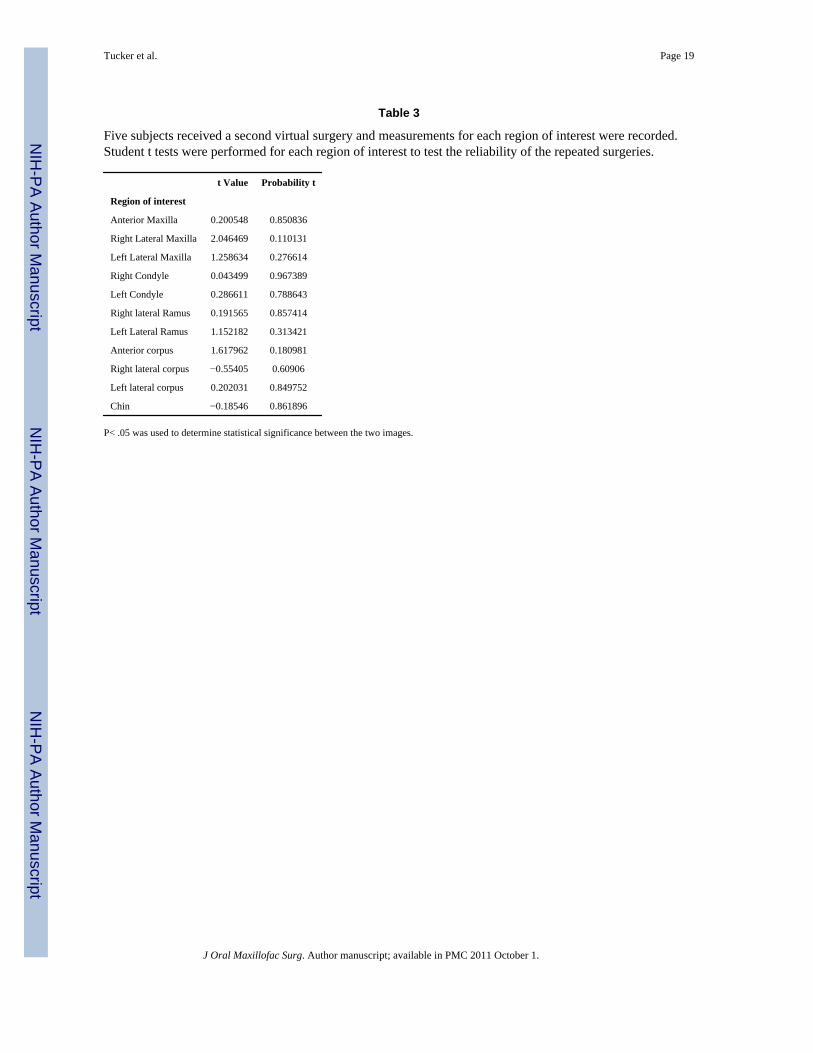
Table 3
Five subjects received a second virtual surgery and measurements for each region of interest were recorded.Student t tests were performed for each region of interest to test the reliability of the repeated surgeries.
t Value Probability t
Region of interest
Anterior Maxilla 0.200548 0.850836
Right Lateral Maxilla 2.046469 0.110131
Left Lateral Maxilla 1.258634 0.276614
Right Condyle 0.043499 0.967389
Left Condyle 0.286611 0.788643
Right lateral Ramus 0.191565 0.857414
Left Lateral Ramus 1.152182 0.313421
Anterior corpus 1.617962 0.180981
Right lateral corpus −0.55405 0.60906
Left lateral corpus 0.202031 0.849752
Chin −0.18546 0.861896
P< .05 was used to determine statistical significance between the two images.
J Oral Maxillofac Surg. Author manuscript; available in PMC 2011 October 1.