Surgical & Cosmetic Dermatology
93
Surgical & Cosmetic Dermatology Publicação Oficial da Sociedade Brasileira de Dermatologia Publicação Trimestral www.surgicalcosmetic.org.br PERIODICIDADE TRIMESTRAL EDITORA-CHEFE Bogdana Victoria Kadunc Pontifícia Universidade Católica de Campinas - PUC - Hospital do Servidor Público Municipal – São Paulo (SP), Brasil. CO-EDITORES Hamilton Stolf Faculdade de Medicina de Botucatu da Universidade Estadual Paulista – Botucatu (SP), Brasil. Mônica Azulay Santa Casa da Misericórdia do Rio de Janeiro - Rio de Janeiro (RJ), Brasil. Surg Cosmet Dermatol. | Rio de Janeiro | v. 7 | n2. | p.89-189 | Abr/Maio/Jun. 2015
-
Upload
khangminh22 -
Category
Documents
-
view
0 -
download
0
Transcript of Surgical & Cosmetic Dermatology

Surgical& Cosmetic Dermatology
Publicação Oficial da Sociedade Brasileira de DermatologiaPublicação Trimestral
www.surgicalcosmetic.org.br
Periodicidade TrimesTral
EDITORA-CHEFE Bogdana Victoria Kadunc
Pontifícia Universidade Católica de Campinas - PUC - Hospital do Servidor Público Municipal – São Paulo (SP), Brasil.
CO-EDITORES Hamilton Stolf
Faculdade de Medicina de Botucatu da Universidade Estadual Paulista – Botucatu (SP), Brasil.
Mônica Azulay Santa Casa da Misericórdia do Rio de Janeiro - Rio de Janeiro (RJ), Brasil.
Surg Cosmet Dermatol. | Rio de Janeiro | v. 7 | n2. | p.89-189 | Abr/Maio/Jun. 2015

90
Sociedade Brasileira de DermatologiaAfiliada à Associação Médica Brasileira
www.sbd.org.br
SUR GI CAL & COS ME TIC DER MA TO LOGYPublicação Oficial da Sociedade Brasileira de DermatologiaOfficial Publication of Brazilian Society of DermatologyPublicação Trimestral (Quarterly Edition)ISSN 1984-5510 l Abril - Junho 2015 l Volume 7 l Número 2
Surgical & Cosmetic Dermatology
Diretoria Executiva
PresidenteGabriel Gontijo | MG
Vice-pre si den teJayme de Oliveira Filho | SP
TesoureiraLeninha Valério do Nascimento | RJ
Secretária GeralLeandra Metsavaht | RJ
1ª SecretáriaFlávia Alvim Sant’Anna Addor | SP
2o SecretáriaOswaldo Delfini Filho | SP
Diretora de BibliotecaAna Paula Meski | SP
Editores
Editora-chefe: Bogdana Victoria KaduncPontificia Universidade Católica De Campinas -PUC /Hospital do Servidor Público Municipal – São Paulo (SP), Brasil.Co-editores: Hamilton Stolf Faculdade de Medicina de Botucatu da Universidade Estadual Paulista – Botucatu (SP), Brasil.
Mônica Azulay Santa Casa da Misericórdia do Rio de Janeiro - Rio de Janeiro (RJ), Brasil.
Editores assistentes
Ada Trindade AlmeidaHospital do Servidor Público Municipal - São Paulo (SP), Brasil.
Alcidarta dos Reis GadelhaFaculdade de Medicina da Universidade Estadual da Amazônia - Manaus (AM), Brasil.
Fabiane Mulinari-BrennerUniversidade Federal do Paraná e Serviço de Dermatologia do Hospital de Clínicas de Curitiba – Curitiba (PR), Brasil.
Gisele Gargantini RezzeDepartamento de Oncologia Cutânea do Hospital A. C. Camar-go – São Paulo (SP), Brasil.
Lauro Lourival Lopes Filho Universidade Federal do Piauí – Teresina (PI), Brasil.
Nilton Di ChiacchioHospital do Servidor Público Municipal – São Paulo (SP), Brasil.
Samira YarakUniversidade Federal do Vale do São Francisco –
Petrolina (PE), Brasil.
Surg Cosmet Dermatol 2015;7(2):90-2.

91
Surgical & Cosmetic Dermatology
Conselho Nacional de Revisores
Adilson CostaEmory University School of Medicine - Atlanta/GA, USA.
Ana Maria Costa PinheiroUniversidade de Brasília - Brasília (DF), Brasil.
Caio César Silva de CastroSanta Casa de Misericórdia de Curitiba –Curitiba (PR), Brasil.
Carlos Baptista BarcauiSanta Casa da Misericórdia do Rio de Janeiro - Rio de Janeiro (RJ), Brasil.
Carlos MachadoFaculdade de Medicina do ABC - São Paulo (SP), Brasil.
Celia KalilSanta Casa de Misericórdia de Porto Alegre - Porto Alegre (RS), Brasil.
Cleide IshidaUniversidade Federal do Rio de Janeiro - Rio de Janeiro (RJ), Brasil.
Denise SteinerFaculdade de Medicina de Mogi das Cruzes – São Paulo (SP), Brasil.
Diego Leonardo BetFaculdade de Medicina da Unversidade de São Paulo – São Paulo (SP), Brasil.
Ediléia BagatinUniversidade Federal de São Paulo – São Paulo (SP), Brasil.
Emerson Vasconcelos de Andrade LimaUniversidade Federal de Pernambuco(UFPE) e Santa Casa de Misericórdia do Recife - Recife (PE), Brasil.
Emmanuel FrançaUniversidade de Pernambuco - Recife (PE), Brasil.
Fernanda RazeraUniversidade Federal de Ciências da Saúde de Porto Alegre - Porto Alegre (RS), Brasil.
Francisco M. PaschoalFaculdade de Medicina do ABC – São Paulo (SP), Brasil.
Gabriel GontijoUniversidade Federal de Minas Gerais – Belo Horizonte (MG), Brasil.
Heitor de Sá GonçalvesSecretaria de Saúde do Estado do Ceará – Fortaleza (CE), Brasil.
Hermênio C. LimaUniversidade Federal do Paraná - Curitiba (PR), Brasil.
Hiram Larangeira de Almeida Jr.Universidade Católica de Pelotas (RS), Brasil.
Humberto PonzioUniversidade Federal do Rio Grande do Sul – Porto Alegre (RS), Brasil.
Iphis CampbellFaculdade de Medicina da Universidade do Planalto Central – Brasília (DF), Brasil.
Izelda Carvalho CostaUniversidade de Brasília - Brasília (DF), Brasil.
Juliano Villaverde SchmidtHospital Universitário Evangélico de Curitiba (PR), Brasil.
Lia Cândida Miranda de CastroUniversidade Federal de Goiás – Goiânia (GO), Brasil.
Luis AntonioTorezanUniversidade de São Paulo - São Paulo (SP), Brasil.
Luis Fernando KopkeClínica privada (SC), Brasil.
Marcia MonteiroFaculdade de Medicina de Mogi das Cruzes – São Paulo (SP), Brasil.
Marcia Ramos e SilvaHospital Universitário Clementino Fraga Filho (UFRJ) – Rio de Janeiro (RJ), Brasil. Marcus MaiaFaculdade de Ciências Médicas da Santa Casa de São Paulo – São Paulo (SP), Brasil.
Maria Claudia IssaUniversidade Federal Fluminense – Rio de Janeiro (RJ), Brasil.
Maria Fernanda GavazzoniSanta Casa da Misericórdia do Rio de Janeiro - Rio de Janeiro (RJ), Brasil.
Mauro EnokiharaUniversidade Federal de São Paulo – São Paulo (SP), Brasil.
Miriam SottoUniversidade de São Paulo – São Paulo (SP), Brasil.
Nilton NasserUniversidade Regional de Blumenau - Blumenau (PR), Brasil.
Oleg Iosifovich D. M. SabatovichUniversidade Federal do Rio de Janeiro – Rio de Janeiro (RJ), Brasil.
Omar LupiUniversidade Federal do Rio de Janeiro – Rio de Janeiro (RJ), Brasil.
Paulo Ricardo CriadoUniversidade de São Paulo – São Paulo (SP), Brasil.
Roberto Gomes TarléServiço de Dermatologia Santa Casa de Curitiba – Curitiba (PR), Brasil.
Rossana Ruth G.V. GonçalvesUniversidade Federal do Pará – Belém (PA), Brasil.
Sarita BezerraUniversidade Federal de Pernambuco – Recife (PE), Brasil.
Selma CerneaHospital do Servidor Público Municipal de São Paulo – São Pau-lo (SP), Brasil.
Tânia CestariUniversidade Federal do Rio Grande do Sul – Porto Alegre (RS), Brasil.
Conselho Internacional de Revisores
Alastair CarruthersUniversity of British Columbia - Canada
AntonelaTostiUniversitàdi Bologna, - Italy
Antonio PicotoCentro de Dermatologia Medico-Cirurgica - Portugal
Dee Anna GlaserSt. Louis University Hospital - USA
Eckart HanekeDepartment of Dermatology University of Witten / Herdecke Health Center Academic Teaching Hospital of the University of Diisseldolf - Germany
Ellen MarmurDivision of Dermatologic and Cosmetic Surgery and Assistant Clinical - USA
Enrique Hernandez Perez Centro de Dermatología y Cirugía Cosmética (CDCC) - San Salvador
Henry RandleSaint Luke’s Hospital – USA
Jean CarruthersUniversity of British Columbia - Canada
Jerry BrewerUniversity of South Carolina - USA
John A. ZitelliUniversity of Pittsburgh Medical Center - USA
Jorge Ocampo CandianiServicio de Dermatologíadel Hospital Universitario dr. José Eleute-rio González – Mexico
Leslie Baumann Director of the Baumann Cosmetic and Research Institute in Miami Beach – USA
Mercedes Florez Universityof Miami - USA
Miguel Sanchez VieraHospital Universitario “Gregorio Marañon”- Spain
Robert BaranHead of the Nail Disease Center in Cannes – France Rompel Rainer Department of Dermatology, Clinic Kassel – Germany
Rompel Rainer Department of Dermatology, Clinic Kassel - Germany
WillianHankeDepartment of Dermatology, Saint Vincent Carmel Medical Cen-ter, Laser & Skin Surgery Center of Indiana - USA
Zoe Diana DraelosWake Forest University School of Medicine Winston-Salem - North Carolina – USA
Surg Cosmet Dermatol 2015;7(2):90-2.

92
A/C SURGICAL & COSMETIC DERMATOLOGY
Av. Rio Branco, 39 18º andarCep: 20.090-003Rio de Janeiro-RJ, Brasil.Fone: 55 (21) 2253-6747web si te: www.surgicalcosmetic.org.br
A Surgical & Cosmetic Dermatology é uma publi ca ção ofi cial da SociedadeBrasileira de Dermatologia (SBD) em par ce ria com a Sociedade Bra-sileira de Cirurgia Dermatológica. O con teú do téc ni co-cien tí fi co apre-sen ta do nesta publi ca ção é de co-pro prie da de da Sociedade Brasileira de Dermatologia.
Editada por: Sociedade Brasileira de Dermatologia.
Infor ma ções sobre a Assi na tu ra da Surgical & Cosmetic Dermatology podem ser encon tra das no site www.sur gi cal cos me tic.org.br
PERIODICIDADE TRIMESTRAL
Assistentes editoriAis
Nazareno Nogueira de SouzaBruno Abraão de SouzaRosalynn Leite
BiBliotecáriAs
Rosalynn LeiteVanessa Zampier
AssinAturAs R$ 250,00 e $180 dólares
Informações de pagamento no site:www.surgicalcosmetic.org.br
©2015 Sociedade Brasileira de Dermatologia.RJ: Tel./Fax:21 2253-6747 E-mail: [email protected]: www.sbd.org.br
Os anún cios vei cu la dos nesta edi ção são de exclu si va res pon sa bi li da de dos anun-cian tes, assim como os con cei tos emi ti dos em arti gos assi na dos são de exclu si-va res pon sa bi li da de de seus auto res, não refle tin do neces sa ria men te a opi nião da SBD.
Todos os direi tos reser va dos e pro te gi dos pela lei 9.610 de 19/02/98. Nenhuma parte dessa publi ca ção pode rá ser repro du zi da sem auto ri za ção pré via por escri to da Sociedade Brasileira de Dermatologia, sejam quais forem os meios empre ga-dos: ele trô ni co, mecâ ni co, foto grá fi co, gra va ção ou quais quer outros.
Material de dis tri bui ção à clas se médi ca.
A revis ta cons ta no Depósito Legal, na Biblioteca Nacional, de acor do com o Decreto nº 1.825, de 20 de dezem bro de 1907.
Indexações
n Sumários. org (www.sumarios.org/)
n Directory of Open Access Journals - DOAJ (http://www.doaj.org)
n Latindex (www.latindex.org)
n Lilacs (http://bases.bireme.br/)
n SCOPUS (http://www.scopus.com/home.url)
n Periódica (http://periodica.unam.mx)
n Redalyc (http://www.redalyc.org)
Surg Cosmet Dermatol 2015;7(2):90-2.

93
Surg Cosmet Dermatol 2015;7(2):93-4.
INST RUÇÕES AOS AUTORESA Surgical & Cosmetic Dermatology, editada em 2009, constitui publicação
médica destinada a difundir conhecimento e experiência nas áreas de Cirurgia Der-
matologica, Cosmiatria e Procedimentos Dermatológicos Diagnósticos e Terapêu-
ticos utilizando novas Tecnologias. É uma publicação trimestral da Sociedade Bra-
sileira de Dermatologia que conta com o apoio científico da Sociedade Brasileira
de Cirurgia Dermatológica e do Colégio Íbero Latino de Dermatologia, que baseia
sua política ética e editorial nas regras emitidas pelo The International Committee
of Medical Journal Editors (www.icmje.org). Os manuscritos devem estar de acor-
do com os padrões editoriais para artigos submetidos a periódicos biomédicos es-
tabelecidos na Convenção de Vancouver (Requisitos Uniformes para Manuscritos
Submetidos a Revistas Biomédicas), regras para relatos de ensaios clínicos e revisões
sistemáticas (metanálises).
Serão produzidos exemplares impressos da versão em língua portuguesa, com
resumos e títulos em inglês. A versão da língua inglesa estará disponível no website
da SBD.
Todos os artigos propostos à publicação serão previamente submetidos à revisão
anônima e confidencial de no mínimo dois membros do Conselho Editorial ou dos
Conselhos Nacional e Internacional de Revisores. Quando aceitos, estarão sujeitos a
pequenas correções ou modificações que não alterem o estilo do autor.
As pesquisas em seres humanos devem ter a prévia aprovação de um Comitê
de Ética em Pesquisa e obedecer aos padrões éticos da Declaração de Helsinki de
1975, revista em 2000.
ORIENTAÇÕES PARA O PREPARO DOS ARTIGOS
A preparação correta do manuscrito torna os processos de revisão e publicação
mais eficientes. Assim, recomendamos alguns cuidados que podem facilitar significati-
vamente a preparação dos manuscritos.
1- Os artigos devem ser originais e redigidos no idioma de origem do autor
(português, espanhol ou inglês): a equipe editorial providenciará as versões necessárias.
2- O título do trabalho deve ser curto e conciso, informado em português e
inglês, com até 150 caracteres sem espaços, acompanhado de um título resumido.
3- Os resumos em português e inglês devem acompanhar o formato adequado
ao tipo de artigo.
4- Os autores devem informar o nome com suas abreviaturas, a titulação máxi-
ma, as instituições aos quais estão vinculados e local de realização do trabalho. Um de-
les deve ser designado como autor correspondente, com endereço completo, números
de telefone comercial e fax e endereço de e-mail.
5- Os autores devem informar se houve conflitos de interesse e suporte fi-
nanceiro.
6- As palavras-chave devem ser citadas em português e em inglês (Keywords),
totalizando 3 a 10 por idioma, devendo ser incluídas em todos os tipos de artigos. Estas
palavras deverão estar contidas no DeCS (Descritores em Ciências da Saúde) e/ou MeSH
(Medical Subject Headings) que podem ser acessados na internet.
7- O número limite de palavras para os textos deve ser obedecido segundo o
tipo de artigo, e computado excluindo as REFERENCES e os resumos em portu-
guês e inglês.
8- Abreviaturas e acrônimos devem ser limitados aos de uso geral, não devendo
constar no título ou no resumo.
9- Devem ser evitadas informações introdutórias extensas e repetitivas, dan-
do-se preferência às mais recentes, ainda não publicadas. Evite textos com repetição
da mesma informação no resumo, introdução e discussão.
10- Pesos e medidas devem ser expressos no sistema métrico decimal, e tem-
peraturas em graus centígrados.
11- Drogas devem ser mencionadas por seus nomes genéricos, seguidos da
dosagem e posologia empregadas, evitando-se a citação de termos comerciais ou mar-
cas. Descrições de quaisquer equipamentos, instrumentos, testes e reagentes devem
conter o nome do fabricante e o local de fabricação.
12- Após a sequência de itens para cada tipo de trabalho podem se acrescenta-
dos agradecimentos, antes das REFERENCES bibliográficas.
13- As REFERENCES bibliográficas devem ser listadas nas últimas páginas do
artigo, e numeradas de acordo com a citação no texto (em ordem numérica seqüen-
cial), seguindo o estilo Vancouver, como indicado pelo International Committee of
Medical Journal Editors (ICMJE). REFERENCES citadas em legendas de tabelas e
figuras devem manter a seqüência com as citações no texto. Todos os autores devem
ser citados se forem até seis; acima disso, devem ser mencionados os seis primeiros e
“et al.”. Seguem-se exemplos dos tipos mais comuns de REFERENCES. Exemplos
de citações no texto retirados do ICMJE:
13A. Artigo em periódico:
Hallal AH, Amortegui JD, Jeroukhimov IM, Casillas J, Schulman CI, Manning
RJ, et al. Magnetic resonance cholangiopancreatography accurately detects common
bile duct stones in resolving gallstone pancreatitis. J Am Coll Surg. 2005;200(6):869-75.
13B. Capítulo de livro:
Reppert SM. Circadian rhythms: basic aspects and pediatric implications. In:
Styne DM, Brook CGD, editors. Current concepts in pediatric endocrinology. New
York: Elsevier; 1987. p .91-125.
13C. Texto na Internet:
Ex. com autor indicado:
Fugh-Berman A. PharmedOUT [Internet]. Washington: Georgetown Univer-
sity, Department of Physiology and Biophysics; c2006 [cited 2007 Mar 23]. Available
from: http://www.pharmedout.org/.
Ex. quando o autor é uma organização:
International Union of Biochemistry and Molecular Biology. Recommen-
dations on Biochemical & Organic Nomenclature, Symbols & Terminology etc.
[Internet]. London: University of London, Queen Mary, Department of Chemistry;
[updated 2006 Jul 24; cited 2007 Feb 22]. Available from: http://www.chem.qmul.
ac.uk/iubmb/.
13D. Apresentação prévia em eventos:
Bruhat M, Silva Carvalho JL, Campo R, Fradique A, Dequesne J, Setubal A,
editors. Proceedings of the 10th Congress of the European Society for Gynaecologi-
cal Endoscopy; 2001 Nov 22-24; Lisbon, Portugal. Bologna (Italy): Monduzzi Editore,
International Proceedings Division; c2001. 474 p.
14- Ilustrações (figuras, quadros, gráficos e tabelas) devem ser referidas em
ordem numérica sequencial no texto em números arábicos (exemplo: Figura 3, Gráf-
ico 7), cabendo ao Editor suprimir as redundantes. As legendas das figuras e gráficos
e os títulos e notas de rodapé das tabelas devem descrever precisamente seu conteúdo
com frases curtas, porém suficientes para a compreensão ainda que o artigo não seja
totalmente lido.
15- As figuras deverão ter resolução mínima de 300 DPI, largura mínima
de 1.200 pixels com altura proporcional, e serem gravadas nos formatos JPG ou TIF.
Podem ser colocadas setas ou linhas para localizar as áreas de interesse. As legendas das
imagens histológicas devem especificar a coloração e o aumento. Se uma figura já foi
publicada anteriormente, deverá citar a fonte original abaixo da mesma e constar nas
REFERENCES. Deverão enviar à revista a permissão do detentor dos direitos auto-
rais para a sua reprodução. No uso de figuras que identifiquem a face de pacientes será
preciso autorização por escrito para divulgação (ver no site da revista o documento
Autorização para uso de fotografias).

Surg Cosmet Dermatol 2015;7(2):93-4.
16-Quanto aos vídeos é necessário inserir legendas contendo informações
como título do manuscrito, autoria, instituição e outros comentários pertinentes. No
uso de imagens de pacientes, a identidade deverá ser resguardada, do contrário, será
preciso anexar-lhes permissão por escrito para divulgação.
17-Os gráficos deverão ser elaborados em Microsoft Excel. As tabelas dispens-
am sua descrição no texto tendo a finalidade de suplementá-lo e não a de aumentá-lo.
As unidades utilizadas para exprimir os resultados (m, g, g/100, mL etc.) figurarão no
alto de cada coluna. Os pacientes devem ser identificados por números ou letras, e
nunca pelos nomes, iniciais ou número de registro hospitalar.
18- O limite máximo de autores aceitável é de cinco, só haverá exceção para
trabalhos de maior complexidade (ex. Artigo Original, Revisão, EMC) mediante jus-
tificativa e aprovação dos editores.
19-As opiniões e declarações contidas na revista são de responsabilidade única
e exclusiva de seus autores, não sendo, necessariamente, coincidentes com as da Equi-
pe Editorial, do Conselho de Revisores ou da Sociedade Brasileira de Dermatologia.
Os autores deverão submeter seu manuscrito para avaliação do Conselho Ed-
itorial da revista no endereço eletrônico que se segue: http://www.sgponline.com.
br/scd/sgp/
Todos os documentos como Consentimento de uso para publicação (Copy-
right), Conflito de interesses e Autorização para publicação de fotografias estão dis-
poníveis no site da revista e no sistema de submissão online. Esses documentos devem
ser assinados e encaminhados obrigatoriamente por carta logo após a submissão do
manuscrito para o endereço abaixo:
A/C Surgical & Cosmetic Dermatology Av. Rio Branco, nº 39, 18º
andar - Rio de Janeiro – RJ, Brasil. CEP: 20090-003.
A revista aceita trabalhos inéditos e não publicados das seguintes categorias:
1- ARTIGO ORIGINAL
É o relato de uma pesquisa investigativa original clínico-cosmiátrica ou rela-
cionada a procedimentos na área de Dermatologia. Exemplos: estudos experimentais,
estudos clínicos, comparações e descrições de técnicas ou de métodos de avaliação,
estudos de áreas afins (ex: estudos farmacêuticos em cosmiatria).
Resumo: deverá conter no máximo 200 palavras e ser estruturado seguindo
os itens: Introdução, Objetivo, Métodos, Resultados e Conclusões. Não é permitido
afirmar que os resultados ou outros dados serão apresentados ou discutidos.
O texto deverá conter até 4000 palavras, 10 ilustrações e 35 REFERENCES
e seguir o formato IMRDC (Introdução e objetivo, Métodos, Resultados, Discussão,
Conclusão)
Introdução: citar as razões que motivaram o estudo, descrevendo o estado atual
do conhecimento sobre o tema. Utilizar o último parágrafo para especificar a principal
pergunta ou objetivo do estudo, e a principal hipótese testada, se houver.
Métodos: Explicar como o estudo foi feito:
a- Tipo de estudo: descrever o seu desenho especificando a direção temporal
(retrospectivo ou prospectivo), o tipo de randomização quando utilizada (pareamen-
to, sorteio, sequenciamento, etc), se o estudo foi cego, comparativo, controlado por
placebo, etc.
b- Local: indicar onde o estudo foi realizado (instituição privada ou pública), citar
que a pesquisa foi aprovada pelo Comitê de Ética em Pesquisa de sua instituição, os pro-
cedimentos de seleção, os critérios de inclusão e exclusão, e o número inicial de pacientes.
c- Procedimentos: descrever as principais características das intervenções re-
alizadas, detalhando a técnica e lembrando que o estudo de investigação deverá ser
reprodutível.
d- Descrição dos métodos utilizados para avaliação dos resultados.
e- Inclusão da análise estatística descritiva e/ou comparativa com descrição
do planejamento da amostra (representativa do universo a ser estudado), a análise e
os testes estatísticos e apresentação dos níveis de significância adotados. A utilização
de análises estatísticas não usuais é incentivada, porém neste caso, deve-se fazer uma
descrição mais detalhada da mesma.
Resultados: descrever os principais resultados que devem ser acompanhados
de estimativas pontuais e medidas de dispersão (p.ex., média e erro padrão) ou de
estimativas intervalares (p.ex., intervalos de confiança), bem como os níveis descritivos
dos testes estatísticos utilizados (p.ex. “p-value”). Esses achados também devem ser
interpretados sob o ponto de vista clínico.
Discussão: enfatizar os novos e importantes resultados encontrados pelo estudo
e que farão parte da conclusão. Relatar observações de outros estudos relevantes. Men-
cionar as limitações dos achados e as implicações para pesquisas futuras.
Conclusões: devem ser concisas e responder apenas aos objetivos propostos. A
mesma ênfase deve ser dada para estudos com resultados positivos ou negativos.
2- COMUNICAÇÕES
Artigos originais, breves, abordando resultados preliminares de novos achados
de interesse para a Cirurgia Dermatológica, Cosmiatria ou Oncologia cutânea entre
outros. Texto com formatação semelhante ao artigo original, resumo estruturado de
até 200 palavras. Limite: texto até 2000 palavras, 8 ilustrações e 15 REFERENCES.
3- ARTIGOS DE REVISÃO
Poderão ser abordados temas cirúrgicos ou de cosmiatria, procedimentos, al-
goritmos , compilações, estatísticas. Estes trabalhos têm formato livre, porem devem
conter resumo não estruturado de até 100 palavras e conclusões ou considerações
finais. Limite: texto até 6000 palavras, 10 ilustrações e 60 REFERENCES. Os artigos
de revisão sistemática ou metanálises devem seguir orientações pertinentes (http://
cochrane.bireme.br)
4- EDUCAÇÃO MÉDICA CONTINUADA
Publicação de cunho educacional, abordando profunda e completamente
grandes temas de Cirurgia Dermatológica, Cosmiatria ou Laser. Deve conter resumo
não estruturado de até 100 palavras. Limite: texto até 4000 palavras, 10 ilustrações e
40 REFERENCES. Para evitar duplicações, os autores devem comunicar o tema aos
editores antes de escrever o artigo.
Os autores são solicitados a definir objetivos educativos para o artigo que trans-
mitam o que o participante deve ter absorvido após completar a atividade de EMC (ex:
identificar uma condição, conhecer seus tratamentos, selecionar a melhor técnica). O
entendimento destes objetivos devem ser mensurados por meio de 10 perguntas com
respostas em 5 alternativas, cujo gabarito deve também ser enviado.
5- NOVAS TÉCNICAS
Descrição de novas técnicas ou detalhes de técnicas. Resumo não estruturado
de até 100 palavras, introdução com revisão de literatura, métodos, resultados, dis-
cussão e conclusão. Limite: 1200 palavras, 8 ilustrações e 30 REFERENCES.
6- DIAGNÓSTICO POR IMAGEM
Imagens de dermatoscopia, microscopia confocal, ultrassom e outros métodos,
aplicadas à cirurgia dermatológica e cosmiatria, acompanhadas de curta descrição.
Resumo não estruturado de até 100 palavras,texto até 1200 palavras, 8 ilustrações e
10 REFERENCES.
7 - RELATO DE CASO
Descrição de casos ou serie de casos de particular interesse nas áreas de Ciru-
rgia Dermatológica, Oncologia Cutânea, Cosmiatria, Tratamento de dermatoses in-
estéticas, Complicações, etc.
Resumo não estruturado de até 100 palavras, introdução com revisão de litera-
tura, métodos, resultados, discussão e conclusão, sempre que pertinentes. Limite: texto
até 1200 palavras, 8 ilustrações e 30 REFERENCES.
8- CARTAS
Comentários objetivos e construtivos sobre matérias publicadas. Texto até 600
palavras, e no máximo 5 REFERENCES.
94

95
Surg Cosmet Dermatol 2015;7(2):95-6.
Surgical & Cosmetic Dermatology Sumário / Table of contentsPublicação Oficial da Sociedade Brasileira de DermatologiaABRIL/MAIO/JUNHO 2015 l Volume 7 l Número 2ISSN:1984-5510
Editorial / editorial Bogdana Victoria Kadunc
Educação Médica Continuada / Continuing Medical Education Fisiopatologia da lipodistrofia ginoide 98 Physiophatology of gynoid lipodystrophy
Marisa Gonzaga da Cunha, Ana Lucia Gonzaga da Cunha Carlos A. Machado
Artigos Originais / Original Articles Melanoma maligno: estudo epidemiológico dos casos diagnosticados em unidade de referência em dermatologia 104 em Bauru-sp de 2007 a 2014
Malignant melanoma: epidemiological study of cases diagnosed at a dermatological reference center in the city
of Bauru, in the Brazilian southeast State of São Paulo, between 2007 and 2014
Ana Cecília Versiani Duarte Pinto, Maria Lopes Lamenha Lins Cavalcante, Gardênia Viana da Silva, Fernanda Freitas de Brito,
Agnes Mayumi Nakano Oliveira, Norma Gondim Cleto
Enxerto de cartilagem auricular para reconstrução nasal após cirurgia micrográfica de Mohs 109 Auricular cartilage graft for nasal reconstruction after Mohs micrographic surgery
Felipe Bochnia Cerci
Influência de um suplemento nutricional com peptídeos de colágeno nas propriedades da derme 116 Influence of a nutritional supplement containing collagen peptides on the properties of the dermis
Flávia Alvim Sant’Anna Addor
Avaliação in vitro da eficácia anti-inflamatória, protetora da barreira cutânea e redutora da hipersensibilidade 123 cutânea de quatro sabonetes líquidos disponíveis no Brasil
In vitro evaluation of four commercially available liquid soaps (in Brazil) for their anti-inflammatory and
protective skin barrier qualities, as well as their impact on the reduction of cutaneous hypersensitivity
Adilson Costa, Samara Eberlin, Stefano Piatto Clerici, Beatrice Martinez Zugaib Abdalla
A aplicação do plasma rico em plaquetas no tratamento da alopecia androgenética 130 The application of platelet-rich plasma in the treatment of androgenic alopecia
Rossana Cantanhede Farias de Vasconcelos, Karla Azuaga, Géssica Cantadori Funes Arenas, João Guilherme Finizola de Vasconcelos, Natalie Schnaider Borelli
Os efeitos clínicos e histológicos doS ultrassoNS de alta frequência minimamente focadoS no tecido subcutâneo humano 138 Clinical and histological effects of weakly focused high-frequency ultrasounds on human subcutaneous
adipose tissue
Daniele Bani, Alessandro Quattrini Li, Giulia Lo Russo
Tratamento das cicatrizes de acne com a técnica de microagulhamento e drug delivery 144 Treatment of acne scars using the microneedling and drug delivery technique
Célia Luiza Petersen Vitello Kalil, Renata Hübner Frainer, Letícia Santos Dexheimer, Renata Elise Tonoli, Ana Letícia Boff

96
Surg Cosmet Dermatol 2015;7(2):95-6.
Artigo de Revisão / Review article Laserlipólise na região cervical 149 Laserlipolysis in the cervical region
Sandra Tagliolatto, Oriete Gerin Leite
Diagnóstico por imagem / Diagnostic imaging Ultrassom de alta frequência (22mhz) na diferenciação entre hidrocistoma e carcinoma basocelular 159 High frequency ultrasound (22MHz) in the differentiation between hidrocystoma and basal cell carcinoma
Elisa de Oliveira Barcaui, Antonio Carlos Pires Carvalho, Paulo Marcos Valiante, Juan Piñeiro-Maceira, Carlos Baptista Barcaui
Novas Técnicas / New Techniques Zetaplastia como alternativa para fechamento de defeito cirúrgico duplo 162 Zetaplasty as an alternative for reconstructing double surgical defects
Airton dos Santos Gon, Fernanda Mendes Araújo
Relatos de Caso / Case Reports Abordagem do carcinoma espinocelular de alto risco no couro cabeludo: série de casos 166 Surgical management of high-risk squamous cell carcinoma of the scalp: series of cases
Lorena Rodrigues Teixeira E. Silva, Ana Maria Quinteiro Ribeiro, Luiz Fernando Fróes Fleury Júnior
Espiroadenoma écrino - relato de caso 171 Eccrine spiradenoma: a case report
Rodolfo Chedid, Janaina Nagel, Taynara de Mattos Barreto, Glaura Tinoco Plata, Anna Luiza Bento Dutra Thales Pereira de Azevedo
Retalho em ilha tunelizado após exérese de carcinoma na face 175 Tunneling island pedicled flap after resection of carcinoma on the face
Alexandre Sabino Sisnando. Luana Oliveira Ramos, Fabio Francesconi
Siringoma condroide benigno simulando carcinoma basocelular 180 Benign chondroid syringoma simulating a basal cell carcinoma
Jeane Jeong Hoon Yang, Antônio José Tebcherani, Ed Wilson Tsuneo Rossoe
Melanoma Desmoplásico - um desafio diagnóstico 184 Desmoplastic melanoma: a diagnostic challenge
Flávia Regina Ferreira, Bruna Ferrari, Livia Mendes Sabia Acedo, Juliana Emi Dias Ujihara,
Marcia Lanzoni de Alvarenga Lira, Samuel Henrique Mandelbaum
Sumário / Table of contents

Surg Cosmet Dermatol 2015;7(2):97.
Editorial
97
Dear SBD members,In its 7th year of publication, the Brazilian Society of Dermatology’s journal,
Surgical & Cosmetic Dermatology, is firmly striding towards strict compliance with the requirements of the discipline’s literature databases.
The most recent and necessary accomplishment was the implementation of the DOI (Digital Object Identifier) system for our articles.
When browsing through scientific papers, you may notice – in general in the area reserved for information about the issue, authors, institutions, publishers, and agen-cies – the abbreviation “DOI” followed by a long sequence of characters, as seen in the examples below:
Dermatologic Surgery - Dermatol Surg 2012; 38:1283DOI: 10.1111/j.1524-4725.2012.02359.xBritish Journal of Dermatology - Br J Dermatol 2009;160:1022-1025DOI 10.1111/j.1365-2133.2009.09066.xClinics in Plastic Surgery – Clin Plastic Surg 2015;42:33-50DOI 10.1016/j.cps.2014.08.007
What is the meaning and the importance of this DOI acronym?Simply put, it can be defined as an identifier of publications on digital networks.
It is a single credential, for individual and permanent identification of a scientific article. It can be used for the purpose of referring to, or retrieval of, the article in the virtual environment.
The International DOI Foundation (IDF), a nonprofit institution founded by the Association of American Publishers, and directed by editors, publishers, software compa-nies and organizations representing the interests of copyright holders, is responsible for the DOI, which can be applied to any form of intellectual property that is manifested in a digital medium.
It consists of a prefix (10 + number assigned to various groups – editors, pu-blishers etc) and a suffix (whose specific and unique sequence of characters identifies each article with a technology that allows it to become “clickable”).
The DOI also facilitates the filling in of scientific information in the Lattes cur-riculum system. When the researcher inputs the characters, the Lattes Platform accesses the DOI database and automatically fills in the title, year, volume, issue, pages and the name of the first author of the publication. This record lends reliability to the registered information.
Thus, in addition to increasing exposure for the journal and its collaborators, it is now possible to assure authors that the digital version of their scientific information will be directed and disseminated to a wide and diversified universe of users, with a fully secure, facilitated search process.
The journal Surgical & Cosmetic Dermatology celebrates this new achievement and would like to thank the SBD’s Board of Directors for its unconditional support, and also compliment our editorial assistants and librarians for their expertise.
Dra. Bogdana Victoria KaduncScientific EditorSurgical & Cosmetic Dermatology
Implementing the DOI (Digital Object Identifier) system for Surgical & Cosmetic Dermatology articles:
DOI: http://dx.doi.org/10.5935/scd1984-8773.2015720

Surg Cosmet Dermatol 2015;7(2):98-103.
ContinuingMedicalEducation
Physiophatology of gynoid lipodystrophyFisiopatologia da lipodistrofia ginoide
DOI: http://dx.doi.org/10.5935/scd1984-8773.2015721
ABSTRACT Cellulite or gynoid lipodystrophy in its various degrees is extremely common in the female
population, setting on between the ages of 15 and 45 years of age, which corresponds to a woman’s reproductive phase. Around 95% of women will present some degree of cellulite at some point in their lives. Despite being highly prevalent, only a few studies have been published in the international literature – many with contradictory conclusions – making it difficult to choose the appropriate treatment regimen.
The present study discusses anatomical differences in the hypodermis of men and women, the complex physiopathology of gynoid lipodystrophy, and mechanisms involved in its de-velopment.
Gynoid lipodystrophy is a skin disorder, which can only be controlled and not fully cured due to the fact that it is not a true disease, but rather a predisposition. Nevertheless, persistent or more advanced cases should be considered pathological, and be treated and controlled, given that they are indicative of peripheral vascular insufficiency.
Keywords: adipocytes; lymphatic system; lipodystrophy
RESU MO A celulite ou lipodistrofia ginoide (LDG) em seus vários graus é extremamente frequente na popu-
lação feminina, com incidência entre 15 e 45 anos, ou seja, na fase reprodutiva da mulher. Cerca de 95% das mulheres apresentarão algum grau de celulite em algum momento da vida. Apesar de ser altamente prevalente, apenas um número reduzido de estudos tem sido publicado na literatura inter-nacional e muito deles com conclusões contraditórias, o que dificulta a escolha do esquema adequado de tratamento.
Neste estudo discutiremos as diferenças anatômicas da hipoderme no homem e na mulher, a complexa fisiopatologia da LDG e os mecanismos envolvidos em seu desenvolvimento.
A LDG é alteração cutânea que só poderá ser controlada e não completamente curada, uma vez que não se trata verdadeiramente de uma doença e sim de uma predisposição. Casos persistentes ou em graus mais avançados, porém, devem ser considerados patológicos, devem ser tratados e controlados, uma vez que são indicativos de insuficiência vascular periférica.
Palavras-chave: adipócitos; lipodistrofia; sistema linfático
INTRODUCTIONDespite having become the norm, the term cellulite is
mistakenly used to describe a regional hypodermic dystrophy almost exclusive to the female population, which can be associated or not with gynoid-type obesity, and which affects the region of the hips, buttocks, lower limbs and, less frequently, the abdomen and the lateral-posterior face of the arms. It has genetic and constitutional factors that predispose an individual to the condition, factors to which multiple complex and interrelated etiologies are
Authors: Marisa Gonzaga da Cunha1 Ana Lucia Gonzaga Cunha2 Carlos A. Machado3
1 PhD candidate, Faculdade de Medicina do ABC (FMABC) – Santo André (SP), Brazil
2 Plastic Surgery Resident Physician, Univer-sidade Federal de São Paulo (UNIFESP) – São Paulo (SP), Brazil
3 Full Professor of Dermatology, FMABC, Brazil
Correspondence: Marisa Gonzaga da Cunha Rua Gonçalo Fernandes, 153 - sala 83 Cep: 09041-410 - Santo André – SP, Brazil E-mail: [email protected]
Received on: 1 March 2015Approved on: 5 May 2015
This study was performed as part of the Der-matology Program at the Faculdade de Me-dicina do ABC (FMABC) – Santo André (SP), Brazil.
Financial support: NoneConflict of interest: None
98

Surg Cosmet Dermatol 2015;7(2):98-103.
Physiophatology of gynoid lipodystrophy 99
part of the equation. Cellulite, or gynoid lipodystrophy (GLD) in its various degrees is extremely common among females, with onset typical between the ages of 15 and 45 years, corresponding to a woman’s reproductive phase. About 95% of women will present some degree of cellulite at some point in life. 1
Despite the clear anatomical distinction between the dermis and hypodermis, the two are structurally and functionally integrated through the network of vessels and nerves and the presence of epidermal appendages. 2 The hypodermis or superficial adipose tissue (SAT) is arranged in vertical compartments, distributed perpendicularly to the skin’s more superficial layers, with a structure similar to a honeycomb, with uniform distribution throughout the tissue. 3 It is formed by fatty lobules interspersed with well-defined fibrous septa (reticula cutis superficialis) composed of elastic and collagen fibers that are oriented perpendicularly towards the surface, strongly anchored to the dermis, and connecting it with the fascia superficialis. These fatty lobules, located between the dermis and the fascia superficialis serve as a passage to vessels and nerves from the subcutaneous tissue, with compartments that are well-vascularized by capillary vessels 4-7 (Figures 1 and 2).
This distribution in septa pattern plays an important role in preserving cell integrity. 2 The fatty lobules are organized into single or multiple layers depending on the fat content and each individual’s SAT thickness. Its thickness increases and decreases according to weight gain during specific situations that determine the formation of GLD. It is important to note that the subcutaneous tissue, or localized fat, is located underneath the skin and that its fatty content is independent of the cells of the hypodermis. 2
GLD is an exclusively female condition due to the anatomical characteristics of the hypodermis in women, in which the lobes are larger and have parallel septa, while in men the fibrous septa are smaller and arranged in oblique planes, with small fat lobules.6 (Figures 3 and 4).
These distinct structures are present in each gender at birth, but due to the hormonal and vascular changes that take place during puberty – when a greater storage of fat and increased interstitial fluid retention occur – the fat lobules increase due to the hypertrophy of adipocytes 1, 7 (Figure 5). Macroscopic anatomical studies of adipose tissue on cadavers has evidenced the arched distribution of women’s adipose conjunctive
Figura 1: Schematic drawing representing the structures of the skin and subcutaneous tissue
Figura 2: Figure 2: Skin MRI, where Number 1 corres-ponds to the dermis, Number 2 corresponds to the small adipose papilla towards the dermis, Number 3 corres-ponds to the hypodermis showing fibrous septa and Numbers 4 and 5 correspond to the subcutaneous cell tissue. Source: Mirrashed F. et al., 2004. 6

100 Cunha MG, Cunha ALG, Machado CA
Surg Cosmet Dermatol 2015;7(2):98-103.
tissue bands within the panniculus, with a protrusion of fat at the dermo-hypodermal interface – which would explain the formation of the skin’s dimpled appearance, with an increased volume of adipocytes. This distribution occurs specifically in women due to the presence of vertical fascial bands. Herniations in the dermis are characteristic of the female anatomy, and their presence was confirmed by high-resolution magnetic resonance in the low-density regions of the dermis 6 (Figures 3 and 4).
PathophysiologySuccess in the treatment of GLD is closely linked to the
understanding of its physiopathology. 7-9 The present article will review the concepts and the etiologic factors involved in its onset and development.
GLD was defined by Merlin as a “segmental or localized lipodystrophy of the subcutaneous connective tissue with respect to the regional venous-lymphatic stasis. This dermo-hypodermosis prefigures a hystangiopathy with a fibroblastic response that precedes the alterations of the capillary-venular segment and is maintained by them.” 1
Under normal conditions, the arterial system joins the venous system through small capillaries, in a way in which blood is never free in the tissues. In this junction area, vessels cease filtering the colorless substance (interstitial fluid) that surrounds all body cells and which contains necessary nutrients to eliminate waste, which will in turn be drained through the venous system. This mechanism is called the Starling principle. The interstitial fluid is not fully reabsorbed, and the remaining amount is collected and drained to the lymph channels. The first alteration of GLD takes place during this lymphatic drainage, which is carried out inefficiently, generating waste.
The connective tissue is then infiltrated by the interstitial fluid and its waste. This happens in the simple congestive phase, which may be transient or permanent. This interstitial edema, in turn, compresses the capillary vessels, hindering the return of circulation and accentuating the stasis and the permeability of the vascular wall, which increases the exudation. A vicious cycle then ensues, leading some authors to state that “cellulitis has the property of producing more cellulitis”.
In a review article, Rossi and Vergnanini described the multifactorial basis for the etiology of GLD. The process would originate with the deterioration of cutaneous vascularization,
Figure 3: Male hypodermis.
Figure 4: Hypodermis of a woman without cellulitis. Note the configura-tion of the fibrous septa and the differences between the genders.
Source: Mirrashed F. et al., 2004. 6
Source: Mirrashed F. et al., 2004. 6
Fonte: Source: Paschoal LHC, et al. 2012.1
Figure 5: Schematic drawing of normal adipose tissue and in GLD.
Normal fat layers Cellulitis
EpidermisDermis
Hipodermis(subcutaneous fat layer)
(Reserve fat layer)

Physiophatology of gynoid lipodystrophy 101
Surg Cosmet Dermatol 2015;7(2):98-103.
particularly in response to alterations in the pre-capillary arteriolar sphincter of the affected areas, along with the deposition of hyper polymerized glycosaminoglycans on the wall of dermal capillaries and between the collagen and elastic fibers. The increase in capillary pressure would lead to increased permeability of venular capillaries and the retention of excess fluid in the dermis, among adipocytes and interlobular septa, causing cellular alterations and tissue hypoxia. 10
The increased lipolytic resistance resulting from hypoxia and increased lipogenesis, the latter caused by the action of estrogen, prolactin, and high-carbohydrate diets, would lead to hypertrophy of adipocytes. The widened adipocytes, along with hypertrophy and hyperplasia of periadipocyte reticular fibers, would form micronodules surrounded by protein fragments that subsequently cause the sclerosis of fibrous septa, leading to the appearance of GLD. The overall effect of this process would be a reduction in the blood flow and lymph drainage. 1
Therefore, it is possible to conclude that GLD is a dystrophic process of complex pathophysiology, with multiple interlinked factors that act through different mechanisms in multiple target elements in the dermal connective and adipose hypodermic tissues, on a genetically predisposed ground, where estrogen is a trigger and which, added to several other contributing endogenous factors and exacerbated by general and local exogenous factors, sets off a slow and progressive cascade reaction.
Due to the complex inter-relationship between the etiological factors that act directly or indirectly in the pathophysiology of GLD, and for a better understanding of this condition, it is convenient to separate the structures of the dermo-hypodermic region into “operating units”, nevertheless bearing in mind that their actions are simultaneous and obey a central integrator command that is effected by different reflex pathways. GLD’s pathophysiology can be explained by the complex and interconnected participation of the four operating units in that tissue. 1
Due to their primary role, fibroblasts – cells responsible for the interstitial matrix unit (IMU) function and which presumably would start the vicious cycle of this condition – should be mentioned. These mesenchymal stem cells, stricken in different harmful ways, primarily alter the turnover of extracellular macromolecules with physico-chemical and structural alterations in glycosaminoglycans and proteoglycans, which constitute the amorphous matrix (fundamental substance, basal membrane, and lining of the cells’ surface) and the fibrillar matrix (collagen and elastin). As a result, changes in the biological functions would take place, which would cause secondary changes in the diffusion of nutrients, metabolites, hormones, and neurotransmitters among the tissue cells, the microcirculatory system (microcirculatory unit - MCU) and the sympathetic nerve endings (neurovegetative/autonomic unit - NVU), influencing the functional properties of neuroreceptors, and thus impairing cellular differentiation, the cell/cell and the cell/
matrix interactions. In this way, the phenomena occurring in the energetic- adipose unit (EAU), with hypertrophy and resistance to the lipolysis of regional adipocytes, are also explained. Furthermore, there are also variations in the pressures of the various tissue compartments with edematous gelloid infiltration (non-mobile) of the interstitial matrix, fibrotic phenomena of interlobular connective trabeculae and finally the vasculopathic and hemodynamic alterations in the microcirculation.
The proliferation and activity of fibroblasts are regulated by the various factors that can cause modifications in the matrix proteoglycans: individual and regional characteristics; age (more cells in the embryonic stage and fewer cells in the senile stage); estrogens (which determine the increased production of hyaluronic and chondroitin sulfuric acids); pregnancy (with increased production of hyaluronic acid and glycosaminoglycans); hypothyroidism (with increased production of hyaluronic and chondroitin sulfuric acids); diabetes (with reduction in the production of glycosaminoglycans and increased heparin); corticosteroids (hydrocortisone inhibits the production of hyaluronic and chondroitin sulfuric acids and heparin, prednisone reduces the production of chondroitin sulfuric acid and increases that of hyaluronic acid); and free radicals (the superoxide depolymerizes the hyaluronic acid).
Regarding histological aspects, GLD has three development stages:
- The initial stage is characterized by alterations in the hypodermis, which differs from the normal adipose tissue by the existence of deformed adipocytes associated with lymphatic stasis, with points of micro-hemorrhages and proliferation of fibroblasts.
- In the second phase, the fibroplasia intensifies, with neocollagenesis and capillary neoformation in addition to mild edema foci in the dermis. This phase corresponds to the appearance of “orange peel”. It is called initial edematous fiber sclerodermic panniculopathy.
- The third phase is represented by the intensification of the foregoing phenomena, corresponding to an edematous fiber sclerodermic dermal panniculopathy with collagen hyperplasia and sclerosis of the conjunctive bands of the hypodermis and deep dermis, which clinically correspond to palpable nodules.
CONCLUSIONIt is always important to bear in mind that while the
cosmetic concern is relevant, GLD is a cutaneous disorder that can only be controlled and not completely cured, since it is not a true disease, but a predisposition.
GLD Grade I is a secondary characteristic in women, while GLD Grade II can occur at some varying point in a woman’s life – during pregnancy or hormonal treatment, for instance. However, although the treatment is only moderate and temporarily effective, 11 persistent cases or more advanced stages should be considered pathological and thus treated and monitored, since they are indicative of peripheral vascular disease. l

102 Cunha MG, Cunha ALG, Machado CA
Surg Cosmet Dermatol 2015;7(2):98-103.
REFERENCES1. Paschoal LHC, Cunha MG, Ciporkin H. Fisiopatologia e Atualização Te-
rapêutica da Lipodistrofia Ginóide - Celulite. 2 ed. rev e ampl. Rio de
Janeiro: Di Livros Ed. Ltda; 2012. p.79-110.
2. Cunha MG, Cunha ALG, Machado CA. Hipoderme e tecido adipo-
so subcutâneo - duas estruturas diferentes. Surg Cosmet Dermatol.
2014;6(4):355-9.
3. Sbarbati A, Accorsi D, Benati D, Marchetti L, Orsini G, Rigotti G, et al. Sub-
cutaneous adipose tissue classification. Eur J Histochem. 2010;54:226-30.
4. Lancerotto L, Stecco C, Macchi V, Porzionato A, Stecco A, De Caro R. Layers
of the abdominal wall: anatomical investigation of subcutaneous tissue
and superficial fascia. Surg Radiol Anat. 2011;33(10):835-42.
5. Lookwood TE. Superficial Fascial System (SFS) of the trunk and extremi-
ties: a new concept. Plast Reconst Surg. 1991;87(6):1009-18.
6. Mirrashed F, Sharp JC, Krause V, Morgan J, Tomanek B. Pilot study of Der-
mal and Subcutaneous Fat Structures by MRI in Individuals that differ in
Gender, BMI, and Cellulite Grading. Skin Res Technol. 2004;10(3):161-8.
7. Proebstle TM. Cellulite. Hautarzt. 2010;61(10):864-72.
8. Khan MH, Victor F, Rao B, Sadick NS. Treatment of cellulite: Part I. Patho-
physiology. J Am Acad Dermatol. 2010;62(3):361-72.
9. de la Casa Almeida M, Suarez Serrano C, Rebollo Roldán J, Jiménez Re-
jano JJ. Cellulite,s aetiology: a review. J Eur Acad Dermatol Venereol.
2013;27(3):273-8.
10. Rossi AB, Vergnanini AL. Cellulite: a review. J Eur Acad Dermatol Venere-
ol. 2000;14(4):251-62.
11. Wanner M, Avram M. An evidence-based assessment of treatments for
cellulite. J Drugs Dermatol. 2008;7(4):341-5.

Physiophatology of gynoid lipodystrophy 103
Surg Cosmet Dermatol 2015;7(2):98-103.
Answers must be submitted online using the website www.surgicalcosmetic.org.br.
The deadline for submitting answers will be provided by e-mail with a direct access link to the journal.
1- A celulite ou lipodistrofia ginoide (LDG) é: a) uma alteração exclusiva do adipócito. b) afeta somente os membros inferiores. c) pode ocorrer em qualquer fase da vida da mulher. d) é doença exclusiva do sexo feminino. e) nenhuma das anteriores.
2- A primeira alteração para ao aparecimento da LDG ocorre: a) nos capilares. b) no sistema arterial. c) no sistema venoso. d) nos fibroblastos. e) no líquido intersticial.
3- Na fase congestiva simples ocorre: a) edema intersticial. b) compressão capilar. c) dificuldade de retorno circulatório. d) acentuação da permeabilidade da parede vascular. e) todas as anteriores.
4- O aumento do volume do adipócito: a) ocorre por diminuição da resistência lipolítica decorrente da hi-
póxia. b) ocorre por aumento da lipogênese pela ação dos estrógenos. c) é o responsável exclusivo pela hipertrofia dos septos fibrosos. d) desencadeia o aumento da drenagem linfática local. e) diminui a pressão capilar.
5- A proliferação e a atividade dos fibroblastos são reguladas: a) por características individuais e regionais. b) pelos estrógenos, corticóides e radicais livres. c) pela gestação. d) pela idade. e) todas as anteriores.
6- Na LDG está incorreto afirmar que: a) os fibroblastos são atingidos de várias maneiras nocivas. b) há modificação do turnover das macromoléculas extracelulares. c) ocorrem alterações estruturais físico-químicas das glicosaminogli-
canas e proteoglicanas. d) a matriz fibrilar não se modifica. e) há alterações na difusão de nutrientes e metabólitos.
7- É correto afirmar que: a) o hipotireoidismo diminui a produção do ácido hialurônico e
condroitin-sulfúrico. b) o diabetes aumenta a produção das glicosaminoglicanas e heparina. c) a hidrocortisona inibe a produção de ácido hialurônico, condroi-
tin-sulfúrico e heparina. d) a prednisona aumenta a produção do condroitin-sulfúrico. e) os radicais livres polimerizam o ácido hialurônico.
8- A fase inicial da formação da LDG é caracterizada pela: a) modificação do “equilíbrio de Satrling”. b) existência de adipócitos deformados. c) estase linfática. d) proliferação de fibroblastos. e) todas as anteriores.
9- Na segunda fase da LDG: a) a fibroplasia diminui. b) há menor neoformação capilar. c) ocorrem focos de edema discreto na derme. d) ainda não ocorre modificação do aspecto geral da pele. e) há paniculopatia edematofibroesclerodérmica terminal.
10- Para a LDG podemos afirmar que: a) não se trata verdadeiramente de doença e sim de uma predisposição. b) é uma característica secundária do sexo feminino. c) a de grau II poderá ocorrer em alguma fase da vida da mulher
adulta. d) os graus mais avançados são indicativos de insuficiência vascular
periférica. e) todas as anteriores.
Key:Do I know the anatomy of the lip? Implications for a successful filling. 2015; 7 (1): 10-16.
Perguntas para educação médica continuada - EMCD
1E; 2E; 3E; 4D; 5C; 6D; 7E; 8D; 9D; 10E

104
Surg Cosmet Dermatol 2015;7(2):104-7.
Malignant melanoma: epidemiological study of cases diagnosed at a dermatologi-cal reference center in the city of Bauru, in the Brazilian southeast State of São Paulo, between 2007 and 2014Melanoma maligno: estudo epidemiológico dos casos diagnosticados em unidade de referência em dermatologia em Bauru-sp de 2007 a 2014
DOI: http://dx.doi.org/10.5935/scd1984-8773.201572651
ABSTRACT Introduction: The trends of malignant melanoma in Brazil are aligned with those world-
wide: increasing incidence with reduced degree of severity at diagnosis. Objective: To use the prognostic criteria of the Grupo Multicêntrico e Multidisciplinar
Brasileiro para Estudo do Melanoma (Brazilian Multicenter and Multidisciplinary Group for the Study of Melanoma) combined with clinical characteristics aimed at developing a clinical and histopathological profile of melanoma cases.
Methods: A cross-sectional descriptive study was carried out, with the retrospective anal-ysis of medical records of patients diagnosed with melanoma at a tertiary dermatology reference unit in the city of Bauru, in the Brazilian southeast State of São Paulo, between January 2007 and July 2014.
Results: Female patients accounted for 56.2%, with ages ranging from 27 to 95 years (Mean = 61.4 years), with a lesion having been detected on physical examination in 36% of cases. The highest incidence of the disease was in the lower limbs (23.5%), with the superficial spreading subtype corresponding to 79.6% of biopsies. The average Breslow thickness was 2.9 mm, and in 28.1% of cases, the lesion was in situ.
Conclusions: The following profile emerged: women, 61-years-old, with lesions in the lower limbs, superficial spreading subtype and with evidence of good prognostic. Studies like this are important due to the fact they provide subsidies for the design of strategies to treat the population.
Keywords: epidemiology; melanoma; histology; pathology, surgical; skin neoplasms
RESU MO Introdução: No Brasil o melanoma maligno segue tendência mundial de aumento da incidência com
redução da gravidade dos casos ao diagnóstico. Objetivo: Utilizar os critérios prognósticos do Grupo Multicêntrico e Multidisciplinar Brasileiro para
Estudo do Melanoma aliados a características clínicas para elaborar um perfil clínico e histopatológico dos casos de melanoma.
Métodos: Trata-se de estudo transversal e descritivo com análise retrospectiva de prontuários dos pacientes diagnosticados com melanoma em unidade terciária de referência em dermatologia na cidade de Bauru (SP) entre janeiro de 2007 e julho de 2014.
Resultados: O sexo feminino correspondeu a 56,2%, a idade variou de 27 a 95 anos com média de 61,4 anos, e em 36% dos casos a lesão foi detectada no exame físico. A maior incidência de acometi-mento foi nos membros inferiores (23,5%), e o tipo extensivo superficial correspondeu a 79,6% das biópsias. A espessura média do Breslow foi de 2,9mm, e em 28,1% dos casos a lesão era in situ.
Conclusões: Delineou-se o seguinte perfil: mulheres, 61 anos, com lesões localizadas em membros inferiores, subtipo extensivo superficial e com indícios de bom prognóstico. Estudos como este adquirem importância por fornecer subsídios para o delineamento de estratégias de abordagens populacionais.
Palavras-chave: epidemiologia; histologia; melanoma; neoplasias cutâneas; patologia cirúrgica
OriginalArticles
Authors: Ana Cecilia Versiani Duarte Pinto1
Maria Lopes Lamenha Lins Cavalcante1
Gardenia Viana da Silva2
Fernanda Freitas de Brito1
Agnes Mayumi Nakano Oliveira2
Norma Gondim Cleto3
1 Resident Physician, Instituto Lauro de Souza Lima - Bauru (SP), Brazil
2 Specialist Candidate, Instituto Lauro de Souza Lima
3 Head Preceptor of Dermatology, Instituto Lauro de Souza Lima
Correspondence: Ana Cecilia Versiani Duarte Pinto Rodovia Comandante João Ribeiro de Barros, km 225/226 – Distrito Industrial, Bauru Cep: 17039-800 - São Paulo – Brazil E-mail: [email protected]
Received on: 19 May 2015Approved on: 17 June 2015
This study was performed at Instituto Lauro de Souza Lima - Bauru (SP), Brazil.
Financial support: NoneConflict of interest: None
Epidemiology of malignant melanoma 105
Surg Cosmet Dermatol 2015;7(2):104-7.
INTRODUCTIONMelanoma is a malignant neoplasm arising from
melanocytes that predominantly occurs in the skin (in over 90% of cases). However, it can also be observed in mucous membranes, on the eyeballs, or the leptomeninges. 1 In Brazil, cutaneous malignant melanoma (CMM) is aligned with the worldwide trend of an increasing incidence yet reduced degree of severity in diagnosed cases. 1-4 Despite being the most lethal skin cancer, 2, 5 the population from which epidemiological data from CMM cases can be collected in Brazil is limited, mainly due to the absence of mandatory reporting, the lack of central registration of cases, and little attention from public health managers. 6 In the present study, the authors used the prognostic criteria of the Brazilian Multicenter and Multidisciplinary Melanoma Study Group (Grupo Multicêntrico e Multidisciplinar Brasileiro para Estudo do Melanoma – GBM), 7, 8 for the preparation of clinical and histological profiles of CMM cases seen in the last seven years in dermatologic referral centers in the Southeast city of Bauru (SP). Thus, the objective of the study is to develop a profile corresponding to a risk group, encourage early diagnosis, and contribute to the targeting of prevention campaigns.
METHODSA cross-sectional descriptive study with retrospective
analysis carried out using the medical records of all patients who had a histological diagnosis of primary CMM by excisional biopsy in a tertiary unit of the dermatology referral in the city of Bauru (SP), between January 2007 and July 2014.
The sampling was non-probabilistic for convenience, including all patients with histological diagnosis of primary cutaneous melanoma by excisional biopsy during the study period. Melanomas of mucous membranes and eyes, metastatic melanomas, residual melanomas, recurrent melanomas, melanomas observed in slides review or in incisional biopsies were excluded. In all, 64 cases of primary cutaneous melanoma were assessed.
For each case, the epidemiological and clinical characteristics were identified: age, gender, time elapsed between the lesion onset and diagnosis, and tumor location. The histological characteristics, such as melanoma subtype classification and GMB’s prognostic criteria 7, 8 were also identified: Clark index, Breslow thickness, mitotic index, presence of lymphocytic infiltrate, presence of angiolymphatic and perineural invasion, presence of ulceration and regression, microscopic satellitosis and compromise of surgical margins.
Because the study was based on data collection from medical records and histological examinations, possible measurement and information bias should be considered.
The data were processed using Microsoft® Excel with frequency and percentage analysis, and preparation of graphs. The principles of the Declaration of Helsinki were observed during the study.
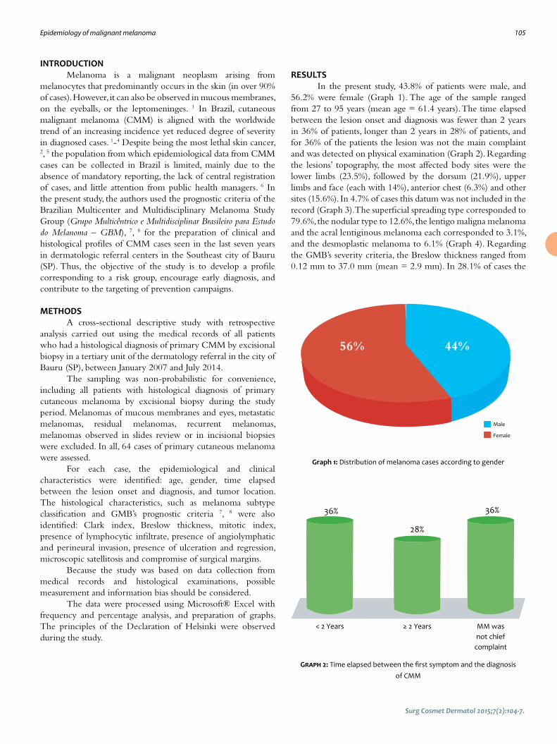
RESULTSIn the present study, 43.8% of patients were male, and
56.2% were female (Graph 1). The age of the sample ranged from 27 to 95 years (mean age = 61.4 years). The time elapsed between the lesion onset and diagnosis was fewer than 2 years in 36% of patients, longer than 2 years in 28% of patients, and for 36% of the patients the lesion was not the main complaint and was detected on physical examination (Graph 2). Regarding the lesions’ topography, the most affected body sites were the lower limbs (23.5%), followed by the dorsum (21.9%), upper limbs and face (each with 14%), anterior chest (6.3%) and other sites (15.6%). In 4.7% of cases this datum was not included in the record (Graph 3). The superficial spreading type corresponded to 79.6%, the nodular type to 12.6%, the lentigo maligna melanoma and the acral lentiginous melanoma each corresponded to 3.1%, and the desmoplastic melanoma to 6.1% (Graph 4). Regarding the GMB’s severity criteria, the Breslow thickness ranged from 0.12 mm to 37.0 mm (mean = 2.9 mm). In 28.1% of cases the
Male
Female
56% 44%
Graph 1: Distribution of melanoma cases according to gender
Graph 2: Time elapsed between the first symptom and the diagnosis of CMM
36%
< 2 Years ≥ 2 Years MM was not chief
complaint
36%
28%

Surg Cosmet Dermatol 2015;7(2):104-7.
106 Pinto ACVD, Cavalcante MLLL, Silva GV, Brito FF, Oliveira AMN, Cleto NG
Anatomical location of the melanomaLower
limbs
Dorsum
Upper limbs
Anterior thorax
Other body sites
Not described
Face
23,50%
21,90%
14%
14%
6,30%
15,60%
4,70%
Graph 3: Distribution of CMM cases according to body topography
lesion was in situ. The percentages of each thickness, Breslow (I to V) and Clark levels (I to V) are highlighted in Graph 5. The ulceration was present in 14.2%, regression areas in 26.6%, and mitotic index higher than zero in 34.4%. Only one case (1.6%) had angiolymphatic invasion, and 42.2% of biopsies showed lymphocytic infiltrate. Satellitosis and neural invasion were not detected. Regarding the compromise of surgical margins, the biopsies were free of neoplastic involvement in 81.3% of cases, while there was a presence of compromise in 15.6% of cases. This datum was not recorded in 3.1% of cases. Research in search of other skin cancers was positive in 43.8% of patients.
DISCUSSIONCMMs were more common in women (56.2%),
consistent with studies conducted in the Brazilian states of São Paulo (Southeast Region) and Santa Catarina (South Region). 2, 3, 9-11 This association relates to women’s greater adherence to prevention campaigns and more frequent use of healthcare services. The average age at diagnosis of 61.4 years is similar to that observed in a Portuguese study (61 years)1 and other Brazilian studies, such as those conducted in the city of Curitiba (South Region), with an average age of 58 years12 and in the city of Brasilia (Mid-West Region), with more cases affecting the age group of 61-80-year-olds. 13 Prevalence in the elderly is a result of increased life expectancy coupled with a greater difficulty in this age group for early detection of neoplastic lesions. In line with other studies, 6,14 the association of CMM with other skin cancers was verified at 43.2%, pointing towards a subgroup of individuals with intense exposure to the sun. In 36% of patients, although CMM was not the complaint leading to the consultation, this condition was detected during an examination. In this context, it is important to recognize the importance of a complete dermatological examination, which includes performing a dermoscopy. This is a non-invasive, ancillary diagnostic tool of high sensitivity (98.8%) and specificity (91.2%) for CMM detection, making it very important in the differentiation of melanocytic and non-melanocytic lesions. 14-16 The most affected topographies were the lower limbs (23.5%) and the dorsum (21.9%), consistent with the literature data. 1, 3, 4, 17, 18 Hospital-based publications of the 1990s and first decade of the century XXI 13, 19, 20 show a higher incidence of the nodular subtype with a lower proportion of non-invasive diagnosis of CMM. The present study confirms the emergence of a new profile for CMM in Brazilian tertiary units, with the superficial spreading type as the most frequent (79.6%) and 28.1% of lesions diagnosed in situ. Regarding GBM’s severity criteria, most patients in the present study offered evidence of a good prognosis, with Breslow levels I and II in half of the cases, Clark levels I, II, and III in 68.8%, and a mitotic index greater than zero in only 34.4%. Moreover, other severity criteria such as regression, ulceration, and angiolymphatic invasion showed low positivity. Still, regarding the degree of severity there was no satellitosis and neural invasion. Most reports (81.3%) described free surgical margins, revealing technical diligence in excisional
Melanoma histological types
Gráfico 4: Distribution of CMM cases according to histological type
Superficial spreading melanoma
Nodular
Malignant melanoma lentigo
Acral lentiginous melanoma
Desmoplastic melanoma
12%3%
80%3% 2%
28%
5%3%
23%
27%
14%
23,50%
25%
21,90%
21,90%
6,10%1,60%
Clark I
Clark II
Clark III
Breslow I
Breslow II
Breslow III
Clark IV
Clark V
Unclassified
Breslow IV
Breslow V
In situ
Clark level Breslow level
Graph 5: Distribution of CMM cases according to histological type

Epidemiology of Malignant Melanoma 107
Surg Cosmet Dermatol 2015;7(2):104-7.
biopsies. Historically, severe cases of CMM prevailed in tertiary hospitals. 13, 19, 21 The present study shows that this scenario is changing, with a greater tendency toward early diagnosis. This early diagnosis profile combined with better prognosis was observed in other Brazilian studies conducted in the South and Southeast regions, 2, 3, 8, 22 However, this profile was not found in similar studies conducted in the North and Northeast regions, 6, 17 a fact that reveals important regional differences that must be considered when planning prevention campaigns for the population.
CONCLUSIONIn this study, the following CMM profile was observed:
female, average age = 61 years, with lesions not always observed before the consultation, located in lower limbs or trunk, with superficial spreading subtype and with good prognostic signs according to the GBM’s criteria. Studies such as the present paper, which strives for the identification of risk groups, prognostic factors, and the understanding of CMM histological behavior, are important for providing subsidies for the design of strategies for approaching populations. l
Acknowledgements:The authors would like to thank the doctors and staff of
the pathology laboratory of the Instituto Lauro de Souza Lima, in Bauru (SP), Brazil.
REFERENCES1. Moreira J, Moreira E, Azevedo F, Mota A. Melanoma maligno cutâneo:
estudo retrospectivo. Acta Med Port. 2014;27(4):480-8.
2. Nasser N. Melanoma cutâneo - estudo epidemiológico de 30 anos em ci-
dade do Sul do Brasil, de 1980-2009. An Bras Dermatol. 2011;86(5):932-41.
3. Konrad P, Fabris MR, Melao S, Blanco LFO. Perfil epidemiológico e his-
topatológico dos casos de melanoma cutâneo primário diagnosti-
cados em Criciúma no período entre 2005 e 2007. An Bras Dermatol.
2011;86(3):457-61.
4. Ríos L, Nagore E, López JL, Redondo P, Martí RM, Fernández-de-Misa R,
et al. Registro nacional de melanoma cutáneo. Características del tu-
mor en el momento del diagnóstico:15 años de experiência. Actas Der-
mosifiliogr. 2013;104(9):789-99.
5. Battisti R, Nunes DH, Lebsa-Weber A, Schweitzer LC, Sgrott I. Avaliação
do perfil epidemiológico e da mortalidade dos pacientes com diagnós-
tico de melanoma cutâneo primário no município de Florianópolis -
SC, Brasil. An Bras Dermatol. 2009;84(4):335-42.
6. Chiba FB, Delfino ACG, Schettini APM, Chirano CA, Damasceno SAS.
Perfil clinicoepidemiológico dos melanomas cutâneos em duas insti-
tuições de referência na cidade de Manaus, Brasil. An Bras Dermatol.
2011;86(6):1239-41.
7. Brasil. Ministério da Saúde. Portaria nº 357, de 8 de Abril de 2013. Apro-
va as Diretrizes Diagnósticas e Terapêuticas do Melanoma Maligno
Cutâneo. Diário Oficial [da] República Federativa do Brasil. 2013 abr. 08
[acesso em 2015 maio 19]. Disponível em: http://bvsms.saude.gov.br/
bvs/saudelegis/sas/2013/prt0357_08_04_2013.html
8. Bonfá R, Bonamigo RR, Bonfá R, Duro KM, Furian RD, Zelmanowicz AM.
A precocidade diagnóstica do melanoma cutâneo: uma observação no
sul do Brasil. An Bras Dermatol. 2011;86(2):215-21.
9. Criado PR, Vasconcellos C, Sittart JAS, Valente NYS, Moura BPS, Barbo-
sa GL. Melanoma maligno cutâneo primário: estudo retrospectivo de
1963 a 1997 no Hospital do Servidor Público Estadual de São Paulo. Rev
Assoc Med Bras. 1999; 45(2):157-62.
10. Ferrari Júnior NM, Muller H, Ribeiro M, Maia M, Sanches Júnior JA. Cuta-
neous melanoma: descriptive epidemiological study. Sao Paulo Med J.
2008;126(1):41-7.
11. Dimatos DC, Duarte FO, Machado RS, Vieira JV, Vasconcellos ZAA, Bins-
-Ely J, Neves RD. Melanoma cutâneo no Brasil. Arquivos Catarinenses de
Medicina. 2009;38(01):14-19.
12. Purim KSM, Sandri CO, Pinto NT, Sousa RHS, Maluf EPC. Perfil de Casos
de Melanoma em um Hospital Universitário, 2003 a 2007. Revista Brasi-
leira de Cancerologia 2013; 59(2): 193-199.
13. Pinheiro AMC, Friedman H, Cabral ALSV, Rodrigues HA. Melanoma cutâ-
neo: características clínicas, epidemiológicas e histopatológicas no
Hospital Universitário de Brasília entre janeiro de 1994 e abril de 1999.
An Bras Dermatol. 2003;78(2): 179-86.
14. Salvio AG, Assumpção Júnior A, Segalla JGM, Panfilo BL, Nicolini HR,
Didone R. Experiência de um ano de modelo de programa de preven-
ção contínua do melanoma na cidade de Jaú-SP. An Bras Dermatol.
2011;86(4):669-74.
15. Schaffer JV, Rigel DS, Kopf AW, Bolognia JL. Cutaneous melanoma - past,
present and future. J Am Acad Dermatol. 2004;51(1 Suppl):S65-9.
16. Argenziano G, Fabbrocini G, Carli P, De Giorgi V, Sammarco E, Delfino M.
Epiluminescence Microscopy for the Diagnosis of Doubtful Melanocy-
tic skin Lesions. Arch Dermatol. 1998;134:1563-70.
17. Vilanova CMA, Lages RB, Ribeiro SM, Almeida IP, Santos LG, Vieira SC.
Perfil epidemiológico e histopatológico do melanoma cutâneo em
um centro do nordeste brasileiro de 2000 a 2010. An Bras Dermatol.
2013;88(4):553-62.
18. Callender GG, Egger ME, Burton AL, Scoggins CR, Ross MI, Stromberg
AJ, et al. 30. Prognostic implications of anatomic location of primary
cutaneous melanoma of 1 mm or thicker. Am J Surg. 2011;202:659-64.
19. Venegas LFP, Flores C, Blacher GG, Daudt AW, Cerski CTS. Melanoma ma-
ligno cutâneo no Rio Grande do Sul: estudo de 101 casos. Rev Ass Med
Brasil.1992;38:122-6.
20. Hampe SV. Estudo da precocidade diagnóstica dos melanomas cutâ-
neos primários em Porto Alegre, por análise de imagem computadori-
zada [Tese]. Porto Alegre (RS): Universidade Federal do Rio Grande do
Sul; 1997. 132 p.
21. Lapa MS, Guedes KF, Schslch FO, Landman G. Melanomas malignos
cutâneos tratados no Hospital do Câncer de São Paulo. Estudo retros-
pectivo para avaliação de distribuição, fatores prognósticos e sobrevi-
da. An Bras Dermatol. 2002;77:313-20.
22. Brandão FV, Pereira AFJR, Gontijo B, Bittencourt FV. Aspectos epidemio-
lógicos do melanoma em serviço de dermatologia de hospital univer-
sitário em um período de 20 anos. An Bras Dermatol. 2013;88(3):348-57.

Surg Cosmet Dermatol 2015;7(2):109-15.
OriginalArticle
Authors: Felipe Bochnia Cerci 1
1 Mohs Surgery and Cutaneous Oncology Preceptor, Department of Dermatology, Hospital Santa Casa de Curitiba – Curitiba (PR), Brazil.
Correspondence: Felipe Bochnia Cerci Praça Rui Barbosa, 245 - Centro Cep: 80010-030 – Curitiba, PR, Brazil E-mail: [email protected]
Received on: 11 May 2015Approved on: 17 June 2015
This study was performed at Hospital Santa Casa de Curitiba - Curitiba (PR), Brazil.
Financial support: NoneConflict of interest: None
Auricular cartilage graft for nasal recons-truction after Mohs micrographic surgeryEnxerto de cartilagem auricular para reconstrução nasal após cirurgia micrográfica de Mohs
DOI: http://dx.doi.org/10.5935/scd1984-8773.201572649
ABSTRACT Introduction: Successful restoration of form and function of the nose after Mohs sur-
gery requires thoughtful reconstructive planning. Nasal defects that are deep and exten-sive, especially those located on the ala, may require a cartilage graft to help restore nasal function, anatomy, and cosmesis.
Objectives: To evaluate the usefulness of auricular cartilage grafts in nasal reconstruction after Mohs micrographic surgery, as well as to describe a cartilage graft harvesting technique.
Methods: Retrospective study of patients with nasal defects following Mohs surgery who were submitted to an auricular cartilage graft.
Results: Ten patients were included in the study. The cartilage graft was harvested from the scaphoid fossa/antihelix in six (60%) patients, and from the concha in four (40%) patients. All scaphoid fossa/antihelix cartilage grafts were harvested through anterior inci-sion, while conchal grafts were removed through posterior incision. One patient develo-ped a hematoma, which drained spontaneously.
Conclusions: auricular cartilage grafts are a versatile, reliable, and predictable method of providing structural support in nasal restoration. It is crucial to identify patients who can benefit from this technique. Through careful planning and adequate execution, ear cartilage grafts help to improve nasal reconstructions results in selected cases.
Keywords: mohs surgery; ear cartilage; surgical flaps; nose neoplasms; basal cell carcinoma
RESU MO Introdução: a restauração da forma e função nasais após cirurgia de Mohs requer planejamento cirúrgico
adequado. Defeitos nasais extensos e profundos, principalmente localizados na asa, podem demandar enxerto de cartilagem para ajudar a restaurar a função, a anatomia e a estética nasais.
Objetivos: avaliar a utilidade de enxertos de cartilagem em reconstrução nasal após cirurgia micrográfica de Mohs, assim como descrever uma das técnicas para sua realização.
Métodos: estudo retrospectivo de pacientes com defeitos cirúrgicos nasais decorrentes de cirurgia de Mohs submetidos a enxerto de cartilagem auricular.
Resultados: dez pacientes foram incluídos no estudo. O enxerto de cartilagem foi retirado da anti-hé-lice/fossa escafoide em seis pacientes (60%) e da concha em quatro pacientes (40%). Todos os enxertos de cartilagem da anti-hélice/fossa escafoide foram retirados através de incisão anterior, enquanto os da concha foram retirados por excisão posterior. Houve uma complicação, hematoma, que drenou esponta-neamente.
Conclusões: Enxertos de cartilagem constituem método versátil, confiável e previsível de fornecer su-porte estrutural em reconstrução nasal. É fundamental identificar os pacientes que podem se beneficiar da técnica. Mediante planejamento cauteloso e execução adequada, enxertos de cartilagem auricular melhoram significativamente os resultados de reconstruções nasais em casos selecionados.
Palavras-chave: cirurgia de mohs; cartilagem da orelha; retalhos cirúrgicos; neoplasias nasais; carcinoma basocelular
109

110 Cerci FB
support “heavy” flaps in order to avoid the collapse of the nasal ala, to keep the nasal valve open, and to provide support for a better contour. The purpose of the present study was to evaluate the usefulness of auricular cartilage grafts in nasal reconstruction after Mohs micrographic surgery, as well as to describe a cartilage graft harvesting technique.
METHODSPatientsA retrospective study was performed with 10 patients
whose nasal defects resulting from Mohs micrographic surgery needed cartilage graft. The cases were selected from a public hospital, from August 2014 to March 2015.
INTRODUCTIONThe restoration of nasal shape and function after Mohs
surgery requires adequate surgical planning. Extensive and deep nasal defects, especially those located in the nasal ala, may require a cartilage graft to help restore function, anatomy, and nasal aesthetics.1,2 In dermatologic surgery, the most common donor site for a cartilage graft is the ear. When selecting the donor subunit – anti-helix/scaphoid fossa or concha – the differences in the cartilage of these locations, as well as the morbidity and ease of removal of the graft should be considered.3 Cartilage grafts are usually performed with interpolation flaps.1,2 However, they can also be associated with single stage flaps, skin grafts, and healing by secondary intention. 4-6 The main functions of the cartilage graft are to prevent tissue contraction and distortion, to
Surg Cosmet Dermatol 2015;7(2):109-15.
Chart 1: Removal and attachment of a cartilage graft from the anti-helix/scaphoid fossa – Steps and comments.
STEPS COMMENTS
1. Create a template for the cartilage graft 1. Use the suture package as a template. Cartilage grafts must be longer than the horizontal extent of the defect in order to be appropriately attached (Figure 1A).
2. Transfer the template to the anti-helix/ 2. Figures 1B-1C. Preferably ipsilateral. scaphoid fossa3. Anesthesia 3. Inject anterior and posterior donor area to hydro-dissect the skin of the cartilage, along
the perichondrium plane.4. Decide on the incision site (anterior X posterior) 4. If posterior, suture the middle portion of the helix in the pre-auricular region or ask your
assistant to traction the ear (with hooks) in order to facilitate the removal.5. Incise the skin 5. Incise the skin (it can be slightly curved) equidistantly from the helix’s rim and the con-
cha’s lateral rim (Figure 1D). Incising too close to the helix’s rim increases the risk of tissular contraction and subsequent deformity.
6. Dissect the cartilage 6. Dissect the auricular skin of the cartilage in the supraperichondrial plane. Visualization can be enhanced with the use of hooks (Figures 1E-1F).
7. Incise the cartilage 7. Incise the anterior perichondrium and cartilage, followed by the posterior perichondri-um, but do not incise the posterior auricular skin (or the opposite when removing the graft posteriorly). A second incision with the same depth is performed parallel to the first (Figures 1E-1F). The distance between them corresponds to the graft’s width. The ends are then incised in a rectilinear – and not conical – manner. Straight ends retain the shape and position of the helix. Elliptical (conical) ends may allow the contraction of the helix’s rim, and distortion secondary to contractile healing forces.
8. Keep the cartilage in saline solution 8. Keep the cartilage in saline solution up until it is attached on the nose.9. Ear closure + dressing 9. The ear is a common site of hematoma after the removal of the cartilage graft. Suture
it first placing a temporarily fixed dressing before incising the flap. At the end of the surgery, check the donor area’s hemostasis and suture a definitive fixed dressing (48-72 hours). A small dental cotton roll allows greater hemostasis than a gauze. The borders of the ear cartilage should not be reapproximated. The suture is performed in a continuous manner with 5.0 mononylon thread. Internal sutures are not required.
10. Prepare the cartilage graft 10. If necessary, trim the graft in order to obtain the desired thickness, shape, borders, and contour. For this step, use a razor blade or a scalpel blade number 15. The razor blade allows the cartilage to be sculpted in a more delicate manner (Figure 2B).
11. Suture cartilage graft on the nose 11. Create “pockets” on each side of the defect with a scalpel blade. The cartilage must be inserted in these pockets (Figure 2C). A Ƥgure-8 suture helps to stabilize the free end of the cartilage. The U-suture or a simple interrupted suture help to stabilize the graft over the underlying cartilage (e.g. a graft for the tip of the nose) or to stabilize the cartilage at the alar rim (Figure 2D).
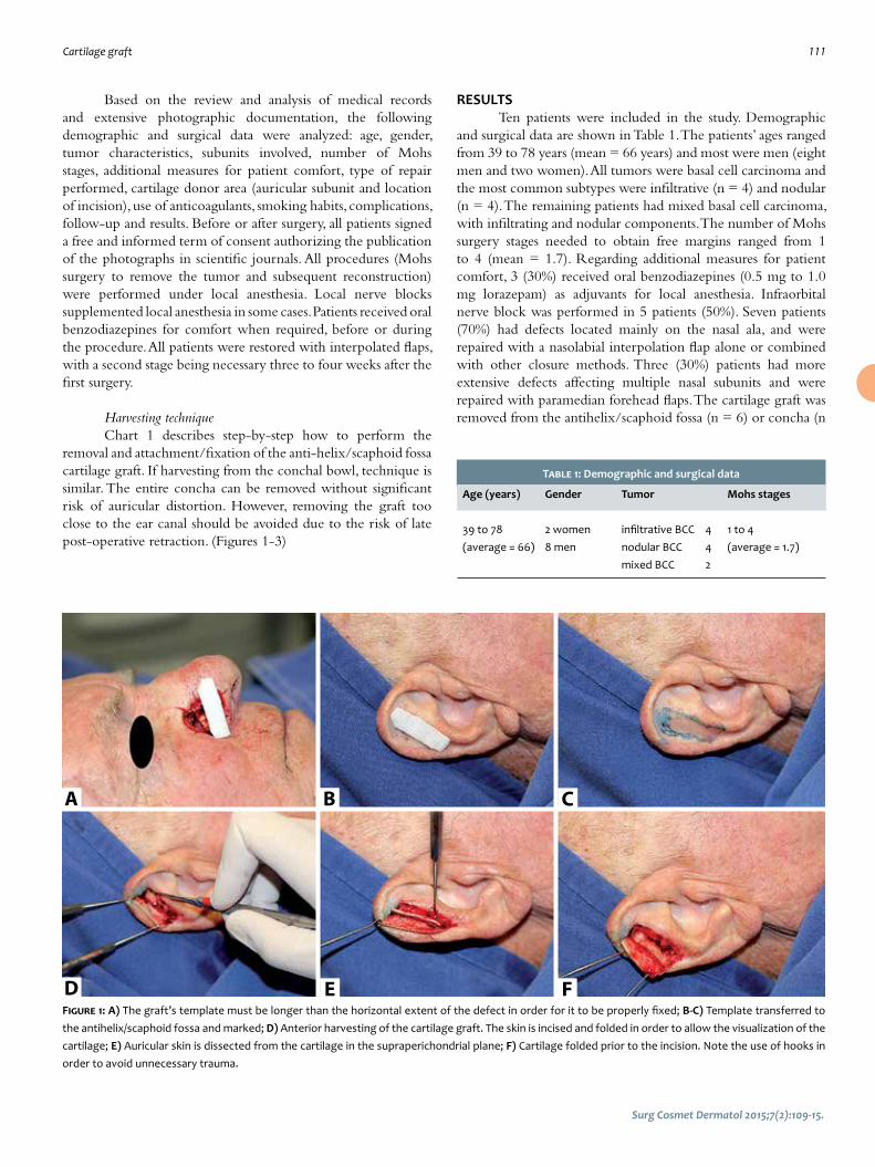
Cartilage graft 111
Surg Cosmet Dermatol 2015;7(2):109-15.
A
D
B
E
C
FFigure 1: A) The graft’s template must be longer than the horizontal extent of the defect in order for it to be properly fixed; B-C) Template transferred to the antihelix/scaphoid fossa and marked; D) Anterior harvesting of the cartilage graft. The skin is incised and folded in order to allow the visualization of the cartilage; E) Auricular skin is dissected from the cartilage in the supraperichondrial plane; F) Cartilage folded prior to the incision. Note the use of hooks in order to avoid unnecessary trauma.
Table 1: Demographic and surgical data
Age (years) Gender Tumor Mohs stages
39 to 78 2 women infiltrative BCC 4 1 to 4(average = 66) 8 men nodular BCC 4 (average = 1.7) mixed BCC 2
Based on the review and analysis of medical records and extensive photographic documentation, the following demographic and surgical data were analyzed: age, gender, tumor characteristics, subunits involved, number of Mohs stages, additional measures for patient comfort, type of repair performed, cartilage donor area (auricular subunit and location of incision), use of anticoagulants, smoking habits, complications, follow-up and results. Before or after surgery, all patients signed a free and informed term of consent authorizing the publication of the photographs in scientific journals. All procedures (Mohs surgery to remove the tumor and subsequent reconstruction) were performed under local anesthesia. Local nerve blocks supplemented local anesthesia in some cases. Patients received oral benzodiazepines for comfort when required, before or during the procedure. All patients were restored with interpolated flaps, with a second stage being necessary three to four weeks after the first surgery.
Harvesting techniqueChart 1 describes step-by-step how to perform the
removal and attachment/fixation of the anti-helix/scaphoid fossa cartilage graft. If harvesting from the conchal bowl, technique is similar. The entire concha can be removed without significant risk of auricular distortion. However, removing the graft too close to the ear canal should be avoided due to the risk of late post-operative retraction. (Figures 1-3)
RESULTSTen patients were included in the study. Demographic
and surgical data are shown in Table 1. The patients’ ages ranged from 39 to 78 years (mean = 66 years) and most were men (eight men and two women). All tumors were basal cell carcinoma and the most common subtypes were infiltrative (n = 4) and nodular (n = 4). The remaining patients had mixed basal cell carcinoma, with infiltrating and nodular components. The number of Mohs surgery stages needed to obtain free margins ranged from 1 to 4 (mean = 1.7). Regarding additional measures for patient comfort, 3 (30%) received oral benzodiazepines (0.5 mg to 1.0 mg lorazepam) as adjuvants for local anesthesia. Infraorbital nerve block was performed in 5 patients (50%). Seven patients (70%) had defects located mainly on the nasal ala, and were repaired with a nasolabial interpolation flap alone or combined with other closure methods. Three (30%) patients had more extensive defects affecting multiple nasal subunits and were repaired with paramedian forehead flaps. The cartilage graft was removed from the antihelix/scaphoid fossa (n = 6) or concha (n

112 Cerci FB
Surg Cosmet Dermatol 2015;7(2):109-15.
= 4). All cartilage antihelix/scaphoid fossa grafts were removed via anterior incision, while conchal grafts were removed via posterior incision. Three patients had full-thickness defects. In one of them, the nasal mucosa was recreated with a hinge flap from the nasal sidewall while in the other two, it was closed primarily due to the small size of the mucosal defect. Only one patient was a smoker. No patients were on anticoagulants. One of them, however, had post-operative bleeding and hematoma,
which drained spontaneously. Excellent functional and aesthetic results were achieved in all patients. There was no infection, hypertrophic scarring, keloid, or distortion of the ear in any of the cases, and the donor area has become virtually unnoticeable after a few months. (Figure 4). The follow-up period ranged from two to nine months (average = seven months), without tumor recurrence.
Figure 3: A-C) Two months after surgery; patient from Figures 1 and 2A B C
Figure 2: A) Anti-helix/scaphoid fossa after cartilage graft harvest. The borders of the cartilage are not reapproximated; B) The cartilage can be trimmed if necessary; C) Graft being inserted in the “pockets” created on the nose; D) Sutured cartilage; E) Nasolabial interpolation flap for right nasal ala repair, im-mediate post-operative period (first stage). The portion of the defect that extended to the nasal sidewall and malar region was closed primarily F) Dressing fixed and sutured in the donor area (the last picture is of a different patient).
A
D
B
E F
C

Cartilage graft 113
Surg Cosmet Dermatol 2015;7(2):109-15.
Figure 4:A) Cartilage donor area one week after, with proper healing and wi-thout functional or aes-thetic deformity. Re-pair performed with a running simple suture, 5.0 nylon. Internal su-tures are not necessaryB) One month after surgery, almost imper-ceptible scar C) Contra-lateral ear of the same patient
A B C
Figure 5: A) Retraction of the alar rim (arrow) after Mohs surgery for basal cell carcinoma removal in the left nasal alaB) Improvement of the retraction after cartilage graft (arrow) C) Two months after surgery
A B C
DISCUSSIONMany factors must be taken into consideration when
planning a nasal reconstruction. In most patients, soft tissue restoration is sufficient to obtain optimal functional and aesthetic results. However, in certain cases an auricular cartilage graft can be necessary for the proper restoration of the nasal ala and valve. 4 It is crucial to recognize those patients who will benefit from the cartilage graft. Signs that help to identify them include the spontaneous collapse of the nasal ala/valve or during the inspiration after the removal of the tumor, or retraction of the nasal rim. In some cases, even without retraction of the rim during surgery, it may occur later due to scarring. The precise location for the fixation of the graft, as well as its size and shape, will result in different benefits. If the objective is only to avoid or correct the retraction of the nasal rim, the cartilage graft can be smaller and should be inserted considerably close to the alar rim (Figure 5). If, however, there is a major collapse of the nasal valve/ala (Figure 6), the cartilage graft should be
larger and be placed in the middle and upper thirds of the ala. For the projection of the nasal tip, the grafts should be directly placed on it. Cartilage grafts can be structural (native cartilage is present but there is a need for additional cartilage to support) or restorative (replacement of removed cartilage). Cartilage grafts for the nasal ala are usually structural and non-restorative, since there is no cartilage in most of the nasal ala, but only fibrofatty tissue. Structural functions of the cartilage include: 1) to prevent tissue contraction and distortion, 2) to support “heavy” flaps, 3) to maintain the nasal valve open, and 4) to provide support for the contour.7 Cartilage donor areas include the anti-helix/scaphoid fossa and the auricular concha.4,8 Cartilage from the anti-helix/scaphoid fossa is ideal for long, flexible, and straight segments, while that of the concha is ideal for grafts that require more curvature, substance, and stiffness.
In the present study, 6 patients (60%) had a cartilage graft removed from the antihelix/scaphoid fossa, while 4 patients

114 Cerci FB
Surg Cosmet Dermatol 2015;7(2):109-15.
(40%) had it removed from the concha. Of these, 3 patients had large defects that were repaired with a paramedian forehead flap (Figure 7).
The incisions for removing the cartilage can be performed anteriorly or posteriorly. Anterior incisions are easier to access, however they result in more visible scars.1,2 Although grafts from the antihelix/scaphoid fossa were removed anteriorly, the incision healed well and was hardly noticeable in all patients. It may be necessary to sculpt the graft to obtain the desired thickness, shape, borders, and contour. This must be done
carefully, since the cartilage is a fragile structure and may fracture during the process. A scalpel blade 15 is traditionally used to carve, however a razor blade allows the graft’s contours to be sculpted more gently. Cartilage grafts can be safely harvested under local anesthesia with low complication rates.1-3,9 Post-operative pain at the cartilage donor site can be significant, therefore adequate analgesia (non-steroidal anti-inflammatory/powerful analgesic) must be provided to all patients.1,2
The primary disadvantage of the cartilage graft is the additional morbidity of creating a second surgical site. Although
Figure 7: A) Auricular con-chal graft harvested through posterior incision B) Cartilage sutured (arrow) in order to support the paramedian fo-rehead flap and prevent the collapse of the nasal ala and valve C) Five months after surgery
A
B C
Figure 6: A) Collapse of the nasal ala after the resection of a BCC in the right nasal ala, even without removal of the alar cartilage; B) Opening of the ala after fixation of the auricular cartilage graftA B
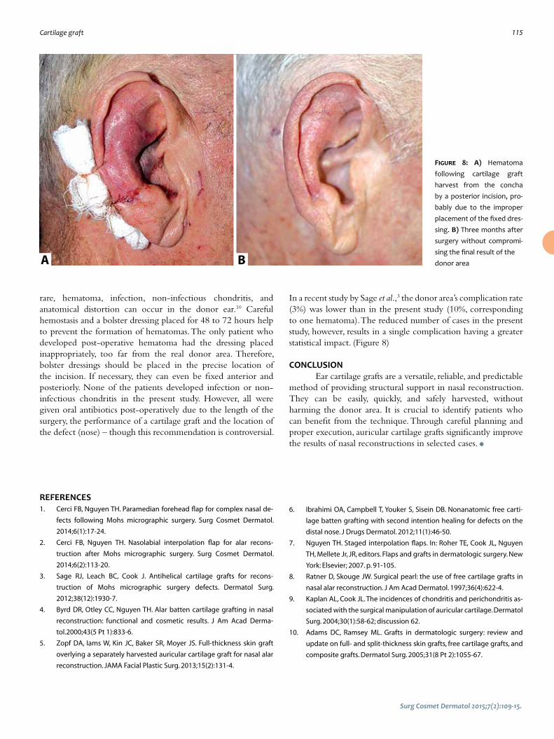
Cartilage graft 115
Surg Cosmet Dermatol 2015;7(2):109-15.
rare, hematoma, infection, non-infectious chondritis, and anatomical distortion can occur in the donor ear.10 Careful hemostasis and a bolster dressing placed for 48 to 72 hours help to prevent the formation of hematomas. The only patient who developed post-operative hematoma had the dressing placed inappropriately, too far from the real donor area. Therefore, bolster dressings should be placed in the precise location of the incision. If necessary, they can even be fixed anterior and posteriorly. None of the patients developed infection or non-infectious chondritis in the present study. However, all were given oral antibiotics post-operatively due to the length of the surgery, the performance of a cartilage graft and the location of the defect (nose) – though this recommendation is controversial.
In a recent study by Sage et al.,3 the donor area’s complication rate (3%) was lower than in the present study (10%, corresponding to one hematoma). The reduced number of cases in the present study, however, results in a single complication having a greater statistical impact. (Figure 8)
CONCLUSIONEar cartilage grafts are a versatile, reliable, and predictable
method of providing structural support in nasal reconstruction. They can be easily, quickly, and safely harvested, without harming the donor area. It is crucial to identify patients who can benefit from the technique. Through careful planning and proper execution, auricular cartilage grafts significantly improve the results of nasal reconstructions in selected cases. l
Figure 8: A) Hematoma following cartilage graft harvest from the concha by a posterior incision, pro-bably due to the improper placement of the fixed dres-sing. B) Three months after surgery without compromi-sing the final result of the donor area
REFERENCES1. Cerci FB, Nguyen TH. Paramedian forehead flap for complex nasal de-
fects following Mohs micrographic surgery. Surg Cosmet Dermatol.
2014;6(1):17-24.
2. Cerci FB, Nguyen TH. Nasolabial interpolation flap for alar recons-
truction after Mohs micrographic surgery. Surg Cosmet Dermatol.
2014;6(2):113-20.
3. Sage RJ, Leach BC, Cook J. Antihelical cartilage grafts for recons-
truction of Mohs micrographic surgery defects. Dermatol Surg.
2012;38(12):1930-7.
4. Byrd DR, Otley CC, Nguyen TH. Alar batten cartilage grafting in nasal
reconstruction: functional and cosmetic results. J Am Acad Derma-
tol.2000;43(5 Pt 1):833-6.
5. Zopf DA, Iams W, Kin JC, Baker SR, Moyer JS. Full-thickness skin graft
overlying a separately harvested auricular cartilage graft for nasal alar
reconstruction. JAMA Facial Plastic Surg. 2013;15(2):131-4.
6. Ibrahimi OA, Campbell T, Youker S, Sisein DB. Nonanatomic free carti-
lage batten grafting with second intention healing for defects on the
distal nose. J Drugs Dermatol. 2012;11(1):46-50.
7. Nguyen TH. Staged interpolation flaps. In: Roher TE, Cook JL, Nguyen
TH, Mellete Jr, JR, editors. Flaps and grafts in dermatologic surgery. New
York: Elsevier; 2007. p. 91-105.
8. Ratner D, Skouge JW. Surgical pearl: the use of free cartilage grafts in
nasal alar reconstruction. J Am Acad Dermatol. 1997;36(4):622-4.
9. Kaplan AL, Cook JL. The incidences of chondritis and perichondritis as-
sociated with the surgical manipulation of auricular cartilage. Dermatol
Surg. 2004;30(1):58-62; discussion 62.
10. Adams DC, Ramsey ML. Grafts in dermatologic surgery: review and
update on full- and split-thickness skin grafts, free cartilage grafts, and
composite grafts. Dermatol Surg. 2005;31(8 Pt 2):1055-67.
A B

116
Surg Cosmet Dermatol 2015;7(2):116-21.
OriginalArticles
Authors: Flavia Alvim Sant’Anna Addor 1
1 MSc, Technical Director at the Clinical Re-search Laboratory – Anvisa Certified, São Paulo (SP), Brazil
Correspondence: Flavia Alvim Sant’Anna Addor Rua Attilio Delanina, 178 - Osasco Cep: 06023-070 - Sao Paulo - SP E-mail: [email protected]
Received on: 30 March 2015Approved on: 17 June 2015
This study was performed at a private practi-ce – São Paulo (SP), Brazil.
Financial support: Farmoquímica S.A.R.
Conflict of interest: None
Influence of a nutritional supplement containing collagen peptides on the properties of the dermisInfluência de um suplemento nutricional com peptídeos de colágeno nas propriedades da derme
DOI: http://dx.doi.org/10.5935/scd1984-8773.201572636
ABSTRACT Introduction: The loss of dermal density and thickness, characterized by sagging and
thinning skin, may be related to various conditions such as aging (both intrinsic and ex-trinsic) as well as abrupt changes in body mass – such as pregnancy or increased weight – and the resulting rupture of collagen fibers. Some nutrients can positively affect the dermal metabolism, thus improving its functional properties.
Objective: To evaluate clinically and through ultrasound the effect of a nutritional su-pplement on the thickness of the dermis and on its functional properties.
Methods: Twenty-eight female patients between the ages of 35 and 65 years, with facial sagging complaints, used the supplement for 90 days, having been evaluated subjectively and through ultrasound for clinical safety and efficacy.
Results: The studied nutritional supplement provided a significant increase in dermal thickness after 30 days of use, in the areas evaluated. There was significant improvement in the firmness, elasticity, hydration and overall appearance of the skin. No adverse reaction related to the product was observed during the study period.
Conclusions: The nutritional supplement was effective in improving the conditions of the dermis, increasing its thickness and clinical parameters of firmness, elasticity, and hy-dration, and has been proven safe and well-tolerated when used as instructed.
Keywords: collagen; dermis; nutrients; skin aging
RESU MO Introdução:A perda de densidade e espessura dérmica, caracterizada por flacidez e afinamento cutâ-
neo, pode estar relacionada com várias condições, como envelhecimento (intrínseco e extrínseco), além de mudanças abruptas de massa corporal, com ruptura das fibras colágenas, como gestação ou aumento de peso. Alguns nutrientes podem interferir positivamente no metabolismo dérmico, melhorando con-sequentemente suas propriedades funcionais.
Objetivo: avaliar o efeito de um suplemento nutricional na espessura e propriedades funcionais da derme, clínica e ultrassonograficamente.
Métodos: 28 pacientes do sexo feminino entre 35 e 65 anos com queixa de flacidez facial utiliza-ram o suplemento durante 90 dias; foram avaliadas com relação à segurança de uso, eficácia clínica, subjetiva e ultrassonográfica.
Resultados: O suplemento nutricional avaliado proporcionou aumento significativo da espessura da derme nas áreas avaliadas a partir de 30 dias de uso; observou-se melhora da firmeza, elasticidade, hidratação e do aspecto geral da pele de maneira significativa; não houve reação adversa relacionada ao produto durante o estudo.
Conclusões: O suplemento nutricional foi eficaz na melhora das condições da derme, aumentando a espessura e os parâmetros clínicos de firmeza, elasticidade e hidratação, além de se mostrar seguro e aceito na forma de utilização orientada.
Palavras-chave: colágeno; derme; envelhecimento da pele; nutrientes

The effect of collagen peptides on the dermis 117
Surg Cosmet Dermatol 2015;7(2):116-21.
INTRODUCTIONCutaneous aging is a progressive degenerative process
resulting from a decline in the physiological functions of the cutaneous tissue, both at the epidermal and dermal levels.
In the dermis, there is a decrease of collagen synthesis, as well as the components of the other extracellular matrices, characteristic of chronological (intrinsic) aging. This decrease can be exacerbated by metalloproteinase, which increases in expression due to photodamage, and leading to its fragmentation. 1
Menopause is also an accelerating factor of degenerative changes to the collagen and thickness of the dermis through the progressive loss of collagen, which reaches its peak in the first five years (with a loss of up to 30%) and then stabilizes at loss rate of 1% to 2% per year. Hormone replacement therapy allows for partial recovery, however not all patients can use it. 2
The use of oral supplements containing collagen as a means to improve the signs of aging is not a recent practice; nevertheless the scarcity of studies and publications on this subject means there have always been doubts about its actual value.
Nonetheless, the topic is once again being discussed with renewed interest due to the development of technologies that allow for the isolation of peptides for oral consumption and the emergence of a new generation of collagen supplementation--specific peptides capable of enhancing the expression of certain molecules linked to the synthesis of collagen and association with other substances, such as vitamins and Phytoextracts, which act synergistically enhancing that effect.
OBJECTIVEThe objective of the present study was to demonstrate
the effects of a nutritional supplement in improving dermal structure, evaluating its thickness, and the clinical properties of cutaneous firmness, elasticity, and hydration.
MATERIALS AND METHODSA monocentric, open, blind, non-comparative study was
carried out at a private research center between August and November 2014.
Thirty female patients (35 to 65 years of age) complaining of some degree of facial sagging were invited and subsequently included in the study. All patients stopped using any cosmiatric treatment four weeks before the beginning of the study. Patients using corticosteroids or immunosuppressants, bearing active endocrine diseases or any clinical condition that could interfere with the evaluations, were excluded.
After signing a term of free and informed consent and undergoing a dermatologic evaluation in order to rule out any dermatosis in the body site to be assessed, a multichannel color Doppler ultrasonography with a frequency transducer of up to 15MHz (Voluson E device – GE Healthcare) was performed in two body sites (submental and malar areas), for standardization. The ultrasound evaluation was carried out with an aim at calculating the dermal thickness of the selected area and capturing any differences between each experimental occasion.
Subsequently, the patients received the dietary supplement containing collagen peptides, vitamin C, and Hibiscus sabdariffa in sachet form, under evaluation and with instructions to consume 2 sachets diluted in 200 ml of cold or hot water once a day for 12 weeks.
Lastly, the patients were asked to return monthly for clinical safety reasons, subjective and ultrasonographic assessments, or if at any time there was a case of doubt or complication. At all visits a questionnaire was given to evaluate the firmness, elasticity, hydration, and overall appearance of the skin. The rating system used in the questionnaire was conceptualized as follows, in order to obtain accuracy in the answers:
• Worsened: there has been visible deterioration as compared to the previous state for this location;
• Unchanged: there was no improvement or worsening as compared to the previous state for this location;
• Partial improvement: there was some degree of perceived improvement;
• Total improvement: the perceived improvement is significant, easily noticeable, and the best possible that the patient could expect with this type of treatment.
The features of the product were also evaluated regarding their solubility and taste, with ratings of very bad, bad, good, or very good.
Ethical aspectsThe study protocol was approved by the Independent
Ethics Committee and conducted in accordance with the Good Clinical Practice standards.
Statistical evaluationIn order to assess the normality of the data’s distribution,
the authors used the Shapiro-Wilk test. To compare the effects in between the experimental occasions, the Wilcoxon test was performed using the medians, with a 95% significance level.
RESULTSOf the 30 patients who started the study, 28 completed
the assessments. The average age was 48.5 years. Two adverse events were reported: one case of headache and another of migraine. Both were classified as not serious and unrelated to the product. The two patients who experienced adverse events were excluded from the study, since they discontinued the use of the product.
None of the patients developed any digestive symptoms, such as nausea, vomiting, or abdominal discomfort during the study. No further complaint or adverse effect was recorded during the study.
The patients showed good adherence to the treatment.Subjective assessmentAt each visit, all patients completed a questionnaire
evaluating the degree of improvement or worsening regarding the studied parameter.
Graph 1 depicts the data collected for the evaluation

118 Addor FAS
Surg Cosmet Dermatol 2015;7(2):116-21.
of cutaneous firmness. Firmness was defined as having a higher resistance to traction or digital pressure.
Graph 1 allows us to observe that after 30 days of
there was a perceived improvement of 89% in the perception of cutaneous hydration; in 60 days, the perceived improvement reached 96% of the sample. At the end of the study, 100% of the sample noticed partial or total improvement of the facial cutaneous hydration.
Finally, the overall appearance of the skin was defined as the perceived appearance of the skin during observation in the mirror without taking into account any specific parameter, except for the impression of vitality and smoothness of features/signs of aging.
Graph 4 shows the results obtained for this variable:Chart 4 shows that after 30 days of supplement use there
Graph 1: Percentage of volunteers’ answers regarding cutaneous firmness at D30, D60, and D90
Cutaneous firmness
Worsened Unchanged Partial Total improvement improvement
100%90%80%70%60%50%40%30%20%10%0%
D30D60D90
0 0 0
25
71
89
71
7 11
29
0 0
Graph 2: Percentage of volunteers’ answers regarding cutaneous elasticity at D30, D60, and D90
Worsened Unchanged Partial Total improvement improvement
Worsened Unchanged Partial Total improvement improvement
Cutaneous elasticity100%90%80%70%60%50%40%30%20%10%0%
D30D60D90
0 7 0
25
7186
79
4 7
21
0 0
Graph 4: Percentage of volunteers’ answers regarding the overall appearance of the skin at D30, D60, and D90
Overall appearance of the skin100%90%80%70%60%50%40%30%20%10%0%
D30D60D90
0 0 0
18
7986
68
414
32
0 0
Graph 3: Percentage of volunteers’ answers regarding cutaneous hydration at D30, D60, and D90
Worsened Unchanged Partial Total improvement improvement
Hydration of the skin100%90%80%70%60%50%40%30%20%10%0%
D30D60D90
0 4 0
11
82 82
68
714
32
0 0
supplement use there was a perceived improvement of 75%; in 60 days, the perceived improve reached 100% of the sample. At the end of the study, 100% of the sample had noticed partial or total improvement in the cutaneous firmness of the face.
Graph 2 shows the results obtained for the item regarding cutaneous elasticity. This variable was defined as the ability to return to its original state after traction or digital pressure.
Graph 2 shows that after 30 days of supplement use there
was a perceived improvement of 75%; in 60 days, the perceived improvement reached 93% of the sample. At the end of the study, 100% of the sample noticed partial or total improvement in facial cutaneous elasticity.
Another parameter evaluated was cutaneous hydration, defined as skin with a healthy, homogenous, bright appearance and smooth to the touch. Graph 3 shows the time results for that variable.
Graph 3 shows that after 30 days of supplement use
was a perceived improvement in the overall appearance of the skin of 83%; in 60 days, an improvement of 100% could be noticed. At the end of the study, 100% of the sample noticed partial (68%) or total (32%) improvement in the hydration of facial skin
Evaluation of the supplement: characteristics of patient self-administration
The product’s solubility, defined as the degree of difficulty of dissolution in 200 ml of cold or hot water, was also evaluated. After 90 days of treatment, 96% of patients reported that the solubility was easy or very easy.

The effect of collagen peptides on the dermis 119
Surg Cosmet Dermatol 2015;7(2):116-21.
Regarding the taste, 100% of the patients considered it good or very good.
Ultrasonographic evaluationBased on the data obtained about the dermal thickness
on each visit, the group’s median was calculated for each experimental event: D0, D30, D60, and D90 (D = Day). It was possible to observe a progressive and significant increase in the median thickness in both evaluated areas, with increases of 17.0% and 18.8%, in the malar and in the submental areas respectively, at the end of the study. Both results were statistically significant according to the Wilcoxon test (p <0.001). Graph 5 details the measurements obtained for the evaluated areas at each experimental occasion:
Comparisons made on D60 relative to D30 and D90
DISCUSSIONThe use of collagen as an oral supplement is not a recent
development. Given that collagen is a nutrient with structural value, cases of malnutrition, rigorous diets, or malabsorption syndromes would naturally and evidently benefit from protein supplementation.
As the studies aimed at delaying the signs of the aging progresses, the current discussion focuses on the possibility of collagen supplementation in offering a beneficial and significant effect on dermal loss related to skin aging.
Few studies have focused on evaluating the impact of collagen supplementation with gelatin and other enriched foods (such as shakes and even capsules) on the parameters of aging.
Gelatin is a polypeptide derived from collagen with a high molecular weight, however it is deficient in essential amino acids. Despite the fact that its nutritional value is indisputable, it does not have specific properties. 3
Finally, the study by Nishimoto et al. on Wistar rats, using a model to histologically demonstrate the synthesis of collagen based on the hydroxyproline index, has demonstrated that isolated peptides would have a significant effect on the collagen synthesis, when compared to that obtained with gelatin, whose levels were not superior to those of the placebo. 4
Based on findings like this, hydrolyzed collagen (HC) has once again attracted attention.
Hydrolyzed collagen is digested and absorbed in the digestive tract –identified in the blood by its constituent peptides – and reaches the skin in up to four days. 5
Due to its similarity to collagen – particularly type I dermal collagen – its effect would be not only that of restoration, but also of promoting type I collagen synthesis, playing a positive role in aging and even in other disorders with dermal involvement, like tissue repair. 6,7
Hydrolyzed collagen is characterized by a relatively low molecular weight (<6 kDa), which facilitates its absorption and bioavailability. Nonetheless, there are many types of hydrolyzed collagen according to the protein source, and the synthesis and supply processes. 8,9
Figure 1: Ultrasound images of the submental region captured at the different experimental occasions D0 (first visit), D30 (30-days visit), D60
(60-days visit), and D90 (90-days visit). The dermal area is delimited between the yellow markings, in the upper portion of each image
Figure 2: Ultrasound images of the malar region captured at the different experimental occasions D0 (first visit), D30 (30-days visit), D60 (60-days visit), and D90 (90-days visit). The dermal area is delimited between the
yellow markings, in the upper portion of each image
Graph 5: Median of the ultrasound measurements of dermal thickness in the right hemiface and submental region, at the different experimental
occasions D0, D30, D60, and D90 (in cm).
Hemiface Submental region
Med
ian
of th
e de
rmal
thic
knes
s (c
m)
D0 D30 D60 D90
0,167
0,115
0,180
0,123
0,187
0,127
0,195
0,137
0,200
0,150
0,100
0,050
0
also showed a statistically significant (p≤0.001) increase in the thickness of the malar and the dermis of the submental regions, characterizing progress in the effect seen over time.
Figures 1 and 2 illustrate the ultrasonographic findings for the study areas.

120 Addor FAS
Surg Cosmet Dermatol 2015;7(2):116-21.
Each hydrolyzed collagen type can have a set of effects, and during the study their effects were evaluated in vitro, often using models that are not comparable. For these reasons, it is very difficult to gather comparable data on the subject, since the term “hydrolyzed collagen” refers to a large group of peptides associations.
Therefore, results obtained for a particular HC cannot be extrapolated to others.
The HC that has been studied in the present article is a polypeptide derived from the biotechnological enzymatic process of animal skin, devoid of impurities and other molecules such as lipids and carbohydrates, and therefore easily digestible, susceptible to the action of proteolytic enzymes, and with an absorption rate of more than 90%. 10
The collagen peptide evaluated in the present study consists of 18 type-I collagen peptides, 8 of which are essential. It is characterized by higher concentrations of glycine, proline, and hydroxyproline – amino acids that represent about 50% of the total content of amino acids in the composition. This composition was demonstrated to be capable of promoting collagenesis from fibroblasts, in addition to the fact that the synthesized collagen is firmer. 3
The specific peptides generated from the digestion of collagen peptides – Gly-Glu and Pro-Hypro – are chemoattracted by dermal fibroblasts as a signal of “destruction” of collagen, thus activating fibroblasts. 11
There is also the activation of the enzyme Hyaluronan synthase 2, with an increased synthesis of hyaluronic acid and glycosaminoglycans. 12,13
The collagen peptide has also been shown to stimulate the production of Decorin, a proteoglycan, which is a connective tissue component that binds to type-1 collagen and plays a role in the organization of the extracellular matrix, and by regulating the aggregation of bundles leads to the production of collagen fibers. 14
Another trend that induces a greater diversity of supplements in this area is the association of other molecules, with protective or beneficial effects in the collagen synthesis, thus favoring the effects of the peptides. The action of vitamin C in stimulating fibroblast proliferation is widely known. 15
Studies by Pinnel in the 1980s demonstrated that the L-ascorbic acid is capable of inducing procollagen synthesis in cultured dermal fibroblasts. 16 These studies demonstrated that this increased synthesis occurred even in older fibroblasts, which becomes especially relevant considering that there is a decrease of physiological vitamin C reserves in the dermis with age. 17
The combination of substances such as phytoextracts can also increase the effects of the peptides associations of hydrolyzed collagen, enabling a better performance through the
neutralization of free radicals, for example.The plant Hibiscus sabdariffa, traditionally used in food
preparation due to its taste, demonstrated antioxidant, lipolytic, hypoglycemic, and diuretic properties, among others, in in vitro models. Its use for reducing resistance to insulin was demonstrated based on its polyphenols, for it contains high concentrations of phenolic acid and proanthocyanidins.18,19
Its broad antioxidant mechanism, however, is based on the combination of additional compounds that are not present in that extract. The Hibiscus sabdariffa’s powerful antioxidant action mechanism has already been studied in neoplasias, having been more recently evidenced with the isolation of its main antioxidant compounds: chlorogenic and neochlorogenic, cryptochlorogenic acids, rutin, and isoquercetin. 20, 21
The present study was aimed at evaluating the effects of a nutritional supplement that brings together the synergistic action of these three molecules (collagen peptide, Vitamin C, and Hibiscus sabdariffa) on the human dermis. A non-invasive study was carried out with the assistance of ultrasonographic evaluation, which allowed for the confirmation of the signs perceived by patients. In addition to the progressive and significant improvement in the cutaneous firmness, elasticity, and hydration, there was a real and measurable increase in the dermal thickness in two facial areas: the malar region (where there was a 17.0% increase of the thickness) and the submental region (where there was an increase of 18.8%). The latter finding is of particular interest, since this area offers more complexity to treat sagging.
The evaluation of these patients allowed for the observation of safety levels in the use of the product, which did not cause any systemic reaction during the three months of consumption.
The daily use of the supplement was demonstrated to be palatable, a fundamental factor for adherence to prolonged treatments. Regarding the taste, it is important to highlight a finding: even during continued use (three months), there was no reduction of tolerance (i.e. patients did suffer from “taste fatigue” when ingesting the supplement over time) – which is a major factor in treatment adherence.
CONCLUSIONThe daily use of a nutritional supplement containing
collagen peptides, Vitamin C, and Hibiscus sabdariffa promoted a significant increase in the dermal thickness of the studied areas of the face (malar and submental), with a positive impact on the evaluation of cutaneous firmness, elasticity, and hydration. The safety of its use combined with its proven effects allows for the conclusion that this association is beneficial in the approach to facial aging, as well as dermal sagging and atrophy. l

The effect of collagen peptides on the dermis 121
Surg Cosmet Dermatol 2015;7(2):116-21.
REFERENCES1. Fisher GJ, Kang S, Varani J, Bata-Csorgo Z, Wan Y, Datta S, et al. Me-
chanisms of photoaging and cronological skin aging. Arch Dermatol.
2002;138(11):1462-70.
2. Brincat M, Mouniz CF, Studd JW, Cooper d. Sex hormones and skin
collagen content in post menopausal women. Br Med J (Clin Res Ed).
1983;287(6402):1337-8.
3. Zague V. A new view concerning the effects of collagen hydrolysate in-
take on skin properties. Arch Dermatol Res. 2008;300(9):479-83.
4. Hiura, N, Sato R, Suzuki K, Asano R. Effect of oral administration of gelatin
and collagen peptides on the hydroxyproline content of rats skin. Jour-
nal of the Japanese Society for Food Science and Technology. 2002;49;3
[acesso em 30 de janeiro de 2015]. Disponível em: http://agris.fao.org/
agris-search/search.do?recordID=JP2002003318
5. Ohara H, Matsumoto H, Ito K, Iwai K, Sato K. Comparison of quantity and
structures of hydroxyproline-containing peptides in human blood af-
ter oral ingestion of gelatin hydrolysates from different sources. J Agric
Food Chem. 2007;55:1532-35.
6. Zague V. Collagen hydrolysate intake increases skin collagen expres-
sion and suppresses matrix metalloproteinase 2 activity. J Med Food.
2011;14(6):618-24.
7. Atamas SP, Luzina IG, Ingels J, Choi J, Wong WK, Furst DE, et al. Stimula-
tion with type I collagen induces changes in gene expression in peri-
pheral blood mononuclear cells from patients with diffuse cutaneous
systemic sclerosis (scleroderma). Clin Exp Immunol. 2010;161(3):426-35.
8. Zhuang, Yongliang, Zhao X, Zhang Z, Li B. Effects of collagen and colla-
gen hydrolysate from jellyfish (Rhopilema esculentum) on mice skin
photoaging induced by UV irradiation. J Food Sci. 2009;74(6):H183-8.
9. Postlethwaite AE, Wong WK, Clements P, Chatterjee S, Fessler BJ, Kang
AH, et al. A multicenter, randomized, double-blind, placebo-controlled
trial of oral type I collagen treatment in patients with diffuse cutaneous
systemic sclerosis: I. oral type I collagen does not improve skin in all
patients, but may improve skin in late-phase disease. Arthritis Rheum.
2008;58(6):1810-22.
10. Oesser S, Adam M, Babel W, Seifert J. Oral administration of (14)C la-
beled gelatin hydrolysate leads to an accumulation of radioactivity in
cartilage of mice (C57/BL). J Nutr. 1999;129(10):1891-5.
11. Postlethwaite AE, Seyer JM, Kang AH. Chemotactic attraction of human
fibroblasts to type I, II, and III collagens and collagen-derived peptides.
Proc Natl Acad Sci U S A. 1978;75(2):871-5.
12. Matsuda N, Koyama Y, Hosaka Y, Ueda H, Watanabe T, Araya T, et al. Ef-
fects of ingestion of collagen peptide on collagen fibrils and glycosami-
noglycans in the dermis. J Nutr Sci Vitaminol (Tokyo). 2006;52(3):211-5.
13. Minaguchi J, Koyama Y, Meguri N, Hosaka Y, Ueda H, Kusubata M,
et al. Effects of ingestion of collagen peptide on collagen fibrils and
glycosaminoglycans in Achilles tendon. J Nutr Sci Vitaminol (Tokyo).
2005;51(3):169-74.
14. Pulkkinen L, Alitalo T, Krusius T, Peltonen L. Expression of decorin in hu-
man tissues and cell lines and defined chromosomal assignment of the
gene locus (DCN). Cytogenet Cell Genet. 1992;60(2):107-11.
15. Hata R, Senoo H. L-ascorbic acid 2-phosphate stimulates collagen ac-
cumulation, cell proliferation, and formation of a three-dimensional
tissuelike substance by skin fibroblasts. J Cell Physiol. 1989;138(1):8-16.
16. Pinnel SR, Murad S, Darr D. Induction of collagen synthesis by ascorbic
acid. A possible mechanism. Arch Dermatol. 1987;123(12):1684-6.
17. Patnaik BK, Kanungo MS. Ascorbic acid and aging in the rat. Uptake of
ascorbic acid by teeth and concentration of various forms of ascorbic
acid in different organs. Biochem J. 1966;100(1):59-62.
18. Da-Costa-Rocha I, Bonnlaender B, Sievers H, Pischel I, Heinrich M. Hibis-
cus sabdariffa L. - a phytochemical and pharmacological review. Food
Chem. 2014;165:424-43.
19. Peng CH, Yang YS, Chan KC, Wang CJ, Chen ML, Huang CN. Hibiscus sab-
dariffa polyphenols alleviate insulin resistance and renal epithelial to
mesenchymal transition: a novel action mechanism mediated by type
4 dipeptidyl peptidase. J Agric Food Chem. 2014;62(40):9736-43.
20. Worawattananutai P, Itharat A, Ruangnoo S. In vitro antioxidant, anti-in-
flammatory, cytotoxic activities against prostate cancer of extracts from
Hibiscus sabdariffa leaves. J Med Assoc Thai. 2014;97 Suppl 8:S81-7.
21. Wang J, Cao X, Jiang H, Qi Y, Chin KL, Yue Y. Antioxidant activity of leaf ex-
tracts from different Hibiscus sabdariffa accessions and simultaneous
determination five major antioxidant compounds by LC-Q-TOF-MS.
Molecules. 2014;17;19(12):21226-38.

123
Surg Cosmet Dermatol 2015;7(2):123-8.
OriginalArticles
Authors: Adilson Costa1
Samara Eberlin2
Stefano Piatto Clerici3
Beatrice Martinez Zugaib Abdalla4
1 Former Clinical Director at Kolderma Ins-tituto de Pesquisa Clínica Campinas (SP), Brazil
2 Technical Manager, Safety and Pre-Clinical Efficacy Laboratory, Kolderma
3 Researcher, Safety and Pre-Clinical Efficacy Laboratory, Kolderma
4 Medical Student, Fundação Faculdade de Medicina do ABC – Santo André (SP), Brazil
Correspondence: Adilson Costa Alameda Franca, 760, ap. 21 - Jd. Paulista, Cep: 01422-000 - Sao Paulo, SP, Brazil E-mail: [email protected]
Received on: 1 April 2015Approved on: 7 May 2015
This study was performed at the Kolderma Instituto de Pesquisa Clínica and at the Der-matology Service of the Pontifícia Universi-dade Católica de Campinas -Campinas (SP), Brazil.
Financial support: Sanofi-Aventis Farmacêuti-ca Ltda., Paulo (SP), Brazil.
Conflict of interest: The study was sponsored by Sanofi-Aventis Farmacêutica Ltda., Paulo (SP), Brazil.
In vitro evaluation of four commercially available liquid soaps (in Brazil) for their anti-inflammatory and protective skin bar-rier qualities, as well as their impact on the reduction of cutaneous hypersensitivityAvaliação in vitro da eficácia anti-inflamatória, protetora da barreira cutânea e redutora da hipersensibilidade cutânea de quatro sabonetes líquidos disponíveis no BrasilDOI: http://dx.doi.org/10.5935/scd1984-8773.2015722
ABSTRACT Introduction: The skin may present dysfunctions that result in histological alterations and elementary lesions, many of them caused by immunologic mechanisms and/or alte-rations in the epidermal barrier. Objective: The present study was aimed at verifying in vitro whether some cosmetic products marketed in Brazil in the form of liquid soaps, have anti-inflammatory and pro-tective efficacy in the skin barrier. Methods: An in vitro study was carried out with four commercial soaps, which were evaluated in a cell culture of human keratinocytes for the determination of their anti-in-flammatory and epidermal barrier restorative effects, as well as their capacity to reduce cutaneous hypersensitivity. Concentrations of keratin 10 and 14, loricrin, IL-12, IFN--g and TRP V-1 were measured in the cell culture supernatants of human keratinocyte through the sandwich ELISA assay. Results: The four test substances led to significant reductions in the synthesis of IFN-g and IL-12. Only the test substance C triggered a significant increase in the synthesis of keratin 10. All test substances showed significant reductions in the synthesis of keratin 14 and TRPV-1, and a significant increase in the synthesis of loricrin. Conclusions: Some cosmetic products in the form of soap can have in vitro results re-garding their anti-inflammatory, epidermal barrier restoration and skin hypersensitivity reduction effects. Keywords: anti-inflammatory agents/adverse effects; dermatitis, atopic; soaps
RESU MOIntrodução:A pele pode apresentar disfunções que levam a alterações histológicas e lesões elementares, muitas delas decorrentes de mecanismos imunológicos e/ou alteração da barreira epidérmica. Objetivo: O objetivo deste estudo é avaliar se alguns produtos cosméticos, em apresentação sabonete líquido, encontrados no mercado brasileiro, apresentam eficácia anti-inflamatória e protetora de barreira cutânea in vitro. Métodos: Trata-se de estudo in vitro, no qual quatro sabonetes comerciais foram avaliados em culturas celulares de queratinócitos humanos para determinação das atividades anti-inflamatória, restauradora de barreira epidérmica e redutora da hipersensibilidade cutânea. As concentrações de queratina 10 e 14, loricrina, IL-12, IFN-g e TRP V-1 foram dosadas nos sobrenadantes das culturas celulares de queratinócitos humanos por ensaio Elisa sanduíche. Resultados: As quatro substâncias-teste promoveram reduções significativas na síntese de IFN-g e IL-12. Somente a substância-teste C desencadeou aumento significativo na síntese de queratina 10. Todas as substâncias-teste demonstraram reduções significativais na síntese de queratina 14, aumento significativo na síntese de loricrina e reduções significativas na síntese de TRPV-1. Conclusões: Alguns produtos cosméticos em sabonete podem apresentar resultados in vitro no tocante às atividades anti-inflamatória, restauradora de barreira epidérmica e redutora da hipersensibilidade cutânea.Palavras-chave: anti-inflamatórios/efeitos adversos; dermatite atópica; sabonetes

124 Costa A, Eberlin S, Clerici SP, Abdalla BMZ
Surg Cosmet Dermatol 2015;7(2):123-8.
INTRODUCTIONLike any body organ, the skin may present degeneration,
metabolic abnormalities, malformations, dysfunction, and inflammation, leading to histological alterations and resulting in elementary lesions.1
Depending on the anatomical location and environmental influences, the skin has exceptional structural and functional diversity, as it is continually exposed to external aggressions such as solar radiation, mechanical stimuli, climate change and/or chemical and biological damage.2 The maintenance of the skin’s structural integrity is therefore critical and, when affected, it should be able to count on mechanisms for the rapid restoration of the epidermal barrier.3-5
Hydration and the integrity of the stratum corneum (SC) are crucial to the skin’s appearance, metabolism, mechanical properties and cutaneous barrier function. 6 During the epidermal differentiation, several proteins are involved in the formation of the stratum corneum, including loricrin, involucrin, filaggrin, and keratins. 7-9
The variation of the genes of the stratum corneum results in the pathogenesis of three major skin disorders: ichthyosis vulgaris, psoriasis, and atopic dermatitis (AD). 10-11 AD is a chronic inflammation characterized by pruritus and one in which hereditary, environmental, and immunological components play a key role. 12
AD fluctuates between two phases: the acute phase, with a predominance of Th2 type response and IgE production, and the chronic, with strong Th1 type response, with the production of interferon-γ (IFN- γ) and interleukin-12 (IL-12). 11
Keratin is a tough, waterproof protein responsible for the skin’s protection.13 The keratins 5 and 14 are expressed in the basal keratinocytes while keratins 1 and 10 participate in the differentiation of suprabasal keratinocytes.14 Mutations in keratin genes can result in human diseases characterized by an alteration in epidermal homeostasis, including the impairment of the barrier function. 14 The rupture of the permeable barrier of the epidermis – both acute and chronic – results in changes in the synthesis of structural proteins in keratinocytes, relating to increased suprabasal cell proliferation and differentiation, in an attempt to repair the skin barrier. 14
The local production of inflammatory mediators is correlated to the integrity of the barrier, as a result of being activated following the release of cytokines, histamine, and eicosanoids. 15 These, in turn, increase the sensitivity of the nociceptors, such as TRPV-1 (V1 transitional channel vanilloid receptor), a nonselective calcium channel widely expressed in the cutaneous tissue, including keratinocytes and peripheral sensory nerve fibers.16 TRPV-1 has its sensitivity increased in AD lesions, which translates into hyperalgesia and additional production of inflammation mediators. 17-18
The availability of cosmetic products that minimize the occurrence of unwanted side effects such as skin xerosis and cutaneous hypersensitivity, can be a key differential to ensure adherence to and success of the treatment. The present study was aimed at evaluating whether some cosmetic products in liquid
soap marketed in Brazil provide in vitro anti-inflammatory and protective efficacy to the skin barrier.
METHODSFour commercial liquid soaps were evaluated in the
present in vitro study: Test-substance A (Dermacyd Infantil, Sanofi-Aventis Farmacêutica Ltda., São Paulo, SP, Brazil, batch 246782); Test-substance B (Sabonete de Glicerina Granado Bebê, Casa Granado Laboratórios, Farmácias e Drogarias S.A., Belém, PA, Brazil, batch R1446); Test-substance C (Huggies Turma da Mônica Recém-Nascido, Kimberly-Clark Brasil Indústria e Comércio de Produtos de Higiene Ltda, Mogi das Cruzes, SP, Brazil, batch Lkg2354); and Test-substance D (Johnson’s Baby Sabonete Líquido Da Cabeça aos Pés, Johnson & Johnson Industrial Ltda, São José dos Campos, SP, Brazil, batch 3013b09).
To that end, human fibroblast cultures (Human Foreskin Fibroblasts-1, “HFF-1”; ATCC SCRC-1041 (Banco de Células do Rio de Janeiro, Rio de Janeiro/RJ, Brazil, Catalogue number 0275) were seeded in 75 cm3 bottles (Nunc, Roskilde, Denmark), cultured and expanded in an incubator at 37°C in the presence of 5% CO2, using a specific culture medium for the determination of cell viability through the XTT method.
Cell viability was determined by a colorimetric method using the XTT dye (2, 3-bis [2-methoxy-4-nitro-5-sulfopheny]-2H-tetrazolium-5-carboxyanilide inner salt), which is converted in water-soluble orange formazan by the mitochondrial enzyme succinate dehydrogenase in viable cells (Xenometrix AG, Switzerland). The fibroblasts were seeded and incubated with the Test-substances in eight concentrations using decimal geometric dilution. 13 After 48 hours of incubation the Test-substances were removed and the culture medium was replaced. Then, the XTT dye was added to the culture, and the plate was incubated for a further three hours. The absorbance (optical density – OD) of each well was determined at 480nm with the assistance of a Multiskan GO monochromator (Thermo Scientific, Finland). The percentage of cell viability was calculated according to the following equation:
Viability % = (DOST / DOCN) x 100Where:- DOST is the optical density of the Test-substance - DOCN is the optical density of the negative control.HaCat human keratinocytes (Banco de Células do Rio
de Janeiro, Rio de Janeiro/RJ, Brazil, Catalogue number 0341) were seeded and cultured in the same way that the HFF-1 human fibroblasts were. Upon reaching confluence, the cells were seeded in plates for later incubation with the Test-substance and quantification of the proposed mediators.
The keratinocyte cultures were incubated with three non-cytotoxic concentrations of the Test-substances determined by the XTT method. Cells were maintained in contact with the test-substances for 48 hours. After this period, the cell culture’s supernatant was collected for measurement of the mediators related to the cutaneous barrier protective activity. In order to evaluate the anti-inflammatory and reductive activity of the

Surg Cosmet Dermatol 2015;7(2):123-8.
Liquid soaps: in vitro evaluation 125
Graph 1: Evaluation of cell viability of Test-substances in cultured human fibroblasts after 48 hours of incubation using the XTT method
dermal hypersensitivity, the inflammatory stress was mimicked through the addition of interleukin-1 alpha (IL-1α, 10ng/mL) to the keratinocyte cultures, concurrently with the treatment using the Test-substance, for 48 hours.
Using commercially available kits for Sandwich ELISA, the concentration of the following mediators were assessed in the supernatants obtained from cell cultures of human keratinocytes: Keratin 10 (USCN Life Science Inc., Wuhan, Hubei, China); keratin 14 (USCN Life Science Inc., Wuhan, Hubei, China); loricrin (USCN Life Science Inc., Wuhan, Hubei, China); interleukin-12 (IL-12; BD Biosciences, San Diego, CA, USA); interferon-γ (IFN-γ, BD Biosciences, San Diego, CA, USA); and transient channel vanilloid receptor V-1 (TRPV-1; USCN Life Science Inc., Wuhan, Hubei, China). The absorbance reading was performed using a Multiskan GO monochromator (Thermo Scientific, Finland).
For the statistical evaluation of the mediators Keratin 10, Keratin 14, loricrin, IL-12, IFN-γ, and TRPV-1, the one-way ANOVA test was used (GraphPad Prism 4 Software Inc. San Diego, CA, USA), which allows for the measuring of the variation in results by comparing the data from the Test-substances, evidencing the differences among them. As the F-test was significant, the authors applied the nonparametric Tukey test. A 5% significance level was adopted.
RESULTSGraph 1 represents the Test-substances’ concentration-
cell viability curve. As can be seen, Test-substances A, C, and D showed non-cytotoxic concentrations starting with the 0.316mg/mL dilution. The Test-substance B showed non-cytotoxic concentration starting with the dilution 0.100 mg/mL.
From a methodological point of view, the incubation of cultured human keratinocytes with pro-inflammatory cytokine interleukin-1 alpha (IL-1α) promoted an increase of 2.7 times and 1.7 times the production of IFN-γ and IL-12, respectively. Aimed at assessing the anti-inflammatory activity of each Test-substance, Graphs 2 and 3 depict the effect of the Test-substances in the IFN-γ and IL-12 synthesis in cultured human keratinocytes. All Test-substances evaluated were able to significantly reduce the synthesis of both inflammatory cytokines.
Regarding IFN-γ, Graph 2 suggests that the Test-substances promoted significant decreases in all concentrations evaluated. At a a concentration of 0.100 mg/mL, Test-substances A, C, and D have shown a slightly stronger anti-inflammatory response (p<0.05) as compared to substance B at the same concentration.
The response obtained in the synthesis of cytokine IL-12 was similar to that of IFN-γ. From Graph 3 it can be seen that Test-substance A at a concentration of 0.100 mg/mL and 0.0316 mg/mL, was able to reduce IL-12 production to levels significantly lower (p<0.05) than those observed with Test-
Cell
viab
ility
(%)
Cell
viab
ility
(%)
Cell
viab
ility
(%)
Cell
viab
ility
(%)

126 Costa A, Eberlin S, Clerici SP, Abdalla BMZ
Surg Cosmet Dermatol 2015;7(2):123-8.
substance D at 0.100 mg/mL concentration.Regarding the protective activity of the skin barrier,
Graph 4 represents the effects of the Test-substances in the synthesis of Keratin 10 in cultured human keratinocytes. As can be seen, only substance C in the concentration of 0.0316 mg/mL produced a significant increase in the synthesis of Keratin 10
(p <0.001) when compared to the control.The response observed for Keratin 14 (Graph 5) was
contrary to that observed with Keratin 10. All Test-substances – either in one or more concentrations evaluated – caused significant reduction in the synthesis of Keratin 14 as compared to the control group.
Graph 4: Effect of Test-substances A, B, C, and D on the production of in-terferon gamma (IFN-γ) in cultured human keratinocytes. The data repre-sent the mean ± standard deviation of three replicates (ANOVA, Tukey).
*** p <0.001 as compared to the control group
Graph 3: Effect of Test-substances A, B, C, and D on the production of Interleukin-12 (IL-12) in cultured human keratinocytes. The data represent
the mean ± standard deviation of three replicates (ANOVA, Tukey).# p <0.001 as compared to the control group; * p<0.05 as compared to
IL-1α group; ** p<0.01 relative to IL-1α group; *** p<0.001 relative to IL-1α group; & p<0.05 regarding Substance D 0.100 mg/mL
Graph 5: Effect of Test-substances A, B, C, and D o the Keratin 14 pro-duction in cultured human keratinocytes. The data represent the mean ±
standard deviation of three replicates (ANOVA, Tukey).* p <0.05 as compared to the control group;
*** p <0.001 as compared to the control group;
Contro
l
Contro
l
Contro
l
Contro
l
Inte
rfer
on γ
(pg/
mL)
Ker
atin
10
(ng/
mL)
Ker
atin
14
(ng/
mL)
Graph 2: Effect of Test-substances A, B, C, and D on Keratin 10 production in cultured human keratinocytes. The data represent the mean ± standard
deviation of three replicates (ANOVA, Tukey).# p <0.001 as compared to the control group;*** P <0.001 as compared to the group IL-1α
& p <0.05 as compared to substance B 0.100 mg/mL
Inte
rleuk
in 1
2 (p
g/m
L)
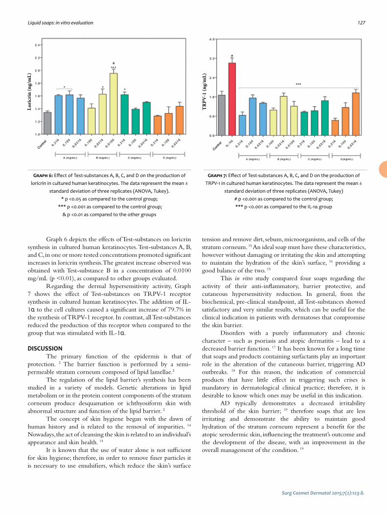
Liquid soaps: in vitro evaluation 127
Surg Cosmet Dermatol 2015;7(2):123-8.
Graph 6 depicts the effects of Test-substances on loricrin synthesis in cultured human keratinocytes. Test-substances A, B, and C, in one or more tested concentrations promoted significant increases in loricrin synthesis. The greatest increase observed was obtained with Test-substance B in a concentration of 0.0100 mg/mL (p <0.01), as compared to other groups evaluated.
Regarding the dermal hypersensitivity activity, Graph 7 shows the effect of Test-substances on TRPV-1 receptor synthesis in cultured human keratinocytes. The addition of IL-1α to the cell cultures caused a significant increase of 79.7% in the synthesis of TRPV-1 receptor. In contrast, all Test-substances reduced the production of this receptor when compared to the group that was stimulated with IL-1α.
DISCUSSIONThe primary function of the epidermis is that of
protection. 2 The barrier function is performed by a semi-permeable stratum corneum composed of lipid lamellae.2
The regulation of the lipid barrier’s synthesis has been studied in a variety of models. Genetic alterations in lipid metabolism or in the protein content components of the stratum corneum produce desquamation or ichthyosiform skin with abnormal structure and function of the lipid barrier. 2
The concept of skin hygiene began with the dawn of human history and is related to the removal of impurities. 14 Nowadays, the act of cleansing the skin is related to an individual’s appearance and skin health. 14
It is known that the use of water alone is not sufficient for skin hygiene; therefore, in order to remove finer particles it is necessary to use emulsifiers, which reduce the skin’s surface
Graph 6: Effect of Test-substances A, B, C, and D on the production of loricrin in cultured human keratinocytes. The data represent the mean ±
standard deviation of three replicates (ANOVA, Tukey).* p <0.05 as compared to the control group;
*** p <0.001 as compared to the control group;& p <0.01 as compared to the other groups
Graph 7: Effect of Test-substances A, B, C, and D on the production of TRPV-1 in cultured human keratinocytes. The data represent the mean ±
standard deviation of three replicates (ANOVA, Tukey)# p <0.001 as compared to the control group;*** p <0.001 as compared to the IL-1α group
tension and remove dirt, sebum, microorganisms, and cells of the stratum corneum. 15 An ideal soap must have these characteristics, however without damaging or irritating the skin and attempting to maintain the hydration of the skin’s surface, 16 providing a good balance of the two. 15
This in vitro study compared four soaps regarding the activity of their anti-inflammatory, barrier protective, and cutaneous hypersensitivity reduction. In general, from the biochemical, pre-clinical standpoint, all Test-substances showed satisfactory and very similar results, which can be useful for the clinical indication in patients with dermatoses that compromise the skin barrier.
Disorders with a purely inflammatory and chronic character – such as psoriasis and atopic dermatitis – lead to a decreased barrier function. 17 It has been known for a long time that soaps and products containing surfactants play an important role in the alteration of the cutaneous barrier, triggering AD outbreaks. 18 For this reason, the indication of commercial products that have little effect in triggering such crises is mandatory in dermatological clinical practice; therefore, it is desirable to know which ones may be useful in this indication.
AD typically demonstrates a decreased irritability threshold of the skin barrier; 19 therefore soaps that are less irritating and demonstrate the ability to maintain good hydration of the stratum corneum represent a benefit for the atopic xerodermic skin, influencing the treatment’s outcome and the development of the disease, with an improvement in the overall management of the condition. 19
Contro
l
Contro
l
Lori
crin
(ng/
mL)
TRPV
-1 (n
g/m
L)

128 Costa A, Eberlin S, Clerici SP, Abdalla BMZ
Surg Cosmet Dermatol 2015;7(2):123-8.
CONCLUSIONBased on the results obtained it is possible to infer that
the four Test-substances promoted significant reductions in the synthesis of IFN-γ (Test-substances A, C, and D showed marked reductions as compared to B) and IL-12 (Test-substance A was able to reduce IL-12 production to levels significantly below those observed with Test-substance D in the concentration of 0.100 mg/mL). Test-substance C triggered a significant
increase in the synthesis of Keratin 10, a phenomenon not seen with other Test-substances, although all have shown significant reductions in the synthesis of Keratin 14. All Test-substances promoted significant increases in loricrin synthesis and significant reductions in TRPV-1 synthesis. Based on these findings, it is possible to conclude that all Test-substances have anti-inflammatory, hypersensitivity reduction, and epidermal barrier restorative activities. l
REFERENCES1. Sampaio AP, Rivitti EA. Dermatologia. 2 ed. São Paulo: Artes Médicas;
2001. p. 39-40.
2. Elias P, Feingold K, Fluhr J. The skin as an organ of protection. In: Frie-
dberg IM, Eisen AZ, Wolff K, Austen KF, Goldsmith LA, Katz SI, editors.
Dermatology in General Medicine. New York: McGraw-Hill; 2003.
p .07–118.
3. Martin P. Wound healing—aiming for perfect skin regeneration. Scien-
ce. 1997;276(5309):75-81.
4. Gurtner GC, Werner S, Barrandon Y, Longaker MT. Wound repair and re-
generation. Nature. 2008;453(7193):314-21.
5. Feingold KR. Thematic review series: skin lipids. The role of epider-
mal lipids in cutaneous permeability barrier homeostasis. J Lipid Res.
2007;48(12):2531-46.
6. Madison KC. Barrier function of: ‘‘la raison d’etre’’ of the epidermis. J In-
vest Dermatol. 2003;121(2):231-41.
7. Hoffjan S, Stemmler S. On the role of the epidermal differentiation com-
plex in ichthyosis vulgaris, atopic dermatitis and psoriasis. Br J Derma-
tol. 2007;157(3):441-9.
8. Kalinin A, Marekov LN, Steinert PM. Assembly of the epidermal corni-
fied cell envelope. J Cell Sci. 2001;114(Pt 17):3069-70.
9. Ishida-Yamamoto A, Iizuka H. Structural organization of cornified cell
envelopes and alterations in inherited skin disorders. Exp Dermatol.
1998;7(1):1-10.
10. Smith FJ, Irvine AD, Terron-Kwiatkowski A, Sandilands A, Campbell LE,
Zhao Y, et al. Loss of function mutations in the gene encoding filaggrin
cause ichtyosis vulgaris. Nat Genet. 2006;38(3):337-42.
11. O’Driscoll J, Muston GC, McGrath JA, Lam HM, Ashworth J, Christiano
AM. A recurrent mutation in the loricrin gene underlies the ichthyotic
variant of Vohwinkel syndrome. Clin Exp Dermatol. 2002;27(3):243-6.
12. Varothai S, Nitayavardhana S, Kulthanan K. Moisturizers for patients
with atopic dermititis. Asian Pac J Allergy Immunol. 2013;31(2):91-8.
13. Chen J, Cheng X, Merched-Sauvage M, Caulin C, Roop DR, Koch PJ. An
unexpected role for keratin 10 end domains in susceptibility to skin
cancer. J Cell Sci. 2006;199(Pt 24):5067-76.
14. Ekanayake-Mudiyanselage S, Aschauer H, Schmook FP, Jensen JM,
Meingassner JG, Proksch E. Expression of epidermal keratins and the
cornified envelope protein involucrin is influenced by permeability
barrier disruption. J Invest Dermatol. 1998;111(3):517-23.
15. Luger TA. Neuromediators – a crucial component of the skin immune
system. J Dermatol Sci 2002;30(2):87-93.
16. Riol-Blanco L, Ordovas-Montanes J, Perro M,Naval E, Thiriot A, Alvarez D,
et al. Nociceptive Sensory Neurons Drive Interleukin-23 Mediated Pso-
riasiform Skin Inflammation. Nature. 2014;510(7503):157-161.
17. Imamachi N, Park GH, Lee H, Anderson DJ, Simon MI, Basbaum AI, et
al. TRPV1-expressing primary afferents generate behavioral responses
to pruritogens via multiple mechanisms. Proc Natl Acad Sci USA. 2009;
106(27):11330-5.
18. Sadofsky LR, Ramachandran R, Crow C, Cowen M, Compton SJ, Mori-
ce AH. Inflammatory stimuli up-regulate transient receptor potential
vanilloid-1 expression in human bronchial fibroblasts. Exp Lung Res.
2012;38(2):75-81.
19. ECVAM-Dbalm. 3T3 Neutral Red Uptake (NRU) Phototoxicity Assay. DB-
-ALM Protocol nº 78. [atualizado 2008; acesso 16 de fevereiro de 2015].
Disponível em: http://ecvam-dbalm.jrc.ec.europa.eu/public_view_
doc2.cfm?id=736F27E9E9F7A9D869FB48087878D2497180BB0BC-
12CB10496CDA74B54630A05A3291B895581F634]

OriginalArticles
The application of platelet-rich plasma in the treatment of androgenic alopeciaA aplicação do plasma rico em plaquetas no tratamento da alopecia androgenética
DOI: http://dx.doi.org/10.5935/scd1984-8773.201572658
ABSTRACT Introduction: Introduction: Androgenic alopecia is the most common type of alopecia.
It is characterized by alterations in the hair cycle, leading to progressive follicular minia-turization. Platelet-rich plasma, promotes angiogenesis in the tissue by action of growth factors, which stimulate the hair follicle
Objective: To eveluate the hair growth in androgenic alopecia, through cell stimulation performed with the subcutaneous injection of platelet-rich plasma.
Methods: Three injections of platelet-rich plasma were performed in 18 patients, at 21-day intervals.The evaluation was performed through comparative clinical photographs, dermoscopy, as well from responses to questionnaires. The Mann-Whitney test and Wilco-xon test were used to analyze the results.
Results: For seven women who completed the study, the average improvement was 42.85% (patients) and 35.71% (external observer). For the 9 males, the average improve-ment was 25.55% (patients) and 18.88% (external observer). In Dermoscopy were obser-ved thickening of hairs, improved local circulation and increased number of follicles.
Conclusions: An effective and satisfactory response in androgenic alopecia was verified in relation to the platelet-rich plasma injection technique.
Keywords: alopecia; platelet-rich plasma; dermatology
RESU MO Introdução: O tipo mais comum de alopecia em ambos os sexos, a androgenética, se caracteriza por
alteração no ciclo capilar que leva à miniaturização folicular progressiva. O plasma rico em plaquetas promove a angiogênese tecidual por ação de fatores de crescimento, estimulando o folículo piloso.
Objetivo: Avaliar o crescimento capilar na alopecia androgenética, através de estímulo celular com plasma rico em plaquetas
Métodos: Foram realizadas três injeções subcutâneas de plasma rico em plaquetas em 18 pacientes, em intervalos de 21 dias. A avalição ocorreu através da comparação de fotos clínicas e dermatoscopia, bem como de questionários respondidos pelos pacientes e observador externo. Para a análise dos resul-tados foram utilizados testes de Mann-Whitney e de Wilcoxon.
Resultados: Para as 7 mulheres que finalizaram o estudo, a média de melhora foi de 42,85% (pa-cientes) e de 35,71% (observador externo). Para os 9 homens, a média de melhora foi de 25,55% (pacientes) e 18,88% (observador externo). Na dermatoscopia foram observados espessamento dos fios, melhora da circulação local e aumento do número de folículos
Conclusões: Foi constatada resposta eficiente e satisfatória na técnica de injeções subcutaneas do plasma rico em plaquetas em alopécia androgenética.
Palavras-chave: alopecia; plasma rico em plaquetas; dermatologia
Authors: Rossana Cantanhede Farias de Vasconcelos1
Karla Azuaga2
Géssica Cantadori Funes Arenas3
João Guilherme Finizola de Vasconcelos4
Natalie Schnaider Borelli5
1 Collaborator Physician and Coordinator, Cosmiatry Ambulatory, Universidade de Santo Amaro (Unisa) – São Paulo (SP), Brazil
2 Dermatologist Physician at private practi-ce – São Paulo (SP), Brazil
3 Biologist specializing in Clinical and Toxi-cological Analysis, Universidade de Guaru-lhos (UNG-SP) - São Paulo (SP), Brazil
4 Collaborator Physician, Surgical Dermato-logy Ambulatory, Unisa
5 Dermatology Resident Physician, Unisa
Correspondence: Rossana Cantanhede Farias de Vasconcelos Rua Barata Ribeiro, 490 – 5º andar - Cerqueira Cesar Cep: 01308-000 - Sao Paulo (SP), Brazil E-mail: [email protected]
Received on: 26 January 2015Approved on: 14 June 2015
This study was performed at the Faculdade de Medicina da Universidade de Santo Ama-ro (Unisa) – São Paulo (SP), Brazil.
Financial support: NoneConflict of interest: None
130
Surg Cosmet Dermatol 2015;7(2):130-7.
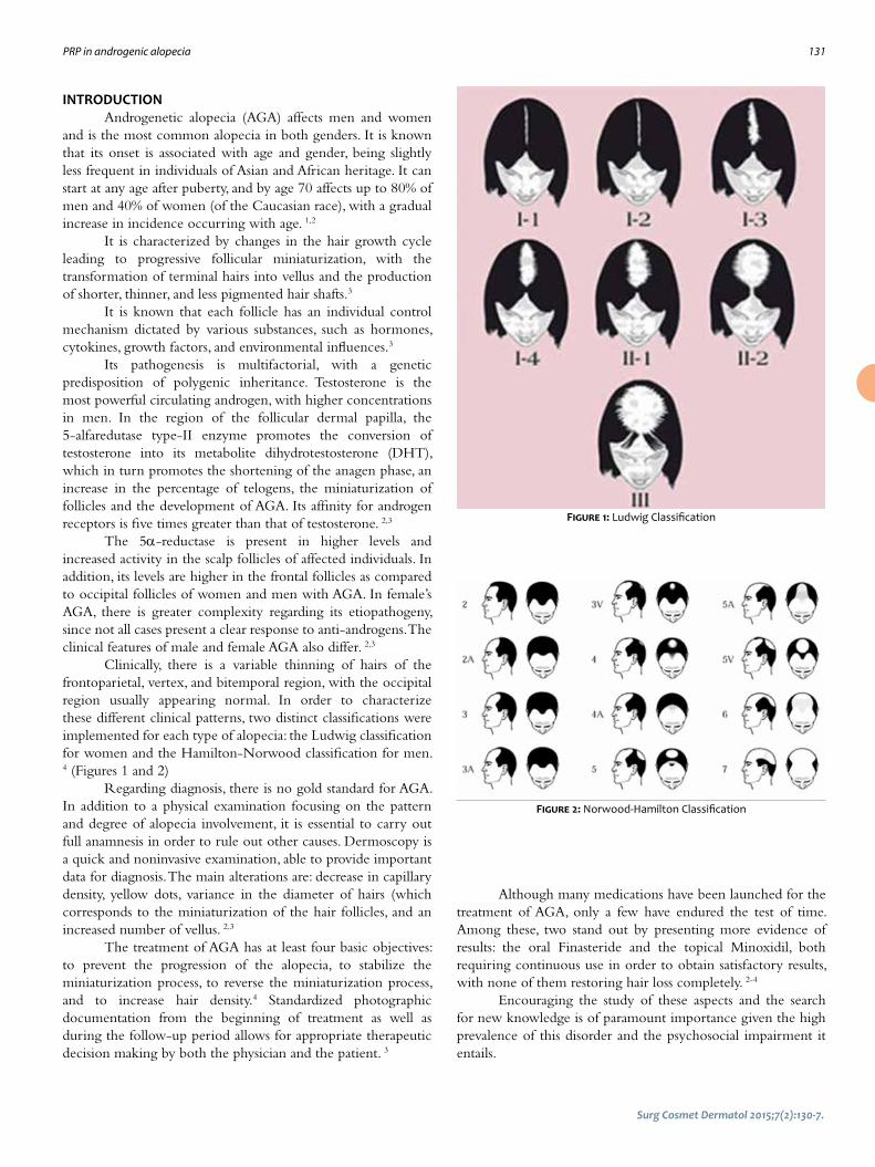
PRP in androgenic alopecia 131
Surg Cosmet Dermatol 2015;7(2):130-7.
INTRODUCTIONAndrogenetic alopecia (AGA) affects men and women
and is the most common alopecia in both genders. It is known that its onset is associated with age and gender, being slightly less frequent in individuals of Asian and African heritage. It can start at any age after puberty, and by age 70 affects up to 80% of men and 40% of women (of the Caucasian race), with a gradual increase in incidence occurring with age. 1,2
It is characterized by changes in the hair growth cycle leading to progressive follicular miniaturization, with the transformation of terminal hairs into vellus and the production of shorter, thinner, and less pigmented hair shafts.3
It is known that each follicle has an individual control mechanism dictated by various substances, such as hormones, cytokines, growth factors, and environmental influences.3
Its pathogenesis is multifactorial, with a genetic predisposition of polygenic inheritance. Testosterone is the most powerful circulating androgen, with higher concentrations in men. In the region of the follicular dermal papilla, the 5-alfaredutase type-II enzyme promotes the conversion of testosterone into its metabolite dihydrotestosterone (DHT), which in turn promotes the shortening of the anagen phase, an increase in the percentage of telogens, the miniaturization of follicles and the development of AGA. Its affinity for androgen receptors is five times greater than that of testosterone. 2,3
The 5a-reductase is present in higher levels and increased activity in the scalp follicles of affected individuals. In addition, its levels are higher in the frontal follicles as compared to occipital follicles of women and men with AGA. In female’s AGA, there is greater complexity regarding its etiopathogeny, since not all cases present a clear response to anti-androgens. The clinical features of male and female AGA also differ. 2,3
Clinically, there is a variable thinning of hairs of the frontoparietal, vertex, and bitemporal region, with the occipital region usually appearing normal. In order to characterize these different clinical patterns, two distinct classifications were implemented for each type of alopecia: the Ludwig classification for women and the Hamilton-Norwood classification for men. 4 (Figures 1 and 2)
Regarding diagnosis, there is no gold standard for AGA. In addition to a physical examination focusing on the pattern and degree of alopecia involvement, it is essential to carry out full anamnesis in order to rule out other causes. Dermoscopy is a quick and noninvasive examination, able to provide important data for diagnosis. The main alterations are: decrease in capillary density, yellow dots, variance in the diameter of hairs (which corresponds to the miniaturization of the hair follicles, and an increased number of vellus. 2,3
The treatment of AGA has at least four basic objectives: to prevent the progression of the alopecia, to stabilize the miniaturization process, to reverse the miniaturization process, and to increase hair density.4 Standardized photographic documentation from the beginning of treatment as well as during the follow-up period allows for appropriate therapeutic decision making by both the physician and the patient. 3
Although many medications have been launched for the treatment of AGA, only a few have endured the test of time. Among these, two stand out by presenting more evidence of results: the oral Finasteride and the topical Minoxidil, both requiring continuous use in order to obtain satisfactory results, with none of them restoring hair loss completely. 2-4
Encouraging the study of these aspects and the search for new knowledge is of paramount importance given the high prevalence of this disorder and the psychosocial impairment it entails.
Figure 1: Ludwig Classification
Figure 2: Norwood-Hamilton Classification

132 Vasconcelos RCF, Azuaga K, Arenas GCF, Vasconcelos JGF, Borelli NS
The role of platelets in hemostasis is well known, nevertheless the use of platelets as a vehicle for storage and transport of cellular signals is a new concept. 5
The platelet-rich plasma (PRP) is a product derived from laboratory processing of autologous blood collected in the pre-operative period, processed for concentration of platelets, and which is rich in growth factors that are released from alpha granules contained within the platelets after platelet activation. All protein load is released within 48 hours. Due to the fact it is autogenous, it is an organic, non-toxic, and non-immunoreactive product. 6-8
Assuming the influence of platelets in the hemostasis processes, the purpose of using PRP is to accelerate tissue regeneration starting from the endothelial lesion, inflammatory processes with the presence of macrophages and neutrophils, and regeneration and healing processes, where there is a presence of growth factors derived from the platelets that cause cell proliferation and differentiation up until the complete repair and regeneration of the damaged tissue. 5
The technique was first used in dentistry and later in orthopedics, with scientifically proven benefits. Currently there are other areas of application of PRP, for example, in the treatment of photoaging, 9 hair implant, 10-12 diabetic foot, 13,14 plastic surgery, 15-17 or simply to promote angiogenesis in tissues with poor vascularization or impaired circulation. 14, 18-24
In the present study the authors will evaluate the effect of PRP in patients with an androgenetic alopecia picture.
OBJECTIVEThe objective of the present study is to assess hair
growth through cell stimulation performed with subcutaneous applications of PRP in the affected regions. No additional treatment was associated in order to allow the assessment of the response of isolated PRP in patients with various degrees of AGA.
METHODOLOGYEighteen volunteer individuals were selected, men and
women between the ages of 18 to 60 years. The exclusion criteria were: patients with hormone disorders, nutritional deficiency, anemia, psoriasis, autoimmune disease, diabetics, pregnant women, nursing mothers, patients on anticoagulants, and use of oral isotretinoin.
The patients underwent an anamnesis conducted by the responsible physician, with the request of laboratory tests such as: complete blood count, serum iron, ferritin, zinc, TSH, free T4, ANA and fasting glucose, for evaluation and selection of individuals.
All project participants signed a free and informed term of consent in which all steps of the study, guidelines, and possible side effects were explained. An authorization form for the disclosure of results and photographs in the media was also signed. The project proposal was sent to an Ethics Committee and was approved under the protocol CAAE 25086613. 4. 0000 0081.
All procedures, such as assessments, PRP preparation, and injections were performed at the Instituto de Terapia Celular
Aplicada – ITCA (Applied Cell Therapy Institute), a private practice involved in the research project, in conjunction with the study team, physicians, and a biologist.
The PRP was processed by the biologist responsible for the research, according to the protocol registered as Protocolo Cantadori (Reg. 508. 102 – Biblioteca Nacional do RJ – Rio de Janeiro National Library) and in compliance with all biosafety and asepsis standards.
Three applications have been programmed PRP with 21 days of interval. In all sessions, the volunteers underwent the process of having blood collected for the PRP preparation and injections in the affected areas immediately after. The procedure had an average duration of 90 minutes per patient, always with the supervision of the biologist and the physician in charge of the project.
The infiltrations were carried out by the physician in charge of the experiment, with sterile material and 26G x ½ needles. PRP was administered intradermally at a dose of 0.2 ml of on each point of the affected region, with spaces of approximately 2 cm between these points. The volume of processed PRP was individualized according to the size of the area to be treated in each patient.
The volunteers were evaluated through photographs and dermoscopy – both carried out before the treatment and before each session. Thirty days after the end of the third application, the patients returned for new photographic and dermoscopic records, having answered a questionnaire containing a satisfaction scale for the treatment, and for the final evaluation of the project.
RESULTSOf the 18 patients treated, one dropped out after the first
application, and another did not return for the final records.Among the seven women in the group, the AGA
classification (according to the Ludwig scale) fell between GI2 and GII1. (Chart 1)
In the nine men, grades ranged from GIIA to GVI, according to Norwood-Hamilton classification. (Chart 2)
All patients were assessed after the end of the application by the patient himself or herself and by an external observer through photographic records, in addition to the pre- and post-treatment dermoscopic analysis.
The results are shown in Table 1.All patients showed some degree of improvement both
in his or her own assessment and in the external observer’s
Chart 1: Classification of female patients according to the Ludwig classification
Patient No. 1 Grade II1Patient No. 2 Grade I2Patient No. 3 Grade I3Patient No. 4 Grade I4Patient No. 5 Grade I3Patient No. 6 Grade I3Patient No. 7 Grade I4
Surg Cosmet Dermatol 2015;7(2):130-7.

PRP in androgenic alopecia 133
evaluation. (Figures 3 to 6)Chart 3 shows the patients’ degree of improvement
regardless of gender.1. The Mann-Whitney test25 was applied in order to
compare the female or male patients regarding the differences in percentages (∆%) observed between the patients’ own evaluations and those of the external observer (Chart 1). For calculating the values of ∆%, the following formula was applied:
1. The Wilcoxon test25 was used in order to compare the evaluation scores (%) of each patient and the observer regarding the degree of improvement. This test was applied regardless of the patient’s gender (Chart 2). As can be seen, in the women’s group the average improvement was 42.85% in the evaluation of the patients themselves, while for the external observer it was 35.71%. In the men’s group, the average improvement according to the patients was 25.55%, while for the external observer it was 18. 88%. The percentage of improvement in all patients was 33.12% according to the patients, and 26.25% for the external observer.
From the dermoscopy, the following alterations were observed: thickening of the hair shafts, improvement of local circulation, and an increased number of hair follicles (Figures
Chart 2: Classification of male patients according to the Hamilton-Norwood classification
Patient No. 2 Grade III VPatient No. 2 Grade III VPatient No. 3 Grade II APatient No. 4 Grade VPatient No. 5 Grade VIPatient No. 6 Grade III VPatient No. 7 Grade IIIPatient No. 8 Grade V APatient No. 9 Grade IV A
Table 1: Male or female patients according to the percentage differences between the patient’s and observer’s assessments
Female Male Patient Observer Δ% Patient Observer Δ% 70 40 42,9 20 20 0 50 50 0 20 10 50 30 30 0 20 10 50 50 40 20 30 20 33,3 40 30 25 30 0 33,3 10 20 100 30 30 0 50 40 20 30 20 33,3 20 20 0,0 30 20 33,3Mean 42,9 35,7 1,1 25 18,9 25,9Median 50 40 20 30 20 33,3
z = 1,32 (p = 0,1858)∆% = patient evalution – observer evalution x 100
patient evalution
Figure 3: Pre- and post-treatment with PRP
Figure 4: Pre- and post-treatment with PRP
Surg Cosmet Dermatol 2015;7(2):130-7.

134 Vasconcelos RCF, Azuaga K, Arenas GCF, Vasconcelos JGF, Borelli NS
7 and 8).Regarding adverse effects, only local pain at the time of
the application was reported by all patients. Two patients had persistent pain for two days, one patient reported a headache picture one day after the application, and the other 12 patients reported improvement in pain a few hours after the procedure. Also, an erythematous halo was also observed at the point of injection, with an absence of cases of local hematoma. The pain and erythema were classified according to a scale ranging from 1+ to 4+ (maximum) and are depicted in Graph 1:
No other symptoms or adverse effects were reported by the patients.
DISCUSSIONGrowth factors can act as mitogenic agents – enhancing
the proliferation of certain cell types or morphogenic agents – thereby altering the cellular phenotype. PRP specific studies identify a complete list of growth factors, including PDGF, FGF, TGF, IGF, EGF, and VEGF. 6-8
For cases of alopecia there are studies showing the PRP efficiency in hair growth. In his thesis on the application of PRP in hair micro-implant surgeries, Mates 26 verified the effectiveness of growth factors in hair density and growth, rapid decrease of apoptosis, and using angiogenesis, he succeeded in stimulating new and efficient mitoses for the resumption of the new anagen phase, thus acting as an accelerator of the regeneration and tissular remodeling in several lesion types.
Based on the scientific evidence of the reported effects of PRP, the authors of the present study proposed to evaluate the isolated response from the PRP in AGA cases. As stated, all patients showed some degree of improvement, resulting in an average of 33%. Statistical analysis also showed that the percentages of satisfaction informed by the patients were significantly higher than those observed by the external observer (p = 0.0072). This fact was aligned with the positive answer of most patients when asked if they would undergo additional sessions of the procedure for better results. Regarding adverse effects, the complaint of all
Figure 5: Pre- and post-treatment with PRP Figure 6: Pre- and post-treatment with PRP
Surg Cosmet Dermatol 2015;7(2):130-7.
Graph 1: Representa-tion of adverse effects (pain and erythema) observed during or after the application
Dor Erythema

PRP in androgenic alopecia 135
patients was pain at the time of the application, with resolution within a few hours.
Regarding the changes observed through dermoscopy in the present study, an improvement in local circulation, improved miniaturization, and increased follicles could be observed. Those findings were in line with an article published in 2013, which evaluated the action of PRP in the hair follicle, revealing a significant difference in the number of new follicles in the treated area. 27
In the treatments already recommended for AGA, pharmacological measures differ between men and women. 1, 4 Uneven results were observed between the genders in the present study, with a clearer and more satisfactory response in the female group (mean = 42.9%) as compared to the men’s (mean = 25.6%).
Although the etiologies of male and female AGA are different, the follicular alteration appears to be similar, with the presence of follicular miniaturization. In women, however, the hormonal interference is uncertain and it is suspected that other
hormones, such as estrogen and prolactin, may be involved. 2, 3
The authors have questioned whether this difference found in the present study’s results could be simply attributed to the lower androgen influence or the picture’s clinical stage regarding the male volunteers., as has been described by JM Pereira et al.. 28
Regarding side effects, the authors observed only pain and erythema, with spontaneous regression. Published studies also show that the biological action of growth factors only accelerates the entire process of regeneration and recovery, without posing any carcinogenic potential. 29 A study conducted in 2009 aimed at treating a basal cell carcinoma in a 54-year-old patient showed a possible regression in the tumor’s stage after treatment with PRP. 29
According to the literature, the PRP’s growth factors act as mediators in cell maturation and are responsible for the tissue damage repair processes. They have an important angiogenesis action, increasing the local microcirculatory process and activating various cell groups in the integration and vitality of tissues. 26, 30,31
Figure 7: Female patient dermoscopy
Figure 8: Male patient dermoscopy
Surg Cosmet Dermatol 2015;7(2):130-7.

136 Vasconcelos RCF, Azuaga K, Arenas GCF, Vasconcelos JGF, Borelli NS
REFERENCES1. Sampaio SAP, Rivitti EA. Dermatologia Básica. 3a ed São Paulo: Artes Mé-
dicas; 2007. p .432-34.
2. Torres F, Tosti A. Atlas de Doenças do Cabelo. Rio de Janeiro: Revinter;
2013. p .13-63.
3. Brenner-Mulinari F, Seidel G, Hepp T. Entendendo a alopecia androgené-
tica. Surg Cosmet Dermatol. 2011;3(4): 329-37.
4. Junior WB, Chiacchio ND, Criado PR. Tratado de Dermatologia. São Paulo:
Atheneu: 2010. p.984-99
5. Pontual MAB, Magini RS. Plasma Rico em Plaquetas PRP e Fatores de
Crescimento das Pesquisas Científicas à Clínica Odontológica. São Paulo:
Ed.Santos; 2004. p .163-87.
6. Lieberman J, Daluiski A, Einhorn TA. The Role of Growth Factors in the Re-
pair of Bone. Biology and Clinical Applications. Investigation performed
at the Department of Orthopaedic Surgery, University of California at Los
Angeles Medical Center, Los Angeles, California, and the Department of
Orthopaedic Surgery, Boston University Medical Center, Boston, Massa-
chuset. J Bone Joint Surg Am. 2002;84(6):1032-44.
7. Landesberg R, Roy M. Glickman RS. Quantification of Growth Factor Le-
vels Using a Simplified Method of Platelet-Rich Plasma Gel Preparation. J
Oral Maxillofac Surg. 2000; 58(3):297-300
8. Sierra ALP, Aranegui RO, Ares MM, Martínez JLQ, González JMM. Quantii-
cation of growth factors by using a new system for obtaining platelet-ri-
ch plasma. Med Oral Patol Oral Cir Bucal. 2011;16(4): 614-8.
9. Redaelli A, Romano D, Marciàno A. Face and Neck Revitalizaton with Pla-
telets - Rich Plasma (PRP).Clinical Outcome in a series of 23 Consecutively
Treated Patients. J Drugs Dermatol.2010;9( 5): 466-72
10. Uebel CO. Ação do plasma rico em plaquetas e seus fatores de cresci-
mento na cirurgia dos microimplantes capilares [tese]. Porto Alegre: PU-
CRS, 2006.
11. Li ZJ, Choi HI, Choi DK, Sohn KC, Im M, Seo YJ, et al. Autologous plate-
let-rich plasma: a potential therapeutic tool for promoting hair growth.
Department of Dermatology, School of Medicine, Chungnam National
University, Daejeon, Korea. Dermatol Surg. 2012; 38(7): 1040-6
12. Takikawa M, Nakamura S, Nakamura S, Ishirara M, Kishimoto S, Sasaki K, et
al. Enhanced Effect of Platelet-Rich Plasma Containing a New Carrier on
Hair Growth. Dermatol Surg. 2011; 37(12): 1721-9.
13. Bennett SP, Griffiths GD, Schor AM, Leese GP, Schor SL. Growth factors in
the treatment of diabetic foot ulcers. J Surg. 2003;90(2): 133-46.
14. Buza PW. Combination Platelet Leukocyte Rich Plasma (PLRP/PRP) and
Hyperbaric Systemic Oxygenation (HBO) is Safe and Effective in the Tre-
atment of Refractory Wounds. 2003, 2-13 Não localizei publicação!
15. Cervelli V, Palla L, Pascali M, Angeli B de, Curcio BC, Gentile P. Autologous
Platelet-Rich Plasma Mixed with Purified . Fat Graft in Aesthetic Plastic
Surgery . Aesth Plast Surg. 2009,33(5): 716-21.
16. Clevens RA . Autologous Platelet Rich Plasma in Facial Plastic Surgery.
Proceedings. 8th International Symposium of Facial Plastic Surgery.
NewYork city, USA. Center for Facial Cosmetic Surgery - Melbourne, FL.
2002, May; 1-5.
17. Sclafani AP, McCormick AS . Induction of Dermal Collagenesis, Angioge-
nesis,and Adipogenesis in Human Skin by Injection of Platelet-Rich Fi-
brin Matrix. Arch Facial Plast Surg. 2012;14 (2):132-6.
CONCLUSIONThe fundamental objectives in the treatment of AGA are
to increase the scalp’s coverage and to slow down the progression of the loss of hair. In addition, explaining the condition’s mechanisms to the patient and managing their expectations with the therapy is critical for treatment adherence.
According to the Brazilian Society of Dermatology, alopecia complaints are among the ten most frequent received by dermatology practices from patients of 15 to 39 years of age. 3 Despite this high frequency and the already available therapeutic measures, satisfactory treatment of AGA remains a challenge for the dermatologist. Thus, although there are many studies on the subject, scientific studies that demonstrate new therapeutic options become relevant.
In this study, the authors found an efficient and satisfactory response with the technique of PRP injection, even as an isolated therapy. It is known that in AGA the action of DHT is important for triggering the picture, and treatments that act on that front are required. As the PRP does not have this action, it emerges as an important measure in the association with androgenetic blockage therapies for the maintenance and improvement of the therapeutic response.
Based on the literature review and the results of the present study, this method offers an interesting alternative for the treatment of AGA, which suggests the availability of an effective option to associate with the existing ones in order to improve the therapeutic results of AGA. l
Surg Cosmet Dermatol 2015;7(2):130-7.

PRP in androgenic alopecia 137
18. Roy S, Driggs J, Elgharably H, Biswas S, Findley M, Khanna S, et al. Platelet-
-rich fibrin matrix improves wound angiogenesis via inducing endothe-
lial cell proliferation. Wound Rep Reg. 2011; 19(6): 753-66.
19. Peerbooms JC, Sluimer J, Bruijn DJ, Gosens T. Positive Effect of an Auto-
logous Platelet Concentrate in Lateral Epicondylitis in a Double-Blind.
Randomized Controlled Trial: Platelet-Rich Plasma Versus Corticosteroid
Injection With a 1-Year. Am J Sports Med. 2010; 38(2): 255-62.
20. Asahara T, Bauters C, Zheng LP, Takeshita S, Bunting S, Ferrara N, et al.
Synergistic Effect of Vascular Endothelial Growth Factor and Basic Fi-
broblast Growth Factor on Angiogenesis in Vivo. Circulation. 1995;92(9
Suppl):II365-71.
21. Matsui M, Tabata T. Enhanced Angiogenesis by Multiple Release of Plate-
let-Rich Plasma Contents and Basic Fibroblast Growth Factor from Gela-
tin Hydrogels. Acta Biomaterialia. 2012; 8: 1792-1801.
22. Anitua E, Sánchez M, Zalduendo MM, Fuente M de La, Prado R, Orive GI, et
al. Fibroblastic response to treatment with different preparations rich in
growth factors. Cell Prolif;. 2009;42(2):162-70.
23. Yoshikawa T, Tohyama H, Katsura T, Kondo E, Kotani Y, Matsumoto H, et
al. Effects of Local Administration of Vascular Endothelial Growth Fac-
tor on Mechanical Characteristics of the Semitendinosus Tendon Graft
After Anterior Cruciate Ligament Reconstruction in Sheep. Department
of Sports Medicine, Hokkaido University School of Medicine, Sapporo,
Japan, the Department of Orthopaedic Surgery, Keio University School
of Medicine,Tokyo, Japan, and the Department of Orthopaedic Surgery,
Hokkaido University School of Medicine, Sapporo, Japan. Am J Sports
Med. 2006; 34(12):1918-25.
24. Vang SN, Brady CP, Christensen KA, Allen KR, Anderson J E, Isler JR, et al.
Autologous Platelet Gel in Coronary Artery Bypass Grafting: Effects on
Surgical Wound Healing. J Extra Corpor Technol. 2007;39(1):31-8.
25. Siegel SE, Castellan JR, NJ. Estatística não paramétrica para ciências do
comportamento. 2ª ed. Porto Alegre: Artmed; 2006. p .448
26. Mates PC. Plasma rico em plaquetas e seus fatores de crescimento na
cirurgia de microimplantes capilares. Curitiba: Universidade Tuiuti do Pa-
raná; 2011.
27. Miao Y1, Sun YB, Sun XJ, Du BJ, Jiang JD, Hu ZQ. Promotional effect of
platelet-rich plasma on hair follicle reconstitution in vivo. Dermatol Surg.
2013;39(12): 1868-76.
28. Yoshitake T, Takaeda A, Ohki K, Inoue Y, Yamawaki T, Otsuka S, et al. Five-
-year efficacy of finasteride in 801 Japanese men with androgenetic alo-
pecia. J Dermatol. 2015; 42(1): 1-4.
29. Kleintjes WG. Treatment of basal cell carcinoma with autogenous growth
factors and adipose-derived stem cells. Plast Reconstr Surg. 2010,;126(6):
312-13.
30. Weinberg WC, Brown PD, Stetler-Stevenson WG, Yuspa SH. Growth factors
specifically alter hair follicle cell proliferation and collagenolytic activity
alone or in combination. Differentiation. 1990;45(3):168-78.
31. Vick VL, Holds JB, Hartstein ME, Rich RM, Davidson BR. Use of autologous
platelet concentrate in blepharoplasty surgery. Ophthal Plast Reconstr
Surg. 2006;22(2):102-4.
Surg Cosmet Dermatol 2015;7(2):130-7.

138
Surg Cosmet Dermatol 2015;7(2):138-43.
OriginalArticles
Clinical and histological effects of weakly focused high-frequency ultrasounds on human subcutaneous adipose tissueOs efeitos clínicos e histológicos doS ultrassoNS de alta frequência minimamente focadoS no tecido subcutaneo humano
DOI: http://dx.doi.org/10.5935/scd1984-8773.201572633
ABSTRACTIntroduction: Lipo-reductive ultrasound devices are commonly used for non-invasive body sculpting purposes because they can achieve clinically appreciable subcutaneous fat pad reduction and are safe and well tolerated by patients.Objective: This study aims to evaluate the morphological changes induced on the diffe-rent cell components of human skin as a result of weakly focused high-frequency ultra-sound. Methods: Full-thickness skin samples exposed or not to ultrasound ex vivo, and skin biop-sies from patients pre-treated or not with ultrasound before lipo-reductive surgery, were analyzed to evaluate possible morphological changes of adipocytes. Adipocyte apoptosis and triglyceride release were also assayed. Clinical evaluation of the effects of repeated ultrasound treatment vs. sham-treatment was also performed.Results: Compared with the control samples, ultrasound treatment induced an appre-ciable reduction in adipocyte size, the appearance of plasma membrane micropores and triglyceride release, without appreciable changes in microvascular, stromal and epidermal components, and in the number of apoptotic adipocytes. Clinically, the ultrasound treat-ment resulted in a time-dependent significant reduction of abdominal fat.Conclusions: This study supports the safety and efficacy of trans-cutaneous weakly focused high-frequency ultrasounds for localized fat reduction, and provides experimental evidence for a possible mechanism of action.Keywords: adipocytes, white; high-intensity focused ultrasound ablation; subcutaneous fat
RESU MO Introdução: Os aparelhos lipo-redutores de ultrassom são comumente utilizados em procedimentos não-invasivos para a estética corporal por provocarem reduções clinicamente significativas dos depósitos de gordura subcutânea.Objetivo: Avaliar as alterações morfológicas induzidas em componentes das células da pele humana, causadas por ultrassom de alta frequência minimamente focado.Métodos: Amostras de pele de espessura total expostas ou não à ultrassonografia ex-vivo e biópsias de pele de pacientes pré-tratados ou não com ultrassom antes da cirurgia lipo-redutora foram analisadas para avaliação de possíveis alterações morfológicas em adipócitos. Foram analisadas apoptose dos adipócitos, liberação de triglicérides e mudança clínica após tratamento repetitivo com ultrassom, em comparação a tratamentos placebo.Resultados: Em comparação com as amostras-controle, o tratamento de ultrassom induziu uma redução significativa no tamanho dos adipócitos, assim como o aparecimento de microporos na membrana plasmá-tica e liberação de triglicérides, sem alterações apreciáveis em componentes microvasculares, do estroma e da epiderme, assim como no número de adipócitos apoptóticos. Clinicamente, o tratamento com ultrassom provocou uma significativa redução, tempo-dependente, da gordura abdominal.Conclusões: O presente estudo reforça a segurança e a eficácia do ultrassom transcutâneo de alta fre-qüência minimamente focado na redução da gordura localizada, fornecendo evidência experimental de um possível mecanismo de ação.Palavras-chave: ablação por ultrassom focalizado de alta intensidade; adipócitos brancos; gordura subcutânea
Authors: Daniele Bani1
Alessandro Quattrini Li2
Giulia Lo Russo3
1 Full Professor of Histology, University of Florence - Florence, Italy
2 Plastic Surgeon, University of Florence
3 Plastic Surgeon, Head of Plastic Surgery Unit, University of Florence
Correspondence: Daniele Bani University of Florence Dept. Experimental & Clinical Medicine, Research Unit of Embryology & Histology Viale G.Pieraccini 6 I-50139 Florence, Italy Phone: +39 055 2758 153 E-mail: [email protected]
This study was performed at the Department of Experimental & Clinical Medicine, Research Unit of Histology & Embryology Dept. Sur-gery & Translational Medicine, Plastic Surgery Unit, University of Florence - Florence, Italy
Received on: 21 December 2014Approved on: 17 June 2015
Financial support: This study was funded by the authors.Conflict of interest: none

Surg Cosmet Dermatol 2015;7(2):138-43.
Focused ultrasound in adipose tissue 139
INTRODUCTIONTo fulfill the increasing demand for non-invasive fat
reduction methods, as an alternative to liposuction, 1 numerous physical treatments--namely mechanical and electric stimulation, radio frequency, and low-level laser--have been investigated. However, most of them have not met with expectations, 1-4 and some have also raised safety issues.5 Of the most promising non-invasive approaches to lipo-reduction, ultrasound holds a pivotal place. 6-9 In spite of its accelerating use in aesthetic medicine (with satisfactory results), the mechanisms of action on adipose cells remain to be fully elucidated and may likely vary according to the mode of ultrasound delivery. In fact, ultrasonic energy can be transmitted to the skin in non-focused or focused modes. In the non-focused mode, energy attenuates with depth: thus, to deliver enough energy to the subcutaneous fat, the superficial skin is exposed to maximum energy intensity and may undergo injury. Instead, focused ultrasound can be concentrated in a defined subcutaneous area to produce clinically relevant fat lysis while limiting damage to the upper tissues. 10,11 However, focused ultrasounds can induce marked heating, thereby causing adipocyte necrosis in the treatment area.12
The most recent lipo-reductive ultrasound devices are specifically designed to prevent tissue injury. Contour ITM (UltraShape, Yoqneam, Israel), a focused ultrasound emitter, was first demonstrated to achieve selective adipocyte lysis and clinically relevant reduction of the volume of subcutaneous fat pad, in the absence of significant adverse reactions.7,13 Med2ContourTM (General Project, Montespertoli, Italy) takes advantage of two angled non-focused transducers that create a weakly focused ultrasound field where the two beams overlap, i.e. within the subcutaneous fat pad.14,15
The cellular mechanisms underlying the lipo-reductive effects of ultrasound are not fully understood and are thus a matter for investigation. It has been shown that the impact of ultrasounds on adipose cells can induce transitory pore opening at the plasma membrane, allowing triglyceride leakage.14,15
The current study aims at providing further evidence for the efficacy of weakly focused high-frequency ultrasounds, delivered by the Med2ContourTM, for non-invasive lipo-reduction and to search for possible morphological clues that can help understand the mechanism of action on adipose cells.
MATERIALS AND METHODSThis study complied with the guidelines of the
Declaration of Helsinki, as amended in Edinburgh, 2008. It was approved by the Ethical Committee of the Faculty of Medicine, University of Florence, Italy. All subjects gave written informed consent to their participation in the study.
Study on human skin explantsFull-thickness biopsies of normal skin, about 15 mm
thick, were taken at a surgery from three patients undergoing abdominoplasty (1 male, 2 females, aged 40-65 years). Each biopsy was cut in half, and each half placed in a Petri dish on ice, the subcutaneous tissue facing downwards, and mixed with 2
ml of pre-oxygenated incubation medium (Dulbecco’s modified Eagle medium, DMEM; Gibco Invitrogen, Milan, Italy). A first sample was treated with non-focused ultrasound using the Med2ContourTM device (General Project, Montespertoli, Italy) set at: 3 W power output, 20 kHz frequency, pulsed mode (2 pulses, 6 s. each, separated by a 10 s. pause). A single transducer of the Med2ContourTM was placed in direct contact with the epidermis through a thin layer of Aquasonic ClearTM ultrasound gel (Parker, Fairfield, USA). The above power and frequency settings were adopted because they were similar to those yielding the best clinical performance,7,14,15 while the timing protocol was chosen to avoid tissue overheating, considering that the skin explants lacked blood flow-related temperature homeostasis. Tissue temperature was continuously monitored with a digital thermometer and found not to exceed 38° C. The other specimen was sham-treated (i.e. subjected to the same handling procedure but with no ultrasound emission) and used as control. At the end of the experiments, fragments of adipose tissue were taken from the central part of the treated and control specimens, fixed in isotonic 4% glutaraldehyde and 1% OsO
4,
dehydrated and embedded in Epon epoxy resin (Fluka, Buchs, Switzerland) for light and electron microscopic studies.
Adipocyte size was measured by computer-aided morphometry on digital photomicrographs of semi-thin sections, 2 mm thick. The surface area of adipocyte lipid vacuoles was measured on 10 randomly chosen micrographs (test area: 65,700 mm2) from each specimen using ImageJ 1.33 image analysis program (http://rsb.info.nih.gov/ij), upon setting an appropriate threshold to only include the osmiophilic lipid vacuoles of the adipocytes. Vacuolar profiles ≤ 1000 mm2, consistent with polar cross-sections, were excluded. Data were reported as mean values (± SEM) of the control and treated groups. For transmission electron microscopy, ultrathin sections were stained with uranyl acetate and alkaline bismuth subnitrate, viewed and photographed under a JEM 1010 transmission electron microscope (Jeol, Tokyo, Japan).
In vivo studyThis was performed on three obese volunteers (2 males, 1
female, aged 34-53 years) scheduled for abdominal lipo-reductive surgery, who were subjected to weakly focused high-frequency ultrasounds using the Med 2ContourTMset at: 2 W power output, pulsed mode, 20 kHz frequency, 15 minute treatment. In each patient, the right hypogastrium was the test area whereas the left hypogastrium was the sham-treated area. Two patients received four treatments at 27, 20, 12, and 1 day before surgery. The remaining patient received three treatments at 27, 20, and 12 days before surgery: the aim of this protocol being to study whether the effects of the ultrasound were maintained over time. During the surgery, fragments of subcutaneous adipose tissue were taken from the central part of the test and control areas and processed for ultrastructural examination, as described above. Similarly, during the experiment on skin explants, morphometry of adipocyte size was performed on semi-thin

140 Bani D, Li AQ, Lo Russo G
Surg Cosmet Dermatol 2015;7(2):138-43.
and focal ruptures of the peripheral cytoplasmic rim (Figure 4). Such ruptures were usually restricted to small areas of the cell surface, approx. 0.5-1.5 mm in diameter, but large enough to allow leakage of triglyceride droplets from the inner cytoplasmic vacuole to the extracellular space. Of note, no signs of adipocyte demise or cell remnants were observed. Of note, the cellular and intercellular stromal components showed a normal appearance, with no signs of damage (data not shown).
Ultrastructural examination of subcutaneous fat biopsies taken at surgery from ultrasound-pretreated abdominal skin areas showed different features from those of the ex vivo specimens. In all the patients examined, regardless of whether the biopsies were taken one or 12 days after the last ultrasound application, triglyceride leakage from adipocytes was not detected in the images. The interstitial stromal components showed a normal appearance (data not shown). However, adipocytes that had been exposed to ultrasounds consistently showed irregular, winding profiles and multiple lipid droplets clustered in the cytoplasmic
Figure 1: Histological and morphometrical findings of subcutaneous adipocytes from ex vivo skin explants
Compared with the sham-treatment, ultrasound treatment by Med2Contour causes a statistically significant reduction of mean cross-section surface area of lipid vacuoles, related to adipocyte overall volume (Student’s t test). OsO4 fixation/staining and toluidine blue counterstaining. Bars = 10 μm.
sections and ultrastructural analysis on ultra-thin sections of the Epon-embedded specimens.
Further, three overweight patients (2 males, 1 female, aged 30-33 years) were enrolled for assessment of subcutaneous adipose tissue mass in control and ultrasound-treated abdominal skin. They were subjected to four weekly ultrasound treatments, similarly to the above-described protocol. Before each treatment and one week after the last treatment, the thickness of subcutaneous fat pads in the control and test areas were assessed by measuring the depth of skin folds with a Harpenden caliper (FitnessAssist, Wrexham, UK). To normalize individual differences, the values were expressed as percent changes over the initial measures.
Statistical analysis The assayed quantitative parameters were statistically
analyzed with Graph Pad Prism 4.03 statistical software (GraphPad, San Diego, CA, USA), assuming the individual patients as sample units (n=3). Statistical significance of differences between groups was assessed by unpaired Student’s t test; p ≤ 0.05 was considered significant.
RESULTSLight microscopic and morphometric analysis of
semi-thin sections of subcutaneous adipose tissue of the ex vivo skin explants (n=3) showed that ultrasound treatment by Med2ContourTM induced a marked, statistically significant decrease (-23%) in the size of adipocyte lipid vacuoles (Figure 1). Similar findings were observed in the subcutaneous fat biopsies taken at surgery from sham- or Med2ContourTM-pretreated abdominal skin (Figure 2). In the biopsies collected one day after the last ultrasound application (n=2), we found a significant reduction (-26%) of the size of adipocyte lipid vacuoles. In the biopsies taken 12 days after the last ultrasound application (n=1), the treatment induced an even more marked reduction (-47%) of adipocyte lipid vacuoles. No differences were observed among the sham-treated biopsies from the three patients (data not shown).
Ultrastructural analysis of adipose tissue from sham-treated ex vivo skin explants demonstrated normal adipocytes, showing a large, osmiophilic lipid vacuole with a peripheral electron-lucent rim contiguous to a thin cytoplasmic layer containing scanty organelles, pinocytosis micro vesicles and small lipid droplets (Figure 3). Cells were surrounded by a continuous basement membrane. Blood micro vessels, mainly capillaries, and interstitial connective tissue composed of a loose matrix containing thin collagen fibers, showed a normal appearance (data not shown). Normal features of adipocytes and stromal components were also observed in the adipose tissue biopsies taken from sham-treated areas of the three patients enrolled in the study (data not shown). Conversely, the subcutaneous adipose tissue of ultrasound-treated ex vivo skin explants displayed well-appreciable differences compared with the sham-treated specimens. In particular, many adipocytes showed peculiar abnormalities, consisting in lipid droplet clustering
área dos adipócitos
PLACEBO TREATMENT MED2CONTOUR
ADIPOCYTES ÁREA
Placebo Med2Contour

Focused ultrasound in adipose tissue 141
Surg Cosmet Dermatol 2015;7(2):138-43.
Figure 2: Histological and morphometrical findings of subcutaneous adipocytes from abdominal skin biopsies taken at the noted times
after the treatment
Compared with the sham-treatment, ultrasound treatment by Med2Con-tour causes a statistically significant reduction of mean cross-section surface area of lipid vacuoles, related to adipocyte overall volume (Student’s t test). OsO4 fixation/staining and toluidine blue counterstaining. Bars = 10 μm.
Figure 3: Representative TEM images of adipose tissue from sham-treated ex vivo skin explants
A) An adipocyte showing lipid droplets of different size in the peripheral cyto-plasm. B) Detail of the previous panel (arrow) showing intact cytoplasm and continuous plasma membrane rimmed by a basement lamina (arrowheads). Bars = 1 μm.
Figure 4: Representative TEM images of adipose tissue from ex vivo skin explants subjected to ultrasound treatment by Med2Contour
A) Adipocyte showing lipid droplet clustering in the marginal cytoplasm. B) Detail of the previous panel (arrow) showing the droplet cluster. C) A lipid droplet about to break the cytoplasmic rim (arrowhead). D & E) Cytoplasmic ruptures (arrowheads) allowing the leakage of triglyceride droplets in the in-terstitium (asterisks); cf: collagen fibers. Bars = 1 μm.
rim (Figure 5). These features were never found in the sham-treated adipocytes and are consistent with marked reduction of cell volume, conceivably related to triglyceride discharge.
Assessment of subcutaneous adipose tissue mass by plicometry showed a time-related decrease of the measured values in the ultrasound-treated abdominal skin regions as compared with the corresponding sham-treated areas (Table 1). Of note, the decreasing trend in the Med2ContourTM-treated regions continued one week after the last treatment (longer times not assayed).
DISCUSSIONThe present findings indicate that weakly focused high-
frequency ultrasounds delivered by the Med2ContourTM device on human skin can yield a substantial reduction of subcutaneous fat and adipose cell size, confirming the previous clinical and
área dos adipócitos
A B
PLACEBO TREATMENT MED2CONTOUR 1 DAY MED2CONTOUR 12 DAYS
PlaceboMed2Contour
1 day 12 day
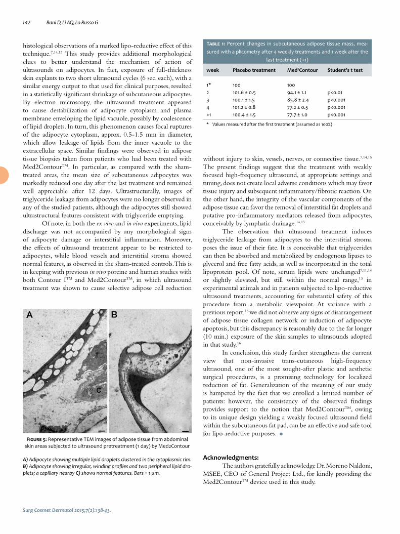
142 Bani D, Li AQ, Lo Russo G
Surg Cosmet Dermatol 2015;7(2):138-43.
histological observations of a marked lipo-reductive effect of this technique.7,14,15 This study provides additional morphological clues to better understand the mechanism of action of ultrasounds on adipocytes. In fact, exposure of full-thickness skin explants to two short ultrasound cycles (6 sec. each), with a similar energy output to that used for clinical purposes, resulted in a statistically significant shrinkage of subcutaneous adipocytes. By electron microscopy, the ultrasound treatment appeared to cause destabilization of adipocyte cytoplasm and plasma membrane enveloping the lipid vacuole, possibly by coalescence of lipid droplets. In turn, this phenomenon causes focal ruptures of the adipocyte cytoplasm, approx. 0.5-1.5 mm in diameter, which allow leakage of lipids from the inner vacuole to the extracellular space. Similar findings were observed in adipose tissue biopsies taken from patients who had been treated with Med2ContourTM. In particular, as compared with the sham-treated areas, the mean size of subcutaneous adipocytes was markedly reduced one day after the last treatment and remained well appreciable after 12 days. Ultrastructurally, images of triglyceride leakage from adipocytes were no longer observed in any of the studied patients, although the adipocytes still showed ultrastructural features consistent with triglyceride emptying.
Of note, in both the ex vivo and in vivo experiments, lipid discharge was not accompanied by any morphological signs of adipocyte damage or interstitial inflammation. Moreover, the effects of ultrasound treatment appear to be restricted to adipocytes, while blood vessels and interstitial stroma showed normal features, as observed in the sham-treated controls. This is in keeping with previous in vivo porcine and human studies with both Contour ITM and Med2ContourTM, in which ultrasound treatment was shown to cause selective adipose cell reduction
without injury to skin, vessels, nerves, or connective tissue.7,14,15 The present findings suggest that the treatment with weakly focused high-frequency ultrasound, at appropriate settings and timing, does not create local adverse conditions which may favor tissue injury and subsequent inflammatory/fibrotic reaction. On the other hand, the integrity of the vascular components of the adipose tissue can favor the removal of interstitial fat droplets and putative pro-inflammatory mediators released from adipocytes, conceivably by lymphatic drainage.14,15
The observation that ultrasound treatment induces triglyceride leakage from adipocytes to the interstitial stroma poses the issue of their fate. It is conceivable that triglycerides can then be absorbed and metabolized by endogenous lipases to glycerol and free fatty acids, as well as incorporated in the total lipoprotein pool. Of note, serum lipids were unchanged7,11,14 or slightly elevated, but still within the normal range,13 in experimental animals and in patients subjected to lipo-reductive ultrasound treatments, accounting for substantial safety of this procedure from a metabolic viewpoint. At variance with a previous report,16 we did not observe any signs of disarrangement of adipose tissue collagen network or induction of adipocyte apoptosis, but this discrepancy is reasonably due to the far longer (10 min.) exposure of the skin samples to ultrasounds adopted in that study.16
In conclusion, this study further strengthens the current view that non-invasive trans-cutaneous high-frequency ultrasound, one of the most sought-after plastic and aesthetic surgical procedures, is a promising technology for localized reduction of fat. Generalization of the meaning of our study is hampered by the fact that we enrolled a limited number of patients: however, the consistency of the observed findings provides support to the notion that Med2ContourTM, owing to its unique design yielding a weakly focused ultrasound field within the subcutaneous fat pad, can be an effective and safe tool for lipo-reductive purposes. l
Acknowledgments:The authors gratefully acknowledge Dr. Moreno Naldoni,
MSEE, CEO of General Project Ltd., for kindly providing the Med2ContourTM device used in this study.
Figure 5: Representative TEM images of adipose tissue from abdominal skin areas subjected to ultrasound pretreatment (1 day) by Med2Contour
A) Adipocyte showing multiple lipid droplets clustered in the cytoplasmic rim. B) Adipocyte showing irregular, winding profiles and two peripheral lipid dro-plets; a capillary nearby C) shows normal features. Bars = 1 μm.
Table 1: Percent changes in subcutaneous adipose tissue mass, mea-sured with a plicometry after 4 weekly treatments and 1 week after the
last treatment (+1)
* Values measured after the first treatment (assumed as 100%)
week Placebo treatment Med2Contour Student’s t test 1* 100 100 2 101.6 ± 0.5 94.1 ± 1.1 p<0.013 100.1 ± 1.5 85.8 ± 2.4 p<0.0014 101.2 ± 0.8 77.2 ± 0.5 p<0.001+1 100.4 ± 1.5 77.7 ± 1.0 p<0.001

Focused ultrasound in adipose tissue 143
Surg Cosmet Dermatol 2015;7(2):138-43.
REFERENCES1. Coleman KM, Coleman WP 3rd, Benchetrit A. Non-invasive, external ultra-
sonic lipolysis. Semin Cutan Med Surg. 2009;28(4):263-7.
2. Neira R, Arroyave J, Ramirez H, Ortiz CL, Solarte E, Sequeda F, et al. Fat li-
quefaction: effect of low-level laser energy on adipose tissue. Plast Re-
constr Surg. 2002;110(3):912-22.
3. Jackson RF, Dedo DD, Roche GC, Turok DI, Maloney RJ. Low-level laser
therapy as a non-invasive approach for body contouring: a randomized
controlled study. Lasers Surg Med. 2009;41(10):799-809.
4. Manuskiatti W, Wachirakaphan C, Lektrakul N, Varothai S. Circumference
reduction and cellulite treatment with a Tri-Polar radiofrequency device:
a pilot study. J Eur Acad Dermatol Venereol. 2009;23(7):820-7.
5. de Felipe I, Del Cueto SR, Pérez E, Redondo P. Adverse reactions after no-
nablative radiofrequency: Follow-up of 290 patients. J Cosmet Dermatol.
2007;6(3):163-6.
6. Zocchi ML. Clinical aspects of ultrasonic liposculpture. Perspect Plast
Surg. 1993;7:153-174.
7. Teitelbaum SA, Burns JL, Kubota J, Matsuda H, Otto MJ, Shirakabe Y, et al.
Noninvasive body contouring by focused ultrasound: safety and efficacy
of the Contour I device in a multicenter, controlled, clinical study. Plast
Reconstr Surg. 2007;120(3):779-89.
8. Brown SA, Greenbaum L, Shtukmaster S, Zadok Y, Ben-Ezra S, Kushkuley
L. Characterization of nonthermal focused ultrasound for noninvasive
selective fat cell disruption (lysis): Technical and preclinical assessment.
Plast Reconstr Surg. 2009;124(1):92-101.
9. Ascher B. Safety and efficacy of UltraShape Contour I treatments to im-
prove the appearance of body contours: multiple treatments in shorter
intervals. Aesthetic Surg J. 2010;30(2):217-24.
10. Fatemi A, Kane MA. High-intensity focused ultrasound effectively reduces
waist circumference by ablating adipose tissue from the abdomen and
flanks: a retrospective case series. Aesthetic Plast Surg. 2010;34(5):577-82.
11. Jewell ML, Baxter RA, Cox SE, Donofrio LM, Dover JS, Glogau RG, et al. Ran-
domized sham-controlled trial to evaluate the safety and effectivenessof
a high-intensity focused ultrasound device for noninvasive body contou-
ring. Plast Reconstr Surg. 2011;128(1):253-62.
12. Kennedy JE, ter Haar GR, Cranston D. High intensity focused ultrasound:
surgery of the future? Br J Radiol. 2003;76(909):590-9.
13. Moreno-Moraga J, Valero-Altés T, Riquelme AM, Isarria-Marcosy MI, de la
Torre JR. Body contouring by non-invasive transdermal focused ultrasou-
nd. Lasers Surg Med. 2007;39(4):315-23.
14. Garcia O Jr, Schafer M. The effects of nonfocused external ultrasound
on tissue temperature and adipocyte morphology. Aesthetic Surg J.
2013;33(1):117-127.
15. Bani D, Quattrini Li A, Freschi G, Russo GL. Histological and ultrastructural
effects of ultrasound-induced cavitation on human skin adipose tissue.
Plast Reconstr Surg Glob Open. 2013;1(6):e41.
16. Palumbo P, Cinque B, Miconi G, La Torre C, Zoccali G, Vrentzos N, et al.
Biological effects of low frequency high intensity ultrasound applica-
tion on ex vivo human adipose tissue. Int J Immunopathol Pharmacol.
2011;24(2):411-22.

OriginalArticles
Treatment of acne scars using the microne-edling and drug delivery techniqueTratamento das cicatrizes de acne com a técnica de microagulhamento e drug delivery
DOI: http://dx.doi.org/10.5935/scd1984-8773.201572641
ABSTRACTIntroduction: Acne vulgaris is one of the most common skin diseases and most often results in scars that are typically associated with aesthetic and psychological problems. To date, there is no standardized treatment for this kind of scarring.Objectives: To evaluate the improvement of distensible type acne scars on the face after treatment with the microneedling technique, followed by the topical application of a gel containing growth factors. Methods: Ten patients with acne scars who were treated from 2012 to 2014 and met the inclusion criteria were selected. All underwent 3 microneedling sessions with intervals of 1 to 2 months, with a 1-year follow up. Pre- and post- procedure photographs were car-ried out with a regular digital camera, and with a digital camera with a 3D system. Skin biopsies were performed prior to treatment, and 30 days after the last session. Results: Eight patients completed the study. Among these, seven showed a random re-duction in the roughness of the scars in the analyzed area, seven showed a decrease in me-lanin with a homogenization of its distribution across the skin, and seven patients showed an increase in hemoglobin in the area being studied. The appearance of the scars varied among the eight patients. Deep ice-pick type scars did not improve with the procedure. Conclusions: The studied treatment promotes overall improvement in skin texture and a discreet effect on acne scars. Keywords: acne vulgaris; cicatrix; combined modality therapy; esthetics
RESU MO Introdução: A acne vulgar é um das doenças de pele mais comuns e, em grande parte das vezes, resulta em cicatrizes que constituem problema estético e psicológico. Até o momento, não há tratamento padronizado para as cicatrizes. Objetivos: Avaliar a melhora das cicatrizes de acne do tipo distensíveis na face após tratamento com a técnica de microagulhamento, seguida da aplicação tópica de gel contendo fatores de crescimento. Métodos: Foram selecionados 10 pacientes portadores de cicatrizes de acne, que preencheram os critérios de inclusão. Todos realizaram três sessões de microagulhamento, com intervalos de um a dois meses, com seguimento de um ano. Foram realizadas fotos pré e pós-procedimento com máquina digital comum e com câmera digital com sistema tridimensional e realizada biópsia de pele prévia ao tratamento e 30 dias após a última sessão. Resultados: Oito pacientes finalizaram o estudo sendo que na analise fotográfica tridimensional, sete apresentaram redução do relevo das cicatrizes na área analisada, sete redução da melanina, e sete aumento de hemoglobina na área estudada, com apresentação variada entre si. As cicatrizes profundas tipo ice picks não apresentaram melhora com o procedimento. Conclusões: Esse tratamento promove a melhora global da textura da pele e discreto efeito nas cicatrizes de acne.
Palavras-chave: acne vulgar; cicatriz; estética; resultado de tratamento; terapia combinada
Authors: Celia Luiza Petersen Vitello Kalil1
Renata Hübner Frainer2
Leticia Santos Dexheimer2
Renata Elise Tonoli3
Ana Leticia Boff4
1 Dermatologist Physician, Head of the Cos-miatry Ambulatory, Dermatology Service, Irmandade da Santa Casa de Misericórdia de Porto Alegre - Porto Alegre (RS), Brazil
2 Dermatologist Physician, Preceptor at the Dermatology Ambulatory, Irmandade da Santa Casa de Misericórdia de Porto Alegre
3 Dermatologist Physician, Irmandade da Santa Casa de Misericórdia de Porto Alegre
4 Pathologist physician, Preceptor at the Dermatology Medical Residency, Irmanda-de da Santa Casa de Misericórdia de Porto Alegre
Correspondence: Renata Hübner Frainer Rua Professor Annes Dias, 295 – Centro Histórico Cep: 90020-090, Porto Alegre (RS), Brazil E-mail: [email protected]
Received on: 12 April 2015Approved on: 17 June 2015
This study was performed at the Irmandade da Santa Casa de Misericórdia de Porto Ale-gre - Porto Alegre (RS), Brazil.
Financial support: The devices used in the study were donated by the company MTO Importadora e Distribuidora, Rio Grande do Sul, Brazil.
Conflict of interest: None
144144
Surg Cosmet Dermatol 2015;7(2):144-8.
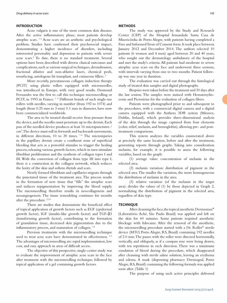
Drug delivery in acne scars 145
INTRODUCTIONAcne vulgaris is one of the most common skin diseases.
After the active inflammatory phase, most patients develop atrophic scars. 1-4 Acne scars are an aesthetic and psychological problem. Studies have confirmed their psychosocial impact, demonstrating a higher incidence of disorders, including introverted personality and depression in patients with severe acne scars.5 To date, there is no standard treatment. Several options have been described with diverse clinical outcomes and complications, such as various surgical techniques, dermabrasion, fractional ablative and non-ablative lasers, chemical peels, resurfacing, autologous fat transplant, and cutaneous fillers. 2
More recently, percutaneous collagen induction therapy (PCIT) using plastic rollers equipped with microneedles, was introduced in Europe, with very good results. Desmond Fernandes was the first to call this technique microneedling or PCIT, in 1993 in France. 1, 4 Different brands of such single-use rollers with needles, varying in number (from 192 to 1074) and length (from 0.25 mm to 3 mm) 0.1 mm in diameter, have now been commercialized worldwide.
The area to be treated should receive firm pressure from the device, and the needles must penetrate up to the dermis. Each pass of the needled device produces at least 16 micropunctures /cm2. The device must roll in forwards and backwards movements, in different directions, 10 to 20 times. 1-6 The microinjuries in the papillary dermis create a confluent zone of superficial bleeding that acts as a powerful stimulus to trigger the healing process, releasing various growth factors, which in turn stimulate fibroblast proliferation and the synthesis of collagen types I and III. With the conversion of collagen from type III into type I, there is a contraction in the collagen network, which reduces the laxity of the skin and softens rhytids and scars.
Newly formed fibroblasts and capillaries migrate through the punctured tissue of the treatment area. The process results in the formation of new tissue that “fills” the atrophic scars and induces repigmentation by improving the blood supply. The microneedling therefore results in neocollagenesis and neoangiogenesis. The tissue remodeling continues for months after the procedure. 2,3,6
There are studies that demonstrate the beneficial effect of topical application of growth factors such as EGF (epidermal growth factor), IGF (insulin-like growth factor) and TGF-β3 (transforming growth factor), contributing to the formation of granulation tissue, decreased skin pigmentation due to the inflammatory process, and maturation of collagen. 7,8
Previous treatments with the microneedling technique used to treat acne scars have demonstrated its effectiveness. 1-6 The advantages of microneedling are: rapid implementation, low cost, and easy approach in areas of difficult access.
The objective of this prospective, single-center study was to evaluate the improvement of atrophic acne scars in the face after treatment with the microneedling technique, followed by topical application of a gel containing growth factors.
METHODSThe study was approved by the Study and Research
Center (CEP) of the Hospital Irmandade Santa Casa de Misericórdia de Porto Alegre, with patients having completed a Free and Informed Term of Consent form. It took place between January 2012 and December 2014. The authors selected 10 patients (6 women and 4 men) aged between 20 and 40 years, who sought out the dermatology ambulatory of the hospital and met the study’s criteria. All patients had moderate to severe atrophic acne scars on the face and underwent three sessions, with intervals varying from one to two months. Patient follow-up was one year in duration.
The evaluation was carried out through the histological study of treated skin samples and digital photographs.
Biopsies were taken before the treatment and 30 days after the last session. The samples were stained with Hematoxylin-eosin and Picrosirius for the evaluation of collagen fibers.
Patients were photographed prior to and subsequent to the procedures, with a commercial digital camera and a digital camera equipped with the Anthera 3D® system (Miravex, Dublin, Ireland), which provides three-dimensional analysis of the skin through the image captured from four elements (color, relief, melanin, and hemoglobin), allowing pre- and post-treatment comparisons.
This system analyzes the variables enumerated above at precisely the same location, before and after the treatment, generating reports through graphs. Taking into consideration melanin, for example, it is possible to assess the following variables, based on the graph:
(1) average value: concentration of melanin in the selected area;
(2) melanin variation: distribution of pigment in the selected area. The smaller the variation, the more homogeneous the distribution of melanin in the area;
(3) relative variation (of the melanin in the target area): divides the values of (1) by those depicted in Graph 2, normalizing the distribution of pigment in the selected area, regardless of skin type.
TECHNIQUEAfter cleansing the face, the topical anesthetic Dermomax®
(Laboratório Aché, São Paulo Brazil) was applied and left on the skin for 60 minutes. Some patients required anesthetic blockage with lidocaine. After the removal of the anesthetic, the microneedling procedure started with a Dr. Roller® sterile device (MTO, Porto Alegre, RS, Brazil) containing 192 needles of 2.0 mm. The passes with the roller were directed horizontally, vertically, and obliquely, as if a compass rose were being drawn, with ten repetitions in each direction. There was a minimum exudation of blood during the procedure, which disappeared after cleansing with sterile saline solution, leaving an erythema and edema. A mask (dispensing pharmacy Dermogral, Porto Alegre, RS, Brazil) containing the following formula was applied soon after. (Table 1)
The purpose of using such active principles delivered
Surg Cosmet Dermatol 2015;7(2):144-8.

146 Kalil CLPV, Frainer RH, Dexheimer LS, Tonoli RE, Boff AL
via a gel mask after the microneedling was to increase hydration, stimulate fibroblasts, improve healing and cause anti-inflammatory action.
Post-operativeAll patients were able to return to their normal activities
the day after the procedure. Momentary erythema and edema could be observed on the skin immediately after the treatment. There were no complaints of post-operative pain . Physical sunscreen of SPF 30 was provided for use during the subsequent days.
RESULTSEight patients completed the study. During the
photographic analysis, it was possible to observe global improvement of the skin’s appearance and a slight improvement of the extensible atrophic scars. Ice pick type atrophic scars (not extensible) did not improve with the procedure. Comparative photographs (Figure 1) show a patient with improvement of extensible depressed scars.
Anthera systemSeven patients had a reduction in the relief of the scars
in the area assessed (Figure 2), seven had a reduction of melanin with homogenization of its distribution in the skin (Figure 3), and seven had an increase in hemoglobin in the study area (Figure 4), with the appearance having varied among the eight selected patients.
The analysis of photographs by the Anthera 3D system demonstrated an improvement in the skin’s texture, a small reduction in the concentration of melanin, and an increase in hemoglobin concentration in the areas selected. (Graphs 1 to 3)
Pathological examinationThe pathological evaluation was performed prior to the
procedure in ten cases and in eight cases after the treatment.The histological findings are described in Table 1, with
the following items having been evaluated before and after the microneedling: periadnexal fibrosis, cicatricial fibrosis (with horizontalized collagen), perivascular infiltrate, perivascular edema, thickened collagen presence in the deep dermis in the Picrosirius staining, and thinning of the epidermis after the treatment.
DISCUSSIONIn a study that analyzed 480 patients undergoing PCIT
aimed at improving scars and wrinkles, Aust et al. 4 identified increased collagen in the pathology of patients after applying the treatment, with the samples having been submitted to the collagen-specific Van Gieson staining method.
In a study that showed the improvement of photodamage with the induction of collagen production by PCIT, Fernandes et al. 6 identified an absence of thinning of the epidermis after the application of the treatment. This finding seems to be an advance regarding the other treatments, which mostly promote epidermal thinning, as they are more invasive and lead to greater rupture of the basal layer.
In an experimental study, Emerson Vasconcelos et al. 9 established the correlation between the length of the cylinder’s needles used in the microneedling procedure and the depth of the injury inflicted. Microscopic examination immediately after the procedure revealed vascular ectasia and extravasation of red blood cells, affecting the papillary dermis with 0.5 mm long needles, and reaching the reticular dermis with 2.5 mm long needles. Thus, microneedling can have a wide range of clinical indications, depending on the depth reached.
In the present study, after the Picrosirius staining (also a collagen-specific staining method), there was no difference in the collagen production after analyzing the deep dermis, the part of the skin’s topography where the treatment reaches its maximum effect. In cases where a biopsy was carried out prior to the treatment, it was possible to observe thicker collagen (Picrosirius) in five of them; where patients were biopsied after the treatment, it was possible to identify thicker collagen in six of them. It was possible to observe that the Picrosirius also colored the collagen more intensely in cases in which more fibrosis secondary to acne scars was identified using hematoxylin and eosin staining (HE). Likewise, there was no difference in the lymphocyte infiltration in the edema or in the thickness of the epidermis before and after treatment, results that can be
Surg Cosmet Dermatol 2015;7(2):144-8.
Table 1: Dispensing formulation of the mask containing growth factors
EGF - epidermal growth factor 1%IGF - insulin growth factor 1%TGF-β3 - transforming growth factor 1%Hyaluronic acid 0. 5%Tranexamic acid 0. 5%Vitamin E 2%Portulaca extract 1%Gel mask (sufficient amount) 30g
Figure 1: Patient 1.A. Before the treatment and, B. Twelve months after the completion of
the treatment
A B

Drug delivery in acne scars 147
Surg Cosmet Dermatol 2015;7(2):144-8.
explained by the small size of the evaluated sample. Also, it was noted that in some cases, the epidermis evidenced thinning and flattening of the interpapillary cones after PCIT, a finding that counters the existing literature.
Graph 1: Reduction of 26.2% in the contour and imperfections in the selected area. (Patient 1)
Graph 2: Slight reduction of melanin concentration in the selected area. (Patient 1)
Graph 3: Increase of 22% in hemoglobin concentration in the selected area. (Patient 1)
Melanin average level
Hemoglobin average level
Relief under 2.5 mm of lateral size
face E 12/17/2012face E 10/31/2013
ReliefSanta Casa POA
Antera Demo Antera Demo
Face E = 17/12/2012 Face E = 17/12/2012Close to the mentum’s left-hand side Relevo (abaixo de 2,5mm) = 15,4 Relief under 2.5 mm of lateral size = 20,9
Figure 2: Three-dimensional image of the reduction in the scars’ relief in the assessed area.
Figure 3: Three-dimensional image of the reduction of melanin with homogenization of its distribution in the skin
HemoglobinSanta Casa POA
Antera Demo Antera Demo
Face E = 17/12/2012 Face E = 17/12/2012Close to the mentum’s left hand side Nível médio = 0,588 = Variation = 0,0455 (7,74%) Average level = 0,61 = Variation = 0,0452 (7,41%)
Figure 4: Three-dimensional image of the increase in hemoglobin in the assessed area
ReliefSanta Casa POA
Antera Demo Antera Demo
Face E = 17/12/2012 Face E = 17/12/2012Close to the mentum’s left-hand side Average level = 1,53 = Variation = 0,126 (8,21%) Average level = 1,25 = Variation = 0,132 (10,5%)

148 Kalil CLPV, Frainer RH, Dexheimer LS, Tonoli RE, Boff AL
REFERENCES1. Percutaneous Collagen Induction with Dermaroller TM for Management
of Atrophic Acne Scars in 31 Thai Patients. Asian Journal of Aesthetic Me-
dicine. 2009;2(1):1-13.
2. Leheta T, Tawdy A. Percutaneous Collagen Induction Versus Full-Concen-
tration Trichloroacetic Acid in the Treatment of Atrophic Acne Scars. Der-
matol Surg. 2011;37(2):207-16.
3. Liebl H, Kloth LC. Skin Cell Proliferation stimulated by microneedles. J Am
Coll Wound Spec. 2012;4(1):2-6.
4. Aust M, Fernandes D, Kolokythas P, Kaplan HM, Vogt PM. Percutaneous
Collagen Induction Therapy: an alternative Treatment for Scars, Wrinkles,
and Skin Laxity. Plast Reconstr Surg. 2008;121(4):1421-9.
5. Majid I. Microneeddling therapy in Atrophic facial scars: an objective as-
sessment. J Cutan Aesthet Surg. 2009;21(1):26-30.
6. Fernandes D, Signorini M. Combating photoaging with percutaneous
collagen induction. Clin Dermatol. 2008;26(2):192-9.
7. Spyrou GE, Naylor IL. The Effect of Basic Fibroblast Growth Factor on Scar-
ring. Br J Plast Surg. 2002;55(4):275-82.
8. Akita S, Akino K, Imaizumi T, Hirano A. Basic Fibroblast Growth Factor
Accelerates and Improves Second-Degree Burn Wound Healing. Wound
Repair Regen. 2008;16(5):635-41.
9. Lima EVA, Lima MA, Takano D. Microagulhamento: estudo experi-
mental e classificação da injúria provocada. Surg Cosmet Dermatol.
2013;5(2):110¬4.
CONCLUSIONSThe studied patients achieved an overall improvement
in skin texture and a slight improvement of atrophic scars, corroborating the findings found in the study conducted by Imran Majid, 5 in which 36 of the 37 patients showed a similar response to PCIT.
The authors of the present study believe that the increase in the hemoglobin visualized by the Anthera system is due to the increased vascularization promoted by the initial tissular injury, which is perpetuated over time and provides neocollagenesis.
Ice pick type atrophic scars showed no improvement with the procedure.
Long-term clinical analysis, associated with a pathological study, could demonstrate delayed effects of this procedure on acne scars. l
Surg Cosmet Dermatol 2015;7(2):144-8.

149
ReviewArticle
Laserlipólise na região cervicalLaserlipolysis in the cervical region
DOI: http://dx.doi.org/10.5935/scd1984-8773.201572544
RESU MO A laserlipólise tem-se mostrado procedimento seguro e eficaz no tratamento do envelhecimento da
região cervical. A técnica consiste na utilização da energia do laser na indução da lipólise e no estímu-lo à neocolagênese, atuando na remoção da gordura do submento e na flacidez cutânea local. Neste artigo realizamos breve revisão sobre a laserlipólise no tratamento da região cervical e relatamos nossa experiência na utilização da técnica.
Palavras-chave: rejuvenescimento; lipólise; colágeno; pescoço; lasers
ABSTRACT Laserlipolysis has been proven a safe and effective procedure for the treatment of the aging
of the cervical region. The technique involves the use of laser energy to induce lipolysis and encouraging neocollagenesis, removing submental fat and acting on local skin sagging. The present article offers a brief review of laserlipolysis in the treatment of the cervical region and reports the authors’ experience in the use of the technique.
Keywords: rejuvenation; lipolysis; colagen; neck; lasers
Authors: Sandra Tagliolatto1
Oriete Gerin Leite2
1 MSc, Dermatologist Physician - Campinas (SP), Brazil
2 Dermatologist Physician - Campinas (SP), Brazil
Correspondence: Sandra Tagliolatto Rua Luzitana, 740, 4º andar - Bosque Cep: 13015-121 – Campinas (SP), Brazil E-mail: dermoclínica@dermoclínica.med.br
This study was performed at the authors’ private practice in Campinas (SP), Brazil.
Received on: 20 September 2014 Approved on: 17 June 2015
Financial support: none Conflict of interest: none
149
INTRODUCTIONThe appearance of the cervical region during aging results
from a combination of changes in the skin, in the distribution of fat, in the platysma muscle and even in the osseous-cartilaginous structure. 1 A youthful silhouette, with a well-defined cervical-mandibular contour – exemplified by the bust of the Egyptian Queen Nefertiti – is considered the ideal, and a model to be achieved through cervical rejuvenating treatments (Figure 1).
Until recently, surgery was the gold standard treatment for cervical rejuvenation. However, in recent decades laserlipolysis has emerged as an effective and minimally invasive therapeutic modality in the reduction of submental fat and in promoting the tightening of the cervical skin. 2
Laserlipolysis works through the selective photothermolysis of fat and collagen in the treated region. The laser acts on the lysis located in the membranes of the adipocytes and on the denaturation of collagen fibers in the adjacent dermis, which leads to the stimulation of collagen remodeling and neocollagenesis. Performed under local tumescent anesthesia, isolated laserlipolysis
Surg Cosmet Dermatol 2015;7(2):149-57.

150 Tagliolatto S, Leite OG
or laserlipolysis in combination with liposuction, is a safe and effective technique for treating localized fat and defining the contour of the body and the face. 3
The main lasers used in laserlipolysis are Nd:YAG and diode lasers, at different wavelengths. In the treatment of the submental and cervical regions, the optical fiber of the laser is inserted directly into the subcutaneous tissue through a microcannula. The energy released by the laser promotes fat liquefaction, coagulation of collagen fibers and blood vessels, as well as the formation of subcutaneous channels that aid in skin retraction. 3
BACKGROUNDKarlin and Ellenbogen established five visual criteria for
the evaluation of the neck and for categorizing its appearance as young looking: a well-delimited mandibular border, presence of sub-hyoid depression, visible prominence of the thyroid cartilage and of the anterior border of the sternocleidomastoid muscle, and a cervical-mental angle (CMA) of between 105º and 120º 1 (Figure 1).
Among the various treatments for the aging cervical region, aimed at restoring the mentioned aesthetic aspects, liposuction under tumescent anesthesia is considered the most effective and minimally invasive procedure due to the fact that it can remove the accumulated fat in the submental region and contribute to the mitigation of local cutaneous sagging. 2 The association of laserlipolysis during the same operative event improves the final result of traditional liposuction, offering even greater improvement in the contour of the neck, due to its action in stimulating the production of collagen. 3-5
Laserlipolysis has been used since the ‘90s with good results in the treatment of localized fat and body contour definition. With the development of lasers, it has become consistent and effective, with good tolerance and a good safety profile. 6
Several scientific studies carried out in the last decade have shown advantages over surgical methods and traditional liposuction. In addition to the double benefit of subcutaneous fat removal and the remodeling of collagen fibers – clinically evidenced by the skin tightening effect – laserlipolysis provides a shorter recovery time, greater convenience to the patient, and lower post-operative complication rates (hematoma, seroma, asymmetries etc). 7-9
Some reports have shown good results using laserlipolysis in isolation, without the traditional liposuction. However, current studies indicate that isolated laserlipolysis can only be used to treat maximum volumes of up to 100cm3, which makes this technology useful most often in association with local aspiration. 9, 10
In order to perform the laserlipolysis procedure in the cervical region, knowledge of the local anatomy is crucial and will assist both in the appropriate selection of suitable patients and in the planning of the treatment. 11, 12
Knowledge of cervical anatomy and its variations caused by the aging process enables greater precision in the treatment of the aging neck, contributing to the improvement of the results obtained, as well as decreasing the incidence of post-operative complications. 2
ANATOMY OF THE CERVICAL REGIONPlatysma muscleThe platysma muscle emerges inferiorly in the cervical
thoracic fascia, inserts superiorly to the angle of the oral depressor, risorius and mentonian muscles, and also intermittently in the mandible (Figure 2). 13
Liposuction of the cervical region aims at removing the pre-platysmal adipose tissue, which covers the platysma muscle. 13 The aging of this muscle can be responsible for the formation of the so-called platysmal bands, which negatively alters the
Figure 1: Bust of Nefertiti (3,000 a. C.),
the ideal model of a young neck.
Note the formation of the cervical-mental
angle (CMA)
Figure 2: Platysma muscle and
its insertion on the mandibular border
Surg Cosmet Dermatol 2015;7(2):149-57.
CMA

Laserlipolysis in the cervical region 151
aesthetic appearance of the neck, for it contributes to the increase of the CMA, and may also directly impact the sagging of the region (muscle sagging). 14
In the treatment of the aging cervical region, it is therefore necessary to combine therapeutic methods: in addition to the use of techniques for the removal of local fat and cutaneous retraction, surgical techniques of plication of the platysma or the use of botulinum toxin can be associated with treatment of local muscle changes. 15, 16
The platysma muscle receives innervation from the cervical branch of the facial nerve, which in turn acts on the depressor musculature of the lower lip. 13
Marginal mandibular branch of the facial nerveThe marginal mandibular branch of the facial nerve runs
deeply to the platysma, along the mandible’s body (in 80% of cases), or 1 to 2 cm below the mandible (in roughly 20% of cases). 2
This branch surfaces in the anterior border of the masseter muscle and is located anteriorly to the region where the facial artery crosses the mandible (Figure 3). 17, 18
There are no reports of transection or permanent paralysis of the marginal mandibular nerve resulting from liposuction with tumescent anesthesia; however neurapraxia resulting from the temporary interruption of the conduction of nerve impulses and motor function can occur and is a consequence of trauma to the nerve fibers without nerve rupture. 18
In the present study, with over 100 patients, there were no cases of permanent paralysis. There were only two reports of neurapraxia, both with spontaneous resolution in up to four weeks, which is aligned with the literature. There are descriptions of neurapraxia of the marginal mandibular nerve during a period of four to six weeks; though there are references of permanence for up to 12 months. 19
Submandibular salivary glandsSalivary glands are present in the lower region of the
mandibular midline. If prominent submandibular glands become visible after liposuction of the neck, surgical procedures such as resection of glands or other techniques, are required to resolve the aesthetic problem. As a result, patients need to be consulted in regards to this issue (Figure 4). 2, 12
Hyoid boneThe CMA may be altered not only due to the
accumulation of fat in the submental area or sagging of the platysma muscle, but also due to the position of the hyoid bone in relation to the mandible. 16
It is generally located roughly at the C3 and C4 level, however in some patients the hyoid bone can be positioned lower, increasing the CMA. This is therefore an anatomical factor that must be taken into account when selecting a patient for the laserlipolysis procedure, since the laser will obviously be ineffective in these patients. 14
In brief, during the clinical examination, the CMA, the submental adiposity accessible to liposuction, the location of the hyoid bone, and the quality of the cervical region’s skin must be evaluated, analyzing the degree of photodamage and skin elasticity, as well as observing if there is muscle sagging and/or if there is a presence of platysmal bands and, finally, to attempt to palpate the submandibular glands.
PATIENT ASSESSMENTBased on the mentioned clinical findings, it is advisable to
discuss the laserlipolysis procedure (with or without liposuction) with the patient in advance, as well as the possible necessity of association with other treatments to achieve the best results in
Figure 3: Marginal mandibular branch of the facial nerve (red arrow) Figure 4: Submandibular salivary glands can be prominent, compromising the esthetic outcome after laserlipolysis
Surg Cosmet Dermatol 2015;7(2):149-57.
Sublingual gland
Sublingual gland
Parotid gland
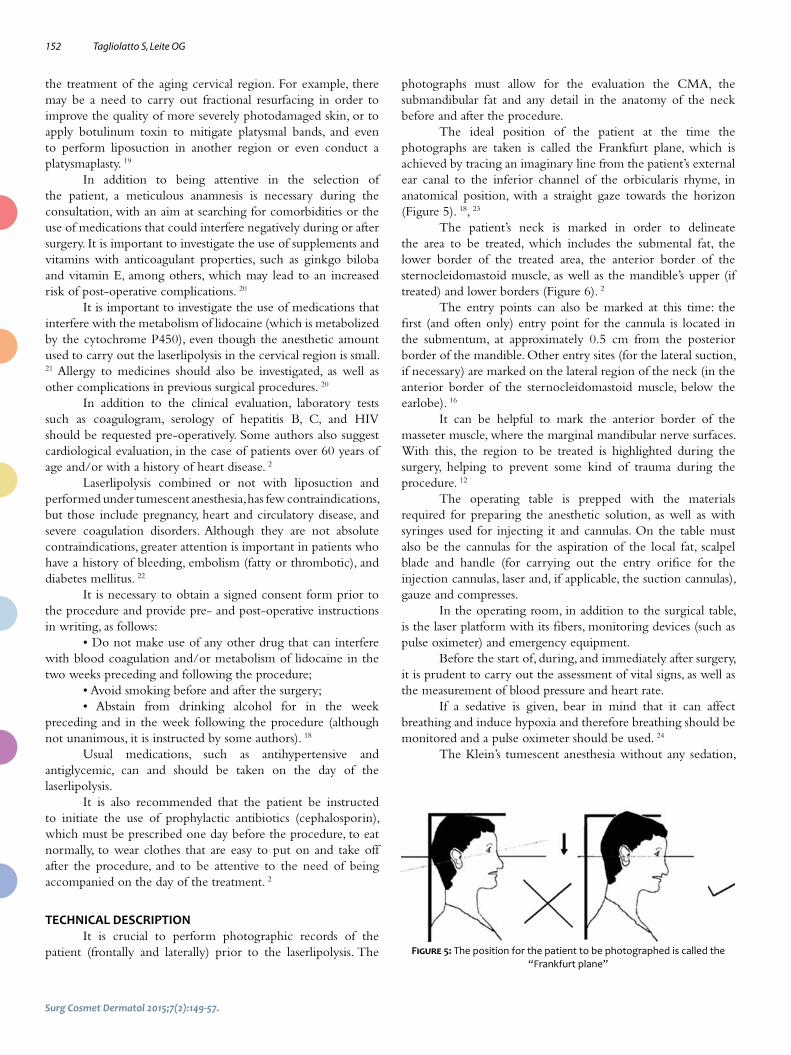
152 Tagliolatto S, Leite OG
the treatment of the aging cervical region. For example, there may be a need to carry out fractional resurfacing in order to improve the quality of more severely photodamaged skin, or to apply botulinum toxin to mitigate platysmal bands, and even to perform liposuction in another region or even conduct a platysmaplasty. 19
In addition to being attentive in the selection of the patient, a meticulous anamnesis is necessary during the consultation, with an aim at searching for comorbidities or the use of medications that could interfere negatively during or after surgery. It is important to investigate the use of supplements and vitamins with anticoagulant properties, such as ginkgo biloba and vitamin E, among others, which may lead to an increased risk of post-operative complications. 20
It is important to investigate the use of medications that interfere with the metabolism of lidocaine (which is metabolized by the cytochrome P450), even though the anesthetic amount used to carry out the laserlipolysis in the cervical region is small. 21 Allergy to medicines should also be investigated, as well as other complications in previous surgical procedures. 20
In addition to the clinical evaluation, laboratory tests such as coagulogram, serology of hepatitis B, C, and HIV should be requested pre-operatively. Some authors also suggest cardiological evaluation, in the case of patients over 60 years of age and/or with a history of heart disease. 2
Laserlipolysis combined or not with liposuction and performed under tumescent anesthesia, has few contraindications, but those include pregnancy, heart and circulatory disease, and severe coagulation disorders. Although they are not absolute contraindications, greater attention is important in patients who have a history of bleeding, embolism (fatty or thrombotic), and diabetes mellitus. 22
It is necessary to obtain a signed consent form prior to the procedure and provide pre- and post-operative instructions in writing, as follows:
• Do not make use of any other drug that can interfere with blood coagulation and/or metabolism of lidocaine in the two weeks preceding and following the procedure;
• Avoid smoking before and after the surgery;• Abstain from drinking alcohol for in the week
preceding and in the week following the procedure (although not unanimous, it is instructed by some authors). 18
Usual medications, such as antihypertensive and antiglycemic, can and should be taken on the day of the laserlipolysis.
It is also recommended that the patient be instructed to initiate the use of prophylactic antibiotics (cephalosporin), which must be prescribed one day before the procedure, to eat normally, to wear clothes that are easy to put on and take off after the procedure, and to be attentive to the need of being accompanied on the day of the treatment. 2
TECHNICAL DESCRIPTIONIt is crucial to perform photographic records of the
patient (frontally and laterally) prior to the laserlipolysis. The
photographs must allow for the evaluation the CMA, the submandibular fat and any detail in the anatomy of the neck before and after the procedure.
The ideal position of the patient at the time the photographs are taken is called the Frankfurt plane, which is achieved by tracing an imaginary line from the patient’s external ear canal to the inferior channel of the orbicularis rhyme, in anatomical position, with a straight gaze towards the horizon (Figure 5). 18, 23
The patient’s neck is marked in order to delineate the area to be treated, which includes the submental fat, the lower border of the treated area, the anterior border of the sternocleidomastoid muscle, as well as the mandible’s upper (if treated) and lower borders (Figure 6). 2
The entry points can also be marked at this time: the first (and often only) entry point for the cannula is located in the submentum, at approximately 0.5 cm from the posterior border of the mandible. Other entry sites (for the lateral suction, if necessary) are marked on the lateral region of the neck (in the anterior border of the sternocleidomastoid muscle, below the earlobe). 16
It can be helpful to mark the anterior border of the masseter muscle, where the marginal mandibular nerve surfaces. With this, the region to be treated is highlighted during the surgery, helping to prevent some kind of trauma during the procedure. 12
The operating table is prepped with the materials required for preparing the anesthetic solution, as well as with syringes used for injecting it and cannulas. On the table must also be the cannulas for the aspiration of the local fat, scalpel blade and handle (for carrying out the entry orifice for the injection cannulas, laser and, if applicable, the suction cannulas), gauze and compresses.
In the operating room, in addition to the surgical table, is the laser platform with its fibers, monitoring devices (such as pulse oximeter) and emergency equipment.
Before the start of, during, and immediately after surgery, it is prudent to carry out the assessment of vital signs, as well as the measurement of blood pressure and heart rate.
If a sedative is given, bear in mind that it can affect breathing and induce hypoxia and therefore breathing should be monitored and a pulse oximeter should be used. 24
The Klein’s tumescent anesthesia without any sedation,
Figure 5: The position for the patient to be photographed is called the “Frankfurt plane”
Surg Cosmet Dermatol 2015;7(2):149-57.

Laserlipolysis in the cervical region 153
was used in the authors procedures. This solution is composed of 1,000 ml of saline, 50 to 100 ml of 1% lidocaine, 1 ml epinephrine 1:1,000 and 12.5 ml of 8.4% sodium bicarbonate. 25
Assuming that the patient is not using any medication that interferes with the metabolism of lidocaine, varying doses of this solution (35 to 55 mg /kg) can be used safely, however this is usually not a problem when the neck region is treated, given the small amount of anesthetic solution (300-500 ml) used to achieve proper tumescence. 26
After performing the anesthesia, there must be a waiting period of 20 to 30 minutes, after which the passage of the 1 mm microcannula containing the laser fiber in the subcutaneous plane is started in slow movements, in a fan pattern, creating tunnels of thermal damage in the adipocytes. 3
The authors point out that the laser must be passed slowly in the subcutaneous, at a speed of 5-10 cm/s, in order to allow its interaction with the target area to take place. On the other hand, the laser should never stop once activated, due to the risk of burns. 6, 7
The laser pass’ end point can be detected by monitoring several parameters. The perception of local heat and the “softening” of the subcutaneous region during laser passage are crucial. The procedure can be guided by the accumulated energy values on the device’s panel, and temperature monitoring by external or internal thermometers, coupled to the tip of some laser devices. 2, 5, 6
In their practice, the authors of the present article use the 924nm/975nm diode laser device (SlimLipo, Palomar, USA), with two wavelengths – one specific for liquefying the adipose tissue (924nm) and another for skin retraction, due to its action on the collagen and elastic fibers at the level of the deep adjacent dermis (975nm). 27, 28
This device does not allow for the taking of the internal temperature, however the authors adopted the use of an external laser digital thermometer in the study’s protocol, for assistance during the treatment. An external temperature of around 40º C is sought during the procedure, since in most cases this leads to good aesthetic results without tissue damage.
The use of devices specific to laserlipolysis requires
attention during surgery, regarding the energy emitted by the laser per region – called “accumulated energy” – in order to promote a better aesthetic result based on the remodeling of the fibrous connective tissue. 29
The control of the accumulated energy is an essential safety parameter aimed at reducing the risk of side effects such as necrosis, prolonged erythema, and dyschromia. 5
Once the end point of the laser passage is reached, the aspiration of “molten fat” can be performed or not, depending on the volume treated. Alternatively, the manual “milking” can be performed without the cannula, in the case of smaller volumes.
While performing the liposuction, it is necessary to use the appropriate cannula for the cervical region in order to avoid unnecessary damage. The delicate passage of the cannula under mild negative pressure is sufficient to remove the liquefied fat. Technical care, such as attention to the region of the approximate path of the marginal mandibular branch of the facial nerve, is necessary in the execution of suction. For this, it is necessary to avoid the lateral rotation of the head (with the risk of forcing the nerve into a more superficial position, 2 to 3 cm below the mandible) and to use the hand as a guide when performing the surgery. 12, 16, 18, 19
At the end of the procedure, local compression is used (occlusive dressing and a compressive neck garment) in order to facilitate the remodeling of the skin, minimizing the risk of folds, preventing bruising and/or seroma formation, and improving absorption and drainage of the remaining tumescent fluid. 22
Post-operative careRegarding the compressive garment, it should be used
continuously (24 hours a day, removing only for bathing) during the first two days, and for 2 to 4 hours a day in the following two weeks. The occlusive dressing should remain in place for the first 18 hours, when the greatest drainage of remaining liquid takes place. 30
It is recommended that the patient avoid physical exercise for 7 days, and some authors suggest that attention must be paid regarding the temperature of the water used when bathing, and the use of ice directly on the skin, in order to avoid damage.
Figure 6: Prior marking delimiting the area to be treated.A - The region of entry of the cannula.B - Paths of laser application (arrows)A B
Surg Cosmet Dermatol 2015;7(2):149-57.

154 Tagliolatto S, Leite OG
Although complications are rare, contact numbers should always be provided to patients in the event of any undesirable side effect, which should always be assessed. 2, 30
The patient must return periodically in order to monitor the progressive improvement of the region: resolution of the edema, formation of collagen, and retraction of the skin, all of which occur within a few months – up until 18 months after the date of the procedure, according to some authors. 16
Discussion of the literature: efficacy and safetyLaserlipolysis is a minimally invasive therapeutic modality
that is being used at an increasing rate to treat localized fat, and for the rejuvenation and definition of the contour of the body and face. 31 Since the first studies were published in the 90’s, several reports have been published every year, with different laser types, wavelengths, and fat and water absorption coefficients. 3-12
The most frequently described lasers are the 920nm, 924/975nm and 980nm diode lasers, as well as the 1,064nm, 1,319nm, 1,320nm, 1,440nm and 1,444nm Nd:YAG lasers. Despite the different wavelengths, all laserlipolysis systems work on the principle of selective photothermolysis, i.e. action of heat in specific tissues, such as fat and collagen. 6, 31
The laser releases energy, which is retained in the form of heat in the subcutaneous tissue in contact with the optical fiber. The heat captured by the adipocytes can lead to the rupture of cell membranes, with the release of intracellular lipases, promoting liquefaction of the adipose tissue, which facilitates liposuction with less trauma and bleeding. 32
When absorbing the heat, collagen fibers of the adjacent dermis undergo denaturation, stimulating the remodeling and contraction of collagen, and promoting neocollagenesis. Histological studies have shown that heat destroys adipocytes via the creation of small pores in the membranes of these cells. 33
In addition to lipolysis, coagulation of blood vessels and sweat glands occurs, and the reorganization of collagen and elastic fibers. Clinically, these findings correlate to the skin tightening effect and to less bleeding during and after surgery. 7, 33
While there is still a search for better parameters, laserlipolysis – with or without aspiration – has been used in the treatment of submental adiposity for two decades with good results (Figure 7). 11, 12
In 2002, Goldman et al. published a case study series with 1,734 patients who underwent laserlipolysis in various body sites, including the submentum, with low blood loss and ecchymosis, little post-operative discomfort, and fast recovery, in addition to a low complication rate. 34 In 2006, the same author published a retrospective with 82 patients who underwent laserlipolysis of the submental region with and without aspiration. In addition to an efficacy comparable to that of conventional liposuction, it was possible to observe clinical and histological retraction of the skin, and collagen neoformation. 7
The use of tumescent anesthesia for liposuction allows for the safe removal of fat, with minimal blood loss and little post-operative pain. There are no reports of mortality or even significant morbidity associated with liposuction under tumescent anesthesia. 35
Hanke et al. studied 15,336 patients and 44,014 body sites treated with liposuction using tumescent anesthesia, and did not find cases of death or serious complications requiring transfer to a hospital. 36 Housman et al. reported 66,570 liposuction procedures performed by dermatologic surgeons from 1994 to 2000, with an absence of reports of deaths. Adverse events requiring hospitalization occurred at a rate of 0.68 per 1,000 cases, with the use of sedatives described as an identified risk factor. 37
The use of the ideal accumulated energy maximizes the effects of the laserlipolysis, which are the removal of subcutaneous fat and the retraction of the skin. Reynaud et al. reviewed the values of the accumulated energy used in the various studies published up until 2009 and detected a progressive increase of the parameters over time and associated with better clinical outcomes. In their series of 534 procedures, 22 patients underwent laserlipolysis in the submentum with 980nm diode laser at 6W of potency. The average accumulated energy in the submental region was 11,700J. 8
More recently Sarnoff evaluated the use of 1,440nm Nd:YAG, without aspiration, in the treatment of the cervical contour in 24 patients, having employed an average accumulated energy 1,205J per 5x5cm. Improvement of 79% was obtained in CMA values and in the overall aesthetic score. 38
In the experience of the authors of the present article, an accumulated energy of 5,000J per 10cm2 area is close to ideal as a progressive and persistent retraction of the skin can be observed at this level, with a reduction of side effects such
A BFigure 7: Thirty-seven-year-old patient with submental adiposity, treated
with laserlipolysisA - Pre-operative. B - 30 days after the procedure.

Laserlipolysis in the cervical region 155
as ecchymosis, prolonged erythema, excessive bleeding, or the formation of dimples in the skin (Figures 8 and 9). 6-8, 12, 31, 38
It is known that low energy levels cause reversible swelling of adipocytes and that high energy levels are responsible for the lysis of adipocytes and significant improvement of the results. Nevertheless, high energies increase the risk of thermal injury to the skin. 11, 12
Kim and Geronemus found a volumetric reduction of 25% in the submental fat, measured by MRI, after three months of laser application in 5 patients. In a total of 29 patients, they observed a positive correlation between the accumulated energy and the reduction in the volume of the treated area. 3
Some studies have begun to emerge that try to correlate the measurement of the external temperature with the safety and effectiveness of laserlipolysis. In 2012, Alexiades-Armenakas achieved good results with the use of 1,064nm and 1,319nm lasers, either isolated or in combination, in the treatment of submental adiposity and sagginess. The internal temperature of the target tissue was 45ºC to 48ºC. Most authors consider external temperatures around 40ºC as safe and effective. 12
Apparently, complication rates of laserlipolysis are lower when compared to that of traditional liposuction. Sarnoff has not recorded cases of seroma, infection, damage to the mandibular nerve, dyschromia or hematomas. 38 After 1,000 consecutive cases of laserlipolysis, Chia et al. described 3 cases of skin burn, 2 of infection, 1 of seroma and 1 of hematoma, clearly related to a learning curve since they occurred in the first 25 cases. In 7.3% of cases retreatment was required due to insufficient improvement. 22
The satisfaction rate with laserlipolysis was assessed in a study by Leclère et al. in 2014. After laserlipolysis in the submentum of 30 patients, it was possible to observe a systematic improvement in the CMA, in the cutaneous retraction, and in the decrease of subcutaneous fat. The procedure was recommended by all patients and was highly satisfactory among evaluators. 39
There is a consensus among authors that the correct patient indication leads to more satisfactory results. Advanced
aging, exuberant adiposity, and a high degree of sagging compromise the final result of laserlipolysis. 2, 16
Several additional techniques can be combined with laserlipolysis in order to further improve the neck’s appearance, including the treatment of photodamaged skin using fractional CO2 laser, the use of botulinum toxin for mitigating platysmal bands and even surgical procedures performed directly on the platysma. 2
In addition to association with conventional liposuction, laserlipolysis is gaining ground as an adjuvant procedure in the facial mini-lifts. Ramirez et al. obtained esthetical improvement of the neck and submentum in 100% of cases after six months of application of 924/975nm diode laser in conjunction with platysmaplasty in 78 patients. 40
In contrast, some authors studied the isolated application of laserlipolysis for improving facial sagging. Holcomb obtained good results in 478 patients who underwent laserlipolysis to define the contour of and rejuvenate the middle and lower thirds of the face with 1,444nm Nd:YAG. 41 McMenamin treated 40 patients with an aim at promoting the improvement of the facial contour and performing a facelift based on the cutaneous retraction. 42 He obtained good results and higher satisfaction rates as compared to those of the traditional surgery.
FINAL CONSIDERATIONSThe use of laserlipolysis in the treatment of cervical
rejuvenation and submental adiposity is a minimally invasive, safe, and effective therapy. The results obtained in several published clinical studies are promising.
The advantages over traditional liposuction are: shorter recovery time, less bleeding, additional effect of cutaneous retraction (or skin tightening) and low retreatment rates.
The different wavelengths and lasers employed appear to have similar effects on the target tissue. Better results are achieved with a greater accumulated energy; nonetheless this fact increases the risk of cutaneous thermal injury. Notwithstanding, reports of
Figure 8: Thirty-six-year-old male patient who underwent laserlipolysis in the submentumA - Pre-operativeB - 2 months after the procedureA B

156 Tagliolatto S, Leite OG
burns are rare and are related to the learning curve.The correct indication of the patient and the association
– when necessary – with other rejuvenation methods, such as platysmaplasty, botulinum toxin, and fractional resurfacing, optimizes the final aesthetic result.
The laserlipolysis of the submentum and cervical region is a well-tolerated technique, with high satisfaction rates among patients and physicians, is considered safe and leads to lasting results. Nevertheless, further controlled, randomized, and multicenter studies are still necessary for establishing the technique as the gold standard cervical rejuvenation treatment. l
Figure 9: Forty-year-old female patient who underwent laserlipolysis in the submental area.
A - Pre-operative. B – 3 months after the procedure. Note the improve-ment in the definition of the jaw line and skin retraction
A B
Surg Cosmet Dermatol 2015;7(2):149-57.

Laserlipolysis in the cervical region 157
REFERENCES1. Ellenbogen, R., and Karlin, J. V. Visual criteria for success in restoring the
youthful neck. Plast Reconstr Surg. 1980;66(6):826-37.
2. Stebbins WG, Hanke CW. Rejuvenation of the neck with liposuction and
ancillary techniques. Dermatol Ther. 2011;24(1):28-40.
3. Kim KH, Geronemus RG. Laser lipolysis using a novel 1,064 nm Nd:YAG
laser. Dermatol Surg. 2006;32(2):241–8.
4. Sun Y, Wu SF, Yan S, Shi HY, Chen D, Chen Y. Laser lipolysis used to treat lo-
calized adiposis: a preliminary report on experience with Asian patients.
Aesthetic Plast Surg. 2009;33(5):701-5.
5. Badin AZ, Moraes LM, Gondek L, Chiaratti MG, Canta L. Laser lipolysis: flac-
cidity under control. Aesthetic Plast Surg. 2002;26(5):335–9.
6. Fakhouri TM; El Tal AK; Abrou AE; Mehregan DA; Barone F. Laser-assisted
lipolysis: a review. Dermatol Surg. 2012;38(2):155-69.
7. Goldman A. Submental Nd:Yag laser-assisted liposuction. Lasers Surg
Med. 2006;38(3):181–4.
8. Reynaud JP, Skibinski M, Wassmer B, Rochon P, Mordon S. Lipolysis using a
980-nm diode laser: a retrospective analysis of 534 procedures. Aesthetic
Plast Surg. 2009;33(1):28–36.
9. Prado A, Andrades P, Danilla S, Leniz P, Castillo P, Gaete F. A prospective,
randomized, double-blind, controlled clinical trial comparing laser-as-
sisted lipoplasty with suction-assisted lipoplasty. Plast Reconstr Surg.
2006;118(4):1032–45.
10. Parlette EC. Kaminer ME. Laser-assisted liposuction: here’s the skinny. Se-
min Cutan Med Surg. 2008;27(4):259-63.
11. Gentile RD. Laser-assisted neck-lift: high-tech contouring and tightening.
Facial Plast Surg. 2011;27(4):331-45.
12. Alexiades-Armenakas M. Combination laser-assisted liposuction and
minimally invasive skin tightening with temperature feedback for tre-
atment of the submentum and neck. Dermatol Surg. 2012;38(6):871-81.
13. Castro CC. The anatomy of the platysma muscle. Plast Reconstr Surg.
1980;66(5):680–3.
14. Prendiville S, Kokoska MS, Hollenbeak CS, et al. A comparative study of
surgical techniques on the cervicomental angle in human cadavers. Arch
Facial Plast Surg. 2002;4(4):236–42.
15. Kane MA. Nonsurgical treatment of platysmal bands with injection of bo-
tulinum toxin A. Plast Reconstr Surg. 1999;103(2):656–63.
16. Rohrich RJ, Rios JL, Smith PD, Gutowski KA. Neck rejuvenation revisited.
Plast Reconstr Surg. 2006;118(5):1251–63.
17. Larrabee W, Makielski K. Surgical anatomy of the face. New York, NY: Ra-
ven Pres; 1993.
18. Butterwick KJ. Liposuction of the neck. In: DraelosZD, ed. Cosmetic der-
matology: products and procedures. Hoboken; New York: Blackwell Pu-
blishing; 2010. p. 463–71.
19. Morrison W, Salisbury M, Beckham P, Schaeferle M 3rd, Mladick R, Ersek
RA. The minimal facelift: liposuction of the neck and jowls. Aesthetic Plast
Surg. 2001;25(2):94–9.
20. Shiffman MA. Warning about herbals in plastic and cosmetic surgery.
Plast Reconstr Surg. 2001;108(7):2180–1.
21. Klein J. Cytochrome P450 3A4 metabolism and lidocaine metabolism. In:
KleinJ, ed. Tumescent technique: tumescent anesthesia and microcannu-
lar liposuction. St. Louis, MO: Mosby, 2000. p. 133.
22. Chia CT, Theodorou SJ. 1,000 consecutive cases of laser-assisted liposuc-
tion and suction-assisted lipectomy managed with local anesthesia. Aes-
thetic Plast Surg. 2012;36(4):795-802.
23. Farkas LG, Sohm P, Kolar JC, Katic MJ, Munro IR. Inclinations of the facial
profile: art versus reality. Plast Reconstr Surg. 1985;75(4):509–19.
24. Svedman KJ, Coldiron B, Coleman WP 3rd, Cox SE, Jacob C, Lawrence N,
et al. ASDS guidelines of care for tumescent liposuction. Dermatol Surg.
2006;32(5):709–16.
25. Coleman WP 3rd, Glogau RG, Klein JA, et al. Guidelines of care for liposuc-
tion. J Am Acad Dermatol. 2001;45(3):438–47.
26. Coleman WP 3rd1, Glogau RG, Klein JA, Moy RL, Narins RS, Chuang TY, et
al. Tumescent anesthesia with a lidocaine dose of 55 mg/kg is safe for
liposuction. Dermatol Surg. 1996;22(11):921–7.
27. Weiss RA, Beasley K. Laser-assisted liposuction using a novel blend of lipid-
and water-selective wavelengths. Lasers Surg Med. 2009;41(10):760-6.
28. Wolfenson M, Roncantti C, Alencar AH, Barros T, Silva Neto JF, Santos
Filho FCN. Laserlipolysis: skin tightening and prevention of flabby um-
bilical region with lipoplasty following security parameters for use of
diode laser with double wavelength 924 and 975 nm. Rev Bras Cir Plast.
2011;26(2):259-65.
29. Mordon SR, Wassmer B, Reynaud JP, Zemmouri J. Mathematical modeling
of laser lipolysis. Biomed Eng Online. 2008;7:10.
30. Jacob C, Kaminer M. Rejuvenation of the neck using liposuction and
other techniques. In: RobinsonJ, HankeC, SiegelD, editors. Surgery of the
skin: procedural dermatology. 2nd Edition. Philadelphia, PA: Elsevier Mos-
by. 2010. p.691-701.
31. McBean JC, Katz BE. Laser Lipolysis: An Update. J Clin Aesthet Dermatol.
2011;4(7):25-34.
32. Goldman A, Geronemus RG, Kim K. Lipoaspiração a laser. Laserlipólise. In:
Toledo LS, editor. Lipoplastia. Rio de Janeiro: Di-Livros. 2006. p. 111-21.
33. Badin AZ, Gondek LB, Garcia MJ, Valle LC, Flizikowski FB, Noronha L. Analy-
sis of laser lipolysis effects on human tissue samples obtained from lipo-
suction. Aesthetic Plast Surg. 2005;29(4):281-6
34. Goldman A, Schavelzon DE, Blugerman GS. Laserlipolysis: liposuction
with Nd: YAG Laser. Rev Soc Bras Cir Plást. 2002;17(1):17-21.
35. Triana L, Triana C, Barbato C, Zambrano M. Liposuction: 25 years of ex-
perience in 26,259 patients using different devices. Aesthet Surg J.
2009;29(6):509-12.
36. Hanke W, Cox SE, Kuznets N, Coleman WP 3rd. Tumescent liposuction re-
port performance measurement initiative: national survey results. Der-
matol Surg. 2004;30(7):967-77.
37. Housman TS, Lawrence N, Mellen BG, George MN, Filippo JS, Cerveny KA,
et al. The safety of liposuction: results of a national survey. Dermatol Surg.
2000;28(11):971-8.
38. Sarnoff DS. Evaluation of the safety and efficacy of a novel 1440 nm
Nd:YAG laser for neck contouring and skin tightening without liposuc-
tion. J Drugs Dermatol. 2013;12(12):1382-8.
39. Leclère FM, Moreno-Moraga J, Alcolea JM, Casoli V, Mordon SR, Trelles
MA, et al. Laser Assisted Lipolysis for Neck and Submental Remodeling
in Rohrich Type I to III Aging Neck: A Prospective Study in 30 Patients. J
Cosmet Laser Ther. 2014 Dec;16(6):284-9.
40. Ramirez P, Leibowitz A, Traylor-Knowles M, Taghizadeh F. Short-flap face-
lift with 924 nm/975 nm laser lipolysis: a retrospective study of 78 pa-
tients. J Cosmet Dermatol. 2014;13(1):22-9.
41. Holcomb JD. Thermally confined micropulsed 1444-nm Nd:YAG intersti-
tial fiber laser in the aging face and neck: an update. Facial Plast Surg Clin
North Am. 2014;22(2):217-29.
42. McMenamin P. Laser face-lifts: a new paradigm in face-lift surgery. Facial
Plast Surg. 2011;27(4):299-307.
Surg Cosmet Dermatol 2015;7(2):149-57.

DiagnosticImaging
High frequency ultrasound (22MHz) in the differentiation between hidrocystoma and basal cell carcinomaUltrassom de alta frequência (22mhz) na diferenciação entre hidro-cistoma e carcinoma basocelular
DOI: http://dx.doi.org/10.5935/scd1984-8773.2015723
ABSTRACT Ultrasonography is a diagnostic imaging method, which is painless and non-radioactive,
and based on the reflection of sound waves through the tissue. The recent development of high frequency and better resolution equipment has enabled the observation of superficial structures and the identification of the different layers, structures, and appendages of the skin, considerably increasing its use in various dermatological conditions – particularly in skin cancer. The authors present two cases in which the association of dermoscopic and ultrasound examinations allowed the careful in vivo analysis of tumor morphology, contents, size, thickness and vascularization, allowing the differentiation between basal cell carcinoma and hidrocystoma, and thus contributing to better pre-operative assessment. Keywords: carcinoma, basal cell; hidrocystoma; neoplasms; ultrasonics
RESU MO A ultrassonografia é método de diagnóstico por imagem, indolor e não radioativo, que se baseia na
reflexão de ondas sonoras através do tecido. O recente desenvolvimento de equipamentos de alta frequência e melhor resolução possibilitou a observação de estruturas superficiais e a identificação das diferentes camadas e estruturas da pele e anexos, ampliando, consideravelmente, seu uso nas diferentes condições dermatológicas, em particular, nas neoplasias cutâneas.
Neste trabalho, apresentamos dois casos em que a associação dos exames dermatoscópico e ultrasso-nográfico possibilitou criteriosa análise in vivo de morfologia, conteúdo, tamanho, espessura e vascu-larização tumoral, permitindo a diferenciação entre o carcinoma basocelular e o hidrocistoma, e assim contribuindo para melhor avaliação pré-operatória.
Palavras-chave: carcinoma basocelular; hidrocistoma; neoplasias; ultrassom
Authors: Elisa de Oliveira Barcaui1
Antonio Carlos Pires Carvalho2
Paulo Marcos Valiante3
Juan Piñeiro-Maceira4
Carlos Baptista Barcaui5
1 MSc Candidate, Graduate Program in Me-dicine (Radiology), Universidade Federal do Rio de Janeiro (UFRJ) – Rio de Janeiro (RJ), Brazil
2 Associate Professor and Coordinator of the Graduate Program in Medicine (Radio-logy), UFRJ
3 Assistant Instructor, Pathology Depart-ment, UFRJ
4 Collaborating Instructor, Dermatology and Pathology Disciplines, Faculdade de Ciên-cias Médicas da Universidade Estadual do Rio de Janeiro (UERJ) – Rio de Janeiro (RJ), Brazil
5 Dermatology Associate Professor, Faculda-de de Ciências Médicas, UERJ
Correspondence: Elisa de Oliveira Barcaui Rua Rodolpho Paulo Rocco, 255 Cidade Universitária Cep: 21941-913, Ilha do Fundão (RJ), Brazil E-mail: [email protected]
Received on: 7 March 2015 Approved on: 23 May 2015
This study was performed at the Radio-logy Department of the Universidade Fe-deral do Rio de Janeiro (UFRJ) and at the Dermatology Service of the Universidade Estadual do Rio de Janeiro (UERJ) – Rio de Janeiro (RJ), Brazil.
Financial support: None Conflict of interest: None
159159
INTRODUCTIONWith the aim of improving clinical-dermatological
diagnosis, a range of new imaging methods is being developed. Techniques such as dermoscopy, confocal microscopy, optical tomography, and High-Frequency Ultrasound (HFUS) enable real time examination of the skin’s surface, assisting the diagnosis, the guiding of surgical procedures, the monitoring of lesions, and follow-up treatments. 1, 2 However, these methods vary considerably regarding their penetration, resolution and applicability. 3
With the aid of these imaging examinations, lesions that are clinically similar but have different behaviors can be better
Surg Cosmet Dermatol 2015;7(2):159-61.

160 Barcaui EO, Carvalho ACP, Valiante PM, Piñeiro-Maceira J, Barcaui CB
assessed in the pre-operative phase, which leads to the indication of the most appropriate therapeutic approach.
Dermoscopy or superficial confocal microscopy is a diagnostic method that allows for the recognition of morphological structures that are not observable with the naked eye, through the aid of a manual device that associates a magnifying lens and cross-polarized light. It has a great impact on dermatology practice, and in most cases allows for the differentiation of malignant and benign skin lesions, as well as delimiting their extent in the longitudinal and horizontal axes. Nevertheless, it is unable to assess tumor consistency (cystic or solid) and the depth of hypopigmented lesions. 4
Used in dermatology since the 1970s, ultrasonography is based on the reflection of sound waves through the tissues. According to the anatomical structure, vascularization and density, the ultrasonic waves are reflected back to the transducer, which transforms them into a gray scale that is observable on the monitor. 5, 6 Images are obtained through vertical sections, and both penetration and resolution vary according to the frequency. 3 The recent development of devices with frequencies higher than 15 MHz are the origin of the high frequency ultrasound (HFUS) that has enabled the identification of the skin’s different layers, structures, and appendages, and thereby considerably increasing the use of ultrasounds in various dermatological conditions. These devices have low penetration and hence excellent resolution for the visualization of superficial structures. 6
In normal skin, the echogenicity of each layer depends on its main component, which in the epidermis, is keratin; in the dermis, the collagen; and in the subcutaneous, the fat lobules. In ultrasound imaging, the epidermis appears as a hyperechoic line, while the dermis appears as a less echogenic hyperechoic band than the epidermis, and the subcutaneous layer appears as hypoechoic, with the presence of hyperechoic fibrous septa inside it. 7
In the case of skin cancer, HFUS allows for the delimiting of the lesion due to the difference in refraction between the tumor area and the perilesional region, also making it possible to measure the dimensions in both the lateral and depth directions. It also enables the study of tumor consistency through its echogenicity. Solid lesions are often hypoechoic, while echogenicity is variable in cystic lesions, depending on the density of its contents. 5, 6
Of all cancers, basal cell carcinoma (BCC) is the most common, representing 75-80% of all skin cancers. Usually found in people over 60 years of age, its incidence has been increasing in young people. BCCs differ in clinical and histological type. Despite the low metastatic power of BCC, local invasions are common. Studies indicate a high rate of recurrence of the lesions located in the face (especially in the eyelid, nose, and ear) and of those previously excised incompletely. As a result, therapeutic measures such as extensive surgical resection are often adopted and can lead to functional and aesthetic problems; differentiating BCC from other diseases that allow more conservative approaches is of fundamental importance.
The hidrocystoma is a benign cystic neoplasia originating
from the sweat gland’s duct. Histologically, it is classified as eccrine and apocrine. It typically appears as translucent papules located most often on the face and despite their cystic origin are often clinically mistaken for solid masses, especially BCCs. Conservative treatments such as CO2 laser, topical application of 1% atropine or botulinum toxin type A injections have therapeutic success. 8, 9
METHODThe authors analyzed two female patients (one with 74
and the other 72 years of age), both with a single lesion located on the left side of the nose, with an average development time of one year. Regarding their past medical history, the first had undergone excision of the lesion in the dorsum of the nose repaired through a rotation flap three years before, with histology indicating a completely excised nodular type BCC. Clinical, dermoscopic and 22MHz HFUS were performed in both cases.
After the completion of pre-operative tests, the patients underwent an excisional biopsy with the material being sent in formaldehyde solution for histological examination. Routine staining with hematoxylin-eosin (HE) was used in the skin lesions and subsequent microscopic analysis was carried out with 40X and 100X magnification.
The patients’ selection criteria was the similarity of the lesion’s location in individuals with the same skin phototype, in addition to similar clinical pictures and approximate age.
RESULTSClinically, the patients had papular, normochromic
lesions, devoid of translucent appearance, located on the left side of the nose, suggesting solid tumor masses (Figure 1A and B).
The dermoscopic examination showed similar findings in both cases, with the presence of arboriform telangiectasia (Figure 2A and B).
Under HFUS, the first patient revealed an oval, hypoechoic, poorly defined lesion located in the dermis. In the second case, a well-defined anechoic lesion located in the dermis was observed with the presence of posterior acoustic enhancement, indicating cystic appearance and the presence of liquid content (Figure 3 A and B).
Surg Cosmet Dermatol 2015;7(2):159-61.
Figure 1: A and B: Discrete normochromic papule located on the left side of the nose
A B

BCC and hidrocystoma 161
In the first case, histology showed basaloid cell nests located in the dermis. In the second case, it revealed a dermal cystic lesion located in the dermis and covered by columnar epithelium. The respective conclusive reports were: nodular type BCC and eccrine hidrocystoma (Figure 4 A and B).
DISCUSSIONTo date, histology is the gold standard for diagnosis
and morphological and structural assessments of cutaneous neoplasias. However, new techniques for in vivo diagnosis have been used to accelerate diagnosis and optimize pre-operative evaluation.
Although the defining dermoscopic criteria for diagnosis of BCC (absence of pigmented network associated with the presence of ovoid nests, ulceration, multiple gray-bluish globules, leaf-like structures, radiated areas and/or arboriform telangiectasia) are well established, the hidrocystoma can present
Surg Cosmet Dermatol 2015;7(2):159-61.
Figure 2: A and B: Dermoscopic examination showing arboriform telangiectasias
Figure 3: A. Hypoechogenic lesion, well delimited and with irregular contour; B. Anechoic lesion with the presence of posterior acoustic
enhancement
dermoscopic structures that simulate these findings, hampering the dermoscopic diagnosis of these two entities. 4
Based on the echogenicity observed through the HFUS, it is possible to distinguish these two types of skin tumors. In BCC, hypoechoic, well-delimited lesions with irregular contours are observed. There is often a presence of hyperechoic spots within. These images are attributed to the presence of corneal cysts, microcalcifications and clusters of apoptotic cells within the tumor mass. 7 A study by Chin et al. using HFUS indicates that hidrocystomas usually have a hyperechoic surface with anechoic content (clear fluid) or hypoechoic center. 10 In the present study, the authors observed delimited lesions, with hyperechoic surfaces, well-defined contours and anechoic content, suggesting cystic content.
FINAL CONSIDERATIONSHFUS can be considered an excellent method for the
evaluation of skin tumors. Unable to assess tumor cellularity, it cannot be used to confirm the diagnosis, nonetheless it enables the performing of a detailed pre-operative study, analyzing the different skin layers and their thicknesses, indicating the tumor’s nature (cystic or solid), gauging their size, precise location, and involvement of adjacent structures, all of which are important parameters to guide the therapeutic approach. l
Figure 4: A. Solid tumor consisting of nests of basaloid cells surrounded by fibrous stroma. Nodule superficially rejecting the epidermis. HE 40x;
B. Intradermal cystic structure with delicate epithelial lining and practically devoid of content. HE 40x
REFERENCES1. Mogensen M, Morsy Ha, Thrane L, Jemec GBE. Morphology and epider-
mal thickness of normal skin imaged by optical coherence tomography.
Dermatology. 2008;217(1):14-20.
2. Psaty EL, Halpern AC. Current and emerging technologies in melanoma
diagnosis: the state of the art. Clin Dermatol. 2009;27(1):35-45.
3. Ulrich M, Stockfleth E, Roewrt-Huber J, Astner S. Noninvasive diagnostic
tools for nonmelanoma skin cancer. Br J Dermatol. 2007;157 Suppl2:56-8.
4. Ferreira CMM, Barcaui C, Piñeiro-Maceira J. Atlas de dermatoscopia apli-
cação clínica e correlação histopatológica. 2 ed. Rio de Janeiro: Di Livros;
2011.
5. Wortsman X, Wortsman J. Clinical usefulness of variable-frequen-
cy ultrasound in localized lesions of the skin. J Am Acad Dermatol.
2010;62(2):247-56.
6. Kleinerman R, Whang TB, Bard RL, Marmur ES. Ultrasound in dermatology:
principles and applications. J Am Acad Dermatol. 2012;67(3):478-87.
7. Worstman X. Sonography of facial cutaneous basal cell carcinoma. A first
line imaging technique. J Ultrasound Med. 2013;32(4):567-72.
8. Sanz-Sanchez T, Dauden E, Perez-Casas A, Ortiz-del Portillo A, Jones M,
García-Díez A. Efficacy and safety of topical atropine in treatment of mul-
tiple eccrine hidrocystomas. Arch Dermatol. 2001;137(5);670-1.
9. Woolery-lloyd H, Rajpara V, Nijhawan RI. Treatment for multiple pe-
riorbital eccrine hidrocystoma: botulinumtoxin A. J Drugs Dermatol.
2009;8(1):71-3.
10. Furuta M, Shields CL, Danzing CL, Eagle RC Jr, Shields JA. Ultrasound
biomicroscopy of eyelid eccrine hidrocystoma. Can J Ophthalmol.
2007;42(5):750-51.
A
A
A
B
B
B
New Techniques
Zetaplasty as an alternative for recons-tructing double surgical defectsZetaplastia como alternativa para fechamento de defeito cirúrgico duplo
DOI: http://dx.doi.org/10.5935/scd1984-8773.201572515
ABSTRACTThe authors present the surgical reconstruction after excision of two synchronous adja-cent basal cell carcinomas in the left infraclavicular region, using the Zetaplasty transposi-tion flap technique. Being versatile and simple to perform, Zetaplasty was proven a useful solution for the reconstruction of double defects within a single procedure. Keywords: carcinoma, basal cell; skin neoplasms; surgical flaps
RESU MOApresentamos a reconstrução cirúrgica após exérese de dois carcinomas basocelulares sincrônicos e próximos, em região infraclavicular esquerda, utilizando a técnica de retalho de transposição do tipo zetaplastia. Por ser versátil e de execução simples, a zetaplastia demonstrou ser técnica útil na recons-trução de defeitos duplos num único procedimento.Palavras-chave: carcinoma basocelular; neoplasias cutâneas; retalhos cirúrgicos
Authors: Airton dos Santos Gon1
Fernanda Mendes Araújo2
1 PhD in Medicine and Health Sciences, As-sociate Professor of Dermatology, Medi-cal Clinic Department, Center for Health Sciences, Universidade Estadual de Londri-na, Londrina (PR), Brazil
2 Dermatologist Physician, Londrina (PR), Brazil
Correspondence: Airton dos Santos Gon Hospital Universitário Regional do Norte do Paraná Depto. de Clínica Médica Centro de Ciências da Saúde Av. Robert Koch, 60 – Vila Operária Cep: 86.038440 – Londrina (PR), Brazil Telephone: +55 (43) 3371-2000 E-mail: [email protected]
Received on: 22 July 2014Approved on: 23 May 2015
This study was performed at the Dermatolo-gy Service, Hospital Universitário Regional do Norte do Paraná, Universidade Estadual de Londrina, Londrina (PR), Brazil.
Financial support: NoneConflict of interest: None
162
Surg Cosmet Dermatol 2015;7(2):162-4.
INTRODUCTIONThe ultimate objective in the surgical treatment of skin
cancer is the complete removal of the tumor with the best possible aesthetic and functional results. In the case of extensive tumors or those located in body areas where a primary closure is not possible, reconstruction techniques using grafts or rotation flaps are required. One of them, the zetaplasty, allows the position of the surgical scar to be changed, adjusting it to the tension lines and favoring greater mobility, making it widely-used in extension areas of limbs and in the revision of burn scars.
Despite being a classic technique, there are few reports of its use in the reconstruction of surgical defects arising from the removal of skin tumors.
The authors describe the use of the zetaplasty in the reconstruction of a double surgical defect caused by the removal of two adjacent basal cell carcinomas (BCC).

Zetaplasty in reconstruction 163
Surg Cosmet Dermatol 2015;7(2):162-4.
CASE REPORTA 65-year-old male patient, Fitzpatrick phototype III,
had two erythematous plaques with pearly borders on the left infraclavicular region, both diagnosed as superficial multifocal BCC, confirmed by histology. The initial treatment plan was surgical removal with 5 mm safety margins and a primary
closure. However, due to the proximity of the lesions, the plan was to carry out a double advancement flap, where one of the defects would act as the compensation triangle (Burow) for the other defect and vice-versa (Figure 1).
During surgery, after the removal of the lesions and the undermining of the borders (Figure 2), a simulation of the flap’s movement showed excessive tension and formation of tissular redundancy, implying the need for the additional removal of healthy tissue in order to compensate for the formation of “dog-ears” (Figure 3). Faced with this situation, the alternative of transposing the flaps (zetaplasty) was simulated when a more natural accommodation was demonstrated, with less tension in the surgical borders (Figure 4). After the transposition of the flaps, the fixation was carried out with mononylon 4.0 (Figure 5), followed by suture of the borders with mononylon 5.0. The final result in the immediate and late post-operative periods are shown in Figures 6 and 7, respectively, with excellent functional and aesthetic results.
Figure 1: Double advancement flap planning; a defect acting as a compensation triangle (Burow) for the other flap
Figure 2: Intraoperative: appearance after the removal of the lesions and detachment of the borders
Figure 3: The simulation of the movement of a double advancement flap showed excessive tension and formation of tissular redundancy
Figure 4: Simulation of an alternative of transposition of flaps (zetaplasty), with more natural accommodation and less tension
Figure 5: Closure using zetaplasty: fixation points of the flap

164 Gon AS, Araújo FM
DISCUSSIONThe occurrence of synchronous BCCs represents a
therapeutic challenge, especially when lesions are in close proximity to each other. In the reconstruction of double surgical defects after the removal of adjacent BCCs – as in the present case – the dermatologic surgeon is required to use knowledge, technique, and creativity in order to get the most appropriate result. In the present case, the first option to be considered was that of taking advantage of the two surgical defects in order to carry out a double advancement flap in opposite directions, so that one of the defects would act as a compensation triangle (Burow) for the second defect, and vice-versa. 1 This is an already described variant of the advancement flap that allows a simultaneous approach for the two closely located lesions. 2,3 During surgery, however, the simulation of this movement did not show a natural accommodation of the borders, which would require the implementation of additional corrections. As a result, the authors opted for the transposition of the flap elements,
in a movement similar to that of a zetaplasty, allowing proper closure, with good accommodation and satisfactory functional and aesthetic results.
The zetaplasty technique is commonly used in reconstructive surgery, facilitating the change of the scar’s direction so that it assumes a more suitable position in relation to the natural lines of the skin, providing greater mobility, relief of the stress caused by scar contracture, and improvement of the aesthetics and functionality. It is a tool that is widely used for the reconstruction of burn scars. 4 In its original conformation, it consists of the rotation of two triangular and symmetrical flaps aimed at closing a central defect, with a 60º angle. 5
Although rarely reported in the literature, the use of zetaplasty for the removal of skin tumors located close to each other should be considered an efficient alternative in the reconstruction of surgical defects, with interesting results. l
REFERENCES1. Metz BJ, Katta R. Burow’s advancement flap closure of adjacent defects.
Dermatol Online J. 2005;11(1):11.
2. Boggio P, Gattoni M, Zanetta R, Leigheb G. Burow’s triangle advancement
flaps for excision of two closely approximated skin lesions. Dermatol
Surg. 1999;25(8):622-5.
3. Krishnan R, Garman M, Nunez Gussman J, Orengo I. Advancement flaps: a
basic theme with many variations. Dermatol Surg. 2005;31(8 Pt 2):986-94.
4. Oliveira CL, Arruda AM, Reis Filho GC, Santos L, Anbar RA. Tratamento ci-
rúrgico de retrações axilares e mentotorácica com zetaplastia. Rev Bras
Cir Plást. 2010;25(1):2136.
5. Rodriguez AHR, Guimarães ASC, Abreu e Souza RR, Padrão TM, Sou-
za NFA. Fissura congênita cervical da linha média. Rev Bras Cir Plást.
2012;27(4):6447.
Surg Cosmet Dermatol 2015;7(2):162-4.
Figure 6: Immediately post-operative Figure 7: Late post-operative

Case Reports Surgical management of high-risk squa-mous cell carcinoma of the scalp: series of casesAbordagem do carcinoma espinocelular de alto risco no couro cabeludo: série de casos
DOI: http://dx.doi.org/10.5935/scd1984-8773.201572606
ABSTRACT Squamous cell carcinoma is the second most common skin tumor; even more aggressive
than basal cell carcinoma. It mainly affects patients over 60 years of age and can be an important factor for morbidity and mortality in the elderly. Its treatment should take into account the characteristics of the lesion and the patient’s general condition. The authors’ goal is to demonstrate that not carrying out the primary closure after tumor excision in the scalp is a therapeutic option in the treatment of high-risk tumors.
Keywords: Carcinoma, squamous cell; Scalp; Wound healing
RESU MO O carcinoma espinocelular é o segundo tumor cutâneo mais comum e apresenta comportamento mais
agressivo do que o carcinoma basocelular. Acomete principalmente pacientes acima de 60 anos e pode ser fator importante de morbidade e mortalidade para os idosos. Sua abordagem deve levar em consi-deração as características da lesão e o estado geral do paciente. Nosso objetivo é mostrar que o não fe-chamento primário após excisão de tumores no couro cabeludo é uma opção terapêutica no tratamento de tumores de alto risco.
Palavras-chave: Carcinoma de células escamosas; Couro cabeludo; Cicatrização
Authors: Lorena Rodrigues Teixeira E. Silva1
Ana Maria Quinteiro Ribeiro2
Luiz Fernando Fróes Fleury Júnior3
1 Dermatologist Physician at private practi-ce – Goiania (GO), Brazil
2 PhD in Health Sciences, Associate Profes-sor of Dermatology, Instituto de Patologia Tropical e Saúde Pública (IPTESP), Univer-sidade Federal de Goiás (UFG) – Goiânia (GO), Brazil
3 MSc in Dermatology, Mohs Micrographic Surgery expert, Head and Instructor at the Dermatologic Surgery Department, Hospi-tal das Clinicas, UFG – Goiania (GO), Brazil
Correspondence: Lorena Rodrigues Teixeira E. Silva 1ª Avenida s/n, Setor Leste Universitário
Hospital das Clínicas Departamento de Dermatologia Cep: 74605-010, Goiânia GO), Brazil E-mail: [email protected]
Received on: 19 February 2012Approved on: 23 May 2015
This study was performed at the Department of Dermatology, Hospital das Clínicas, Univer-sidade Federal de Goiás (UFG) – Goiânia (GO), Brazil.
Financial support: NoneConflict of interest: None
166
INTRODUCTIONApproximately 90% of skin cancers occur on the head
and neck as a result of the cumulative damage of ultraviolet radiation. 1 Squamous cell carcinoma (SCC) is the second most common malignant skin tumor, accounting for about 20% of non-melanoma skin tumors. 2 Most lesions occur in sun-exposed areas with severe actinic damage.
This neoplasia can substantially contribute to morbidity and mortality in elderly patients. 2 The risk of local recurrence is 3-16% and less than 5% for metastasis, however that percentage can be higher in high-risk tumors, reaching 15-38%. 1-6
Larger tumors (from 2 cm in diameter, or greater than 2 mm in thickness), more aggressive histological subtypes, perineural invasion, and those located on the lips and ears are considered to be high-risk lesions.
Surg Cosmet Dermatol 2015;7(2):166-70.

167SCC in the scalp 167
Surg Cosmet Dermatol 2015;7(2):166-70.
The authors present the surgical approach to SCC on the scalp of patients with advanced tumors who also have a history of chronic exposure to the sun, and that were treated at the Dermatology Department of the Hospital das Clínicas, Universidade Federal de Goiás
METHODSFour SCC cases on the scalp, observed over two years,
will be described (in patients ranging from 72 to 92 years, 3 men and 1 woman). Photographs were taken before and after tumor resection. The surgeries were performed under local anesthesia with tumescent solution, involving sedation when necessary.
Case 1A 78-year-old male patient, with an ulcerated lesion
in the frontal region measuring 2 cm in diameter (Figure 1A). An excision with a 0.5 cm margin and O-Z flap closure were performed (Figure 1B). Pathological examination revealed a well-differentiated SCC, with a thickness of 0.5 cm and free margins, which nevertheless were minimal in depth, without perineural invasion. There was recurrence beneath the surgical scar four months after (Figure 1C). Computerized Tomography (CT) scan showed an absence of bone invasion. A new excision was performed including the periosteum, without the closure of the surgical wound. There was healing by secondary intention within three months (Figure 2). There was no recurrence in the 13-month follow-up.
Case 2A 72-year-old male patient with a 5.5 cm x 5.0 cm SCC
on the forehead (Figure 3). The tomography showed a thickness of 2.0 cm without periosteal invasion. The excision of the lesion was carried out with a 0.5 cm margin. The pathology revealed a moderately differentiated, 2.5 cm thick SCC, with free margins (however minimal in depth), without perineural invasion. A graft was performed on the area with good granulation, after one month (Figure 4). The 28-month follow-up evidenced no recurrence.
Case 3A 77-year-old female patient with a vegetating lesion
on the forehead (Figure 5). A skull CT scan ruled out bone invasion. Excision was carried out and a graft over an area with good granulation was performed one month after (Figure 6). Pathological examination showed a moderately differentiated 2.4 cm thick SCC, with free margins without perineural invasion. Recurrence took place at the graft’s edge after one year with a new local excision performed. There is no record of recurrence in the 14-month follow-up.
Case 4A 92-year-old male patient with a left temporal tumor
measuring 7.0 cm in diameter (Figure 7). The skull CT scan did not show bone invasion. An excision with a 0.5 cm margin was performed, with the deep margin having been extended due to clinical observation of involvement of the periosteum. There was complete healing by secondary intention in three months. The patient died six months later due to pneumonia.
RESULTSFour patients with SCC on the scalp were operated on.
It was observed that all had a history of chronic exposure to the sun. The male patients also had some degree of androgenetic alopecia. The lesions were large and thick (0.5 cm to 2.4 cm); the preferred option was to perform a wide excision with closure in a second surgical stage. None of the patients had compromise of the skull cap. A primary lesion flap was carried out in one patient, with recurrence beneath the surgical scar in four months. In the re-treatment however, the lesion was even larger, and the preference was given for healing by secondary intention. Due to the local anatomy, all patients showed limited deep margins despite the fact that the excision had reached the periosteum, and two patients had local recurrence in the observed period.
Figure 1: Case 1 images. Ulcerated lesion measuring 2.0 cm in the frontal region (A). Immediate post-operative period with closure using an O-Z flap (B). Recurrence of SCC underneath the surgical scar after four months (C)

168 Silva LRTE, Ribeiro AMQ, Fleury Júnior LFF
DISCUSSIONFour SCC cases on the scalp were reported. They had
tumors of large diameters and thicknesses in common. The choice of surgical method was a challenge for the medical team. The majority of SCC cases reported in the literature occur in
the head and neck, and 8.3% to 25.2% of these affect the scalp. 3, 4 In a recent study carried out in Australia, 10% of patients with nodal metastasis had primary lesions in the scalp. 7 The main sites of metastases are the cervical lymph nodes and the parotid. 5, 6
Figure 3: Case 2 images. Exophytic tumor measuring 5.5 cm x 5.0 cm in the frontal region
Figure 4: Case 2 images. Intraope-rative image of the wound with the granulation tissue before the placement of the skin graft (A). Surgical scar three months after grafting (B)
Figure 5: Case 3 images. Vegetating tumor measuring 6.0 cm in diameter in the frontal area (A). Surgical wound with exposed bone, showing slots in the skull cap carried out with surgical scalpel and hemostatic suture in the borders (B)
Figure 2: Case 1 images. Exposure of the skull cap one week after excision of tumor recurrence, including the periosteum and showing granula-tion islands (A). Two-month de-velopment of the scar by secondary intention (B).
Surg Cosmet Dermatol 2015;7(2):166-70.
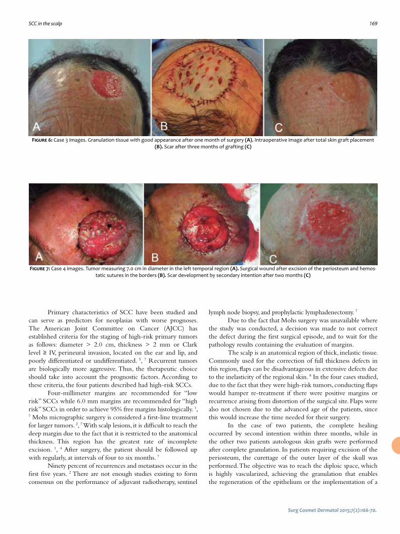
SCC in the scalp 169
Primary characteristics of SCC have been studied and can serve as predictors for neoplasias with worse prognoses. The American Joint Committee on Cancer (AJCC) has established criteria for the staging of high-risk primary tumors as follows: diameter > 2.0 cm, thickness > 2 mm or Clark level ≥ IV, perineural invasion, located on the ear and lip, and poorly differentiated or undifferentiated. 5, 7 Recurrent tumors are biologically more aggressive. Thus, the therapeutic choice should take into account the prognostic factors. According to these criteria, the four patients described had high-risk SCCs.
Four-millimeter margins are recommended for “low risk” SCCs while 6.0 mm margins are recommended for “high risk” SCCs in order to achieve 95% free margins histologically. 1, 7 Mohs micrographic surgery is considered a first-line treatment for larger tumors. 2, 7 With scalp lesions, it is difficult to reach the deep margin due to the fact that it is restricted to the anatomical thickness. This region has the greatest rate of incomplete excision. 1, 4 After surgery, the patient should be followed up with regularly, at intervals of four to six months. 7
Ninety percent of recurrences and metastases occur in the first five years. 2 There are not enough studies existing to form consensus on the performance of adjuvant radiotherapy, sentinel
Figure 6: Case 3 images. Granulation tissue with good appearance after one month of surgery (A). Intraoperative image after total skin graft placement (B). Scar after three months of grafting (C)
Figure 7: Case 4 images. Tumor measuring 7.0 cm in diameter in the left temporal region (A). Surgical wound after excision of the periosteum and hemos-tatic sutures in the borders (B). Scar development by secondary intention after two months (C)
lymph node biopsy, and prophylactic lymphadenectomy. 7
Due to the fact that Mohs surgery was unavailable where the study was conducted, a decision was made to not correct the defect during the first surgical episode, and to wait for the pathology results containing the evaluation of margins.
The scalp is an anatomical region of thick, inelastic tissue. Commonly used for the correction of full thickness defects in this region, flaps can be disadvantageous in extensive defects due to the inelasticity of the regional skin. 8 In the four cases studied, due to the fact that they were high-risk tumors, conducting flaps would hamper re-treatment if there were positive margins or recurrence arising from distortion of the surgical site. Flaps were also not chosen due to the advanced age of the patients, since this would increase the time needed for their surgery.
In the case of two patients, the complete healing occurred by second intention within three months, while in the other two patients autologous skin grafts were performed after complete granulation. In patients requiring excision of the periosteum, the curettage of the outer layer of the skull was performed. The objective was to reach the diploic space, which is highly vascularized, achieving the granulation that enables the regeneration of the epithelium or the implementation of a
Surg Cosmet Dermatol 2015;7(2):166-70.

170 Silva LRTE, Ribeiro AMQ, Fleury Júnior LFF
skin graft. 8 The aesthetic and functional results were satisfactory, mainly due to the fact that the patients were bald. Choosing not to perform flaps has facilitated the follow-up of local recurrence, as in Case 3.
Treatment of SCC on the scalp with wide excision and without closure flaps during the first operative event is an option for high-risk tumors. This technique allows the evaluation of local recurrences without anatomic distortion of the bed,
since long-term follow up is recommended. It also allows short surgeries and less post-operative morbidity, which can be decisive in the case of elderly patients. On the downside, caring for the surgical wound is laborious and it can take several weeks to achieve complete healing. The scar area will be left without hairs and with thinner skin, becoming more fragile and therefore more susceptible to injuries. l
REFERENCES1. Jenkins G, Smith AB, Kanatas AN, Houghton DR, Telfer MR. Anatomical
restrictions in the surgical excision of scalp squamous cell carcinomas:
does this affect local recurrence and regional nodal metastases? Int J Oral
Maxillofac Surg. 2014;43(2):142-6.
2. Alam M, Ratner D. Cutaneous Squamous Cell Carcinoma. N Engl J Med.
2011;344(13):975-83.
3. Clayman GL, Lee JJ, Holsinger FC, Zhou X, Duvic M, El-Naggar AK, et al. Mor-
tality risk from squamous cell skin cancer. J Clin Oncol. 2005;23(4):759-65.
4. Baker NJ, Webb AA, Macpherson D. Surgical management of cutaneous
squamous cell carcinoma of the head and neck. Br J Oral Maxillofac Surg.
2001;39(2):87-90.
5. Ch’ng S, Clark JR, Brunner M, Palme CE, Morgan GJ, Veness MJ. Relevance
of the primary lesion in the prognosis of metastatic cutaneous squa-
mous cell carcinoma. Head Neck. 2013;35(2):190-4.
6. Vauterin TJ, Veness MJ, Morgan GJ, Poulsen MG, O’Brien CJ. Patterns of
lymph node spread of cutaneous squamous cell carcinoma of the head
and neck. Head Neck. 2006;28(9):785-91.
7. Nuño-González A, Vicente-Martín FJ, Pinedo-Moraleda F, López-Esteba-
ranza JL. High-Risk Cutaneous Squamous Cell Carcinoma. Actas Dermo-
sifiliogr. 2012;103(7):567-78.
8. Hoffmann JF. Reconstrução do escalpo. In: Baker SR. Retalhos Locais em
Reconstrução Facial. 2ª ed. Rio de Janeiro: Di Livros; 2009. p. 641-67.
Surg Cosmet Dermatol 2015;7(2):166-70.
171
Surg Cosmet Dermatol 2015;7(2):171-3.
Case Reports
Authors: Rodolfo Chedid1
Janaina Nagel2
Taynara de Mattos Barreto3
Glaura Tinoco Plata4
Anna Luiza Bento Dutra3
Thales Pereira Azevedo3,1
1 Plastic Surgeon - Rio de Janeiro (RJ), Brazil
2 MSc in Pathology from the Universidade Federal do Rio de Janeiro (UFRJ) - Rio de Janeiro (RJ), Brazil
3 Physician from the UFRJ
4 Dermatologist Physician - Rio de Janeiro (RJ), Brazil
Correspondence: Glaura Tinoco Plata Rua Voluntários da Pátria, 445, sala 702 - Botafogo Cep: 22270-000 - Rio de Janeiro (RJ), Brazil E-mail: [email protected]
Received on: 26 August 2015 Approved on: 13 May 2015
This study was performed at a private practice – Rio de Janeiro (RJ), Brazil.
Financial support: None Conflict of interest: None
Eccrine spiradenoma: a case reportEspiroadenoma écrino - relato de caso
DOI: http://dx.doi.org/10.5935/scd1984-8773.201572387
ABSTRACTEccrine spiradenoma is a rare benign adnexal epithelial neoplasm characterized by a bluish-pink, slow-growing solitary nodule, coursing with paroxysmal pain, mainly in you-ng adults. It is most commonly located in the trunk and extremities. Bleeding and/or ulceration are common in the giant vascular variant, which is characterized by a size greater than 2 cm in diameter and a high degree of vascularization seen on histology. Its clinical characteristics are unspecific and require biopsy with histological analysis for diagnosis. Its histology presents one or more basophilic intradermal lobules enveloped in a fibrous capsule without connection to the epidermis. It presents two types of cells: small cells with dark nuclei in the periphery and large cells with light nuclei in the center. An accurate diagnosis is important due to the fact that it can develop into a potentially fatal malignancy – which is a rare phenomenon and occurs most often in the multiple variant. A rare case of eccrine spiradenoma in the abdomen with bleeding associated is described in the present paper, as well as a discussion on possible treatments. Keywords: eccrine spiroadenoma, benign epithelial neoplasia, sebaceous glands
RESU MOResumo: Espiroadenoma écrino é rara neoplasia epitelial benigna de anexos cutâneos, que se ca-racteriza por nódulo róseo-azulado solitário de crescimento insidioso, cursando com crises dolorosas paroxísticas em adultos jovens. Localiza-se preferencialmente no tronco e extremidades. Sangramento e/ou ulceração são comuns na variante gigante vascular, caracterizando-se por diâmetro superior a 2cm e alta vascularização na histopatologia. Suas características clínicas são inespecíficas e requerem biópsia com análise histopatológica para diagnosticá-lo. Na histopatologia apresentam-se um ou mais lóbulos intradérmicos basofílicos envoltos em cápsula fibrosa sem conexão com epiderme e apresentam dois tipos de células: uma pequena, de núcleo escuro, na periferia; e outra grande, de núcleo claro, no centro. Seu diagnóstico acurado é importante, pois pode evoluir para malignidade potencialmente fatal, o que, entretanto, é fenômeno raro e ocorre mais frequentemente na variante múltipla. Aqui reporta-remos uma rara apresentação de espiroadenoma écrino no abdômen e com sangramento associado, e discutiremos suas possíveis abordagens. Palavras-chave: espiroadenoma écrino, neoplasia epitelial benigna, glândulas sebáceas
INTRODUCTIONEccrine spiradenoma is a benign tumor of the sweat
glands. It was described in 1956 by Kersting and Helwig as an intradermal nodule, usually solitary, with paroxysmal pain crises. It occurs mostly in people aged 15 to 35, and most typically on the chest and face. 1-3 The tumor is usually covered by skin of normal or bluish color, and its typical location is the superficial or deep dermis, but can occasionally be found in the subcutaneous tissue. Although the face and chest are the most common sites, the tumor can occur in any body part. 1 It is a tumor with a long medical history and slow growth. 4 Malignization is rare but can take place. 5

172 Chedid R, Nagel J, Barreto TM, Plata GT, Dutra ALB, Azevedo TP
Surg Cosmet Dermatol 2015;7(2):171-3.
CASE REPORTAn 82-year-old female patient sought treatment for a
lesion on the abdomen, around one year in development and with intermittent bleeding during the previous month. On examination, an erythematous, hardened nodule not adhered to the deep planes was found. (Figures 1 and 2)
The initial diagnostic hypotheses were sarcoma, amelanotic melanoma, B cell lymphoma, and cutaneous metastasis.
An incisional biopsy was performed, showing diffuse proliferation of monomorphic ovoid cells, sometimes with basaloid appearance, arranged in lobes septated by hyalinized bundles, and compatible with eccrine spiradenoma. The pathologist suggested the removal of the entire lesion (Figures 3 to 5), as well as new histology. The removal of the entire lesion was performed in a hospital setting by a plastic surgeon, with the new histology revealing a benign adnexal neoplasm composed of solid cell blocks arranged in the dermis, with no connection to the epidermis (Figure 6). The neoplastic blocks revealed small epithelial cells with a hyperchromatic nucleus and larger epithelial cells with pale cytoplasm around the luminal ductal structures (Figure 7).
DISCUSSIONThe eccrine spiradenoma is an uncommon, benign tumor
that arises from the sweat glandular apparatus and is one of nine painful skin tumors – the others being leiomyoma, neuroma, dermatofibroma, angiolipoma, neurilemmoma, endometrioma, glomus tumor, and granular cell tumor. 6
The tumor occurs either as solitary lesion (in 97% of cases and can reach a diameter of 5 cm) or as multiple lesions, which in some cases arise in linear or zosteriform distribution. 1, 6, 7 The lesions are typically small, well defined, and incorporated in normal sweat glands. 1
The precise etiology of the tumor is uncertain. The eccrine spiradenoma has never been observed in the glabra skin and arises as a benign adnexal neoplasm, with little or no differentiation, and historically assumed as originating from the eccrine lineage. However, it is known that the tumor often occurs along with other skin adnexal neoplasias such as: cylindroma, trichoepithelioma, and trichoblastoma – which favors the development of a follicle sebaceous-apocrine lineage rather than eccrine differentiation. Also, if the tumor was truly eccrine, it would be seen frequently on the palms or soles, rather than not at all, as in this case. 7
The main clinical feature is the presence of pain or sensitivity in about 91% of the patients, which usually occurs paroxysmally. It is believed that the pain is linked to small demyelinated axons that permeate the hyaline stromal mantle. In 1996, Crinton and Aravindan proposed that the pain could be related to the contraction of the tumor’s myoepithelial cells, nevertheless electron microscopy has not proven the existence of these cells. Several studies have demonstrated the expression of the immunohistochemical marker S-100 in the tumor, suggesting
Figure 1: Erythematous nodule with ulceration in the abdominal region
Figure 2: Close-up of the lesion
Figure 3: The site where the exeresis of the lesion will be carried out
Figure 4: Immediately post-operative
Figure 5: Macroscopic appearance of the excised lesion

Eccrine spiradenoma 173
Surg Cosmet Dermatol 2015;7(2):171-3.
the existence of a link with neural tissue, since S-100 protein is generally present in neural crest-derived cells, and chondrocytes, adipocytes, myoepithelial cells, macrophages, Langerhans cells, dendritic cells, and keratinocytes. Due to the presence of this protein in various cell lineages, it is not possible to precisely establish the relationship with the neural tissue. Nonetheless, a different study has demonstrated the presence of Langerhans cells with thin and irregular expansions dispersed among the tumor cells in the nodule. In a case study carried out by Park et al., 6 the sample was positive for S-100 only in the tumor’s capsule, which was the exact position of disorganized, thickened nerve fibers. The authors of that study also confirmed the presence of a neural element with the positivity of an axon marker. 6 The differential diagnosis should include other causes of painful tumors but also: anaplastic carcinoma, adenocarcinoma, squamous and basal cell
carcinomas, and other adnexal neoplasias, such as cylindromas; in addition to mesenchymal tumors. 1, 7
There can be a suspicion of malignant eccrine spiradenoma when there is the emergence of pain that did not previously exist, increased sensitivity, color change, rapid growth, or ulceration of a lesion that has remained stable for a long time. The suspicion of malignization should be thoroughly evaluated due to the aggressiveness of the malignant tumor, with a mortality rate that varies between 20% and 39%, and the lymph nodes, bones, lungs, and brain as main metastasis sites. 1, 4
Clinical suspicion is useful, however it is not sufficient to make the diagnosis. The definitive diagnosis will come from the histology of the skin biopsy. The cytology of a fine needle aspirate can also confirm the diagnosis. 7 In the histological examination, basophilic lobes are seen under smaller magnification due to the dense nuclei of tumor cells. Under greater magnification, basaloid cells comprising two distinct morphologies can be seen: in the first, the cells are larger, clearer, and with ovoid nuclei; in the second, the cells are smaller, darker, and with compact hyperchromatic nuclei. 5, 7
The treatment is essentially surgical, with the more common options being conventional surgery or the Mohs technique yielding small recurrence rates – and therefore the best treatment option aimed at preventing malignant transformation. 7
CONCLUSIONThe present article reports the finding of a benign eccrine
spiradenoma – a tumor of the sweat glands, with a poorly-defined etiology. The eccrine spiradenoma is a rare tumor that can emerge in various clinical forms. Its diagnosis is crucial due to the potential for malignant transformation of these lesions, especially in cases of multiple or symptomatic lesions. Malignant eccrine spiradenoma can be lethal if not diagnosed and treated, and a high index of suspicion is necessary in any benign lesion that rapidly changes its characteristics, i.e. consistent with a longstanding disease. Case reports, like the present, are important to maintain heightened suspicion regarding the diagnosis of this rare condition and in assisting in guiding an appropriate approach. l
REFERENCES1. Yesilkaya Y, Demirbas B, Gokoz O, Akata D. Eccrine Spiradenoma of the
Thigh: Sonographic findings and review of the literature. J Diagn Med
Sonogr. 2012;28(6):311-313.
2. Hashimoto K, Gross BG, Nelson RG, Lever WF. Eccrine Spiradenoma: Histo-
chemical and electron microscopic studies. J Invest Dermatol. 1966;46(4):
347-65.
3. Azulay RD, Azulay DR, Azulay-Abulafia L. Dermatologia. 5 ed. Rio de Janei-
ro: Guanabara Koogan; 2008. p.554.
4. Berçin S, Kutluhan A, Metin A, Süren D. Malignant eccrine spiradenoma
on the lateral margin of nose as an infrequent localization. Indian J Der-
matol. 2009;54(2):173-175.
5. Storm CA, Seykora JT. Cutaneous Adnexal Neoplasms. Am J Clin Pathol.
2002;118(Suppl):S33-49.
6. Park HR, Im SB, Kim HK, Shin DS, Park YL. Painful Eccrine Spiradenoma
Containing Nerve Fibers: A Case Report. Dermatology. 2012;224(4):301-6.
7. Englander L, Emer JJ, McClain D, Amin B, Turner RB. A Rare case of multiple
segmental eccrine spiradenomas. J Clin Aesthet Dermatol. 2011;4(4):38-44.
Figure 6: HE 10x. Benign adnexal neoplasm composed of neoplasic blocks arranged in the dermis, without connection
Figure 7: HE 20x. Greater detail of neoplasic blo-cks consisting of two cell populations

Case ReportsTunneling island pedicled flap after resection of carcinoma on the faceRetalho em ilha tunelizado após exérese de carcinoma na face
DOI: http://dx.doi.org/10.5935/scd1984-8773.201572629
ABSTRACT Carcinomas on the face are prevalent lesions, with potentially serious repercussions from
aesthetic, psychological, and functional perspectives. Surgical reconstruction after excision of these lesions is a challenge for the dermatologic surgeon, requiring good knowledge of the anatomy of the face and accurate implementation of the most appropriate method for each case. Among the various reconstructive procedures are skin flaps, of which the island pedicle flap and its variants stand out. The present article demonstrates the use of the island pedicle flap transferred to the surgical defect through a subcutaneous tunnel, showing the application and characteristics of this method. Reconstruction using a tunneling island pedicle flap after the excision of a facial carcinoma showed good aesthetic and functional results, despite the difficult location of the lesions. This type of flap is a good option for facial reconstruction in difficult areas.
Keywords: carcinoma, basal cell; surgery, plastic; surgical flaps
RESU MO Os carcinomas da face são lesões prevalentes, com repercussões potencialmente graves do ponto de vista
estético, psicológico e funcional. A reconstrução cirúrgica na face demanda apurada execução da técnica adequada a cada situação. Dentre as diversas modalidades reconstrutivas, incluem-se os retalhos cutâ-neos, destacando-se o retalho em ilha e suas variantes. Este artigo demonstra o emprego do retalho em ilha transferido ao defeito cirúrgico através de túnel criado no subcutâneo. A reconstrução com retalho em ilha tunelizado apresentou bons resultados estéticos e funcionais apesar de as lesões estarem em locais de difícil correção. Esse tipo de retalho constitui boa opção para esse tipo de lesões.
Palavras-chave: carcinoma basocelular; procedimentos cirúrgicos ambulatórios; retalho perfurante
Authors: Alexander Sabino Sisnando1
Luana Oliveira Ramos2
Fabio Francesconi3
1 Dermatology Resident Physician, Fun-dação de Medicina Tropical Heitor Vieira Dourado (FMT-HVD) – Manaus (AM), Brazil
2 Dermatology Resident Physician, Hospi-tal Universitário Getúlio Vargas (HUGV) da Universidade Federal do Amazonas (Ufam), Manaus (AM), Brazil
3 Head Preceptor at the Dermatology Me-dical Residency Program (accredited by SBD), FMT-HVD. Dermatology Instructor, Ufam
Correspondence: Alexandre Sisnando Rua Rio Iça, 1.111 – Ed. Dona Neide, apt. 206 - Bairro N.S. das Graças, Cep: 69053-100 Manaus (AM), Brazil E-mail: [email protected]
Received on: 7 March 2015Approved on: 23 May 2015
This study was performed with patients ope-rated on at the Fundação Centro de Controle de Oncologia do Estado do Amazonas (FCE-CON), an associate hospital for supporting the medical residency services of the Funda-ção de Medicina Tropical Heitor Vieira Dou-rado (FMT-HVD) and of the Hospital Getúlio Vargas (HUGV) – Manaus (AM), Brazil.
Financial support: None
Conflict of interest: None
175175
INTRODUCTIONThe island flap has two basic characteristics: 1) the shape
of the donor skin resembles an island that is detached from the surrounding epidermis and dermis on all sides; 2) a subcutaneous pedicle is maintained, thereby ensuring vascularization and allowing some mobility to the nearby receptor area. Variations of this type of flap depend on the shape of the island and the way it is transferred to the receptor area. Among these variations are: the traditional triangular island with V-Y advance, which can be single or double (bipedicled); the variant where the shape of the island and its size are similar to those of the defect and the transference is performed by transposition (the skin between the donor and receptor areas is excised) or interpolation (where a pedicle is left between the donor and the receptor area, with its subsequent removal during a second subsequent surgery); and the variant where the island, whose size and shape correspond
Surg Cosmet Dermatol 2015;7(2):175-8.

176 Sisnando AS, Ramos LO, Francesconi F
to those of the surgical defect, is transferred through a tunnel created in the upper portion of the subcutaneous tissue. 1 The latter variant can be considered a type of interpolation, as the skin between the primary and secondary defects is not excised.
The present article describes two cases of basal cell carcinoma (BCC) in facial areas, each with difficult reconstruction, where the tunneled island flap technique was used, with an aim of demonstrating the application, characteristics, and possible complications of this method.
CASE 1A 57-year-old woman presented with a round shaped
basal cell carcinoma (BCC) with elevated and pearly borders in the medial corner of the upper left eyelid (Figure 1A). The patient underwent excision that resulted in a surgical defect 10 mm in diameter. An advancement flap would not be a good option in this case, as it would distort the anatomy of the region. A decision was made in favor of using a non-contiguous donor area (in the glabellar region). 2 For the preparation of the flap, a similar shape and size to those of the defect were implemented, however fusiform, aiming at facilitating the closure (Figure 1B). The dissection of the donor area was carried out, preserving a subcutaneous pedicle intended to nourish the flap. In the existing healthy skin between the primary and secondary surgical defects, a tunnel was created in the subcutaneous level using the blunt dissection technique (Figure 2A). Next, the island skin flap – together with the pedicle – was driven through the tunnel using a hook (which may be improvised with a small syringe and a 25x7 needle carefully bent at the tip, as in the present case) (Figure 2B).
Once positioned in the defect, the flap was sutured with 5.0 nylon thread and interrupted sutures. The post-operative period coursed with significant local edema, which is common in these cases and can be minimized with the application of cold compresses for 20 minutes, several times a day. There was
no infection or necrosis (Figure 2C). The result was satisfactory, with no trapdoor effect, 3 nevertheless there was a slight bulging of the skin between the donor and the receiving areas (Figure 3). The post-operative histologic control of the surgical specimen revealed free margins.
CASE 2An 86-year-old female patient presented with a centrally
ulcerated, pigmented BCC on the upper lip, advancing towards the right nostril, and measuring about 9 mm at its widest diameter. After excision with a 4.0 mm margin, a difficult to correct defect regarding the functional aspect was observed (Figure 4). A decision was made for a tunneled interpolation flap, in which the island’s fusiform design in the nasolabial fold allowed for the suture of the secondary defect to be positioned in the fold, providing a good aesthetic appearance to the scar (Figure 5). In order to rebuild the base of the columella, a small island with traditional transposition was performed and
Surg Cosmet Dermatol 2015;7(2):175-8.
Figure 1: A. Rounded Basal Cell Carcinoma, with defined, elevated and pearly borders, 6.0 mm in diameter. B. A surgical defect 10.0 mm in diameter after excision of the lesion with margins, in a region difficult for reconstruction; preparation of the island flap with a size similar to that of the defect, with a fusiform design and extracted from the glabella region, in order to facilitate the closure. A subcutaneous pedicle is left, aimed at nourishing the flap
Figure 2: A. Subcutaneous tunnel; B. Transfer of the flap and its pedicle via the subcutaneous tunnel; C. Post-operative edema and hematoma
A
A
B
C
B

Island flap 177
tractionned with its pedicle into the defect using 5.0 nylon thread (also used in the suture), in a way that distributed the tension and facilitated the positioning of the tunneled flap (Figure 5). The post-operative period coursed uneventfully (Figure 6). The final result was functionally and aesthetically satisfactory, and the traditional trapdoor defect could be observed in the island 3 (in the columella’s base), but not in the tunneled island (Figure 7).
DISCUSSIONBCC is the most prevalent skin cancer in humans.
However, due to its low metastasizing capacity, it is usually curable with surgical exeresis in a single surgical procedure. 4
The tunneled island is a flap with a random pattern, whose blood supply depends on the vascular plexus of the deep dermis and subcutaneous. 5 It has the advantage of providing the surgical defect with a skin non-adjacent to the lesion but with the same characteristics of the surrounding area, thereby preserving the local anatomy. 6 The transference via a subcutaneous tunnel prevents the need for an incision in the skin between the donor
Surg Cosmet Dermatol 2015;7(2):175-8.
Figure 3: Good aesthetic and functional result of the reconstruction
Figure 4: A and B – Surgical removal of a pigmented and ulcerated BCC in the upper lip
Figure 6: Post-operative.A. Immediately post-operative: flaps positioned without tension and sutu-red with interrupted stitches in the reconstruction of the upper lip, base of the nostril and columella; secondary defect closed with a continuous
suture. B. D8 post-operative: absence of infection or necrosis
Figure 7: Final result. D29 post-operative: the trapdoor effect can be observed in the traditional island (base of the columella) and satisfactory functional and aesthetical outcome of the tunneled flap and secondary
defect scar, positioned in the fold
A
A
B
B
Figure 5:A. Preparation of the flap. The island is dissected in the shape of the nasolabial fold’s fuse. Notice the tra-ditional small island in the base of the columella, aimed at distributing the tension in the area and facilitating the positioning of the other flap in the reconstruction of the nostril.B. Intraoperative. Transfer of the tunneled flap tractionned through the subcutaneous tissue with the aid of a surgical thread. Notice the secon-dary defect properly positioned in the nasolabial fold.
A
B

178 Sisnando AS, Ramos LO, Francesconi F
and the receiving areas and allows the flap to be removed from a non-contiguous area 1, observing local characteristics and yielding a good cosmetic result. 7
In Case 1, the tunneled interpolation of the island avoided the distortion of the eyebrow region, as the primary defect was located on the upper eyelid. However, an undesirable effect of this flap is that at the moment of subcutaneous transfer, there is the addition of material beneath the skin, resulting in the elevation in the area through which the tunnel has been created. 1 This took place in the first case described. Had the lesion been located in the nasociliary region, 2 a traditional transposition of the island would have been be the best option, producing however, a secondary defect in the glabellar region. 8
In Case 2, due to the fact that the lesion was located on the upper lip, the reconstruction was aimed at preserving the anatomy, owing to the aspects of the functional and aesthetic appearance, requiring extra attention in regards to the positioning of the vermilion and the transition line between the skin and the semimucosa, preserving the lip contour, the position of the philtrum, and the bilateral symmetry regarding the nasolabial folds. 9 Among the various techniques available for the reconstruction of this region, the island flap has been shown to be a good option for it causes little distortion of the anatomy and low scar retraction, and also offers ease in positioning the suture of the secondary defect in the nasolabial fold, yielding a good cosmetic result.
CONCLUSIONThe island flap with subcutaneous tunnel interpolation
is a very useful resource in the surgical repair of certain surgical defects following the exeresis of a carcinoma in the face, especially when it is possible to achieve a cosmetically acceptable secondary defect scar – in a natural fold of the skin or in an area where the appearance of rhytids is common – and when seeking consistency between the flap and the skin around the primary defect. l
REFERENCES1. Kimyai-Asadi A, Goldberg LH. Island pedicle flap. Dermatol Clin.
2005;23:113-27.
2. Tamura BM. Topografia facial das áreas de injeção de preenchedores e
seus riscos. Surg Cosmet Dermatol. 2013;5(3):234-8.
3. Li JH, Xing X, Liu HY, Li P, Xu J. Subcutaneous Island Pedicle Flap - Variations
and Versatility for Facial Reconstruction. Ann Plast Surg. 2006;57(3):255-9.
4. Morselli P, Zollino I, Pinto V, Brunelli G, Carinci F. Evaluation of clinical prog-
nostic factors in T1 N0 M0 head and neck basal cell carcinoma. J Cranio-
fac Surg. 2009;20(1):98-100.
5. Wang SQ, Goldberg LH, Kimyai-Asadi A. Tunneled Island Pedicle Flap for
an Earlobe Defect. Dermatol Surg. 2007;33(7):835-7.
6. Mahlberg MJ. Tunneled Melolabial Pedicle Flap for Small but Deep Late-
ral Alar Rim Defect. Dermatol Surg. 2013;39(10):1527-9.
7. Pontello Júnior R, Kondo RN, Pontello R. A utilização do retalho A-T para
reconstrução de ferida operatória no dorso da mão. Surg Cosmet Derma-
tol. 2013;5(3):270-2.
8. Campbell LB, Ramsey ML. Transposition island pedicle flaps in the
reconstruction of nasal and perinasal defects. J Am Acad Dermatol.
2008;58(3):434-6.
9. Lopes Filho LL, Lopes LRS, Villa Neto AB, Costa TS. Carcinoma basocelular
no lábio superior: tratamento cirúrgico e reconstrução com retalho de
transposição. Surg Cosmet Dermatol. 2011;3(3):243-5.
Surg Cosmet Dermatol 2015;7(2):175-8.

Case Reports Benign syringoma chondroid syringoma a basal cell carcinomaSiringoma condroide benigno simulando carcinoma basocelular
DOI: http://dx.doi.org/10.5935/scd1984-8773.201572469
ABSTRACTChondroid syringoma is a rare benign cutaneous adnexal tumor, also called mixed tumor of the skin, due to the presence of epithelial and mesenchymal components, as well as sweat glands elements. Due to the fact that it clinically resembles a variety of skin lesions, histological examination is crucial for diagnosis and hence treatment. Keywords: neoplasms, adnexal and skin appendage; syringoma; adenoma, pleomorphic
RESU MO O siringoma condroide é tumor benigno raro de anexos cutâneos, também denominado tumor misto de pele, devido à presença de componentes epiteliais, mesenquimais e elementos de glândulas sudorí-paras. Por se assemelhar clinicamente a diversas lesões de pele, o exame histopatológico é fundamental para definir o diagnóstico e, consequentemente, a terapêutica.Palavras-chave: neoplasias de anexos e de apêndices cutâneos; siringoma; adenoma pleomorfo
Authors: Jeane Jeong Hoon Yang1
Antonio José Tebcherani2
Ed Wilson Tsuneo Rossoe3
1 Dermatologist Physician, Complexo Hos-pitalar Padre Bento de Guarulhos (CHPBG) - Guarulhos (SP), Brazil
2 Head of the Dermatopathology Labora-tory, CHPBG
3 Plastic Surgeon, Oncologic Surgery Pre-ceptor, CHPBG
Correspondence: Jeane Jeong Hoon Yang Setor de Dermatologia Av. Emilio Ribas, 1.819 - Gopoúva Cep: 07051-000 – Guarulhos (SP), Brazil E-mail: [email protected]
Received on: 15 March 2014Approved on: 22 November 2014
This study was performed at Complexo Hos-pitalar Padre Bento de Guarulhos (CHPBG) - Guarulhos (SP), Brazil.
Financial support: NoneConflict of interest: None
180
INTRODUCTIONChondroid syringoma, also known as cutaneous mixed
skin tumor, is a rare tumor of the skin appendages, and consists of epithelial and mesenchymal elements. The presence of sweat glands surrounded by cartilage matrix, led to the name chondroid syringoma. 1 It can clinically resemble different skin lesions, for it does not present particular clinical features. 2 Among the differential diagnoses are basal cell carcinoma, pilomatricoma, steatocystoma; therefore histological examination is required for diagnostic confirmation. 3 We report the case of a rare clinical presentation of a chondroid syringoma simulating a basal cell carcinoma.
CASE REPORTA 71-year-old Caucasian male patient described the
appearance of a lesion on the left brow five years earlier, gradually increasing in size during the last two years, but asymptomatic. The patient was a former alcoholic (in recovery for 10 years). A dermatological examination showed a pearlescent/pearly colored nodule with telangiectasias on the surface, measuring around 1.5 cm (Figure 1). A diagnostic hypotheses of a basal cell carcinoma,
Surg Cosmet Dermatol 2015;7(2):180-3.

Chondroid syringoma 181
Surg Cosmet Dermatol 2015;7(2):180-3.
pilomatricoma, sebaceous cyst was formulated and a decision was made for the surgical removal of the lesion. The patient is receiving outpatient treatment, with good clinical improvement.
DISCUSSIONChondroid syringoma is a rare, benign adnexal skin
tumor, originally described in 1859 by Billroth as a mixed skin tumor due to the existence of both epithelial and stromal components. The presentation of sweat gland components defined in cartilaginous stroma led Hirsch and Helwig to name this lesion chondroid syringoma in 1961. 1 These authors proposed five histologic criteria for its diagnosis: 1) nests of cubic or polygonal cells; 2) intercommunicating tubular alveolar structures flanked by two or more rows of cubic cells; 3) ductal structures consisting of one or two rows of cubic cells; 4) occasional cysts with keratin; 5) a variable composition matrix. 1 It is clinically characterized by a single nodule with a smooth surface, non-adherent to the deep planes and with precise limits, asymptomatic, located mainly on the face or neck, and most commonly on the nose, lips, and eyebrows, as in the present case. Less commonly, it may involve hands, feet, axillae, abdomen, penis, scrotum, and vulva. 4 It grows slowly and progressively. Its size typically varies between 0.5 and 3.0 cm 5 (the studied patient had lesions of 1.5 cm in diameter). It is usually found in men (mean age around 50 years), with a gender ratio ranging from 1.3/1 and 5/1.3 There is an absence of any particular clinical feature, and the diagnosis is essentially histological. 4 There is a presence of epithelial components (glandular structures with eccrine or apocrine differentiation, or both) and mesenchymal components (tissue with myxoid, chondroid, adipose or fibrous tissue differentiation). The epithelial component comprises glands that can be histologically of two types: the apocrine (as in the present case – the most common type, constituted by focally connected tubular or cystic glands, fringes with a double
seat of cubic or flattened cells); and the eccrine (characterized by a narrow lumen and glands framed by a single layer of cells). Although the immunohistochemical study is dispensable for the diagnosis due to the lesion’s dual population feature (glandular structures and cartilaginous components), there are studies reporting the expression of cytokeratin (CK), epithelial membrane antigen (EMA), carcinoembryonic antigen (CEA) in the glandular structures, vimentin, S-100 protein, neuron-specific enolase (NSE), and in some cases glial fibrillary acidic protein (GFAP) in the cartilaginous component. 6
The case reported has considerably exuberant epithelial and mesenchymal components, in addition to the important fact that the lesion’s limits are well defined (Figure 2). This, together with the absence of atypias, favors its benign nature. The mesenchy-mal component has myxoid (Figure 3) and chondroid (Figure 4) elements, in addition to foci of adipose differentiation. The glan-dular part of the tumor is sometimes constituted by glands with regular and reduced lumen (which resemble the secretory por-Figure 1: Chondroid syringoma. Pearly color nodule with telangiectasias
on the surface. Diameter = 1.5 cm
Figure 2: EH-40x. Details of the lesion’s well defined and regular contours in contrast to the adjacent dermis
Figure 3: EH-400x. Details of the myxoid component (basophilic and amorphous material). Note the glandular structure in the upper left corner

182 Yang JJH, Tebcherani AJ, Rossoe EWT
Surg Cosmet Dermatol 2015;7(2):180-3.
tion of eccrine sweat glands) (Figure 5), and sometimes by glands with ectatic and tortuous lumen, suggestive of apocrine differ-entiation. Cysts filled with keratin are also present (Figure 5). The chondroid syringoma has no specific clinical characteristics and resembles other skin lesions. Among the differential diagno-sis it is possible to mention epidermal inclusion cysts, amelanotic nevi, sebaceous cysts, dermoid cysts, schwannomas, neurofibro-mas, pilomatrixomas and basal cell carcinomas. 7 In the present case, the patient had clinical features resembling those of a basal cell carcinoma. Most cases are benign, although there are rare malignant forms. 1,2 Malignant types most commonly occur in young women and often measure more than 3.0 cm, are locally invasive and show a predilection for the trunk or extremities. 1,2
Histological features considered to be signs of malignant trans-formation are: cytologic atypia, infiltrative margins, satellite tu-mor nodules, tumor necrosis, and the involvement of deep struc-tures, 8 – which were not seen in the present case. The treatment of choice is excision with the removal of 4.0 mm of normal peritumoral tissue. Surgery is often straightforward due to the good delimitation of the lesion by a capsule. In the present case, the lesion was excised entirely. There was no recurrence during the initial three months of follow-up. The patient remains under observation. Other treatment options described are dermabra-sion, electrodessication, and vaporization with Argon or CO2 laser. 4 Incompletely resected benign tumors require periodic monitoring aimed at detecting possible recurrence or malignant transformation. 7
l
Figure 4: HE-400x. Details of the chon-droid component. Note the numerous glandular structures with tortuous lumen
Figure 5: EH- 400x. Details of the gland with eccrine differentiation (center) and the cyst filled with keratin (bottom right)

Chondroid syringoma 183
Surg Cosmet Dermatol 2015;7(2):180-3.
REFERENCES1. Hirsch P, Helwig EB. Chondroid syringoma. Mixed tumor of skin, salivary
gland type. Arch Dermatol. 1961;84:835-47.
2. Solanki LS, Dhingra M, Bhalla M, Thami GP, Punia RPS, Shilpy Batra S. Chon-
droid syringoma: Report of two cases in young patients. Dermatol Online
J. 2011;17(12):7.
3. Abila S, Bouhllaba J, Zarkika S, Lamchahaba FZ, Saidib A, Ismailia
N, et al. Syringome chondroïde de la face. Ann Dermatol Venereol.
2012;139(10):681-3.
4. Yavuzer R, Basterzi Y, Sari A, Bir F, Sezer C. Chondroid syringoma: a diag-
nosis more frequent than expected. Dermatol Surg. 2003;29(2):179-81.
5. Tokyol C, Aktepe F, Yavas BD, Yildiz H, Aycicek A. Chondroid syringoma: a
case report. Acta Cytol. 2010;54(8):973-6.
6. Argenyi ZB, Balogh K, Goeken JA. Immunohistochemical characterization
of chondroid syringomas. Am J Clin Pathol. 1988;90(6):662-9.
7. Varsori M, Dettwiler S, Chaloupka K. Syringome chondroïde de la pau-
pière: à propos d’un cas. J Fr Ophtalmol. 2007;30(1):3.
8. Bates AW, Baithun SI. Atypical Mixed Tumor of the Skin: histologic, immu-
nohistochemical, and ultrastructural features in three cases and a review
of the criteria for malignancy. Am J of Dermatopathol. 1998;20(1):35-40.

184
Case Reports Melanoma Desmoplásico - um desafio diagnósticoDesmoplastic melanoma: a diagnostic challenge
DOI: http://dx.doi.org/10.5935/scd1984-8773.201572560
ABSTRACTDesmoplastic melanoma is a rare variant of melanoma characterized by an invasive lesion of spindle cells and varying degrees of desmoplasia. It is more frequent in men of an ad-vanced age having a history of chronic exposure to sunlight. The extremely variable and nonspecific clinical appearance makes it a challenging diagnosis. The authors present an exuberant case of desmoplastic melanoma in an unusual location, with an initial diagnosis of dermatofibrosarcoma. Keywords: melanoma; diagnosis; histology; immunohistochemistry
RESU MOO melanoma desmoplásico é variante rara, caracterizada por lesão invasiva de células fusiformes e graus variáveis de desmoplasia. Mais frequente em homens, com idade avançada e história de expo-sição solar crônica. A apresentação clínica extremamente variável e inespecífica, torna-o diagnóstico desafiador. Apresentamos caso exuberante de melanoma desmoplásico, em localização pouco comum e com hipótese diagnóstica inicial de dermatofibrossarcoma. Palavras-chave: melanoma; diagnóstico; histologia; imuno-histoquímica
Authors: Flavia Regina Ferreira1
Bruna Ferrari2
Livia Mendes Sabia Acedo3
Juliana Emi Dias Ujihara4
Marcia Lanzoni de Alvarenga Lira5
Samuel Henrique Mandelbaum6
1 PhD, Dermatology Assistant Professor, Medicine Department, Universidade de Taubaté (Unitau) and Preceptor at the Dermatology Service of the Hospital Uni-versitário de Taubaté (HUT) – Taubaté (SP), Brazil
2 2nd year Resident Physician, Dermatology Service, HUT
3 3rd year Resident Physician, Dermatology Service, HUT
4 1st year Resident Physician, Dermatology Service, HUT
5 Pathologist Physician, Assistant instructor, Pathology Discipline, Medicine Depart-ment, Unitau
6 Assistant Professor, Dermatology Discipli-ne, Medicine Department, Unitau, Head of the Dermatology Service, HUT
Correspondence: Hospital Universitário de Taubaté Serviço de Dermatologia Flávia Regina Ferreira Avenida Granadeiro Guimarães, 270 Cep: 12020-130 – Taubaté (SP), Brazil E-mail: [email protected]
Received on: 22 July 2014 Approved on: 22 February 2015
This study was performed at the Hospital Universitário de Taubaté (HUT) – Taubaté (SP), Brazil.
Financial support: None
Conflict of interest: None
184
INTRODUCTIONCutaneous melanoma is a malignant tumor located in
the dermal-epidermal junction of the skin, arising from the atypical transformation of melanocytes. It expresses a variety of phenotypes with different clinical and cytological variants, such as the extensive superficial, the nodular, the acral lentiginous and the lentigo maligna – which are the most classic – and the desmoplastic, the spitzoid and the amelanotic – which are the most unusual. 1-3
The desmoplastic melanoma (DM) is a rare variant characterized by invasive proliferation of spindle cells in the dermis and variable degrees of stromal deposition of collagen (desmoplasia). 1-3 It is more frequent in males of advanced age (mean age = 66 years) and with a history of chronic exposure to the sun, which may explain its prevalence in sun-exposed areas, especially the head and neck (53.2%). 2-4
The extremely variable and nonspecific clinical appearance means its diagnosis is challenging. The authors report an exuberant case of desmoplastic melanoma, atypical in appearance and at an unusual location, with an initial diagnosis of dermatofibrosarcoma.
Surg Cosmet Dermatol 2015;7(2):184-7.

Desmoplastic melanoma 185
CASE REPORTAn 83-year-old Caucasian male patient, a farmer by
occupation, sought medical attention complaining of a “wound” located on the dorsum for eight months (Figures 1 and 2). The dermatological exam showed an erythematous tumoration in the lower left back, with firm consistency, and a bright and lobular surface, measuring about 15 cm at its longest axis. He described the onset as a small crusted papule that grew rapidly over the course of two weeks. There was an absence of local symptoms and palpable lymph nodes.
Once it was diagnosed as a dermatofibrosarcoma protuberans, the patient underwent an incisional fusiform biopsy. The pathology (Figures 3 and 4) suggested a spindle cell neoplasm at least 4.7 mm thick, involving the dermis and hypodermis. Based on this report, immunohistochemistry was carried out (Figure 5) evidencing negative HMB-45 and Melan-A, positive S-100, and a positive proliferative index (Ki67) in 10-15% of cells. The findings were compatible with the diagnosis of desmoplastic melanoma.
The patient was referred to the surgical oncology sector, undergoing a complete exeresis of the lesion and a new pathology, which confirmed the diagnosis of desmoplastic melanoma with a 14 mm Breslow, Clark V, one mitosis/mm mitotic index, absence
of vascular and perineural invasion, and surgical margins free of neoplastic involvement. At the time this article was accepted for publication, the patient was under dermatologic and clinical oncologic follow up care.
DISCUSSIONFirst described in 1971 by Conley et al., the DM is a
distinct and uncommon variant, representing less than 4% of cases of primary cutaneous melanoma, affecting two individuals per one million inhabitants. 1-4 It is characterized by a fibrous tumor constituted by spindle-shaped cells isolated in a dense fibrous matrix, producing or liberating collagen. The tumor often presents neurotropism, a growth pattern similar to that of the neuroma and neural differentiation – also described as neural transformation phenomenon.4 Unlike the non-desmoplastic melanomas, it presents a greater tendency for local growth and a lower tendency for lymph node metastasis. 4,5
Due to its extremely variable and nonspecific clinical appearance, the DM comprises a true diagnostic challenge. It commonly appears as a hypomelanotic or amelanotic nodule, papule or plaque, with firm consistency, affecting the dermis or
Figure 1: Erythematous tumoration with lobulated surface in the left lumbar region.
Figure 2: Detail of the tumoration
Figure 3: Detail of spindle cell neoplasm with lymphoid aggregates (HE 100x)
Figure 4: Detail of spindle cells (HE 400x)
Surg Cosmet Dermatol 2015;7(2):184-7.

186 Ferreira FR, Ferrari B, Acedo LMS, Ujihara JED, Lira MLA, Mandelbaum SH
even the subcutaneous, similarly to other fibrous lesions, which leads to misdiagnosis. The highest incidence of DM misdiagnosis occurs among the malignancies, with carcinomas, fibrosarcomas and amelanotic melanomas. Among the benign lesions, are the fibromatosis, the dermatofibroma, the melanocytic nevi and the scars.3,4,6,7 The association with intraepidermal atypical melanocytic proliferation – such as lentigo maligna – covering the lesion or present in the resection margins, has been reported, thus facilitating its recognition and diagnosis. 2,3,6,7 In the present description, the uncommon clinical presentation (lobular tumoration) and topography (lumbar region), the large proportions of the lesion (about 15 cm in its longest axis) and the initial suspicion of dermatofibrosarcoma protuberans rather than DM, reinforce the great variability and clinical non-specificity of this melanoma variant, justifying the possible diagnostic errors.
Dermoscopy has limited use in DM due to its variable clinical presentation, usual amelanotic appearance and the scarcity of information about its dermoscopic features. 1,4 In 2008, Debarbieux et al. evaluated six patients with DM and only half of them showed positive dermoscopic criteria for melanocytic lesions. 8 Some of the predictive characteristics of DM in cases of amelanotic lesions are: the presence of signs of regression – such as scarring areas and “salt and pepper” appearance – as well as abnormal vascular patterns (serpentine, dotted and/or speckled), and pink/milky-red areas. 1,4,8
Histologically, it presents a spindle cell infiltrate with mild to marked nuclear atypia, invading the dermis and the subcutaneous tissue. They are arranged in variable patterns of
desmoplasia, neurotropism and neural differentiation.3,4 Staining with hematoxylin and eosin may be insufficient for diagnosis, since the tumor cells are almost always depigmented, which calls for an immunohistochemical study. The S-100, neuron specific enolase and vimentin markers are positive in 95% of cases, with the vast majority of DMs being negative for HMB-45 and Melan A.3 In a relatively recent study of a series of 11 DM patients, the presence of diffusely positive marking was demonstrated in 100% of the cases with the use of WT1, a potential marker for this clinical type.9 Dense intratumoral lymphocyte aggregates can also be very commonly present, in addition to in situ melanoma.4
At diagnosis, it has an average thickness of 2.5 mm to 6.5 mm, reaches the reticular dermis, and is most often classified as Clark IV-V,6 aligned with the findings of the clinical case in question – except for the latter, which had a surprising thickness of 14 mm.
Based on the degree of desmoplasia, the DM was classified into two histological subtypes: pure or combined. The pure subtype relates to minor lymph node involvement, less aggressive clinical course and more than 90% of desmoplastic compromise, while the combined subtype has less than 90% desmoplastic involvement, thick cell tumor focus without fibrosis, with irregular nuclei and higher rate of mitosis. 1,4
The role of the sentinel node in DM is not clear, and its routine use is not recommended. Some authors advocate the importance of staging cases with 1 mm or more, while others avoid this practice due to the lower tendency for lymph node metastasis. Other indications for sentinel lymph node biopsy are: the mixed subtype, the presence of neurotropism, ulceration, and a high mitotic rate. 2,10
Wide and early surgical resection is the treatment of choice. In lesions of 1- 2 mm thick, a 2 cm margin is recommended, while in lesions of over 2 mm, a 2 cm margin is mandatory. Adjuvant radiation therapy has shown beneficial effects in cases of local recurrence, excision with narrow margins, residual tumors and neural involvement. Systemic metastases were observed in 7-44% of DM cases with the lung, liver, and bones being the organs most commonly affected. In such cases, Ipilimumab and Vemurafenib are therapeutic options, however their effectiveness has not been fully proven. 1,4,6
In conclusion, the unusual clinic-pathological presentation and controversies in the diagnosis, staging and treatment of the DM make it a challenging diagnosis for both dermatologists and dermatopathologists. Additional studies are necessary for a better understanding and management of this variant. l
Surg Cosmet Dermatol 2015;7(2):184-7.
Chart 1: Immunohistochemical panel
MARKER CLONE EXPRESSION
AE1 + AE3 AE1 / AE3 positive in epithelium without atypiaAML (smooth muscle actin), 1A4 1A4 NegativeCD34 QBEnd 10 NegativeDesmin D33 NegativeEMA (epithelial membrane antigen) E29 NegativeHMB-45 HMB-45 NegativeKi-67 MIB-1 Positive in 10-15% of cellsMelan-A M27C10 NegativeMITF D5 Positive in a few cellsS-100 Protein Polyclonal Positive

Desmoplastic melanoma 187
REFERENCES1. Chen LL, Jaimes N, Barker CA, Busam KJ, Marghoob AA. Desmoplastic me-
lanoma: A review. J Am Acad Dermatol. 2013;68(5):825-33.
2. Ashwin Alva K, Rajeshwara KV, Udaykumar. Desmoplastic Melanoma: A
Diagnostic Dilemma. J Clin Diagn Res. 2013;7(6): 1172-3.
3. Bastos-Junior CS, Piñeiro-Maceira JM, Moraes FMB. Melanoma desmo-
plásico associado a lesão lentiginosa intraepidérmica, com evolução
de 10 anos: relato de caso e revisão bibliográfica. An Bras Dermatol.
2013;88(3):413-8.
4. Paschoal FM, Yamada VL, Enokihara MMSS, Filho CDSM. Melanoma Des-
moplásico. Surg Cosmet Dermatol. 2012;4(1):1-7.
5. Conley J, Latterly R, Orr W. Desmoplastic malignant melanoma (a rare va-
riant of spindle cell melanoma). Cancer. 1971; 28(4): 914-36.
6. Carlson JA, Dickersin GR, Sober AJ, Barnhill RL. Desmoplastic neuro-
tropic melanoma: a clinicopathologic analysis of 28 cases. Cancer.
1995;75(2):478-94.
7. Egbert B, Kempson R, Sagebiel R. Desmoplastic malignant melanoma: a
clinicohistopathologic study of 25 cases. Cancer. 1988;62(9):2033-41.
8. Debarbieux S, Ronger-Salve S, Dalle S, Balme B, Thomas L. Dermos-
copy of desmoplastic melanoma: report of six cases. Br J Dermatol.
2008;159(2):360-3.
9. Wilsher M, Cheerala B. WT1 as a complementary marker of malignant
melanoma: an immunohistochemical study of whole sections. Histopa-
thology. 2007;51(5):605-10.
10. Pawlik TM, Ross MI, Prieto VG, Ballo MT, Johnson MM, Mansfield PF, et al.
Assessment of the role of sentinel lymph node biopsy for primary cuta-
neous desmoplastic melanoma. Cancer. 2006; 106(4):900-6.
Surg Cosmet Dermatol 2015;7(2):184-7.

188
Surgical & Cosmetic DermatologyAbril/ Maio/ Junho de 2015
Impresso em Junho de 2015
Surg Cosmet Dermatol 2015;7(2):188.