issue20161.pdf - Our Dermatology Online
155
Volume 7, Number 1, January 2016 ISSN: 2081-9390 p. 1 - 130 DOI: 10.7241/ourd Issue online since Thursday, January 07, 2016 Dermatology Online www.odermatol.com Our 1 / 2016 - Uwe Wollina, Birgit Heinig, Andreas Nowak Medical Leech Therapy (Hirudotherapy) - Abbas Darjani, Hojat Eftekhari, Nahid Nickhah Gorlin syndrome: A case report - Laouali Salissou, Saidou Mamadou, Rachid Sani, Nouhou Hassan Giant cervico-facial mycetoma caused by Strep- tomyces somaliensis in a 14-year-old girl - Mercedes Lidia Hassan, Graciela Fátima Sanchez, Ignacio Luis Calb, José Gabriel Casas Chronic hypertrophic discoid lupus erythema- tosus mimicking sqamous-cell neoplasia
-
Upload
khangminh22 -
Category
Documents
-
view
0 -
download
0
Transcript of issue20161.pdf - Our Dermatology Online

Volume 7, Number 1, January 2016 ISSN: 2081-9390
p. 1 - 130 DOI: 10.7241/ourd
Issue online since Thursday, January 07, 2016
Dermatology Online www.odermatol.com
Our
1 / 2016
- Uwe Wollina, Birgit Heinig, Andreas Nowak Medical Leech Therapy (Hirudotherapy)
- Abbas Darjani, Hojat Eftekhari, Nahid Nickhah Gorlin syndrome: A case report
- Laouali Salissou, Saidou Mamadou, Rachid Sani, Nouhou Hassan Giant cervico-facial mycetoma caused by Strep-tomyces somaliensis in a 14-year-old girl
- Mercedes Lidia Hassan, Graciela Fátima Sanchez, Ignacio Luis Calb, José Gabriel Casas Chronic hypertrophic discoid lupus erythema-tosus mimicking sqamous-cell neoplasia

Editorial Pages
Quarterly published since 01/06/2010 yearsOur Dermatol Online
www.odermatol.com
Editor in Chief: Publisher:Piotr Brzeziński, MD Ph.D Our Dermatology Online
Address: Address:ul. Braille’a 50B, 76200 Słupsk, Poland ul. Braille’a 50B, 76200 Słupsk, Polandtel. 48 692121516, fax. 48 598151829 tel. 48 692121516, fax. 48 598151829e-mail: [email protected] e-mail: [email protected]
Associate Editor:Ass. Prof. Viktoryia Kazlouskaya (USA)
Indexed in:Universal Impact Factor for year 2012 is = 0.7319
system of opinion of scientific periodicals INDEX COPERNICUS (6,51)(Academic Search) EBSCO
(Academic Search Premier) EBSCOMNiSW (kbn)-Ministerstwo Nauki i Szkolnictwa Wyższego (4.00)
DOAJ (Directory of Open Acces Journals)Geneva Foundation for Medical Education and Research (GFMER), Google Scholar, Open J-Gate, NewJour,
International Committee of Medical Journal Editors (ICMJE), Genamics JournalSeek, Hinari,Bielefeld Academic Search Engine (BASE), WorldCat, e -journal, WorldWideScience.org, National Science Library,
LibSearch, Sciencegate, Virtual Science Library (VSL), Wanfang Data, COnnecting REpositories (CORE),CAB Abstracts, Global Health, Journal Indexed in Directory of Research Journals Indexing,OAIster: The Open Access Initiative, OAJSE - Open Access Journals Search Engine, Scirus
Previous website: issue 1.2010 www.ndermatol.like.pl since issue 2.2010 to issue 3.2011 www.odermatol.like.pl since issue 4.2011 www.odermatol.comPrevious shortcut: since issue 1.2010 to issue 3.2011 N Dermatol Online since issue 4.2011 Our Dermatol Online
Open access journal:This is an open access journal which means that all content is freely available without charge to the user or his/her institution. Users are allowed to read, download, copy, distribute, print, search, or link to the fullor texts of the articles in this journal without asking prior permission from the publisher or the author.Our Dermatology Online is a international journal that publishes original contributions in the field of dermatology, including papers on biochemistry, morphology and immunology of the skin.The journal is among the few not related to dermatological associations or belonging to respective societies which guarantees complete independence. Offers a platform for review articles in areas of interest for dermatologists.OurDermatologyOnline offers article in English as well as in other languages. This is in accordance with the BOAI definition of open access.
e-ISSN: 2081-9390DOI: 10.7241/ourd

Editorial Board
Abdel-Naser, Mohamed Badawy, Prof. (Egypt)Abdul-Lateef Mousa Haider, MD (Iraq)Al Aboud Khalid, MD (Saudi Arabia)Al-Kamel Mohamed A., MD (Yemen)Al-Mashaleh Manal Sulaiman, MD (Jordan)Abreu-Velez Ana Maria, Prof. (USA)Abreu Hilda, MD (Urugway)Adaskevich Uladzimir, Prof. (Belarus)Afifi Mustafa, MD (United Arab Emirates)Aghaei Shahin, Ass. Prof. (Iran)Akpaka Patrick Eberechi, Prof. (Trinidad and Tobago)Akyshbayeva Kulbarshin, Prof. (Kazakhstan)Amichai Boaz, MD (Israel)Arakelyan Hayk S. Prof. (Armenia)Arenas Roberto, Prof. (Mexico)Arif Tasleem, MD (India)Asuquo Maurice Efana, Prof. (Nigeria)Auto James, Ass. Prof. (Solomon Islands)Fatou Barro-Traoré, Prof. (Burkina Faso)Christian Muteba Baseke, MD (Democratic Republic of the Congo)Bharti Rakesh, MD (India)Bonifaz Alexandro, Prof. (Mexico)Borowska Katarzyna, Ass. Prof. (Poland)Borruto Franco, Prof. (Monaco)Bouadjar Bakar, Prof. (Algeria)Bukhari Iqbal A., Prof. (Saudi Arabia)Cabo Horacio, Prof. (Argentina)Chamcheu Jean Christopher, Ph.D (USA)Chang Patricia, MD Ph.D (Guatemala)Chihanga Simon, MD (Botswana)Choon Siew Eng, MD (Malaysia)Chuh An Tung Antonio, Prof. (Hong Kong)Crump Vincent, MD (New Zealand)Daboul Mohamed Wael, MD (Syria)Daisley Hubert, Prof. (Trinidad and Tobago)Darlenski Razvigor, MD Ph.D (Bulgaria)Diouf Assane, Ass. Prof. (Senegal)Dobrev Hristo, Prof. (Bulgaria)Doganay Mehmet, Prof. (Turkey)Dong Huiting, Prof. (China)Dori Geme Urge, PhD (Ethiopia)Draganita Ana Maria, MD PhD (Romania)Drljević Irdina, MD, Ph.D. Ass. Prof. (Bosnia and Herzegovina)Dubakienė Rūta, Prof. (Lithuania)Edwards Carl, Ass. Prof. (USA)Elhassan Elizabeth, MD (Senegal)Farkas Arpad, MD PhD (Hungary)Fernandez-Flores Angel, MD Ph.D (Spain)Fortuna Giulio, Ass. Prof. (USA)Gołąb Elżbieta, Prof. (Poland)
Gómez Cuevas Alina, Prof. MD (Nicaragua)Grattan Clive (United Kingdom)Grivcheva-Panovska Vesna, Prof. (Macedonia)Guzmán Antonio, MD (Paraguay)Hashimoto Takashi, Prof. (Japan)Hassan Iffat, Prof. (India)Hegyi Vladimir, Prof. (Slovakia)Hidalgo-Matlock Benjamin, MD (Costa Rica)Hysi Katerina, MD (Albania)Janjua Shahbaz, MD (Pakistan)Jeseňák Miloš, Ass. Prof. (Slovakia)Jeewon Rajesh, Ph.D. (Mauritius)Jordán Rodriguez Ramiro, Prof. (Bolivia)Julian Rolando, Prof. (El Salvador)Kaszuba Andrzej, Prof. (Poland)Kaštelan Marija, Prof. (Croatia)Katsambas Andreas, Prof. (Greece) Khawaja Shakeel Ahmed, PhD (Eritrea)Kibbi Abdul-Ghani, Prof. (Lebanon) Kossi Metowogo, Ph.D (Togo)Kuiate Jules-Roger, Prof. (Cameroon)Lan Cheng-Che E., Ass. Prof. (Taiwan) Lopez-Granja Jorge, MD (Belize) Lotti Torello, Prof. (Italy)Mahassadi Alassan Kouamé, Ass. Prof. (Côte d’Ivoire’)Mahdi Juma Husain Ali, MD (Bahrain)Maibach Howard I., Prof (USA)Maio Paula, MD (Portugal) Mekokishvili Lali, Prof. (Georgia) Mikkelsen Carsten Sauer, MD (Denmark) Mourad Mokni, Prof. (Tunisia)Mota Luiz Alberto Alves, Prof. (Brazil) Mrisho Fatma, MD (Tanzania) Muvunyi Claude Mambo, MD (Rwanda) Ndugwa Christopher, Prof. (Uganda) Nedelciuc Boris, Ass. Prof. (Moldova) Nhlengethwa Winnie, Prof. (Swaziland) Nigam Pramod Kumar, Prof. (India) Nikolic Milos, Prof. (Serbia)Nowicki Roman, Prof. (Poland)Nwabudike Lawrence Chukwudi, MD Ph.D (Romania)Odeh Samuel, Prof. (Gabon)Olszański Romuald, Prof. (Poland)Oranje Arnold, Prof. (Netherlands) Parajuli Sudip, MD (Nepal) Parvin Rukhsana, MD (Bangladesh)du Plessis Jeanetta, Prof. (South Africa) Puri Neerja, MD (India)Pusahai-Riman Paula, BSc, MS (Papua New Guinea)Qurashi Mohd, MD (Sudan)Riedl Elisabeth, Ass. Prof. (Austria)

Editorial Board
Ríos Yuil José Manuel, Prof. (Panama) Ranotsi Amelia, PhD (Lesotho) Rubio-Texeira Marta Ph.D. (Belgium) Rusnak Martin, Prof. (Slovakia) Sayad Ibrahim, Prof. (Kuwait) Sharquie Khalifa E., Prof. (Iraq) Shawa Mary, MD (Malawi)Shkilna Mariia, MD Ph.D (Ukraine)Sinclair Rodney Daniel, Prof. (Australia) Singh Harjeet, MD (Qatar)Slavic Vjerolsva, MD PhD (Montenegro)Srinivasan Sundaramoorthy, Prof. (India)Sumathipala Gayan Saranga, MD (Sri Lanka) Tapia Felix J., Ass. Prof. (Venezuela)Tatu Alin, MD (Romania)
Teixeira Roni Leonardo, MD (Brazil) Tincopa-Wong Oscar Wilfredo, MD (Peru) Tresh Amani, MD (Libya)Tylewska-Wierzbanowska Stanisława, Prof. (Poland)Uraga Pazmiño Enrique, MD (Ecuador)Usha Rani Anaparthy, Prof. (India) Valdebran Manuel, MD (Dominican Republic) Vok Marko, MD (Slovenia)Win Oo Soe, MD (Myanmar)Wollina Uwe, Prof. (Germany) Wortsman Ximena, Ass. Prof. (Chile) Yamamoto Toshiyuki, Prof. (Japan) Yuil de Ríos Emma, MD (Panama) Zabielski Stanisław, Prof. (Poland) Zawar Vijay, Prof (India)

Contents
Original articles
Impact of hand eczema severity on quality of life: a hospital based cross-sectional study ���������������������������������������� 1Bharat Bhushan Mahajan, Sandeep Kaur
A clinical study of the cutaneous manifestations of hyperthyroidism in Kashmir valley - India ���������������������������������������������������������������������������������������������������������������������������������������������������������� 5Mohamad Abid Keen, Mohamad Hayat Bhat, Iffat Hassan, Parvaiz Ahmad Shah, Yasmeen Jabeen Bhat
The impact of psoriasis on the lifequality: a cohort of 140 Moroccan patients ����������������������������������������������������� 10Awatef Kelati, Mariame Meziane, Mounir Jaafari, Fatima Zahra Mernissi
Brief repOrts
Chilhood leprosy: Clinical and epidemiological study in the Department of Dermatology, Clinicas Hospital, Faculty of Medical Sciences, National University of Asuncion-Paraguay, 2005-2014 ��������������������������������������������������������������������������������������������������������������������������� 17Beatriz Di Martino Ortiz, María Laura Sánchez, Celeste Valiente, Gabriela Martínez, Mirtha Rodriguez Masi, Oilda Knopfelmacher, Lourdes Bolla de Lezcano
Histopathological spectrum of benign melanocytic nevi – our experience in a tertiary care centre ����������������������������������������������������������������������������������������������������������������������������������������������� 21Shivanand Gundalli, Kadadavar Smita, Singhania Somil, Rutuja Kolekar
Vitriols do guarantee an efficacious reduction of the human sweat when secreted from eccrine glands ��������������������������������������������������������������������������������������������������������������������������������������������� 26Lorenzo Martini
case repOrts
Chronic hypertrophic discoid lupus erythematosus mimicking sqamous-cell neoplasia ��������������������������������������� 30Mercedes Lidia Hassan, Graciela Fátima Sanchez, Ignacio Luis Calb, José Gabriel Casas
Chronic cutaneous lupus erythematosus with systemic symptoms or systemic lupus erythematosus? ���������������������������������������������������������������������������������������������������������������������������������������������������� 37Suresh K. Malhotra, Nidhi Sharma, Daljit Singh
A multiple drug allergy syndrome, multiple drug intolerance syndrome and/or allergic drug reaction with multiple immune reactants ���������������������������������������������������������������������������������������������������� 40Ana Maria Abreu Velez, Vickie M. Brown, Michael S. Howard
Primary squamous cell carcinoma kidney: A rare case report ������������������������������������������������������������������������������� 45Vijay Domblae, Shivanand Gundalli, MH Prabhu, Singhania Somil, Sonali
Sebaceoma of the lip originating in Fordyce’s spot – A rarity ������������������������������������������������������������������������������� 48Anuradha Calicut Kini Rao, Bhavna Nayal, Sushmitha Malpe Gopal, Manna Valliathan, Rajgopal Shenoy
Bilateral linear nevus comedonicus on eyelids - A rare presentation ��������������������������������������������������������������������� 51Vijay Zawar, Swati Zawar, Antonio Chuh
Giant primary melanoma of the skin arising on the left foot�������������������������������������������������������������������������������� 54Vladimír Bartoš, Zuzana Štofová
An unusual misleading multiple nodules on the extremities – a case report ��������������������������������������������������������� 59Kiruba Dheenadhayalan, Sundaramoorthy Srinivasan, Sowdhamani Bakthavatsalam
Giant cervico-facial mycetoma caused by Streptomyces somaliensis in a 14-year-old girl ������������������������������������� 62Laouali Salissou, Saidou Mamadou, Rachid Sani, Nouhou Hassan
A case of adult onset dissemınated juvenile xanthogranuloma������������������������������������������������������������������������������ 66Havva Hilal Ayvaz, Gökçen Çelik, Müzeyyen Gönül, Arzu Kılıç, Nimet Özcan, Aysel Çolak

Contents
Bullous Wells’ syndrome �������������������������������������������������������������������������������������������������������������������������������������� 69Bengu Cevirgen Cemil, Necip Enis Kaya, Aysun Gokce, Muzeyyen Gonul
Gorlin syndrome: a case report ���������������������������������������������������������������������������������������������������������������������������� 72Abbas Darjani, Hojat Eftekhari, Nahid Nickhah
Bell’s palsy in a case of Darier’s disease - a rare disease association or coincidental finding? ���������������������������������� 75Kritika Pandey, Mankesh Lal Gambhir, Suresh Kumar Malhotra
Rosacea-like tinea faciei ��������������������������������������������������������������������������������������������������������������������������������������� 78César Bimbi, Piotr Brzezinski
Intravascular fasciitis in foot – a rare entity in a rare site �������������������������������������������������������������������������������������� 81Ashitha Nanaiah, Padmapriya Jaiprakash, Annappa Kudva, Kanthilatha Pai
Porokeratosis of the scrotum �������������������������������������������������������������������������������������������������������������������������������� 87Khalifa E. Sharquie, Raafa K. AL-Hayani, Waqas S. Abdulwahhab
Phakomatosis pigmentovascularis with lower limb vascular abnormalities in a young Kashmiri male child-Report of a first child from Kashmir Valley (India) and review of literature ������������������������� 84Majid Jehangir,Seema Quyoom, Jahangeer Bhat, Peerzada Sajad, Ishfaq Sofi, Aresalan Amin, Mudasir Bhat
review articles
Medical leech therapy (Hirudotherapy) ��������������������������������������������������������������������������������������������������������������� 91Uwe Wollina, Birgit Heinig Andreas Nowak
Melanocytic lesions and dermoscopy in childhood: diagnosis, therapy and foloving �������������������������������������������� 97Irdina Drljević, Edin Bjelošević, Amir Denjalić, Kenan Drljević
clinical images
A case of onychomadesis following hand, foot, and mouth disease �������������������������������������������������������������������� 101Hristo Dobrev, Reni Hristova
Plaque with pearly raised borders on the forearm ����������������������������������������������������������������������������������������������� 103Ruzeng Xue, Manuel Valdebran, David Terrero, Bin Yang
letters tO the editOr
Pediculosis Capitis� Report of 2 cases ����������������������������������������������������������������������������������������������������������������� 105Patricia Chang, Monica Vanesa Vásquez Acajabón
A black nodule on the temple ���������������������������������������������������������������������������������������������������������������������������� 108Yuka Inamura, Hiroo Hata, Keisuke Imafuku, Shinya Kitamrua, Hiroshi Shimizu
Treatment option of advanced of vulvar carcinoma with cisplatin, 5-FU, and TS-1 ������������������������������������������� 110Yuka Inamura, Shinya Kitamura, Keisuke Imafuku, Hiroo Hata, Hiroshi Shimizu
A case of purpura annularis telangiectodes of Majocchi ������������������������������������������������������������������������������������� 112Havva Ozge Keseroglu, Müzeyyen Gönül, Hasan Benar, Unsal Han
Graham little picardi lassueur syndrome ������������������������������������������������������������������������������������������������������������ 114Ritu Rawat, Vikram K Mahajan, Bal Chander, Karaninder S. Mehta, Pushpinder S. Chauhan, Mrinal Gupta
Would you consider pilomatricoma as a differential diagnosis? �������������������������������������������������������������������������� 117Yuka Inamura, Hiroo Hata, Keisuke Imafuku, Shinya Kitamura, Hiroshi Shimizu
Cutaneous creeping eruption in a child ������������������������������������������������������������������������������������������������������������� 119Shrikiran Aroor, Sandeep Kumar, Suneel Mundkur
Perinatal varicella ����������������������������������������������������������������������������������������������������������������������������������������������� 121Anca Chiriac, Piotr Brzezinski, Adina Coroaba, Meda Bradeanu, Vlad Gorduza

Contents
The fascination of mineral pigments in organic and natural eye shadows and compact cakes: are they risky or innocuous? �������������������������������������������������������������������������������������������������������������������� 123Lorenzo Martini
histOrical article
Nomenclature in medicine; a perspective ����������������������������������������������������������������������������������������������������������� 127Ahmad Al Aboud, Nora Mohammed Al-Aboud

© Our Dermatol Online 1.2016 1
Impact of hand eczema severity on quality of life: a hospital based cross-sectional studyBharat Bhushan Mahajan1, Sandeep Kaur2
1Department of Dermatology, Venereology & Leprology, Government Medical College, Amritsar, Punjab, India, 2Department of Dermatology, Venereology & Leprology, Guru Gobind Singh Medical College & Hospital, Sadiq Road, Faridkot, Punjab, India
Corresponding author: Dr. Sandeep Kaur, E-mail: [email protected]
INTRODUCTION
Hands are important organs of expression, communication, and are necessary for household and work-related activity. Hand eczema is a common and chronic dermatological condition. Though the exact prevalence of hand eczema is difficult to estimate as it is not a reportable disease and many patients even do not seek treatment. An estimated 2-10% of population is likely to develop hand eczema at some point of time during life. It appears to be the most common occupational skin disease, constituting up to 80% or more of all occupational contact dermatitides [1]. The disease has onset before 20 years of age in one-third of the patients [2]. Due to its high prevalence, early onset with chronic course and relation to occupation,
it can have enormous socioeconomic consequences and a massive impact on patient’s quality of life. For the assessment of its impact on quality of life (QoL), generic and dermatology-specific questionnaires can be used as disease-specific questionnaire is missing presently [3]. This study assessed QoL in hand eczema patients presenting in our dermatology outpatient clinic, related QoL to disease severity and morphological subgroup of hand eczema, and identifies various modifying factors influencing QoL.
MATERIALS AND METHODS
A cross-sectional study was done in the dermatology outpatient department at a tertiary care center in North India from January to July 2014.
ABSTRACT
Introduction: Hands are important organs of expression, communication, and are necessary for household and work-related activity. Thus, hand eczema can deteriorate quality of life. This study aims to find impact of hand eczema severity on quality of life. Methods: A cross-sectional study was done in a tertiary care hospital in Punjab from January to July, 2014. A total of 69 hand eczema patients of either gender aged ≥16 years were enrolled after taking an informed consent. Disease severity was assessed by hand eczema severity index (HECSI) score; and quality of life by dermatology life quality index (DLQI) questionnaire. The data was evaluated using statistical tests like frequency, chi-square, oneway ANOVA, t-test etc. Results: Out of 69 patients, 63.8% were males and 36.2% females. The commonest age group affected was 21-40 years (55.1% cases). Aggravating factors were reported by 76.8% patients, the commonest trigger being summer season (47.8%) followed by soaps and detergents (21.7%). The mean±S.D. for DLQI was 6.22±5.42 and for HECSI was 18.54±17.05. There was no statistically significant impact of age, occupation and duration of disease on DLQI or disease severity except gender (p-value being 0.028 for DLQI; 0.035 for HECSI). There was no significant correlation between HECSI score and DLQI. Conclusion: Majority of the patients with hand eczema had a significant impairment of their quality of life. There was a statistically significant impact of gender on hand eczema severity; although no correlation was found between DLQI and HECSI score in this study.
Key words: Hand eczema; Hand eczema severity index; Quality of life
Original Article
How to cite this article: Mahajan BB, Kaur S. Impact of hand eczema severity on quality of life: a hospital based cross-sectional study. Our Dermatol Online. 2016;7(1):1-4.Submission: 16.06.2015; Acceptance: 14.09.2015DOI:10.7241/ourd.20161.1

www.odermatol.com
© Our Dermatol Online 1.2016 2
Inclusion Criteria
All patients of either gender and aged 16 years or more who presented with hand lesions suggestive of eczema were included in the study after informed consent.
Exclusion Criteria
• Patients less than 16 years of age.• Patients whose skin scraping for fungus was positive
on potassium hydroxide (KOH) mount.• Patients who had palmar psoriasis (biopsy proved
or with other psoriatic skin lesions and/or nail involvement).
• Patients who did not give consent to be part of the study.
Demographic profile, symptoms, details of occupation, duration, and aggravating factors were recorded. Examination included sites of involvement, morphology of the lesions. Morphological diagnosis was categorized as pompholyx, fissured hand eczema, hyperkeratotic hand eczema, nummular hand eczema, fingertip eczema and interdigital eczema.
Data on QoL was obtained from a self-administered questionnaire using the Dermatology Life Quality Index (DLQI). This is a dermatology-specific questionnaire which has been proven useful for assessment of QoL in hand eczema patients [4]. The DLQI is a 10-item questionnaire, which covers six aspects of daily life experienced during the past week: (i) symptoms and feelings, (ii) daily activities, (iii) leisure items, (iv) work and school, (v) personal relationship items, and (vi) treatment. The DLQI score is calculated by summing the score of each question, with a maximum score of 30 and a minimum score of 0. The higher the score, the greater the impairment of life.
Hongbo, et al. in his study looked at the relationship between DLQI and the patients’ views of the overall impairment of their skin-related quality of life. He proposed the following classification of DLQI score: 0-1: No effect on patient’s life, 2-5: Small effect on patient’s life; 6-10: Moderate effect on patient’s life; 11-20: Very large effect on patient’s life; 21-30: Extremely large effect on patient’s life. He stated that classifying DLQI will aid in the clinical interpretation of an individual’s DLQI score, thus help in making clinical decisions [5]. The severity of hand eczema was assessed using a scoring system (Hand Eczema Severity Index, HECSI) [6]. It includes scoring of morphological signs
such as erythema, infiltration, vesicles, fissures, scaling, and oedema as well as scoring of the affected area on the hands (fingertips, fingers, palms, back of hands, and wrists). The final score varies from 0-360.
Statistical Analysis
The data was evaluated using statistical tests like frequency, chi-square, oneway ANOVA, t-test etc.
RESULTS
A total of 69 patients were included in the study. Out of these, 63.8% (44/69) were males and 36.2% (25/69) were females with M:F ratio being 1.76:1. The commonest age group affected was 21-40 years (55.1% cases) followed by 41-60 years (23.2%), less than 20 years (17.4%) and >60 years (4.3%). The commonest age of onset of hand eczema was 21-40 years (49.3%) followed by less than 20 years of age (34.8%). However, males had an earlier onset of disease when compared to females (Table 1).
In our study, 30.4% were housekeepers followed by construction workers including masons, manual labourers (30.4%), farmers (13.0%), and others including doctor, engineer, student, driver (21.7%). About 23.2% (16/69) patients were illiterate while 34.8% (24/69) had passed primary school and 42.0% (29/69) had read up to secondary school or higher. Around 42% (29/69) patients had disease for more than 5 years; 36.2% cases had disease duration between 1-5 years and only 21.7% (15/69) had symptoms for less than 1 year. Aggravating factors were reported by 76.8% patients, the commonest trigger being summer season (47.8%) followed by soaps and detergents (21.7%), cement (11.6%), metals (10.1%) and cutting vegetables (7.2%). Out of total 5 patients who reported exacerbation of hand eczema with vegetables, all were housewives. Again, 80% of cases reporting worsening with soaps and detergents were housewives. Based on clinical findings, 44.9% patients were diagnosed with
Table 1: Age of onset of hand eczemaAge at onset of disease (years)
Males Females TotalN % of all
malesn % of all
femalesn % of all
cases≤20 18 40.9 6 24 24 34.821‑40 20 45.5 14 56 34 49.341‑60 5 11.4 5 20 10 14.5>60 1 2.3 0 0 1 1.4Total 44 100 25 100 69 100

www.odermatol.com
© Our Dermatol Online 1.2016 3
pompholyx, 27.5% had fissured hand eczema, 23.2% had hyperkeratotic hand eczema, 2.9% had nummular eczema, and fingertip eczema was seen in 1.4% cases (Fig. 1).
The mean±S.D. for DLQI was 6.22±5.42 and for HECSI was 18.54±17.05. The median values for DLQI and HECSI were 4.00 and 12.00 respectively. Dermatology life quality index ranged from 1 to 20 while corresponding range for HECSI was 2 to 84. Statistically significant higher HECSI scores were found in males as compared to females, while no significant difference was found for DLQI values (Table 2).
Age, duration of disease, educational status, and occupation did not significantly affect the quality of life (P values = 0.261, 0.386, 0.698, 0.378) (Table 3). Disease severity did not show any significant correlation with the above parameters (P values = 0.597, 0.782, 0.645, 0.324) (Table 3).
DISCUSSION
Hand eczema is a common occupational dermatoses requiring dermatological care. Studies published in the past have shown significant negative impact of severity of hand eczema on the quality of life of an individual.
Men were almost twice as commonly affected than females which correlate well with increasing trend of hand eczema seen in males as stated in some of the recent studies [7,8]. According to literature, in one-third of patients, the disease occurs before the age of 20 years [9] which is quite similar to what was observed in our study. Also, it was seen in our study that males had earlier onset of disease than females. Higher prevalence and earlier age of onset in males may be attributed to the fact that men are employed in occupations such as construction, farming etc. where they get exposed to numerous allergens which may contribute enormously to hand eczema. Also such exposure starts at a younger age amongst males so cumulative exposure in men is higher than females.
The risk of hand eczema is occupation-related as well, being higher in industrial workers and masons due to exposure to various chemicals [10]. In our study, 34.8% were housekeepers, 30.4% were involved in construction work (including masons, construction site labourers)
and 13% were farmers, thereby, predominantly affecting population that is regularly exposed to diverse types of chemical allergens.
The mean DLQI in the study was 6.22, underlining that hand eczema has a significant negative impact on the quality of life. This finding is similar to the observation in other similar studies [11,12].
Similar to the study by Agner et al, it was observed that although females had less severe hand eczema than males, QoL was equally affected [13]. This may stem out of greater cosmetic concern in females when compared to males. However in contrast to their study, age, occupation, and duration of disease did not significantly affect the quality of life or disease severity in our patients. There was no significant correlation between disease severity assessed by HECSI score and quality of life, however patients with even low HECSI score had a significant negative impact on their quality of life.
Figure 1: Various morphological patterns of hand eczema seen in our patients.
Table 2: Mean values for DLQI and HECSI for males and femalesScoring system Total Males Females p‑valueDLQI 6.22 5.32 7.80 0.067HECSI 18.54 21.77 12.84 0.035
Table 3: DLQI and HECSI in terms of gender, age, duration, education status and occupationVariables p‑value
DLQI HECSIGender 0.067 0.035Age 0.261 0.597Duration 0.386 0.782Education 0.698 0.645Occupation 0.378 0.324
There was no significant correlation between HECSI and DLQI in this study
www.odermatol.com
© Our Dermatol Online 1.2016 4
CONCLUSION
Majority of the patients with hand eczema had a significant impairment of their quality of life. There was a statistically significant impact of gender on hand eczema severity; although no correlation was found between DLQI and HECSI score in this study.
Statement of Human and Animal Rights
All procedures followed were in accordance with the ethical standards of the responsible committee on human experimentation (institutional and national) and with the Helsinki Declaration of 1975, as revised in 2008.
Statement of Informed Consent
Informed consent was obtained from all patients for being included in the study.
REFERENCES
1. Elston DM, Ahmed DD, Watsky KL, Schwarzenberger K. Hand dermatitis. J Am Acad Dermatol. 2002;47:291-9.
2. Meding B, Järvholm B. Incidence of hand eczema-a population-based retrospective study. J Invest Dermatol. 2004;122:873-7.
3. Wallenhammar LM, Nyfjall M, Lindberg M, Meding B. Health-related quality of life and hand eczema – a comparison of two instruments, including factor analysis. J Invest Dermatol. 2004;122:1381-89.
4. Cvetkovski RS, Zachariae R, Jensen H, Olsen J, Johansen JD, Agner T. Quality of life and depression in a population of occupational hand eczema patients. Contact Dermatitis. 2006;54:106-111.
5. Hongbo Y, Thomas CL, Harrison MA, Salek MS, Finlay AY. Translating the science of quality of life into practice: What do dermatology life quality index scores mean? J Invest Dermatol. 2005;125:659-64.
6. Held E, Skoet R, Johansen JD, Agner T. The hand eczema severity index (HECSI): A scoring system for clinical assessment of hand eczema. A study of inter- and intra-observer reliability. Br J Dermatol. 2005;152:302-7.
7. Handa S, Kaur I, Gupta T, Jindal R. Hand eczema: Correlation of morphologic patterns, atopy, contact sensitization and disease severity. Indian J Dermatol Venereol Leprol. 2012;78:153-8.
8. Suman M, Reddy BS. Pattern of contact sensitivity in Indian patients with hand eczema. J Dermatol. 2003;30:649-54.
9. Meding B, Järvholm B. Incidence of hand eczema-a population-based retrospective study. J Invest Dermatol. 2004;122:873-7.
10. Elston DM, Ahmed DD, Watsky KL, Schwarzenberger K. Hand dermatitis. J Am Acad Dermatol. 2002;47:291-9.
11. Moberg C, Alderling M, Meding B. Hand eczema and quality of life: A population-based study. Br J Dermatol. 2009;161:397-403.
12. Thomson KF, Wilkinson SM, Sommer S, Pollock B. Eczema: Quality of life by body site and the effect of patch testing. Br J Dermatol. 2002;146:627-30.
13. Agner T, Andersen KE, Brandao FM, Bruynzeel DP, Bruze M, Frosch P, et al. Hand eczema severity and quality of life: A cross-sectional, multicentre study of hand eczema patients. Contact Dermatitis. 2008;59:43-7.
Copyright by Bharat Bhushan Mahajan, et al. This is an open access article distributed under the terms of the Creative Commons Attribution License, which permits unrestricted use, distribution, and reproduction in any medium, provided the original author and source are credited.Source of Support: Nil, Conflict of Interest: None declared.

© Our Dermatol Online 1.2016 5
A clinical study of the cutaneous manifestations of hyperthyroidism in Kashmir valley - IndiaMohamad Abid Keen1, Mohamad Hayat Bhat2, Iffat Hassan1, Parvaiz Ahmad Shah2, Yasmeen Jabeen Bhat1
1Departments of Dermatology, STD & Leprosy, Government Medical College, University of Kashmir, Srinagar, India, 2Departments of Medicine, Government Medical College, University of Kashmir, Srinagar, India
Corresponding author: Prof. Iffat Hasan, MD., E-mail: [email protected]
INTRODUCTION
Endocrine disorders may occasionally present with cutaneous manifestations. Thyroid disorders have a high prevalence in medical practice and are associated with a wide range of diseases with which they may or may not share the etiological factors. One of the organs which best shows this wide range of clinical signs is the skin [1]. In thyroid diseases, many symptoms arise on the skin and most of these symptoms disappear with the treatment of thyroid disease.
Some dermatological skin findings and diseases may be the first symptoms of thyroid diseases. Since most of these cutaneous manifestations of thyroid disorders are nonspecific, these do not allow diagnosis without the estimation of endocrine function [2].
There is a very limited data available in literature regarding the cutaneous changes associated with hyperthyroidism. So, the present study was designed to ascertain the varied cutaneous manifestations of hyperthyroidism.
METHODS
This study was a hospital based cross-sectional clinical study conducted in collaboration with the endocrinology division of SMHS Hospital (associated teaching hospital of Government Medical College Srinagar). The present study was conducted over a period of six months from February 2010 to July 2010. A total of forty consecutively diagnosed cases of hyperthyroidism were included in the present study. There was no age limit for inclusion in the study. The
ABSTRACT
Introduction: Thyroid hormones are instrumental in regulating the health and appearance of skin and when the thyroid gland becomes underactive or overactive, a variety of skin problems result. These dermatologic manifestations may occur secondary to the abnormal thyroid hormone levels or due to the presence of thyroid autoantibodies that interact with skin components. Aims: The present study was designed to ascertain the varied cutaneous manifestations ofhyperthyroidism. Methods: This was a hospital based cross sectional study conducted over a period of one year. A total of forty diagnosed cases of hyperthyroidism constituted the subject material for the study and were evaluated for the presence of any cutaneous manifestation. Results: In our study group of 40 patients, the predominant cutaneous symptom was increased sweating (80%), followed by heat intolerance (42.5%). The predominant cutaneous sign in hyperthyroid patients was increased skin temperature, noticed in 47.5% of patients. This was followed by soft, smooth and velvety skin (37.5%), palmar erythema (35%), fine thin hair (22.5%) and hyperpigmentation (10%). Conclusions: The interaction between thyroid gland and skin is very complex. So, dermatologists need to be cognizant of the ways in which these two organs interact.
Key words: Thyroid hormones; Cutaneous; Hyperthyroidism
Original Article
How to cite this article: Keen MA, Bhat MH, Hassan I, Shah PA, Bhat YJ. A clinical study of the cutaneous manifestations of hyperthyroidism in Kashmir valley - India. Our Dermatol Online. 2016;7(1):5-9.Submission: 05.06.2015; Acceptance: 20.10.2015DOI:10.7241/ourd.20161.2

www.odermatol.com
© Our Dermatol Online 1.2016 6
diagnostic criteria for hyperthyroidism were:a) Clinical manifestations of hyperthyroidismb) Depressed or negligible TSH levelsc) Elevated serum T3 and T4 levels.
Diagnosis of hyperthyroidism was made by a suppressive TSH level in blood. Levels of T3 and T4 were also measured in blood and if one of both were elevated, the diagnosis was confirmed.
These patients were evaluated for the presence of any cutaneous manifestation. A detailed medical history pertaining to hyperthyroidism was elicited in each case with particular reference to the cutaneous complaints including duration, history of evolution and progression. An informed consent was taken from each patient, after which a general physical examination, systemic examination and a detailed dermatological examination was carried out and the relevant details recorded and tabulated. Apart from routine laboratory investigations, thyroid function tests (TSH, T3 and T4) were done by electro-chemiluminesense assay (ECLIA). Statistical analysis of the data was performed by appropriate statistical methods using Statistical Package for Social Sciences (SPSS Version 17) and inferences were drawn.
Ethics
This study was performed on human subjects; thus, all patients were aware of the presence of the study and they were fully informed about the drug and its side-effects.
RESULTS
A total of 40 hyperthyroid patients were included in the study. Age range, mean age, male/female ratio and type of hyperthyroidism is depicted in (Table 1). The medical complaints in the history in our study group are depicted in (Table 2). The most common cutaneous symptom was increased sweating, complained by a total of 32 (80%) patients. This was followed by heat intolerance, reported by 17 (42.5%) patients. Diffuse alopecia was complained by a total of 14 (35%) patients and hyperpigmentation by 6 (15%) patients. Fast nail growth and fine thin hair were reported as a cutaneous symptom by 5 (12.5%) patients each. Soft and friable nails were noticed by just 1 (2.5%) patient. Alopecia areata was reported by 2 (5%) patients. Most of these patients had more than one cutaneous symptom (Table 3). We noticed 5 (12.5%) hyperthyroid patients, who had no cutaneous symptom as such.
The predominant cutaneous sign in hyperthyroid patients was increased skin temperature, noticed in 19 (47.5%) patients. This was followed by soft, smooth velvety skin, which was seen in 15 (37.5%) patients. Palmar erythema (Fig. 1), was noticed as a cutaneous sign in 14 (35%) patients, while as hyperpigmentation in 4 (10%) hyperthyroid patients. The least common cutaneous sign was facial flushing, seen in only 2 (5%) patients. We did not observe pretibial myxedema in any patient. There were 9 (22.5%) hyperthyroid patients with no cutaneous signs on examination. The cutaneous signs in our patients are tabulated in (Table 4).
Table 1: Age range, mean age, male/female ratio and type of hyperthyroidismAge range
Mean age Male/female ratio
Type of hyperthyroidism (%)
16 to 50 years
31.9±7.43 years 1:4 Graves disease: 31 patients (77.5)Toxic adenoma: 5 patients (12.5)Toxic multinodular goiter: 4 patients (10)
Table 2: Distribution of hyperthyroid patients as per clinical history (N=40)Clinical history* No. of patients PercentageHyperactivity and irritability 5 12.50Heat Intolerance and sweating 23 57.50Palpitations 21 52.50Weight loss 28 70.0Increased appetite 12 30.0Weakness and fatigue 13 32.5Diarrhea 3 7.50Insomnia and decreased concentration 3 7.50
*Some patients had more than one symptom in clinical history
Table 3: Cutaneous symptoms in hyperthyroid patients (N=40)Cutaneous symptom* No. of patients PercentageHeat intolerance 17 42.50Increased sweating 32 80.0Hyperpigmentation 6 15.0Fast nail growth 5 12.50Soft nails 1 2.50Fine thin hair 5 12.50Diffuse alopecia 14 35.0Alopecia areata 2 5.0
*Some patients had more than one cutaneous symptom
Table 4: Cutaneous signs in hyperthyroid patients (N=40)Cutaneous sign* No. of patients PercentageIncreased skin temperature 19 47.50Palmar erythema 14 35.0Facial flushing 2 5.0Pretibial myxedema 0 0.0Soft, smooth skin 15 37.5Hyperpigmentation 4 10.0
*Some patients had more than one cutaneous sign

www.odermatol.com
© Our Dermatol Online 1.2016 7
The predominant finding on examination of hair was fine thin hair with diffuse non-scarring alopecia (Fig. 2), seen in a total of 9 (22.5%) patients. Alopecia areata (Fig. 3) was noticed in just 2 (5%) patients. No hair changes were noticed in 29 (72.5%) of our hyperthyroid patients. Hair changes are depicted in (Table 5).
In our study group, the predominant nail change was fast nail growth, seen in 3 (7.5%) patients. Distal onycholysis (Plummer’s nail) was not noticed in any of our patients. The other associated cutaneous diseases which we noticed in the patients in the hyperthyroid group were vitiligo, acne vulgaris, dermatitis herpetiformis, acropachy (Fig. 4) and ephelides. The associated cutaneous diseases are depicted in (Table 6).
Mean and median values of TSH, T3 and T4 in our patients are shown in (Table 7).
In our study group, majority of the patients were on antithyroid drugs, few of them were receiving radioactive iodine and 2 patients were scheduled for surgery for toxic thyroid nodules. Most of these patients were lost to follow up, so the effect of various treatment modalities over various cutaneous manifestations of hyperthyroidism could not be ascertained.
DISCUSSION
The skin in hyperthyroidism is warm, moist and smooth, bearing a resemblance to infantile skin. Warmth can
be attributed to increased cutaneous blood flow and peripheral vasodilatation, which may also lead to the commonly noticed facial flushing and palmar erythema seen in hyperthyroid patients. Generalized hyperhidrosis may be noted with a predilection for palms and soles.
Figure 1: Palmar erythema in a patient of hyperthyroidism.
Figure 2: Fine thin hair in a hyperthyroid female.
Figure 3: Alopecia areata in a female with hyperthyroidism.
Table 5: Hair changes in hyperthyroid patients (N=40)Hair changes No. of patients PercentageFine thin hair 9 22.5Alopecia areata 2 5.0
Table 6: Associated cutaneous diseases in hyperthyroid patients (N=40)Associated cutaneous disease No. of patientsVitiligo 1Acne vulgaris 1Dermatitis herpetiformis 1Acropachy 1Ephelides 1Total 5
Table 7: Thyroid function statusThyroid function status Mean MedianT3 (ng/ml) 2.89±1.10 2.77
T4 (mg/dl) 16.61±4.06 15.86
TSH (mIU/ml) 0.13±0.92 0.09
Thyroid function status

www.odermatol.com
© Our Dermatol Online 1.2016 8
Scalp hair is soft and fine and sometimes accompanied by diffuse, non-scarring alopecia [3]. Approximately 5% of patients may present with nail findings. Characteristic, though not pathognomonic, is the “Plummer’s nail” with a concave contour and distal onycholysis. Hyperpigmentation may be seen in a distribution resembling that seen in Addison’s disease, and is particularly pronounced in darker skin types [4]. Hyperpigmented eyelids have been described (Jellinek’s sign).
Grave’s disease is characterized by the cutaneous f indings of hyperthyroidism in addition to distinctive cutaneous features including pretibial myxedema or dermopathy (0.5 to 4% of patients) and acropachy (1%). Clinical presentation may vary from a “peau d orange” appearance to the extensive infiltration resembling elephantiasis verrucosa nostra. Most often, lesions appear as bilateral asymmetric, raised, firm plaques or nodules varying in color from pink to purple-brown and sometimes accompanied by woody induration [5].
Grave’s dermopathy occurs less frequently than ophthalmopathy, and although it is usually seen to occur with ophthalmopathy, it may occur alone [5]. The vast majority of patients with dermopathy have Grave’s disease. Histologically, there is an accumulation of hyaluronic acid in the dermis more so than in the subcutis.
Thyroid acropachy is a triad consisting of distal clubbing, soft tissue swelling of hands and feet, and periosteal new bone formation. The first, second, and fifth metacarpals, the proximal phalanges of hands, and first metatarsal and proximal phalanges of feet are the
most commonly affected. Pathognomic radiographic osseous changes are comprised of periosteal reaction of a lamellar type paralleling the diaphysis and has been described as “feathery”. The vast majority of cases are associated with Grave’s disease.
In our study group of 40 patients, there were 32 (80 %) females and just 8 (20 %) males. This was in concordance with the observations made by Rai D et al in their study, in which the percentage of females was found to be 83% [6]. This observation of female preponderance may be due to an increased association of autoimmune disorders in females, autoimmunity being an important cause of hyperthyroidism.
The most common cutaneous symptom in our hyperthyroid patients was increased sweating, seen in 80% of cases, followed by heat intolerance(42.5%). Comparable results were obtained by Rai D et al in their study. Hyperpigmentation was a cutaneous symptom in 15% of patients. Hair changes reported by our patients included fine thin hair (12.5%), diffuse alopecia (35%) and alopecia areata (5%). These findings were in contrast to the study by Rai D et al, in which hair changes were reported by 64% of cases [6]. Nail changes reported by our patients included fast nail growth (12.5%) and soft nails (2.5%).
We observed that the most common cutaneous sign in our hyperthyroid group of patients was increased skin temperature, seen in 47.5% of cases, followed by soft smooth skin (37.5%) and palmar erythema (35%). Ideally skin temperature can be assessed by either a wired skin electrode or a wireless skin temperature data logger or a more sophisticated thermal imaging system. Due to the non-availability of either of these in our institution, we could not record the skin temperature of the patients.
Hyperpigmentation was observed in 10% of hyperthyroid patients. None of our patients had pretibial myxedema, while as Leonhardt JM et al reported pretibial myxedema to be present in 0.5-4% of patients of Grave’s disease [7]. In our study, we observed that the predominant finding on examination of hair in hyperthyroid patients was fine thin hair (22.5%) and alopecia areata, noticed in 5% of cases. In other study, they noticed hair changes in just 2.6% of their cases [8]. In our hyperthyroid group, we observed that the predominant nail change was fast nail growth, seen in 7.5% of our cases. Distal onycholysis (Plummer’s nail) was not noticed in any of our patients whereas in some
Figure 4: Acropachy in a male with hyperthyroidism.

www.odermatol.com
© Our Dermatol Online 1.2016 9
studies, nail changes were noticed in 5% of the cases [9]. We noticed acropachy in 1 patient (2.5%), while as some studies have reported acropachy to be present in 0.1-1% of cases [7].
Associated cutaneous manifestations in our hyperthyroid patients included vitiligo, seen in 1 patient similar to other studies [10]. We found one patient of hyperthyroidism with dermatitis herpetiformis as an associated cutaneous diseases similar to that in other studies [11,12].
We also found 1 hyperthyroid patient each with acne vulgaris and ephelides. We believe that these findings in our patients were coincidental.
CONCLUSIONS
We conclude that there definitely exists an association between cutaneous signs and symptoms with hyperthyroidism but this interrelationship is complex. Advances in molecular and immunological studies have heightened our understanding of the pathogenesis of some of the aspects of these disorders, although innumerable questions remain.
Limitations
• Oursamplesizewasnotlarge• Thecomplaintsofthepatientsweresubjective• Therewasnofollowupofourpatientsinorderto
ascertain the effects of various antithyroid treatment modalities on the cutaneous manifestations of hyperthyroidism.
Suggestions
Cutaneous signs suggestive of hyperthyroid state should be followed up by routine thyroid function studies. Should the thyroid disease be classified as autoimmune, the clinician should be vigilant for any of the potential associated disorders for which the patient may be at risk throughout the patient’s entire life. Optimal management of the cutaneous manifestations of hyperthyroidism relies on an understanding of their pathophysiology, early recognition and treatment.
Copyright by Mohamad Abid Keen, et al. This is an open access article distributed under the terms of the Creative Commons Attribution License, which permits unrestricted use, distribution, and reproduction in any medium, provided the original author and source are credited.Source of Support: Nil, Conflict of Interest: None declared.
Statement of Human and Animal Rights
All procedures followed were in accordance with the ethical standards of the responsible committee on human experimentation (institutional and national) and with the Helsinki Declaration of 1975, as revised in 2008.
Statement of Informed Consent
Informed consent was obtained from all patients for being included in the study.
REFERENCES
1. Niepomnisczhe H, Ahad RH. Skin disorders and thyroid diseases. J Endocrinol Invest. 2001;24:628-38.
2. Burman KD, Mc Kinley-Grant L. Dermatologic aspects of thyroid disease. Clin Dermatol. 2006;24:247-55.
3. Freinkel RK, Freinkel N. Cutaneous manifestations of endocrine disorders. In Fitzpatrick TB, Eisen AZ, Wolff K, et al, eds. Dermatology in general medicine. 3rd ed. New York: Mc Graw Hill, 1987:2063-81.
4. Banba K. Hyperpigmentation caused by hyperthyroidism: difference from pigmentation of Addison’s disease. Clin Exp Dermatol. 1999;24:196-9.
5. Anderson CK, Miller OF. Triad of exopthalmos, pretibial myxedema and acropachy. J Am Acad Dermatol. 2003;48:970-2.
6. Rai D, Wahid Z, Zaidi AN. Cutaneous manifestations of thyroid disease. J Pak Assoc Dermatol. 2000;10:8-22.
7. Leonhardt JM, Heymann WR. Cutaneous manifestations of other cutaneous diseases. In: Freedberg IM, Eisen AZ, Wolff K, Austen KF, Goldsmith LA, Katz SI, eds. Fitzpatrick’s dermatology in general medicine. 6th ed. McGraw Hill: New York; 2003. P1664-1665.
8. Ramanathan M, Abidin NM, Muthukumarappan M. The prevalance of skin manifestations in thyrotoxicosis – a retrospective study. Med J Malaysia 1989;44:324-8.
9. Heymann WR. Cutaneous manifestations of thyroid diseases. J Am Acad Dermatol. 1992;26:885-902.
10. Ochi Y, De Groot L J. Vitiligo in Grave’s disease. Ann Int Med. 1969;71:935-7.
11. Zettinig G, Weissel M, Flores J, Dudczak R, Vogelsang H. Dermatitis herpetiformis is associated with atrophic but not with goitrous variant of Hashimoto’s thyroiditis. Eur J Clin Invest. 2000;30:53-7.
12. Cunningham MJ, Zone JJ. Thyroid abnormalities in dermatitis herpetiformis. Prevalence of clinical thyroid disease and thyroid autoantibodies. Ann Int Med. 1985;102:194-6.

© Our Dermatol Online 1.2016 10
Our Dermatology Online
Th e impact of psoriasis on the lifequality: a cohort of Th e impact of psoriasis on the lifequality: a cohort of 140 Moroccan patients140 Moroccan patientsAwatef Kelati1, Mariame Meziane1, Mounir Jaafari2, Fatima Zahra Mernissi1
1Department of Dermatology, Hospital Hassan II of Fez, Fez, Morocco, 2Department of Psychiatry, Hospital Hassan II of Fez, Fez, Morocco
Corresponding author: Dr. Awatef Kelati, E-mail: [email protected]
INTRODUCTION
The Psoriasis is an inflammatory, systemic and multifactorial skin disease affecting about 2% of the Moroccan population [1]. It is considered as an autoimmune disease with abnormality of mitosis and differentiation of keratinocytes where the cyclic nucleotide and lipid mediators play a key role [2,3].
This systemic disease is also known linked to a great number of comorbidities especially metabolic [4] and psychiatric [5] ones.
The alteration of the life quality of psoriatic patients has been shown in many epidemiological studies in adults and in children [6-14]. So the aim of our study was to evaluate this psycho-social impact of psoriasis in Moroccan psoriatic patients.
MATERIALS AND METHODS
It was a descriptive, analytical, unicentric and prospective study of 140 psoriatic patients treated
in the Department of Dermatology ofthe Hospital Hassan II of Fez,during a period of 1 year: 2013/2014. Epidemiological andclinical data were collected in the psoriasis consultation by our doctors of the Department of Dermatology.
Psoriasis severity was calculated based on the Psoriasis Area Severity Index (PASI): Mild psoriasis: PASI <7, Moderate psoriasis: PASI between 8 and 12 and Severe psoriasis: PASI> 12.
we used the 16-Skindex as a questionnary for the evaluation of the impact of psoriasis on the life quality of psoriatic patients which include several items: pruritus, psychological impact: “No improving and recidivism, fear of worsening or persisting lesions or scars, frustration, shame, depression and anger “, relationships with others and the integrity in society, Emotional life, impact on daily activities and hobbies.
We estimated that a low impact on the life quality is a score of Skindex<10, a moderate impact on the
ABSTRACT
Introduction: The alteration of the life quality in psoriasis is currently proved. Aim: To evaluate the particularity of this impact in Moroccan psoriatic population. Methods: It was a prospective cohort of 140 psoriatic patients who filled the 16-Skindex questionnary to evaluate this alteration of the life quality. Results: The life quality was significantly affected in patients having severe or old psoriasis and in young females, also it was related to the low Socioeconomic level and the living in rural areas, to the presence of psoriatic arthritis, to the scalp, the nails and mucosal involvement and to the use of systemic treatments. However, the emotional life, theimpact on daily activities and the sleep quality were not affected in our population. Conclusions: we had a low negative impact on the sleep quality and the emotional life which may be explained by the role of the family support in our society.
Keys words: Psoriasis; quality of life; Prospective cohort study
Original Article
How to cite this article: Kelati A, Meziane M, Jaafari M, Mernissi FZ. The impact of psoriasis on the lifequality: a cohort of 140 Moroccan patients. Our Dermatol Online. 2016;7(1):10-16.Submission: 17.07.2015; Acceptance: 09.09.2015DOI:10.7241/ourd.20161.3

www.odermatol.com
© Our Dermatol Online 1.2016 11
life quality if the Skindexis between 10 and 50,and a significant impact on the life quality if the Skindex> 50.
Data analysis was performed using the SPSS 20 software, 2 kinds of analysis were carried out: Descriptive and univariate analysis.
In the univariate analysis: we analyzed the epidemiological and clinical data significantly related to each item of the Skindex.
RESULTS
We collected 140 patients with psoriasis for this study, the mean age was 30.5 years.
58.9 % of our patients were young (between 15 years and 45 years old) and we had a slight male predominance (51 %). Our patients had a moderate socioeconomic level in 55.5 % of cases and 50,4 % of them had a recent psoriasis.
Plaque psoriasis was the most frequent form (80,5%) and we had a scalp involvement in 74.1 % of cases. 51,8% of patients had a mild psoriasis while 31.4 % had a severe one.
Besides, severe itching was present in 31,9% of our patients and 26,8% of patients had moderate pruritus.
Furthermore, 50% of our patients had an important impact on the life quality, 35 % had a moderate impact and 15 % of patients had a low impact on the life quality.
Concerning the treatments used in our patients: 74.5 % of patients used topical treatments, 24.8 % used systemic treatments (30 patients: methotrexate, 2 patients: Infliximab (Remicade) for Psoriatic arthritis, 2 patients: Retinoids), and 26.6% of patients used phototherapy UVB TL01 (Table. I).
In the Univariate Analysis
The characteristics of psoriasis significantly related to the Pruritus in our population study were: the low SEL (p= 0,05), the Mild psoriasis (p=0,011) and the living in rural areas (p=0,05) while the No improving and recidivism was significantly associated with Young age (p=0,000), Female gender (p=0,000), Psoriatic arthritis (p= 0,004) and Psoriasis of the scalp,the nails and mucosal psoriasis (p=0,000).
Number of patients=140 N (%)Demographic data
Age (years)
≤15 4 (2,9)
15-45 82 (58,9)
> 45 52 (37,4)
Gender
Male 71 (51)
Female 67 (48,2)
Socio economiclevel
Low 51 (37,2)
Moderate 76 (55,5)
High 10 (7,3)
Cultural level
None 17 (12,4)
Primary 33 (24,1)
Secondary 52 (38)
Academic 35 (25,5)
Origin
Rural 42 (30,7)
Urban 95 (69,3)
Duration of the disease (years)
<5 69 (50,4)
5-10 36 (26,3)
>10 32 (23,4)
Clinical Data
Type of psoriasis
Plaque 112 (80,5)
Guttate psoriasis 12 (8,6)
Psoriaticarthritis 5 (3,6)
Pustular psoriasis 3 (2,1)
Erythrodermic psoriasis 6 (4,3)
Particular locations of psoriasis
Scalp 103 (74,1)
mucosal psoriasis 7 (5)
Inverse psoriasis 1 (0,7)
Nail psoriasis 55 (39,5)
Palmoplantar psoriasis 13 (9,3)
Body surface area (BSA) (%)
<10 71 (51,8)
Between 10 and 30 23 (16,8)
Between 30 and 50 29 (21,2)
BSA >50 14 (10,2)
PASI
Mild psoriasis 87 (68,5)
Moderate psoriasis 16 (12,6)
Severe psoriasis 24 (18,9)
The impact on the life quality
Pruritus
Intense 44 (31,9)
Moderate 37 (26,8)
Psychological impact
No improving and recidivism 65 (46,7)
Fear of worsening or persisting lesions or scars 73 (53,3)
Frustration 41 (41,3)
Shame 51 (37,2)
Depression 59 (42,8)
Anger 64 (46,4)
Emotionnal life 23 (16,7)
Integrity in the society
Relationships with others 30 (21,7)
Table 1: Descriptive analysis
Cond..

www.odermatol.com
© Our Dermatol Online 1.2016 12
The Fear of worsening or persisting lesions or scars was more frequent in women (p=0,05) and the Shame was significantly related to the Young age (p=0,02) and Severe psoriasis (p=0,006). Anger was significantly related to severe psoriasis (p=0,013), old one (0,07) and the use of systemic treatments (p=0,046).
Besides, Relationships with others was altered in female patients (p=0,041),young patients (p=0,019) and patients having Severe psoriasis (p=0,056), and generally, the quality of life was significantly altered in patients having severe psoriasis (p=0,016) and old psoriasis (p=0,009).
However, the following items were not significantly affected or related to a particular characteristics of psoriasis: the frustration, the emotional life, the desire to be accompanied, the impact on daily activities, the sleep quality and Hobbies (Table. II).
DISCUSSION
Psoriasis is a complex multifactorial skin disease which is known linked to manymetabolic [15], autoimmune and psychiatric [10] comorbiditiesthat must be taken into account in the management of this disease [16].
The psychological impact of psoriasis and the alteration of the life quality iscurrently proven in many studies,There are even some series that have proven that this negative impact is stronger than other chronic dermatitis such as atopic dermatitis [17], and that this impact of psoriasis is similar to other dangerous diseases such as breast cancer and certain serious heart diseases [18]. Other studies proposed a theory that this alteration of the life quality is compounded by the other comorbidities of psoriasis. Furthermore, a recent study
proved that this alteration of the quality of life affects not only psoriatic patients but also their families [19].
In our study, we aimed to evaluate this alteration of the life quality in Moroccan patients, we didn’t study the life quality on psoriatic patients families, or the relationship of this impact and comorbidities. So we noticed that the Severity and theduration of psoriasis are the two characteristics significantly related to the general alteration of the life quality which is almost the same for other studies (resumed in Table III).
This impact is also increased by the scalp and nailsinvolvement which affects the general health, emotional life, and increase the severity of psoriasis [20-22]. In our psoriatic patients,the scalp and nail involvement were significantly related to the fear of recurrence and persistence of lesions which may be explained by the fact that these areas of the body are the symbol of beauty especially in women.
Besides, there is currently a great interest in the psoriatic arthritis (PSA) and his negative impact on the life quality,especially if it complicates skin psoriasis, So several scales for assessing the quality of life of these patients were validated [23,24].
Furthermore, many studies proved the alteration of the life quality in patients with PSA than patients
Number of patients=140 N (%)Desire to beaccompanied 31 (22,6)
Impact on daily activities and hobbies
Daily activities and quality of sleep 38 (27,7)
Hobbies 31 (22,5)
Total skindex
Low impact on the life quality 21 (15)
Moderate impact on the life quality 49 (35)
Important impact on the life quality 70 (50)
Treatements
Topicaltreatment 102 (74,5)
Systemictreatment 34 (24,8)
Phototherapy 37 (26,6)
Table 1: (Continued...) Table 2: Univariate analysisThe skindex items Characteristics of
psoriatic patients signifi cantly associated with the item affected
P value
Pruritus Low SEL 0,05
Mild psoriasis 0,011
Originfrom rural regions 0,05
No improving and recidivism
Young age 0,000
Female 0,000
Psoriaticarthritis 0,004
Psoriasis of the scalp, the nails and mucosal psoriasis
0,000
Fear of worsening or persisting lesions or scars
Female 0,005
Shame Young age 0,02
Severe psoriasis 0,006
Depression BSA >30% 0,026
Anger Severe psoriasis and BSA >30% 0,013
Old psoriasis 0,007
Systemictreatment 0,046
Relationshipswithothers Young age 0,019
Female 0,041
BSA >30% 0,056
Total skindex Severe psoriasis and BSA >30% 0,01
Old psoriasis 0,009

www.odermatol.com
© Our Dermatol Online 1.2016 13
with cutaneous psoriasis only [25,26], except one study which noticed that there was no change in the impairment of the quality of life with the presence of PSA in patients with cutaneous psoriasis using the PSAQOL questionnary [27]. In our study, we had a small sample of PSA with skin psoriasis (5 patients) because these patients are also followed by Rheumatologists especially those without skin psoriasis. Despite this fact, we proved that it affected significantly the life quality of our patients especially the fear of no improving and recidivism.
Pruritus
Psoriasis is known among the most pruritic inflammatory dermatoses according to the results of several studies [37-41]. Furthermore, this pruritus increases the negative impact of psoriasis on the life quality such as sleep disorders, sexual, appetite and concentration troubles and the alteration of the quality of work [42-44].
In our study,Pruritus was present in 58,7% of our patients and was intense in 31,9%. It was significantly related to the Lower SEL, the origin from rural regions of and it increases the severity of psoriasis.
Depression
If psychiatric comorbidity is important in psoriasis, depression is by far the most common psychiatric illness encountered [45-47]. The links between psoriasis and
depression are not only psychopathological, biological factors may explain this association (elevated levels of substance P and TNF, decreased serotonin levels) [48]. There is thus a vicious circle “psoriasis- alteration of the life quality- depression” which may further complicate the management of psoriasis, because the treatment of psoriasis doesn’t improve necessarily the depression. On the contrary, it is obvious that depressed psoriatic patient could not treat correctly his psoriasis [49].
A UK population-based cohort study of 146,042 patients [50] demonstrated an increased incidence of diagnoses of depression, anxiety and suicidality in psoriasis; the authors estimated that over 10400 diagnoses of depression, 7100 diagnoses of anxiety, and 350 diagnoses of suicidality were attributable to psoriasis each year, while Gupta [51] found that 5.5% of patients with psoriasis had active suicidal ideation and that 9.5% expressed a death wish and that this depression is increased by the pruritus. Another study demonstrated an increased use of antidepressant drugs in psoriasis [52].
In our study, Depression was significantly observed in patients with severe psoriasis and fortunately we had no cases of suicidality.
Sleep Quality
the sleep quality is among the domains the most affected in patients with psoriasis and this Sleep disturbances can cause significant quality of life impairment,which
Table 3: Review of literature Study Year Number of
patientsMethod of measurement Results
Taiwanese[28] 2011 480 Dermatology life quality index (DLQI) The psoriasis severity and the young age have a negative impact on quality of life
Chilean[29] 2011 153 Skindex 29 Important impact in males , young age , recent psoriasis, facial involvement
Japanese[30] 2012 213 Questionnary(total work and productivity impairment)
The severity of psoriasis has an impact on work and productivity
American[31] 2012 5604 The severity of psoriasis increases the feeling of anger, frustration, embarrassment, pruritus and pain
Spanish(Pso life study)[5]
2013 304 Dermatology life quality index (DLQI) Impact parallel to the severity of psoriasis and the involvement of uncovered areas
Polish[32] 2013 100 Satisfaction with life scale The importance of satisfaction level increases with age
Polish[33] 2013 168 Skindex 29 Important Impact parallel to the severity of psoriasis, young age, somatic symptoms and disease acceptance
Iranian[34] 2014 55 Questionnary (social functioning (SF)-36) alteration of the well-being and the quality of work
Spanish (Arizona study)[35]
2014 1022 DLQI, Short form 36 questionnary Alteration of quality of life in females with sleep disorders, depression, anxiety
Malaysian[36] 2013 250 (DLQI) and Version 2 of the 12-Item Short-Form Health Survey
The severity of psoriasis and the young age have a negative impact on quality of life with increased health care costs
Our study 2013/2014
139 Skindex 16 The severity, duration of psoriasis, young age, female gender, low CL and SEL have a negative impact on the life quality

www.odermatol.com
© Our Dermatol Online 1.2016 14
was proved in many studies (koo and al [53]; Delfino and al [54]; Hu and al [55]. This sleep impairment in psoriasis is known linked to many reasons such as the pruritus, the psychological burden and the obstructive sleep apnea which is a common and an increasingly prevalent sleep disorder that is receiving attention in terms of a potential association with psoriasis, psoriatic arthritis and rheumatoid arthritis [56].
In our study, this sleep quality was not significantly affected,maybe because of spiritual and religious reasons and the role of the family support in our society.
Relationships and Social Integrity
Human kind is known very sociable, but this sociability could be injured in some situations that makes the person want to be alone and distant from others. This situation could be in some chronic diseases like psoriasis.In the same time, psoriasis may attract attention and cause avoidance and public rejection which may cause a disturbance in the social integrity in psoriatic patients.
This social integrity disturbance is proven in some epidemiological studies [57,58] like the survey of Poot [59] that found severe family dysfunction in these patients in comparison with families without a psoriasis. Even in our study, we noticed an alteration of relationships with others especially in young female patients and patients with severe psoriasis with a significant persistence of the desire to be accompanied.
The psoriasis affects also sexual functioning. In Gupta’s cross –sectional survey [60] of 120 inpatients, 40% reported a decline in sexual activity since the onset of psoriasis. Another survey of Sampogna and al [61] proved this sexual dysfunctioning.
In our study the sexual life has not been well exploited but psoriasis did not influence significantly the emotional life of our patients.
Anger
Anger is also among the most psychological troubles that we can observe in chronic diseases, unfortunately, it’s association with psoriasis has not been well described.
In anIndian recent study [62] of 48 psoriatic patients,the prevalence of anger was estimated: 58.3%, and in
another American study, the prevalence of anger was more important: 89% [63].
In our study, we had a low prevalence of anger in comparison with others which may be explained by the religious convictions of our patients.
However, anger was significantly related to severe and old psoriasis and the use of systemic treatments in our patients.
Othersitems: like the fear of no improving and recidivism; the fear of worsening or persisting lesions or scars and the shame were items proved related to psoriasis in our population but were not described in other publications according to our knowledge.
Besides, the frustration and the desir to be accompanied were not related to psoriasis in our population and it were not described enough in the literature since there is just one descriptive American study of 75000 patients that reported the frustration in 89% of psoriatic patients [63].
CONCLUSION
In our study, Severe and old psoriasis causes an important impact on the life quality especially in young females which leads us to insist on the psychiatric approach of these patients to complete the global management of this chronic disease by the realization of a team work containing a psychologist in the special consultation of psoriasis patients.
Statement of Human and Animal Rights
All procedures followed were in accordance with the ethical standards of the responsible committee on human experimentation (institutional and national) and with the Helsinki Declaration of 1975, as revised in 2008.
Statement of Informed Consent
Informed consent was obtained from all patients for being included in the study.
REFERENCES
1. Ouahidi FE, Hocar O, Akhdari N, Amal S. Formes graves du psoriasis: étude rétrospective de 42 cas. Ann Dermatol Vénéréol. 2013;140(Suppl 1):S83
2. Azfar RS, Gelfand JM. Le psoriasis et les maladies métaboliques:

www.odermatol.com
© Our Dermatol Online 1.2016 15
épidémiologie et physiopathologie. Current Opinion in Rheumatology, vol.Curr Opin Rheumatol. 2008;20, no.204, pp. 416 – 4 22, 2008:416-22.
3. Denis J. Psoriasis as a chronic inflammatory syndrome. Ann Dermatol Venerol. 2008;135:S296-S300.
4. Zindancı I, Albayrak O, Kavala M, Kocaturk E, Can B, Sudogan S, et al. Prevalence of Metabolic Syndrome in Patients with Psoriasis. Scient World J. 2012;312463:1-5.
5. Dauden E, Herrera E. Impact of active and stable psoriasis on health-related quality of life: the PSO-LIFE study. Actas Dermosifi liogr. 2013;104:685-93.
6. Eskin M, Savk E. Social problem-solving, perceived stress, negative life events, depression and life satisfaction in psoriasis. J Eur Acad Dermatol Venerol. 2014;28:1553-9.
7. Feldman SR. Disease burden and treatment adherence in psoriasis patients. Cutis. 2013;92:258-63.
8. Ekelund M, Mallbris L. A higher score on the dermatology life quality index, being on systemic treatment and having a diagnosis of psoriatic arthritis is associated with increased costs in patients with plaque psoriasis. Acta Dermvenereol. 2013;93:684-8.
9. Tang MM, Chang CC. Quality of life and cost of illness in patients with psoriasis in Malaysia: a multicenter study. Int J Dermatol. 2013;52:314-22.
10. Rabin F, Bhuiyan SI. Psychiatric and psychological comorbidities in patients with psoriasis- a review. Mymensingh Med J. 2012;21:780-6.
11. Böhm D, Stock S. Perceived relationships between severity of psoriasis symptoms, gender, stigmatization and quality of life. J Eur Acad Dermatol Venereol. 2013;27:220-6.
12. Varnj JW, Globe DR. Health-related quality of life of pediatric patients with moderate to severe plaque psoriasis: comparisons to four common chronic diseases. Eur J Pediatr. 2012;171:485-92.
13. Ganemo A, Wahlgren CF. Quality of life and clinical features in Swedish children with psoriasis. Pediatrdermatol. 2011;28:375-9.
14. Basavaraj KH, Navya MA. Stress and quality of life in psoriasis: an update. Int J Dermatol. 2011;50:783-92.
15. Shapiro J, Cohen AD, Weitzman D, Roy T, Michael D. psoriasis and cardiovascular risk factors: a case control study on inpatients comparing psoriasis to dermatitis. JAAD. 2012;66:252-8.
16. Mrowietz U, Steinz K. Psoriasis: to treat or to manage? Exp Dermatol. 2014;10:705-9.
17. Chernyshov PV. Health related quality of life in adult atopic dermatitis and psoriatic patients matched by disease severity. G Ital Dermatol Venereol. 2014. Jun 14.
18. Bhutani T, Patel T, Koo B. A prospective,interventional assessment of psoriasis quality of life using a nonskin-specifi c validated instrument that allows comparison with other major medical conditions. JAAD. 2013;69:79-88.
19. Martinez-Garcia E, Arias-santiago S. Quality of life in persons living with psoriasis patients. JAAD. 2014;71:302-7.
20. Phillipp S, Koerber A. Nail and scalp involvement in plaque type psoriasis affects patients’ quality of life but can be improved by adequate systemic therapy: A German study. JAAD. 2014:AB176.
21. Klaassen KM, Van de Kerkhof PC. Nail Psoriasis, the unknown burden of disease. J Eur Acad Dermatol Venereol. 2014;28:1690-5.
22. Zampieron A, Buja A, Fusco M, Linder D, Bortune M, Piaserico S, et al. Quality of life in patients with scalp psoriasis. G Ital Dermatol Venereol. 2015;150:309-16.
23. Coacioli S, Bruno AA. Validation of an origin questionnaire for patients with psoriatic arthritis: the psoriatic arthritis impact profi le (PAIP). Clin Ter. 2014;165:100-8.
24. Torre-Alonso JC, Gratacos J. Development and Validation of a New Instrument to Measure Health-related Quality of Life in Patients with Psoriatic Arthritis: The VITACORA-19. J Rheumatol. 2014;41:2008-17.
25. Ekelund M, Mallbris L. A higher score on the dermatology life quality index, being on systemic treatment and having a diagnosis of psoriatic arthritis is associated with increased costs in patients
with plaque psoriasis. Acta Dermvenereol. 2013;93:684-8.26. Boehncke WH, Menter A. Burden of disease: psoriasis and psoriatic
arthritis. Am J Clin Dermatol. 2013;14:377-88.27. Tezel N, Yilmaz Tasdelen O, Bodur H, Gul U, Kulcu Cakmak S,
Oguz ID, et al. Is the health-related quality of life and functional status of patients with psoriatic arthritis worse than that ofpatients with psoriasis alone? Int J Rheum Dis. 2015;18:63-9.
28. Lin TY, See LC. Quality of life in patients with psoriasis in northern Taiwan. Chang Gung Med J. 2011;34:186-96.
29. Valenzuela F, Silva P. Epidemiology and quality of life of patients with psoriasis in Chile. Actas Dermosifi liogr. 2011;102:810-6.
30. Hayashi M. Impact of disease severity on work productivity and activity impairment in Japanese patients with psoriasis. J Dermatol Science. 2013;72:183-201.
31. Armstrong AW, Schupp C. Quality of life and work productivity impairment among psoriasis patients: findings from the National psoriasis foundation survey data 2003-2011. Plos One. 2012;7:e52935.
32. Jankowiak B, Sekmistrz S. Satisfaction with life in a group of psoriatis patients. Post Dermatol Alergol. 2013;2:85-90.
33. Miniszewska J, Juczynski Z. Health-related quality of life in psoriasis: important role of personal ressources. Acta Derm Venereol. 2013;93:551-6.
34. Darjani A, Heidarzadeh A. Quality of Life in Psoriatic Patients: A Study Using the Short Form-36. Int J Prev Med. 2014;5:1146-52.
35. Sanchez-Carazo JL, Lopez-Estebaranz JL. Comorbidities and health-related quality of life in Spanish patients with moderate to severe psoriasis: a cross-sectional study (Arizona study). J Dermatol. 2014;41:673-8.
36. Nyunt WW, Low WY. Determinants of Health-Related Quality of Life in Psoriasis Patients in Malaysia. Asia Pac J Public Health. 2015;27:NP662-73.
37. Gupta MA, Gupta AK, Kirkby S, Weiner HK. Pruritus in psoriasis. A prospective study of some psychiatric and dermatologic correlates. Arch. Dermatol. 1988;124:1052-7.
38. Yosipovitch G, Goon A, Wee J, Chan YH. The prevalence and clinical characteristics of pruritus among patients with extensive psoriasis. Br J Dermatol. 2000;143:969–73.
39. Szepietowski JC, Reich A, Wiśnicka B. Itching in patients suffering from psoriasis. Acta Derm Venereol. 2002;10:10216–221.
40. Amatya B, Wennersten G, Nordlind K. Patients’ perspective of pruritus in chronic plaque psoriasis: A questionnaire-based study. J Eur Acad Dermatol Venereol. 2008;22:822–6.
41. Stinco G, Trevisan G. Pruritus in chronic plaque psoriasis: a questionnaire-based study of 230 Italian patients. Acta Dermatol Venereol 2014;22:122-8.
42. Reich A, Hrehorów E, Szepietowski J C. Pruritus is an important factor negatively infl uencing the well-being of psoriatic patients. Acta Derm Venereol. 2010;90:257–63.
43. Lewis-Beck C, Abouzaid S. Analysis of the relationship between psoriasis symptom severity and quality of life,work productivity and activity impairment among patients with moderate -to -severe using structural equation modeling. Patient Pref Adheren. 2013;7:199-205.
44. Remröd C, Sjöström K. Pruritus in Psoriasis: A Study of Personality Traits, Depression and Anxiety. Acta Derm Venereol. 2015;95:439-43.
45. McDonough E, Avearst R. Depression and anxiety in psoriatic disease: prevalence and associated factors. J Rheumatol. 2014;41:887-96.
46. Bangemann K. Depression and anxiety disorders among psoriasis patients: Protective and exacerbating factors. Hautarzt. 2014;65:1056-61.
47. Dominguez PL, Han J. Depression and the risk of psoriasis in US women. J Eur Acad Dermatol Venereol. 2013;27:1163-7.
48. Bouguéon K, Misery L. Dépression et psoriasis. Ann Dermatol Venerol. 2008;135:254-8.

www.odermatol.com
© Our Dermatol Online 1.2016 16
49. Misery L. Dépression et psoriasis. Ann Dermatol Venerol. 2012;139:S53-7.
50. Kurd SK, Troxel AB, Crits-Christoph P, Gelfand JM. The risk of depression, anxiety and suicidality in patients with psoriasis: a population-based cohort study. Arch Dermatol. 2010;146:891-5.
51. Gupta MA, Gupta AK. Quality of life of psoriasis patients. J Eur Acad Dermatol Venereal. 2000;14:241-2.
52. Dowlatshahi EA, Wakkee M. Increased antidepressant drug exposure in psoriasis patients: a longitudinal population-based cohort study. Actaderm Venereol. 2013;93:544-50.
53. Koo J. Population-based epidemiologic study of psoriasis with emphasis on quality of life assessment. Dermatol Clin. 1996;14:485-96.
54. Delfi no M Jr, Holt EW, Taylor CR, Wittenberg E, Qureshi AA. Quality-of-life domains in psoriasis: a pilot study. J Am Acad Dermatol. 2008;59:439-47.
55. Hu SW, Holt EW, Husni ME, Qureshi AA. Willingness-to-pay stated preferences for 8 health-related quality-of-life domains in psoriatic arthritis: a pilot study. Semin Arthritis Rheum. 2010;39:384-97.
56. Smitha G, Ogoldblum OM, Vaughn McCall W. Factors affecting sleep quality in patients with psoriasis. J Am Acad Dermatol. 2010;63:114-23.
57. Eghlileb AM, Davies EEG, Finlay AY. Psoriasis has a major secondary impact on the lives of family members and partners.
Br J Dermatol. 2007;156:1245-50.58. Moon HS, Mizara A. Psoriasis and psycho-Dermatology. Dermatol
Ther (Heidelb). 2013;3:117-30.59. Poot F, Antoine E. A case control study on family dysfunction in
patients with alopecia ariata, psoriasis and atopic dermatitis. Acta Derm Venereol. 2011;91:415-21.
60. Gupta MA, Gupta AK. Psoriasis and sex: a study of moderately to severely affected patients. Int J Dermatol. 1997;36:259-62.
61. Sampona F, Gisondi P. impairment of sexual life in patients with psoriasis. Dermatology. 2007;214:144-50.
62. Sarkar S. Psoriasis and Psychiatric Morbidity: a Profi le from a Tertiary Care Centre of Eastern India. J Family Med Prim Care. 2014;3:29-32.
63. Armstrong AW, Schupp C, Wu J, Bebo B. Quality of Life and Work Productivity Impairment among Psoriasis Patients: Findings from the National Psoriasis Foundation Survey Data 2003–2011. PLoS One. 2012;7:e52935.
Copyright by Awatef Kelati, et al. This is an open access article distributed under the terms of the Creative Commons Attribution License, which permits unrestricted use, distribution, and reproduction in any medium, provided the original author and source are credited.Source of Support: Nil, Confl ict of Interest: None declared.

© Our Dermatol Online 1.2016 17
Chilhood leprosy: Clinical and epidemiological study in the Department of Dermatology, Clinicas Hospital, Faculty of Medical Sciences, National University of Asuncion-Paraguay, 2005-2014Beatriz Di Martino Ortiz, María Laura Sánchez, Celeste Valiente, Gabriela Martínez, Mirtha Rodriguez Masi, Oilda Knopfelmacher, Lourdes Bolla de Lezcano
Department of Dermatology, Clinicas Hospital, Faculty of Medical Sciences, National University of Asuncion, Paraguay
Corresponding author: Prof. Dra. Beatriz María Di Martino Ortiz, E-mail: [email protected]
INTRODUCTION
Hansen’s disease is a contagious disease of chronic course caused by Mycobacterium leprae that has tropism for the skin, mucous membranes and peripheral nerves. Other organs may also be compromised.
Because the consequences, degree of disability and deformity that occurs, are indispensable early diagnosis and treatment, allowing an endemic disease control.
In the field of pediatric dermatology leprosy remains a little described and undervalued in daily practice, so it
becomes a diagnostic challenge because of the diversity of clinical manifestations that may occur, making necessary a thorough skin and neural examination in all children presenting suspicious lesions suggestive and an infectious source [1].
Aims
GeneralTo describe the clinical and epidemiological characteristics of childhood leprosy in the Department of Dermatology, Clinicas Hospital, in the period from January 2005 to July 2014.
ABSTRACT
Introduction: Leprosy in childhood is not a common finding. The risk of a child to develop the disease is 4 times greater in contact with close people and 9 times higher among household contacts. The maximum risk observed is when the contact is Multibacillary (MB) and intradomicilliary. Leprosy in childhood reflects the clinical characteristics of adult, with some peculiar aspects. Non-contagious forms (IL and TT) are common during childhood. The contagious forms (BB, LB and LL) are less frequent due to higher required incubation period. Aim: To describe the clinical and epidemiological characteristics of childhood leprosy in the Department of Dermatology, Clinicas Hospital from January 2005 to July 2014. Methods: Retrospective, observational cross-sectional study with an analytical component. Results: The total number of leprosy patients was 369, and of these 11 were pediatric patients (2.98%) with a predominance of males (8/11) from 3 to 16 years. The BI ranged from negative to 3+. 6/11 were MB. The evolution was good in all cases and two patients developed leprorreactions. The lesions were predominant in facial location. 6/11 patients had family contacts. Conclusions: Leprosy in children is more common than is reported, especially in endemic areas. In <5 years, the disease is very rare. More than half of the cases of children with leprosy have a positive contact. It is considered that in <5 years the spread is always intradomiciliary; this shows the importance of monitoring contacts, which will be possible with the determination of all stakeholders in order to banish the undetected cases and prevent damage.
Key words: Hansen disease; Leprosy; Childhood hansen disease
Brief Report
How to cite this article: Di Martino Ortiz B, Sánchez ML, Valiente C, Martínez G, Rodriguez Masi M, Knopfelmacher O, de Lezcano LB. Chilhood leprosy: Clinical and epidemiological study in the Department of Dermatology, Clinicas Hospital, Faculty of Medical Sciences, National University of Asuncion-Paraguay, 2005-2014. Our Dermatol Online. 2016;7(1):17-20.Submission: 17.07.2015; Acceptance: 08.09.2015DOI: 10.7241/ourd.20161.4

www.odermatol.com
© Our Dermatol Online 1.2016 18
Specific1. To set the epidemiological characteristics of the
study population.2. To determine the clinical characteristics of leprosy
in childhood.3. To describe the type of treatment given and
evolution.
MATERIALS AND METHODS
Design
Retrospective, observational cross-sectional study with an analytical component. The study was conducted at the Department of Dermatology of CH, FMS-NUA, between January 2005 and July 2014.
Reference Population
Asunción is the capital of the Republic of Paraguay and is located on the right bank of the Paraguay River (which divides the country into two regions) standing in the Eastern Region. Its metropolitan area called Gran Asunción has a population of 2,870,000 inhabitants. Its area is 118 km2 and population density 4,411 inhabitants/km2.
Inclusion Criteria
All patients with leprosy in childhood, diagnosed clinically and with pathological confirmation, that have consulted in the Department of Dermatology of CH. FMS-NUA.
Exclusion Criteria
No leprosy patients in pediatric age or no pathological confirmation of the disease.
Sources
Medical records of patients with diagnosis of Hansen’s disease in childhood.
RESULTS
• Thetotalnumberofleprosypatientswas369,andof these 11 were in pediatric age (2.98%).
• Thediseasepredominatedinmales.• Theageofonsetwasfrom3yearsto16years.• BacillaryIndex(IB)rangedfromnegativeto3+.• Sixof theelevencasespresentedMultibacillary
forms; the TT and LL forms were 4 cases each.• The evolution was good in all cases and two
developed leproreactions.• Thepredominantlocationofthelesionswasonthe
face.• Sixoftheelevencaseshadafamilycontact.
The summary of findings stated in Table 1.
DISCUSSION
Pediatric patients suffering from leprosy were 11, which corresponds to 2.98% of the total infected patients (369), lower than in other series, as Terencio de las Aguas (7.7%) and Fakhouri et al (17.3%) [1-5].
Among the children affected, males predominated coinciding with a national study of another service of dermatology; other authors did not find any differences in sex [4,5].
Ages of children affected ranged from 3-16 years, with school children (6-12 years) and adolescents the hardest hit, coinciding with the literature reviewed [1].
Table 1: Summary of findingsSex/age/orig FMH CC Local Evol PD BI Treat ReacF/3/R Father LL Papules LLL 3 months TT Negative PB NoF/8/U Mother LL Hipocrom macules LL and UL 1 month IL Negative PB NoF/10/U Father LL Hipocrom macules RUL 2 years BB Negative MB NoM/11/U No Eritem macules Face 1 year TT Negative PB NoM/12/R No Eritem macules UL 3 years LL 2+ MB NoM/12/U Mother LL Hipocrom macules Back 1 year BL 2+ MB YesM/13/U No Eritem macules Face, UL, LL 3 weeks LL 3+ MB YesM/13/U No Hipocrom macules Face 2 years TT Negative PB NoM/14/R Father LL Eritem macules RLL 4 months TT Negative PB NoM/16/U No Ulcer LL 6 years LL 1+ MB NoM/16/R Aunt Hipocrom macules Face/back 2 years LL 3+ MB No
Sex: M: Male; F: Female, Age: Years, Origen: U: Urban; R: Rural, FMH: Familiar medical history, CC: Cause of consultation, Local: Localization of lesions, Evol: Evolution, PD: Pathological diagnosis, BI: Bacillary index, Treat: Treatment, Reac: Leprosy reactions, LLL: Left lower limb, LL: Lower limb, UL: Uper limb, RUL: Right uper limb, RLL: Right lower limb, PB: Pauci bacillary, MB: Multi bacillary, IL: Indeterminate leprosy, TT: Tuberculoid leprosy, LL: Lepromatous leprosy, BB: Borderline borderline leprosy, BL: Lepromatous borderline leprosy, BT: Tuberculoid borderline leprosy

www.odermatol.com
© Our Dermatol Online 1.2016 19
The BI ranged from negative to 3+, with 5 positive cases (45%), is inconsistent with the literature in which the positivity percentage is lower (2%). This is because the predominant number of MB cases in our study (6/11), opposite as usually PB cases described more frecquent in children [5,6].
Evolution was good in all cases and two developed leproreactions.
The elementary dermatological more frecquent lesion was a macule, with facial location, and in exposed areas, which is the most frequent site of occurrence in children with leprosy in other countries [7].
Six of eleven cases had a family contact. In most series this is the constant [1,3,8].
In developing countries, where leprosy is a public health problem, it is well accepted that children below five years are more likely to develop it than adults. About 17.13% of all cases of leprosy in India are in children under 15 years. Van Beers et al. show that the risk of a child developing leprosy is 4 times greater in contact with close people and 9 times higher among household contacts. The maximum risk is observed when the contact is Multibacillary and intradomicilliary [3].
Leprosy in childhood presents with a variety of clinical and histological manifestations, which necessitate a thorough skin examination in every child with suggestive or suspicious skin lesions and an infectious source (Figs 1ab and 2). Many skin lesions are usually asymptomatic and often mimic other dermatologic pictures [1,3].
CONCLUSIONS
1. Leprosy in children is more common than we tend to think, especially in endemic areas, as is our country [9]. In patients with less than five years, the disease is very rare [10].
2. More than half of the cases of children with leprosy have a positive contact.
3. It is consider that in patients with less than five years the spread is always intradomiciliary; this shows the importance of monitoring contacts, which will be possible with the determination of all stakeholders in order to banish the undetected cases and prevent damage.
Statement of Human and Animal Rights
All procedures followed were in accordance with the ethical standards of the responsible committee on human experimentation (institutional and national) and with the Helsinki Declaration of 1975, as revised in 2008.
Statement of Informed Consent
Informed consent was obtained from all patients for being included in the study.
REFERENCES
1. Táquez A, Cerón C, Chaparro M, Sales A, Nery J, Miranda A, Lupi O. Lepra en la infancia. Desafío diagnóstico. Rev Argent Dermatol. 2011;92:2-10.
2. Fakhouri R, Soto M, Manini M, Margarido L. Nodular leprosy of childhood and tuberculoid leprosy: a comparative, morphologic,
Figure 2: Histopathology. Peri adnexal chronic inflammatory infiltrate with multiple bacilli (ZiehlNeelsen 40X).
Figure 1: (a) Clinic. Hypochromic macules, between 2 and 6 cm, net limits, in the back, (b) Clinic. Erythematous plates between 0.3 and 0.8 cm, net limits, jagged edges, with the center of colored skin (“inverted dish”) in arms. There are nodules, also.
ba

www.odermatol.com
© Our Dermatol Online 1.2016 20
immunopathologic and quantitative study of skin tissue reaction. Int J Lepr Other Mycobact Dis. 2003;71:218-26.
3. Di Martino Ortiz B, Rodriguez Masi M, Knopfelmacher O, Bolla de Lezcano L. Lepra infantil. Presentación de un caso. Childhoodleprosy. Report of a case. Dermatol Online J. 2011;17:13.
4. Terencio de las Aguas J. Situación de la lepra en el mundo. Med Cután Iber Lat Am. 2005;33:191-2.
5. Mata Jiménez O, Aguilar Aguilar N, Miranda A, Freitas de Souza M, Azulay R, da Costa Nery J. Lepra en la infancia: caracterización de parámetros clínicos en los estados reaccionales. Med Cutan Iber Lat Am. 2006;34:263-9.
6. Dias Gomes C, Andrade Pontes M, SáGonçalves H, Oliveira G, Penna. An Bras Dermatol. 2005;80:283-8.
7. Rodríguez E, Díaz O, Hernández E. Boletín Epidemiológico Semanal. 2013;21:1-13.
8. Martinez Braga G, Guglielmone C, Di Martino Ortiz B,
Bolla de Lezcano L, Aldama A, Mendoza G. Lepra infantil: estudio clínico y epidemiológico en dos servicios de dermatología del Paraguay. Periodo 2005-2011. Fontilles, RevLeprol. 2012;28:293-301.
9. Rongioletti F, Gallo R, Cozzani E, Parodi A. Leprosy: a diagnostic trap for dermatopathologists in non endemic area. Am J Dermopathol. 2009;31:607-10.
10. Dias Gomes CC, de Andrade Pontes MA, de Sá Gonçalves H, Oliveira Penna G. Clinical and epidemiological profile of patients diagnosed with leprosy in a reference center in the northeast of Brazil. An Bras Dermatol. 2005;80(supl 3):S283-8.
Copyright by Beatriz Di Martino Ortiz, et al. This is an open access article distributed under the terms of the Creative Commons Attribution License, which permits unrestricted use, distribution, and reproduction in any medium, provided the original author and source are credited.Source of Support: Nil, Conflict of Interest: None declared.

© Our Dermatol Online 1.2016 21
Our Dermatology Online
Histopathological spectrum of benign melanocytic Histopathological spectrum of benign melanocytic nevi – our experience in a tertiary care centrenevi – our experience in a tertiary care centreShivanand Gundalli1, Smita Kadadavar1, Somil Singhania1, Rutuja Kolekar2
1Department of Pathology, S N Medical College, Bagalkot, Karnataka, India, 2Department of Obstetrics and Gynaecology, S N Medical College, Bagalkot, Karnataka, India
Corresponding author: Dr. Shivanand Gundalli, E-mail: [email protected]
INTRODUCTION
Benign Pigmented Lesions and Malignant Melanoma
Melanocytic proliferations are composed of one or more of three related types of cells: Melanocytes, nevus cells or melanoma cells, each of which may be located in the epidermis or in the dermis. Melanocytes are solitary, dendritic cells with small regular nucleus. Nevus cells are rounded or spindle shaped cells arranged in clusters with small regular nucleus. Melanoma cells are rounded or spindle shaped arranged in clusters and sheets with large irregular hyperchromatic nucleus [1].
Benign tumors of nevus cells are called melanocytic nevi, while malignant tumors are called malignant melanomas and the cells of these lesions are called as melanoma cells. Melanocytic lesions are of importance primarily because of malignant melanoma, which is the single most common potentially lethal neoplasm of skin [1].
Melanocytic Nevus
Melanocytic nevus is generally considered to be a benign neoplastic proliferation of melanocytes (Fig. 1). Melanocytic nevi are only rarely present at birth. Most nevi appear in adolescence and early adulthood. Melanocytic nevi are defined by the presence of nevus cells, which, even though they are melanocytes, differ from ordinary melanocytes by being arranged at least partially in clusters or nests, by the tendency to be round rather than having dendritic cell shape and a propensity to retain pigment in their cytoplasm [1]. Many nevi appear to be clonal [2].
There are transitional stages in the life cycle of nevi, which are believed to start out as junctional nevi, then become compound nevi and after having become intradermal nevi, undergo involution [1].
Junctional Nevi
It presents as a well circumscribed brown to black macule which can appear anywhere on the body
ABSTRACT
Introduction: Melanocytic lesions show great morphological diversity in their architecture and the cytomorphological appearance of their composite cells. Histological assessment of these melanocytic nevi constitutes a substantial proportion of a dermatopathologist’s daily workload. The aim of our study was to observe the histological spectrum and types of benign melanocytic nevi and melanoma and also to identify the unusal/atypical histological features in these melanocytic nevi. Results: Intradermal nevus was the most common benign melanocytic nevi comprising 11 (62.5%) out of the total 13 cases. In ten cases lesions were located on head and neck region. Maximum number (60%) of cases were seen between 30-40 years of age. Conclusion: Melanocytic lesions of the skin are of notorious challengefor the pathologist. Face was the most common site and intradermal nevus was the most common lesion in our study.
Key words: Melanocytic lesions; Histological features; Intradermal nevus
Brief Report
How to cite this article: Gundalli S, Kadadavar S, Singhania S, Kolekar R. Histopathological spectrum of benign melanocytic nevi – our experience in a tertiary care centre. Our Dermatol Online. 2016;7(1):21-25.Submission: 06.05.2015; Acceptance: 12.09.2015DOI:10.7241/ourd.20161.5

www.odermatol.com
© Our Dermatol Online 1.2016 22
surface and appears during childhood or early adolescence [2].
HistopathologyIt is composed of nevus cells that lie in well-circumscribed nests either entirely within the lower epidermis or bulging downward into the dermis but still in contact with the epidermis. The nevus cells in these nests generally have a regular, cuboidal appearance, although they are occasionally spindle-shaped.Varying amounts of melanin granules are seen in the nevus cells [1].
Compound Nevi
They occur more commonly in children and adolescents [2].Clinically, a compound nevus is a pigmented papule or a plaque [1].
HistopathologyA compound nevus possesses features of both a junctional and an intradermal nevus. Nevus cells in the upper, middle, and lower dermis may present characteristic morphologic variations called types A, B, and C, respectively. Usually, the type A nevus cells in the upper dermis are round to cuboidal and show abundant cytoplasm containing varying amounts of melanin granules. Melanophages are occasionally seen in the surrounding stroma. The cells in the mid-dermis usually are type B cells. They are smaller than the type A cells, display less cytoplasm and less melanin, and generally lie in well-defined aggregates or cords.
Type C nevus cells in the lower dermis tend to resemble fibroblasts or Schwann cells, because they are usually elongated and possess a spindle-shaped nucleus. They often lie in strands and only rarely contain melanin [1].
Intradermal Nevus
It is the most common type of melanocytic nevi and vast majority are found in adults [2]. Intradermal nevi show essentially no junctional activity. Nevus cells are confined to the dermis where they are arranged in nests and cords. Multinucleate cells may be seen. In the deeper parts of the lesion nevus cells may assume a neuroid appearance [1].
Baloon cell nevus
This is a rare lesion clinically indistinguishable from ordinary nevus [2]. The ballooning is believed to develop from melanosome degeneration [2].
Histologically, nevus may be compound or dermal and the swollen nevus cells have clear cytoplasm and a central hyperchromatic nuclei. The diagnosis is restricted to lesions containing over 50% of balloon cells and not nevi showing focal balloon cell changes [2,3].
Halo Nevus
It is characterised by the presence of a depigmented halo several cms in width, around a melanocytic nevus [2]. Most persons with halo nevus are children or young adults, and the back is the most common site [1].
Histologically, there is dense lymphocytic infiltrate within the dermis and nevus cells are seen surviving in nests or singly among lymphocytes. Macrophages are also present in the infiltrate. The depigmented halo shows an absence of melanin pigment and melanocytes in the basal layer. Rarely a halo nevus is devoid of inflammatory cells [2]. At a later stage, only a few, and finally no distinct nevus cells can be identified [1].
Spitz Nevus
SynonymSpindle cell nevus, epithelioid cell nevus, benign juvenile melanoma [2].
The Spitz nevus, named after Sophie Spitz, who first described it in 1948 [1,2]. The lesion usually is solitary and is encountered most commonly on the lower
Figure 1: Melanocytic nevus Histologically Melanocytic nevi are defi ned by the presence of nevus cells, which, are partially in clusters or nests, by the tendency to be round rather than having dendritic cell shape and a propensity to retain pigment in their cytoplasm.

www.odermatol.com
© Our Dermatol Online 1.2016 23
extremities and face. In most instances, it consists of a dome-shaped, hairless, small pink nodule. In 95% of the patients, the size of the tumor is <1 cm. Its differentiation from a melanoma can often be very difficult and occasionally even impossible [1].
Histologically, most Spitz nevi are compound, 5-10% are junctional and 20% are intradermal lesions (Fig. 2).
The Major Diagnostic Criteria Include
1. Symmetrical appearance of lesion2. Cell type- epitheloid and/or spindle cells3. Maturation of nevus cells4. Lack of pagetoid spread of single melanocytes5. Coalescent, pale pink Kamino bodies.
Minor Criteria
1. Junctional cleavage2. Pseudoepitheliomatous hyperplasia3. Superficial dermal edema and telangiectasia4. Giant nevus cells (multinucleate and uninucleate)5. Absence of pleomorphism.
Congenital Melanocytic Nevus
A congenital melanocytic nevus may be defined as a lesion present at birth and containing nevus cells (Fig. 3). Congenital nevi are found in about 1% to 2% of newborn infants [1]. Majority are less than 10mm in diameter [1,2]. Giant congenital nevi have the distribution of a garment(garment nevi) [1].
HistopathologyCongenital nevi may be junctional, compound or intradermal in type, depending on the age at which they are removed. Nevi removed after neonatal period show the presence of nevus cells between collagen bundles singly or in Indian file or extension of nevus cells around nerves, vessels and adnexae [2,4].
Blue Nevus
It is a small slate blue to blue black macule or papule found in the extremities, acquired after infancy. Histologically, the common blue nevus is composed of elongated, sometimes finely branching melanocytes in the interstices of dermal collagen in upper and middermis. Laterally the lesion merges with the dermis without a clear margin and macrophages containing melanin are often found [2,4].
The cellular blue nevus is composed of dendritic melanocytes with islands of epitheloid and plump spindle cells with abundant pale cytoplasm and usually little pigment. Heavily pigmented varieants do occur.
MATERIALS AND METHODS
A total of 13 cases of melanocytic nevi were studied retrospectively over a period of seven years. Skin biopsies with a primary clinical diagnosis of nevus/mole/melanoma were considered in the study. Relevant data was recorded and analysed.
Mesenchymal tumours of skin, haematological tumours of skin, neural tumours of skin, nonneoplastic lesions of skin and all tumours arising from mucosal area of
Figure 2: Spitz nevus Histologically The Spitz nevus, Histologically The lesion usually is solitary Cell type- epitheloid and/or spindle cells with orderly maturation of nevus cells.
Figure 3: Congenital melanocytic nevus: A congenital melanocytic nevus lesion present at birth.

www.odermatol.com
© Our Dermatol Online 1.2016 24
mucocutaneous junction such as glans penis and eyelid margin were excluded.
The study was prospective (2years) as well as retrospective (5 years) and was done during the period of September 2004 to September 2011 i.e.7 years.Data for retrospective study was obtained from departmental records, tissue blocks and slides. Data for prospective study was obtained from clinical records, tissue specimens, tissue blocks and slides Clinical details were obtained and maintained according to the proforma.
All the biopsies and resected specimens received in the histopathology section were immediately fixed in 10% formalin for 24 hours. Gross features of the specimen were noted. Multiple sections of the specimen were taken. Then they were processed and embedded in paraffin wax. Three-five microns thick sections were prepared and then stained with Haematoxylin & Eosin.
Detailed study of the sections was performed under the light microscope and then the final diagnosis was given.
Ethical clearanceEthical clearance has been obtained from Ethical committee of institution.
Statistical Methods Applied
Following Statistical methods were applied in the present study.1. Number and percentage2. Descriptive statistics
Table 1 shows age distribution of melanocytic tumours of skin.
Benign Melanocytic Tumours
Intradermal nevusThis lesion was seen in 11 cases. In all 11 cases the lesion was located in head and neck region. Maximum number (60.3%) of cases were seen between 30-40 years of age. Histologically the dermis showed nests, cords
and sheets of nevus cells showing maturation. The cells showed varied amount of intracytoplasmic melanin pigment. Also few melanophages were seen in the dermis [4].
Compound nevusThis lesion was diagnosed in the two cases. One patient was a seven year old child and another was 70 year old male patient. Histologically epidermis was thinned out and showed junctional activity at dermoepidermal junction with dermal component of nevus cells showing maturation.
Table 2 shows comparison of melanocytic tumours of skin with distribution of different benign tumours of skin.
So to summarize the above table benign adnexal tumours formed the majority 29 cases (54.74%) followed by melanocytic tumours 13 (24.56%) and benign epidermal tumours 11 (20.70%). Thus indicating predominance of benign adnexal tumours of total 53 benign skin tumours [1,4].
Table 1: Age distribution of benign melanocytic tumours of skinAge in years
Benign tumours
0-9 10-19 20-29 30-39 40-49 50-59 60-69 70-79 80-89 Total
Melanocytic
Intradermal nevus - 1 1 7 1 - - - 1 11
Compound nevus 1 - - - - - 1 - - 2
Total 3 5 7 11 10 5 6 3 3 53
Table 2: Comparison of melanocytic tumours of skin with distribution of different benign tumours of skinTumours (total) Number of cases PercentageEpidermal (11)
Actinic keratosis 2 3.76
Verruca vulgaris 4 7.54
Verruca plataris 1 1.88
Seborrheic keratosis 2 3.76
Warty dyskeratoma 1 1.88
Keratocanthoma 1 1.88
Adnexal- Hair follicle (13)
Pilomatricoma 8 15.14
Trichofolliculoma 1 1.88
Trichoepithelioma 4 7.54
Sweat gland (16)
Chondroid syringoma 5 9.44
Hidradenoma 2 3.76
Apocrine hidrocystoma 1 1.88
Poroma 5 9.44
Spiradenoma 3 5.66
Melanocytic (13)
Intradermal nevus 11 20.80
Compound nevus 2 3.76
Overall total 53 100

www.odermatol.com
© Our Dermatol Online 1.2016 25
CONCLUSION
Benign Melanocytic Nevus
In the present study there were 13 cases of benign melanocytic naevi, out of these 11 were intradermal and two were compound naevi. In ten cases lesions were located on head and neck region. Maximum number (60%) of cases were seen between 30-40 years of age. Shoko M, et al [1] has analyzed 531 cases of naevi out of which 15 (2.82%) were junctional, 134 (25.23%) cases were compound naevi, and 382 (71.9%)cases were dermal naevi. Thus we also noted maximum number of intradermal naevi in our study as that of Shoko M, et al.
Statement of Human and Animal Rights
All procedures followed were in accordance with the ethical standards of the responsible committee on human experimentation (institutional and national) and with the Helsinki Declaration of 1975, as revised in 2008.
Statement of Informed Consent
Informed consent was obtained from all patients for being included in the study.
REFERENCES
1. Shoko M. The histopathological analysis of 531 cases of melanocytic nevus of the face. Jap J Dermatol. 2002;112:803-10.
2. Elder DE, Elenitsas R. Benign pigmented lesions and malignant melanoma. In Lever’s histopathology of skin. 9th ed. Philadelphia: Lippincott Raven; 2005. p. 715-804.
3. Weedon D. Lentigenes, nevi and melanomas. In: Weedon David’s Skin Pathology. 2nd ed.Churchill Livingstone; 2002. p. 803-835.
4 Mckee PH, Brenn T. Tumours of surface epithelium. In pathology of skin. Elsevier Mosby, 3rd ed; p1153-1237.
Copyright by Shivanand Gundalli, et al. This is an open access article distributed under the terms of the Creative Commons Attribution License, which permits unrestricted use, distribution, and reproduction in any medium, provided the original author and source are credited.Source of Support: Nil, Confl ict of Interest: None declared.

© Our Dermatol Online 1.2016 26
How to cite this article: Martini L. Vitriols. do guarantee an efficacious reduction of the human sweat when secreted from eccrine glands. Our Dermatol Online. 2016;7(1):26-29.Submission: 05.05.2015; Acceptance: 09.07.2015DOI:10.7241/ourd.20153.6
Vitriols do guarantee an efficacious reduction of the human sweat when secreted from eccrine glandsLorenzo Martini
University of Siena, Department of Pharmaceutical Biotechnologies, Via A.Moro 2, 53100 Siena, Italy
Corresponding author: Lorenzo Martini, M.Sc., E-mail: [email protected]
INTRODUCTION
In man, sweating is chiefly a means of thermoregulation which is achieved by the water-rich secretion from the eccrine glands, which are the major sweat glands of the human body, found in virtually all skin and produce a clear, quasi-odorless substance, consisting primarily of water and chlorides of Sodium and Potassium, urea and lactic acid. Maximum sweat rates of an adult can be up to 2–4 liters per hour or 10–14 liters per day (10–15 g/min•m²).Evaporation of sweat from the skin surface has a cooling effect due to evaporative cooling. Hence, in hot weather, or when the individual’s muscles heat up due to exertion, more sweat is produced.
We are well acquainted with the fact that 20-minute stay in a climate chamber at 40°C (e.g. an hammam) results in a 5 % reduction in body weight [1], by means of a production of a copious sweat extremely rich in urea and chlorides. The same body weight loss can be induced by the same period of time occupied by running exercise, even if during exercise human sweat is richer in lactic acid and sodium lactates than urea
and chlorides [2]. Objectively it must be stressed that sodium and chloride concentrations anyway result much lower in the sweat induced by thermal exposure than that induced by the running exercise (p <0.01), while urea concentrations are significantly higher after thermal exposure than after the running exercise (p <0.01). Potassium concentrations do not differ significantly with either procedure. These findings suggest that sweat composition varies with the kind of induction and that more salt seems to be lost through exercise induced sweating than by just sitting in a hot environment.
On the other hand [3-5) it has been observed that 15 min of exercises (e.g.:cycling) at different temperatures give raise to elevated pools of lactic acid and sodium, meanwhile the pool of urea remains constant, at whichever temperature the gymnastic exercise is performed.
Notwithstanding these general and empirical rules, there is an avalanche of physical-chemical variables that must kept in consideration when one decides to find a way of abating the secretion of eccrine glands:
ABSTRACT
Introduction: Eccrine glands in human body are designed to secrete a salty solution containing variable percentages of urea and/or lactic acid and/or lactates. Aim: We want to demonstrate that it is possible to determine the preponderance of the former or of the latter in order to achieve the astringency of the same eccrine glands using vitriols, that is white or blue vitriol (zinc sulphate or copper sulphate). Results and Conclusion: The results are really thrilling, and it is suggestive to notice that gymnastical exercises and climate temperatures may or not influence the production of urea and/or lactic acid, nevertheless vitriols are exceptional to achieve the desired expectations, as far as astringency and anti-perspiration is concerned.
Key words: Blue vitriol, White vitriol, eccrine glands, sweat urea, sweat lactic acid
Brief Report

www.odermatol.com
© Our Dermatol Online 1.2016 27
first of all the diet that can be rich or poor in minerals and abuse of spices, the individual’s skin pH, his age and the drugs he uses to take.
Anyway, exercise, like running or cycling, especially when marathoners or pedalers practice their sport during hot temperatures and wear lycra pants, may produce an enormous quantity of sweat by the eccrine glands, and the pool of lactic acid and/or urea must be anyway determined case by case, in order to find a real solution to exert an astringency with regards to the eccrine glands, when they tend to produce more urea than lactic acid or vice versa, for causes that do not pertain this dissertation.
The phenomenon of astringency is the mirror opposite of the phenomenon of inflammation: as inflammation is characterised by hyperaemia with redness, hypersensitivity connected with sensation of pain, increased and altered permeability of the cell membranes and of vasal endothelia accompanied by oedema and exudation, so astringency is characterised by its capability to coagulate the upper layers of epidermal tissues letting the permeability of cell membranes decrease with the subsequent obstacle to the exchanges between cells and extra cellular liquids and vasoconstriction of arterioles and capillaries pertaining to the same eccrine glands.
It must be even considered that a hyperproduction of lactic acid, that tends to remain onto the stratum corneum of the epidermis, may be considered a severe source of inflammation, and thus white vitriol (zinc sulphate) is advisable amongst all the types of astringent substances, since it behaves as an excellent anti-inflammatory agent [6,7].
On the other hand, when urea, hyperproduced by eccrine cells, tends to remain onto stratum corneum of the epidermis, it risks to dissolve the intercellular matrix of the cells of the same stratum corneum, promoting desquamation with simultaneous secretion of bradykinin, and therefore blue vitriol (cupric sulphate heptahydrate) seems to represent the best astringent agent owing to the fact that it is connected with a favourable mild escariothic activity,endowed with a mild capacity of denaturing the proline of the keratin of the stratum corneum itself [8].
And thus each individual which tends to hyper produce lactic acid during his phase of sweating [5]
(independently from his gymnastic activities and the temperatures he performs his training) shall be treated by the use of an acqueous solution of white vitriol (2.5%) and each individual which tends to hyper produce urea (and it will be stated in this stated that women represent the majorpart, and hormonal reasons are to be placed before whichever other cause or contributory cause) shall be treated by the use of a solution of blue vitriol (2.5%).
It is supervacaneous to stress that, being human sweat a physiological solution that presents the same percentage of urea or lactic acid independently from the quantity of secreted liquid, whenever the pool of lactic or urea decreases, by artificial ways like inducted astringency, as it is possible to observe in this study, the minor is the production of lactic acid or urea, the minor too is the secretion of salty water.
MATERIALS AND METHODS
For the detection of the lactic acid in the sweat secretion, an organdy pad was rubbed onto the inner thighs and groin, and each single pad was then immerged in a solution of deionised water for the entire night.
The solution was added by 2cg of pyrocathechin and 5ml of Oleum (smocking sulphuric acid). The determination of the quantity of lactic acid (ml) is detectable thanks to the coloration that runs from weak pinkish to frank crimson. It is to stress that 0.02ml of lactic acid turns crimson, and a colorimetric scale exists for this purpose.
For the detection of urea, the same type of organdy pads rubbed on the skin of inner thighs were immerged in a solution of deionised water for all the entire night and the presence of urea was detectable by observing the coalescence (that indicates the presence of just 1 μg/100 ml) or indeed the progressive formation of brilliant white crystals, that indicated the presence of urea (50 mg/100 ml) [7] dissolving the same solution of urea in concentrated oxalic acid.
I have recruited 20 volunteers (ten male individuals 18-35 y old, designed as A,B,C,D,E,F,G,H,I,L and ten female individuals 18-55 y old, designed as M,N,O,P,Q,R,S,T,U,V) with the perspective of determining the different secretion of urea or lactic acid in each of everybody.

www.odermatol.com
© Our Dermatol Online 1.2016 28
Among the 10 women 7 of those showed a strong secretion of urea and only three, being young cyclists (cases N, R and T) showed a strong secretion of lactic acid indeed.
Among the ten men 5 of those showed a strong secretion of lactic acid and the others a strong secretion of urea (independently from their attitude to practice some sport or taking saunas).
In Table I it is possible to observe the first measurements of lactic acid and urea, taken in the afternoon of a working or rest day, in a springtime day when temperature is normal (22°C) and relative humidity is regular (65%).
After the first evaluation of contents of lactic acid and urea of every single case, we have prayed all the 20 volunteers to apply the solutions, according to the following logical rule:
A,B,C,D,E,N,R,T were asked to apply the solution of white vitriol
F,G,H,I,L,M,O,P,Q,S,U,V were asked to apply the solution of blue vitriol
The applications were made in the morning before to begin the working day or rest day (that eventually involves naturally taking saunas or making whichever
sport or gymnastic activity) for three consecutive days.
Final evaluations of contents of lactic acid and urea were performed in the afternoon of the third day of experiments.
In Table II are plotted the values of lactic acid and urea after the evaluation at the third day.
RESULTS
It is evident that when individuals hypersecreting lactic acid are treated with white vitriol for three days, the pool of lactic acid, measured after the complete treatment, shows a decrease of 39.51%. Comparatively when individuals hypersecreting urea are treated with blue vitriol for three days, the pool of urea, measured after the complete treatment, shows a decrease of 43.07%.
In the case of treatment with white vitriol, the pool of urea decreases very slightly and thus not significantly, likewise in the case of treatment with blue vitriol, the pool of lactic acid decreases equally very slightly.
In order to avoid the perception of malodour evoked by the presence of urea, it is advisable to add a fragrance to the formula, which is capable to camouflage the smell of ammonia, and the fragrances suggested are MENTHA SPICATA LEAF OIL and VIOLA ODORATA OIL [9].
Table 1: Contents of lactic acid and urea before the beginning of treatment with vitriolsCase Content of lactic acid
(mmol/lt of sweat)Content of urea
(mmol/lt of sweat)A 18.4 11.8B 22.2 14.3C 19.5 16.2D 24.5 17.9E 17.6 11.1F 5.1 24.2G 11.4 23.8H 10.2 22.9I 7.3 24.8L 9.4 25.1M 5.1 27.1N 26.4 9.2O 6.5 22.8P 7.3 27.5Q 3.4 25.1R 29.1 11.0S 7.7 22.8T 25.2 8.9U 3.3 27.3V 5.2 25.9
Table 2: Contents of lactic acid and urea after three days of treatment with the solutions of white and blue vitriolsCase Content of lactic acid
(mmol/lt of sweat)Content of urea
(mmol/lt of sweat)A 7.7. 11.0B 8.9 13.7C 6.6 15.4D 11.1 11.3E 8.7 10.8F 5.0 7.8G 10.9 14.1H 10.0 9.5I 6.9 11.2L 9.1 10.7M 5.1 8.9N 12.2 8.8O 5.5 11.6P 6.5 10.9Q 3.2 6.8R 9.7 10.5S 7.3 12.0T 6.6 8.7U 2.8 9.8V 4.9 11.1

www.odermatol.com
© Our Dermatol Online 1.2016 29
DISCUSSIONS AND CONCLUSIONS
It is possible to determine prior the hypersecretion of lactic acid or urea in individuals when they have had a regular perspiration during 8-12 hours and thus it is possible to abate the hyperprodution of lactic acid and urea by using lotions containing white or blue vitriol.
It is very interesting to emphasize that the astringency evoked by white vitriol does not concern the decrease of urea and vice versa the astringency evoked by blue vitriol does not concern the decrease of lactic acid, so the determination of people lactic-acid secreting or urea secreting is compulsory to find the right method to choose the best dermal cosmetic remedy.
Statement of Human and Animal Rights
All procedures followed were in accordance with the ethical standards of the responsible committee on human experimentation (institutional and national) and with the Helsinki Declaration of 1975, as revised in 2008.
Statement of Informed Consent
Informed consent was obtained from all patients for being included in the study.
REFERENCES
1. Jessen C. Temperature Regulation in Humans and Other Mammals. Berlin: Springer.2000.
2. MackGW,NadelER.Bodyfluid balance duringheat stress inhumans. inFreglyMJ.;Blatteis,CM:Handbookof Physiology.Section4:EnvironmentalPhysiology.NewYork:OxfordUniversityPress.pp.187–214.1996.
3. FukumotoD,TanakaDH,FujiokamD,YoshiharaMD,OchiMD,KuroiwamD.Differences inComposition of Sweat InducedbyThermalExposure and byRunningExercise.ClinCardiol.1988;11:707-9.
4. SawkaML,WengerCB,Pandolf KB.Thermoregulatoryresponsestoacuteexercise-heat stressandheatacclimation. inFreglyMJ,BlatteisCM:Handbookof Physiology.Section4:EnvironmentalPhysiology.NewYork:OxfordUniversityPress.1996.
5. SakharovDA,ShkurnikovMU,VaginMY,YashinaEI,KaryakinAA,TonevitskyAG.RelationshipbetweenLactateConcentrationsinActiveMuscle Sweat andWholeBlood. BullExpBiolMed.2010;150:83-5.
6. Åstrand I. Lactate content in sweat. Acta Physiol Ccand.1963;58:359-67.
7. SaulWB,EllenHG.SoluteandWaterSecretioninSweat.JClinInvest.1964;43:477–84.
8. HuangCT,ChenML,HuangLL,MaoIF.Uricacidandureainhumansweat.ChinJPhysiol.2002;45:109-15.
9. Martini L. The choice of fragrances can be effectuated inkindergartens;CosmeticNews,2000;133:249-250.
Copyright by Lorenzo Martini. This is an open access article distributed under the terms of the Creative Commons Attribution License, which permits unrestricted use, distribution, and reproduction in any medium, provided the original author and source are credited.Source of Support: Nil, Conflict of Interest: None declared.

© Our Dermatol Online 1.2016 30
Our Dermatology Online
How to cite this article: : Hassan ML, Sanchez GF, Calb IL, Casas JG. Chronic hypertrophic discoid lupus erythematosus mimicking sqamous-cell neoplasia . Our Dermatol Online. 2016;7(1):30-36.Submission: 22.05.2015; Acceptance: 10.07.2015DOI:10.7241/ourd.20161.7
Chronic hypertrophic discoid lupus erythematosus Chronic hypertrophic discoid lupus erythematosus mimicking sqamous-cell neoplasiamimicking sqamous-cell neoplasiaMercedes Lidia Hassan1, Graciela Fátima Sanchez2, Ignacio Luis Calb2, José Gabriel Casas3
1Consulting Professor of Dermatology of Buenos Aires University, Ex Head of Department of Dermatology of Ramos Mejía Hospital, Buenos Aires, Argentine, 2Dermatopathologist, University of Buenos Aires, Buenos Aires, Argentin, 3Consulting Professor of Pathology of Buenos Aires University, Ex Head of Department of Pathology of German Hospital and British Hospital of Buenos Aires, Buenos Aires. Argentine
Corresponding author: Prof. Dra. Mercedes Lidia Hassan, E-mail: [email protected]
ABSTRACT
Introduction: Cutaneous Hypertrophic Lupus Erythematosus (CHLE) is a rare variant Of Chronic Discoid Lupus Erythematosus (CDLE) that is characterizated for the involvement of sites of the previous lesions, specially the face, with hyperkeratotic ,elevated borders and crusted and erosive centers, refractories to therapy, that simulates epidermal neoplasias. The histopathological aspect was almost confusing due to the presence of atipias, loss of polarity of keratynocites,basal membrane interruption and heavy band of basal and dermic infiltrates of lymphocytes. Local treatment with potent corticosteroids was found it made up the involution of lesions, that had been presumed neoplastic in nature. Material and Methods: Clinical history,follow-up and outcome of a case. Results: We present a 69 year-old female patient with a 16 year-history of CDLE. After systemic treatment with cloroquine remained without cutaneouslesions until development of hypertrophic CDLE lesions in the previously affected skin. These lesions become worse with the use of tachrolimus topic and exhibited the aspect actinic keratosis-like in the three biopsies performed. Thereafter local Imiquimod induced its clinical tumoral aspect. Immunohistochemic with CD123 identificates some dendritic plasmocitoid cells in the upper dermis and dermoepidermal limit, althoug not too prominent. Only after the use of local high potency corticosteroid treatment the lesions were completely resolved and the patient remained asymptomatic for one year long,until present. Conclusion: The relevance of topic clobetasol response in the differential diagnosis of HCDLE vs actinic keratosis/squamous neoplasia, which made up the complete resolution of cutaneous lesions and avoid aggressive surgical behaviour.
Keywords: Cutaneous hypertrophic Lupus erythematosus; Hypertrophic Discoid Lupus erythematosus
Case Report

© Our Dermatol Online 1.2016 31
Our Dermatology Online
Lupus eritematoso crónico discoide hipertrófi co Lupus eritematoso crónico discoide hipertrófi co simulando precursor de neoplasia de células escamosassimulando precursor de neoplasia de células escamosasMercedes Lidia Hassan1, Graciela Fátima Sanchez2, Ignacio Luis Calb2, José Gabriel Casas3
1Profesor Titular consulto de Dermatología de la Universidad de Buenos Aires, Ex Jefe de División Dermatología del Hospital JM Ramos Mejía, Buenos Aires, Argentina, 2Dermatopatólogo Universitario, Universidad de Buenos Aires, Buenos Aires, República argentina, 3Profesor Titular consulto de Patología de la Universidad de Buenos Aires, Ex Jefe de Patología del Hospital Alemán y Hospital Británico de Buenos Aires, Argentina
Corresponding author: Prof. Dra. Mercedes Lidia Hassan, E-mail: [email protected]
How to cite this article: Hassan ML, Sanchez GF, Calb IL, Casas JG. Lupus eritematoso crónico discoide hipertrófi co simulando precursor de neoplasia de células escamosas . Our Dermatol Online. 2016;7(1):30-36.Submission: 22.05.2015; Acceptance: 10.07.2015DOI:10.7241/ourd.20161.7
La forma Hipertrófica (LECDH) es rara,constituye aproximadamente el 2% de LECD y fue descripta por Paul Esnard Bechet en 1940 con esa denominación inicial, a la que luego denominó "Lupus Eritematoso Hipertrófico y Profundo" [2,3].
Años después, otros autores señalaron la característica hipertrófica, proliferativa y atípica de las lesiones epidérmicas en la microscopía, y describieron pacientes que provenían de una cirugía micrográfica de Mohs previa, sin haber hallado tumor alguno para ser extirpado [4]. Estas observaciones adquieren interés
INTRODUCCIÓN
El Lupus Eritematoso Crónico Discoide (LECD) es una forma clínica del Lupus eritematoso,caracterizada por lesiones cutáneas eritematosas, con atrofia,escama, telangiectasias y discromía. En un porcentaje variable estas lesiones se ubican sólo en cabeza, cuello y cuero cabelludo (LECD fijo), o afectan mayor superficie (LECD extendido). El riesgo de evolucionar a LES en los que se inician como LECD se ha estimado en 17 a 30% y en el LES habrían lesiones discoides en el 8 a 28% de los casos[1].
Case Report
RESUMEN
Introducción: El Lupus eritematoso crónico discorde hipertrófico (LECDH) es una rara variante del Lupus eritematoso crónico discoide (LECD) caracterizada por aparecer en sitio de lesiones previas, especialmente en la extremidad cefálica, con lesiones hiperqueratósicas, con borde elevado y centro erosivo y costroso, resistentes a la terapéutica, que simulan neoplasias epidérmicas. La histopatología contribuye a esta confusión por la existencia de hiperqueratosis, atipías, pérdida de polaridad de los queratinocitos e interrupción de la membrana basal por un denso infiltrado de linfocitos. El tratamiento con corticoides locales puede ser definitorio, ya que logra la regresión de las lesiones, presuntamente neoplásicas o preneoplásicas. Material y Método: Historia clínica y seguimiento de un caso clínico. Resultados: Se presenta una paciente femenina de 69 años,argentina, portadora de un LECDH que, tras 16 años de tratamiento sistémico con cloroquina y ya libre de lesiones cutaneas activas, desarrolla este tipo de lesiones en algunos sitios de afectación previa. Las mismas empeoran notablemente con el uso de tacrolimo tópico primero, e imiquimod después, con aspecto clínico erosivo y costroso, y diagnóstico de queratosis actínicas en las tres tomas biópsicas efectuadas. La tinción con CD123 identifica algunas células dendríticas plasmocitoides en la dermis superior y límite dermoepidérmico, sin ser prominentes. El uso de clobetasol local logra la resolución de las mismas. Conclusiones: Se destaca la importancia de la aplicación local de corticoides para efectuar el diagnóstico diferencial entre HCDLE y queratosis actínica/carcinoma de células escamosas, relevante para descartar una conducta quirúrgica agresiva.
Palabras Clave: Lupus eritematoso crónico discoide hipertrófico

www.odermatol.com
© Our Dermatol Online 1.2016 32
actual, ya que, si bien es aceptado que el LECD puede preceder y favorecer el cáncer cutáneo, según ellas el LECDH tambien puede simularlo, y lo más importante, puede requerir otra terapéutica menos agresiva, que debiera ser intentada antes de la cirugía.
Presentamos una paciente que padece LECDH con compromiso cutáneo exclusivo desde hace 16 años, con seguimiento y tratamientos locales y sistémico con cloroquina y fotoprotección que, tras permanecer sin lesiones durante años, presenta otro tipo de lesiones, clínica e histológicamente compatibles con lesiones preneoplásicas, que empeoran notablemente con tratamiento orientado hacia ese diagnóstico (queratosis actínicas) y mejoran notablemente con el uso de corticoides locales.
CASO CLINICO
MM, 69 años, médica,viuda,argentina, enviada por reumatólogo en 1998 (55 años).
Antecedentes Personales: No tuvo embarazos, actualmente menopáusica. HTA transitoria en noviembre del 2000 (trae RMN de cerebro por ACV transitorio).
Antecedentes Familiares: Prima hermana fallecida padeció LES desde los 20 años.
Examen Físico: Consulta en el 2003 (59) presentando Livedo reticular moderado y extendido y alopecia difusa. Lesiones hiperqueratósicas en labio superior, eritemato- escamosas con costra, telangiectasias y pigmentación en mejillas.
Laboratorio: Se registran fuera de lo normal Acps ANA Ho 1/320, Acps anti- fosfolípido β2 GP1 IgG 14.5 (dudoso),eritrosed. 8mm e hipercolesterolemia.
Estudios complementarios: Ecodoppler arterial de vasos cuello sp, ecografia renal s/p., doppler cardíaco: s/p. Biopsia 1998 de piel de cara: LECD (Dr.I: Calb).
Tratamientos previos: Inicialmente se indicó cloroquina 100,Deflazacort 7,5 mg/día, isotretinoina 20 mg, protector solar, vit A ácida 0.025 local que produjo eritema, erosión y ardor,interrumpido al mes por piel seca,fisuras, y escamas. No toleró talidomida.
En septiembre del 2003 prueba tacrolimo 0.03% y cloroquina logrando estar en un mes libre de lesiones.
En el 2012 consulta de control SIN LESIONES (Fig. 1) con Cloroquina 100 mg/dia.
Motivo de consulta actual (febrero 2013) aparecen nuevas lesiones de igual característica y mayor tamaño distribuídas en mejilla derecha, labio superior e inferior, (Fig. 1a) que no mejoran (empeoran) con el uso de tacrolimo 0.1% local (Fig. 1b).
Todas las determinaciones de acps resultan ser negativas,salvo ANA 1/160. Se realiza una 1er biopsia marzo 2014: “Queratosis actínica hipertrófica” (Foto 2a) Tratamiento: Imiquimod 5% crema local 5 días por semana, luego tres.Se advierte empeoramiento notable con ulceración, costra necrótica en labio, mayor tamaño, eritema y descamación (Fig. 1c).
2da Biopsia abril 2014 N°14-03424 (mejilla) Queratosis actínica hipertrófica. 1-Las secciones semiseriadas muestran en la epidermis acentuada hiperqueratosis,
Figure 1: Sin lesiones en 2012 ,tratamiento con cloroquina.
Figure 1: (a) Lesiones nuevas faciales, (b) posterior al uso de imiquimod 5%, (c) posterior a uso de tacrolimo 0.1%
c b
a

www.odermatol.com
© Our Dermatol Online 1.2016 33
paraqueratosis, acantosis, pérdida de la polaridad celular, queratinocitos atípicos,y en la dermis superficial linfocitos en banda y elastoidosis actínica (Figs. 2b - 2d).
2- labio: queratosis actínica. Las sesiones hp semiseriadas muestran epidermis con paraqueratosis, atipías de queratinocitos basales y suprabasales y en la dermis superficial linfocitos en banda subepidérmica y elastoidosis actínica.
Tratamiento: Se consulta para método de Mohs, o criocirugía en las lesiones más chicas. En el interín se intenta clobetasol (crema) local, dos veces por día. Mejoría a las dos semanas y cuatro semanas, apreciando sólo eritema y leve descamación en un sector de la periferia de la lesión de la mejilla izquierda. Labios sin costra ni erosión. Intenta nuevamente tacrolimo tópico logrando nuevo empeoramiento, por lo que reanuda clobetasol local logrando rápida mejoría y casi desaparición de las lesiones hasta la actualidad (Fig. 3).
Se obtuvo el consentimiento informado del paciente.
Antes del estudio, el paciente dio consentimiento escrito para el examen y la biopsia, tras haber sido informado sobre el mismo y el objetivo de éste.
DISCUSION
Bechet en 1940 presenta un paciente discutido en un ateneo de la Sociedad de Dermatología de Nueva York. Da el nombre de Lupus Eritematoso hipertrófico a la lesión localizada en labio inferior y borde bermellón, de bordes elevados, centro deprimido o cribiforme que evoluciona a costra y es diagnosticada histológicamente como Lupus eritematoso. Refiere que ha observado en otros casos que deja cicatriz importante con pérdida de sustancia y que, afortunadamente es una forma poco común[2]. En 1942 en la misma revista describe el "LE hipertrófico y profundo",que se inicia igual y avanza a la hipodermis (citado por Ottani en 1977) quien presenta un caso más parecido al nuestro, con lesiones en cuero cabelludo y cuello,con costras y escamas adherentes en el centro. La histología demuestra afectación inflamatoria severa de la epidermis y la interfase.,pero responde al tratamiento antimalárico [3].
Más recientemente otros autores (dermatólogos y patólogos) presentan otros casos, haciendo referencia a la descripción original y unos pocos señalan otras características como: las atipias e hiperplasia
de la epidermis, la respuesta a los corticoides locales, y la falta habitual de compromiso sistémico, señalando el diagnóstico diferencial con las neoplasias escamosas [4-7]. Las lesiones son hiperqueratósicas, verrugosas,en áreas de fotodaño generalmente. A veces atróficas cribiformes en el centro con bordes indurados. Otras desarrollan pápulas hiperqueratósicas con tapones córneos en el centro,que remedan en su aspecto clínico e histopatológico el queratoacantoma.
Figure 2: (a) He y e 40x ,2b 150x , 2c 400x,2d. Hiperplasia epidérmica, numerosas atipias basales, y células apoptóticas, denso infi ltrado mononuclear en banda, borramiento de la MB en sectores. 2d Notar hiperqueratosis con tapones córneos en 2d.
Figure 3: Mejoría y desaparición de lesiones luego de tratamiento con clobetasol tópico.
c b
a
d

www.odermatol.com
© Our Dermatol Online 1.2016 34
y han sido descriptas en sitios de lesiones previas discoides. Responden a retinoides y esteroides tópicos a diferencia de las neoplasias escamosas. Generalmente este lupus no tiene compromiso sistémico. Las lesiones frecuentemente presentan atipía reactiva escamosa y pueden simular otras lesiones cutáneas escamoproliferativas. Recientemente por inmunohistoquímica se ha demostrado presencia de abundantes células CD123 +(cel dendríticas plasmocitoides) que no se hallan en las queratosis actínicas y son útiles para el diagnóstico diferencial [4].
En un estudio hp de 14 casos de Daldon, todos presentaron atipia de queratinocitos basales y elastosis solar y se acompañaron de lesiones discoides, nueve con compromiso facial, y uno desarrolló carcinoma espinocelular (CSS) en la nariz 26 años después del inicio [8].
En la microscopía se describen dos patrones: 1- la mayoría tiene infiltrados linfocitarios en banda tipo liquen plano con hiperplasia pseudoepiteliomatosa irregular y cambios vacuolares de la interfase. Numerosos queratinocitos apoptóticos y es común la atipia reactiva de los queratinocitos basales. Son claves diagnósticas de LECDH: la hiperqueratosis c/tapones foliculares asociados a infiltrados perivasculares y anexiales, mucina dérmica distribuida en forma uniforme y engrosamiento de la MB. Se observaron células plasmáticas prominentes en dermis superficial.Hay fibras elásticas entre las células epidérmicas y el tope de los brotes papilares alongados pudiendo observarse eliminación transepitelial de las mismas. 2-Queratoacantoma-like: centro queratinoso crateriforme,con proliferación epitelial escamosa exuberante. El Diagnóstico Diferencial se establece con: la queratosis liquenoide benigna, queratosis actínica liquenoide y carcinoma epidermoide. Liquen plano hipertrófico y erupciones por drogas y, como menos probables:
halogenodermas, y micosis profundas. La correlación clínicopatológica (ubicación de lesiones, coexistencia con lesiones de LECDH) favorece el diagnóstico de LECDH. Se aconseja probar con corticoides tópicos, pero tener en cuenta que el Carcinoma epidermoide puede ser tambien una complicación tardía del LECDH [4,6].
La incidencia de SCC en LECD ha sido comunicada en 3.3% y aparecerían en forma tardía, después de dos o tres décadas [9]. El labio inferior ha sido el área más
comunmente afectada, mientras que el labio superior se afectó en 2,3% de los SCC relacionados al LECD. Ocasionalmente se describió en cuero cabelludo. Los factores predisponentes serían: fotosensibilidad, cicatrices, infección crónica, e inmunosupresión. El SCC en LECD ha demostrado adoptar una conducta más agresiva. Las recurrencias, metástasis y mortalidad fueron 10% a 20% mayores que as no relacionadas al lupus eritematoso y por consiguiente se aconseja una terapéutica agresiva [9-13].
Asanalfi y Werth [7] sugieren que el diagnóstico debiera ser hecho después de repetir biopsias, y una cuidadosa reevaluación del curso de una lesión específica en relación a las lesiones lúpicas, porque diferenciar entre LECDH y SCC puede ser dificultoso clínica e histológicamente. En última instancia, Perniciaro aconseja probar el tratamiento corticoide local como elemento definitorio ante la duda [5]. Además de la descripción de esta rara forma clínica, queremos destacar en este caso:
a- el aspecto tumoral de estas lesiones que pueden inducir al error, notable en los casos publicados que provienen de cirugía de Mohs previa, sin hallazgos tumorales. b- el empeoramiento notable con imiquimod local que no encontramos referido antes (administrado como tratamiento de queratosis actínicas en una superficie amplia) que puede explicarse por el patrón de interferón que caracteriza las lesiones cutáneas del lupus eritematoso.
c- La historia previa de 16 años, sin manifestaciones sistémicas clínicas ni laboratoriales en los últimos diez años, con buena respuesta previa a la terapéutica habitual hasta entonces (cloroquina,tacrolimo local, protector solar).Livedo reticular persistente.
d- Reaparición de lesiones en los sitios afectados anteriormente (mejilla, labio inferior y superior), sin haber suspendido la terapéutica.
e- Biopsias compatibles con queratosis actínica hipertrófica (tres) en dos oportunidades. Exhiben numerosas atipias basales y suprabasales, pérdida de polaridad celular, hipertrofia epidérmica, infiltrado linfocitario prominente que borra la basal en sectores. No se advierten mitosis.
f- Mejoría notable con tratamiento corticoide local, y empeoramiento con tacrolimo local reiterado en dos oportunidades.

www.odermatol.com
© Our Dermatol Online 1.2016 35
g-Presencia de cel dendríticas plasmocitoides CD123+ en el infiltrado (Fig. 4). Recientemente Walsh y col. analizando 27 muestras de 9 pacientes con este diagnóstico comparadas con 39 muestras de 36 pacientes portadores de neoplasia escamosa considera que las células dendríticas plasmocitoides podrían ser importantes para el diagnóstico de esta forma clínica si cumpliera tres condiciones: constituir el 10% del infiltrado, afectar el límite dermoepidérmico, y disponerse en grupos de 10 células o más [14].
Surgen preguntas acerca de la naturaleza de las lesiones informadas en estos pacientes como queratosis actínicas, que no responden al imiquimod o empeoran, mientras que, ocasionalmente, se ha referido respuesta favorable al tratamiento habitual del LECD [15] o al imiquimod, con el mismo aspecto histológico. Qué proporción de estas lesiones serán verdaderas neoplasias incipientes, ya que se habla de mejoría con corticoides locales, pero no se documenta la desaparición de las mismas, que nosotros observamos en este caso. Recientemente se aconseja el análisis dermatoscópico de toda la lesión para detectar zonas de neoplasia incipiente [16].
Efectuamos marcación con CD123 (para células dendríticas plasmocitoides) que se han referido como prominentes en las lesiones del lupus eritematoso crónico y especialmente en esta variante [17], pero no en las lesiones preneoplásicas [18], observandose la presencia de algunas células en dermis superficial (Dr. José G. Casas (Fig. 4), lo cual no parece ser concluyente.
CONCLUSIONES
Concluímos que la característica más prominente de esta forma clínica,al menos en nuestra paciente, es la respuesta sostenida al tratamiento local con corticoides de alta potencia. En cuanto a la presencia de las células dendríticas plasmocitoides, otros aportes deberán contribuir a definir los criterios de ubicación y cantidad que ayudan a avalar este diagnóstico.
Consent
Written informed consent was obtained from the patient for publication of this case report and any accompanying images. A copy of the written consent is available for review by the Editor-in-Chief of this journal.
BIBLIOGRAFIA
1. Larroca Skare T, Stadler B, Weingraber E, De Paula DF. Prognosis of patients with lupus erythematosus and discoid lesions. An Bras Dermatol. 2013;88.755-8.
2. Behcet. Hypertrophic Lupus Erythematosus. Arch Dermatol and Syphilology. 1940;42:211.
3. Ottani A. Lupus Erythematosus hipertrophicus et profundus.Case report. Br J Dermatol. 1977;96:75-7.
4. Arps DP, Patel RM. Cutaneous hypertrophic Lupus erythematosus. A challenging histopathologic diagnosis in the absence of clinical information. Arch Pathol Lab Med. 2013;137:1205-10.
5. Peerniciaro Ch, Randle HW, Perry HO. Hypertrophic discoid lupus erythematosus resembling squamous cell carcinoma. Dermatol Surg. 1995;21:255-7.
6. Farley-Loftus R, Elmariah SB, Raiston J, Kamino H, Franks AG Jr., Hypertrophic discoid lupus erythematosus. Dermatol Online J. 2010;16:1-3.
7. Asanalfi S, Werth VP. Squamous cell carcinomas arising in discoid lupus erythematosus scars: unusual occurrence in an African-American and in a sun-protected area. J Clin Rheumatol. 2011;17:35–6.
8. Daldon PE. Macedo de Souza E, Cintra ML. Hypertrophic lupus erythematosus: a clinicopathological study of 14 cases. J Cutan Pathol. 2003;30:443-8.
9. Millard LG, Barker DJ. Development of squamous cell carcinoma in chronic discoid lupus erythematosus. Clin Exp Dermatol. 1978;3:161-6.
10. Kim DY, Rha EY, Yoo G, Lim JS. Squamous Cell Carcinoma on the Upper Lip of a Patient with Discoid Lupus Erythematosus. Arch Plast Surg. 2013;40:155-7.
11. Motswaledi MH, Khammissa RA, Wood MH, Meyerov L, Lemmer J, Feller M. Discoid lupus erythematosus as it relates to cutaneous squamous cell carcinoma and photosensitivity. SADJ. 2011;66:340-3.
12. Kar BR, Nair V, Ebenezer G, Job CK. Squamous cell carcinoma of the scalp arising from chronic cutaneous lupus erythematosus: Report of two Indian patients.India. Dermatol Venereol Leprol. 2004;70:236-8.
13. Parikh N, Choi J, Li M, Sharma R, Fernandez Peñas P. Squamous cell carcinoma arising in a recent plaque of discoid lupus erythematous, in a sun-protected area. Lupus. 2010;19:210-2.
14. Walsh, NM, Lai J, Hanly JG, Green PJ, Bosisio F, Garcias-Ladaria J,
Figure 4: (a, b, c) IHQ. CD123+ . Observar acúmulos de células dendríticas plasmocitoides en dermis superior e interfase dermoepidérmica.
c
ba

www.odermatol.com
© Our Dermatol Online 1.2016 36
et al. Plasmacytoid Dendritic Cells in Hypertrophic Discoid Lupus Erythematosus: An objective evaluation of their diagnostic value. J Cutan Pathol. 2015;42:32-8.
15. Silva E, Labrador N, Shetman A, Cabrera H. Lupus eritematoso discoide crónico hipertrófi co. Buena respuesta terapéutica. Act Terap Dermatol. 2005;28:170-5.
16. Giacomel, J, Zalaudek I, Argenziano G, Lallas A. Dermoscopy of hypertrophic lupus erythematosus and differentiation from squamous cell carcinoma. J Am Acad Dermatol. 2015;72:S33-36.
17. Braunstein I, Klein R, Okawa J, Werth VP. Aumento en la expresión de proteínas reguladas por IFN tipo I y tejidos target de pacientes con LE cutáneo (CLE) y sistémico(SLE). Br J Dermatol. 2012;166:971-5.
18. Freeman A, Bridge JA, Maruthayahar P, Overgaard NH, Jung NW, Simpson F, et al. Comparative Immune Phenotypic Analysis of Cutaneous Squamous Cell Carcinoma and Intraepidermal Carcinoma in Immune-Competent Individuals: Proportional Representation of CD8+ T-Cells but Not FoxP3+ Regulatory T-Cells Is Associated with Disease Stage. PLoS One. 2014;9:e110928.
Copyright by Mercedes Lidia Hassan, et al. This is an open access article distributed under the terms of the Creative Commons Attribution License, which permits unrestricted use, distribution, and reproduction in any medium, provided the original author and source are credited.Source of Support: Nil, Confl ict of Interest: None declared.

© Our Dermatol Online 1.2016 37
Chronic cutaneous lupus erythematosus with systemic symptoms or systemic lupus erythematosus?Suresh K. Malhotra, Nidhi Sharma, Daljit Singh
Department of Dermatology, Venereology and Leprology, Government Medical College, Amritsar, Punjab, India
Corresponding author: Dr. Daljit Singh, E-mail: [email protected]
INTRODUCTION
Chronic cutaneous lupus erythematosus (CCLE) is a benign disorder characterised with skin lesions. But in some cases in addition to skin lesions patient may present with systemic symptoms, thus mimicking Systemic lupus erythematosus (SLE). Here, we present a case of CCLE with systemic symptoms distinguished from SLE by the absence of Anti-nuclear antibody, Anti-dsDNA and Rheumatoid factor.
CASE REPORT
A 50 year old female presented to our hospital with complaints of hypopigmented itchy plaques over back and both pinnae since 6 years. She also complained of similar lesions in the scalp which were associated with loss of hair. History dates back to 6 years when patient noted an itchy papule on the upper back which gradually increased in size along with development of similar lesions over both pinnae and scalp over a period of next 4 years. History of photosensitivity was present. She also complained of recurrent oral ulcerations, pain and swelling in both ankle and knee joints for the past 6 years. On general physical examination she was poorly
built, had pallor and diffuse non-scarring alopecia of the scalp. On mucocutaneous examination the lesions on the scalp, both pinnae and upper back were in the form of hypopigmented plaques with central atrophy and well defined irregular hyperpigmented margins of size ranging from 0.5X0.6 cm to 3X4cm (Figs 1 - 2). The lesion on the upper back had yellow coloured adherent scales in the lower half (Fig. 3). On removing the adherent scale, its undersurface showed horny plugs, so called tin-tack sign. Oral mucosa and nails were normal. On being investigated, her hemoglobin was 8.5g/dl. Total leucocyte count, Differential leucocyte count, Erythrocyte sedimentation rate, total serum proteins, differential serum proteins,fasting blood sugar, VDRL, Renal profile and Liver profile were within normal range. LE cells were not seen. Tests for Anti-nuclear antibodies, Anti-dsDNA antibodies and Rheumatoid factor were negative. Xrays showed effusion in Right knee joint and right ankle joint. Histopathology of the atrophic plaque showed acanthosis, keratotic plugging, necrotic keratinocytes, thickened basement membrane, ectatic blood vessels and focal areas of pigment incontinence. Superficial and deep perivascular and periadnexal lymphocytic infiltrate and leucocytoclastic vasculitis was also noticed. Histopathology from the scaly plaque showed marked orthohyperkeratosis,
ABSTRACT
Chronic cutaneous lupus erythmatosus (CCLE) and Systemic lupus erythematosus(SLE) are two separate diseases with some overlapping features. Upto 28% patients with CCLE are susceptible to develop SLE. But in some cases patients may present with CCLE with systemic features without developing SLE. Here, we present a case of Chronic cutaneous lupus erythematosus with systemic symptoms simulating SLE with no positive immunological markers to support the diagnosis of SLE.
Key words: Chronic cutaneous lupus erythematosus; Negative anti-nuclear antibody; Systemic lupus erythematosus
Case Report
How to cite this article: Malhotra SK, Sharma N, Singh D. Chronic cutaneous lupus erythematosus with systemic symptoms or systemic lupus erythematosus?. Our Dermatol Online. 2016;7(1):37-39.Submission: 18.06.2015; Acceptance: 05.08.2015DOI: 10.7241/ourd.20161.8

www.odermatol.com
© Our Dermatol Online 1.2016 38
parakeratosis, acanthosis, necrotic keratinocytes, basal vacuolization and focal lymphocytic infiltrate at dermoepidermal junction. The findings were consistent
with Chronic Cutaneous Lupus Erythematosus. Hence, based upon the clinical symptoms and histopathological findings she was diagnosed as a case of Chronic Cutaneous Lupus Erythematosus with systemic symptoms. The Diagnosis of SLE was not made as although the clinical criteria were fulfilled but no immunological criteria was present.
DISCUSSION
Discoid lupus erythematosus (DLE) also known as Chronic cutaneous lupus erythematosis (CCLE) is a benign disorder of the skin, clinically characterized by red scaly patches which heal with atrophy, scarring and pigmentary changes, and histopathologically by vacuolar degeneration of basal cell layer of epidermis and patchy dermal lymphocytic infiltrate. DLE is subdivided into a localized form in which lesions are confined to the face and neck or a disseminated form in which lesions also occur elsewhere on the body [1]. It has clinical variants like hypertrophic CCLE, lupus panniculitis, mucosal CCLE, tumid CCLE and chilblain CCLE. CCLE is more commonly seen in females between the age group of 20-40 years. Though all races are affected, the prevalence may be higher in African Americans than in Caucasians [2]. Genetic factors and somatic mutations are implicated in the pathogenesis of DLE. Three to five somatic mutations affecting autosomal genes and one mutation involving X-linked gene in lymphocytic stem cells of predisposed individuals, results in development of a forbidden clone of lymphocytes which synthesize cellular autoantibodies. Four temporally sequential phases are a prerequisite for clinical expression of lupus viz. inheritance of susceptibility genes, induction of autoimmunity by autoreactive T cells, expansion of autoimmune process with autoantibodies and immunological injury. Immunological injury is attributed to the action of autoantibodies and the immune complexes they form, which causes tissue damage [3].
Systemic features may be seen along with cutaneous lupus erythematosus. Up to 28% of patients with DLE are susceptible to develop systemic lupus erythematosus SLE. Various clinical and laboratory indicators, such as widespread DLE lesions, arthralgias/arthritis, nail changes, anaemia, leucopenia, high erythrocyte sedimentation rates (ESRs) and high titres of antinuclear antibodies (ANAs) are associated with progression to SLE in patients with DLE [4].
Figure 1: Hypopigmented atrophic plaque in right pinna.
Figure 2: Hypopigmented atrophic plaque with loss of hair on the scalp.
Figure 3: Hypopigmented atrophic plaque with hyperpigmented margins and yellow coloured adherent scales in the lower half of the lesion.

www.odermatol.com
© Our Dermatol Online 1.2016 39
The Diagnosis of SLE can be made using the SLICC criteria given as under.
Requirements
≥ 4 criteria (at least 1 clinical and 1 laboratory criteria) or biopsy proven lupus nephritis with positive ANA or Anti-DNA.
Clinical Criteria
1-Acute cutaneous lupus. 2-Chronic cutaneous lupus. 3-oral or nasal ulcers. 4-Non-scarring alopecia. 5-Arthritis. 6-Serositis. 7-Renal. 8-Neurologic. 9-Hemolytic anemia. 10-Leukopenia. 11-Thrombocytopenia.
Immunologic Criteria
1-ANA. 2-Anti-DNA. 3-Anti-Sm. 4-Antiphospholipid Ab. 5-Low complement (C3,C4,CH50). 6-Direct Coombs’ test [5]. Although in our case four clinical criteria could be fulfilled namely 1- Chronic cutaneous lupus, 2- oral ulcers, 3- Non-scarring alopecia, 4- Arthritis but no immunological criteria was present hence a diagnosis of CCLE with systemic sympyoms was made rather than SLE.
There have been sporadic reports of neoplastic change in DLE which range from squamous cell carcinoma and basal cell carcinoma to malignant fibrous histiocytoma and atypical fibroxanthoma [6]. The incidence of squamous cell carcinoma developing in DLE ranges from 3.3 -3.4% in various studies [7].
There are various treatment modalities available for cutaneous lupus erythematosus like topical corticosteroids, topical tacrolimus, hydroxychloroquine,
azathioprine, methotrexate, mycophenolate mofetil, cyclophoaphamide, cyclosporine, high dose intravenous immunoglobulin, etc [4].
Consent
The examination of the patient was conducted according to the Declaration of Helsinki principles. Written informed consent was obtained from the patient for publication of this article and any accompanying images.
REFERENCES
1. GoodfieldMJ,JonesSK,VealeDJ.Theconnectivetissuediseases(DiscoidLupusErythematosus).In:BurnsT,BreathnachS,CoxN,GriffithsC, editors. Rook’sTextbook of Dermatology, 7th ed.Oxford:BlackwellScienceLtd;2004.p.5-24.
2. KanwarAJ,DeD. Systemic collagen disorders. In:ValiaRG,ValiaAR, editors. IADVLTextbook of dermatology, 3rd ed.Mumbai:Bhalanipublishinghouse;2008.p.1220-66.
3. GrossmanD,LeffellDJ.SquamousCellCarcinoma.In:FreebergIM,EisenAZ,Wolff K,AusterKF,GoldsmithLA,KatzSI,editors.Fitzpatrick’sDermatologyinInternalMedicine.6thed.NewYork:McGrawHill;2003.p.737-47.
4. Chong BF, Song J, OlsenNJ.Determining risk factors fordevelopingsystemiclupuserythematosusinpatientswithdiscoidlupuserythematosus.BrJDermatol.2012;166:29-35.
5. PetriM,OrbaiA,AlarconG,GordonC,Merrill JT,FortinPR,etal.DerivationandValidationof SystemicLupusInternationalCollaboratingClinicsClassificationCriteria for SystemicLupusErythematosus.ArthritisRheum.2012;64:2677-86.
6. GroverS,MurthyPS,RajagopalR,JalpotaYP,SudhaKV.DiscoidLupusErythematosusleadingtoSquamousCellCarcinoma.MedJArmedForcesIndia.2007;63:184-5.
7. PandhiRK,GuptaR,Kumar SA,BhutaniLK.DiscoidLupusErythematosusinNorthernIndia.IndJDermatol.1984;50:97-9.
Copyright by Suresh K Malhotra, et al. This is an open access article distributed under the terms of the Creative Commons Attribution License, which permits unrestricted use, distribution, and reproduction in any medium, provided the original author and source are credited.Source of Support: Nil, Conflict of Interest: None declared.

© Our Dermatol Online 1.2016 40
Our Dermatology Online
A multiple drug allergy syndrome, multiple drug A multiple drug allergy syndrome, multiple drug intolerance syndrome and/or allergic drug reaction intolerance syndrome and/or allergic drug reaction with multiple immune reactantswith multiple immune reactantsAna Maria Abreu Velez1, Vickie M. Brown2, Michael S. Howard1
1Georgia Dermatopathology Associates, Atlanta, Georgia, USA, 2 Vickie M. Brown Dermatology, Milledgeville, Georgia, USA
Corresponding author: Ana Maria Abreu Velez, M.D., Ph.D., E-mail: [email protected].
INTRODUCTION
Multiple drug allergy syndrome (MDAS) is a clinical diagnosis made in patients with adverse reactions to two
or more chemically unrelated drugs, with an underlying immune-mediated mechanism causing the reaction [1].
The term multiple drug intolerance syndrome (MDIS) has been used to describe patients who express adverse
ABSTRACT
Background: Adverse drug reactions (ADR), multiple drug allergy syndrome (MDAS) and multiple drug intolerance syndrome (MDIS), are very common in the clinical practice worldwide due to an aging population. Case report: Here we describe a 61 year old Caucasian female, who presented complaining of large, tense bullae on her right anterior arm; the bullae had been present for one week, with pruritus. The patient was taking multiple medications. The patient presented with multiple large, tense blisters and scattered, smaller blisters on her legs and axillae. Materials and Methods: Biopsies for hematoxylin and eosin (H&E) examination, direct immunofluorescence (DIF) and immunohistochemistry (IHC) analysis were performed. The H&E staining demonstrated diffuse, moderate epidermal spongiosis, with a subepidermal blister. Lymphocytes and eosinophils were present within the blister lumen and around superficial dermal blood vessels, hair follicles and sweat glands. Notably, blood vessels and lymphatics had altered shapes, many being narrowed, twisted and or dilated. DIF showed significant deposits of fibrinogen, Complement/C3c and IgA around the dermal vessels; some reactivity was also present at the basement membrane zone of the skin, as well as around dermal skin appendices. Of great interest was the reactivity seen with anti-human fibrinogen against some dermal cell junctions. IHC staining showed the presence of mainly CD4 and BCL-2 positive cells around skin appendices, with a few CD8 positive cells also present. Cyclooxygenase-2 was also very positive around the dermal blood vessels, as well as within cells of the dermal inflammatory infiltrate. The dermal lymphatics and dermal blood vessels appeared dilated, demonstrated by staining for D2-40/podoplanin and von Willembrand factor. Conclusion: Adverse drug reactions and multiple drug allergy syndrome have several overlapping features; the spectrum of immunopathologic and histopathologic features are not fully established. Our case contributes to our knowledge of some of these features. In the authors’ experience, we have noticed that the hallmark of these reactions is the prevalence of strong deposits of fibrinogen in several vessels and dermal skin appendices. In addition, we have noted positivity on some types of dermal cell junctions. MDAS, MDIS and ADRs represent examples of how to categorize adverse, simultaneous reactions to multiple classes of chemically unrelated drugs.
Key words: Multiple drug allergy syndrome, multiple drug intolerance syndrome, adverse drug reactions, fibrinogen, vessels, intraepidermal blisters
Abbreviations: Multiple drug allergy syndrome (MDAS), multiple drug intolerance syndrome (MDIS), adverse drug reactions (ADRs), direct immunofluorescence (DIF), immunohistochemistry (IHC), basement membrane zone (BMZ), Ulex europaeus agglutinin 1 (UEA), hematoxylin and eosin (H&E), 4’,6-diamidino-2-phenylindole (DAPI), B-cell lymphoma 2 gene (BCL-2), cyclooxygenase-2 (COX-2).
How to cite this article: Abreu Velez AM, Brown VM, Howard MS. A multiple drug allergy syndrome, multiple drug intolerance syndrome and/or allergic drug reaction with multiple immune reactants. Our Dermatol Online. 2016;7(1):40-44.Submission: 02.06.2015; Acceptance: 10.08.2015DOI:10.7241/ourd.20161.9
Case Report

www.odermatol.com
© Our Dermatol Online 1.2016 41
drug reactions to three or more drugs, without a known immunological mechanism [2,3]. Thus, it is difficult in some cases to know if these nosologic entities refer to the same phenomenon. Even more confusing is to consider how single drug adverse drug reactions (ADRs) relate to a defined MDAS or MDIS case. ADRs usually affect the skin and/or mucosae (toxicoderma) [4-12], and are challenging to dermatologists, toxicologists, emergency physicians, allergists and immunologists.
A better understanding of the etiopathogenic mechanisms of drug reactions is needed, and especially when these reactions are concomitant with the presence of other sequelae such as viral infections. Such non-drug reaction forces could make selected immunologic processes more complex than previously considered [5-9]. It is also believed that allergic drug reactions may have a genetic predisposition, and that epidemiologic factors can also play a role. We describe a case that exemplifies the complexity of adverse drug reactions.
CASE REPORT
An obese, diabetic 61 year old female presented to the dermatologist with flaccid bullae and plaques, on erythematous bases. The patient was taking Doxycycline hyclate 100 mgs caps one at day orally; Micardis HCt; oral Metformin® 850 Mgs tablets; an oral multivitamin; Symlin® SubQ, Lantus® SubQ; Novolag® SubQ. The patient described no personal or family drug allergy history. Biopsies for hematoxylin and eosin (H&E) examination, direct immunofluorescence (DIF) and immunohistochemistry (IHC) analysis were performed. After receiving the biopsy results, the patient was prescribed with econazole 1% topical cream once or twice a day, and hydrocortisone topical 2.5% lotion; the dermatologist also began working to change and/or decrease her medications in consultation with her other physicians.
MATERIALS AND METHODS
Skin biopsies were taken for histology (H&E) studies, for immunohistochemistry (IHC) and for direct immunofluorescence studies (DIF); our techniques were performed as previously described [7-12].
Direct immunofl uorescence (DIF)
For DIF, the skin was imbedded in optimal cutting temperature (OCT) compound for frozen-sectioning.
The skin was then incubated with primary and/or secondary antibodies. We utilized FITC conjugated rabbit antisera to human IgG, IgA, IgM, IgD, IgE, Complement/C1q, Complement/C3, Complement/C4, Kappa light chains, Lambda light chains, albumin and fibrinogen. All of our antibodies were obtained from Dako(Carpinteria, California, USA) with the following details and exceptions. Our anti-human IgA antiserum (alpha chain) and anti-human IgM antiserum (mu chain) were obtained from Dako. Our anti-human-IgE antiserum (epsilon chain) was obtained from Vector Laboratories (Burlingame, California, USA). Our anti-human IgD antibodies were obtained from Southern Biotechnology (Birmingham, Alabama, USA), and utilized at dilutions of 1:20 to 1:40. The slides were counterstained with 4’,6-diamidino-2-phenylindole(DAPI; Pierce, Rockford, Illinois, USA). We also used Texas Red conjugated Ulex europaeus agglutinin 1 (UEA) (from Vector Burlingame, CA, USA) as a vascular marker. The samples were consistently run with positive and negative controls. We classified our findings as negative (-), weakly positive (+), positive (+++) and strongly positive (++++).
IHC double staining
Our double stained IHC was performed using a double staining system; we utilized the Leica Bond Max platform autostainer with bond polymer refined Red detection DS9390, alkaline phosphatase linker polymer and fast red chromogen (red staining) as previously described [5-9]. We also used a bond polymer refined detection DS9800, horseradish peroxidase linked polymer and DAB chromogen (brown staining). The following antibodies were utilized from Dako: Mouse monoclonal anti-human CD4; CD8; CD15; CD68; B-cell lymphoma 2 gene (BCL-2) oncoprotein, clone 124; cyclooxygenase-2 (COX-2), clone CX-294; polyclonal rabbit anti-human vonWillembrand factor, and D2-40/podoplanin. The following antibodies were used from Novocastra (Chicago, Illinois, USA): anti-human CD4 clone 4B12; anti-human C8 Clone C8/144B, and von Willembrand factor. We counterstained the slides with hematoxylin. Positive and negative controls were consistently performed.
RESULTS
Microscopic description
A: Examination of the H&E tissue sections demonstrated diffuse, moderate epidermal spongiosis present.

www.odermatol.com
© Our Dermatol Online 1.2016 42
A subepidermal blister was noted, with small numbers of lymphocytes and eosinophils noted within the blister lumen. Significant superficial papillary dermal edema was noted. The dermis also displayed a mild, superficial, perivascular infiltrate of lymphocytes and histiocytes; eosinophils and neutrophils are rare. No frank vasculitis was present; however, some fibrinoid alterations were seen in dermal blood vessel walls (Fig. 1).
Direct immunofl uorescence (DIF)
Our DIF demonstrated the following results: IgG (+, focal dermal perivascular); IgA (++, focal dermal perivascular and faint linear basement membrane zone staining) (Fig. 1); IgM (+, focal dermal perivascular and focal epidermal); IgD (+, focal dermal perivascular); IgE (-); Complement/C1q (+, focal dermal perivascular); Complement/C3 (++, focal dermal perivascular, and focal epidermal); Complement/C4 (-); Kappa light chains (+, focal dermal perivascular); Lambda light chains (+, focal dermal perivascular and focal epidermal); albumin (+, focal punctate dot epidermal stratum corneum) and fibrinogen (++++, focal dermal perivascular, perieccrine and +, faint linear BMZ). Of interest was the positivity of some type of dermal cells junctions seen with fibrinogen (++) (see Fig. 1).
Immunohistochemistry (IHC)
D2-40 seemed to be overexpressed on lymphatic vessels in areas such as under the blister, and around hair follicles and sweat glands. Von Willembrand factor staining showed that the blood vessels in the upper dermis were somehow compartmentalized under the blister (Fig. 1). CD15 was negative, as well as CD68. CD8 was positive only in small amounts around some upper dermal vessels. In contrast, CD4 was positive around several upper dermal blood vessels and around eccrine sweat gland ducts. BCL-2 followed the same pattern of positivity as CD4. COX-2 was very positive around the dermal blood vessels, as well as in other areas of the inflammatory infiltrate.
DISCUSSION
From the immunologic point of view, we have seen allergic skin reactions becoming more common in an aging population. These reactions involve interactions between multiple medications, and also reactions to soaps, detergents, hair dyes, shampoos, toothpastes and food preseratives [5-9]. Multiple simultaneous medications can alter how the body metabolizes the drugs, and also can produce alterations in the immune system. The ADR/MDAS/MDIS classifications are potentially more complex than the classic Gell and Coombs classification [13]. The Gell-Coombs classification divides drug hypersensitivity and other immune reactions into four categories, known as type I-IV reactions. In cases like ours, we see an immune response that features several units of the Gell-Coombs
Figure 1: (a) H&E stain shows a subepidermal blister (black arrow), with dermal edema and dilation of dermal vessels. (b) DIF using FITC conjugated anti-human fi brinogen, staining positive around the upper dermal vessels (green staining; white arrow). The nuclei of endothelial cells were counterstained with DAPI (light blue staining), and the vessels were stained with Ulex europaeus (orange staining). (c) An H&E stain, featuring the infl ammatory infi ltrate around the upper and intermediate dermal vessels (black arrow; 200X). (d) IHC staining, showing positivity of the lymphatic marker D2-40/podoplanin on lymphatic vessels around a hair follicle (brown staining; black arrow), and also neo-formation of lymphatics in the adjacent dermis (brown staining; red arrow). (e) DIF using FITC conjugated anti-human IgA, and showing positive staining against the upper and intermediate vessels (green/white staining; red arrows). The white arrow highlights the linear BMZ staining. overexpressed under the blister. (f) IHC, showing von Willembrand factor expressed and grouped under the blister (brown staining; black arrow). (g) Double color IHC, showing positive staining for with BCL-2 in brown and COX-2 in red in the infl ammatory infi ltrate around dermal blood vessels (black arrows). (h) zDIF, using FITC conjugated anti-human fi brinogen and showing positive staining around upper dermal blood vessels (green staining; white arrow). The vessel endothelial cells were stained with Texas red conjugated Ulex europaeus to confi rm colocalization (pink/red staining). In addition, the nuclei of endothelial cells and keratinocytes were counterstained with DAPI (light blue). (i) Double color IHC, showing positive staining with BCL-2 in brown and COX-2 in red around blood vessels supplying a dermal eccrine sweat gland duct (red arrows).
d
h
c
g
b
f
i
a
e

www.odermatol.com
© Our Dermatol Online 1.2016 43
classification, manifested by our COX-2, B and T lymphocytic and fibrinogen findings.
Our laboratory has extensive experience with skin biopsies from patients with multiple drug reactions. In many of our ADR/MDAS/MDIS cases, we note that a histologic hallmark is the presence of fibrinoid material in the superficial and deep dermal blood vessels [9-12].
The classic immune response is demonstrated by a significant anti-human-fibrinogen antibody component. In the current case, we noted that the dermal vessels demonstrated changes in their sizes and shapes; these changes could trigger deposits from the clotting cascade. We also demonstrated that both lymphatics and blood vessels were affected by these changes. We speculate that these vessels could become immunologically active by the drug reaction damage, and molecules such as inducible COX-2 could become active in these sites [14]. It also has been reported that many drug eruptions are due to T cell-mediated hypersensitivity reactions; these reactions can involve activation of multiple pro-inflammatory mechanisms, which would explain the varied manifestations. However, other allergic drug reaction immune components have also been described [15,16]. In this context, our CD68 staining was largely negative, suggesting that significant activation of the classic antigen presenting cell pathway did not occur. Moreover, some immunologic aspects of drug reactions challenge our classical understanding of antigen processing and presentation. New immunologic hypotheses have been proposed in their study, including whether complement and/or immunoglobulins can formally alter molecules. The altered molecules could then, in theory, act as new haptens [15,16].
In our case, we also found that some of the cells staining with CD4 and CD8 were also positive with BCL-2; BCL-2 is considered an important anti-apoptotic protein [17]. In addition, recent discoveries have also noted that BCL2 molecules are indispensable for activation and maturation of T lymphocytes following antigen presentation [17]. One group of authors identified patient factors that could increase the risk of MDIS, in a total of 25,695 patients with documented drug intolerance. Their findings demonstrated that MDIS was associated with female gender, multiple comorbidities, and previous hospital admissions. A documented allergy to penicillin did not increase the likelihood of MDIS [2]. The cases seen in our laboratory also had shown agreement with these findings, especially relative to comorbidities and advanced patient age.
In our experience, when a multiple drug allergy syndrome and/or allergic drug reaction is being evaluated the DIF usually demonstrates significant reactivity with fibrinogen and complement. We have noted significantly less reactivity with IgG and IgA, contrary to classic autoimmune blistering diseases that often demonstrate significant deposits of these immunoglobulins, Complement/C3 and fibrinogen at the basement membrane zone. Additionally, most linear IgA deposits at the BMZ seen by DIF in adults present in allergic drug reactions [17].
CONCLUSIONS
The approach and assessment of patients with possible ADRs/MDAS/MDIS involves taking a comprehensive drug allergy history, ruling out viral or other concomitant infections [18], and immediately contacting other physicians that are treating the patient to work together to decrease the dosage of and/or discontinue some medications.
ACKNOWLEDGMENTS
We would like to thank Jonathan S. Jones, HT (ASCP) for excellent technical work at Georgia Dermatopathology Associates.
Consent
The examination of the patient was conducted according to the Declaration of Helsinki principles.
REFERENCES
1. Blumenthal KG, Saff RR, Banerji A. Evaluation and management of a patient with multiple drug allergies. Allergy Asthma Proc. 2014;35:197-203.
2. Omer HM, Hodson J, Thomas SK, Coleman JJ. Multiple drug intolerance syndrome: a large-scale retrospective study. Drug Saf. 2014;37:1037-45.
3. Macy E, Ho NJ. Multiple drug intolerance syndrome: prevalence, clinical characteristics, and management. Ann Allergy Asthma Immunol. 2012;108:88-93.
4. de la Torre C, Suh Oh HJ. Advances in the diagnosis of drug eruptions. Actas Dermosifi liogr. 2013;104:782-88.
5. Calbo S. Severe drug eruptions revisited. Immunol Res. 2012;53:162-7.
6. Sukasem C, Puangpetch A, Medhasi S, Tassaneeyakul W. Pharmacogenomics of drug-induced hypersensitivity reactions: challenges, opportunities and clinical implementation. Asian Pac J Allergy Immunol. 2014;32:111-23.
7. Abreu Velez AM, Jackson BL, Howard MS A. Deposition of immunoreactants in a cutaneous allergic drug reaction. North Am J Med Sci. 2009;1:180-3.
8. Abreu Velez AM, Klein AD, Howard MS. Bullous allergic drug

www.odermatol.com
© Our Dermatol Online 1.2016 44
eruption with presence of myeloperoxidase and reorganization of the dermal vessels observed by using CD34 and collagen IV antibodies. North Am J Med Sci. 2011;3:82-4.
9. Abreu Velez AM, Jackson BL, Howard MS. Salt and pepper staining patterns for LAT, ZAP-70 and MUM-1 in a vasculitis bullous allergic drug eruption. Our Dermatol Online. 2011;2:211-5.
10. Abreu Velez AM. Klein AD, Howard MS. An allergic bullous drug reaction triggered by levofloxacin and trimethoprim/sulfamethoxazole mimicking an autoimmune blistering disease. Our Dermatol Online. 2012; 3:341-3.
11. Abreu Velez AM, Loebl AM. Howard MS. Spongiotic dermatitis with a mixed infl ammatory infi ltrate of lymphocytes, antigen presenting cells, immunoglobulins and complement. Our Dermatol Online. 2011;2:52-7.
12. Abreu Velez AM, Klein AD, Howard MS. LAT, EGFR -pY197, PCNL2, CDX2, HLA-DPDQDR, bromodeoxyuridine, JAM-A, and ezrin immunoreactants in a rubbed spongiotic dermatitis. Our Dermatol Online. 2011;2:211-5.
13. Gell PGH, Coombs RRA, eds. Clinical Aspects of Immunology. 1st ed. Oxford, England: Blackwell; 1963.
14. Dubois RN, Abramson SB, Crofford L, Gupta RA, Simon LS, Van
De Putte LB, et al. Cyclooxygenase in biology and disease. FASEB J. 1998;12:1063-73.
15. Asero R. Detection of patients with multiple drug allergy syndrome by elective tolerance tests. Ann Allergy Asthma Immunol. 1998;80:185-8.
16. Farsaci B, Sabzevari H, Higgins JP, Di Bari MG, Takai S, Schlom J, et al. Effect of a small molecule BCL-2 inhibitor on immune function and use with a recombinant vaccine. Int J Cancer 2010;127,1603-3.
17. Abreu Velez AM, Vasquez-Hincapie DA, Howard MS. Autoimmune basement membrane and subepidermal blistering diseases. Our Dermatol Online. 2013;4(Suppl.3):647-62.
18. Demoly P, Guglielmi P, Guglielmi L. Drug allergy and hypersensitivity. Risk factors. Bull Acad Natl Med. 2006;190:1733-42.
Copyright by Ana Maria Abreu Velez, et al. This is an open access article distributed under the terms of the Creative Commons Attribution License, which permits unrestricted use, distribution, and reproduction in any medium, provided the original author and source are credited.Source of Support: Nil, Confl ict of Interest: None declared.

© Our Dermatol Online 1.2016 45
Primary squamous cell carcinoma kidney: A rare case reportVijay Domblae, Shivanand Gundalli, MH Prabhu, Singhania Somil, Sonali
Department of Pathology, S N Medical College, Bagalkot, Karnataka, India
Corresponding author: Dr Shivanand Gundalli, E-mail: [email protected]
INTRODUCTION
Primary neoplasms of the renal collecting system are uncommon accounting for only 4% to 5% of all urothelial tumours. The transitional cell type is the most frequently diagnosed (85%-95%) followed by squamous cell carcinoma (6%-15%) [1]. Squamous cell carcinoma of the renal pelvis is a rare tumour. The incidence of this tumour is 1.4% of all renal malignancy [2]. Primary renal squamous cell carcinoma is a very rare tumour and few cases have been reported in world literature [3].
CASE REPORT
A 68 year old female patient presented with complaints of pain in the right flank pain for 2 month. Fever and chills for 15 days. Physical examination was unremarkable Routine haematology revealed. neutrophilic leucocytosis and moderate degree of normochromic normocytic anemia. Ultrasonography of abdomen and CT showed enlarged right kidney with loss of normal renal architecture and a large mass measuring (5×4.) cm. On gross examination, the kidney was larger in size measuring (11×5) cm.
Cut surface revealed (Fig. 1) Variegated G/W solid areas, thinned out cortex, dilated calyces and a large stone. Growth of tumour was infiltrating in nature.
Histopathological examination revealed features of Well differentiated invasive squamous cell carcinoma with urolithiasis (Fig. 2).
Prior to the study, patient gave written consent to the examination and biopsy after having been informed about the procedure.
DISCUSSION
Squamous cell carcinoma of the renal collecting system is a rare malignancy with poor prognosis accounting for about 10 % of renal pelvic tumors and 0.5 % of all renal tumors [4,5]. They are frequently associated with long standing staghorn calculi, chronic kidney infection, hydronephrosis and analgesic abuse [5].
Hypercalcemia, leukocytosis and trombocytosis have been reported as a part of paraneoplastic syndromes in RSCC cases [6,7]. Although being nonspecific, a solid mass, hydronephrosis and calcifications are common
ABSTRACT
Primary squamous cell carcinoma (SCC) of the kidney is a very rare clinical entity. Only a few cases have been reported in world literature. Here we report a case with renal SCC. The patient presented with flank pain, fever and vomiting. In ultrasonography, renal mass was detected and after nephrectomy followed by histopathological examination, it was diagnosed as SCC. It was associated with renal calculi and hydronephrosis. The lack of characteristic presentation like hematuria, pain and palpable mass causes delay in diagnosis results in locally advanced or metastatic disease at presentation.
Key words: Renal; Carcinoma; Squamous cell carcinoma
How to cite this article: Domblae V, Gundalli S, Prabhu MH, Somil S, Sonali. Primary squamous cell carcinoma kidney: A rare case report. Our Dermatol Online.2016;7(1):45-47.Submission: 09.05.2015; Acceptance: 13.10.2015DOI:10.7241/ourd.20161.10
Case Report

www.odermatol.com
© Our Dermatol Online 1.2016 46
radiologic findings, which may explain why the diagnosis could be missed before the histopatological examination.
In a series of 4 patients with squamous cell carcinoma of renal pelvis, mean age was 60 yrs. M: F was 3:1, right to left side ratio was 1:1 and most common presenting symptom was flank pain and haematuria. In 100% of cases, there was presence of staghorn type of renal stones [8].
Renal pelvic tumours are almost never palpable clinically, however they may block the urinary out flow and lead to palpable hydronephrosis.
Hydronephrosis is more common in renal tumour than renal pelvic ones2. In 50% of renal pelvic tumours there is precxisting or concomitant bladder urothelial tumour [9].
In general, these tumours are highly aggressive and are at high stage when detected. Most of them are histologically high grade and outcome is generally unfavorable. Extensive infiltration of the renal parenchyma and retroperitoneal soft tissues are very common [10]. In one series, 84% of the tumour were found at operation to be locally advanced or metastatic [11]. The prognosis was very poor. The current primary treatment of renal squamous cell carcinoma is nephrectomy with or without ureterectomy3 followed by radiotherapy and chemotherapy.
In the present case the patient was 68 yrs old, presented with flank pain, fever and chills. Associated etiological factors like Calculi and hydronephrosis were detected.
Grossly, infiltrative pattern of tumour mass occupied most of the renal parenchyma was seen. So the origin of the tumour was from renal parenchyma not from renal pelvis.
CONCLUSION
Squamous cell carcinoma of urothelial tract particularly renal pelvis is thought to arise through a process of metaplasia of urothelium. Various etiological factors are responsible for squamous metaplasia and subsequent carcinoma.
Consent
The examination of the patient was conducted according to the Declaration of Helsinki principles
REFERENCES
1. Karabulut A, Emir L, Gonultas M, Incel N, Germiyanoglu C, et al. Squamous cell carcinoma located in the renal calyceal system; A case report and review of the literature. Turk J Cancer. 2002;32:20-4.
2. Bandyopadhyoy R, Biswas S, Nag D, Ghosh AK. Squamous cell carcinoma of the renal pelvis presenting as hydronephrosis. J Can Res Ther. 2010;6:537-9.
3. Singh V, Sinha RJ, Sankhwar SN, Mehrotra B, Ahmed N, et al. Squamous cell carcinoma of the kidney-rarity redefined; Case series with review of literature. J Cancer Sci Ther. 2010;2:55-6.
4 Blacher EJ, Johnson DE, Abdul-Karim FW, Ayala AG. Squamous cell carcinoma of renal pelvis. Urology. 1985;25:124-6.
5. B u s b y J E , B r o w n G A , Ta m b o l i P, K a m a t A M , Dinney CP, Grossman HB, et al. Upper urinary tract tumors with nontransitional histology: a single-center experience. Urology. 2006;67:518-23.
6. Cadeddu JA, Jarrett TW. Hypercalcemia associated with squamous cell carcinoma of the renal pelvis. J Urol. 1998;160:1798.
7. Er O, Coskun HS, Altinbas M, Akgün H, Cetin M, Eser B, et al. Rapidly relapsing squamous cell carcinoma of the renal pelvis
Figure 1: Variegated G/W solid areas, thinned out cortex, dilated calyces and a large stone.
Figure 2: Shows kidney, Glomeruli and nests of squamous cell carcinoma.

www.odermatol.com
© Our Dermatol Online 1.2016 47
associated with paraneoplastic syndromes of leukocytosis, thrombocytosis and hypercalcemia. Urol Int. 2001;67:175-7.
8. Jain A. Mittal D, Jindal A, Solanki R, Khatri S, Parikh A, et al. Incidentally detected squamous cell carcinoma of renal pelvis in patients with staghorn calculi; case series with review of the literature. ISRN Oncol. 2011;2011:620574.
9. Al Pers CE. The kidney in Kumar V, Abbus AK, Fausto N and Aster J.C, Pathologic basis of disease, Saunders. Philadedphila, USA, 8th ed. PP- 967. 2010.
10. Fletcher CDM. Tumours of the urinary tract; Diagnostic
Histopathology of tumours Elsevier, Philadelphia 3rd ed PP-522-523. 2007.
11. Rosai J. Urinary tract, surgical pathology MOSBY, st louis USA 9th ed. Vol I, PP-1274. 2004.
Copyright by Vijay Domblae, et al. This is an open access article distributed under the terms of the Creative Commons Attribution License, which permits unrestricted use, distribution, and reproduction in any medium, provided the original author and source are credited.Source of Support: Nil, Conflict of Interest: None declared.

© Our Dermatol Online 1.2016 48
Sebaceoma of the lip originating in Fordyce’s spot – A rarityAnuradha Calicut Kini Rao1, Bhavna Nayal1, Sushmitha Malpe Gopal2, Manna Valliathan1, Rajgopal Shenoy3
1Department of Pathology, Kasturba Medical College, Manipal, Manipal University, Karnataka, India, 2Department of Pathology, Melaka Manipal Medical College, Manipal, Manipal University, Karnataka, India, 3Department of Surgery, Kasturba Medical College, Manipal, Manipal University, Karnataka, India
Corresponding author: Dr. Sushmitha Malpe Gopal, E-mail: [email protected]
INTRODUCTION
Sebaceomas are uncommon benign dermal lesions [1].
Troy and Ackerman coined the term sebaceoma to describe what was earlier known as sebaceous epithelioma, two decades ago [1,2]. Sebaceoma is a benign sebaceous tumour with >50% basaloid cell content. Sebaceoma is usually not suspected clinically due to its rarity [1]. Clinically, it presents as solitary papule or nodule on the face and scalp affecting elderly individuals with a female preponderance [2]. Sebaceoma may be associated with visceral malignancies. Sebaceoma is characterised by variably sized lobules composed of basaloid cells along with single or clustered mature sebaceous cells, and exhibit sebaceous ductal differentiation [3]. Fordyce’s
spots are ectopically located sebaceous glands, more common on the vermillion border of the upper lip [4].
We present a case of sebaceoma arising in Fordyce’s spot which is a rarity.
CASE REPORT
A 75 five year old lady presented to the surgical clinic with a single asymptomatic papule over the upper lip since two months. On clinical examination, the papule was well defined, flesh coloured and painless. There was no evidence of visceral or any other malignancy. Clinical diagnosis of papilloma was given. The lesion was excised. The specimen received in the pathology department measured 0.5x0.5x0.5cm weighed <1
ABSTRACT
Introduction: Sebaceomas are relatively rare benign neoplasms differentiating towards sebaceous glands. These dermal neoplasms present as solitary papule or nodule. The age of presentation is sixth to ninth decade with a female preponderance. They may be associated with underlying visceral malignancies. We present a case of sebaceoma of the lip. This case is being presented because of its rare site of occurrence. Case report: A seventy five year old lady presented with a single, well defined, painless and progressively enlarging flesh coloured papule over the upper lip of two months duration. No systemic signs or symptoms suggestive of visceral malignancy were present. Clinical diagnosis of papilloma was proferred. Histopathological examination of the excision biopsy revealed nests and lobules of basaloid cells and few mature sebocytes seeming arising from central hyperplastic sebaceous gland. Final diagnosis of sebaceoma was rendered. Conclusion: Sebaceoma is an adnexal tumour typically affecting the face and the scalp. To the best of our knowledge, this is the first case to be reported in literature in the lip. The possible origin of sebaceoma in the present case could be ectopic sebaceous glands (Fordyce’s spots) which occur in increasing frequency in elderly individuals, especially in the lip.
Key word: Sebaceoma, lip, papule, Fordyce's spot, papilloma
How to cite this article: Rao ACK, Nayal B, Malpe Gopal S, Valliathan M, Shenoy R. Sebaceoma of the lip originating in Fordyce’s spot – A rarity. Our Dermatol Online.2016;7(1):48-50.Submission: 21.05.2015; Acceptance: 14.11.2015DOI:10.7241/ourd.20161.11
Case Report

www.odermatol.com
© Our Dermatol Online 1.2016 49
gms and was dome shaped single skin covered tissue bit, cut section of which showed grey white areas. Microscopically, a well circumscribed, symmetrical dermal lesion was noted, around a central sebaceous gland which showed hyperplasia of the basaloid cells and was continuous with the tumor (Fig. 1). The tumour was composed of closely packed nests and lobules of basaloid cells with few vacuolated mature sebocytes along with duct like structures (Fig. 2). Based on all these features the final histopathological diagnosis of sebaceoma was given.
Prior to the study, patient gave written consent to the examination and biopsy after having been informed about the procedure.
DISCUSSION
Sebaceomas are benign sebaceous neoplasms presenting as a solitary yellow to flesh-colored papule on the face and scalp of elderly females [2]. Histopathological examination is characterized by a dermal tumor consisting of multiple variably sized discrete lobules composed predominantly of basaloid cells, admixed with single or clustered mature sebaceous cells lacking an organized lobular architecture separated by dense eosinophilic connective tissue. The basaloid cells are typically small and uniform with round to oval nuclei. Absence of nuclear pleomorphism, stromal retraction, peripheral palisading and sparse mitotic activity is typical. The sebaceous cells are mature, with eosinophilic bubbly cytoplasm and scalloped nuclei. Sebaceous ductal differentiation and cyst formation are common features [2,3]. Stromal retraction and peripheral palisading are absent. Epidermal involvement is sometimes seen [2]. Foci of squamous metaplasia may rarely be seen [3].
A verrucous variant of sebaceoma has been separated from the classical sebaceoma based on the connection with a hyperplastic infundibulum in the upper portion of the lesion, prominent granular layer and basosquamous differentiation. Its architecture and cytology are however similar [3,5].
Classical sebaceoma has to be differentiated from sebaceous adenoma, sebaceous carcinoma, and basal cell carcinoma with sebaceous differentiation and trichoblastoma with sebaceous differentiation on histopathological examination [2].
Sebaceous adenomas and sebaceomas can be considered as two ends of a spectrum of benign sebaceous neoplasia, with the former being more organoid while the latter shows extensive basaloid differentiation. [1]. Histopathologically, sebaceous adenoma is a well-circumscribed dermal nodule formed of lobules of predominate central mature, bland sebaceous cells with peripherally located one or two layers of germinal basaloid epithelial cell. There is no central draining duct [3]. The central sebocytes are larger with eosinophilic bubbly cytoplasm, although indentation of the nuclei is often less prominent. There can be connection with the overlying squamous epithelium [1].
Sebaceous carcinomas show irregular lobular patterns with evidence of asymmetry, poor circumscription, and infiltrative growth pattern with preponderance of pleomorphic, basaloid cells that are arranged
Figure 1: Nests and lobules of basaloid cells with few mature sebocytes arising from central hyperplastic sebaceous gland, (H&E, 10X).
Figure 2: Basaloid cell aggregate with few vacuolated mature sebocytes around duct like structure, (H&E, 40X).

www.odermatol.com
© Our Dermatol Online 1.2016 50
in solid sheets showing cytonuclear atypia, high mitotic activity and necrosis which is not seen in sebaceoma [2,3]. Scattered sebocytes are often present within the basaloid tumour mass. Peripheral palisading and artefactual clefting are absent [3].
Some authors believe that sebaceoma is synonymous with basal cell carcinoma (BCC) with sebaceous differentiation. BCC with sebaceous differentiation is similar to classical BCC, but with a component of sebaceous differentiation [3]. Sebaceomas can be differentiated from basal cell carcinoma with sebaceous differentiation as the latter shows aggregate of follicular germinative basaloid cells composed of pleomorphic basaloid cells showing brisk mitotic activity along with distinct peripheral basal cell palisading, loose fibromucinous stroma, focal sebaceous differentiation, basaloid tumour necrosis and tumour–stroma separation artefact in formalin fixed sections [3,4].
Histopathologic features of trichoblastoma with sebaceous differentiation are those of the large nodular type of trichoblastoma characterized by large nodular aggregations composed of follicular germinative cells with palisading borders and highly fibrotic stroma, Limited differentiation toward follicular germs and rudimentary papillae is seen. In addition to these features, sebocytes and sebaceous duct-like structures are observed within the basaloid aggregations [5].
Sebaceoma can occur in association with Muir–Torre syndrome, an autosomal dominantly inherited disorder characterised by visceral malignancies (colorectal, upper gastrointestinal, endometrial and urological malignant neoplasms), tumours of sebaceous glands or keratoacanthoma [3]. But in this case no associated visceral malignancies or any other tumours were observed.
Lazar et al mentions that the common sites of sebaceoma are the head and neck area [1]. In a study by Misago et al, the most common site of sebaceoma is the head [5]. In the present case, the site of sebaceoma is the Fordyce’s spot in the upper lip.
Fordyce’s spots are ectopically located sebaceous glands described by Fordyce in 1896. The common sites of Fordyce’s spots are lips, oral mucosa and rarely genital mucosa. JH Lee, et al reported a male predilection for Fordyce’s spots, with most patients in their early
or middle adulthood. Previous studies have reported female predilection for Fordyce’s spots involving the elderly patients. In the present case, Fordyce’s spot is involving an elderly female patient [4].
Studies have documented that the most common site of involvement of Fordyce’s spots is vermilion border of the upper lip with most patients presenting with asymptomatic tiny papules and plaques. In the present case, the patient presented with asymptomatic papule over the upper lip. On histopathology examination, Fordyce’s spots shows normal sebaceous gland composed of single sebaceous lobule or gland which consists of small clusters of mature sebocytes with a sebaceous duct, opening directly onto the epithelial surface which is located in the dermis or submucosa [4]. In the present case, Fordyce’s spot showed features of sebaceoma which is a rarity.
CONCLUSION
• Tothebestofourknowledge,thisisthefirstcaseof sebaceoma to be reported in literature in the lip
• Thepossible originof sebaceoma in thepresentcase could be ectopic sebaceous glands (Fordyce’s spots)
Consent
The examination of the patient was conducted according to the Declaration of Helsinki principles.
REFERENCES
1. Lazar AJ, Lyle S, Calonje E. Sebaceous neoplasia and Torre -Muir syndrome. Curr Diagn Pathol. 2007;13:301-19.
2. Too EY, Wang YS. Fleshy Facial Lesion on an 80-Year-Old Dayak Woman. Arch Dermatol. 2009;145:1325-30.
3. Alsaad KO, Obaidat NA, Ghazarian D. Skin adnexal neoplasms—part 1: An approach to tumours of the pilosebaceous unit. J Clin Pathol. 2007;60:129-44.
4. Lee JH, Lee JH, Kwon NH, Yu DS, Kim GM, Park CJ, et al. Clinicopathologic Manifestations of Patients with Fordyce’s Spots. Ann Dermatol. 2012;24:103-6.
5. Misago N, Mihara I, Ansai S, Narisawa Y. Sebaceoma and related neoplasms with sebaceous differentiation: a clinicopathologic study of 30 cases. Am J Dermatopathol. 2002;24:294-304.
Copyright by Anuradha Calicut Kini Rao, et al. This is an open access article distributed under the terms of the Creative Commons Attribution License, which permits unrestricted use, distribution, and reproduction in any medium, provided the original author and source are credited.Source of Support: Nil, Conflict of Interest: None declared.

© Our Dermatol Online 1.2016 51
Bilateral linear nevus comedonicus on eyelids - A rare presentationVijay Zawar1, Swati Zawar2, Antonio Chuh3
1Department of Dermatology, Tulsi Eye Hospital, Nashik, Maharashtra State, India, 2Department of Ophthalmology, Tulsi Eye Hospital, Nashik, Maharashtra State, India, 3School of Public Health, The Chinese University of Hong Kong and the Prince of Wales Hospital, Hong Kong
Corresponding author: Dr. Vijay Zawar, e-mail: [email protected]
INTRODUCTION
Naevus Comedonicus (NC) is an uncommon developmental defect of the pilosebaceous apparatus (or rarely, may be of the sweat duct) characterised by group of slightly elevated papules which have central, dark, firm hyperkeratosis plug resembling a comedo [1]. It was first described by Kofmann in 1895 [2]. It commonly affects scalp, face, trunk and uncommonly the genitalia [3], palms and soles [4]. Usually it is unilateral and an isolated cutaneous defect. Widespread cutaneous involvement or giant lesions are exceptional as are the systemic associations such as skeletal defects, cataract, cerebral anomalies [1,5,6] and arterio-hepatic dysplasias (Alagille syndrome) [7].
CASE REPORT
An 8-years old female child was brought by her parents for asymptomatic “string of black dots” on both of her eyelids noticed since six months which were slowly progressive.
She was otherwise healthy. There was no history of similar eruptions in the other family members. Her past health was good. There was no preceding history
of trauma or eyelid surgery. There is no history of convulsions, headache or jaundice. There is no history of usage of mascara, eyeshadows or other cosmetics. We also specifically asked about the history of topical application of steroid creams.
On examination, there were multiple, firm, pin-head sized papules with a keratotic, pigmented centre with linear configuration on the upper eyelids starting 1cm supero-medially to the medial canthus, extending upto the medial third of the length of upper eyelids on each side. (Fig. 1). The lesions had more or less, a symmetrical distribution. These were non-tender and not adherent to the deeper structures. There were no lesions on other areas of skin. Her gait and posture was normal. Her complete ocular, cutaneous and systemic examinations were normal.
Skin biopsy revealed multiple dilated follicular infundibuli below the epidermis, surrounded by dense mononuclear inflammatory infiltrate. Individual dilated follicular opening was filled with eosinophilic, keratinous material (Fig. 2). Overlying epidermis was unaffected. Her baseline work up including blood counts, sugar, urinalysis and liver function tests was normal. She was prescribed topical tretinoin cream
ABSTRACT
We describe a female child with linear comedone-like lesions over both the eyelids which histologically confirmed Naevus Comedonicus. She only partially responded to topical tretinoin cream. There was no clinically evident overt systemic association in our case.
Key words: Developmental defects; Eye lids; Hair Follicle; Nevus comedonicus; Tretinoin
Case Report
How to cite this article: Zawar V, Zawar S, Chuh A. Bilateral linear nevus comedonicus on eyelids - A rare presentation. Our Dermatol Online. 2016;7(1):51-53.Submission: 22.05.2015; Acceptance: 04.07.2015DOI: 10.7241/ourd.20161.12

www.odermatol.com
© Our Dermatol Online 1.2016 52
0.025% twice daily, which resulted in partial flattening of comedones at the end of three weeks. She was further lost to follow-up.
Prior to the study, patient gave written consent to the examination and biopsy after having been informed about the procedure.
DISCUSSION
NC usually has unilateral, grouped or linear configuration. Symmetrically bilateral linear affection is rare [8]. NC may result into cosmetic disfigurement especially when the lesions are large and are on the exposed areas such as face. NC may be confused at times with the other linear eruptions such as epidermal naevus (which may actually be associated with NC), linear adnexal tumours, lichen planus, porokeratosis, lichen striatus, lipoid proteinosis or tattoo reactions. However, a distinctive clinical appearance of linear papules with central dark, keratin plug is virtually diagnostic. In atypical cases, the diagnosis is resolved on histopathogical examination. The lesions of NC may occasionally have acute inflammatory component with or without secondary infection, may then result into scarring.
NC is usually treated with topical retinoic acid. However, it may be fairly resistant to the topical therapy [8-11]. Topical treatments with keratolytics including salicylic, lactic and tartaric acids [8], pore-strip packs [10], Ammonium lactate solution [9] and Tacalcitol [8] dermabrasion; manual extraction of the comedones [11] and surgical excision [11] with or without plastic reconstruction are the other modalities
of the treatment. Tissue expansion has been tried in one patient with giant NC [11].
Ophthalmologists may be the first physician to see cutaneous problems such as NC in their patients. It is, therefore, prudent to know of the conditions which are diagnostic by their morphological appearance so as to facilitate early clinical diagnosis and associated serious systemic defects, if any. The patient of NC may be informed about possible complications like inflammation, abscess formation which may result in scar formation and likely onset of cataracts in young age [12].
To our best knowledge, only one case of bilateral NC on eyelids is reported on pubmed in a 79-yrs old male patient [13]. The cause in this patient was unclear. In contrast, our case of NC in a child truely represents a developmental defect and therefore, is certainly unique.
In conclusion, bilateral linear NC on the upper eyelids is an extremely rare presentation. It causes significant cosmetic concern, which may be associated with anxiety of the parents of the affected children.
Consent
The examination of the patient was conducted according to the Declaration of Helsinki principles.
REFERENCES
1. Patrizi A, Neri I, Fiorentini C, Marzaduri S. Nevus comedonicus syndrome: a new pediatric case. Pediatr Dermatol. 1998;15:304-6.
Figure 1: Bilateral, linear papules with central, black comedones on upper eyelids.
Figure 2: Skin Biopsy (X10): Individual follicular opening filled with eosinophilic keratinous material surrounded by dense mononuclear infiltrate.

www.odermatol.com
© Our Dermatol Online 1.2016 53
2. Kofmann S. Ein Fall von seltener Localisation und Verbreitung von Comedonen. Arch Derm Syph. 1895;32:177–8.
3. Gonzalez-Martinez R, Marin-Bertolin S, Martinez-Escribano J, Amorrortu-Velayos J. Nevus comedonicus: report of a case with genital involvement: Cutis. 1996;58:418-9.
4. Harper KE, Spielvogel RL. Nevus comedonicus of the palm and wrist. Case report with review of five previously reported cases: J Am Acad Dermatol. 1985;12:185-8.
5. Filosa G, Bugatti L, Ciattaglia G, Salaffi F, Carotti M: Naevus comedonicus as dermatologic hallmark of occult spinal dysraphism: Acta Derm Venereol. 1997;77:243.
6. Seo YJ, Piao YJ, Suhr KB, Lee JH, Park JK. A case of nevus comedonicus syndrome associated with neurologic and skeletal abnormalities. Int J Dermatol. 2001;40:648-50.
7. Woods KA, Larcher VF, Harper JI. Extensive naevus comedonicus in a child with Alagille syndrome. Clin Exp Dermatol. 1994;19:163-4.
8. Wakahara M, Kiyohara T, Kumakiri M, Kuwahara H, Fujita T: Bilateral nevus comedonicus: efficacy of topical tacalcitol ointment: Acta Derm Venereol. 2003;83:51.
9. Milton GP, DiGiovanna JJ, Peck GL. Treatment of nevus
comedonicus with ammonium lactate lotion. J Am Acad Dermatol. 1989;20:324-8. J Am Acad Dermatol. 1989;20:324-8.
10. Inoue Y, Miyamoto Y, Ono T. Two cases of nevus comedonicus: successful treatment of keratin plugs with a pore strip: J Am Acad Dermatol. 2000;43:927-9.
11. Marcus J, Esterly NB, Bauer BS: Tissue expansion in a patient with extensive nevus comedonicus: Ann Plast Surg. 1992;29:362-6.
12. Whyte HJ. Unilateral comedo nevus and cataract. Arch Dermatol. 1968;97:533–5.
13. Mendoza PR, Jakobiec FA, Townsend DJ. Bilateral nevus comedonicus of the eyelids. Ophthal Plast Reconstr Surg. 2013;29:e95-8.
Copyright by Vijay Zawar, et al. This is an open access article distributed under the terms of the Creative Commons Attribution License, which permits unrestricted use, distribution, and reproduction in any medium, provided the original author and source are credited.Source of Support: Nil, Conflict of Interest: None declared.

© Our Dermatol Online 1.2016 54
Giant primary melanoma of the skin arising on the left footVladimír Bartoš1, Zuzana Štofová2
1Department of Pathology, Faculty Hospital in Žilina, V. Spanyola 43, Žilina, Slovakia, 2Department of Clinical Oncology, Faculty Hospital in Žilina, V. Spanyola 43, Žilina, Slovakia
Corresponding author: Bartoš Vladimír, MD., PhD., MHA., E-mail: [email protected]
INTRODUCTION
Melanoma of the skin is one of the most aggressive and prognostically the most unfavorable malignancies in humans. Currently, it is extensively studied oncological entity, mostly due to its dramatically increasing incidence all over the world [1,2]. Cutaneous melanoma usually does not reach a greater dimension. In recent analysis, Seidenari et al. [3] found a mean (microscopically verified) horizontal diameter 1.06 cm. In contrast to skin carcinomas, pathological TNM (Tumor, Nodes, Metastasis) classification of cutaneous melanoma [4] does not include horizontal, but vertical dimension (Breslow’s thickness), which is prognostically much more important. Considering an aggressive biological behaviour and high metastatic potential of melanoma there is a small chance, it could grow to the greater size. Moreover, some melanomas may also completely regress, what is usually accompanied by metastases [5]. Therefore, a presence of metastasis of malignant melanoma with unknown primary origo is not unique finding in clinical practice [6,7]. Anyway,
medical literature has sporadically documented case reports of huge (so-called giant) melanomas, although their cut-off limit has not been consensually defined. Some papers [8,9] have considered giant melanomas those, diameter of which exceed 10 cm. However, several authors [10-15] have described cases of giant melanomas not reaching this size. Herein, we present a patient with a huge (giant) cutaneous melanoma arising on the foot.
CASE REPORT
A 56-year-old man presented with a huge solid tumor involving the toes and instep of the left foot. He admitted the lesion was long standing and originally started to arise on the skin between the fourth and fifth toe almost 3 years ago. Initially, it was asymptomatic and painless. During the last months, however, tumor has began to grow rapidly, progressively increased in size and was accompanied by extensive ulceration, intermittent bleeding and pain. Therefore, he had to see a medical attention and visit an ambulatory surgeon
ABSTRACT
Melanoma of the skin usually does not reach a greater dimension. The authors describe unique case of the patient with a huge (giant) primary cutaneous melanoma. 56-year-old man was diagnosed to have melanoma arising on the left foot. Partial surgical amputation with a complete tumor removal was done. Melanoma measured 8 x 4 cm and exhibited massive ulceration, Breslow’s thickness 15 mm, Clark’s level V, high mitotic and proliferative activity. A patient was classified as stage III B. After surgery, he was relegated to the oncological dispensary health care and took part additional clinical investigations. Finally, PET/CT scan revealed pulmonary and regional lymph node metastases. Therefore, paliative chemotherapy was started. Huge skin melanomas like this are very rarely diagnosed in clinical practice and they are almost always the result of long-term neglect of growing lesion. Currently, it is not answered, whether certain specific factors predispose some melanomas to grow such enormously.
Key words: Malignant melanoma; Biological behaviour; Foot
Case Report
How to cite this article: Bartoš V, Štofová Z. Giant primary melanoma of the skin arising on the left foot. Our Dermatol Online. 2016;7(1):54-58.Submission: 14.07.2015; Acceptance: 12.09.2015DOI: 10.7241/ourd.20161.13

www.odermatol.com
© Our Dermatol Online 1.2016 55
(March, 2015), who performed a probatory incision. After getting a histologically proven diagnosis, he reffered him to the hospital. Subsequently, a patient was admitted to the Department of Surgery, where a partial amputation of the foot was preferentially planned. Routine preoperative clinical investigations were done. Among them, none revealed an evidence of distant metastases. X-ray imaging of affected leg did not confirm a tumor infiltration of the bone tissue. A chest X-ray showed no obvious pathology in the pulmonary parenchyma. The standard laboratory parameters were (except slightly elevated serum glucose) within normal limits.
A partial surgical amputation and exarticulation of the foot was done, biopsy specimen was immediately fixed in formalin and sent for definitive histopathological examination. A received biopsy sample consisted of a part of the foot with three toes. On macroscopic examination, the toes, interdigital and metatarsal regions were massively infiltrated by gray-brownish tumor mass. The fourth toe was affected most severely, it was twisted and deformated. A tumor measured 8 x 4 cm in greatest dimension, it was irregularly shaped, protuberant and ulcerated. All nails were intact without tumor changes. There were four colored surgical stitches fixed on the basis of the sample to better indicate resection margins.
Histopathology revealed a primary malignant melanoma predominantly composed of atypical spindle-shaped cells (Figure 1). It was markedly ulcerated with a purulent detritus on the ulcer basis. Breslow’s thickness was at least 15 mm and Clark’s level V. Immunohistochemically, it was strongly positive for melan A (Figure 2), HMB-45, S-100 protein and negative for polyclonal cytokeratins. Disseminated deposits of melanin pigment were present in the entire sections. Mitotic activity reached 25 mitoses per 1 mm2. Proliferative activity (Ki-67 index) was also high (cca 40%). Neither lymphovascular nor perineural tumor invasion was found in excised sections. In one region, small tumor satellite occurred 8 mm from the main tumor mass. There was scattered intratumorous lymphocyte infiltration (TIL +). Because of advanced stage of the lesion, it was not possible to clearly determine an original histological type of melanoma. However, in one histological section, a small focus of residual atypical lentiginous melanocyte proliferation was detected (Figure 3), which might suggest (even in the contex with anatomical site) an acral lentiginous
Figure 1: Melanoma predominantly composed of atypical spindle-shaped cells accompanied by secondary desmoplastic reaction and leukocyte infiltration (hematoxylin & eosin staning, original magnification, 100x).
Figure 2: Strong immunopositivity for melan A. (monoclonal mouse antibody against melan A, clone A 103, DAKO, original magnification, 100x).
Figure 3: A focus of atypical lentiginous melanocytic proliferation at the periphery of melanoma. (hematoxylin & eosin staning, original magnification, 100x).

www.odermatol.com
© Our Dermatol Online 1.2016 56
type. According to the UICC (Union for International Cancer Control) TNM staging system [4], the patient was classified as stage III B (pT4b,N2c,MX). All resection margins marked with surgical stitches were free of tumor and a minimum of 15-mm clearance was achieved. Surrounding skin tissue did not exhibit a solar damage.
Shortly after surgery, CT (computed tomography) scan of thoracic cavity and mediastinum was realized. It confirmed multiple intraparenchymatous nodular lesions in both lungs, some of which unsharply demarcated. The largest one measured 13 x 11 mm and grew out the pleural surface. Thus, melanoma metastases or mycotic infection were considered in differential diagnosis. Furthermore, enlarged lymph node (15 x 11 mm) in the right pulmonary hilus was visible. CT imaging of the abdomen and pelvic cavity did not reveal obvious pathological changes in the visceral organs, either intraabdominal lymphadenopathy. Similarly, both groins were without evident enlargement of the lymph nodes. A postoperative course was uneventful, the patient left the hospital and consequently, he was relegated to the oncologic dispensary health care. An oncologist recommended further examinations to elucidate the origin of pathologic nodules in the lungs. Bronchoscopy did not confirm persuasive pathological changes within the bronchial tree. An aspiration lavage was done and sent for cytological analysis, but no tumor cells nor mycotic microorganisms were found in the fluid. Therefore, the patient underwent PET/CT (positron emission tomography – computed
tomography) examination, that showed an increased glucose metabolism (FDG, 2-fluoro-2-deoxy-D-glucose) within the nodules in both lungs, as well as in the left groin. Based on clinically very suspcicious evidence of metastases, a paliative chemotherapy was started. At the time this report was written, our patient had underwent monochemotherapy with dacarbazine. Additionally, there was not detected BRAF gene mutation in tumor tissue.
Prior to the study, patient gave written consent to the examination and biopsy after having been informed about the procedure.
DISCUSSION
Huge skin melanomas like this are very rarely diagnosed in routine clinical practice, mainly because the patients tend to come for treatment early before the tumor enlarges. Therefore, they are almost always the result of long-term neglect of growing lesion. An overview of the cases that have been published until now, including our present case report are summarized in Table 1. As may be seen, they usually occur in the middle or elderly age with approximatelly equal sex distribution. However, sometimes have been observed in young people up to 35 years of age. In the most documented cases they were localized on the trunk [8,10,13,16-18], but involved also other sites, such as scalp [9,19], arm [20,21], shoulder [22], thumb [11,12], and eyelid [14]. To the best of our knowledge, there has been published only one paper describing giant cutaneous melanoma involving the
Table 1: Summary of giant cutaneous melanomas, that have been published until now, including our present case reportLiterature Age Sex Location The largest dimension Breslow’s depth MTSChing & Gould [9] 70 Male Scalp 14.5 cm 18 mm +Tseng et al. [20] 88 Male Left arm 10 cm 31 mm +Tseng et al. [20] 63 Male Right arm 23 cm 75 mm +Kim et al. [11] 56 Female Left thumb 7 cm > 4 mm +Eisen et al. [13] 47 Male Back 9 cm 40 mm +Grisham [8] 45 Female Back 13 cm 55 mm +Harting et al. [16] 29 Male Back 25 cm 54 mm +Kruijff et al. [10] 56 Female Back 8 cm 48 mm +Panajovic et al. [19] 57 Male Scalp 12 cm 100 mm a/nDel Boz et al. [21] 29 Female Left arm 20 cm 70 mm +Zeebregst et al. [12] 74 Female Left thumb 7 cm No mention +Pai et al. [14] 53 Male Left eyelid 5 cm 45 mm +Di Meo et al. [17] 60 Female Abdomen 18 cm 40 mm +De Giorgi et al. [18] 45 Female Abdomen 16 cm 0.45 mm a/nZou & Lam [15] 35 Female Left leg 8 cm No mention a/nZou & Lam [15] 72 Male Right leg 8 cm No mention +Liu et al. [12] 60 Female Left shoulder 15 cm > 100 mm +Our case 56 Male Left leg 8 cm 15 mm +
MTS: Metastases in regional lymph nodes or distant organs, confirmed either clinically or histologically. +: Present. a/n: Absent or not mentioned

www.odermatol.com
© Our Dermatol Online 1.2016 57
foot. Zou and Lam [15] briefly introduced two patients with nodular malignant melanoma located on the sole, both reaching the largest diameter of 8 cm.
According to the literature data [17], a majority of giant melanomas of the skin grew up primarily (de novo) without association with precursor melanocytic lesion. Occasionally, there have been described the cases arising from congenital [16] or acquired pigmented melanocytic nevus [14,21]. In our case, it was likely to develop de novo, because a patient did not state a previous pigmented skin lesion on the incriminated region. In advanced melanomas, it is very difficult (and usually impossible) to determine an original histological type. Therefore, several case reports of giant melanomas have not precisely classified an histological type. We suppose that our patient could originally have acral lentiginous melanoma, which is the most typical for this anatomical site. In addition, microscopic features of atypical junctional lentiginous melanocytic proliferation were detected at the periphery of the tumor mass. Anyway, in such advanced stages, an exact histological typing does not play a prognostical significance. It is not surprising, a majority of reported giant melanomas were accompanied by metastases in regional lymph nodes or distant organs, whether detected within primary tumor diagnosis, or during further investigations. Except two case reports [18,19], all others corresponded to the clinical stage III or IV and the patients were usually subsequently treated with paliative modalities (i.e. chemotherapy or α-interferon immunotherapy). However, in such cases, cure effect, as well as disease outcome and survival are hardly predictable.
In conclusion, it is not clear, whether melanomas, which are capable to progress in such large dimension exhibit different biological phenotype from that of ”usual“ melanoma of equivalent thickness. Further, we have insufficient knowledge, whether certain specific factors predispose some melanomas to grow such enormously. Some melanoma patients with much more smaller primary lesions behave poorly with early distant metastasis, while others develop very extensive primary tumors without or with only late metastases. In addition, some melanomas may fully regress and this phenomenon is usually accompanied by metastatic spread. More light remains to be shed on the pathobiology, malignant progression and pathways of metastasis in this neoplasia, as they are still only partially understood.
ACKNOWLEDGEMENTS
The authors wish to thank Dr. Kuchár Pavol (surgeon) for assistance concerning the clinical information and Dr. Doboszová Jana (pathologist) for technical assistance.
CONSENT
The examination of the patient was conducted according to the Declaration of Helsinki principles.
REFERENCES
1. Godar DE. Worldwide increasing incidences of cutaneous malignant melanoma. J Skin Cancer. 2011;2011:858425.
2. Bartoš V, Kullová M. Age-related differences in the incidence and clinicopathological findings of malignant melanoma of the skin. Our Dermatol Online. 2015;6:140-4.
3. Seidenari S, Fabiano A, Al Jalbout S, Bassoli S, Borsari S, Magnoni C, et al. Relationship between histological and computer based assessment of melanoma diameter and thickness in head & neck vs. trunk melanoma. Head Neck Oncology. 2013;5:32.
4. Sobin LH, Gospodarowicz MK, Wittekind Ch. TNM Classification of Malignant Tumours, 7th Edition, Wiley-Blackwell, 2009; 336 pages. ISBN: 978-1-4443-3241-4
5. High WA, Stewart D, Wilbers CR, Cockerell CJ, Hoang MP, Fitzpatrick JE. Completely regressed primary cutaneous malignant melanoma with nodal and/or visceral metastases: a report of 5 cases and assessment of the literature and diagnostic criteria. J Am Acad Dermatol. 2005;53:89-100.
6. Brzeziński P, Abdulaziz A, Andruszkiewicz J, Chiriac A. Malignant melanoma of unknown primary site in patient with pustulosis plantaris. OncoReview. 2014;4:A69-71.
7. Savoia P, Fava P, Osella-Abate S, Nardò T, Comessatti A, Quaglino P, et al. Melanoma of unknown primary site: a 33-year experience at the Turin Melanoma Centre. Melanoma Res. 2010;20:227-32.
8. Grisham AD. Giant melanoma: novel problem, same approach. South Med J. 2010;103:1161-2.
9. Ching JA, Gould L. Giant scalp melanoma: a case report and review of the literature. Eplasty. 2012;12:e51.
10. Kruijff S, Vink R, Klaase J. Salvage surgery for a giant melanoma on the back. Rare Tumors. 2011;3:e28.
11. Kim JH, Jeong SY, Shin JB, Ro KW, Seo SH, Son SW, et al. Giant acral melanoma on the left thumb of a korean patient. Ann Dermatol. 2009;21:171-3.
12. Zeebregts CJ, Schraffordt Koops H. Giant melanoma of the left thumb. Eur J Surg Oncol. 2000;26:189-90.
13. Eisen DB, Lack EE, Boisvert M, Nigra TP. Giant tumor of the back. Arch Dermatol. 2002;138:1245-50.
14. Pai RR, Kini H, Kamath SG, Kumar S. Giant hanging melanoma of the eyelid skin. Indian J Ophthalmol. 2008;56:239-40.
15. Zou Y, Lam A. Giant primary plantar melanoma. J Eur Acad Dermatol Venereol. 2009;23:361.
16. Harting M, Tarrant W, Kovitz CA, Rosen T, Harting MT, Souchon E. Massive nodular melanoma: a case report. Dermatol Online J. 2007;13:7.
17. Di Meo N, Stingo G, Gatti A, Errichetti E, Bonin S, Albano A, et al. Giant melanoma of the abdomen: case report and revision of the published cases. Dermatol Online J. 2007;20:pii:13030/qt4pp2825w.
18. De Giorgi V, Massi D, Carli P. Giant melanoma displaying gross

www.odermatol.com
© Our Dermatol Online 1.2016 58
features reproducing parameters seen on dermoscopy. Dermatol Surg. 2002;28:646-7.
19. Panajotovic L, Dordevic B, Pavlovic MD. A giant primary cutaneous melanoma of the scalp - can it be that big ? J Eur Acad Dermatol Venereol. 2007;21:1417-8.
20. Tseng WW, Doyle JA, Maguiness S, Horvai AE, Kashani-Sabet M, Leong SP. Giant cutaneous melanomas: evidence for primary tumour induced dormancy in metastatic sites? BMJ Case Rep. 2009;2009:pii: bcr07.2009.2073.
21. Del Boz J, Garcia JM, Martinez S, Gomez M. Giant melanoma and depression. Am J Clin Dermatol. 2009;10:419-20.
22. Liu F, Kong LM, Ng S, Hunter-Smith DJ, Findlay MW. Massive primary melanoma without clinically detectable metastases. ANZ J Surg. 2015;85:688-9.
Copyright by Vladimír Bartoš, et al. This is an open access article distributed under the terms of the Creative Commons Attribution License, which permits unrestricted use, distribution, and reproduction in any medium, provided the original author and source are credited.Source of Support: Nil, Conflict of Interest: None declared.

© Our Dermatol Online 1.2016 59
How to cite this article: Dheenadhayalan K, Srinivasan S, Bakthavatsalam S. An unusual misleading multiple nodules on the extremities – a case report. Our Dermatol Online. 2016;7(1):59-61.Submission: 15.08.2015; Acceptance: 30.09.2015DOI: 10.7241/ourd.20161.14
An unusual misleading multiple nodules on the extremities – a case reportKiruba Dheenadhayalan, Sundaramoorthy Srinivasan, Sowdhamani Bakthavatsalam
Chettinad Hospital and Research Institute, Kelambakkam, Tamilnadu, India
Corresponding author: Prof. Sundaramoorthy Srinivasan, E-mail: [email protected]
INTRODUCTION
Dermatofibroma (DF), originally described by Unna [1] in 1894 [2], is a common benign dermal neoplasm formed by proliferation of histiocytes and fibroblast, so also called as benign fibrous histiocytoma) [3]. They usually present with solitary or multiple, flesh coloured to brown, firm, asymptomatic or mildly tender papule, plaque or nodule of 1cm in diameter with tethering of the overlying epidermis to the underlying lesion. On lateral compression of lesion they show a dimpling over the surface known as “dimple sign” or “button holing”. Most common on extremities, especially the lower limbs and often seen in women [4]. There are numerous clinicopathological variants such as cellular, aneurysmal, atypical, epitheloid, atrophic, lichenoid, keloidal and ulcerative fibrous histiocytoma [5-7] out of which our case presented with a rare group of atypical benign fibrous histiocytoma.
CASE REPORT
A 52 year old lady came with c/o asymptomatic multiple pigmented raised skin lesions over her right arm and both lower limbs since 5 years. Initially started as a single lesion over her left leg which then gradually increased in number and started to involve the other leg & Rt arm over the past 1 year. H/o topical application of clobetasol with salicylic acid cream over the lesion for 3 weeks present but did not showed any change in lesion except the surrounding skin hypo pigmentation. The patient had no h/o trauma prior or any other significant medical problem.
On examination, multiple well defined oval hyperpigmented nodules of varying size of 2 cm× 1.5 cm present over anterior aspect of right arm and medial as well as lateral aspect of both lower limbs. On palpation, it was firm, non tender, mobile with tethering of skin to underlying structure and dimpling was present on lateral pressure of the lesion (Figs. 1 and 2).
ABSTRACT
Benign fibrous histiocytoma is a common benign dermal neoplasm mainly composed of a mixture of fibroblastic and histiocytic cells. It is also known as dermatofibroma, sclerosing haemangioma, or nodular subepidermal fibrosis. There are many histological variants of fibrous histiocytoma such as aneurysmal, cellular, epitheloid, atypical, keloidal and palisading subtypes. The diagnosis of cutaneous benign fibrous histiocytoma is generally easy; however, rare variants may be difficult to identify and the diagnosis can only be confirmed after histopathological examination and by immunohistochemical staining. We report a case of 52 yr old woman with asymptomatic multiple pigmented raised skin lesions over both lower limb and right arm which was histopathologically diagnosed as atypical benign fibrous histiocytoma(ABFH) involving subcutaneous tissue and the immunohistochemical staining was done and the treatment was proceeded with complete wide surgical excision due to its higher tendency to recur locally and the patient was advised for regular follow up.
Key words: Dermatofibroma, Atypical fibrous histiocytoma, fibrous histiocytoma.
Case Report

www.odermatol.com
© Our Dermatol Online 1.2016 60
Routine investigations like CBC, LFT, RFT, Urinalysis, serology was within normal limit. We arrived into a differential diagnosis of dermatofibroma, malignant melanoma, atypical dysplastic nevus, angioma and fibrous xanthogranuloma.
Although the initial clinical diagnosis was dermatofibroma, establishing a conclusive diagnosis was difficult initially. A wide local excisional biopsy of single lesion was done which revealed atypical benign fibrous histiocytoma with feature of mild irregular acanthosis &dermis showed ill defined lesion comprised of spindle cells which exhibit mild atypia and giant cells. These cells were surrounded by collagen bundles and the lesions were extending upto subcutaneous tissue (Figs. 3 and 4) and later immunohistochemical staining was done which showed CD 34 negative and positive for Factor XIIIa and vimentin.
Prior to the study, patient gave written consent to the examination and biopsy after having been informed about the procedure.
DISCUSSION
Atypical benign fibrous histiocytoma (ABFH) was first described by Fukamizu et al in 1983. It is also known as atypical fibrous histiocytoma or dermatofibroma with monster cell [8]. It is due to proliferation of fibroblastic
and histiocytic cells, occurs frequently in the dermis. A deep penetrating type involving subcutaneous tissue is usually rare comprising less than 2% of all FH [9]. They usually occurs as a nodule on the lower extremities, especially in adults with male predominance [7,10].
Histopathological variants like cellular, atypical, aneurysmal DF as well as dermatofibroma arising on the face, subcutaneous and deep soft tissues have an increased risk for local recurrence (upto 20%) and have been reported to metastasize to the lymph node and lungs and even caused death in some patients [11-13]. Out of all these AFH alone tends to show a higher recurrence rate of 14% than ordinary fibrous histiocytoma (2-3%) and even rare metastases have been described [7,11,14,15].
Interestingly, our case reported herein was clinically characterized by the presence of multiple lesions,
Figure 2: A well defined hyperpigmented nodule over the right arm
Figure 3: Tumour shows irregular acanthosis with a large area of illdefined lesion composed of spindle cells with atypia & giant cells with focal extension into subcutaneous tissue.
Figure 4: Dermis shows ill defined lesion comprised of spindle cells with atypia and giant cells which are surrounded by collagen bundles
Figure 1: (a). Multiple well defined hyperpigmented oval nodules over both lower limbs (b). A closer view of nodule over left upper thigh.
A B

www.odermatol.com
© Our Dermatol Online 1.2016 61
involving both upper as well as lower extremities, histopathologically revealed an unusual atypical variant of DF with invasion of atypical cells into the subcutaneous tissue. Immunohistochemical staining showed positivity for factor XIIIa and vimentin and negativity for CD34 thus differentiates ABFH from dermatofibrosarcoma protuberans [9,15].
As per the treatment modality, all the tumors was surgically excised completely with clear margins and the patient was adviced for regular follow up.
CONCLUSION
ABFH is a poorly recognized variant of fibrous histiocytoma which usually lacks a clear cut predictive morphological pattern. Recognition of this variant is important because it has high potential for local recurrence and metastasis and so, a complete surgical excision and regular follow up is recommended in all cases after the final diagnosis.
REFERENCES
1. Unna PG. Histopathologie der Hautkrankheiten. Berlin: August Hirschwald. 1894;839 – 42.
2. ThappaMD.Multiple dermatofibromaswith unusual features.IJDVL. 1995;61:120-2.
3. Levine N, Levine CC. A-Z essentials dermatology therapy. Springer-VerlagBerlinHeidelberg2004;pg:180.
4. Fitzpatrick dermatology in general medicine. 2008, seventh edition; Vol 1; section 9; 556-7.
5. FletcherCD.Benignfibroushistiocytomaof subcutaneousanddeepsofttissues:Aclinicopathologicanalysisof 21cases.AmJSurg Pathol. 1990;14:801-9.
6. Ferrari A, Argenziano G. Typical and atypical dermoscopic presentationsof dermatofibroma:JEADV.2013;27:1375-80.
7. RookA.Textbookof dermatology. 2010; eighthedition,vol 3;chapter 56:56.16
8. IshitsukaY,OharaK.Atypicalfibroushistiocytomaof theskinwith necrobiotic granuloma like features.ActaDermVenerol.2011;91:482-3.
9. GarridoRuizMC.Subcutaneousatypicalfibroushistiocytoma.AmJ Dermatopathol. 2009;31: 499-501.
10. KaminoH, JacobsonH.Dermatofibroma extending into thesubcutaneoustissue.AmJSurgPathol.1990;14:1156–64.
11. Kaddu S,McMenaminME, Fletcher CDM.Atypical fibroushistiocytomaof theskin.Clinicopathologicanalysisof 59caseswithevidenceof infrequentmetastasis.AmJSurgPathol.2002;26:35–46.
12. GleasonBC,FletcherCDM.Deep‘benign’fibroushistiocytoma:clinicopathologicanalysisof 69casesof araretumorindicatingoccasional metastatic potential. Am J Surg Pathol. 2008;32:354–62.
13. Mentzel T, Wiesner T, Cerroni L, Hantschke M, Kutzner H, RüttenA, et al.Malignant dermatofibroma: clinicopathological,immunohistochemical,andmolecularanalysisof sevencases.ModPathol. 2013;26:256-67.
14. Leyva WH, Santa Cruz DJ. Atypical cuataneous fibroushistiocytoma. Am J Dermatopathol. 1986; 8:467-71.
15. IADVL.Textbookof dermatology,thirdedition,2008,vol2;1509-1510
Copyright by Kiruba Dheenadhayalan, et al. This is an open access article distributed under the terms of the Creative Commons Attribution License, which permits unrestricted use, distribution, and reproduction in any medium, provided the original author and source are credited.Source of Support: Nil, Conflict of Interest: None declared.

© Our Dermatol Online 1.2016 62
Our Dermatology Online
How to cite this article: Salissou L, Mamadou S, Sani R, Hassan N. Giant cervico-facial mycetoma caused by Streptomyces somaliensis in a 14-year-old girl. Our Dermatol Online. 2016;7(1):62-65.Submission: 16.07.2015; Acceptance: 12.10.2015DOI: 10.7241/ourd.20161.15
Giant cervico-facial mycetoma caused by Giant cervico-facial mycetoma caused by Streptomyces somaliensis in a 14-year-old girlStreptomyces somaliensis in a 14-year-old girlLaouali Salissou1, Saidou Mamadou2, Rachid Sani3, Nouhou Hassan4
1Department of Dermatology and Venereology, National Hospital of Niamey, Niamey, Niger, 2Laboratory of Microbiology, National Hospital of Lamorde, Niamey, Niger, 3Department of Surgery, National Hospital of Niamey, Niamey, Niger, 4Laboratory of Pathology, Faculty of Health Sciences, Abdou Moumouni University, Niamey, Niger
Corresponding author: Dr Salissou Laouali, E-mail: [email protected]
INTRODUCTION
The mycetoma is a pathological condition in which fungal or actinomycotic exogenous agents produce parasitic buds [1]. It is a chronic infectious disease that mostly affects the foot and, more rarely, other parts of the body [2-4]. The rural population is most exposed to the infection due to small injuries sustained in contact with thorny shrubs harboring the infectious agents [3,5]. Gill, was first to recognize mycetoma as a disease entity in 1842 when he worked at a dispensary in the southern province of Madura [6]. The treatment is primarily surgical in the case of fungal mycetomas and medicinal in the case of actinomycotic mycetomas. Both types of treatments are problematic, having rather variable results [5,6]. This paper reports on the case of a 14-year-old girl afflicted with a giant cervico-facial mycetoma due to Streptomyces somaliensis. Despite a favorable development was observed, the disease, unfortunately, lead to the death of the patient after the parents, because of poverty and neglect, interrupted the treatment.
CASE REPORT
The patient, a girl aged 14, was born in a rural area of Niger at 1500 km to the east from Niamey, the capital city, to poor and illiterate parents. The parents brought her to medical attention after trying during four years an unspecified traditional medication. When we examined her in April 2011, she had a giant swelling at the cervico-facial area that has deformed the right side of the face, with sores and crusted lesions on the eyelids. This swelling reached the posterior cervical area and formed a block with an occipital swelling. The whole affected area was covered with watery sores that emit yellowish buds. A yellowish purulence was oozing from the right ear (Fig. 1). The remainder of the physical examination turned out nothing remarkable. Radiography of the head made from the face, showed diffuse sclerosis and osteoporosis on the skull and the right maxillary in particular (Fig. 2). An anatomical and pathological examination was carried out on a tissue sample after a hematoxylin and eosin (H&E) staining. This examination showed in a PNN
ABSTRACT
Mycetomas are inflammatory pseudo-tumors in multiple locations that affect the skin, the subcutaneous tissues and, sometimes, the bones. Their treatment depends on the type of parasite. Fungal mycetomas, also called eumycetomas, are treated mainly through surgery, while actinomycotic mycetomas are treated primarily with drugs. We report here the case of a 14-year-old girl afflicted with a giant cervico-facial mycetoma. The patient was born to poor and illiterate parents in a rural area of the Diffa province, at 1500 km from the capital city of Niamey. Histological examination of a biopsy specimen allowed a diagnosis of actinomycetoma due to Streptomyces somaliensis. The patient showed a remarkable sensitivity to ketoconazole, but she ultimately died due to a lack of sufficient medication.
Key words: Cervico-facial mycetoma, Streptomyces somaliensis, ketoconazole, Niger
Case Report

www.odermatol.com
© Our Dermatol Online 1.2016 63
infiltrate several greyish buds of actinomycetes from the Streptomyces somaliensis species, thus confirming the actinomycotic mycetoma diagnosis (Fig. 3). The pre-therapeutic assessment (notably transaminases, NFS, creatinine and glucose tests) revealed nothing remarkable. In May 2011 the treatment was started based on ketoconazole at 200 mg per day. The girl’s father declined hospitalization, opting for an outpatient regime. We examined the patient after three months and noticed an improvement, despite an array of malnutrition symptoms. In particular, most lesions have dried and healed (Fig. 4). The same tests as in the pre-therapeutic assessment were conducted but turned out nothing remarkable. The ketoconazole treatment was renewed at 200 mg per day and an examination appointment was set for December 2011. Unfortunately, the patient died in June 2012, after six months without medication.
DISCUSSION
Mycetomas mostly affect the feet or other parts of the lower limbs. However, the manifestations of the disease on other body parts are the result of a direct inoculation or are due to parasite metastasis [7]. People are generally ignorant of contamination risks following injuries against shrubs, and this is the case for our patient, who was aged 14, in spite of the fact that she and her parents live in an area known to have a lot of thorny shrubs [5,6]. The clinical diagnosis of mycetomas is usually easy, but other chronic conditions must be ruled out, such as other deep fungal infections, tuberculosis and benign or malignant tumors [6,8,9]. Generally, people in Niger have such confidence in traditional treatments that when these fail, they tend to think that medical treatments, too, will fail. As a result, they do not seek treatment or come in only when it is very late [3]. In the case of our patient, the parents waited four years before coming to the hospital and this led to the development of a tumorous swelling with multiple sores, as is characteristic of the actinomycotic forms of the disease [10,11]. Mycetomas are prevalent
in Niger, both in their fungal and actinomycotic forms [5]. Pathological and anatomical examinations are necessary in order to specify the pathological agent, as noted by some authors [6,12-14]. In our case, these examinations allowed the identification of Streptomyces somaliensis organisms. Streptomyces Somaliensis appears to be equally prevalent in Niger, Mauritania and Yemen, when compared with other actinomycetes [2,7], but it is absent in other areas despite the important variations in their geography [4]. Just as is the case with our patient,
Figure 2: Diffuse sclerosis and osteoporosis (noticeable despite the poor picture quality) in the entire head skeleton (skull and jaws).
Figure 3: H&E stain (x40) of a PNN infi ltrate: Numerous greyish buds (Streptomyces somaliensis organisms).
Figure 1: A view of the swelling before treatment: from the face (A), the left profi le (B), the right profi le (C) and from the back (D).
A B C D

www.odermatol.com
© Our Dermatol Online 1.2016 64
Streptomyces Somaliensis seems to affect areas other than the foot, in particular the facial and cervical areas [15,17]. Its parasitic effects on the bone are, as is the case with our patient, sclerosis and osteocondensation [15,16]. Streptomyces Somaliensis, just like Nocardia farcinita, has become resistant to certain antibiotics, which explains the failure of many treatment protocols [18,19]. Despite the fact that the imidazole family of compounds is not indicated for the treatment of actinomycetomas, a complete remission with ketoconazole was observed on a similar case of head and neck mycetoma caused by Streptomyces somaliensis [15]. Similarly, a favorable development was observed in the case of a knee actinomycetoma due to the Nocardia otitidiscaviarum after 4 months of treatment, as reported from the Comoros [20]. We believe that in our case, the favorable course observed after 3 months of treatment could have also lead to a complete recovery with the ketoconazole, had the treatment been followed for at least one year. Unfortunately, poverty and neglect decided on the case, and the patient died after six months without drugs.
CONCLUSION
We believe that at least a one-year antifongic treatment based on ketoconazole would be effective, no matter the size of a tumor. Given the endemic nature of the pathological fungus in mycetomas, an awareness campaign is necessary so that newly affected people seek early medical treatment. The imidazole family of compounds must also be tested in further studies for minimal one year, in order to confirm their effectiveness on actinomycotic mycetomas, especially those due to Streptomyces somaliensi organisms.
Statement of Informed Consent
Informed consent was obtained from the patient’s father for being included in the study.
Written informed consent was obtained from the patient’s father for publication of this article and any accompanying images.
REFERENCES
1. Univers Centro Occidental “l’Alvarado“, Mem 1978. 1st Symp. Int. Mycetomas Barquisimento, 1978.
2. Khatri ML, AL-Halali HM, Khali MF, Saif SA, Vyas MC. Mycetoma in Yemen: Clinicoepidemiologic and histopathologic study. Inter Soc Dermatol. 2002;41:586-93.
3. Dieng MT, Niang SO, Diop B, Ndiaye B. Actinomycétomes au Sénégal. Etude de 90 cas.Bull Soc Pathol Exot. 2005;98:14-7.
4. Bonifaz A, Tirado-Sanchez A, Calderon L, Saul A, Araiza J, Hernandez M, et al. Mycetoma: Experience of 482 cases in a single center in Mexico. PLOS Nel Trop Diseas. 2014;8:P7.
5. Audoin DM, Treguer J, Vetter JM, Warter A, Cenac A. Mycetoma in Republic of Niger: Clinical features an epidemiology. Am J Trop Hyg. 1988;38:386-90.
6. Iffat H, Abid K. Mycetoma revised. N Dermatol Online 2011;2:147-59.7. Denguezli M, Kourda M, Ghariani N, Belajouza C, Mokni B,
Chebil F, et al. Les mycétomes en Tunisie (region du centre). Ann Dermatol Venereol. 2003;130:515-8.
8. Pitche P, Napo Koura G, Kpodzro K, Tchangai-Wallam K. Les mycétomes au TOGO : Aspects épidémiologique et étiologiques des cas histologiquement diagnostiqués. Med Afr Noire. 1999;46:323-5.
9. Mohammad N, Arf C, Rukhsana P, Rokon U, Abdur R, Moydul H. The Madura foot: Case report. N Dermatol Online. 2001;2:70-3.
10. Dieng MT, Sy MH, Diop BM, Niang SO, Ndiaye B. Mycétomes : 130 cas. Ann Dermatol Venereol. 2003;130:16-19.
11. Develoux M, Dieng MT, Kane A, Ndiaye B. Prise en charge des mycétomes en Afrique de l’Ouest. Bull Soc Pathol Exot. 2003;96:376-82.
12. Serrano JA, Beaman B, Mejia MA, Viloria I, Zamora R. Histological and microbiological aspects of actinomycetoma cases in Venuzuela. Rev Inst Med Trop Sao Polo. 1988;30:297-304.
13. Maiti PK, Ray A, Bandyopadhyay. Epidemiological aspects of mycetoma from a retrospective study of 264 cases in West Bengal. Trop. Med Intern Heal. 2002;7:788-92.
14. Trabelsi A, Ben Abdelkrim S, Boussofara L, Mestiri S, Denguezli M, Sriha B, et al. Mycétome actinomycosique: A propos d’un cas tunisien. Rev Tun infectiol. 2009;3:26-8.
15. Salissou L, Sani R, Adehossi E, Nouhou H. Giant cephalo-cervical mycetoma caused by Streptomyces somaliensis. Egypt Dermatol Online J. 2012;8:6.
16. Barrel L, Boiron P, Manceron V, Ould Ely SO, Jamet P, Favre E, et al. Craniofacial Actinomycetoma due to Streptomyces Somaliensis that required Salvage Therapy with Amiklin and Imipenem. CID. 1999;29:460-61.
Figure 4: Improvement after a 3-month treatment based on ketoconazole (drying and healing of the lesions) from 4 perspectives (A : Face, B : Left profi le, C : Right profi le, D : Back).
A B C D

www.odermatol.com
© Our Dermatol Online 1.2016 65
17. Gumuaa SA, Mahgoub ES, El Sid M.A. Mycetoma of head and neck. Am J Trop Med Hyg. 1966;35:594-600.
18. Kirby R, Sangal V, Tucker NP, Zakrzewska-Czerwinska J, Wierzbicka K, Herron P.R. et al. Draft Genom Sequence of the Human Pathogen Streptomyces somaliensis, A Signifi cant Cause of Actionmymetoma. J Bact. 2012;194:5344-5.
19. Nasher MA, Hay RJ. Synergy of antibiotics against Streptomyces somaliensis isolates in vitro. J Antimic Chemother. 1998;41:281-4.
20. Epelboin L, Woessner J, Roussin C, Benoit-Cattin T, Noca P,
Godefroy C, et al. Actinomycétome du genou à Nocardia otitidiscaviarum aux Comores. Ann Dermatol Venereol. 2013;140:287-90.
Copyright by Laouali Salissou, et al. This is an open access article distributed under the terms of the Creative Commons Attribution License, which permits unrestricted use, distribution, and reproduction in any medium, provided the original author and source are credited.Source of Support: Nil, Confl ict of Interest: None declared.

© Our Dermatol Online 1.2016 66
A case of adult onset dissemınated juvenile xanthogranulomaHavva Hilal Ayvaz1, Gökçen Çelik1, Müzeyyen Gönül1, Arzu Kılıç2, Nimet Özcan2, Aysel Çolak3
1Dermatology Department, Ankara Dıºkapı Yıldırım Beyazıt Education and Research Hospital, Ankara, Turkey, 2Dermatology Department, Ankara Numune Education and Research Hospital, Ankara, Turkey, 3Pathology Department, Ankara Numune Education and Research Hospital, Ankara, Turkey
Corresponding author: Assoc. Prof. Müzeyyen Gönül, MD., E-mail: [email protected]
INTRODUCTION
Juvenile xanthogranuloma (JX) is a rare, non-Langerhans histiocytic proliferative disease, generally seen in childhood [1]. JX in adult is very rare and in some of adult cases, there were reported concurrent hematological malignancies [2-4]. In the present case, a 41-year-old female patient who has disseminated JX lesions with adult onset is presented.
CASE REPORT
41 year old female patient was admitted to our clinic with papules all over her body. Lesions started 3 years ago on her face and they remained stabile until having a traffic accident 1 year ago. After accident and subsequently operation, the lesions began spreading on whole body. In her personal history, there were a surgical
operation because of the accident and concurrently diagnosis of diabetes mellitus. In her family history there was nothing important. In dermatological examination, multiple yellowish-pink coloured, some dome-shaped, indurated papules were detected on her head, hairy skin, external auditory canal, pubis and upper torso (Figs 1 - 3). Routine laboratory tests were normal. Histopathological examination of the lesions showed histiocytic infiltration and multinuclear giant cells and actin, desmin, S100 and factor XIIIa stains were found negative whereas CD68 was positive (Figs 4a - d). Based on clinical and histopathological findings, patient was diagnosed as JX. There were no pathological findings in ocular and respiratory examinations of the patient. The patient was followed because the lesions has possibility of spontaneous resolution. But, 6 months later, there were no regression in control examination.
ABSTRACT
Juvenile xanthogranuloma (JX) is a rare, benign, non-Langerhans histiocytic proliferative disease that etiology is unknown. It is usually seen in children and infants. JX in adult is very rare. A 41-year-old female patient was admitted to our clinic with papules on her face, torso and extremities. A few lesions had occured 3 years ago on her face, they disseminated all over her body after having a traffic accident one year ago which for she had operations and she also concurrently was diagnosed asdiabetes mellitus (DM). Based on clinical and histopathological findings, the diagnosis of JX was made. There is no systemic involvement of JX detected. JX seen in adults are very rare and usually associated with hematological malignancy. The present case is a rare adult onset disseminated JX case without malignancy association and representative for the opinion that trauma and DM may be triggering factors.
Key words: Non-Langerhans histiocytosis; Juvenile xanthogranuloma; Adult
Case Report
How to cite this article: Hilal Ayvaz H, Çelik G, Gönül M, Kılıç A, Özcan N, Çolak A. A case of adult onset dıssemınated juvenıle xanthogranuloma. Our Dermatol Online. 2016;7(1):66-68.Submission: 14.07.2015; Acceptance: 07.09.2015DOI: 10.7241/ourd.20161.16

www.odermatol.com
© Our Dermatol Online 1.2016 67
Prior to the study, patient gave written consent to the examination and biopsy after having been informed about the procedure.
DISCUSSION
JX is an uncommon non-langerhans histiocytic proliferative disease which %75 of cases were diagnosed in the first year of life. It is rarely seen in adults and the lesions generally seen as solitary and resisted to spontaneous regression [1]. Some of the adult cases with disseminated lesions were associated hematologic malignancy, mostly leukemias [2-4]. Our case is adult onset disseminated JX case without malignancy association.
The etiology of JX is still unknown however, it is suggested that it might be a reactive disorder responding to any traumatic or infectious stimulus [1].
Our case has trauma history and diabetes mellitus and dissemination of the lesions was simultaneously with trauma diagnosis of diabetes mellitus. So, we think that trauma and/or diabetes mellitus may be potential triggering factors for JX.
In many cases, lesions are limited to the skin and benign characterized [1]. Extracutaneous infiltration was reported in %5 of cases. The most common site are eye and lung. Early recognition is important to avoid complications [5]. In our case, there was no pathological findings in physical or radiological examinations that indicates any systemic involvement.
Associations between JX and neurofibromatosis type 1 (NF1), juvenile myelomonocytic leukemia (JML), urticaria pigmentosa, Niemann-Pick disease and
Figure 1: Multiple yellowish-pink coloured, some dome-shaped, indurated papules on the torso.
Figure 2: Multiple yellowish-pink coloured papules on the scalp.
Figure 3: Papule on external auditory canal.
Figure 4: a) Histiocytic infiltration and multinuclear giant cells (H&E x40), b) Closer apperance arrow: Touton type giant cell (H&E x200), c) CD68 positivity of histiocyts (CD68x 40), d) Closer apperance arrow: Touton type giant cell (CD68x200).
dc
ba

www.odermatol.com
© Our Dermatol Online 1.2016 68
diabetes mellitus were reported [6]. Our case has also diabetes mellitus.
Differential diagnosis firstly includes non Langerhans-Langerhans cell histiocytosis. The other differential diagnoses are xanthomas, molluscum contagiosum, neurofibroma, spitz nevus. Diagnosis of JX is done by clinical, histopathological and immunohistochemical examinations [7,8]. In histological examination of JX lesions, characterized Touton cells with ring shaped nucleus, foamy cytoplasma are seen and S100, CD1a stains are negative whereas CD68 stain is positive similarly in our case [9].
The lesions usually regress spontaneously in 3-6 years or sometimes with hyperpigmentation, atrophy or anetoderma. For diagnosis or cosmetic concerns, they could be excised [1]. Our case has multiple lesions, so excision was not suggested.
In conclusion, JX may occur in adult and dermatologist should keep in mind in the patients with yellowish-pink coloured, indurated papules. Trauma and diabetes mellitus may be a triggering factor for dissemination of JX.
CONSENT
The examination of the patient was conducted according to the Declaration of Helsinki principles.
REFERENCES
1. Goodman WT, Barret TL. Histiocytoses. In: Bolognia JL, Jorizzo J, Rapini R, eds. Vol.1. Dermatology. 1st ed. Edinburgh: Mosby 2003;1429-45.
2. Navajas B, Eguino P, Trébol I, Lasa O, Gardeazábal J, Díaz-Pérez JL. Multiple adult xanthogranuloma. Actas Dermosifiliogr. 2005;96:171-4.
3. Biswas A, Hamid B, Coupland SE, Franks A, Leonard N. Multiple periocular adult onset xanthogranulomas in a patient with chronic lymphocytic leukemia. Eur J Dermatol. 2010;20:211-3.
4. Narvaez- Moreno B, Pulpillo-Ruiz A, De Zulueta-Dorado T, Conejo-Mir J. Disseminated juvenile xanthogranuloma associated with follicular lymphoma in an adult: successful treatment with chemotherapy and rituximab. Actas Dermosifiliogr. 2013;104:242-6.
5. Chang MW. Update on juvenile xanthogranuloma: unusual cutaneous and systemic variants. Semin Cutan Med Surg. 1999;18:195-205.
6. Cohen BA, Hood A. Xanthogranuloma: report on clinical and histological findings in 64 patients. Pediatr Dermatol. 1989;6:262–6.
7. Tahan SR, Pastel-Levy C, Bahn AK, Mihm MC Jr. Juvenil xanthogranuloma: clinical and pathologic characterization. Arch Pathol Lab Med. 1989;113:1057-61.
8. Yamanaka K, Suita S, Kakumori S, Zaizen Y, Noguchi S, Tsuneyosi M. Juvenile xanthogranuloma of the pelvic origin: a case report. Eur J Pediatr Surg. 1995;5:246-7.
9. Fartasch M, Vigneswaran N, Diepgen TL, Hornstein OP. Immunohistochemical and ultrastructural study of histiocytosis X and non- X histiocytoses. J Am Acad Dermatol. 1990;23:885-92.
Copyright by Havva Hilal Ayvaz, et al. This is an open access article distributed under the terms of the Creative Commons Attribution License, which permits unrestricted use, distribution, and reproduction in any medium, provided the original author and source are credited.Source of Support: Nil, Conflict of Interest: None declared.

© Our Dermatol Online 1.2016 69
Bullous Wells’ syndromeBengu Cevirgen Cemil1, Necip Enis Kaya1, Aysun Gokce2, Muzeyyen Gonul1
1Department of Dermatology, Ministry of Health Diskapi Yildirim Beyazit Education and Research Hospital, Ankara, Turkey, 2Department of Pathology, Ministry of Health Diskapi Yildirim Beyazit Education and Research Hospital, Ankara, Turkey
Corresponding author: Bengu Cevirgen Cemil MD., E-mail: [email protected]
INTRODUCTION
Wells syndrome is an acute, rare dermatosis characterized by painful and itchy urticarial and cellulitis-like plaques. The lesions evolve rapidly over 2-3 days into plaques that resolve spontaneously over 2-8 weeks without scarring. Definitive diagnosis is made by pathology which is rich edema and infiltration of eosinophils in the dermis. In the subacute stage, flame figures surrounding collagen bands are observed. The presence blisters on the lesion is very rare in Wells syndrome. Wells syndrome’s pathophysiology has not been elucidated and this disease often shows recurrence [1,2]. We present a case of Wells syndrome characterized by blisters here.
CASE REPORT
A 44-year-old female presented with redness, fluid-filled blisters and pain on left arm. Past history at both dorsum of the feet, extensor surface of the tibia and malar region redness and blisters were complaints. The patient’s resume had urticaria history and antihistamine use. On dermatological examination, an erythematous and edematous well demarcated plaque with multiple tense and flaccid bullae containing serous fluid were observed on the left forearm extensor surface (Fig. 1). The
Nikolsky’s sign was negative. Blood count, peripheral smear, renal and liver function tests, immunoglobulin E (IgE) level were within normal limits. Erythrocyte sedimentation rate (ESR) in the first hour was 45 mm (0-20). Chest X-ray and ultrasonography (USG) for lymph nodes and abdominal USG produced normal results. Histopathology examination from a biopsy taken from the skin showed subepidermal blister containing, eosinophils, fibrinous material, lymphocytes and occasional polymorphonuclear leukocytes (Fig. 2). Dermal edema, perivascular, interstitial and periappenducular eosinophils intermingled with mononuclear inflammatory cells were seen in the dermis (Fig. 3). Direct immunofluorescence staining was negative. Based on the clinical presentation and the histopathology, a diagnosis of bullous Wells syndrome was made. Systemic steroid (prednisolone 60 mg/day), anti-histamine therapy (cetirizine 5 mg once daily) was combined with eau borique 2% daily dressing and topical antibiotics wound care. The lesions dramatically regressed within several days and steroid dose was tapered. The patient’s lesions healed without scarring.
Prior to the study, patient gave written consent to the examination and biopsy after having been informed about the procedure.
ABSTRACT
Wells’ syndrome (WS) is an uncommon inflammatory skin disease which typically presents single or multiple erythematous and edematous urticarial plaques similar to cellulitis. The lesions may evolve into blue-grey morphea-like lesions and may persist for weeks or months. They ultimately heal without scar. Other clinical presentations reported in literature include papular and nodular and, rarely, bullous eruptions. Previously, bullous Wells’ syndrome was rarely reported in the literature. Herein, we describe a case of a female patient with bullous Wells’ syndrome localized to the upper limbs without any associated disorders.
Key words: Bullous lesion; Drug therapy; Wells syndrome
Case Report
How to cite this article: Cemil BC, Kaya NE, Gokce A, Gonul M. Bullous Wells’ syndrome. Our Dermatol Online. 2016;7(1):69-71.Submission: 22.07.2015; Acceptance: 12.09.2015DOI:10.7241/ourd.20161.17

www.odermatol.com
© Our Dermatol Online 1.2016 70
DISCUSSION
Classical clinic presentation of Wells syndrome is painful and slightly itchy recurrent urticarial and cellulitis-like plaques. Papulonodular lesions, bluish-green, slate gray, pink or violaseous plaques have also been described [3,4]. Blisters may occur very rarely on cellulite-like plaques. To our knowledge, bullous Wells syndrome cases have been reported less than 15 in the literature to date [5-8]. Wells syndrome affects mostly adults and may involve any race and both genders [2]. Fever, fatigue, joint pain may be seen. Blood eosinophilia and increased IgE may be found [4]. In our patient, there was no accompanying symptoms, peripheral eosinophilia and increased immunoglobulin E level.
The exact etiology of Wells syndrome are unknown, however, it has been reported that insect bites, leukemia, bacterial, viral, fungal and parasitic infections, myeloproliferative disorders, drugs may be triggering factor for the disease [1,4-6]. Our patient did not have any triggering factors. Also, the pathogenesis of the disease is not known exactly, but urticarial lesions may occur with the abnormal reaction of eosinophils [4]. Aberrant and inadequate eosinophil skin homing is one of the major fact in disease expression of Wells’ syndrome. An increase in Interleukin (IL)-5 levels have been observed due to Wells’ syndrome. IL-5 does not only mobilize eosinophils from the bone marrow but also promotes homing of eosinophils by altering expression of adhesion molecules. In addition, increased levels of IL-5 appear to induce expression of CD25, the alfa chain of the IL-2 receptor, which increases eosinophil degranulation and following tissue destruction [9].
Histopathologically, dermal edema, rich infiltration of eosinophils in the superficial and deep dermis is observed in the acute stage. Flame figures are formed in the subacute stage when degranulating eosinophils coat basophilic collagen bundles with eosinophilic major basic protein (MBP). Phagocytic histiocytes are observed around the flame figures during the resolving stage. The flame figures are distinctive, but not pathognomonic for Wells syndrome. They may occur approximately 50% of patients with Wells syndrome [3]. Also, flame figures may be seen in other dermatosis such as bullous pemphigoid, insect bite reactions, cutaneous mastocytoma, eczema, prurigo, fungal infections and herpes gestationis [1]. Flame
Figure 1: Erythematous and oedematous plaque with multiple tense and flaccid bullae on the forearm extensor surface.
Figure 2: Subepidermal blister formation, dermal edema, both interstitial and angiocentric mononuclear inflammatory cells accompanied by eosinophils (H&E x40).
Figure 3: In the dermis, both interstitial and angiocentric mononuclear inflammatory cells accompanied by eosinophils (H&E x200).

www.odermatol.com
© Our Dermatol Online 1.2016 71
figure was not detected in our patient. This condition may be associated with biopsy performed in the acute phase (3th days after onset of lesions).
There is bacterial cellulitis, erysipelas; arthropods bite reactions, allergic contact dermatitis, bullous pemphigoid, and Churg-Strauss syndrome in differential diagnosis of Wells syndrome. Detailed history, clinical examination and infection markers such as leukocyte count, sedimentation rate, C-reactive protein and fecal parasite search, serum IgE levels and looking parasite-specific antibodies, patch test, histopathological examination and direct immunofluorescence staining allow making differential diagnosis [10].
Topical steroids may be preferred in the treatment of mild cases, but oral corticosteroids are the first choice in severe cases. A few days therapy of systemic steroid achieved a dramatic response. Antihistamines such as cetirizine, minocycline, colchicine, antimalarials, dapsone, griserofulvin, interferon-alpha and cyclosporine are other treatment options [10]. In our case, the lesions dramatically regressed within several days with the oral cetirizine and oral prednisolone 60 mg/day therapies. Complete cure of the lesions was obtained within 2 weeks and gradually steroid therapy was discontinued. The patient has been in remission for 5 months.
In conclusion, presence of bullous lesions in the Wells’ syndrome is a rare finding. Wells syndrome should be considered at the differential diagnosis of diseases characterized by blistering.
Consent
The examination of the patient was conducted according to the Declaration of Helsinki principles.
REFERENCES
1. KaracaS,KulacM,AktepeF,ÖzelH.EosinofilikSelülit(WellsSyndromu):OlguSunumu.KocatepeTıpDergisi.2005;6:59-62.
2. Sezgin AO, Ercal HE, Karaarslan IK, Ertam İ, Dereli T,KandillioğluG,etal.EosinophilicCellulitis:An11-Year-OldMalePatient.TurkJDermatol.2010;4:40-3.
3. BansalM,RaiT,Pandey SS.Wells syndrome. IndianDermatolOnlineJ.2012;3:187-9.
4. MoossaviM,MehreganDR.Wells’ syndrome: a clinical and histopathologicreviewof sevencases.IntJDermatol.2003;42:62-7.
5. FelicianiC,MottaA,TortorellaR,DeBenedettoA,AmerioP,TulliA.BullousWellssyndrome.JEurAcadDermatolVenereol.2006;20:1021-2.
6. SpinelliM, Frigerio E, Cozzi A,Garutti C,GaravagliaMC,AltomareG.BullousWells’SyndromeAssociatedwithNon-Hodgkin’sLymphocyticLymphoma.ActaDermVenereol.2008;88:530-1.
7. KatoulisAC,BoziE,SamaraM,KalogeromitrosD,PanayiotidesI,StavrianeasNG.Idiopathicbullouseosinophiliccellulitis (Wells-syndrome).JClinExpDermatolRes.2009;34:375-6.
8. VermaP, SingalA, Sharma S. Idiopathic bullous eosinophiliccellulitis (Wells syndrome) responsive to topical tacrolimus andantihistaminecombination. Indian JDermatolVenereolLeprol.2012;78:378-80.
9. GilliamAE,BrucknerAL,HowardRM,LeeBP,WuS,FriedenIJ.Bullous“cellulitis”witheosinophilia: case reportandreviewof Wells’syndromeinchildhood.Pediatrics.2005;116:149-55.
10. Jean L. Bolognia, JulieV. Schaffer,KarynneO.Duncan andChristine J.Ko (Eds.)Eosinophilic dermatoses.DermatologyEssentials(1sted.),SaundersElsevier,Philadelphia(2014),p195-8.
Copyright by Bengu Cevirgen Cemil, et al. This is an open access article distributed under the terms of the Creative Commons Attribution License, which permits unrestricted use, distribution, and reproduction in any medium, provided the original author and source are credited.Source of Support: Nil, Conflict of Interest: None declared.

© Our Dermatol Online 1.2016 72
Our Dermatology Online
Gorlin syndrome: a case reportGorlin syndrome: a case reportAbbas Darjani, Hojat Eftekhari, Nahid Nickhah
Department of Dermatology, Razi Medical Center, Guilan University of Medical Sciences, Rasht, Iran
Corresponding author: Assist. Prof. Hojat Eftekhari, E-mail: [email protected]
INTRODUCTION
The Gorlin syndrome, also known as nevoid basal cell carcinoma, resulting from mutation in the PTCH1 gene and inherited as an autosomal dominant trait with high penetration and variable expressivity. The estimated prevalence varies from about 1/57000 to 1/256000 with a male to female ratio of 1:1 [1]. It arises in all ethnic groups, but most of reported cases are in whites [2].
In the following we describe a case of this syndrome in a 38 year old man with emphasizing on its clinical and radiologic manifestations.
CASE REPORT
A 38 year old male patient presented to our dermatology department with numerous slow growing pigmented papules on his face and scalp. The patient first begun noticing the appearance of these lesions when he was teenager (14 years old) and then they became larger in size and number over the time and some of them became erosive recently.
He gave history of a facial swelling of the right mandible 10 years ago and subsequently had been operated for a mandibular cyst few years back.
He denied any same history and lesions in his family and child. Patient’s medical, personal and family history was non-contributory.
In physical examination dysmorphic features such as macrocephaly, prognatism and linear scar of operation in right side of his mandible were observed. Inspection revealed numerous (over 20) pigmented, some skin colour, papules and nodes on his cheeks, eyebrow, nose and scalp, predominantly on sun exposed areas (Fig. 1a). Some of these lesions were erosive. No regional lymph node was involved. The skin of his palms has tiny pits (Fig. 1b).
Multiple biopsies were done, the specimens were fixed in 10% formalin and submitted to histological examination and microscopically all of them showed basal cell carcinomas (multiple types such as morpheaform, micro nodular, nodular) (Figs. 2a - 2c).
The skull x-ray (AP view) showed a linear vertical radiopaque line in the midsagital plane of the cranium suggestive of calcified falx cerebri (Fig. 3a). The chest x-ray (PA) showed bifida rib (Fig. 3b).
Based on clinical, radiographic and microscopic data the diagnosis of nevoid basal cell carcinoma syndrome was established. Panoramic radiograph revealed well defined unilocular radiolucencie in maxilla (Fig. 4).
Patient underwent multiple surgical excisions with primary closure and flap for his lesions and we recommended him for regular check-ups and protection from sun exposure.
ABSTRACT
Gorlin syndrome is a rare autosomal dominant disorder which characterize by multi-organ abnormities such as odontogenic keratocysts in the jaw, skeletal abnormities and multiple basal cell carcinoma etc. We report a case of this syndrome in a young man with palmar pits, multiple facial BCC, clacifications of the falx cerebri and bifid rib.
Key words: Gorlin- Golts syndrome; Nevoid basal cell carcinoma syndrome; Odontogenic keratocysts; Bifid rib
Case Report
How to cite this article: Darjani A, Eftekhari H, Nickhah N. Gorlin syndrome: a case report. Our Dermatol Online. 2016;7(1):72-74.Submission: 15.06.2015; Acceptance: 16.10.2015DOI:10.7241/ourd.20161.18

www.odermatol.com
© Our Dermatol Online 1.2016 73
Prior to the study, patient gave written consent to the examination and biopsy after having been informed about the procedure.
DISCUSSION
Nevoid basal cell carcinoma syndrome (NBCCS) or Gorlin-Goltz syndrome (GS) was described for the first time in 1894 by Jarisch and White [3]. This syndrome represents a series of multi-organ abnormalities known to be the consequence of abnormalities in the PTCH gene located in long arm of chromosome 9q22.3-q31 [4]. This gene has important roles in neurologic and anterior-posterior axis development as well as hair follicle formation. This explains the broad range of developmental anomalies including craniofacial, skeletal defects and the propensity for meduloblastomas, odontogenic keratocysts and multiple basal cell carcinomas to arise [5].
Evans et al proposed a diagnostic criteria that was further modified by Kimoni et al in 1997 (Table 1) [6,7]. The clinical diagnosis is made in the presence of two major criteria or one major associated with two minor.
Basal cell carcinomas favour the sun exposed areas of the face neck and trunk but may occur in sun protected sites too [5].
In general,the mean age of onset is about 25 years of age but it usually occur between puberty and 35th year of life [1]. And suspicion for Gorlin syndrome should be high if basal cell carcinoma are detected in pediatric age range as in our patient which is reported the onset of his lesions from fourteen years old.
Isolated cases are frequent, despite the hereditary characteristics of syndrome almost 60% of patients have no known affected family members and the absence of positive family history doesn’t exclude the diagnosis [1].
Palmoplantar pits occur in 30-65% of the patients and are 2-3 mm in diameter and 1-3 mm in depth they are found on palms commonly than soles (77% versus 50%) [8]. Lamellar calcification of the falx cerebri occur in 70-85% of these patients [9].
Figure 4: Unilocular radiolucencie in maxilla.
Figure 1: Multiple facial BCC (a), Palmar tiny pits (b).
ba
Figure 2: Biopsy of lesions microscopically showed basal cell carcinomas.
ba
c
Figure 3: Calcifi ed falx cerebri (a) bifi da rib (b).
ba

www.odermatol.com
© Our Dermatol Online 1.2016 74
Odontogenic keratocyst (OKC) is the cystic lesion of odontogenic origin that illustrates the behaviour of a benign neoplasm with recurring propensity following surgical treatment. They are rarely symptomatic and can leads to the loss of bone and pathological fractures by losing the bone around the tooth [10,11].
Keratocysts may show a uni- or multilocular pattern and the cystic spaces may have a smooth or scalloped border [12]. Because of orthopantogram (OPG) findings we referred the patient to a dentist.
Through positive correlation of BCC with UV exposure the patients should be recommended for good UV protection with using sunglasses, sunscreen and avoiding from excess sun exposure.
Since the criteria of multiple BCC, multiple palmar pits, bifid rib in CXR, falx cerebri calcification, macrocephaly were present in our patient the final diagnosis of Gorlin syndrome was given.
The patient with Gorlin syndrome must be managed by several specialists such as dentist for odontogenic keratocysts, dermatologists for skin lesions and neurologists so familiarity with this syndrome is
important for clinicians because of propensity of these patients to develop multiple neoplasms including basal cell carcinoma and meduloblastoma.
Consent
The examination of the patient was conducted according to the Declaration of Helsinki principles.
REFERENCES
1. Muzio L. Nevoid basal cell carcinoma syndrome (Gorlin syndrome). Orphanet J Rare Dis.Diseases. 2008;3:32.
2. Nikam B, Kshirsagar A, Shivhare P, Garg A. Familial Multiple Basal Cell Carcinoma (Gorlin’s Syndrome): A Case Report of a Father and Son. Indian J Dermatol. 2013;58:481-4.
3. Shobha CB, Ashwinirani S, Ajit SV, Veerendra SP. Gorlin Goltz syndrome: A clinicopathological case report. J Ind Acad Oral Med Radiol. 2014;26:85.
4. Garg P, Karjodkar F, Garg SK. Gorlin-Goltz Syndrome – Case Report. J Clin Diag Research. 2011;5:393-5.
5. Bolognia JL, Jorizzo JL, Schaffer JV. Bolognia’s Dermatology 3rd Edition.
6. Kimonis VE, Goldstein AM, Pastakia B, Yang ML, Kase R, DiGiovanna JJ, et al. Clinical manifestations in 105 persons with nevoid basal cell carcinoma syndrome. Am J Med Genet. 1997;69:299-308.
7. Evans DG, Ingham SL. Reduced life expectancy seen in hereditary diseases which predispose to early-onset tumors. Appl Clin Genet. 2013;6:53-61.
8. Gorlin RJ. Nevoid basal-cell carcinoma syndrome. Med. (Baltimore). 1987;66:98-113.
9. Muzio LL, Nocini PF, Savoia A, Consolo U, Procaccini M, Zelante L, et al. Nevoid basal cell carcinoma syndrome. Clinical fi ndings in 37 Italian affected individuals. Clin Genet. 1999;55:34-40.
10. Agani Z, Loxha PM, Krasniqi VH, Ahmedi J, Namani A, Murtezani A. Nevoid basal cell carcinoma: Case report. Open J Stomatol. 2014;4:29-32.
11. Sharif FN, Oliver R, Sweet C, Sharif MO. Interventions for the treatment of keratocystic odontogenic tumours. Cochrane Database Syst Rev. 2015;11:CD008464.
12. Kalogeropoulou C, Zampakis P, Kazantzi S, Kraniotis P, Mastronikolis NS. Gorlin-Goltz syndrome: Incidental fi nding on routine ct scan following car accident. Cases J. 2009;2:9087.
Table 1: Diagnostic criteria that was modifi ed by Kimoni et al. in 1997[6]Major criteria
1. More than 2 BCCs or 1 BCC in a person younger than an age of 202. Odontogenic keratocysts of the jaws before age 153. Pitted depression on hand and feet (palmar plantar pits) (three or more)4. Lamellar calcifi cation of the fl ax cerebri under age 205. Fused bifi d or markedly splayed ribs6. First degree relative with NBCSS7. PTCH gene mutation in normal tissue
Minor criteria
1. Macrocephaly determined after adjustment for height2. Congenital malformation; Frontal bossing; Coarse face; Moderate to
severe hypertelorism; Cleft lip/cleft palate3. Skeletal abnormalities; Sprengel deformity; Marked pectus deformity;
Marked syndactyly of digits4. Radiological abnormalities; Bridging of sells turcica; Lucencies of hand
and feet; Bifi d and splayed ribs; Hemivertebra; Fusion of vertebral bodies5. Ovarian fi broma6. Medulloblastoma
BCC: Bassal Cell Carcinoma. NBCSS: Nevoid Basal Cell Carcinoma Syndrome. PTCH: Protein Patched Homologue 1
Copyright by Darjani, et al. This is an open access article distributed under the terms of the Creative Commons Attribution License, which permits unrestricted use, distribution, and reproduction in any medium, provided the original author and source are credited.Source of Support: Nil, Confl ict of Interest: None declared.

© Our Dermatol Online 1.2016 75
Our Dermatology Online
Bell’s palsy in a case of Darier’s disease - a rare disease Bell’s palsy in a case of Darier’s disease - a rare disease association or coincidental fi nding?association or coincidental fi nding?Kritika Pandey, Mankesh Lal Gambhir, Suresh Kumar Malhotra
Department of Dermatology, Venereology & Leprosy, Government Medical College, Amritsar, Punjab, India
Corresponding author: Dr. Kritika Pandey, E-mail: [email protected]
INTRODUCTION
Darier’s disease (DD) is an autosomal dominant condition characterized by a persistent eruption of hyperkeratotic papules, histological examination of which shows suprabasal acantholysis with dyskeratosis. Patients with DD have an increased susceptibility to herpes simplex infection. A number of clinical studies have described co-occurrence of various neurological and psychiatric symptoms with DD, but occurrence of Bell’s palsy with DD has never been reported [1]. We report here a case of DD with Bell’s palsy.
CASE REPORT
A 20 year old man presented with a 3-year history of itchy greasy yellow brown papules and plaques over face, neck, shoulders, upper back, axillae and groins (Fig. 1). On examination patient also had minute Palmar pits (Fig. 1) and V- shaped nicking of the free edge of the nails, characteristic of DD (Fig. 2). A skin biopsy was done from back to support clinical diagnosis, came consistent with diagnosis of DD (Fig. 3). Five days later, the patient presented with pain and weakness
over right side of the face, was diagnosed as Bell’s palsy (Fig. 4) and treated with oral steroids and valacyclovir. Serological test for both HSV-1 and HSV- 2 were non-reactive. A week later, there was improvement in both facial palsy and skin lesions (Fig. 5).
Prior to the study, patient gave written consent to the examination and biopsy after having been informed about the procedure.
DISCUSSION
Darier’s disease was described independently by White and Darier in 1889. It is an autosomal disorder with variable penetrance, related to mutations in ATP2A2 gene at chromosome 12q24.1, which encodes the sarco-endoplasmic reticulum calcium AT4Pase type 2 (SERCA2). This defect results in impaired intercellular adhesions [2,3].
It has world-wide distribution, with the prevalence estimated to vary from one in 36,000 to one in 100,000 and an incidence of new cases of four per million per 10 years [2,4].
ABSTRACT
Darier’s disease (DD) is a rare acantholytic dyskeratotic autosomal dominant genodermatosis characterized by the presence of warty, brown papules and plaques affecting the seborrhoeic areas. Frequent bacterial, fungal and viral particularly herpes simplex virus (HSV) infections complicate DD. Bell’s palsy is an acute onset, idiopathic facial paralysis resulting from a dysfunction anywhere along the peripheral part of the facial nerve. Reactivation of HSV is considered to be the main cause of Bell’s palsy. This case represents, to the best of our knowledge, the first case of DD presenting with Bell’s palsy. This case underlines the importance of recognizing HSV infection in DD.
Key words: Darier disease; Bell’s palsy; Skin disease
Case Report
How to cite this article: Pandey K, Gambhir ML, Malhotra SK. Bell’s palsy in a case of Darier’s disease - a rare disease association or coincidental fi nding?. Our Dermatol Online. 2016;7(1):75-77.Submission: 17.06.2015; Acceptance: 12.10.2015DOI: 10.7241/ourd.20161.19

www.odermatol.com
© Our Dermatol Online 1.2016 76
The diagnosis can be easily established in patients with unexplained unilateral isolated facial weakness. The onset is sudden and symptoms typically peak within a few days. Additional symptoms may include pain in or behind the ear, numbness or tingling in the affected side of the face usually without any neural deficit, hyperacusis and disturbed taste on the ipsilateral anterior part of the tongue [7].
Autoimmune process, viral infections, and even ischemia are the cause of initiation of inflammation. Different viruses from herpes virus family, herpes simplex virus -1 (HSV-1), HSV-2, human herpes virus -6 (HHV-6), and varicella zoster virus (VZV) have been considered to play role in bell’s palsy. HSV has been considered particularly as the etiological agent in recent studies. Despite studies in favour of seropositivity of HSV in Bell’s palsy, most studies could not find any definite association between antibody titres and Bell’s palsy [8,9].
Figure 1: Greasy yellow brown papules and plaques over face, neck, shoulders, upper back, axillae and groins, with minute palmar pits.
Figure 2: V- shaped nicking of the free edge of the nails.
Figure 3: Skin biopsy showing suprabasal cleft with acantholytic dyskeratosis.
Figure 4: Weakness over right side of face (Bell’s Palsy).
Clinically, the distinctive lesions are firm, rather greasy, crusted papule that is skin-coloured or yellow–brown. Papules coalesce to produce irregular warty plaques or papillomatous masses, which, in the flexures, become hypertrophic, fissured and malodorous. Lesions are distributed in seborrhoeic pattern. The palms and soles may show punctate keratoses and minute pits. Nail changes include nail fragility, longitudinal ridging and splitting [2].
DD, either active or in remission, predisposes to some infectious complications, such as herpes simplex virus (HSV), varicella-zoster virus (VZV) and pox virus infections. It may present as atypical clinical presentation that frequently delay the recognition of viral infection and postpone adequate antiviral treatment. Increase in the susceptibility to various infections in Darier’s disease has been linked to impaired cellular and/or humoral immunity [5].
Bell’s palsy the most common disease of facial nerve, is an acute idiopathic peripheral facial paralysis. It is the cause of 60-75% of cases of unilateral facial paralysis with incidence of 20-30 cases per 100,000 per year [6].
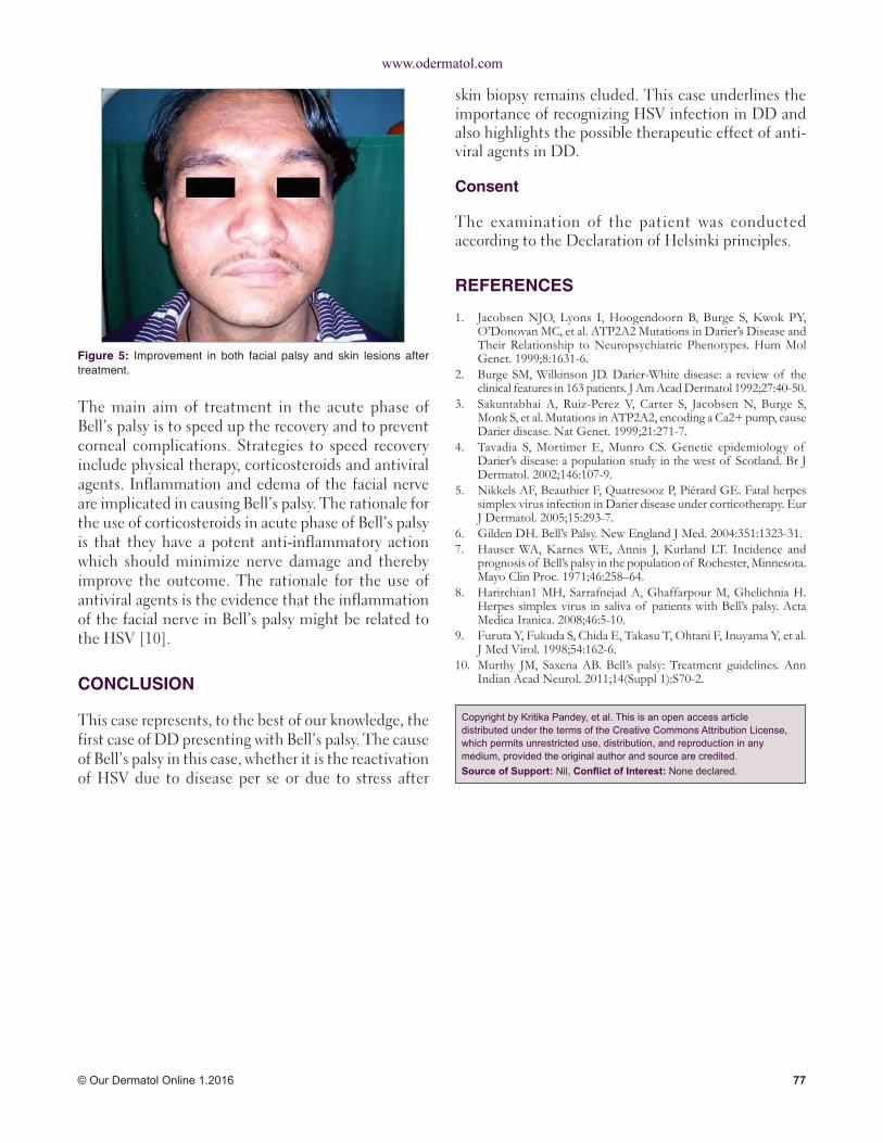
www.odermatol.com
© Our Dermatol Online 1.2016 77
The main aim of treatment in the acute phase of Bell’s palsy is to speed up the recovery and to prevent corneal complications. Strategies to speed recovery include physical therapy, corticosteroids and antiviral agents. Inflammation and edema of the facial nerve are implicated in causing Bell’s palsy. The rationale for the use of corticosteroids in acute phase of Bell’s palsy is that they have a potent anti-inflammatory action which should minimize nerve damage and thereby improve the outcome. The rationale for the use of antiviral agents is the evidence that the inflammation of the facial nerve in Bell’s palsy might be related to the HSV [10].
CONCLUSION
This case represents, to the best of our knowledge, the first case of DD presenting with Bell’s palsy. The cause of Bell’s palsy in this case, whether it is the reactivation of HSV due to disease per se or due to stress after
skin biopsy remains eluded. This case underlines the importance of recognizing HSV infection in DD and also highlights the possible therapeutic effect of anti-viral agents in DD.
Consent
The examination of the patient was conducted according to the Declaration of Helsinki principles.
REFERENCES
1. Jacobsen NJO, Lyons I, Hoogendoorn B, Burge S, Kwok PY, O’Donovan MC, et al. ATP2A2 Mutations in Darier’s Disease and Their Relationship to Neuropsychiatric Phenotypes. Hum Mol Genet. 1999;8:1631-6.
2. Burge SM, Wilkinson JD. Darier-White disease: a review of the clinical features in 163 patients. J Am Acad Dermatol 1992;27:40-50.
3. Sakuntabhai A, Ruiz-Perez V, Carter S, Jacobsen N, Burge S, Monk S, et al. Mutations in ATP2A2, encoding a Ca2+ pump, cause Darier disease. Nat Genet. 1999;21:271-7.
4. Tavadia S, Mortimer E, Munro CS. Genetic epidemiology of Darier’s disease: a population study in the west of Scotland. Br J Dermatol. 2002;146:107-9.
5. Nikkels AF, Beauthier F, Quatresooz P, Piérard GE. Fatal herpes simplex virus infection in Darier disease under corticotherapy. Eur J Dermatol. 2005;15:293-7.
6. Gilden DH. Bell’s Palsy. New England J Med. 2004:351:1323-31.7. Hauser WA, Karnes WE, Annis J, Kurland LT. Incidence and
prognosis of Bell’s palsy in the population of Rochester, Minnesota. Mayo Clin Proc. 1971;46:258–64.
8. Harirchian1 MH, Sarrafnejad A, Ghaffarpour M, Ghelichnia H. Herpes simplex virus in saliva of patients with Bell’s palsy. Acta Medica Iranica. 2008;46:5-10.
9. Furuta Y, Fukuda S, Chida E, Takasu T, Ohtani F, Inuyama Y, et al. J Med Virol. 1998;54:162-6.
10. Murthy JM, Saxena AB. Bell’s palsy: Treatment guidelines. Ann Indian Acad Neurol. 2011;14(Suppl 1):S70-2.
Figure 5: Improvement in both facial palsy and skin lesions after treatment.
Copyright by Kritika Pandey, et al. This is an open access article distributed under the terms of the Creative Commons Attribution License, which permits unrestricted use, distribution, and reproduction in any medium, provided the original author and source are credited.Source of Support: Nil, Confl ict of Interest: None declared.

© Our Dermatol Online 1.2016 78
Rosacea-like tinea facieiCésar Bimbi1,2, Piotr Brzezinski3
1Dermatological Comittee of State Medical Council of Rio Grande do Sul Brazil, 2Brazilian Society of Dermatology, 3Department of Dermatology, Military Support Unit, Ustka, Poland
Corresponding author: Dr. César Bimbi, E-mail: [email protected]
INTRODUCTION
Tinea faciei (TF) is a dermatophyte infection of glabrous skin of the face sometimes displaying a wide and variable range of clinical features from erythema, patches, induration, vesicles, pustules, papular and circinate lesions. So, this infection is often deceptive and may clinically mimic other facial dermatoses. Discoid lupus erythematosus, lymphocytic infiltration, seborrheic dermatitis, granuloma annulare and contact dermatitis are the most frequent misdiagnoses [1].
We describe 3 patients with lesions that clinically appeared to be Rosacea.
CASE REPORT
Case 1
A 66-year-old woman complained of a 2 years history of facial eruptions – “ - an allergy “ - that had initiated by the nose (Fig. 1a). She was treated firstly with antibiotics for acne rosacea, but with no improvement, and then prednisone (20 mg daily) still for presumed acne rosacea was tried, also unsucessfully (Fig. 1a). Physical examination revealed extensive erythematous lesions on the face with some pustules. Clinically, the lesions appeared to be acne rosacea. The well defined delimited
edges of the forehead lesion lead to the suspicion of tinea, and KOH examination was positive. The patient improved considerably in just two weeks of teatment. We used oral terbinafine and ciclopirox olamine cream.
Case 2
A 32-year-old woman came to the office with facial erythema to which had been prescribed cortisone creams and again no improvement was noted by the patient (Fig. 2a). Clinically, the lesions seemed to be irritant dermatitis, eczema or rosacea. This time,
ABSTRACT
Tinea faciei, is a facial superficial mycosis. The most frequent etiological agents are Microsporum canis, Trichophyton rubrum and T. tonsurans. It is often overlooked when considering the differential diagnoses of Rosacea. The most well known dermatology textbooks list acne, LE, perioral dermatitis, nasal sarcoidosis, carcinoid syndrom and other conditions but do not mention TF.We describe 3 patients with lesions that clinically appeared to be Rosacea.
Key words: Tinea; dermatophytoma; Rosacea
Case Report
How to cite this article: Bimbi C, Brzezinski P. Rosacea-like Tinea Faciei. Our Dermatol Online. 2016;7(1):78-80.Submission: 08.07.2015; Acceptance: 06.09.2015DOI:110.7241/ourd.20161.20
a b
Figure 1: (a) June 2015-Papular and pustules lesions with flat patches of erythema on the nose and cheeks and annular circinate lesions with a raised margins on the forehead where KOH examination was positive. (b) One week later-The patient improved considerably in just two weeks of teatment; we used oral terbinafine and ciclopirox olamine cream.

www.odermatol.com
© Our Dermatol Online 1.2016 79
instead of well defined edges of case 1-patient, we could note a fine dusty scaling that suggested micotic scales and KOH examination was again positive. We used oral terbinafine and ciclopirox olamine cream. The patient returned 8 days after and already a good improvement was easily detected (Fig. 2b).
Case 3
A 68-year-old woman came to the office with facial erithema, telangiectasia and some papulo-granulomatous indurated lesions to which again had been prescribed betamethasone creams (Fig. 3). Clinically, the lesions seemed a bit of sarcoidosis or rosacea, but not tinea. Scales were scrapped and KOH examination was again hyphae positive. We used oral terbinafine and ciclopirox olamine cream. The patient still did not return but improvement is expected since this medication works well when direct mycologic test is positive.
DISCUSSION
Superficial fungal infections of the face seem not to be sufficiently or routinely investigated as differential
diagnosis possibility and as a result, some facial dermatoses are often incorrectly diagnosed. As many as 70% of patients with tinea faciei are initially misdiagnosed as having other dermatoses. In a 20-year survey of tinea faciei, Nicola et al [2] examined 107 cases of tinea faciei. Typical forms were 57.1% whereas in 42.9% atypical forms were observed, mainly mimicking discoid lupus erythematosus (9 cases), polymorphous light eruption (8 cases) and Rosacea-like presentations [1,2]. In another study [1], in 100 cases of tinea faciei in adults, 52 mimicked a discoid lupus erythematosus, 15 lymphocytic infiltratation and four polymorphous light eruption
Although tinea faciei is tinea itself, some authors claim it as a distinct entity. Facial anatomy, physiology characteristics, exposure to sunlight, repeated washing and the use of cosmetics often determine atypical clinical presentation that leads to incorrect diagnosis.
“Four-in-one” anti: fungal-bacterial-histamine+ corticosteroid topical preparations are widely used by nondermatologists in the treatment of superficial fungal infections, but may be associated with persistent/recurrent infections [3].
Therapy with terbinafine associated with topical anti-fungal therapy leads to healing within 3–6 weeks without relapse or side effects. An added benefit of using ciclopirox and terbinafine is their anti-inflammatory effect [4].
Our patients had pruritic erythematous facial eruptions and had been treated for other dermatosis but with no improvement. Direct microscopy revealed multiple hyphae in all 3 cases. The first patient is a poor elderly woman coming from a peripheral village where it is difficult to get lab tests, so for practical purposes and the patient immediate relief we chose the commencement of treatment even to the detriment of a culture examination in order not to delay the relief of the symptoms. The patient improved considerably in just 8 days. Complete resolution is expected after 6 weeks of therapy. The same option was made for the other 2 patients.
Tinea faciei accounts for 3–4% of cases of tinea corporis and is often initially misdiagnosed and treated as other dermatoses in as many as 70% of patients [5]. It may be easily diagnosed or excluded by KOH preparation. It is important that we are aware of the possibility of tinea faciei and that diagnostic procedures are put in
Figure 3: Erythema on the face with telangiectasia and some papulo-granulomatous indurated lesions.
Figure 2: (a) Skin lesions with flat patches of erythema on the nose and cheeks. (b) Lesions in 8 day of treatment by oral terbinafine and ciclopirox olamine cream
ba

www.odermatol.com
© Our Dermatol Online 1.2016 80
place to identify, or eliminate tinea facei from the final diagnosis.
CONSENT
The examination of the patient was conducted according to the Declaration of Helsinki principles.
REFERENCES
1. Alteras I, Sandback M, David M, Segal R. 15-year survey of tinea faciei in the adult. Dermatologica. 1988;177:65-9.
2. Nicola A, Laura A, Natalia A, Monica P. A 20-year survey of tinea faciei. Mycoses [serial online]. 2010;53:504-8.
3. Alston SJ, Cohen BA, Braun M. Persisten/t and recurrent tinea corporis in children treated with combination antifungal/corticosteroid agents. Pediatrics. 2003;111:201-3.
4. Baxter DL, Moschella SL, Johnson BL. Tinea of the face. Arch Dermatol. 1965;91:184-5.
5. Torres-Guerrero E, Ramos-Betancourt L, Martínez-Herrera E, Arroyo-Camarena S, Porras-López C, Arenas R. Dermatophytic blepharitis due to Microsporum gypseum. An adult variety of tinea faciei with dermatophytoma. Our Dermatol Online. 2015;6:36-8.
Copyright by César Bimbi, et al. This is an open access article distributed under the terms of the Creative Commons Attribution License, which permits unrestricted use, distribution, and reproduction in any medium, provided the original author and source are credited.Source of Support: Nil, Conflict of Interest: None declared.
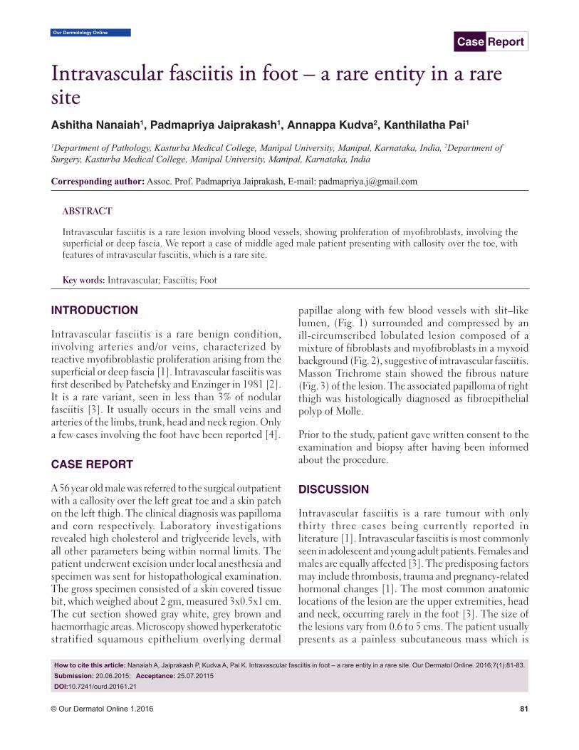
© Our Dermatol Online 1.2016 81
Intravascular fasciitis in foot – a rare entity in a rare siteAshitha Nanaiah1, Padmapriya Jaiprakash1, Annappa Kudva2, Kanthilatha Pai1
1Department of Pathology, Kasturba Medical College, Manipal University, Manipal, Karnataka, India, 2Department of Surgery, Kasturba Medical College, Manipal University, Manipal, Karnataka, India
Corresponding author: Assoc. Prof. Padmapriya Jaiprakash, E-mail: [email protected]
INTRODUCTION
Intravascular fasciitis is a rare benign condition, involving arteries and/or veins, characterized by reactive myofibroblastic proliferation arising from the superficial or deep fascia [1]. Intravascular fasciitis was first described by Patchefsky and Enzinger in 1981 [2]. It is a rare variant, seen in less than 3% of nodular fasciitis [3]. It usually occurs in the small veins and arteries of the limbs, trunk, head and neck region. Only a few cases involving the foot have been reported [4].
CASE REPORT
A 56 year old male was referred to the surgical outpatient with a callosity over the left great toe and a skin patch on the left thigh. The clinical diagnosis was papilloma and corn respectively. Laboratory investigations revealed high cholesterol and triglyceride levels, with all other parameters being within normal limits. The patient underwent excision under local anesthesia and specimen was sent for histopathological examination. The gross specimen consisted of a skin covered tissue bit, which weighed about 2 gm, measured 3x0.5x1 cm. The cut section showed gray white, grey brown and haemorrhagic areas. Microscopy showed hyperkeratotic stratified squamous epithelium overlying dermal
papillae along with few blood vessels with slit–like lumen, (Fig. 1) surrounded and compressed by an ill-circumscribed lobulated lesion composed of a mixture of fibroblasts and myofibroblasts in a myxoid background (Fig. 2), suggestive of intravascular fasciitis. Masson Trichrome stain showed the fibrous nature (Fig. 3) of the lesion. The associated papilloma of right thigh was histologically diagnosed as fibroepithelial polyp of Molle.
Prior to the study, patient gave written consent to the examination and biopsy after having been informed about the procedure.
DISCUSSION
Intravascular fasciitis is a rare tumour with only thirty three cases being currently reported in literature [1]. Intravascular fasciitis is most commonly seen in adolescent and young adult patients. Females and males are equally affected [3]. The predisposing factors may include thrombosis, trauma and pregnancy-related hormonal changes [1]. The most common anatomic locations of the lesion are the upper extremities, head and neck, occurring rarely in the foot [3]. The size of the lesions vary from 0.6 to 5 cms. The patient usually presents as a painless subcutaneous mass which is
ABSTRACT
Intravascular fasciitis is a rare lesion involving blood vessels, showing proliferation of myofibroblasts, involving the superficial or deep fascia. We report a case of middle aged male patient presenting with callosity over the toe, with features of intravascular fasciitis, which is a rare site.
Key words: Intravascular; Fasciitis; Foot
Case Report
How to cite this article: Nanaiah A, Jaiprakash P, Kudva A, Pai K. Intravascular fasciitis in foot – a rare entity in a rare site. Our Dermatol Online. 2016;7(1):81-83.Submission: 20.06.2015; Acceptance: 25.07.20115DOI:10.7241/ourd.20161.21

www.odermatol.com
© Our Dermatol Online 1.2016 82
gradually increasing in size for a duration varying from 2 weeks to 8 years [3,5].
Microscopically, intravascular fasciitis is characterized by plump spindle cells arranged in intersecting fascicles, a storiform pattern, or in a haphazard manner. The lesion may be present inside the lumen or associated with the walls of arteries or veins of all sizes. The separation of mass from the vessel wall may cause clefts [3,5]. Patchefsky and Enzinger reported that among their cases, the soft tissue component often was more prominent than the vascular component [2]. Though the infiltrative growth pattern resembles a sarcoma, it can be differentiated by the absence of significant cytologic pleomorphism and abnormal mitotic figures and the overlying epidermis is usually intact [1]. The background stroma varies from a dense hyalinized to edematous, myxoid appearance. Often associated are scattered multinucleated giant cells, lymphocytes and red blood cells [2].
The proliferative cells in intravascular fasciitis are of myofibroblastic phenotype, being positive to both vimentin and α-smooth muscle actin expression and negative for keratin, S100 protein, desmin, CD31, CD34, and c-kit. The multinuclear giant cells were CD68 positive, confirming the histiocytic origin. CD31 and CD34 can demonstrate the highly vascular stromal background and the vascular location of the lesion. Special stains for elastic fibers can be used to demonstrate the walls of associated vessels [2].
The pathogenesis of intravascular fasciitis is currently under study. According to Patchefsy and Enzinger [5], the lesion was due to the proliferation of myofibroblasts from the walls of systemic arteries and veins and they attributed the frequent extension into perivascular connective tissue to the ‘‘field effect’’ of myofibroblastic transformation [2]. The myofibroblastic origin of the spindle cells is confirmed by immunohistochemistry. Factors initiating myofibroblast proliferation may be preceding trauma, thrombosis, and high levels of estrogen and has to be further studied [1].
In the present case, the diagnosis was made by histology and use of special stains like Masson Trichrome. Differential diagnosis include sarcoma, benign fibrous histiocytoma, organizing thrombus, pyogenic granuloma, peripheral nerve tumours, spindle cell carcinoma, spindle cell melanoma, myxoid liposarcoma, fibrosarcoma and leiomyosarcoma [4]. This entity has been described as ‘‘pseudosarcoma’’ due to the misinterpretation of intravascular growth as vascular invasion by a sarcoma in addition to increased
Figure 1: Hyperkeratotic epidermis overlying lobulated lesion in deep dermis, Haematoxylin and Eosin, x50.
Figure 2: Mixture of fibroblasts and myofibroblasts in a myxoid background, Haematoxylin and Eosin, x200.
Figure 3: Masson trichrome stain showing fibrous nature of lesion, x 200.

www.odermatol.com
© Our Dermatol Online 1.2016 83
mitotic activity, involvement of muscle and infiltrative borders of soft tissue [2].
The clinical behavior of this lesion is usually benign but a few cases have been reported to recur after excision. Since the soft tissue component comprises a much greater proportion of the tumor than the vascular component, diagnosis may be difficult in small biopsy specimens [1]. If blood vessel walls cannot be seen easily, the multinodular or serpentine growth pattern may be useful along with special stains for elastic fibers and smooth muscle [2].
CONCLUSION
Intravascular fasciitis is a rare variant of nodular fasciitis involving with blood vessels. The recognition of intravascular growth is required for diagnosis on small biopsy specimens and may be facilitated by special stains for elastic fibers or immunohistochemical stains for vascular markers. This case is reported with the intention of creating awareness of this lesion to avoid misdiagnosis and aggressive treatment.
Consent
The examination of the patient was conducted according to the Declaration of Helsinki principles.
REFERENCES
1. Zheng Y, George M, Chen F. Intravascular fasciitis involving the flank of a 21-year-old female: a case report and review of the literature. BMC Research Notes. 2014;7:118.
2. Chi AC, Dunlap WS, Richardson MS, Neville BW. Intravascular Fasciitis: Report of an Intraoral Case and Review of the Literature. Head Neck Pathol. 2012;6:140-5.
3. Enzinger and Weiss. Benign fibroblastic/myofibrolblastic proliferations. In: Sharon W. Weiss, John R. Goldblum (eds). Soft tissue tumours. 5th ed: Elsevier; 2008. 175-225.
4. Sugaya M, Tamaki K. Does Thrombosis Cause Intravascular Fasciitis? Acta Derm Venereol. 1987:369-70.
5. Patchefsky AS, Enzinger FM. Intravascular fasciitis: a report of 17 cases. Am J Surg Pathol. 1981;5:29-36.
Copyright by Ashitha Nanaiah, et al. This is an open access article distributed under the terms of the Creative Commons Attribution License, which permits unrestricted use, distribution, and reproduction in any medium, provided the original author and source are credited.Source of Support: Nil, Conflict of Interest: None declared.

© Our Dermatol Online 1.2016 84
Our Dermatology Online
Porokeratosis of the scrotumPorokeratosis of the scrotumKhalifa E. Sharquie1, Raafa K. AL-Hayani2, Waqas S. Abdulwahhab2
1Department of Dermatology & Venereology, College of Medicine, University of Baghdad, Baghdad, Iraq, 2Department of Dermatology &Venereology, Baghdad Teaching Hospital, Baghdad, Iraq
Corresponding author: Prof. Khalifa E. Sharquie, E-mail: [email protected]
INTRODUCTION
PK is disorder of keratinization characterized by annular lesions surrounded by raised sharply marginatedkeratotic borders with a characteristic histopathological finding named cornoid lamella. It consists of a heterogeneous group of disorders inherited in a autosomal dominant fashion. PK has a wide variety of manifestations including classical plaque-type porokeratosis of mibelli, disseminated superficial actinic porokeratosis, linearporokeratosis, porokeratosispalmaris, plantaris, etdisseminata and punctate porokeratosis [1].
In Iraq, a special variety of pk has been reported affecting the face only called solar facial porokeratosis [2]. Localized pk of the genitalia is a rare in occurrence with 24 cases reported in the literature [3]. The aim of present report is to document anew patient with his father suffering from localized scrotal PK.
CASE REPORT
55-year-old male patient presented to Department of Dermatology, Baghdad Teaching Hospital in 20-8-2012 suffering from severely itchy genital lesions. Since 1982 the patient described a rash on the scrotum which was gradually enlarging in size and increasing in number. Also he mentioned the occurrence of exactly the similar
disease of his father (dead) in genital area and this in favor of autosomal dominant inheritance. All the lesions are persistent with no history of spontaneous resolutions of any them. On examination, numerous papules, nodules and plaques distributed over the scrotum (>40 lesions) with none of them on the shaft of penis (Fig. 1). Those lesions were indurated skin colored while others were dark with typical annular configurations as the borders seemed more active while the center had the tendency towards atrophy. Some of the lesions showed thickened surface with pigmentation
ABSTRACT
Porokeratosis (PK) is disorder of keratinization characterized by annular lesions surrounded by raised sharply marginatedkeratotic borders with a characteristic histopathological finding named cornoid lamella. PK of genitalia is very rare condition and mostly reported among Asian population. The aim of present report is to document a new patient with localized scrotal PK with his father suffering from the same disease in the scrotum.
Key words: Porokeratosis; Cornoid lamella; Genitalia; Neurodermatitis
Case Report
How to cite this article: Sharquie KE, AL-Hayani RK, Abdulwahhab WS. Porokeratosis of the scrotum. Our Dermatol Online. 2016;7(1):84-86.Submission: 10.07.2015; Acceptance: 05.09.2015DOI: 10.7241/ourd.20161.22
Figure 1: Numerous papules, nodules and plaques distributed over the scrotum.

www.odermatol.com
© Our Dermatol Online 1.2016 85
without the typical annular ring of the most of the lesions and looked like neurodermatitis (Fig. 2).
The histopathology of the disease as follow: theepidermis wasacanthotic with basket weave hyperkeratosis. There was invagination of epidermis by column of keratin reaching the basal layer of epidermis. At the site of invagination there was absence of granular layer. This column consisted of parakeratotic cells, forming a typical feature of cornoidlamella. Many individual dyskeratoticcells were seen under the base of lamella reaching almost the basal layer of epidermis. While the
Figure 2: Some of the lesions showed thickened surface with pigmentation without the typical annularring of the most of the lesions and looked like neurodermatitis.
dermis consisted of many dilated blood vessels with severe inflammatory reaction at the base of cornoid lamella, consisting of many lymphoid cells. In addition pieces of manydartos muscles were observed in the dermis (Fig. 3).
Prior to the study, patient gave written consent to the examination and biopsy after having been informed about the procedure.
DISCUSSION
Porokeratosis of genitalia is very rare condition and mostly reported among Asian population [3-5]. The present report is the first case study that is being reported in Arab region. Family history of the present case was positive as his father had the similar condition and this might support the autosomal dominant inheritance like other types of pk. Itching is a prominent feature of present case and this was similarly reported [4]. There are many dermatosis involvingthe genital area like psoriasis, lichen planus and dermatitis and these are usually associated with itching. Lichen simplex is commonly superimposed on the top of these skin diseases [6]. Accordingly the present case was diagnosed and treated for 30years by most of dermatologists as case of lichen simplex chronicus. In most of reported genital pk, the lesions were scanty (1-3) while in our patient, numerous lesions were seen that widely distributed over the scrotum.Malignant transformation of pk had been reported [7], but fourtounitly no malignant changes were observed in present case.
There are no effective therapies of pk but many treatment had been suggested like topical steroid, cryotherapy, electrocautry, Co2 laser and others. We recommend Co2 laser removal as one of most effective modality [8,9], in order to relieve the patient complaint and to prevent the possibility of malignant changes.
Consent
The examination of the patient was conducted according to the Declaration of Helsinki principles.
REFERENCES
1. Spencer LV. Porokeratosis. Available from: http://www.emedicine.com/derm/topic 343 htm. [last accessed on 2006 oct 9].
2. Sharquie KE, AL-Baghdady BA. Solar facial porokeratosis. J Dermatol. 2003;30:216-21.
Figure 3: Histopathology showing epidermal acanthosis with basket weave hyperkeratosis. Invagination of epidermis by column of keratin reaching the basal layer of epidermis consisted of parakeratotic cells, forming a typicalfeature of cornoid lamella. Many individualdyskeratoic cells were seen under the base of lamella reaching almost the basal layer of epidermis. The dermis consisted of many dilated blood vessels with severeinfl ammatory reaction at the base of cornoid lamella, consistingof many lymphoid cells. [Hematoxylin and eosin stain; original magnifi cation (a) x4, (b) x10, (c) x40, (d) x40.
dc
ba

www.odermatol.com
© Our Dermatol Online 1.2016 86
3. Valdivielso-Ramo M. Genital porokeratosis: Case Reports. Actas Dermosifi liog. 2008;99:217-20.
4. Chen T, Chou Y, Chen C, Kuo T, Hong H. Genital porokeratosis: a series of 10 patients and review of the literature. Br J Dermatol. 2006;155:325.
5. Sengupta S, Das J, Gangopadhyay A. Porokeratosis confi ned to the genital area: a report of three cases. Indian J Dermatol Venereol Leprol. 2008;74:80.
6. Lynch PJ. Lichen simplex chronicus (atopic/neurodermatitis) of the anogenital region. Dermatol Ther. 2004;17:8.
7. Maubec E, Duvillard P, Margulis A, Bachollet B, Degois G, Avril M-F. Common skin cancers in porokeratosis. Br J Dermatol. 2005;152:1389.
8. Trcka J, Pe t tke -Rank CV. Brocker EB, Hamm H. Genitoanocruralporokeratosis following chronic exposure to benzene. Clin Exp Dermatol. 1998;23:28-31.
9. Huang SL, Liu YH, Chen W. Genitoglutealporokeratosis. J Eur Acad Dermatol Venerol. 2006;20:899-900.
Copyright by Khalifa E, et al. This is an open access article distributed under the terms of the Creative Commons Attribution License, which permits unrestricted use, distribution, and reproduction in any medium, provided the original author and source are credited.Source of Support: Nil, Confl ict of Interest: None declared.

© Our Dermatol Online 1.2016 87
Phakomatosis pigmentovascularis with lower limb vascular abnormalities in a young Kashmiri male child-Report of a first child from Kashmir Valley (India) and review of literatureMajid Jehangir1,Seema Quyoom2, Jahangeer Bhat1, Peerzada Sajad2, Ishfaq Sofi3, Aresalan Amin1, Mudasir Bhat1
1Department of Radiodiagnosis and Imaging, Government Medical College, Srinagar (University of Kashmir), J&K, India, 2Department of Dermatology, SKIMS Medical College Srinagar, Bemina, Srinagar, Kashmir, J&K, India, 3Postgraduate, Department of Ophthalmology, Government Medical College, Srinagar (University of Kashmir), J&K, India
Corresponding author: Dr. Peerzada Sajad, E-mail: [email protected]
INTRODUCTION
Phakomatosis, a neurocutaneous syndrome is a developmental abnormality which is characterised by the involvement of skin, eyes and central nervous system. Phakomatosis pigmentovascularis is a rare cutaneous disorder which was first described by Ota et al. in 1947 [1]. There is no sex predilection. Four types with two subtypes have been described, where subtype ‘a’ refers to cases presenting with only cutaneous manifestations and subtype ‘b’ is characterised by systemic association like in, Struge – Weber syndrome and Klippel Trenaunay syndrome [2]. Recently a fifth type with cutis marmorata and aberrant Mongolian blue spot has also been added to the classification.
CASE REPORT
A 12 – year old male child product of a non-consanguinous marriage presented with history of non-blanchable erythematous macules, which was noted to be portwine stain (naevus flammeus) on examination involving face, forearms, and right lower limb since birth (Fig. 1 and 2). There was history of increase in the size of these macules with age. The facial portwine stain was more marked and abundant on the left side.There was associated history of abnormal gait. However there was no history of headache, seizures, abnormal body movements or blurring of vision.The patient was noted to have hypertrophy of right lower limb with disparity in the length of legs. A visible vessel was seen in right groin crossing the midline (Fig. 3). Ophthalmological
ABSTRACT
Phakomatosis is a developmental abnormality simultaneously involving eyes, central nervous system, and skin. Phakomatosis pigmentovascularis (PPV) is a rare cutaneous disorder which is characterised by a combination of capillary malformations and pigmented anomalies. It arises sporadically. PPV was first described by Ota et al., in 1947. There is no sex predilection, but Japanese have been found to be affected more. There are four main types of PPV. Recently a fifth type with cutis marmorata and aberrant Mongolian blue spot has also been added to the classification. Here we report a case of PPV with Struge – Weber syndrome and Klippel Trenaunay syndrome in a young Kashmiri male child, which has been rarely reported in the literature.
Key words: Phakomatosis pigmentovascularis(PPV); Klippel Trenaunay syndrome(KTS); Struge – Weber syndrome(SWS)
Case Report
How to cite this article: Jehangir M, Quyoom S, Bhat J, Sajad P, Sofi I, Amin A, Bhat M. Phakomatosis pigmentovascularis with lower limb vascular abnormalities in a young Kashmiri male child-Report of a first child from Kashmir Valley (India) and review of literature. Our Dermatol Online. 2016;7(1):87-90.Submission: 02.07.2015; Acceptance: 20.10.2015DOI:10.7241/ourd.20161.23

www.odermatol.com
© Our Dermatol Online 1.2016 88
examination revealed raised intraocular pressure in left eye. Optical coherence tomography revealed severe thinning of Retinal nerve fibre layer (RNFL) in left eye, loss of neuro-retinal rim (NRR), gross asymmetry in cup-disc ratio and RNFL thickness (Fig. 4 and 5).
X ray of lower limbs showed limb lengthing in right limb, however, no bony hypertrophy was seen (Fig. 6). Colour Doppler and CT angiography of limbs revealed absence of right iliac vein with right femoral vein draining through collateral vein crossing midline into
Figure 1: Portwine stain involving face.
Figure 2: Portwine stain involving upper limbs.
Figure 3: Picture showing a visible vessel was seen in right groin crossing the midline.
Figure 4: OCT showing severe thinning of RNFL.
Figure 5: OCT showing ocular changes.
Figure 6: X-ray showing limb length disparity(R>L).

www.odermatol.com
© Our Dermatol Online 1.2016 89
left external iliac vein. High origin of superficial femoral artery and deep femoral artery was also noted (Fig. 7). Non contrast CT of brain showed foci of dense gyral calcification in left temporoparietal region (Fig. 8).
DISCUSSION
The term phakomatosis refers to a developmental abnormality s imultaneously involving skin, eyes and central nervous system. Phakomatosis pigmentovascularis is a rare syndrome characterised by the combination of a capillary malformation with pigmented naevi of various types. It is very uncommon and arises sporadically. Both, males and females are affected. There is no sex predilection, but Japanese have been reported to be affected more often [1,2]. Twin spotting phenomenon of Happle and Steijen has been proposed to explain this phenomenon (PPV). In this hypothesis,somatic mutations on nearby
genes leads to mosaic spots in close proximity to one another [3]. PPV has been classified into four types namely; type I: Nevus flammeus and epidermal nevus, type II: Nevus flammeus, Mongolian spots, ± nevus anemicus, type III: Nevus flammeus, nevus spilus, ± nevus anemicus, and type IV: Nevus flammeus, Mongolian spots, nevus spilus, ± nevus anemicus. Recently a fifth type with cutis marmorata and aberrant Mongolian blue spot has also been added to the classification. Each type is subdivided into two subtypes; subtype ‘a’, cutaneous involvement only; and subtype ‘b’, both cutaneous and systemic involvement.Type II is reported to account for approximately 80% of all cases. PPV type I, III, and IV have very rarely been reported in literature.The capillary malformation may occur anywhere on the body and can be segmental.The aberrant Mongolian spots occur on atypical sites and tend to persist, unlike conventional Mongolian blue spots. It is estimated that 50% of patients with PPV present with systemic involvement. The cases that have been most studied are those associated with PPV type IIb. In the case of PPVs, neurologic anomalies develop in the first months of life. The most common ocular abnormality which has been found to be associated with PPV is ocular melanosis, which consists of unilateral or bilateral blue-gray pigmentation in the sclera (Naevus of Ota). Other ophthalmologic abnormalities are much less common. However, a large number of abnormalities have been published in relation to PPVs, but with the exception of Sturge-Weber syndrome, Klippel-Trenaunay syndrome, and ocular melanosis, the abnormalities are varied [4,5].
It is recommended that all the affected cases should be investigated depending on the sites involved, like for glaucoma if the capillary malformation affects the eyelids, or for Klippel-Trenaunay syndrome if there is limb involvement. A patient’s quality of life may be improved by treating nevus flammeus using a pulsed dye laser and by treating pigmentary nevus using Q-switched lasers. Some authors suggest that the pigmentary nevus should be treated first [6].
Various systemic syndromes have been reported to be associated with PPV in the literature, and these include; Sturge-Weber syndrome, Nevus of Ota, and Klippel-Trenaunay syndrome [7].
Sturge –Weber syndrome (SWS) is defined as a facial portwine stain with ipsilateral vascular malformation of leptomeninges and eyes. Eye involvement is however not necessary for the diagnosis of SWS. Brain
Figure 7: CT angiography absence of right iliac vein with right femoral vein draining through collateral vein crossing midline into left external iliac vein.
Figure 8: Non contrast CT of brain showing foci of dense gyral calcification in left temporoparietal region.

www.odermatol.com
© Our Dermatol Online 1.2016 90
changes show calcification with atrophy and increased vascularity of unilateral leptomeninges.
Naevus of Ota also known as naevus fuscoceruleus ophthalmomaxillaris is a dermal melanocytosis and is characterised by slate-grey or bluish hyperpigmentation involving the periorbital area, sclera and conjunctiva. It may be congenital or may develop around puberty.
Klippel Trenaunay syndrome is characteried by the association of cutaneous capillary malformation of limbs(naevus flammeus) and soft tissue swelling of limb with or without bone hypertrophy. Original description of this syndrome included vericosities of the limb [8,9].
Since its first description in 1947, 222 cases of PPV have been published, most of which are sporadic and from Japan, Mexico, or Argentina. The most common type is type IIb (45%), followed by IIa (30%). The rest are much less frequent and no isolated cases of PPV Ib, IVb, or Vb have been published. To date only two case series of PPV have been published. The largest series was published by Cordisco et al [10], who presented 25 patients in Argentina.In this series, type IIb was the most common type. Vidaurri-de la cruz et al [11] presented a series of 24 patients in Mexico in which 75% of the cases were type IIb, 25% were IIa, and the rest of the subtypes were not found.
Literature shows that till date only few well established cases of phakomatosis pigmentovascularis (PPV) have been reported, but PPV presenting with Sturge-Weber syndrome and lower limb vascular abnormalities is extremely rare. Sumita S et al [12] reported a case of PPV with Sturge–Weber syndrome and Klippel-Trenaunay Syndrome in a 13 year old female who presented with port wine stain over face, limbs and trunk, with a visible vessel along the lateral aspect of left lower leg together with associated hypertrophy of the involved limb.
Consent
The examination of the patient was conducted according to the Declaration of Helsinki principles.
REFERENCES
1. Ota M, Kawamura T, Ito N. Phakomatosis pigmentovascularis. Dermatol Surg. 1947;52:1-3.
2. Marti-Bonmati L, Menor F, Mulas F. The Struge –Weber syndrome: correlation between the clinical status and radiological CT AND MRI findings. Childs Nervs Syst. 1993;9:107-9.
3. Danarti R,Happle R. Paradominant inheritance of twin spotting: phakomatosis pigmentovascularis as a further possible example. Eur J Dermatol. 2003;13:612.
4. Young AE. Combined vascular malformations. in: Mulliken JB, Young AE, Editors. Vascular birthmarks: Haemangiomas and malformations. Philadelphia: WB Saunders; 1988:246-74.
5. Happle R. Stejlen PM. Phacomatosis pigmentovascularis interpreted as a phenomenon of twin spots. Hautarzt. 1989;40:721–24.
6. Du LC, Delaporte E, Catteau B, Destee A, Piette F. Phakomatosis pigmentovascularis type II. Eur J Dermatol. 1998;8:569–72.
7. Gupta A, Dubey S, Agarwal M. A case of Sturge-Weber syndrome in association with phacomatosis pigmentovascularis and developmental glaucoma. J AAPOS. 2007;11:398–9.
8. Goyal T, Varshney A. Phacomatosis cesioflammea: First case report from India. Indian J Dermatol Venereol Leprol. 2010;76:307.
9. Sawada Y, Iwata M, Mitsuhashi Y. Nevus pigmentovascularis. Ann Plast Surg. 1990;25:142-5.
10. Cordisco MR, Campo A, Castro C, Bottegal F, Bocian M, Persico S, et al. Phakomatosis pigmentovascularis: report of 25 cases. Pediatr Dermatol. 2001;18:70.
11. Vidaur r i -de la cr uz H, Tamayo-Sanchez L, Duran-McKinster C, Orozco-Covarrubias ML, Ruiz-Maldonado R. Phakomatosis pigmentovascularis IIa and IIb: clinical findings in 24 patients. J Dermatol. 2003;30:381-8.
12. Sumita S, Sanchaita B, Chinmay H, Rahul A, Anusree G. Phakomatosis pigmentovascularis presenting with Sturge-Weber syndrome and Klippel-Trenaunay syndrome. Indian J Dermatol 2015;60:77-81.
Copyright by Majid Jehangir, et al. This is an open access article distributed under the terms of the Creative Commons Attribution License, which permits unrestricted use, distribution, and reproduction in any medium, provided the original author and source are credited.Source of Support: Nil, Conflict of Interest: None declared.

© Our Dermatol Online 1.2016 91
Our Dermatology Online
How to cite this article: Wollina U, Heinig B, Nowak A. Medical Leech Therapy (Hirudotherapy). Our Dermatol Online. 2016;7(1):91-96.Submission: 18.08.2015; Acceptance: 29.09.2015DOI: 10.7241/ourd.20161.24
Medical leech therapy (Hirudotherapy)Medical leech therapy (Hirudotherapy)Uwe Wollina1, Birgit Heinig2 Andreas Nowak3
1Department of Dermatology and Allergology, Academic Teaching Hospital Dresden-Friedrichstadt, Dresden, Germany, 2Center for Physical Therapy and Rehabilitative Medicine, Academic Teaching Hospital Dresden-Friedrichstadt, Dresden, Germany, 3Department of Anesthesiology and Intensive Care Medicine, Emergency Medicine and Pain Management, Academic Teaching Hospital Dresden-Friedrichstadt, Dresden, Germany
Corresponding author: Prof. Dr. Uwe Wollina, E-mail: [email protected]
HISTORY
Therapy with leeches is one of the oldest minor invasive procedures in medicine that was already mentioned 1,500 BC in Egypt. Sanskrit writings described leech therapy from 1,300 BC on. Hippocrates introduced leech therapy in Greece but the method was also known to ancient Mesopotamians, Egyptians and Aztecs, and Mayans. Medical leech therapy is part of the concept of the Greko-Arab Unani System of Medicine [1].
The procedure has seen a first renaissance in the 18th and early 19th century when it became extremely popular again. This has led to an eradication of naturally occurring leeches in Ireland where leech export was an important trade [2].
Medical leech therapy reemerged in the 70ies of the last century as an adjuvant to surgery. In 2004 the FDA
approved medical leeches as medical devices in plastic and reconstructive surgery [3].
LEECHES
Leeches are hermaphroditic, bloodsucking annelid worms. Medicinal leeches belong to the order Arhynchobdellida, family Hirudinidae. Only 15 of the more than 600 of the known species are classified as medical leeches, such as Hirudo medicinalis, H. verbana and H. orientalis [4].
H. medicinalis has 33 to 34 body segments, is brown or black, and has six long reddish stripes on the back. The cylindrical body is slightly flattened and can measure up to 20 cm. Although they have 5 pairs of eyes they use the olfactory system to find their hosts. Adult animals have two suckers at the end of their body, a
ABSTRACT
Leeches have been used in medicine long time before BC. In recent years medical leech therapy has gained increasing interest in reconstructive surgery and pain management and other medical fields. The possible indications and success rates of this treatment are discussed. There is a special interest in salvage of flaps and grafts by the use of medical leeches. Retrospective analysis indicates a success rate of >80%. Randomized controlled trials have been performed in osteoarthritis. Case reports and smaller series are available for the treatment of chronic wounds, post-phlebitic syndrome and inflammatory skin diseases. The most common adverse effects are prolonged bleeding and infection by saprophytic intestinal bacteria of leeches. Medical leech therapy is a useful adjunct to other measures wound management.
Key words: Acute wounds; Chronic wounds; Medical leech therapy; Reconstructive surgery; Hirudis medicinalis
Key Messages:• medical leeches offer several advantages in venous congestive syndromes seen after reconstructive surgery• medical leeches secrete substances which can reduce pain and itch• infection control is necessary for safe medical leech therapy
Review Article

www.odermatol.com
© Our Dermatol Online 1.2016 92
large posterior sucker and a smaller disc-shaped on the head that contains the mouths with tree jaws (Fig. 1).
Jaws consist of up to 100 teeth and salivary glands that release more than 100 known substances. Hirudin is the most powerful natural thrombin inhibitor. Hirudin works in synergy with two Factor Xa inhibitors, i.e. antistasin and ghilanten also found in the saliva of this worm. Calin is a platelet adhesion and activation inhibitor. Hyaluronidase supports the spread of active saliva compounds in the tissue. Destabilase dissolves fibrin. Bdellin, eglins and hirustatin are anti-inflammatory substances with protease inhibitory activity. There is a number of neurotransmitters like dopamine or serotonin in the saliva that reduce pain perception in the host. Acetylcholine works as a vasodilator [4].
Leeches can survive a year from a single blood meal, were they ingest about 10 times of their own body weight [4].
HOW TO USE MEDICAL LEECHES
Only medical leeches from serious suppliers should be used. Wild animals increase the risk of severe infections [5].
The site where leeches are to be placed should be clean and free of ointments, pasts etc. Cleaning can be done by sterile Ringer solution, physiological sodium chloride solution or sterile water.
Leeches are fast and elegant swimmers in water (“sweet water dolphins”). And they can move rapidly on the patient’s skin as well. To apply them correctly, a 5 ml syringe were the nozzle was removed by scissor, can be used. The leech is placed into the prepared syringe and the syringe is directly applied on the skin surface to be treated with its open end. When the leech is feeding, the syringe is removed [6].
Some authors use anchoring sutures on medical leeches to ensure that the leeches stay in place. In a small trial no adverse effect on leech survival was noted by such a method [7]. The question remains whether this will negatively affect the feeding behavior.
The number of leeches applied depends on the size of the area that has to be treated. The Iowa Head and Neck Protocol recommends to applied leeches every 2 hours. The spots where the leeches are placed should be changed. The duration of leech treatment is until
recovery of the compromised flap, mostly about one week [8]. For other indications different schedules may be more appropriate.
A single feeding lasts up to 60 min. After that the leech stops sucking and can gently be removed. Actively feeding leeches should not be pulled off. Forced detachment by a forceps or chemicals can lead to regurgitation of the leech stomach contents into the wound [4,6].
Wounds after leech biting lose some blood and may be oozing for about 24 hours [6] (Fig. 2).
Wounds should be disinfected and covered by a sterile dressing. Repeated application of heparin or sodium chloride has been recommended to stimulated hemorrhage from these wounds [6].
Although large randomized controlled trails are completely missing in the field of medical leech therapy
Figure 2: Three bite marks two days after medical leech therapy of post-phlebitic syndrome.
Figure 1: Disc-shaped sucker on the head of a medical leech.

www.odermatol.com
© Our Dermatol Online 1.2016 93
we will present indications and results. Please be aware of these limitations.
FLAPS
Medical leeches are extremely helpful in salvage of venous outflow compromised pedicle and free flaps. Leeches are used in the critical phase postoperatively in the dusky areas of a flap (Fig. 3). Animal studies suggested that leech therapy replaces congestive venous blood by fresh arterial blood improving survival of tissue [9].
Initially leeches may be necessary more often than just once daily for. Treatment is continued a couple of days until the flap recovers and venous congestion is overcome [10-17]. However, success rate is not 100% in particular in patients with lower hemoglobin levels and a need of more erythrocyte transfusions [13]. Leeches will not attach to dead flaps [14]. The success rate – complete and partial salvage – is 81.9 % (Table 1).
Leeches have also been used successfully to compensate venous congestion in replants of fingers, toes or nose [18]. Leech therapy increases perfusion resulting in hyperemia in dynamic and blood phases of Tc-99 m HDP bone scintigraphy [19]. Hirudin in leech saliva increases the level of messenger RNA for the vascular endothelial growth factor (VEGF) and VEGF expression in flap vessels [20].
HEMATOMA
Leeches can reduce postsurgical and posttraumatic hematomas (Fig. 4). This may help to salvage
flaps and grafts [21] - or testicles in case of scrotal hematoma [22]. Evidence level is low (V) since no trials have been published.
PAIN MANAGEMENT
Leeches are used in pain syndromes of various origins. The pain relief is rapid and sometimes long-lasting [23]. There are reports on successfully leech therapy in severe cancer pain [24]. Studies in osteoarthritis argue for symptomatic improvement by leech therapy by analgesic and anti-inflammatory effects [25].
An open trial in 32 patients with osteoarthritis leech therapy improved pain, stiffness and movement of joints [26]. In a randomized trial with 52 patients with either leech therapy or transcutaneous electrical nerve stimulation (TENS) their osteoarthritis of the knee responded significantly better to leeches. That was in particular relevant in pain reduction and improvement
Table 1: Salvage of fl aps by medical leech therapyReference No of
patientsSalvage of fl aps Remarks
Jones et al. (2015) 1 1 Nasal fl ap
Pannucci et al. (2014) 4 1/4 (partial salvage) Free fl ap breast reconstruction
Kim et al. (2013) 2 2/2 Paramedian forehead fl aps
Nguyen et al. (2012) 27 18/27(9 partial salvage)
Local, regional and free fl aps
Whitacker et al. (2012) 277 216/277 (total and partial salvage)
Meta-analysis
Gröbe et al. (2012) 148 94 partial salvage54 complete salvage
Local or pedicle fl apsor grafts
Soengkar et al. (2012) 1 1/1 Free fl ap
Oh et al. (2012) 1 1/1 Free-style propeller fl ap
Our data 4 3/4 Local fl aps
Total 465 381
Figure 3: Medical leech for nasal fl ap salvage. The treatment resulted in complete fl ap survival.
Figure 4: Application of a medical leech on a postsurgical hematoma.

www.odermatol.com
© Our Dermatol Online 1.2016 94
of the Lequesne’s index [27]. The Lequesne’s index scores the severity of knee osteoarthritis by three dimensions: (I) pain or discomfort, (II) maximum distance walked, and (III) activities of daily living.
In a meta-analysis of four trials where 237 patients with osteoarthritis were included there was strong overall evidence for immediate and short-term pain reduction. The authors also found evidence for immediate improvement in patients’ physical function, and both immediate and long-term improvement in joint stiffness. Leech therapy was not associated with any serious adverse events [28].
VARICOSE VEINS, LEG AND FOOT ULCERS
Application of medical leeches for the treatment of varicose leg ulcers decreased edema, limb girth, and improved ulcer healing [29]. In a randomized controlled study 50 patients with varicose veins were treated either by compression therapy or by medical leech therapy. After two months the group with medical leech therapy (n = 30) achieved a significant reduction in pain, edema and hyperpigmentation [30]. Leeches diminish the time of healing in post-phlebitic syndrome [31]. Since anticoaguagulant therapy is a contraindication for medical leeches (see below) this may limit the use of medial leech therapy in particular in venous leg ulcer patients. On the other hand, there is evidence level I for compression therapy for venous leg ulcers and leeches are not a substitute for medical compression.
There are case reports on the successful use of medical leech therapy for diabetic foot ulcers to salvage the leg [32]. Here, the monitoring of the treatment must be especially careful to avoid leech-borne infection.
SKIN DISEASES
In an open label study 27 patients with atopic eczema participated. Medical leech therapy was performed once a week for at least four times. The effects were measures by Eczema Area and Severity Index (EASI) score, SCORing of Atopic Dermatitis (SCORAD) Index, and Dermatology Life Quality Index (DLQI). The number leeches gives dependent on the size of the lesions. No concomitant treatment was applied. Reduction of EASI was 54.5%, of SCORAD 55%, and DLQI improved by 62.4% [33].
Leech application on five times reduced the hyperpigmentation of a nevus of Ota in a 23-year-old female patient from India [34].
ADVERSE EFFECTS
Prolonged bleeding of up to ten hours can be observed from time to time after medical leech therapy [21,35]. Usually, bleeding can be stopped by pressure but sometimes a primary suture might be necessary [36]. Repeated application of large numbers of leeches can cause anemia that needs transfusion. Control of hemoglobin during medical leech therapy is recommended [35].
Medical leeches may cause primarily local infections at a rate of 2 to 25 %. The infections are due to microbiota of the leech. A number of species have been characterized form the midgut of Hirudis spp. Morganella morganii, Rikenella, and Aeromonas veronii are dominant members but bacteria like Magnetospirillium spp. and Roseospira marina have also been detected [37].
In a US multicenter trial antimicrobial prophylaxis was documented in 91.5% of the included patients. Surgical site infection was observed in 11.9% of patients – all of them received antibiotic prophylaxis. In four infections Aeromonas spp. was isolated. Aeromonas spp. are Gam-negative bacteria that live symbiotically in the leech intestine. Those isolates were resistant to the antibiotic agents used in prophylaxis. It was found that trimethoprim-sulfamethoxazole and ciprofloxacin were equally effective appear in preventing leech-associated infections [38]. However, ciprofloxacine-resistant Aeromonas infections have been reported [39,40].
In a retrospective study from Belgium wound infections were registered 27.5%. Levofloxacin has been recommended for prophylaxis of Aeromonas infection [41]. Other authors suggested dipping the leech in 0.02% chlorhexidine solution to reduce the infectious risk [35].
The development of inflammatory epidermal cysts has been observed after leech therapy with unidentified species, probably due to infection [42].
A very rare adverse effect of medical leech therapy is the development of diffuse pseudolymphoma [43, 44].
Allergic reactions have been described in rare cases. In selected patients hirudin was identified as possible
www.odermatol.com
© Our Dermatol Online 1.2016 95
allergen, in others the cause remained obscure [45, 46]. Local irritant contact dermatitis is another possible adverse reaction [47].
Another aspect of safety has been stressed by Siddall et al. (2007) who investigated commercially available leeches by molecular methods. They identified H. verbana in a number of cases instead of ordered H. medicinalis [48].
CONTRAINDICATIONS TO MEDICAL LEECH THERAPY
There are some disorders that are absolute contraindications to medical leech therapy such as blood clotting disorder (e.g. hemophilia), severe anemia, arterial insufficiency, hematological malignancies, hypotension, septic disorders, known allergic reaction to active ingredients of the leech saliva (hirudin, hyaluronidase, destabilase, etc.), and patient refusal to leech therapy. Pregnancy and lactation are contraindication due to the risk of infection and bleeding. Patients on anticoagulants and immunosuppressive therapy should not be treated with leeches. Anticoagulants will increase the risk of major bleeding. Immunosuppression increases the risk of infections. Since erythrocyte transfusions may be necessary during medical leech therapy, patients who refuse transfusions should not be treated by leeches [6].
CONCLUSIONS
Medical leech therapy is an effective treatment modality in plastic and reconstructive surgery for flap salvage, in the treatment of hematomas, post-phlebitis syndrome, and possibly for chronic wounds. There is very limited experience for other diseases. The most important adverse effect is the risk of leech-borne infection with Aeromonas spp. Prophylactic antibiosis can decrease this risk factor. The risk of anemia warrants hemoglobin control.
REFERENCES
1. Lone AH, Ahmad T, Anwar M, Habib S, Sofi G, Imam H. Leech therapy - a holistic approach of treatment in unani (greeko-arab) medicine. Anc Sci Life. 2011;31:31-5.
2. Sawyer RT. History of the leech trade in Ireland, 1750-1915: microcosm of a global commodity. Med Hist. 2013;57:420-41.
3. Rados C. Beyond bloodletting: FDA gives leeches a medical makeover. FDA Consum. 2004;38:9.
4. Hildebrandt JP, Lemke S. 2011. Small bite, large impact – saliva and salivary molecules in the medical leech, Hirudo medicinalis.
Naturwissenschaften. 2011;98:995-1008.5. Slesak G, Inthalad S, Strobel M, Marschal M, Hall M Jr, Newton PN.
Chromoblastomycosis after a leech bite complicated by myiasis: a case report. BMC Infect Dis. 2011;11:14.
6. Mumcuoglu KY. Recommendations for the use of leeches in reconstructive plastic surgery. Evid Based Complement Alternat Med. 2014;2014:205929.
7. Davila VJ, Hoppe IC, Landi R, Ciminello FS. The effect of anchoring sutures on medicinal leech mortality. Eplasty. 2009;9:e29.
8. Colombo MR. Iowa Head and Neck Protocols. Medical Leech Therapy on Head and Neck Patients. 2013. Aveilable from: https://wiki.uiowa.edu/display/protocols/Leech+Therapy+-+Anticoagulation+Protocols (accessed on April 4, 2015).
9. Kashiwagi K, Hashimoto I, Abe Y, Kotsu K, Yamano M, Nakanishi H. Quantitative analysis of hemodynamics of congested island fl aps under leech therapy. J Med Invest. 2013;60:213-20.
10. Jose M, Varghese J, Babu A. Salvage of venous congestion using medicinal leeches for traumatic nasal fl ap. J Maxillofac Oral Surg. 2015;14(Suppl 1):251-4.
11. Elyassi AR, Terres J, Rowshan HH. Medicinal leech therapy on head and neck patients: a review of literature and proposed protocol. Oral Surg Oral Med Oral Pathol Oral Radiol. 2013;116:e167-72.
12. Nguyen MQ, Crosby MA, Skoracki RJ, Hanasono MM. Outcomes of flap salvage with medicinal leech therapy. Microsurgery. 2012;32:351-7.
13. Pannucci CJ, Nelson JA, Chung CU, Fischer JP, Kanchwala SK, Kovach SJ, et al. Medicinal leeches for surgically uncorrectable venous congestion after free flap breast reconstruction. Microsurgery. 2014;34:522-6.
14. Gröbe A, Michalsen A, Hanken H, Schmelzle R, Heiland M, Blessmann M. Leech therapy in reconstructive maxillofacial surgery. J Oral Maxillofac Surg. 2012;70:221-7.
15. Kim JH, Kim JM, Park JW, Hwang JH, Kim KS, Lee SY. Reconstruction of the medial canthus using an ipsilateral paramedian forehead fl ap. Arch Plast Surg. 2013;40:742-7.
16. Soengkar A, Kusumastuti N, Haryanti KD, Adib A. Medicinal leech therapy as an alternative treatment for vein problems after free fl ap surgery: a case report. Jurnal Plastik Rekonstruksi. 2012;1:543-7.
17. Oh TS, Hallock G, Hong JP. Freestyle propeller fl aps to reconstruct defects of the posterior trunk: a simple approach to a diffi cult problem. Ann Plast Surg. 2012;68:79-82.
18. Marsden NJ, Kyle A, Jessop ZM, Whitaker IS, Laing H. Long-term outcomes of microsurgical nasal replantation: review of the literature and illustrated 10-year follow-up of a pediatric case with full sensory recovery. Front Surg. 2015;2:6.
19. Ozyurt S, Koca G, Demirel K, Baskın A, Korkmaz M. Findings of bone scintigraphy after leech therapy. Mol Imaging Radionucl Ther. 2014;23:25-7.
20. Yingxin G, Guoqian Y, Jiaquan L, Han X. Effects of natural and recombinant hirudin on VEGF expression and random skin fl ap survival in a venous congested rat model. Int Surg. 2013; 98:82-7.
21. Riede F, Koenen W, Goerdt S, Ehmke H, Faulhaber J. Medicinal leeches for the treatment of venous congestion and hematoma after plastic reconstructive surgery. J Dtsch Dermatol Ges. 2010;8: 881-8.
22. Goessl C, Steffen-Wilke K, Miller K. Leech therapy for massive scrotal hematoma following percutaneous transluminal angioplasty. J Urol. 1997;158:545.
23. Koeppen D, Aurich M, Rampp T. Medicinal leech therapy in pain syndromes: a narrative review. Wien Med Wochenschr. 2014;164:95-102.
24. Kalender ME, Comez G, Sevinc A, Dirier A, Camci C. Leech therapy for symptomatic relief of cancer pain. Pain Med. 2010;11:443-5.
25. Nouri M, Karimi-Yarandi K, Etezadi F, Amirjamshidi A. Leech therapy for pain relief: Rational behind a notion. Surg Neurol Int. 2012;3:159.
26. Rai PK, Singh AK, Singh OP, Rai NP, Dwivedi AK. Effi cacy of

www.odermatol.com
© Our Dermatol Online 1.2016 96
leech therapy in the management of osteoarthritis (Sandhivata). Ayu. 2011;32:213-7.
27. Stange R, Moser C, Hopfenmueller W, Mansmann U, Buehring M, Uehleke B. Randomised controlled trial with medical leeches for osteoarthritis of the knee. Complement Ther Med. 2012;20:1-7.
28. Lauche R, Cramer H, Langhorst J, Dobos G. A systematic review and meta-analysis of medical leech therapy for osteoarthritis of the knee. Clin J Pain. 2014;30:63-72.
29. Bapat RD, Acharya BS, Juvekar S, Dahanukar SA. Leech therapy for complicated varicose veins. Indian J Med Res. 1998;107:281-4.
30. Nigar Z, Alam MA. Effect of taleeq (leech therapy) in dawali (varicose veins). Anc Sci Life. 2011;30:84-91.
31. Eldor A, Orevi M, Rigbi M. The role of the leech in medical therapeutics. Blood Rev. 1996;10: 201-9.
32. Zaidi SA. Unani treatment and leech therapy saved the diabetic foot of a patient from amputation. Int Wound J. 2014; doi: 10.1111/iwj.12285.
33. Shankar KM, Rao SD, Umar SN, Gopalakrishnaiah V. A clinical trial for evaluation of leech application in the management of Vicarcikā (Eczema). Anc Sci Life. 2014;33:236-41.
34. Rastogi S, Chaudhari P. Pigment reduction in nevus of Ota following leech therapy. J Ayurveda Integr Med. 2014;5:125-8.
35. Haycox CL, Odland PB, Coltrera MD, Raugi GJ. Indications and complications of medicinal leech therapy. J Am Acad Dermatol. 1995;33:1053-5.
36. Zengin S, Yarbil P, Kilic H, Al B. Prolonged bleeding due to a medicinal leech bite: another treatment method, primary suture. BMJ Case Rep. 2012;2012. pii: bcr0220125759.
37. Whitaker IS, Maltz M, Siddall ME, Graf J. Characterization of the digestive tract microbiota of Hirudo orientalis (medicinal leech) and antibiotic resistance profi le. Plast Reconstr Surg. 2014; 133:408e-18e.
38. Kruer RM, Barton CA, Roberti G, Gilbert B, McMillian WD. Antimicrobial prophylaxis during Hirudo medicinalis therapy: a multicenter study. J Reconstr Microsurg. 2015;31:205-9.
39. van Alphen NA, Gonzalez A, McKenna MC, McKenna TK, Carlsen BT, Moran SL. Ciprofl oxacin-resistant Aeromonas infection
following leech therapy for digit replantation: report of 2 cases. J Hand Surg Am. 2014;39:499-502.
40. Giltner CL, Bobenchik AM, Uslan DZ, Deville JG, Humphries RM. Ciprofl oxacin-resistant Aeromonas hydrophila cellulitis following leech therapy. J Clin Microbiol. 2013;51:1324-6.
41. Bauters T, Buyle F, Blot S, Robays H, Vogelaers D, Van Landuyt K, Vanhove W, Claeys G. Prophylactic use of levofl oxacin during medicinal leech therapy. Int J Clin Pharm. 2014;36: 995-9.
42. Rasi A, Faghihi A, Jalali MA, Zamanian A, Ghaffarpour G. Leech therapy for epidermoid cysts and review of the literature. Adv Biomed Res. 2014;3:112.
43. Altamura D, Calonje E, Liau Jl, Rogers M, Verdolini R. Diffuse cutaneous pseudolymphoma due to therapy with medicinal leeches. JAMA Dermatol. 2014;150:783-4.
44. Khelifa E, Kaya G, Laffi tte E. Cutaneous pseudolymphomas after leech therapy. J Dermatol.2013;40:674-5.
45. Pietrzak A, Kanitakis J, Tomasiewicz K, Wawrzycki B, Kozłowska-Łój J, Dybiec E, Chodorowska G. Cutaneous complications of improper leech application. Ann Agric Environ Med. 2012;19: 790-2.
46. Blaise S, Le Brun V, Sparsa A, Delrous JL, Bonnetblanc JM. Contact dermatitis with Hirudo medicinalis. Ann Dermatol Venereol. 2002;129:1380-2.
47. Karadag AS, Calka O, Akdeniz N, Cecen I. A case of irritant contact dermatitis with leech. Cutan Ocul Toxicol. 2011;30:234-5.
48. Siddall ME, Trontelj P, Utevsky SY, Nkamany M, Macdonald KS. Diverse molecular data demonstrate that commercially available medicinal leeches are not Hirudo medicinalis. Proc Royal Soc B. 2007;274:1481–7.
Copyright by Uwe Wollina, et al. This is an open access article distributed under the terms of the Creative Commons Attribution License, which permits unrestricted use, distribution, and reproduction in any medium, provided the original author and source are credited.Source of Support: Nil, Confl ict of Interest: None declared.

© Our Dermatol Online 1.2016 97
Melanocytic lesions and dermoscopy in childhood: diagnosis, therapy and folovingIrdina Drljević, Edin Bjelošević, Amir Denjalić, Kenan Drljević
Faculty of Health of the University of Zenica, Zenica, Bosnia and Herzegovina
Corresponding author: Assoc. Prof. Irdina Drljevic, MD, PhD., E-mail: [email protected]
Knowledge of pigmented lesions dates back to ancient times beginning with Hippocrates and Celsius, who called such outgrowths naevi nigricans. In the 16th century they were described by Russius Lauretius and then in 17th century by Highmore as tumors resembling coal. During the 20th century those tumors had different names: melanotic tumor, melanotic cancer, anthracite cancer etc. One of recent publications is the thesis by Bonnta in Lion (1911) titled ”Melanosis and melanotic tumors“. In 1967 an international body within the World Health Organization was established for the evaluation of diagnostic methods and treatment of melanoma, and under its auspices comparative clinical research has been conducted worldwide [1].
Moles (nevi) are confined, circumscribed inherited skin anomalies, occurring as a consequence of embryonic development [2]. Nevus cells appear in the shape of a nest on epidermal border and epidermis, and unlike melanocytes, they have no dendritic endings, tonofilaments and desmosomes. On the other hand, melanocytes and nevocytes produce the only
endogenous autochthon skin pigment – melanin. The process of melanogenesis is monitored by genes, hormones and UV radiation [3].
Early detection of malignant skin tumors, particularly malignant melanoma, which is most malignant of all, is of utmost importance for the prevention, treatment and outcome of the disease. With the production of the first portable dermatoscope in 1958 by Leon Goldman a new era in the diagnostics of pigmented skin lesion and early detection of melanoma has begun [4].
Dermatoscopy (also known as dermoscopy, epiluminescence microscopy, surface microscopy, video dermoscopy) is a diagnostic method in dermatovenerology performed by covering of a skin lesion with mineral oil, alcohol or water and examination of the lesion with handheld dermatoscope, stereo microscope, camera or digital imaging system. It enables in vivo visualization of skin structures that cannot be seen with the naked eye such as epidermis, dermoepidermal borders and papillary dermis [5,6].
ABSTRACT
Early detection of malignant skin tumors, particularly malignant melanoma in childhood, which is most malignant of all, is of utmost importance for the prevention, treatment and outcome of the disease. If an observed lesion is characterized as melanocytic, the second step can be performed in the so called ”two-step dermoscopy” algorithm, which includes the differentiation between a benign pigmented lesion (nevus) and a malignant lesion (melanoma). Gigantic nevi, as a subgroup of large congenital nevi, may pose a huge psycho-social burden for children and their parents, primarily because of potential complications. Melanoma can develop in healthy children but also in those with identified genetic disorder or immune diseases. Dermoscopy, at the same time, has reduced the number of unnecessary biopsies and excisions of benign melanocytic lesions in childhood.
Key words: Nevi; Melanoma; Dermoscopy; Childhood; Therapy; Foloving
Review Article
How to cite this article: Drljević I, Bjelošević E, Denjalić A, Drljević K. Melanocytic lesions and dermoscopy in childhood: diagnosis, therapy and foloving. Our Dermatol Online. 2016;7(1):97-100.Submission: 29.07.2015; Acceptance: 12.11.2015DOI: 10.7241/ourd.20161.25

www.odermatol.com
© Our Dermatol Online 1.2016 98
In the past decades different epiluminescence microscopy (ELM) parameters have been set to differentiate melanocytic (moles, melanoma) from non-melanocytic lesions (basalioma, spinalioma, dermatofibroma, hemangioma and seborrheic keratosis), which is the so called first step in dermatoscopy.
If an observed lesion is characterized as melanocytic, the second step can be performed in the so called ”two-step dermoscopy” algorithm, which includes the differentiation between a benign pigmented lesion (nevus) and a malignant lesion (melanoma). With that regard several different melanocytic algorithms have been established [7].• Patternanalysis• ABCDrule• Menziesmethod• 7-pointchecklist• 3-pointchecklist• ABC-pointlist
Classical dermatoscopic algorithm in the diagnostics of pigmented skin lesions is the so called pattern analysis setbyPehambergeraetal.in1987andfurthermodifiedandsupplementedbyArgenzianetal.in2000[8,9].Itisestablished on the analysis of numerous dermatoscopic structures, and based on the analysis of different colors and patterns, along with the so called clue, can lead to a specific diagnosis. The structures are often described metaphorically (e.g. blue-whitish veil) and they are difficult to translate into our language, for example, structureless blue zone vs. blue veil [10].
Melanocytic neoplasia in childhood can be divided into three classes: congenital moles (small, medium size, big and gigantic), acquired moles and melanoma. Congenital melanocytic nevi are found in 1-6% of newborns and are defined as benign neoplasia present already in utero. They include moles found during the birth but also tardive nevi, which become visible shortly after thebirth.According to their size theyare divided to small nevi, up to 1.5 cm in diameter, medium-size 1.5-19.9 cm and large ones of 20 cm and over [11].
Gigantic nevi, as a subgroup of large congenital nevi, are found in 1:500.000 newborns and they pose a huge psycho-social burden for children and their parents, primarily because of potential complications: melanoma, rhabdomyosarcoma, and manifestations of neurocutaneous melanocytosis. Nevus spilus and
segmental speckled lentiginous nevi are also classified as congenital melanocytic lesions found in 0.2% of newborns. They need to be distinguished from agminated nevi, which lack brownish background. In early childhood sebaceous nevi composed mainly of fully formed sebaceous glands located in dermis are commonly found on the scalp. In around 30% of cases they can develop into basal cell carcinoma. From the time of birth or during early childhood a verrucous mole with hyperkeratotic surface, linear and/or bizarre shape located mainly unilaterally may occur. Its prognosis is good and malignant alteration is not known [12].
Specific dermoscopic structures, which can be found in congenital nevi include hypertrichosis, perifollicular hyper/hypopigmentation, multi-component pattern and structures that are found in seborrheic keratosis, so called milia-like cysts [13].
Melanoma incidence in children below the age of 14 is generally low worldwide, however, its increase has beenevidentoverthepastdecades.Aroundahalfofmelanoma in children occur de novo, and a half from pre-existing melanocytic lesions [14].
Melanoma can develop in healthy children but also in those with identified genetic disorder or immune diseases. A special risk comes from the giganticcongenital nevus of over 50 cm in diameter, and a relatively high risk of malignant alteration exists for a small congenital nevus of 1.5-2 cm in diameter [15].
In terms of clinical but also dermoscopic examinations, pitfalls occur in vascular lesions (capillary and cavernous hemangioma) in childhood, particularly if they are thrombosed. In case of an injury of such lesions, they can be of livid and black color, they can bleed and be affected by a secondary infection and definitely resemble melanoma or pyogenic granuloma [16]. Differential diagnosis may also include angiokeratoma, which is a benign vascular lesion with dilated capillaries located below hyperkeratotic and acanthotic epidermis. Multiple angiokeratoma appear mainly before puberty and they are mostly symmetrically distributed on the skin of the scrotum, gluteus and periumbilically [17].
Furthermore, melanocytic nevi, which occur several months or years after the birth (so called acquired melanocytic nevi), such as the blue nevus (naevus coeruleus vs. blue naevus), Spitz and Reed naevus,

www.odermatol.com
© Our Dermatol Online 1.2016 99
and Clark’s nevus are a clinical, dermoscopic but also histopathological challenge for establishing a proper diagnosis.
Atypical vs. dysplastic nevi are defined as acquiredmelanocytic nevi with diameter over 6 millimeters, irregular borders and different colors, therefore, in terms of dermoscopy several types of such moles can be differed. Special attention should be paid to moles with an eccentric hyperpigmented patch or focally pronounced or prominent reticular pigment network [18].
Nevocellular nevus of “Halo” type (Sutton’s naevus), which is clinically presented as depigmented halo around the junction mole, is relatively common in childhood. The mole in the center of the halo may completely disappear leaving behind a leucodermic patch resembling vitiligo. Children with a higher number of such lesions need to be observed and dermoscopic monitoring should be recommended [19].
A real challenge in clinical diagnosis is posed byhypopigmented and depigmented melanocytic lesions, because in everyday clinical practice, melanoma is rarely suspected in children, especially amelatonic or hypomelanotic type of tumors. Juvenile melanoma is a special form of a complex nevocellular nevus in children, which is most often located on the face. Clinically, it is recognized as a dome-shaped, red and brownish in color, with smooth surface and hard to elastic consistence. Althoughmalignant alterationis very are, surgical treatment is recommended, i.e., full removal of the lesion and histopathological verification [20].
Neurocutaneous melanosis is a very rare disease in children manifested with rather large hyperpigmented areas located mainly in the bathing trunk site. It is described within the neurocutaneous syndrome, which involves nevocellular nevi on the brain, spinal cord and meninges. Having in mind symptoms originating from the central nervous system (e.g. convulsions) and the development of hydrocephalus at a later stage, the prognosis is very bad and there is no successful treatment. Malignant alteration is possible [21].
Aspecialriskisfoundinchildrenwithahighernumberof moles (more than a hundred) and atypical nevus syndrome,especiallyincaseofahereditaryAMStype(atypical mole syndrome).Anatomicsiteofthemolesand child’s phototype are of special importance for
several reasons. Namely, regions exposed to light as well as sites of permanent or occasional mechanic irritation are given special importance, particularly in terms of recommendations for prophylactic excision biopsy. On the other hand, dermoscopically, such lesions have special algorithms for recognition because they belong to so called specific-site lesions, which particularly involve lesions of the face, hairy part of the head, palms, soles and nails as well as all milky-line lesions affecting mammae areola, umbilicus, vulva and penis [22].
Dermoscopy, as a non-invasive and pain-free diagnostic procedure, has given a new morphological dimension to nevi, their dermoscopic monitoring and as well as early detection of atypical melanocytic lesions andmelanoma in childhood. At the sametime, it has reduced the number of unnecessary biopsies and excisions of benign melanocytic lesions in childhood.
REFERENCES
1. PutnikM.IstorijatU.PutnikM.Malignimelanomkože-klinikaiterapija.Beograd:Institutzaonkologijuiradiologiju,1979:16-7.
2. Markovic SN. Erickson AL, Rao DR, Weenig RH, Pockaj BA, Bardia A, et al. Malignant Melanoma in the 21st Century, Part1: Epidemiology, Risk Factors, Screening, Prevention, and Diagnosis. MayoClinProc.2007;82:364-80.
3. Drljeviæ I. Influenceof detrimental radiationof sunraysonanincrease inmalignantmelanoma and non-melanomamalignantskin tumors (master thesis). Sarajevo: Sarajevo University; 2006.
4. Goldman L. A simple portable skinmicroscope for surfacemicroscopy. Arch Dermatol. 1958;78:246.
5. Bahmer FA, Fritsch P, Kreusch J, Pehamberger H, Rohrer C, SchinderaI,etal.Terminologyinsurfacemicroscopy.JAmAcadDermatol.1990;23:1159-62.
6. MarghoobAA, Braun RP,Kopf AW.Atlas of dermoscopy.New York: Taylor Francis; 2004.
7. MarghoobAA, Braun RP,Kopf AW.Atlas of dermoscopy.New York: Taylor Francis; 2004.
8. PehamberegerH, SteinerA,Wolff K. In vivo epiluminiscencemicroscopy of pigmented skin lesion. I. Pattern analysis of pigmentedskinlesions.JAmAcadDermatol.1987;17:571-83.
9. Stolz W, Riemann A, Cognetta AB, Pillet L, Abmayr W. ABCD role of dermoscopy: anewpracticalmethodforearlyrecognitionof malignantmelanoma.EurJDermatol.1994;4:521-7.
10. Bahmer FA, Fritsch P, Kreusch J, Pehamberger H, Rohrer C, SchinderaI,etal.Terminologyinsurfacemicroscopy.JAmAcadDermatol.1990;23:1159-62.
11. ŠitumM.MalignantTumorsof Skin andSoftTissue.Medicus.2001;10:237-44.
12. Tannous ZS, Mihm MC Jr, Sober AJ, Duncan LM. Congenital melanocytic nevi: clinical and histopathologic features, risk of melanoma, and clinical management. J Am Acad Dermatol. 2005;52:197-203.
13. Zalaudek I, Manzo M, Savarese I, Docimo G, Ferrara G, ArgenzianoG.Themorphologic universe of melanocytic nevi.SeminCutanMedSurg.2009;28:149-156.
14. IngordoV,IannazzoneSS,CusanoF,NaldiL.Reproducibilityof

www.odermatol.com
© Our Dermatol Online 1.2016 100
dermoscopicfeaturesof congenitalmelanocyticnevi.Dermatology.2008;217:231-4.
15. Zalaudek I, Manzo M, Ferrara G, Manzo M, Savarese I, Argenziano G. Problematic melanocytic lesions in children. Expert RevDermatol.2009;4:249-61.
16. Wolf I.Dermoscopicdiagnosisof vascularlesions.ClinDermatol.2002;154:1108-11.
17. Garzon MC, Enjolras O, Frieden IJ. Vascular tumors and vascular malformations: evidenceforanassociation.JAmAcadDermatol.2000;42:275-9.
18. Krengel S. Nevogenesis: new thougts regarding a classical problem. AmJDermatopathol.2005;27:456-65.
19. Inamadar AC, Palit A, Athanikar SB, Sampagavi VV, Deshmukh NS. Unusualcourseof ahalonevus.PediatrDermatol.2003;20:542-3.
20. Ferrara G, Argenziano G, Soyer HP, Chimenti S, Di Blasi A,
PellacaniG,etal.Thespectrumof spitznevi: a clinicopathologic studyof 83cases.ArchDermatol.2005;141:1381-7.
21. BittencourtFV,MarghoobAA,Kopf AW,KoenigKL,BartRS.Largecongenitalmelanocyticneviandtheriskfordevelopmentof malignanatmelanoma and neurocutaneousmelanocytosis.Pediatrics.2000;106:736-41.
22. Elder DE. Precursors to melanoma and their mimics: neviof specialsites.MedPathol.2006;19:4-20.
Copyright by Irdina Drljević, et al. This is an open access article distributed under the terms of the Creative Commons Attribution License, which permits unrestricted use, distribution, and reproduction in any medium, provided the original author and source are credited.Source of Support: Nil, Conflict of Interest: None declared.

© Our Dermatol Online 1.2016 101
A case of onychomadesis following hand, foot, and mouth diseaseHristo Dobrev, Reni Hristova
Department of Dermatology and Venereology, Medical University, Plovdiv, Bulgaria
Corresponding author: Prof. Dr. Hristo Dobrev, E-mail: [email protected]
We report a 3-year-old girl with onychomadesis on big toes, 2nd left finger, and 1st right finger since 4 weeks (Fig. 1). Five weeks before that, she suffered from fever, maculopapular and vesicular rash involving his hands, feet and mouth that was diagnosed as Hand-foot-mouth disease (HFMD). Onychoscopy with polarized light and magnification x20 well demonstrated the separation of nail plate from the proximal nail bed with subsequent shedding of the nails (Fig. 2).
HDMD is an acute infection caused most often by Coxsackie A virus type 6 and Enterovirus 71. It is more common among children than elderly. The disease is characterized by maculopapular and vesicular lesions on the hands, feet, and mouth. Onychomadesis is a shedding of the nail beginning at its proximal end, caused by temporary arrest of the function of the nail matrix. Since the first description in 2000, there are several reports of nail changes following HFMD. Recently, Shin et al. observed a group of 13 children with median age of 33 months. They found an average interval from HFMD to the nail changes 5.9 weeks (range, 3 to 12 weeks), average number of involved digits 7.4 (range, 2 to 14) and the most common involvement of left great toe (85%). The nail changes varied from transverse ridging of the nail plate (Beau’s lines) up to complete nail shedding (onychomadesis). The mechanism of nail involvement remains unclear but it is considered that the inhibition of nail matrix proliferation is due to direct inflammation spreading from skin eruptions around nail. This was supported by the observation of Shikuma et al. that onychomadesis was developed only on the fingers and toes having cutaneous eruptions around nails. Onychomadesis occurring after HFMD is temporary with spontaneous normal regrowth [1-4].
CONSENT
The examination of the patient was conducted according to the Declaration of Helsinki principles.
Clinical Image
How to cite this article: Dobrev H, Hristova R. A case of onychomadesis following hand, foot, and mouth disease. Our Dermatol Online. 2016;7(1):101-102.Submission: 16.07.2015; Acceptance: 11.09.2015DOI: 10.7241/ourd.20161.26
Figure 1: Clinical images of onychomadesis of the right big toe (a), left big toe (b), 2nd left finger (c), and 1st right finger (d).
dc
ba
Figure 2: Onychoscopy images of onychomadesis of the right big toe (a), left big toe (b), 2nd left finger (c), and 1st right finger (d).
dc
ba

www.odermatol.com
© Our Dermatol Online 1.2016 102
REFERENCES
1. Clementz G, Mancini A. Nail matrix arrest following hand-foot-mouth disease: a report of five children. Pediatric Dermatology. 2000;17:7-11.
2. Shin JY, Cho BK, Park HJ. A Clinical Study of Nail Changes Occurring Secondary to Hand-Foot-Mouth Disease: Onychomadesis and Beau’s Lines. Ann Dermatol. 2014;26:280-3.
3. Shikuma E, Endo Y, Fujisawa A, Tanioka M, Miyachi Y. Onychomadesis Developed Only on the Nails Having Cutaneous Lesions of Severe Hand-Foot-Mouth Disease. Case Rep Dermatol
Med. 2011;2011:324193.4. Chiriac A, Birsan C, Chiriac AE, Pinteala T, Foia L. Hand, Foot
and Mouth disease in northeastern part of Romania in 2012. Our Dermatol Online. 2013;4:226-9.
Copyright by Hristo Dobrev, et al. This is an open access article distributed under the terms of the Creative Commons Attribution License, which permits unrestricted use, distribution, and reproduction in any medium, provided the original author and source are credited.Source of Support: Nil, Conflict of Interest: None declared.

© Our Dermatol Online 1.2016 103
Plaque with pearly raised borders on the forearmRuzeng Xue1, Manuel Valdebran2, David Terrero3, Bin Yang1
1Department of Dermatology, Guangdong Provincial Dermatology Hospital, Guangzhou, China, 2Ackerman Academy of Dermatopathology, New York, NY. USA, 3Research Department of the National Evangelical University Santo Domingo, Santo Domingo, Dominican Republic
Corresponding author: Bin Yang, M.D., E-mail: [email protected]
A 29-year-old male presented with an annular plaque on his arm for 7 months. Physical examination revealed 6 mm annular plaque with raised borders and a shiny stellate-purple center (Fig. 1). Dermoscopic evaluation revealed a ring of white spiked structurless areas at the periphery; an
intense structurless steel purple area was observed at the center of the lesion (Fig. 2). Hystopathological examination showed a band-like infiltrate of lymphocytes, pigment incontinence, saw tooth rete ridges, hypergranulosis and hypekeratosis (Figs. 3 and 4).
Clinical Image
How to cite this article: Xue R, Valdebran M, Terrero D, Yang B. Plaque with pearly raised borders on the forearm. Our Dermatol Online. 2016;7(1):103-104.Submission: 31.08.2015; Acceptance: 18.11.2015DOI: 10.7241/ourd.20161.27
Figure 1: Annular plaque with raised borders and a shiny stellate-purple center.
Figure 2: Ring of white spiked structurless area at the periphery; with an intense structurless steel purple area at the center.
Figure 3: Band-like infiltrate of lymphocytes, pigment incontinence, saw tooth rete ridges hypergranulosis.
Figure 4: Band-like infiltrate of lymphocytes, pigment incontinence, saw tooth rete ridges hypekeratosis.

www.odermatol.com
© Our Dermatol Online 1.2016 104
Lichen planus annularis (LPA) is a rare variant of lichen planus; it might comprise less than 7% of LP cases [1,2]. Most frequently described locations include the genital and intertriginous areas, however, lesions on the trunk, extremities, eyelids and neck have been reported as well [3].
The pathogenesis of LPA has not been clearly elucidated nevertheless published data has revealed that the lichenoid tissue reaction might be triggered by a sequential activation of Langerhans cells, ultimately triggering the release of activated T cells which in turn migrate to the dermis and release several cytokines such as interleukins 1 and 2, and interferon gamma [4].
Interesting dermoscopic findings were the spiked white structurless areas in a circular disposition corresponding to Wickham striae; histopathologically it may correlate to the overall hyperplastic epidermis. Pigmentation at the center of the lesion is better appreciated dermoscopically showing a structurless steel purple area, corresponding histopathologically to areas of pigment incontinence in the papillary and reticular dermis. The black color of melanin is perceived as steel purple due to a tyndall effect. Long
wavelengths are not reflected to the open air whereas shorter wavelengths are reflected and perceived by the eye as violet or blue [5].
In conclusion, interpretation of dermoscopic findings may help to infere histopathological changes to render an accurate diagnosis.
REFERENCES
1. Altman J, Perry HO. The variations and course of lichen planus. Arch Dermatol. 196;84:179-91.
2. Voron D, MacVicar D. Annular lichen planus. Cutis. 1973;11:635-6.3. Reich HL, Nguyen JT, James WD. Annular lichen planus: a case
series of 20 patients. J Am Acad Dermatol. 2004;50:595-9.4. Yamanaka Y, Akiyama M, Shibaki A, Kikuchi T, Shimizu H. Annular
lichen planus: study of the cellular mechanisms of annularity. Dermatology. 2004;208:335-8.
5. Rootman DB, Lin JL, Goldberg R. Does the Tyndall effect describe the blue hue periodically observed in subdermal hyaluronic acid gel placement? Ophthal Plast Reconstr Surg. 2014;30:524-7.
Copyright by Ruzeng Xue, et al. This is an open access article distributed under the terms of the Creative Commons Attribution License, which permits unrestricted use, distribution, and reproduction in any medium, provided the original author and source are credited.Source of Support: Nil, Conflict of Interest: None declared.

© Our Dermatol Online 1.2016 105
Pediculosis Capitis. Report of 2 casesPatricia Chang1, Monica Vanesa Vásquez Acajabón2
1Department of Dermatology, Hospital General de Enfermedades IGSS and Hospital Ángeles, Guatemala, 2Hospital General de Enfermedades IGSS and Hospital Ángeles, Guatemala
Corresponding author: Dra. Patricia Chang, E-mail: [email protected]
Sir,
Pediculosis capitis is a common parasitic infestation of the scalp. pediculus humanus is the etiologic agent involved. We hereby present two classic cases of pediculosis capitis, for its numerous amount of parasites, in different stages of the disease. Both patients were treated with Ivermectin 200 mcg per Kg, a single dose.
CASE 1
Female patent 80 years who came to emergency due to urinary tract infection, during her clinical examination we found patient abandoned in poor hygienic conditions without any family or personal history importance.
During her clinical examination a patient in bad condition and neglected was found with the presence of numerous parasites on scalp and hair, observing many hairs with the presence of nits (Fig. 1 a-c) and microscopic view of the multiple nits (Fig. 2). Rest of the physical examination was normal
CASE 2
Female patient 45 years old hospitalized due to ovarian cancer, during her clinical examination head lice was seen, dermatological examination showed the presence of nits, lice walking on her hair (Fig 3), Dermatoscopic and microscopic view of parasite walking (Fig. 4) Rest of the physical examination was normal.
Both patient received ivermectin 200 mcg/kg one single dose and cured their pediculosis capitis.
DISCUSSION
Pediculosis capitis is a head lice infestation and it is an endemic parasitosis affecting many countries in
Letter to the Editor
How to cite this article: Chang P, Vásquez Acajabón MV. Pediculosis Capitis. Report of 2 cases. Our Dermatol Online. 2015;7(1):105-107 Submission: 30.04.2015; Acceptance: 08.07.2015DOI: 10.7241/ourd.20161.28
Figure 1: (a-c) Multiple nits on hair.
c
b
a

www.odermatol.com
© Our Dermatol Online 1.2016 106
the world. It is one of the most common infections in childhood, the incidence has been estimated to be 800 and 2400 new cases per 10 000 children every year [1].
The etiologic agent is the pediculus or lice, which come in three varieties, being Pediculus humanis capitis the one who causes head infection. The other two cause body and pubic infections. Pediculus human capitis can only propagate on human scalp [2,3] and
if they are away from the host head for more than 2 days, they die.
Transmission of head lice requires intimate head to head contact, and it also occurs through fomites (combs, hats, clothing, etc) but it has less relevance [2,4]. It occurs that way because these wingless ectoparasites cannot jump, but they move from hair to hair quickly when the hair is dry [4].
Pruritus of the scalp is the most common symptom of head lice infestation, and it increases depending on the number of lice present and the duration of the scalp infestation [2,4]. Usually patients carry less than 20 mature head lice, which survive for 30 days [4] other signs may be reddish and itching papules frequently surrounded by erythema. These primary signs can be delayed for 4 - 6 weeks, which means that the infestation cannot be diagnosed on the early phase. In the case of reinfection, primary signs may appear within 24 – 48 hours due to the immune mediated reaction against the components of the lice saliva [2].
If the patient is not treated and continues scratching, secondary lesions as excoriations may appear, and they reduces the natural barrier function of the epidermis and this leads to a superinfection typically by Staphylococcus aureus and streptococci. The chronic infestation and persistent scratching may lead to chronic impeginization of the scalp or regional lymphadenopathy (less frequent) [2,4]. Many head lice infections cause no symptoms, and probably less than half cause itching, so this symptoms are unreliable as indicators of lice presence [3].
There are two methods to confirm the presence of pediculus human capitis on human scalp: visual inspection and diagnostic combing. In clinical practice the visual diagnosis is the most common, which consists in the direct observation of juvenile or adult head lice on the scalp. Detection combing can be performed on dry or wet hair. Combs must have parallel sided teeth and a distance of <0.3mm between teeth so that the even juvenile lice are caught [2,5]. The most important matter is to diagnose active head lice infestation in order to prescribe appropriate treatment, and the wrong diagnosis may lead to a potentially antilouse or pediculicide formulations over applied [5].
Figure 3: Parasites walking on the hair of the patient
Figure 4: Dermatoscopic and microscopic view of the louse
Figure 2: 4 Microscopic view of the nits.

www.odermatol.com
© Our Dermatol Online 1.2016 107
The treatment consists of manual removal and topical or systemic treatment. The first one consist in the extraction of the lice with a comb of short space between each teeth, there are also electric combs that can be used [6].
A pediculicide (lindane 1%, pyretrhin, permetrhin 1%, etc) should be applied, and all household members with a ctive infestation should be treated. A reapplication of the treatment is recommended 7-10 days after the initial procedure. In children under 2 years old, manual removal of lice, eggs and nits is recommended [4].
There are several different kinds of topical treatment available: chemical, physical and natural. Permetrhin is the most commonly used and the least toxic for humans. It should be applied during 10 to 30 minutes and the procedure must be repeated seven days later. Ivermectin may be used as oral treatment for pediculosis, and it must be administered as a single dose of 200mcg/kg.
CONSENT
The examination of the patient was conducted according to the Declaration of Helsinki principles.
REFERENCES
1. Feldmeier H. Pediculosis captis: die wichtigste Parasitose des Kindesalters. Kinder – und Jugendmedizin. 2006; 6249-259.
2. Feldmeier MD. Diagnosis of Parasitic Skin Diseases in Evidence Based Dermatology, USA, People’s Medical Publishing House. 2011; 73-6.
3. Canyon D, Speare R, Muller R. Spatial and Kinetic Factors for the Transfer of Head Lice (Pediculus capitis) Between Hairs. J Invest Dermatol. 2002;119;629-31.
4. Thappa DM. Clinical Pediatric Dermatology, India, EL SEVIER, 2009; 48-53
5. Jahnke C, Bauer E, Hengge U, Feldmeier H. Accuracy of Diagnosis of Pediculosis Capitis, Visual Inspection vs Wet Combing. Arch Dermatol. 2009;1453:309-13.
6. Chang P, Solares A. Pediculosis. Dermatol Rev Mex. 2013;57:485-90.
Copyright by Patricia Chang, et al.. This is an open access article distributed under the terms of the Creative Commons Attribution License, which permits unrestricted use, distribution, and reproduction in any medium, provided the original author and source are credited.Source of Support: Nil, Conflict of Interest: None declared.

© Our Dermatol Online 1.2016 108
A black nodule on the templeYuka Inamura, Hiroo Hata, Keisuke Imafuku, Shinya Kitamrua, Hiroshi Shimizu
Department of Dermatology, Hokkaido University Graduate School of Medicine, North 15, West 7, Kita-ku, Sapporo 060-8638, Japan
Corresponding author: Dr. Hiroo Hata, M.D., Ph.D., E-mail: [email protected]
Sir,
A 56-year-old female was referred to our hospital complaining of a nodule on the left temple. In the history-taking interview, she reported that the lesion had appeared 3 years before referral to our hospital as a tiny pinkish papule and had gradually enlarged. Physical examination found the nodule to be dark purple to black, well circumscribed, hemispherically raised and 5 x 5 mm in diameter (Fig. 1a). Dermoscopic examination revealed reddish-blue lacunae (Fig. 1b). We suspected hemangioma, venous malformation, apocrine hidrocystoma and basal cell carcinoma. We surgically removed the lesion for the correct diagnosis. In the histopathological findings, there were large vascular channels filled with abundant erythrocytes from the upper dermis to the subcutaneous tissues (Fig. 2a). There was no evidence of epidermal change. The vascular channels were lined by a single layer of endothelial cells or a thin wall of fibrous tissue (Fig. 2b).
Prior to the study, patient gave written consent to the examination and biopsy after having been informed about the procedure.
DIAGNOSIS
Venous lake
DISCUSSION
Venous lakes were first described by Bean and Walsh in 1956 [1].
They noted that venous lakes clinically present as soft, dark gray papules of a few millimeters in diameter with domed surface, often on the face, head and neck
area, especially on the lower lips of elderly people. Histopathological study shows one to several dilated spaces with erythrocytes, and the lesions have very thin, fibrous walls. Usually, severe sun damage is evident in adjacent dermis. The pathogenesis of venous lake is still unclear. It is speculated that they venous lakes occur as a result of severe sun damage to collagen fibers that prevents venous dilation [2].
Venous lake clinically resembles angiokeratoma, apocrine hidrocystoma, basal cell carcinoma and malignant melanoma. It is comparatively easy to rule
Letter to the Editor
How to cite this article: Inamura Y, Hata H, Imafuku K, Kitamrua S, Shimizu H. A black nodule on the temple. Our Dermatol Online. 2015;7(1):108-109.Submission: 08.06.2015; Acceptance: 13.10.2015DOI:10.7241/ourd.20161.29
Figure 2: (a) Histopathorogy, low magnification. (hematoxylin and eosin stain; x20). (b) Histopathorogy, high magnification. (hematoxylin and eosin stain; x200).
ba
Figure 1: (a) There is a solitary soft, dark, well-circumscribed nodule and an area of 5x5 mm on the left temple. (b) In dermoscopy, the lesion presents red-blue lacunae.
ba

www.odermatol.com
© Our Dermatol Online 1.2016 109
out basal cell carcinoma and malignant melanoma with dermoscopy; however, it is difficult to dermoscopically distinguish venous lake from other non-melanocytic tumors.
Venous lake often histopathologically mimics many kinds of vascular tumors. Angiokeratoma histopathologically demonstrates numerous dilated, thin-walled capillaries, mainly in the papillary dermis, whereas the epidermis presents acanthosis with elongated rete ridges and hyperkeratosis [3]. Epidermal changes and the location of the lesions are different from those in venous lake. Apocrine hidrocystoma is clinically similar to venous lake and histopathologically shows dilated ducts in the dermis; however, the walls of apocrine hidrocystomas are lined by rows of columnar cells and show decapitation secretion indicative of apocrine glands [4]. Apocrine hidrocystoma clinically resembles venous lake, although the pathological features are different.
Above all, we confirmed the diagnosis as venous lake. Venous lake usually appears on the lower lips, and if this
lesion had been located on the lower lip, the diagnosis would have been easy. However, in this case, the black nodule was located on the left temple, so the diagnosis was more difficult.
Consent
The examination of the patient was conducted according to the Declaration of Helsinki principles.
REFERENCES
1. Bean WB, Walsh JR. Venous lakes. Arch Dermatol. 1956;74:459-63.2. Alcalay J, Sandbank M. The ultrastructure of cutaneous venous
lakes. Int J Dermatol. 1987;26: 645-6.3. Schiller PI, Itin PH. Angiokeratomas: An update. Dermatology.
1996;193:275-82.4. Smith JD, Chernosk.Me. Apocrine hidrocystoma (cystadenoma).
Arch Dermatol. 1974;109:700-2.
Copyright by Yuka Inamura, et al. This is an open access article distributed under the terms of the Creative Commons Attribution License, which permits unrestricted use, distribution, and reproduction in any medium, provided the original author and source are credited.Source of Support: Nil, Conflict of Interest: None declared.

© Our Dermatol Online 1.2016 110
Treatment option of advanced of vulvar carcinoma with cisplatin, 5-FU, and TS-1Yuka Inamura, Shinya Kitamura, Keisuke Imafuku, Hiroo Hata, Hiroshi Shimizu
Department of Dermatology, Hokkaido University, Graduate School of Medicine, Sapporo, Japan
Corresponding author: Dr. Shinya Kitamura, M.D., E-mail: [email protected]
Sir,
The incidence of vulvar squamous cell carcinoma (SCC) has been increasing, [1,2] and remarkable progress has been made in the surgical management of resectable vulvar SCC to the date [3-5]. These modifications in surgical management have maintained oncologic outcomes while significantly reducing morbidity. However, there has been no improvement in survival for those diagnosed with advanced or recurrent disease to the date. Some chemotherapy regimens have been used for treating advanced cutaneous vulvar SCC today; there is unfortunately no current standard treatment. Herein we describe a case of vulvar SCC with lung metastasis that had treated and kept stable disease (SD) with chemotherapy by cisplatin and 5-fluorouracil (high dose FP), and TS-1 as the additional chemotherapy after surgical treatment.
A 67-year-old Japanese woman referred to our hospital complaining of a reddish nodule on her vulva with four months history. Physical examination revealed a reddish nodule measuring approximately 20 x 20 x 8 mm on the right labia minora (Fig. 1a). A biopsy specimen from the nodule showed typical findings of SCC (Figs. 1b and c). Computed tomography (CT) scan showed no evidence of metastasis to the other organs. The tumor was completely excised by using the butterfly excision and both inguinal region lymphadenectomy. One year after the primary operation, CT scan showed multiple pulmonary nodules and they were diagnosed as multiple pulmonary metastases (Figs 2a and c). The treatment with cis-daimminedichloroplatin (cisplatin) and 5-fluorouracil (5-FU) (high dose FP; cisplatin 80 mg/m2, 5-FU 800mg/m2) was started.
After 6 cycles of administration, the lung metastases did not show any change in size or number, and the condition of lung lesions were evaluated for SD. 2 cycles of treatment with TS-1 as the additional chemotherapy were done after 6 cycles of high dose FP. She had been examined by CT scan every 3 months, and her disease was kept for SD more than one year after the treatment (Figs 2b and d). There were no serious side effects throughout the chemotherapy.
Vulvar SCC is extremely rare, which is account for only about 2% of malignant neoplasm of the female genital tract and 3.3% of all SCCs. As the background vulvar dystrophy and lichen sclerosis are known to be as pre-existing phenomena in some patients [6]. Owing to the low incidence, randomized trials have not been carried out and current therapeutic strategies have been based on retrospective studies. The role of surgery has been done at the initial stages. At the early stage, single external beam radiotherapy or even brachytherapy has achieved excellent results in improving 5-year survival rate. However, 5-year survival rate in advanced cases are still poor, being 52.2% for patients with stage 2, 42.5% for patients with stage 3, and 20.5% for patients with stage 4A disease [7]. Only a few previous studies of concurrent chemoradiation therapy for primary SCC of the vagina have been reported, but the conclusions from these studies were limited by their small numbers [8].
Only one prospective study evaluating the use of single chemotherapy in the adjuvant setting was published by Bellati [9]. Consisting of single chemotherapy, radiation, or both of them, has been recommended.
In our presenting case, the patient had treated with high dose FP and TS-1 as the additional chemotherapy after surgical treatment without radiation therapy, and
Letter to the Editor
How to cite this article: Inamura Y, Kitamura S, Imafuku K, Hata H, Shimizu H. Treatment option of advanced of vulvar carcinoma with cisplatin, 5-FU, and TS-1. Our Dermatol Online. 2016;7(1):110-111.Submission: 16.06.2015; Acceptance: 20.10.2015DOI:10.7241/ourd.20161.30

www.odermatol.com
© Our Dermatol Online 1.2016 111
In conclusion, the regimen of combination with high dose FP and TS-1 might be new treatment option for advanced vulvar SCC.
Consent
The examination of the patient was conducted according to the Declaration of Helsinki principles.
REFERENCES
1. Howlader N, Noone AM, Krapcho M, Neyman N, Aminou R, Waldron W, et al. SEER Cancer Statistics Review, 1975-2009. 2011 Nov [cited 2012 April].
2. Ramanah R, Lesieur B, Ballester M, Darai E, Rouzier R. Trends in treatment and survival of late-stage squamous cell vulvar carcinomas: analysis of the surveillance, epidemiology, and end results (SEER) database. Int J Gynecol Cancer. 2012;22:854-9.
3. Moore DH. Principles and practice of gynecologic oncology, 6th ed. Baltimore: Lippincott Williams & Wilkins, 2009.
4. Hacker NF, Leuchter RS, Berek JS, Castaldo TW, Lagasse LD. Radical vulvectomy and bilateral inguinal lymphadenectomy through separa te g ro in inc i s ions. Obste t Gyneco l . 1981;58:574-9.
5. Van der Zee AG, Oonk MH, De Hullu JA, Ansink AC, Vergote I, Verheijen RH, et al. Sentinel node dissection is safe in the treatment of early-stage vulvar cancer. J Clin Oncol. 2008;26:884-9.
6. Parkin DM, Bray F, Ferlay J, Pisani P. Global cancer statis tics, 2002. CA Cancer J Clin. 2005;55:74-108.
7. Kim JY, Lee KA, Kim BG, Bae DS, Lee JW. Vaginal cancer with multiple liver and pulmonary metastases that achieved long-term survival. Obstet Gynecol Sci. 2013;56:416-9.
8. Reade CJ, Eiriksson LR, Mackay H. Systemic therapy in squamous cell carcinoma of the vulva: Current status and future directions. Gynecol Oncol. 2014;132:780-9.
9. Bellati F, Angioli R, Manci N, Angelo Zullo M, Muzii L, Plotti F, et al. Single agent cisplatin chemotherapy in surgically resected vulvar cancer patients with multiple inguinal lymph node metastases. Gynecol Oncol. 2005;96;227-31.
Figure 2: Computed tomography scan imaging. (a) (b) There are multiple pulmonary nodules in her lung one year after the primary operation, (c) (d) Multiple pulmonary nodules are kept for SD more than one year after finishing chemotherapy.
dc
ba
Copyright by Yuka Inamura, et al. This is an open access article distributed under the terms of the Creative Commons Attribution License, which permits unrestricted use, distribution, and reproduction in any medium, provided the original author and source are credited.Source of Support: Nil, Conflict of Interest: None declared.
Figure 1: Clinical and histopathological findings. (a) A reddish nodule is observed on the right side of labia minor, (b,c) Histopathology findings showed atypical squamoproliferative lesion composed of atypical, keratinizing tumor islands and infiltrating strands with moderate- to high-grade nuclear atypical and miotic figures, consistent with squamous cell carcinoma. (Hematoxylin and eosin staining, original magnification x40 (b), x100 (c)).
c
b
a
her condition has been kept for SD for three years up to the present.
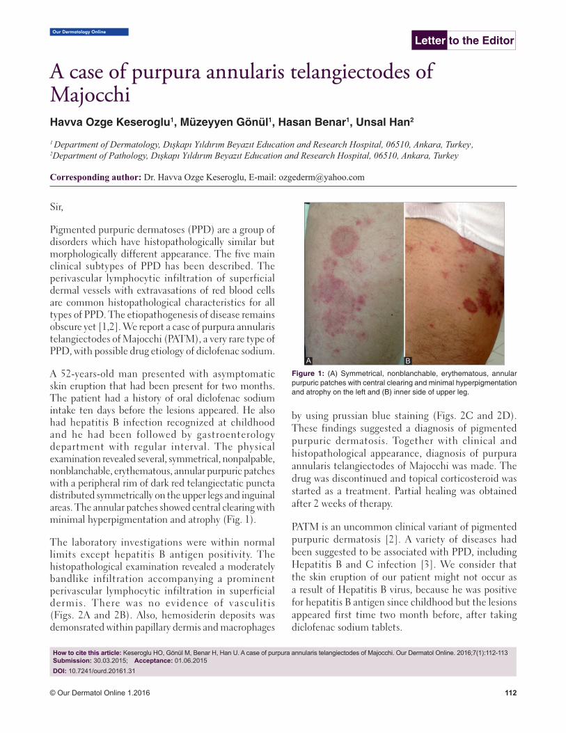
© Our Dermatol Online 1.2016 112
Our Dermatology Online
How to cite this article: Keseroglu HO, Gönül M, Benar H, Han U. A case of purpura annularis telangiectodes of Majocchi. Our Dermatol Online. 2016;7(1):112-113 Submission: 30.03.2015; Acceptance: 01.06.2015DOI: 10.7241/ourd.20161.31
A case of purpura annularis telangiectodes of A case of purpura annularis telangiectodes of MajocchiMajocchiHavva Ozge Keseroglu1, Müzeyyen Gönül1, Hasan Benar1, Unsal Han2
1 Department of Dermatology, Dışkapı Yıldırım Beyazıt Education and Research Hospital, 06510, Ankara, Turkey,, 2Department of Pathology, Dışkapı Yıldırım Beyazıt Education and Research Hospital, 06510, Ankara, Turkey
Corresponding author: Dr. Havva Ozge Keseroglu, E-mail: [email protected]
Sir,
Pigmented purpuric dermatoses (PPD) are a group of disorders which have histopathologically similar but morphologically different appearance. The five main clinical subtypes of PPD has been described. The perivascular lymphocytic infiltration of superficial dermal vessels with extravasations of red blood cells are common histopathological characteristics for all types of PPD. The etiopathogenesis of disease remains obscure yet [1,2]. We report a case of purpura annularis telangiectodes of Majocchi (PATM), a very rare type of PPD, with possible drug etiology of diclofenac sodium.
A 52-years-old man presented with asymptomatic skin eruption that had been present for two months. The patient had a history of oral diclofenac sodium intake ten days before the lesions appeared. He also had hepatitis B infection recognized at childhood and he had been followed by gastroenterology department with regular interval. The physical examination revealed several, symmetrical, nonpalpable, nonblanchable, erythematous, annular purpuric patches with a peripheral rim of dark red telangiectatic puncta distributed symmetrically on the upper legs and inguinal areas. The annular patches showed central clearing with minimal hyperpigmentation and atrophy (Fig. 1).
The laboratory investigations were within normal limits except hepatitis B antigen positivity. The histopathological examination revealed a moderately bandlike infiltration accompanying a prominent perivascular lymphocytic infiltration in superficial dermis. There was no evidence of vasculitis (Figs. 2A and 2B). Also, hemosiderin deposits was demonsrated within papillary dermis and macrophages
by using prussian blue staining (Figs. 2C and 2D). These findings suggested a diagnosis of pigmented purpuric dermatosis. Together with clinical and histopathological appearance, diagnosis of purpura annularis telangiectodes of Majocchi was made. The drug was discontinued and topical corticosteroid was started as a treatment. Partial healing was obtained after 2 weeks of therapy.
PATM is an uncommon clinical variant of pigmented purpuric dermatosis [2]. A variety of diseases had been suggested to be associated with PPD, including Hepatitis B and C infection [3]. We consider that the skin eruption of our patient might not occur as a result of Hepatitis B virus, because he was positive for hepatitis B antigen since childhood but the lesions appeared first time two month before, after taking diclofenac sodium tablets.
Letter to the Editor
Figure 1: (A) Symmetrical, nonblanchable, erythematous, annular purpuric patches with central clearing and minimal hyperpigmentation and atrophy on the left and (B) inner side of upper leg.
A B

www.odermatol.com
© Our Dermatol Online 1.2016 113
A wide variety of medications including NSAIDs, diuretics, sedatives, stimulants antibiotics, cardiovascular drugs, vitamins, antihyperglycemics, isotretinoin have been implicated as drugs responsible for the development of PPD [4,5]. It was suggested that as a result of antigen-antibody complex deposition in which drugs act as a hapten, an immune system-mediated vascular damage occur and this results in capillary leakage and erythrocyte extravasation [5]. The skin eruption of our patient appeared at 10th day of oral diclofenac sodium treatment. So, we thought it might be the possible cause of skin eruption in our patient. Also, as in our case, the drug induced cases are
more likely to resolve after discontinuation of drug in contrast to chronic or relapsing nature of idiopathic ones [5]. Due to possible risk of provocation of a systemic reaction [4] we couldn’t confirm the diagnoses by oral challenge test or patch test.
We intended to present this case because of the rarity of the PATM and it should be kept in mind that diclofenac sodium could play role in etiopathogenesis.
CONSENT
The examination of the patient was conducted according to the Declaration of Helsinki principles.
REFERENCES
1. Sardana K, Sarkar R, Sehgal VN. Pigmented purpuric dermatoses: an overview. Int J Dermatol. 2004;43:482-8.
2. Hoesly FJ, Huerter CJ, Shehan JM. Purpura annularis telangiectodes of Majocchi: case report and review of the literature. Int J Dermatol. 2009;48:1129-33.
3. Dessoukey MW, Abdel-Dayem H, Omar MF, Al-Suweidi NE. Pigmented purpuric dermatosis and hepatitis profi le: a report on 10 patients. Int J Dermatol. 2005;44:486-8.
4. Díaz-Jara M, Tornero P, Barrio MD, Vicente ME, Fuentes V, Barranco R. Pigmented purpuric dermatosis due to pseudoephedrine. Contact Dermatitis. 2002;46:300-1.
5. Kaplan R, Meehan SA, Leger M. A case of isotretinoin-induced purpura annularis telangiectodes of Majocchi and review of substance-induced pigmented purpuric dermatosis. JAMA Dermatol. 2014;150:182-4.
Copyright by Havva Ozge Keseroglu, et al. This is an open access article distributed under the terms of the Creative Commons Attribution License, which permits unrestricted use, distribution, and reproduction in any medium, provided the original author and source are credited.Source of Support: Nil, Confl ict of Interest: None declared.
Figure 2: (A) A moderately bandlike and prominent perivascular lymphocytic infi ltration in superfi cial dermis. (H&E, x100), (B) The closer view of infi ltration, the pseudovasculitis appearence of lymphocytic infi ltration encircling vessels without fi brin deposition (H&E, x200), (C,D) The presence of blue coloured hemosiderin within the papillary dermis freely or in macrophage system (Prussian blue, x40).
DC
BA

© Our Dermatol Online 1.2016 114
Graham little picardi lassueur syndromeRitu Rawat1, Vikram K Mahajan1, Bal Chander2, Karaninder S. Mehta1, Pushpinder S. Chauhan1, Mrinal Gupta1
1Department of Dermatology, Venereology & Leprosy, Dr. R. P. Govt. Medical College, Kangra (Tanda)-176001, (Himachal Pradesh), India, 2Department of Pathology, Dr. R. P. Govt. Medical College, Kangra (Tanda)-176001, (Himachal Pradesh), India.
Corresponding author: Dr. Vikram K Mahajan, E-mail: [email protected]
Sir,
A 27-years-man presented with progressive and wide spread hair loss over scalp, axillae and pubic area for the last 3-4 years. A multitude of therapies did not benefit him and the initial hair loss over occipital area had progressed to involve the whole scalp and other body sites. His medical history was unremarkable and no other family member had similar problem. Cutaneous examination (Figs. 1 - 4) showed smooth and whitish parchment-like scalp skin, areas of variable brownish pigmentation, atrophy, scarring, and minimal scaling, and was devoid of hair. The cicatricial alopecia involved the whole scalp with few hair strands and tufts of remnant hairs particularly at scalp margins. The skin interspersed between intact hairs too had similar texture. Hair pull test was positive. The eyebrows
were sparse at lateral half. Numerous erythematous-brownish follicular papules were noted over scalp margins, beard area, neck, and whole trunk. There was complete non-cicatricial alopecia in both the axillae and the pubic region showed partial non-cicatricial alopecia. Nails and mucosae were normal. Systemic examination and routine laboratory parameters including complete blood counts, serum biochemistry, ANA and thyroid function tests were normal. Histology showed features of lichen planopilaris (Figs. 5 and 6). Treatment with systemic prednisolone 30mg/day was initiated after counselling for long-term follow up in view of protracted clinical course and prognosis.
Graham Little Picardi Lassueur Syndrome is a very rare presentation of lichen planopilaris. Clinically, the triad of progressive cicatricial alopecia of the scalp, non-cicatricial alopecia involving axillae and groin, and follicular keratotic papules on the glabrous
Letter to the Editor
Figure 1: Late lichen planoplaris showing diffuse hair loss, whitish atrophic scarring, few follicular plugging, and residual single hairs and tufts of hair especially at scalp margins.
Figure 2: Red-brownish follicular papules over fronto-temporal area, cheek and side of neck. Note: alopecia involving lateral eyebrows.
How to cite this article: Rawat R, Mahajan VK, Chander B, Mehta KS, Chauhan PS, Gupta M. Graham Little Picardi Lassueur syndrome. Our Dermatol Online. 2016;7(1):114-116.Submission: 23.05.2015; Acceptance: 07.07.2015DOI:10.7241/ourd.20161.32

www.odermatol.com
© Our Dermatol Online 1.2016 115
skin is characteristic. These three clinical features usually appear concurrently but scalp alopecia may precede the follicular keratosis in most instances. Pruritus, when present, is often severe. Follicular inflammation destroys hair follicles permanently with hardly any possibility of hair re-growth causing substantial scarring alopecia and significant cosmetic embarrassment leading to anxiety, psychological distress and psychosocial morbidity necessitating treatment that is more aggressive. Its exact etiology remains obscure and there is no underlying systemic disorder except for one report of its association with androgen insensitivity syndrome [1]. Most patients are females between 30 and 70 years while young males are affected very rarely [2]. There is no racial predilection but familial cases have occurred [3]. Its association with the hepatitis B vaccination too has been speculated [4]. It is considered immune mediated on the analogy of its other more common variant, cutaneous or mucosal lichen planus that occurs
concurrently in about 50% cases [5]. The histologic features of infundibular hyperplasia, follicular plugging, wedge-shaped hypergranulosis, mild pigment incontinence, and dense perifollicular lichenoid infiltrate extending around its base (hugging type) are characteristic. Perifollicular lymphocytic infiltrate at the level of the infundibulum and the isthmus along with vacuolar changes of the outer root sheath are of early lichen planopilaris. More developed lesions show perifollicular fibrosis and epithelial atrophy at the level of the infundibulum and isthmus giving rise to a characteristic hourglass configuration. Alopecia with vertically oriented elastic fibres that replace the destroyed hair follicles is characteristic of advanced stage of the disease. The disease has a chronic unrelenting clinical course and needs differentiation
Figure 5: Histology shows infundibular hyperplasia, follicular plugging, wedge-shaped hypergranulosis, mild pigment incontinence, and dense perifollicular lichenoid infiltrate extending around its base (hugging type) and comprising mainly lymphocytes (H&E, x10).
Figure 6: The lichenoid infiltrate is permeating lower follicullar epithelium and vacuolar changes of the outer root sheath are seen. (H&E, x40).
Figure 4: Non-scarring hair loss of axillae. Similar changes were noted over pubic skin.
Figure 3: (a and 3b) Disseminated red-brownish follicular papules over front and back of trunk.
ba

www.odermatol.com
© Our Dermatol Online 1.2016 116
from folliculitis spinulosa decalvans, keratosis pilaris atrophicans, pityriasis rubra pilaris, pseudopelade of Brocq and discoid lupus erythematosus. The treatment is usually symptomatic and targeted to arrest progression of disease and alopecia. Topical, intralesional or systemic corticosteroids, oral retinoids, PUVA therapy, and antimalarials have been used with a limited success. Few reports on efficacy of cyclosporin (5 mg/kg/day) are also available [6]. However, the claimed efficacy of thalidomide in lichen planopilaris remains unsubstantiated [7,8].
Consent
The examination of the patient was conducted according to the Declaration of Helsinki principles.
REFERENCES
1. Vega Gutierrez J, Miranda-Romero A, Perez Milan F, Martinez Garcia G. Graham Little-Piccardi-Lassueur syndrome associated with androgen insensitivity syndrome (testicular feminization). J
Eur Acad Dermatol Venereol. 2004;18:463-6.2. László FG. Graham-Little-Piccardi-Lasseur syndrome: case
report and review of the syndrome in men. Int J Dermatol. 2014;53:1019-22.
3. Viglizzo G, Verrini A, Rongioletti F. Familial Lassueur-Graham-Little-Piccardi syndrome. Dermatology. 2004;208:142-4.
4. Bardazzi F, Landi C, Orlandi C, Neri I, Varotti C. Graham Little-Piccardi-Lasseur syndrome following HBV vaccination. Acta Derm Venereol. 1999;79:93.
5. Rodriguez-Bayona B, Ruchaud S, Rodriguez C, Linares M, Astola A, Ortiz M, et al. Autoantibodies against the chromosomal passenger protein INCENP found in a patient with Graham Little-Piccardi-Lassueur syndrome. J Autoimmune Dis. 2007;4:1.
6. Mirmirani P, Willey A, Price VH. Short course of oral cyclosporine in lichen planopilaris. J Am Acad Dermatol. 2003;49:667-71.
7. Boyd AS, King LE Jr. Thalidomide-induced remission of lichen planopilaris. J Am Acad Dermatol. 2002;47:967-8.
8. Jouanique C, Reygagne P, Bachelez H, Dubertret L. Thalidomide is ineffective in the treatment of lichen planopilaris. J Am Acad Dermatol. 2004;51:480-1.
Copyright by Ritu Rawat, et al. This is an open access article distributed under the terms of the Creative Commons Attribution License, which permits unrestricted use, distribution, and reproduction in any medium, provided the original author and source are credited.Source of Support: Nil, Conflict of Interest: None declared.

© Our Dermatol Online 1.2016 117
Our Dermatology Online
Would you consider pilomatricoma as a diff erential Would you consider pilomatricoma as a diff erential diagnosis?diagnosis?Yuka Inamura, Hiroo Hata, Keisuke Imafuku, Shinya Kitamura, Hiroshi Shimizu
Department of Dermatology, Hokkaido University Graduate School of Medicine, Sapporo, Japan
Corresponding author: Dr. Hiroo Hata, M.D., Ph.D., E-mail: [email protected]
Sir,
A 26-year-old man presented with a pedunculated, ulcerated, reddish and partially yellowish tumor of 6.5 cm in length by 6.0 cm in width by 3.0 cm in height on his left upper arm (Fig. 1a). The tumor had gradually developed during the half-year before the patient came to us on referral. There was no past history. We performed surgical removal of the entire tumor with a 1.0-cm margin, and the lesion was found to be a histopathologically poorly demarcated tumor without a capsule. There were darkly stained basophilic cells and so-called shadow cells with missing nuclei. There was no obvious nuclear atypia, and calcium deposits were found in the lesions. Based on these findings, we made the final diagnosis of pilomatricoma.
In a second case, a 42-year-old man presented with a pedunculated, non-ulcerated, reddish tumor of 5.0 cm in length by 2.5 cm in width by 0.8 cm in height on the left neck (Fig. 1b). We were able to see small yellowish dots on the surface. The tumor had gradually developed during the 3 months before the patient came to us on referral. The entire tumor was excisionally removed, and the surgical specimen showed the typical features of pilomatricoma.
Pilomatricoma is a common tumor derived from hair matrix cells; it is most often diagnosed in young children but may also affect adults. The clinical presentation is characterized by a subcutaneous nodule, usually up to 1 cm in diameter, with or without high mobility and calcification within the lesion that makes it feel hard and bony. For these reason it is easily diagnosed [1]. However, once the tumor is larger than 5 cm as the Case 1 and 2, they are called ‘giant pilomatricoma’, and it may become very difficult to make an accurate clinical diagnosis [2,3]. To date, several cases of giant pilomatricoma have been reported [4,5].
In Case 1, we initially suspected sebaceous carcinoma, squamous cell carcinoma or dermatofibrosarcoma protuberans, because of the unusual clinical manifestations. However, we considered pilomatricoma as an initial diagnosis when we encountered the Case 2, so we were able to avoid over-treatment on Case 2.
In conclusion, we should keep giant pilomatricoma in mind as a differential diagnosis when we see pedunculated, firm reddish tumor of >5.0 cm in size, with or without ulceration. This knowledge can avoid misdiagnosis and unnecessary examination and treatment.
Letter to the Editor
How to cite this article: Inamura Y, Hata H, Imafuku K, Kitamura S, Shimizu H. Would you consider pilomatricoma as a differential diagnosis? Our Dermatol Online. 2016;7(1):117-118.Submission: 08.062015; Acceptance: 14.09.2015DOI:10.7241/ourd.20161.33
Figure 1: (a) A pedunculated, ulcerated, reddish and partially yellowish tumor of 6.0 cm in length by 5.0 cm in width by 4.0 cm in height is seen on the left arm. (b) A pedunculated, non-ulcerated, reddish tumor of 5.0 cm in length by 2.5 cm in width by 0.8 cm in height is seen on the left upper neck.
ba

www.odermatol.com
© Our Dermatol Online 1.2016 118
Consent
The examination of the patient was conducted according to the Declaration of Helsinki principles.
REFERENCES
1. Julian CG, Bower PW. A clinical review of 209 pilomatricomas. J Am Acad Dermatol. 1998;39:191-5.
2. Krausen AS, Ansel DG, Mays BR Jr. Pilomatrixoma masquerading as a parotid mass. Laryngoscope. 1974;84:528-35.
3. Yamauchi M, Yotsuyanagi T, Saito T, Ikeda K, Urushidate S, Higuma Y. Three cases of giant pilomatrixoma-considerations for diagnosis and treatment of giant skin tumours with abundant inner
calcifi cation present on the upper body. J Plast Reconstr Aesthet Surg. 2010;63:e519-24.
4. Loader DE, Ortlechner K, Breier F, Wasilewicz-Stefani G, Steiner A, Feldmann R. Giant pilomatrixoma of the right arm. Eur J Dermatol. 2014;24:257.
5. Resende CI, Gomes J, Duarte Mda L, Brito C. Giant pilomatricoma in a patient with tuberous sclerosis, both diagnosed in the adult life. BMJ Case Rep. 2013: published online 29 August 2013.
Copyright by Yuka Inamura, et al. This is an open access article distributed under the terms of the Creative Commons Attribution License, which permits unrestricted use, distribution, and reproduction in any medium, provided the original author and source are credited.Source of Support: Nil, Confl ict of Interest: None declared.

© Our Dermatol Online 1.2016 119
Cutaneous creeping eruption in a childShrikiran Aroor, Sandeep Kumar, Suneel Mundkur
Department of Pediatrics, Kasturba Medical College, Manipal University, Manipal, India
Corresponding author: Dr Sandeep Kumar, E-mail: [email protected]
Sir,
A 6 year-old boy from coastal area presented with history of intensely pruritic skin lesions over the right forearm for last 3 days. There were no other symptoms. The boy used to play on the beach barefoot daily. Clinical examination revealed an erythematous, serpentine lesion on the dorsal aspect of the right forearm (Fig. 1). Systemic examination was unremarkable. Hemogram was normal except for eosinophilia (absolute eosinophil count-1400/mm3). A diagnosis of cutaneous larva migrans was made and he was treated with single dose of albendazole (400 mg) and ivermectin (6 mg). Lesion had healed during his subsequent followup after 1 week.
Cutaneous larva migrans (CLM) also called creeping eruption, plumber’s itch, is characterized by classical serpentine skin lesions in a tropical setting [1]. Bare foot walkers, farmers, children playing in beaches, sandy and moist arears are at high risk. CLM is mainly caused by infection with larvae of animal hookworms like Ankylostoma caninum and A. braziliens. Other offenders include A. ceylonicum, Bubostomum phlebotomum etc [2,3]. Larval penetration of skin and migration cause itchy erythematous, raised vesicular and serpentine cutaneous lesion. The disease is usually self-limiting and lasts for 4-6 weeks until the larva dies and humans are accidental and ‘dead-end’ host [2,4]. Severe infestations manifest as Loeffler’s syndrome of pulmonary eosinophilia and rarely as eosinophilic enteritis [5]. Biopsy is of no value as the larvae advance ahead of the clinical tract. Optical coherence tomography is a non-invasive modality for diagnosis [6]. We treated with a single dose of ivermectin and albendazole [7]. Other treatment regimens include oral albendazole
(400 mg) daily for 3 days and topical application of 10% thiabendazole [8]. CLM can be prevented by avoiding skin contact with soil contaminated with animal feces and adequately covering the feet when visiting sandy and moist areas.
Consent
The examination of the patient was conducted according to the Declaration of Helsinki principles.
REFERENCES
1. Yap FB. Creeping eruption. Int J Infect Dis. 2010;14:e545.2. Brenner MA, Patel MB. Cutaneous larva migrans: the creeping
eruption. Cutis. 2003;72:111-5.3. Purdy KS, Langley RG, Webb AN, Walsh N, Haldane D. Cutaneous
larva migrans. Lancet. 2011;377:1948.4. Criado PR, Belda W Jr, Vasconcellos C, Silva CS. Cutaneous larva
migrans: a bad souvenir from the vacation. Dermatol Online J. 2012;18:11.
5. Butland RJ, Coulson IH. Pulmonary eosinophilia associated with cutaneous larva migrans. Thorax. 1985;40:76-7.
6. Morsy H, Mogensen M, Thomsen J, Thrane L, Andersen PE, Jemec GB. Imaging of cutaneous larva migrans by optical coherence tomography. Travel Med Infect Dis. 2007;5:243-6.
Letter to the Editor
How to cite this article: Aroor S, Kumar S, Mundkur S. Cutaneous creeping eruption in a child. Our Dermatol Online. 2016;7(1):119-120.Submission: 01.07.2015; Acceptance: 12.09.2015DOI: 10.7241/ourd.20161.34
Figure 1: Raised curvilinear serpentine lesion of cutaneous larva migrans.

www.odermatol.com
© Our Dermatol Online 1.2016 120
7. Coumas E,Datry A, Paris L. Efficacy of ivermectin in thetreatment of cutaneous larva migrans. Arch Dermatol. 1992;128:994-5.
8. Chiriac A, Birsan C, Chiriac AE, Murgu A, Solovan C. Cutaneous larva migrans: Report of three cases with excellent response to albendazole. Our Dermatol Online. 2012;3:126-7.
Copyright by Shrikiran Aroor, et al. This is an open access article distributed under the terms of the Creative Commons Attribution License, which permits unrestricted use, distribution, and reproduction in any medium, provided the original author and source are credited.Source of Support: Nil, Conflict of Interest: None declared.

© Our Dermatol Online 1.2016 121
Perinatal varicellaAnca Chiriac1,2,3, Piotr Brzezinski4, Adina Coroaba1,5, Meda Bradeanu6, Vlad Gorduza7
1Centre of Advanced Research in Bionanoconjugates and Biopolymers, “Petru Poni” Institute of Macromolecular Chemistry of Romanian Academy, 2Dermato-Physiology Department, Apollonia University Iasi, Iasi, Romania, 3Department of Dermatology, Nicolina Medical Center, Iasi, Romania, 4Department of Dermatology, 6th Military Support Unit, Ustka, Poland, 5The East European Network of Excellence for Research and Development in Chronic Diseases CHRONEX-RD, “Grigore T. Popa” University of Medicine and Pharmacy Iasi, Romania, 6Department of Neonatology, “Elena Doamna” Obstetrics Hospital, Iasi, Romania, 7“Grigore T. Popa” University of Medicine and Pharmacy Iasi, Romania
Corresponding author: Adina Coroaba, PhD student, E-mail: [email protected]
Sir,
Neonatal varicella infection has been rarely reported since vaccine introduction. Neonatal varicella occurs in the first 28 days of life and is due to infection developed by mother during last 5-7 days of delivery and 5- 7 days post delivery or in case of similar infection acquired by other siblings [1,2].
Neonatal varicela should be differentiated from “fetal varicella syndrome, congenital varicela with high morbidity and mortality.
Rapid and accurate diagnosis is of great importance on therapy and prognosis.
Two cases of neonatal varicela treated with systemic acyclovir are presented.
Case 1: A 28-day old female patient was seen in Dermatology Hospital for vesicular rash distributed on the limbs and trunk, few vesicles were scattered mostly on the superior part of the body (Fig. 1).
No systemic symptoms, usual lab analysis within normal limits for the age. Tzanck smear showed multinucleate giant cells, history of recent chicken pox in older brother was admitted by the mother, and serology confirmed the diagnosis of neonatal varicella.
Good response and complete recovery was achieved under systemic administration of acyclovir 20mg/kg iv 8 hourly for 10 days.
Case 2: A 17-day old male premature born baby was referred to Dermatology Unit for vesicular lesions
Letter to the Editor
How to cite this article: Chiriac A, Brzezinski P, Coroaba A, Bradeanu M, Gorduza V. Perinatal varicella. Our Dermatol Online. 2016;7(1):121-122.Submission: 09.09. 2015; Acceptance: 12.11.2015DOI:10.7241/ourd.20161.35
Figure 1: Vesicle on the left foot.
Figure 2: Crops of vesicles on the trunk.

www.odermatol.com
© Our Dermatol Online 1.2016 122
spread on the limbs, trunk and scalp accompanied by high fever (Fig. 2). His mother was treated for varicella one week before admittance to the hospital, meaning approximately 10 days after delivery.
Based on clinical aspect of the lesions, close contact with mother infected with varicella 10 days postpartum, the presence of typical multinucleate giant cells on Tzanck smear a diagnosis of neonatal varicella was admitted and a treatment with acyclovir iv was started. Serology was negative. For the following 14 days the baby boy has received acyclovir 20 mg/kg 8 hourly with good response and no complications. A close follow-up was recommended.
DISCUSSION
If maternal infection occurs during 8–20 weeks of pregnancy fetus has a risk of developing varicella embryopathy known as “fetal varicella syndrome” characterized by ophtalmological, muscle and skeletal anomalies, mental retardation, microcephaly, urological defects [3,4].
Congenital varicella occurs if the mother got the infection at 13-20 weeks gestation and presents in new borns with skin scars, growth retardation, limb defects, chorioretinitis, neurological involvement [1]. The prognosis is poor, mortality high, around 30% [2].
When the maternal infection occurs after 20 weeks of gestation some reports highlighted an increased risk of herpes zoster during childhood [5].
Neonatal varicella occurs in the first 28 days of life and is due to infection developed by mother during last 5- 7 days of delivery and 5- 7 days post delivery or in case of similar infection acquired by other siblings [3,4].
Two types of neonatal varicela exist: if the mother has been infected less than 5 days before birth or 2 days after delivery, varicella-associated antibodies are not transmitted to the newborn and the infection is severe; if the mother has got the infection more than 5 days prior to birth, antibodies have been transmitted to the child causing a less severe form of the disease [2].
It is of outmost importance to consider the moment of varicela virus infection that will guide the therapeutical approach.
Maternal treatment includes oral acyclovir within 24 h of onset [6].
If maternal infection occurs 5-7 days before delivery or 2 days after giving birth a prophylactic approach to the child is mandatory using varicella zoster immunoglobulin or intravenous acyclovir, if immunoglobulin therapy is not available or the diagnosis was made 96 hours after exposure [6].
ACKNOWLEDGEMENTS
This work received financial support through the “Program of Excellence in multidisciplinary doctoral and postdoctoral research in chronic diseases”, contract no. POSDRU/159/1.5/S/133377, project co-financed by the European Social Fund Operational Programme “Human Resources Development” for 2007-2013.
REFERENCES
1. Enders G, Miller E, Cradock-Watson J, Bolley I, Ridehalgh M. Consequences of varicella and herpes zoster in pregnancy: prospective study of 1739 cases. Lancet. 1994;343:1548-51.
2. Lécuyer A, Levy C, Gaudelus J, Floret D, Soubeyrand B, Caulin E, et al. Pediatricians Working Group. Hospitalization of newborns and young infants for chickenpox in France. Eur J Pediatr. 2010;169:1293-7.
3. Ghosh S. Neonatal pustular dermatosis: an overview. Indian J Dermatol. 2015;60:211.
4. Sharma CM, Sharma D. A classical case of neonatal varicella. J Clin Neonatol. 2013;2:200.
5. De Araújo T, Schachner L. Benign vesicopustular eruptions in the neonate. An Bras Dermatol. 2006;4:359-66.
6. American Academy of Pediatrics Varicella-Zoster Infections. In: Pickering LK, Baker CJ, Kimberlin DW, Long SS, editors., eds. Red Book: 2009 Report of the Committee on Infectious Diseases. 28th Edition Elk Grove Village, Illinois: American Academy of Pediatrics; 2009:714-27.
Copyright by Anca Chiriac, et al. This is an open access article distributed under the terms of the Creative Commons Attribution License, which permits unrestricted use, distribution, and reproduction in any medium, provided the original author and source are credited.Source of Support: Program of Excellence in multidisciplinary doctoral and postdoctoral research in chronic diseases and European Social Fund Operational Programme “Human Resources Development” for 2007-2013, Conflict of Interest: None declared.

© Our Dermatol Online 1.2016 123
Our Dermatology Online
Th e fascination of mineral pigments in organic and Th e fascination of mineral pigments in organic and natural eye shadows and compact cakes: are they risky natural eye shadows and compact cakes: are they risky or innocuous?or innocuous?Lorenzo Martini
University of Siena, Department of Pharmaceutical Biotechnologies, Via A.Moro 2, 53100 Siena, Italy
Corresponding author: Lorenzo Martini, M.Sc., E-mail: [email protected]
INTRODUCTION
A recent study conducted at the Ahmadu Bello University of Zaria, Nigeria [1] refers that different samples of made in China eye shadows randomly selected from products available in the shops at several towns of Nigeria, were analysed to determine the levels of heavy metals (Pb, Cd, Ni, Cu, Zn, Cr, Co, and Mn).
Moreover an avalanche of experiments conducted all over the world have been showing since immemorial times that a plentitude of decorative make up products for eyes that had been examined contained huge amounts of hazardous heavy metals [2].
Here follows Table 1 where the results of these analysis are referred [2].
Purpose of my research, indeed, is to determine the levels of certain minerals that are usually included (because legally admitted) in the formulas of eye shadows, kohls and encaustics (crayons) which are retrievable in human blood after application onto eyelids, eyelashes and eyebrows of the aforesaid decorative products.
I intend to discover the pool of heavy minerals and other chemical constituents that are reputed dangerous for Human health in volunteers’ blood, even though my attention is not focused on the accidental presence of heavy metals, but actually on the same metals that are admitted in the Strasbourg’s International Nomenclature Cosmetics Index, and therefore are designed as inorganic pigments.
All the inorganic pigments useful to decorate eyes belong to the category of colours (designed as Colour Index, whereof the acronym is C.I.) that is comprised between the progressive number 77000 and 77999, throughout the entire world, unless only a minor part of those is allowed by EC and is referred below, quite completely (Box 1):
Letter to the Editor
How to cite this article: Martini L. The fascination of mineral pigments in organic and natural eye shadows and compact cakes: are they risky or innocuous?. Our Dermatol Online. 2016;7(1):123-126.Submission: 04.06.2015; Acceptance: 07.08.2015DOI:10.7241/ourd.20161.36
Table I: Elemental Results for 30 Eye Shadows from 19 Manufacturers: Mean results (mg/kg)Sample As Cd Cr Co Pb Hg Niά 0.36 0.07 18 5.5 5.0 NF 16
έ 0.62 NF 8.5 7.9 4.4 NF 16
ή 0.65 0.14 15 8.1 3.9 TR 20
ί 0.38 TR 3.8 1.9 4.7 0.07 2.9
ΰ 0.50 NF 7.7 4.0 3.4 NF 9.4
α 0.19 NF 2.0 0.35 0.05 NF 0.91
β 0.78 NF 7.7 1.6 6.5 0.008 3.1
γ 0.36 NF 12 3.7 0.66 0.04 8.2
δ 0.63 0.36 8.3 1.7 6.7 0.0075 4.8
ε 0.24 NF 4.5 0.88 4.7 TR 2.5
ζ 0.25 TR 11 5.3 0.77 0.03 42
η TR NF 3.9 1.3 3.1 NF 4.8
θ 0.45 TR 21 2.8 5.7 0.03 9.6
ι 0.62 NF 18 5.5 4.1 0.0085 15
κ 0.62 NF 9.6 2.1 3.6 0.006 7.5
λ 0.30 NF 1100 2.7 4.2 NF 13
μ 0.41 NF 7.4 2.4 3.4 0.01 8.8
ν 1.0 NF 11 1.6 5.9 NF 10
ξ 0.36 TR 12 7.8 27 TR 32
ο 0.33 NF 16 0.94 3.9 0.02 12
π 0.69 TR 420 4.9 8.6 0.0085 10
ρ 1.7 TR 350 64 2.1 TR 1600
ς 0.47 TR 22000 1.1 2.4 NF 17
σ 0.26 0.11 6.6 6.8 2.8 0.02 14
τ 0.07 NF 4.7 0.31 2.2 NF 5.0
υ 0.79 TR 3500 2.9 7.5 0.015 16
φ 0.29 NF 40 13 4.2 TR 18
χ 0.48 TR 5.1 2.4 5.7 TR 9.4
ψ 1.2 TR 32 0.64 14000 40 9.0
ω NF NF 1.1 0.11 0.11 NF 1.3
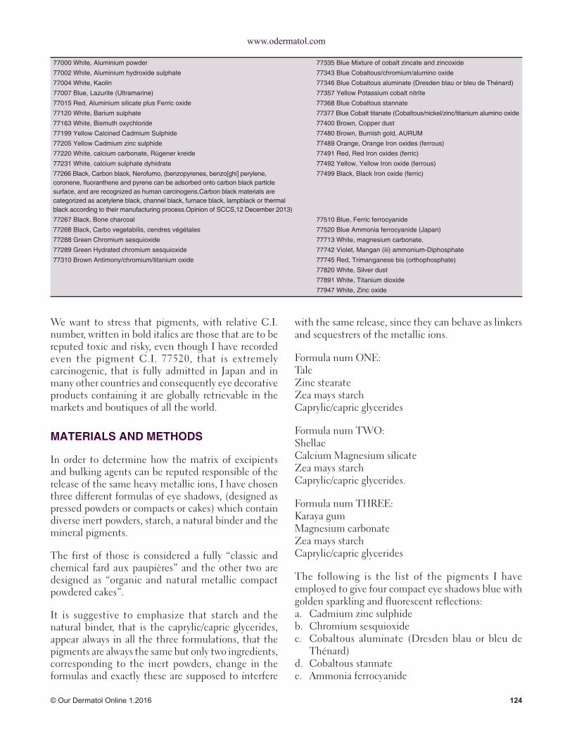
www.odermatol.com
© Our Dermatol Online 1.2016 124
We want to stress that pigments, with relative C.I. number, written in bold italics are those that are to be reputed toxic and risky, even though I have recorded even the pigment C.I. 77520, that is extremely carcinogenic, that is fully admitted in Japan and in many other countries and consequently eye decorative products containing it are globally retrievable in the markets and boutiques of all the world.
MATERIALS AND METHODS
In order to determine how the matrix of excipients and bulking agents can be reputed responsible of the release of the same heavy metallic ions, I have chosen three different formulas of eye shadows, (designed as pressed powders or compacts or cakes) which contain diverse inert powders, starch, a natural binder and the mineral pigments.
The first of those is considered a fully “classic and chemical fard aux paupières” and the other two are designed as “organic and natural metallic compact powdered cakes”.
It is suggestive to emphasize that starch and the natural binder, that is the caprylic/capric glycerides, appear always in all the three formulations, that the pigments are always the same but only two ingredients, corresponding to the inert powders, change in the formulas and exactly these are supposed to interfere
with the same release, since they can behave as linkers and sequestrers of the metallic ions.
Formula num ONE:TalcZinc stearateZea mays starchCaprylic/capric glycerides
Formula num TWO:ShellacCalcium Magnesium silicateZea mays starchCaprylic/capric glycerides.
Formula num THREE:Karaya gumMagnesium carbonateZea mays starchCaprylic/capric glycerides
The following is the list of the pigments I have employed to give four compact eye shadows blue with golden sparkling and fluorescent reflections:a. Cadmium zinc sulphideb. Chromium sesquioxidec. Cobaltous aluminate (Dresden blau or bleu de
Thénard)d. Cobaltous stannatee. Ammonia ferrocyanide
77000 White, Aluminium powder 77335 Blue Mixture of cobalt zincate and zincoxide
77002 White, Aluminium hydroxide sulphate 77343 Blue Cobaltous/chromium/alumino oxide
77004 White, Kaolin 77346 Blue Cobaltous aluminate (Dresden blau or bleu de Thénard)
77007 Blue, Lazurite (Ultramarine) 77357 Yellow Potassium cobalt nitrite
77015 Red, Aluminium silicate plus Ferric oxide 77368 Blue Cobaltous stannate
77120 White, Barium sulphate 77377 Blue Cobalt titanate (Cobaltous/nickel/zinc/titanium alumino oxide
77163 White, Bismuth oxychloride 77400 Brown, Copper dust
77199 Yellow Calcined Cadmium Sulphide 77480 Brown, Burnish gold, AURUM
77205 Yellow Cadmium zinc sulphide 77489 Orange, Orange Iron oxides (ferrous)
77220 White, calcium carbonate, Rügener kreide 77491 Red, Red Iron oxides (ferric)
77231 White, calcium sulphate dyhidrate 77492 Yellow, Yellow Iron oxide (ferrous)
77266 Black, Carbon black, Nerofumo, (benzopyrenes, benzo[ghi] perylene, coronene, fl uoranthene and pyrene can be adsorbed onto carbon black particle surface, and are recognized as human carcinogens.Carbon black materials are categorized as acetylene black, channel black, furnace black, lampblack or thermal black according to their manufacturing process.Opinion of SCCS,12 December 2013)
77499 Black, Black Iron oxide (ferric)
77267 Black, Bone charcoal 77510 Blue, Ferric ferrocyanide
77268 Black, Carbo vegetabilis, cendres végétales 77520 Blue Ammonia ferrocyanide (Japan)
77288 Green Chromium sesquioxide 77713 White, magnesium carbonate,
77289 Green Hydrated chromium sesquioxide 77742 Violet, Mangan (iii) ammonium-Diphosphate
77310 Brown Antimony/chromium/titanium oxide 77745 Red, Trimanganese bis (orthophosphate)
77820 White, Silver dust
77891 White, Titanium dioxide
77947 White, Zinc oxide

www.odermatol.com
© Our Dermatol Online 1.2016 125
The total amount of the powdered pigments represents the 7.00% in each formula;the remnant is constituted by inert powders, bulking agents and the binder.
We have a class of galenic preparations laboratory and the class numbers 21 pupils, 18 of whom are female and the other 3 are male.
Even the boys do not distaste to wear eye shadow when go out to dance on Saturday night and so it has been easy to let them prepare the three formulas and pray them to decorate their own eyelids by the aids of three simple sponge-tipped plastic rods (in order not to inquinate the different products) and to undergo two mineralogram blood tests, the former before and the latter after the application of the cosmetic products onto their eyelids.
It is well known that skin penetration of whichever chemical substance is performed through the conjunctival sac and through the stratum corneum of the eyelid itself, that is the thinnest epidermal layer in all the human body.
Since these toxic substances are often ingested or inhaled accidentally a list of tolerable limits of their presence in blood exists and is to be considered official; and therefore is plotted in Table II:
The three types of coloured cakes (pressed powders) were prepared during the class of laboratory preparations and were shared in 63 small tureens, and each of every pupil had three tureens available, to spread onto his own eyelids in three different occasions, during the Carnival feasts and precisely on Thursday, on Sunday and next Tuesday night (Jeudi Gras, Samedi Gras and Mardi Gras).
The successive morning of every night of amusement, at 7.00 a.m., before classes, the pupils were invited to
have their blood examined to detect the plot of heavy minerals and tiocyanates.
Here follow Tables III and IV where all the scores regarding the classic and chemical “compact fard aux paupières” and regarding the organic and natural compacts cakes are reported:
Table 3: Values of risky substances levels retrieved in blood, after application of the chemical eyeshadow (Formula num ONE)Pupils Cadmium
μg/ltChromium μg/ml
Cobalt μg/lt
Tin ng/ml
Tiocyanates μmol/lt
1F 4.5 0.7 2.0 2.2 77
2F 4.2 0.9 2.1 2.2 81
3F 3.9 0.2 2.2 2.3 88
4F 5.0 0.6 2.4 2.1 92
5F 4.6 0.1 1.9 2.8 65
6F 4.4 0.09 2.3 2.3 89
7F 3.8 0.4 2.2 2.7 93
8F 4.7 0.6 2.1 2.5 67
9F 4.3 0.8 2.2 2.0 99
10F 4.1 0.6 2.4 2.1 91
11F 4.0 0.8 2.2 2.0 78
12F 5.1 0.9 2.0 2.8 83
13F 4.2 0.08 2.1 2.9 77
14F 3.8 0.6 2.1 2.3 82
15F 4.3 0.7 2.3 2.1 75
16F 4.1 0.5 2.7 2.5 69
17F 5.0 0.47 2.4 2.3 73
18F 4.9 0.08 2.1 1.8 88
19M 3.3 0.03 1.8 1.4 101
20M 3.7 0.04 1.7 1.5 105
21M 3.1 0.1 1.8 1.9 121
Table 4: Average values of risky substances levels retrieved in blood, after application of the organic natural eyeshadows (Formula num ONE)Pupils Cadmium
μg/ltChromium μg/ml
Cobalt μg/lt
Tin ng/ml
Tiocyanates μmol/lt
1F 3.8 0.14 1.1 0.2 18
2F 3.3 0.13 1.4 0.3 21
3F 2.9 0.14 1.6 0.1 22
4F 3.7 0.16 1.7 0.4 34
5F 3.9 0.08 1.8 0.6 11
6F 4.0 0.09 1.3 0.2 23
7F 3.3 0.18 1.6 1.0 24
8F 3.6 0.09 0.9 0.8 17
9F 3.7 0.13 0.9 0.5 16
10F 4.0 0.12 1.1 1.1 22
11F 3.2 0.17 1.6 1.4 36
12F 2.9 0.09 1.7 0.8 23
13F 3.1 0.12 0.9 0.9 17
14F 3-0 0.13 1.1 0.7 14
15F 2.8 0.07 1.3 1.2 15
16F 3.3 0.05 1.4 1.6 11
17F 3.1 0.1 1.0 1.4 14
18F 3.7 0.03 1.5 1.2 15
19M 4.1 0.02 1.8 1.9 56
20M 4.2 0.01 1.7 2.1 65
21M 3.9 0.04 1.8 1.9 77
Table 2: Standard Levels and tolerated limits of Heavy metals in human serum according to Test 34506 :Heavy Metals Screen with Demographics, Blood Clinical Information Mayo Foundation for Medical Education and ResearchType of chemical substance
Pool of chemical substance tolerated in non exposed individuals
Cadmium 4 μg/lt
Chromium 0.05-0.5 μg/ml (serum)
Cobalt 1.8 μg/lt
Tin 2 ng/ml
Ferrocyanide (as thiocyanate, biotransformed in the liver)
15-70 μmol/lt
www.odermatol.com
© Our Dermatol Online 1.2016 126
DISCUSSIONS
It is evident that formula num ONE that contains Talc and Zinc stearate, as prime bulking agent and excipient, releases substantially the metallic ions and the ferrocyanides, cause of their intrinsic chemical structure, being inert powders, that is that are not prone to sequestrate the ions, as no free electric charges are available on their molecules.
Therefore high levels of Cadmium, Chromium, Cobalt, tin and ferrocyanide (as tiocyanates) are present in the blood of volunteers that applied this chemical formula of pressed powder.
On the other hand, the natural cakes can boast about the presence of gum resins and bioplymers as shellac and/or karaya gum.
Shellac, a resin secreted by the female lac bug, on trees in the forests of India and Thailand, according to manifold A.A. [3-7]. contains polyhydroxy acids and it has been suggested that these might occur as lactides or as other inter-esters‘ in producing the resin., giving its peculiar liquid-viscous texture, with the capability of linking bivalent and polyvalent metallic ions.
Karaya gum, instead, is a vegetable gum produced as an exudate by trees of the genus Sterculia. Chemically, gum karaya is an acid polysaccharide composed of galactose, rhamnose and galacturonic acid, so that the carboxylate ions are capable to capture and sequester the risky ions and ferrocyanides.
It is suggestive to observe that as far as tiocyanates levels (derived from the biotransformation of ferrocyanides in liver and detectable in human plasma) are higher in man than in female, and several are the A.A. which have attempted to clarify this obscure phenomenon [8,9].
Finally, male can more easily intoxicated by Cobalt and Tin, and this matter of fact is utterly unclear.
REFERENCES
1. Omolaoye JA, Uzairu A Gimba CE. Heavy metal assessment of some eye shadow products imported into Nigeria from China: Scholars Research Library. Arch Applied Science Research. 2010;2:76-84.
2. Hepp NM, Mindak WR, Gasper JW, Thompson CB, Barrows JN. Survey of cosmetics for arsenic, cadmium, chromium,cobalt, lead, mercury, and nickel content J Cosmet Sci. 2014;65:125-45.
3. Bhattaoharya R. J Sac Chem Ind. 1935;54:82.4. Gardner WH. Shellac Research Bur.,1928: Polytech. Inst. Brooklyn
Reports5. Harries C, Nagel W. Ber. 1922;55:3833.6. Jamieson GS. Am J Sci. 1912;4:33.7. Schaeffer BB, Gardner WH. Ind Eng Chem. 1938;30:3338. El Hadri L, Chanas B, Ghanayem BI. Comparative metabolism of
methacrylonitrile and acrylonitrile to cyanide using cytochrome P4502E1 and microsomal epoxide hydrolase-null mice. Toxicol Appl Pharmacol. 2005;205:116-25.
9. Kouichiro T, Mieko K, Yasuo S. Cyanide and Thiocyanate Levels in Blood and Saliva of Healthy Adult Volunteers. J Health Science. 2000;46:343-50.
Copyright by Lorenzo Martini. This is an open access article distributed under the terms of the Creative Commons Attribution License, which permits unrestricted use, distribution, and reproduction in any medium, provided the original author and source are credited.Source of Support: Nil, Confl ict of Interest: None declared.
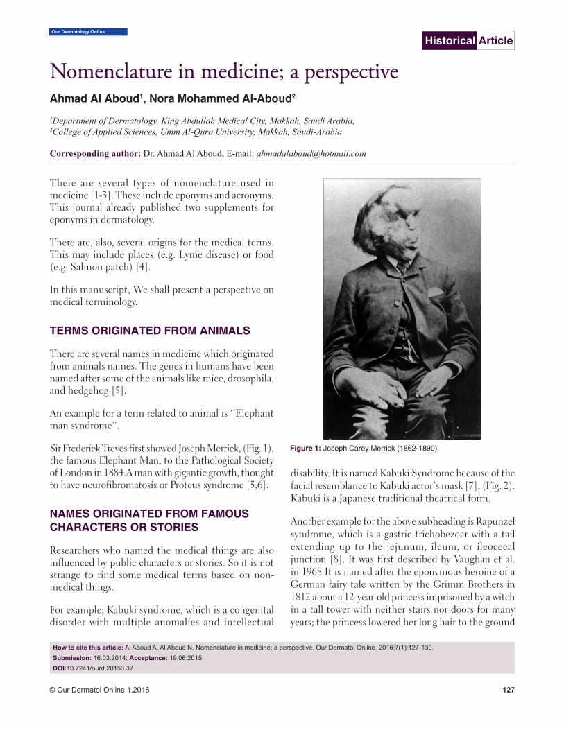
© Our Dermatol Online 1.2016 127
How to cite this article: Al Aboud A, Al Aboud N. Nomenclature in medicine; a perspective. Our Dermatol Online. 2016;7(1):127-130.Submission: 16.03.2014; Acceptance: 19.06.2015DOI:10.7241/ourd.20153.37
Nomenclature in medicine; a perspectiveAhmad Al Aboud1, Nora Mohammed Al-Aboud2
1Department of Dermatology, King Abdullah Medical City, Makkah, Saudi Arabia, 2College of Applied Sciences, Umm Al-Qura University, Makkah, Saudi-Arabia
Corresponding author: Dr. Ahmad Al Aboud, E-mail: [email protected]
There are several types of nomenclature used in medicine [1-3]. These include eponyms and acronyms. This journal already published two supplements for eponyms in dermatology.
There are, also, several origins for the medical terms. This may include places (e.g. Lyme disease) or food (e.g. Salmon patch) [4].
In this manuscript, We shall present a perspective on medical terminology.
TERMS ORIGINATED FROM ANIMALS
There are several names in medicine which originated from animals names. The genes in humans have been named after some of the animals like mice, drosophila, and hedgehog [5].
An example for a term related to animal is ‘’Elephant man syndrome’’.
Sir Frederick Treves first showed Joseph Merrick, (Fig. 1), the famous Elephant Man, to the Pathological Society of London in 1884.A man with gigantic growth, thought to have neurofibromatosis or Proteus syndrome [5,6].
NAMES ORIGINATED FROM FAMOUS CHARACTERS OR STORIES
Researchers who named the medical things are also influenced by public characters or stories. So it is not strange to find some medical terms based on non-medical things.
For example; Kabuki syndrome, which is a congenital disorder with multiple anomalies and intellectual
disability. It is named Kabuki Syndrome because of the facial resemblance to Kabuki actor’s mask [7], (Fig. 2). Kabuki is a Japanese traditional theatrical form.
Another example for the above subheading is Rapunzel syndrome, which is a gastric trichobezoar with a tail extending up to the jejunum, ileum, or ileocecal junction [8]. It was first described by Vaughan et al. in 1968 It is named after the eponymous heroine of a German fairy tale written by the Grimm Brothers in 1812 about a 12-year-old princess imprisoned by a witch in a tall tower with neither stairs nor doors for many years; the princess lowered her long hair to the ground
Historical Article
Figure 1: Joseph Carey Merrick (1862-1890).

www.odermatol.com
© Our Dermatol Online 1.2016 128
from her window, allowing a young prince to climb up and rescue her [8] (Fig. 3).
NAMES ORIGINATED FROM A PATIENT’S NAME OR THINGS RELATED TO THE PATIENTS
Rarely a disease may be named after something related to the paients. For example; Ambras syndrome (AS), which is a form of hereditary, generalized hypertrichosis. The Ambras name was given because the family portraits of Gonzales (The first recorded case of AS) were discovered in Ambras castle, (located
in Austria) amongst an art collection started by the archduke Ferdinand II (1529-1595) [9].
Petrus Gonzales (Fig. 4) was born in the Canary Islands in 1556. Out of curiosity, Petrus was brought to France where he was presented as a gift to the nobles. He subsequently produced offspring with similar AS features. Currently, the same paintings of Gonzalez which were in Ambras castle, hang in the Kunsthistorisches museum in Vienna [9].
Figure 3: Rapunzel story. From: Walt Disney Studios Motion Pictures and New Media Strategies.
Figure 4: Petrus Gonsalvus.
Figure 5: Henry Louis Gehrig (1903-1941).
Figure 2: Two Kabuki actors.
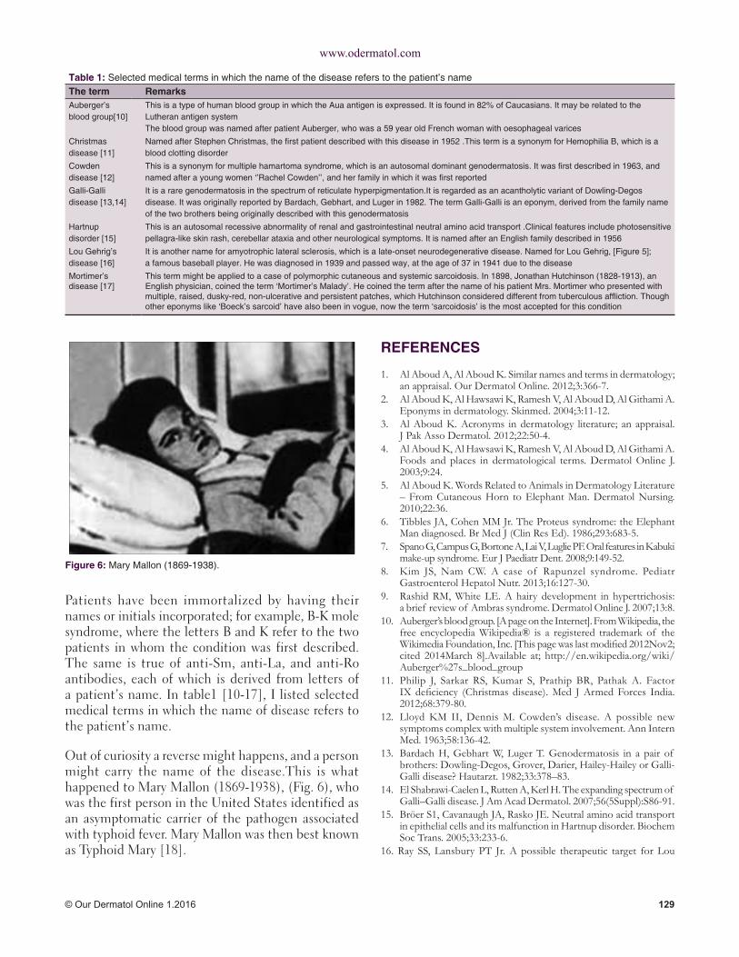
www.odermatol.com
© Our Dermatol Online 1.2016 129
Patients have been immortalized by having their names or initials incorporated; for example, B-K mole syndrome, where the letters B and K refer to the two patients in whom the condition was first described. The same is true of anti-Sm, anti-La, and anti-Ro antibodies, each of which is derived from letters of a patient’s name. In table1 [10-17], I listed selected medical terms in which the name of disease refers to the patient’s name.
Out of curiosity a reverse might happens, and a person might carry the name of the disease.This is what happened to Mary Mallon (1869-1938), (Fig. 6), who was the first person in the United States identified as an asymptomatic carrier of the pathogen associated with typhoid fever. Mary Mallon was then best known as Typhoid Mary [18].
REFERENCES
1. Al Aboud A, Al Aboud K. Similar names and terms in dermatology; an appraisal. Our Dermatol Online. 2012;3:366-7.
2. Al Aboud K, Al Hawsawi K, Ramesh V, Al Aboud D, Al Githami A. Eponyms in dermatology. Skinmed. 2004;3:11-12.
3. Al Aboud K. Acronyms in dermatology literature; an appraisal. J Pak Asso Dermatol. 2012;22:50-4.
4. Al Aboud K, Al Hawsawi K, Ramesh V, Al Aboud D, Al Githami A. Foods and places in dermatological terms. Dermatol Online J. 2003;9:24.
5. Al Aboud K. Words Related to Animals in Dermatology Literature – From Cutaneous Horn to Elephant Man. Dermatol Nursing. 2010;22:36.
6. Tibbles JA, Cohen MM Jr. The Proteus syndrome: the Elephant Man diagnosed. Br Med J (Clin Res Ed). 1986;293:683-5.
7. Spano G, Campus G, Bortone A, Lai V, Luglie PF. Oral features in Kabuki make-up syndrome. Eur J Paediatr Dent. 2008;9:149-52.
8. Kim JS, Nam CW. A case of Rapunzel syndrome. Pediatr Gastroenterol Hepatol Nutr. 2013;16:127-30.
9. Rashid RM, White LE. A hairy development in hypertrichosis: a brief review of Ambras syndrome. Dermatol Online J. 2007;13:8.
10. Auberger’s blood group. [A page on the Internet]. From Wikipedia, the free encyclopedia Wikipedia® is a registered trademark of the Wikimedia Foundation, Inc. [This page was last modified 2012Nov2; cited 2014March 8].Available at; http://en.wikipedia.org/wiki/Auberger%27s_blood_group
11. Philip J, Sarkar RS, Kumar S, Prathip BR, Pathak A. Factor IX deficiency (Christmas disease). Med J Armed Forces India. 2012;68:379-80.
12. Lloyd KM II, Dennis M. Cowden’s disease. A possible new symptoms complex with multiple system involvement. Ann Intern Med. 1963;58:136-42.
13. Bardach H, Gebhart W, Luger T. Genodermatosis in a pair of brothers: Dowling-Degos, Grover, Darier, Hailey-Hailey or Galli-Galli disease? Hautarzt. 1982;33:378–83.
14. El Shabrawi-Caelen L, Rutten A, Kerl H. The expanding spectrum of Galli–Galli disease. J Am Acad Dermatol. 2007;56(5Suppl):S86-91.
15. Bröer S1, Cavanaugh JA, Rasko JE. Neutral amino acid transport in epithelial cells and its malfunction in Hartnup disorder. Biochem Soc Trans. 2005;33:233-6.
16. Ray SS, Lansbury PT Jr. A possible therapeutic target for Lou
Figure 6: Mary Mallon (1869-1938).
Table 1: Selected medical terms in which the name of the disease refers to the patient’s nameThe term RemarksAuberger’s blood group[10]
This is a type of human blood group in which the Aua antigen is expressed. It is found in 82% of Caucasians. It may be related to the Lutheran antigen systemThe blood group was named after patient Auberger, who was a 59 year old French woman with oesophageal varices
Christmas disease [11]
Named after Stephen Christmas, the first patient described with this disease in 1952 .This term is a synonym for Hemophilia B, which is a blood clotting disorder
Cowden disease [12]
This is a synonym for multiple hamartoma syndrome, which is an autosomal dominant genodermatosis. It was first described in 1963, and named after a young women ‘’Rachel Cowden’’, and her family in which it was first reported
Galli-Galli disease [13,14]
It is a rare genodermatosis in the spectrum of reticulate hyperpigmentation.It is regarded as an acantholytic variant of Dowling-Degos disease. It was originally reported by Bardach, Gebhart, and Luger in 1982. The term Galli-Galli is an eponym, derived from the family name of the two brothers being originally described with this genodermatosis
Hartnup disorder [15]
This is an autosomal recessive abnormality of renal and gastrointestinal neutral amino acid transport .Clinical features include photosensitive pellagra-like skin rash, cerebellar ataxia and other neurological symptoms. It is named after an English family described in 1956
Lou Gehrig’s disease [16]
It is another name for amyotrophic lateral sclerosis, which is a late-onset neurodegenerative disease. Named for Lou Gehrig, [Figure 5]; a famous baseball player. He was diagnosed in 1939 and passed way, at the age of 37 in 1941 due to the disease
Mortimer’s disease [17]
This term might be applied to a case of polymorphic cutaneous and systemic sarcoidosis. In 1898, Jonathan Hutchinson (1828-1913), an English physician, coined the term ‘Mortimer’s Malady’. He coined the term after the name of his patient Mrs. Mortimer who presented with multiple, raised, dusky-red, non-ulcerative and persistent patches, which Hutchinson considered different from tuberculous affliction. Though other eponyms like ‘Boeck’s sarcoid’ have also been in vogue, now the term ‘sarcoidosis’ is the most accepted for this condition

www.odermatol.com
© Our Dermatol Online 1.2016 130
Gehrig’s disease. Proc Natl Acad Sci U S A. 2004;101:5701-2.17. Pandhi D, Sonthalia S, Singal A. Mortimer’s Malady revisited: a
case of polymorphic cutaneous and systemic sarcoidosis. Indian J Dermatol Venereol Leprol. 2010;76:448.
18. Brooks J. The sad and tragic life of Typhoid Mary. CMAJ. 1996;154:915-16.
Copyright by Ahmad Al Aboud, et al. This is an open access article distributed under the terms of the Creative Commons Attribution License, which permits unrestricted use, distribution, and reproduction in any medium, provided the original author and source are credited.Source of Support: Nil, Conflict of Interest: None declared.


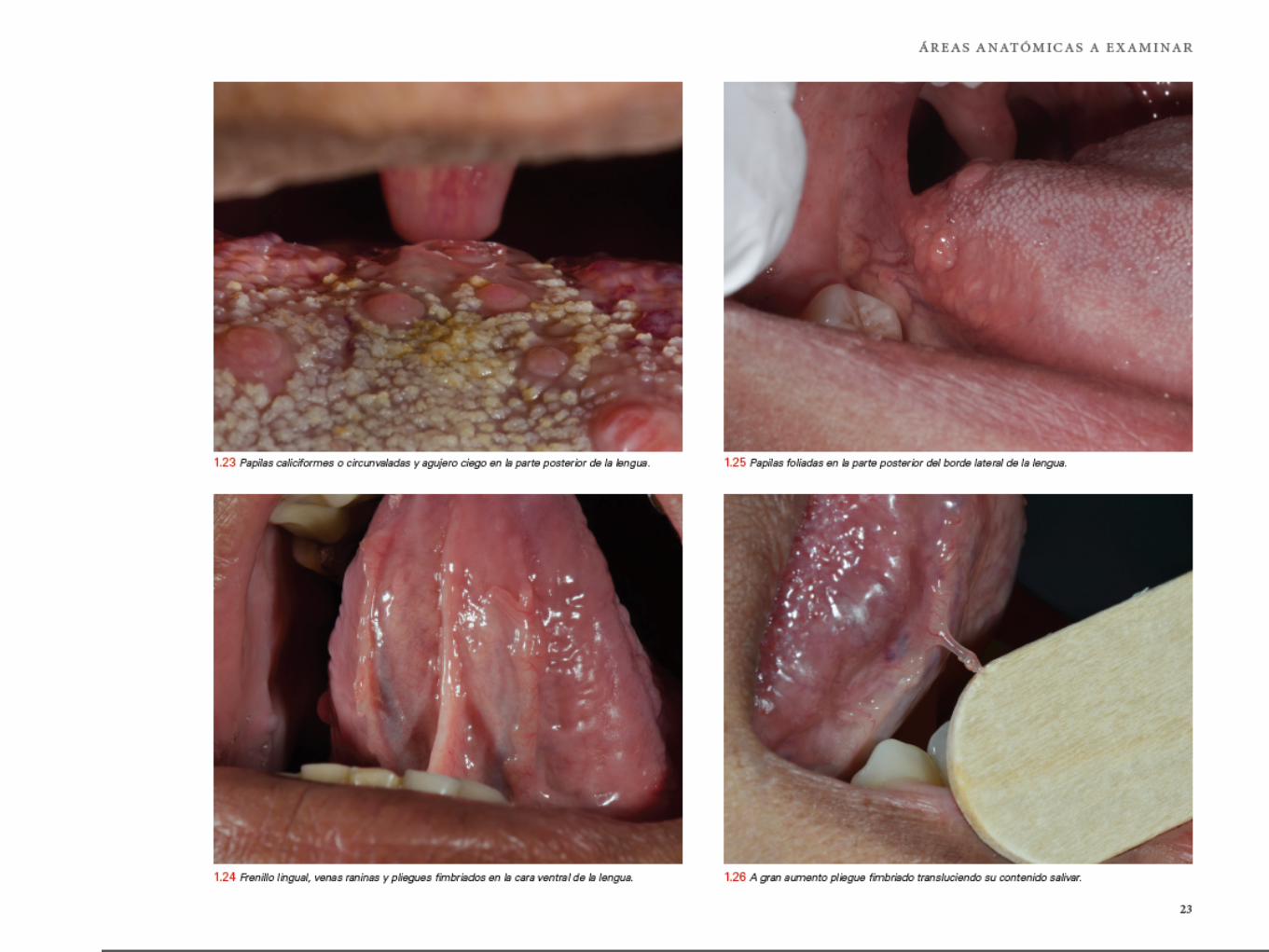
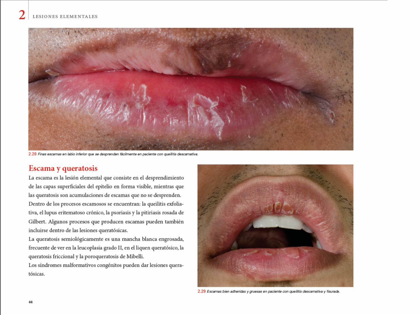


11 capítulos. 1540 fotos a color. 520 páginas. libro con tapa dura.
Examen del paciente. Lesiones elementales. Lesiones blancas. Lesiones rojas. Lesiones pigmentadas. Lesiones vesiculares, erosivas y ulceradas. Lesiones ampollares.
ADOLFO ARTHUR NOUEL
MUCOSA ORALENFERMEDADESDE LA
Contenido Lesiones vegetantes y verrugosas. Crecimientos de tejidos blandos. Tumores benignos de tejidos blandos. Tumores malignos de tejidos blandos. Tablas de diagnóstico diferencial. Bibliografía consultada. Índice.
Ventas: Instituto Dermatológico Dominicano y Cirugía de Piel “Dr. Huberto Bogaert Díaz” Valor: $90.00 dólares más envío por courrier Contacto: [email protected]

O u r D e r m a t o l o g y O n l i n e
w w w . o d e r m a t o l . c o m1.2016 (07.January.2016)