issue20113.pdf - Our Dermatology Online
71
Issue Online since Friday, July 01, 2011 GFMER Volume 2, Number 3, July 2011 ISSN: 2081-9390 p. 101 - 170 NASZA DERMATOLOGIA NASZA DERMATOLOGIA NASZA DERMATOLOGIA NASZA DERMATOLOGIA Online Online Online Online www.odermatol.like.pl OUR DERMATOLOGY OUR DERMATOLOGY OUR DERMATOLOGY OUR DERMATOLOGY Online Online Online Online Index Copernicus 3,42 DOAJ SPARC Europe
-
Upload
khangminh22 -
Category
Documents
-
view
0 -
download
0
Transcript of issue20113.pdf - Our Dermatology Online

Issue Online since Friday, July 01, 2011
GFMER
Volume 2, Number 3, July 2011 ISSN: 2081-9390 p. 101 - 170
NASZA DERMATOLOGIANASZA DERMATOLOGIANASZA DERMATOLOGIANASZA DERMATOLOGIA OnlineOnlineOnlineOnline www.odermatol.like.pl
OUR DERMATOLOGYOUR DERMATOLOGYOUR DERMATOLOGYOUR DERMATOLOGY OnlineOnlineOnlineOnline
Index Copernicus 3,42 DOAJ SPARC Europe

101
Editorial Pages / Strona Redakcyjna
e-ISSN: 2081-9390
Quarterly (kwartalnik) published since 01/06/2010 years
Editor in Chief (Redaktor Naczelny): Piotr Brzeziński, MD
Address:
ul. Andersa 5/8, 76200 Słupsk, Poland
tel. 48 692121516, fax.48 598151829
e-mail: [email protected]
Editorial Board:
Abreu-Velez Ana Maria, MD Ph.D (USA)
Abreu Hilda, MD (Urugway)
Adaskevich Uladzimir, Prof. (Belarus)
Aghaei Shahin, MD (Iran)
Arenas Roberto, MD (Mexico)
Bharti Rakesh, MD (India)
Bonifaz Alexandro, MD (Mexico)
Bukhari Iqbal A., MD (Saudi Arabia)
Chamcheu Jean Christopher, Ph.D (USA)
Chang Patricia, MD Ph.D (Guatemala)
Chuh An Tung Antonio, Prof. (Hong Kong)
Daboul Mohamed Wael, MD (Syria)
Darkoska Jovanka, MD (Macedonia)
Doganay Mehmet, Prof. (Turkey)
Drljević Irdina, MD (Bosna i Hercegovina)
Dubakien÷ Rūta, Prof. (Lithuania)
Guzmán Antonio, MD (Paraguay)
Hashimoto Takashi, MD (Japan)
Hassan Iffat, MD (India)
Howard I. Maibach, Prof. (USA)
Jordán Rodriguez Ramiro, Prof. (Bolivia)
Kaszuba Andrzej, Prof. (Poland)
Khamesipour Ali, MD (Iran)
Lopez-Granja Jorge, MD (Belize)
Lotti Torello, Prof. (Italy)
Al-Mashaleh Manal Sulaiman, MD (Jordan)
Mikkelsen Carsten Sauer, MD (Denmark)
Mota Luiz Alberto Alves, MD (Brazil)
Mrisho Fatma, MD (Tanzania)
Nowicki Roman, Prof. (Poland)
Nwabudike Lawrence Chukwudi, MD Ph.D (Romania)
Parvin Rukhsana, MD (Bangladesh)
du Plessis Jeanetta, Prof. (South Africa)
Sharquie Khalifa E., Prof. (Iraq)
Shawa Mary, MD (Malawi)
Tatu Alin, MD (Romania)
Teixeira Roni Leonardo, MD (Brazil)
Tincopa-Wong Oscar Wilfredo, MD (Peru)
Usharani Anaparthy, MD (India)
Zabielski Stanisław, Prof. (Poland)
Publisher (Wydawca):
Piotr Brzeziński
ul. Andersa 5/8, 76200 Słupsk, Poland
tel. 48 692121516, fax.48 598151829
e-mail: [email protected]
© N Dermatol Online 3.2011

102
CONTENTS / SPIS TREŚCI .
Editorial Pages / Strona Redakcyjna 101
.
Original Articles / Prace Oryginalne
► Abreu Velez Ana Maria, Jackson Billie L., Howard Michael S.
Salt and pepper staining patterns for LAT, ZAP-70 and MUM-1 in a vasculitic bullousallergic drug eruption 104 Wzór barwienia sól i pieprz dla LAT, ZAP-70 i MUM-1 w naczyniowych pęcherzach reakcji alergicznych na lek . ► Bonifaz Alexandro, Vázquez-González Denisse, Saúl Amado, Fierro-Arias Leonel, Ponce-Olivera M. Rosa
Refractory onychomycosis due to Trichophyton rubrum: combination therapy with itraconazole and terbinafine 108 Oporna na leczenie grzybica paznokci wywołana przez Trichphyton rubrum: kombinowana terapia itrakonazolem i terbinafiną . ► Abreu Velez Ana Maria, Howard William R., Howard Michael S.
Upregulation of anti-human ribosomal protein S6-p240, topoisomerase II ά, cyclin D1, Bcl-2 and anti-corneal antibodies in acute psoriasis 113 Aktywacja przeciwciał przeciwko ludzkiej rybosomalnej proteinie S6-p240, topoizomezazie II ά, cyklinie D1, Bcl-2 oraz przeciwko warstwie rogowej naskórka w ostrej postaci łuszczycy
Comment: Takashi Hashimoto, Daisuke Tsuruta, Takahiro Hamada, Teruki Dainichi, Norito Ishii Department of Dermatology, Kurume University School of Medicine, and Kurume University Institute of Cutaneous Cell Biology, Kurume, Fukuoka, Japan
. ► Anaparthy Usharani, M. Bharathi, Cautha Sandhya
Isolation and characterisation of Candida species from oropharyngeal secretions of HIV positive individuals 119 Izolacja i charakterystyka Candida species z wydzieliny z jamy ustno-gardłowej u HIV pozytywnych osób . ► Al-Bdour Mohammed, Al-Khateeb Maher
Reconstruction of nasal skin defects following excision of basal cell carcinoma 125 Rekonstrukcja ubytków skóry nosa po wycięciu raka podstawnokomórkoego . ► Martinez Braga Gabriela, Di Martino Ortiz Beatriz, Rodriguez Masi Mirtha, Knopfelmacher Oilda, Bolla de Lezcano Lourdes
Tuberculosis ganglionar con afectación cutánea (escrofulodermia) en paciente inmunocompetente. Reporte de un caso 130 Ganglionar tuberculosis with skin involvement (scrofuloderma) in an inmmunocompetent patient. A case report . ► Sierra-Téllez Daniela, Ponce-Olivera Rosa María, Tirado-Sánchez Andrés, Hernández Marco Antonio, Bonifaz Alexandro
Gram-negative folliculitis. A rare problem or is it underdiagnosed? Case report and literature review 135 Gram-ujemne zapalenie mieszków włosowych. Rzadki problem czy rzadko diagnozowany? Opis przypadku i przegląd piśmiennictwa Comment: Prof. Antonio Chuh
School of Public Health, The Chinese University of Hong Kong Comment: Prof. Mehmet Doganay
Department of Infectious Diseases, Faculty of Medicine,Erciyes University, Kayseri, Turkey . ► Alendar Faruk, Soskic Samra, Helppikangas Hana, Gavrankapetanovic Alma, Alendar Temeida
Erythematodes chronicus profundus as dermatology, surgery and cosmetology problem 141 Erythematodes chronicus profundus jako dermatologiczny, chirurgiczny i kosmetologiczny problem . ► Brzezinski Piotr, Poklękowska Katarzyna
Granulosis rubra nasi – a case report. A literature review 144 Granulosis rubra nasi – opis przypadku. Przegląd literatury
© N Dermatol Online 3.2011

103
. Review Articles / Prace Poglądowe .
► Hassan Iffat, Keen Abid
Mycetoma revisited 147 Nowe spojrzenie na mycetoma Clinical Images / Obrazy Kliniczne .
► Chang Patricia
Subungual frictional hematoma due to overriding toe 151 Podpaznokciowe krwiaki jako skutek deformacji palca u nogi . ► Rakesh Bharti
Gummas 153 Gummas . ► Brzeziński Piotr, Poklękowska Katarzyna
Bowen disease – clinic, dermoscopy, patology 154 Choroba Bowena – klinika, dermoskopia, histopatologia . ► Chang Patricia
Pediculosis pubis 156 Pediculosis pubis . Dermatology Eponyms
► Brzeziński Piotr, Wollina Uwe, Poklękowska Katarzyna, Khamesipour Ali, Herrero Gonzalez Jose Eugenio,
Bimbi Cesar, Di Lernia Vito, Karwan Krzysztof
Dermatology eponyms – phenomen / sign – Lexicon (D) 158
© N Dermatol Online 3.2011

104
SALT AND PEPPER STAINING PATTERNS FOR LAT, ZAP-70 AND MUM-1 IN A VASCULITIC BULLOUS ALLERGIC DRUG ERUPTION WZÓR BARWIENIA SÓL I PIEPRZ DLA LAT, ZAP-70 I MUM-1 W NACZYNIOWYCH PĘCHERZACH REAKCJI ALERGICZNYCH NA LEK Abreu Velez Ana Maria1, Jackson Billie L.2, Howard Michael S.1
1Georgia Dermatopathology Associates, Atlanta, Georgia, USA. [email protected] 2Billie L. Jackson, M.D., LLC, Macon, Georgia, USA.
N Dermatol Online. 2011; 2(3): 104-107 Date of submission: 10.02.2011 / acceptance: 19.03.2011 Conflicts of interest: None
Abstract Background. The term bullous drug eruption refers to clinically adverse drug reactions that result in fluid-filled blisters or bullae. Blistering can be elicited by multiple medications, prescribed or over-the-counter, natural or synthetic. Case Report: A 78-year-old female was evaluated for the presence of a rapidly appearing, diffuse rash with vesicles, bullae and abdominal edema. Methods: Skin biopsies for hematoxylin and eosin examination, as well as for direct immunofluorescence and immunohistochemistry analysis were performed. Results: H&E staining demonstrated a subepidermal blistering disorder. Within the dermis, a mild, superficial, perivascular infiltrate of lymphocytes, histiocytes and eosinophils was seen. No frank leukocytoclastic vasculitis was appreciated. Direct immunofluorescence revealed a strong presence of Complement/C3, IgM and fibrinogen in the upper dermal blood vessels. Staining with LAT, MUM-1, and ZAP-70 was identified in the inflamed vessels, in a delicate salt and pepper pattern. Conclusions: In bullous drug eruptions, inflammation of the dermal blood vessels without frank leuckocytoclasis is often noted; vascular alterations subjacent to the blisters are frequently described as nonspecific. We document specific activation markers of the T cell immune response; further secondary cell signaling pathway molecules are overexpressed in dermal blood vessels, indicative of a complex immune response in these patients
Streszczenie Wstęp: Termin pęcherzowa wysypka polekowa odnosi się klinicznie niepoŜądanych reakcji polekowych, w których występują wypełnione płynem większe (bullae) lub mniejsze pęcherze (blister). Pęcherze mogą być wywołane przez wiele leków, przepisanych na receptę albo bez recepty over-the-counter (OTC), naturalnych lub syntetycznych. Opis przypadku: 78-letnia kobieta została zbadana na obecność szybko pojawiającej się wysypki z rozproszonymi pęcherzami róŜnej wielkości i obrzęku brzucha. Metody: Wykonano biopsję skóry z barwieniem hematoksyliną i eozyną, jak równieŜ immunofluorescencję bezpośrednią i analizę immunohistochemiczną. Wyniki : Barwienie H & E wykazało zaburzenia- subepidermalne pęcherze. W skórze właściwej obserwowano, o łagodnym przebiegu, powierzchowną, okołonaczyniową infiltrację limfocytów, eozynofili i histiocytów. Nie prawdziwe leukocytoklastyczne zapalenie naczyń było mile widziane. Immunofluorescencja ujawniła silną obecność Complement/C3, IgM i fibrynogenu w górnych warstwach skórnych naczyń krwionośnych. Barwienie z LAT, MUM-1 i ZAP-70 było zidentyfikowane w zapaleniu naczyń, jako delikatny wzór soli i pieprzu. Wnioski: W pęcherzowych wysypkach polekowych, zapalenie naczyń krwionośnych skóry bez prawdziwej leuckocytoclazji było często zauwaŜalne; zmiany naczyniowe na spodzie pęcherza są często opisywane jako niespecyficzne. Udokumentowaliśmy swoiste markery aktywacji limfocytów T odpowiedzi immunologicznej; dalsze wtórne ogniwo cząsteczek sygnałowych szlaku są w nadmiernej ekspresji w naczyniach krwionośnych skóry, co wskazuje na złoŜoną odpowiedź odpornościową u tej pacjentki. Key words: vasculitides, bullous allergic drug reaction, LAT, ZAP-70, MUM 1, skin. Słowa klucze: vasculitides, reakcje alergiczne polekowe, LAT, ZAP-70, MUM 1, skóra
Introduction
Bullous drug reactions (BDRs) may occur secondary to various medications, both prescribed and over-the-counter, and natural or synthetic. Blistering may
be clinically localized and mild, or widespread and severe. Blisters may be the major feature of the reaction; alternatively, blisters may be seen focally, or in localized areas of a more extensive rash. The reaction may show features of more than one condition (overlap) or be
Original Articles / Prace Oryginalne
© N Dermatol Online 3.2011

105
clinically unclassifiable. BDRs represent one of the most common blistering disorders encountered in dermatopathology, being more prevalent than the classic nosologic autoimmune cutaneous blistering diseases. Hematoxylin and eosin staining as well as direct immunofluorescence (DIF) of the skin, often demonstrate findings that can be shared by several diseases, thus being of limited help in establishing a definitive diagnosis. The typical histologic differential diagnosis includes 1) a bullous allergic drug reaction, 2) bullous pemphigoid, or 3) a bullous arthropod bite reaction. Indirect immunofluorescence (IIF)/salt split skin studies may be helpful in further distinguishing between these diagnostic possibilities, if clinically indicated. We obtained skin biopsies for hematoxylin and eosin (H&E) staining, for direct immunofluorescence (DIF), for indirect immunofluorescence (IIF) with salt split skin studies and for immunohistochemistry (IHC). Case report
A 78-year-old female was evaluated for 2 day duration of blisters on the abdomen that were mild to moderately painful. The patient was taking docusate, diltiazem, KCl, furosemide, omeprazole, ranitidine, pravachol and acetaminophen. On physical examination, the abdomen displayed one intact bulla that was tense, and showed mild erythema at the base. A second, adjacent site on the abdomen was consistent with a ruptured bulla with a denuded surface. There was no significant crusting to suggest pemphigus vulgaris. The face, chest and neck showed no involvement. The patient had an allergy to Codeine. A lesional skin biopsy was taken for hematoxylin and eosin (H&E) analysis. In addition, direct and indirect immunofluorescence (DIF, IIF) and immunohistochemistry (IHC) studies were performed. DIF and IIF/salt split skin were performed as previously described. Skin cryosections were prepared, and incubated with multiple fluorochromes, as previously reported [1-4]. IHC: Performed as previously described, including antibodies against the linker of activated T cells (LAT protein), Zeta-chain-associated protein kinase 70 (ZAP-70) and MUM-1(multiple myeloma oncogene-1) protein [2-4]. Results Microscopic description. H&E examination demonstrated a subepidermal blistering disorder. Partial blister re-epithelialization was noted. Within the blister lumen, eosinophils were present, with occasional neutrophils and lymphocytes. Mast cells were rare. Within the dermis, a mild, superficial, perivascular infiltrate of lymphocytes, histiocytes and eosinophils was seen. Direct immunofluorescence (DIF) studies were performed; results were classified as 4+ (very strong positvity) to (-) negative. Case results were as follows: IgG(+, focally positive BMZ); IgG3(-); IgG4(-); IgA(-); IgM (++, at superficial upper dermal vessels); IgD(-); IgE(-); Complement/C1q(-); Complement/C3 (++), positive around the upper dermal vessels; albumin(-) and
fibrinogen (++), positive around the upper dermal blood vessels and inside the subepidermal blister. Antibodies to human plasminogen were negative (Fig. 1). Discussion
Bullous drug eruptions are often diagnosed clinically, i.e., by careful history and physical examination. However, in many cases, these reactions can mimic other diseases [2,4]. The H&E skin biopsy may help to make the correct diagnosis, but does not usually help in establishing whether the reaction is drug-induced. The presence of a vasculitis-like reaction is often noted, not fulfilling the diagnostic criteria of a leukocytoclastic vasculitis.[4-7]. Although we were not able to appreciate a true leukocytoclastic vasculitis, the case DIF clearly indicated an immune response against dermal blood vessels when utilizing FITC conjugated anti-human fibrinogen and complement/C3. Further, by IHC we were able to appreciate staining with antibodies against ZAP-70, LAT and MUM-1 proteins. The protein encoded by the LAT gene is phosphorylated by ZAP-70/SYK protein tyrosine kinases, following activation of the T-cell antigen receptor (TCR) signal transduction pathway [8]. The LAT transmembrane protein localizes to lipid rafts (also known as glycosphingolipid-enriched microdomains or GEMs), and acts as a docking site for SH2 domain-containing proteins [8]. Upon phosphorylation, this protein recruits multiple adaptor proteins and downstream signaling molecules into multimolecular signaling complexes located near the site of TCR engagement [8]. ZAP-70 is normally expressed in T cells and natural killer cells and has a critical role in the initiation of T-cell signaling [9]. ZAP-70 is a member in the protein-tyrosine kinase family. T lymphocytes are activated by engagement of the T cell receptor with processed antigen fragments presented by professional antigen presenting cells (e.g. macrophages, dendritic cells and B cells) [9]. Upon this activation, the tyrosine kinase Lck becomes activated, and phosphorylates the intracellular portions of the CD3 complex (called ITAMs). The most important member of the CD3 family is CD3-zeta, to which ZAP-70 binds [9]. MUM-1 is a 50 kDa protein encoded by the MUM-1 gene [10]. The MUM-1 molecule also has other monikers, including 1) interferon regulatory factor 4 (IRF4) and 2) interferon consensus sequence binding protein for activated T cells (ICSAT). MUM-1 has been associated with melanocytic cells, and is involved in the DNA damage response pathway by contributing to the maintenance of chromatin architecture. Recruited to the vicinity of DNA breaks by TP53BP1, it plays an accessory role in facilitating damage-induced chromatin changes and promoting chromatin relaxation. MUM-1 is required for efficient DNA repair and cell survival following DNA damage [10].
We conclude that in this case of a bullous allergic drug eruption, we observed some degree of inflammation of the dermal vessels without frank leukocytoclastic changes. We have described specific activation markers, especially of the T cell immune response; and further, overexpression of secondary cell signaling pathway molecules in dermal blood vessels, indicating a complex immune response in this patient.
© N Dermatol Online 3.2011

106
Figure 1. a-c. H&E shows a subepidermal blister (upper blue arrow) with a positive inflammatory infiltrate of the upper epidermal vessels (lower blue and maroon arrows). Positive staining of the blister containing FITC conjugated fibrinogen (white arrow). The blister lumen is shown by yellow stars. d, f. DIF . d. Positive staining of the upper dermal blood vessels with FITC conjugated anti-human C3(green staining; white arrows). The nuclei of the cells were counterstained with Topro III (red staining). Blister lumen is indicated by yellow star. f. FITC conjugated anti-human-IgM positive staining of the upper dermal vessels (green staining, white arrows). anti-human-IgM positive stain of the upper dermal vessels. The nuclei of the cells were counterstained with Topro III (red staining). Blister lumen is indicated by yellow star. e, g, h, i. IHC. e. Positive staining with MUM-1 in the small vessels under the blister (brown staining; red arrows). Blister lumen is indicated by yellow star. g. Positive staining with LAT in the same small vessels as MUM-1, under the blister (brown staining; red arrows). h. Positive staining with ZAP-70 in the same small vessels as MUM 1 and LAT, under the blister (brown staining; red arrows). Blister lumen is indicated by yellow star. i. Positive staining with HLA-DPDQDR in upper dermal inflamed vessels under the blister (brown staining; red arrows). Blister lumen is indicated by yellow star.
© N Dermatol Online 3.2011
a b c
d e f
g h i

107
REFERENCES / PIŚMIENNICTWO :
1. Ghohestani RF, Nicolas JF, Rouselle P, Claudy AL: Diagnostic value of indirect immunofluorescence on sodium chloride-split skin in the differential diagnosis of subepidermal autoimmune blistering dermatoses. Arch Dermatol 1997; 133: 1102-1107. 2. Abreu Velez AM, Jackson BL, Howard MS: Deposition of immunoreactants in a cutaneous allergic drug reaction. North Am J Med Sci . 2009; 1: 180-183. 3. Abreu-Velez AM, Smith JG Jr., Howard MS: IgG/IgE bullous pemphigoid with CD45 lymphocytic reactivity to dermal blood vessels, nerves and eccrine sweat glands. North Am J Med Sci 2010; 2: 538-541. 4. Abreu-Velez, AM, Klein AD III, Howard MS: Junctional adhesion molecule overexpression in Kaposi varicelliform eruption skin lesions -as a possible herpes virus entry site. North Am J Med Sci 2010; 2: 433-437.
5. Cotliar J: Approach to the patient with a suspected drug eruption. Semin. Cutan. Med. Surg. 2007; 26: 147-154 6. Carr DR, Houshmand E, Heffernan MP:. Approach to the acute, generalized, blistering patient. Semin. Cutan. Med. Surg. 2007; 26: 139-146. 7. Roujeau JC: Clinical heterogeneity of drug hypersensitivity. Toxicology 2005; 209: 123–129. 8. Zhang W, Sloan-Lancaster J, Kitchen J, Trible RP, Samelson LE: LAT: the ZAP-70 tyrosine kinase substrate that links T cell receptor to cellular activation. 1998. Cell. 92; 1: 83–92. 9. Chan AC, Iwashima M, Turck CW, Weiss A:. ZAP-70: a 70 kd protein-tyrosine kinase that associates with the TCR zeta chain. Cell. 1992. 71: 649–662. 10. Natkunam Y, Warnke RA, Montgomery K, Falini B, van de Rijn M: Analysis of MUM1/IRF4 protein expression using tissue microarrays and immunohistochemistry. Mod Pathol 2001; 14: 686-694.
Funding source: Georgia Dermatopathology Associates, Atlanta, Georgia, USA
© N Dermatol Online 3.2011

108
REFRACTORY ONYCHOMYCOSIS DUE TO TRICHOPHYTON RUBRUM: COMBINATION THERAPY WITH ITRACONAZOLE AND TERBINAFINE OPORNA NA LECZENIE GRZYBICA PAZNOKCI WYWOŁANA PRZEZ TRICHPHYTON RUBRUM: KOMBINOWANA TERAPIA ITRAKONAZOLEM I TERBINAFINĄ Bonifaz Alexandro, Vázquez-González Denisse, Saúl Amado, Fierro-Arias Leonel, Ponce-Olivera M. Rosa
Dermatology Service & Mycology Department, General Hospital of Mexico OD. [email protected]
N Dermatol Online. 2011; 2(3): 108-112 Date of submission: 06.03.2011 / acceptance: 20.04.2011 Conflicts of interest: None
Abstract Objectives: Evaluate the efficacy and tolerability of itraconazole plus terbinafine for refractory onychomycosis. This is a prospective clinical trial. Patients with proven Trychophyton rubrum onychomycosis of toenails were enrolled; the treatment consisted of weekly administration: itraconazole 200mg/day and terbinafine 250mg/day, for four months. Results: Thirty-two patients with onychomycosis were studied. Twenty-eight cases had distal subungual onychomycosis and 4 total dystrophic onychomycosis. At the end of the follow-up 17/32 patients had clinical and mycologic cure (53.12%), 5 had clinical improvement only (15.6%), and 10 (31.2%) failed. Conclusion: Weekly alternate therapy with itraconazole + terbinafine represents a safe rescue treatment. Streszczenie Cel: Ocena skuteczności i tolerancji itrakonazolu plus terbinafiny w opornej na leczenie grzybicy paznokci. Jest to prospektywne badanie kliniczne. Pacjentów z potwierdzoną grzybicą paznokci wywołaną przez Trychophyton rubrum zakwalifikowano do badania; leczenie polegało na cotygodniowym przyjmowaniu: itrakonazol 200 mg/dobę i terbinafiny 250mg/dobę, przez cztery miesiące. Wyniki: Przebadano trzydziestu dwóch pacjentów z grzybicą paznokci. Dwadzieścia osiem przypadków miało dystalną podpaznokciową grzybicę paznokci, a czterech całkowicie dystroficzną grzybicę paznokci. Pod koniec obserwacji 17/32 pacjentów było wyleczonych klinicznie i mykologicznie (53,12%), u 5 uzyskano tylko poprawę stanu klinicznego (15,6%), a u 10 (31,2%) leczenie nie powiodło się. Wnioski: Cotygodniowa zastępcza terapia itrakonazol + terbinafina stanowi bezpieczną metodę leczenia w takich pryzpadkach. Key words: onychomycosis; refractory; distal subungual onychomycosis; itraconazole; terbinafine; Trichophyton rubrum Słowa klucze: grzybica paznokci; oporność na leczenie; dystalna podpaznokciowa grzybica paznokci; itrakonazol; terbinafina; Trichophyton rubrum
Introduction
Onychomycosis is the most frequent nail disorder. Its etiology includes three agents: dermatophytes, yeasts and molds. Dermatophytic onychomycosis is the most frequent type and is caused by Trichophyton rubrum. Most cases occur in the toe nails and are seen more frequently in adults [1-4]. The treatment of onychomycoses has experienced changes in the past fifteen years as a result of the development of new oral antifungals, particularly the triazole derivative itraconazole and the allylamine terbinafine[5,6]. Overall, both treatments result in high cure rates and, although figures are variable,
effectiveness does not exceed 80% at the one year follow-up [7]. Nevertheless in meta-analysis studies, terbinafine proved to be superior to itraconazole in efficacy, safety and drug interactions.8-10 Both are the most widely used agents for the treatment of dermatophytic onychomycosis; itraconazole is administered continuously or as pulses, and terbinafine is given continuously [7]. Approximately 20-25% of the patients do not respond to initial therapy and they frequently switch to a different treatment. An undetermined percent do not respond to any of the therapies and this may be due to various factors [11].
Original Articles / Prace Oryginalne
© N Dermatol Online 3.2011

109
In a recent paper Gupta et al. [12] report the use of the itraconazole and terbinafine combination administered alternately and combined in patients with chromoblastomycosis refractory to various treatments. Despite the small number of patients in the series, important improvement and cure were observed with the two-drug combination. The response might be due to a synergistic effect of the two drugs; we have also tried the combination successfully. Based on the former findings, this study enrolled patients with dermatophytic onychomycosis that had been refractory to both medications given at the right dose and dosing schedule, this time using an alternate weekly treatment regimen consisting of itraconazole and terbinafine. Material and methods
This is a prospective, linear clinical trial that enrolled patients with clinically and mycologically proven dermatophyte-related onychomycosis of toenails. The patients included in the study were of both sexes, 18 years of age and older, all of them signed a consenting form to go in to the trial; all patients had received previously treatment against onychomycosis and never achieved cure (clinical and mycologic). All of them had previously been refractory to treatment with itraconazole and terbinafine given at the right doses and dosing schedules; itraconazole as 3 or more pulses (400 mg/day for one week) and terbinafine as continuous therapy for 3 or more months (250 mg/day).
This means that patients had received both treatments during different periods and did not experience neither clinical nor mycologic changes. The watch out time period with both therapy schemes was about 9 months after the last dose, because this is the minimum time period to observe the action of both antifungal agents. Patients with immunosuppresive conditions or state were excluded, same as patients with other onychodystrophy-related disorders (psoriasis). Mycologic tests were performed consisting of KOH direct exam and culture in Sabouraud dextrose agar and Sabouraud dextrose agar with antibiotics (Mycobiotic). Only patients with a positive KOH and isolation of the causative agent were included in the study. The treatment regimen consisted in terbinafine and itraconazole administered in alternate weeks. This means that terbinafine 250mg/day was administered after lunch for one week, followed by itraconazole 200mg/day after lunch in the following week, during the 4 months of treatment (Fig 1).
Each of the patients underwent a complete blood count and liver function tests (alkaline phosphatase, lactic dehydrogenase and transaminases, TGO and TGP) at the onset, at two months and at the end of the treatment. Clinical follow-up visits took place every two months and a final follow-up visit was scheduled for ten months after the last drug administration, i.e., one year and two months after the onset of the study.
Figure 1. Scheme of treatment regimen
Results
We included 40 patients that fulfill the selection criteria, 8 patients were excluded of the study due to the lack of compliance to the therapy during the follow-up time period. Thirty-two patients who met the inclusion criteria and who had previously failed both treatments (itraconazole and terbinafine) were enrolled in this study, during 2 years and two months period (between August 2006 and September 2008). Figure 1 shows the patients’
treatment regimen. The 32 patients comprised 18 females and 14 males; the youngest patient was 32 years old and the oldest 68; mean age was 45.6 years. The minimal period of wait after the last medication was 9 months and the maximal was 1.5 years, with 11.5 months average. Clinically, 28 (87.5%) patients had distal subungual onychomycosis (DSO), and 4 (12.5%) had total dystrophic onychomycosis (TDO). In all patients the
© N Dermatol Online 3.2011

110
direct exam showed filaments and the causative agent isolated in all cases was Trichophyton rubrum. One case had a mixture: T. rubrum plus Candida parapsilosis. At the end of the follow-up (10 months after the last drug administration), 17 patients (53.12%) had clinical and mycologic cure, 15 of them had DSO and 2 TDO; 5 patients (15.62%) had only clinical improvement, all of them with DSO, and 10 patients failure (31.25%) did not have neither clinical nor mycologic improvement, 8 of them had DSO and 2 TDO. Concerning patients with diabetes mellitus, 3 attained cure (clinical and mycologic) and 2 failed (Fig 2). No changes in the blood count and the liver function tests were seen during the trial. Two patients reported side effects (6.25%), one of them had mild headache that lasted two days and the other one had moderate dysgeusia for 7 days. None of them warranted stopping the treatment.
Figure 2. Example of treatment sequence Discussion
The treatment of onychomycosis has changed as a result of the advent of new antifungal agents; however, this disease continues to be a problem for a certain proportion of them. It is common that after failure of oral therapy, patients try a different treatment; they may sometimes try topical therapy or switch to a different systemic therapy. In general and according with the meta-analysis studies, the best response to the oral treatment is obtained with terbinafine, which is superior to other oral antifungal agents [11-13]. There is thus an important number of patients who have failed the two most widely used drugs (itraconazole and terbinafine), even if given at the right dose and treatment duration. It is difficult to find the reason for the failure of both therapies, but some factors involved may be poor absorption of the drugs in the GI tract, patient non-compliance or probable fungal resistance to antifungals. The latter has not been proven and most in vitro studies only report the MIC ranges for the various dermatophytes [4,6,7]. This study stemmed from a publication by Gupta et al [12] that reported cases of chromoblastomycosis that responded poorly. Since we have had good results with this therapy, we decided to extrapolate it to cases of onychomycosis that did not respond to standard therapies. Even though the behavior of dermatophytes
differs from that of dematiaceous fungi such as Fosecaea pedrosoi, we thought that both might share the same bases for the likely synergistic effect of these drugs. The results of this study indicate that clinical and mycologic cure was obtained in 17/32 (53.12%) cases, meaning that more than half of the cases that had previously used both drugs were rescued. However, 15.6% of them only achieved clinical improvement and no mycologic cure, and approximately one third of the cases had no change. Concerning onychomycosis related to type 2 diabetes mellitus, 5 cases were included, 3 of which attained clinical and mycologic cure, indicating that most cases showed good treatment response. Nevertheless, we think that a specific study of patients with this condition who are refractory to the available treatments for onychomycosis is warranted to have a better idea of their response to this combination therapy. Considering the response as it relates to the clinical form, since most cases were DSO, they responded to treatment as well as 2/4 cases of TDO. This indicates that regardless of the clinical form of the disease, treatment response did occur in both types of onychomycosis [1,4,5]. A self-criticism of this study is that it should have been conducted together with an in vitro study to determine the initial MICs and compare them with the clinical treatment response. Various in vitro studies, like the one by Santos & Hamdan,[13] show that the MIC for the two drugs with the best results against the dermatophytes Trichophyton rubrum and Trichophyton mentagrophytes, is 0.031-0.5 µg/ml for itraconazole and <0.031 µg/ml for terbinafine, and various authors think that the ranges of those MICs are appropriate for the treatment of onychomycosis. Although these results are similar to those of other studies, higher values are reported for some strains [14-16]. Although the cure rates with the weekly alternate therapy with itraconazole plus terbinafine are considered as low, it is important to stress the fact that those were salvage cases. The two-drug combination might have a synergistic effect, as happened in the treatment of chromoblastomycosis [12]. We did not find any in vitro studies in the literature indicating a synergistic effect of the two drugs, particularly against dermatophytes, particularly Trichophyton rubrum. However, a series of studies indicates that the combination of triazoles (fluconazole and itraconazole) + terbinafine may have a synergistic effect. For example, this combination has had a possible synergistic effect against Candida albicans strains that, in turn, has resulted in a decrease in the minimum inhibitory concentration for both triazoles [17,18]. Other studies indicate that the itraconazole + terbinafine combination may also have a synergistic action against Aspergillus fumigatus [19] and Scedosporium prolificans strains [20]. More recently, Gómez-López et al [21] also reported synergistic effects of this combination against several Zygomycete species. In vitro synergy was shown recently when terbinafine was combined with itraconazole and voriconazole against Pythium insidiosum strains [22]. In conclusion, there is a series of filamentous and yeast fungi in which a
© N Dermatol Online 3.2011

111
direct and synergistic effect has been proven and this is probably what happens with dermatophytes as well [23]. The use of weekly alternate therapy with itraconazole and terbinafine is possible because both drugs have particular pharmacokinetic properties; both are depot drugs, especially at the stratum corneum level, so when given at the right doses they remain in the nails for long periods of time. More specifically, the plasma elimination half-life is 21 ± 5 hours for itraconazole 200mg [12,24] and 22 hours for terbinafine 250mg [25]. It is important to underscore that both drugs remain in plasma for short periods of time and they are deposited in the stratum corneum, where their concentration increases and thus allows them to act directly on dermatophytes. It is important to emphasize that during the 4 months of treatment (two with itraconazole and two with terbinafine) no changes in laboratory test results occurred, especially in liver function tests, given that all patients remained within the normal ranges. Side effects occurred in two patients (6.25%); one had moderate headache and the other one mild loss of taste (dysgeusia). Both effects have been reported for both drugs and are considered as minor; treatment discontinuation was not necessary [5,7,24,26]. It is important to emphasize that this is a clinical-therapeutic report, and we consider it as a pilot study.
Undoubtedly the critiques of this study consists of several points: In the future is necessary to have in vitro studies to value the MIC of the etiologic agents and to compare one or two control groups, as well as the possibility to make this a randomized trial, this will result in precise information about the effectiveness of the double therapy; the evaluation of the placebo percentage and a broad sample (n) will avoid to consider the results are owed at random. In our criteria, we continue considering the oral monotherapy as the first choice of treatment. It is important to highlight that this study would be statistically more accurate with a control group, however as the patients are refractory to therapies is difficult to create a control group using monotherapy, that is why conclusions are basically descriptive. Despite the small number of cases, this study shows that around half of the cases may be rescued and it is important to emphasize that we do not have new medications to offer to the onychomycosis refractory cases. In general, this study might be considered as a pilot or pioneer study, which brings a new hypothesis for future studies with randomized inclusion of cases.
REFERENCES / PIŚMIENNICTWO :
1. André J, Achten G. Onychomycosis. Int J Dermatol 1987; 26:481-90 2. Baran R, Hay RJ, Tosti A, Haneke E. A new classification of onychomycosis. Br J Dermatol 1998; 139:567-71. 3. Gupta AK, Ryder JE, Summerbell RC. Onychomycosis: classification and diagnosis. J Drugs Dermatol 2004; 3:51-6. 4. Hay R. Literature review. Onychomycosis. J Eur Acad Dermatol Venereol 2005; 19 Suppl 1:1-7. 5. Jain S, Sehgal VN. Itraconazole versus terbinafine in the management of onychomycosis: an overview. J Dermatolog Treat 2003; 14:30-42. 6. Baran R, Gupta AK, Piérard GE. Pharmacotherapy of onychomycosis. Expert Opin Pharmacother 2005; 6:609-24 7. Thappa DM. Current treatment of onychomycosis. Indian J Dermatol Venereol Leprol 2007; 73:373-6. 8. Haugh M, Helou S, Boissel JP, Cribier BJ. Terbinafine in fungal infections of the nails: a meta-analysis of randomized clinical trials. Br J Dermatol 2002; 147:118-21. 9. Gupta AK, Ryder JE, Johnson AM. Cumulative meta-analysis of systemic antifungal agents for the treatment of onychomycosis. Br J Dermatol 2004; 150:537-44 10. Chang CH, Young-Xu Y, Kurth T, Orav JE, Chan AK.The safety of oral antifungal treatments for superficial dermatophytosis and onychomycosis: a meta-analysis.Am J Med 2007; 120:791-98. 11. Scher RK, Baran R. Onychomycosis in clinical practice: factors contributing to recurrence. Br J Dermatol 2003; 149 Suppl 65:5-9. 12. Gupta AK, Taborda PR, Sanzovo AD. Alternate week and combination itraconazole and terbinafine therapy for
chromoblastomycosis caused by Fonsecaea pedrosoi in Brazil. Med Mycol 2002; 40:529-34 12. Gupta AK, Taborda PR, Sanzovo AD. Alternate week and combination itraconazole and terbinafine therapy for chromoblastomycosis caused by Fonsecaea pedrosoi in Brazil. Med Mycol 2002; 40:529-34 13. Santos DA, Hamdan JS. In vitro antifungal oral drug and drug-combination activity against onychomycosis causative dermatophytes. Med Mycol 2006; 44:357-62. 14. Bradley MC, Leidich S, Isham N, Elewski BE, Ghannoum MA. Antifungal susceptibilities and genetic relatedness of serial Trichophyton rubrum isolates from patients with onychomycosis of the toenail. Mycoses 1999; 42 Suppl 2:105-10. 15. da Silva Barros ME, de Assis Santos D, Hamdan JS. Evaluation of susceptibility of Trichophyton mentagrophytes and Trichophyton rubrum clinical isolates to antifungal drugs using a modified CLSI microdilution method (M38-A). J Med Microbiol 2007; 56(Pt 4):514-8. 16. Sarifakioglu E, Seçkin D, Demirbilek M, Can F. In vitro antifungal susceptibility patterns of dermatophyte strains causing tinea unguium. Clin Exp Dermatol 2007; 32:675-9. 17. Barchiesi F, Di Francesco LF, Compagnucci P, Arzeni D, Giacometti A, Scalise G. In-vitro interaction of terbinafine with amphotericin B, fluconazole and itraconazole against clinical isolates of Candida albicans. J Antimicrob Chemother 1998; 41:59-65. 18. Ryder NS, Wagner S, Leitner I. In vitro activities of terbinafine against cutaneous isolates of Candida albicans and other pathogenic yeasts. Antimicrob Agents Chemother 1998; 42:1057-61 19. Ryder NS, Leitner I. Synergistic interaction of terbinafine with triazoles or amphotericin B against Aspergillus species. Med Mycol 2001; 39:91-5.
© N Dermatol Online 3.2011

112
20. Meletiadis J, Mouton JW, Rodriguez-Tudela JL, Meis JF, Verweij PE. In vitro interaction of terbinafine with itraconazole against clinical isolates of Scedosporium prolificans. Antimicrob Agents Chemother 2000; 44:470-2. 21. Gómez-López A, Cuenca-Estrella M, Mellado E, Rodríguez-Tudela JL. In vitro evaluation of combination of terbinafine with itraconazole or amphotericin B against Zygomycota. Diagn Microbiol Infect Dis 2003; 45:199-202. 22. Argenta JS, Santurio JM, Alves SH, Pereira DI, Cavalheiro AS, Spanamberg A, Ferreiro L. In vitro activities of voriconazole, itraconazole, and terbinafine alone or in combination against Pythium insidiosum isolates from Brazil. Antimicrob Agents Chemother 2008; 52:767-9.
23. Revankar SG, Nailor MD, Sobel JD. Use of terbinafine in rare and refractory mycoses. Future Microbiol 2008; 3:9-17. 24. Caputo R. Itraconazole (Sporanox) in superficial and systemic fungal infections. Expert Rev Anti Infect Ther 2003; 1:531-42. 25. Jensen JC. Clinical pharmacokinetics of terbinafine (Lamisil). Clin Exp Dermatol 1989; 14:110-3.
© N Dermatol Online 3.2011

113
UPREGULATION OF ANTI-HUMAN RIBOSOMAL PROTEIN S6-P240, TOPOISOMERASE II ά, CYCLIN D1, BCL-2 AND ANTI-CORNEAL ANTIBODIES IN ACUTE PSORIASIS AKTYWACJA PRZECIWCIAŁ PRZECIWKO LUDZKIEJ RYBOSOMALNEJ PROTEINIE S6-P240, TOPOIZOMEZAZIE II ά, CYKLINIE D1, BCL-2 ORAZ PRZECIWKO WARSTWIE ROGOWEJ NASKÓRKA W OSTREJ POSTACI ŁUSZCZYCY Abreu Velez Ana Maria1, Howard William R.2, Howard Michael S.1
1Georgia Dermatopathology Associates, Atlanta, Georgia, USA, [email protected] 2William R. Howard, M.D, Valdosta, Georgia, USA.
N Dermatol Online. 2011; 2(3): 113-117 Date of submission: 12.02.2011 / acceptance: 06.05.2011 Conflicts of interest: None
Abstract Background. The immunopathogenesis of psoriasis is complex, and involves alterations in the innate immunologic system Case Report: A 57-year-old female was evaluated for the presence of rapidly appearing plaques on the knees and elbows. Methods: Skin biopsies for hematoxylin and eosin examination, as well as for direct immunofluorescence and immunohistochemistry analysis were performed. Results: H&E examination demonstrated classic features of psoriasis. Direct immunofluorescence revealed positive anti-corneal antibodies with several immunoglobulins, as well as positivity to upper and deep small dermal blood vessels. Immunohistochemistry revealed an increased expression of survivin, anti-human-ribosomal protein S6-p240, Topoisomerase II ά, cyclin D1, and Bcl-2 in lesional plaques. Conclusions: The pathobiology of psoriasis seems to involve a series of molecules involved in a complex interaction between the inflammation itself, cell cycle regulation, and ectopic expression of selected molecules. Streszczenie Wstęp: Immunopatogeneza łuszczycy jest skomplikowana i obejmuje zmiany we wrodzonym systemie immunologicznym. Opis przypadku: U 57-letniej kobiety oceniano obecność szybko pojawiających się blaszek na kolanach i łokciach. Metody: Wykonano biopsję skóry z hematoksyliną i eozyną, jak równieŜ immunofluorescencję bezpośrednią i analizę immunohistochemiczną. Wyniki: Badania H & E wykazały klasyczne cechy łuszczycy. Immunofluorescencja bezpośrednia wykazała pozytywne przeciwciała przeciwko warstwie rogowej naskórka z kilkoma immunoglobulinami, a takŜe była pozytywna w stosunku do powierzchownych i głębokich, małych naczyń krwionośnych skóry. Immunohistochemia wykazała zwiększoną ekspresję surwiwiny przeciwko ludzkiej rybosomalnej proteinie S6-p240, topoizomerazie II ά, cyklinie D1, i Bcl-2 w obrębie zmian plackowatych. Wnioski: W patobiologii łuszczycy wydaje się angaŜować seria cząsteczki zaangaŜowanych w złoŜone interakcje pomiędzy stanem zapalnym, regulacją cyklu komórkowego i ektopową ekspresją wybranych cząsteczek. Key words: psoriasis; anti-human protein; ribosomal protein S6-pS240; cyclin D1; surviving; topoisomerase II ά Słowa klucze: łuszczyca; anty-ludzkie proteiny; rybosomalne proteiny S6-pS240; cyklina D1; suwiwina; topoisomeraza II ά
Introduction:
Psoriasis is a noncontagious, common skin condition that causes rapid skin keratinocyte proliferation, resulting in erythematous, dry patch and plaque lesions [1,2]. The dry flakes and skin scales are thought to result from the rapid buildup of skin cells.
Psoriasis commonly affects the skin of the elbows, knees, and scalp. The immunopathogenesis of psoriasis is complex, and involves alterations in the innate immunologic system (keratinocytes, dendritic cells, histiocytes, neutrophils, mast and endothelial cells) and acquired immunologic system (T lymphocytes) [1,2].
Original Articles / Prace Oryginalne
© N Dermatol Online 3.2011

114
When activated, cells of the innate immunologic system produce growth factors, cytokines and chemokines that act upon the cells of the acquired immunologic system. An inverse relationship also exists, with activated cells of the acquired system acting upon cells of the innate system. Multiple environmental factors, including mechanical trauma, infections, medications and emotional stress may contribute to an outbreak of psoriasis.
Case report
A 57-year-old female was evaluated for the presence of rapidly appearing plaques on the knees and elbows, with superficial silver scale. A lesional skin biopsy was taken for hematoxylin and eosin (H&E) analysis. In addition, direct immunofluorescence (DIF) and immunohistochemistry (IHC) studies were performed.
DIF : In brief, skin cryosections were prepared and incubated with multiple fluorochromes, as previously reported [3,6]. IHC: Performed as previously described3-6. In addition; we utilized anti-human Dako antibodies for HLA-ABC, p53, bcl-2 and membrane attack complex (MAC; complement/C5b-9, myeloma oncogene-1 (MUM1), chromogranin, factor XIIIa, p53 and PNL-2surviving, anti-human ribosomal protein S6-pS240, cyclin D1, Topoisomerase II ά and Ki-67). Results Microscopic description
Examination of the tissue sections demonstrates focal, confluent parakeratosis within the epidermal stratum corneum. In addition, scattered collections of neutrophils were present within the stratum corneum; attenuation of the stratum granulosum was noted. Overall, the epidermis displayed mild psoriasiform hyperplasia. Minimal epidermal spongiosis was appreciated. Within the underlying dermis, a mild, superficial, perivascular infiltrate of lymphocytes, histiocytes and neutrophils was observed. No frank vasculitis was present. DIF findings were as follows: IgG (++, focal epidermal stratum corneum); IgA (+, punctuate, discontinuous deposits at the epidermal basement membrane zone (BMZ), and superficial and deep dermal perivascular); IgM (+, focal punctate epidermal stratum spinosum and focal dermal perieccrine); IgD(-); IgE (+, focal epidermal stratum corneum); complement/C1q (-); complement/C3 (+, epidermal stratum corneum and dermal perivascular); complement/C4 (-); Kappa light chains(+, focal epidermal stratum corneum and dermal perivascular); Lambda light chains(+, focal epidermal stratum corneum and dermal perivascular); albumin(-) and fibrinogen (++, focal linear BMZ and superficial and deep dermal perivascular). By IHC, staining for multiple myeloma oncogene-1 (MUM1), chromogranin, factor XIIIa, p53 and PNL-2 were negative. Survivin, anti-human-ribosomal protein S6-p240, cyclin D1, bcl-2, anti-human-Topoisomerase II ά, anti-corneal antibodies, and Ki-67 antigen were positive in cells in the areas of the
epidermal proliferative changes, as well as surrounding some upper dermal blood vessels (fig. 1,2). Discussion
The skin harbors a complex and unique immune system that protects against a complex array of pathogens. Although many of the mechanisms of immune activation in human cutaneous psoriasis have been investigated, many aspects of this process remain to be elucidated. Here we clearly demonstrated reactivity for the anti-corneal antibodies previously described by Beutner et. al., utilizing multiple immunoglobulins [7,9].
In addition, we noted an upregulation of multiple molecules within the epidermis, including positivity to anti-human ribosomal protein S6-p240. The antibody labels human ribosomal protein S6, only when phosphorylated via serine residue 240 (pS240) [10,11]. Human ribosomal protein S6, an approximately 31-kDa protein, is involved in protein synthesis, cell growth and proliferation. Ribosomal protein S6 is phosphorylated on multiple serine residues via the action of p70 S6 kinase [10,12]. Further, phosphorylation of ribosomal protein S6 correlates with increased translation of mRNAs that encode for 1) proteins involved in cell cycle progression and 2) proteins associated with protein synthesis per se, including ribosomal proteins and elongation factors [10,12]. We document an upregulation of anti-human ribosomal protein S6-p240 in psoriasis. In addition, we also detected positive focal expression of anti-human survivin. Survivin represents an inhibitor of an apoptosis repeat motif protein that is principally expressed in G2 and mitosis; survivin has been associated with protection against apoptosis in cells that exit mitosis aberrantly [13,14]. Mammalian survivin has been reported to associate with both centrosomes and the mitotic spindle in apoptosis. The role of survivin in the pathophysiology of psoriasis warrants further clarification.
We found also positivity to Cyclin D1. The cyclin protein family is involved the regulation of cell cycle progression. The synthesis of Cyclin D1 is initiated during G1, and drives the G1/S phase transition [14]. Cyclin D1 is one of the major known cyclins, in terms of its functional roles. Cyclin D1 interacts with four Cdks: Cdks 2, 4, 5, and 6. In proliferating cells, the Cyclin D1-Cdk4/6 complex accumulation is of great importance for cell cycle progression [14]. The topoisomerase II enzymes control DNA topology by cleaving and rejoining DNA strands, and passing other DNA strands through the process transient gaps.15 The topoisomerase IIα isoform is a 170 kDa nuclear protein, that is only expressed in proliferating cells [15]. Conclusion
Psoriasis seems to represent a pathophysiologic product of multiple immune system events, including 1) the production of anti-corneal antibodies, and 2) ectopic, pathologic expression of selected molecules within the upper epidermis. In addition, the disease process appears to include 3) phosphorylation of ribosomal proteins, with resultant increased translation of proteins involved in cell cycle progression, and 4) a directed autoimmune response to these upper epidermal and cell cycle progression molecules.
© N Dermatol Online 3.2011

115
Figure 1. a H & E showing localized hyperparakeratosis in two areas of the epidermal corneal layer (red arrows). b. IHC positivity in some areas of the epidermal stratum corneum and stratum spinosum for anti-human-cytokeratin AE1/AE3 antibody (red/brown staining; red arrows). c IHC positive staining with anti-human kappa light chains antibody within the hyperparakeratotic corneal layer (red/brown staining; red arrows). d. DIF positive staining with FITC conjugated anti-human IgG antibody against the hyperkparakeratotic corneal layer (green staining; red arrows) Note cell nuclei staining positive for 4',6-diamidino-2-phenylindole (Dapi; light blue staining) within the corneal layer, highlighting the pathologic persistence of nuclei in this area. e Positive DIF staining with anti-human kappa light chains antibody within the corneal layer (green staining; red arrow). Please note again the pathologic staining of cell nuclei within the corneal layer with Dapi (light blue staining). We also highlight positive staining with rhodamine conjugated anti-human-IgM (reddish staining; blue arrow). f and g, IHC positive staining in focal parts of the corneal and granular cell layers of the skin with anti-human ribosomal protein S6-pS240 (brown staining; red arrows). h. Positive IHC survivin staining in focal areas of the epidermis and subjacent dermis where the abnormal cell proliferation is occurring (red/brown staining; red arrows). i. Positive IHC staining on individual cells with Cyclin D1 antibody within the epidermal stratum spinosum (brown staining; red arrows). j and k Positive IHC staining for bcl-2 in selected cells of the epidermal basement membrane zone, and in focal, upper dermal perivascular areas (brown staining; red arrows). l. BAX antibody demonstrating weakly positive IHC staining around superficial dermal blood vessels (brown staining; red arrow).
© N Dermatol Online 3.2011

116
Figure 2. a through d. Positive IHC staining within small blood vessels with anti-human antibodies to lambda light chains (a and b); and kappa light chains (c and d). Further, note positive staining is demonstrated in subcutaneous adipose tissues in a and d, and within dermal blood vessels in b and c. e. Positive bcl-2 staining around upper dermal blood vessels (brown staining; red arrows). f. Positive PAS staining within dermal blood vessel walls (red/blue staining; red arrows). g. Focal S-100 positive IHC staining in focal areas of the epidermis and dermis (brown staining; red arrows). h. DIF positive staining within the epidermal stratum corneum using FITC conjugated anti-human kappa light chain antibodies (green staining; red arrow). i. Positive DIF staining of FITC conjugated anti-human IgA antibody against deep dermal blood vessels (green staining; red arrows). j and k IHC positive staining against anti-human Topoisomerase II ά in focal upper dermal and epidermal areas(brown staining; red arrows). l. IHC positive staining in focal areas of the epidermal spinous cell layer with anti-human ribosomal protein S6-pS240 (brown staining; red arrows).
© N Dermatol Online 3.2011

117
REFERENCES / PIŚMIENNICTWO :
1. Sanchez AP: Immunopathogenesis of psoriasis. An Bras Dermatol. 2010; 85: 747-749. 2. Lowes MA, Bowcock AM, Krueger JG: Pathogenesis and therapy of psoriasis. Nature. 2007; 445: 866-873. 3. Abreu-Velez AM, Howard MS, Hashimoto T, Grossniklaus HE: Human eyelid meibomian glands and tarsal muscle are recognized by autoantibodies from patients affected by a new variant of endemic pemphigus foliaceus in El-Bagre, Colombia, South America.J Am Acad Dermatol. 2010; 62: 437-447. 4. Abreu Velez AM, Howard MS, Hashimoto T: Palm tissue displaying a polyclonal autoimmune response in patients affected by a new variant of endemic pemphigus foliaceus in Colombia, South America. Eur J Dermatol. 2010; 20: 74-71. 5. Howard MS, Yepes MM, Maldonado-Estrada JG, Villa-Robles E, Jaramillo A, Botero JH, et al. Broad histopathologic patterns of non-glabrous skin and glabrous skin from patients with a new variant of endemic pemphigus foliaceus-part 1.J Cutan Pathol. 2010; 37: 222-230. 6. Abreu-Velez AM, Howard MS, Hashimoto K, Hashimoto T: Autoantibodies to sweat glands detected by different methods in serum and in tissue from patients affected by a new variant of endemic pemphigus foliaceus. Arch Dermatol Res. 2009; 301: 711-718. 7. Qutaishat SS, Kumar V, Beutner EH, Jablonska S: A distinct stratum corneum antigen in psoriasis and its reactions with stratum corneum autoantibodies. APMIS. 1992; 100: 341-46.
8. Jablonska S, Beutner EH: Immunopathology of psoriasis. J Invest Dermatol. 1983; 4: 381-382. 9. Kumar V, Jones P, Beutner EH, Jablonska S: Immunofluorescence studies in psoriasis: detection of antibodies to stratum corneum in psoriatic scales. Ann NY Acad Sci. 1983; 420: 361-368. 10. Ferrari S, Bandi HR, Hofsteenge J, Bussian BM, Thomas G: Mitogen-activated 70K S6 kinase. Identification of in vitro 40 S ribosomal S6 phosphorylation sites. J Biol Chem 1991; 266: 22770-22775. 11. Lekmine F, Sassano A, Uddin S, Smith J, Majchrzak B, Brachmann SM, et al: Interferon-gamma engages the p70 S6 kinase to regulate phosphorylation of the 40S S6 ribosomal protein. Exp Cell Res 2004; 295: 173-182. 12. Volarevic S, Thomas G: Role of S6 phosphorylation and S6 kinase in cell growth. Prog Nucleic AcidRes Mol Biol 2001; 65: 101-127. 13. Li F, Ambrosini G, Chu EY, Plescia J, Tognin S, Marchisio PC, et al: Control of apoptosis and mitotic spindle checkpoint by survivin. Nature. 1998; 396: 580–584. 14. Bloom J, Cross FR: Multiple levels of cyclin specificity in cell-cycle control. Nat. Rev. Mol. Cell Biol. 2007; 8: 149–160. 15. Woessner RD, Mattern MR, Mirabelli CK, Johnson RK, Drake FH: Proliferation- and cell cycle-dependent differences in expression of the 170 kilodalton and 180 kilodalton forms of topoisomerase II in NIH-3T3 cells. Cell Growth Differ 1991; 2: 209-214.
Funding source: Georgia Dermatopathology Associates, Atlanta, Georgia, USA
© N Dermatol Online 3.2011

118
UPREGULATION OF ANTI-HUMAN RIBOSOMAL PROTEIN S6-P24 0, TOPOISOMERASE II ά, CYCLIN D1, BCL-2 AND ANTI-CORNEAL ANTIBODIES IN ACUTE PSORIASIS Abreu Velez Ana Maria, Howard William R., Howard Michael S.
Dr. Takashi Hashimoto, Dr. Daisuke Tsuruta PhD, Dr. Takahiro Hamada, Dr. Teruki Dainichi, Dr. Norito Ishii
The pathogenesis of psoriasis is complex and includes innate and acquired immunological abnormality and various environmental factors, such as mechanical trauma, infections and emotional stress. However, the real mechanisms in the skin lesion production are still largely unknown. In this study be Dr. Abreu-Verez and her colleagues, the presence of autoantibodies and the altered expression of many epidermal component proteins were examined by direct immunofluorescence and immunohistochemistry, using skin biopsies obtained from active psoriasis skin lesions [1].
Most interestingly, direct immunofluorescence showed multiple immunoglobulin and complement depositions in various areas in the skin; i.e., IgG in the stratum corneum, IgA in the epidermal basement membrane and dermal vessels, IgM in the stratum spinosum and sweat glands, IgE in the stratum corneum, C3 in the stratum corneum and dermal vessels, light chains in the stratum corneum and dermal vessels, and fibrinogen in the basement membrane and dermal vessels.
In addition, the authors also performed immunohistochemistry using specific antibodies various epidermal components. In the epidermis of the patient skin, myeloma oncogene-1 (MUM1), chromogranin, factor XIIIa, p53 and PNL-2 were negative, while surviving, S6-p240, cyclin D1, BCL2, topoisomerase II and Ki-67 were positive.
The results in this study first confirmed the old finding that psoriasis patients show complement activating autoantibodies to stratum corneum, that was reported by the group of Dr. Beutner [2]. In addition, the results in this study also suggested that psoriasis patients may have antibodies of various classes to stratum spinosum, basement membrane, sweat glands and dermal vessels, although the significance of these findings are not known at the present. Moreover, by immunohistochemistry using specific antibodies, the authors showed that expression of some epidermal components in the psoriatic skin may alter.
These changes may contribute to the pathogenesis in pemphigus. CONFLICT OF INTEREST The authors state no conflict of interest. REFERENCES / PIŚMIENNICTWO :
1. Abreu-Verez AM, Howard WR, Howard M): Upregulation of anti-human ribosomal protein S6-P240, topoisomerase II a, cynclin D1, BCL-2 and anti-corneal antibodies in acute psoriasis. N Dermatology Online 2011; 2: 113-117 (this issue) 2. Kumar V, Jones P, Beutner EH, Jablonska S: Immunofluorescence studies in psoriasis. Detection of antibodies to stratum corneum in psoriatic scales. Ann NY Acad Sci 1983. 420:361-368. Department of Dermatology, Kurume University School of Medicine, and Kurume University Institute of Cutaneous Cell Biology, Kurume, Fukuoka, Japan Correspondence: Dr. Takashi Hashimoto, Department of Dermatology, Kurume University School of Medicine, and Kurume University Institute of Cutaneous Cell Biology, 67 Asahimachi, Kurume, Fukuoka 830-0011, Japan. E-mail: [email protected]
Comments to the article
© N Dermatol Online 3.2011

119
ISOLATION AND CHARACTERISATION OF CANDIDA SPECIES FROM OROPHARYNGEAL SECRETIONS OF HIV POSITIVE INDIVIDUALS IZOLACJA I CHARAKTERYSTYKA CANDIDA SPECIES Z WYDZIELINY Z JAMY USTNO-GARDŁOWEJ U HIV POZYTYWNYCH OSÓB Anaparthy Usharani, M. Bharathi, Cautha Sandhya
Dept. of Microbiology, Andhra Medical College, VisaKhapatnam-2, Andhra Pradesh, India [email protected]
N Dermatol Online. 2011; 2(3): 119-124 Date of submission: 20.01.2011 / acceptance: 23.04.2011 Conflicts of interest: None
Abstract Material and methods: Oropharyngeal secretions were collected from 100 HIV positive individuals with CD4 counts less than 500, over a period of 4 months from June-September 2010. Samples were processed by standard methods for isolation of candida. Speciation was done by the color of growth on HiCrome agar, germ tube test, pellicle formation on SDA broth, chlamydospore production on CMA, growth at 450c and growth on Pal’s agar. Results: Out of 100, 63 samples were positive for fungal growth. Among 63, 17 samples yielded mixed growth and 46 samples yielded single isolates. Total isolates from 63 samples were 80. C. albicans was the predominant species both in mixed cultures (17 out of 34 mixed isolates i.e 50%) & single isolates (18out of 46 samples i.e 39%) and also in total isolates 35 out of 80 isolates, (43.7%) followed by C. tropicalis (17.6%, 20.9%, 18.7%), C. parapsilosis (20.5%, 6.9% and 12.5%), C. dubliniensis (11.8, 8.7%, 10%) in mixed, single and total isolates respectively. C. krusei and C. glabrata were obtained as single isolates. More positive cases were seen in individuals with CD4 count less than 200/cumm. Cultures (17 out of 34 mixed isolates i.e 50%) and single isolates (18 out of 46 samples i.e 39%) and also in total isolates (43.7%) followed by C. tropicalis (17.6%, 20.9%, 18.7%), C. parapsilosis (20.5%, 6.9%, 12.5%), C. dubliniensis (11.8, 8.7%, 10%) in mixed, single and total isolates respectively. C. krusei and C. glabrata were isolated as single isolates. More positive cases were seen in individuals with CD4 count less than 200/cumm. Streszczenie Materiał i Metody: Wydzielina z jamy ustnej i gardła została zebrana od 100 osób HIV pozytywnych z CD4 poniŜej 500, w okresie 4 miesięcy od czerwca do września 2010 roku. Próbki przetwarzano przez standardowe metody izolacji Candida. Specjacja byłą badana poprzez ocenę koloru wzrostu na agarze HiCrome, test kiełkowania, tworzenie błonki na bulionie SDA, tworzenie chlamydospor na CMA, wzrost w 450c oraz wzrost na agarze Pal. Wyniki: Spośród 100, 63 próbki były pozytywne dla wzrostu grzybów. Wśród 63 próbek 17 dały mieszany wzrost i w 46 próbkach uzyskano pojedynczą izolację. Całkowita liczba izolacji z 63 próbek wynosiła 80. C. albicans była dominującym gatunkiem zarówno w hodowlach mieszanych (17 spośród 34 mieszanych izolacji tj. 50%) i pojedyncze izolacje (18 z 46 próbek, czyli 39%), w sumie 35 izolacji z 80 (43,7%), następnie C. tropicalis (17,6%, 20,9% i 18,7%), C. parapsilosis (20,5%, 6,9% i 12,5%), C. dubliniensis (11,8, 8,7% i 10%) w mieszanych, pojedynczych i odpowiednio łącznej liczbie izolacji. C. krusei i C. glabrata uzyskano w pojedynczych izolacjach. Więcej pozytywnych przypadków obserwowano u osób z CD4 poniŜej 200/cumm. Kultury (17 z 34 izolacji mieszanych tj. 50%) i pojedyncze izolacje (18 z 46 próbek tj. 39%), a takŜe łącznie izolacje (43,7%), następnie C. tropicalis (17,6%, 20,9% i 18,7%), C. parapsilosis (20,5%, 6,9% i 12,5%), C. dubliniensis (11,8, 8,7% i 10%) odpowiednio w mieszanych, pojedynczych i łącznej liczbie izolacji. C. krusei i C. glabrata wyodrębniono jako pojedyncze izolaty. Więcej pozytywnych przypadków obserwowano u osób z CD4 poniŜej 200/cumm. Key words: colonization; C. albicans; non albicans species; opportunistic infections; HIV positive individuals Słowa klucze: kolonizacja; C. albicans; non albicans species; infekcja oportunistyczna; HIV pozytywne osoby
Introduction
Candida species colonize the mucosal surfaces of all humans soon after birth and the risk of endogenous infection is ever-present [1]. Carriage rate of candida spp. tends to increase with age. Candidiasis of the oral mucosa is a disease, recognized since antiquity which has gained renewed significance more recently as an infection frequently seen in AIDS patients and in other
conditions. Two medical events have received the interest in fungal diseases in general and candida infections in particular. The first was the introduction of antibacterial drugs in the second half of twentieth century. These drugs, especially those having broad spectrum of activity, may act as predisposing factors for mycotic infections by causing imbalance of the host’s natural micro flora in
Original Articles / Prace Oryginalne
© N Dermatol Online 3.2011

120
favor of fungi, upon which they have no inhibitory activity. The second event was the increase in the prevalence of immunosuppressed patients during the last few decades, as a result of chemotherapy or disease –AIDS which led to a parallel increase in the incidence of candida infection in general and the less pathogenic non candida albicans spp. in particular. Candida spp. are the fifth most common cause of blood stream infections and fourth common cause of nosocomial infections [1,2]. C. albicans is generally considered the major pathogenic among the candida spp. Although an increase in the prevalence of non-albicans spp. has been noted during the past decade, because of the extensive use of anti mycotic drugs particularly azoles, for prolonged periods. Therapeutic courses has led to changes in the relative prevalence of various candida spp. with a decrease in the proportion of C. albicans as the etiological agent of candidiasis and an increase in the proportion of non albicans spp. such as C. glabrata and C. krusei [3]. C. glabrata is associated with severe complications than other species [4]. The newly recognized C. dubliniensis is an opportunistic pathogen that has been linked to oropharyngeal candidiasis in HIV infected patients. C. dubliniensis is closely related to C. albicans and is normally found in culture with C. albicans. Oropharyngeal candidiasis (OPC) occurs primarily in individuals with HIV. In general most date to date data suggest that CD4+T Helper cells are critical for host
defence against infections. Clinically OPC is most
common when CD4 + T cell count drops below 200
cells/cu mm. A threshold number of CD4 + T cells was
required to protect oral cavity against infection by the commensal organisms [5].
Material Methods
Oropharyngeal secretions from 100 HIV positive individuals who attended to ART center (with a complaint of Sore throat) during a period of 4 months from June to September 2010 were isolated. We got approval from Ethics Committee, Andhra Medical College, Visakhapatnam to conduct the study. The copy of the Ethics Committee Approval was enclosed. Oropharyngeal secretions (swab from posterior pharyngeal wall) were collected with sterile swab, processed to isolate candida species . Commonest age group was between 21-30 Yrs (41%) & male patients predominate in all age groups. Samples were processed by standard methods. Direct smears were prepared and Gram Staining was done to look for Gram positive budding yeast cells and pseudo hyphae. Samples were inoculated on SDA with antibiotics and
incubated at 280C in BOD incubator for one week. SDA bottles were observed daily for growth and bottles which did not yield any growth after one week were considered as negative. SDA bottles with growth were processed. The growth that showed Gram positive budding yeast cells on Gram’s staining was further processed by germ tube test, inoculation in SDA broth, inoculation on cornmeal agar, HiCrome agar, Pal’s agar, for speciation of candida. To differentiate C .albicans from C. dubliniensis growth was inoculated on SDA and
incubated at 450C. Speciation was done by the following characteristics (Tab. 1).
Table 1. Showing properties of candida species Results (Fig. 1-10)
Out of 100 samples 63 samples showed gram positive budding yeast cells in direct Gram’s staining and the same number yielded growth on SDA. Among these 63 samples, 17 samples yielded mixed growth & C.albicans was the common species in all mixed growths. Single isolates were obtained from the
remaining 46 samples. C. albicans & C. parapsilosis were isolated in combination in 7 cases followed by C. albicans ,C. tropricalis & C. albicans ,C. dublieniensis (Tab. 2). Total cases 17. As single isolate C. albicans was found in 18 samples and non albicans spp. in 28 samples. Among non
Species Color on Chrome agar Germ Tube Test
Chlamydorpores on CMA
Pellicle on SDA broth
Growth at 450C
Growth paints age
C. albicans Light green + + - + C. dubliniensis Dark green ++ ++ NA - C. tropicalis Purphe halo in agar dark
blue green colour - - Small Pellicle -
C. parapsilosis Pale colour - Pine forest appearance
NA - Rough Colour
C. krusei Pale pink centre with white edge rough,spreading colony
- - Thick Pellicle -
C. glabrata Dark pink - - -
© N Dermatol Online 3.2011

121
albicans, C. tropicalis C. krusei were the predominate isolates (Tab. 3). Total samples & isolates – 46. Total isolates from 63 samples were 80 .Of total 80 isolates, C albicans in 35 samples (43.7%) followed by C. tropicalis (18.7%) C. parapsilosis (12.5%) [Tab. 4].
Of total 100individuals, CD4 counts were less than 100 in 10 and all were positive for OPC. 15 persons showed countsbetween101-200, 13 were positive among them.
Table 2. Showing no of mixed isolates and their percentage. Total samples 17. Total isolates in mixed growth 34
Candida Spp No. of isolates % C. albicans C. dubliniensis C. tropicalis C. krusei C. glabrata C. parapsilosis
18 4 9 7 5 3
39.1 8.7 20.9 16.2 11.6 6.9
Table 3. Showing single isolates and their number. Total samples 46. Total isolates 46
Candida Spp No. of Samples %
C. albicans C. dubliniensis C. tropicalis C. krusei C. glabrata C. parapsilosis
35 8
15 7 5
10
43.7% 10% 18.7% 8.7% 6.25% 12.5%
Table 4. Showing No. of Isolates in both single and mixed growth
Figure 1. Growrh of Candida In SDA Figure 2. Pine forest appearence on CMA-C
Figure 3. Growth of Candida on Figure 4. Different colonies in Figure 5. C.albicans, C.dublieninsis, HiCrome agar HiCrome agar C.glabrata
Name of Isolates No of Samples Total isolate SPP C. albicans & dubliniensis C. albicans & C. tropicalis C. albicans & C. parapsilosis
4 6 7
17(50%) & 4(11.8%) 6(17.6%) 7 (20.5 %)
17,4* 6 7
© N Dermatol Online 3.2011

122
Figure 6. Pale colonies of C. parapsilosis
Figure 7. C.albicans and C.tropicalis Discussion
We know that colonization is the first step for disease (invasion preceded by colonization). Colonization leads to disease when conditions are favorable for eg. general factors like fall in CD4 count,
malnutrition, antibacterial therapy, DM etc. and local factors such as xerostomia and trauma from unhygienic or improperly fitted dentures etc. Saliva contains antifungal proteins including histstins and calprotectin that helps to protect from candida infection. These protective proteins absent inpatients with Xerostomia [6,7,8]. As HIV patients are vulnerable to many bacterial diseases, treatment and prophylaxis with antibacterial agents is most frequent in them and make them predispose to fungal infections. So early recognition of fungal colonization and preventive measures like improvement in general health ,oral hygiene, discriminate use of antibiotics may prevent fungal infections. Today’s concern about candidiasis is emergence of fluconazole resistant C. albicans in AIDS patients with recurrent attacks of oral thrush and less susceptibility of C. krusei and C. glabrata to fluconazole [6,8]. More over now non-candida albicans develop resistance to azoles for eg. C. krusei has known resistance to ketoconazole and C. dubliniensis has acquired resistance to fluconazole. OPC involves infections of hard and soft palate, buccal mucosa, gingiva and tongue. The infection can be atrophic with erythematous or pseudomembranous (thrush) with characteristic white lesions. Chewing and swallowing can be difficult under these conditions. Infections can be acute or recurrent and
Figure 8. Light green colonies of C.albicans
Figure 9. C.tropicalis
Figure 10. Chlamydospores of C. dublieninsis are common in immunocompromised patients especially those infected with HIV. Although OPC will occur under several immunosuppressed conditions, it appears to be much more common in HIV infected persons than any other conditions. Infact OPC is often one of the first clinical signs of underlying HIV infection and will occur in 50-95% of HIV positive persons some time during
© N Dermatol Online 3.2011

123
their progression to full blown AIDS. Then it is possible that link between HIV &OPC is present that enhances susceptibility to OPC [5]. Clinically OPC is most common in HIV positive persons when CD4 counts
drops below 200cumm/ml as CMI plays an important role. In vitro immune analyses using peripheral blood mononuclear cells(PBMC) show that cells from most individuals respond to Candida antigens with Th1 type cytokines. Then it is generally considered that susceptibility to OPC is enhanced under reduced CD4 T cells due to either lack of Th1 type of response/shift to Th2 type responses. Studies suggested that a threshold no. of CD4 cells are required to protect oral cavity against infection by this commensal organism. Below this threshold no. of cells, local immune mechanisms must function exclusively for protection. The prevalence of OPC may then depend on status of local immune mechanisms and non specific inhibitory factors like inhibitors in serum such as unsaturated transferrin and epithelial proliferation. As PMNC appear to play a role, neutropenic patients are susceptible to OPC [5,9].
Although C. albicans is most frequently isolated etiological agent, C. glabrata, C. tropicalis, C. parapsilosis and C. guillermondii are also implicated in OPC [5]. Reviews have shown that candida esophagitis may occur frequently without thrush [1]. So by studying the prevalence of colonization of oropharynx among HIV the individuals, we can assess the risk of developing esophageal candidiasis. In a study from Cameroon, C. albicans is the only yeast isolated in oral swabs and it accounts for 73% [10]. Candidiasis was diagnosed in most cases of oropharyngeal lesions in 78 cases (about 50%) out of 160 cases in a study of opportunistic fungal infections in HIV/AIDS by Rakhmanova A [11]. In the present study out of 100 cases we isolated C. albicans in 17 cases as a mixed isolate and in 18 cases as a single isolate accounts for 43.7% as total isolates. C. dubliniensis is an opportunistic yeast that has been increasingly implicated in OPC in HIV infected patients. But because of its phenotypic similarities with C. albicans, C. dubliniensis is underreported. Moreover most C. dublienensis isolates are susceptible to the fluconazole. The inducibility of azole resistance in vitro has been reported. Thus use of fluconazole prophylaxis in the treatment of HIV patients may have contributed to the increasing rates of isolation of C. dubliniensis. Martinez M used CHROM agar candida medium for initial isolation of C. dubliniensis and further identified by southern blot analysis with species specific probes Ca 3 (for C-albicans) Cd 25 (for C dubliniensis). They isolated C. albicans from 42 cases of OPC and long term follow up for 3-6 months, C. albicans was replaced by C. dublineniesis in 8 cases whereas C. albicans failed to develop resistance to the fluconazole [12]. In the
present study Hi Crome agar, growth at 450C, CMA, were used to differentiate C. dubliniensis from C. albicans. C. albicans accounts for 43.7% isolates, C. dubliniensis accounts for 8.9% isolates in the present study .Usha Arora isolated 45 C. albicans and 15 non C. albicans species out of 60 candida isolates. Among non C. albicans spp. C. tropicalis was the commonest. The found low CD4 counts<200 Cumm in 46 patient [13]. The
Study of vp. Baradkar et al. Candida species were isolated from 50 patients out of 60 cases studied. Candida albicans was the commonest isolate (70 %) followed Candida parapsilosis (15%), Candida glabrata (7.5%) and Candida tropicalis (5%) respectively [14]. Candida dubliniensis was isolated from a single case only. In our study C. albicans accounts for 43.7% followed by C. tropicalis. 18.7%, C. krusei 8.7%, C. glabrata6.25%,C. dubliniensis 10% and C. parapsilosis 6.9% (Tab. 4). Conclusion:
As PCR is expensive and not available at all places ,speciation of candida can be done by using HiCrome candida differential base, modified (HIMEDIA), growth on CMA for appearance of
chalmydospore formation, growth at 450c, growth on Pal’s agar. In the present study though candida albicans is the major single isolate but non-albicans species were implicated in more number in OPC as a whole. C. tropicalis & C. parapsilosis were the next most common species after C. albicans This work was done for the first time in and around Visakhapatnam, AP, India .Periodic study of fungal infection in HIV infected patients is the present day need, to know the changing pattern in incidence of candida species. Acknowledgements:
We are very much thankful to our Professor and HOD-Dr I Jyothi Padmaja for her valuable guidance. We extend our thanks to Professor Dr K Surya Kirani and other faculty members. We are thankful to Dr Arunasree , DR Lakshmi & Dr Parvathi and staff of ICTC and ART. The work of media section staff is very much appreciated.
REFERENCES / PIŚMIENNICTWO : 1. Jawetz, Melnick, Adelberg’s: Text book of Medical
Microbiology .Publisher Mc Graw Hill Medical: 24th edition; Chapter 45: 632-43. 2. Harrison’s Principles of Internal Medicine: 17 th ed.: Publishers Mc Graw Hill Medical: 1: Chapter 196: 1254-1255.
3. Chander J: A Text Book of Medical Mycology: 2nd edition Publishers Interprint. 1998; Chapter 21: 134. 4. Stites DP, Abbal, Tristran T, Parslow G: Text Book of Medical Immunology: 10 th ed. Publisher Prentice –Hall International Inc.: 717. 5. Topley, Wilson’s: Microbiology and Microbial
infections. Medical Mycology(2005) 10th edition: Publisher Hodder Arnold, 338 Easton Road, London NW-1 38 H. Chapters 15&30: 256, 263, 598, 605. 6. Kauffmann CA: MD Division of infectious diseases, Dept. of Internal Medicine, University of Michigan, Medical school, Ann Arbor, Michigan.1998. Fungal infections in immuno compromised hosts. Epidemiological aspects of infections. 7. Guptha G, Wendel K: Candidiasis oropharyngeal. John Haffkins POC -11. HIV Guide, CME centre 08-03. 8. Zunt SL: Oral candidiasis –Diagnosis and Treatment. J Pract Hyg. 2000; 1:31-36.
© N Dermatol Online 3.2011

124
9. Wood DG, Slack R, Peutherer J: Medical Microbiology- A guide to Microbial infections, pathogenesis, immunity, laboratory diagnosis and control: 17 th ed., Publisher Churchill &Living stone. 2008: Chapter 60: 603. 10. Yongabi K A, Mbacham W F, Nubia K K: Yeast strains isolated from HIV sero positive patients in Cameroon and their sensitivity to extracts of eight medicinal plants. Afr J Microbiol Res. 2009; 3: 133-136. 11. Rakhmanova A, Gyaurgieva OH, Romanova EL, Gurkalo GM, Prigozhina VK, Viasov NN, et al: Medical post graduate academy, St Petersburg, Russia. International Conference of AIDS. 1998; 12: 308.
12. Martinez M, Lopez Ribot JL, Kirkpatrick WR, Coco BJ, Bachmann SP, Patterson TF: Replacement of C. albicans with C. dubliniensis in Human immunodeficiency virus infected patients with oropharyngeal candidiasis treated with fluconazole. J Clin Microbiol. 2002. 40: 3135-3139. 13. Arora U, Jagdev M, Jindal N: Immunosuppressation level in HIV positive patients with oropharyngeal candidiasis. IJMM. 2009; 27: 2. 14. Baradkarand BV, Kumar S: Species identification of Candida isolates obtained fromoral lesions of HIV positive patients. Indian J Dermatol.2009; 54: 385-386.
© N Dermatol Online 3.2011

125
RECONSTRUCTION OF NASAL SKIN DEFECTS FOLLOWING EXCISION OF BASAL CELL CARCINOMA REKONSTRUKCJA UBYTKÓW SKÓRY NOSA PO WYCIĘCIU RAKA PODSTAWNOKOMÓRKOEGO Al-Bdour Mohammed, Al-Khateeb Maher
Department of plastic Surgery, Royal Rehabilitation Centre, King Hussein Medical Center (KHMC), Amman, Jordan [email protected]
N Dermatol Online. 2011; 2(3): 125-129 Date of submission: 05.03.2011 / acceptance: 19.03.2011 Conflicts of interest: None
Abstract Objective: To present our experience in reconstruction of nasal defects following excision of nasal basal cell carcinoma. Patients and Methods: Retrospective analysis of 36 patients who underwent surgical excision with reconstruction of nasal BCC over the period from March 2009 to December 2010 was performed. After full history and physical examination the diagnosis of nasal BCC was done clinically and confirmed by the final histopathology report, pre and post-operative photographs were taken. The surgery was done in both inpatient and outpatient settings under local or general anesthesia depending on each individual case circumstance: age, co morbidity, size of the lesion and reconstructive option used. After planning of reconstruction surgical excision was done, with immediate reconstruction done in 30 patients and late reconstruction in 6 patients. Our reconstructive options included: primary closure in 3 patients, full thickness skin grafts harvested from pre-auricular area in 13 patients, local and loco regional flaps in 20 patients. Local and loco regional flaps included: forehead flap in 3 patients, nasolabial flap in 6 patients, glabellar flap in 4 patients, V-Y advancement flap in 2 patients, bilobed flap in 3 patients and dorsal nasal flap in 2 patients. Results: Nasal BCC in our study was more common in males 58.3 %( 21 patients) compared to females 41.7 %( 15 patients). The most common presentation was asymptomatic lesion 55.5% (20 patients), other presentations includes itching, local ulceration and bleeding. The most common nasal subunit affected by BCC was the ala 36.1%, followed by tip25%, sidewalls 22.3%, and dorsum 16.6%. The most common histopathological type of nasal BCC was the nodular BCC 55.5% followed by the sclerosing 22.2%, superficial 8.4%, basosquamous 8.4% and pigmented 5.5%. Complications included: hematoma in two cases, incomplete excision in two cases, and partial skin necrosis in one case. Conclusions: Surgical excision with immediate well planned reconstruction performed in selected patients is the best option for management of nasal BCC, with local and loco regional flaps are superior to skin grafts in providing aesthetic subunit restoration of nose. Streszczenie Cel: Prezentacja doświadczeń w rekonstrukcji nosa po wycięciu raka podstawnokomórkowego (BCC). Materiał i Metody: Została wykonana retrospektywna analiza 36 chorych poddanych chirurgicznej rekonstrukcji nosa z BCC w okresie od marca 2009 do grudnia 2010 roku. Po pełnej historii i badaniu fizykalnym diagnoza BCC nosa została potwierdzona klinicznie i poprzez raport końcowy histopatologii, przed i po zabiegu zrobiono zdjęcia. Zabiegi przeprowadzono zarówno w warunkach szpitalnych i ambulatoryjnych w znieczuleniu miejscowym lub ogólnym w zaleŜności od okoliczności kaŜdego indywidualnego przypadku: wiek, zachorowalność, współpraca, wielkość zmiany i opcji rekonstrukcyjnej. Po planowaniu rekonstrukcji przeprowadzono wycięcie chirurgiczne, z natychmiastową rekonstrukcją wykonaną u 30 pacjentów i późniejszą rekonstrukcją u 6 chorych. Nasze rekonstrukcyjne opcje to: pierwotne zamknięcie u 3 pacjentów, przeszczepy skóry pełnej grubości zebranych z obszaru zauszny u 13 pacjentów, miejscowych i regionalnych obszarów u 20 pacjentów. Lokalne i regionalne płaty to: płaty czoła u 3 pacjentów, nosowo-wargowe płaty u 6 chorych, płat gładzizny u 4 chorych, przesunięcie wzdłuŜne, plastyka V-Y 2 chorych, płat wielokątny u 3 chorych i płat grzbietu nosa u 2 pacjentów. Wyniki: BCC nosa w naszym badaniu były częstsze u męŜczyzn 58,3% (21 chorych) w porównaniu do kobiet 41,7% (15 chorych). Najczęstsze były zmiany bezobjawowe 55,5% (20 chorych), inne dolegliwości obejmowały swędzenie, owrzodzenia i krwawienia. Najczęściej zmiany BCC były zlokalizowane na skrzydle nosa w 36,1%, a następnie na czubku nosa w 25%, boku 22,3% i 16,6% na grzbiecie. Najczęstszym typem histopatologicznym BCC nosa był sferoidalny BCC 55,5%, a następnie stwardniający 22,2%, powierzchowny 8,4%, 8,4% i basosquamous barwnikowy 5,5%. Powikłania: krwiak w dwóch przypadkach, niekompletne wycięcie w dwóch przypadkach, częściowa martwica skóry w jednym przypadku. Wnioski: Wycięcie chirurgiczne z natychmiastową dobrze zaplanowaną odbudową wykonywaną u wybranych chorych jest najlepszym rozwiązaniem w odniesieniu do BCC okolicy nosa, lokalne i regionalne płaty skóry są lepsze od przeszczepów skóry w dostarczaniu estetycznej odbudowy nosa. . Key words: basal cell carcinoma; nasal reconstruction; full thickness skin grafts; local flaps Słowa klucze: rak podstawnokomórkowy; rekonstrukcja nosa; przeszczepy skóry pełnej grubości; lokalne płaty
Original Articles / Prace Oryginalne
© N Dermatol Online 3.2011

126
Introduction The nose is the most prominent part of the face
resembling corner aesthetic unit in the whole body. Furthermore the nose is divided into topographic sub units; each sub unit has its own characteristics. The etiology of nasal defects include: trauma, resection of skin cancer and infection, resection of skin cancer especially BCC is the most common cause. Reconstruction of nasal defects with best skin color, contour, texture and match is one of the most challenging issues to plastic surgeon. In this retrospective study we analyze our center experience in nasal skin reconstruction and emphasizing the rule of local and regional flaps as the most versatile method for nasal skin reconstruction. Materials and methods Retrospective analysis of 36 patients who underwent surgical excision with reconstruction of nasal BCC over the period from March 2009 to December 2010 was performed. The age of patients ranged between (35-70) years. After full history and physical examination the diagnosis of nasal BCC was done clinically depending on the slow growing nature and gross macroscopic features of the lesions and confirmed by the final histopathology report, pre and post-operative photographs were taken. The surgery was done in both inpatient and outpatient settings under local or general anesthesia depending on each individual case circumstance: age, co morbidity, size of the lesion and reconstructive option used. After planning of reconstruction surgical excision was performed under Loup magnification (2.5 xs, 3.5 xs), with immediate reconstruction done in 30 patients and late reconstruction in 6 patients. Our reconstructive options included: primary closure in 3 patients, full thickness skin grafts harvested from pre-auricular area in 13 patients, local and loco regional flaps in 20 patients. Local and loco regional flaps included: forehead flap in 3 patients, nasolabial flap in 6 patients, glabellar flap in 4 patients, V-Y advancement flap in 2 patients, bilobed flap in 3 patients and dorsal nasal flap in 2 patients. Paraffin gauze with tie over dressing was used for cases of skin grafts with graft exposure done after 7 days of surgery, exposed dressing done for flap cases. Discussion Nasal defects following resection of skin tumors present great challenge to plastic surgeons. Basal cell carcinoma is the most common skin cancer and more than 90% of lesions are found in head and neck, BCC is an indolent slowly growing tumor with rare distant metastasis, risk factors include: sun exposure, advancing age, fair complexion, immunosuppresion and long term exposure to posoralens and UVA therapy (i.e. PUVA therapy for psoriasis). Basal keratinocytes are the cell of origin, residing in the basal layer of epidermis at the dermo-epidermal junction. Histopathological types of BCC include: nodular BCC (the most common type), superficial BCC, sclerosing BCC, pigmented BCC and adenaxial BCC [1]. The skin of the nose is divided into two zones: Zone of the thin skin (dorsum, sidewalls and columella), is loose and mobile, with few sebaceous glands.
Zone of the thick skin (nasal tip and ala), is fixed to underlying cartilage and is oily with many sebaceous glands. The nose can be divided into nine topographic subunits composed of the dorsum, tip, columella, paired sidewalls, ala and soft triangles. Each subunit has a characteristic skin quality, unit outline and three dimensional contours. The normal nose is reestablished only if each of these characteristics is restored [2]. Following excision immediate reconstruction is undertaken unless: the tumor margins are questionable, aggressive tumor histology, if there is deep bony or perineural invasion and if radiation therapy is planned [1]. The goals for nasal reconstruction are to maintain patent airway and to achieve an optimal aesthetic appearance, analysis of nasal defect characteristics (position, size, depth, aesthetic units and nasal layers involved) is very important in creation of reconstructive planning. Incisions should be designed if possible along the borders of adjacent subunits for camouflage and as general rule if the defect occupies more than 50% of subunit enlarge the defect to incorporate the entire subunit, because the reconstruction of an entire subunit is generally aesthetically more pleasing. And if available use undamaged contra lateral subunit as model or template to pattern the missing subunit. It’s advisable to divide large defects into multiple subunits and to address each subunit with a separate graft or flap, it’s important to replace nasal skin with similar color, thickness and texture. Reconstruction options for nasal skin include: skin grafts, local and regional flaps [2- 4]. Skin grafts For superficial defects with vascularised bed, full thickness grafts can be used to resurface defects of the upper two thirds of the nose in the zone of smooth skin, but usually are inappropriate choice for defects within the thicker sebaceous skin of tip or ala. Donor sites include: preauricular skin, post auricular skin and supraclavicular area, of these the preauricular skin provides the best ideal match of color and texture. Split thickness skin grafts are used only as temporary wound dressing and are infrequently used in nasal reconstruction because they are prone to contracture, distortion and color mismatch [5-8].
Figure 1
© N Dermatol Online 3.2011

127
Figure 2
Figure 3 LOCAL FLAPS For small nasal defects local flaps are an excellent option, local; flap can be used if the defect is less than 1.5 cm in diameter and if cartilage grafts are not needed. Banner flap: Essentially a transposition flap designed as a narrow triangle tangential to the defect. Its use limited to defects less than 1.2 cm in diameter of dorsum or sidewall. Bilobed (zitelli) flap: Is a double transposition flap, the second flap designed at 90-100 degrees from the first, limiting the rotation of each lobe to less than 50 degrees, performing a primary excision of the dog ear and wide undermining of the submuscular plane just above the perichondrium and periosteum aid in its success. This flap is an excellent option for nasal tip and alar defects.
Figure 4
Figure 5
Figure 6 Glabellar flap: This flap utilized the loose glabellar skin to the nasal radix to repair defects in this area .The incision can be well hidden in glabellar furrows
Figure 7
Figure 8 Dorsal nasal flap (rieger flap): It is a rotation-advancement sickle-shaped flap, based on branches of the angular artery as it descends along the base of the nasal wall approaching the medial canthus, the flap should be designed to fall within the proper aesthetic units, the main disadvantage of this flap is that it does not reach as far caudally as one might expect and will not reach adequately around the nasal tip or columella .if placed under tension, it will distort the alar rims. Used mainly for nasal tip and mid nasal defects.
© N Dermatol Online 3.2011
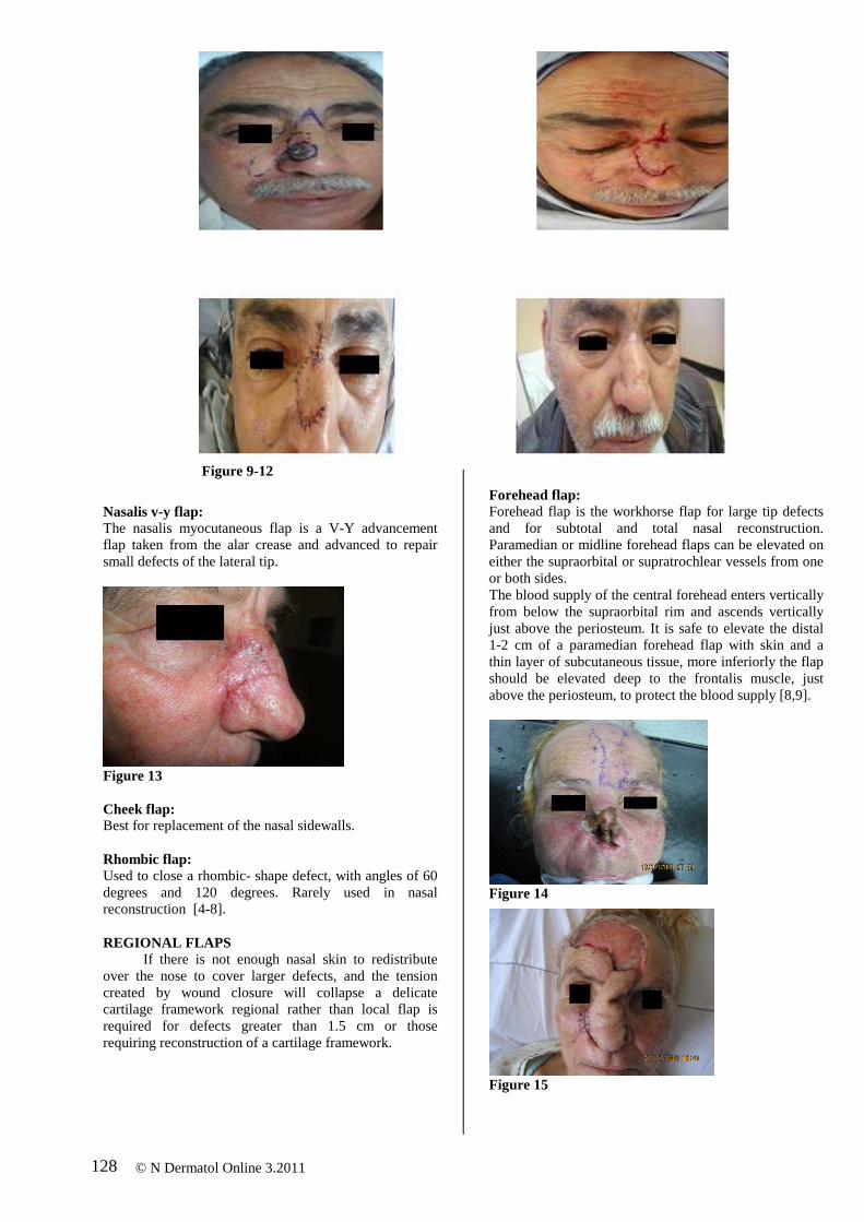
128
Figure 9-12
Nasalis v-y flap: The nasalis myocutaneous flap is a V-Y advancement flap taken from the alar crease and advanced to repair small defects of the lateral tip.
Figure 13 Cheek flap: Best for replacement of the nasal sidewalls. Rhombic flap: Used to close a rhombic- shape defect, with angles of 60 degrees and 120 degrees. Rarely used in nasal reconstruction [4-8]. REGIONAL FLAPS If there is not enough nasal skin to redistribute over the nose to cover larger defects, and the tension created by wound closure will collapse a delicate cartilage framework regional rather than local flap is required for defects greater than 1.5 cm or those requiring reconstruction of a cartilage framework.
Forehead flap: Forehead flap is the workhorse flap for large tip defects and for subtotal and total nasal reconstruction. Paramedian or midline forehead flaps can be elevated on either the supraorbital or supratrochlear vessels from one or both sides. The blood supply of the central forehead enters vertically from below the supraorbital rim and ascends vertically just above the periosteum. It is safe to elevate the distal 1-2 cm of a paramedian forehead flap with skin and a thin layer of subcutaneous tissue, more inferiorly the flap should be elevated deep to the frontalis muscle, just above the periosteum, to protect the blood supply [8,9].
Figure 14
Figure 15
© N Dermatol Online 3.2011

129
Figure 16
Figure 17 Nasolabial flap: Primarily used to reconstruct portions of the lobule and the nasal sidewall. the flap design is positioned just lateral to the nasolabial fold , the distal tip of the flap, situated at or just distal to the oral commissure ,on closure ,the scar lies exactly within the nasolabial crease. The flap can be pedicled superiorly or inferiorly, tunneled, made into an island, or turned over to create nasal lining, however, the blood supply is put under risk by folding, primary cartilage grafts are difficult to position, and the result usually appears bulky and thick. Tow stages are generally required for division and alar base inset. Excessive skin from the nasolabial fold also may be transferred in one stage as an extension of a random cheek flap; however, a second revision is usually required to recreate the normal alar crease [6-8].
Figure 18
Figure 19
Conclusions Nasal BCC in our study was more common in males 58.3% (21 patients) compared to females 41.7% (15 patients). The most common presentation was asymptomatic lesion 55.5% (20 patients), other presentations includes itching, local ulceration and bleeding. The most common nasal subunit affected by BCC was the ala 36.1%, followed by tip25%, sidewalls 22.3%, and dorsum 16.6%. The most common histopathological type of nasal BCC was the nodular BCC 55.5% followed by the sclerosing 22.2%, superficial 8.4%, basosquamous 8.4% and pigmented 5.5%. Complications included: hematoma in two cases, incomplete excision in two cases, and partial skin necrosis in one case.
Figure 20 REFERENCES / PIŚMIENNICTWO :
1. Fleming ILL, Amonette R, Monaghan T, Fleming MD: principles of management of basal and squamous cell carcinoma of the skin. cancer 1995; 75: 699-704. 2. Burget GC, Menick FJ: subunit principle in nasal reconstruction. Plast Reconstr Surg 1985; 76: 239-247. 3. Barton FE JR: aesthetic aspects of nasal reconstruction. Clin Plast Surg 1988; 15: 155-166. 4. Burget GC, Menick FJ: nasal reconstruction: seeking a fourth dimension. Plast Reconstr Surg 1986; 78: 145-157. 5. Burget GC: Aesthetic reconstruction of the tip of the nose. Dermatol Surg. 1995; 21: 419-429. 6. Deutsch HL, Orentreich N: Treatment of small external cancers of the nose. Ann Plast Surg 1979; 3: 567-571. 7. Baker SR, Swanson NA: Management of nasal cutaneous malignant neoplasms. Arch Otolaryngol 1983; 109: 473-478. 8. Langford J: Nasal Reconstruction Following Soft Tissue Resection. EMedicine.
9. Menic FJ: Nasal Reconstruction: forehrad flap. Plast Reconstr Surg 2004; 113: 100E.
© N Dermatol Online 3.2011
www.emedicine.medscape.com/article/880073

130
TUBERCULOSIS GANGLIONAR CON AFECTACIÓN CUTÁNEA (ESCROFULODERMIA) EN PACIENTE INMUNOCOMPETENTE. REPORTE DE UN CASO GANGLIONAR TUBERCULOSIS WITH SKIN INVOLVEMENT (SCROFULODERMA) IN AN INMMUNOCOMPETENT PATIENT. A CASE REPORT GANGLION GRUŹLICZY W SKÓRZE (SCROFULODERMA) U IMMUNOKOMPETENTNEGO PACJENTA. OPIS PRZYPADKU Martinez Braga Gabriela, Di Martino Ortiz Beatriz, Rodriguez Masi Mirtha, Knopfelmacher Oilda, Bolla de Lezcano Lourdes
Cátedra de Dermatología. Hospital de Clínicas de la Facultad de Ciencias Médicas de la Universidad Nacional de Asunción. Paraguay. [email protected]
N Dermatol Online. 2011; 2(3): 130-134 Date of submission: 14.02.2011 / acceptance: 21.03.2011 Conflicts of interest: None
Resumen La tuberculosis (TB) es la infección de mayor prevalencia en el mundo y su frecuencia continua aumentando, siendo en el Paraguay endémica. La enfermedad tuberculosa viene definida por la presencia de síntomas y signos clínicos en función de la localización de la enfermedad. La localización primaria más frecuente es la pulmonar. Sin embargo existen otras localizaciones menos frecuentes como la pleural, pericárdica, ganglionar, miliar, meníngea, osteo-articular, gastrointestinal, renal, pancreática, mamaria, cutánea, ocular y genitourinaria. Presentamos el caso de un paciente de sexo masculino, de 21 años de edad, inmunocompetente con tuberculosis extrapulmonar (ganglionar) con afectación cutánea secundaria (escrofulodermia). Abstract Tuberculosis (TB) infection is most prevalent in the world and its frequency continues to increase, being endemic in Paraguay. TB disease is defined by the presence of clinical symptoms and signs depending on the location of the disease. The most common primary site is the lung. But there are other less common locations such as pleural, pericardial, lymph nodes, miliary, meningeal, osteoarticular, gastrointestinal, renal, pancreatic, breast, skin, eye and genitourinary tract. We report the case of a 21 years old male patient, immunocompetent, with extrapulmonary tuberculosis (lymph node) with secondary skin involvement (scrofuloderma). Streszczenie Gruźlicze (TB) zakaŜenie jest najbardziej rozpowszechnione na świecie i jej częstotliwość nadal rośnie; jest chorobą endemiczną w Paragwaju. Zapadalność na gruźlicę jest definiowane przez obecność objawów klinicznych w zaleŜności od lokalizacji choroby. Najczęściej pierwotną lokalizacją są płuca. Ale są teŜ inne, mniej typowe lokalizacje, takie jak opłucna, osierdzie, węzły chłonne, postać prosówkowa, opon mózgowo-rdzeniowych, kostno-stawowa, przewodu pokarmowego, nerek, trzustki, piersi, skóry, oczu i układu moczowo-płciowego. Opisujemy przypadek 21 letniego pacjenta, immunokompetentnego, z gruźlicą pozapłucną (węzłów chłonnych), z wtórnym zajęciem skóry (scrofuloderma). Palabras clave: tuberculosis; extrapulmonar; escrofulodermia; inmunocompetente Key words: extrapulmonary; tuberculosis; scrofuloderma; immunocompetent Słowa klucze: gruźlica; pozapłucna; scrofuloderma; immunokompetencja
Original Articles / Prace Oryginalne
© N Dermatol Online 3.2011

131
Introduccion La tuberculosis extrapulmonar supone el 10 a
20% del total de TB que padecen los pacientes inmunocompetentes aunque esta frecuencia de presentación se incrementa notablemente en las personas portadoras de algún grado de inmunodeficiencia. Los enfermos con TB y VIH severamente inmunodeprimidos pueden presentar localizaciones extrapulmonares hasta en un 60% de los casos [1,2]. Estas formas son habitualmente de peor pronóstico que las formas pulmonares y de más difícil diagnóstico, con clínica insidiosa que puede demorar el diagnóstico hasta años, dado que no se sospechan. La confirmación bacteriológica se consigue sólo en el 60%. Sin embargo la ausencia de aislamiento microbiológico no excluye la TB, sobre todo en casos muy sugestivos y con prueba de tuberculina (PT) positiva (reacción de Mantoux) [3]. Caso clinico
Paciente de sexo masculino de 21 años de edad, que procede de medio urbano, sin patología de base conocida y sin antecedentes familiares de interés. Consultó por un cuadro de 2 años de evolución de tumoración en región cervical derecha con crecimiento progresivo y con fistulización, drenando material purulento de forma recurrente. Frotis y cultivos negativos para gérmenes comunes y hongos en varias oportunidades. Recibió tratamiento el primer año con trimetropim sulfametoxazol y azitromicina por 2 meses luego clindamicina más amikacina sin mejoría. Seis meses antes de la consulta presenta tumoración de similares características en axila derecha. El cuadro se acompaña de sensación febril nocturna y pérdida de peso de 5 kg desde el inicio. En la exploración física se aprecia un paciente consumido. Presenta tumoración eritematosa de limites netos bordes irregulares, adherida a planos profundos de 5 cm. de diámetro en axila derecha y lesiones similares en región cervical derecha de 1,5 cm. (Fig. 1,2,3,4). Análisis de sangre y orina dentro de parámetros normales excepto una eritrosedimentación de 120 mm en la primera hora (VSG). Serología para HIV, rubéola y citomegalovirus negativa. Baciloscopía seriada de esputo para BAAR negativa. Biopsia de lesión axilar informa: Ganglio linfático con proceso inflamatorio crónico granulomatoso. Tejido conectivo periganglionar con inflamación crónica granulomatosa. En ambos casos los granulomas son de tipo tuberculoide, centrados por células epitelioides y rodeados de abundante corona linfocitaria periférica. Se observan células gigantes multinucleadas. No se observó caseosis. Coloración de Ziehl Neelsen negativa para BAAR en ambos tejidos (Fig. 5,6). Radiografía de tórax: ensanchamiento hiliar. TAC de tórax: formación tumoral sólida bien delimitada en región cervical lateral derecha la cual se proyecta al tórax acompañado de ganglios acentuados de la cadena cervical del mismo lado, hilios pulmonares asimétricos con componente ganglionar bilateral. Con estos hallazgos y por las características clínicas, se efectúa el diagnóstico de tuberculosis extrapulmonar
(ganglionar) con afectacion cutanea secundaria (escrofulodermia). Se inicia tratamiento antibacilar con rifampicina 120 mg, pirazinamida 300 mg, isonicida 50 mg seis comprimidos al día por 2 meses y luego rifampicina 300 mg, isoniacida 150 mg dos comprimidos al día. Actualmente tras cuatro meses de tratamiento el paciente presenta mejoría importante de las lesiones con cicatrización de la de la región cervical y disminución del tamaño de la lesión en axila (Fig. 7,8).
Figura 1
Figura 2 Comentarios
Si exceptuamos la afectación tuberculosa pleural, la ganglionar es la más frecuente seguida de la urogenital y osteoarticular [2]. El compromiso ganglionar en el contexto de tuberculosis extrapulmonar aislada puede ocurrir en cualquier región de la economía. Los ganglios cervicales son los más afectados, alcanzando hasta el 5% de los casos en pacientes inmunocompetentes [4].
© N Dermatol Online 3.2011

132
Figura 3 Figuras 1,2,3,4. Clínica. Tumoración eritematosa de profundos de 5 cm. de diámetro en axila derecha y Figures 1, 2, 3,4. Clinic. Erythematous tumor of in the right axilla and similar lesions in right cervical
Figura 5. Histopatología ganglio linfático axilar. Linfadenitis granulomatosa Figure 5. Histopathology. Axillary lymph node with granulomatous lymphadenitis La presencia de una fístula secundaria a esta patología (escrófula) en un paciente sano como el que presentamos es verdaderamente infrecuente. Actualmente el 95% de los casos de tuberculosis son producidas por Mycobacterium tuberculosis y el 5% restante por mycobacterias atípicas, siendo éstas más frecuentes en pacientes inmunocomprometidos y en la infancia [5,6].
Figura 4
limites netos bordes irregulares, adherida a planos
lesiones similares en región cervical derecha de 1,5cm irregular edges, adhered to deep layers of 5 cm.
diameter region of 1.5 cm
Figura 6. Histopatología tejido conectivo axilar periganglionar y cutáneo. Proceso inflamatorio crónico granulomatoso, con granulomas de tipo tuberculoide con células gigantes multinucleadas Figure 6. Histopathology. Periganglionar axillary connective tissue and skin.- Granulomatous inflammatory process with tuberculoid granulomas with multinucleated giant cells La linfadenitis tuberculosa puede afectar todas las razas, siendo más frecuente la blanca. Presenta un ligero predominio en mujeres con una relación 2:1 según los estudios de Dandapat y cols., quienes sugieren que esto podría deberse a un estado nutricional más precoz en la mujer [7].
© N Dermatol Online 3.2011

133
Figura 7
Figura 8 Figuras 7,8. Evolución clínica. Tras cuatro meses de tratamiento mejoría importante de las lesiones con cicatrización de la de la región cervical y disminución del tamaño de la lesión axilar Figures 7,8. Clinical Outcome. After four months of treatment significant improvement of lesions with healing and decreased size of the injuries in the cervical and axillary regions La tasa de mortalidad por tuberculosis en la población general es del 20%; sin embargo no existen estadísticas que nos indiquen la tasa de mortalidad por linfadenitis tuberculosa [4]. La evaluación de un paciente con tuberculosis incluye en una buena historia clínica, exploración física, estudios radiológicos y microbiológicos. En la linfadenitis tuberculosa es impresindible la exploración física, un estudio epidemiológico y microbiológico, TAC, ecografía, PAAF y/o biopsia [8]. La radiografía de tórax es una prueba básica ya que permite descartar una lesión pulmonar como origen de la
patología ganglionar. Si bien hasta en el 84% de los pacientes no se encuentran cambios radiológicos [8]. La ecografía y la PAAF son las pruebas iniciales de elección para diferenciar una patología benigna de una maligna, con una sensibilidad del 92% y especificidad del 97% [9]. En opinión del Centro de Control de Enfermedades de Atlanta el diagnóstico de certeza se basa en el hallazgo de mycobacterium tuberculosis en cualquier muestra clínica. Sin embargo en los casos donde no es posible obtener la confirmación bacteriológica y se mantiene una fuerte sospecha clínica, la decisión de iniciar el tratamiento se debe basar en la clínica y estudios complementarios como en nuestro caso [10]. La presencia de síntomas sistémicos se presenta en un 34% de los casos e incluyen: pérdida de peso (63%), anemia (34%), malestar general (18%) y fiebre (41%) [11]; la media de duración entre el inicio de los síntomas y el diagnóstico es de 3 meses, pudiendo transcurrir entre 15 días y 36 meses [8]. El diagnóstico tardío de la patología puede llevar a la formación de abscesos y fístulas en un 22% de los casos, como sucedido en nuestro paciente [12]. El estudio anatomo-patológico de los ganglios afectados muestra múltiples granulomas formados por células epitelioides y células gigantes de Langhans. Los granulomas son confluentes, rodeados de linfocitos y fibroblastos [13]. En el diagnóstico diferencial se incluyen: patologías congénitas como linfangioma, hemangioma, quistes dermoides; patologías infecciosas bacterianas, virales, actinomicosis, tularemia, brucelosis, espiroquetas; procesos neoplásicos [4,5]. En el estudio de Morad y cols. sobre 212 pacientes diagnosticados de linfadenitis, el 45,3% tenía origen tuberculoso; 30,2% hiperplasia reactiva; 5,7% patología metastásica y 4,2% misceláneas [14]. El tratamiento de la linfadenitis primaria de origen tuberculoso es fundamentalmente médico con tratamiento antibacilar de 6 meses, con rifampicina, isoniacida, pirazinamida por 2 meses seguido de 4 meses con rifampicina e isoniacida [15]. Con esta terapia, a los 3 y 6 meses de tratamiento, el 40% y 82% de los nódulos reducen su tamaño a menos de 5 mm respectivamente. Los abscesos y senos mejoran tras el cuarto mes de tratamiento [8]. En algunos casos puede estar indicado el tratamiento quirúrgico cuando fracasa el tratamiento médico, ante grandes masas, recurrencias y manejo de fístulas [8,9].
BIBLIOGRÁFICAS / REFERENCES
1. Fanlo P, Tiberio G: Tuberculosis extrapulmonar. An. Sist. Sanit. Navar. 2007; 30 (Supl. 2): 143-162. 2. Menta JB, Dutt A, Harvill L, Mathews KM: Epidemiology of extrapulmonary tuberculosis. A comparative analysis with pre-AIDS era. Chest 1991; 99: 1134-1138. 3. Iichaurrga I, Herrejón A, Plaza P, Blanquer Olivas R: Tuberculosis miliar, ganglionar, pancreática y costal. Presentación clínica y revisión bibliográfica. An Med Interna 2001; 18: 483-485.
© N Dermatol Online 3.2011

134
4. Clearly KR, Batsaki JG: Mycobacterial disease of the head and neck: current perspective. Ann Otol Rhinol Laryngol 1995; 104: 830-833. 5. Manolidis S, Frenkiel S, Yoskovitch A: Mycobacterial infectionsof head and neck. Otolaryngol head Neck Surg 1997; 18: 202-205. 6. Ibekwe AO, Shareef Z, Kindy S: Diagnostic problems of tuberculous cervical adenitis (scrofula). Am J otolaryngol 1993; 109: 427-433. 7. Dandapat MC, Mishra BM, Dash SP, Kar PK: Peripheral lymph node tuberculosis: a review of 80 cases. Br J Surg 1990; 77: 911-912. 8. Jha BC, Dass A, Nagarkar NM, Gupta R, Singhal S: Cervical tuberculous lymphadenopathy: changing clinical pattern and concepts in management. Postgrad Med J 2001; 77: 185-187. 9. Cheung WL, Siu KF, Ng A: Tuberculous cervical abscess comparing the result of total excision against simple incision and drenage. Br J Surg 1988; 75: 563-564.
10. Center for Diseases Control and Prevention. Case definitions for public health surveillance. MMWR 1990; 39(RR-13): 40. 11. Zakirullah MT: Tuberculous cervical lymphadenopathy. J Postgrad Med Inst 2001; 15: 151-156. 12. Baatenburg de Jong RJ, Verwoerd CD, Van Overhagen H, Lameris JS, Knegt P: Ultrasound-guided fine-needles apiration biopsy of neck nodes. Arch Otoleryngol Head Neck Surg 1991; 117: 402-404. 13. Rosai J: Lymph nodes. En: Rosai and Ackerman´s surgical pathology. Edinburg. Mosby, 2004: 1877-2018. 14. Morad Na: Tuberculous cervical lymphadenopathy: should antituberculous therapy be preceded by histological proof ?. Trop Doc 2000; 30: 18-20. 15. Yuen APW, Wong SHW, Tam CM: Prospective randomized study of thrice weekly six-month and nine- month chemotherapy for cervical tuberculous lymphadenopathy. Otolaringol Head Neck Surg 1997; 116: 189-192.
© N Dermatol Online 3.2011

135
GRAM-NEGATIVE FOLLICULITIS. A RARE PROBLEM OR IS IT UNDERDIAGNOSED? CASE REPORT AND LITERATURE REVIEW GRAM-UJEMNE ZAPALENIE MIESZKÓW WŁOSOWYCH. RZADKI PROBLEM CZY RZADKO DIAGNOZOWANY? OPIS PRZYPADKU I PRZEGLĄD PIŚMIENNICTWA Sierra-Téllez Daniela1, Ponce-Olivera Rosa María1, Tirado-Sánchez Andrés1, Hernández Marco Antonio2, Bonifaz Alexandro2
1Dermatology Department, General Hospital of Mexico. 2Micology Section, Dermatology Department, General Hospital of Mexico. [email protected]
N Dermatol Online. 2011; 2(3): 135-138 Date of submission: 06.03.2011 / acceptance: 22.03.2011 Conflicts of interest: None
Abstract Gram-negative folliculitis may be the result of prolonged antibacterial treatments in patients with acne and rosacea. It is caused by alteration of facial skin flora and the nasal mucous, a decrease of Gram-positive bacteria and a proliferation of Gram-negative bacteria (for example Escherichia coli, Pseudomonas aeruginosa, Serratia marcescens, Klebsiella sp. and Proteus mirabilis). It should be considered in patients with acne who have not had a clinical improvement after 3-6 months of treatment with tetracyclines. The disease is underestimated, probably because bacteriological studies are rarely requested and the increased use of oral isotretinoin for acne management. One of the most effective treatments for Gram-negative folliculitis is oral isotretinoin (0.5-1 mg / kg / day for 4-5 months). We report the case of Gram negative folliculitis successfully treated with oral isotretinoin. Streszczenie Gram-ujemne zapalenie mieszków włosowych moŜe być wynikiem długotrwałego leczenia przeciwbakteryjnego u pacjentów z trądzikiem i trądzikiem róŜowatym. Jest to spowodowane zmianą flory bakteryjnej skóry twarzy i błony śluzowej nosa, zmniejszeniem ilości bakterii Gram-dodatnich i wzrost liczby bakterii Gram-ujemnych (na przykład Escherichia coli, Pseudomonas aeruginosa, Serratia marcescens, sp Klebsiella. i Proteus mirabilis). NaleŜy rozwaŜyć u pacjentów z trądzikiem, którzy nie mieli poprawy klinicznej po 3-6 miesiącach leczenia tetracyklinami. Częstotliwość tej choroby jest zaniŜona, prawdopodobnie dlatego, Ŝe badania bakteriologiczne rzadko są wykonywane i istnieje zwiększone uŜywanie doustnej izotretynoiny w leczeniu trądziku. Jednym z najbardziej skutecznych metod leczenia Gram-ujemnych zapaleń mieszków włosowych jest doustna izotretynoina (0,5-1 mg / kg mc. / dobę przez 4-5 miesięcy). Prezentujemy przypadek Gram-ujemnego zapalenia mieszków włosowych skutecznie leczonego doustną izotretynoiną. Key words: folliculitis; Gram-negative; acne; isotretinoin Słowa klucze: zapalenie mieszków włosowych; Gram-ujemne; trądzik; izotretynoina
Introduction Gram-negative folliculitis (GNF) is a hair follicle infection by Gram-negative organisms that can occur as a complication in patients receiving prolonged treatment with broad spectrum antibiotics for the treatment of acne vulgaris and rosacea. It must be suspected in a sudden exacerbation of acne treatment or in patients non-responding to conventional acne treatments [1]. There are two clinical variants of GNF; type I, is the most common, about 80% of cases, with the presence of multiple papules and pustules in the middle of the face;
the type II occurs in 20% of cases and is characterized by inflammatory nodules or cysts [1-4]. Oral isotretinoin is the treatment of choice at doses of 0.5 to 1mg/kg/day for 4-5 months [4]. Its mechanism of action is to control the proliferation of Gram-negative bacteria through microenvironmental changes produced in the skin and nasal mucous [5,6]. Case report
A 24-year old female came to our service with a skin disease that affects the perioral zone, characterized by multiple papules and pustules (Fig. 1A). Patient started one week before their assessment and with a
Original Articles / Prace Oryginalne
© N Dermatol Online 3.2011

136
history of having received tetracycline hydrochloride for 2 months for mild to moderate acne, as well as topical clindamycin intermittently for six months.
The patient underwent clinical examination and laboratory test such as Gram stain (Fig. 1C) and culturing on McConkey agar (Fig. 1D). A treatment with oral isotretinoin was proposed with resolution of the skin disease within a 3 month treatment period (Fig. 1B).
Figure 1. A. Perioral pustules. B. Post-treatment control. C. Gram-negative bacilli (x100). D. Lactose +, red-pink colonies (McConkey agar). Escherichia Coli
Discussion
Gram-negative folliculitis was first reported in 1968 by Fulton et al [1], in a group of patients with acne vulgaris resistant to conventional treatments [1-4]. It is a hair follicle infection that occurs mainly in patients with inflammatory acne or rosacea that have long treatments with broad spectrum antibiotics, mainly tetracycline [3-5]. It should be suspected when there is an increase in pustules with resistance to systemic treatment [4,6]. It is reported a prevalence of 4%. Prolonged treatment alters the normal bacterial flora of the nasal mucous and adjacent skin with reduced Gram-positive bacteria and coagulase positive aerobic diphtheroids, with an increase in Gram-negative bacilli mainly enteric bacteria [2-4].
Its characteristic features include: predominance in male gender, severe seborrhea, papules, pustules and perinasal and/or perioral involvement, recurrent folliculitis of the scalp, and prolonged antibacterial pretreatment, asymptomatic intervals tend to be shortened, acne and rosacea resistant to conventional treatment and isolation repeatedly of Gram-negative bacteria in cultures of pustules and facial nasal mucous [2-3]. Gram-negative folliculitis has been reported after eradication of recurrent staphylococcal pyodermas and prolonged treatment with topical antibacterials. Bartholow & Maibach [7], described a patient with acne who had been treated with topical clindamycin, followed by benzoyl peroxide and topical erythromycin and developed GNF due to E. coli. Fulton et al [1], described patients using antibacterial soaps, which selectively inhibit Gram-positive bacteria [8].
© N Dermatol Online 3.2011

137
Leyden et al [9], clinically differentiated two types of GNF. The type I, superficial or pustular, is the most common (80-90%), with presence of multiple papules and pustules in the middle of the face, 3 to 6 mm in diameter with an erythematous halo, mainly caused by E. coli, Klebsiella sp., Enterobacter sp. and P. aeruginosa. The type II, deep or nodular (10-20%), is characterized by deep and painfully inflammatory nodules or cysts, on the face, neck and/or chest, caused by Proteus mirabilis. Sebaceous follicles are colonized with these bacteria, mainly in the perioral and perinasal zone, with subsequent follicular and perifollicular inflammation with formation of papules and pustules [3,4,10,11]. Marples et al [12], confirmed an inverse ratio between nasal carriers of S. aureus and enterobacteria. The proportion of Gram-negative bacteria results in a 1% of the total flora under normal conditions. In the case of nasal carriers this ratio increases 3-4%, being nasal cavity the reservoir for facial cases of GNF [2,3]. The role of immune mechanisms of defense as a factor in the development of the GNF in patients with acne is not clear yet. Neubert et al [3], report that in addition to seborrhea and microflora changes induced by antibiotics, there are various mechanisms in the host immune defense, which appear to be an important factor in the GNF, induced by a depressed cell-mediated immunity, as evidenced by weak or absent response of hypersensitivity, which results in increased susceptibility to infections, showing a decrease of serum IgM, with a weak or absent response to enterobacterial antigens, similar events occur in patients with type V dysgammaglobulinemia (selective deficiency of IgM), which are liable to septicemia episodes by Gram-negative bacteria, and a deficiency in the complement system causing alterations in opsonization, chemotaxis and bacterial lysis. There is also evidence that chemotaxis of neutrophils may be inhibited by IgE-mediated histamine release, which is elevated in these patients.
It is well known that tetracyclines, the most commonly used systemic antibiotic in acne and rosacea, impair protein synthesis and function of lymphocytes and neutrophil chemotaxis, increasing the risk of bacterial infection [3,4]. The diagnosis should be confirmed with a smear of the pustules. The lesions (pustules) and the anterior nasal mucosa should be sampled for bacteriological studies. Treatment antibiotics must be withdrawn [4,10]. Oral isotretinoin is the treatment of choice [4-5,11,13]. Oral doses ranged from 0.5 to 1mg/kg/day for a period of 4-5 months, remission of the symptoms of facial and nasal colonization is achieved since the first three months of treatment [5]. The therapeutic success of isotretinoin in the GNF is due to decreased secretion of the sebaceous glands (thereby reducing the amount of sebum in more than 90%) and reduction of the follicular space size and anti-inflammatory effect, returning the environment uninhabitable for Gram-negative bacteria [5,8]. The most effective dose with a lower recurrence rate is 1mg/kg/day [4,11,13]. Systemic antibiotics such as cephalosporins combined with oral isotretinoin for 2 weeks are often useful on patients with frequent relapses [4]. Ampicillin and trimethoprim-sulfamethoxazole are reported with good results with remission of lesions in two weeks, with subsequent gradual reduction in dose. The clinical course determines the time of withdrawal, with remission rates ranged from 4 to 48 months [3] (Tab. I). Finally, we concluded the gram-negative folliculitis is an underdiagnosed disease, probably because the increased use of oral isotretinoin for acne management. We must suspected it when there is an exacerbation of centrofacial papules, pustules and/or nodules and cyst in patients with inflammatory acne or rosacea that have long treatments with broad spectrum antibiotics, non-responding to conventional treatments. We must request the bacteriological studies before initiating the oral isotretinoin.
Literature Reference
Patients Clinical Status Agent Treatment
Neubert et al 10 n= 46
Centrofacial folliculitis combined with open and/or closed comedon.
Klebsiella spp., Escherichia coli, Enterobacter spp., and Proteus spp.
Isotretinoin, mean duration 18.6 weeks, mean total dosage 109 mg/kg.
James et al 5
n=21
Patients with nodulocystic acne with several facial pustules resistant to all therapies.
Escherichia aerogenes.
Isotretinoin 0.48-0.74 mg/kg/day for 20 weeks.
© N Dermatol Online 3.2011

138
n=11
Patients with nodulo-cystic acne with several facial pustules resistant to all therapies.
Escherichia aerogenes, Proteus mirabilis, Klebsiella pneumoniae, E. coli and S. marcescens.
Isotretinoin 1mg/kg/day for 5 months.
n=35 Folicular pustules grouped around the nose.
Lactose fermenting Gram-negative rods often Klebsiella, Enterobacter.
Ampicillin 1gr/day for 7-14 days, lowered to a maintenance level 250mg twice daily.
Leyden et al 9
n=15 Deep, nodular and cystic lesions.
Proteus. Ampicillin 1gr/day for 7-14 days, lowered to a maintenance level 250mg twice daily.
Eady et al 13 n= 8 Non-responding acne patients.
Gram-negative rods. Trimethoprim or co-trimoxazole.
Presented Case
n=1 Perioral pustules with acne resistant to all therapies.
Escherichia coli. Isotretinoin, 0.3mg/kg/day for 10 months.
Table 1. Summary of Gram-negative Folliculitis. Case Reports REFERENCES / PIŚMIENNICTWO :
1. Fulton JE Jr, McGinley K, Leyden J: Gram-negative folliculitis in acne vulgaris. Arch Dermatol 1968; 98: 349-53. 2. Blankenship ML: Gram-negative folliculitis. Follow-up observations in 20 patients. Arch Dermatol 1984; 120: 1301-1303. 3. Neubert U, Jansen T, Plewig G: Bacteriologic and immunologic aspects of gram-negative folliculitis: a study of 46 patients. Int J Dermatol 1999; 38: 270-274. 4. Böni R, Nehrhoff B: Treatment of gram-negative folliculitis in patients with acne. Am J Clin Dermatol 2003; 4: 273-276. 5. James WD, Leyden JJ: Treatment of gram-negative folliculitis with isotretinoin: positive clinical and microbiologic response. J Am Acad Dermatol 1985; 12: 319-324. 6. Ruocco E, Donnarumma G, Baroni A, Tufato M: Bacterial and viral skin diseases. Dermatol Clin 2007; 25: 663-676. 7. Bartholow P, Maibach HI: Gram-negative folliculitis without systemic antibiotics?. Arch Dermatol 1979; 115: 676.
8. Harkaway KS, McGinley KJ, Foglia AN, Lee WL, Fried F, Shalita AR, et al. Antibiotic resistance patterns in coagulase-negative staphylococci after treatment with topical erythromycin, benzoyl peroxide and combination therapy. Br J Dermatol 1992; 126: 586-590. 9. Leyden JJ, Marples RR, Mills OH Jr, Kligman AM: Gram-negative folliculitis-a complication of antibiotic therapy in acne vulgaris. Br J Dermatol 1973; 88: 533-538. 10. Neubert U, Plewing G, Ruhfus A: Treatment of gram-negative folliculitis with isotrenoin. Arch Dermatol Res 1986; 278: 307-313. 11. Chivot M. Residual acne lesions after treatment. Ann Dermatol Venereol 1996: 123: 594-600. 12. Marples RR, Fulton JE, Leyden J, McGinley KJ: Effect of antibiotics on the nasal flora in acne patients. Arch Dermatol 1969; 99: 647-651. 13. Eady EA, Cove JH, Blake J, Holland KT, Cunliffe WJ: Recalcitrant acne vulgaris. Clinical, biochemical and microbiological investigation of patients not responding to antibiotic treatment. Br J Dermatol 1988 Mar; 118: 415-423.
© N Dermatol Online 3.2011

139
GRAM-NEGATIVE FOLLICULITIS. A RARE PROBLEM OR IS IT UNDERDIAGNOSED? CASE REPORT AND LITERATURE REVIEW Sierra-Téllez Daniela, Ponce-Olivera Rosa María, Tirado-Sánchez Andrés1, Hernández Marco Antonio, Bonifaz Alexandro
Professor Antonio Chuh
I congratulate the authors for having documented an unusually severe case of Gram-negative folliculitis (GNF) necessitating systemic retinoid therapy. Marked clinical remission was seen at three months. I wonder whether there was a clinical relapse in the few months after cessation of systemic retinoids. The epidemiology, pathophysiology, symptomatology, and management of GNF were well reviewed by the authors. I entirely agree with them that this condition is under-diagnosed and under-treated. The incidence is unknown as we lack the denominator to start with.
For patients with flares of acne vulgaris or rosacea, GNF should be considered. I have been seeing patients with perioral and periorbital dermatitis exacerbated by GNF. A descriptive study for GNF for this group of patients would be highly worthwhile. In patients with nodulocystic acne, open- and closed-comedones could be mistaken for GNF. For these patients, we have found that digital epiluminescence dermatoscopy can be of much diagnostic assistance. We hope to document such images when we see further patients with GNF captured digitally to be submitted as case reports.
Antonio Chuh Adjunct Associate Professor School of Public Health The Chinese University of Hong Kong
Professor Mehmet Doganay
Folliculitis is a typical pyoderma located within hair follicles and apocrine regions. Hair follicles can become inflamed by physical injury, chemical irritant or infection that leads to folliculitis. The lesions are characterized with a small, erythematous and sometimes pruritic papules by central pustule and fine surrounding collar of desquamation. It may be deep seated or superficial. The most common form is superficial folliculitis that manifest as a tender or painless pustule that heals without scarring. The lesion may be appearing as single or multiple on the skin bearing hair including the head, neck, trunk, buttocks and extremities. Fever or associated systemic symptoms rarely exist. Folliculitis sometimes can progress to form subcutaneous abscess (furuncles) or carbuncle (1,2). Staphylococcus aureus is the usual cause of folliculitis. Gram-negative bacteria and fungi are less frequently responsible from folliculitis. Among gram-negative bacteria, Klebsiella spp. Escherichia coli, Enterobacter spp. , Proteus spp and Pseudomonas spp. are more frequently isolated (2,3). The face is generally involve in gram-negative folliculitis and the majority of
patients have a history of long-term antibiotic therapy for acne. It can be treated with isotretinoin, but it should not be forgotten of the side effect, including birth defect ( 2). In this issue of Our Dermatology Online Journal, Sierra-Téllez Daniela and et al (4) reported a case with E. coli folliculitis on the face The history of patient , clinical features and etiologic agent are well described in this case report. The patient’s lesion picture and demonstration of etiological agent in microscopy and on bacteriologic media are very educative materials for young physicians. The patient had also received tetracycline hydrochloride for 2 months for mild to moderate acne, as well as topical clindamycin intermittently for six months. The data for the combination with topical treatments (topical benzoyl peroxide or retinoids) suggest synergistic effects. In this case the combined use of topical and systemic antibiotics is not suitable for acne treatment. A healing was obtained in this case with the therapy of oral isotretinoin.
This paper shows that in a resistant to conventional therapy in cases with acne, some rare etiological agents should be considered.
Comments to the article
© N Dermatol Online 3.2011

140
REFERENCES / PIŚMIENNICTWO :
1. Pasternack M S, Swartz MN. Cellulitis. Necrotizing fasciitis and subcutaneous tissue infections. In: Mandell GL, Bennett J, Dolin R (Editors). Principless and Practice of Infectious Diseases, 7th edition, Philadelphia: Churchill Livingstone-Elsevier, 2010:1289-1312. 2. Stulberg DL. Penrod MA, Blatny RA. Common bacterial skin infections. Am Fam Physician 2002; 66:119-124. 3. Neubert U, Jansen T, Plewing G. Bacteriologic and immunologic aspects of Gram-negative folliculitis: a study of 46 patients. Int J Dermatol 1999; 38: 270-274. 4. Gram-negative folliculitis. A rare problem or is it under diagnosed? Case report and literature revıew. N Dermatol Online. 2011; 3(2): 134-137. Mehmet Doganay, M.D. Professor in Infectious Diseases Department of Infectious Diseases, Faculty of Medicine,Erciyes University, 38039- Kayseri / Turkey Email. [email protected]
© N Dermatol Online 3.2011

141
ERYTHEMATODES CHRONICUS PROFUNDUS AS DERMATOLOGY, SURGERY AND COSMETOLOGY PROBLEM ERYTHEMATODES CHRONICUS PROFUNDUS JAKO DERMATOLOGICZNY, CHIRURGICZNY I KOSMETOLOGICZNY PROBLEM Alendar Faruk, Soskic Samra, Helppikangas Hana, Gavrankapetanovic Alma, Alendar Temeida
Dermatovenerology Department Clinical Center University of Sarajevo, Bosnia & Herzegowina [email protected]
N Dermatol Online. 2011; 2(3): 141-143 Date of submission: 30.03.2011 / acceptance: 22.04.2011 Conflicts of interest: None
Abstract Lupus profundus is a rare skin condition originally described by Kaposi in 1883. It is a clinical variant of LE in which the deep dermis and subcutaneous fat are predominantly affected. A 38-year-old female presented with history of development of well-circumscribed patches of hair loss since 1994 year. In 2004 on the skin of left cheek she noticed a single erythematous plaque on which place later became dint of subcutaneous tissue. Today she has massive defects across whole scalp, right cheek and the upper part of left arm with multiple ulcers(fig.3and4). case reposrt, laboratory examinations, skin biopsy and influence of corticosteroids on further progression /regression of this disease. Streszczenie Lupus Profundus jest rzadkim schorzeniem skóry pierwotnie opisanym przez Kaposiego w 1883 roku. Jest to wariant kliniczny LE, w którym zmiany zlokalizowane są w przewaŜającym stopniu głęboko w skórze właściwej i tkance podskórnej. 38-letnia chora zgłosiła się z historią rozwiniętych, ograniczonych plackowatych ognisk utraty włosów od 1994 roku. W 2004 r. na skórze lewego policzka zauwaŜyła pojedynczą rumieniową zmianę, w której miejscu później powstało wgłębienie do tkanki podskórnej. Obecnie pacjentka z rozległymi ubytkami na całej skórze głowy, policzkach i w górnej prawej części lewego ramienia, z wieloma owrzodzeniami. Opisano przypadek kliniczny, wykonane badania laboratoryjne, diagnostyczne oraz wpływ kortykosteroidów na dalszy rozwój / regresję choroby. Key words: lupus profundus; chloroquin; corticosteroids Słowa klucze: lupus profundus; chlorochina; kortykosteroidy
Introduction
Lupus profundus is a rare skin condition originally described by Kaposi in 1883 and later by Irgang in 1940. It is a clinical variant of LE in which the deep dermis and subcutaneous fat are predominantly affected. It may occur on its own or in association with discoid lupus erythematosus (DLE) or SLE. In cases associated with SLE, it may precede SLE by several years. In the majority of cases, it tends to have a mild chronic course marked by recurrent nodules or plaques. Case report
A 38-year-old female presented with history of development of well-circumscribed patches of hair loss
since 1994. year. The hair loss was rapid and asymptomatic, with no hair re-growth. There was no family history of any autoimmune rheumatic diseases. In 2004. she was first time hospitalised on our clinic. Clinical symptoms were: on the skin of left cheek a single, erythematous plaque (Fig. 1). On left arm two small ulcers size 1 x 2 cm, oval in shape and firm in consistency (Fig. 2). Since than she was treated with chloroquin in dose 250mg per day with gradually reduction, but with no results. Today she have massive defects across whole scalp, right cheek and the upper part of left arm (Fig. 3,4).
Original Articles / Prace Oryginalne
© N Dermatol Online 3.2011

142
Figure 1. On the skin of left cheek a single, Figure 2. On left arm two small ulcers size 1 x 2 cm, erythematous plaque oval in shape and firm in consistency
Figure 3. Massive defects across whole the upper Figure 4. Massive defects across whole scalp, right cheek part of left arm
The lesion, on the left arm in 2004. initially, was about 1 Χ 2 cm 2 in size, oval in shape and firm in consistency. It gradually increased in size to about 8 Χ 5 cm 2, in 20l0. with multiple ulcers colonized with Staphilococcus aureus,and with extensive atrophy of cutaneus and subcutaneous tissue. There was no history of preceding trauma. No history of taking injection. She denied arthralgia, arthropathy, myalgia, fatigue, fever, Raynaud's phenomenon and gastrointestinal symptoms. She notifies pain in right lumbal area, and the ultrasound showed signs of bilateral nephropathy. The following laboratory tests were done: complete blood cell count, differential cell count, ESR was 25, total serum proteins 86.0, with A:G ratio 1.0,y globulins were increased 0.27. Blood sugar, blood urea, serum creatinine, serum electrolytes, liver function tests and urinalysis were in normal rates. X-ray chest was normal. Rheumatoid factor was normal,but antistreptolysin was 221,0IU/ml. C3 1.090 g/l, C4 0.l65 g/l, ANA positive, Anti ds DNA negative and CiC was increased and his amount was 58.o8IU/ml.
Skin biopsy showed lymphocytic panniculitis, hyaline degeneration of the fat, hyaline papillary bodies, and lymphoid nodular structures in the lower dermis and subcutaneous tissue. Result is in high correlation with diagnosis of erytematodes cronicus profundus. In 2010. we treated her with systemic corticosteroid therapy in dose of 60mg per day with gradually reduction of dose. After 2 month of therapy further progression has stoped (Fig. 5,6). Her maintence dose today is 20 mg prednisolon/day. Discussion
Lupus erythematosus profundus or panniculitis is an unusual but distinct clinical variety of lupus erythematosus. The inflammatory reaction in takes place primarily in the deep corium and the subcutaneous tissues leading to deep indurated nodules or sharply defined plaques. The overlying skin usually appears normal but there may be erythema, atrophy, ulceration or poikilodermatous or hyperkeratotic changes.
© N Dermatol Online 3.2011

143
Figure 5. Lesions after 2 month of therapy
Figure 6. Lesions after 2 month of therapy
The lesions are most frequent on cheeks but other sites of predilection are face, upper arms, hands, chest, buttocks and thighs. It may develop in association with discoid lupus erythematosus or systemic lupus erythematosus or may occur as an isolated phenomenon. The differential diagnoses include panniculitis due to other connective tissue disorders like dermatomyositis or scleredema and Weber Christian panniculitis or Jessner's lymphocytic infiltration, lyrnphocytoma cults and sarcoidosis. Lupus panniculitis often responds to treatment with antimalarials, such as hydroxychloroquine (200 mg once
or twice a day). Some cases respond to a combination of antimalarials (for example, hydroxychloroquine 200 mg and quinacrine 100 mg daily) when monotherapy is ineffective. Systemic glucocorticoids should be reserved for widespread and resistant lesions. Intralesional glucocorticoids are usually ineffective and may exacerbate the atrophic healing process. Success with dapsone, azathioprine, and thalidomide has been described in isolated case reports. Surgical debridement or resection of individual lesions may be attempted when all other modalities have failed and there is appreciable debilitation. Conclusion
Progress of Erythematodes profundus is unpredictable. As we can see on example of this patient therapy with cloroquine was insufficient to stop progression of disease. With dose of 60 mg methylprednisolon there was no more further progression.Today, after 8 months of last hospitalisation we have not noticed any progression. Defects on skin with which she needs to live daily, are horrifying, what caused her phsychiatric problem and antidepresive therapy. Today she stay as dermatology, surgery and cosmetology problem. REFERENCES / PIŚMIENNICTWO :
1. Tuffanelli DL: Lupus erytheratosus panniculitis (profundus). Arch Dermatol 1971; 103: 231-242. 2. Black MM, Cunliffe WJ: Inflammatory disorders of subcutaneous fat. In: Champion RH, Burton JL, Burns DA, et al., editors. Textbook of Dermatology. London: Blackwell Science LTD; 1998. 3. Braun-Falko O,Wolf HH: Dermatology and venerology, New York; 1997. 4. Sanchez NP, Peters MS, Winkelmann RK: The histopathology of lupus erythematosus panniculitis. J Am Acad Dermatol. 1981; 5: 673-680. 5. Ahmed I, Ahmed D. Lupus erythematosus panniculitis: a unique subset within the lupus erythematosus spectrum. Am J Dermatopathol. 2000; 22: 352. 6. Kündig TM, Trüeb RM, Krasovec M: Lupus profundus/panniculitis. Dermatology. 1997; 195: 99-101. 7. Diaz-Jouanen E, DeHoratius RJ, Alarcon-Segovia D, Messner RP: Systemic lupus erythematosus presenting as panniculitis (lupus profundus) Ann Intern Mes. 1975; 82: 376-379. 8. Masood Q, Manzoor R: Lupus erythematosus profundus (panniculitis) J K Practioner. 1995; 2: 135–136.
© N Dermatol Online 3.2011

144
GRANULOSIS RUBRA NASI – A CASE REPORT. A LITERATURE REVIEW GRANULOSIS RUBRA NASI – OPIS PRZYPADKU. PRZEGLĄD LITERATURY Brzeziński Piotr1, Poklękowska Katarzyna2
16th Military Support Unit, Ustka, Poland, [email protected] 2Mazowiecki Branch of the National Health Fund, Warsaw, Poland
N Dermatol Online. 2011; 2(3): 144-146 Date of submission: 21.02.2011 / acceptance: 21.03.2011 Conflicts of interest: None
Abstract Granulosis Rubra Nasi (GRN), a rare childhood dermatitis that was also defined as "Acne papulo-rosacea of the nose". Is an inflammatory dermatosis which consists on erythema, papules and itch. The hyperhidrosis the most conspicuous features of the disease and small beads of sweat and erythema on the tip of the nose may often occur. Sometimes small red papules and comedo-like lesions may be present. Etiology is unknown. lt's uncommon, chronic process and benign course. Usually subsides spontaneously at adolescence. The treatment is symptomatic and cosmetic. An 14-year-old boy presented with erythematous lesions over the nose of childhood duration without sensitivity to sunlight. Physical examination showed also excessive sweating of the nose, and erythema of the nose covered by small pustules, papules ang macules. The patient responded well to low doses of oral Isotretinoine. Should remember GRN could be a complication of hyperhidrosis. Streszczenie Granulosis Rubra Nasi (GRN), rzadkie zapalenie skóry w dzieciństwie, które zostało równieŜ określony jako "trądzik grudkowo-róŜowaty nosa". Jest to zapalna choroba skóry, która polega występowaniu rumienia, grudek i świądu. Nadmierne pocenie się jest najbardziej wyrazistą cechą choroby, a małe krople potu i rumień na czubku nosa często mogą wystąpić. Czasami mogą być obecna małe czerwone grudki i zmiany zaskórnikopodobne. Etiologia jest nieznana. Często ma przewlekły i łagodny przebieg. Zwykle ustępuje samoistnie w wieku dojrzewania. Stosuje się leczenie objawowe i kosmetyczne. 14-letni chłopiec zgłosił się z rumieniowymi zmianami na nosie trwającymi od dzieciństwa, bez wraŜliwość na światło słoneczne. Badanie lekarskie wykazało równieŜ nadmierne pocenie się na nosie, rumień w nosie w obrębie drobnych krostek, grudek i plamek. Pacjent dobrze zareagował na małe dawki doustnej izotretinoiny. NaleŜy pamiętać, Ŝe GRN moŜe być powikłaniem nadmiernej potliwości. Key words: granulosis rubra nasi; dermatosis; hyperhydrosis; izotretinoin Słowa klucze: ziarnistość czerwona nosa; choroby skóry; nadpotliwość, izotretinoina
Introduction The first case Granulosis Rubra Nasi (GRN) was described in 1901 by German dermatologist Josef Jadasson as "Nasi hyperhidrotic Erythematosa micropapules Dermatosis Infantum” [1]. Some footnotes Italian treat of the first case described by Luthen in 1900 [2]. The present accepted name we owe to Jadassohn. The malady is usually limited to the nose, to the front and sides, in addition to involving this part, it has been observed to affect also the upper lip, cheek, and eyebrow. A case report
14 years old boy was adopted in the Dermatology surgery of changes in the nasal skin under way since childhood, with periodic remissions and exacerbations. So far, no treatment [fig. 1].
Wstęp Pierwszy przypadek Granulosis Rubra Nasi
(ziarnistość czerwona nosa) (GRN) został opisany w 1901 roku przez niemieckiego dermatologa Josef Jadasson jako "Nasi hyperhidrotic Erythematosa micropapules dermatozy infantum" [1]. Niektóre włoskie przypisy traktują o pierwszym przypadku opisanym przez Luthena w 1900 roku [2]. Obecnie przyjętą nazwę zawdzięczamy Jadassohnowi. Choroba jest zwykle ograniczona do nosa, z przodu i po bokach, wyjątkowo, zaobserwowano zmiany nad górną wargą, na policzkach, w brwiach. Opis przypadku
Chłopiec lat 14 został przyjęty w Poradni Dermatologicznej ze zmianami na skórze nosa trwającymi od dzieciństwa, z okresowymi remisjami i zaostrzeniami. Do tej pory nie leczony [fig. 1].
Original Articles / Prace Oryginalne
© N Dermatol Online 3.2011

145
Figure 1. Granulosis Rubra Nasi to 14-year-old boy Nose leather on the front and sides were inflamed. Redness gradually disappeared to the sides. On the skin of nose could be seen a small red specs and papules. The papules gradually develop into pustules lesions. On the sides can often be seen drops of sweat, which gave the appearance of moist, slightly shiny lesions. Lesions without sensitivity to sunlight. In addition, generally healthy. The patient was treated initially with liquid zinc, has no effect. After application of 20mg oral Isotretinoine, after months skin lesions regressed, erythema decreased. Also decreased sweating in the area. Due to side effects (excessive drying and cracking of the lips) and the price of the drug the boy's mother decided to leave after 2 months of medication. This resulted in recurrence of symptoms and to obtain a definitive starting point. Discusion
GRN is rare, and the precise frequency is unknown. Most occurrences of granulosis rubra nasi are genetically determined, with an autosomal dominant or autosomal recessive pattern. [3]. The gene locus has not been identified. Just as in the analyzed case in GRN usually starts in early childhood in patients aged 6 months to 10 years [4]. Men suffer more. 7of 6 patients described by Jadassohna were boys [1], Mendoza described case of 10-year-old boy [5] and Akhdari case of a 18-year-old boy [6]. Initially, hyperhidrosis is the most conspicuous feature of the disease. Small beads of sweat can be seen on the tip of the nose. With persistent hyperhidrosis, diffuse erythema develops on the tip of the nose. Erythema gradually extends and may involve cheeks, upper lip, and chin. Erythema is covered by small beads of sweat. Can be seen small erythematous macules, erythematous papules, and vesicles or pustules lesions [6-9]. Many affected patients have poor peripheral circulation and hyperhidrosis of palms and soles [5,10]. GRN usually resolves spontaneously at puberty, but sometimes it lasts longer. Pathogenesis is unknown. Granulosis rubra nasi is a disorder of the eccrine glands, possibly representing a unique form of sweat retention [6,9].
Skóra nosa z przodu i po bokach była zaczerwieniona. Zaczerwienienie stopniowo zanikało ku bokom. Na skórze nosa widać było małe plamki i grudki barwy czerwonej. Grudki stopniowo przekształcały się w zmiany krostkowe. Po bokach często zauwaŜyć było moŜna kropelki potu, co dawało wygląd wilgotnych, lekko lśniących zmian. Zmiany nie wykazywały wraŜliwości na światło słoneczne. Chłopiec podawał wzmoŜona potliwość dłoni. Nadmierna potliwość w innych okolicach nie występowała. Poza tym chłopiec ogólnie zdrowy. W leczeniu zastosowano początkowo płyn z cynkiem, bez efektu. Po zastosowaniu 20mg doustnej izotretinoiny, po miesiącu zmiany skórne ustępowały, rumień zmniejszył się. Zmniejszyła się równieŜ nadmierna potliwość w tej okolicy. Ze względu na objawy niepoŜądane (nadmierne wysychanie i pękanie warg) oraz cenę leku matka chłopca postanowiła po 2 miesiącach odstawić lek. spowodowało to nawrót objawów i ostateczne uzyskanie punktu wyjściowego. Dyskusja
GRN jest rzadkim schorzeniem, a dokładna częstotliwości choroby nie jest znana. Większość wystąpień GRN są uwarunkowane genetycznie, o dziedziczeniu autosomalnym dominującym lub autosomalnym recesywnym [3]. Locus genu nie został zidentyfikowany. Podobnie jak w analizowanym przypadku GRN rozpoczyna się zwykle we wczesnym dzieciństwie u pacjentów w wieku od 6 miesięcy do 10 lat [4]. Częściej chorują męŜczyźni. 7 z 6 opisanych pacjentów Jadassohna to chłopcy [1], Mendoza przedstawia przypadek 10-letniego chłopca [5], a Akhdari przypadek 18-letniego chłopca [6]. W listach do redakcji Willams i Goldsmith równieŜ przedstawiają przypadki GRN u chłopców [7,8]. Początkowo nadmierna potliwość jest najbardziej widoczną cechą choroby. Małe kropelki potu moŜna zauwaŜyć na czubku nosa. Z utrzymującą się nadmierną potliwością rozwija się rozlany rumień zwykle na czubku nosa. Rumień stopniowo rozszerza i moŜe obejmować policzki, wargę górną i brodę. Rumień jest objęty małymi kroplami potu. W obrazie klinicznym mogą być widoczne małe plamy rumieniowe, rumieniowe grudki, pęcherzyki lub krosty [6-9]. Wielu pacjentów ma słabe krąŜenie obwodowe oraz nadmierną potliwość dłoni i stóp [5,10]. GRN zwykle samoistnie ustępuje w okresie dojrzewania, jednak czasem utrzymuje się dłuŜej. Patogeneza nie jest znana. GRN jest zaburzeniem gruczołów ekrynowych i prawdopodobnie stanowi unikalną formę retencji gruczołów potowych [6,9]. Występowanie znacznego wzrost produkcji potu na nosie oraz nadmierne pocenie się obecne równieŜ w centralnej części twarzy, dłoni, i stóp, jest odpowiedzialne za drugorzędowe zmiany rumieniowe: rumień i rumieniowe grudki. W diagnostyce róŜnicowej naleŜy wziąć pod uwagę rumieniowe grudkowo-krostkowe dermatozy twarzy, szczególnie trądzik pospolity i róŜowaty, w których nie występuje nadmierne pocenie się, fotodermatozy, które są bardziej rozległe z brakiem pocenia się, poza tym toczeń rumieniowaty układowy, lupus vulgaris,
© N Dermatol Online 3.2011

146
The occurrence of a significant increase in the production of sweat on his nose and hyperhidrosis is present on the central face, palms, and soles; appears to be responsible for the secondary changes of erythema and erythematous papules. In differental diagnosis be taken into account: erythematous papulopustular dermatosis of the face, particularly acne vulgaris and rosacea, which do not have excessive sweating, photodematosis, which are more extensive with the absence of hyperhidrosis, otherwise lupus erythematosus, lupus vulgaris and leishmaniasis, actinic keratosis or skin cancer. However, evidence of chronic skin damage in childhood are rare (excluding specific genodermatosis) [5,9,11]. Pinkus and Lebet and give a possibility of coexistence GRN with hydrocystoma, also in one of case reports by Jadassohna been a few changes hydrocystoma [12,13]. MacLeod describes the symptoms of the disease in hot weather. Heid describes a case of 19-year-old woman with hyperhidrosis, GRN, tachycardia and pheochromocytoma [12]. After surgical removal of pheochromocytoma was sweating involution and regression of the GRN. The lesions usually disappear during puberty. Treatment is symptomatic and cosmetic. Brazilian authors have used in the treatment of botulinum toxin type A [2]. Conclusion
Granulosis Rubra Nasi is a rare disorder. Should remember GRN could be a complication of hyperhidrosis. In the treatment of low doses Isotretinoine can be used with good effect.
lejszmaniozę, rogowacenie słoneczne lub raka skóry. Jednak dowody przewlekłego uszkodzenia skóry w dzieciństwie są rzadkie (wykluczając swoiste genodermatozy). [5,9,11]. Lebet i Pinkus podają moŜliwość współistnienia GRN z hydrocystoma, równieŜ w jednym z opisów przypadków przez Jadassohna było kilka zmian typu hydrocystoma [12,13]. MacLeod opisuje nasilenie objawów choroby w czasie upałów. Heid opisuje przypadek 19-letniej kobiety z nadmierną potliwością, GRN, tachykardią oraz guzem chromochłonnym [12]. Po chirurgicznym usunięciu guza chromochłonnego nastąpiła inwolucji nadmiernego pocenie się i regresji GRN. Zmiany zwykle ustępują w okresie dojrzewania. Leczenie jest objawowe i kosmetyczne. Autorzy brazylijscy w leczenie GRN stosowali toksynę botulinową [2]. Podsumowanie Granulosis Rubra Nasi to rzadkie schorzeniem. NaleŜy pamiętać, Ŝe GRN moŜe być powikłaniem nadmiernej potliwości. W leczeniu moŜna stosować niskie dawki izotretinoiny z dobrym efektem.
REFERENCES / PIŚMIENNICTWO :
1. Jadasson J. Granulosis Rubra Nasi. Archiv Dermatol und Syph 1901: 58:145-58. 2. Grazziotin TC, Buffon RB, da Silva Manzoni AP, Libis AS, Weber MB: Treatment of granulosis rubra nasi with botulinum toxin type A. Dermatol Surg. 2009; 35: 1298-1299. 3. Grinoni F. Contributo clinico allo studio dell'etiopatogenesi della Granulosis Rubra Nasi. G Dermatol Sif. 1955; 96: 227. 4. Zuccati G, Filippeschi C, Mastrolorenzo A, Rapaccini AL, Tiradritti L, Staderini C: Granulosis rubra nasi. G Ital Dermatol Venereol. 1990; 125: 275-276. 5. Mendoza JP, Saldaña LS, Yokota ARRPL, Sialer MC, Anduaga ES: Granulosis rubra nasi. Dermatol. Peru. 2003; 13: 125-127. 6. Akhdari N. Granulosis rubra nasi. Int J Dermatol. 2007; 46: 396. 7. Williams DI: Granulosis rubra nasi. Proc R Soc Med. 1947; 499: 37 8. Goldsmith WM: Granulosis rubra nasi (Jadassohn). Proc R Soc Med. 390: 20 9. Bocian M, Bettina P, García R, Laterza A y col. Granulosis Rubra Nasi. A propósito de dos casos. Arch Arg Dermatol 2003: 53: 171-3. 10. Raymond GP, Delgrange-Delcourt T, Tétrault C: Granulosis rubra nasi: report of a new case. Union Med Can. 1978; 107: 800-801. 11. Brody M: Granulosis rubra nasi. Br J Dermatol Syph. 1947; 59: 380. 12. Heid E, Samain F, Jelen G, Boivin S: Granulosis rubra nasi and pheochromocytoma. Ann Dermatol Venereol. 1996; 123: 106-108. 13. Winkelried W: Case of granulosis rubra nasi. Proc R Soc Med. 1915: 20
© N Dermatol Online 3.2011

147
MYCETOMA REVISITED NOWE SPOJRZENIE NA MYCETOMA Hassan Iffat, Keen Abid
Postgraduate Department of Dermatology, STD & Leprosy Govt. Medical College & Associated SMHS Hospital, Srinagar-Kashmir, India, [email protected]
N Dermatol Online. 2011; 2(3): 147-150 Date of submission: 25.02.2011 / acceptance: 06.03.2011 Conflicts of interest: None
Abstract Mycetoma or ‘Madura foot’ is a chronic infection of skin and subcutaneous tissues, fascia and bone. It may be caused by true fungi (eumycetoma) or by filamentous bacteria (actinomycetoma). The lesions are composed of suppurating abscesses and draining sinuses with the presence of grains which are characteristic of the etiologic agents. The introduction of new broad spectrum antimicrobials and antifungals offers the hope of improved drug efficacy. This article discusses the historical aspects, epidemiology, clinical findings, laboratory diagnosis and treatment of mycetoma. Streszczenie Mycetoma lub ‘stopa madurska’ to przewlekłe zakaŜenie skóry i tkanki podskórnej, powięzi i kości. MoŜe być spowodowane przez grzyby prawdziwe (eumycetoma) lub bakterie nitkowate (actinomycetoma). Zmiany przedstawiają się jako ropiejące wrzody i zatoki z obecnością ziaren, które są charakterystyczne dla tych czynników etiologicznych. Wprowadzenie nowych o szerokim spektrum antybiotyków i leków przeciwgrzybiczych daje nadzieję na poprawę skuteczności leczenia. W tym artykule omówiono aspekty historyczne, epidemiologię, objawy kliniczne, diagnostykę laboratoryjną i leczeniu mycetoma. Key words: mycetoma; Madura foot; actinomycetoma; eumycetoma Słowa klucze: mycetoma; stopa madurska; actinomycetoma; eumycetoma
Introduction
Mycetoma is a localized chronic, suppurative and deforming granulomatous infectious disease of subcutaneous tissue, skin and bones, that is present worldwide and is endemic in tropical and subtropical regions. Mycetoma is a pathological process in which the causative agents – a fungus (eumycetoma) or a bacterium (actinomycetoma) from an exogenous source produce grains. Since the treatment of these two etiologies is entirely different, a definite diagnosis after histopathological and microbiolological examination is mandatory. The disease is notoriously difficult to treat. Treatment consists of long courses of antifungals and antibacterials often combined with surgery.
Historical aspects
Gill, who worked at a dispensary in the southern province of Madura, first recognized mycetoma as a disease entity in 1842 [1]. The condition had been known there for many years. Godfrey first documented a case of mycetoma in Madras, India. Native people of the province commonly called the disease as ‘Madura foot’. It was Vandyke Carter, who studied the condition over a
period of of several years from 1860-1874 and established the fungal etiology of this disorder. He proposed the term ‘Mycetoma’, literally meaning fungal tumor for the condition, since he found it could also effect other parts of the body than the foot [1]. He classified his cases by the colour of the grains found in the sinus tracts as pale or white, black or red. Pinoy in 1913 recognized the possibility of classifying the cases of mycetoma by grouping the causative organisms, and in 1916 Chalmers and Archibald reviewed the reported cases and published such a classification dividing them into two groups [2,3]: Group 1 - Maduramycosis, caused by true fungi exhibiting septate filaments usually with chlamydospores and; Group 2 - Actinomycosis, caused by delicate non-septate filaments of the Actinomyces which belong to higher bacteria. Epidemiology
Mycetomas are mainly but not exclusively found in the dry tropics where there is a low annual rainfall. It is a disease of poverty, most commonly
Review Articles / Prace Poglądowe
© N Dermatol Online 3.2011

148
affecting agricultural workers and people who are habitually barefoot. The species causing mycetoma vary from country to country and agents that are commoner in one region, are rarely seen in others. Actinomycete Streptomyces somaliensis is isolated most often from from patients originating from Sudan and the Middle East. In India, actinomycotic mycetoma is more commonly encountered than eumycotic mycetoma [4]. However, eumycotic mycetoma accounts for the majority of cases reported from the northern region [5]. Men are more commonly affected than the women and the maximal incidence is seen in the age group of 21-40 years. Since it mostly affects young men, it has a socioeconomic effect on the dependent family members. Pathogenesis
The disease is usually acquired while performing agricultural work. The organisms are implanted subcutaneously, usually after a penetrating injury. It is usual to find any underlying predisposition in patients with mycetoma, and the persistence of the organism after an initial inoculation appears to be related to its ability to evade host defenses through a variety of adaptations such as cell wall thickening and melanin production.
Etiology
Mycetomas may be caused by various species of fungi and bacteria, which occur as saprophytes in soil or on the plants. Actinomycotic mycetoma is caused by aerobic species of actinomycetes belonging to the genera Nocardia, Streptomyces and Actinomadura. Eumycotic mycetoma is associated with a variety of fungi, the most common being Madurella mycetomatis, Pseudoallescheria boydii and Acremonium species. Common causative agents of actinomycotic mycetoma: Agent Grain colour Nocardia asteroids White Nocardia brasilienses White Nocardia otitidiscaviarum White Actinomadura madurae White Actinomadura pelletieri Red to pink Streptomyces somaliensis White-to-yellow Common causative agents of eumycotic mycetoma:- Agent Grain colour Madurella mycetomatis Black to brown Madurella grisea Black to brown Leptosphaeria senegalensis Black Curvularia lunata Black Neotestidina rosatii Yellow Acremonium spp. White to yellow Fusarium spp. White to pale yellow Scedosporium apiospermium White to pale yellow Clinical presentation of the disease
Mycetoma is a chronic suppurative infection of the subcutaneous tissue and contagious bone. The clinical features are fairly uniform, regardless of the organism involved [6,7]. Since trauma favours infection, feet are the most common site for infection and account for almost two-thirds of cases. Other sites include the lower leg, hands, head, neck, chest, shoulder and arms.
Most cases start out as a small, painless, subcutaneous nodule at the site of injury. The nodule over a period of time begins to soften on the surface and ulcerates to discharge a viscous, purulent, seropurulent or serosanguinous fluid containing characteristic granules. The granules vary in size, colour and consistency depending on the etiological species. These grains are the hallmark of mycetoma. With time papules, pustules and nodules appear which also break down to form draining sinuses appearing on the skin surface. The disease progresses to involve the surrounding tissues which become swollen, indurated and deformed by fibrous tissue reaction and multiple sinus formation. The condition is usually painless, but become very painfull with the involvement of bones or as a result of secondary bacterial infection. Mycetoma is usually localized but may extend slowly by direct contiguity along the fascial planes, invading the subcutaneous tissue, fat, ligaments, muscles, and bones. In eumycotic mycetoma, there may be multiple punched out lytic lesions in bones. Actinomycotic mycetoma is characterized by both osteolytic and osteosclerotic lesions. The end result is gross swelling of the affected foot or other part with serious deformity. Complications
The disease causes disfigurement but is rarely fatal. In advances cases, deformities or ankylosis may occur. Chronic neglected infection may necessitate amputation. Immunocompromised patients may develop invasive infection. Lymphatic obstruction and fibrosis may cause lymphoedema. Complications may also result from toxicity due to prolonged antimicrobial or antifungal therapy.
Differential Diagnosis
Mycetoma has to be differentiated from the following:
• Osteomyelitis (bacterial or tubercular) • Actinomycosis • Botryomycosis • Chromoblastomycosis • Sporotrichosis • Atypical mycobacterial infection
Laboratory Diagnosis Clinical material:
Serosanguinous fluid or seropurulent fluid, scrapings of sinuses, tissue biopsy or excised sinus should be examined for the presence of grains. Saline dressings applied overnight over the swelling can also be observed for the presence of grains. The grains discharged from the sinuses vary in size, colour and consistency, features used for the rapid provisional identification of the etiological agent [8,9].
Direct microscopy:
A Gram stain is of considerable value in distinguishing between actinomycetoma and eumycetoma. The fine branching filaments, only about 1 micron thick, within the grains of actinomycetoma are gram-positve. The grains of eumycetoma are gram negative [10,11]. The filaments and hyphae of the causal agent can be stained better in biopsy samples with Gram
© N Dermatol Online 3.2011

149
stain (actinomycetoma) or Gomori methamine silver or periodic-acid-Schiff stains (eumycetoma). The study of discharged granules crushed on the slide and stained with lactophenol blue particularly allows differentiation between the thin filaments of actinomycetoma and the thicker hyphae of eumycetoma. Hence, these special stains are of value in further confirming the the nature of the organism. Histology:
Histological sections stained with H & E stain show suppurative granulomas (composed of neutrophills), surrounding characteristic grains in the subcutaneous tissue. Neutrophillic infiltrate is surrounded by palisading histiocytes beyond which a mixed inflammatory infiltrate comprising of lymphocytes, plasma cells, eosinophills, macrophages and fibrosis was seen with multinucleated gaint cells. In case of Actinomycotic mycetoma, the actinomycetoma grain is surrounded by homogenous eosinophillic material (Splendore-Hoeppli reaction). In cases of eumycotic mycetoma, thick club-shaped structures (chlamydospores) are seen. Culture:
Grains of many species have overlapping morphological features and therefore culture is required for an accurate identification of the causative agent. Clinical specimens should be inoculated into primary isolation media, like Sabouraud’s dextrose agar.
Serology:
Serolological tests are not available routinely. However, they are gaining importance as highly usefull diagnostic procedures in the early stages of the disease, before the granules are formed. ELISA appears to be a sensitive test for the detection of antibodies in mycetoma infections, especially in epidemiological work [12].
Advanced diagnostic techniques:
Modern molecular techniques have been developed, including rapid and inexpensive species-specific PCR analysis, which have permitted identification of new species and phylogenetic relationships [13-15].
Imaging techniques
Radiology and ultrasonography enable assessment of disease extent and bony involvement if any [16]. The use of helical computed tomography has recently been shown to provide detailed assessments of soft tissue and visceral involvement [17]. Magnetic resonance imaging (MRI) provides the most comprehensive method for assessment of the bone and soft tissue involvement and may also be useful in evaluating the differential diagnosis of the swelling [18,19].
Treatment
The choice of treatment for mycetoma depends on the causative organism which has been identified on the basis of morphology of grain in histopathology sections. Actinomycetomas are usually amenable to antibiotic treatment. In the treatment of actinomycotic mycetoma,
sulphonamides, rifampicin, tetracyclines, isoniazid, streptomycin, amikacin and amoxicillin-clavulanic acid have been used with variable results [20-22]. The addition of aminoglycosides and cotrimoxazole gives higher efficacy, and is associated with shorter treatment duration. Parenteral amikacin and oral cotrimoxazole combination is especially advocated for use in cases at risk of pulmonary spread or vertebral involvement [21,22]. Cure rates vary widely, ranging from 60% to 90%. Combined treatment is preferred to prevent the development of drug resistance and to eradicate any residual infection [23]. Eumycotic mycetomas usually respond less well to drug therapy and are therefore managed by long courses of antifungals, combined with aggressive surgical excision or debulking of the lesions. Thus complete surgical excision of the lesion and post surgical medical therapy should form the first line of management in eumycotic mycetoma. Griseofulvin, amphotericin and terbinafine have shown a limited or poor response [24,25]. Fluconazole has not been found to be effective but ketoconazole and itraconazole have both been shown to have a good efficacy [26,27]. Itraconazole and terbinafine are presently the major antifungal agents considered for the medical management of eumycetoma [28]. Newer broad-spectrum triazoles, such as Voriconazole and Posaconazole have been reported to have promising results for patients of eumycetoma which is refractory to standard therapies [29,30]. However their high costs are prohibitive for use in moost endemic regions. Conclusion
Since mycetoma is a relatively painless condition, it is often diagnosed at an advanced stage. There is a high incidence of secondary bacterial infection in mycetoma lesions. This can cause increased pain and disability as well as septicemia which may be fatal if untreated. This emphasizes the need for its correct diagnosis after meticulous clinical examination, assisted by histological and microbiological studies along with the use of special stains and a proper treatment.
REFERENCES / PIŚMIENNICTWO :
1. Carter HV. On a new and striking form of fungus disease principally affecting the foot and prevailing endemically in many parts of India. Transactions of the Medical and Physical Society of Bombay. 1860; 6: 104-42. 2. Pinoy E: Actinomycoses et Mycetomas. Bull de Inst Pasteur 1913; 11: 929. 3. Chalmers AJ, Archibald RG: A Sudanese Maduromycoses. Ann Trop Med 1916; 10: 169. 4. Venugopal TV, Venugopal PV, Paramasivan CN, Shetty BMV, Subramanian S: Mycetomas in Madras. Sabouraudia 1977; 15: 17-23. 5. Venugopal TV, Venugopal PV, Arumugam S, Subramanian S: Mycetoma caused by Madurella mycetomii in Madras. Australas J Dermatol 1978; 19: 125-129. 6. Mahgoub ES, Murray IG: Mycetoma. London: Heinemann Medical, 1973. 7. Zaios N: Mycetoma. Arch Dermatol 1969; 99: 215-225. 8.Palestine RF, Rogers RS: Diagnosis and treatment of mycetoma. J Am Acad Dermatol 1982; 6: 107-111. 9.Magana M: Mycetoma. Int J Dermatol 1984; 23: 221-236.
© N Dermatol Online 3.2011

150
10. Zaios N, Teplin D, Rebel G: Mycetoma. Arch Dermatol 1969; 99: 215-225. 11. Pilsczek FH, Augenbraun M: Mycetoma fungal infection: Multiple organisms as colonizers or pathogens. Rev Soc Bras Med Trop 2004; 40: 403-405. 12. Salinas-Carmona MC, Welsh O, Casillas SM: Enzyme-linked immunosorbent assay for serological diagnosis of Nocardia brasiliensis and clinical correlation with mycetoma infection. J Clin Microbiol 1993; 31: 2901-2906. 13. Ahmed AO, Mukhtar MM, Kools-Sijmons M, Fahal AH, de Hoog S, van den Ende BG, et al: Development of a species-specific PCR-restriction fragment length polymorphism analysis procedure for the identification of Madurella mycetomatis. J Clin Microbiol 1999; 37: 3175-3178. 14. Desnos-Ollivier M, Bretagne S, Dromer F, Lortholary O, Dannaoui E: Molecular identification of black-grain mycetoma agents. J Clin Microbiol 2006; 44: 3517-3523. 15. Borman AM, Linton CJ, Miles SJ, Johnson EM: Molecular identification of pathogenic fungi. J Antimicrob Chemother 2008; 61( suppl.1): 7-12. 16. Lupio O, Tyring SK, Mc Ginnis MR: Tropical dermatology: funfal tropical diseases. J Am Acad Dermatol 2005; 53: 931-51. 17. Bonifaz A, Gonzalez-Silva A, Albrandt- Salmeron A et al. Utility of helical computed tomography to evaluate the invasion of actinomycetoma: a report of 21 cases. Br J Dermatol 2008; 158: 698-704. 18. Sarris I, Berendt AR, Athanasous N, Ostlere SJ: MRI of mycetoma of the foot: two cases demonstrating the dot-in-circle sign. Skeltal radiol 2003; 32: 179-183. 19. Czechowski J, Nork M, Haas D, Lestringant G, Ekelund L: MR and other imaging methods in the investigation of mycetomas. Acta Radiol 2001; 42: 24-26.
20. Mahgoub ES: Medical management of mycetoma. Bull World Health Organ 1976; 54: 3003-311. 21. Welsh O, Sauceda E, Gonzalez J, Ocampo J: Amikacin alone and in combination with trimethoprim-sulphamethoxazole in the treatment of actinomycotic mycetoma. J Am Acad Dermatol; 171: 443-438. 22. Bonifaz A, Flores P, Saúl A, Carrasco-Gerard E, Ponce RM: Treatment of actinomycetoma due to Nocardia spp. With amoxicillin-clavulanate. Br J Dermatol 2007; 156: 308-311. 23. Fahal AH: Mycetoma: a thorn in the flesh. Trans R Soc Trop Med Hyg 2004; 98: 3-11. 24. Welsh O, Salinas MC, Rodriguez MA. Treatment of eumycetoma and actinomycetoma. Curr Trop Med Mycol 1995; 6: 44-71. 25. N'diaye B, Dieng MT, Perez A, Stockmeyer M, Bakshi R: Clinical efficacy and safety of oral terbinafine in fungal mycetoma. Int J Dermatol 2006; 45: 154-157. 26. Mahgoub ES, Gumaa SA: Ketoconazole in the treatment of eumycetoma due to Madurella mycetomii. Trans R Soc Trop Med Hyg 1984; 78: 376-9. 27. Venugopal PV, Venugopal TV: Treatment of eumycetoma with ketoconazole. Australas J Dermatol 1993; 34: 27-29. 28. Queiroz-Telles F, McGinnis MR, Salkin I, Graybill JR: Subcutaneous mycosis. Infect Dis Clin North Am 2003; 17: 59-85. 29. Porte L, Khatibi S, Hajj LE, Cassaing S, Berry A, Massip P, et al: Scedosporium apiospermum mycetoma with bone involvement treated with Voriconazole. Trans R Soc Trop Med Hyg 2006; 100: 891-894. 30. Negroni R, Tobón A, Bustamante B, Shikanai-Yasuda MA, Patino H, Restrepo A: Posaconazole treatment of refractory eumycetoma and chromoblastomycosis. Rev Inst Med Trop Sao Paulo 2005; 47: 339-346.
© N Dermatol Online 3.2011

151
SUBUNGUAL FRICTIONAL HEMATOMA DUE TO OVERRIDING TOE PODPAZNOKCIOWE KRWIAKI JAKO SKUTEK DEFORMACJI PALCA U NOGI Chang Patricia
Dermatologist Hospital General de Enfermedades IGSS y Hospital Ángeles Guatemala [email protected]
N Dermatol Online. 2011; 2(3): 151-152 Date of submission: 04.02.2011 / acceptance: 12.02.2011 Conflicts of interest: None
We report a male patient of 77 years with
subungual frictional hematoma secondary to the deformity of his toe (overriding toe), this finding was incidental during his examination due to the presence of seborrheic keratosis on the scalp.
and pincer nails
Figure 1 Overriding left second toe and pincer nails
Subungual hematomas of the nails are common it could be cause by major and minor trauma predominantly on the big toe, the different toe deformity predispose to have subungual frictional hematoma like in the present clinical case.
Clinical Images / Obrazy kliniczne
© N Dermatol Online 3.2011

152
Figure 2 Close up overriding toe
Figure 3 Subungual frictional hematoma due to
overriding toe lateral view
Figure 4 Subungual frictional hematoma due to verriding toe
© N Dermatol Online 3.2011

153
GUMMAS GUMMAS Rakesh Bharti
AAHIVS (American Academy of HIV Medicine), BDC Research Center, 27-D, Sant Avenue, The Mall, Amritsar, Punjab, 143001, India. [email protected]
N Dermatol Online. 2011; 2(3): 153 Date of submission: 13.02.2011 / acceptance: 22.02.2011 Conflicts of interest: None
Secondary syphilis has become a rare entity after the advent of Penicillin, but with the arrival of HIV on scene cases can be seen again.
Here is a young newly wed who presented with these lesions, syphilis was confirmed by VDRL and TPHA (positive in high dilutions). Thanks to Penicillin, after only three weeks of Benzathine Penicillin 2.4 mega units every week, after test dose with C-Pen, the lesions disappeared.
Figure 1. Gumma
Figure 2. Gumma
Figure 3. Gumma-after treatment
Figure 4. Gumma-after treatment
Clinical Images / Obrazy kliniczne
© N Dermatol Online 3.2011

154
BOWEN DISEASE – CLINIC, DERMOSCOPY, PATOLOGY
CHOROBA BOWENA – KLINIKA, DERMOSKOPIA, HISTOPATOLOGIA Brzeziński Piotr1, Poklękowska Katarzyna2
6th Military Support Unit, Ustka, Poland, [email protected] 2Mazowiecki Branch of the National Health Fund, Warsaw, Poland
N Dermatol Online. 2011; 2(3): 154-155 Date of submission: 21.03.2011 / acceptance: 28.04.2011 Conflicts of interest: None
Bowen Disease is squamous cell carcinoma in situ in which the basement membrane is intact on histopathology. Lesions are usually solitary but may be multiple in 10-20 percent of cases. It typically presents as an erythematous enlarging plaque having irregular borders with scaling and crusting. The lesions may be fissured or verrucous or, rarely, pigmented. Ulceration may occur and is often a sign that invasive disease is developing. The risk of progression of Bowen disease to invasive carcinoma is about 3%. Bowen disease is most commonly found in patients over 60 years old. Oher risk factors: include chronic sun exposure, immunosuppression, arsenic exposure and cutaneous human papillomavirus (HPV- 16, 18, 34 i 48) infection. Dermoscopy: The majority of cases of Bowen disease revealed a peculiar dermoscopic pattern characterized by glomerular vessels (90%) and a scaly surface (90%). In addition, in pigmented BD small brown globules regularly packed in a patchy distribution (90%), and structureless grey to brown pig-mentation (80%). Histopathology: Proliferation of numerous atypical keratinocytes throughout the entire thickness of the epidermis with hyperkeratosis, mitotic figures, multinucleated cells and dyskeratotic cells, full thickness dysplasia of the squamous epithelum, disorderly maturation of the epidermis, parakeratosis and loss of granular layer. Histological patterns include: psoriasiform pattern, atrophic form, verrucous hyperkeratotic type, irregular variant, pigmented type and pagetoid variant. Rarely mucinous and sebaceous metaplasia may be noted.
Choroba Bowena jest rakiem kolczystokomórkowym in situ, w którym warstwa podstawna naskórka jest nienaruszona w obrazie histopatologicznym. Zmiany skórne z reguły występują pojedynczo, ale w 10-20% mogą być mnogie. Typowo choroba Bowena prezentuje się jako rumieniowa zmiana o nieregularnych granicach pokryta strupami. Zmiany chorobowe mogą być popękana, brodawkowate lub rzadziej barwnikowe. Mogą wystąpić owrzodzenia, często jest to znak inwazyjnego rozwoju schorzenia. Ryzyko progresji choroby Bowena do inwazyjnego raka wynosi około 3%. Choroba Bowena jest stwierdzana jest najczęściej u pacjentów w wieku powyŜej 60 lat. Inne czynniki ryzyka to: przewlekła ekspozycja na słońce, immunosupresja, ekspozycja na arsen i skórne wirusy brodawczaka ludzkiego (HPV- 16, 18, 34 i 48). Dermoskopia: W większości przypadków obserwuje się specyficzny wzór dermoskopowy, dla którego charakterystyczne są naczynia kłębkowate (90%) i powierzchowne złuszczanie (90%). Dodatkowo w przebarwionych postaciach choroby Bowena obserwowane są małe, brązowe globulki ułoŜone regularnie w plamisty wzór (90%) oraz obszary szarej do brązowej bezstrukturalnej pigmentacji (80%). Histopatologia: Proliferacja wielu nietypowych keratynocytów na całej grubości nadmiernego hyperkeratotycznego naskórka, figury mitotyczne, komórki wielojądrowe i dyskeratotyczne, pełna dysplazja na całej grubości warstwy kolczystokomórkowej, bezład dojrzewania naskórka, parakeratoza i utrata warstwy ziarnistej. Wzory histologiczne: łuszczycopodobny, forma zanikowa, typ brodawkowaty, hiperkeratotyczny, nieregularny wariant, typ barwnikowy i pagetoidalny. Rzadko moŜna zauwaŜyć śluzową i łojową metaplazję.
Clinical Images / Obrazy kliniczne
© N Dermatol Online 3.2011

155
Figure 2. Erythematous lessions having irregular borders with crusting
Figure 4. Dermoscopy. Glomerular vessels
Figure 3. Histopathology. Proliferation of numerous atypical keratinocytes, dyskeratotic cells, full thickness dysplasia of the squamous epithelum
REFERENCES / PIŚMIENNICTWO :
1. Osmun WE, Parr J: Question: Can you identify this condition? Bowen disease. Can Fam Physician. 2010; 56: 665. 2. Gupta S, Nutan, Dogra S, Kanwar AJ: Bowen Disease over photoprotected site in an Indian male. Dermatol Online J. 2009; 15: 16. 3. Zalaudek I, Di Stefani A, Argenziano G: The specific dermoscopic criteria of Bowen's disease. J Eur Acad Dermatol Venereol. 2006; 20: 361-362.
© N Dermatol Online 3.2011

156
PEDICULOSIS PUBIS PEDICULOSIS PUBIS Chang Patricia
Dermatologist Hospital General de Enfermedades IGSS y Hospital Ángeles Guatemala. [email protected]
N Dermatol Online. 2011; 2(3): 156-157 Date of submission: 14.02.2011 / acceptance: 22.02.2011 Conflicts of interest: None
We report a male patient of 24 years old with itching at groin and pubis (fig.1). Pubic hair test showed the presence of lices (fig.2-4). Pediculosis is an infestation of man and animals by insects Anoplura (Lice) Vampire ectoparasites. It´s important poor hygiene, promiscuity, fomites, sexual transmission. Etiology: Pediculus humanus capitis, Human Pediculus corporis, Phthirus pubis or pubic lice. Clinically pediculosis can be classify in :head lice, body lice, pubic pediculosis. Phthirus pubis or crab lice is the agent of pediculus pubis, the sites affected are pubic region other sites as thighs, trunk, armpit, eyebrows, eyelashes, beard and scalp.
Figure 1. Aspect of hair infection and hematic crust
Pruritus hematic crusts, excoriations, stains 0.5 cm -1 blue cerulean spots may be associated with other STD. The diagnosis is based in the clinical aspect and observes the parasite. Phthirus short pubis or pubic lice parasite, it´s size: 0.8-1.2 mm his first legs resemble those of a crab with sucking, slow walking 10 cm / min. Treatment is based on head lice or nits destruction benzyl benzoate 25% 8 to 12 hrs once, crotramiton 10% 1 / day x 8 days and ivermectin 200 micrograms / kg in a single dose.
on groins
Clinical Images / Obrazy kliniczne
© N Dermatol Online 3.2011

157
Figure 2. Panoramic view of the hair with lices
Figure 3. Lice with blood
Figure 4. Close up of lice´s legs
© N Dermatol Online 3.2011
© N Dermatol Online 3.2011

158
DERMATOLOGY EPONYMS – PHENOMEN / SIGN – LEXICON (D)
Brzeziński Piotr1, Wollina Uwe2, Poklękowska Katarzyna3, Khamesipour Ali4, Herrero Gonzalez Jose Eugenio5, Bimbi Cesar6, Di Lernia Vito7, Karwan Krzysztof
16th Military Support Unit, Ustka, Poland. [email protected] 2Department of Dermatology & Allergology, Hospital Dresden-Friedrichstadt, Academic Teaching Hospital of the Technical University of Dresden, Dresden, Germany [email protected] 3Mazowiecki Branch of the National Health Fund, Warsaw, Poland [email protected] 4Center for Research and Training in Skin Diseases and Leprosy, Tehran University of Medical Sciences, Tehran, Iran [email protected] 5Malalties Ampul.lars i Porfíries, Departament de Dermatologia, Hospital del Mar, Barcelona, Spain [email protected] 6Brazilian Society of Dermatology [email protected] 7Department of Dermatology, Arcispedale Santa Maria Nuova, Reggio Emilia, Italy [email protected] 8The Emergency Department, Military Institute of Medicine, Warsaw, Poland [email protected]
N Dermatol Online. 2011; 2(3): 158-170 Date of submission: 08.04.2011 / acceptance: 29.05.2011 Conflicts of interest: None
DANIELSSEN’S SIGN
Anesthetic leprosy. A form of leprosy chiefly affecting the nerves, marked by hyperesthesia succeeded by anesthesia, and by paralysis, ulceration, and various trophic disturbances, terminating in gangrene and mutilation. In 1895 I presented to the Ohio State Medical Society two sisters, natives of Ohio, who manifested appearances of anesthetic leprosy. Synonyms: Danielssen disease, Danielssen-Boeck disease, dry leprosy, trophoneurotic leprosy. OBJAW DANIELSSENA
Anesthetic leprosy. Postać trądu głównie wpływająca na nerwy, początkowo charakteryzuje się oznaczone przeczulicą, następcą znieczulicą i paraliŜem, owrzodzeniem i róŜnymi zaburzeniami troficznymi, kończąca się w gangreną i okaleczeniem. W 1895 roku przedstawiono w Ohio State Medical Society dwie siostry z Ohio, u których występowały objawy anesthetic leprosy. Synonimy: choroba Danielssena, choroba Danielssen-Boeck, suche trąd, trofoneurotyczny trąd.
© N Dermatol Online 3.2011
Dermatology Eponyms
Figure 1. Danielssen’s sign
8

159
DANIEL CORNELIUS DANIELSSEN
In 1839, Daniel Cornelius Danielssen (1815–1894) was appointed as physician-in-chief. Initiated a scientific study of the Leprosy in collaboration with a another dermatologist. Director of the Bergen Leprosy Hospitals and mentor of Hansen. W 1839 roku Daniel Cornelius Danielssen (1815- 94) został powołany na stanowisko lekarza naczelnego. Zainicjował badania naukowe dotyczące Trądu we współpracy z innymi dermatologami. Dyrektor Bergen Leprosy Hospitals i mentor Hansena.
Figure 2. Daniel Cornelius Danielssen
DAPAONG SIGN
City in Togo (Savanes Region). Fever and multi-ndular disease of the colon saused bu infection wiyh the zoonotic Oesophagostomum bifurcum roundworm.
Figure 3. Oesophagostomum bifurcum roundworm OBJAW DAPAONG
Miasto w Togo (region Savanes). Gorączka i choroba wieloguzkowa jelita grubego wynikająca z zakaŜenia glistą Oesophagostomum bifurcum.
DARIER’S SIGN
Refers to the urtication and erythematous halo that are produced in response to rubbing or scratching of lesions of cutaneous mastocytosis.
Figure 4. Cutaneous mastocytosis. Darier’s sign
Figure 5. Darier’s sign
OBJAW DARIERA
Odnosi się do bąbla pokrzywkowego i rumieniowego halo, które są wytwarzane w odpowiedzi na pocieranie i drapanie zmian skórnych w skórnej mastocytozie. FERDINAND JEAN DARIER
French dermatologist, 1856-1931. Called the "father of modern dermatology in France". Born in Budapest,
© N Dermatol Online 3.2011
© N Dermatol Online 3.2011
© N Dermatol Online 3.2011
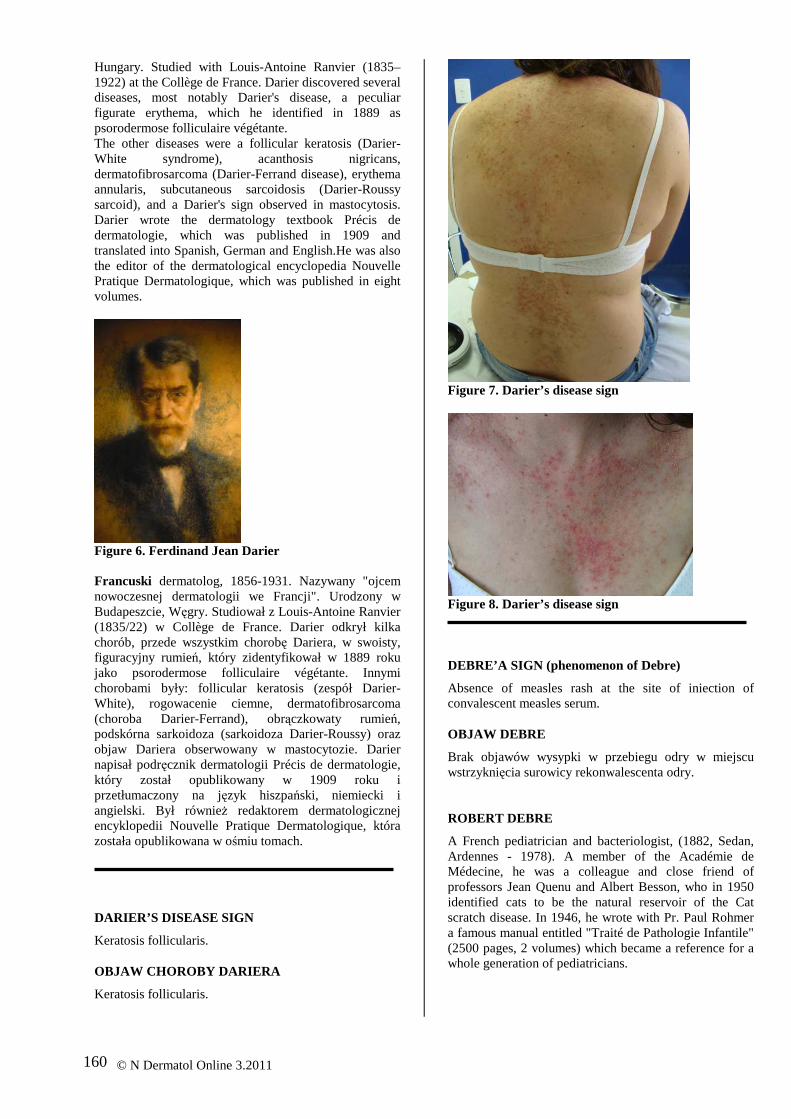
160
Hungary. Studied with Louis-Antoine Ranvier (1835–1922) at the Collège de France. Darier discovered several diseases, most notably Darier's disease, a peculiar figurate erythema, which he identified in 1889 as psorodermose folliculaire végétante. The other diseases were a follicular keratosis (Darier-White syndrome), acanthosis nigricans, dermatofibrosarcoma (Darier-Ferrand disease), erythema annularis, subcutaneous sarcoidosis (Darier-Roussy sarcoid), and a Darier's sign observed in mastocytosis. Darier wrote the dermatology textbook Précis de dermatologie, which was published in 1909 and translated into Spanish, German and English.He was also the editor of the dermatological encyclopedia Nouvelle Pratique Dermatologique, which was published in eight volumes.
Figure 6. Ferdinand Jean Darier Francuski dermatolog, 1856-1931. Nazywany "ojcem nowoczesnej dermatologii we Francji". Urodzony w Budapeszcie, Węgry. Studiował z Louis-Antoine Ranvier (1835/22) w Collège de France. Darier odkrył kilka chorób, przede wszystkim chorobę Dariera, w swoisty, figuracyjny rumień, który zidentyfikował w 1889 roku jako psorodermose folliculaire végétante. Innymi chorobami były: follicular keratosis (zespół Darier-White), rogowacenie ciemne, dermatofibrosarcoma (choroba Darier-Ferrand), obrączkowaty rumień, podskórna sarkoidoza (sarkoidoza Darier-Roussy) oraz objaw Dariera obserwowany w mastocytozie. Darier napisał podręcznik dermatologii Précis de dermatologie, który został opublikowany w 1909 roku i przetłumaczony na język hiszpański, niemiecki i angielski. Był równieŜ redaktorem dermatologicznej encyklopedii Nouvelle Pratique Dermatologique, która została opublikowana w ośmiu tomach. DARIER’S DISEASE SIGN
Keratosis follicularis. OBJAW CHOROBY DARIERA
Keratosis follicularis.
Figure 7. Darier’s disease sign
Figure 8. Darier’s disease sign DEBRE’A SIGN (phenomenon of Debre)
Absence of measles rash at the site of iniection of convalescent measles serum. OBJAW DEBRE
Brak objawów wysypki w przebiegu odry w miejscu wstrzyknięcia surowicy rekonwalescenta odry. ROBERT DEBRE
A French pediatrician and bacteriologist, (1882, Sedan, Ardennes - 1978). A member of the Académie de Médecine, he was a colleague and close friend of professors Jean Quenu and Albert Besson, who in 1950 identified cats to be the natural reservoir of the Cat scratch disease. In 1946, he wrote with Pr. Paul Rohmer a famous manual entitled "Traité de Pathologie Infantile" (2500 pages, 2 volumes) which became a reference for a whole generation of pediatricians.
© N Dermatol Online 3.2011
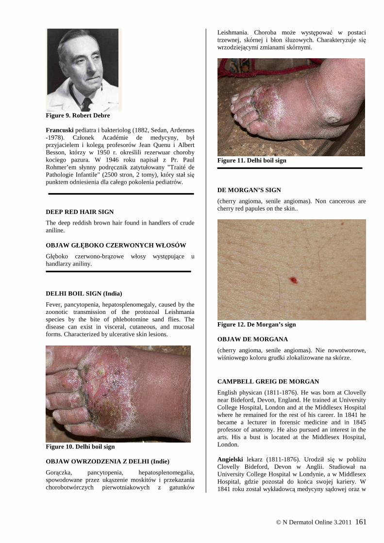
161
Figure 9. Robert Debre Francuski pediatra i bakteriolog (1882, Sedan, Ardennes -1978). Członek Académie de medycyny, był przyjacielem i kolegą profesorów Jean Quenu i Albert Besson, którzy w 1950 r. określili rezerwuar choroby kociego pazura. W 1946 roku napisał z Pr. Paul Rohmer’em słynny podręcznik zatytułowany "Traité de Pathologie Infantile" (2500 stron, 2 tomy), który stał się punktem odniesienia dla całego pokolenia pediatrów. DEEP RED HAIR SIGN
The deep reddish brown hair found in handlers of crude aniline. OBJAW GŁĘBOKO CZERWONYCH WŁOSÓW
Głęboko czerwono-brązowe włosy występujące u handlarzy aniliny. DELHI BOIL SIGN (India)
Fever, pancytopenia, hepatosplenomegaly, caused by the zoonotic transmission of the protozoal Leishmania species by the bite of phlebotomine sand flies. The disease can exist in visceral, cutaneous, and mucosal forms. Characterized by ulcerative skin lesions.
Figure 10. Delhi boil sign OBJAW OWRZODZENIA Z DELHI (Indie)
Gorączka, pancytopenia, hepatosplenomegalia, spowodowane przez ukąszenie moskitów i przekazania chorobotwórczych pierwotniakowych z gatunków
Leishmania. Choroba moŜe występować w postaci trzewnej, skórnej i błon śluzowych. Charakteryzuje się wrzodziejącymi zmianami skórnymi.
Figure 11. Delhi boil sign DE MORGAN’S SIGN
(cherry angioma, senile angiomas). Non cancerous are cherry red papules on the skin..
Figure 12. De Morgan’s sign OBJAW DE MORGANA
(cherry angioma, senile angiomas). Nie nowotworowe, wiśniowego koloru grudki zlokalizowane na skórze. CAMPBELL GREIG DE MORGAN
English physican (1811-1876). He was born at Clovelly near Bideford, Devon, England. He trained at University College Hospital, London and at the Middlesex Hospital where he remained for the rest of his career. In 1841 he became a lecturer in forensic medicine and in 1845 professor of anatomy. He also pursued an interest in the arts. His a bust is located at the Middlesex Hospital, London. Angielski lekarz (1811-1876). Urodził się w pobliŜu Clovelly Bideford, Devon w Anglii. Studiował na University College Hospital w Londynie, a w Middlesex Hospital, gdzie pozostał do końca swojej kariery. W 1841 roku został wykładowcą medycyny sądowej oraz w
© N Dermatol Online 3.2011

162
1845 roku profesorem anatomii. Wykazywał takŜe zainteresowanie sztuką. Jego popiersie znajduje się w Middlesex Hospital w Londynie.
Figure 13. Campbell Greig de Morgan DENNIE’S SIGN
Dennie-Morgan sign. A secondary crease in the lower eye lids. A sign of atophic dermatitis. Also known as Morgan’s line.
Figure 14. Dennie sign OBJAW DENNIEGO
Objaw Dennie-Morgana. Dodatkowy fałd na dolnej powiece. Objaw atopowego zapalenia skóry. Znany równieŜ jako linie Morgana. CHARLES CLAYTON DENNIE
American dermatologist (1883-1971). Remembered for Dennie-Marfan syndrome and Dennie-Morgan fold. Received his BS degree from Baker University in 1908 and his medical degree from the University of Kansas in 1912. During the war he served as a major in the Army Medical Corps and was in charge of the embarkation camp at Bordeaux, France, in 1918. His experiences in Boston and in France and the new and fairly recent
discoveries in the fields of bacteriology and immunology whetted his interest in the study of syphilis. His study of syphilis led to the publication of four books including one on congenital syphilis and another on the history of syphilis.
Figure 15. Charles Clayton Dennie Amerykański dermatolog (1883-1971). Pamiętany z opisu zespołu Dennie-Marfana oraz fałdu Dennie-Morgana. Uzyskał stopień licencjata w Uniwersytecie Baker w 1908 roku i stopień medyczny na Uniwersytecie w Kansas w 1912 roku. Podczas wojny słuŜył w stopniu majora w armii Medical Corps i był odpowiedzialny za obóz w Bordeaux, Francja, w 1918 roku. Jego doświadczenia w Bostonie i Francji i najnowsze odkrycia w dziedzinie bakteriologii i immunologii zapoczątkowały zainteresowania w badaniu nad kiłą. Jego badania nad kiłą doprowadziły do opublikowania czterech ksiąŜek, w tym jednej o kile wrodzonej, a pozostałe na temat historii kiły. DERCUM’S SIGN
Synonim adiposis dolorosa. Dercum disease. Is an unusual progressive syndrome of unknown etiology characterized by multiple painful lipomas that arise in adult life, most often affecting postmenopausal women who are obese.
Figure 16. Dercum’s sign
© N Dermatol Online 3.2011

163
OBJAW DERCUMA
Synonim adiposis dolorosa. Choroba Dercuma. Jest niezwykłym progresywnym zespółem o nieznanej etiologii, charakteryzuje się mnogimi tłuszczaki, które pojawiają się w dorosłym Ŝyciu, najczęściej dotyczą kobiet po menopauzie cierpiących na otyłość.
Figure 17. Francis Xavier Dercum FRANCIS XAVIER DERCUM
American neurologist (1856–1931). Graduated from the University of Pennsylvania Medical School in 1877. Specialised in treating nervous and mental disorders. Amerykański neurolog (1856–1931). Absolwent University of Pennsylvania Medical School w 1877 roku. Specjalizował się w leczeniu zaburzeń nerwowych i psychicznych. DESCOT’S SIGN
Scrotal hematoma suggesting pelvic fracture. OBJAW DESCOTA
Krwiak moszny sugerujący złamanie miednicy.
Figure 18. Descot’s sign
DEVERGIE’S SIGN
Synonym pityriasis rubra pilaris.
OBJAW DEVERGIE
Synonim pityriasis rubra pilaris. MARIE GUILLAUME ALPHONSE DEVERGIE
French physician (1798-1879). In 1834 he became a physician of Parisian hospitals (médecin des hôpitaux), and in 1840 at the Hôpital Saint-Louis, where he practiced medicine until his retirement. In 1874 he was elected president of the Académie de Médecine. In 1856 Devergie was the first to describe a rare dermatological disorder known as pityriasis rubra pilaris, which is sometimes referred to as Devergie's disease. In 1854 published an important textbook on skin diseases titled Traité pratique des maladies de la peau In France Devergie is justly considered one of the founders of forensic medicine. In 1836 he published a two-volume book on judicial medicine called Medecine legale, theorique et pratique.
Figure 19. Marie Guillaume Alphonse Devergie Francuski lekarz (1798-1879). W 1834 roku został lekarzem w paryskim szpitalu (médecin des Hôpitaux), a w 1840 roku w Hôpital Saint-Louis, gdzie praktykował medycyny aŜ do emerytury. W 1874 roku został wybrany na prezydenta w Académie de Médecine. W 1856 Devergie był pierwszym, który opisał rzadką chorobą dermatologiczną znaną jako pilaris rubra pityriasis, co jest czasem określane jako choroba Devergie's. W 1854 roku opublikował waŜny podręcznik chorób skóry pt Traité pratique des maladies de la peau We Francji Devergie jest słusznie uwaŜany za jednego z twórców medycyny sądowej. W 1836 roku opublikował ksiąŜkę dwutomową o nazwie Medecine legale, théorique et pratique.
© N Dermatol Online 3.2011

164
DICKINSON’S SIGN
Blue saliva. Dickinson describes a women whose saliva was blue; besides this nothing was definitely the matter with her. It seemed however, that the color was due to some chemic-pencil poisoning rather than to a pathologic process. Paget cites an instance of blue saliva due to staining the tonuge in the the same manner. Most cases of anomalus coloring of this kind can be subsequently traced to artificial substances; unconsciously introduced. OBJAW DICKINSONA
Niebieska ślina. Dickinson opisuje kobiety, których ślina miała niebieski kolor, poza tym były zdrowe. Wydawało się jednak, Ŝe kolor miał związek z pewnym zatruciem chemikaliami-ołówka, a nie procesem chorobowym. Paget przytacza przykład niebieskiej śliny ze względu na barwienia języka w tym kolorze. Większość przypadków anomalii zabarwienia tego rodzaju moŜe być następnie nieświadomie wprowadzonych sztucznych substancji. WILLIAM HOWSHIP DICKINSON
British physician (1832-1913). He worked at Great Ormond Street Hospital. He was involved in the early characterization of what came to be known as Alport syndrome. Dr Dickinson was physician at the children’s hospital from 1869 to 1874. He was censor and curator of the museum at the Royal College of Physicians, and examiner in medicine to the Royal College of Surgeons, and to the universities of Cambridge, London and Durham. Although a general physician, he was best known as a kidney specialist with regards his adult patients.
Figure 20. William Howship Dickinson Angielski lekarz (1832-1913). Pracował w Great Ormond Street Hospital. Brał udział w opisie zespołu Alporta. Dr Dickinson był lekarzem w szpitalu dla dzieci 1869/74. Był cenzorem i kustoszem muzeum w Royal College of Physicians, ekspertem w dziedzinie medycyny w Royal College of Surgeons oraz na uniwersytetach w Cambridge, Londynie i Durham. Mimo, Ŝe był lekarzem ogólnym, najbardziej znany był jako specjalista w zakresie chorób nerek u osób dorosłych.
DIMPLE OR BUTTON SIGN
Sign in dermatofibroma. Lateral pressure produces a with an overlying depression in the center of the papule. A central depression or dimple elicited within a lesion when it is squeezed along its margins. OBJAW DOŁKA LUB GUZIKA
Objaw w dermatofibroma. Ciśnienie boczne wytwarza depresję leŜącej w centrum grudki. Wgłębienie lub dołek wywołany rozpręŜeniem zmiany wzdłuŜ jej brzegów.
Figure 21. Dimple sign DIRT EATER’S SIGN
Visceral larva migrans, fever, rash and eye lesions that mimic retinoblastoma. Infection is from eating dirt with contains the embryonic eggs of the zoonotic Toxocara roundworms in dog and cat faeces. OBJAW ZANIECZYSZCZONEGO JEDZENIA
Larwa wędrująca trzewna, gorączka, wysypka i zmiany oczne, które naśladują retinoblastoma. ZakaŜenie następuje po zjedzeniu zanieczyszczonego pokarmu kałem psów i kotów, zawierającego chorobotwórcze jaja glisty Toxocara. DIRTY NECK SIGN
On the sides of neck gray-brown discoloration meshed in atopic dermatitis. OBJAW BRUDNEJ SZYI
Na skórze, na bocznych powierzchniach szyi szarobrązowe, siatkowate przebarwienie w AZS.
© N Dermatol Online 3.2011

165
DOBRAVA SIGN
Dobrava/Belgrade virus, Bulgaria. Rapid fever, kidney failure, severe pain and bleeding rash which progresses to death in 15% of victims. Caused by a zoonotic hantaviruses infectious process known sa hemorrhagic fever with renal syndrome. OBJAW DOBRAVY
Dobrava/Belgrad wirus, Bułgaria. Gwałtowna gorączka, niewydolność nerek, silny ból i krwotoczna wysypka, które doprowadza do śmierci w 15% przypadkach. Spowodowany przez odzwierzęce hantawirusy; znana jako gorączka krwotoczna z zespołem nerkowym DOBRIZHOFFER’S SIGN
At age seven Abipones boys pierce their arms in imitation of their parents. This is a sign of cosmetic mutilation. OBJAW DOBRIZHOFFERA
W wieku siedmiu lat chłopcy z plemiona Abipones przekłuwali swoje ramiona, naśladując swoich rodziców. Jest to znak, kosmetycznego okaleczenia. MARTIN DOBRIZHOFFER
September 7, 1717 – July 17, 1791. Was an Austrian Roman Catholic missionary. Was born in Graz. He joined the Society of Jesus in 1736, and in 1749 proceeded to Paraguay, where for eighteen years he worked devotedly first among the Guaranis, and then among the Abipones. Returning to Europe and wrote the history of his mission.
Figure 22. Martin Dobrizhoffer with Abipones 07. września 1717 - 17 lipca 1791. Był austriackim katolickim misjonarzem. Urodził się w Graz. Wstąpił do Towarzystwa Jezusowego w 1736 roku, w 1749 wyjechał do Paragwaju, gdzie przez osiemnaście lat pracował z oddaniem wśród Guaranis, a następnie wśród Abipones. Po powrocie do Europy i napisał historię swojej misji.
DOG SIGN
When a dog trained to detect cancer has a positive finding from smelling a patient’s skin, breath, urine, etc. OBJAW PSA
Występuje, kiedy pies przeszkolony w wykrywaniu raka u pacjenta osiągnie pozytywny wynik po zapachu skóry pacjenta, oddechu, moczu, itp. DOVER’S POWDER SIGN
Pin-point pupils, muscular limpness, face pale or blue. Indication of opium poisoning. Dover's Powder, developed and described by the British physician Thomas Dover in 1732, was one of the more popular and enduring of the opium-based medications that were widely used in the United States and Europe prior to the twentieth century. The result was a pain-reducing potion that might induce a sense of euphoria but could not be ingested in large quantities because of its emetic properties. Taken as a nonprescription medicine over 200 years.
Figure 23. Dover's Powder OBJAW PROSZKU DOVERA
Plamki typu pin-point, osłabienie mięśni, blada lub niebieskawa twarz. Wskazuje na zatrucie opium. Proszek Dovera, został opracowany i opisany przez brytyjskiego lekarza Thomasa Dovera w 1732 roku. Był jednym z bardziej popularnych i trwałych na bazie opium leków, który był powszechnie stosowany w Stanach Zjednoczonych i Europie przed XX wiekiem. W efekcie zmniejszający ból eliksir, wywoływał uczucie euforii, ale nie mógł być spoŜywany w duŜych ilościach ze względu na jego właściwości wymiotne. Wydawano go jako lek bez recepty przez 200 lat. THOMAS DOVER
English physician and explorer. 1660-1742. Studied medicine at Oxford University in the 1680s. Dover
© N Dermatol Online 3.2011

166
practiced medicine for over fifty years, although during his lifetime he was more famous for his exploits as an adventurer. His involvement in the early slave trade and in the plundering of Spanish settlements off the coast of South America brought him fortune and fame. In 1732, probably to attract patients to his new practice in London, Dover published An Ancient Physician's Legacy to His Country, one of the earliest medical treatises written for the general public. The book listed forty-two ailments with successful treatments used by Dover, and included the testimonial letters of many "cured" patients. The book enjoyed popular success and was reprinted eight times. One remedy described in the book, the use of mercury, earned him the nickname during his lifetime of the Quicksilver Doctor, but the formula for Dover's Powder, has proven to be his most enduring legacy.
Figure 24. Thomas Dover greets Alexander Selkirk on the South Pacific island of Más a Tierra. Illustration by an anonymous artist in Strong LAG. Angielski lekarz i badacz. 1660-1742. Studiował medycynę na Uniwersytecie w Oksfordzie w 1680 r. Dover praktykował od ponad pięćdziesięciu lat, choć za Ŝycia bardziej znane są jego wyczyny jako awanturnika. Jego zaangaŜowanie na początku handlem niewolnikami i grabieŜą hiszpańskich osiedli na wybrzeŜu Ameryki Południowej przyniosła mu fortunę i sławę. W roku 1732, prawdopodobnie w celu przyciągnięcia pacjentów do jego nowej praktyki w Londynie, Dover opublikował „An Ancient Physician's Legacy to His Country”, jeden z pierwszych traktatów medycznych napisany dla ogółu społeczeństwa. W ksiąŜce wymienione są czterdzieści dwie dolegliwości z powodzeniem wyleczonych przez Dovera, zawierają referencje i listy wielu "wyleczonych" pacjentów. KsiąŜka cieszyła sukcesem i została przedrukowana osiem razy. Jednym ze środków opisanych w ksiąŜce, była rtęć, co przyniosło mu przydomek Doktor Quicksilver, ale formuła „Powder Dover”, okazała się jego najbardziej trwałym dziedzictwem.
DRACULA’S SIGN
Severe mutilating skin lesions caused by photosensivity, neurological disruptions, liver pathology, and purple urine. These are indications of forms of porphyria. It is now suggested that Vlad Dracul the 15th century slayer prince, also known as Vlad the Implaner suffered from hereditary porphyria. Disease can be manifested with painful cutaneus photosensitivity, allowing some victims to only come out after dark caused them to be sadly mistaken for vampires. Also called Vampire’s disease. OBJAW DRACULI
CięŜkie okaleczające zmiany skórne spowodowane przez nadwraŜliwość na światło słoneczne, zaburzenia neurologiczne, patologia wątroby oraz fioletowy mocz. Są to wykładniki odmiany porfirii. Obecnie te objawy sugerują, Ŝe Vlad Dracul, XV-to wieczny ksiąŜę, zabójca, znany takŜe jako Vlad cierpiał na dziedziczną porfirię. Choroba moŜe manifestować się z bolesną, skórną nadwraŜliwością na światło, pozwalając „ofierze” wyjść tylko po zmroku spowodowało to niestety mylone określanie ich jako wampirów. Zwany takŜe chorobą Wampira. VLAD III DRACULA (VLADISLAUS)
Vlad III the Impaler. Prince of Wallachia, dynasty of Basarab. (1431–1476). It's best known for his resistance against the Ottoman Empire and its expansion. His post-mortem moniker of "Impaler" originated in his killing opponents by impalement. In Turkish, he was known as "Kazıklı Voyvoda" which means "Impaler Prince". Was born in Sighişoara, Transylvania (part of the Kingdom of Hungary at the time).
Figure 25. Vlad III Dracula Vlad III Palownik. KsiąŜę Wołoski, Z dynastii Basarabów. (1431-1476). Jest najbardziej znany z oporu przeciwko Imperium Ottomańskiemu i jego ekspansji. Jego przydomek „Palownik” maił związek z nabijaniem jego przeciwników na pal. W Turcji, był znany jako "Kazıklı Voyvoda" co oznacza "ksiąŜę Palownik”. Urodził się w Sighişoara, Siedmiogród (część Królestwa Węgier w tym czasie).
© N Dermatol Online 3.2011
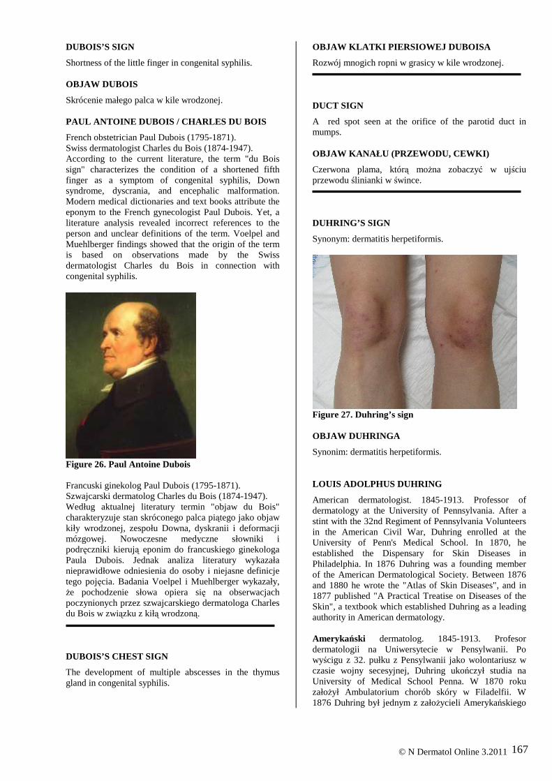
167
DUBOIS’S SIGN
Shortness of the little finger in congenital syphilis. OBJAW DUBOIS
Skrócenie małego palca w kile wrodzonej. PAUL ANTOINE DUBOIS / CHARLES DU BOIS
French obstetrician Paul Dubois (1795-1871). Swiss dermatologist Charles du Bois (1874-1947). According to the current literature, the term "du Bois sign" characterizes the condition of a shortened fifth finger as a symptom of congenital syphilis, Down syndrome, dyscrania, and encephalic malformation. Modern medical dictionaries and text books attribute the eponym to the French gynecologist Paul Dubois. Yet, a literature analysis revealed incorrect references to the person and unclear definitions of the term. Voelpel and Muehlberger findings showed that the origin of the term is based on observations made by the Swiss dermatologist Charles du Bois in connection with congenital syphilis.
Figure 26. Paul Antoine Dubois Francuski ginekolog Paul Dubois (1795-1871). Szwajcarski dermatolog Charles du Bois (1874-1947). Według aktualnej literatury termin "objaw du Bois" charakteryzuje stan skróconego palca piątego jako objaw kiły wrodzonej, zespołu Downa, dyskranii i deformacji mózgowej. Nowoczesne medyczne słowniki i podręczniki kierują eponim do francuskiego ginekologa Paula Dubois. Jednak analiza literatury wykazała nieprawidłowe odniesienia do osoby i niejasne definicje tego pojęcia. Badania Voelpel i Muehlberger wykazały, Ŝe pochodzenie słowa opiera się na obserwacjach poczynionych przez szwajcarskiego dermatologa Charles du Bois w związku z kiłą wrodzoną. DUBOIS’S CHEST SIGN
The development of multiple abscesses in the thymus gland in congenital syphilis.
OBJAW KLATKI PIERSIOWEJ DUBOISA
Rozwój mnogich ropni w grasicy w kile wrodzonej. DUCT SIGN
A red spot seen at the orifice of the parotid duct in mumps. OBJAW KANAŁU (PRZEWODU, CEWKI)
Czerwona plama, którą moŜna zobaczyć w ujściu przewodu ślinianki w śwince. DUHRING’S SIGN
Synonym: dermatitis herpetiformis.
Figure 27. Duhring’s sign OBJAW DUHRINGA
Synonim: dermatitis herpetiformis. LOUIS ADOLPHUS DUHRING
American dermatologist. 1845-1913. Professor of dermatology at the University of Pennsylvania. After a stint with the 32nd Regiment of Pennsylvania Volunteers in the American Civil War, Duhring enrolled at the University of Penn's Medical School. In 1870, he established the Dispensary for Skin Diseases in Philadelphia. In 1876 Duhring was a founding member of the American Dermatological Society. Between 1876 and 1880 he wrote the "Atlas of Skin Diseases", and in 1877 published "A Practical Treatise on Diseases of the Skin", a textbook which established Duhring as a leading authority in American dermatology. Amerykański dermatolog. 1845-1913. Profesor dermatologii na Uniwersytecie w Pensylwanii. Po wyścigu z 32. pułku z Pensylwanii jako wolontariusz w czasie wojny secesyjnej, Duhring ukończył studia na University of Medical School Penna. W 1870 roku załoŜył Ambulatorium chorób skóry w Filadelfii. W 1876 Duhring był jednym z załoŜycieli Amerykańskiego
© N Dermatol Online 3.2011

168
Towarzystwa Dermatologicznego. W latach 1876 i 1880 roku napisał "Atlas of Skin Diseases", a w 1877 opublikował "Praktyczny Traktat o chorobach skóry" podręcznik, który usytuował Duhringa jako czołowy autorytet w Amerykańskiej dermatologii.
Figure 28. Louis Adolphus Duhring DUKE’S SIGN
An alleged exanthematosus contagious disease resembling rubella, scarlatina, and measles. It is marked by lamellar desquamation of the skin. Also called fourth disease, rubeola scarlatinea, Dukes disease, Dukes-Fiłatow disease. OBJAW DUKESA
Rzekomy wysypka choroby zakaźnej przypominająca róŜyczkę, szkarlatynę i odrę. Cechuje ją lamelarne złuszczanie skóry. Zwany równieŜ czwartą chorobą, rubeola scarlatinea, chorobą Dukesa, chorobą Dukesa-Fiłatowa. CLEMENT DUKES
English physician. (1845-1925). In 1871 was appointed medical officer at Rugby school, a position he held until 1908. At Rugby he won world-wide renown for his books and articles on schoolboy health and health care. In the 80s Nineteenth century provided a description of the epidemic in Rugb (known English private school.) Never subsequently described in other cases, the fourth disease. In 1900 he published an article entitled «On the confusion of two diseases under the name of rubella (Rose Rash). Angielski lekarz. (1845-1925). W 1871 został powołany na stanowisko lekarza w szkole Rugby, stanowisko to piastował aŜ do 1908 roku. Na Rugby zdobył światową sławę za jego ksiąŜki i artykuły dla uczniów i opieki zdrowotnej. W latach 80. XIX wieku przedstawił opis epidemii w Rugb (znana angielska szkoła prywatna). Nigdy później nie opisano innych przypadków czwartej choroby. W 1900 roku opublikował artykuł zatytułowany "Na pomieszanie dwóch chorób pod nazwą róŜyczka (RóŜowa wysypka).
NICOLAI FEODOROVICH FILATOV
Russian pediatrician (1847-1902). Father of Russian Pediatrics. After graduating from the University of Moscow he returned to his district to practise as a country doctor. Undertook further training in Vienna, Paris, Berlin and in Prague. After two years he returned to Moscow and began working and teaching in the Old Children’s Hospital in 1876. In 1891 he was appointed professor of paediatrics. In 1892 the Moscow Paediatric Society was founded and Filatov was appointed as its president.
Figure 29. Nicolai Feodorovich Filatov Rosyjski pediatra (1847-1902). Ojciec Pediatrii rosyjskiej. Po ukończeniu Uniwersytetu Moskiewskiego powrócił do wykonywania zawodu lekarza. Podjął dalsze kształcenie w Wiedniu, ParyŜu, Berlinie, w Pradze. Po dwóch latach wrócił do Moskwy i rozpoczął pracę w Old Children’s Hospital w 1876 roku. W 1891 roku został mianowany profesorem pediatrii. W 1892 roku powstało Moskiewskie Towarzystwo Pediatryczne, a Filatov został powołany na stanowisko jej przewodniczącego. DUNCAN-BIRD’S SIGN
Defined area of dulness and absence of respiratory sounds. A sign of hydatid cyst in the lungs or liver, caused by tapeworms. Also known as Bird’s sign. OBJAW DUNCANA-BIRDA
Określony obszar stłumienia i braku dźwięku. Objaw torbieli bąblowcowej w płucach lub w wątrobie, spowodowany przez tasiemce. Znany równieŜ jako objaw Birda. SAMUEL DOUGAN-BIRD
Australian physician. 1832-1904. He was president of the Medical Society of Victoria in 1869 and physician to the
© N Dermatol Online 3.2011
Benevolent Asylum and the Immigrants Aid Society. For thirty years he was chief medical officer to the Australian Mutual Provident Society in Victoria. His lectures were said to be lucid and forthright and the same qualities can be seen in his medical writings. Showed interest in chest disease.

169
Figure 30. Samuel Dougan-Bird Australijski lekarz. 1832-11904. Był prezesem Towarzystwa Lekarskiego Victorii w 1869 roku i lekarzem Benevolent Asylum i Immigrants Aid Society. Przez trzydzieści lat był głównym oficerem medycznym w Australian Mutual Provident Society w Viktorii. Jego wykłady medyczne były uwaŜane za przejrzyste i proste i w tej samej jakości moŜna był zobaczyć je w jego pismach medycznych. Wykazywał zainteresowania chorobami klatki piersiowej. DUTTON’S SIGN
Relapsing fever caused by a west African tick. DUTTON’S SIGN
Rzuty gorączki spowodowane przez zachodnioafrykańskiego kleszcza. JOSEPH EVERETT DUTTON
English physician (1879-1905). In 1900, he joined the School's third expedition to Nigeria and in 1901 he was elected the Walter Myers Fellow in Parasitology. In the same year Dutton undertook his next expedition to the Gambia alone and first recognised the presence of trypanosomes in human blood which would eventually lead to the discovery of the cause of sleeping sickness.
Angielski lekarz (1879-1905). W 1900 roku dołączył do trzeciej szkolnej ekspedycji do Nigerii, a w 1901 roku został wybrany Fellow Walter Myers w Parazytologii. W tym samym roku podjął kolejną wyprawę do Gambii i jako pierwszy stwierdził obecność świdrowców we krwi człowieka, co w końcu doprowadziło do odkrycia przyczyny śpiączki afrykańskiej.
Figure 31. Joseph Everett Dutton
DUVENHAGE FEVER SIGN (South Africa)
Encephalitic illness with near 100 percent mortality. Caused by zoonotic rabies lyssavirus carried by infected micro bats. The virus was discovered in 1970 when a South African farmer (after whom the virus is named) died of a rabies-like encephalitic illness after being bitten by a bat. Microbats are believed to be the natural reservoir of Duvenhage virus. OBJAW GORĄCZKI DUVENHAGE
Zapalna choroba mózgu ze śmiertelnością blisko 100%. Spowodowana przez odzwierzęcy, chorobotwórczy wirus wścieklizny lyssaviride, wprowadzony przez zainfekowanego Microchiroptera-Microbats (podrząd nietoperzy). Wirus został odkryty w 1970 r., kiedy rolnik Południowej Afryki (od którego nazwiska pochodzi nazwa wirusa) zmarł na wściekliznę z objawami grypopodobnymi i zapaleniem mózgu po ukąszeniu przez nietoperza. Microbats uwaŜany jest za naturalny rezerwuar wirusa Duvenhage. DYNAMITE SIGN
A headache, throbbing, tingling in extremities, pale blue lips. Caused by exposure to toluene and acid fumes when entering recently exploded caves for gold excavation. Dahlonega city Georgia c.1828. OBJAW DYNAMITU
Pulsujący ból głowy, mrowienie w kończynach, jasnoniebieskie usta. Spowodowane naraŜeniem na działanie toluenu i oparów kwasu przy wejściu do niedawno wysadzonej jaskini wykopu złota. Dahlonega, Georgia około 1828 r.
ACKNOWLEDGEMENT:
Figure 1 Estelle LAMBERT BIUMInfo 12, rue de l'Ecole de Médecine - 75006 Paris, France [email protected]
© N Dermatol Online 3.2011

170
PIŚMIENNICTWO / REFERENCES: 1. Danielssen and Boeck (Norway), Tralleckla Sp&lalskhcd, with atlas of twenty-four coloured plates, Paris, 1848 2. Sobanko JF, Freeman AF, Palmore TN, Mendoza D, Richard Lee CC, Cowen EW: A Sri Lankan woman with rheumatoid arthritis and anesthetic plaques. J Am Acad Dermatol. 2009; 60: 1018-1021. 3. Robertson J: Leprosy and the elusive M. leprae: colonial and imperial medical exchanges in the nineteenth century. Hist. cienc. saude-Manguinhos 2003; 10: 13-40. 4. letter: Daniel Cornelius Danielssen. 1815-1894. Int J Lepr Other Mycobact Dis. 1973; 41: 152-153. 5. Ziem JB, Spannbrucker N, Magnussen P, Olsen A, Amon-Kotey DN, Frenzel K, et al: Oesophagostomum bifurcum-induced nodular pathology in a highly endemic area of Northern Ghana. Trans R Soc Trop Med Hyg. 2005; 99: 417-422. 6. Surjushe A, Jindal S, Gote P, Saple DG: Darier's sign. Indian J Dermatol Venereol Leprol 2007; 73: 363-364. 7. Ségal A: Ferdinand-Jean Darier (1856-1938) dermatologist of world renown. Hist Sci Med. 2009; 43: 389-394. 8. Moulonguet I; sous l'égide du Groupe d'histopathologie cutanée de la Société française de dermatologie (SFD). Darier's disease. Ann Dermatol Venereol. 2010; 137: 752-754. 9. Phillips WR: Protection phenomenon in measles. Debre phenomenon or aussparphanomen. Am J Dis Child. 1938; 55: 1257-1260. 10. Stowers JH: Case of Delhi Boil or Sore (Syn.: Oriental Sore; Aleppo Boil). Proc R Soc Med. 1920; 13: 81–83. 11. Kim JH, Park HY, Ahn SK: Cherry Angiomas on the Scalp. Case Rep Dermatol. 2009; 1: 82-86. 12. Tokura Y: Extrinsic and intrinsic types of atopic dermatitis. J Dermatol Sci. 2010; 58: 1-7. 13. Sams WM: Charles Clayton Dennie 1883-1971. Arch Dermatol. 1971; 104: 94-95. 14. Wollina U, Goldman A, Heinig B: Microcannular tumescent liposuction in advanced lipedema and Dercum's disease. G Ital Dermatol Venereol. 2010; 145: 151-159. 15. Sorbie C: Francis Xavier Dercum. Orthopedics. 2009; 32: 882. 16. Totz KA: Images in emergency medicine. Scrotal hematoma with open book pelvis fracture and prostatic urethral disruption. Ann Emerg Med. 2004; 43: 533, 538. 17. Ahn SY, Kim JH, Ahn SK, Oh YS: Clinical improvement of pityriasis rubra pilaris with antibiotic therapy. Eur J Dermatol. 2011; 21: 106-107.
18. Waltz M, Shattock P: Autistic disorder in nineteenth-century London: three case reports. Autism. 2004;8:7-20. 19. Meffert JJ, Peake MF, Wilde JL: 'Dimpling' is not unique to dermatofibromas. Dermatology. 1997; 195: 384-386. 20. Roldán WH, Espinoza YA, Huapaya PE, Jiménez S: Diagnóstico de la toxocarosis humana. Rev Peru Med Exp Salud Publica. 2010; 27: 613-620. 21. Tanei R, Katsuoka K: Clinical analyses of atopic dermatitis in the aged. J Dermatol. 2008; 35: 562-269. 22. Kitzmantel A: Die Jesuitenmissionare Martin Dobrizhoffer und Florian Paucke und ihre Beiträge zur Ethnographie des Gran Chaco im 18. Jahrhundert. Inaugural-Dissertation zur Erlangung des Doktorgrades der Philosophie an der Ludwig-Maximilians-Universität München, 2004. 23. Osler W: Thomas Dover, M.B. (of Dover's Powder), physician and buccaneer. Acad Med. 2007; 82: 880. 24. Tormosov DD: Dubois symptom in late congenital syphilis. Vestn Rentgenol Radiol. 1960; 34: 69-70. 25. Voelpel JH, Muehlberger T: The du Bois sign. Ann Plast Surg. 2011; 66: 241-244. 26. Vrebos J, Dupuis CC: À la rencontre du Pr Paul Dubois (Paris 1795-1871). De la chéiloplastie néonatale dans les fentes labiales. Ann Chir Plast Esthet. 2006; 51: 253-259. 27. Bogenrieder T, Stolz W: From the New World. Louis A. Duhring and dermatitis herpetiformis. Hautarzt. 2003; 54:167-172. 28. Weisse ME: The fourth disease, 1900-2000. Lancet. 2001; 357: 299-301. 29. Il'inskii PI: Nil Fedorovich Filatov, 1847-1902; materials for history of Russian pediatrics. Klin Med (Mosk). 1954; 32: 89-95. 30. Scarlata F, Giordano S, Saporito L, Marasa L, Li Pani G, Odierna Contino A, et al.: Cystic hydatidosis: a rare case of spine localization. InfezMed. 2011;19:39-41. 31. Heaulme M, Couvreur P, Sicard JM: Tick bite fever in West Africa: difficulties in diagnosis of an emerging disease. Med Trop (Mars). 2009 Dec;69(6):595-8. 32. Cook GC, Braybrooke J: Joseph Everett Dutton (1874-1905): pioneer in elucidating the aetiology of west African trypanosomiasis. J Med Biogr. 1997; 5: 131-136. 33. Markotter W, Van Eeden C, Kuzmin IV, Rupprecht CE, Paweska JT, Swanepoel R, et al: Epidemiology and pathogenicity of African bat lyssaviruses. Dev Biol (Basel). 2008; 131: 317-325.
© N Dermatol Online 3.2011