Community Dermatology
16
COMMUNITY DERMATOLOGY: 2008; 5: 1-16 Issue No. 7 1 www.icthesworldcare.com Neil H Cox BSc (Hons) FRCP Dermatology Department Cumberland Infirmary Carlisle CA2 7HY UK E-mail: [email protected] Introduction T his article will assess the published evidence for some tra- ditional remedies; evidence for use of honey was covered in a previous article (Cox N H, J Comm Dermatol 2006; 3: 20-22). The present discussion will include comments on saline, as well as fruit and vegetable agents, such as banana leaf and potato peel. Many plants contain chemicals that are also used as wound healing agents, but most of these are in the form of alcoholic (or some- times aqueous) extracts, rather than being applied as intact plant material. Some, especially those that are applied directly (e.g., as an intact leaf, or as a crushed pulp) are discussed briefly here. One issue that may apply generally to plants is that several botanical polysaccharides activate macrophage immune responses leading to several effects, including enhanced wound healing. 1 Another non- specific issue that may explain benefits on wounds from freshly pulped plant materials is that ‘wounding’ plants causes them to generate hydrogen peroxide. 2 This chemical is used medically in TRADITIONAL WOUND DRESSINGS CONTENTS LEAD ARTICLES Traditional Wound Dressings Neil H Cox 1 Presentations and Local Remedies of White Jellyfish (Rhizostoma Sp.) Stings in Iraqi Fishermen Kathem K Al-Rubiay, Laith K Alrubaiy, Hefaa A H Al-Musaoi, Mohammed G K Al-Freje 4 Diagnosing and Managing Leishmaniasis in the Community Rachael Morris-Jones, Stephen Morris-Jones 7 QUIZ Quiz: Itchy Lines L Claire Fuller 12/14 RDTC RESEARCH REPORTS Namibian Men and Condoms The late Yvonne Siyongo 13 Partner Notification in Swaziland Mpumelolo Dlamini 13 Self Medication in Cameroon and Zambia Marie Michelle Boum, Charles Hakoma 13 BOOK REVIEW HIV Related Skin Diseases And Sexually Transmitted Infections In Africa (Review by Chris Lovell) Merja Kousa, Cornelus Sanders 15 Community De r ma t ology An International Journal for Community Skin Health J Comm Dermatol 2008; 5: 1-16 Issue No.7 C o m m u n i t y D e r m a t o l o g y A banana leaf prepared for use as a natural non-stick dressing Photo: Dr S Walton, Hull, UK various proprietary topical creams for its antimicrobial activity, and may be a further non-specific way in which plants work in wound healing. As in humans, plants also produce cationic pep- tides that have antimicrobial properties and improve wound heal- ing, but whether application of plant materials has additional effect beyond the natural production that occurs in man is unclear.
-
Upload
khangminh22 -
Category
Documents
-
view
7 -
download
0
Transcript of Community Dermatology

COMMUNITY DERMATOLOGY: 2008; 5: 1-16 Issue No. 7 1www.icthesworldcare.com
Neil H Cox BSc (Hons) FRCPDermatology DepartmentCumberland InfirmaryCarlisle CA2 7HYUK
E-mail: [email protected]
Introduction
This article will assess the published evidence for some tra-ditional remedies; evidence for use of honey was covered in a previous article (Cox N H, J Comm Dermatol 2006; 3:
20-22). The present discussion will include comments on saline, as well as fruit and vegetable agents, such as banana leaf and potato peel.
Many plants contain chemicals that are also used as wound healing agents, but most of these are in the form of alcoholic (or some-times aqueous) extracts, rather than being applied as intact plant material. Some, especially those that are applied directly (e.g., as an intact leaf, or as a crushed pulp) are discussed briefly here. One issue that may apply generally to plants is that several botanical polysaccharides activate macrophage immune responses leading to several effects, including enhanced wound healing.1 Another non-specific issue that may explain benefits on wounds from freshly pulped plant materials is that ‘wounding’ plants causes them to generate hydrogen peroxide.2 This chemical is used medically in
TRADITIONAL WOUND DRESSINGS
CONTENTS
LEAD ARTICLES
Traditional Wound Dressings Neil H Cox 1
Presentations and Local Remedies of White Jellyfish (Rhizostoma Sp.) Stings in Iraqi Fishermen
Kathem K Al-Rubiay, Laith K Alrubaiy, Hefaa A H Al-Musaoi, Mohammed G K Al-Freje
4
Diagnosing and Managing Leishmaniasis in the Community Rachael Morris-Jones, Stephen Morris-Jones 7
QUIZ
Quiz: Itchy Lines L Claire Fuller 12/14
RDTC RESEARCH REPORTS
Namibian Men and Condoms The late Yvonne Siyongo 13
Partner Notification in Swaziland Mpumelolo Dlamini 13
Self Medication in Cameroon and Zambia Marie Michelle Boum, Charles Hakoma 13
BOOK REVIEW
HIV Related Skin Diseases And Sexually Transmitted Infections In Africa(Review by Chris Lovell)
Merja Kousa, Cornelus Sanders 15
Community Dermatology
An International Journal for Community Skin Health
J Comm Dermatol 2008; 5: 1-16 Issue No.7
Community Dermatology
A banana leaf prepared for use as a natural non-stick dressingPhoto: Dr S Walton, Hull, UK
various proprietary topical creams for its antimicrobial activity, and may be a further non-specific way in which plants work in wound healing. As in humans, plants also produce cationic pep-tides that have antimicrobial properties and improve wound heal-ing, but whether application of plant materials has additional effect beyond the natural production that occurs in man is unclear.

2 COMMUNITY DERMATOLOGY: 2008; 5: 1-16 Issue No. 7 www.icthesworldcare.com
Traditional Wound Dressings
There is documented benefit of wound healing for numerous plants and extracts from plants. The emphasis of this article is on those that are applied as dressings, rather than those in which some form of therapeutic extract is used.
SalinePublications that use the word ‘saline’ run into tens of thousands, but it is difficult to find articles specific to therapeutic trials in wound treatment. Where these are available, saline is usually viewed as a control that keeps wounds moist, but that has no other activity.
Briefly, picking out some relevant studies, saline soaked-gauze was as effective as silver sulphadiazine cream or an alginate emul-sion in treating fresh partial thickness burns (in pigs) in one ran-domised controlled experimental study,3 but less effective than sil-ver sulphadiazine in another study. However, a more recent study suggests that saline-treated wounds heal slowly (saline was one of two control substances), and that silver sulphadiazine and other antibacterials may inhibit wound contraction by comparison to various other agents (such as aqueous cream, which was the other control substance).4 An antimicrobial (0.057% sodium hypo-chlorite) was compared with saline as a wound cleanser and was associated with lower bacterial counts and faster healing.5 Aloe vera compared with saline appears to have an anti-inflammatory effect, with faster healing times than saline alone, and that this or nystatin can reverse the impaired healing associated with antimi-crobials.4 Overall, it would appear that a dilute antiseptic is likely to be more effective than saline alone, that the latter is better than nothing, but that other agents have better wound-healing proper-ties. Saline should be viewed mainly as a wound cleanser, rather than as a therapeutic dressing.
Banana LeafMy personal experience of banana leaves is that I have eaten fish cooked in them, presumably to keep the fish moist. This may sound irrelevant, but this property is probably also important in their use as a wound dressing.
Banana leaf is non-adherent, protects wounds from trauma and prevents them becoming too dry. It is readily available and widely used, not only for wounds but also for eroded areas in dermato-logical conditions such as pemphigus (Figure 1) and toxic epider-mal necrolysis. One study compared banana leaves with a tradi-tional boiled potato peel bandage, and showed that the banana leaf
dressings were equivalent in healing rates, but had the advantage of much lower cost and were easy to use.6 The same authors have also shown that pain related to dressing changes is much less with banana leaf than with Vaseline (yellow soft paraffin impregnated) gauze.7 However, a recent article raised concerns about the risk of infection from use of banana leaves, showing that only autoclaving and aseptic handling prevented culture of organisms from banana leaves (flame heating, boiling, and scorching with burning methyl-ated spirits were not effective).8
Infection is a serious risk when there are large areas of skin erosion. Historically, in the UK, infection in pemphigus was a major cause of deaths. Otherwise, much of the literature on therapeutic uses of banana, or various extracts from bananas, relates to an anti-gastric ulcer property – this appears to be both prophylactic and also related to increased mucosal healing, but it is not clear that it has any role in wound healing.
Potato Peel and Other Potato ConstituentsThere are many articles that discuss use of boiled potato peel as a wound dressing.9-12 Most reported experience relates to burns, and the best results, as might be expected, are in superficial partial thickness burns. However, deeper wound granulation may also be improved and full thickness wounds have been specifically investigated in rats. Other wounds that have been studied include pemphigus, pemphigoid and leg ulcers.12 The active constituents are unclear but homogenated extracts are less effective than intact boiled potato peel. Potato peel fixed to gauze bandages is more effective than gauze bandage alone, suggesting that the outer cork layer (periderm) has a physical protective effect and probably prevents the wound from getting too dry. To be most effective, therefore, the dressing needs to be manufactured so that the pulp side is applied to the wound (usually with wheat starch paste on top and then a cotton gauze). Other potato constituents that have been suggested as useful are steroidal glycosides (steroidal glycol-alkaloids), present in the boiled peel, and the fibre from sweet potato (this seems to be effective for absorbing exudates).13
In practical use, potato peel does not seem to be antibacterial. However, some studies have used it applied in preference to sil-ver sulphadiazine or other antiseptics.12 Boiled potato peel can be prepared as bandage rolls to make it convenient for dressing burn wounds. As with banana leaf, autoclaved dressings are prefera-ble. One systematic review documented potato peel as being less effective than honey as a wound dressing (although numbers in all studies were small) but it may be more easily available.14 Another concern is that contact urticaria to potato peel is reasonably com-mon, although this may affect those preparing dressings, rather than the patient, as the causative protein is probably altered and non-allergenic after the boiling process.
Papaya (Paw paw; Carica papaya)Papaya (paw paw) contains papain, chymopapain and other simi-lar cysteine proteinases. In cooking, these have a tenderising effect on meat. In human medicine, paw paw (papaya) has been used on burns and other wounds – it is cheap, readily available, and easily applied by mashing the pulp for daily application.15 Its protein-ase activity probably gives it a desloughing activity, and it may also have an antibacterial effect. An extract of papaya has been shown to inhibit bacterial catalase (allowing bacteria to be more easily killed by macrophages) and has an antioxidant effect that reduces tissue injury.16 Seed extracts have also been shown to have antibac-terial effects against various organisms.17
Fig. 1: Banana leaf dressing in a patient with pemphigus. The patient lies on the leaves which provide a non-sticking surface, avoiding damage when removing absorbent dressings from raw skin.
Photo: Dr S Walton, Hull, UK

COMMUNITY DERMATOLOGY: 2008; 5: 1-16 Issue No. 7 3www.icthesworldcare.com
Papaya has also been used in shampoo form to remove head lice18 but, because it was combined with thymol and tea tree oil, it is uncertain which component was effective. It can, however, cause an immediate hypersensitivity reaction, and studies of systemic use in rats have shown that it damages testes and sperm to cause reduced fertility.19 Data on absorption from wounds is not avail-able but the threat of testicular damage suggests that it should only be used with caution.
Sphagnum Moss (Sphagnum junghuhnianum and others)Sphagnum and related mosses (bryophytes) are found worldwide. They absorb water and have antiseptic properties. Use as a wound dressing has been documented as far back as the Bronze Age, and huge amounts were used during World War I to make wound dressings that were apparently less irritant and more absorbent than cotton dressings.20 In India, recent research has compared 15 types of moss and documented sphagnum to be one of the more effective antimicrobial types (using ethanol extracts).21
Other Natural DressingsAmniotic membrane has been used for many years as a natural dressing for burns and leg ulcers. Infection would now be consid-ered a significant problem.
Collagen has been used in various forms of dressing, and is a com-ponent of manufactured (and expensive) ‘skin equivalent’ dress-ings. A novel method to produce a less expensive form of collagen dressings was reported in India recently. Fish scales, usually viewed as a waste product, were cleaned and processed to make them into a ‘fish scale collagen sheet’. This is fibrous, allows transfer of water, and is strong enough to be used as a wound dressing.22
The MessagesHoney and banana leaf both appear to be superior in promot-ing wound healing compared with saline-soaked gauze. Honey is superior to some other traditional agents (e.g., potato peel) or fre-quently used antibacterials (e.g., silver sulphadiazine) as a wound dressing. However, there is evidence and at least some scientific rationale to suggest that boiled potato peel and mashed papaya have benefits in wound healing, and these plants are both cheap and readily available in many countries. It is possible that ‘high technology’ collagen sheet dressings might be available from fish scales in the future.
References1. Schepetkin I. A., Quinn M.T. Botanical polysaccharides:
macrophage immunomodulation and therapeutic poten-tial. Int Immunopharmacol 2006; 6(3): 317-333.
2. Orozco-Cardenas M., Ryan C. A. Hydrogen peroxide is generated systemically in plant leaves by wounding and systemin via the octadecanoid pathway. Proc Natl Acad Sci USA 1999; 96(11): 6553-6557.
3. Glesinger R., Cohen A. D., Bogdanov B. A., et al. A rand-omized controlled trial of silver sulphadiazine, biafine, and saline-soaked gauze in the treatment of superficial partial-thickness burn wounds in pigs. Acad Emerg Med 2004; 11(4): 339-342.
4. Muller M. J., Hollyoak M. A., Moaveni Z., et al. Retardation of wound healing by silver sulfadiazine is reversed by Aloe vera and nystatin. Burns 2003; 29(8): 834-836.
5. Lindfors J. A comparison of an antimicrobial wound cleanser to normal saline in reduction of bioburden and its effect on wound healing. Ostomy Wound Manage 2004; 50(8): 28-41.
6. Gore M. A., Akoleekar D. Evaluation of banana leaf dress-ing for partial thickness burn wounds. Burns 2003; 29 (5): 487-492.
7. Gore M. A., Akoleekar D. Banana leaf dressing for skin graft donor areas. Burns 2003; 29(5): 483-486.
8. Srinivas C. R., Sundaram V. S., Raju B. A., Prabhu S. K., Thirumurthy M., Bhaskar A. C. Achieving asepsis of banana leaves for the management of toxic epidermal necrolysis. Indian J Dermatol Venereol Leprol 2006; 72(3): 201-202.
9. Keswani M. H., Patil A. R. The boiled potato peel as a burn wound dressing: a preliminary report. Burns Incl Therm Inj 1985; 11(3): 220-224.
10. Keswani M. H., Vartak A. M., Patil A., Davies J. W. Histological and bacteriological studies of burn wounds treated with boiled potato peel dressings. Burns 1990; 16(2): 137-143.
11. Dattatreya R. M., Nuijen S., van Swaaij A. C., Kloper P. J. Evaluation of boiled potato peel as a wound dressing. Burns 1991; 17(4): 323-328.
12. Patange V. S., Fernandez, Motla M. U., Mahajan S. A. Dressing wounds with potato peel. Indian J Dermatol Venereol Leprol 1996; 62: 286-288.
13. Suzuki T., Tada H., Sato E., Sagae Y. Application of sweet potato fiber to skin wound in rat. Biol Pharm Bull 1996; 19(7): 977-983.
14. Moore O. A., Smith L.A., Campbell F., Seers K., et al. Systematic review of the use of honey as a wound dressing. BMC Complementary and Alternative Medicine 2001; 1: 2.
15. Starley I. F., Mohammed P., Schneider G., Bickler S. W. The treatment of paediatric burns using topical papaya. Burns 1999; 25(7): 636-639.
16. Michalchik E. V., Ivanova A. V., Anurov M. V., et al. Wound-healing effect of papaya-based preparation in experimental thermal trauma. Bull Exp Biol Med 2004; 137(6): 560-562.
17. Dawkins G., Hewitt H., Wint Y., et al. Antibacterial effects of Carica papaya fruit in common wound organisms. West Indian Med J 2003; 52(4): 290-292.
18. McCage C. M., Ward S. M., Paling C. A., et al. Development of a paw paw herbal shampoo for the removal of head lice. Phytomedicine 2002; 9(8): 743-748.
19. Udoh P., Essien I., Udoh F. Effects of Carica papaya (paw paw) seeds extract on the morphology of pituitary-gonadal axis of male Wistar rats. Phytother Res 2005; 19(12): 1065-1068.
20. Fisk R. J. Collecting Sphagnum for surgical dressings. Bull Br Bryological Soc 1992; 59: 32-33.
21. Singh M., Rawat A. K. S, Govindaran R. Antimicrobial activity of some Indian mosses. Fitoterapia 2007; 78(2): 156-158.
22. Sankar S., Sekar S., Mohan R., Rani S., Sundaraseelan J., Sastry T. P. Preparation and partial characterization of collagen sheet from fish (Lates calcarifer) scales. Int J Biol Macromol 2008; 42(1): 6-9.
AcknowledgementsI am grateful to Dr S Walton for the illustration of a banana leaf dressing. Prof T Ryan kindly provided interesting original litera-ture on boiled potato peel dressings.
Traditional Wound Dressings

4 COMMUNITY DERMATOLOGY: 2008; 5: 1-16 Issue No. 7 www.icthesworldcare.com
Kathem K Al-Rubiay MBChB MSc PhDDepartment of Dermatology, Basra College of Medicine University of Basra, Iraq
Laith K Alrubaiy MBChB MSc MRCS(A&E)Specialist Traineee in Medicine, Ysbyty GwyneddBangor, UK
Email: laithalrubaiy.com
Hefaa A H Al-Musaoi MSc PhDMohammed G K Al-Freje BSc MScCollege of Science, University of Basra, Iraq
Introduction
Jellyfish stings are common worldwide with an estimated 150 million cases annually.¹ More than 100 species are toxic to humans and their stings cause a wide range of clinical mani-
festations - from skin inflammation to cardiovascular and respira-tory collapse.2-4 Fatalities and hospitalisations may occur, mainly in the Indo-Pacific regions.5
Jellyfish are marine invertebrates belonging to the class Scypho-zoa of the phylum Cnidaria. They can be found in every ocean in the world and in some fresh waters. The use of the term ‘jellyfish’ is actually a misnomer since scyphozoans are not fish, which are vertebrates.4 The body of an adult jellyfish consists of a bell shape producing jelly and enclosing its internal structure, from which tentacles are suspended (Figures 1 and 2). Each tentacle is covered with cells called cnidocytes that can sting or kill other animals. Most jellyfish use these cells to secure prey or for defence. Others, such as the Rhizostomae, do not have tentacles at all.6,7
Persons who are involved in water activities such as swimming, sailing, saltwater fishing are more likely to get affected.8,9 The toxin affects the sodium and calcium ion transport across the cellular membranes. It may affect skin, the myocardium, nervous tissue, hepatic tissue, and kidneys.3 Skin lesions from jellyfish stings may present as immediate skin reaction such as local erythema, pain,
pruritus, paresthesiae, blistering, and swelling.10-12 A delayed skin reaction occurs within a few days and mainly presents as pru-ritic papules13,14 with histological findings similar to that of aller-gic contact dermatitis.16 Systemic reactions are usually associated with exposure to a large amount of toxin. They are usually limited to nausea, headache, and chills, but may lead to major anaphylac-tic reaction with a drop in blood pressure, acute heart failure, and acute renal failure.16-19
Jellyfish stings are commonly treated by the application of normal saline or 5% acetic acid (house vinegar), ice pieces, seawater or tap water. Medical treatment can be achieved by antihistamine drugs and topical corticosteroids. Recently, a topical sting inhibitor has been used for prevention of jellyfish stings.20-23 Several studies on jellyfish stings were carried out. However, none of these have been carried out in Basra which lies on the Arab Gulf in the south of Iraq. Our study is the first of its kind to study the clinical presenta-tion and management of jellyfish stings in Basra.
Materials and MethodsOne hundred fifty-five fishermen were selected randomly in this cross -sectional study at three Marine stations of fishermen in Basra. They were in Al-Fao (70 fishermen), Khour Abid Allah (45 fishermen) and Um-Kasser (35 fishermen). All were males and their ages ranged between 24-45 years (mean 33.6 ± 6.5 years). The study was conducted over a six month period. We used a ques-tionnaire specifically developed for the purpose of this study. This questionnaire included the type of skin reactions, any systemic involvement, time of onset, the progression of the reaction, types and methods of treatments by fishermen, and any hospitalisation and fatality caused by jellyfish. The data were collected through face to face interviews and were statistically analysed.
ResultsThe study showed that the type of jellyfish was white jellyfish (Rhi-zostoma sp), with a white translucent umbrella and eight oral arms. Locally, the jellyfish is called Thaklol, Zaklol or white jellyfish (Fig-ure 1).
Overall, 78.7% of fishermen gave a history of stings during the previous three months with no significant difference between
PRESENTATIONS AND LOCAL REMEDIES OF WHITE JELLYFISH (RHIZOSTOMA SP.) STINGS IN IRAQI FISHERMEN
Presentations and Local Remedies of White Jellyfish Stings
Fig. 2: Rhizostoma speciesPhoto: Kathem K Al-Rubiay et al
Fig. 1: Rhizostoma speciesPhoto: Kathem K Al-Rubiay et al

COMMUNITY DERMATOLOGY: 2008; 5: 1-16 Issue No. 7 5www.icthesworldcare.com
them in three stations ( X2 =0.74, P=0.6907 ) (Table 1). The com-mon sites were the hands (67.8 %), followed by the legs (32.2 %). Other parts of the body were attacked, such as the abdomen, eyes and back, with no significant difference according to sites among fishermen in the stations (Table 2). The majority of fishermen (95.2%) claimed that stinging led to skin reactions immediately after exposure and within 5 minutes. The presenting complaints were pain (89 %), itching (68%), burning sensation (45%), and erythematous weals (Figure 3) in 90.5%. Others reported faint-ing (3%) and inability to move the part, especially when the attack was on the lower extremities. After a few days (3 days) of expo-
sure, a new group of painless and itchy erythematous, monomorphic, papu-lar rashes (Figures 4 and 5) occurred at the site of contact in 62% of cases, as delayed-type of reaction which resolved spontaneously in the majority of the cases (Table 3).
The most common type of local treat-ment used among fishermen was seawa-ter in 65.3 % of cases, followed by tap water (44.1%) and ice pieces (39.8 %). About a quarter of fishermen (24.6%) considered stings were not significant and did not believe there was any need for medical help (Table 4).
DiscussionFishermen are commonly exposed to jellyfish stings, especially in summer, because of fishing. From the informa-tion we gathered, the commonest type of jellyfish in the three stations was Rhizos-toma sp., class Scyphozoa, which belong to phylum Cnidaria and called locally Thaklol, Zaklol or white jellyfish.6 Dur-ing fishing, most white jellyfish die but remain attached to fishing nets. Fisher-men may try to remove them. This may explain the high percentage (78.7%) of a positive history of stinging in this study among fishermen.
The study showed that white jellyfish envenomation can cause an immediate
local skin reaction, presenting as weals in 90.5%.6 Also, the study showed that delayed cutaneous hypersensitivity reactions were also common (62%) after three days of exposure and appeared clinically as itchy, erythematous, painless, monomorphic papu-lar rashes. However, other reporters claimed that it occurs after five days or more, and is pruritic and painless.13,14 Some authors described fatal or near-fatal stings and mortality rates up to 20%. 2,18,19 Our study did not support this and showed that no fatality and no admission to hospital was reported among workers in three stations during the study. The hand was the commonest site stung
by this jellyfish, more than the legs, because during their work the fishermen used to pull the fishing net over the side of a boat. This finding is similar to that found in other stud-ies.5,16,17 Most of the fishermen reported that stings by jellyfish were simple and not life threatening and so there was no need to visit the doctor or the hospital. They were satisfied with many local remedies and some of them used sea or tap water or ice pieces; the pain disappeared and the rash improved rapidly. This approach of treatment was similar to that found by many studies, except the habit of washing with vinegar that was mentioned in some reports.20,23 In conclusion, white jel-lyfish (Rhizostoma sp.) caused high numbers of the stings among fishermen in Basra city - that led to immediate and delayed reaction to the skin. Self treatment is common practice. Although life-threatening envenomation was
Stations Local name No. of fishermen No. of stings % of stings*Al-Fao Thaklol 70 60 85.7Khour Abid Allah Zaklol 45 30 66.6Um-Kasser Zaklol 35 28 80
Stations No. of stings
Sites (%)*
Hands Legs Abdomen Eyes OthersAl-Fao 60 68.3 28.3 16.7 10 21,7Khour Abid Allah 30 76.7 36.7 20 1 3 26.7Um-Kasser 28 57.1 35.7 17.1 10.7 25
Signs andsymptoms
Immediatereactions Delayed reactions
OnsetNumberPainItchingBurning sensationRashesFaintingCourse
Less than 5 minutes In all casesVery painful (89 %)Severe (68 %)Severe (45 %)Fingerprint in 90.5%Some cases (3%)Disappear during 48 hours
After 72 hoursIn 62% of casesPainless (92 %)Mild (61%)+/- MildMonomorphic papular (89.3%)Not recordedCure within few weeks
Stations No.of stings
Types of treatments (%) *
Seawater Tap water Ice pieces No treatmentAl-Fao 60 66.7 48.3 41.7 25Khour Abid Allah 30 60 36.7 40 20Um-Kasser 28 67.9 42.9 35.7 28.6
Fig. 3: Twelve hours after the exposure to jellyfish stingsPhoto: Kathem K Al-Rubiay et al
Table 1: Local name and percentage of stings among fishermen in three stations (* Overall percentage 78.7 in three stations)
Table 2: Distribution of cases according to sites of stings among fishermen in three stations during the previous three months (*Overall % - hand 67.8%; legs 32.2%; abdomen 29.7%; eyes 11%)
Table 3: Clinical features of immediate and delayed skin reactions
Table 4: Distribution of cases according to type of treatment (*Overall % - seawater 65.3%; tap water 44.1%; ice pieces 39.8%)
Presentations and Local Remedies of White Jellyfish Stings

6 COMMUNITY DERMATOLOGY: 2008; 5: 1-16 Issue No. 7 www.icthesworldcare.com
not recorded, we recommend stressing the importance of public education, prevention policies and protective clothing, especially for fishermen. Physicians practising in these areas must be aware of the problem and advise the patients about the hazards of jel-lyfish.
References
1. Williamson J.A., Fenner P.J., Burnett J.W., Rifkin J. Venomous and poisonous marine animals: a medical and biological handbook. Surf Life Saving Australia and University of New South Wales Press Ltd, Sydney, 1996.
2. Fenner P.J., Williamson J.A. Worldwide deaths and severe envenomation from jellyfish stings. Med J Aust 1996; 165: 658-661.
3. Perkins R.A., Morgan S.S. Poisoning, evenomation and trauma from marine creatures. Amer Family Phys 2004; 69: 885-890.
4. Burke W.A.B. Cnidarians and human skin. Dermatol Ther 2002; 15(1):18-25.
5. Kokelj F., LoBrutto R., Boccucci N. Epidemiological study of human injuries following jellyfish stings in the Gulf of Trieste. Contact Dermatitis 1999; 41(6): 349-350.
6. Al-Freje G.K.M. Description of white jellyfish Rhizostoma sp. in Iraq: Marine water and the effect of venom on human and animals. MSc thesis. College of Science, University of Basra, Iraq, 2006.
7. Pages F., Gili J.M., Bouillon J. Planktonic cnidarians of the Benguela current. Scientia Marina 1992; 56(suppl): 1-444.
8. Silfen R., Vilan A., Wohl I., Leviav A. Mediterranean jellyfish (Rhopilema nomadica) sting. Burns 2003; 29: 868-870.
9. Fenner P.J. Dangers in the Ocean: The Traveler and Marine Envenomation by jellyfish. J Travel Med 1998; 5: 135-141.
10. Fisher A.A. Aquatic dermatitis: dermatitis caused by coelenterates. Cutis 1999; 64: 84– 86.
11. Haddad V., Silveira F.L., Cardoso J.L.C., Morandini A.C. A report of 49 cases
of cnidarian envenoming from southeastern Brazilian coastal waters. Toxicon 2002; 40: 1445–1450.
12. Letot B., Pierard-Franchimont C., Pierard G.E. Acute reactions to coelenterates. Dermatologica 1990; 180(4): 224-227.
13. O’Donnell B.F., Tan C.Y. Persistent con-tact dermatitis from jellyfish sting. Contact Dermatitis 1993; 28(2): 112-113.
14. Uri S., Marina G., Liubov G. Severe delayed cutaneous reaction due to Mediterranean jellyfish (Rhopilema nomadica) envenomation. Contact Dermatitis 2005; 52(5): 282-283.
15. Pierard G.E., Letot B., Pierard F. Histologic study of delayed reactions to coelenterates. J Am Acad Dermatol 1990; 22: 599-601.
16. Gerard M.O., Geoffrey K.I., Paula M.L., Greg T., et al. Prospective study of jellyfish
stings from tropical Australia, including the major box jel-lyfish Chironex fleckeri. Med J Aust 2001; 175: 652-655.
17. Burnett J.W. Clinical manifestations of jellyfish envenoma-tion. Hydrobiologia 1991; 216/217: 629-635.
18. Fenner P.J., Hadok J.C. Fatal envenomation by jellyfish causing Irukandji syndrome. Med J Aust 2002; 177: 362-363.
19. Kaplan D.H. Holiday hazards: common stings from new world visits. Clinic Experiment Dermatol 2003; 28(1): 85-88.
20. Landow K. Best treatment of jellyfish stings? Postgrad Med 2000; 107(4): 27-28.
21. Pereira P.L., Carrette T., Cullen P., Mulcahy R.F., Little M., Seymour J. Pressure immobilisation bandages in first-aid treatment of jellyfish envenomation: current recommenda-tions reconsidered. Med J Aust 2001; 174(12): 666-667.
22. Boulware D.R. A randomized, controlled field trial for the prevention of jellyfish stings with a topical sting inhibitor. J Travel Med 2006; 13(3): 166-171.
23. Exton D.R., Fenner P.J., Williamson J.A.H. Ice packs: an effective first aid treatment for Physalia and other painful jellyfish stings. Med J Aust 1989; 151: 625-626.
Fig. 4: Four days after the exposure to jellyfish stingsPhoto: Kathem K Al-Rubiay et al
Fig. 5 : Seven days after the exposure to the stingsPhoto: Kathem K Al-Rubiay et al
Presentations and Local Remedies of White Jellyfish Stings

COMMUNITY DERMATOLOGY: 2008; 5: 1-16 Issue No. 7 7www.icthesworldcare.com
Leishmaniasis in the Community
Rachael Morris-Jones FRCP PhD PCMEDermatology ConsultantKings College HospitalLondon, UK
Stephen Morris-Jones FRCPath MRCP DTM&HConsultant in Infectious DiseasesUniversity College London HospitalLondon, UK
Introduction
The term leishmaniasis is given to infection with the proto-zoan parasite Leishmania, of which more than 20 species can cause disease in humans. Although the different species
of parasites are microscopically indistinguishable, and all share the same basic life cycle pattern, with transmission to humans by the bite of a female sandfly, there are very fundamental differences in the diseases that they induce. For clinical purposes, leishmaniasis must be categorised as one of three forms, and not as a single dis-ease.
Disease is localised to the skin in cutaneous leishmaniasis (CL). In muco-cutaneous infection (MCL) there is a predilection for late involvement and destruction of nasal and oropharyngeal muco-sae, whilst in visceral disease (VL), infection becomes systemic. Although presentation of each of the three forms of this infection may be modulated to a certain extent by changes in the human host immunity, the manifestations are mainly determined by the species of Leishmania.
A little geography and epidemiology, therefore, goes a long way towards understanding the distribution of the clinical presenta-tions of leishmaniasis.
The geography of human leishmaniasis is dictated by the distribu-tion of appropriate sandfly vectors, intermediate hosts and Leish-mania species. A knowledge of which species are found where, will often not only allow an accurate diagnosis of leishmaniasis to be made, but also dictate the most suitable therapies (Table 1). An informed approach allows appropriate treatment in the absence of
laboratory diagnostics. Unfortunately, there are regions where dif-ferent parasites overlap - for example, L. major and L. tropica in parts of the Middle East, or in South America where the L. Vian-nia complex that causes MCL is present alongside the L. mexicana complex that does not. Here, laboratory diagnostics can guide case management.
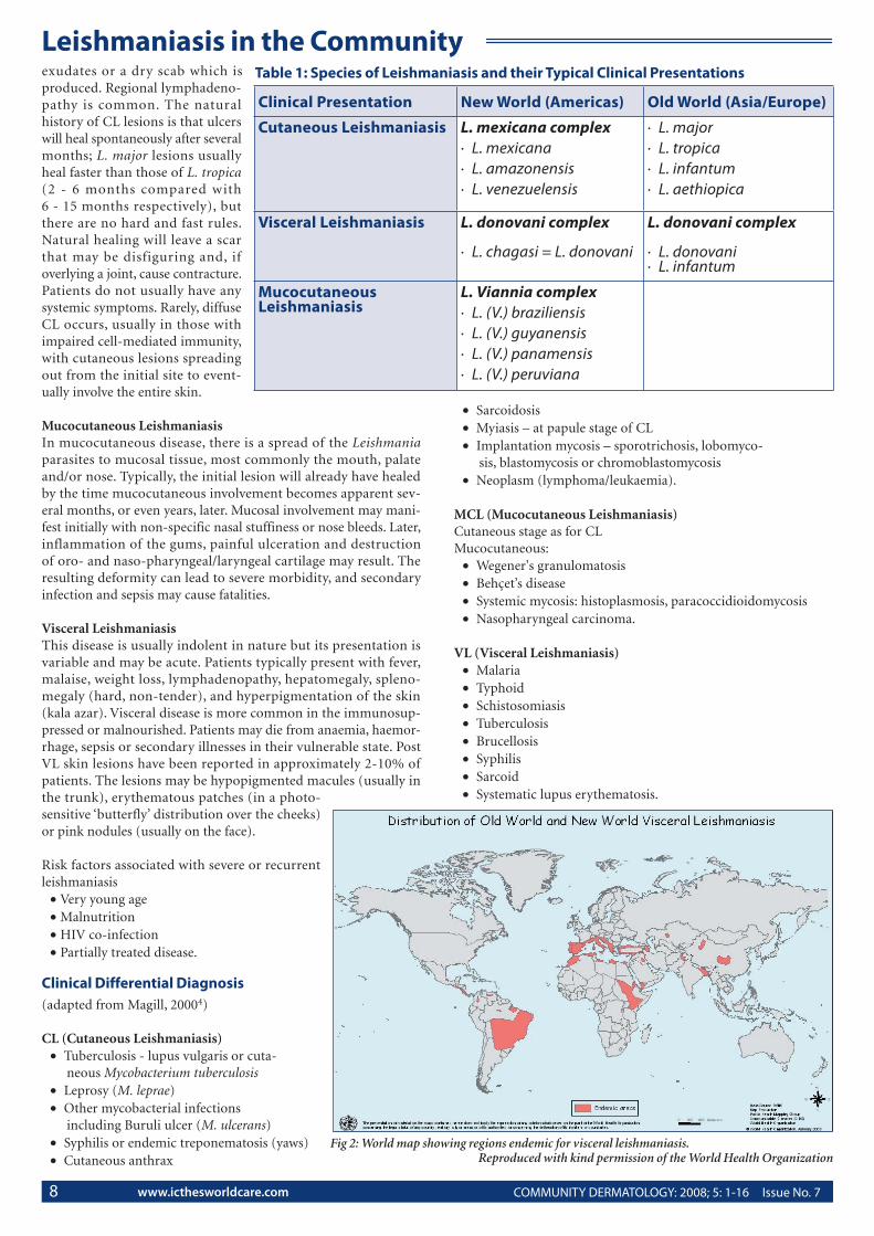
These maps of the world shows the distribution of leishmaniasis disease (Figures 1 and 2) across 88 countries with very different ecological habitats – from rainforest to arid desert. Reservoirs of infection for transmission of the different Leishmania species via the sandfly vector may be human (anthroponotic) or animal (zoo-notic), and these differences have fundamental implications for disease control strategies.
The highest prevalence of zoonotic CL is seen in Iraq, Afghanistan, Iran, Saudi Arabia, Brazil and Peru, whereas over 90% of global anthroponotic VL is found in India, Nepal, Bangladesh, Brazil and the Sudan. Historically, CL was mainly confined to rural areas where reservoir host animals live, but more recently the spread to peri-urban populations is increasing.
It is estimated that 350 million people are living at risk in leishma-nia-endemic regions and that 2 million new cases of leishmaniasis occur annually (3:1 CL:VL). This may, however, be a gross underes-timate as only one third of the affected countries have compulsory reporting.1 The disease has been somewhat neglected as a major global health problem until recently when several factors raised its profile. These include large epidemics in refugee populations, a gradual shift from rural to more populated peri-urban areas, an increasing prevalence of co-infection of leishmaniasis with HIV, producing more severe forms of visceral disease with higher mor-tality rates.2 Also, significant drug resistance has emerged to pose major treatment problems in some regions.
The female sandfly (Figure 3) typically bites animal or human hosts in the evening or night. Bites are usually not recalled as being painful. Exposed skin sites are the most commonly bitten, espe-cially the face and hands. Children and men working outdoors are most frequently affected, although a single sandfly may bite and
infect several members of the same household. Bed-nets and curtains impregnated with insecti-cide help to prevent transmission.3
In endemic areas, communities have long recog-nised the local disease manifestations with descriptions such as ‘Balkan sore’ (CL), ‘Delhi boil’ (CL), and ‘black fever’ (VL). Young children have been deliberately inoculated at covered skin sites in order that they may develop immunity and, thus, prevent scarring facial lesions in later life.
Clinical PresentationCutaneous LeishmaniasisAt the s i te o f the sandf ly b i te a smal l erythematous papule develops within a few weeks. The painless papule enlarges, develops a dark red or purple colour and after a few months ulcerates (Figure 4). Traditionally the ulcers have been termed ‘dry’ or ‘wet’, depending on ulcer
DIAGNOSING AND MANAGING LEISHMANIASIS IN THE COMMUNITY
������������
Fig 1: World map showing regions endemic for cutaneous and muco-cutaneous leishmaniasis. Reproduced with kind permission of World Health Organization
8 COMMUNITY DERMATOLOGY: 2008; 5: 1-16 Issue No. 7 www.icthesworldcare.com
Leishmaniasis in the Communityexudates or a dry scab which is produced. Regional lymphadeno-pathy is common. The natural history of CL lesions is that ulcers will heal spontaneously after several months; L. major lesions usually heal faster than those of L. tropica (2 - 6 months compared with 6 - 15 months respectively), but there are no hard and fast rules. Natural healing will leave a scar that may be disfiguring and, if overlying a joint, cause contracture. Patients do not usually have any systemic symptoms. Rarely, diffuse CL occurs, usually in those with impaired cell-mediated immunity, with cutaneous lesions spreading out from the initial site to event-ually involve the entire skin.
Mucocutaneous LeishmaniasisIn mucocutaneous disease, there is a spread of the Leishmania parasites to mucosal tissue, most commonly the mouth, palate and/or nose. Typically, the initial lesion will already have healed by the time mucocutaneous involvement becomes apparent sev-eral months, or even years, later. Mucosal involvement may mani-fest initially with non-specific nasal stuffiness or nose bleeds. Later, inflammation of the gums, painful ulceration and destruction of oro- and naso-pharyngeal/laryngeal cartilage may result. The resulting deformity can lead to severe morbidity, and secondary infection and sepsis may cause fatalities.
Visceral Leishmaniasis This disease is usually indolent in nature but its presentation is variable and may be acute. Patients typically present with fever, malaise, weight loss, lymphadenopathy, hepatomegaly, spleno-megaly (hard, non-tender), and hyperpigmentation of the skin (kala azar). Visceral disease is more common in the immunosup-pressed or malnourished. Patients may die from anaemia, haemor-rhage, sepsis or secondary illnesses in their vulnerable state. Post VL skin lesions have been reported in approximately 2-10% of patients. The lesions may be hypopigmented macules (usually in the trunk), erythematous patches (in a photo-sensitive ‘butterfly’ distribution over the cheeks) or pink nodules (usually on the face).
Risk factors associated with severe or recurrent leishmaniasis
• Very young age• Malnutrition• HIV co-infection• Partially treated disease.
Clinical Differential Diagnosis (adapted from Magill, 20004)
CL (Cutaneous Leishmaniasis)• Tuberculosis - lupus vulgaris or cuta-
neous Mycobacterium tuberculosis • Leprosy (M. leprae) • Other mycobacterial infections
including Buruli ulcer (M. ulcerans) • Syphilis or endemic treponematosis (yaws) • Cutaneous anthrax
• Sarcoidosis • Myiasis – at papule stage of CL • Implantation mycosis – sporotrichosis, lobomyco-
sis, blastomycosis or chromoblastomycosis • Neoplasm (lymphoma/leukaemia).
MCL (Mucocutaneous Leishmaniasis) Cutaneous stage as for CLMucocutaneous:
• Wegener's granulomatosis • Behçet’s disease • Systemic mycosis: histoplasmosis, paracoccidioidomycosis • Nasopharyngeal carcinoma.
VL (Visceral Leishmaniasis)• Malaria • Typhoid • Schistosomiasis • Tuberculosis • Brucellosis • Syphilis • Sarcoid • Systematic lupus erythematosis.
Clinical Presentation New World (Americas) Old World (Asia/Europe)
Cutaneous Leishmaniasis L. mexicana complex· L. mexicana· L. amazonensis· L. venezuelensis
· L. major· L. tropica· L. infantum· L. aethiopica
Visceral Leishmaniasis L. donovani complex
· L. chagasi = L. donovani
L. donovani complex
· L. donovani· L. infantum
Mucocutaneous Leishmaniasis
L. Viannia complex· L. (V.) braziliensis· L. (V.) guyanensis· L. (V.) panamensis· L. (V.) peruviana
Table 1: Species of Leishmaniasis and their Typical Clinical Presentations
Fig 2: World map showing regions endemic for visceral leishmaniasis. Reproduced with kind permission of the World Health Organization

COMMUNITY DERMATOLOGY: 2008; 5: 1-16 Issue No. 7 9www.icthesworldcare.com
Investigations Experienced health care workers in endemic areas often diagnose leishmaniasis on the clinical appearances alone with a high degree of accuracy.5 Moreover, in many endemic regions laboratory inves-tigations are not routinely available.
1. MicroscopyWhere light microscopy can be performed, training to recognise parasites on tissue smears will allow a definitive diagnosis of leish-maniasis. Smears are spread on a glass slide and stained before viewing.
• CL (MCL)For CL or the cutaneous stage of MCL, smears can be prepared from a slit skin scalpel scraping or a biopsy touch preparation of an ulcer. Scalpel scrapings are best taken from the indurated mar-gin of the most recent lesions, as these are most likely to harbour more parasites. The skin should be cleaned before sampling, and then the ulcer edge stretched tight. A small incision should be made radially across the indurated edge to a depth of a few mil-limetres. While the skin is still stretched to produce a bloodless field, the scalpel blade is turned 90o and scraped along the indu-rated cut edge. A small amount of serous tissue fluid can then be gathered on the scalpel blade and smeared immediately onto a glass microscope slide. This material is menthol-fixed (70%) and then stained with Giemsa/Leishman or Wright stains and exam-ined (usually x 500-1000, under oil immersion). Leishmania par-asite amastigotes may be seen inside macrophages and mononu-clear cells, but not uncommonly are squeezed out and lie ‘free’ between cells. The amastigotes are typically ovoid, measuring 2-5 µm by 1-2 µm and are readily recognised by their large nucleus and distinct pathognomonic kinetoplast (giving the so-called ‘dot and dash’ appearance; see Figure 5).
• VLSmears for diagnostic microscopy may also be made in cases of possible VL, using peripheral venous blood, fine-needle splenic aspiration, or rarely liver or bone marrow biopsy. When using venous blood, samples should either be centrifuged for buffy coat examination or thick blood films made to improve the detection rate, as parasite density in peripheral blood may be very low.
2. CultureLeishmania parasites can be cultured over 1-4 weeks from skin, blood, bone marrow or splenic aspirates using Novy-McNeal-
Nicolle (NMN) medium. The technique is time consuming and requires a laboratory with facilities for microscopy, refrigerated storage of medium, and parasite incubation at 22oC.
3. Leishmanin skin test (Montenegro test)This intradermal injection of killed promastigotes is used to assess cell mediated immunity to Leishmania parasites in the form of a delayed type hypersensitivity reaction. In areas endemic for CL and MCL, a positive result is frequently present in a large proportion of the population, reflecting previous exposure, and may, there-fore, have limited diagnostic use in individual cases. False positive results have been reported with diseases such as sporotrichosis.6
In VL, the sensitivity of the test is initially poor, due to the global immunosuppression caused by the disease, although this improves by the time of treatment completion.
The use of the test is further complicated by the variability in batch production, and it is, therefore, perhaps best reserved for epidemi-ological studies rather than individual case diagnosis.
4. SerologyA more specific test for diagnosing visceral disease is the direct agglutination test (DAT). This uses stable freeze-dried antigen, the test is inexpensive, reliable, easy to use and highly sensitive and specific.7 This test can be used in the community setting. The test is also reasonably reliable at diagnosing VL in the context of HIV co-infection.
5. Molecular methodsThere are a huge range of molecular techniques that have been described to diagnose leishmaniasis. Many use the multiple copies of kinetoplast DNA to detect infection. However, given the enor-mous variety of Leishmania species and sub-species, there is no single molecular test that will reliably detect and distinguish all human Leishmania parasites. Such tests are most appropriate for diagnosis in areas where there is little genetic variation in the para-sites being detected.
Currently, molecular tests remain too expensive for routine use in resource-stretched settings. Nevertheless, in laboratories where there are sufficient resources, molecular tests have been applied with great success to detect, speciate* and, therefore, direct treat-ment. However, a negative PCR result does not rule out leishmani-asis.8
6. Indirect methodsVL produces a profound hypergammaglobulinaemia. Historically, in field conditions, a crude and non-specific but rapid method for diagnosing leishmaniasis was to add formalin (40%) to patient serum and look for a white precipitant indicating high protein levels.
Non-specific haematological parameters that may support the diagnosis of leishmaniasis include low haemoglobin, low lympho-cyte and neutrophil counts as well as low platelets, all reflecting the suppression of the bone marrow by parasite infiltration.
ManagementImmunisationAlthough there has been much speculation about the potential for successful immunisation against leishmaniasis, the unfortunate
Leishmaniasis in the Community
Fig 3: Female phlebotomine sandflyFirst published in Morris-Jones S & Weber M. New Engl J Med 2004:
350; 2313-4. Copyright © [2004] Massachusetts Medical Society. All rights reserved.
*Speciation – The evolutionary formation of new biological species, usually by the division of a single species into two or more genetically distinct ones

10 COMMUNITY DERMATOLOGY: 2008; 5: 1-16 Issue No. 7 www.icthesworldcare.com
reality is that no effective vaccine is available. Traditional inocu-lation of ‘live’ leishmaniasis into the skin at a covered skin site has long been recognised as a way of producing long-lasting immunity. However vaccination with killed parasites or recombinant proteins induces only temporary protection.9
CL
Cutaneous leishmaniasis is a disfiguring disease associated with social stigma but otherwise does not cause significant morbidity or mortality. Lesions will resolve spontaneously (usually within 5-15 months) and many of those affected never seek medical attention. The rationale for therapy is that more rapid healing will result in less scarring. Both cryotherapy10 and heat treatment11 have been used with some success for isolated lesions. When cutaneous lesions are single or few in number, they may be treated simply with a series of intralesional sodium stibogluconate (pentavalent antimony) injections. The entire lesion/ulcer margin should be infiltrated at each injection, which may be painful. The lesions usually heal with some post-inflammatory pigment change and mild scarring.
However, in many countries those affected do not usually attend a medial practitioner unless they have exhausted traditional rem-edies first. A study in Ecuador shows that locals were very familiar with sarna brava (‘angry sore’) which they would treat themselves using a variety of therapies including heat, cold, cauterization, strong chemicals and plant preparations. Extracts from 45 differ-ent plants were used – including banana pulp, papaya juice, lemon peel/juice, and tobacco leaves.12 In Afghanistan, electro-thermo-cauterisation under local anaesthetic has been used successfully to treat CL for many years. Honey has been shown to keep cutaneous lesions clean, reduce secondary bacterial infection and even reduce the size of some lesions in an animal study.13
In Sudan, there are studies showing the effectiveness of gar-lic methanol extracts which were as effective as antimonial ther-apy; 66% of cutaneous lesions healed completely and 24% par-tially at around 20 days.14 Neem paste was less effective than garlic, although 33% of patients had complete healing and 50% partial healing at 20 days. Garad paste was not shown to be effective.14
Several studies undertaken in Iran have shown some encourag-ing results with the use of Z-HE paste made from a herbal extract and applied directly to the skin lesions. After 6 weeks of treatment, 74% were completely cured, 12% had a partial response and 14% no response.15
15% paromomycin (aminoglycoside antibiotic) in an ointment used twice daily for around 15 days has been shown to be effective with cure rates of 74% in old world disease and 85% in new world cutaneous leishmaniasis.
Recently, topical 5% imiquimod (Aldara) applied to cutaneous lesions, in addition to conventional therapies, has been shown to be effective with a higher proportion of lesions healing in a shorter time.16
For extensive cutaneous lesions, treatment should be systemic as for MCL or VL (see below).
MCL / VL / Extensive CL
For these forms of leishmaniasis treatment should be systemic. Traditionally, pentavalent antimonials (sodium stibogluconate and meglumine antimoniate) have been used IV (filtered and given slowly over 5 minutes) or IM at a dose of 20mg/kg/day for 20-40 days (10 days for CL). Patients not infrequently develop gastroin-
testinal symptoms, arthralgia, headache, abnormal liver function, raised amylase, low platelets and a disordered heart rhythm. Rarely, bleeding from mucous membranes and substernal pain may result. Rarely, when MCL is treated, a severe inflammatory reaction can result in significant tissue swelling in the oropharynx that should be managed with systemic corticosteroids. Although reasonably high cure rates of 76% are reported,17 there are a number of prob-lems with antimonial therapy. The drug is not universally avail-able; its side effect profile may limit adherence, as does the dura-tion of required treatment. Recently, the rapid emergence of resis-tance to pentavalent antimonials has been reported, particularly in the Indian Sub-Continent.
Second line therapy varies according to drug availability and costs. Pentamidine IM has been used, 3-4mg/kg on alternate days for a maximum of 10 injections, to treat VL or MCL, and twice weekly for CL, until the lesions resolve clinically. Initial responses are often good but high relapse rates are reported with visceral disease, and the treatment may need repeating.17 Adverse effects include hypo-tension (which may be severe, so that all patients should be treated lying down), hypoglycaemia, pancreatitis, arrhythmias, leucopenia, thrombocytopenia and acute renal failure.
Amphotericin B may also be used to treat leishmaniasis. The drug is administered IV at 1mg/kg/day for 10-21 days or, if available as the liposomal formulation, at 3mg/kg/day for 5 consecutive days, followed by a single dose one week later. Treatment doses and durations may need to be increased in the context of co-infection with HIV.
Leishmaniasis in the Community
Fig 4: Characteristic ulcer of cutaneous leishmaniasisFirst published in Morris-Jones S & Weber M. New Engl J Med 2004:
350; 2313-4. Copyright © [2004] Massachusetts Medical Society. All rights reserved.

COMMUNITY DERMATOLOGY: 2008; 5: 1-16 Issue No. 7 11www.icthesworldcare.com
Paromomycin (an aminoglycoside) given IM daily for 21 days, at a dose of 11mg/kg, has shown comparable results to amphotericin B in some studies.18
Standard treatments for VL/MCL are mostly cumbersome, expen-sive and often with a poor toxicity profile, underlining the urgent need for inexpensive, safe alternative therapies. The most promis-ing development in the treatment of VL has been in India where oral miltefosine, 100mg daily (2.5mg/kg/day) for 4 weeks, seems to be highly effective. Cure rates for visceral disease of 94% at 6 months are reported.19 The main drawbacks are compliance and teratogenicity (pregnancy must be avoided during the treatment and for 2 months afterwards).
VL / HIV Co-infection
Dual infection with HIV and VL poses particular clinical prob-lems. Diagnosis may be more difficult as presentations are atypi-cal, and parasites are more frequently found in unusual locations. Relapse and mortality rates are both higher than in VL alone, and drug toxicities, especially of the antimonials, are similarly more common in co-infected patients. Where highly active antiretrovi-ral therapy (HAART) is available, successful sustained suppression of viral replication may allow secondary prophylaxis to be consid-ered.
References
1. WHO website www.who.int/leishmaniasis
2. Desjeux P. The increase in risk factors for leishmaniasis worldwide. Trans Roy Soc Trop Med Hyg 2001; 95: 239-243.
3. Kroeger A., Avila E.V., Morison L. Insecticide impregnated curtains to control domestic transmission of cutaneous leishmaniasis in Venezuela: cluster randomised trial. BMJ 2002; 325: 810-813.
4. Magill A.J. Leishmaniasis. In Hunter’s Tropical Medicine and Emerging Infectious Diseases. Hunter G.W., Strickland G.T, Eds. 2000; 8th Ed. Philadelphia: Saunders. 665-687.
5. Arfanul B., Simeenber R. Correlation of clinical, his-topathological and microbiological findings in 60 cases of cutaneous leishmaniasis. Indian J Dermatol Venereol Leprosy 2006; 72(1): 28-32.
6. de Lima B., Schubach A., Francesconi-do-Valle A.C., et al. Positive Montenegro skin test among patients with sporot-richosis in Rio De Janeiro, Acta Trop 2005; 93(1): 41-47.
7. Abdallah K.A.A., Nour B.Y.M., et al. Evaluation of the direct agglutination test based on freeze-dried Leishmania donovani promastigotes for the serodiagnosis of visceral leishmaniasis in Sudanese patients. Trop Med Int Health 2004; 9(10): 1127-1131.
8. Singh S., Sivakumar R. Recent advances in the diagnosis of leishmaniasis. J Postgrad Med 2003; 49: 55-60.
9. Okwor I., Uzonna J. Persistent parasites and immunologic memory in cutaneous leishmaniasis: implications for vac-cine designs and vaccination strategies. Immunol Res 2008; April 4 (Epub ahead of print).
10. Gurei M.S., Tatli N., Ozbilge H., et al. Efficacy of cryother-apy and intralesional pentosan in treatment of cutaneous leishmaniasis. J Egypt Soc Parasitol 2000; 30: 169-176.
11. Navin T.R., Arana B.A., Arana F.E., et al. Placebo-controlled clinical trial of meglumine antimonate vs. localized con-trolled heat in the treatment of cutaneous leishmaniasis in Guatemala. Am J Trop Med Hyg 1990; 42: 43-50.
12. Weigel M.M., Armijos R.X. The traditional and convention-al medical treatment of cutaneous leishmaniasis in rural Ecuador. Pan Am J Public Health 2001; 10(6): 395-405.
13. Bafghi A.F., Zadeh H.F., Hejazian S.H. Leishmania major in the Balb/c mice and treatment with natural sweetener as a home remedy. World J Zoology 2008; 3(1): 19-24.
14. Khalid F.A., Abdalla N.M., Mohomed H.E., et al. Treatment of cutaneous leishmaniasis with some local Sudanese plants (Neem, Garad & Garlic). Turkiye Parazitoloji Dergisi 2004; 28(3): 129-132.
15. Zerehsaz F., Salmanpour R., Handjani F., et al. A double-blind randomized clinical trial of a topical herbal extract (Z-HE) vs. systemic meglumine antimoniate for the treat-ment of cutaneous leishmaniasis in Iran. Int J Dermatol 1999; 38: 610-612.
16. World Health Organization. New Treatment for Leishmaniasis is 95% Effective. Bull World Health Organ 2003; 80(8): 688.
17. Tuon F.F., Amato V.S., Graf M.E., et al. Treatment of New World cutaneous leishmaniasis – a systematic review with a meta-analysis. Int J Dermatol 2008; 47(2): 109-124.
18. Sundar S., Jha T.K., Thakur C.P., et al. Injectable paromo-mycin for visceral leishmaniasis in India. N Engl J Med 2007; 356(25): 2571-2581.
19. Sundar S., Olliaro P.L. Miltefosine in the treatment of leishmaniasis: Clinical evidence for informed clinical risk management. Ther Clin Risk Manag 2007; 3(5): 733-740.
Leishmaniasis in the Community
Fig 5: Leishmania amastiogtes seen on a slit skin smear taken from the edge of a cutaneous ulcer
First published in Morris-Jones S & Weber M. New Engl J Med 2004: 350; 2313-2314. Copyright © [2004] Massachusetts Medical Society.
All rights reserved.

12 COMMUNITY DERMATOLOGY: 2008; 5: 1-16 Issue No. 7 www.icthesworldcare.com
QuizQUIZ: ITCHY LINES QUESTIONS
L Claire Fuller MA FRCPConsultant Dermatologist East Kent Hospitals NHS TrustUK
Formerly: Senior Lecturer Regional Dermatology Training Centre (RDTC)Moshi, Tanzania
Question 1:
This fisherman’s son complains of intense itching on the buttock area. He is not itchy elsewhere:
a) What do you see?
b) What is the diagnosis?
c) What is the treatment?
Question 2:
This patient is itchy all over. She keeps noticing these straight linear raised streaks that last about 2 hours, then go down, but may appear somewhere else:
a) What is the diagnosis?
b) How would you treat it?
Question 3:
This child has a serpiginous wheal on his buttock. It has been there for about an hour. Earlier today he had a similar one on his back and yesterday one on his abdomen:
a) What is the diagnosis?
b) What is it caused by?
c) How would you treat it?
All figures are by kind permission of Professor Ben Naafs - with thanks to the Regional Dermatology Training Centre, Moshi, Tanzania

COMMUNITY DERMATOLOGY: 2008; 5: 1-16 Issue No. 7 13www.icthesworldcare.com
RDTC Research Reports
Provided by Dr Barbara Leppard
The late Yvonne Siyongo Caprivi, Namibia
In sub-Saharan Africa, the prevalence of STIs and AIDS is higher than anywhere else in the world. To look at one possible reason for this, Yvonne interviewed 125 male government workers from Katima Mulilo in Caprivi, Namibia and 11 local pastors - to ask about their knowledge and attitudes to the use of condoms. The government workers were asked if they knew what a condom was, if they knew how to use one, if they had ever used one, if they used one regularly, and if not, why not. The pastors were asked what they were teaching their congregations about the use of condoms.Seventy-four percent (74%) of male government workers knew what a condom was and how to use it, but 39% had never used one. The reasons given for not using them included:
• They are unnatural• They reduce sexual satisfaction• Their partner did not like them• Their partner would assume they were
being unfaithful if they used them• They are anti-Christian• Traditional herbs are a better protection
against STIs and AIDS than condoms• Traditional herbs are better for family planning than condoms• They can only be obtained at the local hospital or health clinic.
All pastors said that their church taught that individuals should be chaste before marriage and faithful afterwards. Therefore, there was no need for the use of condoms.
She concluded that there was a good case for the education of both men and women in the use of condoms in order to stop the spread of STIs and AIDS and that they should be more widely available, e.g., in shops as well as health centres and hospitals. The church needs to emphasise the importance of behaviour change as the real way to prevent STIs and AIDS.
Mpumelolo Dlamini Mbabane, Swaziland
Contact tracing and partner notification is an important part of the strategy for treating STIs all over the world. In the STI clinic at Mbabane Government Hospital, this is done by giving the patient a slip of paper to give to their sexual contacts, advising them to attend the clinic.
In a study of 318 patients attending the clinic (60% male, 40% female), only 27% of their contacts attended. Contacts of patients who had multiple sexual partners were less likely to respond to this method of partner notification than those with only one partner.These findings indicate that this type of partner notification is not an effective method of contact tracing, particularly for individuals with multiple sexual partners. The clinic needs to revise its strat-egy if they want to be more successful.
Marie Michelle Boum Yaounde, Cameroon
Charles HakomaMazabuka, Zambia
Self medication in patients with STIs can result in:
• Drug resistance• Increased incidence of HIV if the wrong treatment is given
(STIs are known to facilitate transmission of HIV)• Pelvic inflammatory disease leading to
ectopic pregnancy and sterility• Vertical transmission of disease to the unborn
child leading to ophthalmia neonatorum, congenital syphilis and HIV infection.
Both studies looked at the extent of self medication in patients attending an STI clinic. The investigators used a questionnaire to find out why patients treated themselves rather than going to an STI clinic, who they asked for advice and what treatment they had used.
ResultsBoth studies concluded that the government of their respective countries needed to look at the fees charged for attending clinics and having the relevant tests done. There was also a need to make sure that the appropriate drugs were both available and afford-able in the clinics. In addition, the general public needs education about the need for proper treatment of STIs in order to prevent spread of these diseases - to prevent complications and as a means of reducing the incidence of HIV/AIDS.
Zambia Cameroon
Number of patients interviewed
112 150
% of patients attending the STI clinic who had already treated themselves
80% 72%
Reasons for self medication
• High fees at clinics
• Lack of drugs at clinics
• Long waiting time at clinics
• High fees for lab tests at clinics
• Lack of drugs at clinics
• Long waiting time at clinics
• Didn’t want wife/husband to know about the problem
Treatment used Antibiotics 19%
Traditional medicine 36%
Traditional medicine and antibiotics 30%
Other drugs 15%
Antibiotics 77%
Traditional medicine 11%
Other drugs 12%
Received advice from Relatives and friends
Relatives and friends
Self medication in patients attending clinics for STIs in Cameroon and ZambiaWhy are Namibian men reluctant to
wear condoms?
Efficacy of partner notification in Swaziland
REGIONAL DERMATOLOGY TRAINING CENTRE RESEARCH REPORTS

14 COMMUNITY DERMATOLOGY: 2008; 5: 1-16 Issue No. 7 www.icthesworldcare.com
Quiz
Answer 1:
a) Serpiginous wheals
b) Cutaneous larva migrans, creeping eruption. Caused by various nematode parasites, e.g., Ancylostoma braziliense
c) Ivermectin or albendazole
Answer 2:
a) Dermographism or physical urticaria
b) Antihistamines, like chlorpheniramine, might help, but also avoiding things that encourage urti-caria, such as some pain killers. This lady was taking ibuprofen for abdominal pains she was experiencing during menses. It can also happen with codeine.
Answer 3:
a) Larva currens
b) Threadworm or human parasitic roundworm called Strongyloides stercoralis
c) Ivermectin, albendazole or thiabendazole
QUIZ: ITCHY LINES ANSWERS
Contact: Dr Murray McGavinICTHES World Care, PO Box 4101Glasgow G4 7BX, Scotland, UKTel: +44 (0) 141 332 1707E-mail: [email protected]
Please state: Name, Full Postal Address, E-mail Address & Occupation
• Community Ear and Hearing Health• Community Dermatology
• Developing Mental Health
www.icthesworldcare.com
Journals available FREE to Developing Countries

COMMUNITY DERMATOLOGY: 2008; 5: 1-16 Issue No. 7 15www.icthesworldcare.com
The International Lymphoedema Framework and Wounds UK are holding the first International Lymphoedema Framework Conference, between 21 and 23 April 2009, at the new Conference Centre in Ascot, near London, UK. This will be an exciting event using innovative approaches with the main focus of the conference being to push forward the development of lymphoedema care worldwide. The conference will be very much about international partnership and bringing together key groups, including patients, involved in this important work.
Book Review
An Illustrated Guide
Merja Kousa and Cornelus Sanders(Kopijyvä Oy, Jyväskylä, 2006)
ISBN 952-92-0163-X
This compact ring-plan handbook is an excellent summary of common skin mani-festations of HIV-AIDS, including concise text and a summary of currently avail-able treatments. This is accompanied by profuse illustrations, mostly of good quality, and many derived (with acknowledgement) from the Regional Dermatology Train-ing Centre, Moshi, Tanzania. Most of the treatment recommendations are very sen-sible (although I would not advocate ‘potent corticosteroid ointments’ for psoria-sis in general). Certainly the book will prove of great value to health care workers in Africa (and other parts of the world, for that matter). The book is currently avail-able, for the cost of postage, from the Teaching Aids at Low Cost (TALC) website – www.talcuk.org - together with many other reasonably priced books.
Chris Lovell
HIV RELATED SKIN DISEASES AND SEXUALLY TRANSMITTED INFECTIONS IN AFRICA
First Annual International Lymphoedema Conference & Exhibition 2009
For more details or to register, please
go online to: www.wounds-uk.com
21 - 23 April, 2009Royal Ascot Conference Centre near London, UK

16 COMMUNITY DERMATOLOGY: 2008; 5: 1-16 Issue No. 7 www.icthesworldcare.com
Community Dermatology
Community Dermatology
EditorDr Paul Buxton
Editorial BoardDr James BinghamDr Neil CoxDr Claire FullerDr Sam GibbsDr Richard GoodwinProfessor Rod HayDr Barbara LeppardDr Chris LovellDr Murray McGavinDr Michele MurdochMs Rebecca PenzerProfessor Terence RyanDr Michael Waugh
International ConsultantsProfessor Henning Grossman (Tanzania)Professor Donald Lookingbill (USA)Professor Robin Marks (Australia)Professor Aldo Morrone (Italy)Professor Ben Naafs (The Netherlands)Professor Gail Todd (South Africa)Dr Shyam Verma (India)
Editorial SecretaryMr John Caulfield
Published by ICTHES World CareHonorary President / EditorDr Murray McGavinInternational Development OfficerMrs Mary BromilowAssociate Editor Ms Caroline McGavinAdministration / DistributionMrs Manon McInarlin
Design/DTPMr Daniel Chadney www.pixelmix.org
Printed bySquare Dot CTP Services Ltd Nicosia, [email protected]
ISSN 1743 - 9906
© Community DermatologyArticles may be photopied, reproduced or translated provided these are not used for commercial or personal profit. Acknowledgements should
be made to the author(s) and to Community Dermatology
Guidelines For AuthorsThe Editorial Board welcomes original articles, reports and letters. All contri-butions are reviewed before publication. Original articles should not exceed 1,200 words; short reports/letters should not exceed 500 words. Contributions should follow the detailed Guidelines which can be obtained from ICTHES World Care (postal and email addresses below).
Contributions should be sent to the Edi-tors.
Dr Paul K Buxton (Editor) & Dr Christopher Lovell (Deputy Editor)Community DermatologyBritish Association of Dermatologists4 Fitzroy SquareLondon W1T 5HQUnited Kingdom
by Email to:[email protected]
Copy to: [email protected]
We look forward to receiving your arti-cles, reports and letters!
DistributionThe Journal is distributed free of charge to health care workers, especially in rural communities, in developing countries. To join the mailing list, please contact: Dr Murray McGavin at ICTHES World Care (address below).
SubscriptionsThe Journal is funded entirely by vol-untary donations and sponsorship. We welcome individual subscriptions (min-imum £20 per annum) to enable pub-lication and distribution to readers in developing countries. If you pay UK tax we can retrieve Gift Aid at no extra cost to you.
Dr Murray McGavinICTHES World CarePO Box 4101GLASGOWG4 7BXScotland, UK Tel: 0044 (0) 141 332 1707Email: [email protected]: www.icthesworldcare.com
An International Journal for Community Skin Health
J Comm Dermatol 2008; 5: 1 – 16 Issue No. 7
For more information, and details of future Courses, contact:
Dr Chavalit Mangkalaviraj,Course Director, COTTISA, c/o Bangrak Hospital, 189 Sathorn Road, Bangkok 10120, ThailandTel: 66-2-676 5383 Fax: 66-2-286 3013
E mail: [email protected] Website: www.cottisa.org
STDs/AIDS Diploma Course
This Course is open to doctors and nurses wishing to develop their skills in diagnosis and management of sexually transmitted diseases.
27th October - 21 November 2008
The Scottish GovernmentCommunity Dermatology is supported by
Community Dermatology
Community Dermatology
Issues 1 to 7 now available online atwww.icthesworldcare.com
Community Dermatology