Artists, Brands and Products Appearances in the Billboard Hot ...
Upload
khangminh22Category
view
0download
0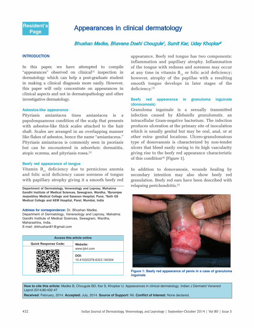
Indian Journal of Dermatology, Venereology, and Leprology | September-October 2014 | Vol 80 | Issue 5432
Appearances in clinical dermatology
Bhushan Madke, Bhavana Doshi Chougule1, Sumit Kar, Uday Khopkar2
INTRODUCTION
In this paper, we have attempted to compile “appearances” observed on clinical[1] inspection in dermatology which can help a post‑graduate student in making a clinical diagnosis more easily. However, this paper will only concentrate on appearances in clinical aspects and not in dermatopathology and other investigative dermatology.
Asbestos‑like appearancePityriasis amiantacea tinea amiantacea is a papulosquamous condition of the scalp that presents with asbestos‑like thick scales attached to the hair shaft. Scales are arranged in an overlapping manner like flakes of asbestos, hence the name “amiantaceus.” Pityriasis amiantacea is commonly seen in psoriasis but can be encountered in seborrheic dermatitis, atopic eczema, and pityriasis rosea.[2]
Beefy red appearance of tongueVitamin B12 deficiency due to pernicious anemia and folic acid deficiency cause soreness of tongue with papillary atrophy giving it a smooth beefy red
appearance. Beefy red tongue has two components: inflammation and papillary atrophy. Inflammation of the tongue with redness and soreness may occur at any time in vitamin B12 or folic acid deficiency; however, atrophy of the papillae with a resulting smooth tongue develops in later stages of the deficiency.[3]
Beefy red appearance in granuloma inguinale (donovanosis)Granuloma inguinale is a sexually transmitted infection caused by Klebsiella granulomatis. an intracellular Gram‑negative bacterium. The infection produces ulceration at the primary site of inoculation which is usually genital but may be oral, anal, or at other extra‑ genital locations. Ulcero‑granulomatous type of donovanosis is characterized by non‑tender ulcers that bleed easily owing to its high vascularity giving rise to the beefy red appearance characteristic of this condition[4] [Figure 1].
In addition to donovanosis, wounds healing by secondary intention may also show beefy red granulation. Beefy red ears have been described with relapsing perichondritis.[5]
Resident’s Page
Department of Dermatology, Venereology and Leprosy, Mahatma Gandhi Institute of Medical Sciences, Sewagram, Wardha, 1Byramjee Jeejeebhoy Medical College and Sassoon Hospital, Pune, 2Seth GS Medical College and KEM Hospital, Parel, Mumbai, India
Address for correspondence: Dr. Bhushan Madke, Department of Dermatology, Venereology and Leprosy, Mahatma Gandhi Institute of Medical Sciences, Sewagram, Wardha, Maharashtra, India. E‑mail: [email protected]
How to cite this article: Madke B, Chougule BD, Kar S, Khopkar U. Appearances in clinical dermatology. Indian J Dermatol Venereol Leprol 2014;80:432‑47.
Received: February, 2014. Accepted: July, 2014. Source of Support: Nil. Conflict of Interest: None declared.
Access this article online
Quick Response Code: Website: www.ijdvl.com
DOI: 10.4103/0378-6323.140304
PMID:***** Figure 1: Beefy red appearance of penis in a case of granuloma
inguinale
Madke, et al. Appearances in clinical dermatology
433Indian Journal of Dermatology, Venereology, and Leprology | September-October 2014 | Vol 80 | Issue 5
Butterfly appearance of rashButterfly rash (malar rash) is a well‑defined fixed erythema located over the butterfly area of face comprising both malar region and bridge of nose with sparing of the naso‑labial fold. Butterfly rash is classically seen in lupus erythematosus. However, other differentials to be considered are rosacea, acne, seborrheic dermatitis, photo‑allergic contact dermatitis polymorphous light eruption, measles, rubella, roseola infantum and dermatomyositis.[6]
Cauliflower appearanceGenital wart, or condylomata acuminata, is a human papillomavirus‑associated sexually transmitted infection and classically presents as a fleshy pink‑colored growth resembling a cauliflower.[7]
Cauliflower‑like growth has also been reported in verrucous carcinoma, a well‑differentiated invasive squamous cell carcinoma.[8]
Cauliflower appearance of ear is a well‑known entity in sports medicine. The outer ear (pinna) is composed mainly of cartilage covered by perichondrium, subcutaneous tissue and skin. Direct trauma or continuous friction to the outer ear can cause acute hematoma within the potential space between perichondrium and the cartilage. Separation of the perichondrium from the cartilage leads to necrosis of the cartilage because of the consequential loss of blood supply resulting in a “cauliflower ear” or “wrestler’s ear” deformity.[9]
Caviar‑like appearanceCaviar is a product made from salt‑cured fish‑eggs of the Acipenseridae family. In dermatology, caviar‑like pin‑point hyperpigmented papules are noted after long‑term application of hydroquinone, a feature of exogenous ochronosis.[10] Sublingual varices are benign vascular dilatations and give a caviar appearance to tongue.[11]
Cayenne pepper appearanceSchamberg disease is a type of pigmented purpuric dermatosis that is characterized by red‑brown purpuric macules that are known as “cayenne pepper” spots.[12]
Cigarette paper scar appearance (papyraceous scar)Tertiary syphilisThe coppery red grouped nodular lesions which tend to arrange in a circinate pattern in tertiary syphilis heal centrally by forming soft fine wrinkled scars (cigarette
paper scars) and extend peripherally. These lesions have a predisposition toward the arms, back and face.
Ehler Danlos syndromeThe skin in patients with Ehler–Danlos syndrome is hyperextensible which gives rise to gaping “fish‑mouth wounds” over bony prominences like the shins, knees, and elbows following minor trauma. Such wide, thin, papyraceous scars over the knees and elbows are also called “cigarette paper scars.”[13]
Cigarette paper appearance of mycosis fungoidesThe earliest form of cutaneous T‑cell lymphoma is the patch stage, which consists of sharply demarcated, erythematous scaly lesions. The scale is typically thin and covers the entire affected area. Mild erythema along with skin atrophy is also present giving a “cigarette paper”‑like wrinkled appearance.[14,15]
Cigarette paper appearance can also be seen in lichen sclerosus,[16] striae distensae and poikiloderma atrophicans vasculare.
Cliff drop appearanceAtrophoderma of Pasini and Pierini is a form of dermal atrophy that manifests as single or multiple sharply demarcated, round to oval, hyperpigmented, non‑indurated patches of varying sizes ranging from a few millimeters to several centimeters. The border of the lesions shows depression of the skin with an abrupt edge, often giving a “cliff drop” appearance.[17]
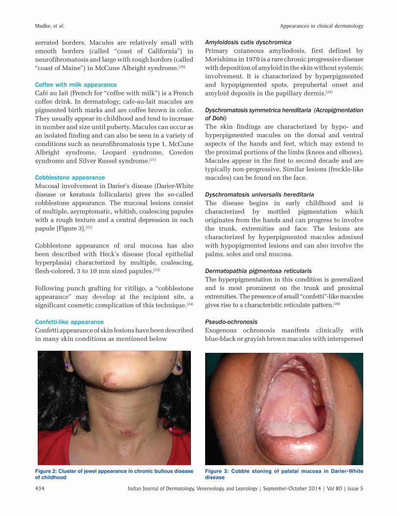
Cluster of jewels or string of pearls in Linear IgA dermatosesChronic bullous dermatosis of childhood (CBDC) is a rare, chronic acquired subepidermal blistering disease of children. Clinically, it is typified by the abrupt onset of pruritic or non‑pruritic tense vesicles or bullae that can arise either on normal or erythematous skin. Most often these lesions occur in a characteristic configuration giving a “cluster of jewels” or “string of pearls” appearance. Vesicles and bullae are arranged in an annular or rosette‑ like pattern around a central crusted plaque[18] [Figure 2].
A similar cluster of jewel appearance has also been noted in Linear IgA dermatosis,[19] bullous pemphigoid and herpes gestationis.
Coast of California and Coast of Maine appearanceCafé‑au‑lait spots, also referred to as café‑au‑lait macules (CALMs), present as well‑circumscribed, evenly pigmented macules with irregular or
Madke, et al. Appearances in clinical dermatology
Indian Journal of Dermatology, Venereology, and Leprology | September-October 2014 | Vol 80 | Issue 5434
serrated borders. Macules are relatively small with smooth borders (called “coast of California”) in neurofibromatosis and large with rough borders (called “coast of Maine”) in McCune Albright syndrome.[20]
Coffee with milk appearanceCafé au lait (French for “coffee with milk”) is a French coffee drink. In dermatology, cafe‑au‑lait macules are pigmented birth marks and are coffee brown in color. They usually appear in childhood and tend to increase in number and size until puberty. Macules can occur as an isolated finding and can also be seen in a variety of conditions such as neurofibromatosis type 1, McCune Albright syndrome, Leopard syndrome, Cowden syndrome and Silver Russel syndrome.[21]
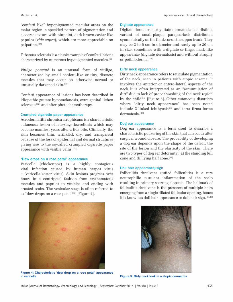
Cobblestone appearanceMucosal involvement in Darier’s disease (Darier‑White disease or keratosis follicularis) gives the so‑called cobblestone appearance. The mucosal lesions consist of multiple, asymptomatic, whitish, coalescing papules with a rough texture and a central depression in each papule [Figure 3].[22]
Cobblestone appearance of oral mucosa has also been described with Heck’s disease (focal epithelial hyperplasia) characterized by multiple, coalescing, flesh‑colored, 3 to 10 mm sized papules.[23]
Following punch grafting for vitiligo, a “cobblestone appearance” may develop at the recipient site, a significant cosmetic complication of this technique.[24]
Confetti‑like appearanceConfetti appearance of skin lesions have been described in many skin conditions as mentioned below
Amyloidosis cutis dyschromicaPrimary cutaneous amyliodosis, first defined by Morishima in 1970 is a rare chronic progressive disease with deposition of amyloid in the skin without systemic involvement. It is characterized by hyperpigmented and hypopigmented spots, prepubertal onset and amyloid deposits in the papillary dermis.[25]
Dyschromatosis symmetrica hereditaria (Acropigmentation of Dohi)The skin findings are characterized by hypo‑ and hyperpigmented macules on the dorsal and ventral aspects of the hands and feet, which may extend to the proximal portions of the limbs (knees and elbows). Macules appear in the first to second decade and are typically non‑progressive. Similar lesions (freckle‑like macules) can be found on the face.
Dyschromatosis universalis hereditariaThe disease begins in early childhood and is characterized by mottled pigmentation which originates from the hands and can progress to involve the trunk, extremities and face. The lesions are characterized by hyperpigmented macules admixed with hypopigmented lesions and can also involve the palms, soles and oral mucosa.
Dermatopathia pigmentosa reticularisThe hyperpigmentation in this condition is generalized and is most prominent on the trunk and proximal extremities. The presence of small “confetti”‑like macules gives rise to a characteristic reticulate pattern.[26]
Pseudo‑ochronosisExogenous ochronosis manifests clinically with blue‑black or grayish brown macules with interspersed
Figure 2: Cluster of jewel appearance in chronic bullous disease of childhood
Figure 3: Cobble stoning of palatal mucosa in Darier-White disease
Madke, et al. Appearances in clinical dermatology
435Indian Journal of Dermatology, Venereology, and Leprology | September-October 2014 | Vol 80 | Issue 5
“confetti like” hypopigmented macular areas on the malar region, a speckled pattern of pigmentation and a coarse texture with pinpoint, dark brown caviar‑like papules (vide supra), which are more appreciable on palpation.[27]
Tuberous sclerosis is a classic example of confetti lesions characterized by numerous hypopigmented macules.[28]
Vitiligo ponctué is an unusual form of vitiligo, characterized by small confetti‑like or tiny, discrete macules that may occur on otherwise normal or unusually darkened skin.[29]
Confetti appearance of lesions has been described in idiopathic guttate hypomelanosis, extra genital lichen sclerosus[30] and after photochemotherapy.
Crumpled cigarette paper appearanceAcrodermatitis chronica atrophicans is a characteristic cutaneous lesion of late‑stage borreliosis which may become manifest years after a tick bite. Clinically, the skin becomes thin, wrinkled, dry, and transparent because of the loss of epidermal and dermal structures giving rise to the so‑called crumpled cigarette paper appearance with visible veins.[31]
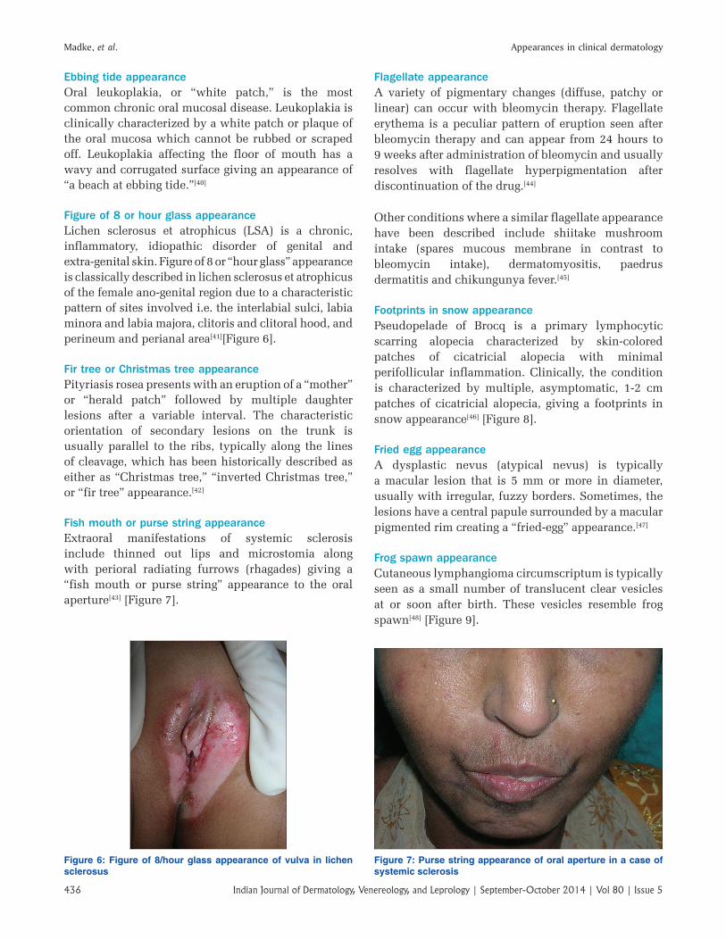
“Dew drops on a rose petal” appearanceVaricella (chickenpox) is a highly contagious viral infection caused by human herpes virus 3 (varicella‑zoster virus). Skin lesions progress over hours in a centripetal fashion from erythematous macules and papules to vesicles and ending with crusted scabs. The vesicular stage is often referred to as “dew drops on a rose petal”[32] [Figure 4].
Digitate appearanceDigitate dermatosis or guttate dermatosis is a distinct variant of small‑plaque parapsoriasis distributed symmetrically on the flanks or on the upper trunk. They may be 2 to 6 cm in diameter and rarely up to 20 cm in size, sometimes with a digitate or finger mark‑like appearance (digitate dermatosis) and without atrophy or poikiloderma.[33]
Dirty neck appearanceDirty neck appearance refers to reticulate pigmentation of the neck, seen in patients with atopic eczema. It involves the anterior or antero‑lateral aspects of the neck It is often interpreted as an “accumulation of dirt” due to lack of proper washing of the neck region by the child[34] [Figure 5]. Other cutaneous disorders where “dirty neck appearance” has been noted include X‑linked ichthyosis[35] and terra firma forme dermatosis.[36]
Dog ear appearanceDog ear appearance is a term used to describe a characteristic puckering of the skin that can occur after surgical wound closure. The probability of developing a dog ear depends upon the shape of the defect, the site of the lesion and the elasticity of the skin. There are two types of dog ear deformity: (a) the standing full cone and (b) lying half cone.[37]
Doll hair appearance/signFolliculitis decalvans (tufted folliculitis) is a rare neutrophilic purulent inflammation of the scalp resulting in primary scarring alopecia. The hallmark of folliculitis decalvans is the presence of multiple hairs emerging from a single dilated follicular opening, hence it is known as doll hair appearance or doll hair sign.[38,39]
Figure 4: Characteristic ‘dew drop on a rose petal’ appearance in varicella Figure 5: Dirty neck look in a atopic dermatitis
Madke, et al. Appearances in clinical dermatology
Indian Journal of Dermatology, Venereology, and Leprology | September-October 2014 | Vol 80 | Issue 5436
Ebbing tide appearanceOral leukoplakia, or “white patch,” is the most common chronic oral mucosal disease. Leukoplakia is clinically characterized by a white patch or plaque of the oral mucosa which cannot be rubbed or scraped off. Leukoplakia affecting the floor of mouth has a wavy and corrugated surface giving an appearance of “a beach at ebbing tide.”[40]
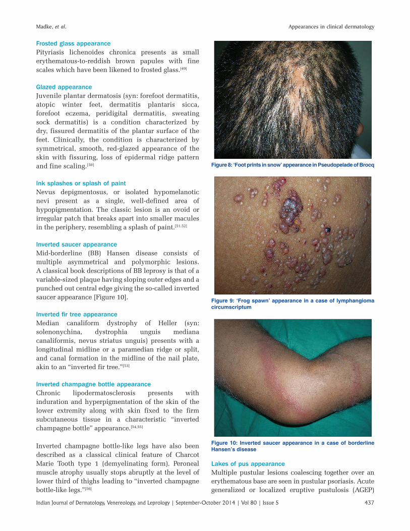
Figure of 8 or hour glass appearanceLichen sclerosus et atrophicus (LSA) is a chronic, inflammatory, idiopathic disorder of genital and extra‑genital skin. Figure of 8 or “hour glass” appearance is classically described in lichen sclerosus et atrophicus of the female ano‑genital region due to a characteristic pattern of sites involved i.e. the interlabial sulci, labia minora and labia majora, clitoris and clitoral hood, and perineum and perianal area[41][Figure 6].
Fir tree or Christmas tree appearancePityriasis rosea presents with an eruption of a “mother” or “herald patch” followed by multiple daughter lesions after a variable interval. The characteristic orientation of secondary lesions on the trunk is usually parallel to the ribs, typically along the lines of cleavage, which has been historically described as either as “Christmas tree,” “inverted Christmas tree,” or “fir tree” appearance.[42]
Fish mouth or purse string appearanceExtraoral manifestations of systemic sclerosis include thinned out lips and microstomia along with perioral radiating furrows (rhagades) giving a “fish mouth or purse string” appearance to the oral aperture[43] [Figure 7].
Flagellate appearanceA variety of pigmentary changes (diffuse, patchy or linear) can occur with bleomycin therapy. Flagellate erythema is a peculiar pattern of eruption seen after bleomycin therapy and can appear from 24 hours to 9 weeks after administration of bleomycin and usually resolves with flagellate hyperpigmentation after discontinuation of the drug.[44]
Other conditions where a similar flagellate appearance have been described include shiitake mushroom intake (spares mucous membrane in contrast to bleomycin intake), dermatomyositis, paedrus dermatitis and chikungunya fever.[45]
Footprints in snow appearancePseudopelade of Brocq is a primary lymphocytic scarring alopecia characterized by skin‑colored patches of cicatricial alopecia with minimal perifollicular inflammation. Clinically, the condition is characterized by multiple, asymptomatic, 1‑2 cm patches of cicatricial alopecia, giving a footprints in snow appearance[46] [Figure 8].
Fried egg appearanceA dysplastic nevus (atypical nevus) is typically a macular lesion that is 5 mm or more in diameter, usually with irregular, fuzzy borders. Sometimes, the lesions have a central papule surrounded by a macular pigmented rim creating a “fried‑egg” appearance.[47]
Frog spawn appearanceCutaneous lymphangioma circumscriptum is typically seen as a small number of translucent clear vesicles at or soon after birth. These vesicles resemble frog spawn[48] [Figure 9].
Figure 6: Figure of 8/hour glass appearance of vulva in lichen sclerosus
Figure 7: Purse string appearance of oral aperture in a case of systemic sclerosis
Madke, et al. Appearances in clinical dermatology
437Indian Journal of Dermatology, Venereology, and Leprology | September-October 2014 | Vol 80 | Issue 5
Frosted glass appearancePityriasis lichenoides chronica presents as small erythematous‑to‑reddish brown papules with fine scales which have been likened to frosted glass.[49]
Glazed appearanceJuvenile plantar dermatosis (syn: forefoot dermatitis, atopic winter feet, dermatitis plantaris sicca, forefoot eczema, peridigital dermatitis, sweating sock dermatitis) is a condition characterized by dry, fissured dermatitis of the plantar surface of the feet. Clinically, the condition is characterized by symmetrical, smooth, red‑glazed appearance of the skin with fissuring, loss of epidermal ridge pattern and fine scaling.[50]
Ink splashes or splash of paintNevus depigmentosus, or isolated hypomelanotic nevi present as a single, well‑defined area of hypopigmentation. The classic lesion is an ovoid or irregular patch that breaks apart into smaller macules in the periphery, resembling a splash of paint.[51,52]
Inverted saucer appearanceMid‑borderline (BB) Hansen disease consists of multiple asymmetrical and polymorphic lesions. A classical book descriptions of BB leprosy is that of a variable‑sized plaque having sloping outer edges and a punched out central edge giving the so‑called inverted saucer appearance [Figure 10].
Inverted fir tree appearanceMedian canaliform dystrophy of Heller (syn: solenonychina, dystrophia unguis mediana canaliformis, nevus striatus unguis) presents with a longitudinal midline or a paramedian ridge or split, and canal formation in the midline of the nail plate, akin to an “inverted fir tree.”[53]
Inverted champagne bottle appearanceChronic lipodermatosclerosis presents with induration and hyperpigmentation of the skin of the lower extremity along with skin fixed to the firm subcutaneous tissue in a characteristic “inverted champagne bottle” appearance.[54,55]
Inverted champagne bottle‑like legs have also been described as a classical clinical feature of Charcot Marie Tooth type 1 (demyelinating form). Peroneal muscle atrophy usually stops abruptly at the level of lower third of thighs leading to “inverted champagne bottle‑like legs.”[56]
Figure 8: ‘Foot prints in snow’ appearance in Pseudopelade of Brocq
Figure 9: ‘Frog spawn’ appearance in a case of lymphangioma circumscriptum
Figure 10: Inverted saucer appearance in a case of borderline Hansen’s disease
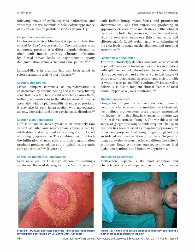
Lakes of pus appearanceMultiple pustular lesions coalescing together over an erythematous base are seen in pustular psoriasis. Acute generalized or localized eruptive pustulosis (AGEP)
Madke, et al. Appearances in clinical dermatology
Indian Journal of Dermatology, Venereology, and Leprology | September-October 2014 | Vol 80 | Issue 5438
following intake of cephalosporins, terbinafine, and vancomycin may also simulate the lake of pus appearance of lesions as seen in pustular psoriasis [Figure 11].
Leopard skin appearanceOnchocerciasis (river blindness) is a parasitic infection caused by Onchocerca volvulus. Onchocerciasis most commonly presents as a diffuse papular dermatitis, often with intense pruritis. Chronic infestation by filarial larvae leads to asymptomatic spotty depigmentation giving a “leopard skin” pattern.[57,58]
Leopard‑like skin eruption has also been noted in sclerodermatous graft‑vs‑host disease.[59]
Leathery appearanceLichen simplex chronicus or neurodermatitis is characterized by chronic itching and a self‑perpetuating scratch‑itch cycle. The constant scratching causes thick, leathery, brownish skin in the affected areas. It may be associated with atopic dermatitis (eczema) or psoriasis. It may also be seen in association with nervousness, anxiety, depression, and other psychological disorders.[60]
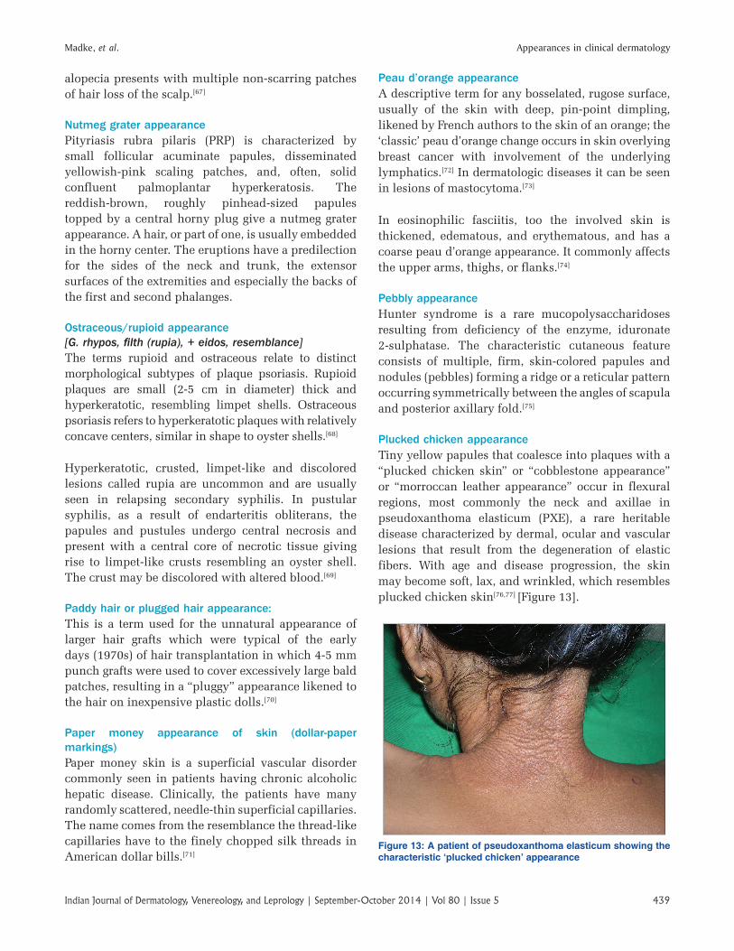
Leather grain appearanceDiffuse cutaneous mastocytosis is an extremely rare variant of cutaneous mastocytosis characterized by infiltration of skin by mast cells giving it a thickened and doughy appearance. The combined result of both the infiltration of mast cells and their degranulation products produces edema and a typical leather‑grain skin appearance[61,62] [Figure 12].
Lemon on match stick appearanceSeen as a part of Cushing’s disease or Cushing’s syndrome, the most striking feature is “central obesity”
with buffalo hump, moon facies, and protuberant abdominal wall and thin extremities, producing an appearance of “a lemon on match sticks.” Other clinical features include hypertension, muscle weakness, signs of excessive androgens (hirsutism, acne, and clitoromegaly). Rapid weight gain with thinning of the skin leads to striae on the abdomen and proximal extremities.[63]
Lobster claw appearanceThe term ectrodactyly denotes congenital absence of all or part of one or more fingers or toes and is synonymous with split hand or foot deformity or lobster claw. Lobster claw appearance of hand or feet is a classical feature of ectrodactyly, ectodermal dysplasia, and cleft lip with or without cleft palate (EEC) syndrome.[64] Lobster claw deformity is also a frequent clinical feature of focal dermal hypoplasia (Goltz syndrome).[65]
Map‑like appearanceGeographic tongue is a common asymptomatic condition characterized by multiple variably‑sized, well‑defined erythematous areas usually surrounded by elevated, whitish‑yellow borders on the anterior two third of dorsal surface of tongue. The variable size and shape of geographic tongue with frequent change in position has been referred as ‘map‑like’ appearance.[66] It has been proposed that benign migratory glossitis is an isolated oral manifestation of psoriasis. Geographic tongue may also be seen in other conditions like Reiter’s syndrome, Down syndrome, Aarskog syndrome, fetal hydantoin syndrome and Robinow’s syndrome.
Moth‑eaten appearanceMoth‑eaten alopecia is the most common and characteristic type of alopecia in syphilis. Moth eaten
Figure 11: Pustular psoriasis depicting ‘lake of pus’ appearance (Photograph contributed by Dr. Sarina Jain, Faridkot)
Figure 12: A child with diffuse cutaneous mastocytosis giving a leather grain appearance to the skin
Madke, et al. Appearances in clinical dermatology
439Indian Journal of Dermatology, Venereology, and Leprology | September-October 2014 | Vol 80 | Issue 5
alopecia presents with multiple non‑scarring patches of hair loss of the scalp.[67]
Nutmeg grater appearancePityriasis rubra pilaris (PRP) is characterized by small follicular acuminate papules, disseminated yellowish‑pink scaling patches, and, often, solid confluent palmoplantar hyperkeratosis. The reddish‑brown, roughly pinhead‑sized papules topped by a central horny plug give a nutmeg grater appearance. A hair, or part of one, is usually embedded in the horny center. The eruptions have a predilection for the sides of the neck and trunk, the extensor surfaces of the extremities and especially the backs of the first and second phalanges.
Ostraceous/rupioid appearance[G. rhypos, filth (rupia), + eidos, resemblance]The terms rupioid and ostraceous relate to distinct morphological subtypes of plaque psoriasis. Rupioid plaques are small (2‑5 cm in diameter) thick and hyperkeratotic, resembling limpet shells. Ostraceous psoriasis refers to hyperkeratotic plaques with relatively concave centers, similar in shape to oyster shells.[68]
Hyperkeratotic, crusted, limpet‑like and discolored lesions called rupia are uncommon and are usually seen in relapsing secondary syphilis. In pustular syphilis, as a result of endarteritis obliterans, the papules and pustules undergo central necrosis and present with a central core of necrotic tissue giving rise to limpet‑like crusts resembling an oyster shell. The crust may be discolored with altered blood.[69]
Paddy hair or plugged hair appearance:This is a term used for the unnatural appearance of larger hair grafts which were typical of the early days (1970s) of hair transplantation in which 4‑5 mm punch grafts were used to cover excessively large bald patches, resulting in a “pluggy” appearance likened to the hair on inexpensive plastic dolls.[70]
Paper money appearance of skin (dollar‑paper markings)Paper money skin is a superficial vascular disorder commonly seen in patients having chronic alcoholic hepatic disease. Clinically, the patients have many randomly scattered, needle‑thin superficial capillaries. The name comes from the resemblance the thread‑like capillaries have to the finely chopped silk threads in American dollar bills.[71]
Peau d’orange appearanceA descriptive term for any bosselated, rugose surface, usually of the skin with deep, pin‑point dimpling, likened by French authors to the skin of an orange; the ‘classic’ peau d’orange change occurs in skin overlying breast cancer with involvement of the underlying lymphatics.[72] In dermatologic diseases it can be seen in lesions of mastocytoma.[73]
In eosinophilic fasciitis, too the involved skin is thickened, edematous, and erythematous, and has a coarse peau d’orange appearance. It commonly affects the upper arms, thighs, or flanks.[74]
Pebbly appearanceHunter syndrome is a rare mucopolysaccharidoses resulting from deficiency of the enzyme, iduronate 2‑sulphatase. The characteristic cutaneous feature consists of multiple, firm, skin‑colored papules and nodules (pebbles) forming a ridge or a reticular pattern occurring symmetrically between the angles of scapula and posterior axillary fold.[75]
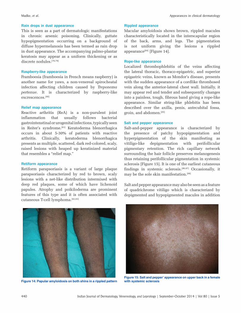
Plucked chicken appearanceTiny yellow papules that coalesce into plaques with a “plucked chicken skin” or “cobblestone appearance” or “morroccan leather appearance” occur in flexural regions, most commonly the neck and axillae in pseudoxanthoma elasticum (PXE), a rare heritable disease characterized by dermal, ocular and vascular lesions that result from the degeneration of elastic fibers. With age and disease progression, the skin may become soft, lax, and wrinkled, which resembles plucked chicken skin[76,77] [Figure 13].
Figure 13: A patient of pseudoxanthoma elasticum showing the characteristic ‘plucked chicken’ appearance
Madke, et al. Appearances in clinical dermatology
Indian Journal of Dermatology, Venereology, and Leprology | September-October 2014 | Vol 80 | Issue 5440
Rain drops in dust appearanceThis is seen as a part of dermatologic manifestations in chronic arsenic poisoning. Clinically, guttate hypopigmentation occurring on a background of diffuse hypermelanosis has been termed as rain drop in dust appearance. The accompanying palmo‑plantar keratosis may appear as a uniform thickening or as discrete nodules.[78,79]
Raspberry‑like appearanceFramboesia (framboesia in French means raspberry) is another name for yaws, a non‑venereal spirocheatal infection affecting children caused by Treponema pertenue. It is characterized by raspberry‑like excrescences.[80]
Relief map appearanceReactive arthritis (ReA) is a non‑purulent joint inflammation that usually follows bacterial gastrointenstinal or urogenital infections, typically seen in Reiter’s syndrome.[81] Keratoderma blenorrhagica occurs in about 5‑30% of patients with reactive arthritis. Clinically, keratoderma blenorrhagica presents as multiple, scattered, dark red‑colored, scaly, raised lesions with heaped up keratinized material that resembles a “relief map.”
Retiform appearanceRetiform parapsoriasis is a variant of large plaque parapsoriasis characterized by red to brown, scaly lesions with a net‑like distribution intermixed with deep red plaques, some of which have lichenoid papules. Atrophy and poikiloderma are prominent features of this type and it is often associated with cutaneous T‑cell lymphoma.[82,83]
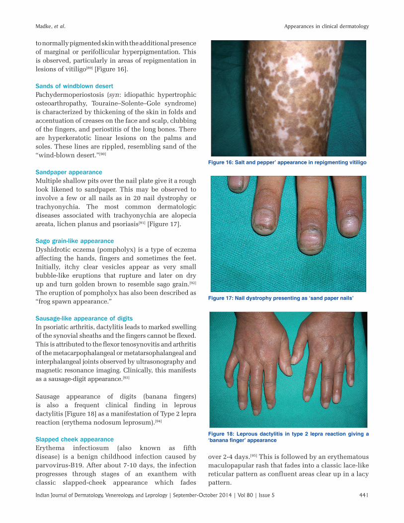
Rippled appearanceMacular amyloidosis shows brown, rippled macules characteristically located in the interscapular region of the back, arms, and legs. The pigmentation is not uniform giving the lesions a rippled appearance[84] [Figure 14].
Rope‑like appearanceLocalized thrombophlebitis of the veins affecting the lateral thoracic, thoraco‑epigastric, and superior epigastric veins, known as Mondor’s disease, presents with the sudden appearance of a cordlike thrombosed vein along the anterior‑lateral chest wall. Initially, it may appear red and tender and subsequently changes into a painless, tough, fibrous band giving a rope‑like appearance. Similar string‑like phlebitis has been described over the axilla, penis, antecubital fossa, groin, and abdomen.[85]
Salt and pepper appearanceSalt‑and‑pepper appearance is characterized by the presence of patchy hypopigmentation and hyperpigmentation of the skin manifesting as vitiligo‑like depigmentation with perifollicular pigmentary retention. The rich capillary network surrounding the hair follicle preserves melanogenesis thus retaining perifollicular pigmentation in systemic sclerosis [Figure 15]. It is one of the earliest cutaneous findings in systemic sclerosis.[86,87] Occasionally, it may be the sole skin manifestation.[88]
Salt and pepper appearance may also be seen as a feature of quadrichrome vitiligo which is characterized by depigmented and hypopigmented macules in addition
Figure 14: Papular amyloidosis on both shins in a rippled patternFigure 15: Salt and pepper’ appearance on upper back in a female with systemic sclerosis
Madke, et al. Appearances in clinical dermatology
441Indian Journal of Dermatology, Venereology, and Leprology | September-October 2014 | Vol 80 | Issue 5
to normally pigmented skin with the additional presence of marginal or perifollicular hyperpigmentation. This is observed, particularly in areas of repigmentation in lesions of vitiligo[89] [Figure 16].
Sands of windblown desertPachydermoperiostosis (syn: idiopathic hypertrophic osteoarthropathy, Touraine–Solente–Gole syndrome) is characterized by thickening of the skin in folds and accentuation of creases on the face and scalp, clubbing of the fingers, and periostitis of the long bones. There are hyperkeratotic linear lesions on the palms and soles. These lines are rippled, resembling sand of the “wind‑blown desert.”[90]
Sandpaper appearanceMultiple shallow pits over the nail plate give it a rough look likened to sandpaper. This may be observed to involve a few or all nails as in 20 nail dystrophy or trachyonychia. The most common dermatologic diseases associated with trachyonychia are alopecia areata, lichen planus and psoriasis[91] [Figure 17].
Sago grain‑like appearanceDyshidrotic eczema (pompholyx) is a type of eczema affecting the hands, fingers and sometimes the feet. Initially, itchy clear vesicles appear as very small bubble‑like eruptions that rupture and later on dry up and turn golden brown to resemble sago grain.[92] The eruption of pompholyx has also been described as “frog spawn appearance.”
Sausage‑like appearance of digitsIn psoriatic arthritis, dactylitis leads to marked swelling of the synovial sheaths and the fingers cannot be flexed. This is attributed to the flexor tenosynovitis and arthritis of the metacarpophalangeal or metatarsophalangeal and interphalangeal joints observed by ultrasonography and magnetic resonance imaging. Clinically, this manifests as a sausage‑digit appearance.[93]
Sausage appearance of digits (banana fingers) is also a frequent clinical finding in leprous dactylitis [Figure 18] as a manifestation of Type 2 lepra reaction (erythema nodosum leprosum).[94]
Slapped cheek appearanceErythema infectiosum (also known as fifth disease) is a benign childhood infection caused by parvovirus‑B19. After about 7‑10 days, the infection progresses through stages of an exanthem with classic slapped‑cheek appearance which fades
Figure 16: Salt and pepper’ appearance in repigmenting vitiligo
Figure 17: Nail dystrophy presenting as ‘sand paper nails’
Figure 18: Leprous dactylitis in type 2 lepra reaction giving a ‘banana finger’ appearance
over 2‑4 days.[95] This is followed by an erythematous maculopapular rash that fades into a classic lace‑like reticular pattern as confluent areas clear up in a lacy pattern.
Madke, et al. Appearances in clinical dermatology
Indian Journal of Dermatology, Venereology, and Leprology | September-October 2014 | Vol 80 | Issue 5442
Spangled hair appearancePili annulati is a rare hair shaft disorder with autosomal dominant inheritance characterized by non‑fragile banding leading to spangled hair.[96] Although it is clinically detectable in blond or lightly pigmented hair as speckled banding, however in dark hair, since the banding is obscured by the pigment, characteristic spangled appearance of hair with reflected light is the only clue. An inherent defect in the hair shaft leads to formation of air‑filled cavities within the cortex that lie parallel to the long axis of the hair. The hair appears as alternating light and dark bands under polarized microscopy.[97]
Speckled appearanceSpeckled lentiginous nevus or speckled nevus is characterized by numerous small, darkly pigmented speckles on a background of tan brown hyperpigmentation. Sometimes, this tan background may be very faint or even invisible. Microscopically, the background pigmentation corresponds to the features of a lentigo simplex, whereas the dark speckles usually show a junctional proliferation of melanocytes.[98]
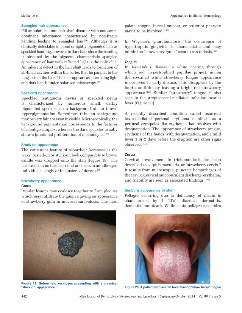
Stuck on appearanceThe consistent feature of seborrheic keratoses is the waxy, pasted‑on or stuck‑on look comparable to brown candle wax dropped onto the skin [Figure 19]. The lesions occur on the face, chest and back in middle‑aged individuals, singly or in clusters of dozens.[99]
Strawberry appearanceGumsPapular lesions may coalesce together to form plaques which may infiltrate the gingiva giving an appearance of strawberry gum in mucosal sarcoidosis. The hard
palate, tongue, buccal mucosa, or posterior pharynx may also be involved.[100]
In Wegener’s granulomatosis, the occurrence of hypertrophic gingivitis is characteristic and may mimic the “strawberry gums” seen in sarcoidosis.[101]
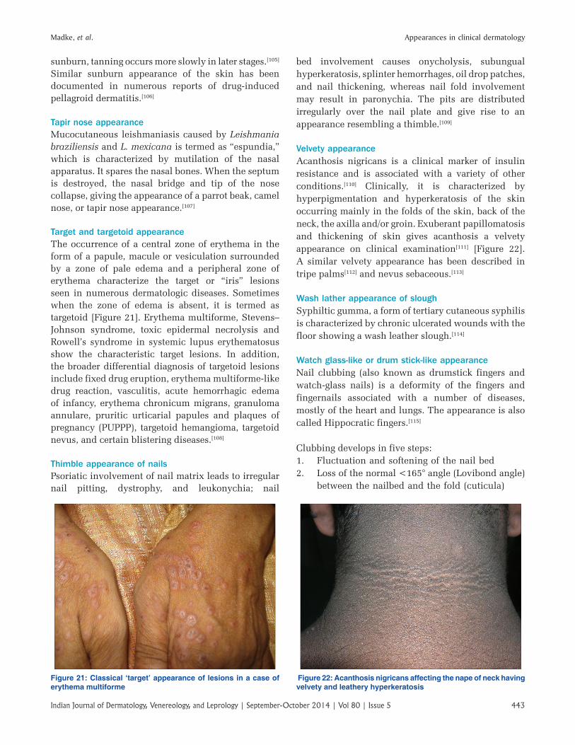
TongueIn Kawasaki’s disease, a white coating through which red, hypertrophied papillae project, giving the so‑called white strawberry tongue appearance is observed in early disease. This disappears by the fourth or fifth day leaving a bright red strawberry appearance.[102] Similar “strawberry” tongue is also seen in the streptococcal‑mediated infection, scarlet fever [Figure 20].
A recently described condition called recurrent toxin‑mediated perianal erythema manifests as a perineal erysipelas‑like erythema that resolves with desquamation. The appearance of strawberry tongue, erythema of the hands with desquamation, and a mild fever 1 or 2 days before the eruption are other signs observed.[103]
CervixCervical involvement in trichomoniasis has been described as colpitis macularis, or “strawberry cervix.” It results from microscopic, punctate hemorrhages of the cervix. Cervical mucopurulent discharge, erythema, and friability are seen as associated findings.[104]
Sunburn appearance of skinPellagra occurring due to deficiency of niacin is characterized by 4 “D’s”: diarrhea, dermatitis, dementia, and death. While acute pellagra resembles
Figure 19: Seborrheic keratoses presenting with a classical ‘stuck-on’ appearance Figure 20: A patient with scarlet fever having ‘straw berry’ tongue
Madke, et al. Appearances in clinical dermatology
443Indian Journal of Dermatology, Venereology, and Leprology | September-October 2014 | Vol 80 | Issue 5
sunburn, tanning occurs more slowly in later stages.[105] Similar sunburn appearance of the skin has been documented in numerous reports of drug‑induced pellagroid dermatitis.[106]
Tapir nose appearanceMucocutaneous leishmaniasis caused by Leishmania braziliensis and L. mexicana is termed as “espundia,” which is characterized by mutilation of the nasal apparatus. It spares the nasal bones. When the septum is destroyed, the nasal bridge and tip of the nose collapse, giving the appearance of a parrot beak, camel nose, or tapir nose appearance.[107]
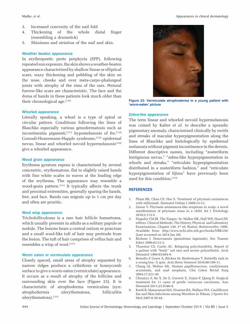
Target and targetoid appearanceThe occurrence of a central zone of erythema in the form of a papule, macule or vesiculation surrounded by a zone of pale edema and a peripheral zone of erythema characterize the target or “iris” lesions seen in numerous dermatologic diseases. Sometimes when the zone of edema is absent, it is termed as targetoid [Figure 21]. Erythema multiforme, Stevens–Johnson syndrome, toxic epidermal necrolysis and Rowell’s syndrome in systemic lupus erythematosus show the characteristic target lesions. In addition, the broader differential diagnosis of targetoid lesions include fixed drug eruption, erythema multiforme‑like drug reaction, vasculitis, acute hemorrhagic edema of infancy, erythema chronicum migrans, granuloma annulare, pruritic urticarial papules and plaques of pregnancy (PUPPP), targetoid hemangioma, targetoid nevus, and certain blistering diseases.[108]
Thimble appearance of nailsPsoriatic involvement of nail matrix leads to irregular nail pitting, dystrophy, and leukonychia; nail
bed involvement causes onycholysis, subungual hyperkeratosis, splinter hemorrhages, oil drop patches, and nail thickening, whereas nail fold involvement may result in paronychia. The pits are distributed irregularly over the nail plate and give rise to an appearance resembling a thimble.[109]
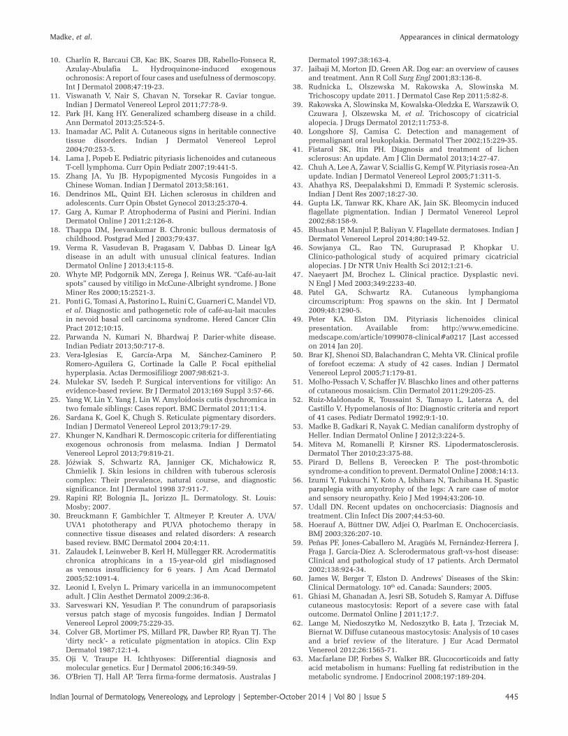
Velvety appearanceAcanthosis nigricans is a clinical marker of insulin resistance and is associated with a variety of other conditions.[110] Clinically, it is characterized by hyperpigmentation and hyperkeratosis of the skin occurring mainly in the folds of the skin, back of the neck, the axilla and/or groin. Exuberant papillomatosis and thickening of skin gives acanthosis a velvety appearance on clinical examination[111] [Figure 22]. A similar velvety appearance has been described in tripe palms[112] and nevus sebaceous.[113]
Wash lather appearance of sloughSyphiltic gumma, a form of tertiary cutaneous syphilis is characterized by chronic ulcerated wounds with the floor showing a wash leather slough.[114]
Watch glass‑like or drum stick‑like appearanceNail clubbing (also known as drumstick fingers and watch‑glass nails) is a deformity of the fingers and fingernails associated with a number of diseases, mostly of the heart and lungs. The appearance is also called Hippocratic fingers.[115]
Clubbing develops in five steps:1. Fluctuation and softening of the nail bed2. Loss of the normal <165° angle (Lovibond angle)
between the nailbed and the fold (cuticula)
Figure 21: Classical ‘target’ appearance of lesions in a case of erythema multiforme
Figure 22: Acanthosis nigricans affecting the nape of neck having velvety and leathery hyperkeratosis
Madke, et al. Appearances in clinical dermatology
Indian Journal of Dermatology, Venereology, and Leprology | September-October 2014 | Vol 80 | Issue 5444
3. Increased convexity of the nail fold4. Thickening of the whole distal finger
(resembling a drumstick)5. Shininess and striation of the nail and skin.
Weather beaten appearanceIn erythropoetic proto porphyria (EPP), following repeated sun exposure, the skin shows a weather‑beaten appearance characterized by shallow linear or elliptical scars, waxy thickening and pebbling of the skin on the nose, cheeks and over meta‑carpo‑phalangeal joints with atrophy of the rims of the ears. Perioral furrow‑like scars are characteristic. The face and the dorsa of hands in these patients look much older than their chronological age.[116]
Whorled appearanceLiterally speaking, a whorl is a type of spiral or circular pattern. Conditions following the lines of Blaschko especially various genodermatosis such as incontinentia pigmenti,[117] hypomelanosis of Ito,[118] Conradi‑Hunermann‑Happle syndrome,[119] epidermal nevus, linear and whorled nevoid hypermelanosis[120] give a whorled appearance.
Wood grain appearanceErythema gyratum repens is characterized by several concentric, erythematous, flat to slightly raised bands with fine white scales in waves at the leading edge of the erythema. The appearance may resemble a wood‑grain pattern.[121] It typically affects the trunk and proximal extremities, generally sparing the hands, feet, and face. Bands can migrate up to 1 cm per day and often are pruritic.
Wool wisp appearanceTrichofolliculoma is a rare hair follicle hamartoma, which usually presents in adults as a solitary papule or nodule. The lesions bears a central ostium or punctum and a small wool‑like tuft of hair may protrude from the lesion. The tuft of hair comprises of vellus hair and resembles a wisp of wool.[122]
Worm eaten or vermiculate appearanceClosely spaced, small areas of atrophy separated by narrow ridges produce a cribriform or honeycomb surface to give a worm‑eaten (vermiculate) appearance. It occurs as a result of atrophy of the follicles and surrounding skin over the face [Figure 23]. It is characteristic of atrophoderma vermiculata (syn: atrophodermia ulerythematosa, folliculitis ulerythematosa).[123]
Zebra‑like appearanceThe term linear and whorled nevoid hypermelanosis was coined by Kalter et al. to describe a sporadic pigmentary anomaly, characterized clinically by swirls and streaks of macular hyperpigmentation along the lines of Blaschko and histologically by epidermal melanosis without pigment incontinence in the dermis. Different descriptive names, including “zosteriform lentiginous nevus,” “zebra‑like hyperpigmentation in whorls and streaks,” “reticulate hyperpigmentation distributed in a zosteriform fashion,” and “reticulate hyperpigmentation of Iijima” have previously been used for this condition.[124]
REFERENCES
1. Pham RK, Chan CS, Hsu S. Treatment of pityriasis amiantacea with infliximab. Dermatol Online J 2009;15:13.
2. Zawar V. Pityriasis amiantacea‑like eruptions in scalp: a novel manifestation of pityriasis rosea in a child. Int J Trichology 2010;2:113‑5.
3. Huguley CM JR. The Tongue. In: Walker HK, Hall WD, Hurst JW, editors. Clinical Methods: The History, Physical, and Laboratory Examinations. Chapter 130. 3rd ed. Boston: Butterworths; 1990. Available from: http://www.ncbi.nlm.nih.gov/books/NBK236/[Last accessed on 2014 Jan 20].
4. Richens J. Donovanosis (granuloma inguinale). Sex Transm Infect 2006;82:21‑2.
5. Thurston CS, Curtis AC. Relapsing polychondritis. Report of a patient with “beefy” red ears and severe polyarthritis. Arch Dermatol 1966;93:664‑9.
6. Benedix F, Geyer A, Röcken M, Biedermann T. Butterfly rash in a young boy: A quiz. Acta Derm Venereol 2010;90:109‑11.
7. Chang GJ, Welton ML. Human papillomavirus, condylomata acuminata, and anal neoplasia. Clin Colon Rectal Surg 2004;17:221‑30.
8. Chuanyu S, Ke X, Jie Z, Guowei X, Zujun F, Qiang D. Surgical treatment for 11 cases of penile verrucous carcinoma. Ann Dermatol 2011;23:S346‑9.
9. Kordi R, Mansournai MA, Nourian RA, Wallace WA. Cauliflower Ear and Skin Infections among Wrestlers in Tehran. J Sports Sci Med 2007;6:39‑44.
Figure 23: Vermiculate atrophoderma in a young patient with ‘worm-eaten’ picture
Madke, et al. Appearances in clinical dermatology
445Indian Journal of Dermatology, Venereology, and Leprology | September-October 2014 | Vol 80 | Issue 5
10. Charlín R, Barcaui CB, Kac BK, Soares DB, Rabello‑Fonseca R, Azulay‑Abulafia L. Hydroquinone‑induced exogenous ochronosis: A report of four cases and usefulness of dermoscopy. Int J Dermatol 2008;47:19‑23.
11. Viswanath V, Nair S, Chavan N, Torsekar R. Caviar tongue. Indian J Dermatol Venereol Leprol 2011;77:78‑9.
12. Park JH, Kang HY. Generalized schamberg disease in a child. Ann Dermatol 2013;25:524‑5.
13. Inamadar AC, Palit A. Cutaneous signs in heritable connective tissue disorders. Indian J Dermatol Venereol Leprol 2004;70:253‑5.
14. Lama J, Popeb E. Pediatric pityriasis lichenoides and cutaneous T‑cell lymphoma. Curr Opin Pediatr 2007;19:441‑5.
15. Zhang JA, Yu JB. Hypopigmented Mycosis Fungoides in a Chinese Woman. Indian J Dermatol 2013;58:161.
16. Dendrinos ML, Quint EH. Lichen sclerosus in children and adolescents. Curr Opin Obstet Gynecol 2013;25:370‑4.
17. Garg A, Kumar P. Atrophoderma of Pasini and Pierini. Indian Dermatol Online J 2011;2:126‑8.
18. Thappa DM, Jeevankumar B. Chronic bullous dermatosis of childhood. Postgrad Med J 2003;79:437.
19. Verma R, Vasudevan B, Pragasam V, Dabbas D. Linear IgA disease in an adult with unusual clinical features. Indian Dermatol Online J 2013;4:115‑8.
20. Whyte MP, Podgornik MN, Zerega J, Reinus WR. “Café‑au‑lait spots” caused by vitiligo in McCune‑Albright syndrome. J Bone Miner Res 2000;15:2521‑3.
21. Ponti G, Tomasi A, Pastorino L, Ruini C, Guarneri C, Mandel VD, et al. Diagnostic and pathogenetic role of café‑au‑lait macules in nevoid basal cell carcinoma syndrome. Hered Cancer Clin Pract 2012;10:15.
22. Parwanda N, Kumari N, Bhardwaj P. Darier‑white disease. Indian Pediatr 2013;50:717‑8.
23. Vera‑Iglesias E, García‑Arpa M, Sánchez‑Caminero P, Romero‑Aguilera G, Cortinade la Calle P. Focal epithelial hyperplasia. Actas Dermosifiliogr 2007;98:621‑3.
24. Mulekar SV, Isedeh P. Surgical interventions for vitiligo: An evidence‑based review. Br J Dermatol 2013;169 Suppl 3:57‑66.
25. Yang W, Lin Y, Yang J, Lin W. Amyloidosis cutis dyschromica in two female siblings: Cases report. BMC Dermatol 2011;11:4.
26. Sardana K, Goel K, Chugh S. Reticulate pigmentary disorders. Indian J Dermatol Venereol Leprol 2013;79:17‑29.
27. Khunger N, Kandhari R. Dermoscopic criteria for differentiating exogenous ochronosis from melasma. Indian J Dermatol Venereol Leprol 2013;79:819‑21.
28. Jóźwiak S, Schwartz RA, Janniger CK, Michałowicz R, Chmielik J. Skin lesions in children with tuberous sclerosis complex: Their prevalence, natural course, and diagnostic significance. Int J Dermatol 1998 37:911‑7.
29. Rapini RP, Bolognia JL, Jorizzo JL. Dermatology. St. Louis: Mosby; 2007.
30. Breuckmann F, Gambichler T, Altmeyer P, Kreuter A. UVA/UVA1 phototherapy and PUVA photochemo therapy in connective tissue diseases and related disorders: A research based review. BMC Dermatol 2004 20;4:11.
31. Zalaudek I, Leinweber B, Kerl H, Müllegger RR. Acrodermatitis chronica atrophicans in a 15‑year‑old girl misdiagnosed as venous insufficiency for 6 years. J Am Acad Dermatol 2005;52:1091‑4.
32. Leonid I, Evelyn L. Primary varicella in an immunocompetent adult. J Clin Aesthet Dermatol 2009;2:36‑8.
33. Sarveswari KN, Yesudian P. The conundrum of parapsoriasis versus patch stage of mycosis fungoides. Indian J Dermatol Venereol Leprol 2009;75:229‑35.
34. Colver GB, Mortimer PS, Millard PR, Dawber RP, Ryan TJ. The ‘dirty neck’‑ a reticulate pigmentation in atopics. Clin Exp Dermatol 1987;12:1‑4.
35. Oji V, Traupe H. Ichthyoses: Differential diagnosis and molecular genetics. Eur J Dermatol 2006;16:349‑59.
36. O’Brien TJ, Hall AP. Terra firma‑forme dermatosis. Australas J
Dermatol 1997;38:163‑4.37. Jaibaji M, Morton JD, Green AR. Dog ear: an overview of causes
and treatment. Ann R Coll Surg Engl 2001;83:136‑8.38. Rudnicka L, Olszewska M, Rakowska A, Slowinska M.
Trichoscopy update 2011. J Dermatol Case Rep 2011;5:82‑8.39. Rakowska A, Slowinska M, Kowalska‑Oledzka E, Warszawik O,
Czuwara J, Olszewska M, et al. Trichoscopy of cicatricial alopecia. J Drugs Dermatol 2012;11:753‑8.
40. Longshore SJ, Camisa C. Detection and management of premalignant oral leukoplakia. Dermatol Ther 2002;15:229‑35.
41. Fistarol SK, Itin PH. Diagnosis and treatment of lichen sclerosus: An update. Am J Clin Dermatol 2013;14:27‑47.
42. Chuh A, Lee A, Zawar V, Sciallis G, Kempf W. Pityriasis rosea‑An update. Indian J Dermatol Venereol Leprol 2005;71:311‑5.
43. Ahathya RS, Deepalakshmi D, Emmadi P. Systemic sclerosis. Indian J Dent Res 2007;18:27‑30.
44. Gupta LK, Tanwar RK, Khare AK, Jain SK. Bleomycin induced flagellate pigmentation. Indian J Dermatol Venereol Leprol 2002;68:158‑9.
45. Bhushan P, Manjul P, Baliyan V. Flagellate dermatoses. Indian J Dermatol Venereol Leprol 2014;80:149‑52.
46. Sowjanya CL, Rao TN, Guruprasad P, Khopkar U. Clinico‑pathological study of acquired primary cicatricial alopecias. J Dr NTR Univ Health Sci 2012;1:21‑6.
47. Naeyaert JM, Brochez L. Clinical practice. Dysplastic nevi. N Engl J Med 2003;349:2233‑40.
48. Patel GA, Schwartz RA. Cutaneous lymphangioma circumscriptum: Frog spawns on the skin. Int J Dermatol 2009;48:1290‑5.
49. Peter KA. Elston DM. Pityriasis lichenoides clinical presentation. Available from: http://www.emedicine.medscape.com/article/1099078‑clinical#a0217 [Last accessed on 2014 Jan 20].
50. Brar KJ, Shenoi SD, Balachandran C, Mehta VR. Clinical profile of forefoot eczema: A study of 42 cases. Indian J Dermatol Venereol Leprol 2005;71:179‑81.
51. Molho‑Pessach V, Schaffer JV. Blaschko lines and other patterns of cutaneous mosaicism. Clin Dermatol 2011;29:205‑25.
52. Ruiz‑Maldonado R, Toussaint S, Tamayo L, Laterza A, del Castillo V. Hypomelanosis of Ito: Diagnostic criteria and report of 41 cases. Pediatr Dermatol 1992;9:1‑10.
53. Madke B, Gadkari R, Nayak C. Median canaliform dystrophy of Heller. Indian Dermatol Online J 2012;3:224‑5.
54. Miteva M, Romanelli P, Kirsner RS. Lipodermatosclerosis. Dermatol Ther 2010;23:375‑88.
55. Pirard D, Bellens B, Vereecken P. The post‑thrombotic syndrome‑a condition to prevent. Dermatol Online J 2008;14:13.
56. Izumi Y, Fukuuchi Y, Koto A, Ishihara N, Tachibana H. Spastic paraplegia with amyotrophy of the legs: A rare case of motor and sensory neuropathy. Keio J Med 1994;43:206‑10.
57. Udall DN. Recent updates on onchocerciasis: Diagnosis and treatment. Clin Infect Dis 2007;44:53‑60.
58. Hoerauf A, Büttner DW, Adjei O, Pearlman E. Onchocerciasis. BMJ 2003;326:207‑10.
59. Peñas PF, Jones‑Caballero M, Aragüés M, Fernández‑Herrera J, Fraga J, García‑Díez A. Sclerodermatous graft‑vs‑host disease: Clinical and pathological study of 17 patients. Arch Dermatol 2002;138:924‑34.
60. James W, Berger T, Elston D. Andrews’ Diseases of the Skin: Clinical Dermatology. 10th ed. Canada: Saunders; 2005.
61. Ghiasi M, Ghanadan A, Jesri SB, Sotudeh S, Ramyar A. Diffuse cutaneous mastocytosis: Report of a severe case with fatal outcome. Dermatol Online J 2011;17:7.
62. Lange M, Niedoszytko M, Nedoszytko B, Łata J, Trzeciak M, Biernat W. Diffuse cutaneous mastocytosis: Analysis of 10 cases and a brief review of the literature. J Eur Acad Dermatol Venereol 2012;26:1565‑71.
63. Macfarlane DP, Forbes S, Walker BR. Glucocorticoids and fatty acid metabolism in humans: Fuelling fat redistribution in the metabolic syndrome. J Endocrinol 2008;197:189‑204.
Madke, et al. Appearances in clinical dermatology
Indian Journal of Dermatology, Venereology, and Leprology | September-October 2014 | Vol 80 | Issue 5446
64. Cyriac MJ, Lashpa E. Lobster‑claw hand: A manifestation of EEC syndrome. Indian J Dermatol Venereol Leprol 2006;72:54‑6.
65. Mianda SB, Delmaestro D, Bertoli R, Marinho T, Lucas E. Focal dermal hypoplasia with exuberant fat herniations and skeletal deformities. Pediatr Dermatol 2005;22:420‑3.
66. Ghalayani P, Tavangar A, Nilchian F, Khalighinejad N. The comparison of salivary level of estrogen and progesterone in 1st, 2nd, 3rd trimester in pregnant women with and without geographic tongue. Dent Res J (Isfahan) 2013;10:609‑12.
67. Bi MY, Cohen PR, Robinson FW, Gray JM. Alopecia syphilitica‑report of a patient with secondary syphilis presenting as moth‑eaten alopecia and a review of its common mimickers. Dermatol Online J 2009;15:6.
68. Langley RG, Krueger GG, Griffiths CE. Psoriatic arthritis and psoriasis: Classification, clinical features, pathophysiology, immunology, genetics. Psoriasis: Epidemiology, clinical features, and quality of life. Ann Rheum Dis 2005;64:18‑23.
69. Bhagwat PV, Tophakhane RS, Rathod RM, Shashikumar BM, Naidu V. Rupioid syphilis in an HIV patient. Indian J Dermatol Venereol Leprol 2009;75:201‑2.
70. Segen’s Medical Dictionary. © 2012 Farlex, Inc.71. Hazin R, Abu‑Rajab Tamimi TI, Abuzetun JY, Zein NN.
Recognizing and treating cutaneous signs of liver disease. Cleve Clin J Med 2009;76:599‑606.
72. Schairer C, Soliman AS, Omar S, Khaled H, Eissa S, Ayed FB, et al. Assessment of diagnosis of inflammatory breast cancer cases at two cancer centers in Egypt and Tunisia. Cancer Med 2013;2:178‑84.
73. Verma KK, Bhat R, Singh MK. Bullous mastocytosis treated with oral betamethasone therapy. Indian J Pediatr 2004;71:261‑3.
74. Velásquez X, Gutiérrez MA, Rosenberg H, Figueroa F, Bronstein E, Jacobelli S. Eosinophilic fasciitis: Report of 3 cases. Rev Med Chil 2002;130:209‑14.
75. Thappa DM, Singh A, Jaisankar TJ, Rao R, Ratnakar C. Pebbling of the skin: A marker of Hunter’s syndrome. Pediatr Dermatol 1998;15:370‑3.
76. Inamadar AC, Palit A. Pseudoxanthoma elasticum. Postgrad Med J 2004;80:297.
77. Gonzalez ME, Votava HJ, Lipkin G, Sanchez M. Pseudoxanthoma elasticum. Dermatol Online J 2009;15:17.
78. Ratnaike RN. Acute and chronic arsenic toxicity. Postgrad Med J 2003;79:391‑6.
79. Smith AH, Arroyo AP, Mazumder DN, Kosnett MJ, Hernandez AL, Beeris M, et al. Arsenic‑induced skin lesions among Atacameno people in northern Chile despite good nutrition and centuries of exposure. Environ Health Perspect 2000;108:617‑20.
80. Framboesia. Dictionary.com. Collins English Dictionary‑Complete and Unabridged 10th ed. HarperCollins Publishers. Available from: http://www.dictionary.reference.com/browse/framboesia [Last accessed on 2014 Jan 23].
81. Kwiatkowska B, Filipowicz‑Sosnowska A. Reactive arthritis. Pol Arch Med Wewn 2009;119:60‑5.
82. Nag F, Ghosh A, Biswas P, Chatterjee G, Biswas S. Ichthyosiform large plaque parapsoriasis: Report of a rare entity. Indian J Dermatol 2013;58:385‑7.
83. Wood GS, Reizner G. Other papulosquamous disorders. In: Bolognia JL, Jorizzo JL, Rapini RP, editors. Dermatology. 2nd ed. Spain: Mosby Elsevier; 2008. p. 137‑48.
84. Patel AB, Kubba R, Kubba A. Clinicopathological correlation of acquired hyperpigmentary disorders. Indian J Dermatol Venereol Leprol 2013;79:367‑75.
85. James WD, Berger TG, Elston DM. Editors. Andrews’ Diseases of the skin: Clinical Dermatology. 11th ed. China: Saunders’ 2011. p. 818‑9.
86. Gonin M, Gerster JC. Pigmentation disorders in systemic sclerodermia. Schweiz Rundsch Med Prax 1994;83:42‑5.
87. Rai VM, Balachandran C. Pseudovitiligo in systemic sclerosis. Dermatol Online J 2005;11:41.
88. Singh A, Ambujam S, Varghese A, Vishranth SP, Sadanandan N.
Salt‑and‑pepper appearance: A cutaneous clue for the diagnosis of systemic sclerosis. Indian J Dermatol 2012;57:412‑3.
89. Halder RM, Chappell JL. Vitiligo update. Semin Cutan Med Surg 2009;28:86‑92.
90. Chander R, Kakkar S, Jain A, Barara M, Agarwal K, Varghese B. Complete form of pachydermoperiostosis: A case report. Dermatol Online J 2013;19:10.
91. Baran R, Dupre A. Vertical striated sandpaper nails. Arch Dermatol 1977;113:1613.
92. Wollina U. Pompholyx: A review of clinical features, differential diagnosis, and management. Am J Clin Dermatol 2010;11:305‑14.
93. Bennett RM. Psoriatic arthritis. In: McCarty DJ, editor. Arthritis and allied conditions. 10th ed. Philadelphia: Lea and Febiger; 1985. p. 850‑66.
94. Pereira HL, Ribeiro SL, Sato EI. Rheumatic manifestations in leprosy. Acta Reumatol Port 2008;33:407‑14.
95. Servey JT, Reamy BV, Hodge J. Clinical presentations of parvovirus B19 infection. Am Fam Physician 2007;75:373‑6.
96. Amichai B, Grunwald MH, Halevy S. Hair abnormality present since childhood: Pili annulati. Arch Dermatol 1996;132:575‑8.
97. Ito M, Hashimoto K, Sakamoto F, Sato Y, Voorhees JJ. Pathogenesis of pili annulati. Arch Dermatol Res 1988;280:308‑18.
98. Torrelo A, de Prada I, Zambrano A, Happle R. Extensive speckled lentiginous nevus associated with giant congenital melanocytic nevus: An unusual example of twin spotting? Eur J Dermatol 2003;13:534‑6.
99. Zhang RZ, Zhu WY. Seborrheic keratoses in five elderly patients: An appearance of raindrops and streams. Indian J Dermatol 2011;56:432‑4.
100. Kowalczyk JP, Ricotti CA, de Araujo T, Drosou A, Nousari CH. “Strawberry gums” in sarcoidosis. J Am Acad Dermatol 2008;59:S118‑20.
101. Siar CH, Yeo KB, Nakano K, Nagatsuka H, Tsujigiwa H, Tomida M, et al. Strawberry gingivitis as the first presenting sign of Wegener’s granulomatosis: Report of a case. Eur J Med Res 2011;16:331‑4.
102. Yuan K, Park JK, Qubti MA, Haque UJ. Recurrent Kawasaki disease with strawberry tongue and skin desquamation in a young adult. J Clin Rheumatol 2012;18:96‑8.
103. James WD, Berger TG, Elston DM, Editors. Andrews’ Diseases of the skin: Clinical dermatology. 11th ed. China: Saunders, 2011. p. 255.
104. Swygard H, Seña AC, Hobbs MM, Cohen MS. Trichomoniasis: Clinical manifestations, diagnosis and management. Sex Transm Infect 2004;80:91‑5.
105. Hegyi J, Schwartz RA, Hegyi V. Pellagra: Dermatitis, dementia, and diarrhea. Int J Dermatol 2004;43:1‑5.
106. Wan P, Moat S, Anstey A. Pellagra: A review with emphasis on photosensitivity. Br J Dermatol 2011;164:1188‑200.
107. James WD, Berger TG, Elston DM. Editors. Andrews’ Diseases of the skin: Clinical dermatology. 11th ed. China: Saunders; 2011. p. 426.
108. Hughey LC. Approach to the hospitalized patient with targetoid lesions. Dermatol Ther 2011;24:196‑206.
109. Singh SK. Finger nail pitting in psoriasis and its relation with different variables. Indian J Dermatol 2013;58:310‑2.
110. Patel AB, Kubba R, Kubba A. Clinicopathological correlation of acquired hyperpigmentary disorders. Indian J Dermatol Venereol Leprol 2013;79:367‑75.
111. Barbato MT, Criado PR, Silva AK, Averbeck E, Guerine MB, Sá NB. Association of acanthosis nigricans and skin tags with insulin resistance. An Bras Dermatol 2012;87:97‑104.
112. Fabroni C, Gimma A, Cardinali C, Lo Scocco G. Tripe palms associated with malignant acanthosis nigricans in a patient with gastric adenocarcinoma: A case report and review of the literature. Dermatol Online J 2012;18:15.
113. Mahajan R, Dogra S, Kanwar AJ, Saikia UN, Agrawal S. Extensive cerebriform nevus sebaceus: An unusual presentation. Dermatol Online J 2012;18:9.
Madke, et al. Appearances in clinical dermatology
447Indian Journal of Dermatology, Venereology, and Leprology | September-October 2014 | Vol 80 | Issue 5
114. Rocha N, Horta M, Sanches M, Lima O, Massa A. Syphilitic gumma‑cutaneous tertiary syphilis. J Eur Acad Dermatol Venereol 2004;18:517‑8.
115. Myers KA, Farquhar DR. The rational clinical examination: does this patient have clubbing?. JAMA 2001;286:341‑7.
116. Botto NC, Shpall RL, Kim J, Bruckner AL. A young girl with weather‑beaten, waxy knuckles. J Pediatr 2010;156:671‑4.
117. Jabbari A, Ralston J, Schaffer JV: Incontinentia pigmenti. Dermatol Online J 2010;16:9.
118. Nicolaidou E, Katsambas AD. Pigmentation disorders: Hyperpigmentation and hypopigmentation. Clin Dermatol 2014;32:66‑72.
119. Shanske AL, Bernstein L, Herzog R. Chondrodysplasia punctata and maternal autoimmune disease: A new case and review of the literature. Pediatrics 2007;120:e436‑41.
120. Metta AK, Ramachandra S, Sadath N, Manupati S. Linear and whorled nevoid hypermelanosis in three successive generations. Indian J Dermatol Venereol Leprol 2011;77:403.
121. Gore M, Winters ME. Erythema gyratum repens: A rare paraneoplastic rash. West J Emerg Med 2011;12:556‑8.
122. Gokalp H, Gurer MA, Alan S. Trichofolliculoma: A rare variant of hair follicle hamartoma. Dermatol Online J 2013;19:19264.
123. Jansen T, Sander CA, Altmeyer P. Atrophodermia vermiculata: Case report and review of the literature. J Eur Acad Dermatol Venereol 2003;17:70‑2.
124. Alimurung FM, Lapenas D, Willis I, Lang P. Zebra‑like hyperpigmentation in an infant with multiple congenital defects. Arch Dermatol 1979;115:878‑91.
Copyright © 2022 FDOKUMEN