Frontiers in Dermatology and Venereology - Medical Journals ...
56
ISSN 0001-5555 A Non-profit International Journal for Interdisciplinary Skin Research, Clinical and Experimental Dermatology and Sexually Transmitted Diseases Frontiers in Dermatology and Venereology - A series of theme issues in relation to the 100-year anniversary of ActaDV Official Journal of - European Society for Dermatology and Psychiatry Affiliated with - The International Forum for the Study of Itch Immediate Open Access Volume 100 2020 Theme issue ADVANCES IN DERMATOLOGY AND VENEREOLOGY Acta DV Acta Dermato-Venereologica www.medicaljournals.se/adv
-
Upload
khangminh22 -
Category
Documents
-
view
0 -
download
0
Transcript of Frontiers in Dermatology and Venereology - Medical Journals ...

ISSN 0001-5555
A Non-profit International Journal for Interdisciplinary Skin Research, Clinical and Experimental Dermatology and Sexually Transmitted Diseases
Frontiers in Dermatology and Venereology
- A series of theme issues in relation to the 100-year anniversary of ActaDV
Official Journal of- European Society for Dermatology and Psychiatry
Affiliated with- The International Forum for the Study of Itch
ImmediateOpen Access
Volume 100 2020 Theme issue
AdvAnces in dermAtology And venereology
ActaDV
Acta Dermato-Venereologica
www.medicaljournals.se/adv

ACTA DERMATO-VENEREOLOGICAThe journal was founded in 1920 by Professor Johan Almkvist. Since 1969 ownership has been vested in the Society for Publication of Acta Dermato-Venereologica, a non-profit organization. Since 2006 the journal is published online, independently without a commercial publisher. (For further information please see the journal’s website https://www.medicaljournals.se/acta)
ActaDV is a journal for clinical and experimental research in the field of dermatology and venereology and publishes high-quality papers in English dealing with new observations on basic dermatological and venereological research, as well as clinical investigations. Each volume also features a number of review articles in special areas, as well as Correspondence to the Editor to stimulate debate. New books are also reviewed. The journal has rapid publication times.
Editor-in-Chief: Olle Larkö, MD, PhD, Gothenburg
Deputy Editors:Anette Bygum, MD, PhD, OdenseMagnus Lindberg, MD, PhD, ÖrebroElisabet Nylander, MD, PhD, UmeåKaisa Tasanen-Määttä, MD, PhD, Oulu
Section Editors:Tasuku Akiyama, Miami (Neurodermatology and Itch - Experimental) Annamari Ranki, Helsinki (Cutaneous lymphoma)Nicole Basset-Seguin, Paris (Skin cancer) Artur Schmidtchen, Lund (Wound healing and Innate immunity)Veronique Bataille, London (Melanoma, Naevi, Photobiology) Matthias Schmuth, Innsbruck (Genodermatoses and Keratinizing disorders,Josip Car, Singapore (Health Services Research and e-Health) Ichthyosis and Retinoids)Brigitte Dréno, Nantes (Acne and Rosacea) Lone Skov, Hellerup (Psoriasis)Regina Fölster-Holst, Kiel (Paediatric dermatology, Atopy and Parasitoses) Enikö Sonkoly, Stockholm (Psoriasis and related disorders)Jürg Hafner, Zürich (Skin cancer, Skin tumours, and Melanoma) Jacek Szepietowski, Wrocław (Psychodermatology)Jürgen Harder, Kiel (Cutaneous innate defense, Skin microbe interactions) Elke Weisshaar, Heidelberg (Itch and Neurodermatology)Roderick Hay, London (Cutaneous Infections) Margitta Worm, Berlin (Atopic dermatitis and Immunology)Kristian Kofoed, Copenhagen (STD and Microbiology) Claus Zachariae, Hellerup (Contact dermatitis, Acute dermatology)Veli-Matti Kähäri, Turku (Skin cancer) Magnus Ågren, Copenhagen (Wound healing & Extracellular matrix)Dennis Linder, Graz/Padua (Psychoderm., Dermato -epidemiology, e-Health)
Advisory Board:Masashi Akiyama, Nagoya Rudolf Happle, Freiburg Lisa Naysmith, Edinburgh Mona Ståhle, StockholmWilma Bergman, Leiden Lars Iversen, Aarhus Jonathan Rees, Edinburgh Sonja Ständer, MünsterTilo Biedermann, Munich Peter van de Kerkhof, Nijmegen Jean Revuz, Paris Jouni Uitto, PhiladelphiaMagnus Bruze, Malmö Irene Leigh, Dundee Johannes Ring, Munich Shyam Verma, VadodaraEarl Carstens, Davis John McGrath, London Matthias Ringkamp, Baltimore Gil Yosipovitch, MiamiThomas Diepgen, Heidelberg Maja Mockenhaupt, Freiburg Inger Rosdahl, Linköping Giovanna Zambruno, RomeCharlotta Enerbäck, Linköping Dedee Murrell, Sydney Thomas Ruzicka, Munich Christos C. Zouboulis, DessauKilian Eyerich, Stockholm All correspondence concerning manuscripts, editorial matters and subscription should be addressed to:Acta Dermato-VenereologicaS:t Johannesgatan 22, SE-753 12 Uppsala, Sweden
Editorial Manager, Mrs Agneta Andersson Editorial Assistant: Ms Anna-Maria AnderssonPlease send an E-mail Please send an E-mail
Information to authors: Acta Dermato-Venereologica publishes papers/reports on scientific investigations in the field of dermatology and venereology, as well as reviews. Case reports and good preliminary clinical trials or experimental investigations are usually published as Short Communications. However, if such papers are of great news value they could still be published as full articles. Special contributions such as extensive feature articles and proceedings may be published as supplements to the journal. For detailed instructions to authors see https://www.medicaljournals.se/acta/instructions-to-author.
Publication information: Everything is Open Access and no subscription fee. For publication fees: see https://www.medicaljournals.se/acta/instructions-to-author.
Indexed in: See https://www.medicaljournals.se/acta/about/adv.
Former Editors:Johan Almkvist 1920–1935Sven Hellerström 1935–1969Nils Thyresson 1969–1987Lennart Juhlin 1987–1999Anders Vahlquist 1999–2017Artur Schmidtchen 2018–2019

Act
aDV
Act
aDV
Advan
ces
in d
erm
ato
logy a
nd v
en
ere
olo
gy
Acta
Derm
ato
-Ven
ere
olo
gic
a
FOREWORD1
This is an open access article under the CC BY-NC license. www.medicaljournals.se/actaJournal Compilation © 2020 Acta Dermato-Venereologica.
1Vahlquist A. ActaDV 100 years – An incomplete history. Forum for Nord Derm Venereol 2020; 24 (in press).
In 1920, Acta Dermato-Venereologica (ActaDV) was founded in Stockholm by the Chairman of Dermatology and Syphilidology at Karolinska Institutet, Professor Johan Almkvist. To begin with, it was a minor tri-lingual journal (German, French, English) with contributions mainly from Europe. Today, 100 years and 6 editors later, the journal is an online, open access and peer-reviewed publication, own by a non-profit society and produced entirely in-house without any dependency on a com-mercial publisher. For an historic review see Vahlquist A. ActaDV 100 years – An incomplete history1. Annually, ActaDV releases 250–300 papers submitted from all around the world. The acceptance rate is around 50% and the impact factor 3.4–4.2 in recent years, i.e. among top 10 in the field.
As part of the celebration of the journal’s 100 years birthday, ActaDV will organize a symposium ”Frontiers in Dermatology and Venereology” with invited speakers from Europe and the USA (see full program below), and publish a series of connected theme issues with contri-butions from the speakers and other invited key-opinion-leaders around the world (see next page). It is hoped that these undertakings will help to spur the scientific interest in Dermatology and Venereology.
On behalf of the editorial board and as principal orga-nizer of the centenary celebrations, I wish to sincerely thank all the authors who kindly accepted to contribute to the theme issues, the specially appointed theme editors, and the skilful work of the editorial office without which nothing of this had been possible.
Uppsala in December 2019
Anders VahlquistFormer Editor-in-Chief and current Chairman of the
Society for Publication of Acta Dermato-Venereologica
Acta Dermato-Venereologica will celebrate its 100-year anniversary with a symposium Date: 15th May 2020. Place: Swedish Society of Medicine, Stockholm, Sweden
Frontiers in Dermatology and VenereologyProgramme:Chairperson: Olle Larkö9.00–10.30 ”ActaDV 100 year; an historic perspective”, Anders Vahlquist ”Skin fragility and blistering diseases”, Leena Bruckner-Tuderman
”Is permanent cure for genodermatoses in sight?”, Jouni Uitto10.50–12.00 ”Psoriasis: News in pathogenesis and therapy”, Jonathan Barker ”Where are we with prevention of atopic dermatitis?”, Hywel WilliamsChairperson: Anders Vahlquist13.00–14.10 ”Melanoma: News in epidemiology and therapy”, Julia Newton-Bishop ”Itch; scratching the surface is not enough”, Gil Yosipovitch14.30–16.00 ”Combatting skin infections: A priority not just in Africa”, Roderick Hay Angelika Stary Olle Larkö
Acta Dermato-Venereologica – 100 years
The registration for this symposium is open. For more information (https://medicaljournals.se/acta/Acta100Year.pdf) and registration form (https://medicaljournals.se/actadv100/registration.php).
The celebrations comprice a symposium and a series of theme issues jointly called ”Frontiers in Dermatology and Venereology”
Act
aDV
Act
aDV
Advan
ces
in d
erm
ato
logy a
nd v
en
ere
olo
gy
Acta
Derm
ato
-Ven
ere
olo
gic
a
Frontiers in Dermatology and Venereology
Centenary theme issues in Volume 100 of Acta Dermato-Venereologica An overview
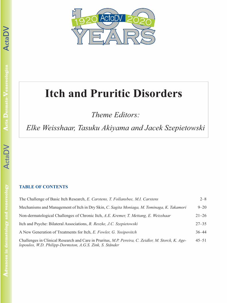
Current issueItch and pruritic disorders
Theme editors: Elke Weisshaar, Tasuku Akiyama and Jacek Szepietowski• The Challenge of Basic Itch Research, E. Carstens, T. Follansbee, M.I. Carstens• Mechanisms and Management of Itch in Dry Skin, C. Sagita Moniaga, M. Tominaga, K. Takamori• Non-dermatological Challenges of Chronic Itch, A.E. Kremer, T. Mettang, E. Weisshaar• Itch and Psyche: Bilateral Associations, R. Reszke, J.C. Szepietowski• A New Generation of Treatments for Itch, E. Fowler, G. Yosipovitch• Challenges in Clinical Research and Care in Pruritus, M.P. Pereira, C. Zeidler, M. Storck, K. Agelopoulos, W.D. Philipp-Dormston,
A.G.S. Zink, S. Ständer
Forthcoming issues
PsoriasisTheme editors: Lone Skov and Enikö Sonkoly
• Psoriasis and Genetics, N. Dand, S. Mahil, F. Capon, C.H. Smith, M.A. Simpson and J. Barker • The Woronoff Ring in Psoriasis and the Mechanisms of Postinflammatory Hypopigmentation, J. Prinz • Psoriasis and Treatment: Past, Present and Future Aspects C. Reid, C.E.M. Griffiths • Psoriasis and Co-morbidity, M. Amin, E.B. Lee, T-F. Tsai, J.J. Wu• Pustular Psoriasis: the Dawn of a New Era, H. Bachelez
Blistering skin disordersTheme editor: Kaisa Tasanen
• Collagen XVII Processing and Blistering Skin Diseases, W. Nishie • Current Concepts of Dermatitis Herpetiformis, T. Salmi, K. Hervonen • Skin Fragility: Perspectives for Evidence-based Therapies, L. Bruckner-Tuderman • Drug Development in Pemphigoid Diseases, K. Bieber, R.J. Ludwig • Bullous Drug Reactions, M. Mockenhaupt
GenodermatosesTheme editors: Anette Bygum and Matthias SchmuthContent details TBA
Skin malignanciesTheme editors: Veronique Bataille and Nicole Basset SeguinContent details TBA
Atopic dermatitisTheme editors: Magnus Lindberg and Carl-Fredrik WahlgrenContent details TBA
Cutaneous and genital infectionsTheme editors: Roderick Hay and Kristian KofoedContent details TBA
Act
aDV
Act
aDV
Advan
ces
in d
erm
ato
logy a
nd v
en
ere
olo
gy
Acta
Derm
ato
-Ven
ere
olo
gic
a
Itch and Pruritic Disorders
Theme Editors:
Elke Weisshaar, Tasuku Akiyama and Jacek Szepietowski
TABLE OF CONTENTS
The Challenge of Basic Itch Research, E. Carstens, T. Follansbee, M.I. Carstens 2–8
Mechanisms and Management of Itch in Dry Skin, C. Sagita Moniaga, M. Tominaga, K. Takamori 9–20
Non-dermatological Challenges of Chronic Itch, A.E. Kremer, T. Mettang, E. Weisshaar 21–26
Itch and Psyche: Bilateral Associations, R. Reszke, J.C. Szepietowski 27–35
A New Generation of Treatments for Itch, E. Fowler, G. Yosipovitch 36–44
Challenges in Clinical Research and Care in Pruritus, M.P. Pereira, C. Zeidler, M. Storck, K. Age-lopoulos, W.D. Philipp-Dormston, A.G.S. Zink, S. Ständer
45–51
Act
aDV
Act
aDV
Advan
ces
in d
erm
ato
logy a
nd v
en
ere
olo
gy
Acta
Derm
ato
-Ven
ere
olo
gic
a
doi: 10.2340/00015555-3343Journal Compilation © 2020 Acta Dermato-Venereologica.
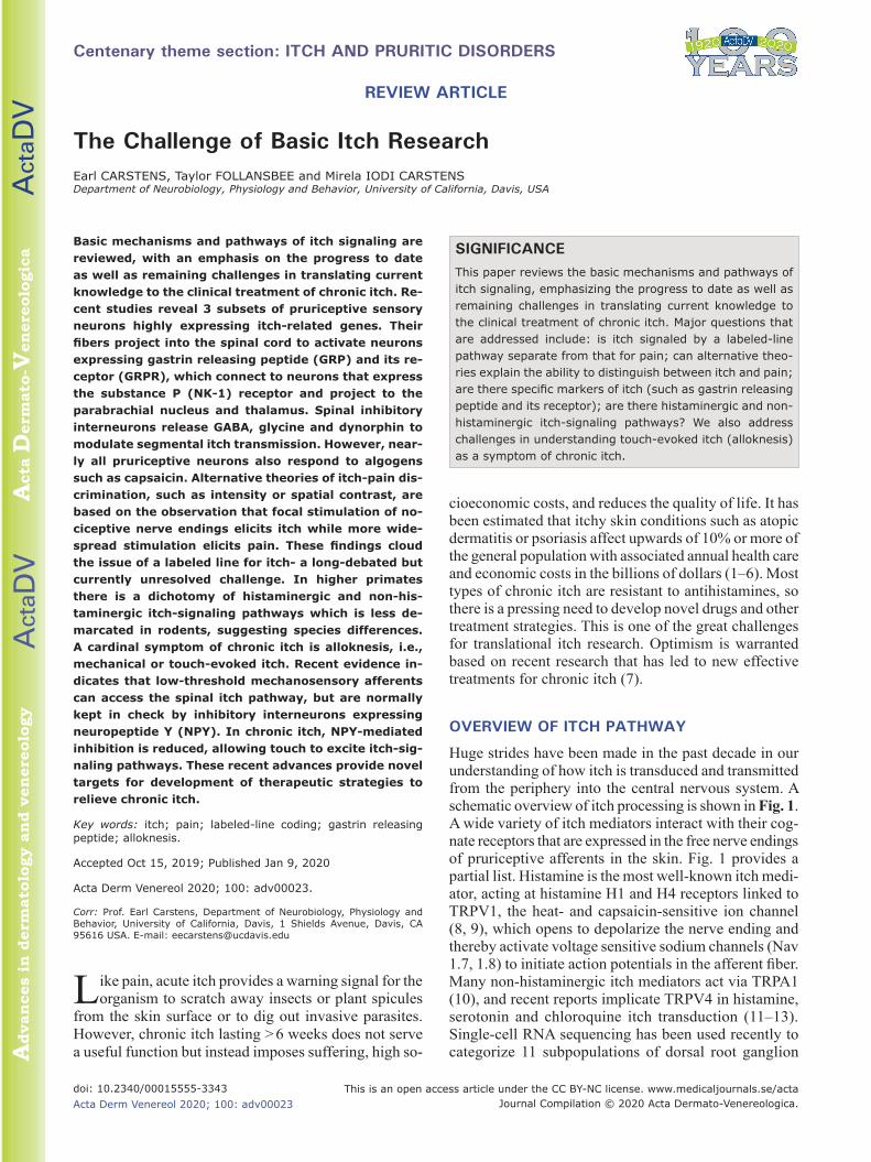
REVIEW ARTICLE
This is an open access article under the CC BY-NC license. www.medicaljournals.se/actaActa Derm Venereol 2020; 100: adv00023
Centenary theme section: ITCH AND PRURITIC DISORDERS
SIGNIFICANCEThis paper reviews the basic mechanisms and pathways of itch signaling, emphasizing the progress to date as well as remaining challenges in translating current knowledge to the clinical treatment of chronic itch. Major questions that are addressed include: is itch signaled by a labeled-line pathway separate from that for pain; can alternative theo-ries explain the ability to distinguish between itch and pain; are there specific markers of itch (such as gastrin releasing peptide and its receptor); are there histaminergic and non-histaminergic itch-signaling pathways? We also address challenges in understanding touch-evoked itch (alloknesis) as a symptom of chronic itch.
Basic mechanisms and pathways of itch signaling are reviewed, with an emphasis on the progress to date as well as remaining challenges in translating current knowledge to the clinical treatment of chronic itch. Re-cent studies reveal 3 subsets of pruriceptive sensory neurons highly expressing itch-related genes. Their fibers project into the spinal cord to activate neurons expressing gastrin releasing peptide (GRP) and its re-ceptor (GRPR), which connect to neurons that express the substance P (NK-1) receptor and project to the parabrachial nucleus and thalamus. Spinal inhibitory interneurons release GABA, glycine and dynorphin to modulate segmental itch transmission. However, near-ly all pruriceptive neurons also respond to algogens such as capsaicin. Alternative theories of itch-pain dis-crimination, such as intensity or spatial contrast, are based on the observation that focal stimulation of no-ciceptive nerve endings elicits itch while more wide-spread stimulation elicits pain. These findings cloud the issue of a labeled line for itch- a long-debated but currently unresolved challenge. In higher primates there is a dichotomy of histaminergic and non-his-taminergic itch-signaling pathways which is less de-marcated in rodents, suggesting species differences. A cardinal symptom of chronic itch is alloknesis, i.e., mechanical or touch-evoked itch. Recent evidence in-dicates that low-threshold mechanosensory afferents can access the spinal itch pathway, but are normally kept in check by inhibitory interneurons expressing neuropeptide Y (NPY). In chronic itch, NPY-mediated inhibition is reduced, allowing touch to excite itch-sig-naling pathways. These recent advances provide novel targets for development of therapeutic strategies to relieve chronic itch.
Key words: itch; pain; labeled-line coding; gastrin releasing peptide; alloknesis.
Accepted Oct 15, 2019; Published Jan 9, 2020
Acta Derm Venereol 2020; 100: adv00023.
Corr: Prof. Earl Carstens, Department of Neurobiology, Physiology and Behavior, University of California, Davis, 1 Shields Avenue, Davis, CA 95616 USA. E-mail: [email protected]
Like pain, acute itch provides a warning signal for the organism to scratch away insects or plant spicules
from the skin surface or to dig out invasive parasites. However, chronic itch lasting > 6 weeks does not serve a useful function but instead imposes suffering, high so-
cioeconomic costs, and reduces the quality of life. It has been estimated that itchy skin conditions such as atopic dermatitis or psoriasis affect upwards of 10% or more of the general population with associated annual health care and economic costs in the billions of dollars (1–6). Most types of chronic itch are resistant to antihistamines, so there is a pressing need to develop novel drugs and other treatment strategies. This is one of the great challenges for translational itch research. Optimism is warranted based on recent research that has led to new effective treatments for chronic itch (7).
OVERVIEW OF ITCH PATHWAY
Huge strides have been made in the past decade in our understanding of how itch is transduced and transmitted from the periphery into the central nervous system. A schematic overview of itch processing is shown in Fig. 1. A wide variety of itch mediators interact with their cog-nate receptors that are expressed in the free nerve endings of pruriceptive afferents in the skin. Fig. 1 provides a partial list. Histamine is the most well-known itch medi-ator, acting at histamine H1 and H4 receptors linked to TRPV1, the heat- and capsaicin-sensitive ion channel (8, 9), which opens to depolarize the nerve ending and thereby activate voltage sensitive sodium channels (Nav 1.7, 1.8) to initiate action potentials in the afferent fiber. Many non-histaminergic itch mediators act via TRPA1 (10), and recent reports implicate TRPV4 in histamine, serotonin and chloroquine itch transduction (11–13). Single-cell RNA sequencing has been used recently to categorize 11 subpopulations of dorsal root ganglion
The Challenge of Basic Itch ResearchEarl CARSTENS, Taylor FOLLANSBEE and Mirela IODI CARSTENSDepartment of Neurobiology, Physiology and Behavior, University of California, Davis, USA

Act
aDV
Act
aDV
Advan
ces
in d
erm
ato
logy a
nd v
en
ere
olo
gy
Acta
Derm
ato
-Ven
ere
olo
gic
a
3Challenge of basic itch research
Theme issue: Itch and pruritic disorders
(DRG) cells, with 3 largely nonpeptidergic (NP) groups expressing genes associated with itch: NP1 (MrgprD), NP2 (MrgprA3), and NP3 (brain natriuretic peptide [BNP] and somatostatin) (14). MrgprD, MrgprA3, BNP and somatostatin have all been implicated in itch (15–18). In addition, neuroimmune interactions have been im-plicated in chronic itch. Recent studies have implicated IL-31 (19, 20), IL-4 and IL-13 (21) in itch and itch sen-sitization, leading to the development of biologics and antagonists that block activation of sensory neurons by cytokines (7). Clearly, improved understanding of the peripheral transduction of itch and immune function is already addressing the challenge of translating basic research into more effective treatments for chronic itch.
Pruriceptive afferent fibers transmit signals into the spinal cord dorsal horn, where they release neuropeptides
including BNP (17), possibly gastrin releasing peptide (GRP) (22, 23), substance P (23), neuromedin B (24), and somatostatin (25) as well as the neurotransmitter glutamate (see below). The spinal circuitry includes excitatory interneurons that express GRP and substance P (26, 27), as well as itch-inhibitory interneurons ex-pressing GABA, glycine and dynorphin (28–31) (Fig. 1). Projection neurons ultimately give rise to ascending itch-signaling pathways to the parabrachial nucleus and somatosensory thalamus. A high percentage of ascending projection neurons express the NK-1 receptor (32, 33). A majority of antidromically identified spinothalamic and spinoparabrachial projection neurons in rats respond to intradermal injection of pruritogens, with most also responding to the algogens capsaicin and mustard oil (34, 35). Using a double-label strategy, we observed similar
Fig. 1. Schematic of itch-signaling pathways. 5-HT: 5-hydroxytryptamine (serotonin); ACC: anterior cingulate cortex; BNP: brain natriuretic peptide; CGRP: calcitonin gene related peptide; Dyn: dynorphin; Glu: glutamate; Gly: glycine; GRP: gastrin releasing peptide; IL: interleukin; LTB4: leukotriene B4; Mrgpr: Mas-related G-protein coupled receptor; PAF: platelet activating factor; PAR: protease-activated receptor; SI: primary somatosensory cortex; SP: substance P; Sst: somatostatin; TLR: toll-like receptor; TLSP: thymic stromal lymphopoietin.

Act
aDV
Act
aDV
Advan
ces
in d
erm
ato
logy a
nd v
en
ere
olo
gy
Acta
Derm
ato
-Ven
ere
olo
gic
a
E. Carstens et al.4
Theme issue: Itch and pruritic disorders
proportions of retrogradely labeled spinothalamic and spinoparabrachial neurons that co-express the activity marker, c-fos, following intradermal injection of hista-mine, chloroquine or capsaicin (36). Finally, the spinal itch-signaling circuitry is very likely under descending modulatory influences from the brainstem, although this has only begun to be experimentally addressed (37, 38).
IS THERE A LABELED LINE FOR ITCH?
On the one hand, there is evidence that spinal transmis-sion involving the neuropeptide GRP provides a specific pathway for itch transmission (discussed further below). On the other hand, based on neural recordings from pe-ripheral and second- or higher-order neurons in the spinal cord and brain, it is evident that neurons that respond to pruritogens invariably also respond to algogens such as capsaicin, mustard oil, and other noxious stimuli. Thus, there appear to be few if any itch-specific neurons, im-plying that itch must be distinguished from pain (and other dysesthetic sensory qualities) by some mechanism that can decode activity in non-selective neurons. A great challenge of basic itch research is to reconcile these se-emingly disparate observations to understand how itch is conveyed to the brain and how it is discriminated from pain and other sensory qualities.
There is much evidence that activation of pruritogen-sensitive primary afferent fibers elicits a sensation of itch via a specific “labeled line” pathway. An older study using electrical stimulation at discrete sites on the skin surface reported that the intensity of the evoked itch increased as a function of increasing stimulus frequency but never transitioned to pain (39). A seminal observation was that mechanically insensitive C-fibers recorded by microneurography responded to cutaneous application of histamine, such that the action potential firing pat-tern closely paralleled the time course of concomitant itch sensation (40). More recent studies suggest that
activation of specific primary afferents elicits itch, even though the afferents respond to algogenic as well as pruritogenic stimuli. For example, MrgprA3 is a Mas-related G-protein-coupled receptor expressed in sensory nerve endings that respond to the itchy antimalarial drug chloroquine (15). When TRPV1 (the capsaicin and heat-sensitive receptor) is genetically engineered into sensory neurons expressing MrgprA3 in otherwise TRPV1-null mice, capsaicin activation of these neurons elicits itch (scratching) rather than the pain behavior that is normally elicited by capsaicin (41). Moreover, MrgprA3-expressing sensory nerves responded not only to chloroquine but also capsaicin and other chemicals, indicating that they are not itch-specific (41). Optogenetic activation of MrgprA3-expressing peripheral afferents also elicited itch-related scratching behavior (27). These findings indicate that activation of MrgprA3-expressing nerve endings elicits itch, regardless of whether they are activated by pruritic, algogenic or artificial stimuli. This implies that although the MrgprA3-expressing afferents are not exclusively activated by itchy stimuli, they ac-cess circuits at higher levels of the nervous system that selectively signal itch but not pain.
This concept is also reflected in a “population coding” theory, which is similar to the selectivity theory (Fig. 2A). Using calcium imaging of DRG sensory neurons, we found that most if not all neurons that responded to itch mediators additionally responded to capsaicin and mustard oil (42, 43). The same was true for superficial dorsal horn neurons (44). Similarly, high percentages of neurons in the ventral posterior medial and posterior triangular thalamic nuclei responded to pruritogens as well as capsaicin (45). We postulated that pruritogen- and algogen-sensitive DRG and spinal dorsal horn neurons signal itch, whereby these non-selective spinal neurons access itch-specific central mechanisms (Fig. 2A). In contrast, pruritogen-insensitive but algogen-sensitive neurons signal pain. Note that this idea still embodies
Fig. 2. Itch theories. A: selectivity (similar to population coding). B: intensity theory. See text for explanation. +: excitatory synapse; –: inhibitory synapse.

Act
aDV
Act
aDV
Advan
ces
in d
erm
ato
logy a
nd v
en
ere
olo
gy
Acta
Derm
ato
-Ven
ere
olo
gic
a
5Challenge of basic itch research
Theme issue: Itch and pruritic disorders
separate, central labeled line mechanisms for itch and pain. Pain-evoking stimuli would activate both popula-tions of neurons, implying that itch and pain are elicited simultaneously. In this case, pain sensation dominates due to the ability of nociceptive spinal input to activate itch-inhibitory interneurons (Fig. 2A) (46), consistent with the selectivity theory of itch. Itch perception may also be masked by stronger pain.
IS GASTRIN RELEASING PEPTIDE AN ITCH-SPECIFIC MARKER?
A role for GRP in itch was first demonstrated by a sig-nificant reduction in pruritogen-evoked scratching, but not pain behavior, in transgenic mice lacking the GRP receptor (GRPR) (22). Neurotoxic destruction of GRPR-expressing spinal neurons also significantly attenuated pruritogen-evoked scratching but not pain behavior (47). These findings were recently corroborated by the report that chemogenetic activation of GRP-expressing dorsal horn neurons elicited behavioral signs of itch but not pain (48). These data strongly support GRP and GRPR expressed in spinal neurons as itch-specific markers.
However, another recent study reported that selective activation of GRP-expressing spinal neurons elicited behavioral signs of both itch and pain (27). The authors genetically engineered TRPV1 into GRP-expressing spinal neurons in otherwise TRPV1-null mice. Intrathecal administration of capsaicin dose-dependently elicited behavioral signs of itch (scratching) as well as pain (licking). At higher doses of capsaicin (1–20 µg), pain (but not itch) responses decreased and were rescued by administration of the µ-opioid antagonist naloxone. The authors suggested that high-dose capsaicin triggered an opioid mechanism that reduced pain but not itch, and that a common population of GRP-expressing spinal neurons signals both itch and pain. This is consistent with the intensity theory of itch (Fig. 2B), which postulates that itch is signaled by a lower firing rate and pain by a higher firing rate in a common population of spinal neurons. In-deed, capsaicin elicited much higher firing rates in GRP-expressing spinal neurons compared to those elicited by the pruritogens SLIGRL, chloroquine or histamine (27). In general, capsaicin and mustard oil elicited consistently higher firing rates compared to pruritogens in spinal dorsal horn neurons, including spinothalamic projection neurons (49). Thus, there is conflicting evidence as to whether GRP- and GRPR-expressing spinal neurons are itch-specific or signal both itch and pain, a challenge to theories of itch-pain discrimination that requires future studies to resolve.
Besides GRP, neuromedin B (24), brain natriuretic peptide (BNP) (17), glutamate (50) and substance P (51) have also been implicated in the spinal transmission of itch. We found that individual intrathecal delivery of receptor antagonists of GRP (RC-3095), substance P
(L-733060) or the AMPA glutamate receptor (CNQX), partially reduced scratching behavior and spinal neuronal responses to chloroquine, while a combination of all 3 antagonists completely inhibited these responses (52). This is supported by another study reporting that of dorsal horn neurons responsive to intradermal histamine or chloroquine, some responded to intrathecal delivery of BNP or GRP, but less commonly to both (53). These findings suggest that there may be parallel spinal path-ways for itch, each utilizing these neurotransmitters/neuropeptides to different extents. CNQX almost com-pletely abolished scratching and neuronal responses to histamine, implicating glutamate as the primary spinal neurotransmitter in histaminergic itch (52). These studies provide points of intervention in the spinal cord to block the transmission of itch signals.
SPATIAL CONTRAST THEORY OF ITCH
A further complication to our understanding of itch mechanisms is the finding that a dominant sensation of itch, together with sub-dominant nociceptive sensations (burning, stinging, pricking), were elicited by insertion of either a single histamine-loaded, or capsaicin-loaded, or native cowhage spicule into the skin (54). This implies that highly localized activation of a minimal number of nociceptive nerve endings in the skin by either pruritoge-nic or algogenic chemicals is sufficient to elicit a domi-nant sensation of itch. This supports the “spatial contrast” theory of itch, which holds that limited activation of nociceptive nerve endings is itchy, while activation of a greater number of nerve endings over a broader area (e.g., by intradermal injection of capsaicin) is painful, possibly due to disruption of a specific pattern for itch via the activation of many nociceptors. This concept was suggested as a possible mechanism of neuropathic itch following nerve injury that results in degeneration of most but not all C-fibers, such that activation of the few spared fibers elicits a sensation of itch (55). The challenge remains to explain how either itch or pain results from localized patterns of activation of nociceptive C-fibers.
HISTAMINERGIC VS. NON-HISTAMINERGIC ITCH: SPECIES DIFFERENCES?
It is a dogma that there are two types of itch, histamin-ergic and non-histaminergic. In humans, histaminergic itch is mediated by the histamine-sensitive, mechanically insensitive C-fiber afferents mentioned above (40, 56). In contrast, non-histaminergic itch can be elicited by spicules of cowhage, which contain proteases (57, 58). Cowhage excites mechanically sensitive polymodal nociceptors (56, 59). The duality of histamine- and cowhage-sensitivity applies to non-human primates as well, since intradermal injections of histamine or place-ment of cowhage spicules activated largely separate po-

Act
aDV
Act
aDV
Advan
ces
in d
erm
ato
logy a
nd v
en
ere
olo
gy
Acta
Derm
ato
-Ven
ere
olo
gic
a
E. Carstens et al.6
Theme issue: Itch and pruritic disorders
pulations of spinothalamic tract neurons (60). However, in rodents there appears to be greater overlap in primary and secondary sensory neurons responsive to histamine and non-histaminergic itch mediators. Using calcium imaging of mouse DRG and trigeminal ganglion (TG) cells, it was variously reported that 100% (15), 50% (61) or 17–23% (62) of chloroquine-responsive cells also responded to histamine. Using in vivo recording from identified MrgprA3-expressing DRG cells in mice, 78% (7/9) responded to both histamine and chloroquine (41). Recordings from mouse spinal dorsal horn neurons revealed that 47–71% of chloroquine-responsive neurons also responded to histamine (63). In rat somatosensory thalamus, all 7 chloroquine-responsive neurons that were additionally tested with histamine responded, although it is noted that a large number of histamine-responsive thalamic neurons did not respond to chloroquine (45). These data imply that histaminergic and non-histamin-ergic pathways may be more segregated in humans and non-human primates compared to rodents. A challenge for the field is to understand the limitations of rodent models for translation to human itch.
THE CHALLENGE OF CHRONIC ITCH AND ALLOKNESIS
Cardinal symptoms of chronic itch are ongoing (“spon-taneous”) itch, alloknesis (mechanical or touch-evoked itch), and hyperknesis (increased itch to a normally itchy or punctate mechanical stimulus). In healthy normal mice, lightly touching the skin does not elicit any be-havioral signs of itch. However, following intradermal injection of histamine and other pruritogens, light touch elicits immediate scratch bouts – a model of alloknesis (64). Chemogenetic silencing of spinal neuropeptide Y (NPY) – expressing neurons led to increased touch-evoked scratching (65), and intrathecal delivery of NPY-1 receptor agonists reduced touch-evoked scratching (66),
implying that the NPY-expressing neurons normally inhibit itch elicited by low-threshold mechanoreceptors. Scratching elicited by intradermal chloroquine, but not mechanical stimulation, was attenuated by antagonizing or ablating GRPR-expressing neurons, implying that me-chanical itch is independent of, or converges downstream of GRP-GRPR signaling in the spinal itch circuit (Fig. 3). A reduction in the number of cutaneous Merkel cells and reduced expression of the mechanotransduction channel piezo2, as occurred in aged mice or under dry skin conditions, was associated with increased alloknesis, while chemogenetic activation of Merkel cells prevented alloknesis in dry skin (67). This suggests that Merkel cells connected to slowly adapting type I (SAI) afferents excite NPY-expressing interneurons to inhibit spinal itch transmission. It was very recently reported that activa-tion of neurons expressing the NPY-1 receptor promotes mechanical itch (68). Mechanical itch was not affected following ablation of spinal neurons expressing the NK-1 receptor, implying that mechanical itch is transmitted via a pathway independent of that for chemical itch (Fig. 3). NPY-mediated inhibition can be overcome by me-chanoreceptor activation of NPY-1 receptor-expressing neurons to drive the mechanical itch-signaling pathway under conditions in which Merkel cell-SAI input is re-duced (Fig. 3).
A number of animal models have been developed to mimic various types of chronic itch and alloknesis, in-cluding atopic dermatitis, psoriasis and others (69, 70). Repeated topical application of ovalbumin induced an atopic dermatitis-like condition in mice, characterized by skin hyperplasia and lesions, increased IgG and Th2 cyto-kines, and importantly, increased spontaneous scratching behavior, alloknesis and hyperknesis (32). Alloknesis, but not hyperknesis or spontaneous scratching, was nearly abolished in OVA-treated mice that received intrathecal injection of substance P-saporin but not bombesin-saporin. This implies that the effect of low-threshold
Fig. 3. Schematic of mechanical itch (alloknesis). See text for explanation. GRP: gastrin releasing peptide; GRPR: gastrin releasing peptide receptor; NK-1R: neurokinin-1 receptor; NPY: neuropeptide Y; NPY-1R: neuropeptide Y-1 receptor; +: excitatory synapse; –: inhibitory synapse.

Act
aDV
Act
aDV
Advan
ces
in d
erm
ato
logy a
nd v
en
ere
olo
gy
Acta
Derm
ato
-Ven
ere
olo
gic
a
7Challenge of basic itch research
Theme issue: Itch and pruritic disorders
mechanoreceptor input to inhibit itch signaling neurons occurred downstream of GRPR-expressing neurons but requires NK-1 receptor-expressing neurons, supporting the idea that mechanoreceptive input converges onto the chemical itch-signaling pathway (Fig. 3; dashed line connecting NPY-1R to NK-1R). Consistent with this, intrathecal NPY agonists suppressed both chemically- and mechanically-evoked itch behavior (66).
Given that alloknesis is quite bothersome to patients suffering from many types of chronic itch, a challenge to the field is to better understand how low-threshold mechanosensory input interacts with spinal itch-signaling pathways and potential anti-alloknesis interventions targeting spinal NPY1 receptors.
CONCLUSIONS
The preceding text has identified a number of challenges arising from basic itch research to explain how itch can be discriminated from pain, and to translate our increasing knowledge of itch signaling into clinical treatment. Given the remarkable progress of the past decade and the current strong interest in itch research, several novel approaches to the treatment of chronic itch are already being used and more can be expected in the near future. Nevertheless, it has been debated for more than 100 years whether itch and pain are signaled by separate labeled-line pathways or by a common population of non-specific neurons. This debate continues unresolved up to the present, with arguments favoring both concepts.
ACKNOWLEDGMENTSThe author’s work has been supported by a grant from the National Institute of Arthritis, Musculoskeletal and Skin Diseases, National Institutes of Health (AR057194).
REFERENCES1. Silverberg JI, Hanifin JM. Adult eczema prevalence and as-
sociations with asthma and other health and demographic factors: A US population–based study. J Allergy Clin Immunol 2013; 132: 1132–1138.
2. Drucker AM, Wang AR, Li WQ, Sevetson E, Block JK, Qureshi AA. The burden of atopic dermatitis: summary of a report for the National Eczema Association. J Invest Dermatol 2017; 137: 26–30.
3. Michalek IM, Loring B, John SM. A systematic review of worldwide epidemiology of psoriasis. J Eur Acad Dermatol Venereol 2017; 31: 205–212.
4. Krueger GG, Bergstresser PR, Lowe NJ, Voorhees JJ, W. G. Psoriasis. J Am Acad Dermatol 1984; 11: 937–947.
5. Bickers DR, Lim HW, Margolis D, Weinstock MA, Goodman C, Faulkner E, et al. The burden of skin diseases: 2004: A joint project of the American Academy of Dermatology Association and the Society for Investigative Dermatology. J Am Acad Dermatol 2006; 55: 490–500.
6. Thorpe KE, Florence CS, Joski P. Which medical conditions account for the rise in health care spending? Health Aff (Millwood). Suppl Web Exclusives, W4–437–45.
7. Yosipovitch G, Rosen JD, Hashimoto T. Itch: From mechanism to (novel) therapeutic approaches. J Allergy Clin Immunol
2018; 142: 1375–1390.8. Shim WS, Tak MH, Lee MH, Kim M, Kim M, Koo JY, et al.
TRPV1 mediates histamine-induced itching via the activation of phospholipase A2 and 12-lipoxygenase. J Neurosci 2007; 27: 2331–2337.
9. Imamachi N, Park GH, Lee H, Anderson DJ, Simon MI, Basbaum AI, et al. TRPV1-expressing primary afferents generate behavioral responses to pruritogens via multiple mechanisms. Proc Natl Acad Sci 2009; 106: 11330–11335.
10. Wilson SR, Gerhold KA, Bifolck-Fisher A, Liu Q, Patel KN, Dong X, et al. TRPA1 is required for histamine-independent, Mas-related G protein-coupled receptor-mediated itch. Nat Neurosci 2011; 14: 595–602.
11. Akiyama T, Ivanov M, Nagamine M, Davoodi A, Carstens MI, Ikoma A, et al. Involvement of TRPV4 in serotonin-evoked scratching. J Invest Dermatol 2016; 136: 154–160.
12. Chen Y, Fang Q, Wang Z, Zhang JY, MacLeod AS, Hall RP, et al. Transient receptor potential vanilloid 4 ion channel fun-ctions as a pruriceptor in epidermal keratinocytes to evoke histaminergic itch. J Biol Chem 2016; 291: 10252–10262.
13. Kim S, Barry DM, Liu XY, Yin S, Munanairi A, Meng QT, et al. Facilitation of TRPV4 by TRPV1 is required for itch transmis-sion in some sensory neuron populations. Sci Signal 2016; 9: ra71.
14. Usoskin D, Furlan A, Islam S, Abdo H, Lönnerberg P, Lou D, et al. Unbiased classification of sensory neuron types by large-scale single-cell RNA sequencing. Nat Neurosci 2015; 18: 145–153.
15. Liu Q, Tang Z, Surdenikova L, Kim S, Patel KN, Kim A et al. Sensory neuron-specific GPCR Mrgprs are itch receptors mediating chloroquine-induced pruritus. Cell 2009; 139: 1353–1365.
16. Liu Q, Sikand P, Ma C, Tang Z, Han L, Li Z, et al. Mecha-nisms of itch evoked by beta-alanine. J Neurosci 2012; 32: 14532–14537.
17. Mishra SK, Hoon MA. The cells and circuitry for itch responses in mice. Science 2013; 340: 968–971.
18. Huang J, Polgár E, Solinski HJ, Mishra SK, Tseng PY, Iwagaki N, et al. Circuit dissection of the role of somatostatin in itch and pain. Nature Neuroscience 2018; 21: 707–716.
19. Cevikbas F, Wang X, Akiyama T, Kempkes C, Savinko T, Antal A, et al. A sensory neuron–expressed IL-31 receptor medi-ates T helper cell–dependent itch: Involvement of TRPV1 and TRPA1. J Allergy Clin Immunol 2014; 133: 448–460.
20. Cevikbas F, Kempkes C, Buhl T, Mess C, Buddenkotte J, Stein-hoff M. Role of Interleukin-31 and Oncostatin M in Itch and Neuroimmune Communication. In: Carstens E, Akiyama T, editors. Itch: Mechanisms and Treatment. Boca Raton (FL): CRC Press; 2014, Ch. 11.
21. Oetjen LK, Mack MR, Feng J, Whelan TM, Niu H, Guo CJ et al. Sensory neurons co-opt classical immune signaling pathways to mediate chronic itch. Cell 2017; 171: 217–228.
22. Sun YG, Chen ZF. A gastrin-releasing peptide receptor medi-ates the itch sensation in the spinal cord. Nature 2007; 448: 700–703.
23. Akiyama T, Tominaga M, Davoodi A, Nagamine M, Blansit K, Horwitz A, et al. Roles for substance P and gastrin-releasing peptide as neurotransmitters released by primary afferent pruriceptors. J Neurophysiol 2013; 109: 742–748.
24. Wan L, Jin H, Liu XY, Jeffry J, Barry DM, Shen KF, et al. Distinct roles of NMB and GRP in itch transmission. Sci Rep 2017; 7: 15466.
25. Stantcheva KK, Iovino L, Dhandapani R, Martinez C, Castaldi L, Nocchi L, et al. A subpopulation of itch-sensing neurons marked by Ret and somatostatin expression. EMBO Rep 2016; 17: 585–600.
26. Gutierrez-Mecinas M, Bell AM, Marin A, Taylor R, Boyle KA, Furuta T, et al. Preprotachykinin A is expressed by a distinct population of excitatory neurons in the mouse superficial spinal dorsal horn including cells that respond to noxious and pruritic stimuli. Pain 2017; 158: 440–456.
27. Sun S, Xu Q, Guo C, Guan Y, Liu Q, Dong X. Leaky gate model: intensity-dependent coding of pain and itch in the spinal cord. Neuron 2017; 93: 840–853.
28. Akiyama T, Carstens M, Carstens E. Transmitters and pat-

Act
aDV
Act
aDV
Advan
ces
in d
erm
ato
logy a
nd v
en
ere
olo
gy
Acta
Derm
ato
-Ven
ere
olo
gic
a
E. Carstens et al.8
Theme issue: Itch and pruritic disorders
hways mediating inhibition of spinal Itch-Signaling neurons by scratching and other counterstimuli. PLoS One 2011; 6: 1–10.
29. Braz JM, Juarez-Salinas D, Ross SE, Basbaum AI. Transplant restoration of spinal cord inhibitory controls ameliorates neuropathic itch. J Clin Invest 2014; 124: 3612–3616.
30. Kardon AP, Polgár E, Hachisuka J, Snyder LM, Cameron D, Savage S, et al. Dynorphin acts as a neuromodulator to inhibit itch in the dorsal horn of the spinal cord. Neuron 2014; 82: 573–586.
31. Foster E, Wildner H, Tudeau L, Haueter S, Ralvenius WT, Jegen M, et al. Targeted ablation, silencing, and activation establish glycinergic dorsal horn neurons as key compo-nents of a spinal gate for pain and itch. Neuron 2015; 85: 1289–1304.
32. Akiyama T, Nguyen T, Curtis E, Nishida K, Devireddy J, De-lahanty J, et al. A central role for spinal dorsal horn neurons that express neurokinin-1 receptors in chronic itch. Pain 2015; 156: 1240–1246.
33. Cameron D, Polgár E, Gutierrez-Mecinas M, Gomez-Lima M, Watanabe M, Todd AJ. The organisation of spinoparabrachial neurons in the mouse. Pain 2015; 156: 2061–2071.
34. Jansen NA, Giesler GJ. Response characteristics of pruricep-tive and nociceptive trigeminoparabrachial tract neurons in the rat. J Neurophysiol 2015; 113: 58–70.
35. Moser HR, Giesler GJ. Characterization of pruriceptive tri-geminothalamic tract neurons in rats. J Neurophysiol 2014; 111: 1574–1589.
36. Akiyama T, Curtis E, Nguyen T, Carstens MI, Carstens E. Anatomical evidence of pruriceptive trigeminothalamic and trigeminoparabrachial projection neurons in mice. J Comp Neurol 2016; 524: 244–256.
37. Carstens E, Iodi Carstens M, Akiyama T, Davoodi A, Nagamine M. Opposing effects of cervical spinal cold block on spinal itch and pain transmission. Itch 2018; 3: e16.
38. Gao ZR, Chen WZ, Liu MZ, Chen XJ, Wan L, Zhang XY et al. Tac1-Expressing neurons in the periaqueductal gray facilitate the itch-scratching cycle via descending regulation. Neuron 2019; 101: 45–59.
39. Tuckett RP. Itch evoked by electrical stimulation of the skin. J Invest Dermatol 1982; 79: 368–373.
40. Schmelz M, Schmidt R, Bickel A, Handwerker HO, Torebjörk HE. Specific C-receptors for itch in human skin. J Neurosci 1997; 17: 8003–8008.
41. Han L, Ma C, Liu Q, Weng HJ, Cui Y, Tang Z, et al. A subpopu-lation of nociceptors specifically linked to itch. Nat Neurosci 2013; 16: 174–182.
42. Akiyama T, Carstens MI, Carstens E. Facial injections of pru-ritogens and algogens excite partly overlapping populations of primary and second-order trigeminal neurons in mice. J Neurophysiol 2010; 104: 2442–2450.
43. Klein A, Carstens MI, Carstens E. Facial injections of pru-ritogens or algogens elicit distinct behavior responses in rats and excite overlapping populations of primary sensory and trigeminal subnucleus caudalis neurons. J Neurophysiol 2011; 106: 1078–1088.
44. Akiyama T, Merrill AW, Carstens MI. Carstens E. Activation of superficial dorsal horn neurons in the mouse by a PAR-2 agonist and 5-HT: potential role in itch. J Neurosci 2009; 29: 6691–6699.
45. Lipshetz B, Khasabov SG, Truong H, Netoff TI, Simone DA, Giesler GJ Jr. Responses of thalamic neurons to itch- and pain-producing stimuli in rats. J Neurophysiol 2018; 120: 1119–1134.
46. Ross SE, Mardinly AR, McCord AE, Zurawski J, Cohen S, Jung C, et al. Loss of inhibitory interneurons in the dorsal spinal cord and elevated itch in Bhlhb5 mutant mice. Neuron 2010; 65: 886–898.
47. Sun YG, Zhao ZQ, Meng XL, Yin J, Liu XY, Chen ZF. Cellular basis of itch sensation. Science 2009; 325: 1531–1534.
48. Albisetti GW, Pagani M, Platonova E, Hösli L, Johannssen HC, Fritschy JM, et al. Dorsal horn gastrin-releasing peptide expressing neurons transmit spinal itch but not pain signals. J Neurosci 2019; 39: 2238–2250.
49. Davidson S, Zhang X, Khasabov SG, Moser HR, Honda CN,
Simone DA, et al. Pruriceptive spinothalamic tract neurons: physiological properties and projection targets in the primate. J Neurophysiol 2012; 108: 1711–1723.
50. Koga K, Chen T, Li XY, Descalzi G, Ling J, Gu J, et al. Gluta-mate acts as a neurotransmitter for gastrin releasing peptide-sensitive and insensitive itch-related synaptic transmission in mammalian spinal cord. Mol Pain 2011; 7: 47.
51. Carstens EE, Carstens MI, Simons CT, Jinks SL. Dorsal horn neurons expressing NK-1 receptors mediate scratching in rats. Neuroreport 2010; 21: 303–308.
52. Akiyama T, Tominaga M, Takamori K, Carstens MI, Carstens E. Roles of glutamate, substance P, and gastrin-releasing peptide as spinal neurotransmitters of histaminergic and nonhistaminergic itch. Pain 2014; 155: 80–92.
53. Kusube F, Tominaga M, Kawasaki H, Yamakura F, Naito H, Ogawa H, et al. Electrophysiological properties of brain-natriuretic peptide- and gastrin-releasing peptide-responsive dorsal horn neurons in spinal itch transmission. Neurosci Let 2016; 627: 51–60.
54. LaMotte RH, Shimada SG, Green BG, Zelterman D. Pruritic and nociceptive sensations and dysesthesias from a spicule of cowhage. J Neurophysiol 2009; 101: 1430–1443.
55. Steinhoff M, Oaklander AL, Szabó IL, Ständer S, Schmelz M. Neuropathic itch. Pain 2019; 160: S11–S16.
56. Namer B, Carr R, Johanek LM, Schmelz M, Handwerker HO, Ringkamp M. Separate peripheral pathways for pruritus in man. J Neurophysiol 2008; 100: 2062–2069.
57. Shelley WB, Arthur RP. Mucunain, the active pruritogenic proteinase of cowhage. Science 1955; 122: 469–470.
58. Reddy VB, Iuga AO, Shimada SG, LaMotte RH, Lerner EA. Cowhage-evoked itch is mediated by a novel cysteine pro-tease: a ligand of protease-activated receptors. J Neurosci 2008; 28: 4331–4335.
59. Johanek LM, Meyer RA, Friedman RM, Greenquist KW, Shim B, Borzan J, et al. A role for polymodal C-fiber afferents in nonhistaminergic itch. J Neurosci 2008; 28: 7659–7669.
60. Davidson S, Zhang X, Yoon CH, Khasabov SG, Simone DA, Giesler GJ Jr. The itch-producing agents histamine and cowhage activate separate populations of primate spinot-halamic tract neurons. J Neurosci 2007; 27: 10007–10014.
61. Akiyama T, Tominaga M, Davoodi A, Nagamine M, Blansit K, Horwitz A, et al. Cross-sensitization of histamine-indepen-dent itch in mouse primary sensory neurons. Neuroscience 2012; 226: 305–312.
62. Roberson DP, Gudes S, Sprague JM, Patoski HA, Robson VK, Blasl F, et al. Activity-dependent silencing reveals functio-nally distinct itch-generating sensory neurons. Nat Neurosci 2013; 16: 910–918.
63. Akiyama T, Tominaga M, Takamori K, Carstens MI, Carstens E. Role of spinal bombesin-responsive neurons in nonhista-minergic itch. J Neurophysiol 2014; 112: 2283–2289.
64. Akiyama T, Carstens MI, Ikoma A, Cevikbas F, Steinhoff M, Carstens E. Mouse model of touch-evoked itch (alloknesis). J Invest Dermatol 2012; 132: 1886–1891.
65. Bourane S, Duan B, Koch SC, Dalet A, Britz O, Garcia-Campmany L, et al. Gate control of mechanical itch by a subpopulation of spinal cord interneurons. Science 2015; 350: 550–554.
66. Gao T, Ma H, Xu B, Bergman J, Larhammar D, Lagerström MC. The Neuropeptide Y System Regulates Both Mechani-cal and Histaminergic Itch. J Invest Dermatol 2018; 138: 2405–2411.
67. Feng J, Luo J, Yang P, Du J, Kim BS, Hu H. Piezo2 channel-Merkel cell signaling modulates the conversion of touch to itch. Science 2018; 360: 530–533.
68. Acton D, Ren X, Di Costanzo S, Dalet A, Bourane S, Bertocchi I, et al. Spinal neuropeptide Y1 receptor-expressing neurons form an essential excitatory pathway for mechanical itch. Cell Rep 2019; 28: 625–639.
69. Bautista DM, Wilson SR, Hoon MA. Why we scratch an itch: the molecules, cells and circuits of itch. Nat Neurosci 2014; 17: 175–182.
70. Kim D, Kobayashi T, Nagao K. Research techniques made simple: mouse models of atopic dermatitis. J Invest Dermatol 2019; 139: 984–990.

Act
aDV
Act
aDV
Advan
ces
in d
erm
ato
logy a
nd v
en
ere
olo
gy
Acta
Derm
ato
-Ven
ere
olo
gic
a
Acta Derm Venereol 2020; 100: adv00024This is an open access article under the CC BY-NC license. www.medicaljournals.se/actaJournal Compilation © 2020 Acta Dermato-Venereologica.
doi: 10.2340/00015555-3344
REVIEW ARTICLE
Centenary theme section: ITCH AND PRURITIC DISORDERS
SIGNIFICANCEItch is an unpleasant sensation that may disturb quality of life, and for which the pathomechanism and appropriate treatments are unclear. Chronic itch, which lasts more than 6 weeks, often accompanies pathological dry skin-based conditions, such as xerosis, atopic dermatitis, liver and kid-ney diseases. A decline in skin barrier function is thought to be the primary cause of itch induced by dry skin. Many kinds of mediators, receptors, and channels are involved in itch signalling among the skin nervous system, skin cells, and central nervous system. Several therapeutic options for itching have thus been developed, such as photothera-py, phospholipids, antioxidants, and emollients.
Chronic itch is a burdensome clinical problem that often accompanies pathological dry skin-based conditions, such as atopic dermatitis, and systemic disorders, such as kidney diseases, with an unclear pathomechanism and treatments. One of the basic mouse models to in-vestigate mechanisms of itch associated with dry skin is a mixture of acetone and ether followed by water. Animal studies using the acetone and ether followed by water model have revealed that many mediators and receptors, e.g. mas-related G protein-coupled re-ceptor family, transient receptor potential, and chemo-kines, are responsible for itch and its hypersensitivity, supporting the hypothesis that dry skin-induced itch is a histamine-independent pathway. New insights have been acquired into the interplay between neurones and non-neuronal cells in the initiation, modulation, and sensitization of itch. Several thera peutic options for itching have thus been developed. This review summarizes the updated pathogenesis and therapeu-tic strategies for itch in dry skin conditions.
Key words: dry skin; hypersensitivity; itch; sensory neuron; mouse model.
Accepted Oct 15, 2019; Published Jan 9, 2020
Acta Derm Venereol 2020; 100: adv00024.
Corr: Mitsutoshi Tominaga, Juntendo Itch Research Center (JIRC), Insti-tute for Environmental and Gender Specific Medicine, Juntendo University Graduate School of Medicine, 2-1-1 Tomioka, Urayasu, Chiba 279-0021, Japan. E-mail: [email protected]
Skin, the body’s largest organ, serves as a first physio-logical barrier against the external environment. The
barrier function of the skin is exerted by the epidermis, the most superficial layer of the skin, of which the stratum corneum (SC) is largely responsible for the barrier fun-ction. There are 2 elements important for the maintenance of SC humidity: intercellular lipids, which form the main barrier against diffusion of water across the SC, and natural moisturizing factor, which has a key role in the absorption of water in the SC. Impaired skin barrier integrity causes excessive water loss and leads to skin dryness (1, 2).
DRY SKIN
Dry skin is characterized by a scaly, rough, cracked, and fissured surface, and is closely associated with the somatosensory sensation of itch, especially chronic itch
(3). Dry skin with chronic itch is the most common clinical manifestation of dermatoses, such as xerosis, atopic dermatitis (AD), and psoriasis, and is a common cutaneous manifestation in pruritic systemic diseases, such as chronic kidney disease (CKD), chronic liver diseases (CLD), and diabetes mellitus (DM) (4).
Histamine is a well-known substance that induces itch; however, antihistamines (histamine H1-receptor blockers) are not fully effective in many dermatological and syste-mic diseases characterized by dry skin, suggesting that dry skin is an important feature of antihistamine-resistant (histamine-independent) itch (2). The underlying condi-tion of dry skin is impaired function of the skin barrier, which can be caused by environmental factors, such as sun exposure, temperature, humidity, and genetic factors, such as filaggrin mutations (1, 5, 6). To assess skin barrier func-tion, transepidermal water loss (TEWL), SC hydration, and pH are commonly used (1). The signs and clinical manifestations of dry skin are not only physically un-comfortable, but also affect patients psychologically (7).
Disease-related dry skinAged skin. Xerosis is one of the most prevalent dry skin conditions in the aged population worldwide (8), affecting over 50% of individuals aged ≥ 65 years (9). Multiple skin changes in the elderly are related to xerosis: (i) al-terations in the barrier function of SC, including cellular and intercellular lipid matrix changes; (ii) pH variations; (iii) alterations in SC proteases; (iv) reduced activity of sebaceous and sweat glands; and (v) decreased oestrogen levels. All of these factors may lead to itch induction (10).
Mechanisms and Management of Itch in Dry SkinCatharina Sagita MONIAGA1, Mitsutoshi TOMINAGA1,2 and Kenji TAKAMORI1–3
1Juntendo Itch Research Center (JIRC), Institute for Environmental and Gender Specific Medicine, 2Anti-aging Skin Research Laboratory, Juntendo University Graduate School of Medicine, and 3Department of Dermatology, Juntendo University Urayasu Hospital, Chiba, Japan
Act
aDV
Act
aDV
Advan
ces
in d
erm
ato
logy a
nd v
en
ere
olo
gy
Acta
Derm
ato
-Ven
ere
olo
gic
a
C. S. Moniaga et al.10
Theme issue: Itch and pruritic disorders
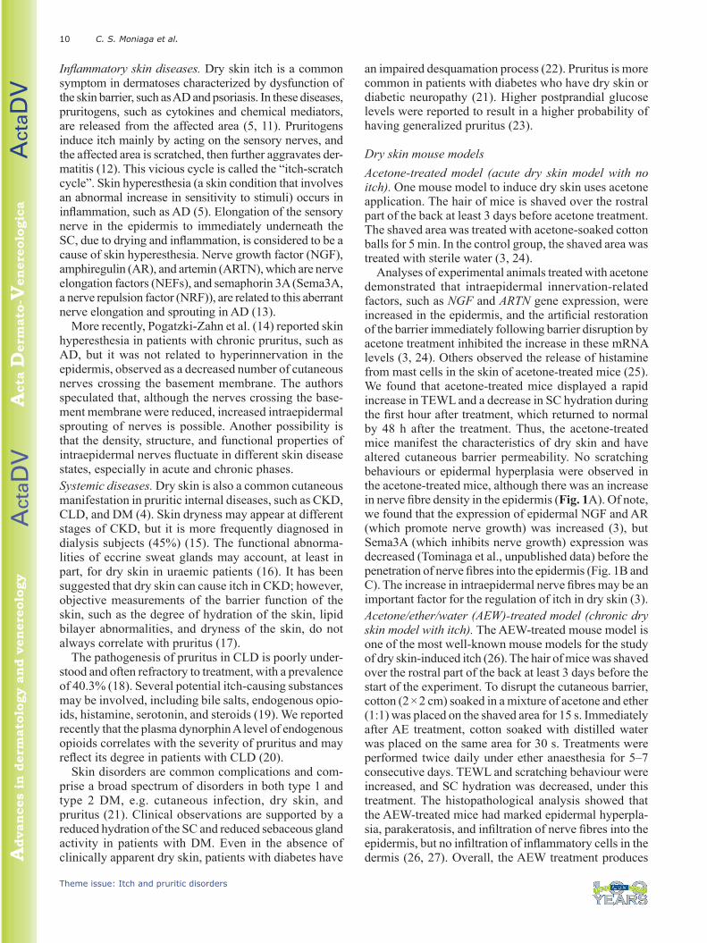
Inflammatory skin diseases. Dry skin itch is a common symptom in dermatoses characterized by dysfunction of the skin barrier, such as AD and psoriasis. In these diseases, pruritogens, such as cytokines and chemical mediators, are released from the affected area (5, 11). Pruritogens induce itch mainly by acting on the sensory nerves, and the affected area is scratched, then further aggravates der-matitis (12). This vicious cycle is called the “itch-scratch cycle”. Skin hyperesthesia (a skin condition that involves an abnormal increase in sensitivity to stimuli) occurs in inflammation, such as AD (5). Elongation of the sensory nerve in the epidermis to immediately underneath the SC, due to drying and inflammation, is considered to be a cause of skin hyperesthesia. Nerve growth factor (NGF), amphiregulin (AR), and artemin (ARTN), which are nerve elongation factors (NEFs), and semaphorin 3A (Sema3A, a nerve repulsion factor (NRF)), are related to this aberrant nerve elongation and sprouting in AD (13).
More recently, Pogatzki-Zahn et al. (14) reported skin hyperesthesia in patients with chronic pruritus, such as AD, but it was not related to hyperinnervation in the epidermis, observed as a decreased number of cutaneous nerves crossing the basement membrane. The authors speculated that, although the nerves crossing the base-ment membrane were reduced, increased intraepidermal sprouting of nerves is possible. Another possibility is that the density, structure, and functional properties of intraepidermal nerves fluctuate in different skin disease states, especially in acute and chronic phases. Systemic diseases. Dry skin is also a common cutaneous manifestation in pruritic internal diseases, such as CKD, CLD, and DM (4). Skin dryness may appear at different stages of CKD, but it is more frequently diagnosed in dialysis subjects (45%) (15). The functional abnorma-lities of eccrine sweat glands may account, at least in part, for dry skin in uraemic patients (16). It has been suggested that dry skin can cause itch in CKD; however, objective measurements of the barrier function of the skin, such as the degree of hydration of the skin, lipid bilayer abnormalities, and dryness of the skin, do not always correlate with pruritus (17).
The pathogenesis of pruritus in CLD is poorly under-stood and often refractory to treatment, with a prevalence of 40.3% (18). Several potential itch-causing substances may be involved, including bile salts, endogenous opio-ids, histamine, serotonin, and steroids (19). We reported recently that the plasma dynorphin A level of endogenous opioids correlates with the severity of pruritus and may reflect its degree in patients with CLD (20).
Skin disorders are common complications and com-prise a broad spectrum of disorders in both type 1 and type 2 DM, e.g. cutaneous infection, dry skin, and pruritus (21). Clinical observations are supported by a reduced hydration of the SC and reduced sebaceous gland activity in patients with DM. Even in the absence of clinically apparent dry skin, patients with diabetes have
an impaired desquamation process (22). Pruritus is more common in patients with diabetes who have dry skin or diabetic neuropathy (21). Higher postprandial glucose levels were reported to result in a higher probability of having generalized pruritus (23).
Dry skin mouse modelsAcetone-treated model (acute dry skin model with no itch). One mouse model to induce dry skin uses acetone application. The hair of mice is shaved over the rostral part of the back at least 3 days before acetone treatment. The shaved area was treated with acetone-soaked cotton balls for 5 min. In the control group, the shaved area was treated with sterile water (3, 24).
Analyses of experimental animals treated with acetone demonstrated that intraepidermal innervation-related factors, such as NGF and ARTN gene expression, were increased in the epidermis, and the artificial restoration of the barrier immediately following barrier disruption by acetone treatment inhibited the increase in these mRNA levels (3, 24). Others observed the release of histamine from mast cells in the skin of acetone-treated mice (25). We found that acetone-treated mice displayed a rapid increase in TEWL and a decrease in SC hydration during the first hour after treatment, which returned to normal by 48 h after the treatment. Thus, the acetone-treated mice manifest the characteristics of dry skin and have altered cutaneous barrier permeability. No scratching behaviours or epidermal hyperplasia were observed in the acetone-treated mice, although there was an increase in nerve fibre density in the epidermis (Fig. 1A). Of note, we found that the expression of epidermal NGF and AR (which promote nerve growth) was increased (3), but Sema3A (which inhibits nerve growth) expression was decreased (Tominaga et al., unpublished data) before the penetration of nerve fibres into the epidermis (Fig. 1B and C). The increase in intraepidermal nerve fibres may be an important factor for the regulation of itch in dry skin (3).Acetone/ether/water (AEW)-treated model (chronic dry skin model with itch). The AEW-treated mouse model is one of the most well-known mouse models for the study of dry skin-induced itch (26). The hair of mice was shaved over the rostral part of the back at least 3 days before the start of the experiment. To disrupt the cutaneous barrier, cotton (2 × 2 cm) soaked in a mixture of acetone and ether (1:1) was placed on the shaved area for 15 s. Immediately after AE treatment, cotton soaked with distilled water was placed on the same area for 30 s. Treatments were performed twice daily under ether anaesthesia for 5–7 consecutive days. TEWL and scratching behaviour were increased, and SC hydration was decreased, under this treatment. The histopathological analysis showed that the AEW-treated mice had marked epidermal hyperpla-sia, parakeratosis, and infiltration of nerve fibres into the epidermis, but no infiltration of inflammatory cells in the dermis (26, 27). Overall, the AEW treatment produces

Act
aDV
Act
aDV
Advan
ces
in d
erm
ato
logy a
nd v
en
ere
olo
gy
Acta
Derm
ato
-Ven
ere
olo
gic
a
11Mechanisms and management of itch in dry skin
Theme issue: Itch and pruritic disorders
marked skin barrier dysfunction, robust scratching, and changes in gene expression in sensory nerves and the skin (26), which recapitulate the dry skin symptoms present in many chronic itchy conditions in humans (28). There was no apparent difference in AEW-induced spontaneous scratching between mast cell-deficient mice (WBB6F1-W/WV) and normal litter-mates (WBB6F1-+/+) (26).
This dry skin model also exhibits alloknesis (scrat-ching behaviour evoked by a stimulus that is normally non-pruriceptive) and hyperknesis (the abnormal pruri-ceptive state, in which a normally pruritic stimulus elicits a greater than normal duration and/or magnitude of itch) (27, 29), as described later in this review.Special diet food model. HR-1 hairless mice fed a special diet (HR-AD) is one of the dry skin-based experimental mouse models. Mice were fed HR-AD for 48 days. These mice exhibited severe dry skin symptoms accompanied by a decrease in skin water-holding capacity, increase in TEWL, and prolonged scratching bout duration. Marked epidermal hyperplasia, and increase in circulating T cells and serum IgE are observed (30). Lipid composition analysis revealed that HR-AD is an essential fatty acid (EFA)-deficient diet. Feeding HR-AD with EFA inhibits the symptoms of dry skin (31). EFA deficiency was re-ported to depress skin barrier function due to structural changes in ceramides, and reduced elaboration and de-position of epidermal intercellular lipids (32). Therefore, HR-AD causes deterioration of the skin barrier function due to EFA deficiency.
MECHANISMS OF ITCH IN DRY SKIN
The sensation of itch is generated by the binding of itch-inducing substances to their cognate receptors on peripheral sensory afferents, e.g. unmyelinated C-fibre afferents and thinly myelinated Aδ-fibre afferents. The evoked action potential is transmitted through the ascending sensory pathway to the somatosensory cortex, resulting in the perception of itch (Fig. 2).
Sensory neuronesNerve elongation and repulsion factors. In healthy skin, most cutaneous nerve fibres terminate under dermoepi-dermal junctions. An increased intraepidermal nerve density has been observed in the skin of patients with pruritic dermatological diseases, such as senile xerosis and AD (13), as well as in dry skin mice models (3, 33). The controlling mechanism of cutaneous nerve density is regulated by the balance of NEFs, such as NGF, ARTN and AR, and NRFs, such as Sema3A, produced by kera-tinocytes (5, 13). These axonal guidance molecules may also act on keratinocytes, immune cells, and vascular endothelial cells, and may be indirectly involved in the regulation of itching (34).Matrix metalloproteinase (MMP)-2 and MMP-8. The process of cutaneous nerve growth in dry skin requires several MMPs for growth cones to penetrate the 3-di-mensional extracellular matrix (ECM) barriers (Fig. 2). Using in vitro models of ECM, we found that MMP-2
Fig. 1. Alterations in nerve fibre distribution, and nerve growth factor (NGF) and Sema3A expression in the epidermis of acetone-treated dry skin model mice. (A) Sequential alteration of intraepidermal nerve growth in acetone-treated mice was examined by immunohistochemistry using an anti-PGP9.5 antibody. (B, C) Maximum expression of NGF (green) was noted 16–24 h after the treatment (B). In contrast, the expression level of Sema3A (green) was decreased 24 h after acetone treatment (C). These expression levels gradually returned to normal by 168 h after the treatment. Nuclei are counterstained by DAPI (blue). The broken lines in panel A indicate the border between the epidermis and dermis (basement membrane). The basement membrane in panel B was stained with an anti-nidogen antibody (red). White and broken lines indicate the skin surface and the border epidermis and dermis (basement membrane), respectively. Arrowheads indicate epidermal nerve fibres (green). Scale bars: 15 μm.

Act
aDV
Act
aDV
Advan
ces
in d
erm
ato
logy a
nd v
en
ere
olo
gy
Acta
Derm
ato
-Ven
ere
olo
gic
a
C. S. Moniaga et al.12
Theme issue: Itch and pruritic disorders
localized on the growth cone functioned in penetration into the basement membrane (35). In addition, MMP-8 secreted by nerve fibres was reported to be involved in nerve growth within the dermis (36). The levels of expression of MMP-2 and MMP-8 were upregulated by NGF and down-regulated by Sema3A. The selection and up-regulation of MMPs corresponding to the ECM components surrounding the growing nerve fibres may be required for efficient nerve fibre penetration, sugges-ting that the coordinated activation of neurotrophin and ECM-integrin signalling is necessary for efficient and long-distance axon extension (37). As class 3 semaphorin signalling inhibits integrin-mediated adhesion signalling, Sema3A stimulation of growing nerve fibres may provide a reverse signalling pathway for these events (38).Peptidergic fibres. Substance P (SP) and calcitonin gene-related peptide (CGRP) are neuropeptides produced by sensory nerves in the dermis to communicate with dif-ferent cell populations in the different layers of the skin, which in turn stimulate nerve fibres. An increase in the elongated epidermal peripheral nerve fibres consist of
SP/CGRP-containing C-fibres, which usually represent epidermal peptidergic nerve fibres, has been reported in the AEW model (39, 40).
The neuropeptide gastrin-releasing peptide (GRP) is characterized as a neurotransmitter that specifically relays itch signals and specifically expressed in a small subset of peptidergic dorsal root ganglion (DRG) neu-rones. Genetic ablation of GRP receptor (GRPR)+ neu-rones resulted in significant reduction in the scratching response to multiple pruritogens (41). The transcription factor Tlx3 in the spinal cord was demonstrated to be essential for the development of GRPR+ neurones (42). Huang et al. (43) reported that Tlx3 conditional knock-out (Tlx3F/F; Nav1.8-Cre mice specifically lost Tlx3 expression in most TrkA-lineage DRG neurones) mice scratched much less compared with controls in the dry skin model, suggesting impairment of dry skin-induced chronic itch in these mice.Non-peptidergic fibers. C-fibres have been divided into peptidergic and non-peptidergic subsets mainly on the ba-sis of neurochemical criteria. The peptidergic neurones are
Fig. 2. Mechanisms and management of dry skin-induced itch. The perception of itch starts when endogenous and exogenous itch mediators activate their respective receptors/channels expressed in peripheral sensory afferents. Electric signals generated in the peripheral nerve endings are transmitted to the somatosensory cortex in the brain through the spinal cord, resulting in the recognition of itch. (A) In healthy skin, Sema3A, a nerve repulsion factor (NRF) produced mainly by keratinocytes (KCs), is dominant. It maintains the cutaneous nerve fibres under the dermo-epidermal junction. (B) During environmental stimuli in acute dry skin conditions, nerve growth factor (NGF), an epidermal nerve elongation factor (NEF) produced by KCs, is prominent and induces the elongation of cutaneous nerve fibres into the epidermis. This elongation may also be affected by thymic stromal lymphopoietin (TSLP) and interleukin (IL)-33 released from KCs. NGF also promotes matrix metalloproteinase (MMP)-2 and MMP-8 production in sensory nerve fibres, which leads to the penetration of nerve fibres into the basement membrane and their growth. Emollients and phospholipids are effective at alleviating the symptoms in this phase. (C) In chronic dry skin accompanying the itch-scratch cycle, such as in systemic or inflammatory skin diseases, more sensory nerve fibres penetrate the epidermis. In addition to substances released from KCs, the non-peptidergic C-fibres (NP cluster) are also involved in itch signalling, along with astrocytosis in the spinal cord. More treatments have been confirmed to be beneficial in this condition.
Act
aDV
Act
aDV
Advan
ces
in d
erm
ato
logy a
nd v
en
ere
olo
gy
Acta
Derm
ato
-Ven
ere
olo
gic
a
13Mechanisms and management of itch in dry skin
Theme issue: Itch and pruritic disorders
mostly marked by neuropeptides, including SP and CGRP, whereas non-peptidergic neurones are commonly labelled by the purinergic P2X3 receptor and the plant lectin isolec-tin B4 (IB4) (44). On the contrary to previous reports of SP and CGRP involvement in the AEW model, a recent study reported that AEW treatment increased non-peptidergic intraepidermal fibres, but not CGRP+ fibres, suggesting that a specific subset of non-peptidergic fibres function in dry skin itch (45), as is observed in itch behaviour of the imiquimod-induced psoriasis mouse model (46).Protease-activated receptors (PARs). PARs consist of 4 members: PAR-1, PAR-2, PAR-3, and PAR-4. PARs other than PAR-3 are expressed in cutaneous nerve fibres, keratinocytes, mast cells, and macrophages, and are con-sidered involved in itch (27, 47). Spontaneous scratching behaviour in dry skin-treated animals was significantly attenuated by a PAR-2 antibody either delivered locally to the dry skin area or systemically. In addition, DRG cells from AEW-treated mice exhibited significantly larger responses to the PAR-2 agonist, implicating a role for endogenous agonists of this receptor in chronic itch (27). Mas-related G protein-coupled receptor family (Mrgpr). The Mrgpr family in mice can be grouped into several subfamilies: MrgprA, MrgprB, MrgprC, and MrgprD-G (48). MrgprA3, MrgprC11, and MrgprD in mice, which are expressed only on small-diameter sensory neurones in the DRG and trigeminal ganglia (TG), and were re-cently suggested to be involved in the transmission of itch (49–51). The expression of mRNAs encoding MrgprA3 and MrgprC11 was found to be higher in AEW-treated dry skin model mice than in water-treated controls (52). Moreover, the ablation of MrgprA3+ DRG neurones redu-ced chronic itch induced by AEW treatment, suggesting that MrgprA3 functions in dry skin-related itch (53). The increases in expression of MrgprA3 and MrgprC11 were inhibited in acid-sensing ion channel 3 (ASIC3) knockout mice (52), suggesting that fluctuations in skin pH are involved in dry skin-related itch.Transient receptor potential family (TRP). The TRP channels are known as polymodal cellular sensors. The ion channel TRP subfamily A member 1 (TRPA1) was previously reported to mediate acute histamine-independent itch, e.g. sensory neurone activation and itch behaviour downstream of 2 histamine-independent pruritogens, chloroquine and BAM8-22 (49, 54). Wilson et al. (55) found that functional TRPA1 is required for the dry-skin-evoked phenotypes, including AEW-evoked scratching, epidermal hyperplasia, and expressional changes in the skin. Among the human disease genes, TRPA1 regulates both scratch-dependent and scratch-independent changes: AQP3, IL-33, chemokine recep-tor CXCR2, lipocalin, Slc9a3r1, and S100A9 require TRPA1, and are independent of the itch-scratch cycle, whereas CCL27 and Tenascin C (TNC) are scratch- and TRPA1-dependent. These genes play diverse roles in the initiation and maintenance of chronic itch (55).
TRP cation channel subfamily V member 1 (TRPV1) is a heat-sensitive cation channel that is selectively expres-sed in a population of primary sensory neurones in TG and DRG, which plays an important role in thermal and pain sensations (56). Yu et al. (28) reported an increased innervation density of TRPV1-expressing sensory fibres in the skin of AEW model mice due to expansion of this channel. This may also be partly involved in the induc-tion and/or enhancement of itch in dry skin.NP clusters of sensory neurones. Usoskin et al. (57) reported 4 neuronal clusters (further divided into 11 fun-damentally distinct types of sensory neurones) in single cells of sensory neurones from mouse lumbar DRGs. The first cluster is the NF cluster (including NF1-5), which expresses neurofilament heavy chain (Nefh) and parvalbumin (Pvalb), and was previously associated with myelinated DRG neurones. The second, the PEP cluster (including PEP1-2), expressed SP (Tac1), TRKA (Ntrk1) and CGRP, which were previously associated with peptidergic nociceptors. The third, the NP cluster (including NP1-3), expressed Mrgprd and P2rx3, which were previously associated with non-peptidergic noci-ceptors. The fourth, the TH cluster, exhibited distinct expression of tyrosine hydroxylase (Th) and has been described in a distinct subclass of unmyelinated neu-rones. Furthermore, NP1, NP2, and NP3 neuronal types were reported to function in itch, and NP3 is likely to sense and transduce inflammatory itch. They detected lysophosphatidic acid–responsive neurones (Lpar3 and Lpar5) in the NP1 class, chloroquine-responsive neu-rones (Mrgpra3 and Mrgprx1) in NP2, and interleukin (IL)-31 (Il31ra and Osmr)- and cysteine leukotriene (Cysltr2)-responsive neurones, neuropeptides natriuretic peptide, neurotensin, and somatostatin (Nppb, Nts, and Sst) markers, and a low level of P2X3 in NP3. Hista-mine receptors (Hth1) were found in NP2 and NP3, and serotonin receptors (Htr1f, Htr2a) were found in NP3 and PEP2. Thus, their data support the existence of at least 3 classes of itch responsive neurones with unique response profiles: lysophosphatidic acid associated with cholestatic disorders may be tuned to NP1 neurones, chloroquine and histamine associated with acute itch may be tuned to NP2 neurones, and mediators, such as IL-31 and cysteine leukotrienes, which are linked to chronic states of inflammatory itch, as well as histamine and serotonin, may engage NP3 neurones (57). These new types and classification of neurones may be closely related to the pathomechanism of dry skin-induced itch.
KeratinocytesTransient receptor potential cation channel subfamily V member 4 (TRPV4) was reported to be involved in acute itch elicited by exogenously applied histamine and 5-hydroxytryptamine (5-HT) (58, 59). Luo et al. (60) revealed that TRPV4 is selectively expressed by epi-

Act
aDV
Act
aDV
Advan
ces
in d
erm
ato
logy a
nd v
en
ere
olo
gy
Acta
Derm
ato
-Ven
ere
olo
gic
a
C. S. Moniaga et al.14
Theme issue: Itch and pruritic disorders
dermal keratinocytes in mice. Lineage-specific deletion of TRPV4 in keratinocytes reduced itch in AEW-treated mice. Moreover, TRPV4-dependent chronic itch requires 5-HT signalling secondary to activation of distinct 5-HT receptors in AEW as downstream signalling.
AD and allergic contact dermatitis (ACD) are cuta-neous diseases characterized by dry skin and chronic itch. Previously, we demonstrated that, possibly through PAR-2 activation in keratinocytes, the cytokine thymic stromal lymphopoietin (TSLP) produced by keratinocytes plays an important role in the development of AD (61). Of note, it was further reported that keratinocytes communicate directly with cutaneous sensory neurones via TSLP to promote itch. Wilson et al. identified the ORAI1/NFAT calcium signalling pathway as an essential regulator of TSLP release from keratinocytes, and TSLP acts directly on a subset of TRPA1-positive sensory neurones to trigger robust itch behaviours (62).
Liu et al. (63) also reported that IL-33 produced and released by keratinocytes is a key cytokine up-regulated in the skin of urushiol-challenged ACD model mice. In this study, IL-33 and its receptor ST2 (expressed in DRG neurones, which innervate the skin) were functionally present in primary sensory neurones and found to lead to pruritus in this model. Studies revealed that hypo-osmotic stress to keratinocytes, such as that noted in AD, and trauma to the skin, such as tape-stripping, promote IL-33 production from keratinocytes (64, 65). Considering the close relationship between itchy-dry skin and AD or ACD, it is highly possible that TSLP and IL-33 produced by keratinocytes play an essential role in the mechanism of dry skin itself.
Spinal cordGastrin-releasing peptide system. The GRP and its recep-tor (GRPR), a Gαq-protein-coupled receptor (GPCR), were reported as itch-specific signalling molecules and expres-sed in the spinal cord. Intrathecal GRP acts via GRPR to induce scratching behaviour (41). PI3Kγ, a member of lipid kinases that participate in the intracellular signal-ling cascade, is activated downstream of GPCRs and is related to itch. In a dry skin model of itch, GRPR blockade or PI3Kγ inhibition by intrathecal or systemic route, at-tenuated the scratching behaviour, suggesting that GRPR is expressed by the central terminals of DRG nociceptive afferents, which transmit itch via the PI3Kγ pathway. These data suggest that the spinal GRP/GRPR system is partly involved in the induction of itch in dry skin (66).Chemokines. Chemokines are expressed in the central nervous system, where they regulate its function under both physiological and pathological conditions, in-cluding neuronal development, synaptic transmission, and disease-associated neuroinflammation (67). Qu et al. (68) reported that C-X-C motif chemokine ligand 10 (CXCL10) and C-X-C motif chemokine receptor 3
(CXCR3) are increased in the DRG in an ACD model, and CXCL10 directly activates a subset of cutaneous DRG neurones through neuronal CXCR3. Of note, AEW treatment induced the expression of CXCR3 and CXCL10 in the spinal cord, and CXCR3–/– mice had fewer scratching responses than control mice. In addi-tion, AEW-induced astrocyte activation was reduced in CXCR3–/– mice, suggesting that the spinal CXCR3 plays an essential role in the pathogenesis of chronic dry skin-induced itch (69).Toll-like receptors (TLR). TLR are type I transmembrane proteins that can mediate innate and adaptive immunity via recognition of exogenous and endogenous ligands produced after tissue injury. There is increasing evidence that primary sensory neurones express TLRs, e.g. TLR3 and TLR4 (70, 71), and their important roles, such as spi-nal cord glial activation in neuropathic pain (72, 73). The AEW mouse model exhibited persistent upregulation of TLR4 mRNA and increased TLR4 expression in GFAP-expressing astrocytes in the spinal dorsal horn. TLR4–/– mice exhibited substantial reductions in scratching and alloknesis, a touch-elicited itch in wild-type mice, after AEW. This model also induced TLR4-dependent astrog-liosis (GFAP upregulation) in the spinal cord. Intrathecal injection of astroglial inhibitor L-α-aminoadipate redu-ced AEW-induced itch and alloknesis. Scratching plays an essential role in spinal astrogliosis because AEW-induced astrogliosis was abrogated by placing collars on the neck to prevent scratching. Intrathecal injection of lipopolysaccharide from Rhodobacter sphaeroides (LPS-RS), a TLR4 antagonist, suppressed AEW-induced itch and alloknesis. These findings suggest that spinal TLR4 signalling is important for spinal astrocyte activation and astrogliosis, which may underlie chronic itch and alloknesis (74).Tumour necrosis factor-α (TNF-α). Emerging evidence suggests that cytokines and chemokines also serve as key itch mediators and/or modulators (75). TNF-α was reported to play a central role in regulating synaptic plasticity in the spinal cord and chronic pain via its receptors, TNFR1 and/or TNFR2 (76). Dry skin itch induced by AEW was reduced by the administration of thalidomide (TNF-α-synthesis inhibitor) and etanercept (TNF-α antagonist), and in TNFR1/R2 double-knockout mice. AEW treatment induced TNF-α expression in the skin, DRG, and spinal cord, and TNFR1 expression only in the spinal cord. Thus, these findings suggest that TNF-α/TNFR1 signalling is partly required for the full expression of chronic itch in dry skin via peripheral and central mechanisms (77).
OthersZeta chain-associated protein kinase 70. The T-cell signal pathway was reported to function in dry skin pruritus (78). Zeta chain-associated protein kinase 70 (ZAP70),

Act
aDV
Act
aDV
Advan
ces
in d
erm
ato
logy a
nd v
en
ere
olo
gy
Acta
Derm
ato
-Ven
ere
olo
gic
a
15Mechanisms and management of itch in dry skin
Theme issue: Itch and pruritic disorders
as a T-cell receptor, may induce IL-2 secretion and pro-mote NGF secretion in skin (79). After AEW treatment, 22-month-old AEW mice exhibited increased sponta-neous scratching compared with 5-month-old AEW mice. ZAP70 expression was significantly increased, in addition to the secretion of IL-2 and NGF in 22-month-old AEW mice compared with 5-month-old AEW mice. This study revealed that increased ZAP70 is involved in dry skin in pruritus in elderly people, probably due to increased secretion of IL-2 and NGF (80).Toll-like receptor 3. TLR3 was found to be an important receptor in murine itch signalling, and is expressed by sensory nerves and DRG in mice. TLR3 is also expres-sed by mast cells and keratinocytes (81). AEW treatment elicited a marked 25-fold increase in TLR3 expression in the skin, but not in the DRGs. Moreover, AEW treatment induced marked NGF upregulation in the skin, which was TLR3-dependent. Spontaneous itch was eliminated in TLR3–/– mice. Thus, TLR3 and its upregulation in the dry skin are important for the induction and sensitization of chronic itch (82).Opioids. Previous studies have identified 4 major ty-pes of opioid receptors, μ-type (MOR, a receptor for β-endorphins), κ-type (KOR, a receptor for dynorphins), δ-type (a receptor for enkephalins), and nociception (a receptor for nociceptin/orphanin FQ). Activation of μ-opioid receptors is thought to induce pruritus, whereas activation of κ-opioid receptors is believed to have sup-pressive effects (18, 83). We previously reported that the κ-opioid system was downregulated in the epidermis of patients with AD, and that psoralen-ultraviolet A (PUVA) therapy downregulated the μ-opioid system and restored the κ-opioid system, concomitant with a decrease in the visual analogue scale (VAS) score (84). Spontaneous scratching after AEW treatment was significantly sup-pressed by subcutaneous injection of μ-opioid antago-nists, such as naloxone and naltrexone (26), and the κ-opioid agonist nalfurafine (85), presumably via both peripheral and central mechanisms.
HyperknesisThe term “hyperknesis” was proposed as an umbrella term encompassing the state in which there is enhanced itch to normally itch-provoking stimuli or lowered itch threshold to a given stimulus (86). The mechanisms of hyperknesis are not clear, and it remains unknown which type of afferents mediate the mild itch resulting from pun-ctate stimuli (87). Hyperknesis may be mediated by type-I Aδ fibres through a central mechanism when secondary to itch provocation or an actively itchy skin lesion (88).
Akiyama et al. (27) reported a significant increase in the number of scratching bouts evoked by intradermal injections of a PAR-2 agonist and 5-HT under dry skin conditions. Moreover, DRG cells from AEW-treated mice exhibited significantly larger responses to the PAR-
2 agonist and 5-HT. Furthermore, enhanced responses of lumbar superficial dorsal horn neurones to intradermal PAR-2 agonist in this model have been reported (89). This implies that acute itch elicited by certain prurito-gens, such as 5-HT and PAR-2 agonists, is enhanced in chronic itchy skin. This reflects hyperknesis, which is consistent with the sensitization of itch-signalling pathways.
AlloknesisInnocuous mechanical stimuli were reported to elicit itch when delivered within a region of normal skin surrounding a site of experimental itch induced by the intradermal injection of histamine, a phenomenon known as itchy skin or alloknesis (90). Alloknesis may reflect a central mechanism in which the activation of low-thres-hold mechanoreceptors excites sensitized itch-signalling neurones in the spinal cord. Innocuous mechanical sti-mulation elicited scratching when delivered at the edge of a region of AEW treatment in mice, suggesting the presence of alloknesis in this animal model of chronic dry skin itch (29).
Merkel cells, the touch receptors in the skin, were re-ported to make “synapse-like” contacts with type I slowly adapting afferents (91). Feng et al. (92) reported that al-loknesis in dry skin is associated with a loss of Merkel cells. Targeted genetic deletion of Merkel cells and its associated mechanosensitive Piezo2 channels produced alloknesis. Chemogenetic activation of Merkel cells pro-tected against alloknesis in dry skin. These data suggest that cutaneous Piezo2 channel-Merkel cell signalling is critical in modulating the conversion of touch to itch.
ITCH OF DRY SKIN AND ANXIETY
Chronic itch is clinically correlated with the develop-ment of mood disorders, such as anxiety and depression, predominantly in dermatological patients (7). The psy-chological burden produced by chronic itch was reported with high incidences of suicidal motivation (21.1%) and psychiatric illnesses (70%) (93). Zhao et al. (94) reported that AEW mice developed anxiety-like symptoms 2–3 weeks and depression-like phenotypes 3–4 weeks after AEW treatment, suggesting that mood impairment due to chronic itch evolves over time. The mood impairment be-haviours were significantly related to the itch-associated behaviour. They also demonstrated primary disturbance of the hypothalamic pituitary adrenal (HPA) axis function in AEW-treated mice with chronic itch.
The amygdala is the key brain region for the generation of anxiety (95). Recently, Sanders et al. (96) reported that acute itch stimuli, such as histamine, induced anxiety-like behaviour and increased neurone activity in a sub-population of the amygdala in adult mice. These results highlight the importance of itch-responsive amygdala

Act
aDV
Act
aDV
Advan
ces
in d
erm
ato
logy a
nd v
en
ere
olo
gy
Acta
Derm
ato
-Ven
ere
olo
gic
a
C. S. Moniaga et al.16
Theme issue: Itch and pruritic disorders
neurones in the regulation of itch-related effects and behaviour, which may also apply to chronic itch condi-tions due to dry skin.
MANAGEMENT OF DRY SKIN-INDUCED ITCH
Since the mechanisms of dry skin-induced itch in animal models were reported, there have been many studies on the management of dry skin-induced itch (Fig. 2 and Table I).
AntihistaminesSecond-generation H1-antihistamines (e.g. bepotastine) were reported to be beneficial for pruritus in patients with AD (97). We recently demonstrated that bepotastine downregulated NEFs (NGF and ARTN) mRNA in nor-mal human epidermal keratinocytes. The alteration was mediated by the transcription activity of AP-1- and/or NF-kB-dependent mechanisms via the histamine H1 receptor. These results provide therapeutic evidence that second-generation H1-antihistamines may be ef-fective for controlling itch associated with epidermal nerve density in dry skin conditions (98). Another report found that topical application of H1 (diphenhydramine) and H2 (famotidine)-antihistamines prevented epidermal hyperplasia in mice whose skin barrier was disrupted by acetone treatment (99). Similar to many intractable pruritic conditions, AEW-induced itch is thought to be histamine-independent; however, antihistamines may partly improve skin barrier function and epidermal hyper-innervation in dry skin conditions.
EmollientsIn our previous study, immediate and delayed application of emollients, e.g. hydrophilic petrolatum and heparinoid cream, onto acetone-induced dry skin reduced the num-ber of penetrated intraepidermal nerve fibres and NGF levels in the mouse skin (33). In addition, application of gel-like moisturizing lotion (TSG), which contained
water, glycerin, urea, methyl paraben, propyl paraben, and agar, reduced the number of infiltrated intraepidermal nerve fibres, and induced higher expression of Sema3A in the epidermis of AEW-treated mice. This suggests that topical application of TSG attenuates itch induced by chronic dry skin through a mechanism involving the inhibition of epidermal hyperinnervation (100).
PhototherapyUV-based therapies, such as PUVA and narrowband-ultraviolet B (NB-UVB), are efficacious in the treat-ment of chronic pruritus in patients with AD (101) and psoriasis (102). In our previous study, PUVA therapy reduces epidermal hyperinnervation in patients with AD (103). Furthermore, in the acetone induced-dry skin mice model, PUVA, PUVA+betamethasone valerate ointment (BV), NB-UVB, and excimer lamp treatments signifi-cantly reduced the intraepidermal nerve growth induced in this model. PUVA+BV and NB-UVB also normalized the abnormal expression of NGF and Sema3A in the epidermis (104).
In addition, we reported that excimer lamp irradia-tion of nerve fibres formed by cultured DRG neurones induced degenerative changes in these fibres. We de-monstrated that attaching a cut-off excimer filter (COF) to the lamp, thus decreasing cytotoxic wavelengths, reduced hyperinnervation and the production of cyclo-butane pyrimidine dimer, a DNA damage marker, in the acetone-induced dry skin mouse model. This suggests that the antipruritic effects of excimer lamp irradiation with COF are due to the induction of epidermal nerve degeneration and reduced DNA damage (105).
OpioidsDry skin-related itch in animal models was suppressed by μ-opioid receptor antagonists (26) and κ-opioid receptor agonists (85). Clinically, μ-opioid receptor antagonists and κ-opioid receptor agonists were found to inhibit itch in dry skin-related cutaneous or systemic diseases (106, 107).
Table I. Therapies for dry skin-induced itch
Therapeutic method Mechanisms of antipruritic effects
Topical treatmentEmollients• hydrophilic petrolatum, heparinoid cream, gel-like moisturizing lotion
• Reduction in epidermal nerve density
Film dressings • Reduction in epidermal nerve density • Prevention of mechanical stimulus
Phototherapy • Psoralen ultraviolet A• Narrowband ultraviolet B• Excimer lamp
• Normalization of expression levels of nerve elongation factors and nerve repulsion factors Reduction in epidermal nerve density• Induction of cutaneous nerve degeneration
Systemic treatmentNeurotropin • Reduction in epidermal nerve densityμ-receptor antagonist & κ-opioid agonist • μ-receptor antagonist and κ-opioid agonist in the central nervous systemCollagen tripeptide • Reduction in epidermal nerve density, normalization of axon-guidance factorsAntioxidants • Inhibition of oxidative stress in the peripheryFish oil • Improvement of skin barrier function
Adjunctive treatmentSecond generation of H1-antihistamines, e.g. bepotastine • Reduction in nerve elongation factors and epidermal hyperplasia

Act
aDV
Act
aDV
Advan
ces
in d
erm
ato
logy a
nd v
en
ere
olo
gy
Acta
Derm
ato
-Ven
ere
olo
gic
a
17Mechanisms and management of itch in dry skin
Theme issue: Itch and pruritic disorders
Nalbuphine is a synthetic opioid analgesic, a mix of κ-opioid receptors agonist-μ-opioid receptors antagonist, clinically indicated for moderate to severe pain (108). A systematic review suggested that nalbuphine is a superior treatment option for opioid-induced pruritus because of its antagonistic effects and high affinity to the μ-opioid receptors (109).
NeurotropinNeurotropin, a non-protein extract isolated from the inflamed skin of rabbits inoculated with vaccinia virus, was reported as an effective treatment for antihistamine-resistant pruritus in a multicentre, open-label, small sample study (110). We found that neurotropin inhibits NGF-induced neurite outgrowth of DRG neurones in vitro (111). Moreover, the intraepidermal nerve density in acetone-treated mice was reduced by the intraperito-neal administration of neurotropin, probably through the expression of Sema3A in the epidermis (112).
PhospholipidsEicosapentaenoic acid (EPA, 20: 5n-3) and docosahexa-enoic acid (DHA, 22: 6n-3) are representative omega 3 (n-3) polyunsaturated fatty acids (PUFA). Previous stu-dies suggested that n-3 PUFA and related monohydroxy metabolites play an essential role in skin homoeostasis because their content within the skin regulates the skin barrier function (113, 114). Supplementation of fish oil, a well-known source of n-3 PUFA, in an acetone-induced dry skin rat model restored the skin barrier defects and improved scratching behaviour (115).
Dietary milk-derived phospholipids (MPLs) and milk-derived sphingomyelin have been reported to have beneficial effects on epidermal functions (116, 117), such as increased SC hydration in normal hairless HR-1 mice (116), and improved skin barrier function in the HR-AD mouse model (117). Recently, we reported that dietary MPLs attenuate the penetration of nerve fibres into the epi-dermis by reducing epidermal NGF levels and increasing the Sema3A level in a mouse model of acetone-induced dry skin. Thus, dietary MPLs may have beneficial effects for the prevention and/or alleviation of dry skin-induced itch by reducing intraepidermal nerve fibre density (118).
Collagen tripeptide Collagen tripeptide is a highly purified, non-antigenic, low allergenic collagen fraction that is known to have many biological effects, such as enhancing hyaluronic acid production in human dermal fibroblasts in vitro and in murine skin in vivo (119). Oral administration of col-lagen tripeptide to acetone-induced dry skin model mice improves dry skin and normalizes axon-guidance factors in the epidermis, in addition to reducing pruritus (120).
AntioxidantsOxidative stress has long been proposed to play a role in the pathogenesis of itch-related skin and systemic diseases, including AD, psoriasis, and chronic renal failure (121). Oxidants were demonstrated to induce histamine-independent itch via the activation of TRPA1 in mice (122). Zhou et al. (123) reported that antioxidants were systematically effective in reducing the scratching bouts of AEW-treated mice, possibly through the inhibi-tion of oxidative stress in the periphery (affected skin) and suppression of p-ERK activation in the spinal cord. Thus, antioxidants, such as N-acetyl-L-cysteine and N-tert-butyl-a-phenylnitrone, may have therapeutic effects on dry skin-induced itch.
Film dressingsMore recently, we reported that the application of film dressings, which are used for wound treatment to provide an appropriately moist environment and act as a barrier to contamination, alleviated the epidermal hyperinnerva-tion and alloknesis in the AEW-induced dry skin model mice. Film dressings may reduce itch hypersensitivity of the skin (124). Consistent with this, we and others found that the level of NGF in the mouse epidermis sig-nificantly decreased by occlusion with emollients (33) or a vapour-impermeable membrane (24) after skin barrier disruption by tape-stripping or acetone. This suggests that skin moisturization prevents epidermal hyper innervation induced by barrier disruption and mechanical stimuli to the skin.
CONCLUSION
This review presented recent knowledge regarding the mechanisms of dry skin-induced itch and its manage-ment. A decline in skin barrier function is thought to be the primary cause of dry skin-induced itch, as observed in the AEW model, the most well-known mouse model of dry skin. Many kinds of mediators, receptors, and channels are involved in itch signalling among the skin nervous system, skin cells, and central nervous systems, including Mrgprs, TLR, cytokines, and TRP channels. Continued studies are required to better understand these complex interactions and to develop antipruritic drugs to improve the quality of life of the patients.
ACKNOWLEDGEMENTSThis work was partly supported by Research Fellowship from the Japan Society for the Promotion of Science (18F18410), Strategic Research Foundation Grant-aided Project for Private Universities from MEXT (S1311011), and KAKENHI (18K07396).
The authors have no conflicts of interest to declare.
Act
aDV
Act
aDV
Advan
ces
in d
erm
ato
logy a
nd v
en
ere
olo
gy
Acta
Derm
ato
-Ven
ere
olo
gic
a
C. S. Moniaga et al.18
Theme issue: Itch and pruritic disorders
REFERENCES1. Toncic RJ, Kezic S, Hadzavdic SL, Marinovic B. Skin barrier
and dry skin in the mature patient. Clin Dermatol 2018; 36: 109–115.
2. Kamo A, Tominaga M, Kamata Y, Takamori K. Mechanisms and treatments of dry skin induced itch. J Clin Cosmetic Dermatol 2017; 1.
3. Tominaga M, Ozawa S, Tengara S, Ogawa H, Takamori K. Intraepidermal nerve fibers increase in dry skin of acetone-treated mice. J Dermatol Sci 2007; 48: 103–111.
4. Kuraishi Y. Methods for preclinical assessment of antipruritic agents and itch mechanisms independent of mast-cell his-tamine. Biol Pharm Bull 2015; 38: 635–644.
5. Murota H, Katayama I. Exacerbating factors of itch in atopic dermatitis. Allergol Int 2017; 66: 8–13.
6. Bohme M, Soderhall C, Kull I, Bergstrom A, van Hage M, Wahlgren CF. Filaggrin mutations increase the risk for per-sistent dry skin and eczema independent of sensitization. J Allergy Clin Immunol 2012; 129: 1153–1155.
7. Marron SE, Tomas-Aragones L, Boira S, Campos-Rodenas R. Quality of life, emotional wellbeing and family repercussions in dermatological patients experiencing chronic itching: a pilot study. Acta Derm Venereol 2016; 96: 331–335.
8. Al-Nuaimi Y, Sherratt MJ, Griffiths CE. Skin health in older age. Maturitas 2014; 79: 256–264.
9. Berger TG, Shive M, Harper GM. Pruritus in the older patient: a clinical review. JAMA 2013; 310: 2443–2450.
10. Valdes-Rodriguez R, Stull C, Yosipovitch G. Chronic pruritus in the elderly: pathophysiology, diagnosis and management. Drugs Aging 2015; 32: 201–215.
11. Augustin M, Kirsten N, Korber A, Wilsmann-Theis D, Itschert G, Staubach-Renz P, et al. Prevalence, predictors and co-morbidity of dry skin in the general population. J Eur Acad Dermatol Venereol 2019; 33: 147–150.
12. Ikoma A, Steinhoff M, Stander S, Yosipovitch G, Schmelz M. The neurobiology of itch. Nat Rev Neurosci 2006; 7: 535–547.
13. Tominaga M, Takamori K. Itch and nerve fibers with special reference to atopic dermatitis: therapeutic implications. J Dermatol 2014; 41: 205–212.
14. Pogatzki-Zahn EM, Pereira MP, Cremer A, Zeidler C, Dreyer T, Riepe C, et al. Peripheral sensitisation and loss of descending inhibition is a hallmark of chronic pruritus. J Invest Dermatol 2019 Jul 2. [Epub ahead of print].
15. Attia EA, Hassan SI, Youssef NM. Cutaneous disorders in ure-mic patients on hemodialysis: an Egyptian case-controlled study. Int J Dermatol 2010; 49: 1024–1030.
16. Park TH, Park CH, Ha SK, Lee SH, Song KS, Lee HY, et al. Dry skin (xerosis) in patients undergoing maintenance hae-modialysis: the role of decreased sweating of the eccrine sweat gland. Nephrol Dial Transplant 1995; 10: 2269–2273.
17. Yosipovitch G, Duque MI, Patel TS, Ishiuji Y, Guzman-San-chez DA, Dawn AG, et al. Skin barrier structure and function and their relationship to pruritus in end-stage renal disease. Nephrol Dial Transplant 2007; 22: 3268–3272.
18. Oeda S, Takahashi H, Yoshida H, Ogawa Y, Imajo K, Yoneda M, et al. Prevalence of pruritus in patients with chronic liver disease: a multicenter study. Hepatol Res 2018; 48: E252–E262.
19. Bergasa NV. The pruritus of cholestasis: From bile acids to opiate agonists: relevant after all these years. Med Hypo-theses 2018; 110: 86–89.
20. Moniaga CS, Iwamoto S, Kitamura T, Fujishiro M, Takahashi N, Kina K, et al. Plasma dynorphin A concentration reflects the degree of pruritus in chronic liver disease: a preliminary report. Acta Derm Venereol 2019; 99: 442–443.
21. Rosen J, Yosipovitch G. Skin manifestations of diabetes mellitus. In: Feingold KR, Anawalt B, Boyce A, Chrousos G, Dungan K, Grossman A, et al., editors. Endotext. South Dartmouth (MA)2000.
22. de Macedo GM, Nunes S, Barreto T. Skin disorders in diabe-tes mellitus: an epidemiology and physiopathology review. Diabetol Metab Syndr 2016; 8: 63.
23. Ko MJ, Chiu HC, Jee SH, Hu FC, Tseng CH. Postprandial blood glucose is associated with generalized pruritus in patients
with type 2 diabetes. Eur J Dermatol 2013; 23: 688–693.24. Liou A, Elias PM, Grunfeld C, Feingold KR, Wood LC. Amphi-
regulin and nerve growth factor expression are regulated by barrier status in murine epidermis. J Invest Dermatol 1997; 108: 73–77.
25. Denda M, Sato J, Tsuchiya T, Elias PM, Feingold KR. Low hu-midity stimulates epidermal DNA synthesis and amplifies the hyperproliferative response to barrier disruption: implication for seasonal exacerbations of inflammatory dermatoses. J Invest Dermatol 1998; 111: 873–878.
26. Miyamoto T, Nojima H, Shinkado T, Nakahashi T, Kuraishi Y. Itch-associated response induced by experimental dry skin in mice. Jpn J Pharmacol 2002; 88: 285–292.
27. Akiyama T, Carstens MI, Carstens E. Enhanced scratching evoked by PAR-2 agonist and 5-HT but not histamine in a mouse model of chronic dry skin itch. Pain 2010; 151: 378–383.
28. Yu G, Yang N, Li F, Chen M, Guo CJ, Wang C, et al. Enhanced itch elicited by capsaicin in a chronic itch model. Mol Pain 2016; 12: 1–9.
29. Akiyama T, Carstens MI, Ikoma A, Cevikbas F, Steinhoff M, Carstens E. Mouse model of touch-evoked itch (alloknesis). J Invest Dermatol 2012; 132: 1886–1891.
30. Fujii M, Tomozawa J, Mizutani N, Nabe T, Danno K, Kohno S. Atopic dermatitis-like pruritic skin inflammation caused by feeding a special diet to HR-1 hairless mice. Exp Dermatol 2005; 14: 460–468.
31. Ishina K, Kobayashi H, Tsuruta D, Wakamatsu K, Hasegawa J, Ishii M. Latent essential fatty acid deficiency in a special diet deteriotes skin barrier. Osaka City Med J 2008; 54: 63–74.
32. Elias PM. The stratum corneum revisited. J Dermatol 1996; 23: 756–758.
33. Kamo A, Tominaga M, Negi O, Tengara S, Ogawa H, Taka-mori K. Topical application of emollients prevents dry skin-inducible intraepidermal nerve growth in acetone-treated mice. J Dermatol Sci 2011; 62: 64–66.
34. Tominaga M, Takamori K. An update on peripheral mecha-nisms and treatments of itch. Biol Pharm Bull 2013; 36: 1241–1247.
35. Tominaga M, Kamo A, Tengara S, Ogawa H, Takamori K. In vi-tro model for penetration of sensory nerve fibres on a Matrigel basement membrane: implications for possible application to intractable pruritus. Br J Dermatol 2009; 161: 1028–1037.
36. Tominaga M, Tengara S, Kamo A, Ogawa H, Takamori K. Ma-trix metalloproteinase-8 is involved in dermal nerve growth: implications for possible application to pruritus from in vitro models. J Invest Dermatol 2011; 131: 2105–2112.
37. Gardiner NJ. Integrins and the extracellular matrix: key mediators of development and regeneration of the sensory nervous system. Dev Neurobiol 2011; 71: 1054–1072.
38. Zhou Y, Gunput RA, Pasterkamp RJ. Semaphorin signaling: progress made and promises ahead. Trends Biochem Sci 2008; 33: 161–170.
39. Schuttenhelm BN, Duraku LS, Dijkstra JF, Walbeehm ET, Holstege JC. Differential changes in the peptidergic and the non-peptidergic skin innervation in rat models for inflamma-tion, dry skin itch, and dermatitis. J Invest Dermatol 2015; 135: 2049–2057.
40. Andoh T, Asakawa Y, Kuraishi Y. Non-myelinated C-fibers, but not myelinated A-fibers, elongate into the epidermis in dry skin with itch. Neurosci Lett 2018; 672: 84–89.
41. Sun YG, Chen ZF. A gastrin-releasing peptide receptor mediates the itch sensation in the spinal cord. Nature 2007; 448: 700–703.
42. Xu Y, Lopes C, Wende H, Guo Z, Cheng L, Birchmeier C, et al. Ontogeny of excitatory spinal neurons processing distinct so-matic sensory modalities. J Neurosci 2013; 33: 14738–14748.
43. Huang C, Lu F, Li P, Cao C, Liu Z. Tlx3 function in the dorsal root ganglion is pivotal to itch and pain sensations. Front Mol Neurosci 2017; 10: 205.
44. Dussor G, Zylka MJ, Anderson DJ, McCleskey EW. Cutaneous sensory neurons expressing the Mrgprd receptor sense ex-tracellular ATP and are putative nociceptors. J Neurophysiol 2008; 99: 1581–1589.
45. Valtcheva MV, Samineni VK, Golden JP, Gereau RWt, Da-

Act
aDV
Act
aDV
Advan
ces
in d
erm
ato
logy a
nd v
en
ere
olo
gy
Acta
Derm
ato
-Ven
ere
olo
gic
a
19Mechanisms and management of itch in dry skin
Theme issue: Itch and pruritic disorders
vidson S. Enhanced nonpeptidergic intraepidermal fiber density and an expanded subset of chloroquine-responsive trigeminal neurons in a mouse model of dry skin itch. J Pain 2015; 16: 346–356.
46. Sakai K, Sanders KM, Youssef MR, Yanushefski KM, Jensen LE, Yosipovitch G, et al. Role of neurturin in spontaneous itch and increased nonpeptidergic intraepidermal fiber density in a mouse model of psoriasis. Pain 2017; 158: 2196–2202.
47. Akiyama T, Lerner EA, Carstens E. Protease-activated recep-tors and itch. Handb Exp Pharmacol 2015; 226: 219–235.
48. Dong X, Han S, Zylka MJ, Simon MI, Anderson DJ. A diverse family of GPCRs expressed in specific subsets of nociceptive sensory neurons. Cell 2001; 106: 619–632.
49. Liu Q, Tang Z, Surdenikova L, Kim S, Patel KN, Kim A, et al. Sensory neuron-specific GPCR Mrgprs are itch receptors mediating chloroquine-induced pruritus. Cell 2009; 139: 1353–1365.
50. Fujisawa D, Kashiwakura J, Kita H, Kikukawa Y, Fujitani Y, Sasaki-Sakamoto T, et al. Expression of Mas-related gene X2 on mast cells is upregulated in the skin of patients with severe chronic urticaria. J Allergy Clin Immunol 2014; 134: 622–633 e9.
51. Reddy VB, Sun S, Azimi E, Elmariah SB, Dong X, Lerner EA. Redefining the concept of protease-activated receptors: cathepsin S evokes itch via activation of Mrgprs. Nat Com-mun 2015; 6: 7864.
52. Peng Z, Li WG, Huang C, Jiang YM, Wang X, Zhu MX, et al. ASIC3 mediates itch sensation in response to coincident stimulation by acid and nonproton ligand. Cell Rep 2015; 13: 387–398.
53. Han L, Ma C, Liu Q, Weng HJ, Cui Y, Tang Z, et al. A subpopu-lation of nociceptors specifically linked to itch. Nat Neurosci 2013; 16: 174–182.
54. Wilson SR, Gerhold KA, Bifolck-Fisher A, Liu Q, Patel KN, Dong X, et al. TRPA1 is required for histamine-independent, Mas-related G protein-coupled receptor-mediated itch. Nat Neurosci 2011; 14: 595–602.
55. Wilson SR, Nelson AM, Batia L, Morita T, Estandian D, Owens DM, et al. The ion channel TRPA1 is required for chronic itch. J Neurosci 2013; 33: 9283–9294.
56. Kim YS, Chu Y, Han L, Li M, Li Z, LaVinka PC, et al. Central terminal sensitization of TRPV1 by descending serotonergic fa-cilitation modulates chronic pain. Neuron 2014; 81: 873–887.
57. Usoskin D, Furlan A, Islam S, Abdo H, Lonnerberg P, Lou D, et al. Unbiased classification of sensory neuron types by large-scale single-cell RNA sequencing. Nat Neurosci 2015; 18: 145–153.
58. Akiyama T, Ivanov M, Nagamine M, Davoodi A, Carstens MI, Ikoma A, et al. Involvement of TRPV4 in serotonin-evoked scratching. J Invest Dermatol 2016; 136: 154–160.
59. Chen Y, Fang Q, Wang Z, Zhang JY, MacLeod AS, Hall RP, et al. Transient receptor potential vanilloid 4 ion channel fun-ctions as a pruriceptor in epidermal keratinocytes to evoke histaminergic itch. J Biol Chem 2016; 291: 10252–10262.
60. Luo J, Feng J, Yu G, Yang P, Mack MR, Du J, et al. Transient receptor potential vanilloid 4-expressing macrophages and keratinocytes contribute differentially to allergic and nonallergic chronic itch. J Allergy Clin Immunol 2018; 141: 608–619 e7.
61. Moniaga CS, Jeong SK, Egawa G, Nakajima S, Hara-Chikuma M, Jeon JE, et al. Protease activity enhances production of thymic stromal lymphopoietin and basophil accumulation in flaky tail mice. Am J Pathol 2013; 182: 841–851.
62. Wilson SR, The L, Batia LM, Beattie K, Katibah GE, Mc-Clain SP, et al. The epithelial cell-derived atopic dermatitis cytokine TSLP activates neurons to induce itch. Cell 2013; 155: 285–295.
63. Liu B, Tai Y, Achanta S, Kaelberer MM, Caceres AI, Shao X, et al. IL-33/ST2 signaling excites sensory neurons and medi-ates itch response in a mouse model of poison ivy contact allergy. Proc Natl Acad Sci U S A 2016; 113: E7572–E7579.
64. Pietka W, Khnykin D, Bertelsen V, Lossius AH, Stav-Noraas TE, Hol Fosse J, et al. Hypo-osmotic stress drives IL-33 pro-duction in human keratinocytes – an epidermal homeostatic response. J Invest Dermatol 2019; 139: 81–90.
65. Dickel H, Gambichler T, Kamphowe J, Altmeyer P, Skrygan M. Standardized tape stripping prior to patch testing induces upregulation of Hsp90, Hsp70, IL-33, TNF-alpha and IL-8/CXCL8 mRNA: new insights into the involvement of ‘alar-mins’. Contact Dermatitis 2010; 63: 215–222.
66. Pereira PJ, Machado GD, Danesi GM, Canevese FF, Reddy VB, Pereira TC, et al. GRPR/PI3Kgamma: partners in central transmission of itch. J Neurosci 2015; 35: 16272–16281.
67. Savarin-Vuaillat C, Ransohoff RM. Chemokines and che-mokine receptors in neurological disease: raise, retain, or reduce? Neurotherapeutics 2007; 4: 590–601.
68. Qu L, Fu K, Yang J, Shimada SG, LaMotte RH. CXCR3 che-mokine receptor signaling mediates itch in experimental allergic contact dermatitis. Pain 2015; 156: 1737–1746.
69. Jing PB, Cao DL, Li SS, Zhu M, Bai XQ, Wu XB, et al. Chemo-kine receptor CXCR3 in the spinal cord contributes to chronic itch in mice. Neurosci Bull 2018; 34: 54–63.
70. Diogenes A, Ferraz CC, Akopian AN, Henry MA, Hargreaves KM. LPS sensitizes TRPV1 via activation of TLR4 in trigeminal sensory neurons. J Dent Res 2011; 90: 759–764.
71. Cameron JS, Alexopoulou L, Sloane JA, DiBernardo AB, Ma Y, Kosaras B, et al. Toll-like receptor 3 is a potent negative regulator of axonal growth in mammals. J Neurosci 2007; 27: 13033–13041.
72. Tanga FY, Nutile-McMenemy N, DeLeo JA. The CNS role of Toll-like receptor 4 in innate neuroimmunity and painful neuropathy. Proc Natl Acad Sci U S A 2005; 102: 5856–5861.
73. Mei XP, Zhou Y, Wang W, Tang J, Wang W, Zhang H, et al. Ketamine depresses toll-like receptor 3 signaling in spinal microglia in a rat model of neuropathic pain. Neurosignals 2011; 19: 44–53.
74. Liu T, Han Q, Chen G, Huang Y, Zhao LX, Berta T, et al. Toll-like receptor 4 contributes to chronic itch, alloknesis, and spinal astrocyte activation in male mice. Pain 2016; 157: 806–817.
75. Storan ER, O’Gorman SM, McDonald ID, Steinhoff M. Role of cytokines and chemokines in itch. Handb Exp Pharmacol 2015; 226: 163–176.
76. Zhang L, Berta T, Xu ZZ, Liu T, Park JY, Ji RR. TNF-alpha contributes to spinal cord synaptic plasticity and inflamma-tory pain: distinct role of TNF receptor subtypes 1 and 2. Pain 2011; 152: 419–427.
77. Miao X, Huang Y, Liu TT, Guo R, Wang B, Wang XL, et al. TNF-alpha/TNFR1 signaling is required for the full expression of acute and chronic itch in mice via peripheral and central mechanisms. Neurosci Bull 2018; 34: 42–53.
78. Yosipovitch G. Assessment of itch: more to be learned and improvements to be made. J Invest Dermatol 2003; 121: xiv–xv.
79. Fischer A, Picard C, Chemin K, Dogniaux S, le Deist F, Hivroz C. ZAP70: a master regulator of adaptive immunity. Semin Immunopathol 2010; 32: 107–116.
80. Zhao N, Gu M, Yang W, Zhang M, Tian Q, Ru L, et al. In-creased ZAP70 is involved in dry skin pruritus in aged mice. Biomed Res Int 2016; 2016: 6029538.
81. Szollosi AG, McDonald I, Szabo IL, Meng J, van den Bogaard E, Steinhoff M. TLR3 in chronic human itch: a keratinocyte-associated mechanism of peripheral itch sensitization. J Invest Dermatol 2019 May 24. [Epub ahead of print]
82. Liu T, Berta T, Xu ZZ, Park CK, Zhang L, Lu N, et al. TLR3 deficiency impairs spinal cord synaptic transmission, central sensitization, and pruritus in mice. J Clin Invest 2012; 122: 2195–2207.
83. Kamimura K, Yokoo T, Kamimura H, Sakamaki A, Abe S, Tsuchiya A, et al. Long-term efficacy and safety of nalfurafine hydrochloride on pruritus in chronic liver disease patients: Patient-reported outcome based analyses. PLoS One 2017; 12: e0178991.
84. Tominaga M, Ogawa H, Takamori K. Possible roles of epider-mal opioid systems in pruritus of atopic dermatitis. J Invest Dermatol 2007; 127: 2228–2235.
85. Akiyama T, Carstens MI, Piecha D, Steppan S, Carstens E. Nalfurafine suppresses pruritogen- and touch-evoked scrat-ching behavior in models of acute and chronic itch in mice. Acta Derm Venereol 2015; 95: 147–150.
86. Brull SJ, Atanassoff PG, Silverman DG, Zhang J, Lamotte

Act
aDV
Act
aDV
Advan
ces
in d
erm
ato
logy a
nd v
en
ere
olo
gy
Acta
Derm
ato
-Ven
ere
olo
gic
a
C. S. Moniaga et al.20
Theme issue: Itch and pruritic disorders
RH. Attenuation of experimental pruritus and mechanically evoked dysesthesiae in an area of cutaneous allodynia. Somatosens Mot Res 1999; 16: 299–303.
87. Ikoma A, Handwerker H, Miyachi Y, Schmelz M. Electrically evoked itch in humans. Pain 2005; 113: 148–154.
88. Andersen HH, Akiyama T, Nattkemper LA, van Laarhoven A, Elberling J, Yosipovitch G, et al. Alloknesis and hyperknesis-mechanisms, assessment methodology, and clinical impli-cations of itch sensitization. Pain 2018; 159: 1185–1197.
89. Akiyama T, Carstens MI, Carstens E. Enhanced responses of lumbar superficial dorsal horn neurons to intradermal PAR-2 agonist but not histamine in a mouse hindpaw dry skin itch model. J Neurophysiol 2011; 105: 2811–2817.
90. Heyer G, Dotzer M, Diepgen TL, Handwerker HO. Opiate and H1 antagonist effects on histamine induced pruritus and alloknesis. Pain 1997; 73: 239–243.
91. Woo SH, Ranade S, Weyer AD, Dubin AE, Baba Y, Qiu Z, et al. Piezo2 is required for Merkel-cell mechanotransduction. Nature 2014; 509: 622–626.
92. Feng J, Luo J, Yang P, Du J, Kim BS, Hu H. Piezo2 channel-Merkel cell signaling modulates the conversion of touch to itch. Science 2018; 360: 530–533.
93. Halvorsen JA, Dalgard F, Thoresen M, Bjertness E, Lien L. Itch and pain in adolescents are associated with suicidal ideation: a population-based cross-sectional study. Acta Derm Venereol 2012; 92: 543–546.
94. Zhao X, Yu C, Ye F, Wang YG, Mei QY, Ma Q, et al. Chronic itch impairs mood and HPA axis function in mice: modulation by CRFR1 antagonist. Pain 2018; 159: 2201–2213.
95. Vierow V, Forster C, Vogelgsang R, Dorfler A, Handwerker HO. Cerebral networks linked to itch-related sensations induced by histamine and capsaicin. Acta Derm Venereol 2015; 95: 645–652.
96. Sanders KM, Sakai K, Henry TD, Hashimoto T, Akiyama T. A subpopulation of amygdala neurons mediates the affective component of itch. J Neurosci 2019; 39: 3345–3356.
97. Church MK, Maurer M. H1-antihistamines and itch in atopic dermatitis. Exp Dermatol 2015; 24: 332–333.
98. Kamata Y, Sakaguchi A, Umehara Y, Suga Y, Ogawa H, Tominaga M, et al. Bepotastine besilate downregulates the expression of nerve elongation factors in normal human epidermal keratinocytes. J Dermatol Sci 2018; 91: 219–222.
99. Ashida Y, Denda M, Hirao T. Histamine H1 and H2 receptor antagonists accelerate skin barrier repair and prevent epi-dermal hyperplasia induced by barrier disruption in a dry environment. J Invest Dermatol 2001; 116: 261–265.
100. Noguchi A, Tominaga M, Ko KC, Matsuda H, Suga Y, Ogawa H, et al. Topical application of tenshino-softgeltm reduces epidermal nerve fiber density in a chronic dry skin model mouse. J Cosmetics Dermatol Sci Applic 2015; 5: 254–261.
101. Meduri NB, Vandergriff T, Rasmussen H, Jacobe H. Phototh-erapy in the management of atopic dermatitis: a systematic review. Photodermatol Photoimmunol Photomed 2007; 23: 106–112.
102. Evers AW, Kleinpenning MM, Smits T, Boezeman J, van de Kerkhof PC, Kraaimaat FW, et al. Itch and scratching as predictors of time to clearance of psoriasis with narrow-band ultraviolet B therapy. Br J Dermatol 2009; 161: 542–546.
103. Tominaga M, Tengara S, Kamo A, Ogawa H, Takamori K. Psoralen-ultraviolet A therapy alters epidermal Sema3A and NGF levels and modulates epidermal innervation in atopic dermatitis. J Dermatol Sci 2009; 55: 40–46.
104. Kamo A, Tominaga M, Tengara S, Ogawa H, Takamori K. Inhibitory effects of UV-based therapy on dry skin-inducible nerve growth in acetone-treated mice. J Dermatol Sci 2011; 62: 91–97.
105. Kamo A, Tominaga M, Kamata Y, Kaneda K, Ko KC, Matsuda H, et al. The excimer lamp induces cutaneous nerve dege-neration and reduces scratching in a dry-skin mouse model. J Invest Dermatol 2014; 134: 2977–2984.
106. Ekelem C, Juhasz M, Khera P, Mesinkovska NA. Utility of naltrexone treatment for chronic inflammatory dermatolo-gic conditions: a systematic review. JAMA Dermatol 2019;
155: 229–236.107. Kumada H, Miyakawa H, Muramatsu T, Ando N, Oh T, Takamori
K, et al. Efficacy of nalfurafine hydrochloride in patients with chronic liver disease with refractory pruritus: a randomized, double-blind trial. Hepatol Res 2017; 47: 972–982.
108. Hawi A, Alcorn H, Jr., Berg J, Hines C, Hait H, Sciascia T. Pharmacokinetics of nalbuphine hydrochloride extended release tablets in hemodialysis patients with exploratory effect on pruritus. BMC Nephrol 2015; 16: 47.
109. Jannuzzi RG. Nalbuphine for treatment of opioid-induced pruritus: a systematic review of literature. Clin J Pain 2016; 32: 87–93.
110. Kaneko S, Kohno K, Honda S, Tohgi K, Niihara H, Chinuki Y, et al. Antipruritic effect of Neurotropin injection on moisturizer- and antihistamine-resistant itch in patients with pruritus: a multicenter, open-label, small sample study. J Cutan Im-munol Allergy 2018; 1: 109–116.
111. Taneda K, Tominaga M, Tengara S, Ogawa H, Takamori K. Neurotropin inhibits both capsaicin-induced substance P release and nerve growth factor-induced neurite outgrowth in cultured rat dorsal root ganglion neurones. Clin Exp Der-matol 2010; 35: 73–77.
112. Kamo A, Tominaga M, Taneda K, Ogawa H, Takamori K. Neurotropin inhibits the increase in intraepidermal nerve density in the acetone-treated dry-skin mouse model. Clin Exp Dermatol 2013; 38: 665–668.
113. Ziboh VA, Cho Y, Mani I, Xi S. Biological significance of essenti-al fatty acids/prostanoids/lipoxygenase-derived monohydroxy fatty acids in the skin. Arch Pharm Res 2002; 25: 747–758.
114. McCusker MM, Grant-Kels JM. Healing fats of the skin: the structural and immunologic roles of the omega-6 and omega-3 fatty acids. Clin Dermatol 2010; 28: 440–451.
115. Barcelos RC, de Mello-Sampayo C, Antoniazzi CT, Segat HJ, Silva H, Veit JC, et al. Oral supplementation with fish oil re-duces dryness and pruritus in the acetone-induced dry skin rat model. J Dermatol Sci 2015; 79: 298–304.
116. Haruta-Ono Y, Setoguchi S, Ueno HM, Higurashi S, Ueda N, Kato K, et al. Orally administered sphingomyelin in bovine milk is incorporated into skin sphingolipids and is involved in the water-holding capacity of hairless mice. J Dermatol Sci 2012; 68: 56–62.
117. Morifuji M, Oba C, Ichikawa S, Ito K, Kawahata K, Asami Y, et al. A novel mechanism for improvement of dry skin by dietary milk phospholipids: effect on epidermal covalently bound ceramides and skin inflammation in hairless mice. J Dermatol Sci 2015; 78: 224–231.
118. Sakaguchi A, Kamata Y, Takahashi N, Matsuda H, Kosaka R, Umehara Y, et al. Oral administration of milk-derived phospholipids inhibits penetration of cutaneous nerve fibres into epidermis in a mouse model of acute dry skin. Clin Exp Dermatol 2017; 42: 890–894.
119. Ohara H, Iida H, Ito K, Takeuchi Y, Nomura Y. Effects of Pro-Hyp, a collagen hydrolysate-derived peptide, on hyaluronic acid synthesis using in vitro cultured synovium cells and oral ingestion of collagen hydrolysates in a guinea pig model of os-teoarthritis. Biosci Biotechnol Biochem 2010; 74: 2096–2099.
120. Okawa T, Yamaguchi Y, Takada S, Sakai Y, Numata N, Na-kamura F, et al. Oral administration of collagen tripeptide improves dryness and pruritus in the acetone-induced dry skin model. J Dermatol Sci 2012; 66: 136–143.
121. Bickers DR, Athar M. Oxidative stress in the pathogenesis of skin disease. J Invest Dermatol 2006; 126: 2565–2575.
122. Liu T, Ji RR. Oxidative stress induces itch via activation of transient receptor potential subtype ankyrin 1 in mice. Neurosci Bull 2012; 28: 145–154.
123. Zhou FM, Cheng RX, Wang S, Huang Y, Gao YJ, Zhou Y, et al. Antioxidants attenuate acute and chronic itch: peripheral and central mechanisms of oxidative stress in pruritus. Neurosci Bull 2017; 33: 423–435.
124. Iwanaga T, Tominaga M, Hirata Y, Matsuda H, Shimanuki T, Ogawa H, et al. Effects of film dressings on itch hypersen-sitivity using murine dry skin models. Acta Derm Venereol 2018; 98: 902–903.
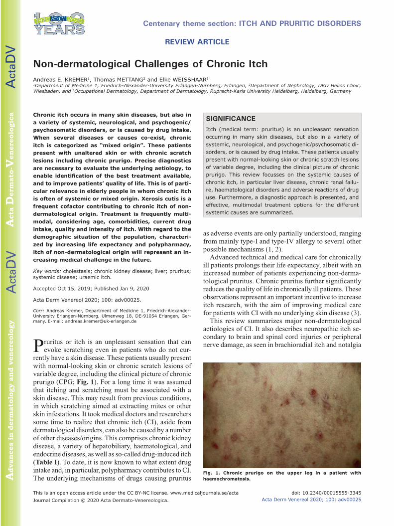
Act
aDV
Act
aDV
Advan
ces
in d
erm
ato
logy a
nd v
en
ere
olo
gy
Acta
Derm
ato
-Ven
ere
olo
gic
a
Acta Derm Venereol 2020; 100: adv00025This is an open access article under the CC BY-NC license. www.medicaljournals.se/actaJournal Compilation © 2020 Acta Dermato-Venereologica.
doi: 10.2340/00015555-3345
REVIEW ARTICLE
Centenary theme section: ITCH AND PRURITIC DISORDERS
SIGNIFICANCEItch (medical term: pruritus) is an unpleasant sensation occurring in many skin diseases, but also in a variety of systemic, neurological, and psychogenic/psychosomatic di-sorders, or is caused by drug intake. These patients usually present with normal-looking skin or chronic scratch lesions of variable degree, including the clinical picture of chronic prurigo. This review focusses on the systemic causes of chronic itch, in particular liver disease, chronic renal failu-re, haematological disorders and adverse reactions of drug use. Furthermore, a diagnostic approach is presented, and effective, multimodal treatment options for the different systemic causes are summarized.
Chronic itch occurs in many skin diseases, but also in a variety of systemic, neurological, and psychogenic/psychosomatic disorders, or is caused by drug intake. When several diseases or causes co-exist, chronic itch is categorized as “mixed origin”. These patients present with unaltered skin or with chronic scratch lesions including chronic prurigo. Precise diagnostics are necessary to evaluate the underlying aetiology, to enable identification of the best treatment available, and to improve patients’ quality of life. This is of parti-cular relevance in elderly people in whom chronic itch is often of systemic or mixed origin. Xerosis cutis is a frequent cofactor contributing to chronic itch of non-dermatological origin. Treatment is frequently multi-modal, considering age, comorbidities, current drug intake, quality and intensity of itch. With regard to the demographic situation of the population, characteri-zed by increasing life expectancy and polypharmacy, itch of non-dermatological origin will represent an in-creasing medical challenge in the future.
Key words: cholestasis; chronic kidney disease; liver; pruritus; systemic disease; uraemic itch.
Accepted Oct 15, 2019; Published Jan 9, 2020
Acta Derm Venereol 2020; 100: adv00025.
Corr: Andreas Kremer, Department of Medicine 1, Friedrich-Alexander-University Erlangen-Nürnberg, Ulmenweg 18, DE-91054 Erlangen, Ger-many. E-mail: [email protected]
Pruritus or itch is an unpleasant sensation that can evoke scratching even in patients who do not cur-
rently have a skin disease. These patients usually present with normal-looking skin or chronic scratch lesions of variable degree, including the clinical picture of chronic prurigo (CPG; Fig. 1). For a long time it was assumed that itching and scratching must be associated with a skin disease. This may result from previous conditions, in which scratching aimed at extracting mites or other skin infestations. It took medical doctors and researchers some time to realize that chronic itch (CI), aside from dermatological disorders, can also be caused by a number of other diseases/origins. This comprises chronic kidney disease, a variety of hepatobiliary, haematological, and endocrine diseases, as well as so-called drug-induced itch (Table I). To date, it is now known to what extent drug intake and, in particular, polypharmacy contributes to CI. The underlying mechanisms of drugs causing pruritus
as adverse events are only partially understood, ranging from mainly type-I and type-IV allergy to several other possible mechanisms (1, 2).
Advanced technical and medical care for chronically ill patients prolongs their life expectancy, albeit with an increased number of patients experiencing non-derma-tological pruritus. Chronic pruritus further significantly reduces the quality of life in chronically ill patients. These observations represent an important incentive to increase itch research, with the aim of improving medical care for patients with CI with no underlying skin disease (3).
This review summarizes major non-dermatological aetiologies of CI. It also describes neuropathic itch se-condary to brain and spinal cord injuries or peripheral nerve damage, as seen in brachioradial itch and notalgia
Non-dermatological Challenges of Chronic ItchAndreas E. KREMER1, Thomas METTANG2 and Elke WEISSHAAR3
1Department of Medicine 1, Friedrich-Alexander-University Erlangen-Nürnberg, Erlangen, 2Department of Nephrology, DKD Helios Clinic, Wiesbaden, and 3Occupational Dermatology, Department of Dermatology, Ruprecht-Karls University Heidelberg, Heidelberg, Germany
Fig. 1. Chronic prurigo on the upper leg in a patient with haemochromatosis.
Act
aDV
Act
aDV
Advan
ces
in d
erm
ato
logy a
nd v
en
ere
olo
gy
Acta
Derm
ato
-Ven
ere
olo
gic
a
A. E. Kremer et al.22
Theme issue: Itch and pruritic disorders
paraesthetica. Finally, this review emphasizes that diag-nosis and treatment is challenged in daily clinical routine by the fact that several origins frequently contribute to the occurrence of CI of non-dermatological origin.
DIAGNOSING NON-DERMATOLOGICAL ITCH
Identifying the underlying disease or itch-causing drug often represents a clinical challenge for the treating physician, but is a major need for the affected patient. A recent cohort analysis of more than 3,000 patients with CI illustrated that it occurs mainly in older patients and in those with many comorbidities (4). In contrast to previous doctrines, there is no correlation between generalized pruritus and a systemic disease as underlying cause. Patients with generalized pruritus suffer comparably often from systemic as from dermatological disorders (5). Thus, careful and detailed medical history taking, clinical examination and interdisciplinary, laboratory and radiological diagnostics are of significant importance (6). For this purpose a structured pruritus questionnaire has been developed (7).
Pruritus on primarily affected skin hints at a dermato-logical disease. In addition to clinical skin investigations, bacterial, mycological, allergic and autoimmune-serolo-gical analyses should be performed. Skin biopsy might further help to establish a clinical diagnosis (6). Pruritus on primarily unaffected skin is often caused by systemic, neurological or psychiatric/somatoform disorders or drug intake (8, 9). If medical history taking and clinical examination are inconclusive a step-wise approach for diagnosis is recommended, including various laboratory diagnostics (6).
It should be noted that CI may occur prior to ma-nifestation and/or clinical diagnosis of the underlying disease. This so-called premonitory itch may present up to several years prior to diagnosis, as seen in polycy-themia vera (10).
In rare cases malignancy is responsible for CI (Table I). Two large cohort studies of 8,744 and 12,813 patients with CI without primary skin changes, respectively, unravelled solely increased rates for haematological and bile duct malignancies (11, 12). In case of CI of unknown origin (PUO) the diagnostic approach should
focus on these 2 cancer entities. If no underlying diag-nosis could be established an annual repetitive work-up may be performed.
HEPATOBILIARY DISEASES
Chronic pruritus is commonly reported by many patients with hepatobiliary diseases, in particular those with cho-lestatic features. Prevalence of hepatic itch varies consi-derably between the different underlying diseases, with 100% as defining symptom in intrahepatic cholestasis of pregnancy, up to 70% in primary biliary cholangitis, primary and secondary sclerosing cholangitis, 15–45% in benign and malignant biliary disorders, and 5–15% in chronic viral hepatitis C infections (13). Affected patients often report the highest intensity on their limbs, in particular the palms of the hands and the soles of the feet, albeit CI may often be generalized (14). In female patients pruritus typically worsens premenstrually, during hormone replacement, and in the last trimester of pregnancy. Hepatic itch presents independently of the severity of cholestasis or liver function.
The pathogenesis of hepatic itch is not yet fully un-derstood. In the past, bile salts, histamine, progesterone metabolites and endogenous opioids have been discus-sed as culprits, albeit a correlation with itch intensity could not be established (15). Interestingly, the semi-synthetic bile salt obeticholic acid, which has been licensed for the treatment of primary biliary cholangitis (PBC) dose-dependently worsens pruritus. Recent data have suggested that bilirubin and bile salts may mediate cholestatic itch via the mas-related G protein-coupled receptor X4 (MRGX4), albeit human data supporting this observation is lacking (16, 17). Cholestatic patients do not present with histamine-induced skin alternations, such as erythema, urticaria or wheals, and antihistamines are largely ineffective (18). Recently, lysophosphatidic acid (LPA) and its forming enzyme autotaxin (ATX) have been identified as potential mediators of hepatic itch (19). However, drugs inhibiting the ATX-LPA axis have not yet been investigated in hepatic itch to prove this pathophysiological concept.
Treatment should focus primarily on adequate therapy for the underlying hepatobiliary disease, which may
Table I. Systemic diseases associated with chronic pruritus
Frequency Organ system Diseases
Common Renal disorders Chronic kidney failureHepatobiliary disorders Primary biliary cholangitis, primary / secondary sclerosing cholangitis, IgG4-related cholangitis, drug-induced toxic
cholestasis, liver cirrhosis, benign or malignant obstructive cholestasis Haematopoietic diseases Polycythaemia vera, essential thrombocytosis, Hodgkin and non-Hodgkin lymphoma, hypereosinophilic syndrome,
mastocytosisRare Endocrine diseases Hyperthyreoidism, hypothyreoidism, hyperparathyreoidism, carcinoid syndrome, diabetes mellitus
Malassimilation syndromes Lactose intolerance, coeliac disease, anorexia nervosa, iron deficiency, vitamin B/D deficiencyInfectious diseases Chronic HBV-/ HCV-/ HIV-/ HSV-infection, H.p. infection, VZV-reactivation (post-herpetic), parasitosesSolid tumours Carcinoma of the thyroid gland, ears, nose and throat, breast, lungs, stomach, pancreas, colon, prostate, uterus and
sarcoma
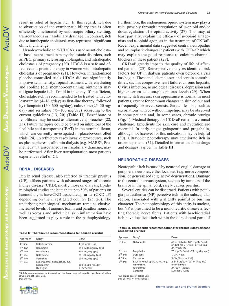
Act
aDV
Act
aDV
Advan
ces
in d
erm
ato
logy a
nd v
en
ere
olo
gy
Acta
Derm
ato
-Ven
ere
olo
gic
a
23Chronic itch in non-dermatological diseases
Theme issue: Itch and pruritic disorders
result in relief of hepatic itch. In this regard, itch due to obstruction of the extrahepatic biliary tree is often efficiently ameliorated by endoscopic biliary stenting, transcutaneous or nasobiliary drainage. In contrast, itch due to intrahepatic cholestasis may represent a significant clinical challenge.
Ursodeoxycholic acid (UDCA) is used as anticholesta-tic baseline treatment in many cholestatic disorders, such as PBC, primary sclerosing cholangitis, and intrahepatic cholestasis of pregnancy (20). UDCA is a safe and ef-fective anti-pruritic therapy in women with intrahepatic cholestasis of pregnancy (21). However, in randomized placebo-controlled trials UDCA did not significantly improve itch intensity. Topical treatment with rehydrating and cooling (e.g. menthol-containing) ointments may mitigate hepatic itch if mild in intensity. If insufficient, cholestatic itch is recommended to be treated with cho-lestyramine (4–16 g/day) as first-line therapy, followed by rifampicin (150–600 mg/day), naltrexone (25–50 mg/day) and sertraline (75–100 mg/day) according to the current guidelines (13, 20) (Table II). Bezafibrate or fenofibrate may be used as alternative approaches (22, 23). Future therapies could be based on inhibitors of the ileal bile acid transporter (IBAT) in the terminal ileum, which are currently investigated in placebo-controlled trials (24). In refractory cases invasive procedures, such as plasmapheresis, albumin dialysis (e.g. MARS®, Pro-metheus®), transcutaneous or nasobiliary drainage, may be performed. After liver transplantation most patients experience relief of CI.
RENAL DISEASES
Itch in renal disease, also referred to uraemic pruritus (UP), affects patients with advanced stages of chronic kidney disease (CKD), mostly those on dialysis. Epide-miological studies indicate that up to 50% of patients on haemodialysis have CKD-associated pruritus (CKD-aP) depending on the investigated country (25, 26). The underlying pathological mechanism remains elusive. Increased levels of uraemic toxins and parathormone, as well as xerosis and subclinical skin inflammation have been suggested to play a role in the pathophysiology.
Furthermore, the endogenous opioid system may play a role, possibly through upregulation of µ-opioid and/or downregulation of κ-opioid activity (27). This may, at least partially, explain the efficacy of μ-opioid antago-nists and κ-opioid agonists in the treatment of CKDaP. Recent experimental data suggested central neuropathic and neuroplastic changes in patients with CKD-aP, which may explain the good response to calcium-channel-blockers in these patients (28).
CKD-aP greatly impacts the quality of life of affec-ted patients (25). Retrospective analyses identified risk factors for UP in dialysis patients even before dialysis has begun. These include male sex and certain comorbi-dities, such as congestive heart failure, chronic hepatitis C virus infection, neurological diseases, depression and higher serum calcium/phosphorus levels (29). When uraemic itch occurs, skin appearance is normal in most patients, except for common changes in skin colour and a frequently observed xerosis. Scratch lesions, such as excoriations with or without impetigo, may be observed in some patients and, in some cases, chronic prurigo (Fig. 1). Medical therapy for CKD-aP remains a clinical challenge. Emollients for skin care and hydration are essential. In early stages gabapentin and pregabalin, although not licensed for this indication, may be helpful (30). Ultraviolet phototherapy may ameliorate itch in uraemic patients (31). Detailed information about drugs and dosages is given in Table III.
NEUROPATHIC DISEASES
Neuropathic itch is caused by neuronal or glial damage to peripheral neurones, either localized (e.g. nerve compres-sion) or generalized (e.g. nerve degeneration). Damage to the central nervous system, such as by tumours of the brain or in the spinal cord, rarely causes pruritus.
Several entities can be discerned. Patients with notal-gia paraesthetica (NP) perceive itch in the subscapular region, associated with a slightly painful or burning character. The pathophysiology of this entity is unclear, but NP is presumed to be a mononeuritic disease affec-ting thoracic nerve fibres. Patients with brachioradial itch have localized itch within the dorsolateral parts of
Table II. Therapeutic recommendations for hepatic pruritus
Approach Druga Dose
1st line Colestyramine 4–16 g/day (po)
2nd line Rifampicin 150–600 mg/day (po)3rd line Bezafibrate 400 mg/day (po)4th line Naltrexone 25–50 mg/day (po)5th line Sertraline 100 mg/day (po)6th line Experimental approaches, e.g.
GabapentinUVB light
300–3,600 mg/day1–2×/week
aSolely colestyramine is licensed for the treatment of hepatic pruritus; all other drugs are off-label use. po: per os.
Table III. Therapeutic recommendations for chronic kidney disease associated pruritus
Approach Druga Dose
1st line Gabapentin After dialysis: 100 mg 3×/week or 300 mg 3×/week or 400 mg 2×/week (po)
2nd line Pregabalin 75 mg 2×/week–75 mg/day (po)3rd line UVB light 1–3×/week4th line Capsaicin 3–5×/day (topical)5th line Experimental approaches, e.g.
Nalfurafine TacrolimusCurcuma
2.5–5 μg/day (po) or 5 μg (iv) after dialysis2×/day (topical)500 mg 3×/day
aAll drugs are off-label-use. po: per os; iv: intravenous.

Act
aDV
Act
aDV
Advan
ces
in d
erm
ato
logy a
nd v
en
ere
olo
gy
Acta
Derm
ato
-Ven
ere
olo
gic
a
A. E. Kremer et al.24
Theme issue: Itch and pruritic disorders
the forearms and, less frequently, around the shoulders. As in NP, patients report mixed itch and pain sensations. Cervical cord compression or radiculopathies have been observed in these patients (32).
The small fibre neuropathy is a systemic neuropathy of peripheral nerves accompanied by severe pruritus in some patients. This sensory disorder leads to a variety of symptoms, including pain, tingling, numbness, deranged thermoregulation and signs of malfunction of the autono-mic nervous system, such as gastrointestinal dysmotility and orthostatic hypotension. The diagnosis is difficult to establish, requiring thermoregulatory sweat testing and skin biopsy with reduced small fibre density (33).
Apart from these entities, neuropathic itch may occur in the course of herpes zoster infections as so-called post-herpetic itch, polyneuropathies and scars induced by trauma or burns.
Treatment of neuropathic itch is difficult. Local treat-ment with capsaicin or systemic treatment with calcium-channel-blockers, such as gabapentin or pregabalin, may alleviate pruritus (3, 34).
ENDOCRINE DISEASES
CI may occur in association with several endocrine di-seases. Whether patients with diabetes mellitus are more frequently afflicted than those without remains a matter of debate. In one study investigating almost 400 patients with diabetes 27.5% reported on generalized itch (35). In another study truncal itch was most prominent in diabetic patients. Of 2,656 patients with diabetes mellitus, 11.3% reported itch located on the trunk, whereas this symptom was present in only 2.9% of 499 age-matched patients without diabetes (36). Further endocrine diseases asso-ciated with the occurrence of CI include Grave’s disease and multiple endocrine neoplasia type II.
HAEMATOLOGICAL DISEASES INCLUDING AQUAGENIC ITCH
Itching is commonly reported in patients with haemato-logical diseases. Aquagenic pruritus is a typical feature in many of these patients, with a pungent itchy charac-ter after contact of the skin with water. Polycythaemia vera is a rare myeloproliferative disease with a clonal dysfunction of pluripotent hematopoietic cells. Affected patients report itch in 30–65% of cases. Aquagenic itch is most commonly observed in patients with a homozygous JAK2 617V mutation (37). Inhibitors of the JAK-STAT pathway, such as ruxolitinib, strongly attenuated pruritus in addition to improving the underlying disease (38, 39). Similarly, patients with essential thrombocytosis and primary myelofibrosis often report aquagenic itch.
Hodgkin’s disease belongs to the class of B-cell lymphoma. CI on primarily unaffected skin is reported by 15–50% of patients (40). Severe itch may precede
the outbreak of the disease by many years. CI worsens at night, often starts at the lower limbs, and may gene-ralize. After successful anti-tumour therapy, CI may also indicate a relapse of Hodgkin’s disease. In patients with non-Hodgkin lymphoma pruritus may affect up to 30% of patients. In leukaemia patients CI is more com-monly observed in lymphatic compared with myelocytic leukaemia and in chronic compared with acute forms. Recommended therapy consists of the calcium channel blockers gabapentin (300–2,400 mg/day) and pregabalin (75–600 mg/day), acetylsalicylic acid (300 mg/day) and mirtazapine (7.5–30 mg/day) (Table IV) (6).
PARANEOPLASTIC DISEASES AND CANCER
In daily clinical practice the term “paraneoplastic itch” (PI) is used to describe itch in patients with cancer. Itch caused by haematological diseases is described above. In general, PI is considered as a rare disorder. It occurs most commonly in lymphoreticular malignancies, while being rarely reported in patients with solid tumours. Its true frequency remains unknown, as epidemiological data is limited. This may mainly be due to other symptoms receiving more attention and being regarded as more important in cancer patients with paraneoplastic diseases. According to the literature and our own clinical studies CI further impairs quality of life in patients with malig-nant diseases. In 2012, an interdisciplinary study interest group (SIG) of physicians and researchers was founded, with the aim of generating a clear definition of PI (40).
Previously, several terms have been used to describe the different types of paraneoplastic itch. The SIG states that the term paraneoplastic itch should be used in case itching occurs as a systemic, but not local, reaction in the presence of a solid tumour or haematological malignancy. This term excludes itch induced either by the local in-vasive growth of cancer cells or by anti-tumour therapy. PI usually disappears with remission of the tumour and may return with its relapse (40).
Diagnosing PI represents a clinical challenge, as it remains difficult to exclude other aetiologies and re-asons for CI, such as paraneoplastic skin diseases, skin or non-dermatological diseases occurring in chronically ill patients, as well as adverse drug reactions. The me-
Table IV. Therapeutic recommendations for pruritus of hematopoietic origin
Approach Druga Dose
1st line Gabapentin 300–2,400 mg/day (po)2nd line Pregabalin 75–600 mg/day (po)3rd line Acetylsalicylic acid 300 mg/day (po)4th line Mirtazapine 7.5–30 mg/day (po)
5th line Experimental approaches, e.g.AprepitantNaltrexoneNalfuraphine
125-80–80 mg/week or 80 mg/day50–150 mg/day2.5–5 μg/day (po)
aAll drugs are off-label use.po: per os.

Act
aDV
Act
aDV
Advan
ces
in d
erm
ato
logy a
nd v
en
ere
olo
gy
Acta
Derm
ato
-Ven
ere
olo
gic
a
25Chronic itch in non-dermatological diseases
Theme issue: Itch and pruritic disorders
chanisms of PI are still not understood. Baseline therapy of PI comprises the treatment of the
underlying malignancy. In many cases, cytoreductive therapies are effective. There are no randomized con-trolled trials (RCTs) for the treatment of PI, which may be explained largely by the rarity and diversity of PI. Topical therapies, including cooling agents, may lead to symptomatic relief. H1-antihistamines are mostly ineffective. PI in lymphoma can improve with oral pred-nisone (see above) acting via different mechanisms, and longer systemic treatment with prednisone may be con-sidered in PI (3). Serotonin reuptake inhibitors (SSRI), such as paroxetine, up to 20 mg daily, calcium alpha (2)-γ-channel blockers, such as gabapentin and prega-balin, can be used for treating PI as well as thalidomide 50–200 mg daily. Opioid receptor antagonist, such as naloxone (0.8–2 mg i.v./day), naltrexone (50–100 mg/day orally) or neurokinin (NK)-1-receptor-antagonists, e.g. aprepitant, have been used for itch, for example in T-cell lymphoma, solid tumours and itch-related biolo-gical cancer treatment (40). It should be considered that patients with cancer frequently receive analgetics that may induce CI as a side-effect.
DRUG-INDUCED ITCH
Itch associated with systemically or locally applied drugs is a common phenomenon (41). Almost every drug may cause localized or generalized itch. Drug-induced itch may emerge as a hypersensitivity reaction towards the drug or, as in many cases, due to another, mostly unknown, pathophysiological process. There is evidence that, with some drugs, the MRGX2-receptor is activated, resulting in a different release of pruritoge-nic compounds than in IgE-mediated hypersensitivity reactions (42). Identifying the responsible compound remains, for many cases, a clinical challenge, as many patients are taking multiple drugs and there might be a considerable delay between the start of drug intake and the development of itch.
Chloroquine, which is mainly used as an antimalarial drug, can induce itch, probably by activating the Mrg-prX1-receptor. Interestingly, chloroquine provokes itch mainly in black Africans, while Caucasians are less com-monly affected potentially by a strong binding capacity to melanin with significantly higher skin concentrations in Africans (43).
Itching provoked by hydroxylethyl starch (HES) infusion is thought to be caused by HES depositions in peripheral nerves (44). Treatment is cumbersome and mostly frustrating (45).
Opioidergic drugs may cause strong itch, especially when applied epidurally or intrathecally (46) which may be mediated by activation of the isoform D of the μ-opioid receptor (47). It can be treated effectively by µ-receptor-antagonists, such as naloxone.
Along with the expansive development and use of targeted tumour therapies, numerous reports have emer-ged about therapy-associated chronic pruritus (48–52). The neurokinin-1-antagonist aprepitant seems to have antipruritic potential in such cases (53).The authors have no conflicts of interest to declare.
REFERENCES1. McNeil BD, Pundir P, Meeker S, Han L, Undem BJ, Kulka M,
et al. Identification of a mast-cell-specific receptor crucial for pseudo-allergic drug reactions. Nature 2015; 519: 237–241.
2. Ebata T. Drug-induced itch management. Curr Probl Dermatol 2016; 50: 155–163.
3. Weisshaar E, Szepietowski JC, Dalgard FJ, Garcovich S, Gie-ler U, Gimenez-Arnau AM, et al. European S2k guideline on chronic pruritus. Acta Derm Venereol 2019; 99: 469–506.
4. Ständer S, Pogatzki-Zahn E, Stumpf A, Fritz F, Pfleiderer B, Ritzkat A, et al. Facing the challenges of chronic pruritus: a report from a multi-disciplinary medical itch centre in Ger-many. Acta Derm Venereol 2015; 95: 266–271.
5. Weisshaar E, Apfelbacher C, Jager G, Zimmermann E, Bruck-ner T, Diepgen TL, et al. Pruritus as a leading symptom: clini-cal characteristics and quality of life in German and Ugandan patients. Br J Dermatol 2006; 155: 957–964.
6. Ständer S, Zeidler C, Augustin M, Bayer G, Kremer AE, Legat FJ, et al. S2k guidelines for the diagnosis and treatment of chronic pruritus – update – short version. J Dtsch Dermatol Ges 2017; 15: 860–872.
7. Weisshaar E, Ständer S, Gieler U, Matterne U, Darsow U. Ent-wicklung eines deutschsprachigen Fragebogens zur Erfassung von chronischem Pruritus (AGP-Fragebogen). Hintergrund und erste Ergebnisse. Hautarzt 2011; 62: 914–927.
8. Pereira MP, Agelopoulos K, Kremer AE, Schmelz M. Aktuelles zur Neurobiologie von Pruritus. Hautarzt 2018; 69: 620–625.
9. Weisshaar E, Szepietowski JC, Darsow U, Misery L, Wal-lengren J, Mettang T, et al. European guideline on chronic pruritus. Acta Derm Venereol 2012; 92: 563–581.
10. Siegel FP, Tauscher J, Petrides PE. Aquagenic pruritus in polycythemia vera: characteristics and influence on quality of life in 441 patients. Am J Hematol 2013; 88: 665–669.
11. Fett N, Haynes K, Propert KJ, Margolis DJ. Five-year ma-lignancy incidence in patients with chronic pruritus: a population-based cohort study aimed at limiting unnecessary screening practices. J Am Acad Dermatol 2014; 70: 651–658.
12. Johannesdottir SA, Farkas DK, Vinding GR, Pedersen L, Lam-berg A, Sorensen HT, et al. Cancer incidence among patients with a hospital diagnosis of pruritus: a nationwide Danish cohort study. Br J Dermatol 2014; 171: 839–846.
13. Dull MM, Kremer AE. Management of chronic hepatic itch. Dermatol Clin 2018; 36: 293–300.
14. Bergasa NV, Mehlman JK, Jones EA. Pruritus and fatigue in primary biliary cirrhosis. Baillieres Best Pract Res Clin Gast-roenterol 2000; 14: 643–655.
15. Beuers U, Kremer AE, Bolier R, Elferink RP. Pruritus in cho-lestasis: facts and fiction. Hepatology 2014; 60: 399–407.
16. Meixiong J, Vasavda C, Green D, Zheng Q, Qi L, Kwatra SG, et al. Identification of a bilirubin receptor that may mediate a component of cholestatic itch. Elife 2019; 8 pii: e44116.
17. Meixiong J, Vasavda C, Snyder SH, Dong X. MRGPRX4 is a G protein-coupled receptor activated by bile acids that may contribute to cholestatic pruritus. Proc Natl Acad Sci U S A 2019. 116: 10525–10530.
18. Jones EA, Bergasa NV. Evolving concepts of the pathogenesis and treatment of the pruritus of cholestasis. Can J Gastro-enterol 2000; 14: 33–40.
19. Kremer AE, Martens JJ, Kulik W, Rueff F, Kuiper EM, van Buuren HR, et al. Lysophosphatidic acid is a potential medi-ator of cholestatic pruritus. Gastroenterology 2010; 139: 1008–1018.
20. EASL Clinical practice guidelines: the diagnosis and manage-

Act
aDV
Act
aDV
Advan
ces
in d
erm
ato
logy a
nd v
en
ere
olo
gy
Acta
Derm
ato
-Ven
ere
olo
gic
a
A. E. Kremer et al.26
Theme issue: Itch and pruritic disorders
ment of patients with primary biliary cholangitis. J Hepatol 2017. 67: 145–172.
21. Williamson C, Geenes V. Intrahepatic cholestasis of preg-nancy. Obstet Gynecol 2014; 124: 120–133.
22. Reig A, Sese P, Pares A. Effects of bezafibrate on outcome and pruritus in primary biliary cholangitis with suboptimal ursodeoxycholic acid response. Am J Gastroenterol 2018; 113: 49–55.
23. Corpechot C, Chazouilleres O, Rousseau A, Le Gruyer A, Habersetzer F, Mathurin P, et al. A placebo-controlled trial of bezafibrate in primary biliary cholangitis. N Engl J Med 2018; 378: 2171–2181.
24. Hegade VS, Kendrick SF, Dobbins RL, Miller SR, Thompson D, Richards D, et al. Effect of ileal bile acid transporter inhi-bitor GSK2330672 on pruritus in primary biliary cholangitis: a double-blind, randomised, placebo-controlled, crossover, phase 2a study. Lancet 2017; 389: 1114–1123.
25. Weiss M, Mettang T, Tschulena U, Passlick-Deetjen J, Weiss-haar E. Prevalence of chronic itch and associated factors in haemodialysis patients: a representative cross-sectional study. Acta Derm Venereol 2015; 95: 816–821.
26. Rayner HC, Larkina M, Wang M, Graham-Brown M, van der Veer SN, Ecder T, et al. International comparisons of pre-valence, awareness, and treatment of pruritus in people on hemodialysis. Clin J Am Soc Nephrol 2017; 12: 2000–2007.
27. Mettang T, Kremer AE. Uremic pruritus. Kidney Int 2015; 87: 685–691.
28. Papoiu AD, Emerson NM, Patel TS, Kraft RA, Valdes-Rodriguez R, Nattkemper LA, et al. Voxel-based morphometry and arterial spin labeling fMRI reveal neuropathic and neuro-plastic features of brain processing of itch in end-stage renal disease. J Neurophysiol 2014; 112: 1729–1738.
29. Khanna D, Singal A, Kalra OP. Comparison of cutaneous manifestations in chronic kidney disease with or without dialysis. Postgrad Med J 2010; 86: 641–647.
30. Nofal E, Farag F, Nofal A, Eldesouky F, Alkot R, Abdelkhalik Z. Gabapentin: a promising therapy for uremic pruritus in hemodialysis patients: a randomized-controlled trial and review of literature. J Dermatolog Treat 2016; 27: 515–519.
31. Tan JK, Haberman HF, Coldman AJ. Identifying effective treatments for uremic pruritus. J Am Acad Dermatol 1991; 25: 811–818.
32. Marziniak M, Phan NQ, Raap U, Siepmann D, Schurmeyer-Horst F, Pogatzki-Zahn E, et al. Brachioradial pruritus as a result of cervical spine pathology: the results of a magnetic resonance tomography study. J Am Acad Dermatol 2011; 65: 756–762.
33. Lauria G, Bakkers M, Schmitz C, Lombardi R, Penza P, Devigili G, et al. Intraepidermal nerve fiber density at the distal leg: a worldwide normative reference study. J Peripher Nerv Syst 2010; 15: 202–207.
34. Savk E. Neurologic itch management. Curr Probl Dermatol 2016; 50: 116–123.
35. Ko MJ, Chiu HC, Jee SH, Hu FC, Tseng CH. Postprandial blood glucose is associated with generalized pruritus in patients with type 2 diabetes. Eur J Dermatol 2013; 23: 688–693.
36. Yamaoka H, Sasaki H, Yamasaki H, Ogawa K, Ohta T, Fu-ruta H, et al. Truncal pruritus of unknown origin may be a symptom of diabetic polyneuropathy. Diabetes Care 2010; 33: 150–155.
37. Vannucchi AM, Antonioli E, Guglielmelli P, Rambaldi A,
Barosi G, Marchioli R, et al. Clinical profile of homozygous JAK2 617V>F mutation in patients with polycythemia vera or essential thrombocythemia. Blood 2007; 110: 840–846.
38. Kirito K, Suzuki K, Miyamura K, Takeuchi M, Handa H, Oka-moto S, et al. Ruxolitinib is effective and safe in Japanese pa-tients with hydroxyurea-resistant or hydroxyurea-intolerant polycythemia vera with splenomegaly. Int J Hematol 2018; 107: 173–184.
39. Verstovsek S, Passamonti F, Rambaldi A, Barosi G, Rosen PJ, Rumi E, et al. A phase 2 study of ruxolitinib, an oral JAK1 and JAK2 Inhibitor, in patients with advanced polycythemia vera who are refractory or intolerant to hydroxyurea. Cancer 2014; 120: 513–520.
40. Weisshaar E, Weiss M, Mettang T, Yosipovitch G, Zylicz Z. Paraneoplastic itch: an expert position statement from the Special Interest Group (SIG) of the International Forum on the Study of Itch (IFSI). Acta Derm Venereol 2015; 95: 261–265.
41. Maleki K, Weisshaar E. Arzneimittelinduzierter Pruritus. Hautarzt 2014; 65: 436–442.
42. Meixiong J, Anderson M, Limjunyawong N, Sabbagh MF, Hu E, Mack MR, et al. Activation of mast-cell-expressed mas-related g-protein-coupled receptors drives non-histaminergic itch. Immunity 2019; 50: 1163–1171
43. Bussaratid V, Walsh DS, Wilairatana P, Krudsood S, Si-lachamroon U, Looareesuwan S. Frequency of pruritus in Plasmodium vivax malaria patients treated with chloroquine in Thailand. Trop Doct 2000; 30: 211–214.
44. Metze D, Reimann S, Szepfalusi Z, Bohle B, Kraft D, Luger TA. Persistent pruritus after hydroxyethyl starch infusion therapy: a result of long-term storage in cutaneous nerves. Br J Dermatol 1997; 136: 553–559.
45. Bork K. Pruritus precipitated by hydroxyethyl starch: a re-view. Br J Dermatol 2005; 152: 3–12.
46. Jacobson L, Chabal C, Brody MC, Ward RJ, Wasse L. Intrat-hecal methadone: a dose-response study and comparison with intrathecal morphine 0.5 mg. Pain 1990; 43: 141–148.
47. Liu XY, Liu ZC, Sun YG, Ross M, Kim S, Tsai FF, et al. Unidi-rectional cross-activation of GRPR by MOR1D uncouples itch and analgesia induced by opioids. Cell 2011; 147: 447–458.
48. Ensslin CJ, Rosen AC, Wu S, Lacouture ME. Pruritus in patients treated with targeted cancer therapies: systematic review and meta-analysis. J Am Acad Dermatol 2013; 69: 708–720.
49. Clabbers JMK, Boers-Doets CB, Gelderblom H, Stijnen T, La-couture ME, van der Hoeven KJM, et al. Xerosis and pruritus as major EGFRI-associated adverse events. Support Care Cancer 2016; 24: 513–521.
50. Wu J, Lacouture ME. Pruritus associated with targeted an-ticancer therapies and their management. Dermatol Clin 2018; 36: 315–324.
51. Phillips GS, Wu J, Hellmann MD, Postow MA, Rizvi NA, Freites-Martinez A, et al. Treatment outcomes of immune-related cutaneous adverse events. J Clin Oncol 2019: JCO1802141.
52. Phillips GS, Freites-Martinez A, Wu J, Chan D, Fabbrocini G, Hellmann MD, et al. Clinical characterization of immunothe-rapy-related pruritus among patients seen in 2 oncoderma-tology clinics. JAMA Dermatol 2019; 155: 249–251.
53. Santini D, Vincenzi B, Guida FM, Imperatori M, Schiavon G, Venditti O, et al. Aprepitant for management of severe pru-ritus related to biological cancer treatments: a pilot study. Lancet Oncol 2012; 13: 1020–1024.

Act
aDV
Act
aDV
Advan
ces
in d
erm
ato
logy a
nd v
en
ere
olo
gy
Acta
Derm
ato
-Ven
ere
olo
gic
a
Acta Derm Venereol 2020; 100: adv00026This is an open access article under the CC BY-NC license. www.medicaljournals.se/actaJournal Compilation © 2020 Acta Dermato-Venereologica.
doi: 10.2340/00015555-3346
REVIEW ARTICLE
Centenary theme section: ITCH AND PRURITIC DISORDERS
SIGNIFICANCEThe relationship between itch and psyche is complex and bilateral. Increasing interest in itch and its associations with psyche is indicated by the abundance of experimen-tal and clinical articles published in this field. This review covers the pathogenesis of itch, associations with stress, the contagiousness of itch, psychological and psychiatric aspects related to itch, and the burden of itch with respect to impairment of health-related quality of life and stigma-tization.
Beginning from embryological development, skin and psyche are closely related to physiological state re-gardless of age. Altering the homeostasis of one of these components impacts on the other, thereby sub-stantiating that the relationship between itch and psyche is bilateral. Itch has a complex pathogenesis, which involves the peripheral and central nervous systems, as well as various inflammatory mediators. This paper reviews key aspects of itch pathogenesis, relevant associations with stress, the contagiousness of itch, psychological and psychiatric considerations related to itch, and the burden of itch with respect to impairment of health-related quality of life (HRQoL) and stigmatization. Despite the fact that itch–psyche associations still pose many questions, current know-ledge supports the role of a holistic, interdisciplinary approach to these patients in order to improve their well-being.
Key words: itch, psyche, pathogenesis, stress, psychiatry, bur-den of disease.
Accepted Oct 15, 2019; Published Jan 9, 2020
Acta Derm Venereol 2020; 100: adv00026.
Corr: Jacek C. Szepietowski, Department of Dermatology, Venereology and Allergology, Wrocław Medical University, 1 Chałubińskiego Street, PL-50-368 Wrocław, Poland. E-mail: [email protected]
Beginning with human embryogenesis, skin and brain are organs that are closely connected due to their
ectodermal origins (1). These associations continue to unfold after birth, and constitute a foundation for the normal psychosocial development of an individual. According to the moi-peau concept (2) (Fig. 1) (as ex-tensively reviewed by Dutray & Misery (1)), skin pos-sesses a wide psychological meaning, reflecting various needs of the psyche. Firstly, skin has the function of a “bag” or “container”, as it embraces the positive stimuli experienced by a baby during nursing and being cared for. Secondly, skin is an “interface” with the outside world, which serves as a protective barrier against ex-ternal aggression. Lastly, skin may be considered as a specific “place” or “means of communication” in order to establish relationships, and as a “surface” on which others may leave their trace. It seems substantiated that chronic skin diseases with their rich symptomatology, especially acquired during early childhood, ensue in altering both physical and psychological homeostasis of an affected individual. The impact is always reciprocal:
the psyche may predispose to cutaneous complaints (e.g. itch, chronic scratch lesions), whereas dermatological signs and symptoms “scar” the psyche.
Itch is defined as an unpleasant sensation leading to scratching, further classified as acute or chronic (lasting less or more than 6 weeks, respectively) (3). Chronic itch (CI) is a feature of various dermatoses, although it may also stem from systemic, neurological, or psychia-tric disorders. Occasionally, the diagnosis of pruritus of unknown origin (PUO) is established. The presence of CI is associated with significant morbidity, mortality, reduction in quality of life (QoL), feelings of stigmatiza-tion, stress, impairment of mood, lack of concentration,
Itch and Psyche: Bilateral AssociationsRadomir RESZKE and Jacek C. SZEPIETOWSKIDepartment of Dermatology, Venereology and Allergology, Wrocław Medical University, Wrocław, Poland
Fig. 1. The moi-peau concept (The Ego-Skin concept) (according to (1)).

Act
aDV
Act
aDV
Advan
ces
in d
erm
ato
logy a
nd v
en
ere
olo
gy
Acta
Derm
ato
-Ven
ere
olo
gic
a
R. Reszke and J. C. Szepietowski28
Theme issue: Itch and pruritic disorders
reduced sexual desire and appetite, as well as inability to express emotions (alexithymia) (4–9). On the other hand, clinicians are aware that various psychological and psychiatric alterations may contribute to the occurrence or exacerbation of itch. One of the models that aims to explain CI mechanisms in the course of cutaneous disorders is the Biopsychosocial Model, proposed by Verhoeven et al. (10). The occurrence of itch is a result of various factors: internal (associated with personality), external (e.g. stressful environmental stimuli), medi-ating (e.g. cognitive, behavioural and social) as well as physiological. Tey et al. (11) have clinically classified the itch–psyche interactions as pruritic disorders with psychiatric sequelae (I); pruritic disorders aggravated by psychosocial factors (II); and psychogenic disorders causing itch (III). These complex bilateral associations between itch and psyche support the need for multidis-ciplinary approach to itch and the associated conditions in the clinical setting (12–14).
This review covers the pathogenesis of itch, cerebral regions associated with pathogenesis of itch, the inter-play between itch and stress, the contagiousness of itch, psychological and psychiatric aspects associated itch, and the burden of itch with respect to impairment of QoL and stigmatization.
ITCH PATHOGENESIS
There is growing data concerning the pathogenesis of itch, as this topic is gaining increased attention in the medical literature. From the historical point of view, itch has been perceived as a subtype of pain (15), and it is thought that the symptom might have developed as an evolutionary defence mechanism against various potentially dangerous stimuli, such as parasites, insects, sharp objects, irritants, and allergens (16). However, itch is currently deemed a separate entity from pain. The role of peripheral (PNS) and central nervous system (CNS) is crucial in eliciting CP, starting from free nerve endings in the epidermis. The statement “it is the brain that itches, not the skin” (16) remains valid, as different cortical areas are included in processing itch intensity, location, associated unpleasant feelings, generating the need to scratch, and preparing and executing scratch behaviour (17–20). In a positron emission tomography (PET) study among human participants histamine in-jection resulted in major activation in the left primary sensory cortex, as well as the primary motor cortex, supplementary motor area and premotor cortex (17). Another study utilizing PET and regional cerebral blood flow (rCBF) revealed that a skin prick test with histamine resulted in activation of the contralateral somatosensory cortex; in addition, both ipsilateral and contralateral motor areas were involved. Itch unpleasantness was associated with the activation of contralateral senso-rimotor cortex, prefrontal cortex, posterior insula and
ipsilateral supplementary motor area (18). A functional magnetic resonance imaging (fMRI) study demonstrated itching and pain both activated the anterior cingulate cortex, the anterior insula, the basal ganglia and pre-supplementary motor area (20). How ever, the activity in posterior cingulate cortex and the posterior insula was more prominent in itching than in pain. Moreover, it was proportional to itching sensation. In an experiment with cowhage-induced itch, as demonstrated by fMRI, itching was associated with increased activity in supp-lementary motor area, premotor cortex, primary motor cortex, and midcingulate cortex (21). The role of the caudate nucleus was also substantiated, as this region is involved in the reward system. Papoiu et al. (22) de-monstrated that active scratching was accompanied by higher pleasure and deactivation of anterior cingulate cortex and insula compared with passive scratching. In addition, the activation of ventral tegmentum area (VTA) of the midbrain, as well as deactivation of periaqueductal grey matter (PAG), are associated with the itch-scratch reward system.
ITCH AND STRESS
Stress is a concept frequently mentioned nowadays, yet despite its widespread appearance in various situations it may be described in different ways, e.g. as “the non-specific response of the body to any demand made upon it” (23) or “a relationship with the environment that the person appraises as significant for his or her well-being and in which the demands tax or exceed available coping resources” (24). The bilateral associations of itch and psyche are perfectly reflected by the interplay of itch and stress. Similarly to the vicious itch-scratch cycle, chronic pruritic conditions (e.g. psoriasis, urticaria, atopic dermatitis) generate huge stress levels, which may subsequently perpetuate exacerbation of the di-sease. Aberrant parasympathetic response may possibly link chronic stress and itch (25), while stress-induced itch is associated with activation of the hippocampus and subcortical regions (corpus callosum and putamen) (26). The itch-stress association does, in fact, pose a certain therapeutic implication towards first-generation H1-antihistamines and GABA-ergics (gabapentin and pregabalin) (11, 26).
In a large population-based study among Norwegian adolescents mental distress (assessed by the Hopkins Symptom Checklist-10) was correlated with the presence of itch (27). Moreover, the severity of itch correlated with the level of mental distress, regardless of sex. Subsequently, another report linked the presence of itch with low self-efficacy in individuals under higher stress (28). The perceived self-efficacy is a concept of people’s beliefs that they can exert control over their motivation, behaviour and social environment (29). Individuals lacking the sense of self-efficacy are not able to manage
Act
aDV
Act
aDV
Advan
ces
in d
erm
ato
logy a
nd v
en
ere
olo
gy
Acta
Derm
ato
-Ven
ere
olo
gic
a
29Itch and psyche: bilateral associations
Theme issue: Itch and pruritic disorders
demanding situations effectively, despite knowing what to do and possessing the necessary skills. The increase of self-efficacy in psychological interventions may exer-cise control over stressors due to its immunomodulating properties (30). Recently, Schut et al. (31) employed cognitive behavioural stress management programmes in patients with atopic dermatitis (AD). The study revealed that individuals subjected to these interventions demon-strated diminished cortisol awakening response, while maintaining calm and presenting lower salivary cortisol levels under acute stress. Thereby, various psychological interventions (reviewed in detail by Schut et al. (32)) should be considered as an adjunctive therapy in patients with itch of various origins. The associations between stress and itch in patients with AD constitute a problem of particular complexity and importance and involve the components of the so-called neuro-endocrino-immuno-cutaneous system (NEICS) and the hypothalamo-pitui-tary-adrenal (HPA axis). The relevant aspects of this issue are covered in detail elsewhere (33–38), although there is evidence that acute or chronic stress have different impacts on the HPA axis (39).
Regarding patients with psoriasis, our group has demonstrated that patients under heavy or extremely heavy stress more commonly suffer from itch, with the severity of stress and intensity of itch being positively correlated (40). Similar results were observed by Amatya et al. (7). Another study revealed that the self-reported stress reactivity was moderately correlated with the de-gree of itching (41). Stress-related exacerbation of itch in psoriatic subjects may also be associated with higher expression of substance P receptor, tropomyosin receptor kinase A and calcitonin gene-related peptide receptor in keratinocytes of psoriatic plaques (42). Other reports mentioned the role of stress in pruritic disorders, such as chronic urticaria (43, 44), acne vulgaris (45, 46), hand dermatoses (47) and post-burn itch (48). Recent research reported that patients with generalized CI reported more tension and subjective stress than healthy controls, with the expectation of the acute stress test (49). Notably, “va-riations of intensity associated with stress” (50) constitute an optional criterion for the diagnosis of functional itch disorder (FID; psychogenic itch).
Itch-stress associations are complex, yet they serve as the foundation of psychological interventions aimed at enhancing coping abilities of the affected individual, e.g. cognitive restructuring (32). A Dutch study reported that a nursing programme “Coping with itch” successfully targeted catastrophizing and helpless itch-related coping (51). Subsequently, it was proven by Evers et al. (52) that patients with AD may benefit from multidisciplinary itch-coping group training. The programme focused on skin care, itch-triggering factors, stress management, long-term goals, relapse prevention, habit reversal and scratch-triggering factors. The training ensued in reducing itch-scratch behaviour, improving skin status,
decreasing the need for dermatological visits and treat-ment. Coping with itch was improved, as the itch-related self-efficacy increased, with decline in catastrophizing. These benefits were regarded both as short-term and long-term.
NOCEBO EFFECT: IS ITCH A CONTAGIOUS PHENOMENON?
The nocebo effect is the negative counterpart of the placebo effect (53). In essence, an individual receives an inert substance or undergoes a neutral procedure, which is intended to induce negative expectations. Interestingly, outbreaks of itch among schoolchildren attributed to epidemic hysteria have been described in the literature (54, 55). Acknowledging the relevant role of psyche in eliciting itch, the nocebo-related concept of “itch contagion” or “contagious itch” was conceived and investigated in different studies. Based on the experience that lectures about pruritic dermatoses induce itching in the listeners, Niemeier et al. (56) proved that a public lecture entitled “Itching – what’s behind it?” caused the participants to feel itch and exert scratching behaviour. Subsequent research revealed that patients with AD, when compared with healthy controls, were more prone to scratch themselves after being subjected to histamine or saline injection, followed by watching a short video of people scratching (57). Holle et al. (58) reported that itch contagion is a normative response experienced by most people and its degree may be associated with neu-roticism as a personality trait (the impact of personality traits on itch is reviewed below). No association between itch contagion and sex or empathy was established in this study. The fMRI examination revealed that itch intensity correlated with the activation of the left Brodmann area (BA) 44, primary somatosensory cortex and BA6. Lloyd et al. (59) revealed that solitary visual stimulants provoke itch in healthy individuals. Another study demonstrated that the combination of conditioning and verbal sugges-tion may result in relevant nocebo and placebo effects on itch in healthy individuals (60). It was subsequently proved that nocebo effects regarding the itch sensation may be minimized or reversed via conditioning with ver-bal suggestion (61). Recently, increased contagiousness of itch in children with autism spectrum disorder was demonstrated (62).
PSYCHOLOGY OF ITCH
The personality of an individual may be defined as a cha-racteristic pattern of behaviours considered in the broad sense, also including thoughts, feelings and motivation (63). One of the most popular models used for the des-cription of personality structure is The Big Five model, which encompasses 5 bipolar dimensions (extraversion, agreeableness, conscientiousness, neuroticism and open-
Act
aDV
Act
aDV
Advan
ces
in d
erm
ato
logy a
nd v
en
ere
olo
gy
Acta
Derm
ato
-Ven
ere
olo
gic
a
R. Reszke and J. C. Szepietowski30
Theme issue: Itch and pruritic disorders
ness to experience) (64). The impact of personality traits on itch sensation was explored in several reports and supports the role of psychological interventions in aiding affected individuals. In a Swedish study (65) the persistence of post-burn itch was associated with lack of assertiveness, as assessed by the Swedish Universities Scales of Personality (SSP). Moreover, the Coping with Burns Questionnaire (CBQ) scores revealed that itch was more persistent among individuals who sought more instrumental and less emotional support. Patients with prurigo nodularis (PN) exhibited higher neuroticism and lower extraversion traits than controls when exami-ned via the revised Eysenck Personality Questionnaire (EPQ-R) (66). Notably, among subjects with psoriasis, severe itch was significantly associated with somatic trait anxiety, embitterment, mistrust, and physical trait aggression (assessed via SSP) (67). Conversely, Janowski et al. (68) found no differences in basic personality traits regarding psoriatic patients with various frequency of itch (assessed via NEO-Five Factor Inventory; NEO-FFI). However, resignation and self-blame were more common coping strategies among patients experiencing itch more frequently (assessed via the Ways of Coping Questionnaire; WCQ). In a German study, individuals with AD and healthy controls were exposed to videos fea-turing crawling insects and skin disorders (69). Compa-red with healthy controls, agreeableness (NEO-FFI) and public self-consciousness (the Self-Consciousness Scale; SCS) were significant predictors of scratching behaviour in subjects with AD. These findings were subsequently replicated (70). Kini et al. (71) investigated patients with CI (recruited from the National Eczema Associa-tion and US Veterans Health Administration National Patient Care Database). The authors observed that the lethargic personality style (defined as low extraversion and conscientiousness) (NEO-FFI) was associated with greater mean total ItchyQoL score. On the other hand, higher ItchyQol symptom score was observed both in overcontrolled (high neuroticism and conscientiousness) and undercontrolled (high neuroticism and low consci-entiousness) patients.
Another concept that has evolved as a potential para-digm for understanding the influence of emotions and personality on physical illness and health is alexithymia (72). In general, this personality construct defines the inability to identify and verbalize emotions. Our group has investigated alexithymia using the Bermond-Vorst Alexithymia Questionnaire (BVALQ-40) among patients with end-stage renal disease on maintenance haemo-dialysis (9). It was observed that patients with uraemic itch exhibited lower scores on the fantasizing subscale score. Another group assessed alexithymia via the To-ronto Alexithymia Scale (TAS) among individuals with chronic urticaria (73).
PSYCHIATRIC PERSPECTIVE ON ITCH
Taking into account the widespread relationship between itch and psyche, one cannot omit the obvious psychiatric background of itch in certain cases, whereas the presence of itch may frequently ensue in a wide spectrum of psy-chiatric comorbidities. In a study by Mazeh et al. (74) among a cohort of patients (n = 111) hospitalized in the psychiatric ward, CI affected 32%. Of those, 45% stated that stress was one of the major aggravating factors of itch. Similarly, our group enrolled inpatients (n = 40) who were hospitalized with depression (75). Itching was experienced by 17.5% of patients during the depressive episode. Notably, itching disappeared in all affected individuals after the depressive symptoms markedly decreased, whereas recurrent itching was associated with recurrent depressive episodes.
A different approach was presented by Schneider et al. (76), who examined 109 dermatology inpatients with itch and observed that in over 70% of them 1–6 psychiatric diagnoses could be established. In over 60% of patients psychotherapeutic or psychiatric treatment was advised. Ferm et al. (77) evaluated the medical records of 139 patients with CI, among whom 31 (22.3%) had an under-lying psychiatric disorder. A recent study among 560 pa-tients with CI who were referred by the dermatologist for a psychosomatic consultation demonstrated that 77.1% had at least one psychosomatic/psychiatric comorbidity (78). The most common comorbidities encompassed psychological/psychosomatic cofactors in itch (F54 according to ICD-10) (74.5%), depression (F32–F34) (30.7%), adjustment disorder (F43.2) (17.8%), disso-ciative/somatoform disorder/hypochondria (F44–F45) (11.2%), anxiety/compulsive disorder (F40–F42) (6.6%) and others (17%). Notably, patients with the psychiatric/psychosomatic comorbidities presented higher intensity of itch, longer duration and coexistence of chronic scratch lesions. Dermatologists and psychiatrists often utilize psychoactive drugs in order to alleviate itch of different origins; however, itch may also be induced by the use of selective serotonin reuptake inhibitors or neuroleptics (79, 80).
There are also reports in the literature concerning “classic” pruritic disorders, which were also evaluated with regard to psychiatric comorbidities. Gupta et al. (81) linked alleviation of itch in psoriasis with changes in depression scores. Subsequently, Conrad et al. (43) executed a complex study of 41 patients with chronic idiopathic urticaria (CIU) and 44 patients with psoria-sis. The investigators assessed the relationship between itch and several domains, including emotional distress and anger (assessed via the Symptom Checklist 90-R (SCL-90-R) and State Trait Anger eXpression Inventory (STAXI) tools, respectively). In patients with chronic

Act
aDV
Act
aDV
Advan
ces
in d
erm
ato
logy a
nd v
en
ere
olo
gy
Acta
Derm
ato
-Ven
ere
olo
gic
a
31Itch and psyche: bilateral associations
Theme issue: Itch and pruritic disorders
idiopathic urticaria (CIU) anger was a predictor of itch severity, whereas depression seemed to influence itch severity in patients with psoriasis. Regarding patients with CIU, the authors discussed possible pathway involving anger and stress, which stimulate corticotro-phin-releasing hormone, subsequently leading to mast cell activation and degranulation of mediators, such as histamine. These aspects might, at least, partially account for the presence of itch accompanying urticarial wheals. In a study by Dazzi et al. (66) 20 subjects with PM were compared with healthy controls with regards to scores in EPQ-R, the Beck Depression Inventory second edition (BDI-II) and the State Trait Anxiety Inventory – form Y (STAI). It was observed that patients with PN exhibited higher scores for the T-anxiety scale (STAI – form y-2; describing how subject feel in general), depression and neuroticism, while lower than the controls concerning extraversion. Subsequently, PN was linked to depres-sion (adjusted odds ratio (OR) 2.82; p < 0.001), the use of antidepressants (adjusted OR 2,6; p < 0.001), anxiety (adjusted OR 2.06; p < 0.05) and the use of anxiolytics (adjusted OR 4.64; p < 0.001) compared with healthy controls (82). Similar relations were reported in a recent study concerning PN burden with respect to depression and anxiety. In addition, patients with PN more often had suicidal ideation (83). In a previously mentioned study by Remröd et al. (67) (n = 101) subjects with plaque psoriasis with severe itch presented higher scores for de-pression and anxiety (as assessed via STAI and BDI-II). A study among 27 patients with AD reported that there is a connection between high scores on the depression scale (Hospital Anxiety and Depression Scale; HADS-D) and higher increase in itch intensity compared with controls (69).
Interestingly, a study by Weisshaar et al. (84) recounted that affective reactions, such as depression and aggres-sion, were more common in German individuals with CI due to dermatological diseases than those with CI associated with underlying systemic disorders (p = 0.04 and p = 0.03, respectively). A comparison between German and Ugandan patients with CI was also per-formed in terms of emotional reactions, revealing that German patients tend to be significantly more aggres-sive (p < 0.0001) and more often do not have any drive (p < 0.0001). In a cohort of patients with end-stage renal disease, the severity of CI (4IIQ) was correlated with depressive symptoms (assessed by BDI) (85). The com-plicated itch and psyche interplay is elegantly embraced in functional itch disorder (FID; also termed psychoge-nic itch). This entity was defined as “an itch disorder, where itch is at the centre of the symptomatology, and where psychological factors play an evident role in the triggering intensity, aggravation or persistence of the pruritus” (50, 86) and can be diagnosed according to several criteria. The 3 compulsory criteria encompass: (i) localized or generalized itch without primary skin
lesions, (ii) chronic pruritus of at least 6 weeks’ duration, and (iii) no somatic cause. In addition, at least 3 out of the following 7 additional criteria have to be found: (i) a chronological relationship of pruritus with 1 or several life events that could have psychological repercussions, (ii) variations in intensity associated with stress, (iii) nocturnal variations, (iv) predominance during rest or inaction, (v) associated psychological disorder, (vi) pru-ritus that could be improved by psychotropic drugs, and (vii) pruritus that could be improved by psychotherapies. Regarding the International Forum for the Study of Itch (IFSI) classification according to its aetiology, FID is associated with the 4th category (psychogenic/psycho-somatic origin) (3). Unfortunately, the detailed aspects associated with psyche and well-being of patients with FID in particular have rarely been investigated (87, 88).
Finally, considerations concerning psychiatric as-sociations with itch are nowhere near complete without mentioning the risk of suicide. In a study by Halvorsen et al. (89), 3,682 adolescents responded to a special questionnaire focusing on itch, pain and suicidal idea-tion. Severe itch was strongly associated with suicidal ideation (OR 3.0). Among the individuals reporting itch, suicidal ideation was reported by 21.1%, in contrast to 8.4% among subjects denying itching. In a large meta-analysis, patients with AD (in which itch is generally considered a constant feature) were 44% more likely to have suicidal ideation and 36% more likely to die by suicide that those without the disease (90).
HEALTH-RELATED QUALITY OF LIFE IMPAIRMENT: THE BURDEN OF SCRATCHING
QoL may be defined as a measure of the goodness of several life aspects, e.g. reactions to life occurrences, dis-position, sense of life fulfilment and satisfaction, as well as satisfaction with work and personal relationships (91). Not infrequently, this term is confused with HRQoL, which has multiple definitions. One of the most relevant encompasses “how well a person functions in their life and his or her perceived well-being in physical, mental and social domains of health” (92). The impairment in HRQoL in dermatological patients can be measured with multiple tools, e.g. the Dermatology Life Quality Index (DLQI) (93) or Skindex (94). Impairment in HRQoL stems from various disease-related signs and symptoms, with a special emphasis on CI. Recently, an itch-specific instrument (ItchyQoL) for assessing HRQoL has been validated in several languages (95).
There is recent literature focusing on itch and HRQoL, both in cutaneous and primarily extracutaneous dis-orders. These issues were studied in detail in subjects with psoriasis. According to Yosipovitch et al. (96), itch bothered 84 (84%) patients, among whom 35% became more agitated, 24% became depressed, 30% had trouble concentrating, and 23% changed their eating habits.

Act
aDV
Act
aDV
Advan
ces
in d
erm
ato
logy a
nd v
en
ere
olo
gy
Acta
Derm
ato
-Ven
ere
olo
gic
a
R. Reszke and J. C. Szepietowski32
Theme issue: Itch and pruritic disorders
Remarkably, two-thirds of the patients were bothered by difficulties falling asleep and night awakenings due to itch. Moreover, 40% of pruritic subjects reported decreased or non-existent sexual desire, whereas 35% reported decreased or non-existent sexual functions. Subsequently, our group found that HRQoL, assessed via the DLQI, was significantly decreased in patients with itch (6). In addition, the DLQI score correlated with itch intensity assessed via the 4-Item Itch Questionnaire (4IIQ) and visual analogue scale (VAS). The impact of itch on HRQoL in AD is well-documented (35, 97); it is imperative to acknowledge its detrimental influence on children’s and their parents’ sleep (98). In our study among patients with hidradenitis suppurativa (HS), itch was reported by 62.1% of patients (99). Its presence did not correlate with DLQI scores, whereas its intensity did. Other researchers proved impaired HRQoL in cutaneous T-cell lymphoma (100), dermatomyositis (101), systemic sclerosis (102) and itch following exposure to sulphur mustard (103). Notably, in a large cohort of dermatolo-gical outpatients (n = 3,485), the presence of itching was associated with sexual dysfunction (assessed by the 9th question of the DLQI) (104).
The HRQoL issues associated with itch have also been investigated in relation to underlying systemic disorders. It was observed among German individuals that, compared with dermatological disorders, systemic disorders causing CI were more commonly associated with decreased HRQoL (p = 0.003) (84). Nevertheless, various systemic conditions afflicting different organs have been associated with CI and impairment in HRQoL. A prominent example is CI due to end-stage renal disease. In a study by Weiss et al. (105), the authors evaluated 860 patients on haemodialysis, revealing that the point prevalence of CI was 25.2%, while the 12-month pre-valence and lifetime prevalence were 27.2% and 35.2%, respectively. The SF-12 questionnaire was used to assess HRQoL, revealing that the physical component subscale was significantly more affected among those with CI (p < 0.05). A subsequent study on the same cohort de-monstrated that the mean severity of CI correlated with the total score of the ItchyQoL (106). The strongest correlation with the mean itch severity was observed with respect to the emotions subscale, followed by self-efficacy, functionality and symptoms. Our group has also evaluated CI in 200 patients with end-stage renal disease, among whom CI concerned 38% (85). Patients with uraemic pruritus had significantly lower quality of life according to the 36-item Short Form Health Survey (SF-36) (93.0 ± 20.4 vs. 99.6 ± 19.9 points, p = 0.03). Among the SF-36 dimensions, general health perception was markedly worse among pruritic subjects (p = 0.0003). In addition, we found significant negative correlations between the total SF-36 score and itch intensity. The debilitating impact of CI on QoL has also been investi-gated in other systemic conditions (chronic venous
insufficiency (107), Sjogren’s syndrome (108), primary sclerosing cholangitis (109), polycythemia vera (110) or HIV infection (111)), although detailed considerations are beyond the scope of this review.
THE SOCIAL WOUNDS: ITCH AND STIGMATIZATION
Stigmatization may be defined as an awareness of social disapproval, discrediting or devaluation, based on an attribute or physical mark and on social rejection (112). Unsurprisingly, stigmatization has been studied in the context of various cutaneous disorders, such as psoria-sis, vitiligo, leprosy or acne, to name just a few (113). The role of itch and fatigue in experiencing stigmatiza-tion in patients with AD or psoriasis may be associated with higher levels of stress (114). In 1989 Ginsburg & Link (115) explored stigmatization in a cohort of 53 psoriatic subjects via an original questionnaire contain-ing 33 questions focusing on 6 factors (anticipation of rejection, feelings of being flawed, sensitivity to the opinions of others, guilt and shame, positive attitudes, and secretiveness). Ninety-three percent of participants reported itch; it was observed that the extent of bleeding at the time of the study (followed by itching) were the strongest predictors of stigmatization. The possible explanation involves itch as an elicitor of scratching be-haviour, which may ensue in bleeding. To the best of our knowledge, this was the first experimental study relating itch to stigmatization. In a study by Lu et al. (114) 131 outpatients with psoriasis and 139 outpatients with AD were evaluated by several tools, including the 6-Item Stigmatization Scale (6ISS) regarding the perceived stigmatization on a 4-point Likert scale. Subsequently, our group investigated the well-being of 102 patients with plaque-psoriasis, among whom itch affected 89.2% (6). The intensity of itch correlated significantly with the level of stigmatization assessed via the 6ISS, as well as the Feelings of Stigmatization Questionnaire. Regar-ding the latter, the domains “feeling of being flawed”, “sensitivity to other attitudes” and “secretiveness” were mostly influenced. Another study among Arabic subjects with psoriasis (n = 108) revealed that itching (present in 78.7%) predicted stigmatization according to the Feelings of Stigmatization Questionnaire, whereas the intensity of itching significantly correlated with stigma-tization level assessed via the 6ISS (116).
CONCLUSION
Despite a constantly increasing volume of data, there are still many unresolved questions about the phenomenon of itch. The known associations between itch and psyche are bilateral and multidimensional, posing challenges for clinicians. Taking into account the abundance of both ex-

Act
aDV
Act
aDV
Advan
ces
in d
erm
ato
logy a
nd v
en
ere
olo
gy
Acta
Derm
ato
-Ven
ere
olo
gic
a
33Itch and psyche: bilateral associations
Theme issue: Itch and pruritic disorders
perimental and clinical findings, coupled with increasing experience and involving psychiatrists, psychologists and other specialists in the field, is the basis of the holistic approach to the patient. This is a sure recipe for better management of both skin and psyche, as they constitute an unusual union that lasts a lifetime.
REFERENCES1. Dutray S, Misery L. Psychosomatics and psychiatry. Psycho-
logical approach. In: Misery L, Ständer S (editors). Pruritus. London: Springer-Verlag; 2010: p. 217–221.
2. Anzieu D. Le Moi-peau. Paris: Dunod; 1985.3. Ständer S, Weisshaar E, Mettang T, Szepietowski JC,
Carstens E, Ikoma A, et al. Clinical classification of itch: a position paper of the International Forum for the Study of Itch. Acta Derm Venereol 2007; 87: 291–294.
4. Pisoni RL, Wikström B, Elder SJ, Akizawa T, Asano Y, Keen ML, et al. Pruritus in haemodialysis patients: international results from the Dialysis Outcomes and Practice Pat-terns Study (DOPPS). Nephrol Dial Transplant 2006; 21: 3495–3505.
5. Carr CW, Veledar E, Chen SC. Factors mediating the impact of chronic pruritus on quality of life. JAMA Dermatol 2014; 150: 613–620.
6. Reich A, Hrehorów E, Szepietowski JC. Pruritus is an important factor negatively influencing the well-being of psoriatic patients. Acta Derm Venereol 2010; 90: 257–263.
7. Amatya B, Wennersten G, Nordlind K. Patients’ perspective of pruritus in chronic plaque psoriasis: a questionnaire-based study. J Eur Acad Dermatol Venereol 2008; 22: 822–826.
8. Ermertcan AT, Gencoglan G, Temeltas G, Horasan GD, Deveci A, Ozturk F. Sexual dysfunction in female patients with neurodermatitis. J Androl 2011; 32: 165–169.
9. Heisig M, Reich A, Szepietowski JC. Alexithymia in uraemic pruritus. Acta Derm Venereol 2016; 96: 699–700.
10. Verhoeven EW, de Klerk S, Kraaimaat FW, van de Kerkhof PC, de Jong EM, Evers AW. Biopsychosocial mechanisms of chronic itch in patients with skin diseases: a review. Acta Derm Venereol 2008; 88: 211–218.
11. Tey HL, Wallengren J, Yosipovitch G. Psychosomatic factors in pruritus. Clin Dermatol 2013; 31: 31–40.
12. Weisshaar E, Büttner M, Ofenloch R, Matterne U. Do pa-tients with chronic pruritus benefit from a specialized itch clinic? A patient survey. Acta Derm Venereol 2012; 92: 553–554.
13. Metz M, Wahn U, Gieler U, Stock P, Schmitt J, Blume-Peytavi U. Chronic pruritus associated with dermatologic disease in infancy and childhood: update from an interdisciplinary group of dermatologists and pediatricians. Pediatr Allergy Immunol 2013; 24: 527–539.
14. Michelle LW, Yan L, Soon-Leong AT, Liang TH. Effectiveness of a multidisciplinary itch clinic in the management of ch-ronic pruritus. Indian J Dermatol 2015; 60: 218.
15. Sun YG, Zhao ZQ, Meng XL, Yin J, Liu XY, Chen ZF. Cellular basis of itch sensation. Science 2009; 325: 1531–1534.
16. Paus R, Schmelz M, Bíró T, Steinhoff M. Frontiers in pruri-tus research: scratching the brain for more effective itch therapy. J Clin Invest 2006; 116: 1174–1186.
17. Darsow U, Drzezga A, Frisch M, Munz F, Weilke F, Barten-stein P, et al. Processing of histamine-induced itch in the human cerebral cortex: a correlation analysis with dermal reactions. J Invest Dermatol 2000; 115: 1029–1033.
18. Drzezga A, Darsow U, Treede RD, Siebner H, Frisch M, Munz F, et al. Central activation by histamine-induced itch: analogies to pain processing: a correlational analysis of O-15 H2O positron emission tomography studies. Pain 2001; 92: 295–305.
19. Mochizuki H, Sadato N, Saito DN, Toyoda H, Tashiro M, Okamura N, Yanai K. Neural correlates of perceptual dif-
ference between itching and pain: a human fMRI study. Neuroimage 2007; 36: 706–717.
20. Mochizuki H, Schut C, Nattkemper LA, Yosipovitch G. Brain mechanism of itch in atopic dermatitis and its possible alteration through non-invasive treatments. Allergol Int 2017; 66: 14–21.
21. Mochizuki H, Papoiu ADP, Nattkemper LA, Lin AC, Kraft RA, Coghill RC, Yosipovitch G. Scratching induces overactivity in motor-related regions and reward system in chronic itch patients. J Invest Dermatol 2015; 135: 2814–2823.
22. Papoiu AD, Nattkemper LA, Sanders KM, Kraft RA, Chan YH, Coghill RC, Yosipovitch G. Brain’s reward circuits mediate itch relief. a functional MRI study of active scratching. PLoS One 2013; 8: e82389. doi: 10.1371/journal.pone.0082389.
23. Selye H. Stress without Distress. In: Selye H (editor). Psychopathology of human adaptation. Springer-Verlag US; 1976: p. 137–146.
24. Lazarus RS, Folkman S. Cognitive theories of stress and the issue of circularity. In: Appley MH, Trumbull RA (edi-tors). Dynamics of stress. Springer-Verlag, New York, US; 1986: p. 63–80.
25. Kim HS, Yosipovitch G. An aberrant parasympathetic re-sponse: a new perspective linking chronic stress and itch. Exp Dermatol 2013; 22: 239–244.
26. Kim HJ, Park JB, Lee JH, Kim IH. How stress triggers itch: a preliminary study of the mechanism of stress-induced pruritus using fMRI. Int J Dermatol 2016; 55: 434–442.
27. Halvorsen JA, Dalgard F, Thoresen M, Thoresen M, Bjert-ness E, Lien L. Itch and mental distress: a cross-sectional study among late adolescents. Acta Derm Venereol 2009; 89: 39–44.
28. Dalgard F, Stern R, Lien L, Hauser S. Itch, stress and self-efficacy among 18-year-old boys and girls: a Norwegian population-based cross-sectional study. Acta Derm Vene-reol 2012; 92: 547–552.
29. Bandura A. Perceived self-efficacy in the exercise of control over AIDS infection. Eval Program Plann 1990; 13: 9–17.
30. Wiedenfeld SA, O’Leary A, Bandura A, Brown S, Levine S, Raska K. Impact of perceived self-efficacy in coping with stressors on components of the immune system. J Pers Soc Psychol 1990; 59: 1082–1094.
31. Schut C, Weik U, Tews N, Gieler U, Deinzer R, Kupfer J. Psychophysiological effects of stress management in pa-tients with atopic dermatitis: a randomized controlled trial. Acta Derm Venereol 2013; 93: 57–61.
32. Schut C, Mollanazar NK, Kupfer J, Gieler U, Yosipovitch G. Psychological interventions in the treatment of chronic itch. Acta Derm Venereol 2016; 96: 157–161.
33. Grandgeorge M, Misery L. Mediators of the relationship between stress and itch. Exp Dermatol 2015; 24: 334–335.
34. Yosipovitch G, Goon AT, Wee J, Chan YH, Zucker I, Goh CL. Itch characteristics in Chinese patients with atopic dermatitis using a new questionnaire for the assessment of pruritus. Int J Dermatol 2002; 41: 212–216.
35. Chrostowska-Plak D, Reich A, Szepietowski JC. Relations-hip between itch and psychological status of patients with atopic dermatitis. J Eur Acad Dermatol Venereol 2013; 27: e239–e242.
36. Oh SH, Bae BG, Park CO, Noh JY, Park IH, Wu WH, Lee KH. Association of stress with symptoms of atopic dermatitis. Acta Derm Venereol 2010; 90: 582–588.
37. Peters EM, Michenko A, Kupfer J, Kummer W, Wiegand S, Niemeier V, et al. Mental stress in atopic dermatitis – neuronal plasticity and the cholinergic system are af-fected in atopic dermatitis and in response to acute ex-perimental mental stress in a randomized controlled pilot study. PLoS One 2014; 9: e113552. doi: 10.1371/journal.pone.0113552.
38. Mochizuki H, Lavery MJ, Nattkemper LA, Albornoz C, Val-des Rodriguez R, Stull C, et al, Yosipovitch G. Impact of acute stress on itch sensation and scratching behaviour in patients with atopic dermatitis and healthy controls. Br J Dermatol 2019; 180: 821–827.
39. Lin TK, Zhong L, Santiago JL. Association between stress

Act
aDV
Act
aDV
Advan
ces
in d
erm
ato
logy a
nd v
en
ere
olo
gy
Acta
Derm
ato
-Ven
ere
olo
gic
a
R. Reszke and J. C. Szepietowski34
Theme issue: Itch and pruritic disorders
and the HPA Axis in the atopic dermatitis. Int J Mol Sci 2017; 18: E2131.
40. Reich A, Szepietowski JC, Wiśnicka B, Pacan P. Does stress influence itching in psoriatic patients? Dermatol Psychoso-mat 2003; 4: 151–155.
41. Zachariae R, Zachariae H, Blomqvist K, Davidsson S, Molin L, Mørk C, Sigurgeirsson B. Self-reported stress reactivity and psoriasis-related stress of Nordic psoriasis sufferers. J Eur Acad Dermatol Venereol 2004; 18: 27–36.
42. Chang SE, Han SS, Jung HJ, Choi JH. Neuropeptides and their receptors in psoriatic skin in relation to pruritus. Br J Dermatol 2007; 156: 1272–1277.
43. Conrad R, Geiser F, Haidl G, Hutmacher M, Liedtke R, Werm-ter F. Relationship between anger and pruritus perception in patients with chronic idiopathic urticaria and psoriasis. J Eur Acad Dermatol Venereol 2008; 22: 1062–1069.
44. Ograczyk-Piotrowska A, Gerlicz-Kowalczuk Z, Pietrzak A, Zalewska-Janowska AM. Stress, itch and quality of life in chronic urticaria females. Postepy Dermatol Alergol 2018; 35: 156–160.
45. Reich A, Trybucka K, Tracinska A, Samotij D, Jasiuk B, Srama M, Szepietowski JC. Acne itch: do acne patients suffer from itching? Acta Derm Venereol 2008; 88: 38–42.
46. Lim YL, Chan YH, Yosipovitch G, Greaves MW. Pruritus is a common and significant symptom of acne. J Eur Acad Dermatol Venereol 2008; 22: 1332–1336.
47. Niemeier V, Nippesen M, Kupfer J, Schill WB, Gieler U. Psychological factors associated with hand dermatoses: which subgroup needs additional psychological care? Br J Dermatol 2002; 146: 1031–1037.
48. Van Loey NE, Bremer M, Faber AW, Middelkoop E, Nieuwen-huis MK. Itching following burns: epidemiology and pre-dictors. Br J Dermatol 2008; 158: 95–100.
49. Schneider G, Stumpf A, Burgmer M, Broecker P, Volme-ring L, Ständer S. Are patients with chronic pruritus more susceptible to social stress than healthy controls? An ex-perimental case-control study. Br J Dermatol 2018; 179: 1174–1176.
50. Misery L, Alexandre S, Dutray S, Chastaing M, Consoli SG, Audra H, et al. Functional itch disorder or psychogenic pru-ritus: suggested diagnosis criteria from the French psycho-dermatology group. Acta Derm Venereol 2007; 87: 341–344.
51. van Os-Medendorp H, Ros WJ, Eland-de Kok PC, Kennedy C, Thio BH, van der Schuur-van der Zande A, et al. Effec-tiveness of the nursing programme ‘Coping with itch’: a randomized controlled study in adults with chronic pruritic skin disease. Br J Dermatol 2007; 156: 1235–1244.
52. Evers AW, Duller P, de Jong EM, Otero ME, Verhaak CM, van der Valk PG, et al. Effectiveness of a multidisciplinary itch-coping training programme in adults with atopic dermatitis. Acta Derm Venereol 2009; 89: 57–63.
53. Colloca L, Miller FG. The nocebo effect and its relevance for clinical practice. Psychosom Med 2011; 73: 598–603.
54. Levine RJ, Sexton DJ, Romm FJ, Wood BT, Kaiser J. Out-break of psychosomatic illness at a rural elementary school. Lancet 1974; 2: 1500–1503.
55. Robinson P, Szewczyk M, Haddy L, Jones P, Harvey W. Outbreak of itching and rash. Epidemic hysteria in an ele-mentary school. Arch Intern Med 1984; 144: 1959–1962.
56. Niemeier V, Kupfer J, Gieler U. Observations during an itch-inducing lecture. Dermatol Psychosom 2000; 1: 15–18.
57. Papoiu AD, Wang H, Coghill RC, Chan YH, Yosipovitch G. Contagious itch in humans: a study of visual ‘transmis-sion’ of itch in atopic dermatitis and healthy subjects. Br J Dermatol 2011; 164: 1299–1303.
58. Holle H, Warne K, Seth AK, Critchley HD, Ward J. Neural ba-sis of contagious itch and why some people are more prone to it. Proc Natl Acad Sci U S A 2012; 109: 19816–19821.
59. Lloyd DM, Hall E, Hall S, McGlone FP. Can itch-related visual stimuli alone provoke a scratch response in healthy individuals? Br J Dermatol 2013; 168: 106–111.
60. Bartels DJ, van Laarhoven AI, Haverkamp EA, Wilder-Smith OH, Donders AR, van Middendorp H, et al. Role of conditio-ning and verbal suggestion in placebo and nocebo effects
on itch. PLoS One 2014; 9: e91727.61. Bartels DJP, van Laarhoven AIM, Stroo M, Hijne K, Peerde-
man KJ, Donders ART, et al. Minimizing nocebo effects by conditioning with verbal suggestion: a randomized clinical trial in healthy humans. PLoS One 2017; 12: e0182959.
62. Schineller M. Increased contagious itch in children with autism spectrum disorder (ASD). Senior Theses, Trinity Col-lege, Hartford, CT 2018. Hartford, CT, USA: Trinity College Digital Repository. Available from: https://digitalrepository.trincoll.edu/theses/705
63. Uher J. Basic definitions in personality psychology: chal-lenges for conceptual integrations. Eur J Pers 2017; 31: 572–573.
64. Soto CJ. Big Five personality traits. In: Bornstein MH, Ar-terberry ME, Fingerman KL, Lansford JE, editors. The SAGE encyclopedia of lifespan human development. Thousand Oaks, CA: Sage; 2018: p. 240–241.
65. Willebrand M, Low A, Dyster-Aas J, Kildal M, Andersson G, Ekselius L, Gerdin B. Pruritus, personality traits and coping in long-term follow-up of burn-injured patients. Acta Derm Venereol 2004; 84: 375–380.
66. Dazzi C, Erma D, Piccinno R, Veraldi S, Caccialanza M. Psychological factors involved in prurigo nodularis: A pilot study. J Dermatolog Treat 2011; 22: 211–214.
67. Remröd C, Sjöström K, Svensson Å. Pruritus in psoriasis: a study of personality traits, depression and anxiety. Acta Derm Venereol 2015; 95: 439–443.
68. Janowski K, Steuden S, Bogaczewicz J. Clinical and psycho-logical characteristics of patients with psoriasis reporting various frequencies of pruritus. Int J Dermatol 2014; 53: 820–829.
69. Schut C, Bosbach S, Gieler U, Kupfer J. Personality traits, depression and itch in patients with atopic dermatitis in an experimental setting: a regression analysis. Acta Derm Venereol 2014; 94: 20–25.
70. Schut C, Reinisch K, Classen A, Andres S, Gieler U, Kupfer J. Agreeableness as predictor of induced scratching in patients with atopic dermatitis: a replication study. Acta Derm Venereol 2018; 98: 32–37.
71. Kini S, Chen KH, Chen SC. Personality traits and styles may affect the reporting of chronic pruritus: a cross-sectional study. Itch 2018; 3: e20.
72. Taylor GJ, Bagby RM, Parker JD. The alexithymia construct. A potential paradigm for psychosomatic medicine. Psycho-somatics 1991; 32: 153–164.
73. Ogłodek EA, Szota AM, Just MJ, Araszkiewicz A, Szromek AR. Sense of alexithymia in patients with anxiety disorders comorbid with recurrent urticaria. Neuropsychiatr Dis Treat 2016; 12: 995–1004.
74. Mazeh D, Melamed Y, Cholostoy A, Aharonovitzch V, Weiz-man A, Yosipovitch G. Itching in the psychiatric ward. Acta Derm Venereol 2008; 88: 128–131.
75. Pacan P, Grzesiak M, Reich A, Szepietowski JC. Is pruritus in depression a rare phenomenon? Acta Derm Venereol 2009; 89: 109–110.
76. Schneider G, Driesch G, Heuft G, Evers S, Luger TA, Ständer S. Psychosomatic cofactors and psychiatric comorbidity in patients with chronic itch. Clin Exp Dermatol 2006; 31: 762–767.
77. Ferm I, Sterner M, Wallengren J. Somatic and psychiatric comorbidity in patients with chronic pruritus. Acta Derm Venereol 2010; 90: 395–400.
78. Schneider G, Grebe A, Bruland P, Heuft G, Ständer S. Ch-ronic pruritus patients with psychiatric and psychosomatic comorbidity are highly burdened: a longitudinal study. J Eur Acad Dermatol Venereol 2019; 33: e288–e291.
79. Sannicandro TJ, Farrar MC, Markowitz JS. Selective seroto-nin reuptake inhibitor-induced rash: case report and review of the literature. Pharmacotherapy 2002; 22: 516–518.
80. Reich A, Ständer S, Szepietowski JC. Drug-induced pruritus: a review. Acta Derm Venereol 2009; 89: 236–244.
81. Gupta MA, Gupta AK, Kirkby S, Weiner HK, Mace TM, Schork NJ, et al. Pruritus in psoriasis. A prospective study of some psychiatric and dermatologic correlates. Arch Dermatol

Act
aDV
Act
aDV
Advan
ces
in d
erm
ato
logy a
nd v
en
ere
olo
gy
Acta
Derm
ato
-Ven
ere
olo
gic
a
35Itch and psyche: bilateral associations
Theme issue: Itch and pruritic disorders
1988; 124: 1052–1057.82. Jørgensen KM, Egeberg A, Gislason GH, Skov L, Thyssen
JP. Anxiety, depression and suicide in patients with pru-rigo nodularis. J Eur Acad Dermatol Venereol 2017; 31: e106–e107.
83. Brenaut E, Halvorsen JA, Dalgard FJ, Lien L, Balieva F, Sampogna F, et al. The self-assessed psychological co-morbidities of prurigo in European patients: a multicentre study in 13 countries. J Eur Acad Dermatol Venereol 2019; 33: 157–162.
84. Weisshaar E, Apfelbacher C, Jäger G, Zimmermann E, Bruckner T, Diepgen TL, Gollnick H. Pruritus as a leading symptom: clinical characteristics and quality of life in German and Ugandan patients. Br J Dermatol 2006; 155: 957–964.
85. Suseł J, Batycka-Baran A, Reich A, Szepietowski JC. Urae-mic pruritus markedly affects the quality of life and depres-sive symptoms in haemodialysis patients with end-stage renal disease. Acta Derm Venereol 2014; 94: 276–281.
86. Misery L, Wallengren J, Weisshaar E, Zalewska A, French Psychodermatology Group. Validation of diagnosis criteria of functional itch disorder or psychogenic pruritus. Acta Derm Venereol 2008; 88: 503–504.
87. Altunay IK, Atis G, Esen K, Kucukunal A. Impact of fun-ctional pruritus compared with mild psoriasis on quality of life: a cross-sectional questionnaire study in Turkey. Am J Clin Dermatol 2014; 15: 365–370.
88. Atış G, Altunay IK, Demirci GT, Bakım B, Tekin A, Önem R, Karamustafalıoğlu O. [The relationship of functional pruritus with anger and associated psychiatric disorders]. Turkderm – Arch Turk Dermatol Venerol 2015; 49: 28–32 (in Turkish).
89. Halvorsen JA, Dalgard F, Thoresen M, Bjertness E, Lien L. Itch and pain in adolescents are associated with suicidal ideation: a population-based cross-sectional study. Acta Derm Venereol 2012; 92: 543–546.
90. Sandhu JK, Wu KK, Bui TL, Armstrong AW. Association bet-ween atopic dermatitis and suicidality: a systematic review and meta-analysis. JAMA Dermatol 2019; 155: 178–187.
91. Theofilu P. Quality of life: definition and measurement. Eur J Psychol 2013; 9: 150–162.
92. Karimi M, Brazier J. Health, health-related quality of life, and quality of life: what is the difference? Pharmacoeco-nomics 2016; 34: 645–649.
93. Finlay AY, Khan GK. Dermatology Life Quality Index (DLQI) – a simple practical measure for routine clinical use. Clin Exp Dermatol 1994; 19: 210–216.
94. Chren MM, Lasek RJ, Quinn LM, Mostow EN, Zyzanski SJ. Skindex, a quality-of-life measure for patients with skin disease: reliability, validity, and responsiveness. J Invest Dermatol 1996; 107: 707–713.
95. Zeidler C, Steinke S, Riepe C, Bruland P, Soto-Rey I, Storck M, et al. Cross-European validation of the ItchyQoL in pruritic dermatoses. J Eur Acad Dermatol Venereol 2019; 33: 391–397.
96. Yosipovitch G, Goon A, Wee J, Chan YH, Goh CL. The prevalence and clinical characteristics of pruritus among patients with extensive psoriasis. Br J Dermatol 2000; 143: 969–973.
97. Silverberg JI, Gelfand JM, Margolis DJ, Boguniewicz M, Fonacier L, Grayson MH, et al. Patient burden and quality of life in atopic dermatitis in US adults: a population-based cross-sectional study. Ann Allergy Asthma Immunol 2018; 121: 340–347.
98. Chamlin SL, Mattson CL, Frieden IJ, Williams ML, Mancini AJ, Cella D, Chren MM. The price of pruritus: sleep distur-bance and cosleeping in atopic dermatitis. Arch Pediatr Adolesc Med 2005; 159: 745–750.
99. Matusiak Ł, Szczęch J, Kaaz K, Lelonek E, Szepietowski JC.
Clinical characteristics of pruritus and pain in patients with hidradenitis suppurativa. Clinical characteristics of pruritus and pain in patients with hidradenitis suppurativa. Acta Derm Venereol 2018; 98: 191–194.
100. Wright A, Wijeratne A, Hung T, Gao W, Whittaker S, Morris S, et al. Prevalence and severity of pruritus and quality of life in patients with cutaneous T-cell lymphoma. J Pain Symptom Manage 2013; 45: 114–119.
101. Goreshi R, Chock M, Foering K, Feng R, Okawa J, Rose M, et al. Quality of life in dermatomyositis. J Am Acad Dermatol 2011; 65: 1107–1116.
102. El-Baalbaki G, Razykov I, Hudson M, Bassel M, Baron M, Thombs BD, Canadian Scleroderma Research Group. As-sociation of pruritus with quality of life and disability in systemic sclerosis. Arthritis Care Res (Hoboken) 2010; 62: 1489–1495.
103. Panahi Y, Davoudi SM, Sadr SB, Naghizadeh MM, Moham-madi-Mofrad M. Impact of pruritus on quality of life in sulfur mustard-exposed Iranian veterans. Int J Dermatol 2008; 47: 557–561.
104. Sampogna F, Abeni D, Gieler U, Tomas-Aragones L, Lien L, Titeca G, et al. Impairment of sexual life in 3,485 dermato-logical outpatients from a multicentre study in 13 European countries. Acta Derm Venereol 2017; 97: 478–482.
105. Weiss M, Mettang T, Tschulena U, Passlick-Deetjen J, Weiss-haar E. Prevalence of chronic itch and associated factors in haemodialysis patients: a representative cross-sectional study. Acta Derm Venereol 2015; 95: 816–821.
106. Weiss M, Mettang T, Tschulena U, Weisshaar E. Health-related quality of life in haemodialysis patients suffering from chronic itch: results from GEHIS (German Epidemio-logy Haemodialysis Itch Study). Qual Life Res 2016; 25: 3097–3106.
107. Duque MI, Yosipovitch G, Chan YH, Smith R, Levy P. Itch, pain, and burning sensation are common symptoms in mild to moderate chronic venous insufficiency with an impact on quality of life. J Am Acad Dermatol 2005; 53: 504–508.
108. Valdes-Rodriguez R, Rowe B, Lee HG, Moldovan T, Chan YH, Blum M, Yosipovitch G. Chronic pruritus in primary Sjögren’s syndrome: characteristics and effect on quality of life. Acta Derm Venereol 2017; 97: 385–386.
109. Gotthardt DN, Rupp C, Bruhin M, Schellberg D, Weiss KH, Stefan R, et al. Pruritus is associated with severely impaired quality of life in patients with primary sclerosing cholangitis. Eur J Gastroenterol Hepatol 2014; 26: 1374–1379.
110. Lelonek E, Matusiak Ł, Wróbel T, Kwiatkowski J, Szepie-towski JC. Burden of aquagenic pruritus in polycythaemia vera. Acta Derm Venereol 2018; 98: 185–190.
111. Kaushik SB, Cerci FB, Miracle J, Pokharel A, Chen SC, Chan YH, et al. Chronic pruritus in HIV-positive patients in the southeastern United States: its prevalence and effect on quality of life. J Am Acad Dermatol 2014; 70: 659–664.
112. van Beugen S, van Middendorp H, Ferwerda M, Smit JV, Zeeuwen-Franssen ME, Kroft EB, et al. Predictors of percei-ved stigmatization in patients with psoriasis. Br J Dermatol 2017; 176: 687–694.
113. Dimitrov D, Szepietowski JC. Stigmatization in dermatology with a special focus on psoriatic patients. Postepy Hig Med Dosw (Online) 2017; 71: 1115–1122.
114. Lu Y, Duller P, van der Valk PGM, Evers AWM. Helpless-ness as predictor of perceived stigmatization in patients with psoriasis and atopic dermatitis. Dermatol Psychosom 2003; 4: 146–150.
115. Ginsburg IH, Link BG. Feelings of stigmatization in patients with psoriasis. J Am Acad Dermatol 1989; 20: 53–63.
116. Dimitrov D, Matusiak Ł, Szepietowski JC. Stigmatization in Arabic psoriatic patients in the United Arab Emirates – a cross sectional study. Adv Dermatol Allergol 2019; doi: 10.5114/ada.2018.80271.

Act
aDV
Act
aDV
Advan
ces
in d
erm
ato
logy a
nd v
en
ere
olo
gy
Acta
Derm
ato
-Ven
ere
olo
gic
a
doi: 10.2340/00015555-3347Journal Compilation © 2020 Acta Dermato-Venereologica.
REVIEW ARTICLE
This is an open access article under the CC BY-NC license. www.medicaljournals.se/actaActa Derm Venereol 2020; 100: adv00027
Centenary theme section: ITCH AND PRURITIC DISORDERS
SIGNIFICANCEItch is a pesky sensation that can be difficult to eliminate. Although a mainstay of anti-itch therapy for many decades, antihistamines are not an effective therapy for patients with chronic, unrelenting itch. With a greater understanding of itch, newer treatments have been developed that are much more effective. These include drugs targeting the neural system and drugs that affect the immune system.
For decades, antihistamines have been the mainstay of treatment for chronic pruritus, yet they often only work by making patients drowsy and forgetful of their itch. A new era of antipruritic drugs is quickly approaching, presenting more effective treatments for patients suf-fering from chronic itch. Several treatments have been developed targeting specific receptors in the nervous system, such as the transient receptor potential chan-nels, sodium channels, neurokinin-1 receptors, opioid receptors, and many more. Additionally, antipruritic therapies developed to work on the immune system have become more targeted, leading to greater safety and efficacy measures. These include crisaborole, se-veral interleukin antagonists, and janus kinase inhibi-tors. The promising results presented with these new antipruritic therapies allow physicians to be better equipped to treat their itchy patients.
Key words: pruritus; antipruritics; cytokines; unmyelinated nerve fibers.
Accepted Oct 15, 2019; Published Jan 9, 2020
Acta Derm Venereol 2020; 100: adv00027.
Corr: Gil Yosipovitch, Dr. Phillip Frost Department of Dermatology and Cu-taneous Surgery, University of Miami Miller School of Medicine, 1600 NW 10th Ave, RMSB 2067B, Miami, FL 33136, USA. E-mail: [email protected]
Chronic itch can negatively impact sleep, mood, and quality of life (1), causing patients to become des-
perate for relief. For decades, clinicians have resorted to antihistamines as a primary treatment for itch. However, the majority of chronic pruritus cases do not respond to antihistamines. In fact, they are not at all effective, and only make patients drowsy, forgetting that they are itchy.
With a more profound understanding of the patho-physiology of itch, newer and better targets for treatment have arisen. Medications working on the nerves, such as gabapentin and pregabalin, have improved symptoms, especially in cases of neuropathic itch such as brachiora-dial pruritus and notalgia paresthetica. Likewise, cases of inflammatory itch, such as psoriasis and recently atopic dermatitis (AD), have been dramatically improved by the advent of immunomodulating therapies.
New treatments for itch are continuously being de-veloped. Herein, we will first discuss new antipruritic therapies working on the nervous system, and next we will discuss the antipruritic therapies targeting the im-mune system.
TREATMENTS TARGETING THE NERVES
The sensation of itch is transmitted by unmyelinated C nerve fibers originating in the skin, synapsing in the spinal cord, and traversing the spinothalamic tract to the thalamus, before being further projected to various areas in the brain (2). Yet, the sensation of itch is not that simple. At the levels of the skin, spinal cord, and brain is an addi-tional mechanism referred to as neural sensitization. This phenomenon causes the itch-selective neurons to become hypersensitive to pruritic stimuli (2). In the skin, neuronal sensitization is the result of inflammation, abnormal epi-dermal innervation, and dysfunction of cutaneous touch receptors. Dysfunction and attenuation of the inhibitory spinal circuits lead to neural sensitization at the level of the spinal cord. Finally, in the brain, chronic pruritus can lead to functional and structural changes in brain connec-tivity and activation, causing neural sensitization (2–4).
At each step of this pathway is an array of receptors involved in transmitting this pesky symptom. Discovery of the involvement of these receptors and their ligands in itch has led to development of novel targeted therapies. Fig. 1 diagrams where these antipruritic drugs targeting the nerves act in the skin, spinal cord, and brain.
Neurokinin-1 inhibitorsNeurokinin-1 (NK-1) serves as a receptor for substance P (SP), a known pruritic mediator. NK-1 is located throug-hout the central nervous system and skin. Activation of NK-1 by SP leads to pro-inflammatory cytokine produc-tion and mast cell release of pruritic mediators such as histamine, tumor necrosis factor (TNF)-α, prostaglandin D2, and leukotriene B4 (5).
Aprepitant, an NK-1 inhibitor originally developed to treat chemotherapy-induced nausea, is effective for treatment-refractory pruritus, as well as prurigo nodularis
A New Generation of Treatments for ItchEmilie FOWLER1,2 and Gil YOSIPOVITCH1,2
1Dr. Phillip Frost Department of Dermatology and Cutaneous Surgery, and 2Miami Itch Center, University of Miami Miller School of Medicine, Miami, USA

Act
aDV
Act
aDV
Advan
ces
in d
erm
ato
logy a
nd v
en
ere
olo
gy
Acta
Derm
ato
-Ven
ere
olo
gic
a
37A new generation of treatments for itch
Acta Derm Venereol 2020
and cutaneous T-cell lymphoma (5, 6). Unfortunately, aprepitant is expensive and has a multitude of potential drug interactions, making it difficult to administer to patients (2, 7)
Serlopitant and tradipitant, newer NK-1 inhibitors, may be better alternatives and are currently being asses-sed in clinical trials (Table I). In randomized, placebo-controlled, phase II clinical trials, serlopitant exhibited a statistically significant decrease in pruritus in patients
with treatment-refractory itch as well as prurigo nodu-laris (8, 9). In patients with AD, tradipitant showed a statistically significant decrease in itch in a randomized, placebo-controlled, phase II trial (10).
OpioidsOpioids are classically thought of as highly effective pain medications, but more recently, opioids have also been shown to play a significant role in the treatment of pruritus. µ-, κ-, and δ-opioid receptors exist throughout the central and peripheral nervous systems, including the peripheral nerve fibers in the skin (11). In the spinal cord, imbalance in the activation status of µ- and κ-opioid receptors result in neuronal sensitization, which can lead to chronic itch (2). Similarly, an imbalance can occur in the periphery, for example a decrease in the expression of κ-opioid receptors seen in the epidermis of patients with AD (12).
µ- and κ-opioid receptors have been well-studied as they relate to pruritus, while the role of δ-opioid recep-tors in itch remains poorly understood (11). Specifically, µ-opioid antagonists and κ-opioid agonists are effective in treating itch.Mixed µ-opioid antagonists and κ-opioid agonists. Butorphanol, both a µ-opioid antagonist and κ-opioid agonist, treats pruritus of varying etiologies with high efficacy. It is administered intranasally and has a rapid onset of action. Most importantly, it has little abuse potential (13). Butorphanol presents a great treatment option especially in cases of refractory chronic itch. However, its mode of intranasal administration is not something that dermatologists feel comfortable to use.
More recent developments are similar drugs like nalbuphine, a mixed µ-opioid antagonist and κ-opioid
Fig. 1. Site of action of new antipruritic drugs targeting the neural system. NK-1: meurokinin-1; TrkA: tropomyosin receptor kinase A; PAR-2: protease-activated receptor-2; KAL: ketamine-amitriptyline-lidocaine; Nav1.7: voltage-gated sodium channel 1.7.
Table I. New antipruritic drugs targeting the neural system with corresponding ongoing clinical trials
Category Drug name Indication Phase Administration NCT #
Neurokinin receptor-1 inhibitor Serlopitant Prurigo nodularisAtopic dermatitisPsoriasis
3 Oral NCT03540160
Chronic pruritus of unknown origin 2 Oral NCT03841331Prurigo nodularis 3 Oral NCT03677401
NCT03546816Epidermolysis bullosa 2 Oral NCT03836001
Tradipitant Atopic dermatitis 3 Oral NCT03568331µ-opioid antagonist/κ-opioid agonist Nalbuphine Prurigo nodularis 2/3 Oral NCT03497975κ-opioid agonist Nalfurafine
AsimadolineCR845
Uremic pruritusAtopic dermatitisUremic pruritus 3
OralOralIV NCT03998163
NCT03636269NCT03281538
Chronic kidney disease 2 Oral NCT03617536Cholestatic pruritus 2 Oral NCT03995212Atopic dermatitis 2 Oral NCT04018027
MR13A9 Pruritus in hemodialysis patients 2 IV NCT03802617SHR0410 Pruritus in hemodialysis patients 1 IV NCT03857568
TrkaA antagonist CT327 Psoriasis OralPAR-2 inhibitor Doxycycline Hives 2 Topical NCT03968562γ-aminobutyric acid analog Pregabalin Recessive dystrophic epidermolysis
bullosa3 Oral NCT03928093
Ileal bile acid transporter inhibitor GSK2646264 Cholestatic pruritus 2 Oral NCT02966834

Act
aDV
Act
aDV
Advan
ces
in d
erm
ato
logy a
nd v
en
ere
olo
gy
Acta
Derm
ato
-Ven
ere
olo
gic
a
E. Fowler and G. Yosipovitch38
www.medicaljournals.se/acta
agonist. Clinical trials measuring its efficacy in treating uremic pruritus and prurigo nodularis have shown en-couraging results (Table I) (14, 15).Κ-opioid agonists. Nalfurafine, a κ-opioid agonist available in Japan, is an effective antipruritic agent in patients with uremic pruritus (16). In the United States, no κ-opioid agonists have yet been approved by the U.S. Food and Drug Administration (FDA) for the treatment of pruritus; however, clinical trials are underway. In a phase II clinical trial (17), intravenous (IV) CR845, a κ-opioid agonist, had significant antipruritic effects for pruritus associated with end-stage chronic kidney disease. Specifically, patients receiving CR845 three times a week after dialysis had a 68% greater reduc-tion from baseline in worst itch scores compared to those receiving placebo (17). Clinical trials evaluating use of CR845 in other pruritic conditions are currently underway (Table I).
Asimadoline, a κ-opioid agonist originally developed for irritable bowel syndrome, completed phase II trials for pruritus associated with AD, but results have not yet been published (18). Likewise, SHR0410 and MR13A9 are undergoing phase I and II clinical trials, respectively, for treatment of pruritus in hemodialysis patients.
Tropomysin receptor kinase A antagonistsEpidermal keratinocytes and eosinophils release nerve growth factor (NGF), which binds to its receptor, tropo-myosin receptor kinase A (TrkA). This leads to neural sensitization of transient receptor potential vanilloid 1 (TRPV1) and increased nerve sensitivity to SP, calcitonin gene-related peptide (CGRP), and brain-derived neuro-trophic factor. Furthermore, NGF causes sensitization of the skin to nonhistaminergic cowhage-induced itch. Together, the TrkA-NGF pathway leads to hypersensiti-vity of peripheral sensory nerves to pruritic stimuli, and therefore presents a compelling target for antipruritic treatment (2).
In a phase IIb clinical trial of patients with psoriasis, CT327, a topical TrkA antagonist, demonstrated a sta-tistically significant reduction in pruritus (19). Another Phase IIb trial with the same TrkA antagonist (SNA-120) also showed a robust reduction of pruritus, with 58% of patients receiving a meaningful itch reduction of 4.3 in Numerical Rating Scale (NRS). However, the vehicle also had significant anti-pruritic effects with 53% of pa-tients receiving reduction in their itch, and the difference was not statistically significant (20).
Protease activated receptor-2 inhibitorsProtease-activated receptor-2 (PAR-2) is a type of G-pro-tein coupled receptor activated by proteolytic cleavage of its extracellular N-terminus. For example, cowhage, the well-known inducer of nonhistaminergic itch, contains the protease mucunian, which activates PAR-2, as well
as PAR-4, causing pruritus. Several other proteases can similarly activate PAR-2, causing symptoms of itch and making it a good target for treatment (2).
PZ-235, a pepducin that inhibits PAR-2, showed ef-ficacy in the reduction of itching behaviors in a mouse model of AD (21). In humans, a one-time application of a different topical PAR-2 inhibitor led to a significant reduction in ratings of cowhage-induced itch intensities in a placebo-controlled study (22). Randomized, placebo-controlled clinical trials are warranted to determine whether the efficacy of PAR-2 inhibitors in treatment of various chronic itch conditions.
Interestingly, doxycycline, an antibiotic, has shown antipruritic properties in the treatment of acne vulgaris. In addition to its ability to reduce inflammation, its antipru-ritic mechanism is most likely due to its attenuation of the PAR-2 interleukin (IL)-8 pathway (23).
GABAergic drugsGabapentin and pregabalin, analogs of γ-aminobutyric acid (GABA) (an inhibitory neurotransmitter), have proven effective in treating various types of neuropathic itch (24). More specific GABAergic drugs are currently in development. In mice, targeting inhibitory α2 and α3 GABAA receptors reduced acute histaminergic and non-histaminergic pruritus. Furthermore, this α2/α3 GABAA modulator reduced chronic pruritus in a mouse model of AD and in dogs who were sensitized to house dust mites (25). Most importantly, these antipruritic effects seemed to come without any unwanted adverse effects (25).
Nav 1.7An antibody inhibiting voltage-gated sodium channel (Nav) 1.7 with high selectivity suppressed chronic and acute itch in mice (26). This study indicated that Nav 1.7 is in fact involved in both histamine-dependent and -in-dependent pruritus, and modulates spinal cord synaptic transmission for both itch and pain (26). Currently, Nav 1.7 antagonists are still in clinical development. Neu-P12, a Nav 1.7 antagonist, is currently being studied in phase I clinical trials for neuropathic pain (27), and will be interesting to see if it has an effect on itch.
TREATMENTS TARGETING THE IMMUNE SYSTEM
The immune system plays an important role in itch, es-pecially in inflammatory pruritic conditions. Classically, systemic immunosuppressive agents, such as glucocor-ticoids, methotrexate, cyclosporine, and azathioprine were the most effective therapeutic agents available, and although still often used as first-line treatment, can come with some potentially serious adverse effects.
Recently developed immunosuppressive treatments have a more specified mechanism of action, producing a

Act
aDV
Act
aDV
Advan
ces
in d
erm
ato
logy a
nd v
en
ere
olo
gy
Acta
Derm
ato
-Ven
ere
olo
gic
a
39A new generation of treatments for itch
Acta Derm Venereol 2020
higher level of efficacy and safety. Moreover, the era of biologic therapies is still ongoing and vastly expanding to include various itchy skin conditions, with clinical trials steadily underway (Table II) (28).
Phosphodiesterase-4 inhibitorsCrisaborole is a topical non-steroidal phosphodieste-rase-4 (PDE4) inhibitor approved for the treatment of moderate-to-severe AD. Applied as an ointment, cri-saborole has proven to be effective in rapidly reducing pruritus in these patients. Pruritus relief was observed in significantly more patients receiving crisaborole oint-ment than vehicle in a post hoc analysis of two phase III clinical trials (29). This study showed that this rapid anti-pruritic effect was seen as early as day 2, and that 20% of patients receiving crisaborole experienced complete relief of their pruritus by day 6 (29).
Furthermore, a significant and strong link has been seen between pruritus and dermatology-specific quality of life scores. In a post hoc analysis of two phase III clinical trials, as patients’ itch improved with the help of crisaborole, so did their quality of life scores (30).
Similarly, improved quality of life scores and greater reductions in pruritus as measured by a visual analogue scale (VAS) were achieved in patients with plaque pso-riasis receiving apremilast, an oral PDE4 inhibitor (31). The efficacy of apremilast as an antipruritic therapy in patients with scalp psoriasis is currently being studied in phase IV trials (Table II).
Interleukin antagonists
Interleukins (IL) are cytokines which help mediate im-mune responses and inflammation. Cytokines can act on a number of different targets including immune cells,
Table II. New antipruritic drugs targeting the immune system with corresponding ongoing clinical trials
Target Drug name Indication Phase Vehicle NCT #
Phosphodiesterase-4 Apremilast Scalp psoriasisPsoriasis vulgaris
4 Oral NCT03553433
Plaque psoriasis 3 Oral NCT03721172Crisaborlole Atopic dermatitis Topical
Interleukin-4Ra Dupilumab Chronic spontaneous urticaria 2 Subcutaneous NCT03749135Cholinergic urticaria 2 Subcutaneous NCT03749148
Pitrakinra Atopic dermatitis SubcutaneousInterleukin-13 Tralokinumab Atopic dermatitis 3 Subcutaneous NCT03587805
NCT03761537NCT03526861
Lebrikizumab Atopic dermatitis SubcutaneousNemolizumab Prurigo nodularis Subcutaneous
Interleukin-31RA Nemolizumab Atopic dermatitis 2 Subcutaneous NCT03921411Atopic dermatitis 3 Subcutaneous NCT03989206
NCT03985943NCT03989349
Oncostatin M receptor-β KPL-716 Prurigo nodularis 2 Subcutaneous NCT03816891Chronic idiopathic urticariaChronic idiopathic pruritusLichen planusLichen simplex chronicusPlaque psoriasis
2 Subcutaneous NCT03858634
Interleukin-17A Secukinumab Atopic dermatitis 2 Subcutaneous NCT03568136Ixekizumab Psoriasis, genital pruritus Subcutaneous
Janus kinase 1/JAK 2 or 3 Baricitinib Atopic dermatitis 3 Oral NCT03435081NCT03733301NCT03334435NCT03428100NCT03952559
Ruxolitinib Atopic dermatitis 3 Topical NCT03745651NCT03745638
Tofacitinib PsoriasisAtopic dermatitis
OralTopical
Janus kinase 1 Upadacitinib Atopic dermatitis 1 Oral NCT03646604Atopic dermatitis 3 Oral NCT03607422
NCT03569293NCT03568318NCT03738397
Abrocitinib Atopic dermatitis 2 Oral NCT03915496Atopic dermatitis 3 Oral NCT03575871
NCT03627767NCT03422822NCT03720470NCT03796676
IgE Ligelizumab Chronic spontaneous urticaria 3 Subcutaneous NCT03580356NCT03580369
Histamine 4 receptor ZPL389 Atopic dermatitis 2 Oral NCT03948334NCT03517566
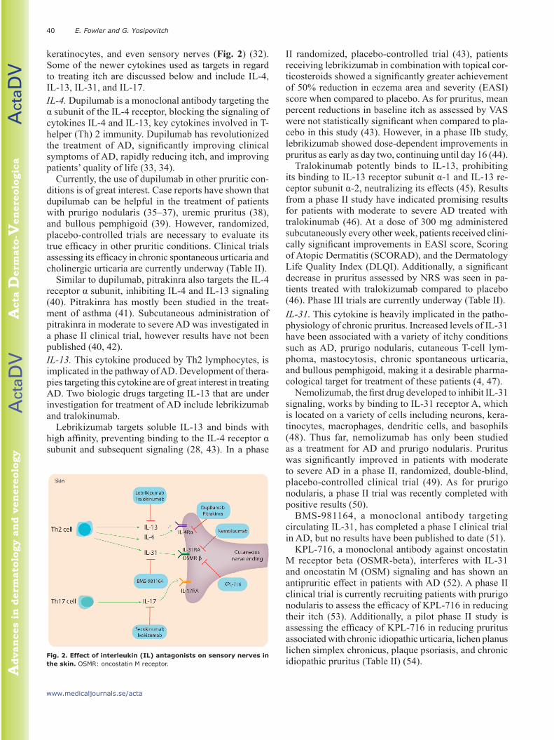
Act
aDV
Act
aDV
Advan
ces
in d
erm
ato
logy a
nd v
en
ere
olo
gy
Acta
Derm
ato
-Ven
ere
olo
gic
a
E. Fowler and G. Yosipovitch40
www.medicaljournals.se/acta
keratinocytes, and even sensory nerves (Fig. 2) (32). Some of the newer cytokines used as targets in regard to treating itch are discussed below and include IL-4, IL-13, IL-31, and IL-17.IL-4. Dupilumab is a monoclonal antibody targeting the α subunit of the IL-4 receptor, blocking the signaling of cytokines IL-4 and IL-13, key cytokines involved in T-helper (Th) 2 immunity. Dupilumab has revolutionized the treatment of AD, significantly improving clinical symptoms of AD, rapidly reducing itch, and improving patients’ quality of life (33, 34).
Currently, the use of dupilumab in other pruritic con-ditions is of great interest. Case reports have shown that dupilumab can be helpful in the treatment of patients with prurigo nodularis (35–37), uremic pruritus (38), and bullous pemphigoid (39). However, randomized, placebo-controlled trials are necessary to evaluate its true efficacy in other pruritic conditions. Clinical trials assessing its efficacy in chronic spontaneous urticaria and cholinergic urticaria are currently underway (Table II).
Similar to dupilumab, pitrakinra also targets the IL-4 receptor α subunit, inhibiting IL-4 and IL-13 signaling (40). Pitrakinra has mostly been studied in the treat-ment of asthma (41). Subcutaneous administration of pitrakinra in moderate to severe AD was investigated in a phase II clinical trial, however results have not been published (40, 42).IL-13. This cytokine produced by Th2 lymphocytes, is implicated in the pathway of AD. Development of thera-pies targeting this cytokine are of great interest in treating AD. Two biologic drugs targeting IL-13 that are under investigation for treatment of AD include lebrikizumab and tralokinumab.
Lebrikizumab targets soluble IL-13 and binds with high affinity, preventing binding to the IL-4 receptor α subunit and subsequent signaling (28, 43). In a phase
II randomized, placebo-controlled trial (43), patients receiving lebrikizumab in combination with topical cor-ticosteroids showed a significantly greater achievement of 50% reduction in eczema area and severity (EASI) score when compared to placebo. As for pruritus, mean percent reductions in baseline itch as assessed by VAS were not statistically significant when compared to pla-cebo in this study (43). However, in a phase IIb study, lebrikizumab showed dose-dependent improvements in pruritus as early as day two, continuing until day 16 (44).
Tralokinumab potently binds to IL-13, prohibiting its binding to IL-13 receptor subunit α-1 and IL-13 re-ceptor subunit α-2, neutralizing its effects (45). Results from a phase II study have indicated promising results for patients with moderate to severe AD treated with tralokinumab (46). At a dose of 300 mg administered subcutaneously every other week, patients received clini-cally significant improvements in EASI score, Scoring of Atopic Dermatitis (SCORAD), and the Dermatology Life Quality Index (DLQI). Additionally, a significant decrease in pruritus assessed by NRS was seen in pa-tients treated with tralokizumab compared to placebo (46). Phase III trials are currently underway (Table II).IL-31. This cytokine is heavily implicated in the patho-physiology of chronic pruritus. Increased levels of IL-31 have been associated with a variety of itchy conditions such as AD, prurigo nodularis, cutaneous T-cell lym-phoma, mastocytosis, chronic spontaneous urticaria, and bullous pemphigoid, making it a desirable pharma-cological target for treatment of these patients (4, 47).
Nemolizumab, the first drug developed to inhibit IL-31 signaling, works by binding to IL-31 receptor A, which is located on a variety of cells including neurons, kera-tinocytes, macrophages, dendritic cells, and basophils (48). Thus far, nemolizumab has only been studied as a treatment for AD and prurigo nodularis. Pruritus was significantly improved in patients with moderate to severe AD in a phase II, randomized, double-blind, placebo-controlled clinical trial (49). As for prurigo nodularis, a phase II trial was recently completed with positive results (50).
BMS-981164, a monoclonal antibody targeting circulating IL-31, has completed a phase I clinical trial in AD, but no results have been published to date (51).
KPL-716, a monoclonal antibody against oncostatin M receptor beta (OSMR-beta), interferes with IL-31 and oncostatin M (OSM) signaling and has shown an antipruritic effect in patients with AD (52). A phase II clinical trial is currently recruiting patients with prurigo nodularis to assess the efficacy of KPL-716 in reducing their itch (53). Additionally, a pilot phase II study is assessing the efficacy of KPL-716 in reducing pruritus associated with chronic idiopathic urticaria, lichen planus lichen simplex chronicus, plaque psoriasis, and chronic idiopathic pruritus (Table II) (54).
Fig. 2. Effect of interleukin (IL) antagonists on sensory nerves in the skin. OSMR: oncostatin M receptor.

Act
aDV
Act
aDV
Advan
ces
in d
erm
ato
logy a
nd v
en
ere
olo
gy
Acta
Derm
ato
-Ven
ere
olo
gic
a
41A new generation of treatments for itch
Acta Derm Venereol 2020
IL-17. This is a pro-inflammatory cytokine that is pri-marily produced by Th17 cells, along with neutrophils and possibly mast cells. Keratinocytes are stimulated by IL-17A to secrete other pro-inflammatory mediators, which recruit neutrophils, Th17 cells, dendritic cells, and lymphoid cells (55). Drugs in this category are currently FDA-approved for the treatment of plaque psoriasis, and based on current studies, have shown the largest magni-tude of effect in reducing psoriatic itch (5).
Secukinumab is a monoclonal antibody that selec-tively binds and neutralizes IL-17A (55). Two phase III, double-blind clinical trials assessing the efficacy of secukinumab in psoriasis showed that secukinumab was significantly more effective in reducing itch compared to placebo and etanercept, a TNF-α inhibitor (55, 56). In fact, in a pooled analysis of these two phase III trials, patients taking secukinumab achieved a significantly greater reduction in itching as early as the second week of treatment, demonstrating its rapid effect (57).
Phase II clinical trials assessing the role of secukinu-mab in treating AD are currently ongoing (Table II) (58, 59).
Ixekizumab is a high affinity monoclonal antibody also targeting IL-17A. Ixekizumab has proven to be effective in treating psoriasis, especially psoriatic itch in which phase III trials showed rapid, significant improvements. As early as the first week of treatment with ixekizumab, a significantly greater percentage of patients receiving the drug reported improvement of pruritus compared to those receiving etanercept or placebo (60).
In a long-term extension study of this trial, patients receiving ixekizumab maintained improvements in itch severity through the end of the study. By week 60, 48.2% of patients receiving ixekizumab every 4 weeks through-out the study achieved an itch NRS of 0. Similarly, after being switched to ixekizumab at 12 weeks, 45.1% and 45.3% of patients originally receiving placebo or etaner-cept, respectively, achieved an NRS of 0 at week 60 (61).
Additionally, ixekizumab is also effective in rapidly reducing genital pruritus. In a phase III, randomized, dou-ble-blind, placebo-controlled trial, a greater percentage of patients with genital psoriasis receiving ixekizumab achieved a significantly greater clinically meaningful itch reduction (greater than or equal to 3 point reduction on a numerical rating sale) than patients receiving placebo (59.7% versus 8.3%, p < 0.001) (62). Furthermore, a significant improvement in genital itch was seen as early as week 2 in those treated with ixekizumab (62).
Janus kinase inhibitorsPruritus induced by cytokines is mediated at least par-tially by the janus kinase (JAK)/signal transducer and activation of transcription (STAT) pathway (2). JAK inhibitors block the JAK/ STAT pathway, which medi-ates signal transduction of cytokines and growth factors. When ligands bind to their receptors, JAKs are activated
which lead to phosphorylation of STATs, which enter the cell nucleus to regulate transcription of target genes (63).
JAK inhibitors are commonly used in the treatment of inflammatory conditions such as rheumatoid arthritis. More recently, these medications are being investigated for use in chronic inflammatory skin conditions, such as AD and psoriasis (Table II).Tofacitinib, an oral JAK inhibitor, works by blocking JAK1 and JAK3. It has been investigated for the treat-ment of psoriasis, with results from two randomized phase III trials showing that tofacitinib improved itch in patients with psoriasis, as soon as one day after tre-atment initiation (64, 65). Patients receiving tofacitinib also achieved a significant improvement in health-related quality of life, which were maintained through the end of the study at week 52 (64). Topical tofacitinib has also been effective for treating pruritus in both patients with psoriasis as well as AD (66, 67).
In a mouse model of psoriasis, tofacitinib significantly decreased mRNA expression of itchy cytokines IL-22, IL-23, and IL-31. This study also demonstrated that tofacitinib increased peptidergic epidermal nerve fiber density, which may aid in rescuing inhibitory itch mecha-nisms, proposing a novel mechanism for itch reduction by tofacitinib (68).
Additionally, JAK inhibitors may show promise in treating other cases of itch, even non-inflammatory causes of pruritus. In five patients with refractory, chronic idiopathic pruritus, oral tofacitinib led to marked improvement in their itch after only one month (69).Baricitinib selectively inhibits JAK1 and JAK2. In a phase II, double-blind, randomized trial, baricitinib signi-ficantly improved AD and resulted in decreased pruritus (70). Phase III studies are currently underway (Table II).Upadacitinib is a newer JAK inhibitor whose mechanism of action is selective for JAK1. In a phase II, randomi-zed, placebo-controlled trial, upadacitinib significantly decreased ratings of itch in patients with AD (71). Phase III trials measuring the efficacy of upadacitinib in AD are currently in progress (Table II) (72). Abrocitinib, another JAK inhibitor specifically targeting JAK1, has demonstrated excellent results in the treat-ment of AD. In a phase III randomized, double-blind, placebo-controlled trial, a statistically significant greater proportion of patients taking abrocitinib achieved a 4 point or larger reduction in itch NRS versus those taking placebo. Likewise, patients taking abrocitinib achieved a statistically significantly greater magnitude of decrease in the Pruritus and Symptoms Assessment for Atopic Dermatitis (PSAAD) compared to patients receiving placebo (73).
Histamine-4 receptor antagonistsAntihistamines, most of which are antagonists to the histamine-1 receptor, are often given to patients endor-

Act
aDV
Act
aDV
Advan
ces
in d
erm
ato
logy a
nd v
en
ere
olo
gy
Acta
Derm
ato
-Ven
ere
olo
gic
a
E. Fowler and G. Yosipovitch42
www.medicaljournals.se/acta
sing itch despite no clear evidence of their effectiveness as antipruritic therapies. Antihistamines specifically targeting the histamine-4 receptor (H4R), however, have shown some promise in treating itch (74). In a mouse model of AD, pretreatment with an H4R antagonist attenuated scratching responses in a dose-dependent manner (75).
ZPL389, an oral H4R antagonist, is currently being studied as a treatment for AD (Table II). However, results from a phase II randomized, double-blind, placebo-controlled study did not show a significant difference in pruritus reduction between patients taking ZPL389 and those receiving placebo (76).
Anti-IgEA monoclonal anti-IgE antibody, ligelizumab, is under-going phase III clinical trials to investigate its efficacy in treating chronic spontaneous urticaria (Table II). Studies have shown that ligelizumab has a much higher affinity to bind IgE in comparison to omalizumab (77), and thus it will be interesting to see its effect on pruritus in these patients.
CONCLUSION
New therapies for itch are continuously being developed. A new era of antipruritic drugs targeting specific neural receptors, itchy cytokines, and small molecules is swiftly approaching. Now that the pathophysiology of pruritus is better understood and research into new targets and mechanisms is perpetually underway, discovery and development of newer and better treatments for itch is ongoing. Unfortunately, treatment for chronic itch is not always simple and every patient requires individualized therapy. With education and development of new targets, clinicians can obtain a greater arsenal of treatment for their patients, to successfully treat them and improve their quality of life.
ACKNOWLEDGEMENTGY has been a consultant or Ad Board member of AbbVie, Kiniska, Menlo, TREVI, Sienna, Galderma, Sanofi, Regeneron, Novartis, Eli Lilly, Pfizer, Bayer, Cerave and received research grants from Sun Pharma, Pfizer, Leo, Kiniska, and Menlo.
REFERENCES1. Tey HL, Wallengren J, Yosipovitch G. Psychosomatic factors
in pruritus. Clin Dermatol 2013; 31: 31–40. 2. Yosipovitch G, Rosen JD, Hashimoto T. Itch: From mechanism
to (novel) therapeutic approaches. J Allergy Clin Immunol 2018; 142: 1375–1390.
3. Fowler E, Yosipovitch G. Chronic itch management: therapies beyond those targeting the immune system. Ann Allergy, Asthma Immunol 2019; 123: 158–165.
4. Hashimoto T, Yosipovitch G. Itching as a systemic disease. J Allergy Clin Immunol 2019; 144: 375–380.
5. Ständer S, Siepmann D, Herrgott I, Sunderkötter C, Luger TA. Targeting the neurokinin receptor 1 with aprepitant: A novel antipruritic strategy. PLoS One 2010; 5: 3–7.
6. Booken N, Heck M, Nicolay JP, Klemke CD, Goerdt S, Utikal J. Oral aprepitant in the therapy of refractory pruritus in erythrodermic cutaneous T-cell lymphoma. Br J Dermatol 2011; 164: 665–683.
7. Leslie TA, Greaves MW, Yosipovitch G. Current Topical and Systemic Therapies for Itch. In: Cowan A, Yosipovitch G, editors. Pharmacology of Itch [Internet]. Springer-Verlag Berlin Heidelberg; 2015. p. 339–356. Available from: http: //link.springer.com/10.1007/978-3-662-44605-8.
8. Yosipovitch G, Ständer S, Kerby MB, Larrick JW, Perlman AJ, Schnipper EF, et al. Serlopitant for the treatment of chronic pruritus: Results of a randomized, multicenter, placebo-controlled phase 2 clinical trial. J Am Acad Dermatol 2018; 78: 882–891.e10.
9. Ständer S, Kwon P, Hirman J, Perlman AJ, Weisshaar E, Metz M, et al. Serlopitant reduced pruritus in patients with prurigo nodularis in a phase 2, randomized, placebo-controlled trial. J Am Acad Dermatol 2019; 80: 1395–1402.
10. Heitman A, Xiao C, Cho Y, Polymeropoulos C, Birznieks G, Polymeropoulos M. Tradipitant improves worst itch and disease severity in patients with chronic pruritus related to atopic dermatitis. J Am Acad Dermatol 2018; 79: AB300.
11. Bigliardi PL, Bigliardi-qi M. Chapter 18: Peripheral Opioids. In: Carstens E, Akiyama T, editors. Itch – Mechanisms and Treatment [Internet]. Boca Raton: CRC Press/Taylor & Fran-cis; 2014. p. 1–10. Available from: https: //www.ncbi.nlm.nih.gov/books/NBK200922/.
12. Tominaga M, Ogawa H, Takamori K. Possible roles of epider-mal opioid systems in pruritus of atopic dermatitis. J Invest Dermatol 2007; 127: 2228–2235.
13. Dawn AG, Yosipovitch G. Butorphanol for treatment of in-tractable pruritus. J Am Acad Dermatol 2006; 54: 527–531.
14. Trevi Therapeutics. Trevi Therapeutics Announces Positive Results from Phase 2 Trial in Prurigo Nodularis. 2016. https://www.trevitherapeutics.com/wp-content/uploads/2019/01/TreviPNToplineDataPR.10_.13_.16_.FINAL_.pdf.
15. Mathur VS, Kumar J, Crawford PW, Hait H, Sciascia T. a mul-ticenter, randomized, double-blind, placebo-controlled trial of nalbuphine ER tablets for uremic pruritus. Am J Nephrol 2017; 46: 450–458.
16. Kumagai H, Ebata T, Takamori K, Muramatsu T, Nakamoto H, Suzuki H. Effect of a novel kappa-receptor agonist, nalfurafine hydrochloride, on severe itch in 337 haemo-dialysis patients: A Phase III, randomized, double-blind, placebo-controlled study. Nephrol Dial Transplant 2010; 25: 1251–1257.
17. Cara Therapeutics Announces Positive Top-Line Data From Part A of Phase 2/3 Trial of I.V. CR845 in Chronic Kidney Disease-Associated Pruritus [Internet]. Cara Therapeutics. 2017. Available from: http: //ir.caratherapeutics.com/news-releases/news-release-details/cara-therapeutics-announces-positive-top-line-data-part-phase-23.
18. ClinicalTrials.gov. Safety, Pharmacokinetics and Preliminary Efficacy of Asimadoline in Pruritus Associated with Atopic Dermatitis [Internet]. [cited 2019 Jun 18]. Available from: https: //clinicaltrials.gov/ct2/show/NCT02475447?term=asimadoline&rank=5.
19. Roblin D, Yosipovitch G, Boyce B, Robinson J, Sandy J, Mainero V, et al. Topical TrkA kinase inhibitor CT327 is an effective, novel therapy for the treatment of pruritus due to psoriasis: Results from experimental studies, and efficacy and safety of CT327 in a phase 2b clinical trial in patients with psoriasis. Acta Derm Venereol 2015; 95: 542–548.
20. Sienna Biopharmaceuticals Inc. Sienna Biopharmaceuticals Announces Its Topical, Non-Steroidal TrkA Inhibitor SNA-120 (0.05%) Demonstrated Significant Impact on Psoriasis in Phase 2b Study and Plans to Initiate Phase 3 Psoriasis Trials in Second Half of 2019. Sienna Press Release.
21. Barr TP, Garzia C, Guha S, Fletcher EK, Nguyen N, Wieschhaus AJ, et al. PAR2 Pepducin-Based Suppression of Inflamma-tion and Itch in Atopic Dermatitis Models. J Invest Dermatol

Act
aDV
Act
aDV
Advan
ces
in d
erm
ato
logy a
nd v
en
ere
olo
gy
Acta
Derm
ato
-Ven
ere
olo
gic
a
43A new generation of treatments for itch
Acta Derm Venereol 2020
2019; 139: 412–421.22. Cao T, Tan WD, Kim H, Tey HL. Efficacy of a topical proteinase-
activated receptor-2 inhibitor on cowhage-induced pruritus: a randomized placebo-controlled double-blind study. J Eur Acad Dermatol Venereol 2017; 31: e191–e192.
23. Cao T, Tan ES, Chan YH, Yosipovitch G, Tey HL. Anti-pruritic efficacies of doxycycline and erythromycin in the treatment of acne vulgaris: A randomized single-blinded pilot study. Indian J Dermatol Venereol Leprol 2018; 84: 458–460.
24. Matsuda KM, Sharma D, Schonfeld AR, Kwatra SG. Gaba-pentin and pregabalin for the treatment of chronic pruritus. J Am Acad Dermatol 2016; 75: 619–625.e6.
25. Ralvenius WT, Neumann E, Pagani M, Acuña MA, Wildner H, Benke D, et al. Itch suppression in mice and dogs by mo-dulation of spinal α2 and α3GABAA receptors. Nat Commun 2018; 9: 1–15.
26. Lee JH, Park CK, Chen G, Han Q, Xie RG, Liu T, et al. A monoclonal antibody that targets a Nav1.7 channel voltage sensor for pain and itch relief. Cell 2014; 157: 1393–1404.
27. Products [Internet]. Neurim Pharmaceuticals. [cited 2019 Jun 24]. Available from: http: //www.neurim.com/research/#neup12.
28. Reich A, Szcz J, Samotij D. Biologics for Itch: IL-4/IL-13, IL-31, IL-17, and IL-23 Antagonists. Curr Dermatol Rep 2017; 6: 263–272.
29. Yosipovitch G, Stein Gold LF, Lebwohl MG, Silverberg JI, Tall-man AM, Zane LT. Early relief of pruritus in atopic dermatitis with crisaborole ointment, a non-steroidal, phosphodieste-rase 4 inhibitor. Acta Derm Venereol 2018; 98: 484–489.
30. Stander S, Yosipovitch G, Bushmakin AG, Cappelleri JC, Luger T, Tom WL, et al. Examining the association between pruritus and quality of life in patients with atopic dermati-tis treated with crisaborole. J Eur Acad Dermatol Venereol 2019; Accepted.
31. Bagel J, Lebwohl M, Stein Gold L, Jackson JM, Goncalves J, Levi E, et al. Efficacy of Apremilast on Quality of Life Measures in Patients with Moderate Plaque Psoriasis (UNVEIL Phase IV Study). J Am Acad Dermatol 2017; 76: AB119.
32. Cevikbas F, Buhl T, Mess C, Buddenkotte J, Steinhoff M. Chapter 13 Role of Interleukin 31 and Oncostatin M in Itch and Neuroimmune Communication. In: Carstens E, Akiyama T, editors. Itch: Mechanisms and Treatment. CRC Press/Taylor & Francis, Boca Raton; 2014.
33. Han Y, Chen Y, Liu X, Zhang J, Su H, Wen H, et al. Efficacy and safety of dupilumab for the treatment of adult atopic dermatitis: A meta-analysis of randomized clinical trials. J Allergy Clin Immunol 2017; 140: 888–891.e6.
34. Yosipovitch G, Reaney M, Mastey V, Eckert L, Abbe A, Nelson L, et al. Peak Pruritus Numerical Rating Scale: psychome-tric validation and responder definition for assessing itch in moderate-to-severe atopic dermatitis. Br J Dermatol 2019; 181: 761–769.
35. Beck KM, Yang EJ, Sekhon S, Bhutani T, Liao W. Dupilumab treatment for generalized prurigo nodularis. JAMA Derma-tology 2019; 155: 118–120.
36. Rambhia PH, Levitt JO. Recalcitrant prurigo nodularis treated successfully with dupilumab. JAAD Case Reports 2019; 5: 471–473.
37. Mollanazar NK, Elgash M, Weaver L, Valdes-Rodriguez R, Hsu S. Reduced itch associated with dupilumab treatment in 4 patients with prurigo nodularis. JAMA Dermatology 2019; 155: 121–122.
38. Silverberg JI, Brieva J. A successful case of dupilumab treatment for severe uremic pruritus. JAAD Case Reports 2019; 5: 339–341.
39. Kaye A, Gordon SC, Deverapalli SC, Her MJ, Rosmarin D. Du-pilumab for the treatment of recalcitrant bullous pemphigoid. JAMA Dermatology 2018; 154: 1225–1226.
40. Montes-Torres A, Llamas-Velasco M, Pérez-Plaza A, Solano-López G, Sánchez-Pérez J. Biological Treatments in Atopic Dermatitis. J Clin Med 2015; 4: 593–613.
41. Wenzel S, Willbraham D, Fuller R, Getz EB, Langphre M. Effect of an interleukin-4 variant on late phase asthmatic response to allergen challenge in asthmatic patients: results of two
phase 2a studies. Lancet 2007; 370: 1422–1431. 42. ClinicalTrials.gov. A Phase 2a Study to Investigate the Effects
of Repeated Administration of AeroDerm in Subjects With Atopic Eczema [Internet]. [cited 2019 Jun 16]. Available from: https: //clinicaltrials.gov/ct2/show/NCT00676884?term=pitrakinra&rank=1.
43. Simpson EL, Flohr C, Eichenfield LF, Bieber T, Sofen H, Taïeb A, et al. Efficacy and safety of lebrikizumab (an anti-IL-13 monoclonal antibody) in adults with moderate-to-severe atopic dermatitis inadequately controlled by topical corti-costeroids: A randomized, placebo-controlled phase II trial (TREBLE). J Am Acad Dermatol 2018; 78: 863–871.e11.
44. Dermira Presents Data From Phase 2b Study of Lebrikizumab in Patients With Atopic Dermatitis at Fall Clinical Dermato-logy Conference [Internet]. BioSpace. 2019 [cited 2019 Oct 31]. Available from: https://www.biospace.com/article/releases/dermira-presents-data-from-phase-2b-study-of-lebrikizumab-in-patients-with-atopic-dermatitis-at-fall-clinical-dermatology-conference/)
45. Popovic B, Breed J, Rees DG, Gardener MJ, Vinall LMK, Kemp B, et al. Structural characterisation reveals mechanism of IL-13-neutralising monoclonal antibody tralokinumab as inhibition of binding to IL-13Rα1 and IL-13Rα2. J Mol Biol 2017; 429: 208–219.
46. Wollenberg A, Howell MD, Guttman-Yassky E, Silverberg JI, Kell C, Ranade K, et al. Treatment of atopic dermatitis with tralokinumab, an anti–IL-13 mAb. J Allergy Clin Immunol 2019; 143: 135–141.
47. Bağci IS, Ruzicka T. IL-31: A new key player in dermatology and beyond. J Allergy Clin Immunol 2018; 141: 858–866.
48. Furue M, Yamamura K, Kido-Nakahara M, Nakahara T, Fukui Y. Emerging role of interleukin-31 and interleukin-31 recep-tor in pruritus in atopic dermatitis. Allergy Eur J Allergy Clin Immunol 2018; 73: 29–36.
49. Ruzicka T, Hanifin JM, Furue M, Pulka G, Mlynarczyk I, Wol-lenberg A, et al. Anti–interleukin-31 receptor a antibody for atopic dermatitis. N Engl J Med 2017; 376: 826–835.
50. ClinicalTrials.gov. Safety and Efficacy of Nemolizumab in PN [Internet]. [cited 2019 Jun 16]. Available from: 6/16/2019 Safety and Efficacy of Nemolizumab in PN – Full Text View – ClinicalTrials.gov Safety and Efficacy of Nemolizumab in PN%0AThe.
51. ClinicalTrials.gov. A Two-Part, Phase 1, Single-Dose Study of IL-31 mAb (Anti-Interleukin 31 Monoclonal Antibody); in Healthy Subjects and Adults With Atopic Dermatitis [Inter-net]. [cited 2019 Jun 16]. Available from: https: //clinical-trials.gov/ct2/show/NCT01614756.
52. Mikhak Z, Bissonnette R, Siri D, Tyring SK, Tessari E, Gandhi R, et al. KPL-716, anti-Oncostatin M receptor beta antibody, reduced pruritus in atopic dermatitis. J Invest Dermatol 2019; 139: S96.
53. ClinicalTrials.gov. A Study to Assess the Efficacy, Safety, and Tolerability of KPL-716 in Reducing Pruritus in Prurigo Nodu-laris [Internet]. [cited 2019 Jun 23]. Available from: https: //clinicaltrials.gov/ct2/show/NCT03816891?term=KPL-716&rank=1.
54. ClinicalTrials.gov. A Study to Assess the Efficacy, Safety and Tolerability of KPL-716 in Reducing Pruritus in Chronic Pruritic Diseases [Internet]. [cited 2019 Jun 23]. Available from: htt-ps: //clinicaltrials.gov/ct2/show/NCT03858634?term=KPL-716&rank=2.
55. Langley RG, Elewski BE, Lebwohl M, Reich K, Griffiths CEM, Papp K, et al. Secukinumab in plaque psoriasis – results of two Phase 3 trials. N Engl J Med 2014; 371: 326–339.
56. Therene C, Brenaut E, Barnetche T, Misery L. Efficacy of Systemic Treatments of psoriasis on pruritus: a systemic literature review and meta-analysis. J Invest Dermatol 2018; 138: 38–45.
57. Yosipovitch G, Soung J, Weiss J, Muscianisi E, Meng X, Gil-loteau I, et al. Secukinumab provides rapid relief from itching and pain in patients with moderate-to-severe psoriasis: patient symptom diary data from two phase 3, randomized, placebo-controlled clinical trials. Acta Derm Venereol 2019; 99: 1–2.
Act
aDV
Act
aDV
Advan
ces
in d
erm
ato
logy a
nd v
en
ere
olo
gy
Acta
Derm
ato
-Ven
ere
olo
gic
a
E. Fowler and G. Yosipovitch44
www.medicaljournals.se/acta
58. ClinicalTrials.gov. Secukinumab for Treatment of Atopic Dermatitis [Internet]. [cited 2019 Jun 10]. Available from: https: //clinicaltrials.gov/ct2/show/NCT02594098?term=secukinumab&draw=3&rank=72.
59. ClinicalTrials.gov. Investigation of Efficacy of Secukinumab in Patients With Moderate to Serve Atopic Dermatitis [Internet]. [cited 2019 Jun 10]. Available from: https: //clinicaltrials.gov/ct2/show/NCT03568136?term=secukinumab&draw=3&rank=36.
60. Griffiths CEM, Reich K, Lebwohl M, Van De Kerkhof P, Paul C, Menter A, et al. Comparison of ixekizumab with etanercept or placebo in moderate-to-severe psoriasis (UNCOVER-2 and UNCOVER-3): Results from two phase 3 randomised trials. Lancet 2015; 386: 541–551.
61. Kimball AB, Luger T, Gottlieb A, Puig L, Kaufmann R, Burge R, et al. Long-term impact of ixekizumab on psoriasis itch severity: results from a Phase III clinical trial and long-term extension. Acta Derm Venereol 2018; 98: 98–102.
62. Yosipovitch G, Foley P, Ryan C, Cather JC, Meeuwis KA, Burge R, et al. Ixekizumab improved patient-reported genital pso-riasis symptoms and impact of symptoms on sexual activity vs placebo in a randomized, double-blind study. J Sex Med 2018; 15: 1645–1652.
63. Villarino A V., Kanno Y, O’Shea JJ. Mechanisms and conse-quences of Jak-STAT signaling in the immune system. Nat Immunol 2017; 18: 374–384.
64. Feldman SR, Thaçi D, Gooderham M, Augustin M, de la Cruz C, Mallbris L, et al. Tofacitinib improves pruritus and health-related quality of life up to 52 weeks: Results from 2 randomized phase III trials in patients with moderate to severe plaque psoriasis. J Am Acad Dermatol 2016; 75: 1162–1170.e3.
65. Ständer S, Luger TA, Cappelleri JC, Bushmakin AG, Mamolo C, Zielinski MA, et al. Validation of the itch severity item as a measurement tool for pruritus in patients with psoriasis: results from a Phase 3 tofacitinib program. Acta Derm Ve-nereol 2018; 98: 340–345.
66. Mamolo C, Bissonnette R, Khan S, Lan S, Ports W, Poulin Y. An evaluation of the effect of topical tofacitinib (CP-690, 550) on pruritus and patient satisfaction with study medication in a phase IIA trial for plaque psoriasis. J Am Acad Dermatol 2013; 68: AB192.
67. Bissonnette R, Papp KA, Poulin Y, Gooderham M, Raman M, Mallbris L, et al. Topical tofacitinib for atopic dermatitis: a phase IIa randomized trial. Br J Dermatol 2016; 175: 902–911.
68. Hashimoto T, Sakai K, Sanders KM, Yosipovitch G, Akiyama
T. Antipruritic effects of janus kinase inhibitor tofacitinib in a mouse model of psoriasis. Acta Derm Venereol 2019; 99: 298–303.
69. Oetjen LK, Mack MR, Feng J, Whelan TM, Niu H, Guo CJ, et al. Sensory Neurons Co-opt Classical Immune Signaling Pat-hways to Mediate Chronic Itch. Cell 2017; 171: 217–228.e13.
70. Guttman-Yassky E, Silverberg JI, Nemoto O, Forman SB, Wilke A, Prescilla R, et al. Baricitinib in adult patients with moderate-to-severe atopic dermatitis: A phase 2 parallel, double-blinded, randomized placebo-controlled multiple-dose study. J Am Acad Dermatol 2019; 80: 913–921.e9.
71. Beck L, Hong C, Hu X, Chen S, Calimlim B, Teixeira H, et al. Upadacitinib effect on pruritus in moderate-to-severe atopic dermatitis; from a Phase 2B randomized, placebo-controlled trial. Ann Allergy Asthma Immunol 2018; 121: S21.
72. ClinicalTrials.gov. Evaluation of Upadacitinib in Adolescent and Adult Patients With Moderate to Severe Atopic Dermatitis (Eczema) [Internet]. [cited 2019 Jun 16]. Available from: https: //clinicaltrials.gov/ct2/show/NCT03569293?term=Upadacitinib&rank=7.
73. Pfizer Announces Positive Top-Line Results from Phase 3 Study of Investigational Oral JAK1 Candidate, Abrocitinib (PF-04965842), in Patients Aged 12 and Older with Moderate to Severe Atopic Dermatitis [Internet]. Pfizer.com. 2019. Available from: https: //www.pfizer.com/news/press-release/press-release-detail/pfizer_announces_positive_top_line_re-sults_from_phase_3_study_of_investigational_oral_jak1_candidate_abrocitinib_pf_04965842_in_patients_aged_12_and_older_with_moderate_to_severe_atopic_dermatitis.
74. Dunford PJ, Williams KN, Desai PJ, Karlsson L, McQueen D, Thurmond RL. Histamine H4 receptor antagonists are superior to traditional antihistamines in the attenuation of experimental pruritus. J Allergy Clin Immunol 2007; 119: 176–183.
75. Cowden JM, Zhang M, Dunford PJ, Thurmond RL. The his-tamine H4 receptor mediates inflammation and pruritus in Th2-dependent dermal inflammation. J Invest Dermatol 2010; 130: 1023–1033.
76. Werfel T, Layton G, Yeadon M, Whitlock L, Osterloh I, Jimenez P, et al. Efficacy and safety of the histamine H 4 receptor antagonist ZPL-3893787 in patients with atopic dermatitis. J Allergy Clin Immunol 2019; 143: 1830–1837.e4.
77. Arm JP, Bottoli I, Skerjanec A, Floch D, Groenewegen A, Maahs S, et al. Pharmacokinetics, pharmacodynamics and safety of QGE031 (ligelizumab), a novel high-affinity anti-IgE antibody, in atopic subjects. Clin Exp Allergy 2014; 44: 1371–1385.

Act
aDV
Act
aDV
Advan
ces
in d
erm
ato
logy a
nd v
en
ere
olo
gy
Acta
Derm
ato
-Ven
ere
olo
gic
a
Acta Derm Venereol 2020; 100: adv00028This is an open access article under the CC BY-NC license. www.medicaljournals.se/actaJournal Compilation © 2020 Acta Dermato-Venereologica.
doi: 10.2340/00015555-3348
REVIEW ARTICLE
Centenary theme section: ITCH AND PRURITIC DISORDERS
SIGNIFICANCEItch lasting for 6 weeks or more is considered chronic and represents a high burden for those affected. Various as-pects of chronic itch, including the underlying origin of the itch, symptoms, skin manifestations, response to thera-pies and impairment of quality of life vary from patient to patient, constituting a challenge for clinicians and clinical researchers. Unmet needs, such as the standardization of experimental and clinical research protocols, diagnostic procedures and therapeutic regimens, as well as a better understanding of associated burdens and the development of novel effective therapies should be targeted by physici-ans and researchers dealing with chronic itch.
Chronic pruritus is a frequent global condition. The pathophysiology, underlying aetiology, clinical mani-festation, associated burden and response to therapy of chronic pruritus varies from patient to patient, ma-king clinical research and management of this condi-tion challenging. There are still several unmet needs, such as the need to standardize translational research protocols, diagnostic and therapeutic procedures and to enhance the knowledge of the humanistic and eco-nomic burden associated with chronic pruritus. Basic and clinical research is of the utmost importance to target these matters. Clinical research has the poten-tial to identify new relevant mechanisms in affected patients, which may lead to identification of novel therapy targets. This article discusses in depth current shortcomings in the daily care of patients with chronic pruritus and the challenges clinical researchers and physicians treating chronic pruritus face in addressing these matters.
Key words: itch; patient-reported outcome; guideline; clinical trials; clinical research.
Accepted Oct 15, 2019; Published Jan 9, 2020
Acta Derm Venereol 2020; 100: adv00028.
Corr: Sonja Ständer, Department of Dermatology and Center for Chro-nic Pruritus, University Hospital Münster, Von-Esmarch-Str. 58, DE-48149 Münster, Germany. E-mail: [email protected]
Chronic pruritus (CP), defined as pruritus lasting for 6 weeks or longer, is highly prevalent, affecting
approximately one-sixth of the population in Germany (1). Population-based analyses of the nationwide search volume of pruritus on Google suggest an even larger number of individuals suffering from CP within Ger-many and the USA (2–4). Those affected often report a substantial burden and a relevant impairment of their quality of life (QoL) (5, 6). The aetiology, clinical mani-festation, diagnostic measures and therapeutic response to CP vary from patient to patient, which contributes to the challenging management of CP (7). Although recent years have witnessed a marked increase in basic and clinical research efforts in this field, CP researchers and physicians treating CP currently face several challenges when targeting unmet needs that impact on clinical rou-tine. The aim of this review is to highlight and discuss
methodologies used in CP research, as well as current challenges and unmet needs in clinical care.
CHRONIC PRURITUS: A MULTIDIMENSIONAL CONDITION
CP is a heterogeneous condition in terms of demogra-phics, clinical presentation and underlying origin. Al-though the prevalence of CP increases with age, patients of all age groups, including children, may be affected (8). Moreover, patients of both sexes and all ethnic back-grounds may suffer from CP. According to the Internatio-nal Forum for the Study of Itch (IFSI), CP may present on inflamed skin (IFSI I), on normal appearing skin (IFSI II), or accompanied by chronic scratch lesions, such as chronic prurigo or lichen simplex chronicus (IFSI III) (9). As for the underlying aetiology, CP may arise from dermatological, systemic, neurological or psychiatric/psychosomatic conditions. Some patients show multiple causes for the pruritus (multifactorial CP), while in few cases the origin of the pruritus remains unknown despite extensive diagnostic work-up (9). Recent studies have further suggested a substantial impact of climate and weather on the prevalence of pruritus, especially invol-ving specific localizations on the body, but these issues have yet to be examined in epidemiological studies (2–4). Adding to these factors, patients with CP vary in terms of medical history, comorbidities, co-medication, socio-economic backgrounds and therapy goals, contributing to the complexity of the management of these patients.
Challenges in Clinical Research and Care in PruritusManuel P. PEREIRA1, Claudia ZEIDLER1, Michael STORCK2, Konstantin AGELOPOULOS1, Wolfgang G. PHILIPP-DORMSTON3, Alexander ZINK4 and Sonja STÄNDER1
1Department of Dermatology and Center for Chronic Pruritus, University Hospital Münster, 2Institute of Medical Informatics, University of Münster, Münster, 3Faculty of Health, University Witten/Herdecke, and 4Department of Dermatology and Allergy, School of Medicine, Technical University of Munich, Munich, Germany

Act
aDV
Act
aDV
Advan
ces
in d
erm
ato
logy a
nd v
en
ere
olo
gy
Acta
Derm
ato
-Ven
ere
olo
gic
a
M. P. Pereira et al.46
Theme issue: Itch and pruritic disorders
UNMET NEEDS IN CLINICAL RESEARCH
Due to the complexity and multidimensional nature of CP, clinical research investigating several aspects of pru-ritic diseases and its management is needed. In particular, clinical research contributes to a better understanding of pathophysiological mechanisms involved in the de-velopment and chronicity of CP in humans. Moreover, clinical researchers should focus on various aspects of clinical management, such as standardizing diagnostic and therapeutic procedures, and assessing the humanistic burden and impact of CP on affected patients. Another important area is the performance of longitudinal trans-lational studies, clinical trials investigating novel agents for the treatment of CP, and psychometric research. It should also be investigated how the care of patients with CP is integrated into the health system in order to identify shortcomings and to adopt innovative strategies, as, for example, digital tools to improve care. The next section of this article discusses the challenges and difficulties with regard to these issues.
CHALLENGES IN CLINICAL RESEARCH AND CARE
Understanding the pathophysiologyBasic and clinical research has led in recent decades to a better understanding of the mechanisms underlying CP. In clinical-translational research, different methods in-vestigating specific pathophysiological features that aug-ment data obtained from morphological and molecular biological research have been established and validated. The collaboration with biostatisticians and experts of medical informatics is essential for the interpretation of data and for obtaining a comprehensive understanding of the results. An overview of these methods is given in Table I.
Several methodologies have been developed in order to investigate the functions of peripheral nerves. Skin stimulation with pruritogens (e.g. histamine, capsaicin, cowhage, β-alanine) has led to the characterization of several subpopulations of C-fibres. Histamine activa-tes mechano-insensitive C-fibres (CMi-fibres), while cowhage and β-alanine activate heat and mechano-sen-
sitive C-fibres (CMH-fibres) (10). Hyperknesis related to cowhage-induced pruritus, but not histaminergic pruritus, is present in several types of CP compared with controls, arguing for sensitization of CMH-fibres in CP (11). One limitation of this methodology is the high inter-individual variability, while intra-individual variability is low (12). Another method, transcutaneous electrical stimulation, allows the selective activation of subpopulations of peripheral nerve fibres. For example, using the Neurometer® C, Aδ and Aβ fibres are activa-ted by electrical stimulation at a frequency of 5, 250 and 2,000 Hz, respectively (13). Higher itch intensities could be induced upon stimulation at 5 and 2,000 Hz in patients with CP of different origins compared with healthy individuals, arguing for hyperknesis in patients with CP (14). Of note, patients are often anxious when undergoing experimental procedures with electrical sti-mulation, which leads to biased ratings of evoked sensory symptoms, thus constituting a limitation of this method. Another functional well-established diagnostic tool that is used for clinical itch research is quantitative sensory testing (QST). It is a battery of psychophysical tests, in which response to graded thermal and mechanical stimuli is assessed (15). This procedure, originally developed for determination of neuropathic pain, informs about gain or loss of function of different peripheral nerve fibre subpopulations and hints at possible signs of central sen-sitization. In itch research it could be shown that patients with CP of different origins have an altered QST profile, arguing for disordered peripheral neuronal mechanisms (11). Although QST is widely used in neurological rou-tine care, it has some limitations. It is time-consuming, requires highly qualified personnel, as well as the col-laboration of patients. Morphological methodologies for investigation of the anatomy of cutaneous cells and expression of receptors and mediators are essential in clinical itch research. In particular, determination of the intraepidermal nerve fibre density is useful in order to reveal potential epidermal neuroanatomical alterations (16). In this examination, a skin biopsy is obtained and intraepidermal nerves are stained with the axonal mar-ker protein gene product 9.5 (17). This simple method, which is also used in routine care, has some limitations.
Table I. Clinical research methodologies used in itch research
Methodology Mechanism
Cutaneous stimulation with histamine Chemical-evoked itch via activation of mechano-insensitive C-fibres (CMi-fibres), mast cell degranulationCutaneous stimulation with cowhage or ß-alanine Chemical-evoked itch via activation of mechano and heat-sensitive C-fibres (CMH-fibres) as well as A-fibres.Hyperknesis Increased itch in response to (chemical, electrical) pruritogens or pinprick stimulationAlloknesis Itch response to non-pruritogenic stimuli: touch/brush strokes. Functional testing of itch sensitization
phenomena with punctuate or dynamic mechanical stimulation (von Frey filaments, cotton swab)Transcutaneous electrical stimulation Electrical-evoked itch via selective activation of peripheral nerve fibre classes Quantitative sensory testing Psychophysical testing of subpopulations of peripheral nerve fibres (C-fibres, Aδ-fibres, Aβ-fibres) and signs
of central sensitization using graded thermal and mechanical stimuliFunctional magnetic resonance imaging/positron
emission tomographyIdentification of activated and deactivated cerebral centres upon pruritic stimulation
Conditioned pruritic modulation Central descending inhibition of pruritusIntraepidermal nerve fibre density Morphological determination of epidermal nerve fibre numbers (skin biopsy)

Act
aDV
Act
aDV
Advan
ces
in d
erm
ato
logy a
nd v
en
ere
olo
gy
Acta
Derm
ato
-Ven
ere
olo
gic
a
47Clinical research and care in itch
Theme issue: Itch and pruritic disorders
The skin probe is sensitive to the fixative and needs to be processed in a timely manner in the laboratory. In ad-dition, reference values are available only for the distal lower leg (18, 19), thus interpretation of values from other body sites is difficult. It is a purely morphological examination, which does not inform on functional alte-rations of peripheral nerve fibres.
Central processing of pruritus is very challenging. Most current knowledge on spinal transmission of itch was obtained from animal studies (20). Functional mag-netic resonance imaging (fMRI) and positron emission tomography (PET) studies have been used to investigate transmission and processing of pruritus in upper centres. Most studies have been performed on healthy volunteers exposed to pruritic stimulation during the scans. Brain regions associated with sensory recognition, cognition, motor response and emotional states are activated in response to pruritus (21). Interestingly, both overlapping and distinct brain areas are activated upon histaminergic and non-histaminergic pruritic stimulation, pointing to differences in cerebral processing of distinct pruritus ty-pes (22). Patients with CP also show differences in brain activation upon stimulation with histamine compared with healthy individuals, arguing for central sensitiza-tion in CP (23). Also, structural changes in grey matter have been observed in patients with CP, showing the neuroplasticity in chronic states (24). Although functio-nal imaging studies have enhanced our understanding of cerebral processing of pruritus, this methodology has some pitfalls. fMRI and PET are very expensive metho-dologies, which are available only in specialized centres. In addition, patients with claustrophobia, ferromagnetic prostheses or implanted devices (e.g. defibrillator) cannot undergo an MRI examination, while others (e.g. children) have difficulty lying still during the procedure, leading to poor imaging quality. More imaging studies, enrolling patients with CP of different origins, are needed in order to establish similarities and differences in cerebral brain processing of pruritus across distinct CP conditions.
Upper centres exert a descending inhibitory modula-tion via a noradrenergic and a serotonergic pathway, as shown by imaging studies (21, 25). The paradigm of conditioned pain modulation (CPM), in which a noxious
stimulus inhibits pain elsewhere in the body, can be used experimentally to assess descending inhibition (26). In patients with CP, as in those with chronic pain, CPM is impaired, which may contribute to the chronicity of the pruritus (11). An analogous paradigm of conditioned pruritic modulation, in which a pruritic stimulus inhibits pruritus elsewhere in the body, has also been investiga-ted, with conflicting results regarding its effect (27, 28). More studies are needed to enhance the understanding of the mechanisms of descending modulation in CP and its role in the chronicity of the disease.
Standardization of assessment instrumentsSince pruritus has a subjective dimension, patient-reported outcomes (PRO) play a pivotal role. A plethora of standardized questionnaires and scales have been developed in order to gather information on several aspects of CP, including pruritus characteristics, course of the disease, QoL, reactive disorders, such as anxiety, depression and sleep impairment, and therapy goals.
Pruritus characteristics (e.g. onset, duration and dist-ribution of pruritus, as well as accompanying sensory symptoms and scratching behaviour) can be assessed using general patient-oriented itch questionnaires (29, 30). Mono-dimensional scales, namely the visual ana-logue scale, the numerical rating scale and the verbal rating scale, have been validated to assess the intensity of pruritus (31). Intensity scales are also validated for the use in electronic diary applications (32). The interpreta-tion of itch intensity scales is challenging, since scores vary from patient to patient according to external factors, such as sex or ethnic background (1, 33). Some efforts have been put into understanding the minimal clinical relevant difference of itch intensity change, e.g. after initiating a therapy (34). However, many factors (e.g. the aetiology of the pruritus, recall period, socioeconomic background) may influence the minimal clinical relevant difference, making the interpretation of results difficult. An overview of other instruments for the assessment of itch is shown in Table II.
As for secondary conditions arising due to CP, standar-dized questionnaires are used to screen for anxiety and
Table II. Examples of instruments for itch assessment
Instrument Description
Visual analogue scale (VAS) Monodimensional scale for assessment of itch intensity; used in clinical trials and routine care (electronic and paper versions available)
Numerical rating scale (NRS) Monodimensional scale for assessment of itch intensity; used in clinical trials and routine care (electronic and paper versions available)
Verbal rating scale (VRS) Categorial scale for assessment of itch intensity; used in clinical trials and routine care (electronic and paper versions available)Labelled magnitude scale (LMS) Monodimensional scale for assessment of itch intensity; used in experimental research (paper version)Itch Severity Scale (ISS) Multidimensional scale for assessment of itch intensity; used in clinical trials (paper version)Dynamic Pruritus Score (DPS) Patient global impression of change for assessment of itch improvement; used in clinical trials and routine care (electronic and
paper versions available)5-D itch scale (5 dimensions itch
scale)Multidimensional questionnaire for assessment of itch characteristics; used in clinical trials (paper version)
ItchyQuant Monodimensional scale for assessment of itch intensity; combination of VAS and cartoon depictions of scratching; developed for populations with cognitive limitations (elderly, children); used in routine care (paper version)
Act
aDV
Act
aDV
Advan
ces
in d
erm
ato
logy a
nd v
en
ere
olo
gy
Acta
Derm
ato
-Ven
ere
olo
gic
a
M. P. Pereira et al.48
Theme issue: Itch and pruritic disorders
depression (e.g. Hospital Anxiety and Depression Scale (35)), to assess sleep disorders (36, 37) and to measure the impairment of QoL (e.g. Dermatological Life Quality Index (38) and pruritus-specific ItchyQol (39)).
PRO measures are usually collected via paper and pencil questionnaires, but electronic PRO systems are emerging. Computerized collection of PRO measures offers a couple of advantages compared with paper questionnaires: reduction of errors emerging through typewriting, reduction of missing data by requiring com-pletion, and reduction of invalid data by implementation of skip patterns (40). Furthermore, electronic assessment can increase the compliance of completing questionnaires at home by up to 70%, using, for example, computerized reminder functionality (40). To overcome the drawbacks of paper questionnaires, an electronic PRO system, enab-ling the patient to complete the PRO measures via digital survey, may be implemented (41–43). Since most PRO measures have been validated as paper tools, it cannot be assumed that the electronic version of the same PRO measures deliver the same results for a patient encounter. Gwaltney et al. (40) showed that “as long as substantial changes are not made to the item text or response scales, equivalence studies should not be necessary to demon-strate anew the equivalence or validity of a computerized measure”. However, in order to ensure the validity of electronic measures, it would be preferable if the con-crete implementations were examined using test–retest or alternate-forms reliability.
Standardization of itch assessment instruments across centres treating patients with CP is of great importance, since it would allow the comparability of data. A first step was already taken by the Task Force Pruritus of the European Academy of Dermatology and Venereology. In a consensus conference, it was agreed that pruritus intensity and QoL should be regarded as the 2 most important parameters to be assessed in routine care. The visual analogue scale and the ItchyQol were regarded as the instruments of choice (44). Another important task is the validation of the assessment tools into various languages in order to be used in clinical trials and routine care across different countries. Future clinical research should focus on showing which instruments have better utility for the clinical practice and which tools are better accepted by patients and physicians.
Understanding the humanistic and economic burdenCP, similarly to chronic pain, can lead to a severe impair-ment of QoL (45–47) and has negative effects on mood, ability to concentrate, quality of sleep, everyday life and work productivity (48, 49). Some patients have mental health problems, such as depression, in response to CP, and are at significantly higher risk of suicide (50, 51). QoL is a highly subjective construct and is influenced by various factors, such as pruritus intensity, but also
duration, frequency and localization of pruritus. Another challenge in understanding the impact of pruritic con-ditions on affected patients is that cross-cultural factors influence the reported intensity of pruritus and QoL rather than the specific dermatological diagnosis. This could be found in a study in 9 European countries enrolling more than 500 patients with CP due to different dermatoses (6). Clinical research should take the cultural background of patients with CP into account when investigating the humanistic burden of pruritic conditions.
In addition to limiting QoL, there are often psychiatric complications. Patients with CP and with a psychoso-matic/psychiatric comorbidity suffer more from CP and its consequences. Interestingly, even if their symptoms improve, psychological suffering is higher in these patients (52). In order to cope with these associated di-sorders, individual multimodal medical care is needed, not only for diagnosing the underlying disease of CP and for symptomatic antipruritic therapy, but also for adjunct therapy against, for example, sleep disorders or mental health conditions. An important challenge for clinical researchers is to detect risk factors for developing mental diseases in patients with CP and thus identify patients at risk. This would aid attending physicians in redirecting vulnerable patients to mental health professionals and ultimately to improve care.
Definition of treatment goalsAs a multifactorial chronic disease, CP requires complex and cost-intensive therapy. Knowledge of the treatment goals of patients with CP is therefore of great importance in order to plan an efficient therapy regimen. Therapy goals were recorded and evaluated in 2,474 patients with CP using the Patient Benefit Index – Pruritus (PBI-P), which was validated in 2009 for patients with CP and consists of 27 items, including various aspects, such as physical and mental well-being, professional and every-day performance, social and leisure activities, and QoL in general. The relevance of these items is calculated on the basis of a 5-step Likert scale (0=”not at all important” to 4=”very important”) (53). It was shown that, in ad-dition to demographic data such as sex and age, the CP IFSI group, pruritus intensity and QoL had a significant impact on different patient needs. Women considered the reduction in physical and psychological symptoms, such as depressive feeling, nervousness, or burning sensations, as more important (54). For men, an important goal was the improvement in aspects of social life, such as social contacts, partnership, and sex life. Patients with CP on inflamed skin (IFSI I) or pruritus with chronic scratch le-sions (IFSI III) considered the healing of the skin lesions as a very important treatment goal (54). Overall, it has been shown that therapeutic goals relate to the diagnosis and medical therapy. Reduction in pruritus or confidence in therapy were considered important or very important

Act
aDV
Act
aDV
Advan
ces
in d
erm
ato
logy a
nd v
en
ere
olo
gy
Acta
Derm
ato
-Ven
ere
olo
gic
a
49Clinical research and care in itch
Theme issue: Itch and pruritic disorders
by the majority of patients with CP. However, patients with CP focus not only on symptom relief, but also on trustworthy and efficient medical action. Their overall need level seems to be higher than in other dermatologi-cal patients as, for example, patients with atopic derma-titis (54). The complexity of patients’ needs constitute a challenge for clinical researchers, since patients’ needs and goals influence how they perceive their disease and the efficacy of a therapy. Patients’ needs should thus be taken into consideration when planning studies involving patients with CP.
Standardization of diagnostic and therapeutic proceduresOwing to the heterogeneity of CP conditions, it is difficult to achieve a standardized diagnostic and therapeutic ap-proach across specialized centres and physicians treating patients with CP. The first guideline addressing CP was published in Germany in 2006 (55) and constituted an important first step in this regard. Since then, the Ger-man guideline has been regularly updated (56), while a European guideline was also developed and recently updated (57).
There is still a lack of randomized controlled trials investigating anti-pruritic drugs, and therefore many of the recommendations of current guidelines are based on case series, case reports and expert opinion. Another challenge that guidelines face is the heterogeneity of CP aetiologies. For instance, while antihistamines are effective in the treatment of urticaria, they do not reduce pruritus in other diseases, such as atopic dermatitis or systemic pruritic conditions (58). Therefore specific therapeutic recommendations are needed for the various pruritic conditions. Clinical trials targeting different pruritic diseases (e.g. atopic dermatitis, uraemic pruritus, cholestatic pruritus, paraneoplastic pruritus) are of the utmost importance in order to generate quality data on which to base therapeutic recommendations.
Dermatologists are usually the first specialists consul-ted for CP and play a key role in the management of CP. Their job is not only to treat the CP, but also to assign the patient to local physicians of other specialties for specific diagnostic procedures (e.g. medical imaging). However, due to time constraints, taking a comprehensive medical history in patients with CP is challenging in a dermato-logical practice. Reactive disorders of CP, such as de-pression and anxiety, need to be considered, as possible causative factors and skin findings, comorbidities and medication must be well documented. The consultation of patients with CP in specialized pruritus centres can offer an important advantage in the management of CP, especially when refractory to basic therapeutic measures, such as antihistamines, topical steroids and the use of emollients. However, the centres are scarce and more are needed for the current demand (7). Pruritus centres
should work on an interdisciplinary basis together with medical specialties other than dermatology, such as inter-nal medicine, neurology, pain medicine, psychosomatics, radiology, and medical informatics (59). In addition to outpatient care, specialized centres should also offer the possibility of inpatient care for highly complex patients who require extensive diagnostic procedures or who have relevant psychosocial factors, such as suicidal ideation or severe sleep impairment (59).
Guideline-based treatment of patients with CP at specialized centres can significantly reduce costs, both in the inpatient and outpatient sectors. This is the result of a recent study in which data from 300 patients with CP were analysed regarding their QoL, health economic burden and therapeutic benefits. Six months after the start of treatment at a specialized pruritus centre there was not only a significant improvement in the pruritus intensity, QoL and therapeutic benefit (PBI-P), but also a significant reduction in all costs (60).
Need for novel effective therapiesOwing to a better understanding of the mechanisms under-lying CP, new promising agents with an anti-pruritic effect have been identified and are being tested in randomized controlled trials. These include monoclonal antibodies (e.g. nemolizumab, tralokinumab), neurokinin-1 receptor antagonists (e.g. serlopitant, aprepitant), opioid modula-tors (e.g. nalbuphine, nalfurafine), phosphodiesterase-4 inhibitors (e.g. apremilast, crisaborole), janus kinase inhibitors (e.g. tofacitinib) among other novel agents.
The development of innovative drugs faces important challenges. So far, clinical trials have been performed only for a few indications, especially atopic dermatitis, chronic prurigo, uraemic and cholestatic pruritus. For many other pruritic conditions clinical trials are lacking, and thus no novel drugs are available.
Since these new agents represent a high cost for the healthcare system, it is important to select the patients who can profit the most from them. Clinical trials and observations from routine care should inform which target population is suitable for each drug.
At present, innovative drugs being tested in clinical trials are available in only a few centres and thus not all patients have access to them. Licensing of these drugs is needed in order to extend the availability of these promising agents to patients in need. Hence, clinical research should focus on producing high-quality data on the safety and efficacy of novel anti-pruritic drugs, so that regulatory agencies can approve these medications.
CONCLUSION
Clinical research efforts in CP have increased in recent years, leading to better understanding of this condition, and ultimately to better care. However, the multidimen-

Act
aDV
Act
aDV
Advan
ces
in d
erm
ato
logy a
nd v
en
ere
olo
gy
Acta
Derm
ato
-Ven
ere
olo
gic
a
M. P. Pereira et al.50
Theme issue: Itch and pruritic disorders
sional nature of this condition and the heterogeneous population affected by CP pose a challenge for clinical researchers and attending physicians. Unmet needs, such as the shortage of knowledge on chronicity mechanisms, insufficient standardization of a diagnostic and therapeu-tic approach to CP patients, and the development of novel promising drugs for refractory CP, ought to be targeted by researchers and physicians dealing with CP.
ACKNOWLEDGEMENTSThis article was supported by the German Research Foundation (DFG, No. FOR2690 to KA and SST) and by the European Aca-demy of Dermatology and Venereology (EADV, No. 2016-012 to MP and SST).
The authors have no conflicts of interest to declare.
REFERENCES1. Matterne U, Apfelbacher CJ, Loerbroks A, Schwarzer T,
Buttner M, Ofenloch R, et al. Prevalence, correlates and characteristics of chronic pruritus: a population-based cross-sectional study. Acta Derm Venereol 2011; 91: 674–679.
2. Zink A, Schuster B, Ruth M, Pereira MP, Philipp-Dormston WG, Biedermann T, et al. Medical needs and major com-plaints related to pruritus in Germany: a 4-year retrospective analysis using Google AdWords Keyword Planner. J Eur Acad Dermatol Venereol 2019; 33: 151–156.
3. Tizek L, Schielein M. Influence of climate on Google internet searches for pruritus across 16 German cities: retrospective analysis. J Med Internet Res 2019; 21: e13739.
4. Wongvibulsin S, Khanna R, Kwatra SG. Anatomic localiza-tion and quantitative analysis of the burden of itch in the United States. J Am Acad Dermatol 2019. pii: S0190-9622(19)30993-4.
5. Hay RJ, Johns NE, Williams HC, Bolliger IW, Dellavalle RP, Margolis DJ, et al. The global burden of skin disease in 2010: an analysis of the prevalence and impact of skin conditions. J Invest Dermatol 2014; 134: 1527–1534.
6. Steinke S, Zeidler C, Riepe C, Bruland P, Soto-Rey I, Storck M, et al. Humanistic burden of chronic pruritus in patients with inflammatory dermatoses: results of the European Academy of Dermatology and Venereology Network on Assessment of Severity and Burden of Pruritus (PruNet) cross-sectional trial. J Am Acad Dermatol 2018; 79: 457–463.e455.
7. Pereira MP, Steinke S, Bruland P, Ständer H, Dugas M, Au-gustin M, et al. Management of chronic pruritus: from the dermatological office to the specialized itch center. a review. Itch 2017; 2: e06.
8. Matterne U, Apfelbacher CJ, Vogelgsang L, Loerbroks A, Weisshaar E. Incidence and determinants of chronic pruri-tus: a population-based cohort study. Acta Derm Venereol 2013; 93: 532–537.
9. Ständer, Weisshaar E, Mettang T, Szepietowski JC, Carstens E, Ikoma A, et al. Clinical classification of itch: a position paper of the International Forum for the Study of Itch. Acta Derm Venereol 2007; 87: 291–294.
10. Johanek LM, Meyer RA, Hartke T, Hobelmann JG, Maine DN, LaMotte RH, et al. Psychophysical and physiological evidence for parallel afferent pathways mediating the sensation of itch. J Neurosci 2007; 27: 7490–7497.
11. Pogatzki-Zahn EM, Pereira MP, Cremer A, Zeidler C, Dreyer T, Riepe C, et al. Peripheral sensitisation and loss of descending inhibition is a hallmark of chronic pruritus. J Invest Dermatol 2019; pii: S0022-202X(19)31851-2.
12. Andersen HH, Sorensen AR, Nielsen GA, Molgaard MS, Stilling P, Boudreau SA, et al. A Test-retest reliability study of human experimental models of histaminergic and non-histaminergic
itch. Acta Derm Venereol 2017; 97: 198–207.13. AAEM Equipment and Computer Committee AAoEM. Techno-
logy review: the Neurometer Current Perception Threshold (CPT). Muscle Nerve 1999; 22: 523–531.
14. Pereira MP, Agelopoulos K, Kollner J, Neufang G, Schmelz M, Ständer. Selective nerve fibre activation in patients with generalized chronic pruritus: hint for central sensitization? Acta Derm Venereol 2019; 99: 1009–1015.
15. Rolke R, Baron R, Maier C, Tolle TR, Treede RD, Beyer A, et al. Quantitative sensory testing in the German Research Network on Neuropathic Pain (DFNS): standardized protocol and reference values. Pain 2006; 123: 231–243.
16. Lauria G, Hsieh ST, Johansson O, Kennedy WR, Leger JM, Mellgren SI, et al. European Federation of Neurological So-cieties/Peripheral Nerve Society Guideline on the use of skin biopsy in the diagnosis of small fiber neuropathy. Report of a joint task force of the European Federation of Neurological Societies and the Peripheral Nerve Society. Eur J Neurol 2010; 17: 903–912, e944–909.
17. Lauria G, Cornblath DR, Johansson O, McArthur JC, Mellgren SI, Nolano M, et al. EFNS guidelines on the use of skin biopsy in the diagnosis of peripheral neuropathy. Eur J Neurol 2005; 12: 747–758.
18. Lauria G, Bakkers M, Schmitz C, Lombardi R, Penza P, Devigili G, et al. Intraepidermal nerve fiber density at the distal leg: a worldwide normative reference study. J Peripher Nerv Syst 2010; 15: 202–207.
19. Provitera V, Gibbons CH, Wendelschafer-Crabb G, Donadio V, Vitale DF, Stancanelli A, et al. A multi-center, multinational age- and gender-adjusted normative dataset for immunofluo-rescent intraepidermal nerve fiber density at the distal leg. Eur J Neurol 2016; 23: 333–338.
20. Akiyama T, Carstens E. Spinal Coding of Itch and Pain. In: Carstens E, Akiyama T, editors. Itch: mechanisms and tre-atment. Boca Raton, FL: CRC Press/Taylor & Francis Group, LLC.; 2014.
21. Mochizuki H, Kakigi R. Central mechanisms of itch. Clin Neurophysiol 2015; 126: 1650–1660.
22. Mochizuki H, Papoiu ADP, Yosipovitch G. Brain processing of itch and scratching. In: Carstens E, Akiyama T, editors. Itch: mechanisms and treatment. Boca Raton, FL: CRC Press/Taylor & Francis Group, LLC; 2014.
23. Schneider G, Ständer, Burgmer M, Driesch G, Heuft G, Weckesser M. Significant differences in central imaging of histamine-induced itch between atopic dermatitis and healthy subjects. Eur J Pain 2008; 12: 834–841.
24. Papoiu AD, Emerson NM, Patel TS, Kraft RA, Valdes-Rodriguez R, Nattkemper LA, et al. Voxel-based morphometry and arterial spin labeling fMRI reveal neuropathic and neuro-plastic features of brain processing of itch in end-stage renal disease. J Neurophysiol 2014; 112: 1729–1738.
25. Mochizuki H, Tashiro M, Kano M, Sakurada Y, Itoh M, Yanai K. Imaging of central itch modulation in the human brain using positron emission tomography. Pain 2003; 105: 339–346.
26. Talbot JD, Duncan GH, Bushnell MC, Boyer M. Diffuse noxious inhibitory controls (DNICs): psychophysical evidence in man for intersegmental suppression of noxious heat perception by cold pressor pain. Pain 1987; 30: 221–232.
27. van Laarhoven AI, Kraaimaat FW, Wilder-Smith OH, van de Kerkhof PC, Evers AW. Heterotopic pruritic conditioning and itch – analogous to DNIC in pain? Pain 2010; 149: 332–337.
28. Andersen HH, van Laarhoven AIM, Elberling J, Arendt-Nielsen L. Modulation of itch by conditioning itch and pain stimulation in healthy humans. J Pain 2017; 18: 1437–1450.
29. Yosipovitch G, Zucker I, Boner G, Gafter U, Shapira Y, David M. A questionnaire for the assessment of pruritus: validation in uremic patients. Acta Derm Venereol 2001; 81: 108–111.
30. Weisshaar E, Ständer, Gieler U, Matterne U, Darsow U. Ent-wicklung eines deutschsprachigen Fragebogens zur Erfassung von chronischen Pruritus (AGP-Fragebogen). Hautarzt 2011; 62: 914–927.
31. Phan NQ, Blome C, Fritz F, Gerss J, Reich A, Ebata T, et al. Assessment of pruritus intensity: prospective study on va-lidity and reliability of the visual analogue scale, numerical
Act
aDV
Act
aDV
Advan
ces
in d
erm
ato
logy a
nd v
en
ere
olo
gy
Acta
Derm
ato
-Ven
ere
olo
gic
a
51Clinical research and care in itch
Theme issue: Itch and pruritic disorders
rating scale and verbal rating scale in 471 patients with chronic pruritus. Acta Derm Venereol 2012; 92: 502–507.
32. Gernart M, Tsianakas A, Zeidler C, Riepe C, Osada N, Pihan D, et al. ItchApp(c): an app-based ediary for assessment of chronic pruritus in clinical trials. Acta Derm Venereol 2017; 97: 601–606.
33. Ständer, Stumpf A, Osada N, Wilp S, Chatzigeorgakidis E, Pfleiderer B. Gender differences in chronic pruritus: women present different morbidity, more scratch lesions and higher burden. Br J Dermatol 2013; 168: 1273–1280.
34. Reich A, Riepe C, Anastasiadou Z, Medrek K, Augustin M, Szepietowski JC, et al. Itch assessment with visual analogue scale and numerical rating scale: determination of minimal clinically important difference in chronic itch. Acta Derm Venereol 2016; 96: 978–980.
35. Snaith RP. The Hospital Anxiety And Depression Scale. Health Qual Life Outcomes 2003; 1: 29.
36. Glenville M, Broughton R. Reliability of the Stanford Sleepi-ness Scale compared to short duration performance tests and the Wilkinson Auditory Vigilance Task. Adv Biosci 1978; 21: 235–244.
37. Johns MW. A new method for measuring daytime sleepiness: the Epworth sleepiness scale. Sleep 1991; 14: 540–545.
38. Finlay AY, Khan GK. Dermatology Life Quality Index (DLQI) – a simple practical measure for routine clinical use. Clin Exp Dermatol 1994; 19: 210–216.
39. Desai NS, Poindexter GB, Monthrope YM, Bendeck SE, Swerlick RA, Chen SC. A pilot quality-of-life instrument for pruritus. J Am Acad Dermatol 2008; 59: 234–244.
40. Gwaltney CJ, Shields AL, Shiffman S. Equivalence of electro-nic and paper-and-pencil administration of patient-reported outcome measures: a meta-analytic review. Value Health 2008; 11: 322–333.
41. Rogausch A, Sigle J, Seibert A, Thuring S, Kochen MM, Him-mel W. Feasibility and acceptance of electronic quality of life assessment in general practice: an implementation study. Health Qual Life Outcomes 2009; 7: 51.
42. Storck M, Zeidler C, Rehr M, Riepe C, Dugas M, Ständer, et al. Validation of pruritus measures gathered with the elec-tronic patient-reported outcome system MoPat. Acta Derm Venereol 2018; 98: 38–43.
43. Wagner LI, Schink J, Bass M, Patel S, Diaz MV, Rothrock N, et al. Bringing PROMIS to practice: brief and precise symp-tom screening in ambulatory cancer care. Cancer 2015; 121: 927–934.
44. Ständer, Zeidler C, Riepe C, Steinke S, Fritz F, Bruland P, et al. European EADV network on assessment of severity and burden of Pruritus (PruNet): first meeting on outcome tools. J Eur Acad Dermatol Venereol 2016; 30: 1144–1147.
45. Erturk IE, Arican O, Omurlu IK, Sut N. Effect of the pruritus on the quality of life: a preliminary study. Ann Dermatol 2012; 24: 406–412.
46. Carr CW, Veledar E, Chen SC. Factors mediating the impact of chronic pruritus on quality of life. JAMA Dermatol 2014; 150: 613–620.
47. Kini SP, DeLong LK, Veledar E, McKenzie-Brown AM, Schaufele M, Chen SC. The impact of pruritus on quality of life: the skin equivalent of pain. Arch Dermatol 2011; 147: 1153–1156.
48. Kimball AB, Edson-Heredia E, Zhu B, Guo J, Maeda-Chubachi T, Shen W, et al. Understanding the relationship between pruritus severity and work productivity in patients with moderate-to-severe psoriasis: sleep problems are a medi-ating factor. J Drugs Dermatol 2016; 15: 183–188.
49. Shutty BG, West C, Huang KE, Landis E, Dabade T, Browder B, et al. Sleep disturbances in psoriasis. Dermatol Online J 2013; 19: 1.
50. Konda D, Chandrashekar L, Rajappa M, Kattimani S, Thappa DM, Ananthanarayanan PH. Serotonin and interleukin-6: Association with pruritus severity, sleep quality and depres-sion severity in Prurigo Nodularis. Asian J Psychiatr 2015; 17: 24–28.
51. Halvorsen JA, Dalgard F, Thoresen M, Bjertness E, Lien L. Itch and pain in adolescents are associated with suicidal ideation: a population-based cross-sectional study. Acta Derm Venereol 2012; 92: 543–546.
52. Schneider G, Grebe A, Bruland P, Heuft G, Ständer. Chronic pruritus patients with psychiatric and psychosomatic comor-bidity are highly burdened: a longitudinal study. J Eur Acad Dermatol Venereol 2019; 33: e288–e291.
53. Blome C, Augustin M, Siepmann D, Phan NQ, Rustenbach SJ, Ständer. Measuring patient-relevant benefits in pruritus treatment: development and validation of a specific outcomes tool. Br J Dermatol 2009; 161: 1143–1148.
54. Steinke S, Bruland P, Blome C, Osada N, Dugas M, Fritz F, et al. Chronic pruritus: evaluation of patient needs and tre-atment goals with a special regard to differences according to pruritus classification and sex. Br J Dermatol 2017; 176: 363–370.
55. Ständer, Streit M, Darsow U, Niemeier V, Vogelgsang M, Ständer H, et al. Diagnostisches und therapeutisches Vorge-hen bei chronischem Pruritus. J Dtsch Dermatol Ges 2006; 4: 350–370.
56. Ständer, Zeidler C, Augustin M, Bayer G, Kremer AE, Legat FJ, et al. S2k-Leitlinie zur Diagnostik und Therapie des ch-ronischen Pruritus – Update – Kurzversion. J Dtsch Dermatol Ges 2017; 15: 860–873.
57. Weisshaar E, Szepietowski JC, Dalgard FJ, Garcovich S, Gie-ler U, Gimenez-Arnau AM, et al. European S2k Guideline on Chronic Pruritus. Acta Derm Venereol 2019; 99: 469–506.
58. Klein PA, Clark RA. An evidence-based review of the efficacy of antihistamines in relieving pruritus in atopic dermatitis. Arch Dermatol 1999; 135: 1522–1525.
59. Ständer, Ständer HF, Steinke S, Bruland P, Dugas M, Augustin M. Chronischer Pruritus: Versorgung in der Praxis. Hautarzt 2016; 67: 640–647.
60. Steinke S, Müller S, Gutknecht M, Borowski M, Augustin M, Ständer S, editors. Versorgung von Patienten mit ch-ronischem Pruritus: Kosten und Nutzen der spezialisierten Versorgung in einem Kompetenzzentrum. 50. Tagung der Deutschen Dermatologischen Gesellschaft; Berlin, 2019.