Mitochondria-Related Nuclear Gene - Frontiers
13
BRIEF RESEARCH REPORT published: 18 November 2021 doi: 10.3389/fpsyt.2021.737389 Frontiers in Psychiatry | www.frontiersin.org 1 November 2021 | Volume 12 | Article 737389 Edited by: Peter Kalivas, Medical University of South Carolina, United States Reviewed by: Matthew Hearing, Marquette University, United States Mark J. Ferris, Wake Forest School of Medicine, United States Alberto Jose Lopez, Vanderbilt University, United States *Correspondence: Mary Kay Lobo [email protected] † Present address: Megan E. Fox, Department of Anesthesiology & Perioperative Medicine, Penn State College of Medicine, Hershey, PA, United States Specialty section: This article was submitted to Addictive Disorders, a section of the journal Frontiers in Psychiatry Received: 07 July 2021 Accepted: 06 October 2021 Published: 18 November 2021 Citation: Calarco CA, Fox ME, Van Terheyden S, Turner MD, Alipio JB, Chandra R and Lobo MK (2021) Mitochondria-Related Nuclear Gene Expression in the Nucleus Accumbens and Blood Mitochondrial Copy Number After Developmental Fentanyl Exposure in Adolescent Male and Female C57BL/6 Mice. Front. Psychiatry 12:737389. doi: 10.3389/fpsyt.2021.737389 Mitochondria-Related Nuclear Gene Expression in the Nucleus Accumbens and Blood Mitochondrial Copy Number After Developmental Fentanyl Exposure in Adolescent Male and Female C57BL/6 Mice Cali A. Calarco, Megan E. Fox † , Saskia Van Terheyden, Makeda D. Turner, Jason B. Alipio, Ramesh Chandra and Mary Kay Lobo* Department of Anatomy and Neurobiology, University of Maryland School of Medicine, Baltimore, MD, United States The potency of the synthetic opioid fentanyl and its increased clinical availability has led to the rapid escalation of use in the general population, increased recreational exposure, and subsequently opioid-related overdoses. The wide-spread use of fentanyl has, consequently, increased the incidence of in utero exposure to the drug, but the long-term effects of this type of developmental exposure are not yet understood. Opioid use has also been linked to reduced mitochondrial copy number in blood in clinical populations, but the link between this peripheral biomarker and genetic or functional changes in reward-related brain circuitry is still unclear. Additionally, mitochondrial-related gene expression in reward-related brain regions has not been examined in the context of fentanyl exposure, despite the growing literature demonstrating drugs of abuse impact mitochondrial function, which subsequently impacts neuronal signaling. The current study uses exposure to fentanyl via dam access to fentanyl drinking water during gestation and lactation as a model for developmental drug exposure. This perinatal drug-exposure is sufficient to impact mitochondrial copy number in circulating blood leukocytes, as well as mitochondrial-related gene expression in the nucleus accumbens (NAc), a reward-related brain structure, in a sex-dependent manner in adolescent offspring. Specific NAc gene expression is correlated with both blood mitochondrial copy number and with anxiety related behaviors dependent on developmental exposure to fentanyl and sex. These data indicate that developmental fentanyl exposure impacts mitochondrial function in both the brain and body in ways that can impact neuronal signaling and may prime the brain for altered reward-related behavior in adolescence and later into adulthood. Keywords: mitochondria, mitochondrial copy number, developmental drug exposure, fentanyl, nucleus accumbens, gene expression
-
Upload
khangminh22 -
Category
Documents
-
view
1 -
download
0
Transcript of Mitochondria-Related Nuclear Gene - Frontiers

BRIEF RESEARCH REPORTpublished: 18 November 2021
doi: 10.3389/fpsyt.2021.737389
Frontiers in Psychiatry | www.frontiersin.org 1 November 2021 | Volume 12 | Article 737389
Edited by:
Peter Kalivas,
Medical University of South Carolina,
United States
Reviewed by:
Matthew Hearing,
Marquette University, United States
Mark J. Ferris,
Wake Forest School of Medicine,
United States
Alberto Jose Lopez,
Vanderbilt University, United States
*Correspondence:
Mary Kay Lobo
†Present address:
Megan E. Fox,
Department of Anesthesiology &
Perioperative Medicine, Penn State
College of Medicine, Hershey, PA,
United States
Specialty section:
This article was submitted to
Addictive Disorders,
a section of the journal
Frontiers in Psychiatry
Received: 07 July 2021
Accepted: 06 October 2021
Published: 18 November 2021
Citation:
Calarco CA, Fox ME, Van
Terheyden S, Turner MD, Alipio JB,
Chandra R and Lobo MK (2021)
Mitochondria-Related Nuclear Gene
Expression in the Nucleus Accumbens
and Blood Mitochondrial Copy
Number After Developmental Fentanyl
Exposure in Adolescent Male and
Female C57BL/6 Mice.
Front. Psychiatry 12:737389.
doi: 10.3389/fpsyt.2021.737389
Mitochondria-Related Nuclear GeneExpression in the NucleusAccumbens and Blood MitochondrialCopy Number After DevelopmentalFentanyl Exposure in AdolescentMale and Female C57BL/6 MiceCali A. Calarco, Megan E. Fox †, Saskia Van Terheyden, Makeda D. Turner, Jason B. Alipio,
Ramesh Chandra and Mary Kay Lobo*
Department of Anatomy and Neurobiology, University of Maryland School of Medicine, Baltimore, MD, United States
The potency of the synthetic opioid fentanyl and its increased clinical availability has
led to the rapid escalation of use in the general population, increased recreational
exposure, and subsequently opioid-related overdoses. The wide-spread use of fentanyl
has, consequently, increased the incidence of in utero exposure to the drug, but the
long-term effects of this type of developmental exposure are not yet understood. Opioid
use has also been linked to reduced mitochondrial copy number in blood in clinical
populations, but the link between this peripheral biomarker and genetic or functional
changes in reward-related brain circuitry is still unclear. Additionally, mitochondrial-related
gene expression in reward-related brain regions has not been examined in the context of
fentanyl exposure, despite the growing literature demonstrating drugs of abuse impact
mitochondrial function, which subsequently impacts neuronal signaling. The current
study uses exposure to fentanyl via dam access to fentanyl drinking water during
gestation and lactation as a model for developmental drug exposure. This perinatal
drug-exposure is sufficient to impact mitochondrial copy number in circulating blood
leukocytes, as well as mitochondrial-related gene expression in the nucleus accumbens
(NAc), a reward-related brain structure, in a sex-dependent manner in adolescent
offspring. Specific NAc gene expression is correlated with both blood mitochondrial copy
number and with anxiety related behaviors dependent on developmental exposure to
fentanyl and sex. These data indicate that developmental fentanyl exposure impacts
mitochondrial function in both the brain and body in ways that can impact neuronal
signaling and may prime the brain for altered reward-related behavior in adolescence
and later into adulthood.
Keywords: mitochondria, mitochondrial copy number, developmental drug exposure, fentanyl, nucleus
accumbens, gene expression

Calarco et al. Developmental Fentanyl Exposure and Mitochondria
INTRODUCTION
Opioid use in the United States has dramatically increased inrecent years, with death by opioid overdose reaching epidemicproportions (1, 2). Despite the high potential for opioid misuseand abuse, they remain some of the most effective treatmentsfor pain management available. More recently, the syntheticopioid fentanyl, which is 50–100× more potent than morphine,has become both commonly prescribed and commonly addedto illicit drugs increasing both use in the general populationand opioid-related overdose deaths (1, 2). The rise in bothuse and misuse of opioids seen in the general population hasalso been observed among pregnant women, increasing bothin utero exposure to opioids and increasing the occurrence ofneonatal opioid withdrawal syndrome (NOWS) (3–6). Previouswork in both humans and rodents has shown that developmentalexposure to traditional opioids like morphine and heroincan lead to behavioral and developmental differences intoadolescence and adulthood, including altered attention, stressresponsivity, and learning and memory (7–10). Recently, studiesexamining the impact of developmental exposure to fentanylhave revealed changes in behavior and somatosensory processinginto adolescence and adulthood (11, 12).
In addition to the many neurobiological changes in rewardrelated processing, and cycles of negative affect that are associatedwith opioid use, escalation of use, and substance use disorders(13–18), opioid exposure is also associated with high degreesof oxidative stress and oxidative damage both centrally andperipherally (19–23). Patients with opioid use disorders showhigher levels of oxidative and inflammatory markers in bloodserum (24), and are more likely to show markers of metabolicsyndrome, indicative of increased risk for mortality due toheart disease or diabetes (25). Multiple pre-clinical studies haveshown metabolic disruptions and oxidative damage in braintissue after morphine or heroin exposure (19, 26). Oxidativedamage in the form of increased reactive oxygen species anddecreased antioxidant enzyme activity caused by drug use canlead to mitochondrial dysfunction and neurotoxicity as wellas other cellular damage (22, 26). Mitochondria specificallyboth absorb inflammatory and metabolic damage (27–29) andmediate brain function, neuroplasticity, and early life braindevelopment (30–34).
Mitochondrial dynamics and changes therein due to stress,damage or altered energy requirements impact mitochondrialcopy number, the ratio of mitochondrial DNA to nuclear DNA,which can be used as a proxy for mitochondrial function (35,36). While brain tissue is not readily available from patientpopulations and does not allow for repeated sampling over
Abbreviations: NAc, Nucleus Accumbens; EPM, elevated plus maze; Cycs,cytochromeC; Park2, parkin; Pink1, PTEN induced kinase 1; Tomm20, translocaseof outer mitochondrial membrane 20; Drp1, dynamin-related protein 1; Fis1,mitochondrial fission 1 protein; Mfn1, mitofusin 1; Mfn2, mitofusin 2; Opa1,OPA1 mitochondrial dynamin like GTPase; Egr3, Early Growth Response 3;Nrf1, nuclear respiratory factor 1; Nrf2, erythroid 2 like 2; Pgc1α, peroxisomeproliferator-activated receptor gamma coactivator 1-alpha; Polγ, DNA PolymeraseSubunit Gamma-1; Tfam, mitochondrial transcription factor transcription factorA; Tfb1, Mitochondrial transcription factor B1.
the course of development, mitochondrial copy number inblood leukocytes is readily accessible in both clinical and pre-clinical samples. Indeed, understanding how peripheral blood-derived mitochondrial DNA copy number is associated withgene expression and mitochondrial function in other tissuesis an active and important avenue of investigation (36). Withrespect to opioid use, mitochondrial copy number is reducedand markers of mitochondrial damage are increased in bothhuman heroin users and rats exposed to chronic morphine (37).In the rats, mitochondrial copy number was also reduced inbrain tissue, specifically the hippocampus (37). In cell culture,acute fentanyl and methadone, but not morphine, specificallynegatively impact mitochondrial morphology and function (38,39). It is currently unclear how the changes in mitochondria,accumulating mitochondrial damage, and other behavioraleffects of opioid use are related. While mitochondrial function inreward-related brain areas, such as NAc, does regulate anxiety-like behaviors in rodent models (40–42), work on how alteredmitochondria function contributes to increases in psychiatricsymptoms and mood disorders in individuals with opioid usedisorders or opioid exposure (43–45) is still needed.
The relationship between opioid use and mitochondrialfunction is still actively being explored and the current studysought to determine if developmental exposure to fentanylcauses long-lasting changes in peripheral and central markersof mitochondrial dynamics comparable to those observedafter adult opioid use. Further, we sought to understandhow peripheral markers of mitochondrial dynamics relate tomitochondrial gene expression in the reward-related brainregions critical for mediating opioid use, escalation of use, andopioid use disorder, specifically the nucleus accumbens (NAc)(46–48). Neuronal morphology and signaling changes in NAchave been shown to be critical for regulating both rewardand anxiety- and depression-like behaviors (49, 50). Finally, weexamined how both blood mitochondrial copy number and NAcgene expression correlated with behavioral measures of anxiety-like behavior and body weight. In this study we expand onthe previously developed model of perinatal fentanyl exposure(11, 12) to explore the effects on blood mitochondrial copynumber and expression of mitochondrial-related genes in NAc inadolescent mice. In this pre-clinical model, the perinatal periodof mouse development consisting of gestation through weaningroughly corresponds to the full gestational period in humans, dueto the developmental differences between species (51, 52). To ourknowledge, this is the first study to examine mitochondrial copynumber or NAc gene expression after developmental fentanylexposure and subsequent forced abstinence.
METHODS
AnimalsAll procedures were conducted in accordance with the Guide forthe Care and Use of Laboratory Animals and approved by theInstitutional Animal Care and Use Committees at the UniversityofMaryland School ofMedicine. Male and female C57BL/6J micewere bred to generate developmentally drug-exposed offspring inour facility. After verification of dam pregnancy by copulatory
Frontiers in Psychiatry | www.frontiersin.org 2 November 2021 | Volume 12 | Article 737389

Calarco et al. Developmental Fentanyl Exposure and Mitochondria
plug, sires were removed, and water was replaced with fentanyl-containing water or vehicle (see below). Vehicle controls receivedplain tap water. Water was monitored daily for consumptionand replenished as necessary until litters were weaned at P21.After weaning, offspring were housed two to five per cage insingle-sex groups, in a temperature- and humidity-controlledvivarium. Food and water were available ad libitum, and lightswere maintained on a 12-h cycle.
DrugsAt the time of pregnancy confirmation 10µg/mL fentanyl citrate(Cayman Chemical; Cat# 22659) in tap water, or plain tapwater (vehicle) was administered as the only source of availabledrinking water. This concentration has been reported previouslyfor use in this developmental exposure model and was selectedbecause mice will readily consume this dose, yet it does not causemotor deficits and is well-below LD50 of fentanyl inmice (11, 12).This dose has previously been shown to induce spontaneouswithdrawal signs after weaning, but is not sufficient to disruptmaternal care behavior (11, 12).
BehaviorAfter weaning at P21, offspring were left undisturbed untilbeginning behavioral testing. Each animal underwent an elevatedplus maze (EPM) test and splash test, with 24 h separating eachtest. Twenty-four hours after the final behavioral test on dayP35 body weight was measured and tissues were collected. Thetissues used for analysis here were obtained from a subset of micewhose developmental exposure to fentanyl and behavioral testingresults have been previously published by Alipio et al. (11). Tissueanalysis was conducted on all 12 of the male water and malefentanyl mice, while a subset of the female mice were used: 17/22female water and 17/31 female fentanyl mice. These mice includeoffspring from 7 different dams receiving water and 10 damsreceiving fentanyl. Average litter size was 6 pups for both groups(Mean, SEM -Water: 6.14, 0.553; Fentanyl: 6.4, 0.476). Mice werehabituated to the testing room before all behavioral procedures.
To measure anxiety-like behavior, mice were placed in thecenter of the EPM and were allowed to roam freely for 5min,as described previously (11, 53, 54). Time spent in the open andclosed arms of the maze in addition to the number of timesthe mouse entered one of the open arms were measured usingcomputer tracking software (TopScan CleverSys, Reston, VA).Open/Closed ratios were calculated by dividing the time spentin the open arms by the time spent in the closed arms.
The splash test was used to measure affective state as has beendescribed previously (11, 54). Mice were placed in an emptyglass cylinder and their dorsal coat surface was sprayed threetimes with a 10% sucrose solution. Five min video recordingswere experimenter scored by a blinded experimenter for timespent grooming.
Tissue CollectionTwenty-four hours following the final behavioral assay, brainswere removed, and trunk blood was collected. Blood wascollected in a 1.5mL microcentrifuge tube containing 10µL EDTA (0.5M, Invitrogen, Cat#15575) to reduce clotting,
vortexed and stored at −80◦C until further processing. Brainswere place on ice, cut into 1mm sections using a brain block(Braintree Scientific), and 14-gague punches surrounding theanterior commissure, encompassing both NAc core and shell,were collected (2 per animal). Tissue punches were stored at−80◦C until further processing.
DNA Extraction and AnalysisTrunk blood was thawed and homogenized to break up any clots.DNA was extracted from whole blood using a QiaAmp DNAMicro Kit (Qiagen, Germantown, MD; Cat# 56304) followingmanufacturer instructions. DNA quality and concentration weremeasured on a Nanodrop (Thermo Scientific), and DNA wasdiluted to 2 ng/µL for qPCR with PerfeCTa SYBR Green FastMix(Quantabio, Beverly, MA; Cat# 95072). To measure relativemitochondrial copy number, expression of the mitochondrialgene NADH dehydrogenase 1 (mt-Nd1) was compared tothe nuclear gene glyceraldehyde 3-phosphate dehydrogenase(Gapdh) using the 2−11Ct method. Forward and reverse primersets are as follows (F, R; 5′-3′): Gapdh AGGTCGGTGTGAACGGATTTG, TGTAGACCATGTAGTTGAGGTCA; mt-Nd1 TACAACCATTTGCAGACGCC, TGTGAGTGATAGGGTAGGTGC.Data was further normalized within sex, such that male animalsexposed to fentanyl were compared to male controls and femaleanimals that received fentanyl were compared to female controls.
RNA Extraction and AnalysisRNA was extracted from NAc tissue punches using Trizol(Invitrogen) and the MicroElute Total RNA Kit (Omega;Cat# R6831) with a DNase step (Qiagen, Germantown, MD;Cat# 79254). RNA quantity and concentration were measuredon a Nanodrop (Thermo Scientific), and 400 ng of RNAwas used to synthesize complementary DNA using a reversetranscriptase iScript complementary DNA synthesis kit (Bio-Rad, Hercules, CA; Cat# 1708891). Resulting cDNA was dilutedto a concentration of 2 ng/µL, which was used to measurerelative mRNA expression changes via quantitative PCR withPerfeCTa SYBR Green FastMix (Quantabio, Beverly, MA; Cat#95072). Sixteen nuclear mitochondrial related genes were tested,and the primer sets are as follows (F, R; 5′-3′): Cycs TACATGCTACCACGGCTCTC, TGAGGTGACATGCCCCTATT; Drp1GGGCACTTAAATTGGGCTCC, TGTATTCTGTTGGCGTGGAAC; Egr3 CCGGTGACCATGAGCAGTTT, TAATGGGCTACCGAGTCGCT; Fis1 GGCTGTCTCCAAGTCCAAATC,GGAGAAAAGGGAAGGCGATG; Mfn1 TATCGATGCCTTGCGGAGAT, GGCGAATCACAACACTTCCA; Mfn2 GGAGACCAACAAGGACTGGA, TGCACAGTGACTTTCAACCG;Nrf1 AGACCTCTGCTAGATTCACCG, CCTGGACTTCACAAGCACTC; Nrf2 TCTACTGAAAAGGCGGCTCA, TTGCCATCTCTGGTTTGCTG; Opa1 CAGCTCAGAAGACCTTGCCA, TCCTTCAACAAGCTGAGGCT; Park2 GCACCTCAAGCAAGAATGAC, TACAGATGAGTGGGTCAGAGC; Pgc1αCGACCATGGTGTTGTTCTTG, ATGGCAGCGACTCCATACTC; Pink1 GGGCTACTGTGTCCTGATGT, CTACTCCAGCTTGTCCCCTG; Polγ ACTCCTGGAACAGTTGTGCT,CGTCCATCTACTCAGGACGG; Tfam TTTGTTGTGTGTGGGTGCTC, CGAAGGGCCATCCCTGTAT; Tfb1m TACG
Frontiers in Psychiatry | www.frontiersin.org 3 November 2021 | Volume 12 | Article 737389
Calarco et al. Developmental Fentanyl Exposure and Mitochondria
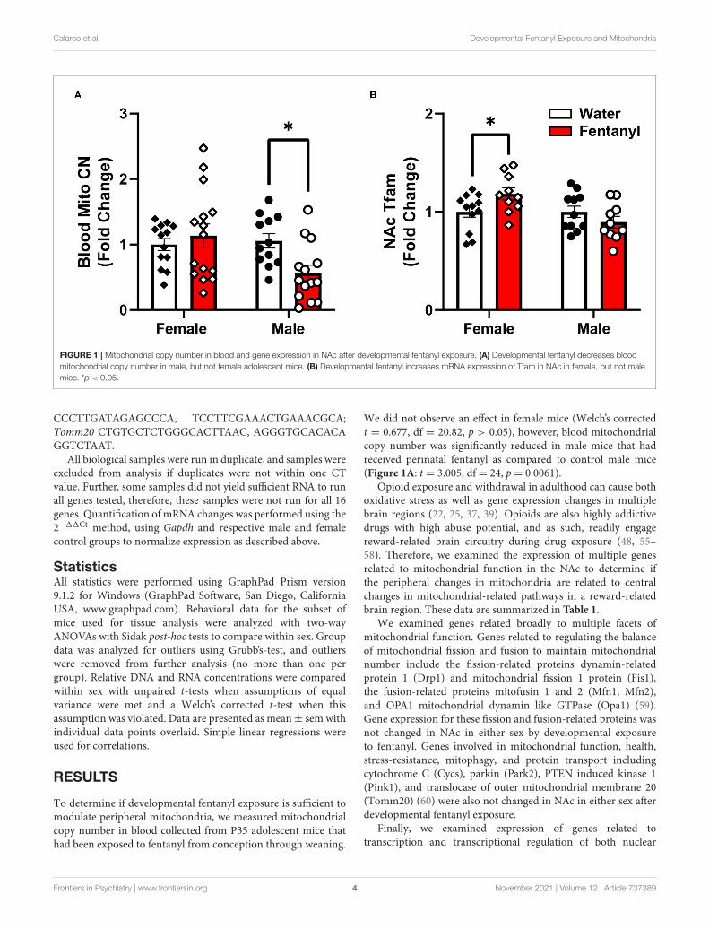
FIGURE 1 | Mitochondrial copy number in blood and gene expression in NAc after developmental fentanyl exposure. (A) Developmental fentanyl decreases blood
mitochondrial copy number in male, but not female adolescent mice. (B) Developmental fentanyl increases mRNA expression of Tfam in NAc in female, but not male
mice. *p < 0.05.
CCCTTGATAGAGCCCA, TCCTTCGAAACTGAAACGCA;Tomm20 CTGTGCTCTGGGCACTTAAC, AGGGTGCACACAGGTCTAAT.
All biological samples were run in duplicate, and samples wereexcluded from analysis if duplicates were not within one CTvalue. Further, some samples did not yield sufficient RNA to runall genes tested, therefore, these samples were not run for all 16genes. Quantification of mRNA changes was performed using the2−11Ct method, using Gapdh and respective male and femalecontrol groups to normalize expression as described above.
StatisticsAll statistics were performed using GraphPad Prism version9.1.2 for Windows (GraphPad Software, San Diego, CaliforniaUSA, www.graphpad.com). Behavioral data for the subset ofmice used for tissue analysis were analyzed with two-wayANOVAs with Sidak post-hoc tests to compare within sex. Groupdata was analyzed for outliers using Grubb’s-test, and outlierswere removed from further analysis (no more than one pergroup). Relative DNA and RNA concentrations were comparedwithin sex with unpaired t-tests when assumptions of equalvariance were met and a Welch’s corrected t-test when thisassumption was violated. Data are presented as mean± sem withindividual data points overlaid. Simple linear regressions wereused for correlations.
RESULTS
To determine if developmental fentanyl exposure is sufficient tomodulate peripheral mitochondria, we measured mitochondrialcopy number in blood collected from P35 adolescent mice thathad been exposed to fentanyl from conception through weaning.
We did not observe an effect in female mice (Welch’s correctedt = 0.677, df = 20.82, p > 0.05), however, blood mitochondrialcopy number was significantly reduced in male mice that hadreceived perinatal fentanyl as compared to control male mice(Figure 1A: t = 3.005, df= 24, p= 0.0061).
Opioid exposure and withdrawal in adulthood can cause bothoxidative stress as well as gene expression changes in multiplebrain regions (22, 25, 37, 39). Opioids are also highly addictivedrugs with high abuse potential, and as such, readily engagereward-related brain circuitry during drug exposure (48, 55–58). Therefore, we examined the expression of multiple genesrelated to mitochondrial function in the NAc to determine ifthe peripheral changes in mitochondria are related to centralchanges in mitochondrial-related pathways in a reward-relatedbrain region. These data are summarized in Table 1.
We examined genes related broadly to multiple facets ofmitochondrial function. Genes related to regulating the balanceof mitochondrial fission and fusion to maintain mitochondrialnumber include the fission-related proteins dynamin-relatedprotein 1 (Drp1) and mitochondrial fission 1 protein (Fis1),the fusion-related proteins mitofusin 1 and 2 (Mfn1, Mfn2),and OPA1 mitochondrial dynamin like GTPase (Opa1) (59).Gene expression for these fission and fusion-related proteins wasnot changed in NAc in either sex by developmental exposureto fentanyl. Genes involved in mitochondrial function, health,stress-resistance, mitophagy, and protein transport includingcytochrome C (Cycs), parkin (Park2), PTEN induced kinase 1(Pink1), and translocase of outer mitochondrial membrane 20(Tomm20) (60) were also not changed in NAc in either sex afterdevelopmental fentanyl exposure.
Finally, we examined expression of genes related totranscription and transcriptional regulation of both nuclear
Frontiers in Psychiatry | www.frontiersin.org 4 November 2021 | Volume 12 | Article 737389

Calarco et al. Developmental Fentanyl Exposure and Mitochondria
TABLE 1 | Relative gene expression of nuclear mitochondrial related genes in NAc.
Control Fentanyl
Gene Sex n Mean 95% CI n Mean 95% CI Analysis p-value Sig
Fission- and fusion-related genes
Drp1 Female 10 1.00 0.6707–1.329 8 0.8643 0.7428–0.9858 Welch’s t-test 0.3798 n.s.
Male 10 1.00 0.8393–1.161 8 0.8145 0.4416–1.187 t-test 0.2666 n.s.
Fis1 Female 12 1.00 0.820–1.180 11 1.067 0.8969–1.238 t-test 0.556 n.s.
Male 11 1.00 0.9138–1.086 11 0.8957 0.7357–1.056 t-test 0.2157 n.s.
Mfn1 Female 12 1.00 0.8323–1.168 12 1.032 0.8772–1.187 t-test 0.7603 n.s.
Male 11 1.00 0.9288–1.07 11 0.9784 0.8127–1.14 Welch’s t-test 0.7938 n.s.
Mfn2 Female 12 1.00 0.9157–1.084 12 1.021 0.9245–1.118 t-test 0.7197 n.s.
Male 12 1.00 0.9372–1.063 10 0.9929 0.8878–1.098 t-test 0.8943 n.s.
Opa1 Female 12 1.00 0.8864–1.114 9 1.022 0.9368–1.108 t-test 0.7443 n.s.
Male 12 1.00 0.9033–1.097 11 1.076 0.9393–1.213 t-test 0.3194 n.s.
Mitochondrial function, health, stress-resistance, mitophagy, and protein transport
Cycs Female 9 1.00 0.7999–1.200 11 0.9385 0.7798–1.097 t-test 0.5868 n.s.
Male 11 1.00 0.8917–1.108 9 1.735 0.7091–2.760 Welch’s t-test 0.1383 n.s.
Park2 Female 10 1.00 0.8322–1.168 12 1.061 0.9342–1.189 t-test 0.5148 n.s.
Male 11 1.00 0.9005–1.099 9 1.01 0.8131–1.207 t-test 0.916 n.s.
Pink1 Female 10 1.00 0.7716–1.228 12 0.7191 0.8210–1.287 t-test 0.7191 n.s.
Male 11 1.00 0.8355–1.164 9 0.8873 0.6134–1.161 t-test 0.4136 n.s.
Tomm20 Female 10 1.00 0.8274–1.173 12 1.159 1.012–1.307 t-test 0.1313 n.s.
Male 11 1.00 0.8941–1.106 9 0.925 0.6860–1.164 Welch’s t-test 0.524 n.s.
Nuclear transcription factors and transcriptional co-activators
Egr3 Female 12 1.00 0.7853–1.215 12 0.9545 0.8528–1.056 Welch’s t-test 0.6794 n.s.
Male 12 1.00 0.8669–1.133 11 1.125 0.8940–1.356 t-test 0.2994 n.s.
Nrf1 Female 11 1.00 0.8882–1.112 12 0.923 0.8101–1.036 t-test 0.2969 n.s.
Male 11 1.00 0.9055–1.095 11 0.9437 0.7722–1.115 t-test 0.5289 n.s.
Nrf2 Female 12 1.00 0.7022–1.298 12 0.9898 0.7989–1.181 t-test 0.9502 n.s.
Male 10 1.00 0.8929–1.107 9 1.234 0.4896–1.978 Welch’s t-test 0.4934 n.s.
Pgc1α Female 12 1.00 0.7647–1.235 10 0.9395 0.7330–1.146 t-test 0.6785 n.s.
Male 10 1.00 0.8878–1.112 8 0.8442 0.4007–1.288 Welch’s t-test 0.4451 n.s.
Mitochondrial transcriptase and transcription factors
Polγ Female 9 1.00 0.9216–1.078 10 1.059 0.8985–1.219 Welch’s t-test 0.4693 n.s.
Male 11 1.00 0.9416–1.058 7 0.99 0.5802–1.400 Welch’s t-test 0.9549 n.s.
Tfam Female 12 1.00 0.8794–1.121 10 1.183 1.043–1.322 t-test 0.0381 *
Male 11 1.00 0.8720–1.128 10 0.8953 0.7612–1.030 t-test 0.2207 n.s.
Tfb1m Female 10 1.00 0.88490–1.151 9 0.946 0.8401–1.052 t-test 0.5231 n.s.
Male 11 1.00 0.9189–1.081 6 1.142 0.6122–1.672 Welch’s t-test 0.5254 n.s.
Significant results in bold.
*p < 0.05, n.s. = not significant.
and mitochondrial genome genes, broad regulators of overallpatterns of gene expression (61, 62). Transcription factorsand transcriptional co-activators that regulate nuclear genesrelated to mitochondrial function including Early GrowthResponse 3 (Egr3), nuclear respiratory factor 1 (Nrf1), nuclearfactor, erythroid 2 like 2 (Nrf2), and peroxisome proliferator-activated receptor gamma coactivator 1-alpha (Pgc1α) werenot changed in NAc in either sex after developmental fentanylexposure. DNA Polymerase Subunit Gamma-1 (Polγ), amitochondrial DNA polymerase which conducts mitochondrialDNA replication was not altered by developmental fentanyl,however, the mitochondrial transcription factor transcription
factor A (Tfam) increased expression in female mice that hadbeen developmentally exposed to fentanyl (Figure 1B). Tfamexpression was not changed in male mice, nor were changesobserved in either sex of transcription factor B1, mitochondrial(Tfb1). Notably, the change in expression of Tfam is doubledissociated from the mitochondrial copy number finding inmale mice.
While many of the genes examined did not show statisticallysignificant differences, we noticed high degrees of variabilityin gene expression for many genes, therefore, we decided toexplore if this variability in gene expression was related eitherto peripheral mitochondrial copy number, performance in the
Frontiers in Psychiatry | www.frontiersin.org 5 November 2021 | Volume 12 | Article 737389

Calarco et al. Developmental Fentanyl Exposure and Mitochondria
FIGURE 2 | Blood mitochondrial copy number correlates with NAc gene expression in female mice. In female control mice blood mitochondrial copy number
correlates with (A) NAc expression of Drp1 (B) Mfn2, and (C) Nrf2. Blood copy number and NAc gene expression are depicted as fold change. *p < 0.05.
measured behavioral tests, or other factors that may relate tooverall metabolic function, specifically body weight at the timeof sacrifice. The behavioral data used here represents a subsetof the previously published animals (11) used for the molecularanalysis. In this subset of animals, male animals showed increasedanxiety-like behavior in the EPM, indicated by a reduced ratioof time spent in the open arms of the maze over the timespent in the closed arms, but there was no effect in females(mean ratio, SEM - male control: 0.4081, 0.07036; male fentanyl:0.1919, 0.02859; female control: 0.3661, 0.0441; female fentanyl:0.3318, 0.0401) [main effect of drug F(1,54) = 7.419, p = 0.0087;post-hoc male adjusted p = 0.0082, female adjusted p > 0.05].There was a main effect of sex in the splash test, with femalesspending more time grooming [F(1,54) = 10.48, p = 0.0021],however there was no effect of fentanyl or interaction (meanseconds, SEM - male control: 99.01, 5.497; male fentanyl: 81.83,11.30; female control: 117.2, 4.954; female fentanyl: 112.8, 8.543).Finally, while males weighed more than females [F(1,54) = 5.622,p= 0.0213], we did not observe an effect of fentanyl on weight atthis time point in this cohort (mean grams, SEM - male control:18.21, 0.6542; male fentanyl: 18.82, 0.4767; female control: 17.2,0.6829; female fentanyl: 17.11, 0.4271). We performed Pearsoncorrelations on our copy number, gene expression and behavioraldata and all correlations are described in Supplementary Table 1.Blood mitochondrial copy number showed a significant positivecorrelation with NAc Drp1, Mfn2, and Nrf1 in female controlmice (Figure 2) but shows no relationship with NAc geneexpression in female mice exposed to fentanyl or male mice ofeither condition. Specifically, there was no correlation betweenblood copy number with NAc Tfam expression in femalemice, which was significantly increased in fentanyl-exposedmice. While developmental fentanyl does not change bloodmitochondrial copy number in female adolescent mice, it doesseem to disrupt the correlations with NAc gene expressionseen in control mice, possibly indicating an uncoupling ofperipheral and central mitochondrial function potentially uniqueto female mice, or a more complex relationship between NAcgene expression and blood mitochondria copy number in thecontext of developmental drug exposure.
Conversely, the ratio of time spent in the open/closed armsof the elevated plus maze, an indicator of anxiety-like behavior,showed no correlation with gene expression in control animals,but is correlated with a number of genes in animals that had beenexposed to fentanyl (Figure 3). In female mice EPM open/closedratio negatively correlates with both Drp1 and Pgc1α expressionin NAc (Figure 3A). In male animals, EPM open/closed ratiopositively correlates with NAc expression of Fis1, Park2, andTomm20 (Figure 3B). Time spent grooming in the splash test didnot correlate with NAc gene expression for either sex under eitherdrug exposure condition. Body weight positively correlated withNAc Tfb1 expression in male control mice, and negatively withTfb1 in female mice exposed to fentanyl (Figure 3C). In malemice exposed to fentanyl body weight was negatively correlatedwith NAc expression of Pink1 (Figure 3D).
DISCUSSION
Opioid exposure is related to increased oxidative damageand mitochondrial damage in adulthood, and many negativeimpacts of chronic opioid use can be particularly long-lastingif the drug is encountered during development, leading toaltered behavior and neurological function (9, 20, 24, 63).Opioid use and opioid use disorders are also associated withan increase in psychiatric mood symptoms as well as moodand anxiety disorders (43, 44). The current study examinedhow developmental exposure to the synthetic opioid fentanylaltered blood mitochondrial copy number, mitochondrial geneexpression, and how these measures related to each otherand anxiety-like behaviors in adolescent male and femalemice. We showed that developmental exposure to fentanylreduces blood mitochondrial copy number in male mice andincreases NAc expression of Tfam mRNA in female mice.Additionally, mice exposed to fentanyl showed different patternsof correlation between blood mitochondrial copy number,anxiety-like behavior, weight, and NAc gene expression in asex-dependent manner.
Mitochondria are particularly impacted by oxidative stressboth as producers and scavengers of reactive oxygen species, and
Frontiers in Psychiatry | www.frontiersin.org 6 November 2021 | Volume 12 | Article 737389

Calarco et al. Developmental Fentanyl Exposure and Mitochondria
FIGURE 3 | NAc mitochondrial gene expression correlates with anxiety-like behavior and weight. (A) In female mice developmentally exposed to fentanyl, NAc
expression of Drp1 and Pgc1α correlate with behavior in the EPM, represented as the ratio of open/closed arm time. (B) In male mice developmentally exposed to
fentanyl, NAc expression of Fis1, Park2, and Tomm20 correlate with behavior in the EPM, represented as the ratio of open/closed arm time. (C) NAc expression of
Tfb1 positively correlates with weight at P35 in female mice developmentally exposed to fentanyl and negatively correlates with weight in male control mice. (D) In
male mice developmentally exposed to fentanyl, NAc expression of Pink1 negatively correlates with weight at P35. Gene expression is depicted as fold change. *p <
0.05; **p < 0.01.
in turn are a critical mediator in downstream cellular processingand homeostatic changes in response to such oxidative stress.Mitochondrial DNA is particularly susceptible to oxidativedamage compared to nuclear DNA, as it both lacks protective
histone proteins and mitochondria have less robust DNA-repair machinery than nuclei (64). The mitochondrial genomeis maintained in a highly dynamic equilibrium, existing inmultiple copies per cell, with 1–10 copies per mitochondrion
Frontiers in Psychiatry | www.frontiersin.org 7 November 2021 | Volume 12 | Article 737389

Calarco et al. Developmental Fentanyl Exposure and Mitochondria
and multiple mitochondria per cell, depending on cell type(35, 65). Because the mitochondrial genome codes for most ofthe enzymatic subunits needed for oxidative phosphorylation,mitochondrial copy number can be used as an indicator ofmitochondrial biogenesis (35, 66), and changes in mitochondrialcopy number may contribute to oxidative stress, inflammation,and mitochondrial dysfunction (67, 68).
Mitochondrial dysfunction has been linked to disorders fromdiabetes, to cancer, and more recently to stress, psychiatricillnesses and substance use disorders (28, 69–74). Mitochondriacopy number is increased in patients with bipolar disorder(75, 76), early childhood maltreatment or adversity (77), andincreased in the prefrontal cortex and hippocampus of rats thathad undergone cocaine self-administration, but is decreased inhuman heroin users and mice and rats exposed to chronic heroin(37). Our findings here mimic the decrease in copy numberseen in male rodents, despite the developmental exposure andabstinence at the time of tissue collection used here. In thehuman patients, copy number did partially recover 3 monthsafter initiation of heroin abstinence, although even after 6months copy number had not fully recover to control levels (37).The clinical population represented female patients, althoughwe did not observe changes in copy number in female mice.Further work will be needed to determine if opioids impactmitochondrial copy number comparably in men and women.Sex differences in substance use, substance use disorders, andsuccessful abstinence have been readily observed for multipleused drugs, including opioids (78–82). A unique feature of thecurrent study is the consistency of gestational fentanyl exposurein utero as our sample represents multiple litters consisting ofboth males and females. Fentanyl dose during the post-natalperiod was dependent on individual variance in pup milk orwater consumption until weaning.
Although a growing number of studies are examiningperipheral copy number, fewer studies are relating this measureto changes in other tissues, including the brain. In neurons,mitochondrial quality control and proper functioning impactsmany aspects of cellular function related to signaling and circuitfunction (31). Specifically, in addition to providing the highlevels of ATP necessary to maintain electro-chemical gradients,mitochondria buffer both intracellular calcium and reactiveoxygen species, influence apoptosis, and have been shownto be critical for dendritic spine formation (31–33). Further,mitochondria and mitochondrial related genes in the NAcspecifically have been shown to mediate behavioral respondingfor cocaine in a mouse model of substance use disorder (34,83, 84), indicating mitochondrial function in NAc as a specificnode for influencing the response to addictive drugs. Of the genesexamined in NAc here, only Tfam showed increased expression.Interestingly, Tfam, because it binds to the mitochondrial DNAas a transcription factor, also has been shown to protectmitochondrial DNA from damage due to oxidative stress (61, 85,86). Thus, it is possible there may be oxidative damage in NAccaused by developmental fentanyl exposure, and the increasedexpression of Tfam may be neuroprotective in female mice,consistent with their unchanged blood copy number. The sex-specific nature of this effect, and the lack of changes in other
genes of interest may indicate tight regulation of mitochondrialfunction in NAc through development and during adolescencedespite the drug exposure. Future studies should examine moredirect measures of mitochondria in NAc, such as copy number.Heroin can reduce mitochondrial copy number in hippocampusindicating drug exposure can influence copy number in brain(37), but copy number can vary independently with brainregion, and changes in one area may not predict changes inother connected brain regions (87). Importantly, differences inoxidative damage or gene expression may vary even within theNAc itself, as the NAc core, medial shell, and lateral shell allhave previously described variance in regulating reward-relatedbehaviors (88–92). The tissue used in this study included allsubregions of the NAc, and future work will be needed tofurther dissect any unique subregion responses to developmentalfentanyl. Mitochondrial morphology in NAc, regulated by manyof the genes examined here, is also responsive to both drugexposure (34) and trait-anxiety measures possibly establishedduring development (40). Fentanyl does impact mitochondrialmorphology in neuronal-like NG108–15 cells (39), but this hasnot been demonstrated in any neuronal type in vivo. Further,the impact of developmental fentanyl exposure on mitochondriamay be cell-type selective; beyond neurons, astrocyte functionis altered by opioid exposure (93–95) and it is possible thatchanges in these glial cells or the immune-related microgliamimic changes seen in peripheral immune cells.
Our data indicate that blood mitochondrial copy numberdoes vary systematically with NAc mitochondrial-related nucleargenes, specifically Drp1, Mfn1, and Nrf1, with higher copynumber corresponding to higher gene expression. Drp1 regulatesmitochondrial fission, promoting the formation of moremitochondria. In the brain, Drp1 is involved in new dendriticspine formation (33). While opioid exposure is linked to adecrease in dendritic spines, due to internalization of muopioid receptors (96, 97), the correlation described here is infemale mice specifically, which were resistant to both changes inblood copy and changes in EPM anxiety-like behavior, althoughfentanyl did disrupt the correlation with NAc gene expression.Mfn2 is usually considered a fusion-related protein (98), butit also plays critical role in mediating mitochondrial contactpoints with the endoplasmic reticulum independent of fusionand its expression in NAc has been linked to anxiety-likebehaviors (40). Nrf1, as a nuclear transcription factor, regulatesexpression of other mitochondrial related genes includingthose related to mitochondrial respiratory function as well asgenes involved in RNA metabolism, DNA damage repair, andubiquitin-mediated protein degradation (99). While the roleof mitochondrial related gene expression and function in NAcwith respect to drug exposure is still a relatively new areaof investigation, mitochondrial function within reward circuitshas already been linked repeatedly to anxiety-like behaviors(41). Specifically, expression of mitochondrial related genes,mitochondria complex I and II function, and mitochondrialrespiratory capacity impact both trait anxiety and expressionssocial dominance (40, 100, 101). Further, increases in ventraltegmental area dopamine input to NAc or D1-dopamine receptoragonism in NAc can increase both mitochondrial respiratory
Frontiers in Psychiatry | www.frontiersin.org 8 November 2021 | Volume 12 | Article 737389

Calarco et al. Developmental Fentanyl Exposure and Mitochondria
activity and facilitate social dominance expression in previouslyidentified higher-anxiety rats (102). Both NRF1 and NRF2regulate mitochondrial function in reward-related brain regions(103, 104), and global NRF2 knockdown is sufficient to decreaseopen arm time in rats in an EPM (104). NRF1 knockdowndid not impact EPM behavior, but did alter expression of othermitochondrial-related proteins in the amygdala, hippocampusand prefrontal cortex (103). Since affect, mood, and anxiety areall impacted by drug use (57, 105–107), including opioid use(43, 44) and withdrawal (108), altered mitochondrial functionmay be a common underlying mechanism for trait anxiety oraltered anxiety-like behavior after exposure to opioids and otherdrugs. Future work on both the genes of interest identified hereand other mitochondrial processes is needed to fully understandtheir role in regulating behavior after drug exposure.
While these relationships with NAc gene expression existin control animals, developmental fentanyl presented otherrelationships with gene expression, which negatively correlatedwith EPM behavior in female mice and positively in malemice. Both Drp1 and Pgc1α, the genes correlated in femalemice, have both been shown in NAc to mediate enhancedbehavioral responding to cocaine (34, 83), and here higherexpression is related to less time spent in the open armsof the EPM. Conversely, Fis1, Park2, and Tomm20 all havefunctions in mitochondrial degradation pathways (109–112),potentially indicating higher degrees of mitochondrial damageor turn over in male mice after fentanyl which also havereduced blood copy number and significantly reduced openarm time in the EPM (11). Fentanyl also produced negativecorrelations between body weight and NAc expression of Pink1in male mice and with Tfb1 in female mice. Pink1is protectiveagainst mitochondrial dysfunction (113), and as a mitochondrialtranscription factor Tfb1 might have the same protective effectsfor mitochondrial DNA as Tfam. Future studies will be neededto further understand the relationship between weight, NAc geneexpression, and opioid exposure. While some genes did correlatewith behavior, Tfam did not, indicating that developmentalfentanyl causes dissociable changes in the periphery and inNAc, and each of these tissues independently relate to behavior.Further, the sex differences seen here in both EPM behavior andin gene expression correlations are consistent with previous workshowing important sex differences in NAc gene expression inthe context of resilience to stress (114) and sex differences inthe NAc proteome after exposure to nicotine, a commonly useddrug (115).
It is important to note that in this study it is impossible todistinguish if the changes and relationships described here aredue to the fentanyl exposure itself or due to the experience ofgoing through withdrawal from the fentanyl after weaning. Thisregimen of fentanyl exposure is sufficient to induce spontaneouswithdrawal signs (12), and it is unclear if the changes in behaviorduring adolescence are a prolonged result of withdrawal-relatedplasticity (58), or indicative of shifted baselines in stress reactivitycaused by developmental insult (6, 9, 10). It is also possible,that the developmental timing of withdrawal (at weaning ratherthan at birth) may be significant. As mitochondria in thebrain and body have been previously shown to modulate
responses to both acute psychological stress (27), and mediatesome of the developmental impacts on the brain of early-lifestress (30), it is possible the effects seen here represent themitochondrial response to opioid withdrawal (116–118). Futurestudies involving both continuous access to fentanyl and longerperiods of abstinence into adulthood will be necessary to resolvethis distinction and determine the persistence of these effects.
Taken together, these data indicate developmental fentanylexposure has similar effects on offspring mitochondrial copynumber as adult opioid use potentially including oxidativedamage that disturbs mitochondrial function. Changes in theNAc are only one component of the reward circuits that maybe impacted by this developmental opioid exposure and futurework should examine the impacts on other brain regions,which may show stronger more significant relationships withblood mitochondrial copy number or behavior than thosedemonstrated here. The relationships with brain gene expressionand behavior indicate coordinated responding throughout thebody to the developmental insult of fentanyl exposure and futurestudies should further explore this relationship to better predicthealth and supplement treatment for infants with prenatalopioid exposure.
DATA AVAILABILITY STATEMENT
The raw data supporting the conclusions of this article will bemade available by the authors, without undue reservation.
ETHICS STATEMENT
The animal study was reviewed and approved by InstitutionalAnimal Care and Use Committee at the University of MarylandSchool of Medicine.
AUTHOR CONTRIBUTIONS
ML, RC, CC, and MF were responsible for study concept anddesign. JA and MF designed the perinatal exposure paradigm.MF, JA, and MT managed breeding, fentanyl dosing, andbehavioral testing. MF, JA, SVT, and CC collected tissues andperformed experiments. CC performed data analysis. CC andML drafted the manuscript. MF provided critical revision of themanuscript for content. All authors critically reviewed contentand approved the final version for publication.
FUNDING
This work was funded by NIH R01DA038613,NIH R01MH106500, T32DK098107, F32DA052966,and K99DA050575.
SUPPLEMENTARY MATERIAL
The Supplementary Material for this article can be foundonline at: https://www.frontiersin.org/articles/10.3389/fpsyt.2021.737389/full#supplementary-material
Frontiers in Psychiatry | www.frontiersin.org 9 November 2021 | Volume 12 | Article 737389
Calarco et al. Developmental Fentanyl Exposure and Mitochondria
REFERENCES
1. Florence CS, Zhou C, Luo F, Xu L. The economic burden of prescriptionopioid overdose, abuse, and dependence in the United States 2013.Med Care.
(2016) 54:901–6. doi: 10.1097/MLR.00000000000006252. Ryan SA. Calculating the real costs of the opioid crisis. Pediatrics. (2018)
141:18–21. doi: 10.1542/peds.2017-41293. Honein MA, Boyle C, Redfield RR. Public health surveillance of prenatal
opioid exposure in mothers and infants. Pediatrics. (2019) 143:e20183801.doi: 10.1542/peds.2018-3801
4. Haight SC, Ko JY, Tong VT, BohmMK, CallaghanWM. Opioid use disorderdocumented at delivery hospitalization - United States, 1999-2014. Morb
Mortal Wkly Rep. (2018) 67:845–9. doi: 10.15585/mmwr.mm6731a15. Whiteman VE, Salemi JL, Mogos MF, Cain MA, Aliyu MH, Salihu HM.
Maternal opioid drug use during pregnancy and its impact on perinatalmorbidity, mortality, and the costs of medical care in the United States. JPregnancy. (2014). 2014:906723. doi: 10.1155/2014/906723
6. Mactier H, Hamilton R. Prenatal opioid exposure - increasingevidence of harm. Early Hum Dev. (2020) 150:105188.doi: 10.1016/j.earlhumdev.2020.105188
7. Šlamberová R. Drugs in pregnancy: the effects on mother and her progeny.Physiol Res. (2012) 61(Suppl. 1):932357. doi: 10.33549/physiolres.932357
8. Ross EJ, Graham DL, Money KM, Stanwood GD. Developmentalconsequences of fetal exposure to drugs: what we know and whatwe still must learn. Neuropsychopharmacology. (2015) 40:61–87.doi: 10.1038/npp.2014.147
9. Abu Y, Roy S. Prenatal opioid exposure and vulnerability to futuresubstance use disorders in offspring. Exp Neurol. (2021) 339:113621.doi: 10.1016/j.expneurol.2021.113621
10. Franks AL, Berry KJ, DeFranco DB. Prenatal drug exposure andneurodevelopmental programming of glucocorticoid signalling. J
Neuroendocrinol. (2020) 32:1–13. doi: 10.1111/jne.1278611. Alipio JB, Brockett AT, Fox ME, Tennyson SS, DeBettencourt CA, El-
Metwally D, et al. Enduring consequences of perinatal fentanyl exposure inmice. Addict Biol. (2021) 26:e12895. doi: 10.1111/adb.12895
12. Alipio JB, Haga C, Fox ME, Arakawa K, Balaji R, Cramer N, et al.Perinatal fentanyl exposure leads to long-lasting impairments insomatosensory circuit function and behavior. J Neurosci. (2021) 41:3400–17.doi: 10.1523/JNEUROSCI.2470-20.2021
13. Koob GF. Neurobiological substrates for the dark side ofcompulsivity in addiction. Neuropharmacology. (2009) 56:18–31.doi: 10.1016/j.neuropharm.2008.07.043
14. Volkow ND, Koob GF, McLellan AT. Neurobiologic advances from thebrain disease model of addiction. N Engl J Med. (2016) 374:363–71.doi: 10.1056/NEJMra1511480
15. Badiani A, Blein D, Epstein D, Calu D, Shaham Y. Opiate versuspsychostimulant addiction: the differences do matter. Nat Rev Neurosci.
(2008) 12:685–700. doi: 10.1038/nrn310416. Shaham Y, Erb S, Stewart J. Stress-induced relapse to heroin and
cocaine seeking in rats: a review. Brain Res Rev. (2000) 33:13–33.doi: 10.1016/S0165-0173(00)00024-2
17. Koob GF, Volkow ND. Neurobiology of addiction: a neurocircuitryanalysis. Lancet Psychiatry. (2016) 3:760–73. doi: 10.1016/S2215-0366(16)00104-8
18. Crombag HS, Bossert JM, Koya E, Shaham Y. Context-induced relapse todrug seeking: a review. Philos Trans R Soc B Biol Sci. (2008) 363:3233–43.doi: 10.1098/rstb.2008.0090
19. Caspani G, Sebok V, Sultana N, Swann JR, Bailey A. Metabolic phenotypingof opioid and psychostimulant addiction: a novel approach for biomarkerdiscovery and biochemical understanding of the disorder. Br J Pharmacol.(2021) 1–29. doi: 10.1111/bph.15475
20. ZahmatkeshM, KadkhodaeeM, Salarian A, Seifi B, Adeli S. Impact of opioidson oxidative status and related signaling pathways: an integrated view. JOpioid Manag. (2017) 13:241–51. doi: 10.5055/jom.2017.0392
21. Cunha-Oliveira T, Rego A, Oliveira C. Oxidative stress and drugsof abuse: an update. Mini Rev Org Chem. (2013) 10:321–34.doi: 10.2174/1570193X113106660026
22. Pavlek LR, Dillard J, Rogers LK. The role of oxidative stress intoxicities due to drugs of abuse. Curr Opin Toxicol. (2020) 20–21:29–35.doi: 10.1016/j.cotox.2020.04.003
23. Katz N, Mazer NA. The impact of opioids on the endocrine system. Clin J
Pain. (2009) 25:170–5. doi: 10.1097/AJP.0b013e3181850df624. Salarian A, KadkhodaeeM, ZahmatkeshM, Seifi B, Bakhshi E, Akhondzadeh
S, et al. Opioid use disorder induces oxidative stress and inflammation: theattenuating effect of methadone maintenance treatment. Iran J Psychiatry.
(2018) 13:46–54.25. Molavi N, Ghaderi A, Banafshe HR. Short communication: examining
metabolic profiles in opioid-dependent patient. Int J Med Toxicol Forensic
Med. (2020) 10:1–6. doi: 10.32598/ijmtfm.v10i3.2868126. Cunha-Oliveira T, Rego AC, Oliveira CR. Cellular and molecular
mechanisms involved in the neurotoxicity of opioid andpsychostimulant drugs. Brain Res Rev. (2008) 58:192–208.doi: 10.1016/j.brainresrev.2008.03.002
27. Picard M, McManus MJ, Gray JD, Nasca C, Moffat C, KopinskiPK, et al. Mitochondrial functions modulate neuroendocrine, metabolic,inflammatory, and transcriptional responses to acute psychological stress.Proc Natl Acad Sci. (2015) 112:E6614–23. doi: 10.1073/pnas.1515733112
28. Picard M, McEwen BS. Psychological stress and mitochondria:a systematic review. Psychosom Med. (2018) 80:141–53.doi: 10.1097/PSY.0000000000000545
29. Picard M, McEwen BS, Epel ES, Sandi C. An energetic view ofstress: focus on mitochondria. Front Neuroendocrinol. (2018) 49:72–85.doi: 10.1016/j.yfrne.2018.01.001
30. Hoffmann A, Spengler D. The mitochondrion as potential interface inearly-life stress brain programming. Front Behav Neurosci. (2018) 12:306.doi: 10.3389/fnbeh.2018.00306
31. Rugarli EI, Langer T.Mitochondrial quality control: amatter of life and deathfor neurons. EMBO J. (2012) 31:1336–49. doi: 10.1038/emboj.2012.38
32. Li Z, Okamoto K, Hayashi Y, Sheng M. The importance of dendriticmitochondria in the morphogenesis and plasticity of spines and synapses.Cell. (2004) 119:873–87. doi: 10.1016/j.cell.2004.11.003
33. Divakaruni SS, Van Dyke AM, Chandra R, LeGates TA, Contreras M,Dharmasri PA, et al. Long-term potentiation requires a rapid burst ofdendritic mitochondrial fission during induction. Neuron. (2018) 100:1–16.doi: 10.1016/j.neuron.2018.09.025
34. Chandra R, Engeln M, Schiefer C, Patton MH, Martin JA, Werner CT, et al.Drp1 mitochondrial fission in D1 neurons mediates behavioral and cellularplasticity during early cocaine abstinence. Neuron. (2017) 96:1327–41.e6.doi: 10.1016/j.neuron.2017.11.037
35. Clay Montier LL, Deng JJ, Bai Y. Number matters: control of mammalianmitochondrial DNA copy number. J Genet Genomics. (2009) 36:125–31.doi: 10.1016/S1673-8527(08)60099-5
36. Yang SY, Castellani CA, Longchamps RJ, Pillalamarri VK, O’Rourke B,Guallar E, et al. Blood-derived mitochondrial DNA copy number isassociated with gene expression across multiple tissues and is predictivefor incident neurodegenerative disease. Genome Res. (2021) 31:349–58.doi: 10.1101/gr.269381.120
37. Feng YM, Jia YF, Su LY, Wang D, Lv L, Xu L, et al. Decreased mitochondrialDNA copy number in the hippocampus and peripheral blood during opiateaddiction is mediated by autophagy and can be salvaged by melatonin.Autophagy. (2013) 9:1395–406. doi: 10.4161/auto.25468
38. Nylander E, Grönbladh A, Zelleroth S, Diwakarla S, Nyberg F, Hallberg M.Growth hormone is protective against acute methadone-induced toxicity bymodulating the NMDA receptor complex. Neuroscience. (2016) 339:538–47.doi: 10.1016/j.neuroscience.2016.10.019
39. Nylander E, Zelleroth S, Nyberg F, Grönbladh A, Hallberg M. Theeffects of morphine, methadone, and fentanyl on mitochondria:a live cell imaging study. Brain Res Bull. (2021) 171:126–34.doi: 10.1016/j.brainresbull.2021.03.009
40. Gebara E, Zanoletti O, Ghosal S, Grosse J, Schneider BL, KnottG, et al. Mitofusin-2 in the nucleus accumbens regulates anxietyand depression-like behaviors through mitochondrial and neuronalactions. Biol Psychiatry. (2021) 89:1033–44. doi: 10.1016/j.biopsych.2020.12.003
Frontiers in Psychiatry | www.frontiersin.org 10 November 2021 | Volume 12 | Article 737389

Calarco et al. Developmental Fentanyl Exposure and Mitochondria
41. Filiou MD, Sandi C. Anxiety and brain mitochondria: a bidirectionalcrosstalk. Trends Neurosci. (2019) 42:573–88. doi: 10.1016/j.tins.2019.07.002
42. Hollis F, van der Kooij MA, Zanoletti O, Lozano L, Cantó C, Sandi C.Mitochondrial function in the brain links anxiety with social subordination.Proc Natl Acad Sci. (2015) 112:15486–91. doi: 10.1073/pnas.1512653112
43. Rosoff DB, Smith GD, Lohoff FW. Prescription opioid use and risk for majordepressive disorder and anxiety and stress-related disorders: a multivariablemendelian randomization analysis. JAMA Psychiatry. (2021) 78:151–60.doi: 10.1001/jamapsychiatry.2020.3554
44. Gros DF, Milanak ME, Brady KT, Back SE. Frequency and severityof comorbid mood and anxiety disorders in prescription opioiddependence. Am J Addict. (2013) 22:261–5. doi: 10.1111/j.1521-0391.2012.12008.x
45. Martins SS, Fenton MC, Keyes KM, Blanco C, Zhu H, Storr CL. Mood andanxiety disorders and their association with non-medical prescription opioiduse and prescription opioid-use disorder: longitudinal evidence from theNational Epidemiologic Study on Alcohol and Related Conditions. PsycholMed. (2012) 42:1261–72. doi: 10.1017/S0033291711002145
46. Russo SJ, Dietz DM, Dumitriu D, Malenka RC, Nestler EJ.The addicted synapse: mechanisms of synaptic and structuralplasticity in nucleus accumbens. Trends Neurosci. (2011) 33:267–76.doi: 10.1016/j.tins.2010.02.002
47. Berridge KC, Kringelbach ML. Pleasure systems in the brain. Neuron. (2015)86:646–64. doi: 10.1016/j.neuron.2015.02.018
48. Volkow ND, Morales M. The brain on drugs: from reward to addiction. Cell.(2015) 162:712–25. doi: 10.1016/j.cell.2015.07.046
49. Fox ME, Lobo MK. The molecular and cellular mechanisms of depression:a focus on reward circuitry. Mol Psychiatry. (2019) 24:1798–815.doi: 10.1038/s41380-019-0415-3
50. Francis TC, Lobo MK. Emerging role for nucleus accumbens mediumspiny neuron subtypes in depression. Biol Psychiatry. (2017) 81:645–53.doi: 10.1016/j.biopsych.2016.09.007
51. Clancy B, Darlington RB, Finlay BL. Translating developmentaltime across mammalian species. Neuroscience. (2001) 105:7–17.doi: 10.1016/S0306-4522(01)00171-3
52. Chen VS, Morrison JP, Southwell MF, Foley JF, Bolon B. Elmore SA.Histology atlas of the developing prenatal and postnatal mouse centralnervous system, with emphasis on prenatal days E75 to E185. Toxicol Pathol.(2017) 45:705–44. doi: 10.1177/0192623317728134
53. Pellow S, Chopin P, File SE, Briley M. Validation of open: closed arm entriesin an elevated plus-maze as a measure of anxiety in the rat. J Neurosci
Methods. (1985) 14:149–67. doi: 10.1016/0165-0270(85)90031-754. Planchez B, Surget A, Belzung C. Animal models of major depression:
drawbacks and challenges. J Neural Transm. (2019) 126:1383–408.doi: 10.1007/s00702-019-02084-y
55. Luscher C, Malenka RC. Drug-evoked synaptic plasticity in addiction:from molecular changes to circuit remodeling. Neuron. (2011) 69:650–63.doi: 10.1016/j.neuron.2011.01.017
56. Nestler EJ, Lüscher C. The molecular basis of drug addiction: linkingepigenetic to synaptic and circuit mechanisms. Neuron. (2019) 102:48–59.doi: 10.1016/j.neuron.2019.01.016
57. Kenny PJ, Hoyer D, Koob GF. Animal models of addiction andneuropsychiatric disorders and their role in drug discovery: honoringthe legacy of Athina Markou. Biol Psychiatry. (2018) 83:940–6.doi: 10.1016/j.biopsych.2018.02.009
58. Thompson BL, Oscar-Berman M, Kaplan GB. Opioid-inducedstructural and functional plasticity of medium-spiny neurons inthe nucleus accumbens. Neurosci Biobehav Rev. (2021) 120:417–30.doi: 10.1016/j.neubiorev.2020.10.015
59. Yapa NMB, Lisnyak V, Reljic B, Ryan MT. Mitochondrial dynamics in healthand disease. FEBS Lett. (2021) 595:1184–204. doi: 10.1002/1873-3468.14077
60. Tilokani L, Nagashima S, Paupe V, Prudent J. Mitochondrial dynamics:overview of molecular mechanisms. Essays Biochem. (2018) 62:341–60.doi: 10.1042/EBC20170104
61. Kang D, Hamasaki N. Mitochondrial transcription factor A in themaintenance of mitochondrial DNA: overview of its multiple roles. Ann N
Y Acad Sci. (2005) 1042:101–8. doi: 10.1196/annals.1338.010
62. Ventura-Clapier R, Garnier A, Veksler V. Transcriptional control ofmitochondrial biogenesis: the central role of PGC-1α. Cardiovasc Res. (2008)79:208–17. doi: 10.1093/cvr/cvn098
63. Lee SJ, Bora S, Austin NC, Westerman A, Henderson JMT.Neurodevelopmental outcomes of children born to opioid-dependentmothers: a systematic review and meta-analysis. Acad Pediatr. (2020)20:308–18. doi: 10.1016/j.acap.2019.11.005
64. Yakes FM, VanHouten B.Mitochondrial DNA damage is more extensive andpersists longer than nuclear DNA damage in human cells following oxidativestress. Proc Natl Acad Sci USA. (1997) 94:514–9. doi: 10.1073/pnas.94.2.514
65. Robin ED, Wong R. Mitochondrial DNA molecules and virtual number ofmitochondria per cell in mammalian cells. J Cell Physiol. (1988) 136:507–13.doi: 10.1002/jcp.1041360316
66. Phillips NR, Sprouse ML, Roby RK. Simultaneous quantification ofmitochondrial DNA copy number and deletion ratio: a multiplex real-timePCR assay. Sci R. (2014) 4:3887. doi: 10.1038/srep03887
67. Malik AN, Czajka A. Is mitochondrial DNA content a potentialbiomarker of mitochondrial dysfunction?Mitochondrion. (2013) 13:481–92.doi: 10.1016/j.mito.2012.10.011
68. Zorzano A, Liesa M, Palaci M. Mitochondrial dynamics inmammalian health and disease. Physiol Rev. (2009) 89:799–845.doi: 10.1152/physrev.00030.2008
69. Ridout KK, Khan M, Ridout SJ. Adverse childhood experiences run deep:toxic early life stress, telomeres, and mitochondrial DNA copy number,the biological markers of cumulative stress. BioEssays. (2018) 40:1–10.doi: 10.1002/bies.201800077
70. Afrifa J, Zhao T, Yu J. Circulating mitochondria DNA, a non-invasivecancer diagnostic biomarker candidate. Mitochondrion. (2019) 47:238–43.doi: 10.1016/j.mito.2018.12.003
71. Wang X, Sundquist K, Rastkhani H, Palmér K, Memon AA, SundquistJ. Association of mitochondrial DNA in peripheral blood withdepression, anxiety and stress- and adjustment disorders in primaryhealth care patients. Eur Neuropsychopharmacol. (2017) 27:751–8.doi: 10.1016/j.euroneuro.2017.06.001
72. Sadakierska-Chudy A, Kotarska A, Frankowska M, Jastrzebska J, WydraK, Miszkiel J, et al. The alterations in mitochondrial DNA copynumber and nuclear-encoded mitochondrial genes in rat brain structuresafter cocaine self-administration. Mol Neurobiol. (2017) 54:7460–70.doi: 10.1007/s12035-016-0153-3
73. Silzer T, Barber R, Sun J, Pathak G, Johnson L, O’Bryant S, et al. Circulatingmitochondrial DNA: New indices of type 2 diabetes-related cognitiveimpairment in Mexican Americans. PLoS ONE. (2019) 14:e0213527.doi: 10.1371/journal.pone.0213527
74. Lee J-Y, Lee D-C, Im J-A, Lee J-W. Mitochondrial dna copy numberin peripheral blood is independently associated with visceral fataccumulation in healthy young adults. Int J Endocrinol. (2014) 2014:1–7.doi: 10.1155/2014/586017
75. Wang D, Li Z, Liu W, Zhou J, Ma X, Tang J, et al. Differential mitochondrialDNA copy number in three mood states of bipolar disorder. BMC Psychiatry.
(2018) 18:1–8. doi: 10.1186/s12888-018-1717-876. Yamaki N, Otsuka I, Numata S, Yanagi M, Mouri K, Okazaki S, et al.
Mitochondrial DNA copy number of peripheral blood in bipolar disorder:the present study and a meta-analysis. Psychiatry Res. (2018) 269:115–7.doi: 10.1016/j.psychres.2018.08.014
77. Tyrka AR, Parade SH, Price LH, Kao HT, Porton B, Philip NS, et al.Alterations of mitochondrial DNA copy number and telomere lengthwith early adversity and psychopathology. Biol Psychiatry. (2016) 79:78–86.doi: 10.1016/j.biopsych.2014.12.025
78. Bobzean SAM, DeNobrega AK, Perrotti LI. Sex differences in theneurobiology of drug addiction. Exp Neurol. (2014) 259:64–74.doi: 10.1016/j.expneurol.2014.01.022
79. Becker JB, Koob GF. Sex differences in animal models: focus on addiction.Pharmacol Rev. (2016) 68:242–63. doi: 10.1124/pr.115.011163
80. Fattore L, Altea S, Fratta W. Sex differences in drug addiction: areview of animal and human studies. Women’s Heal. (2008) 4:51–65.doi: 10.2217/17455057.4.1.51
Frontiers in Psychiatry | www.frontiersin.org 11 November 2021 | Volume 12 | Article 737389

Calarco et al. Developmental Fentanyl Exposure and Mitochondria
81. Lee CWS, Ho IK. Sex differences in opioid analgesia and addiction:interactions among opioid receptors and estrogen receptors. Mol Pain.
(2013) 9:1–10. doi: 10.1186/1744-8069-9-4582. Becker JB, Chartoff E. Sex differences in neural mechanisms mediating
reward and addiction. Neuropsychopharmacology. (2019) 44:166–83.doi: 10.1038/s41386-018-0125-6
83. Chandra R, Engeln M, Francis TC, Konkalmatt P, Patel D, Lobo MK, et al.Role for peroxisome proliferator-activated receptor gamma coactivator-1αin nucleus accumbens neuron subtypes in cocaine action. Biol Psychiatry.(2017) 81:564–72. doi: 10.1016/j.biopsych.2016.10.024
84. Cole S, Chandra R, Harris M, Patel I, Wang T, KimH, et al. Cocaine-inducedneuron subtype mitochondrial dynamics through Egr3 transcriptionalregulation. Mol Brain. (2021) 14:101. doi: 10.1186/s13041-021-00800-y
85. Xu S, Zhong M, Zhang L, Wang Y, Zhou Z, Hao Y, et al.Overexpression of Tfam protects mitochondria against β-amyloid-induced oxidative damage in SH-SY5Y cells. FEBS J. (2009) 276:3800–9.doi: 10.1111/j.1742-4658.2009.07094.x
86. Feng Q, Shao M, Han J, Tang T, Zhang Y, Liu F, et al. a potentialoxidative stress biomarker used for monitoring environment pollutantsin Musca domestica. Int J Biol Macromol. (2020) 155:524–34.doi: 10.1016/j.ijbiomac.2020.03.208
87. Fuke S, Kubota-Sakashita M, Kasahara T, Shigeyoshi Y, KatoT. Regional variation in mitochondrial DNA copy number inmouse brain. Biochim Biophys Acta - Bioenerg. (2011) 1807:270–4.doi: 10.1016/j.bbabio.2010.11.016
88. Laurent V, Leung B, Maidment N, Balleine BW. µ- and δ-Opioid-relatedprocesses in the accumbens core and shell differentially mediate the influenceof reward-guided and stimulus-guided decisions on choice. J Neurosci.
(2012) 32:1875–83. doi: 10.1523/JNEUROSCI.4688-11.201289. Peciña S, Berridge KC. Dopamine or opioid stimulation of
nucleus accumbens similarly amplify cue-triggered “wanting” forreward: entire core and medial shell mapped as substrates for PITenhancement. Eur J Neurosci. (2013) 37:1529–40. doi: 10.1111/ejn.12174
90. Di Ciano P, Robbins TW, Everitt BJ. Differential effects of nucleus accumbenscore, shell, or dorsal striatal inactivations on the persistence, reacquisition,or reinstatement of responding for a drug-paired conditioned reinforcer.Neuropsychopharmacology. (2008) 33:1413–25. doi: 10.1038/sj.npp.1301522
91. Di Chiara G. Nucleus accumbens shell and core dopamine: differentialrole in behavior and addiction. Behav Brain Res. (2002) 137:75–114.doi: 10.1016/S0166-4328(02)00286-3
92. Castro DC, Bruchas MR. A motivational and neuropeptidergic hub:anatomical and functional diversity within the nucleus accumbensshell. Neuron. (2019) 102:529–52. doi: 10.1016/j.neuron.2019.03.003
93. Doke M, Pendyala G, Samikkannu T. Psychostimulants and opioidsdifferentially influence the epigenetic modification of histoneacetyltransferase and histone deacetylase in astrocytes. PLoS ONE. (2021)16:e0252895. doi: 10.1371/journal.pone.0252895
94. Lacagnina MJ, Rivera PD, Bilbo SD. Glial and neuroimmune mechanisms ascritical modulators of drug use and abuse.Neuropsychopharmacology. (2017)42:156–77. doi: 10.1038/npp.2016.121
95. Kruyer A, Chioma VC, Kalivas PW. The opioid-addictedtetrapartite synapse. Biol Psychiatry. (2020) 87:34–43.doi: 10.1016/j.biopsych.2019.05.025
96. Miller EC, Zhang L, Dummer BW, Cariveau DR, Loh H, Law PY,et al. Differential modulation of drug-induced structural and functionalplasticity of dendritic spines. Mol Pharmacol. (2012) 82:333–43.doi: 10.1124/mol.112.078162
97. Liao D, Gringoriants OO, Wang W, Wiens K, Loh HH, Law PY. Distincteffects of individual opioids on the morphology of spines depend upon theinternalization of mu opioid receptors.Mol Cell Neurosci. (2007) 35:456–69.doi: 10.1016/j.mcn.2007.04.007
98. AdebayoM, Singh S, Singh AP, Dasgupta S.Mitochondrial fusion and fission:the fine-tune balance for cellular homeostasis. FASEB J. (2021) 35:1–13.doi: 10.1096/fj.202100067R
99. Satoh JI, Kawana N, Yamamoto Y. Pathway analysis of ChIP-seq-basedNRF1 target genes suggests a logical hypothesis of their involvement inthe pathogenesis of neurodegenerative diseases. Gene Regul Syst Bio. (2013)2013:139–52. doi: 10.4137/GRSB.S13204
100. Beyer F, García-García I, Heinrich M, Schroeter ML, Sacher J, Luck T,et al. Neuroanatomical correlates of food addiction symptoms and bodymass index in the general population. Hum Brain Mapp. (2019) 40:2747–58.doi: 10.1002/hbm.24557
101. Alonso-Caraballo Y, Jorgensen ET, Brown T, Ferrario CR. Functionaland structural plasticity contributing to obesity: roles for sex, diet,and individual susceptibility. Curr Opin Behav Sci. (2018) 23:160–70.doi: 10.1016/j.cobeha.2018.06.014
102. Van Der Kooij MA, Hollis F, Lozano L, Zalachoras I, Abad S, ZanolettiO, et al. Diazepam actions in the VTA enhance social dominance andmitochondrial function in the nucleus accumbens by activation of dopamineD1 receptors.Mol Psychiatry. (2018) 23:569–78. doi: 10.1038/mp.2017.135
103. Khalifeh S, Oryan S, Khodagholi F, Digaleh H, Shaerzadeh F, MaghsoudiN, et al. Complexity of compensatory effects in Nrf1 knockdown:linking undeveloped anxiety-like behavior to prevented mitochondrialdysfunction and oxidative stress. Cell Mol Neurobiol. (2016) 36:553–63.doi: 10.1007/s10571-015-0236-0
104. Khalifeh S, Oryan S, Digaleh H, Shaerzadeh F, Khodagholi F, MaghsoudiN, et al. Involvement of Nrf2 in development of anxiety-like behavior bylinking Bcl2 to oxidative phosphorylation: estimation in rat hippocampus,amygdala, and prefrontal cortex. J Mol Neurosci. (2015) 55:492–9.doi: 10.1007/s12031-014-0370-z
105. Haass-Koffler CL, Bartlett SE. Stress and addiction: contribution of thecorticotropin releasing factor (CRF) system in neuroplasticity. Front Mol
Neurosci. (2012) 5:e00091. doi: 10.3389/fnmol.2012.00091106. Lüthi A, Lüscher C. Pathological circuit function underlying addiction and
anxiety disorders. Nat Neurosci. (2014) 17:1635–43. doi: 10.1038/nn.3849107. Kutlu MG, Parikh V, Gould TJ. Nicotine addiction and psychiatric disorders.
De Biasi M, editor. Nicotine Use in Mental Illness and Neurological Disorders.Amsterdam: Elsevier Inc. (2015). p. 171–208.
108. Koob GF. Neurobiology of opioid addiction: opponent process,hyperkatifeia, and negative reinforcement. Biol Psychiatry. (2020) 87:44–53.doi: 10.1016/j.biopsych.2019.05.023
109. Sprenger HG, Langer T. The good and the bad of mitochondrial breakups.Trends Cell Biol. (2019) 29:888–900. doi: 10.1016/j.tcb.2019.08.003
110. Yu W, Sun Y, Guo S, Lu B. The PINK1/Parkin pathway regulatesmitochondrial dynamics and function in mammalian hippocampaland dopaminergic neurons. Hum Mol Genet. (2011) 20:3227–40.doi: 10.1093/hmg/ddr235
111. Wai T, Langer T. Mitochondrial dynamics and metabolic regulation. TrendsEndocrinol Metab. (2016) 27:105–17. doi: 10.1016/j.tem.2015.12.001
112. Wang L, Qi H, Tang Y, Shen H. Post-translational modifications of keymachinery in the control of mitophagy. Trends Biochem Sci. (2020) 45:85–75.doi: 10.1016/j.tibs.2019.08.002
113. Narendra DP, Youle RJ. Targetingmitochondrial dysfunction: role for PINK1and parkin in mitochondrial quality control. Antioxidants Redox Signal.
(2011) 14:1929–38. doi: 10.1089/ars.2010.3799114. Hodes GE, Pfau ML, Purushothaman I, Ahn HF, Golden SA, Christoffel DJ,
et al. Sex differences in nucleus accumbens transcriptome profiles associatedwith susceptibility versus resilience to subchronic variable stress. J Neurosci.(2015) 35:16362–76. doi: 10.1523/JNEUROSCI.1392-15.2015
115. Lee AM,Mansuri MS,Wilson RS, Lam TT, Nairn AC, PicciottoMR, et al. Sexdifferences in the ventral tegmental area and nucleus accumbens proteomeat baseline and following nicotine exposure. Front Mol Neurosci. (2021)14:657064. doi: 10.3389/fnmol.2021.657064
116. Chartoff EH, Carlezon WAJ. Drug withdrawal conceptualized as astressor. Behav Pharmacol. (2014) 25:473–92. doi: 10.1097/FBP.0000000000000080
117. Neugebauer NM, Einstein EB, Lopez MB, McClure-Begley TD, Mine3 urYS, Picciotto MR. Morphine dependence and withdrawal induced changesin cholinergic signaling. Pharmacol Biochem Behav. (2013) 109:77–83.doi: 10.1016/j.pbb.2013.04.015
118. Kaufling J, Aston-Jones G. Persistent adaptations in afferents toventral tegmental dopamine neurons after opiate withdrawal.
Frontiers in Psychiatry | www.frontiersin.org 12 November 2021 | Volume 12 | Article 737389

Calarco et al. Developmental Fentanyl Exposure and Mitochondria
J Neurosci. (2015) 35:10290–303. doi: 10.1523/JNEUROSCI.0715-15.2015
Conflict of Interest: The authors declare that the research was conducted in theabsence of any commercial or financial relationships that could be construed as apotential conflict of interest.
Publisher’s Note: All claims expressed in this article are solely those of the authors
and do not necessarily represent those of their affiliated organizations, or those of
the publisher, the editors and the reviewers. Any product that may be evaluated in
this article, or claim that may be made by its manufacturer, is not guaranteed or
endorsed by the publisher.
Copyright © 2021 Calarco, Fox, Van Terheyden, Turner, Alipio, Chandra and Lobo.
This is an open-access article distributed under the terms of the Creative Commons
Attribution License (CC BY). The use, distribution or reproduction in other forums
is permitted, provided the original author(s) and the copyright owner(s) are credited
and that the original publication in this journal is cited, in accordance with accepted
academic practice. No use, distribution or reproduction is permitted which does not
comply with these terms.
Frontiers in Psychiatry | www.frontiersin.org 13 November 2021 | Volume 12 | Article 737389