Morphometric and histological analysis of the lungs of - NCBI
Upload
khangminh22Category
view
1download
0
A. J. BALLANTYNE
THE HISTOLOGICAL INTERPRETATION OFAPPEARANCES IN THE FUNDUS OCULI
A Scheme for Methodical InveStigationBY
A. J. BALLANTYNE(FROM THE TENNENT MEMORIAL INSTITUTE OF
OPHTHALMOLOGY, GLASGOW UNIVERSITY)
ALTHOUGH, as Duke-Elder has remarked, ophthalmology has hadits period of intensive ophthalmoscopic investigation and its histo-logical phase, and must now look for greater advances in the fieldsof bio-physics and bio-chemistry, we must admit that our know-ledge of the normal and pathological histology of the fundus oculiis far from complete and still calls for a good deal of expansionand clarification.We frequently remind ourselves that the ophthalmologist enjoys
the privilege of being in the position to study, in the living state,the retina-an extension of the brain-and its blood vessels-animportant part of the peripheral circulation, closely related to theintracranial, renal and other vessels. But these facilities are per-haps not employed as fully or as systematically as they might be,for the purpose of correlating ophthalmoscopic appearances withhistological findings. The limitations of our knowledge in thisfield are shown by the continued use of a nomenclature of eyediseases based on faulty ideas of their pathology.
In our practice and teaching we are constantly under thenecessity of interpreting the details of the ophtlialmoscopic picture,normal or pathological, in terms of the structures which contributeto that picture; but, glancing over the field as a whole, it issurprising to find in how few instances we have unequivocal proofof the histological conditions represented by the ophthalmoscopicpicture and of the pathological processes that have led up to them.The ophthalmoscope has been in use for ninety years, with a
steady advance in its design and in its capacity for revealing detail,while the resources of histological technique have enormouslyincreased; yet, the two aspects have been, and still are, to someextent un-co-ordinated.The achievement of valuable results in this field demahds certain
conditions which are not easily fulfilled. It is desirable1. That the fundus should be examined by every available
method and that the detailed description of the fundus under suchconditions should be supported by accurate drawings or photo-graphs, and by careful measurement of the size and position ofthe structures or lesions under investigation. Microscopic identi-fication of the actual lesion is most important, but it is usually
480
copyright. on M
arch 15, 2022 by guest. Protected by
http://bjo.bmj.com
/B
r J Ophthalm
ol: first published as 10.1136/bjo.25.10.480 on 1 October 1941. D
ownloaded from

APPEARANCES IN THE FUNDUS OCULI
difficult, e.g., when it comes to the examination of a small hae-morrhage or a selected part of a selected blood vessel.
2. That the histological material should be obtained under themost favourable conditions as regards fixation and conservationof anatomical relationships, and that all appropriate histologicalmethods should be employed in its investigation. Here also photo-graphic and other records can be made and correlated with thosealready obtained at the clinical stage.The number of cases in the literature in which these ideal con-
ditions have been fulfilled is surprisingly small, and it must beadmitted that the difficulties are great. There are, for example,many fundus lesions which do not lead to excision of the eyeballand in which the material can only be obtained after death fromintercurrent disease. These may be discovered accidentally at post-mortem examination, and there may be no record of their ante-mortem appearances. In other eyes which have been excised onaccount of pain, or for other reasons, a preliminary ophthalmo-scopic examination may be impossible through the presence ofopacity of the optical media; or, the interval between an ophthal-moscopic examination and excision of the eye may be so longthat the lesion in question may have completely changed.To these handicaps we have to add the results of post-mortem
changes which destroy or greatly alter the histological material,and the chemical destruction of certain substances, such as lipoiddeposits, in the course of fixation and embedding.
In the Tennent Institute, with the aim of making the fullest useof the material which offers itself, the scheme here described iscarried out as completely as each case allows. It is not claimedthat the methods of examination referred to are new or original;but their employment in a definite sequence, and in the fullestdetail, gives a completeness to the records which would otherwise.be lacking.
I. Ophthalmoscopic ExaminationIt is not necessary to deal here with methods of ophthalmoscopy
in any detail.Indirect ophthalmoscopy, direct ophthalmoscopy with the elec-
tric ophthalmoscopes of Keeler, Hamblin, Curry and Paxton andothers, and with the reflecting ophthalmoscope of Morton in con-junction with the sodium vapour and mercury vapour lamps,are our routine methods. The Cardell-Keeler polarised ophthal-moscope and the Holloway twin-beam ophthalmoscope are undertrial, and have given various interesting results; but requirefurther investigation. The binocular ophthalmoscopes of Gull-strand and of Franceschetti-Miiller, and the Bausch and Lombmodel, are also in use but have not been found of special value.
481
copyright. on M
arch 15, 2022 by guest. Protected by
http://bjo.bmj.com
/B
r J Ophthalm
ol: first published as 10.1136/bjo.25.10.480 on 1 October 1941. D
ownloaded from
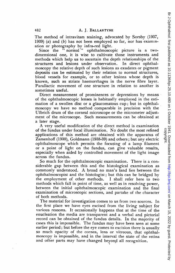
The method of intravitam staining, advocated by Sorsby (1937,1939) (a) and (b) has not been employed so far, nor has examin-ation or photography by infra-red light.
Since the " normal " ophthalmoscopic picture is a two-dimensional one, it is wise to cultivate those instruments andmethods which help us to ascertain the depth relationships of thestructures and lesions under observation. In direct ophthal-moscopy the relative depth of such lesions as exudates or pigmentdeposits can be estimated by their relation to normal structures,blood vessels for example, or to other lesions whose depth isknown, such as striate haemorrhages in the nerve fibre layer.Parallactic movement of one structure in relation to another issometimes useful.
Direct measurement of prominences or depressions by meansof the ophthalmoscopic lenses is habitually employed in the esti-mation of a swollen disc or a glaucomatous cup; but in ophthal-moscopy we have no method comparable in precision with theUlbrich drum of the corneal microscope or the micrometer adjust-ment of the microscope. Such measurements can be obtained ata later stage.A very useful modification of the direct method is examination
of the fundus under focal illumination. No doubt the most refinedapplications of this method are obtained with the apparatus ofZamenhoff (1930), Goldmann (1938-39) and others; but any electricophthalmoscope which permits the focusing of a lamp filamentor a point of light on the fundus, can give valuable results,especially when aided by controlled movement-of the light imageacross the fundus.So much for the ophthalmoscopic examination. There is a con-
siderable gap between this and the histological examination ascommonly understood. A broad no man's land lies between theophthalmoscopist and the histologist; but this can be bridged bythe employment of other methods. I shall refer here to twomethods which fall in point of time, as well as in resolving power,between the initial ophthalmoscopic examination and the finalexamination of microscopic sections, and partake of the characterof both methods.The material for investigation comes to us from two sources. In
the first place we have eyes excised from the living subject forvarious reasons. It occasionally happens that at the time of theenucleation the media are transparent and a verbal and pictorialrecord can be obtained of the fundus details. In the majority ofcases this is impossible. The fundus may have been seen at someearlier period; but before the eye comes to excision there is usuallyso much opacity of the cornea, lens or vitreous, that ophthal-moscopy is impossible, and in the interval the state of the retinaand other parts may have changed beKond all recognition.
482 A. J. BALLANTYNE
copyright. on M
arch 15, 2022 by guest. Protected by
http://bjo.bmj.com
/B
r J Ophthalm
ol: first published as 10.1136/bjo.25.10.480 on 1 October 1941. D
ownloaded from

APPEARANCES IN THE FUNDUS OCULI
It is otherwise in the case of eyes obtained in the" post-mortemroom. It is true that a great deal of valuable material is lostthrough lack of co-operation between the medical, ophthalmic andpathological departments; but where a proper liaison is maintainedit is often possible to obtain accurate ante-mortem records to cor-relate with the post-mortem findings. In either case it is importantto obtain, if possible, good illustrations of the fundus as a whole,or of important details. And here one may stress the inestimablevalue of coloured drawings made by one who is expert with theophthalmoscope as well as with the brush. Fundus photographyin black and white is usually a poor substitute, and even photo-graphy in colour leaves a good deal to be desired, although thebeautiful examples shown by Bedell before the OphthalmologicalSociety (1937 and 1939) showed what can be done by the expertin this method. Photography at its best gives a faithful statementof the size, form and situation of a lesion, and is invaluable forserial illustrations of its progress. It is quick and less tiring topatient and surgeon. But the magnification is low and the finerdetails are often lost. A drawing, even a rough sketch, is oftena more useful adjunct to a verbal description.
II. Slit Lamp ExaminationFor our present purpose specimens are fixed with formalin or
some formalin mixture which preserves their transparency. Eyesexcised from the living subject are placed directly in the fixingsolution and are ready in 48 hours to be divided as convenient forthe next stage of the examination.
In the case of the post-mortem material it is seldom possibleto get permission for removal of the whole eyeball; but in everycase where the skull is opened, the roof of the orbit can beremoved, and the posterior part of the globe, with the attachedoptic nerve, excised after removal of the orbital tissues to thenecessary extent. If the intracranial as well as the intra-orbitalpart of the optic nerve is to be removed, the roof of the optic canalmust be chiselled away. In excising the posterior segment of theglobe there is always a danger that the vitreous may escape, thesclera collapse and the retina become hopelessly detached. Wehave overcome this difficulty almost entirely by arranging thatas soon as possible after death a few minims of ten per cent.formalin solution are injected into the eye through the sclerabehind the ciliary body. If no post-mortem examination is allowedno harm is done, but when the opportunity occurs to obtain thespecimen, it will be found to be in firm condition and when care-fully removed the retina lies smoothly in position. In any casecare must be taken in excising the specimen. With the subject in
483
copyright. on M
arch 15, 2022 by guest. Protected by
http://bjo.bmj.com
/B
r J Ophthalm
ol: first published as 10.1136/bjo.25.10.480 on 1 October 1941. D
ownloaded from

A. J. BALLANTYNE
the usual supine position the eye is looking upward. The exposedsclera is punctured by a very sharp worn Graefe knife and anincision with fine scissors carried round at or behind the equatorof the eyeball. The vitreous will often be in the condition of afairly consistent jelly and should be divided by the scissors atthe same time. We should aim at keeping it in situ within thecup formed by the sclera. Before the scissors complete the scleralincision a small bottle (about 1 oz. capacity) filled with the formol
FIG. 1
Method of supporting optic nerve and posterior segment of eyeballduring fixation. Both bottles are filled with the fixing fluid.
fixative is brought into position so that the optic nerve passesvertically into the bottle and when the scleral incision is completedthe posterior part of the eye rests on the mouth of the bottle. Inthis way, spilling of the vitreous is prevented, and the retina keptin position. The small bottle with the specimen is then loweredinto a larger tube or jar of the same fixing fluid (Fig. 1) and setaside,for 24 hours before we proceed with the slit lamp examina-tion. At this stage the vitreous must be removed. If it has a firmconsistency and cannot be removed by irrigation it will be neces-sary to employ careful swabbing.What follows applies equally to eyes excised from the living
and to specimens removed after death.
484
copyright. on M
arch 15, 2022 by guest. Protected by
http://bjo.bmj.com
/B
r J Ophthalm
ol: first published as 10.1136/bjo.25.10.480 on 1 October 1941. D
ownloaded from

APPEARANCES IN THE FUNDUS OCULI
It is necessary to have some form of holder to carry the eyeunder examination which will allow the specimen to be setin convenient positions and to " stay put." The carrier illustratedin Fig. 2 was made out of oddments but meets the requirementsvery well. It clips on to the forehead rest of the slit lamp stand,and an essential feature is the ball and socket joint, which formerlybelonged to the " cat's whisker '; of a crystal wireless set and
FIG. 2.
Appliance for holding the eye during slit-lamp examination. Thesharp pins and adjustable screws grip the specimen. The ball andsocket joint allows the eye to be placed and retained in any desiredposition.
altows of free movement of the specimen into any desiredposition. The eye is examined in the moist state, and it is un-necessary to place it under water until it is to be photographed.The corneal microscope and slit-lamp give us a monochromatic
view of the fundus, under magnifications of 9 to 35 diameters.In the normal eye the vessels are more or less completely filledwith blood and 4he arteries and veins are readily distinguishedfrom one another. The nerve fibre bundles can be easily followed.The macula presents its yellow tint and the margins of the de-pression are somewhat swollen. When it has been impossible,for the reasons already referred to, to make an ophthalmoscopicexamination of the living eye, this slit-lamp examination providesan excellent substitute; indeed in most cases interesting featuresare revealed which evade observation with the ophthalmoscope.
485
copyright. on M
arch 15, 2022 by guest. Protected by
http://bjo.bmj.com
/B
r J Ophthalm
ol: first published as 10.1136/bjo.25.10.480 on 1 October 1941. D
ownloaded from

A. J. BALLANTYNE
The stereoscopic view and higher magnifications are an advantage;but much more important is the control over the direction ofobservation and of illumination. As in slit-lamp examination ofthe anterior part of the eye, we can employ the broad or narrowbeam (the latter giving optical section) direct or indirect illumin-ation, or illumination from the scleral side (retro-illumination) bythe Gullstrand lamp or by any one of the familiar hand inspectionlamps.By this method we get a three-dimensional view of the fundus
and can obtain micrometer measurements of the size, situation
FIG. 3.
One of a pair of stereoscopic photographs of the posterior segment ofthe eye. Case of subhyaloid haemorrhage.
and space relationships of the parts, thus checking and amplifyingour ophthalmoscopic records.When the object of investigation is a small lesion such as a
haemorrhage, an exudate, an arterio-venous crossing or the like,we select and locate it, measure its size and position and makedrawings or photographs. In larger lesions we make stereophoto-graphs under lower magnifications, of which Fig. 3 is an example.
III. Examination of the Retina in BulkThe next step is the excision of selected portions of the retina.
Having noted the lesion in question we punch out the affectedportion of the retina by means of a three or four millimetre trephine
486
copyright. on M
arch 15, 2022 by guest. Protected by
http://bjo.bmj.com
/B
r J Ophthalm
ol: first published as 10.1136/bjo.25.10.480 on 1 October 1941. D
ownloaded from

APPEARANCES IN THE FUNDUS OCULI
applied at right angles to the retinal surface and pressed againstthis with a slight rotary movement. If necessary a binocular loupeor dissecting microscope can be employed. The small disc thusoutlined can be easily lifted with an iris repositor or fine forceps.It is placed in a watch glass containing water and transferred toglycerine on a glass slide and protected with a cover glass.The microscopic examination in this form is in some respects
an extension of the ophthalmoscopic and slit-lamp examinations;but employing higher magnifications. The isolated retina, withoutits pigmented epithelium, is seen by transillumination, and readilyreveals such details as the nerve fibres and nerve fibre bundles,the pattern of Henle's fibre layer and of other tissue elements notso fully understood, the mosaic of the rods and cones and theperforations of the external limiting membrane, the structure ofthe vessel walls, the relationship of arteries and veins at theircrossings and the pattern of the capillary plexuses. The resolutionof these and other details is assisted by the stopping down of thesubstage diaphragm and the occasional movement of the mirror.The micrometer indications on the fine adjustment of the micro-scope enable us to measure the depth relationships of normal andpathological structures with a degree of refinement which is im-possible in the ophthalmoscopic and even in the slit-lamp exam-ination.
For most purposes this stage of the examination may be com-pleted without staining or other treatment; but some investigators,notably Shaw Dunn in Glasgow and Friedenwald in Baltimore,have employed the examination of retina " on the flat" in con-junction with scarlet red or sudan staining for the lipoids, andin the Tennent Institute Professor A. Lowenstein has elaboratedand modified the technique in many ways and with fruitful results.It lends itself especially to the demonstration of the retinal capil-lary plexuses, both in their natural state and by the injection andstaining method described by Michaelson and Campbell (1940).Naturally, the specimens at this stage also lend themselves toillustration by drawing and microphotography.
IV. Final StageWhen the possibilities of this method of examining the tre-
phined discs of retinal tissue have been exhausted the specimensare passed through the usual stages for embedding in paraffin,and serial sections are cut for further study.Having followed the selected lesions through the earlier stages,
and having kept note of the appropriate measurements, it is notdifficult to identify these in the microscopic sections. Figs. 4 to11 show some examples of the " follow up " from one stage toanother.
487
copyright. on M
arch 15, 2022 by guest. Protected by
http://bjo.bmj.com
/B
r J Ophthalm
ol: first published as 10.1136/bjo.25.10.480 on 1 October 1941. D
ownloaded from
4A. J. BALLANTYNE
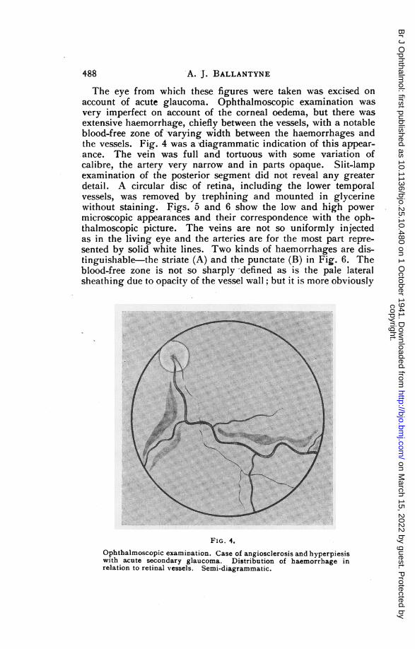
The eye from which these figures were taken was excised onaccount of acute glaucoma. Ophthalmoscopic examination wasvery imperfect on account of the corneal oedema, but there wasextensive haemorrhage, chiefly between the vessels, with a notableblood-free zone of varying width between the haemorrhages andthe vessels. Fig. 4 was a diagrammatic indication of this appear-ance. The vein was full and tortuous with some variation ofcalibre, the artery very narrow and in parts opaque. Slit-lampexamination of the posterior segment did not reveal any greaterdetail. A circular disc of retina, including the lower temporalvessels, was removed by trephining and mounted in glycerinewithout staining. Figs. 5 and 6 show the low and high powermicroscopic appearances and their correspondence with the oph-thalmoscopic picture. The veins are not so uniformly injectedas in the living eye and the arteries are for the most part repre-sented by solid white lines. Two kinds of haemorrhages are dis-tinguishable-the striate (A) and the punctate (B) in Fig. 6. Theblood-free zone is not so sharply defined as is the pale lateralsheathing due to opacity of the vessel wall; but it is more obviously
FIG. 4.
Ophthalmoscopic examination. Case of angioscierosis and hyperpiesiswith acute secondary glaucoma. Distribution of haemorrhage inrelation to retinal vessels. Semi-diagrammatic.
488
copyright. on M
arch 15, 2022 by guest. Protected by
http://bjo.bmj.com
/B
r J Ophthalm
ol: first published as 10.1136/bjo.25.10.480 on 1 October 1941. D
ownloaded from

APPEARANCES IN THE FUNDUS OCULI
FIG. 5.
From same eye as Fig. 4. Selected piece of retina removed by trephineand viewed microscopically on the flat, unstained. Vein accompaniedby broad blood-free zone. At some points there is narrow whitesheathing due to opacity of the vein wall.
A\J,
FIG. 6.
Part of Fig. 5 under higher magnification. Retinal nerve fibres out-lined by effused blood. Superficial, striatp, haemorrhage at (A) anddeep, punctate, haemorrhage at (B).
489
copyright. on M
arch 15, 2022 by guest. Protected by
http://bjo.bmj.com
/B
r J Ophthalm
ol: first published as 10.1136/bjo.25.10.480 on 1 October 1941. D
ownloaded from
A. J. BALLANTYNE
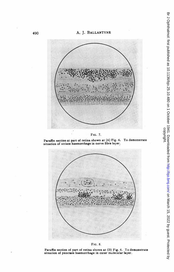
FIG. 7.
Paraffin section at part of retina shown at (A) Fig. 6. To demonstratesituation of striate haemorrhage in nerve fibre layer.
FIG. 8.
Paraffin section of part of retina shown at (B) Fig. 6. To demonstratesituation of punctate haemorrhage in outer molecular layer.
490
copyright. on M
arch 15, 2022 by guest. Protected by
http://bjo.bmj.com
/B
r J Ophthalm
ol: first published as 10.1136/bjo.25.10.480 on 1 October 1941. D
ownloaded from

APPEARANCES IN THE FUNDUS OCULI
related to the course of the vessel where the haemorrhage is ofthe striate variety. The micrometer of the fine adjustment of themicroscope indicated that the punctate haemorrhages lay some50 / deeper than the striate, and the sections taken from this pieceof tissue .after embedding in paraffin show that the striate haemorr-hage is confined to the nerve fibre and ganglion cell layers(Fig. 7) and the punctate haemorrhage to the external molecularlayer. (Fig. 8.)
Fig. 9 represents a more peripheral piece of the retina preparedin the same way. At (A) the retinal haemorrhage is separated from
.4
A1P:;. 9.
From the same eye as Figs. 4 to 8. Retina at periphery, removed bytrephining and seen on the flat, unstained. At (A) the superficialhaemorrhage is limited by the opaque vessel wall. At (B) the vesselis largely concealed by haemorrhage.
the vascular blood column by a narrow, well-defined, blood-freezone, while at (B) the vessel is largely concealed by blood. Fig.10 shows that at (A) the haemorrhage is confined to the innerlayers and the narrow pale zone or bilateral sheathing is entirelydue to the vessel wall. The concealment of the vessel at (B) isseen in Fig. 11 to be due to haemorrhage in front of and behindthe vessel.
Other sections showed that the blood-free zone illustrated inFigs. 4, 5 and 6, is also most typically found when the haemorr-hage is confined to the inner layers (striate haemorrhage) and isseparated from the blood vessels by a variable interval. The reasonfor this " avoidance " of the vessels has not been determined,
491
copyright. on M
arch 15, 2022 by guest. Protected by
http://bjo.bmj.com
/B
r J Ophthalm
ol: first published as 10.1136/bjo.25.10.480 on 1 October 1941. D
ownloaded from

A. J. BALLANTYNE
l .pi
¢ ~<'i*2 ^X ¢e* *
4 ~ q\ .,~~~~~~~~~~~~ w . _SSpFIG. 10.
Paraffin section of retina shown in FIG. 9. Vessel and haemorrhageas seen at (A).
FIG. 11.
Paraffin section at retina shown in Fig. 9. Vessel and haemorrhage as
seen at (B).
492
copyright. on M
arch 15, 2022 by guest. Protected by
http://bjo.bmj.com
/B
r J Ophthalm
ol: first published as 10.1136/bjo.25.10.480 on 1 October 1941. D
ownloaded from

BOECK'S SARCOIDOSIS
but in some cases this haemorrhagic pattern is very obvious. Ifone considers these pictures in conjunction with some of thoseillustrating the paper of Michaelson and Campbell (1940) theresemblance between the capillary-free zone of the injected retinaand the blood-free zone in retinal haemorrhage is sufficientlystriking to suggest that the latter is found in those cases wherethe haemorrhage has taken place from the capillary plexus internalto the inner nuclear layer.
It is outside the scope of this paper to deal with the preparationof the microscopic sections, which follows the usual lines. Mypurpose has been to point out that much essential information maybe hidden in the gap between the ophthalmoscopic examinationand the examination of paraffin or celloidin sections; and to showhow complete a survey of the normal and abnormal histology ofthe retina can be obtained by the serial application of the methodsdescribed.
REFERENCES
SORSBY, A. (1937).-Proc. Roy. Soc. Med., Section of Ophthalmology, Vol. XXX,Pt. 1.
(1939).-(a) Brit. Jl. OQhthal., Vol. XXIII, p. 20; (b) Trans. Obhthal. Soc.U.K., Vol. LXIX, p 727.
ZAMENHOFF (1930).-Arch. f. Ojhthal., Vol. CXXIV, p. 87.GOLDMANN (1938).-OPhthalmologica, Vol. XCVI, p. 90.BEDELL (1937).-Trans. Obhthal. Soc. U.K., Vol. LVII, p. 468.
(1939).-Trans. Ophthal. Soc. U.K., Vol. LIX, p. 219.MICHAELSON and CAMPBELL (1940).-Trans. Obhthal. Soc. U.K., Vol. LX, p. 71.
A CASE OF BOECK'S SARCOIDOSISBY
E. V. SRINIVASANMYLAPORE, MADRAS
MRS. L., aged 45 years, with cataract in both eyes, with absolutelyno contra-indication for immediate operation, had an extraction per-formed in the left eye, the right eye being quite immature. She wasdischarged as satisfactory within a month with correction for theoperated eye. She returned a month later with signs of cyclitis inthe operated eye. After getting the usual treatment for about fivemonths she disappeared without being completely cured, butreappeared in a month's time with pain and marked loss of vision inthe eye.On examination she had well-marked ptosis of the left eyelid.
Underneath, the eye was tender with deep keratitis, vascularisationobscuring deeper details as it extended nearly to the upper two-thirds of the cornea. Tension was low. The clear lower cornea
493
copyright. on M
arch 15, 2022 by guest. Protected by
http://bjo.bmj.com
/B
r J Ophthalm
ol: first published as 10.1136/bjo.25.10.480 on 1 October 1941. D
ownloaded from
Copyright © 2022 FDOKUMEN