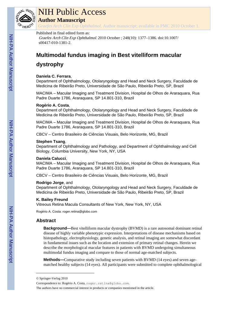
Multimodal fundus imaging in Best vitelliform macular dystrophy
17
Multimodal fundus imaging in Best vitelliform macular dystrophy Daniela C. Ferrara, Department of Ophthalmology, Otolaryngology and Head and Neck Surgery, Faculdade de Medicina de Ribeirão Preto, Universidade de São Paulo, Ribeirão Preto, SP, Brazil MACIMA – Macular Imaging and Treatment Division, Hospital de Olhos de Araraquara, Rua Padre Duarte 1786, Araraquara, SP 14.801-310, Brazil Rogério A. Costa, Department of Ophthalmology, Otolaryngology and Head and Neck Surgery, Faculdade de Medicina de Ribeirão Preto, Universidade de São Paulo, Ribeirão Preto, SP, Brazil MACIMA – Macular Imaging and Treatment Division, Hospital de Olhos de Araraquara, Rua Padre Duarte 1786, Araraquara, SP 14.801-310, Brazil CBCV – Centro Brasileiro de Ciências Visuais, Belo Horizonte, MG, Brazil Stephen Tsang, Department of Ophthalmology and Pathology, and Department of Ophthalmology and Cell Biology, Columbia University, New York, NY, USA Daniela Calucci, MACIMA – Macular Imaging and Treatment Division, Hospital de Olhos de Araraquara, Rua Padre Duarte 1786, Araraquara, SP 14.801-310, Brazil CBCV – Centro Brasileiro de Ciências Visuais, Belo Horizonte, MG, Brazil Rodrigo Jorge, and Department of Ophthalmology, Otolaryngology and Head and Neck Surgery, Faculdade de Medicina de Ribeirão Preto, Universidade de São Paulo, Ribeirão Preto, SP, Brazil K. Bailey Freund Vitreous Retina Macula Consultants of New York, New York, NY, USA Rogério A. Costa: [email protected] Abstract Background—Best vitelliform macular dystrophy (BVMD) is a rare autosomal dominant retinal disease of highly variable phenotypic expression. Interpretations of disease mechanisms based on histopathology, electrophysiology, genetic analysis, and retinal imaging are somewhat discordant in fundamental issues such as the location and extension of primary retinal changes. Herein we describe the morphological macular features in patients with BVMD undergoing simultaneous multimodal fundus imaging and compare to those of normal age-matched subjects. Methods—Comparative study including seven patients with BVMD (14 eyes) and seven age- matched healthy subjects (14 eyes). All participants were submitted to complete ophthalmological © Springer-Verlag 2010 Correspondence to: Rogério A. Costa, [email protected]. The authors have no commercial interest in products or companies mentioned in the article. NIH Public Access Author Manuscript Graefes Arch Clin Exp Ophthalmol. Author manuscript; available in PMC 2010 October 1. Published in final edited form as: Graefes Arch Clin Exp Ophthalmol. 2010 October ; 248(10): 1377–1386. doi:10.1007/ s00417-010-1381-2. NIH-PA Author Manuscript NIH-PA Author Manuscript NIH-PA Author Manuscript
Transcript of Multimodal fundus imaging in Best vitelliform macular dystrophy
Multimodal fundus imaging in Best vitelliform maculardystrophy
Daniela C. Ferrara,Department of Ophthalmology, Otolaryngology and Head and Neck Surgery, Faculdade deMedicina de Ribeirão Preto, Universidade de São Paulo, Ribeirão Preto, SP, Brazil
MACIMA – Macular Imaging and Treatment Division, Hospital de Olhos de Araraquara, RuaPadre Duarte 1786, Araraquara, SP 14.801-310, Brazil
Rogério A. Costa,Department of Ophthalmology, Otolaryngology and Head and Neck Surgery, Faculdade deMedicina de Ribeirão Preto, Universidade de São Paulo, Ribeirão Preto, SP, Brazil
MACIMA – Macular Imaging and Treatment Division, Hospital de Olhos de Araraquara, RuaPadre Duarte 1786, Araraquara, SP 14.801-310, Brazil
CBCV – Centro Brasileiro de Ciências Visuais, Belo Horizonte, MG, Brazil
Stephen Tsang,Department of Ophthalmology and Pathology, and Department of Ophthalmology and CellBiology, Columbia University, New York, NY, USA
Daniela Calucci,MACIMA – Macular Imaging and Treatment Division, Hospital de Olhos de Araraquara, RuaPadre Duarte 1786, Araraquara, SP 14.801-310, Brazil
CBCV – Centro Brasileiro de Ciências Visuais, Belo Horizonte, MG, Brazil
Rodrigo Jorge, andDepartment of Ophthalmology, Otolaryngology and Head and Neck Surgery, Faculdade deMedicina de Ribeirão Preto, Universidade de São Paulo, Ribeirão Preto, SP, Brazil
K. Bailey FreundVitreous Retina Macula Consultants of New York, New York, NY, USARogério A. Costa: [email protected]
AbstractBackground—Best vitelliform macular dystrophy (BVMD) is a rare autosomal dominant retinaldisease of highly variable phenotypic expression. Interpretations of disease mechanisms based onhistopathology, electrophysiology, genetic analysis, and retinal imaging are somewhat discordantin fundamental issues such as the location and extension of primary retinal changes. Herein wedescribe the morphological macular features in patients with BVMD undergoing simultaneousmultimodal fundus imaging and compare to those of normal age-matched subjects.
Methods—Comparative study including seven patients with BVMD (14 eyes) and seven age-matched healthy subjects (14 eyes). All participants were submitted to complete ophthalmological
© Springer-Verlag 2010Correspondence to: Rogério A. Costa, [email protected] authors have no commercial interest in products or companies mentioned in the article.
NIH Public AccessAuthor ManuscriptGraefes Arch Clin Exp Ophthalmol. Author manuscript; available in PMC 2010 October 1.
Published in final edited form as:Graefes Arch Clin Exp Ophthalmol. 2010 October ; 248(10): 1377–1386. doi:10.1007/s00417-010-1381-2.
NIH
-PA Author Manuscript
NIH
-PA Author Manuscript
NIH
-PA Author Manuscript
examination, fundus photography, and standardized multimodal fundus imaging protocolincluding Fourier-domain optical coherence tomography (Fd-OCT) combined with near-infraredreflectance and blue-light fundus autofluorescence (FAF).
Results—In two eyes in the “subclinical” stage, Fd-OCT revealed thickening of the middlehighly reflective layer (HRL) localized between the photoreceptors’ inner/outer segments junction(inner-HRL) and RPE/Bruch’s membrane reflective complex (outer-HRL) throughout the macula.In one eye in the “vitelliform” stage, a homogeneous hyper-reflective material on Fd-OCT wasobserved between the middle-HRL and outer-HRL; this material presented increased fluorescenceon FAF. The outer nuclear layer (ONL) was thinned in the central macula and subretinal fluid wasnot identified in these earlier disease stages. In patients of “pseudohypopyon” (two eyes),“vitelliruptive” (eight eyes) and “atrophic” (one eye) stages, Fd-OCT revealed a variety of changesin the middle- and inner-HRLs and thinning of ONL. These changes were found to be associatedwith the level of visual acuity observed. Thickening of the middle-HRL was observed beyond thelimits of the clinically evident macular lesion in all eyes.
Conclusions—Multimodal fundus imaging demonstrated thickening of the reflective layercorresponding to the photoreceptors’ outer segments throughout the macula with no subretinalfluid accumulation as the earliest detectable feature in BVMD. Changes detected in thephotoreceptors’ reflective layers (middle- and inner- HRLs) and ONL thinning seemed to beprogressive with direct implications for the level of visual acuity impairment observed among thedifferent stages of the disease.
KeywordsBest disease; Fourier-domain; Fundus autofluorescence; Infrared; Retinal pigment epithelium;Spectral; Tomography, optical coherence; Vitelliform macular dystrophy
Best vitelliform macular dystrophy (BVMD) is a rare autosomal dominant retinal diseasewith highly variable phenotypic expression [1,2]. The disease-causing gene BEST1 islocated on chromosome 11q13 and encodes bestrophin-1 [3–5], a multifunctionaltransmembrane protein that is localized in the basolateral plasma membrane and intracellularspace of retinal pigment epithelial (RPE) cells [6–9]. Whether bestrophin functions directlyas a Ca2+-sensitive chloride channel or as a regulator of ion transport, compelling evidencesuggests that it is involved in the light peak conductance registered on electrooculography(EOG) [10–15]. Clinical suspicion of BVMD is based on the typical macular findingsinitially described by Best, who recognized an evolving spectrum of vitelliform macularlesions with advancing age [16] often in the setting of a positive family history. Thediagnosis is confirmed by an abnormal light rise on the EOG [17–19], or by theidentification of BEST1 gene mutation [20].
The definite diagnosis of BVMD, however, may be complicated. From a clinicalperspective, atypical presentations such as multifocal vitelliform lesions [21–26], as well asthe occurrence of choroidal neovascularization in various stages of the disease mayconfound the diagnosis at first [24,27–36]. A reliable EOG test is dependent on patients’ fullcooperation, which is a challenge in the pediatric population. Finally, while genetic tests toidentify the BEST1 gene mutation are routinely performed in some referral centers, theaccess to this high-cost analysis remains restricted in many countries, and the phenotypic-genotypic correlation has been inconsistent so far since specific mutations were associatedwith a wide range of clinical manifestations [1,21–23,37,38].
Therefore, the identification of additional phenotypic findings that would facilitate thediagnosis of BVMD is highly desirable. In this manuscript, we describe the morphologicalmacular features in patients with BVMD undergoing multimodal fundus imaging and make
Ferrara et al. Page 2
Graefes Arch Clin Exp Ophthalmol. Author manuscript; available in PMC 2010 October 1.
NIH
-PA Author Manuscript
NIH
-PA Author Manuscript
NIH
-PA Author Manuscript

comparisons with those of normal age-matched subjects, with particular interest in findingsobtained on Fourier-domain optical coherence tomography (Fd-OCT) guided by blue-lightfundus autofluorescence (FAF) and by near-infrared reflectance (NIR).
Materials and methodsPatients with BVMD identified through a review of medical records between January 2003and September 2009 at the Macular Imaging and Treatment Division of the Hospital deOlhos de Araraquara and at the Department of Ophthalmology of Columbia University wereinvited to participate in the study. First-order relatives were contacted when possible andinvited to participate. The study protocol followed the statements of the Declaration ofHelsinki and was approved by local Institutional Review Boards (#10121/2008 and#AAAB6560, respectively). Written informed consent was obtained for all participants priorto their inclusion in the study.
All participants underwent a complete ophthalmologic examination including best-correctedvisual acuity (BCVA) utilizing early treatment diabetic retinopathy study (ETDRS) charts,slit-lamp biomicroscopy, and applanation tonometry. For illiterate participants, BCVA wasmeasured using Snellen “E” charts. EOG testing was performed followed by a dilatedfundus examination [39]. Due to local unavailability of genetic analysis, the diagnosis ofBVMD was based on the presence of a positive family history with at least one affectedfirst-order relative and an abnormally low to absent EOG light rise (Arden ratio <1.5) [39]with or without abnormal funduscopic changes [2,9,17–20]. For those patients meeting thediagnostic criteria, color, red-free, NIR, and FAF imaging were performed. The FAF wasdocumented with an excitation wavelength of 488 nm and the emitted light detected above a500-nm barrier filter, using the “high-resolution” mode with the “automatic real time”(ART) mean module set at 25 frames and “normalized” function activated. Stereoscopicfluorescein and indocyanine green (ICG) angiography were also performed if choroidalneovascular proliferation associated with the vitelliform lesion was suspected based uponprior investigations.
For the purpose of the current study, BVMD lesions were staged essentially according to theclinical classification proposed by Gass [2]. A deposit of yellowish material in the center ofthe macula resembling an egg yolk characterizes the “vitelliform” stage. Initial disruption ofthe vitelliform lesion with gravitational displacement of the material refers to the“pseudohypopyon” stage. Further disruption with remnants of yellowish material irregularlydistributed and associated with RPE mobilization characterizes the “scrambled-egg” or“vitelliruptive” stage. Eventual absorption of the material and subretinal fluid leaving a flatarea of RPE changes defines the “atrophic” stage.
Age-matched subjects were enrolled as controls. For inclusion, control subjects had to havea LogMAR ETDRS BCVA (converted to Snellen) equal or better than 20/25, a sphericalequivalent refractive error of no greater than ±1.50 diopter, intraocular pressure of less than21 mmHg and no prior history or clinical evidence of retinal or optic nerve disease.
All participants underwent Fd-OCT imaging on a commercially available device(Spectralis® HRA+OCT; Heidelberg Engineering Inc., Heidelberg, Germany). Two types ofscan acquisition protocols were used. The first protocol utilized the built-in scan acquisitionfunction termed “volume” to acquire three-dimensional Fd-OCT data in a raster patternconsisting of 49 B-scans covering a 20°×20° fundus area (equivalent to ~5.6×5.6 mm). Thesecond protocol utilized the built-in scan acquisition function termed “section” to acquireseveral B-scans covering a linear 30° fundus area (equivalent to ~9 mm) in both thehorizontal and vertical orientations which were positioned under discretion of the examiners
Ferrara et al. Page 3
Graefes Arch Clin Exp Ophthalmol. Author manuscript; available in PMC 2010 October 1.
NIH
-PA Author Manuscript
NIH
-PA Author Manuscript
NIH
-PA Author Manuscript

during manual raster scanning of the entire macular area according to the reference fundusimages (such as FAF, for example). The “high-resolution” mode with the ART meanmodule set at 25 frames was utilized for both acquisition protocols. The Fd-OCT data werequalitatively assessed in all participants, and simultaneous acquisition of Fd-OCT scans andmultimodal reference images allowed point-to-point correlation of fundus features.
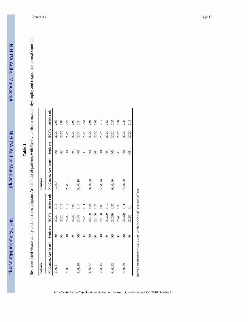
ResultsFourteen eyes of seven patients with BVMD and 14 eyes of seven age-matched controlswere included. The mean age of enrolled subjects was 33 years (range 5 to 62 years) forpatients and 34 years (range 7 to 66 years) for controls, and they were all male. The medianLogMAR EDTRS BCVA (converted to Snellen) of patients and controls was 20/50 (range,20/20 to 20/320) and 20/20 (range, 20/20 to 20/25), respectively (Table 1). According to theclinical staging adopted in the current study, one patient had a “vitelliform” lesion in the lefteye and a “vitelliruptive” lesion in the right eye, one patient had “pseudohypopyon” lesionsin both eyes, three patients had “vitelliruptive” lesions in both eyes, and one patient had a“vitelliruptive” lesion in the left eye and an “atrophic” lesion in the right eye. The remainingpatient had no macular lesion detectable on clinical examination.
“Subclinical” stageIn one patient (age 5 years) with a positive family history of BVMD (his father, one first-order uncle, and a first-order cousin have BVMD with symptomatic fundus lesions), anabnormally low light rise result was observed in both eyes (Arden ratio equal to 1.19 [righteye], and to 1.17 [left eye]). Although the clinical evaluation was otherwise unremarkable,Fd-OCT imaging demonstrated a prominent highly reflective layer (HRL) between theouter-HRL (corresponding to the RPE/Bruch’s membrane reflective complex) and the inner-HRL (corresponding to the junction of the inner and outer segments of the photoreceptors,or IS/OS junction) in both eyes (Fig. 1). This reflective layer at the level of thephotoreceptors’ outer segments (OS) was revealed with the advent of high-resolution OCT[40], and for the purpose of this study is referred to as the middle-HRL as its precisehistological correlation remains a matter of debate. When compared to the Fd-OCT imagesof the age-matched control eyes, the middle-HRL in this patient was abnormally thickenedthroughout the macula, particularly at the fovea. The total retinal thickness at the center ofthe fovea was not increased due to concomitant thinning of the outer nuclear layer (ONL).Hyporeflective spaces corresponding to fluid were not observed on Fd-OCT evaluation. Nodifferences between patient and control were noted on NIR images. The FAF was apparentlyunremarkable, but high-quality documentation was not achieved due to poor cooperation(Fig. 1). Given that no follow-up is presented and no genetic analysis was performed as wellas the fact that BVMD is an autosomal dominant disease with incomplete penetrance andvariable expressivity, the term “subclinical” was preferred to the terms “previtelliform” or“carrier” to stage this particular lesion.
“Vitelliform” stageIn one patient (age 6 years), an elevated yellow round macular lesion measuringapproximately 3/4 disc diameter across was found in the left eye. On Fd-OCT imaging, thefoveal retina was elevated due to a homogeneous hyper-reflective material located justabove the outer-HRL (RPE/Bruch complex). The middle-HRL was elevated at the marginsof the hyper-reflective material and followed the contour of the lesion with areas ofthickening and fragmentation above the hyper-reflective material. The ONL appearedthinned above the hyper-reflective material due to heterogeneous hyper-reflective signalsthat are not commonly observed at this layer. Hyporeflective spaces corresponding to fluidwere not observed on Fd-OCT evaluation. The NIR imaging clearly delineated the margins
Ferrara et al. Page 4
Graefes Arch Clin Exp Ophthalmol. Author manuscript; available in PMC 2010 October 1.
NIH
-PA Author Manuscript
NIH
-PA Author Manuscript
NIH
-PA Author Manuscript
of the vitelliform lesion. The hyper-reflective material observed between the outer-HRL andmiddle-HRL (thus localized presumably between the OS and RPE layers) presentedincreased fluorescence on FAF (Fig. 2). Combined Fd-OCT and FAF evaluation revealedthat the ONL was somewhat thinned beyond the clinically evident margins of the lesionalong its entire contour. In addition, thickening of the middle-HRL, identical to thatobserved in the child with subclinical stage above described (Fig. 1), was also documentedin regions of normal fluorescence on FAF close to the vitelliform macular lesion (Fig. 2).
“Pseudohypopyon” stageIn one patient (age 19 years), an elevated oval macular lesion measuring approximatelythree disc diameters across was observed in both eyes. A yellowish material located moreprominently at the inferior aspect of the lesion was evident. In the center of the macula, Fd-OCT imaging showed separation of the neurosensory retina from the outer-HRL (RPE/Bruch complex) by an optically clear space and clumps of homogeneous hyper-reflectivematerial overlying the RPE inferiorly displaced (Fig. 3). In the elevated neurosensory retina,the ONL was thinned when compared to the control eyes and presented focal regions ofheterogeneous reflectivity. The inner-HRL (IS/OS junction) exhibited focal areas ofdisruption superiorly and nasally to the fovea in both eyes. The elevated middle-HRL(photoreceptors’ OS) was irregularly thickened. The outer-HRL (RPE/Bruch complex) wasrelatively preserved beneath the elevated neurosensory retina, with small hyper-reflectivemounds at this level. On FAF, increased autofluorescence from the hyper-reflective materialoverlying the RPE was observed from the inferior aspect of the lesion and, to a lesserdegree, from the margins of the lesion. Fundus NIR clearly delineated the retinal elevation.Of note, in the macular region above the vitelliform lesion with normal fluorescence onFAF, the Fd-OCT imaging revealed a distinctively thickened middle-HRL when comparedwith corresponding areas in the control eyes.
“Vitelliruptive” stageIn eight eyes of five patients (age 6–62 years) elevated macular lesions measuring from 11/2 to 3 disc diameters across were observed. Variable amounts of yellowish subretinalmaterial were dispersed within and at the margins of the macular lesions in all eight eyes; infive of these, a distinct grayish subretinal tissue was evident at the center of the lesions. Inall eight eyes, Fd-OCT imaging showed the neurosensory retina separated from the outer-HRL (RPE/Bruch complex) centrally by an optically clear space. In the elevatedneurosensory retina, there was marked thinning of the ONL and a variable degree ofalterations of the middle- and inner- HRLs (photoreceptors’ OS and IS/OS junction,respectively) when compared to control eyes. In one patient (age 6 years), thephotoreceptors’ OS were irregularly elongated. In the remaining patients (age 37–62 years),regions of hyper-reflectivity from the IS/OS junction and abnormal elongation of the OSwere interspersed with areas of complete absence of photoreceptors’ signal.
The subretinal grayish tissue at the center of the dome-shaped lesion corresponded to hyper-reflective mounds at the level of outer-HRL (RPE/Bruch complex). Two somewhat distincthyper-reflective mounds were identified on integrated evaluation of multimodal fundusimaging. The first mound was associated with relative shadowing of the underlying choroidsignals on Fd-OCT, had no characteristic findings on FAF and was poorly delineated onNIR (Fig. 4). The second hyper-reflective mound was invariably associated with secondaryretinal changes; the outer retinal layers were indistinguishable and generally collapsed overthe mound. In addition, unlike the first, this mound was associated with relative hyper-reflectivity of the underlying choroid, and it was clearly delineated on FAF and NIRimaging (Fig. 4). The yellowish subretinal material observed clinically tended to accumulateat the margins of the macular lesion and presented characteristics similar to the yellowish
Ferrara et al. Page 5
Graefes Arch Clin Exp Ophthalmol. Author manuscript; available in PMC 2010 October 1.
NIH
-PA Author Manuscript
NIH
-PA Author Manuscript
NIH
-PA Author Manuscript
material also observed in eyes with vitelliform and pseudohypopyon stages, appearing as ahomogeneous hyper-reflective material just above the outer-HRL (RPE/Bruch complex) onFd-OCT and exhibiting increased fluorescence on FAF. The Fd-OCT imaging beyond thedome-shaped lesion demonstrated diffuse thickening of the middle-HRL throughout themacular area when compared with control eyes.
“Atrophic” stageIn one eye of one patient (age 62 years), we observed an irregular oval area of RPEmobilization of approximately 2 1/2 disc diameters across encompassing the macula with noapparent retinal elevation. Within the lesion area, Fd-OCT imaging revealed overall thinningof the neurosensory retina with a shallow separation from the outer-HRL (RPE/Bruchcomplex) by an optically clear space. The photoreceptors’ reflective complex (OS/inner-HRL/IS) was practically absent in the fovea, and the ONL was thinned throughout themacula when compared to the control eye. The outer-HRL (RPE/Bruch complex) wasrelatively preserved, and one isolated hyper-reflective mound with underlying choroidalshadowing was observed. A mild stippled fluorescence on FAF was observed throughout themacula. Fundus NIR was not helpful in delineating the extension of the macular lesion.Evaluation of the macular area around the lesion demonstrated that the middle-HRL wasmore evident and diffusely thicker throughout the macula when compared to control eyes.
DiscussionThe pathogenesis of BVMD has been a matter of speculation since the disorder was firstdescribed. Since the encoded protein of BEST1 is expressed in the RPE, the primarydysfunctional site of BVMD apparently lies within the RPE cells [3,6,41]. In addition to theproposed mechanisms involving ionic conductance in the RPE cell, bestrophin activitypossibly influences intracellular processes such as phagocytosis and lysosomal function[9,42–44] as well as vascular endothelium growth factor production by the RPE [45]. Thefew histopathological studies reported to date are conflicting in fundamental issues such aswhether the primary tissue damage evolves with RPE or neurosensory retinal changes[28,29,41,46–48]. Interpretations of pathophysiologic mechanisms based upon OCTfindings in BVMD have also lacked agreement among investigators [19,20,49–54]. Someauthors concluded that the initial morphological events in patients with BVMD also occur atthe level of the RPE monolayer [49]. In the current study, no obvious alteration could bedemonstrated on Fd-OCT specifically at the reflective layer corresponding to thedysfunctional RPE monolayer in the macula of eyes with subclinical or vitelliform lesions.Conversely, the reflective layer (middle-HRL) corresponding presumably to photoreceptors’OS was apparently thickened throughout the macula based on our interpretation (Fig. 1).Our impression was that the middle-HRL was also apparently thickened in macular regionswith otherwise normal fluorescence on FAF (Fig. 2).
In patients with BVMD, Querques et al. recently demonstrated the presence of“previtelliform” lesions that were characterized by a “thicker and more reflective appearanceof the layer between the RPE and the photoreceptor IS/OS interface in the central region,compared with the normal macula” [54]. These authors correlated this thickened layer to theVerhoeff’s membrane [54,55]. Additionally, in the previtelliform stage, they reported a“normal appearance of all major intraretinal layers from ILM to ELM, as well as a normal-appearing RPE and IS/OS interface, were found in two eyes (50%); focal disruption of IS/OS interface and of the layer between the RPE and the IS/OS interface was observed inanother two eyes (50%)” [54]. In the current study, we also documented a thickened layer atthe outer retina in both eyes of a 5-year-old child with an unremarkable clinical examination.However, unlike Querques et al., we believe that this particular finding occurs at the level ofthe middle-HRL, which corresponds to photoreceptors’ OS reflective layer and not to
Ferrara et al. Page 6
Graefes Arch Clin Exp Ophthalmol. Author manuscript; available in PMC 2010 October 1.
NIH
-PA Author Manuscript
NIH
-PA Author Manuscript
NIH
-PA Author Manuscript
“Verhoeff’s membrane” [54], which corresponds to junctional complexes along the lateralcell membranes of the RPE that on light microscopy produce the effect of a continuousmembrane at the RPE level [56]. Moreover, the middle-HRL was apparently thickenedthroughout the macula of both eyes with secondary ONL thinning observed centrally (Fig.1). Based on the multimodal imaging documentation we obtained, these particular findingswere observed in regions of normal fluorescence on FAF with no evidence of subretinalfluid accumulation (Figs. 1 and 2).
Histopathological studies of vitelliruptive lesions reported variable degrees ofphotoreceptors’ cell damage with descriptions ranging from “total atrophy” to “IS ofsurviving photoreceptors shortened and tipped by only small bits of OS material”. Notableattenuation of the ONL was a constant finding in these studies [28,29,46]. Thesehistopathologic observations are in agreement with our findings. We demonstrated avariable degree of photoreceptor damage on Fd-OCT, which ranged from the absence of theentire photoreceptors’ reflective layer with adjacent ONL thinning in an eye at the atrophicstage to a relatively well-preserved IS/OS junction associated with elongated OS andreduced ONL in eyes at earlier disease stages. Coupled with the clinical results (BCVAmeasurements) and observations from FAF documentation, we believe that the hyper-reflective signals at the level of the OS/inner-HRL/IS reflective layer observed on Fd-OCTmay correspond mainly to segments of “surviving photoreceptors’ cells”. In the currentstudy, as long as reflective signals from the OS/inner-HRL/IS layer were present in thefoveolar region, the BCVA was as good as 20/25 in eyes with vitelliform orpseudohypopyon lesions, and 20/32 in an eye with a vitelliruptive lesion. In contrast, BCVAwas decreased to 20/320 in an eye with atrophic lesion and absence of the entire OS/inner-HRL/IS reflective layer within the foveola (Fig. 5).
This study has obvious limitations, such as the small number of patients and the absence of anormative database for the thickness of the different photoreceptors’ reflective layers at thepresent moment, which preclude any definitive conclusion to be made. Genetic analysislooking for possible genotype-phenotype correlation regarding the Fd-OCT findings hereinsuggested as well as for potential differences in Fd-OCT features between subclinical stagesand carrier status should also be considered in future studies. However, the concordancebetween our multimodal imaging findings and previous histopathological [29] andfunctional [57] studies in BVMD patients apparently support our observations. Finally, asfar as we are aware, this is the first study to suggest in vivo morphological changes in thephotoreceptors’ OS reflective layer diffusely throughout the macula (and not only in thelesion area), as well as the early involvement of the ONL centrally in the macula. Theseimpressions, if confirmed in future studies utilizing multimodal fundus imaging, mayultimately have implications for early diagnosis in patients with BVMD.
References1. Maloney WF, Robertson DM, Duboff SM. Hereditary vitelliform macular degeneration: variable
fundus findings within a single pedigree. Arch Ophthalmol 1977;95:979–983. [PubMed: 869756]2. Gass, JDM. Stereoscopic atlas of macular diseases, diagnosis and treatment. Mosby: St. Louis;
1997.3. Stone EM, Nichols BE, Streb LM, Kimura AE, Sheffield VC. Genetic linkage of vitelliform
macular degeneration (Best’s disease) to chromosome 11q13. Nat Genet 1992;1:246–250.[PubMed: 1302019]
4. Petrukhin K, Koisti MJ, Bakall B, Li W, Xie G, Marknell T, Sandgren O, Forsman K, Holmgren G,Andreasson S, Vujic M, Bergen AAB, McGarty-Dugan V, Figueroa D, Austin CP, Metzker ML,Caskey CT, Wadelius C. Identification of the gene responsible for Best macular dystrophy. NatGenet 1998;19:241–247. [PubMed: 9662395]
Ferrara et al. Page 7
Graefes Arch Clin Exp Ophthalmol. Author manuscript; available in PMC 2010 October 1.
NIH
-PA Author Manuscript
NIH
-PA Author Manuscript
NIH
-PA Author Manuscript
5. Marquardt A, Stohr H, Passmore LA, Kramer F, Rivera A, Weber BH. Mutations in a novel gene,VMD2, encoding a protein of unknown properties cause juvenile-onset vitelliform maculardystrophy (Best disease). Hum Mol Genet 1998;7:1517–1525. [PubMed: 9700209]
6. Marmorstein AD, Marmorstein LY, Rayborn M, Wang X, Hollyfield JG, Petrukhin K. Bestrophin,the product of the Best vitelliform macular dystrophy gene (VMD2), localizes to the basolateralplasma membrane of the retinal pigment epithelium. Proc Natl Acad Sci USA 2000;97:12758–12763. [PubMed: 11050159]
7. Bakall B, McLaughlin P, Stanton JB, Zhang Y, Hartzell HC, Marmorstein LY, Marmorstein AD.Bestrophin-2 is involved in the generation of intraocular pressure. Invest Ophthalmol Vis Sci2008;49:1563–1570. [PubMed: 18385076]
8. Yu K, Qu Z, Cui Y, Hartzell HC. Chloride channel activity of bestrophin mutants associated withmild or late-onset macular degeneration. Invest Ophthalmol Vis Sci 2007;48:4694–4705. [PubMed:17898294]
9. Boon CJF, Klevering BJ, Leroy BP, Hoyng CB, Keunen JEE, den Hollander AI. The spectrum ofocular phenotypes caused by mutations in the BEST1 gene. Prog Retin Eye Res 2009;28:187–205.[PubMed: 19375515]
10. Tsunenari T, Sun H, Williams J, Cahill H, Smallwood P, Yau KW, Nathans J. Structure-functionanalysis of the bestrophin family of anion channels. J Biol Chem 2003;278:41114–41125.[PubMed: 12907679]
11. Marmorstein AD, Kinnick TR. Focus on molecules: Bestrophin (Best-1). Exp Eye Res2007;85:423–424. [PubMed: 16720022]
12. Sun H, Tsunenari T, Yau KW, Nathans J. The vitelliform macular dystrophy protein defines a newfamily of chloride channels. Proc Natl Acad Sci USA 2002;99:4008–4013. [PubMed: 11904445]
13. Yu K, Cui Y, Hartzell HC. The bestrophin mutation A243V, linked to adult-onset vitelliformmacular dystrophy, impairs its chloride channel function. Invest Ophthalmol Vis Sci2006;47:4956–4961. [PubMed: 17065513]
14. Hartzell HC, Qu Z, Yu K, Xiao Q, Chien LT. Molecular physiology of bestrophins:multifunctional membrane proteins linked to Best disease and other retinopathies. Physiol Rev2008;88:639–672. [PubMed: 18391176]
15. Marmorstein AD, Cross HE, Peachey NS. Functional roles of bestrophins in ocular epithelia. ProgRetin Eye Res 2009;28:206–226. [PubMed: 19398034]
16. Best F. Über eine hereditäre Makulaaffektion. Z F Augenheilk 1905;13:199–212.17. Deutman AF. Electro-oculography in families with vitelliform dystrophy of the fovea. Detection of
the carrier state. Arch Ophthalmol 1969;81:305–316. [PubMed: 5774285]18. Cross HE, Bard L. Electro-oculography in Best macular dystrophy. Am J Ophthalmol 1974;77:46–
50. [PubMed: 4824173]19. Glybina IV, Frank RN. Localization of multifocal electro-retinogram abnormalities to the lesion
site: findings in a family with Best disease. Arch Ophthalmol 2006;124:1593–1600. [PubMed:17102007]
20. Boon CJF, Theelen T, Hoefsloot EH, van Schooneveld MJ, Keunen JEE, Cremers FPM, KleveringBJ, Hoyng CB. Clinical and molecular genetic analysis of Best vitelliform macular dystrophy.Retina 2009;29:835–847. [PubMed: 19357557]
21. Renner AB, Tillack H, Kraus H, Kramer F, Mohr N, Weber BHF, Foerster MH, Kellner U. Lateonset is common in Best macular dystrophy associated with VMD2 gene mutations.Ophthalmology 2005;112:586–592. [PubMed: 15808248]
22. Wabbels B, Preising MN, Kretschmann U, Demmler A, Lorenz B. Genotype-phenotype correlationand longitudinal course in ten families with Best vitelliform macular dystrophy. Graefes Arch ClinExp Ophthalmol 2006;244:1453–1466. [PubMed: 16612637]
23. Boon CJ, Klevering BJ, den Hollander AI, Zonneveld MN, Theelen T, Cremers FPM, Hoyng CB.Clinical and genetic heterogeneity in multifocal vitelliform dystrophy. Arch Ophthalmol2007;125:1100–1106. [PubMed: 17698758]
24. Mohler CW, Fine SL. Long-term evaluation of patients with Best vitelliform dystrophy.Ophthalmology 1981;88:688–692. [PubMed: 7267039]
Ferrara et al. Page 8
Graefes Arch Clin Exp Ophthalmol. Author manuscript; available in PMC 2010 October 1.
NIH
-PA Author Manuscript
NIH
-PA Author Manuscript
NIH
-PA Author Manuscript
25. Boon CJ, Klevering JB, Keunen JE, Hoyng CB, Theelen T. Fundus autofluorescence imaging ofretinal dystrophies. Vis Res 2008;48:2569–2577. [PubMed: 18289629]
26. Querques G, Regenbogen M, Soubrane G, Souied EH. High-resolution spectral domain opticalcoherence tomography findings in multifocal vitelliform macular dystrophy. Surv Ophthalmol2009;54:311–316. [PubMed: 19298908]
27. Noble KG, Scher BM, Carr RE. Polymorphous presentations in vitelliform macular dystrophy:subretinal neovascularization and central choroidal atrophy. Br J Ophthalmol 1978;62:561–570.[PubMed: 687557]
28. Frangieh GT, Green WR, Fine SL. A histopathologic study of Best macular dystrophy. ArchOphthalmol 1982;100:1115–1121. [PubMed: 7092655]
29. O’Gorman S, Flaherty WA, Fishman GA, Berson EL. Histopathologic findings in Best vitelliformmacular dystrophy. Arch Ophthalmol 1988;106:1261–1268. [PubMed: 3415551]
30. Blodi CF, Stone EM. Best vitelliform dystrophy. Ophthalmic Paediatr Genet 1990;11:49–59.[PubMed: 2190134]
31. Fishman GA, Baca W, Alexander KR, Derlacki DJ, Glenn AM, Viana M. Visual acuity in patientswith Best vitelliform macular dystrophy. Ophthalmology 1993;100:1665–1670. [PubMed:8233392]
32. Chung MM, Oh KT, Streb LM, Kimura AE, Stone EM. Visual outcome following subretinalhemorrhage in Best disease. Retina 2001;21:575–580. [PubMed: 11756879]
33. Andrade RE, Farah ME, Cardillo JA, Hofling-Lima AL, Uno F, Costa RA. Optical coherencetomography in choroidal neovascular membrane associated with Best vitelliform dystrophy. ActaOphthalmol Scand 2002;80:216–218. [PubMed: 11952492]
34. Andrade RE, Farah ME, Costa RA. Photodynamic therapy with verteporfin for subfoveal choroidalneovascularization in Best disease. Am J Ophthalmol 2003;136:1179–1181. [PubMed: 14644242]
35. Leu J, Schrage NF, Degenring RF. Choroidal neovascularization secondary to Best disease in a 13-year-old boy treated by intravitreal bevacizumab. Graefes Arch Clin Exp Ophthalmol2007;245:1723–1725. [PubMed: 17605026]
36. Querques G, Bocco MC, Soubrane G, Souied EH. Intravitreal ranibizumab (Lucentis) for choroidalneovascularization associated with vitelliform macular dystrophy. Acta Ophthalmol 2008;86:694–695. [PubMed: 18752521]
37. Bakall B, Marknell T, Ingvast S, Koisti MJ, Sandgren O, Li W, Bergen AAB, Andreasson S,Rosenberg T, Petrukhin K, Wadelius C. The mutation spectrum of the bestrophin protein—functional implications. Hum Genet 1999;104:383–389. [PubMed: 10394929]
38. Kramer F, White K, Pauleikhoff D, Gehrig A, Passmore L, Rivera A, Rudolph G, Kellner U,Andrassi M, Lorenz B, Rohrschneider K, Blankenagel A, Jurklies B, Schilling H, Schutt F, HolzFG, Weber BH. Mutations in the VMD2 gene are associated with juvenile-onset vitelliformmacular dystrophy (Best disease) and adult-onset vitelliform macular dystrophy but not age-related macular degeneration. Eur J Hum Genet 2000;8:286–292. [PubMed: 10854112]
39. Brown M, Marmor M, Vaegan ZE, Brigell M, Bach M. ISCEV. ISCEV standard for clinicalelectro-oculography (EOG). Doc Ophthalmol 2006;113:205–212. [PubMed: 17109157]
40. Srinivasan VJ, Monson BK, Wojtkowski M, Bilonick RA, Gorczynska I, Chen R, Duker JS,Schuman JS, Fujimoto JG. Characterization of outer retinal morphology with high-speed,ultrahigh-resolution optical coherence tomography. Invest Ophthalmol Vis Sci 2008;49:1571–1579. [PubMed: 18385077]
41. Mullins RF, Kuehn MH, Faidley EA, Syed NA, Stone EM. Differential macular and peripheralexpression of bestrophin in human eyes and its implication for Best disease. Invest OphthalmolVis Sci 2007;48:3372–3380. [PubMed: 17591911]
42. Deguchi J, Yamamoto A, Yoshimori T, Sugasawa K, Moriyama Y, Futai M, Suzuki T, Kato K,Uyama M, Tashiro Y. Acidification of phagosomes and degradation of rod outer segments in ratretinal pigment epithelium. Invest Ophthalmol Vis Sci 1994;35:568–579. [PubMed: 8113008]
43. Jentsch TJ. Chloride and the endosomal-lysosomal pathway: emerging roles of CLC chloridetransporters. J Physiol 2007;578:633–640. [PubMed: 17110406]
Ferrara et al. Page 9
Graefes Arch Clin Exp Ophthalmol. Author manuscript; available in PMC 2010 October 1.
NIH
-PA Author Manuscript
NIH
-PA Author Manuscript
NIH
-PA Author Manuscript
44. Karl MO, Kroeger W, Wimmers S, Milenkovic VM, Valtink M, Engelmann K, Strauss O.Endogenous Gas6 and Ca2+-channel activation modulate phagocytosis by retinal pigmentepithelium. Cell Signal 2008;20:1159–1168. [PubMed: 18395422]
45. Rosenthal R, Heimann H, Agostini H, Martin G, Hansen LL, Strauss O. Ca2+ channels in retinalpigment epithelial cells regulate vascular endothelial growth factor secretion rates in health anddisease. Mol Vis 2007;13:443–456. [PubMed: 17417605]
46. Weingeist T, Kobrin J, Watzke R. Histopathology of Best macular dystrophy. Arch Ophthalmol1982;100:1108–1114. [PubMed: 7092654]
47. Mullins RF, Oh KT, Heffron E, Hageman G, Stone EM. Late development of vitelliform lesionsand flecks in a patient with Best disease. Arch Ophthalmol 2005;123:1588–1594. [PubMed:16286623]
48. Bakall B, Radu RA, Stanton JB, Burke JM, McKay BS, Wadelius C, Mullins RF, Stone EM,Travis GH, Marmorstein AD. Enhanced accumulation of A2E in individuals homozygous orheterozygous for mutations in BEST1 (VMD2). Exp Eye Res 2007;85:34–43. [PubMed:17477921]
49. Pianta MJ, Aleman TS, Cideciyan AV, Sunness JS, Li Y, Campochiaro BA, Campochiaro PA,Zack DJ, Stone EM, Jacobson SG. In vivo micropathology of Best macular dystrophy with opticalcoherence tomography. Exp Eye Res 2003;76:203–211. [PubMed: 12565808]
50. Men G, Batioglu F, Ozkan SS, Huban A, Ozdamar Y, Aslan O. Best vitelliform macular dystrophywith pseudohypopyon: an optical coherence tomography study. Am J Ophthalmol 2004;137:963–965. [PubMed: 15126177]
51. Vedantham V, Ramasamy K. Optical coherence tomography in Best disease: an observational casereport. Am J Ophthalmol 2005;139:351–353. [PubMed: 15734003]
52. Spaide RF, Noble K, Morgan A, Freund KB. Vitelliform macular dystrophy. Ophthalmology2006;113:1392–1400. [PubMed: 16877078]
53. Spaide RF. Autofluorescence from the outer retina and subretinal space. Hypothesis and review.Retina 2008;28:5–35. [PubMed: 18185134]
54. Querques G, Regenbogen M, Quijano C, Delphin N, Soubrane G, Souied EH. High-definitionoptical coherence tomography features in vitelliform macular dystrophy. Am J Ophthalmol2008;146:501–507. [PubMed: 18619572]
55. Zawadzki RJ, Jones SM, Olivier SS, Zhao M, Bower BA, Izatt JA, Choi S, Laut S, Werner JS.Adaptive-optics optical coherence tomography for high-resolution and high-speed 3D retinal invivo imaging. Opt Express 2005;13:8532–8546. [PubMed: 19096728]
56. Verhoeff FH. A hitherto undescribed membrane of the eye and its significance. Boston Med Surg J1903;149:456.
57. Maia-Lopes S, Silva ED, Reis A, Silva MF, Mateus C, Castelo-Branco M. Retinal function in Bestmacular dystrophy: relationship between electrophysiological, pychophysical, and structuralmeasures of damage. Invest Ophthalmol Vis Sci 2008;49:5553–5560. [PubMed: 18775865]
Ferrara et al. Page 10
Graefes Arch Clin Exp Ophthalmol. Author manuscript; available in PMC 2010 October 1.
NIH
-PA Author Manuscript
NIH
-PA Author Manuscript
NIH
-PA Author Manuscript
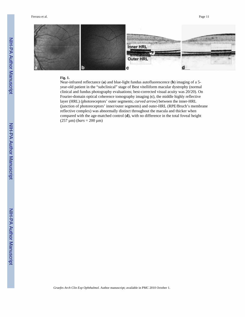
Fig. 1.Near-infrared reflectance (a) and blue-light fundus autofluorescence (b) imaging of a 5-year-old patient in the “subclinical” stage of Best vitelliform macular dystrophy (normalclinical and fundus photography evaluations; best-corrected visual acuity was 20/20). OnFourier-domain optical coherence tomography imaging (c), the middle highly reflectivelayer (HRL) (photoreceptors’ outer segments; curved arrow) between the inner-HRL(junction of photoreceptors’ inner/outer segments) and outer-HRL (RPE/Bruch’s membranereflective complex) was abnormally distinct throughout the macula and thicker whencompared with the age-matched control (d), with no difference in the total foveal height(257 µm) (bars = 200 µm)
Ferrara et al. Page 11
Graefes Arch Clin Exp Ophthalmol. Author manuscript; available in PMC 2010 October 1.
NIH
-PA Author Manuscript
NIH
-PA Author Manuscript
NIH
-PA Author Manuscript

Fig. 2.Blue-light fundus autofluorescence (FAF) (a) and Fourier-domain optical coherencetomography (Fd-OCT) imaging (horizontal B-scans, b, c, and d) in a 6-year-old patient withBest vitelliform macular dystrophy in the “vitelliform” stage. Increased autofluorescence onFAF was observed from the vitelliform lesion (a). The Fd-OCT imaging guided bysimultaneous FAF revealed that the middle highly reflective layer (photoreceptors’ outersegments) remained apparently thickened, even in areas with normal fluorescence on FAF.Right panel shows corresponding magnifications of the white rectangles
Ferrara et al. Page 12
Graefes Arch Clin Exp Ophthalmol. Author manuscript; available in PMC 2010 October 1.
NIH
-PA Author Manuscript
NIH
-PA Author Manuscript
NIH
-PA Author Manuscript
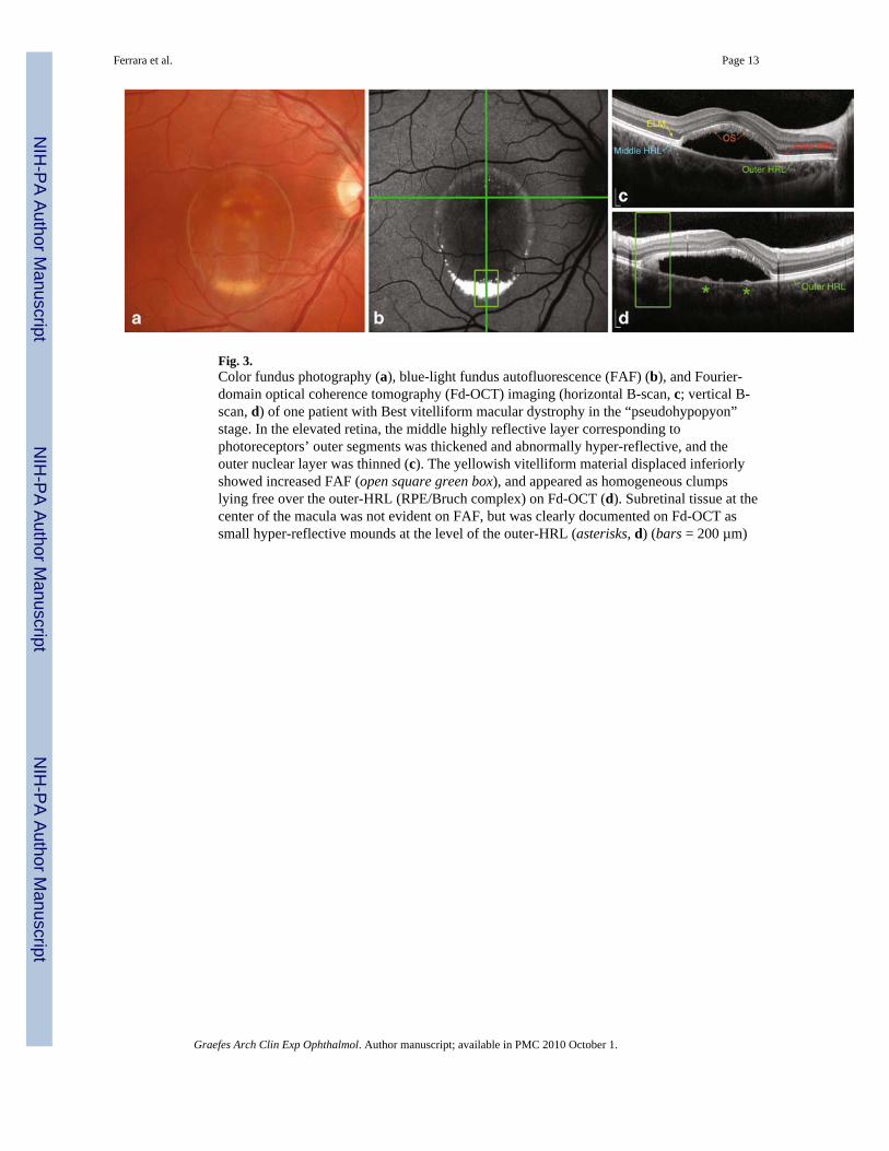
Fig. 3.Color fundus photography (a), blue-light fundus autofluorescence (FAF) (b), and Fourier-domain optical coherence tomography (Fd-OCT) imaging (horizontal B-scan, c; vertical B-scan, d) of one patient with Best vitelliform macular dystrophy in the “pseudohypopyon”stage. In the elevated retina, the middle highly reflective layer corresponding tophotoreceptors’ outer segments was thickened and abnormally hyper-reflective, and theouter nuclear layer was thinned (c). The yellowish vitelliform material displaced inferiorlyshowed increased FAF (open square green box), and appeared as homogeneous clumpslying free over the outer-HRL (RPE/Bruch complex) on Fd-OCT (d). Subretinal tissue at thecenter of the macula was not evident on FAF, but was clearly documented on Fd-OCT assmall hyper-reflective mounds at the level of the outer-HRL (asterisks, d) (bars = 200 µm)
Ferrara et al. Page 13
Graefes Arch Clin Exp Ophthalmol. Author manuscript; available in PMC 2010 October 1.
NIH
-PA Author Manuscript
NIH
-PA Author Manuscript
NIH
-PA Author Manuscript
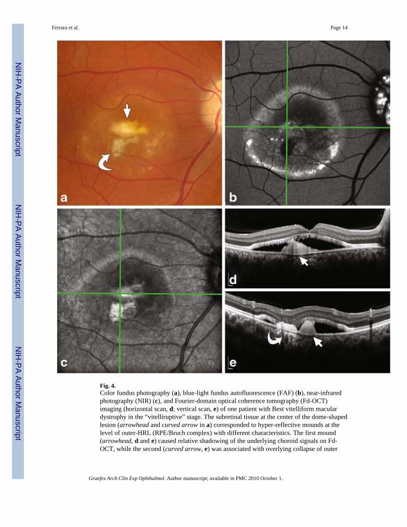
Fig. 4.Color fundus photography (a), blue-light fundus autofluorescence (FAF) (b), near-infraredphotography (NIR) (c), and Fourier-domain optical coherence tomography (Fd-OCT)imaging (horizontal scan, d; vertical scan, e) of one patient with Best vitelliform maculardystrophy in the “vitelliruptive” stage. The subretinal tissue at the center of the dome-shapedlesion (arrowhead and curved arrow in a) corresponded to hyper-reflective mounds at thelevel of outer-HRL (RPE/Bruch complex) with different characteristics. The first mound(arrowhead, d and e) caused relative shadowing of the underlying choroid signals on Fd-OCT, while the second (curved arrow, e) was associated with overlying collapse of outer
Ferrara et al. Page 14
Graefes Arch Clin Exp Ophthalmol. Author manuscript; available in PMC 2010 October 1.
NIH
-PA Author Manuscript
NIH
-PA Author Manuscript
NIH
-PA Author Manuscript

retinal layers and with relative hyper-reflectivity of the underlying choroid (bars = 200 µm).Note increased FAF from optic nerve drusen
Ferrara et al. Page 15
Graefes Arch Clin Exp Ophthalmol. Author manuscript; available in PMC 2010 October 1.
NIH
-PA Author Manuscript
NIH
-PA Author Manuscript
NIH
-PA Author Manuscript
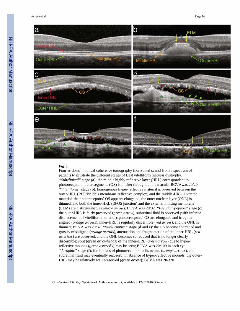
Fig. 5.Fourier-domain optical coherence tomography (horizontal scans) from a spectrum ofpatients to illustrate the different stages of Best vitelliform macular dystrophy.“Subclinical” stage (a): the middle highly reflective layer (HRL) correspondent tophotoreceptors’ outer segments (OS) is thicker throughout the macula; BCVAwas 20/20.“Vitelliform” stage (b): homogenous hyper-reflective material is observed between theouter-HRL (RPE/Bruch’s membrane reflective complex) and the middle-HRL. Over thematerial, the photoreceptors’ OS appears elongated, the outer nuclear layer (ONL) isthinned, and both the inner-HRL (IS/OS junction) and the external limiting membrane(ELM) are distinguishable (yellow arrow); BCVA was 20/32. “Pseudohypopyon” stage (c):the outer-HRL is fairly preserved (green arrow), subretinal fluid is observed (with inferiordisplacement of vitelliform material), photoreceptors’ OS are elongated and irregularaligned (orange arrows), inner-HRL is regularly discernible (red arrow), and the ONL isthinned; BCVA was 20/32. “Vitelliruptive” stage (d and e): the OS become shortened andgrossly misaligned (orange arrows), attenuation and fragmentation of the inner-HRL (redasterisks) are observed, and the ONL becomes so reduced that is no longer clearlydiscernible; split (green arrowheads) of the inner-HRL (green arrow) due to hyper-reflective mounds (green asterisks) may be seen; BCVA was 20/100 in each eye.“Atrophic” stage (f): further loss of photoreceptors’ cells occurs (orange arrows), andsubretinal fluid may eventually reabsorb; in absence of hyper-reflective mounds, the outer-HRL may be relatively well preserved (green arrow); BCVA was 20/320
Ferrara et al. Page 16
Graefes Arch Clin Exp Ophthalmol. Author manuscript; available in PMC 2010 October 1.
NIH
-PA Author Manuscript
NIH
-PA Author Manuscript
NIH
-PA Author Manuscript
NIH
-PA Author Manuscript
NIH
-PA Author Manuscript
NIH
-PA Author Manuscript
Ferrara et al. Page 17
Tabl
e 1
Bes
t-cor
rect
ed v
isua
l acu
ity a
nd e
lect
rooc
ulog
ram
Ard
en ra
tio o
f pat
ient
s with
Bes
t vite
llifo
rm m
acul
ar d
ystro
phy
and
resp
ectiv
e no
rmal
con
trols
Patie
nts
Con
trol
s
N°,
Gen
der,
Age
(yea
rs)
Stud
y ey
eB
CV
AA
rden
rat
ioN
°, G
ende
r, A
ge (y
ears
)St
udy
eye
BC
VA
Ard
en r
atio
1, M
, 5O
D20
/20
1.19
1, M
, 7O
D20
/20
2.67
OS
20/2
01.
17O
S20
/25
2.84
2, M
, 6O
D20
/32
1.17
2, M
, 8O
D20
/25
2.22
OS
20/3
21.
05O
S20
/20
2.09
3, M
, 19
OD
20/3
21.
123,
M, 1
9O
D20
/20
2.3
OS
20/2
51.
11O
S20
/20
2.51
4, M
, 37
OD
20/1
001.
284,
M, 3
8O
D20
/20
2.91
OS
20/1
001.
25O
S20
/20
2.93
5, M
, 45
OD
20/1
001.
085,
M, 4
4O
D20
/20
2.17
OS
20/3
201.
12O
S20
/20
2.56
6, M
, 62
OD
20/3
201.
276,
M, 6
6O
D20
/20
2.37
OS
20/4
01.
3O
S20
/25
2.34
7, M
, 59
OD
20/3
201.
117,
M, 5
8O
D20
/20
2.48
OS
20/5
01.
1O
S20
/20
2.16
BCVA
Bes
t-cor
rect
ed v
isua
l acu
ity; M
Mal
e; O
D R
ight
eye
; OS
Left
eye
Graefes Arch Clin Exp Ophthalmol. Author manuscript; available in PMC 2010 October 1.