Late gadolinium enhancement: precursor to cardiomyopathy in Duchenne muscular dystrophy?
Upload
khangminh22Category
view
2download
0
Duchenne Muscular Dystrophy:
mutation profiling
in view of the emerging gene-based therapies.
By
Alina Esterhuizen N. H. Dip. Med.Tech.
ESTALI001
SUBMITTED TO THE UNIVERSITY OF CAPE TOWN In fulfilment of the requirements for the degree of
MSc (Med) in Human Genetics
August 2010
Supervisor: Dr R. Goliath Co-supervisor: Prof. J. Greenberg
Division of Human Genetics, University of Cape Town

DECLARATION
I, Alina Esterhuizen, hereby declare that the work on which this dissertation is
based is my original work (except where acknowledgements indicate
otherwise) and that neither the whole work nor any part of it has been, is
being or is to be submitted for another degree in this or any other university.
This study has been approved by the Research Ethics Committee of the
Faculty of Health Sciences: reference number 416/2008.
I empower the university for the purpose of research to use either the whole
or any portion of the contents in any manner whatsoever.
II

ACKNOWLEDGEMENTS I would like to thank the following, for their role in making it possible for me to have attempted and completed this research:
The Muscular Dystrophy Foundation SA, who funded the project and
whose keen interest throughout, made me believe I was making a
difference.
Professor Egbert Bakker of Leiden University, for his collaboration
regarding the methodology and primer sequences.
A/Professor Jo Wilmshurst, the Head of Paediatric Neurology at the
Red Cross Children’s War Memorial Hospital, a first-class clinician and a
clinical co-investigator in this study, for her enthusiastic support of the
project and input regarding the clinical aspects, and selection of the study
cohort.
Celtic Diagnostics, especially Crystelle Klopper, for being ever-available
for advice and technical assistance with the RotorGene™6000, and
providing a service well beyond the call of duty.
The National Health Laboratory Service, for awarding me a study
bursary for the first year of this study course.
Professor Raj Ramesar, the Head of Division of Human Genetics at the
University of Cape Town for affording me this opportunity to learn.
My heartfelt thanks must go to the following special people:
My supervisor, Dr. René Goliath, for her calm, steady guidance and the
ability to “lead from behind” not only throughout this project but also
along the path of a new carrier direction.
Mrs. Maureen Lambrick, the NHLS laboratory complex manager, for her
encouragement, mentorship and friendship over the years.
My co-supervisor, Professor Jacquie Greenberg, for her kindness and
approachability for any kind of advice.
Finally, to my husband André, without whose steady presence, and his
willingness – and great capability - to “hold the fort”, I could never have
done it.
III

CONTENTS PAGE
LIST OF FIGURES……………………………………………………………………...VIII
LIST OF TABLES……………..………..…………………………………………....X
LIST OF ABBREVIATIONS……………………………………………………………..XI
ABSTRACT.…………………………………..…………………………………………XIII
PLAN OF THE THESIS………………………………………………………………...XIV
CHAPTER 1: LITERATURE REVIEW…………………………...………………………1
1.1 Introduction……………………………………………………..……………………….1
1.2 Muscular dystrophies………………………………………………………………..…1
1.3 Duchenne/Becker muscular dystrophy (D/BMD)……………………………………2
1.4 Disease manifestation………..………………………………………………………. 3
1.5 Clinical diagnosis……………………………………………………………………….5
1.6 Treatment and care…………………………………………………………………….6
1.7 The DMD gene and its product……………………………………………………….7
1.7.1 Dystrophin……………………………………………………………..…………9
1.8 Dystrophin-associated protein complex……………………………………….……10
1.9 Pathology of dystrophin-deficient muscle………………………..…………………12
1.10 Mutations in the DMD gene………………………………………………………...14
1.11 Genotype versus phenotype……………………………………………………….15
1.12 Exceptions to the reading frame rule……………………………………………...17
1.12.1 Exon skipping and alternative splicing....................................................17
1.12.2 Disruption of exonic splicing enhancer (ESE) sites and exonic
recognition sequences (ERS’s)…………………………………………….18
1.12.3 Activation of alternative translation initiation sites..………………………19
1.12.4 Somatic mosaicism…………………………………………………………..19
1.12.5 Unusual changes…………………………………………………………….20
1.13 Mutation detection…………………………………………………………………...20
1.13.1 Detection of exonic rearrangements……………………………………….20
1.13.2 Detection of point mutations………………………………………………...21
1.13.3 Haplotyping…………………………………………………………………...23
1.14 DMD carriers: genetic aetiology and testing……………………………………...23
1.15 Gene-based therapy………………………………………………………………...24
1.15.1 Exon skipping…………………………………………………………………25
1.15.2 Exon editing…………………………………………………………………..28
1.15.3 Nonsense read-through……………………………………………………..29
1.16 Animal models……………………………………………………………………….35
IV

1.16.1 The mdx mouse………………………………………………………………35
1.16.2 The Golden Retriever Muscular Dystrophy dog (GRMD)………………..36
1.16.3 The dystrophic cat……………………………………………………………36
1.17 The local perspective………………………………………………………………..37
1.18 Aims and objectives………………………………………………………………....39
1.18.1 Aims……………………………………………………………………………39
1.18.2 Objectives……………………………………………………………………..39
CHAPTER 2: MATERIALS AND METHODS………………………………………….41
2.1 Study cohort…………………………………………………………………………...41
2.2 Sample collection and DNA extraction…………………..…………………………42
2.3 Screening for exonic rearrangements……………………………………………...42
2.3.1 MLPA® Principle……………………………………………………………….42
2.3.2 MLPA® quality control………………….……………………………………...44
2.3.3 MLPA® probes……………………..…………………………………………..44
2.3.4 Sample preparation……………………………………………………………44
2.3.5 MLPA® reaction set-up………………………………………………………..44
2.3.6 MLPA® product separation……………………………………………………45
2.3.7 Data analysis and interpretation……………………………………………..45
2.4 Screening for small/point mutations………………………………………………...46
2.4.1 HrMCA background and principle……………………………………………47
2.4.2 HRM curve shape, shift and data normalization……………………………47
2.4.3 Validation of the hrMCA approach…………………………………………..48
2.4.4 PCR primers……………………………………………………………………49
2.4.5 PCR and hrMCA experiment set-up…………………………………………49
2.4.6 HRM data analysis…………………………………………………………….50
2.5 Cycle sequencing……………………………………………………………………..50
2.5.1 Sequencing reaction set-up…………………………………………………..50
2.5.2 Sequence resolution and analysis…………………………………………...51
2.6 Bioinformatic analyses………………………………………………………………..51
2.6.1 Effect on the translational open reading frame (ORF)…………………….51
2.6.2 ESE-finder and Human Splicing Finder (HSF)……………………………..52
2.6.3 SIFT (Sorting of Intolerant From Tolerant) analysis……………………….53
2.6.4 The Transcription Element Search System (TESS)……………………….54
2.7 Post-PCR mixing and HRM……………………………………….…………………55
2.7.1 Post-PCR mixing reaction set up…………………………………………….55
2.8 Family studies…………………………………………………………………………55
2.8.1 Restriction Endonuclease (RE) analysis……………………………………56
V

2.9 Population studies…………………………………………………………………….57
2.9.1 Experiment setup………………………………………………………………57
CHAPTER 3: RESULTS………………………………………………………………….59
3.1 MLPA® analysis………………………………………………………………………59
3.2 HrMCA….……………………………………………………………………………...61
3.2.1 HrMCA validation run…………………………………………………………61
3.2.2 HrMCA screen...……………………………………………………………….61
3.3 Cycle sequencing and bioinformatic analyses ……………………………………62
3.3.1 Disease associated mutations……………………………………………….62
3.3.2 Polymorphisms………………………………………………………………...68
3.3.3 Variants of uncertain significance……………………………………………70
3.4 Post-PCR mixing and HRM………………………………………………………….70
3.5 Family studies…………………………………………………………………………70
3.6 Population studies…………………………………………………………………….74
3.7 Potential eligibility for gene- based therapy………………………………………..74
CHAPTER 4: DISCUSSION……………………………………………………………..77
CHAPTER 5: CONCLUDING REMARKS……………………………………………...93
REFRENCES………………………………………………………………………………97
APPENDIX A……………………………………………………………………………..116
APPENDIX B……………………………………………………………………………..121
1. Advantages of MLPA® ……………………………………………………………….121
2. Limitations of MLPA®…………………………………………………………………121
3. MLPA® quality control………………………………………………………………...122
4. SALSA DMD MLPA® probes………………………………………………………...124
APPENDIX C……………………………………………………………………………..129
1. Important considerations for hr-MCA assay design………………………………129
Choice of a dsDNA intercalating dye………………………………………. 131
PCR design and optimization………………………………………………...133
Primer design…………………………………………………………………..133
MgCl2
concentration…………………………………………………………..133
VI

PCR reagents………………………………………………………………….133
2. Factor
………………………………………………………………….138
s affecting the melting curve and reliability of hr-MCA…………………….134
DNA and amplicon quality/quantity………………………………………….134
Amplicon size…………………………………………………………………..134
Primer dimers…………………………………………………………………..135
Effect of mutation type or the surrounding sequence context…………….135
Sample-to-sample uniformity…………………………………………………136
Temperature range during melting…………………………………………..136
Regions of normalisation……………………………………………………..136
The four classes of base changes…………………………………………...137
3. Primer sequences…
VII

igure 1.1 Distribution of predominant muscle weakness in different
…………….10
igure 1.4 Examples of immunocytochemical findings in normal and …………….13
igure 1.5 The correlation between the location of in-frame deletions
……………….33
igure 1.9 Immunohistochemistry of myotubules from primary cell
……………47
PO34–A2: deleted exons 22, 23, 24, 25, 26, 27, 28, 29 …………………..59
igure 3.2 MLPA® electropherogram for DMD420.1SIY, probe
………….61
igure 3.4 ESEfinder output for the splice site mutation c.2293-1G>A …………….….65
reference sequence of exon 55 in the DMD gene with that ……...….66
igure 3.6 The SIFT (Sorting Intolerant From Tolerant) analysis output …………67
Figure 3.7 Output screen from the UCSC Genome Browser on Human
analysis with TESS………………..69
LIST OF FIGURES
F types of dystrophy……………………………………………………………...2 Figure 1.2 Linear representation of the DMD gene and its products………………….8 Figure 1.3 Dystrophin and its associations within the cell……………… F dystrophin-deficient muscle…………………………………… F and the BMD or DMD phenotype…………………………………………...16 Figure 1.6 Exon skipping to restore the ORF…………………………………………..26 Figure 1.7 A model for nonsense mediated decay…………………………………….31 Figure 1.8 PTC124 suppresses premature nonsense codons………
F cultures derived from muscle biopsies…....33
Figure 2.1 Components of a SALSA MLPA® probe……………………………………43 Figure 2.2 The 4 steps of an MLPA® procedure…………………………………….....43 Figure 2.3 Fundamentals of a typical HRM (high resolution melt) plot… Figure 3.1 MLPA® electropherogram for DMD457.1MOG, probe set and 30..……………….……………………………………
F set PO34- A2: duplicated exons 3, 4, 5, 6, 7, 8, 9, 10…….……………...61
Figure 3.3 Coffalyser chart for DMD420.1SIY, probe set P034-A2……… F in patient DMD372.1DIN…………………………………… Figure 3.5 Graphical representation of the output from the Human Splicing Finder (HSF) program for the comparison of the of the mutant in patient DMD372.1PIE….……………………… F for the missense mutation c.587T>C in patient DMD389.1PIE
Feb. 2009 (GRCh37/hg19) Assembly………………………………………68
Figure 3.8 The “annotated sequence” output of
VIII

LIST OF FIGURES cont. Figure 3.9 Examples of the variable results of the post-PCR mixing
………………….71
igure 3.10 HrMCA analysis of family DMD411 with a nonsense ………………...73
affected proband in family DMD411 with a G>A change
……………75
igure A.1 National Health Laboratory Service (NHLS) request form
…………..117
School of Clinical and Laboratory Medicine,
………………………123
igure B.2 Effect of a poor denaturation (due to a high salt ………………....123
approximately 50 ng human male control DNA
……………………130
igure C.2 The dye re-distribution theory and the 2nd and 3rd
igure C.3 Adjustable hrMCA normalisation regions. ……………………………….136
hrMCA experiment on the RotorGene™ 6000…………
F mutation in exon 47…….…………………………………
Figure 3.11 A section of the sequencing electropherogram of the in exon 47(c.6905G>A)………………………………………………….....73 Figure 3.12 RE analysis and pedigree of family DMD411…………………………….73 Figure 3.13 Exon phasing of the DMD gene……………………………… F for genetic testing.. …………………………………………………………116 Figure A.2 The NHLS consent form for genetic testing and/or research Figure A.3 Consent for molecular studies (Division Human Genetics, University of Cape Town)…….…………………………………………….118 Figure A.4 Information sheet for participation in this research project……………..119 Figure A.5 Consent for participation in this research project………………………..120 Figure B.1 Effect of DNA quantity on Q-fragments. ………… F concentration) on D-fragments………………………… Figure B.3 Capillary electrophoresis pattern from a sample of analyzed with SALSA MLPA® kit P035-A2 DMD (lot 0508)……………123 Figure C.1 The RotorGene™6000 with its centrifugal design… F generation dsDNA intercalating dyes…………………………………….131 F
IX

able 1.1 Detection rates of exonic rearrangements with
able C.2 HRM and sequencing primers………………...……………………………138
LIST OF TABLES
T mPCR and MLPA® in the diagnostic service at GSH…………….………..38 Table 2.1 The complete study cohort……………………………………………………41 Table 3.1 Exonic rearrangements detected with the MLPA®…………………………60 Table 3.2 Disease-associated small mutations detected..……………………………63 Table 3.3 Previously reported polymorphisms…………………………………………64 Table 3.4 Changes of uncertain significance…………………………………………..64 Table 3.5 Family Studies………………………………………………………………....72 Table 3.6 Eligibility for potential gene modification therapy…………………………..76 Table B.1 SALSA MLPA® P034-A2 DMD probe mix…………………………………124 Table B.2 SALSA MLPA® PO35 - A2 DMD probe mix……………………………....125 Table B.3 DMD probes arranged according to chromosomal order………………..126 Table C.1 Classification of SNPs………………………………………………………137 T
X

e
e
ation
phoresis chromatograph
mutations d protein complex
y
elting curve analysis
dependant probe amplification robe hybridisation
cay oxide synthase
LIST OF ABBREVIATIONS ABD - actin-binding domain
enzym
ACE - angiotensin convertingAON - antisense oligonucleot
id
BMD – Becker muscular dystrophy CBC - cap-binding complex
cDNA – complementary DNA CHG - comparative genomic hy
bridiz
CK - creatine kinase
CNS – central nervous system
CpG – C-phosphate-G dinucleotide D/BMD - Duchenne and Becker muscular dystrophy DBS - dystrobrevin-binding domain
DGGE - denaturing gradient gel-electro DHPLC - denaturing high performance liquidDMD – Duchenne muscular dystrophy
y
DNA – deoxyribonucleic acid DOVAM - detection of virtually all DPC - dystrophin-associate DSBs - double stranded breaks dsDNA - double stranded
ECG – electrocardiogram EDL - extensor digitorum longus EJC - exon junction complex
ERG - electroretinogram ERS - exonic recognition sequences ESE - exonic splicing enhancer
ESS - exonic splice silencer sites FSIQ - full scale intelligence quotient
FVC - forced vital capacity GRMD - golden retriever muscular dystroph GSH - Groote Schuur Hospital HGVS - Human Genome Variation Society HRM – high resolution melting hrMCA - high-resolution m HSF - Human Splicing Finder ID - intellectual disability
IQ - intelligence quotient LOVD - Leiden open source variation database MLPA - multiple ligation- MLPH - multiple ligation-dependant pmPCR - multiplex PCR
mPCR - multiplex PCR mRNA – messenger RNA
NHEJ - nonhomologous end joining NHLS - National Health Laboratory Service
NMD - nonsense mediated de
nNOS - neuronal-type nitric
XI

ODN - oligodeoxynucleotide
trostomy
orpholino oligomer on
clease
Hospital al primer sequencing
ormation analysis ational polymorphism
ent Search System site/s
niversity of Cape Town
rsal Mutation Database for DMD,
IQ - verbal IQ LDCM - X-linked dilated cardiomyopathy
ORF - open reading frame ABBREVIATIONS cont. PABP - poly(A)-binding protein PCR – polymerase chain reaction PEG - percutaneous endoscopic gas PMO - phosphorodiamidate m PTC - premature termination cod PTT - protein truncation test RE - Restriction Endonu RFLP - restriction-fragment-length polymorphism RNA – ribonucleic acid RXH - Red Cross War Memorial Children’s SCAIP - single condition amplification/intern SIFT - Sorting of Intolerant From Tolerant SR proteins - serine/arginine-rich proteins SSCA - single-strand conf SSCP - single strand conform ssDNA - single stranded STR - small tandem repeat TAE - Tris-acetate EDTA TAP - mRNA nuclear export mediator TESS - Transcription Elem TFB - transcription factor binding Tm - melting temperature UCT - U
UMD–DMD France - UniveFrance UTR – untranslated region V X
XII

ABSTRACT
Duchenne Muscular Dystrophy (DMD) is a lethal, X-linked, recessive muscle-wasting
disorder affecting 1 in 3 500 live male births worldwide, for which only palliative care is
available to date. Large exonic deletions or duplications are found in approximately 70% of
DMD patients, for which diagnostic testing is available. The remaining 30% carry point
utations, which go largely undetected, as no testing is currently offered due to the great
s revealed 10 small/point
athogenic changes, 39 polymorphisms and 4 changes of uncertain significance. No
mutation
arrying patients in the cohort and skipping exons 45 - 55 could benefit a further 12,5%,
ion detection in the DMD gene, to help find “family mutations”,
us facilitating genetic counselling and ultimate determination of eligibility for mutation-
ased therapy in the future.
m
size of the DMD gene and the logistical challenges involved.
Positive outcomes of research into gene-based therapies necessitate availability of protocols
which will extend the scope of testing to detection of point mutations. In this study, the DNA
samples of 24 patients previously tested negative for exonic deletions with the mPCR
method, were subjected to a complete mutation screen of the coding region of the DMD
gene using the MLPA and hrMCA. Four deletions and 2 duplications were revealed by the
initial screen with the MLPA. The DNA of the remaining 18 patients was then subjected to
mutation scanning with hrMCA on the RotorGene™6000, of 96 PCR fragments
encompassing the entire coding region of the DMD gene, amplified using M13-tailed primers,
and subsequent sequencing of variant fragments. The analysi
p
pathogenic mutations were found in 8 patients of the cohort.
The 10 disease-causing changes identified, consisted of 3 nonsense, 4 frameshifts, 1 splice-
site, 1 compound mutation (a GGTG duplication and a missense), 1 missense, and 1 point
substitution in the Dp427promoter/exon1 region of the DMD gene. The deleterious nature of
the mutations detected was inferred by their nature and by the output of bioinformatic
analyses with regard to the effect on splicing, amino acid changes and regulatory
sequences. Also, the results of family studies showed that the pathogenic mutations were
familial in their origins and that they tracked with the disease within the families. Predictions
of future therapeutic options revealed that by virtue of cohort selection none of the patients
would benefit from AON-induced skipping of exon 51, which is currently undergoing clinical
trials. However, multiexon skipping of exons 6 – 8 could benefit ~30% of the
c
emphasizing the potential therapeutic impact of the multiexon skipping approach.
The study findings suggested that hrMCA could be successfully incorporated into the
diagnostic protocol for mutat
th
b
XIII

PLAN OF THE THESIS
This thesis is divided into five chapters. The first chapter provides an introductory
background to neuromuscular disorders as well as a general overview of the aim of
the study and the scientific approach to the investigation. The second chapter
details the scientific rationale for each of the molecular investigations undertaken in
this project. The third chapter follows with a presentation of the findings of the study.
The fourth chapter discusses each finding with respect to the global genetic
landscape of the DMD gene and patients. Genotype/phenotype correlations are
discussed and future prospects for further investigations are addressed. The thesis
is concluded in the fifth chapter, where the findings of the molecular studies are
ummarised and placed in context of the emerging DNA-based therapies currently
undergoing clinical trials.
s
XIV

CHAPTER 1: LITERATURE REVIEW
1.1 INTRODUCTION
Muscular dystrophies are a group of inherited disorders, caused by the lack of a
specific protein component in the muscle tissue. Starting with identification of the
dystrophin gene in the 1980’s, a number of muscular dystrophy genes and their
proteins have been characterised. The impressive amount of knowledge since
gathered on the molecular basis of muscular dystrophies has paved the way to a
better understanding of the disease mechanisms and improved diagnosis, both in
the clinic and in the laboratory. Availability of more reliable and informative genetic
testing is a direct benefit of this knowledge, which allows for more effective patient
management and accurate genetic counselling of the family.
Despite the advances in medical technology, muscular dystrophies particularly the
allelic Duchenne and Becker muscular dystrophies (D/BMD) remain lethal and
devastating to the patients and their families. While standards of care are continually
improving worldwide, currently available interventions are limited to the
management of symptoms and complications. Progress in terms of therapy has
been slow and fraught with pitfalls and setbacks. However, much research has been
focused on genetic therapies, and in recent years a number of new experimental
approaches appear to hold promise. Of those, read-through therapy and exon
skipping are especially relevant to D/BMD and to this study, and will be addressed in
more detail later in this review.
Still, the first step remains identification of the disease-causing mutations. Potential
availability of mutation-specific gene therapy marks the advent of personalised
medicine and emphasizes the need for a rapid and accurate method of detecting
small nucleic acid changes in D/BMD and other genetic disorders. Ideally, the
technology should be cost-efficient, sensitive yet robust and rapid enough to fit into
a diagnostic environment.
1.2 MUSCULAR DYSTROPHIES
Muscular dystrophies fall into the spectrum of neuromuscular diseases and are
defined as a group of heterogeneous, inherited disorders, characterised by
progressive weakness and wasting of skeletal muscle tissues. The classification
scheme devised by Walton and Nattrass in 1954, based on their own clinical
observations and still applied today, relies mostly on the distribution of muscle
1

weakness (Figure1.1), and the mode of inheritance (Walton, 1954). It identifies the
following main groups:
Duchenne/Becker-type (X-linked)
Facioscapulohumeral (autosomal dominant)
Limb girdle (genetically heterogeneous but mostly autosomal recessive)
Emery-Dreifuss (X-linked, autosomal dominant and recessive sub-types)
Distal (autosomal dominant and recessive clinically and genetically distinct
subgroups)
Oculopharyngeal (autosomal dominant)
Congenital (autosomal recessive with a more generalised muscle weakness)
Figure 1.1: Distribution of predominant muscle weakness in different types of dystrophy. A: Duchenne-type and Becker-type, B: Emery-Dreifuss, C: limb-girdle, D: facioscapulohumeral, E: distal, F: oculopharyngeal. Shaded=affected areas. (Emery, 2002)
Muscular dystrophies have since been mapped to 29 different chromosomal loci,
extending the above classification into 34 distinct disorders varying in the age of
onset, degree of severity, mode of inheritance and the primarily affected muscle
groups (Dalkilic & Kunkel, 2003).
1.3 DUCHENNE/BECKER MUSCULAR DYSTROPHY (D/BMD) (OMIM#310200)
Also known as Meryon’s disease, DMD is one of the most common and severe of
the inherited dystrophies, with an incidence of 1 in 3500 live, male births worldwide
(Emery, 2001). Long before the gene and its protein were characterised, early
2

investigators noted the X-linked recessive heritability of DMD, based upon scrutiny
of the affected family pedigrees (Meryon, 1851; Tyler, 2003). Also, early post
mortem studies of muscle tissues and the spinal chord suggested the myogenic
nature of the disease, with no involvement of the central or peripheral nervous
system (Emery & Emery, 1995). The milder, allelic form of DMD, Becker’s muscular
dystrophy (BMD), has a lower frequency (1 in 30 000 boys), a milder progression
and longer life expectancy (Becker & Kiener, 1955; Blake & Kroger, 2000a).
1.4 DISEASE MANIFESTATION
Duchenne muscular dystrophy, usually manifests in boys between the ages of 2 and
5 years, when parents typically notice a delay in motor milestones and symptoms
such as frequent falling, difficulty in getting up, gait problems, toe-walking and flat-
footedness. About 50% of the affected boys cannot walk independently at 18
months (Bushby et al., 2005).
The natural course of the disease is fairly predictable, although severity varies
between patients, depending on the causative mutation (Tuffery-Giraud et al., 2009).
Clinical examination generally reveals calf enlargement (pseudohypertrophy),
lumbar lordosis, which disappears on sitting, and weakness of the neck flexors.
Most DMD patients never learn to jump with both feet together. Weakness of the
knee and hip extensors causes the typical Gower’s manoeuvre: in an effort to
stand up from lying on his back, the child needs to turn onto his front and push
himself erect by moving his hands up his thighs. Muscle weakness is progressive,
starting with proximal weakness of the lower limbs, moving onto the distal lower and
then upper limbs, ultimately leading to wheelchair dependence (Emery, 2001;
Manzur & Muntoni, 2009). Loss of independent ambulation, as defined in DMD
occurs at 13 (although the mean is 9), in intermediate-type muscular dystrophy
(IMD) between 13 and 16 and in BMD, beyond 16 years of age, though the use of
steroids has made this data less fixed. Muscle weakness leads to scoliosis in 90%
of the cases, with ultimate loss of sitting balance, exacerbated by formation of
asymmetric contractures of the Achilles tendons and hips. In untreated patients,
respiratory and cardiac complications are the cause of death at the mean age of 19
(Bushby et al., 2010a). BMD has a later age of onset with a more diverse
presentation and progression, in some cases showing only mild myalgia and muscle
cramps with no weakness (Beggs et al., 1991).
Cardiac involvement is seen in all D/BMD patients but generally remains
subclinical in the early stages. It is likely that the late presentation of cardiac
3

symptoms is due to the decrease in physical activity, relative to the progressive
general muscle weakness. The spread of fibrosis caused by repetitive strain results
in left ventricular dysfunction and eventual dilative cardiomyopathy, if left untreated
(van Bockel et al., 2009). Cardiomyopathy is seen as the determinant of survival in
BMD patients, with an incidence of approximately 72%. It is the cause of death in
about 20% of the DMD, and 50% of the BMD patients (Manzur & Muntoni, 2009).
For the most part, respiratory function in D/BMD patients is normal before loss of
ambulation. As a rule, early loss of ambulation predicts early need for ventilation and
respiratory failure. The respiratory function parameter in D/BMD patients is the
forced vital capacity (FVC), which peaks shortly before loss of independent
ambulation and progressively drops thereafter, with eventual respiratory failure
manifesting as lowered energy levels, generalised malaise, weight loss, headaches,
sleep disturbance, nocturnal and subsequent daytime hypercapnia. The concurrent
increase in the frequency of respiratory infections raises the risk of death from
respiratory failure during an infection (Bushby et al., 2005).
Non-progressive cognitive impairment in DMD ranges in severity from borderline
neuropsychological deficits to severe intellectual disability (ID). Investigators
consistently report the full scale intelligence quotient (FSIQ) in DMD patients as
approximately 1 standard deviation below the population mean, with FSIQ scores of
under 70 points seen in 19–35% of DMD cases, and moderate to severe ID
(FISQ<50) noted in 3% of DMD patients (Cotton et al., 2001; Cotton et al., 2005;
Taylor et al., 2010). The gross anatomical morphology of DMD-affected brains
appears normal, although Yoshioka and colleagues found slight cerebral atrophy in
20 of the 30 DMD cases they examined, the extent of atrophy correlating directly
with age and low intelligence quotient (IQ) (Yoshioka et al., 1980). The results of a
study by Taylor et al. (2010) correlating FSIQ results with the location of the DMD
mutations are highly suggestive of a link between cognitive deficits and cumulative
loss of dystrophin isoforms expressed in the central nervous (CNS). Intellectual
disability in BMD patients is reported infrequently, and there is speculation that since
cognitive disabilities in D/BMD can occur before the onset of muscle weakness,
some patients diagnosed with X-chromosome-linked mental retardation might carry
mutations in the DMD gene (Blake & Kroger, 2000b).
Dystrophin is also expressed in the retina and some DMD patients show impaired
scotopic and photopic responses obtained by full-field electroretinogram (ERG).
Their visual function does not appear to be seriously compromised, although a
4

degree of non-progressive, red-green colour-blindness has been documented
(Costa et al., 2007).
Long bone fractures and fractures of the vertebrae are common in D/BMD,
because of low bone mineral density, possibly caused by relative immobility, further
exacerbated by the long-term use of corticosteroids. Progressive loss of mobility
along with steroid treatment can also lead to excessive weight gain, which in turn
leads to early immobility. On the other hand, loss of appetite frequently
accompanying respiratory failure, results in weight loss. Constipation is a
common complaint in older boys, due to the involvement of smooth muscle. In later
stages, difficulty in swallowing and frequent aspiration creates further nutritional
and even respiratory complications. D/BMD patients can also present with a
potentially fatal ‘‘malignant hyperthermia-like’’ reaction with rhabdomyolysis,
hyperkalaemia and myoglobinuria, upon exposure to suxamethonium or a
halogenated inhaled anaesthetic. This rhabdomyolytic risk is a major
consideration in their anaesthetic management, and easy access to monitoring aids
and intensive care facilities is strongly indicated (Manzur & Muntoni, 2009).
D/BMD along with other muscle disorders ranging in presentation from muscle
cramps and myoglobinuria, to DMD-associated dilated cardiomyopathy or X-linked
dilated cardiomyopathy (XLDCM), are also referred to as dystrophinopathies,
since all are caused by production of defective or insufficient levels of dystrophin
(Cardamone et al., 2008).
1.5 CLINICAL DIAGNOSIS
The assessments and interventions used in the diagnosis and management of
D/BMD have been recently evaluated by the DMD Care Considerations Working
Group, in an effort facilitated by the US Centers for Disease Control and Prevention
(Bushby et al., 2010a; Bushby et al., 2010b). According to the clinical care
recommendations, as set out by the Group, the diagnosis of DMD should be
considered irrespective of family history, following one of three triggers:
observation of abnormal muscle function in a male child,
detection of increased serum creatine kinase (CK) levels (in DMD massively
elevated by 10 – 100 x normal, since birth), a result of muscle tissue break
down,
5

discovery of increased transaminases (aspartate aminotransferase and
alanine aminotransferase, which are produced by muscle as well as liver cells)
(Bushby et al., 2010a).
The clinical diagnosis should be confirmed by:
Genetic testing
Mutation detection provides conclusive evidence and diagnosis. However, a
negative result does not negate the diagnosis of D/BMD. A good understanding
of the test limitations is therefore required. Testing the mother’s carrier status is
not strictly part of the diagnosis but facilitates genetic counseling of the family
(Abbs et al., 2010).
Muscle biopsy
Immunohistochemical staining for dystrophin in muscle tissue will reveal absent
or reduced dystrophin levels, which can be used to confirm a diagnosis of
D/BMD. In most centres however, because of the invasive nature of the
procedure, biopsies are taken only if molecular testing is uninformative (Bushby
et al., 2010a).
1.6 TREATMENT AND CARE
Ideally, clinical management of a D/BMD patient is a team effort coordinated by the
muscle clinic physician and involving a plethora of specialists: orthopedic, cardiac,
physiotherapists, dieticians, occupational therapists, psychologists, and family and
parent/patient support groups.
Glucocorticosteriods are the mainstay of treatment for muscle weakness in
D/BMD, best commenced at the plateau of the child’s physical performance
but long before loss of ambulation. Careful dosage monitoring and
prophylaxis are needed to counteract the negative effects of steroid therapy
such as weight gain, behavioural problems, bone density reduction, and
compromised immune function after prolonged use.
Physiotherapy: passive or active exercise as well as appropriate orthodesis
to prevent and treat contractures, scoliosis and for walking and/or sitting
postural support (seating / wheelchairs).
Splinting: as appropriate, depending of the degree of ambulation and ankle
dorsiflection.
Surgery: possible elongation of Achilles tendons and correction of scoliosis.
Anasthaesia: careful preoperative assessment of cardiac and respiratory
function and consideration of the rhabdomyolytic risk.
6

Respiratory management: prophylaxis, prompt diagnosis and treatment of
lung infections, positive pressure ventilation to treat hypercapnia and
respiratory failure.
Cardiovascular management: regular monitoring of cardiac function
(echocardiogram and ECG), treatment with ACE (angiotensin converting
enzyme) inhibitors. Additional agents after onset of symptoms.
Monitoring of bone health: diet supplementation with vitamin D and
calcium, intravenous biphosphonates for vertebral fractures, early
mobilization post long bone fracture, to prevent early loss of ambulation.
Nutrition: weight monitoring and diet adjustments, use of mild laxatives to
relieve constipation, intubation or percutaneous endoscopic gastrostomy
(PEG).
Addressing learning and emotional difficulties: occupational and speech
therapy, consultation with psychologists and support groups.
Access to adaptive technologies i.e. electric beds and wheel chairs,
computers etc., to aid an independent and functional life (Bushby et al.,
2005; Bushby et al., 2010b).
1.7 THE DMD GENE AND ITS PRODUCT
Localisation of the gene to the short arm of the X chromosome (Xp) was achieved
by extensive work with positional cloning and restriction fragment polymorphisms
(Davies et al., 1983; Monaco et al., 1986; Burghes et al., 1987; Koenig et al., 1987)
as well as cytogenetic studies of DMD-manifesting females, with balanced
X;autosome translocations breaking at Xp21 (Verellen-Dumoulin et al., 1984). The
full extent of the gene was eventually revealed by work with deletion-detecting
clones on DNA of a patient with four different X-linked disorders, in whom a large
contiguous-gene-deletion was confirmed (Francke et al., 1985). Similarly, BMD was
shown to map to the same region of the X chromosome (Kingston et al., 1984).
The DMD gene encompasses 2.5Mb of genomic sequence, with a full-length protein
encoded by a 14kb RNA transcript (Monaco et al., 1992). This is approximately
1.5% of the X chromosome and 0.1% of the entire genome, making the DMD gene
one of the largest, single protein-encoding genes described in humans to date. It
consists of 79 exons with 7 tissue-specific promoters (Figure1.2), which drive the
expression of dystrophin isoforms in various tissues (Manole, 1995; Muntoni et al.,
2003a):
7

Full-length isoforms are encoded by promoters with unique first exons, spliced
to 78 common exons:
Dp427M (muscle promoter) drives expression of dystrophin in the skeletal
muscle, cardiomyocytes and to a small extent in glial cells of the brain.
Dp427P (Purkinje promoter), on the other hand, is expressed in the cerebellar
Purkinje cells and at low concentrations, in skeletal muscle.
Dp427B (brain promoter), is expressed primarily in cortical neurons and the
hippocampus of the brain (Blake et al., 2002; Muntoni et al., 2003a).
Dp427L (putative lymphocyte promoter, not included in Figure 1.2), whose status
is uncertain but is at this time thought to be physiologically insignificant (Wheway
& Roberts, 2003).
Figure 1.2 Linear representation of the DMD gene and its products - adapted from A: The DMD gene position at Xp21 and its linear representation: exons represented as black lines and promoters as arrows). B: Linear representation of the dystrophin isoforms (Blake & Kroger, 2000b; Muntoni et al., 2003b). *skeletal and cardiac muscle
8

Short isoforms are expressed by at least four internal promoters, which splice
to various exons within the gene, generating protein products of 260 kDa (Dp260
to exon 30), 140 kDa (Dp140 to exon 45), 116 kDa (Dp116 to exon 56), and 71
kDa (Dp71 to exon 63) (Blake et al., 2002).
Alternative isoforms of dystrophin are generated through a number of
alternative, tissue specific splicing events, such exon skipping and exon
scrambling. These events sometimes result in the formation of circular RNA
molecules in co-existence with linear RNA, the biological function of which is still
unclear (Surono et al., 1999; Gualandi et al., 2003).
1.7.1 Dystrophin
Dystrophin is a large cytoskeletal protein, 427kDa in size, localized to the
cytoplasmic face of the muscle cell membrane. It acts as a mechanical link between
the cytoskeletal actin and the extracellular matrix (Figure.1.6) (Arahata et al., 1988;
Hoffman et al., 1987) and plays a role in intracellular signal trunsduction (Rando,
2001). The dystrophin molecule consists of four, main functional domains (Figure
1.2), each with its own protein-binding capabilities (Blake et al., 2002):
The NH2-terminus, along with a part of the rod domain binds directly to, but
does not cross-link cytoskelatal actin.
The central rod domain, makes up most of the protein and consists of 24
spectrin-like repeats, interrupted by 4 proline-rich hinges. The repeats confer the
molecule’s structural rigidity and the interspersed hinges allow its flexibility.
Spectrin repeats 11 through 17 within the rod region constitute a second actin
binding site.
The dystroglycan-binding domain consists of:
o WW domain follows the fourth hinge of the rod region and separates
it from the cysteine-rich and the COOH-terminal regions. It is a
protein-binding module and is part of a number of signaling and
regulatory molecules.
o The cysteine-rich domain consists of two EF-hand motifs, which
bind intracellular Ca2+ , and a ZZ domain containing a number of
cysteine-rich residues predicted to form coordination sites for divalent
metal cations e.g. Zn2+. This ZZ-domain is similar to other types zinc
finger found in nuclear and cytoplasmic proteins.
The COOH-terminal domain or CC-domain contains two –helical coiled coils,
which mediate its interaction with the syntrophins.
9

The shorter dystrophin isoforms (Figure 1.2), lack parts of the actin-binding and rod
domains but retain the cysteine-rich and the CC-terminal domains, which mediate
binding to the dystrophin-associated proteins (dystroglycan, dystrobrevin and
syntrophin).
1.8 DYSTROPHIN-ASSOCIATED PROTEIN COMPLEX
Initial biochemical purification of the compound from muscle led to the observation
that dystrophin co-purifies with a group of sarcolemmal and sub-sarcolemmal
proteins (Ervasti & Campbell, 1991; Ervasti et al., 1991). Together, they have been
shown to interact as the dystrophin-associated protein complex (DPC), which
consists of dystrophin, sarcoglycans, dystroglycan, dystrobrevins, syntrophins,
sarcospan, caveolin-3, and neuronal-type nitric oxide synthase (nNOS) (Blake et al.,
2002; Judge et al., 2006). The DPC form a scaffold connecting the actin-based
cytoskeleton with the basal lamina, with dystrophin acting as an essential structural
link in its assembly (Figure 1.3). Absence or dysfunction of dystrophin therefore,
causes instability of the DPC and an abnormally increased susceptibility to damage.
The DPC proteins are found in various combinations depending on muscle tissue
type and can be subdivided into three distinct protein subcomplexes, according to
their location and binding associations within the cell and between one another:
Figure 1.3 Dystrophin and its associations within the cell. (www.humgen.nl/lab-vdeutekom/introduction.html).
10

The Dystroglycan Complex
Dystroglycan forms an essential core of the DPC, as it connects the
cytoskeletal components of the DPC to the extracellular matrix (Figure 1.5).
Dystroglycan is produced from a single gene and is post-translationally
cleaved into and subunits (Lapidos et al., 2004). The extreme COOH
terminus of the -dystroglycan binds directly to the WW domain and the EF
hands of the cystein-rich region of dystrophin, emphasizing the functional
importance of both these domains (Crawford et al., 2000). -dystroglycan also
binds to Grb2, providing a known signaling pathway for -dystroglycan.
Caveolin-3, a transmembrane protein specifically expressed in muscle, also
interacts with -dystroglycan, and may compete for the same binding site as
dystrophin. This theory is supported by the reduced levels of dystrophin and -
dystroglycan in autosomal dominant limb-girdle dystrophy type 1C, which is a
result of calveolin-3 mutations. On the extracellular side, the -dystroglycan
forms an important connection to the 2-chain of laminin 2, forging a link is
between the sarcolemma and the extracellular environment (Rando, 2001;
Blake et al., 2002; Lapidos et al., 2004).
The Sarcoglycan Complex
The sarcoglycans are a group of transmembrane-spanning glycoproteins,
which co-purify as a complex within the DPC (Figure 1.3). The precise function
of the sarcoglycans within the cell membrane remains unclear and although
absence of the sarcoglycan-complex in mutant cells does not affect normal
distribution of dystrophin, it appears to cause defective cell membrane
permeability and subsequent increased fragility and degeneration (Rando,
2001; Blake et al., 2002; Lapidos et al., 2004).
Sarcospan is tightly associated with the sarcoglycans (Figure 1.3) and the
DPC and is thought to stabilise the dystroglycan complex in sarcolemma. It is
not crucial for normal function of the DPC, nor is it essential for formation of
the sarcoglycan complex. Sarcospan may have a signalling function, since it is
a member of the tetraspanin protein family, which has been implicated in
mediating the integrin-signaling responses in other tissues (Rando, 2001;
Blake et al., 2002; Lapidos et al., 2004).
The Cytoplasmic (dystrophin containing) Complex
Various isoforms of dystrobrevin bind directly to dystrophin in the brain,
muscle and other tissue. The syntrophins bind directly to dystrophin and
11

dystrobrevin. While the precise role of dystrobrevin in this context is unclear, it
is thought to play a role in intracellular signal transduction. The syntrophins
possibly function as a link between the membrane-associated proteins and the
DPC. Syntrophins also bind to the neuronal-type nitric oxide synthase (nNOS),
which may play a role in regulation of the vascular tone in skeletal muscle
fibres. Binding between dystrobrevin and syntrophin and by extension that
between syntrophin and neuronal nitric oxide synthase (nNOS), is mediated by
the CC-domain of the dystrophin molecule. In mdx mice, nNOS is known to be
dislodged from its site in the plasma membrane and becomes cytoplasmic.
Vasoconstriction in these mice is impaired, which is an indication of the
importance of association between the DPC and nNOS (Blake & Kroger,
2000a; Rando, 2001; Lapidos et al., 2004).
1.9 PATHOLOGY OF DYSTROPHIN-DEFICIENT MUSCLE
It is hypothesised that since dystrophin forms an integral part of a mechanical link
between the extracellular matrix and the cytoskeleton, faulty dystrophin causes
destabilization of that link and subsequent weakening of the sarcolemma (Judge et
al., 2006). Mechanical damage of fragilised membranes, caused by repetitive
eccentric contractions, provokes microlesions that are thought to lead to physical
sarcolemmal breaks or calcium leak channel openings. This in turn, elevates
intracellular free calcium levels triggering calcium-activated proteases and
subsequent fibre necrosis. Destabilisation of the sarcolemma is may also be linked
to the DPC’s involvement in cellular signalling, important for microvascular function,
muscle fibre type-determination and general myofiber homeostasis (Judge et al.,
2006). The primary manifestations of DMD are therefore thought to result from an
imbalance between muscle fiber necrosis and myoblast regeneration, with necrosis
as the primary pathologic feature (Deconinck & Dan, 2007).
Histological analysis of muscle samples from affected patients generally reveals
changes in the fibre size, fibre necrosis and increased amounts of fat and
connective tissue. Degenerating fibers are often observed in clusters confined to
segments of the muscle fiber (grouped necrosis). Necrotic or degenerating muscle
fibers are characteristically seen in postnatal DMD muscle biopsies even before
muscle weakness is clinically observed. In early disease, active regeneration is a
sign of fibre necrosis, with the regenerating fibers recognized by their small
diameter, basophilic RNA-rich cytoplasm, and large, centrally placed myonuclei. In
later stages, this regenerative capacity is exhausted and muscle is replaced by
connective and adipose tissue. Necrotic muscle fibers are subject to phagocytosis
12

and inflammatory cells, predominantly macrophages and CD4 lymphocytes, are
seen at perimysial and endomysial sites (Blake et al., 2002).
Immunohistochemical analysis of normal muscle shows dystrophin localised to the
sarcolemma with uniform labelling of each fibre. In DMD, dystrophin is mostly
absent, whereas in BMD, the fibres show reduced and/or uneven sarcolemmal
labeling (Figure 1.4). Some muscle biopsies of DMD, show “revertant fibres”, which
manifest low or even near normal expression of dystrophin. Immunolabelling is often
uneven on the fibre periphery and longitudinal sections reveal only partial labeling of
the fibre. This type of expression arises as a result of restoration of the translational
reading frame in some cells but the mechanisms by which this occurs are uncertain
(Sewry, 2000). Studies of revertant fibres in the mdx mouse suggest various
mutation-dependant mechanisms which result in alternative splicing events e.g.
‘‘exon skipping’’ (Winnard et al., 1995; Lu et al., 2000).
An estimated 26% of non-manifesting carriers are reported to exhibit a degree of
dystrophin-defective staining (E. M. Hoogerwaard et al., 2005), with occasional
dystrophin negative fibres, or uneven labelling. In symptomatic carriers, dystrophin-
negative muscle fibres are thought to result from a skewed pattern of X-inactivation
in the muscle (Sewry, 2000). The numbers of ‘‘negative’’ fibres are variable and
transverse sections in young cases often show mosaic patterns of positive and
negative staining (Figure 1.4).
Figure 1.4 Examples of immunocytochemical findings in normal and dystrophin-deficient muscle. Normal control (A): localization to the periphery of each muscle fibre. Patients with BMD (B and C): low level, discontinuous protein expression. Manifesting DMD carrier (D): “mosaic” expression of dystrophin in different fibres. Intermediate phenotype (IMD) (E): relatively abundant revertant fibres. DMD (F): absent protein expression. (Muntoni et al., 2003b)
13

1.10 MUTATIONS IN THE DMD GENE
Because of its size, the DMD gene is predisposed to mutations, approximately 65%
of which are estimated to be exonic deletions, and 5% to 15% are duplications
(Beggs et al., 1991). This broad estimate for the frequency of duplications has been
put down to the variable sensitivities of the techniques used, which should change
with the current, improved detection methods (Muntoni et al., 2003a). In a recent
study by Kesari et al. (2008), duplications were detected in as many as 21% of
mutation-positive BMD patients.
While exonic rearrangements can occur at any point within the gene, two hot-spot
regions have been identified: one situated towards the central part of the gene and
includes exons 45 to 53 (hot-spot region 1), and the other at the 5’ end of the gene,
spanning exons 2 to 20 (hot-spot region 2). Entries in the Leiden Open source
Variation Database (LOVD) for DMD (www.dmd.nl) record the most deletions in hot-
spot region 1, with the deletion of exon 45 being most common (1.7% of all DMD
mutations) (Aartsma-Rus et al., 2006). Deletions within the DMD gene (and other
genes on the X chromosome) are predominantly maternally inherited, as opposed to
duplications, which mostly originate in the male germline (Hu et al., 1990).
Consequently, duplications are usually seen as familial cases, and the recurrence
risk is high. Most duplications occur near hot-spot region 2, with exon 2 being most
commonly duplicated (0.4%of all mutations) (Aartsma-Rus et al., 2006; Gualandi et
al., 2009). Interestingly, the reciprocal deletion of the most common duplication
(exon 2) has never been reported (White et al., 2006; Flanigan et al., 2009).
Analyses of the deletion junctions indicate that they arise from rejoining of broken
ends via nonhomologous end joining (NHEJ) of double stranded breaks (DSBs).
DSBs arise during DNA replication and other endogenous processes, and are seen
to cluster in DSB hot-spot regions. There is evidence to suggest that in DMD, the
DSB-prone regions coincide with deletion/duplication hotspots, which also represent
major meiotic recombination hot-spots (Sironi et al., 2006; del Gaudio et al., 2008).
The remaining 30% of gene changes in DMD are small/point mutations, mostly
resulting in nonsense, frame-shift or splice site mutations (Roberts et al., 1994).
Deep intronic mutations have also been described (Béroud et al., 2004), as well as
exonic insertions of repetitive sequences and rarely, missense mutations.
Small/point mutations in the DMD gene do not appear to exhibit the same clustering
effect as exonic rearrangements and as mentioned earlier, their detection continues
to present a diagnostic challenge. However, compared to the amount of data
available on exonic rearrangements, the information on frequencies and distribution
14

of small/point mutations within the DMD gene, is less abundant, mostly due to the
technical difficulties in finding them. Therefore, as methods improve and data
becomes available, currently undetected trends may be uncovered. For example,
Deburgrave et al. (2007), found that of the 124 pathogenic point mutations identified
in that study cohort, 20 were in exons 53, 66 and 70 or in their flanking splice sites.
Also, the recently published genotype-phenotype analysis of all entries in the UMD-
DMD France database (www.umd.be) reported a deficit of point mutations in exons
32 and 48 and conversely, and excess of mutations in exons 4, 6, 7, 35, 66, and 70
(Tuffery-Giraud et al., 2009) . Also, the CpG dinucleotide has been shown to be a
hot spot for mutations in humans because it can undergo oxidative deamination of
5-methyl cytosine (Krawczak et al., 1998). Tuffery-Giraud et al. (2009) report that
43.8% (81/185) of substitutions leading to stop codons, involve CpG dinucleotides,
with the C-to-T substitution in the CGA codon as the most common event (90%).
The highest mutation rate was observed at a CpG site in exon 66. Three CpG sites
are located within exon 70 making this exon particularly prone to mutations.
Nonsense mutations due to other types of substitutions showed a wider distribution
along the gene (Tuffery-Giraud et al., 2009). Data gathered from a large cohort
study by Flanigan et al. (2009) supports uniform hypermutability of CGA>TGA
mutations but emphasises the absence of hot-spots for point mutations seen to
segregate within families as “private mutations”.
1.11 GENOTYPE VERSUS PHENOTYPE
The severe DMD phenotype results from mutations, which cause synthesis of a non-
functional and unstable protein, as opposed to BMD, where a smaller but semi-
functional protein is produced. The disease presentation (DMD as opposed to BMD
or IMD) is therefore not relative to the size of the mutation, as much as it is to:
Disruption of the DNA translational open reading frame (ORF)
The “reading frame hypothesis” can be applied to 90% of DMD cases (Monaco
et al., 1988), and it argues that mutations, which disrupt the ORF result in more
severe phenotypes than those, where the reading frame is retained. ORF
disruptions cause premature termination of translation which triggers the cellular
process of nonsense mediated decay (NMD) with its degradation of the mRNA
and the faulty protein (if any).
The mutation’s position within the gene
Disruptions of the actin-binding N-terminus and the carboxy-terminus of
dystrophin generally cause a severe phenotype, even if the rearrangement is in-
frame. Large, in-frame deletions at the 5’ end of the gene, which stretch across
15

the actin-binding and into the middle of the rod domain (deletions of exons 3 –
31, 3 – 25, 4 – 41, or 4 – 18) result in a DMD phenotype and account for some
of the exceptions to the reading frame hypothesis (Nevo et al., 2003). The
functional importance of the actin-binding domain is also emphasised by the
DMD-causing, smaller deletions at the 5’ region, such as those involving exons 3
– 13 (Muntoni et al., 2003b). The central and distal rod domains, on the other
hand, appear to be almost dispensable and cases of in-frame deletions of
approximately half of the gene have been described, with only a mild effect on
the phenotype (England et al., 1990). Conversely, some single exon deletions
e.g. exon 44, result in a severe DMD phenotype (Muntoni et al., 2003a).
Mutations involving the cysteine-rich domain invariably cause DMD, whereas
mutations located in exon 74 or higher are found in both BMD and DMD
patients. Phenotypes can also vary between patients carrying identical
mutations, even within one family which could be a result of the mutation’s
epigenetic context (Aartsma-Rus et al., 2006). The schematic overview in Figure
1.5 reflects general findings for in-frame deletions.
Figure 1.5 Correlation between the location of in-frame deletions and the BMD or DMD phenotype (Aartsma-Rus et al., 2006). Schematic drawing of the dystrophin protein with its fuctional domains. The location of the different exons is shown underneath the protein. (B) Graphic overview of the relation between the location of in-frame deletions and the severity of the phenotype (Beggs et al., 1991).
The effect of an exonic deletion on the ORF and possibly the phenotype can be
predicted, as the intron-exon boundaries have been well characterised and show
that some exons of the DMD gene do not contain an integral number of triplet
codons and if deleted, will result in a frame-shift of the mRNA (Baumbach et al.,
1989; Koenig et al., 1989). Such predictions are valuable to the clinician, especially
in early diagnoses of cases with no family history, as they help to anticipate the
disease severity and may influence the approach to treatment. Useful prediction
tools such as the Reading-Frame Checker accessible via The Leiden Muscular
Dystrophy Pages (www.dmd.nl), designed to predict the effect of the rearrangement
16

on the ORF. Such predictions however, can only be made reliably when based on
the mRNA changes. Genomic DNA-based findings may not reflect the true extent of
the rearrangements due to events such as altered or inefficient splicing. Reading
frame predictions for duplications, based on genomic testing are not recommended,
as non-tandem or complex duplications are being reported with increasing
frequency (with availability of more efficient testing), and determination of the
fragment orientation requires mRNA analysis (Flanigan et al., 2009).
1.12 EXCEPTIONS TO THE READING FRAME RULE
It has been estimated that 8 – 9% of DMD cases violate the reading frame rule,
(Aartsma-Rus et al., 2006) and the incidence may be much higher in BMD patient
populations, as suggested by results of a study by Kesari et al. (2008), where as
many as 30% had out of frame mutations. In-depth studies of such cases have
elucidated a number of underlying mechanisms.
1.12.1 Exon skipping and alternative splicing
Alternative splicing events can cause skipping of one or more exons around the
deletion/duplication, affecting the ORF and dystrophin production. Rearrangements
in certain parts of the gene appear to be more likely to result in exon skipping (5’
end, exons 3 – 6; 3 – 7; 5 – 7 and at the 3’ end, exons 44, 45, 51, 47 – 52 and 49 –
50) (Beggs et al., 1991; Gualandi et al., 2009). Similar genomic changes, however,
have been seen to result in different phenotypes. This could be explained by the
unusually large size of some of the introns in the DMD gene, ranging in size from
107 to 248 000 bp (average intron size of 27 kb), where the deletion/duplication
break-points can occur at different positions, disrupting regulatory sequences in
some cases. (Béroud et al., 2004). Gualandi et al. (2003) described a case in point,
where deletion of a single exon 5, resulted in DMD in some, and BMD in other
patients. Molecular scrutiny of mRNA revealed that the DMD patients produced
circular and linear RNA with missing exons 6 and 9. The BMD patients on the other
hand, exhibited a linear transcript only, with a probably in-frame deletion of exon 5
(Gualandi et al., 2003).
While the majority of the small/point mutations disrupt the ORF and result in a
severe phenotype, a number of BMD patients with nonsense and frameshift
mutations have been reported (Franz et al., 2000; Ginjaar et al., 2000; Disset et al.,
2006; Deburgrave et al., 2007). Again, alternative splicing and exon skipping events
are the mechanisms shown to be active in some of these patients’ phenotype
rescue. Exon-skipping is also the most common consequence of splice-site
17

mutations and may result in production of in- or out-of-frame transcripts, depending
on whether the particular exon boundry ends with an entire codon (Deburgrave et
al., 2007). Splice mutations can also activate cryptic splice sites and alternative
splicing events, where intronic sequences become incorporated into the mRNA.
Deep intronic mutations can activate entire cryptic exons (Dwi Pramono et al., 2000;
Gualandi et al., 2003; Béroud et al., 2004; Z. Zhang et al., 2007).
Studies also suggest that trans-acting splicing factors might play a role. According to
this hypothesis, splicing within the DMD gene might be affected by individual
differences in the levels of expression of CUG-binding proteins, which regulate
splicing via direct binding to the gene (Sironi et al., 2003).
1.12.2 Disruption of exonic splicing enhancer (ESE) sites and exonic
recognition sequences (ERS’s)
Splicing often involves a complex series of interactions between the sarcoplasmic
reticulum (SR) proteins, which bind to ESE sites and ERS, which in turn recruit other
splicing factors. Regulation of alternative splicing appears to depend on mutation-
specific creation of new, or disruption of the existing ESEs and ERS’s, or the less
common exonic splice silencer sites (ESS) (Cartegni et al., 2002). Reports of BMD
patients with nonsense mutations, such as those mentioned by Aartsma-Rus et al.
(2006) in their review article, are examples of such events. The two BMD patients
each carried a different nonsense mutation in exon 29. This is an apparent paradox,
as nonsense mutations generally result in severe phenotypes due to nonsense
mediated decay (NMD). In these two patients however, the mutations disrupted
exonic ESE sites, causing in-frame splicing out of exon 29, thus effecting restoration
of the ORF and production of a truncated dystrophin and a milder phenotype. Once
again, the effect is variable and depends on the strength of the particular enhancer or
silencer sequence (Aartsma-Rus et al., 2006). ESEs are well documented in the DMD
gene, and may be present in most exons (Caceres & Kornblihtt, 2002; Cartegni et al.,
2002). ESSs on the other hand appear less common (Disset et al., 2006). As in
prediction of the frameshift caused by exonic rearrangements, a number of
bioinformatic tools such as ESEfinder (Cartegni et al., 2003), RESCU-ESE
(Fairbrother et al., 2004) or the Human Splice Finder (Desmet et al., 2009) are
available to predict the presence of ESE motifs and to assess the consequence of a
single base alteration.
18

1.12.3 Activation of alternative translation initiation sites
Mutation-specific activation of an alternative translation initiation ATG codon, has
been described in a BMD patient with an out-of-frame deletion of exons 3 – 7 and no
evidence of exon-skipping in cDNA (Winnard et al., 1995). The authors suggest that
the mutation caused activation of an alternative translation initiation site present in
exon 8. Activation of an alternative translation initiation site from a methionine in exon
6 has since been directly experimentally demonstrated by Gurvich et al. (2009), in the
context of a founder nonsense mutation in exon 1.
1.12.4 Somatic mosaicism
Apparent violation of the frameshift rule can also be seen with somatic mosaicism in
the affected tissue. This is considered when immunohistochemical analysis of tissue
samples reveals a dual population of dystrophin-positive and dystrophin-negative
fibres (Hoffman et al., 1987) and molecular studies reveal two DNA populations
(normal and mutant) in blood DNA and in muscle cDNA, in the absence of
alternatively spliced transcripts (Deburgrave et al., 2007; Kesari et al., 2009). Since
the large DMD gene has a high (1/10 000) rate of de novo mutations in germ line
cells (sperm, eggs) (Caskey et al., 1980; Haldane, 2004), somatic mutations could
occur early in embryonic development, leading to production of tissues with both
dystrophin-positive and dystrophin-negative muscle fibres.
Despite the gene’s high mutation rate, reports of male somaticism are few (Saito et
al., 1995; Uchino et al., 1995; Deburgrave et al., 2007; Kesari et al., 2009), possibly
due to the low mutation rate in somatic cells relative to germ line cells, or
alternatively, poor ascertainment due to mild or unexpected clinical symptoms. An
interesting case of somatic mosaicism was reported by Kesari et al (2009). in a
patient presenting with acute cardiac failure, and raised CK levels in the absence of
overt muscle symptoms. Dystrophin immunostaining of muscle biopsy material
revealed a mosaic pattern, with a predominance of dystrophin positive fibres. DNA
studies showed no exonic rearrangements but cDNA sequencing presented
apparent heterozygosity for a C>T (U) change at position 8713 (r.8713c>u),
predicted to cause a nonsense mutation in the DMD gene (Arg2905X). The mutation
was not identified in the patient’s mother, although a number of shared
polymorphisms were seen, confirming a case of somatic mosaicism. The
frequencies of normal versus mutant genes were determined in blood/DNA (50:50),
muscle/DNA (80:20) and muscle/mRNA (90:10) (Kesari et al., 2009). The
discordance between muscle and blood, could be explained either as incidental
segregation of more dystrophin-positive stem cells into the biopsy region or by
19

“genetic normalization”, a process whereby the necrotic fibres are replaced with
normal stem cells, as seen in the majority of manifesting female carriers (functionally
somatic mosaics, but due to X inactivation rather than somatic mutations) who also
often present with cardiac symptoms. This process explains the reduction in serum
CK levels and improvement in clinical symptoms of some manifesting carriers, with
advancing age (Pegoraro et al., 1995; Kesari et al., 2009).
1.12.5 Unusual changes
Non-contiguous duplications, partial triplications and complex rearrangements in the
DMD gene are being reported with increasing frequency as a result of testing with
sophisticated technologies like oligonucleotide array-CGH and shown to have
varying effects on the phenotype (White et al., 2002; Kesari et al., 2008; White et al.,
2006). Detailed scrutiny of the complex junctions have led investigators to suggest
regional genomic instability, aided by the presence of repetitive elements, a stem-
loop structure, and possibly preexisting mutations, as the mechanism behind
complex rearrangements of the DMD gene (del Gaudio et al., 2008; Oshima et al.,
2009).
In most instances, the apparent violation of the reading-frame hypothesis is
disproved upon in-depth analysis and shown to hold. The importance of
understanding the exact genomic context of each mutation with its downstream
effect on the transcript and the protein is thus emphasised, not only in predicting its
phenotypic consequence, but also as relevant to application of the emerging
personalised gene therapy.
An interesting finding of a completely healthy male with a deletion of exon 16 and
part of introns 15 and 16 has been published (exon 16 encodes for 60 amino acids
situated within the central rod domain of dystrophin), which suggest that even large
changes in the dystrophin gene may not always be disease-causing The
investigators therefore caution against hasty diagnosis of dystrophinopathy in
sporadic cases of single exon in-frame deletions (Schwartz et al., 2007). In an effort
to avoid misdiagnosing young patients without positive family history, many
neurology centres confirm molecular findings of isolated cases with muscle biopsies.
1.13 MUTATION DETECTION
1.13.1 Detection of exonic rearrangements The Beggs and Chamberlain multiplex PCR, is a simple and reliable method used
to amplify selected exons across the hot-spot regions, still used in many diagnostic
20

laboratories today (Chamberlain et al., 1988; Beggs et al., 1990). In a multiplex
reaction, each target is amplified with its own set of primers, which is a limiting
factor, as large quantities of primers within a single PCR, result in problems such as
dimerisation and false priming. Therefore, the multiplex PCR deletion screen for
DMD involves at least three PCR reactions, which together amplify selected exons
across the 5’ and the 3’ mutation hotspots. This allows for detection of ~ 95% of the
DMD-causing deletions but all duplications and the remaining 5% of deletions
located outside of the hotspots, remain undetected.
The Salsa Multiple Ligation-dependant Probe Amplification (MLPA) test, (MRC-
Holland) is a sophisticated variant of PCR, which incorporates specific probes for all
the exons in the DMD gene (Lalic et al., 2005; Lai et al., 2006). The output is
analysed quantitatively and allows for detection of all exonic copy number changes.
MLPA currently features in the The Leiden Muscular Dystrophy Pages (www.dmd.nl)
as the recommended method for deletion/duplication detection in the DMD gene. It
also used in this study and will be addressed in more detail in Chapter 2 (Materials
and Methods). A number of other tests for detection of exonic rearrangements have
been developed with varying degrees of success.
The following are listed in the Leiden Muscular Dystrophy Pages as the most
effective, following the MLPA and the multiplex PCR:
MAPH (Multiple Amplifiable Probe Hybridisation): as informative as the
MLPA but more labour intensive and requires more DNA (White et al., 2002).
qPCR: theoretically simple but technically challenging and difficult to
optimise in multiplex (Ashton et al., 2008).
Southern Blotting: along with multiplex PCR and qPCR, can be used to
determine the deletion/duplication breakpoints (www.dmd.nl).
recently described high-resolution comparative genomic hybridization (CGH)
microarray: very expensive but capable of precise detection of intronic
breakpoints (Bovolenta et al., 2008; del Gaudio et al., 2008; Hegde et al.,
2008). It does not present a major diagnostic advantage at this point but may
become a valuable tool with the advent of “personalised gene therapy”.
FISH, CA-repeat marker analysis and exon-specific qPCR: useful for
confirmation of known mutations in carriers but not as an initial screen.
1.13.2 Detection of point mutations.
Currently, few laboratories in the world and certainly none in Africa offer detection of
point mutations as part of a diagnostic genetic work up for DMD. However, research
21

into development of personalised genetic and other forms of therapy are beginning
to show promise and much effort is directed at formulation of testing protocols and
development of methodologies for point mutation detection. The Leiden Muscular
Dystrophy Pages (www.dmd.nl) and the NCBI GeneTests (www.ncbi.nlm.nih.gov)
list the following methods as most successful, although others have been described:
High-resolution melting curve analysis (hrMCA)
HrMCA is simple and very sensitive (>98%). Although it requires specialised
equipment with high resolution melting (HRM) capability, it is particularly cost
effective as a pre-sequencing screen to resolve variant fragments. HrMCA is
the technological platform used to detect point mutation in this study and will
be addressed in detail in Chapter 2 (Materials and Methods).
Denaturing Gradient Gel-Electrophoresis (DGGE)
DGGE approaches 100% detection sensitivity (Hofstra et al., 2004).
However, it requires several PCR and electrophoresis conditions, which
make it laborious and it difficult to automate.
Direct sequencing
Direct sequencing (or SCAIP: single condition amplification/internal primer)(),
straightforward and powerful method but it is very costly, as over 79 separate
exon fragments must be amplified, sequenced and analysed (Flanigan et al.,
2003).
Single-Strand Conformation Analysis (SSCA)
SSCA / DOVAM (SSCA/detection of virtually all mutations)() is simple, cheap
and effective but laborious, demanding electrophoresis of over 79 PCR
fragments each using several electrophoretic conditions (Mendell et al.,
2001; Buzin et al., 2005).
Denaturing High Performance Liquid Chromotography (DHPLC)
Characteristics for DHPLC are similar to those for SSCA, although it is easier
to automate. It does however require specific, specialised equipment
(Bennett et al., 2001).
Protein Truncation Test (PTT) is an RNA-based screening method. It is
very effective in detecting truncating mutations but requires availability of
RNA extracted from a muscle biopsy, which is not always available. PTT
lymphocyte RNA is possible, but more difficult to perform (Tuffery-Giraud et
al., 2004). The cDNA fragments obtained after reverse transcriptase PCR
(RT-PCR) can also be used for sequencing to determine the exact mutation
(Hamed & Hoffman, 2006).
MyoD-induced in vitro muscle differentiation can be a source of RNA as
well a substrate for protein expression studies in pre and post-natal
22

diagnoses of cases where DNA analyses are uninformative. This is achieved
by transfection and subsequent forced expression of the MyoD gene in a
non-muscle cell types to initiate the process of myogenesis. This approach of
analysis of muscle protein in non-muscle cells, has been applied to prenatal
and postnatal diagnosis of DMD using fibroblasts, amniocytes and chorion
villi cells. (Sancho et al., 1993; Davis et al., 1987; Roest et al., 1996; Roest
et al., 1999).
For the most part, mutation detection techniques focus on the protein coding regions
of the DMD gene. Studies analysing other regions (promoters, 5'UTR and 3'UTR)
have so far not revealed many changes (e.g. (Tubiello et al., 1995; Flanigan et al.,
2003). While DNA-based screening is offered most widely, RNA-based screening is
considered most effective, as cDNA analysis can resolve all mutations resulting in
protein truncation and those affecting RNA-splicing. The difficulty lies primarily in
sample acquisition, as mRNA is best obtained from muscle tissue. Lymphocyte
mRNA in this context is not ideal, as certain mutations (e.g. splice mutations) in
lymphocyte transcripts may behave differently to those in muscle (Ferlini et al.,
1998). This is of particular relevance with the advent of personalised genetic therapy
as establishing the downstream effects of the mutation is imperative for therapy
design e.g. antisense oligonucleotide (AON) design for exon skipping.
1.13.3 Haplotyping
This is a DNA-based approach to identify the risk chromosome in cases where no
mutation could be found with other methods. The original restriction-fragment-length
polymorphism (RFLP) assay (Bakker et al., 1985), has been refined to construction
of a risk haplotype using informative small tandem repeats (STRs) in and around the
DMD gene.
1.14 DMD CARRIERS: GENETIC AETIOLOGY AND TESTING
DMD female carriers, although unaffected, have CK levels 2 -10 times the upper
level of normal in over 50% of the cases. It is estimated that approximately one fifth
of female carriers show some signs of the disease (Hoogerwaard et al., 1999).
These symptoms range widely from mild cramps on muscle exertion, calf
pseudohypertrophy, varying degrees of muscle weakness (sometimes slowly
progressive), to severe weakness and wheelchair dependence in rare cases.
Impaired cognitive and/or cardiac function can be a part, or an exclusive
manifestation of the disorder. Apart from a few cases associated with chromosomal
rearrangements, most girls have been assumed to be affected as a result of skewed
X inactivation, though this is not supported by the recent report of Soltanzedeh et al.
23

(2010), where no definite link could be established between the phenotype in
female carriers and the pattern of X-linked inactivation. Muscle weakness, if present,
is asymmetric and proximal, involving the pelvic girdle rather than the shoulder
girdle in most cases, and can lead to misdiagnosis as the limb-girdle variety of
dystrophy. Age of onset is variable, ranging from the first to fourth decade, with early
onset (before the age of 15) linked to a more severe involvement. As is the case
with male patients, carriers of BMD are less frequently and less severely affected
than carriers of DMD (van Essen et al., 1997).
A DMD gene mutation in a female can be inherited from a carrier or a
somatic/germline mosaic mother, or from a germline/somatic mosaic father.
Alternatively, her mutation could have arisen de novo in the ovum or sperm from
which she arose or in her own germline or somatic cells. Approximately 1/3 of DMD
cases result from new mutations and the a priori carrier risk for mothers of sporadic
cases is estimated at 66%. Absence of other affected family members and low
maternal blood CK levels reduce this risk. An affected child of a non-carrier mother
can be a result of a new mutation in the ovum from which the child arose or of
somatic mosaicism in the affected child. Sporadic cases arising from somatic and/or
germline mosaic mother are also seen as new mutations, as are mutations which
originated in a germline of a mosaic grandfather, which were passed onto the
mother. The recurrence risk of a germline mosaic mother of an affected child lies at
an estimated 5 – 10% (Bakker et al., 1989; van Essen et al., 1992). Identification of
exonic rearrangements in females is now possible with quantitative analyses such
as the MLPH and MLPA. In cases where no mutation is detected in the proband,
haplotype analysis is used to track inheritance of the risk chromosome.
Immunohistochemical staining of muscle tissue can be helpful in ascertaining
D/BMD carrier status where neither mutation detection nor haplotyping is
informative. In absence of a genetic diagnosis, it is essential in differentiating
between manifesting DMD female carriers from autosomal forms of muscular
dystrophy. A young, dystrophic female with normal muscle dystrophin
immunolabelling is unlikely to be a manifesting carrier of DMD. This, however, may
not be the case in older females, due to genetic normalization (Webster & Blau,
1990; Hoogerwaard et al., 2005).
1.15 GENE-BASED THERAPY
Much of the research into gene therapy for DMD has been facilitated by studies on
two naturally occurring models of DMD: the dystrophic golden retriever dog (GRMD)
24

mdx mouse (Banks & Chamberlain, 2008). It must be emphasised that that not all
of the therapeutic approaches discussed under the broad heading of “gene therapy”
can truly be classified as such, as some use conventional pharmacological agents
and others target the transcript rather than the underlying gene (Lim & Rando,
2008). In this context, the term is used loosely to describe treatments developed
specifically to target genetic disorders.
The approaches to development of genetic therapies can be roughly divided into two
main fields: gene replacement and gene modification (Lim & Rando, 2008). Gene
replacement strategies (viral vectors, utrophin upregulation, myoblast and stem-cell
transplantation), aim at delivery of a normal copy of the gene directly to the affected
tissue, either as naked DNA in a viral vector (Wolff et al., 2005; Blankinship et al.,
2006), or as a gene copy in a myoblast or stem cell (Farini et al., 2009). Proof-of-
principle experiments on animal models (the mdx mouse), have been conducted
and demonstrate that dystrophin expression can be restored to varying degrees to
single muscles, muscle groups (limbs) or the entire body. The major advantage of
this strategy is that it can be applied to patients regardless of the underlying
mutations. Although the research is showing promise, much work still needs to be
done to overcome the mechanical and practical difficulties of gene delivery to the
muscles, the immunological concerns and the high cost. The other approach to
genetic therapy, namely, gene modification, also referred to as mutation- or gene-
based therapy is currently receiving much attention. Exon skipping and read-through
of stop codons, discussed below in more detail, have reached the point of phase II
clinical trials. In terms of their application and delivery, these strategies are seen as
less difficult and risky as well as significantly cheaper than the gene replacement
approaches. Effectively, as identification of the particular mutation is the basis for
this approach, “personalised gene therapy” is becoming a reality and is of
particular relevance to this study.
1.15.1 Exon skipping
The theoretical basis of exon skipping is the naturally occurring phenomenon of
restoration of the ORF by mutation-triggered alternative splicing mechanisms, such
as those seen in BMD patients with frameshift mutations. Also, characterisation of
the DMD gene and dystrophin with its functional domains, and experimentation with
truncated mini-genes on mdx mice has shown that a degree of dystrophin
functionality is preserved with parts of the protein missing (Phelps et al., 1995;
Harper et al., 2002). Therefore, the aim of exon skipping therapy is restoration of the
ORF and amelioration of the phenotype from DMD to BMD, by induced splicing out
(or skipping) of one or more exons. Modification of dystrophin mRNA splicing is
25

achieved with the use of antisense oligonucleotides (AONs, also known as
“molecular patches”). These small DNA or RNA analogues attach to mRNA
sequences crucial to the splicing process and induce alternative splicing out of
targeted exons (exon skipping) (Manzur & Muntoni, 2009) (Figure 1.6). The
sequences targeted by the AONs include 5’ and 3’ consensus splice sites and intra-
exonic splice regulatory sequences (Lim & Rando, 2008). The feasibility of this
approach was tested in the initial in vitro studies on human muscle and the in vivo
gene model in mdx mice (Mann et al., 2001; Lu et al., 2003). Subsequently, much
work has been done using in vivo animal models, in search of the optimal
oligonuclear conditions and strategies to improve efficiency of delivery, with minimal
toxicity and immunogenicity.
Modifications to the chemistry of AONs are still required to increase their half-life
and to prevent the cleavage of RNA-RNA hybrids by RNAse H. The most commonly
used chemistries for exon-skipping antisense oligonucleotides are 2’O-methyl RNA
with a negatively charged phosphorothioate backbone (2’OMePS) (Lu et al., 2003)
and uncharged phosphorodiamidate morpholino oligomers (PMO) (Fletcher et al.,
2007), because both are stable and non-toxic.
Figure 1.6 Exon skipping to restore the ORF.
Exon 51 is the focus of the first clinical trials, as AONs able to effect efficient
skipping of exon 51 have been identified. Also, skipping of this single exon would
restore the ORF in patients with deletions of exons 50, 52, 52–63, 45–50, 47–50, 48
– 50 and 49–50, which comprise 13% of the DMD population. Two independent,
proof-of-concept and safety trials on DMD patients, both targeting exon 51, have
been successfully completed. Intramuscular injections of the 2-O-methyl AON
PRO051 were administered by the group in Netherlands and the AVI-4658
morpholino approach was adopted by the Muntoni group in the Imperial Collage of
London (Kinali et al., 2009; van Deutekom et al., 2007). Both studies yielded good
26

results without drug-related adverse events. These encouraging results led to the
initiation of further trials by both groups. The phase I/II open-label study using
subcutaneous administration of PRO051 aimed to evaluate the safety profile of
PRO051 and its ability to induce specific exon 51 skipping and novel dystrophin
expression, has been recently completed. The preliminary data showed good
tolerability and induction of dystrophin expression in a dose related manner. The
published results of this study are however still pending. The results of a similar trial
in the United Kingdom involving intravenous infusions of AVI-4658 at escalating
doses is due to be completed in the first half of 2010 (http://www.clinicaltrials.gov).
Systemic delivery of the AONs over an extended period of time will however truly
test the promise of this therapy (Aartsma-Rus & van Ommen, 2009; Guglieri &
Bushby, 2010).
Successful application of exon 51 skipping, will lead to design, development and
testing of AONS for skipping other single exons. However development of AONs
specific to each individual mutation, in a gene this size, is a tremendous endeavor,
with its risks of variable effect and an immune response to possible newly created
epitopes. Since it has been shown that dystrophin can maintain some of its essential
function with large parts of the gene (and protein) missing, the feasibility of
designing a cocktail of AONs, capable of inducing double- or multi-exon skipping in
approximately 60% of all DMD cases, is under investigation. Multi-exon skipping
experiments have been conducted with encouraging results on animal models:
studies with mdx4cv mice, which require skipping of exons 52 and 53 (Mitrpant et
al., 2009), and the canine muscular dystrophy dog model (CXMD), where skipping
of exons 6 and 8 corrects the splice mutation in exon 7 (Yokota et al., 2009). Also,
an AON cocktail targeting all exons between 45 and 55 has already been tested on
human cells in vitro, however, effective skipping of this number of exons is yet to be
achieved (van Vliet et al., 2008).
Theoretically, large duplications in the DMD gene should be an ideal target for the
exon skipping therapy, as skipping of the duplicated exons should result in
production of the wild type protein. Experiments on cultured muscle cells from
duplication-carrying DMD patients, transfected with AONs targeting the duplicated
exons, showed some degree of success. Further work is however required, as
specific targeting of the duplicated, as opposed to the constitutive exons (or both)
has proved problematic in cases (Aartsma-Rus et al., 2007).
27

An interesting approach towards a more permanent repair has been tested in cell
cultures and the mdx mouse, where AONs in tandem with a modified U7 small
nuclear RNA are expressed from an adeno-associated virus (Goyenvalle et al.,
2009). Again, more work is required, as the problems accompanying the gene
replacement therapies are also relevant here (Trollet et al., 2009).
1.15.2 Exon editing
Small oligonucleotides are also the basis of another form of therapy, known as
oligonucleotide-mediated gene editing or gene repair, which aims at correcting
small, in-frame mutations such as nonsense, missense and splice-site mutations
(Igoucheva et al., 1998). It is based on the hypothesis that in the presence of an
oligonucleotide exactly matching the genomic sequence, except for the single base
mismatch, the cellular repair systems can be induced to modify the mutated
genomic sequence in a proportion of cells, possibly through mismatch repair
enzymes (Gamper et al., 2000; Andersen et al., 2002). This should result in a
normal transcript and a full-length protein. The originally used small DNA sequences
flanked by 2’-methyl RNA sequences (chimeroplasts) have been superseded by
short all-DNA ologonucleotides, which anneal to one strand of the genomic
sequence and have been shown to yield more reproducible results and are cheaper
and simpler to synthesise. In vitro and in vivo experiments on the mdx mouse model
(nonsense mutation in exon 23) and in the golden retriever dog (splice mutation at
exon 7) have shown that this oligodeoxynucleotide (ODN)-mediated gene repair
technology is capable of successful modification of single base changes in genomic
DNA and thus correction of dystrophin expression at the sarcolemma, with wild-type
dystrophin detectable at the site of the injection for up to 48 weeks. The clear
advantage of this therapy is its permanence without the need for ongoing drug
administration, although this is yet to be fully established by determining the
therapeutic effect of a single dose administration. Also, while the findings are
encouraging from the proof-of-concept viewpoint, the efficiency of gene expression
achieved in these animal models was low, at only 20% at the injection site. Systemic
delivery generally yields even lower levels and therefore much research is focused
on optimal ODN design and on improving the efficacy and efficiency of delivery
(Bertoni, 2005; Robinson, 2009).
Recently Maguire et al. (2009), took the ODN-mediated gene repair a step further
with an experiment on mdx5cv mice, in which the repair takes place ex vivo, in cell
cultures enriched with myoblasts. Mdx5cv mice have a single point mutation (A to T),
which creates a donor splice site within exon 10 and gives rise to a 53-base deletion
28

in the transcript, which in turn causes a frameshift and a stop codon in exon 11. In
normal muscle, the presence of damaged fibres activates the surrounding resting
precursor cells into active myoblasts, which divide rapidly and effect regeneration of
the injured tissue. Cycles of damage and regeneration are more frequent in DMD-
affected tissue, creating an abundance of myoblasts. The authors speculate that
isolated myoblasts subjected to ODN-mediated gene repair, could be transplanted
back into the muscle, creating long-term restoration of dystrophin expression. To
facilitate oligonucleotide binding and higher levels of gene repair, the investigators
attempted to further strengthen the reaction by the use of RNAi to knock down the
Msh2 protein, a known inhibitor of gene repair. While the results of the experiment
with primary myoblast-enriched cell cultures showed significant gene repair and
increased dystrophin production, purified myoblasts proved less susceptible to both
repair and Msh2 knockdown. The proposed explanation is the difficulty in genetic
manipulation of progenitor cells in general, due to natural barriers including lower
transfection efficiencies and dosage-dependent toxicity (Maguire et al., 2009). This
however, does not negate the possible potential of this approach and reinforces the
need for further experiments.
The main challenges of exon skipping and editing therapies lie in overcoming the
efficiency of delivery, as according to the best estimates, 30 – 40% of normal levels
of dystrophin are needed to effect an improvement in clinical signs and symptoms
(Neri et al., 2007). Coupling AONs with carrier molecules such as nanopolymers of
polyethylene glycol and polyethyleneimine or polyactide-co-glycolic acid
nanospheres and cationic core shell nanoparticles, have shown some promise,
although potential toxicities have yet to be established (Williams et al., 2008; Sirsi et
al., 2009). The nature and position of the target sequence (e.g. exonic or intronic)
also play a significant role. Another current limitation of AON therapy is the inability
to target the heart in mdx mouse at doses suggested for clinical applications
(Townsend et al., 2008). Recent studies have suggested incorporation of peptide or
chemical moieties with the AONs to facilitate cell uptake and prevent disruption of
cardiac sarcolemma, cardiac hypertrophy and dysfunction in mdx mice (Townsend
et al., 2008; Wu et al., 2008; Yin et al., 2008).
1.15.3 Nonsense read-through
According to the Human Gene Mutation Database, nonsense mutations account for
~12% of all mutations causing human inherited disease (Mort et al., 2008). They
arise as a result of single point alterations, which create in-frame UAA, UAG or UGA
codons in mRNA coding regions, leading to premature termination of translation and
29

subsequent nonsense-mediated decay (NMD). NMD is a quality-control
mechanism, whereby aberrant mRNA molecules containing premature termination
codons (PTCs) are degraded to prevent accumulation of incomplete, potentially
toxic protein fragments (Figure 1.7). Pre-mRNA splicing in the nucleus, leads to
placement of several proteins, including UPF3, on the CBC-bound mRNA, 20–24
nucleotides upstream of exon–exon junctions, to form the exon junction complex
(EJC). During translation of normal transcripts, the EJC is remodelled and CBC is
replaced by 4E, which promotes mRNA export to the cytoplasm. If a transcript
contains a PTC, the ribosome stalls and is converted into a surveillance complex,
which is hypothesized to consist of the 40S ribosomal subunit bound to NMD
effector molecules (UPF1-3 and SMG1-7). This surveillance complex acts as a link
between the premature translation termination factors (eRF1 and eRF3) and the
general cellular mRNA decay machinery. Recognition of a stop codon as premature
rather than normal appears to depend on its location relative to the downstream
sequence elements and associated proteins. Natural stops are typically located
within terminal exons, whereas PTCs lie more than 50 - 55 nucleotides upstream of
the last exon–exon boundary (Maquat, 2004). While EJCs located in an open
reading frame upstream of exon-exon junctions may facilitate ribosomal recruitment
prior to displacement by a "pioneer" round of translation, EJCs located downstream
of a nonsense codon (and 20–24 nucleotides upstream of splice junctions) (Chang
et al., 2007) are not displaced because the ribosome is released from the transcript
before reaching it. These remaining EJCs function as tags for recruitment of UPF1,
followed by the mRNA's transport out of the nucleus and into the cytosol where the
RNA is degraded by, for example, the exosome complex. NMD, however, does not
appear to be an invariable consequence of a PTC. Indeed, PTCs located either less
than 50–55 nucleotides upstream of the last exon–exon junction or downstream of
this junction generally fail to elicit NMD (Behm-Ansmant et al., 2007). Also, even
when NMD is triggered, some 5 to 25% of nonsense mutation-containing mRNA
routinely escapes NMD (Isken & Maquat, 2007). The pathological effects of these
mutations are therefore thought to be due to degradation of the newly synthesized
PTC-containing mRNA by NMD, and also in part to the failure of the small amount of
the PTC-containing mRNA that escaped NMD, to yield a (semi-) functional protein
(Khajavi et al., 2006).
Aminoglycosides are a group of antibiotics, which bind to the 30S or 50S subunits
of the bacterial ribosome and can induce misreading of the mRNA codons, as part
of their mechanism of action. This capability has been studied as a potential
mechanism to read through disease-causing nonsense mutations in humans, as
30

theoretically, this should lead to generation of a full-length proteins with a single
amino acid substitution, thus correcting the primary genetic defect. The initial
experiments were conducted on cystic fibrosis, where read-through of known
disease-associated nonsense mutations in the transmembrane conductance
regulator (CFTR) gene, was achieved in cell lines exposed to high doses of
aminoglycosides (Howard et al., 1996).
Figure 1.7 Model for nuclear nonsense mediated decay. CBC: cap-binding complex, EJC: exon junction complex, PABP2: nuclear poly(A)-binding protein, PABP: cytoplasmic poly(A)-binding protein, TAP: mRNA nuclear export mediator, 4E: translation initiation factor eIF-4E (Wilkinson & Shyu, 2002).
In 1999, the first in vivo evidence of phenotypic suppression of a premature stop
codon was obtained by subcutaneous injection of high doses of gentamicin into
transgenic mdx mice carrying a nonsense mutation in exon 23 of the DMD gene
(Barton-Davis et al., 1999). Subsequent evaluation of gentamicin as a treatment for
cystic fibrosis proved it be of limited clinical use, due to its known oto- and
nephrotoxicity in high doses, as well as the need for regular intramuscular and
31

intravenous administration (Clancy et al., 2001; Politano et al., 2003). Under
investigation, is a newly developed aminogylcoside (NB54), with a higher read-
through efficacy and an improved toxicity profile in different cell lines, as compared
to gentamicin. However, further studies in animal models are required before
considering its potential applicability (Nudelman et al., 2009).
The results of the experiments with aminoglycosides did show that read-through of
PTCs could be achieved biochemically, which prompted a search for a molecule
with a more suitable profile of low toxicity and higher read-through efficiency
(Politano et al., 2003). Two high-throughput screens of approximately 8000 000
molecules were conducted against a luciferase reporter that harbors a UGA PTC,
identifying PTC124 as a candidate for further development (Welch et al., 2007).
Ataluren (PTC124) is a 284.24Da, achiral, 1,2,4-oxadiazole linked to fluorobenzene
and benzoic acid rings. It has no structural similarity to aminoglycosides and is orally
bioavailable when prepared in aqueous suspension. Findings of experiments with
stable cell lines harbouring LUC-190 nonsense alleles showed that, as seen in
studies with aminoglycosides, the efficiency of read-through in response to Ataluren
differed according to the sequence of the stop codon: the UGA codon showed the
most efficient read-through, followed by UAG and UAA (Figure1.8). The nature of
the nucleotide following the nonsense codon (the +1 position) also appeared to
influence its termination efficiency. Similarly to gentamicin, Ataluren was most active
with a pyrimidine (especially cytosine, C) in the +1 position. The UGAG termination
context was the only efficiently suppressed exception to this trend. Ataluren was
shown to be a more powerful nonsense suppressor than gentamicin, exhibiting 4 to
15-fold increase in levels of in vitro read-through. Additional experiments with LUC-
190 constructs established that Ataluren did not suppress multiple proximal
nonsense codons even at exposure levels far exceeding those achieving maximal
activity, and that continuous exposure to Ataluren maximized and maintained
suppression activity. Suppression of nonsense alleles was also evident in Ataluren-
treated and untreated human and mouse primary muscle cell cultures. Restoration
of correctly localized dystrophin expression at the myofibre membrane was noted at
all concentrations by immunohistochemistry with the antibody recognizing the
carboxy-terminal dystrophin epitope (Welch et al., 2007; Peltz et al., 2009)
(Figure1.9).
Protection against contraction-induced injury is seen as the best predictor of long-
term therapeutic outcome in DMD. In the same study, Welch et al. (2007) used the
32

mean percentage drop in force after five eccentric contractions of extensor digitorum
longus (EDL) muscles, as an indicator of contraction-induced injury in dystrophic
mice. Administration of Ataluren orally, and in the form of intraperitoneal injections
into the EDL muscles were assessed respectively at 2, 4 and 8 weeks. Partial
protection against contraction-induced injury was seen at only 2 weeks, without
significant change at 4 and 8 weeks. A combination of oral and intraperitoneal
dosing, resulted in further improvement, where protection against contraction
induced injury observed in the treated mice was comparable to that of the wild-type.
The combined oral and intraperitoneal dosing also demonstrated significant
reductions in serum creatine kinase levels after 2 weeks, which were maintained for
up to 8 weeks.
Figure 1.8 PTC124 suppresses premature nonsense codons.
a and b: cultured HEK293 cells harbouring various LUC-190 nonsense alleles, treated with increasing concentrations of PTC124 (a) or gentamicin (b) for 16h and assayed for luciferase activity. c and d: synthetic LUC mRNAs, with different PTCs, subjected to varying concentrations of PTC124 (c) or gentamicin (d), and assayed for luciferase activity after 4h (Welch et al., 2007).
.
Figure 1.9 Immunohistochemistry of myotubules from primary cell cultures derived from muscle biopsies. The effect of adding varying amounts of PTC124 on levels of detectable dystrophin (green). Myosin (red) levels remain unchanged (Welch et al., 2007).
33

The authors therefore concluded that treatment with Ataluren leads to decreased
muscle fragility in animal models. Recovery of dystrophin production and function
was demonstrated by western blotting. Full length dystrophin was detected in
muscles of both wild type and ataluren-treated mdx mice, with the mdx mice
exhibiting dystrophin levels 25 – 30% of that in the wild type (Welch et al., 2007).
The exact molecular mechanism of the drug’s action is not as yet clear (Welch et al.
2008). However, in an effort to elucidate the compound’s mechanism of nonsense
suppression, experiments designed to monitor its effects on the translation and
stability of nonsense-containing mRNAs were carried out. The results indicate that
Ataluren has a negligible if any effect on the synthesis and stability of cellular
mRNAs and does not exhibit off-target transcriptional activity. It also does not
manifest any antibacterial activity (Welch et al., 2007). It is believed that the drug’s
selectivity for read-through of premature as opposed to normal stop codons, is a
result of the positional and mechanical differences between the two termination
processes. Welch et al. (2006) tested this hypothesis by experiments monitoring the
accumulation of truncated, full-length and read-through luciferase polypeptides in
drug-treated HEK293 cells. They found a truncated product in untreated cells, a full-
length product after treatment with Ataluren, and no product of the size expected,
after read-through of a normal termination codon. Similar analyses in the same
study also failed to detect polypeptides corresponding to putative read-through
products in multiple tissues isolated from Ataluren-treated human subjects, rats and
dogs. The investigators thus concluded that Ataluren acts on premature and not
normal stop codons, even at the drug levels far exceeding the optimal therapeutic
dose. Absence of toxic side effects was an important observation, as well as the
apparent lack of effect on the NMD pathway, which, considering the importance of
NMD as a genomic surveillance mechanism, is seen as a component of its safety
(Hirawat et al., 2007).
Recently, a question arose as to Ataluren’s actual capability to induce nonsense
suppression. In cell-based assays similar to those used by Welch et al. (2006), it
was shown that Ataluren is capable of stabilising the firefly (Fluc) luciferase protein,
implying that its initial discovery as a nonsense suppressor may have been biased
by this effect (Auld et al., 2009). This hypothesis, however, has been rejected (Peltz
et al., 2009) due to the differences in the respective assay conditions and most
importantly, the fact that the efficacy of Ataluren in the study by Welch et al. (2006),
was validated in actual dystrophin deficient muscle in vitro and in vivo (Welch et al.,
34

2007). The therapeutic potential and efficacy of Ataluren is under investigation in
clinical trails for DMD, cystic fibrosis and haemophilia A/B (www.ptcbio.com).
The promise and potential availability of Ataluren as therapy for a sub-population of
DMD patients, served as the initial trigger for undertaking this project, as eligibility
for treatment can only be determined by identification of the putative mutations in
each patient. The phase I and II clinical trials of Ataluren for DMD showed good
safety and tolerability (Hirawat et al., 2007). However, the preliminary results of the
phase IIb clinical trial revealed no statistically significant change in the 6-minute walk
distance within the 48-week duration of the study, though the drug was well
tolerated and no trial patients discontinued treatment due to an adverse
event. Additional efficacy analyses in the subgroup of patients receiving lower doses
are currently underway. Ataluren development will also continue for other indications
including cystic fibrosis and haemophilia (Allamand et al., 2008; Kerem et al., 2008)
(www.ptcbio.com).
1.16 ANIMAL MODELS 1.16.1 The mdx mouse
Much of what is known about the molecular biology of dystrophin and its associated
proteins along with their interactions and the role of inflammation in regeneration
and fibrosis, has come from studies on animal models. A number of mammalian
models have been used to this end, some naturally occurring and some engineered,
chosen according to the type and purpose of the research at hand. The naturally
occurring dystrophin-deficient mouse (C57BL/10ScSn-DMDmdx/J), commonly
referred to as the mdx-mouse, with a nonsense mutation in exon 23 has been the
most widely utalised model for the study of molecular pathogenesis and the effects
of gene manipulation and cell transplantation in DMD. Mdx mice have only a slightly
shorter life span than the wild type mice. There is has less muscle fiber
degeneration, more regeneration, and less fibrosis than seen in DMD patients.
Despite that, it has served extremely well as a model in which to study mechanisms
of disease and potential gene, cell, and experimental drug treatments. A number of
allelic variants of the mdx mouse have been discovered and the specific mutations
identified, although the original mdx strain remains the most widely chosen DMD
rodent model. A number of variant strains have also been created to study the
effects of different mutations and phenotypes and effects of experimental drug
treatments:
35

mdx variants (termed mdx2Cv, mdx3Cv, mdx4Cv, and mdx5Cv), recovered from a
cross of male mice (C3H.X25 _ C57BL/6Ros) treated with mutagen N-ethyl-
nitrosourea, and female C57BL/10Sn.mdx animals.
mdx52, with a disruption (deletion) of exon 52 to mimic a patients with exonic
rearrangements.
mdx double mutants, generated by crossing mdx mice with mice deficient in
proteins such as utrophin, MyoD, -dystrobrevin, 7 integrin and parvalbumin.
These were created to test the roles of these proteins/factors in the muscle
function and disease pathology, and to be used as animal models in
experiments for potential therapies (Willmann et al., 2009).
1.16.2 The Golden Retriever Muscular Dystrophy dog (GRMD) has also become
a recognized animal model of DMD and is widely used for the experimental
evaluation of new treatments prior to human clinical trials. The GRMD carries a point
mutation in the 3’ consensus splice site of intron 6, leading to skipping of exon 7,
consequently disrupting the open reading frame in exon 8 and creating a premature
stop codon. Unlike the mdx mouse, GRMD dogs suffer from a rapidly progressing,
fatal disease similar to DMD in humans. There is, however, a large variation in
disease severity as some pups survive only for a few days, while others are
ambulant for months or even years. As seen in DMD patients, GRMD dogs display
selective muscle involvement although the most affected muscles in dogs (i.e.
tongue, masticatory and trunk muscles) are different to those of humans. In addition,
myocardial involvement in the GRMD dog very closely matches the cardiac
complications observed in DMD patients. Recently, the Golden Retriever mutation
was transferred on to the Beagle, which is a more suitable canine model because of
its smaller size and extensive prior use in research. Canine dystrophinopathies have
been reported in many other purebred and mixed breed dogs (Shelton & Engvall,
2005; Willmann et al., 2009).
1.16.3 The dystrophic cat carries a naturally occurring deletion of the muscle
dystrophin promoter and lacks approximately 200 kb of the dystrophin gene,
resulting in development of extensive muscle hypertrophy. The cats eventually die
due to compression of the esophagus by the hypertrophied diaphragm or because
of impaired water intake caused by glossal hypertrophy, which then leads to renal
failure. Histological analysis has reveals localized foci of necrosis and regeneration
but no signs of fibrosis. Thus, the pathology of the dystrophic cat does not resemble
DMD as closely as the GRMD dog because it lacks the hallmarks of generalized
muscle wasting (Shelton & Engvall, 2005).
36

Recently (2008), “TREAT-NMD”, a European Network of Excellence, compared the
currently used mammalian animal models for DMD with the aim of selecting and
recommending the most appropriate ones for preclinical efficacy testing of new
therapeutic strategies. The mdx and GRMD models emerged as the best suited
animal models for preclinical therapy testing. The particular advantages of using the
mdx mouse over other mouse models include the good understanding of its muscle,
cardiac and respiratory pathology gathered over time from studies and publications,
the reproducibility of the phenotype, commercial availability and the relatively low
cost. Importantly, the mdx model has been used to demonstrate efficacy of several
potential treatment strategies including small molecule pharmaceuticals (PTC124)
as well as gene correction and cell replacement approaches.
There are a number of significant factors against the use of the GRMD model, such
as the high degree of variability in disease severity between breeds and among
littermates and a subsequent high number of animals required to substantiate any
conclusions about the treatment efficacy. This model is expensive due to the high
cost of breeding and the need for a veterinary facility to treat large-sized animals.
However, dog models closely resemble the human disease and are becoming
appropriately characterized having already been used in tests on treatment efficacy.
Therefore, the recommendation by ‘‘TREAT-NMD” is to use the mdx mouse as the
model of choice for pre-clinical tests and proof-of-concept studies. The GRMD
model is recommended in well controlled experimental settings, when using
complementary tests, such as those used in supportive therapy for respiratory and
cardiac functions, or as an alternative to mdx for specific experimental therapeutic
approaches such as the use of stem cells or the testing of viral vectors (Willmann et
al., 2009).
1.17 THE LOCAL PERSPECTIVE
The genetic service for DMD was the first of its kind offered nationally by the
government health services at the Groote Schuur Hospital (GSH) and the Division of
Human Genetics, University of Cape Town (UCT). The service has continued since
its inception in 1987 and through the transition to the National Health Laboratory
Service (NHLS).
The early testing protocol involved tracking inheritance of the putative chromosome
in the affected families using linkage markers and the multiplex PCR (mPCR)
deletion screen was introduced later. However, before implementation of the
37

Multiple Ligation-dependant Probe Amplification (MLPA®) in 2007, studies with
linkage markers were the strategy of choice for determining female carrier status
and as such, comprised a large part of the referral base.
In total, 1749 individuals from 571 DMD families have been referred for testing to
date. Of those, 1264 individuals from 89 families were either referred for linkage
analysis only, or were excluded from testing for reasons such as an autosomal
recessive inheritance pattern or insufficient or inadequate sample quality. The
MLPA® has superseded the mPCR deletion screen as a test for exonic
rearrangements in DMD, as it offers a broader mutation detection range and can be
used for carrier testing in females, to a large extent removing the need for linkage
analysis. Its implementation has significantly improved mutation detection rate, as
can be seen from the data in Table 1.1, even though comparatively few individuals
have been tested with the MLPA® (40 patients) as opposed to the mPCR (445
patients) thus far. The MLPA® assay itself is described in detail in Chapter 2 and in
Appendix B.
PRE-2007 (mPCR):
445 cases
POST-2007 (MLPA®):
40cases
TOTAL TESTED:
485 cases FAMILIAL SOLATED FAMILIAL ISOLATED FAMILIAL ISOLATED
41/101(41%) 135/344(39%) 4/6 (67%) 16/34(47%) 45/107(41%) 151/378(40%)
Deletion pick up rate:
176/445(40%)
Del/dup pick up rate:
20/40(50%)
Del/dup pick up rate:
196/485(40%)
Table 1.1 Detection rates of exonic rearrangements with mPCR and MLPA in the diagnostic service at GSH. The Duchenne service is now offered by two academic centres in South Africa,
namely, the NHLS at GSH in Cape Town and in Johannesburg, with their academic
links to UCT and the University of Wittwatersrand respectively. Both serve the state
and private hospitals and clinics, as well as clinicians in private practice. Many of the
patients tested at GSH are referred from the developmental and especially the
neuromuscular clinics at the Red Cross War Memorial Children’s Hospital (RXH) in
Cape Town. The hospital, as South Africa's leading centre for post-graduate
specialist paediatric medical and surgical training, offers specialised care facilities
and high levels of expertise. “The Guidelines for Medical approach to Care of
Children with DMD – Guidelines from the Neuromuscular Clinic”, authored by
A/Professor Jo Wilmshurst, head of Paediatric Neurology at RXH (and a clinical co-
investigator in this study), and approved by PANDA (Paediatric Neurology and
Development Association of Southern Africa) offer a clear guide as to the
38

appropriate and achievable standard of care within the South African context,
based on the recommendations set by international bodies (i.e. US Centres for
Disease Control and Prevention, DMD Care Considerations Working Group).
It is therefore fitting that a long-standing genetic service for DMD, with links to
academic centres with specialist clinics, should offer testing which is comprehensive
and as informative as possible to both to the patient and his family. Complementing
the MLPA for exonic deletions/duplications, with a screen for small/point mutations
in the DMD gene, would create such a service. This will be of value to the patients
and their families not only from the genetic counselling point of view but also by
preparing the groundwork for application of mutation-based therapies in the future.
This said, while the systems and expertise in most tertiary centres (and in the
private sector) in South Africa are in keeping with the international standards, easy
access to such facilities at a primary care level and in rural areas is still limited to
many. Those patients able to take full advantage of the available care can live
relatively comfortably and productively well into their thirties. However the social
problems of the African context get in the way of every patient receiving timeous
intervention. Community health issues such as HIV and especially TB, add a further
challenge to the care of a South African DMD patient. The Muscular Dystrophy
Foundation SA, who funded this project, is a subsidiary of the International Muscular
Dystrophy Association and is actively involved in helping to support and educate all
South African families affected with DMD.
1.18 AIMS AND OBJECTIVES
1.18.1 Aims
To test for the presence of small/point mutations in the dystrophin gene of DMD
patients previously reported as deletion-negative, as determined by mPCR, for the
purpose of mutation profiling, family counselling and future determination of eligibility
for gene-based therapies.
1.18.2 Objectives
To re-test the entire cohort of 24 DMD-affected boys for large
deletions/duplications using the MLPA, as all had been reported deletion-
negative, based on the results of the mPCR deletion hotspot screen.
To amplify the entire coding region of the DMD gene of each
deletion/duplication-negative patient using 96 primer sets designed for
subsequent hrMCA.
39

To screen for variant fragments with hrMCA in a high-throughput, closed-tube
format using RotorGene™6000 (Corbett Life Science, Australia).
To sequence all hrMCA variants in search of sequences changes.
To perform bioinformatic analyses of the sequence variants to determine:
o disease association, by determining possible downstream effects of the
mutation on the transcript and the protein
o mutation-induced alteration of restriction enzyme sites, which may be
useful for future testing of family members.
To perform family studies (where possible) of patients in whom pathogenic
mutations are detected, for the purpose of authenticating “family mutations”
and for genetic counseling of the families.
To establish a prospective laboratory testing protocol for patients such as
those in the project cohort, for possible incorporation into the testing repertoire
of the diagnostic component of the Division of Human Genetics (NHLS, Groote
Schuur Hospital).
To perform population studies, where required, to aid in determining possible
disease-association of novel mutations.
40

CHAPTER 2: MATERIALS AND METHODS
2.1 STUDY COHORT
The full study cohort consisted of 24 unrelated boys, diagnosed with D/BMD on the
basis of clinical presentation, elevated CK levels and in some cases, muscle biopsy
results. The data presented in Table 2.1 was gathered by communication with
clinicians and by perusal of the request forms. All 24 boys had tested negative for
exonic deletions with the mPCR hotspot screen, as part of a diagnostic investigation
for DMD. Of those, 19 had been referred for testing by the muscle clinic at the RXH,
and 5 had been referred via other avenues.
Table 2.1 The complete study cohort
# DNA code
diagnosis
age/ dob
age first walked
age first symptoms
prominent clinical findings
familial/ isolated CK
muscle biopsy
1 DMD83.1MTH ?D/BMD 24 ? 7 calf hypertrophy isolated ? not done
2 DMD306.1GV DMD ? ? ? Learning difficulties
from early on isolated ? not done
3 DMD342.2RIC DMD 18yrs 18 months 2 years in-toeing, slight calf
hypertrophy isolated 35950 dystrophin negatiave
4 DMD347.1RIC ?D/BMD ? ? ?
Neuropathy of uncertain type,
wheelchair bound isolated ? ?
5 DMD352.2BRE DMD 19yrs 12 months ~2years waddling,falling, calf
hypertrophy familial 5570 dystrophin negative
6 DMD357.1MAS DMD ? ? ? ? familial ? not done
7 DMD372.1DIN DMD 24 ? ? ? isolated ? not done
8 DMD378.1TEM DMD 24yrs <5yrs 5 yrs
Learning difficulties, calf hypertrophy,
waddling gait isolated 3465 not done
9 DMD386.1ZIP DMD 16 ? ? Calf hypertrophy familial ? ?
10 SMS389.1PIE DMD ? ?
presented for testing at
10yrs ? familial 2910 ?
11 DMD402.1PET DMD 19yrs no details 7 yrs calf hypertrophy,
waddling gait familial 14130 dystrophin negative
12 DMD411.1PET DMD Died ? ? ? familial ? Dystrophin negative
13 DMD414.1PHI DMD 21yrs 14 months 10yrs waddling,toe-walking isolated 31650 dystrophin negative
14 DMD420.1SIY DMD 17yrs 1 yr 3 yrs
severe course, posterior cardiac
infarction, good intellect isolated 11712 not done
15 DMD 423.1NIC DMD 18yrs 14 mo 7yrs calf hypertrophy,
waddling gait isolated 14000 ?
16 DMD428.1AND
Not confirme
d 20yrs ? ? Late presentation, last
seen in 2002, ? 1567 not done
17 DMD433.1LUN DMD ? ? ? ? familial ? not done
18 DMD447.1THA ?D/BMD 25 ? ? ? familial ? ?
19 DMD457.1MOG DMD 16yrs 18months 18months
Poor respiratory function, overweight, stopped walking at
11yrs Isolated 15624 dystrophin negative
20 DMD501.1NAZ DMD 12yrs <2 yr 5 yrs calf hypertrophy,
waddling gait isolated 6778 dystrophin
negative
21 DMD530.1THA DMD 15yrs 10 mo 7 yrs calf hypertrophy,
waddling, toe-walking familial >10000 dystrophin negative
22 DMD548.1THE DMD 10yrs 11months 5 yrs ADHD, borderline cardiac function Isolated 12900
Dystrophin negative
23 DMD550.1SHA DMD 16yrd 1yr 4yrs Typical pattern Isolated >1000 dystrophin negative
24 DMD552.1RIV DMD 12yrs 12 mo 3yrs calf hypertrophy,
waddling gait isolated 23716 absent
dystrophin
Blood for DNA extraction was also collected from healthy male volunteers to be
used anonymously as wild type test controls. The project was approved by the
University of Cape Town Ethics Committee (REF 416/2008).
41

2.2 SAMPLE COLLECTION AND DNA EXTRACTION
Fresh EDTA-anticoagulated blood samples were collected where possible, and DNA
was extracted from buffy coats using the Gentra® PureGene® Blood kit (Qiagen,
Germany) or the Maxwell® 16 System (Promega, USA, UK) for automatic nucleic
acid extraction, as per the manufacturers’ instructions. In cases where fresh blood
could not be obtained, previously extracted DNA was retrieved from the DNA
Registry at the Division of Human Genetics (Institute of Infectious Diseases and
Molecular Medicine, Medical School, UCT, REF 230/2010). The patients’ consent
for inclusion in the study had been obtained either as part of the NHLS, or the
Division of Human Genetics’ protocol for informed consent (Appendix A), in keeping
with the tenets of the Declaration of Helsinki (2008). Mutation-positive samples for
use as controls in the MLPA runs and also for validation of the HRM approach were
obtained from the divisional DNA Registry.
2.3 SCREENING FOR EXONIC REARRANGEMENTS
In view of the limitations of the mPCR method, the entire study cohort of 24 DMD
patients was re-tested for exonic rearrangements with the SALSA Multiple Ligation-
dependant Probe Amplification (MLPA®) test from MRC-Holland (www.mrc-
holland.com). The advantages and limitations of the procedure are addressed in
Appendix B1 and B2.
2.3.1 MLPA® Principle
MLPA is a sophisticated variety of mPCR, where simultaneous amplification of up to
40 target fragments is achieved with a single primer pair. Each DNA fragment of
interest is targeted by an MLPA® probe consisting of two oligonucleotides
(hybridization sequences), which recognise adjacent target sites on the DNA
(Figure 2.1). One of the oligonucleotides contains the sequence recognised by the
forward primer, and the sequence on the other is recognised by the reverse
primer. The SALSA MLPA® probes range in size from 130 to 490 nucleotides (nt),
and are prepared by restriction enzyme digestion from phage M13 derived clones
(www.mrc-holland.com). The length of the amplification product is adjusted with the
aid of a “stuffer sequence”, positioned between the hybridization sequence and the
primer sequence in one of the oligonucleotides. This allows for a clear resolution of
the subsequent PCR product. It is also possible to prepare cheaper synthetic
MLPA® probes for research purposes, the design protocols for which are available
online (www.mlpa.com). The lengths of fragments generated by synthetic probes
however, have to be limited to 90 -160 nt due to their comparatively inferior quality.
42

Figure 2.1 Components of a SALSA MLPA® probe (www.mrc-holland.com).
Figure 2.2 The 4 steps of an MLPA® procedure (www.mrc-holland.com).
The first step of a standard MLPA® reaction (Figure 2.2) is denaturation of the test
DNA and hybridization of both probe oligonucleotides to the target sequence.
Ligation into a complete probe will follow only after the target sequence is found.
Next, PCR primers act to amplify the fragment. In this way, it is the ligated
oligonucleotides, and not the DNA or the unbound probe oligonucleotides, which are
amplified. In absence of the DNA target, there will be no ligation of a complete probe
and no PCR product formation. The fluorescent labelled primers (FAM) allow for
MLPA® product separation by capillary electrophoresis. A comparison is made of
peak heights (Figure 2.2, step 4) generated from the test sample and a reference
(wild type) sample, and the differences reflect copy number changes.
43

2.3.2 MLPA® quality control
The quality and reliability of MLPA® results is controlled both internally and
externally. Internal quality control is achieved by the presence of quantity control
fragments (Q-fragments), denaturation control fragments (D-fragments), as well as
genomic and sex chromosome-specific reference probes in all MLPA probe mixes.
An unusual appearance of the control fragments can be an indication of unreliable
results. Each assay is also controlled externally, by inclusion of reference samples.
The details of the quality control aspect of an MLPA® assay are available online
(www.mrc-holland.com → MLPA® procedure → Interpretation of MLPA® Results).
The main aspects are also outlined in Appendix B3.
2.3.3 MLPA® Probes
Two probe mixes were required to screen all 79 exons in the DMD gene. The cohort
was tested with the MLPA® probe mix version P034-A2 and P035-A2, containing 55
and 54 probes respectively (including control fragments), the details of which are set
out in Appendix B4 (also downloadable from www.mrc-holland.com → Products →
Complete Product List → DMD). Two MLPA® reactions (one per probe mix) were
therefore set up for each patient.
2.3.4 Sample preparation
The concentrations of all test and control DNA samples were measured with the
NanoDrop® ND-1000 Spectrophotometer (NanoDrop® Technologies) and brought to
a working concentration of 50 – 100 ng/l in 1 X TE buffer (10mM Tris-HCl pH 8.2 +
0.1 mM EDTA), as recommended in the MLPA® protocol (downloadable from
www.mrc-holland.com → MLPA® reaction → MLPA® DNA - Detection/
Quantification protocol). Each MLPA® run included reference DNA from 3 wild type
males, and 2 mutation positive samples: a deletion of exons 45 to 53 and a
duplication of exons 45 to 53 selected from the DNA Registry.
2.3.5 MLPA® reaction set-up
All MLPA® reactions were set up according to the same basic protocol. This, along
with any changes made to the reagents and probe sets (which are regularly
updated), as well as additional information and tips on the reaction set up and
subsequent product resolution and result interpretation, is clearly set out in the
documentation provided with every MLPA® kit (downloadable from www.mrc-
holland.com → MLPA® reaction → MLPA® DNA - Detection/ Quantification
protocol).
44

The test was performed according to the manufacturers’ instructions, with the
exception of using half of the prescribed volumes, throughout the assay. This
change features as a possible alternative in the MLPA® protocols online (www.mrc-
Holland.com) and has been validated in-house by the Molecular Human Genetics
diagnostic laboratory at NHLS GSH. This cost-saving adjustment has also been
reported by other laboratories (Lalic et al., 2005).
The brief outline of the MLPA® method is as follows:
DNA denaturation: 98°C for 5 minutes.
Hybridization: DNA + SALSA probemix + MLPA® buffer at 95°C for
1min, followed by 16 hrs at 60°C.
Ligation: addition of ligase mix at 54°C and incubation at 54°C for
15 min, ligase inactivation at 98°C for 5 min.
PCR: ligation reaction mix + primers + dNTPs + polymerase
Capillary electrophoresis and product analysis.
2.3.6 MLPA® product separation
MLPA® products were prepared for capillary electrophoresis by mixing 0.7μl of the
product with 9 μl of HiDi formamide and 0.3 μl of the ROX500 size standard (Applied
Biosystems, USA), followed by denaturation at 80°C for 2 min and rapid cooling on
ice for 5 min. Electrophoresis was carried out on the ABI 3100 Genetic Analyzer
(Applied Biosystems, USA) using a 36cm capillary and POP-4 polymer (Applied
Biosystems, USA). The specific instrument settings and trouble shooting guidelines
are available online at www.mrc-holland.com → MLPA® Procedure → Fragment
Separation.
2.3.7 Data analysis and interpretation
The generated data files were analysed using the GeneMapper v3.0 software
(Applied Biosystems, USA). The resulting electropherograms were initially
scrutinized visually for missing peaks representing exonic deletions in males, since
the DMD gene is X-linked. Duplications in males and copy number variations in
female heterozygotes result in a 35-50% reduction/increase in the relative peak
area, making visual recognition possible in most cases. It is however not a reliable
way of data interpretation and a statistical analysis of data files for copy number
variations was performed using the Coffalyser program recommended by MRC-
Holland (www.mrc-holland.com). All data normalization steps are built-in functions of
Coffalyser, as is correction for the probe length-dependent decrease in peak
area/height (the ski-slope effect). Probe–ratios between 0.7-1.3 were read as
45

normal, in keeping with the definition by MRC-Holland. Normalization was performed
on data which run within the same experiment and tested with the same probe mix.
It is also recommended to normalise samples extracted with the same method,
although it was not possible in this cohort, as some samples had been retrieved
from the DNA Registry’s long-term storage facility.
2.4 SCREENING FOR SMALL/POINT MUTATIONS
All 18 patients in the cohort who tested negative for exonic rearrangements with the
MLPA®, were further investigated for small/point mutations in the DMD gene.
At present, searching for point mutations in this vast gene is considered beyond
realistic capabilities of many diagnostic laboratories, though a number of mutation
screening technologies have been described in an attempt to reduce the burden of
sequencing. This includes single strand conformational polymorphism (SSCP)
analysis (Mendell et al., 2001; Buzin et al., 2005), denaturing gradient gel-
electrophoresis (DGGE) (Hofstra et al., 2004) and denaturing high performance
liquid chromatography (DHPLC) (Bennett et al., 2001). However, in addition to the
expense, labour intensity and specialized equipment, all of these methods involve
post-PCR sample handling and some require additional processing by enzymatic or
chemical reactions and subsequent toxic waste disposal. More recently, assays
such as DOVAM (detection of virtually all mutations) and single condition
amplification/internal primer (SCAIP) sequencing have successfully achieved
sequencing of all the exons and the flanking intronic regions of the DMD gene
(Buzin et al., 2005; Flanigan et al., 2003). Again, even with an aid of robotics and
bioinformatic software, the expense and time required especially during data
analysis, is considered too great for full integration into the local routine diagnostic
process.
High resolution melting curve analysis (hrMCA) is a powerful new technology
currently receiving much attention, capable of high throughput detection of
mutations, polymorphisms and epigenetic differences in double stranded DNA. The
mutation detection sensitivity of hrMCA is comparable or superior to the
prescreening techniques mentioned above and it is inexpensive, rapid and accurate.
Also, the technology has already been successfully applied to the DMD gene by
Almomani et al. (2009). HrMCA was therefore chosen as a pre-sequencing
screening tool to be used for detection of small/point mutations in the DMD gene, in
this study.
46

2.4.1 HrMCA background and principle
High resolution melting curve analysis (hrMCA) is based on the natural process of
DNA denaturation or “melting” upon exposure to a gradual increase in temperature;
DNA unwinds and dissociates from a double stranded (dsDNA) to a single stranded
(ssDNA) molecule. The reaction takes place in the presence of a DNA intercalating
dye, which is included in the preceding PCR reaction set up, and fluoresces only
while incorporated into dsDNA. The melting process can be monitored by measuring
the gradually diminishing amount of fluorescence during strand dissociation and a
melting curve is derived by plotting fluorescence versus temperature (Figure 2.3).
Figure 2.3 Fundamentals of a typical HRM (high resolution melt) plot. Fluorescence decreases as DNA intercalating dye is released from double-stranded DNA during “melting” into single strands. The midpoint of the melt phase, at which the rate of change in fluorescence is greatest, defines the temperature of melting (TM) of the particular DNA fragment under analysis (CorProtocol™ 6000-1-July06).
Melting curve scrutiny has, until recently, been used mostly to determine the melting
temperature (Tm) of amplified dsDNA, which is defined as the temperature at which
50% of dsDNA has dissociated to ssDNA, under standard conditions. It has also
been recognized that the precise shape of the melting curve is a function of the DNA
sequence. The observed thermal denaturation profile is characteristic of a specific
PCR product and is dependent on its sequence length, base and GC content (HRM
Assay Design and Analysis Booklet, www.corbettlifescience.com).
2.4.2 HRM curve shape, shift and data normalization
Sample differentiation based on hrMCA, is based on two main characteristics of the
melt curve:
The curve shape, where the actual shape of the curve is examined.
The curve shift, i.e. the thermal offset of a curve from other curves.
47

Before the curves are plotted, the HRM raw data is normalized. The fluorescence
axis (Y) is normalized onto a 0 to 100% scale, which allows all the curves to be
compared with the same starting and ending fluorescent signal level to aid
interpretation and analysis. Normalisation of the temperature (X) axis, known as
“temperature shifting” is applied by the analysis software of some instruments e.g.
Idaho Technology HR-1™ and the Roche LightCycler® 480 (White & Potts, 2006;
Herrmann et al., 2007). This has the desired effect of compensating for well-to-well
temperature measurement variations between samples. It is particularly useful when
analysing heterozygous samples, because their curves have a complex shape and
often become more easily discriminated after temperature shifting normalization.
Differences between curves of homozygous samples on the other hand, become
obscured by temperature normalisation, since the differentiating feature of the curve
is its temperature shift and not a change of shape. Temperature normalisation can
therefore detrimental for a number of applications such as discrimination of
homozygous SNPs or mutation screening in sex-linked disorders (such as DMD).
Although homozygous samples have identical HRM curve shapes, they can usually
be discriminated by observing a change in their respective TMs, provided that
temperature shifting normalization is not applied and the HRM temperature data is
precise enough (Krypuy et al., 2006; White & Potts, 2006; Herrmann et al., 2007).
The RotorGene™6000 (Corbett Research, Australia), used for hrMCA in this study
does not use temperature shifting normalization, as the rotor format (as opposed to
the heating block on the others), allows for sufficiently precise collection of
temperature data due to minimal well-to-well thermal variation
(www.corbettlifescience.com). Important considerations for the hrMCA experiment
design and the factors affecting the outcome are set out in Appendix C1 and C2.
2.4.3 Validation of the hrMCA approach
Before commencement of the study, the hrMCA approach was validated for this
application on the RotorGene™6000, by testing 4 DNA samples from DMD-affected
patients with known point mutations in exons 3, 16, 34 and 56. The DNA was
sourced from the DNA Registry at the Division of Human Genetics, UCT, and the
mutations had been detected during previous research conducted, using SSCP,
DHPLC and cycle sequencing.
While successful application of the HRM technology for detection of small changes
in the DMD gene had already been described by Almomani et al. (2009), a
validation run was performed to make sure that a similar approach using the
available instrument (RotorGene™6000) as a platform, was a viable undertaking.
48

The relevant test and wild type DNA samples were amplified with the Sensimix HRM
kit (Quantace) and WAVE Leiden DMD primers (Bennett et al., 2001), which were
available at the Division of Human Genetics, UCT at the time, and subjected to
hrMCA on the Rotor-Gene™6000. PCR and hrMCA was performed on all 4 of the
relevant exons in each of the 4 patients, in order to test the specificity as well as
sensitivity of the method. The instrument settings were as described below.
2.4.4 PCR primers
Following the validation study, 96 sets of hrMCA-optimised primers with M13 tails
were used to amplify all 79 exons of the DMD gene, with their respective flanking
intronic regions in each of the 18 samples. The primer sequences were obtained
through an online enquiry to the Leiden Muscular Dystrophy Pages (www.dmd.nl)
and subsequent personal communication with Prof. Egbert Bakker of Leiden
University in the Netherlands (Almomani et al., 2009). The primers were
manufactured by Integrated DNA Technologies (IDT), Whitehead Scientific (Pty,)
Ltd. All primer sequences with their annealing temperatures and the resulting PCR
fragment sizes are tabulated in Appendix C3.
2.4.5 PCR and hrMCA experiment set-up
PCR amplification and hrMCA of 96 fragments was performed on the DNA of each
of the 18 deletion/duplication negative patients (18 x 96 = 1728). The PCR and
hrMCA were performed consecutively in a closed-tube system and in real-time, on
the RotorGene™6000. The reactions were set up in 0.1 ml PCR tubes and run on
the 72-well rotor. A single run tested 8 exons on 6 patients, 2 wild type male controls
and a DNA-free control (blank). Therefore, full hrMCA on 6 patients required 12 test
runs. Sample uniformity is essential to the success of this analysis. The test and wild
type DNA were brought to the required concentration (100ng/µl) with nuclease-free
distilled water (Promega, USA).
A reaction per 0.1 ml PCR tube consisted of 100 ng test DNA, 10pmols of each
(forward and reverse) primer, 12.5 µl of the Sensimix HRM (Quantace), I µl of
EvaGreen (Biotum Inc., included in the Sensimix HRM™), made up to a total
volume of 25.0 µl with nuclease-free distilled water (Promega, USA). The PCR
cycling parameters were as follows: 10 minutes at 95°C, 45 cycles of 15 seconds at
95°C, 30 seconds at the annealing temperature for the primer set (Appendix C3)
and 40 seconds at 72°C.
49

The HRM fluorescence data for the amplicon scanning was collected from 65°C to
87°C at a temperature transition rate of 0.1°C with a 2 second hold at each step.
Amplification data was set to acquire to the green channel at the end of each
extension step, with auto-gain optimization before the first acquisition. After
exponential background subtraction, fluorescence data was normalized between 0%
and 100%. The HRM data on the RotorGene™6000 automatically acquires to the
HRM channel, with the selected option for automatic gain (sensitivity) optimization,
at the fluorescence value of less than 70 units.
2.4.6 HRM data analysis
The melting curves generated were analyzed by a combination of the
RotorGene™6000 series analysis software set at the default confidence threshold of
90%, and visual scrutiny of the melting profiles. Since the PCR and HRM on the
RotorGene™6000 take place in real time and in a closed-tube system, there is no
need to check for the presence of PCR product with checking gels or any type of
post PCR manipulation. Unsuccessful PCR reactions with no or late amplification
were simply terminated or excluded from HRM analysis and repeated. A number of
factors can influence the DNA melting profile and must be considered during the
analysis. This is addressed in detail in Appendix C2.
2.5 CYCLE SEQUENCING
Sequencing of the variant fragments detected by hrMCA was performed on the
GeneAmp®9700 thermocycler (Applied Biosystems, USA) in the sense and
antisense direction, using the BigDye®Terminator v3.1 mix (Applied Biosystems,
USA) and universal M13 sequencing primers (all primer sequences are listed in
Appendix C3). HR-MCA is a non-destructive method, and the EvaGreen™ dye
(Biotum Inc.) does not interfere with post HRM fragment manipulation. All amplicons
with variant melting profiles could thus be sequenced directly from the original PCR
reactions. The pre-sequencing PCR product clean-up was performed using the
NucleoSpin® Extract II columns (Macherey-Nagel, Germany), according to the
manufacturer’s instructions. The presence of all the putative variants detected was
confirmed by repeat PCR and cycle sequencing.
2.5.1 Sequencing reaction set-up
A sequencing reaction per 0.2ml PCR tube included 50 – 100 ng of PCR product,
3.2 pmole forward or reverse M13 primer, 1µl of BigDye®, Terminator v3.1, and 2µl
of the X5 sequencing buffer (Applied Biosystems, USA), in a total volume of 10µl
with nuclease-free distilled water (Promega, USA). The cycling parameters were as
50

follows: 5 minutes at 95°C, 40 cycles of 15 seconds at 95°C, 15 seconds at 50°C
and 4 minutes at 60°C.
2.5.2 Sequence resolution and analysis The sequencing products were run on the ABI3100 Genetic Analyzer (Applied
Biosystems, USA) and the output files analysed with the BioEdit Sequence
Alignment Editor and the NCBI Basic Local Alignment Search Tool (BLAST) both
freely available online (www.ncbi.nlm.nih.gov). The sequences were compared to
the Leiden coding reference sequence (www.dmd.nl), based on the GenBank
reference file NM_004006.1 of the Dp427m dystrophin isoform (with one difference:
12505G>A). The DMD gene genomic reference sequence used, was generated in
collaboration with the NCBI's RefSeqGene project, record NG_012232.1, as the
basis for the description of sequence variants in the DMD gene. All sequence
variants identified in this study were described according to the Human Genome
Variation Society’s (HGVS) recommendations (den Dunnen & Antonarakis, 2000) .
2.6 BIOINFORMATIC ANALYSES
Over and above the BioEdit and the NCBI BLAST programs, the mutations detected
were subjected to select bioinformatic analyses to gain insight into their downstream
effects and possible disease association. It must be emphasised that the results of
such analyses must be seen as indicators of probable effects rather than definitive
proof thereof. The final effect of a given mutation may be influenced by a
combination of factors, not necessarily taken into account during a computational
search for a specific effect.
2.6.1 Effect on the translational open reading frame (ORF)
The exonic changes detected with the MLPA in 6 patients of the cohort were
assessed for their effect on the ORF. An indication of whether or not an exonic
rearrangement maintains or disrupts the ORF is intended to help in anticipating the
disease severity, which may influence the approach to treatment, especially in
isolated cases.
The predictions were made with the aid of the Reading-frame Checker program
which is freely accessible online via the Leiden Muscular Dystrophy Pages
(www.dmd.nl) (Aartsma-Rus et al., 2006). The UMD-DMD France, which is the
Locus Specific Data Base (LSDB) for DMD (www.umd.be), was also used for
reading-frame predictions and takes into account the impact of the mutation on the
junctional codon, i.e. even if the translational reading frame is maintained and the
51

two exons are in the same phase, a premature stop codon could be generated at
the junction (Tuffery-Giraud et al., 2009). The predictions are based on the exon
phasing of the DMD gene and direct translation of mRNA, generated by the deletion
/duplication of exons, as selected by the user. The program creators caution that the
reading frame predictions of changes detected by DNA-based testing cannot be
used as evidence for the effect the change will have at the RNA level, as for
example, additional exons may be missing in the transcript, due to the disruption or
deletion of the splicing signals. Alternatively, intronic sequences may be recognised
as exons due to mutation-activated cryptic splicing signals (Dwi Pramono et al.,
2000; Caceres & Kornblihtt, 2002; Disset et al., 2006). The Leiden program creators
also caution against predicting the effect of duplications detected by genomic DNA
testing, as non-contiguous and complex duplications which are not easily detectable
at that level, are being reported with increasing frequency (Kesari et al., 2008;
Gualandi et al., 2009). More accurate predictions can therefore be achieved with
RNA-based results.
2.6.2 ESE-finder and Human Splicing Finder (HSF)
Bioinformatic analyses to search for a possible effect on the splicing elements were
carried out with the ESE-finder (Cartegni et al., 2003) and Human Splice Finder v2.4
(HSF) (Desmet et al., 2009) programs, both available online. The programs were
used to confirm the deleterious effect of the mutation detected in the consensus
splice site of intron 18 in patient DMD372.1DIN (c2293-1G>A), as well as the
compound mutation found in exon 55 of patient DMD357.1MAS
[c.8028_8031dupGGTG; c.8044G>T]. HSF analysis was also performed on the
missense mutation in exon 7 of patient DMD389.1 (c.587T>C) and the silent
mutation in exon 26 of patient DMD447.1THA(c.3513A>G). Similarly, the nonsense
mutations in exons 7 and 47 of patients DMD552.1RIC and DMD411.1PET
respectively, were analysed with HSF for an effect on splice elements and the
possibility of exon skipping, which is relevant to eligibility for read-through therapy.
The nonsense mutation in exon 34 of patient DMD352.2BRE (c.4729C>T) had been
previously analysed for this effect by Nishiyama et al. (2008).
Bioinformatic analyses for a possible effect on splicing can help shed light on the
nature and effect of a mutation. It is known that exon-intron junctions are defined by
weakly conserved intronic cis-elements: the 5’ splice site, 3’ splice site and branch
site, which are important but not necessarily sufficient for accurate splicing.
Additional intronic and exonic cis-elements required for correct splice-site
identification act by either stimulating (enhancers) or repressing (silencers) splicing,
52

and may be especially relevant for regulating alternative splicing. Exonic splicing
enhancers (ESEs), in particular, appear to be prevalent, and may be present in most
exons (Cartegni et al., 2002). Nonsense, missense and even translationally silent
mutations can disrupt exonic splicing enhancers (ESEs) and cause altered splicing
resulting in changes with significant effects on the gene product e.g. exon skipping.
Since predictions of the downstream effects of a mutation in most cases, are made
on DNA-based sequence information, the prevalence of mutations which cause
aberrant splicing may be underestimated. Different classes of ESE consensus
motifs have been described, but they are not always easily identified (Cartegni et al.,
2002).
ESE-finder (http://exon.cshl.edu/ESE/) is a web-based resource that facilitates rapid
analysis of exon sequences to identify putative ESEs responsive to the human SR
proteins (serine/arginine-rich proteins) SF2/ASF, SC35, SRp40 and SRp55, and to
predict whether exonic mutations disrupt such elements (Cartegni et al., 2003). The
HSF software, freely available via the UMD-DMD France website
(http://www.umd.be/HSF), includes new algorithms derived from the UMD to allow
the evaluation of the strength of the 5’ and 3’ splice sites and branch points. It
includes already published algorithms for identification of cis-acting elements, such
as those incorporated into RESCUE-ESE and ESE-Finder as well as new algorithms
designed to use available or newly created matrices (Desmet et al., 2009). To allow
the study of virtually any human sequence, HSF includes all genes and alternative
transcripts as well as intronic sequences that were extracted from the Ensembl
human genome database (http://www.ensembl.org/).
2.6.3 SIFT (Sorting of Intolerant From Tolerant) analysis
The SIFT program (www.sift.jcvi.org) was used to further investigate the possible
pathogenicity of the amino acid substitution caused by the missense mutation in
exon 7 (c.587.T>C) of patient DMD389.1PIE. This was done using the SIFTBlink
single protein tool option, using the NCBI protein Reference Sequence:
NP_003997.1, with the amino acid of interest in position 196, at a 0.05 threshold for
intolerance.
The SIFT analysis was performed as a missense mutation does not necessarily lead
to an appreciable protein change. An amino acid may be replaced by another amino
acid of very similar chemical properties, in which case, the protein may still function
normally ("quiet" or “conservative” mutation). Alternatively, the amino acid
substitution could occur in a region of the protein which does not significantly affect
53

the protein secondary structure or function. SIFT is a multi-step algorithm which
classifies amino acid substitutions using a sequence homology-based approach. For
a given protein sequence, SIFT compiles a dataset of functionally related protein
sequences by searching a protein database using the PSI-BLAST algorithm
(Altschul et al., 1997) and builds an alignment from the homologous sequences with
the query sequence. Each position in the alignement is scanned with a calculation of
probabilities for all possible 20 amino acids at that position. These probabilities are
normalized by the probability of the most frequent amino acid and are recorded in a
scaled probability matrix. SIFT predicts a substitution to affect protein function if the
scaled probability, also termed the SIFT score, lies below a certain threshold value.
Generally, a highly conserved position is intolerant to most substitutions, whereas a
poorly conserved position can tolerate most substitutions (Kumar et al., 2009). The
SIFT algorithm was developed at the Fred Hutchinson Cancer Center, and the SIFT
server has been transferred to the J. Craig Venter Institute.
2.6.4 The Transcription Element Search System (TESS)
The single base substitution in the Dp427promoter/exon 1 region in patient
DMD548.1THE, was analysed with the TESS program (www.cbil.upenn.edu), for
possible disruption of transcription factor binding site/s (TFBs), which may
negatively affect expression of the DMD gene.
TESS is a web tool for predicting transcription factor binding sites in DNA
sequences. It can identify binding sites using site or consensus strings and
positional weight matrices from the TRANSFAC, JASPAR, IMD, and our CBIL-
GibbsMat database. TESS can be used to search own sequences or user-defined
cis-regulatory modules (CRMs) genome-wide, near genes throughout genomes of
interest. The process of gene regulation includes binding of transcription factors
(trans-elements) to short sequences of DNA (cis-elements). The presence of a
factor bound to a location in the promoter region of a gene can increase or reduce
its expression. Each transcription factor recognizes a family of similar sequences,
usually about 4-10 bases long with varying degrees of conservation at each position.
The bonds formed with the DNA sequences are stronger with some than with others
and identification of a good binding site in the gene promoter suggests the possibility
that the corresponding factor may play a role in the regulation of that gene.
However, the sequences recognised by transcription factors are typically short and
allow for some amount of mismatch. Because of this, binding sites for a factor can
typically be found at random every few hundred to a thousand base pairs. TESS has
54

features to help sort through and evaluate the significance of predicted sites (Schug,
2008).
2.7 POST-PCR MIXING AND HRM
To test if an improved detection rate can be achieved, PCR amplification of all 96
fragments was repeated on DNA from 4 of the 8 patients in the study with no
pathogenic mutations detected by the initial hrMCA, as well as on DNA of a wild
type male. The PCR products of each test was mixed with that of the wild type and
subjected to hrMCA, after a denaturing and cooling step to facilitate heteroduplex
formation. The presence of polymorphisms was a specific inclusion criterion for this
experiment, in order to compare the degree of variance in mixed and unmixed
amplicons.
Theoretically, post-PCR mixing should not be required on the RotorGene 6000,
since hrMCA data is not automatically temperature normalized, as is the case with
block-based instruments, which is known to obscure homo- or hemizygous changes.
However, post-PCR mixing was shown to result in a significantly improved mutation
detection rate in the study by Almomani et al. (2009). The melting profiles of variant
fragments in heteroduplexes with the wild type were more easily detectable as such,
than the same variants previously missed in the unmixed reactions. Although the
Almomani et al. (2009) study had been conducted using a block-based instrument
(Light Scanner®, Idaho Technology), a similar experiment was carried out to test if
an improved detection rate can be achieved with the RotorGene™6000 in this study.
2.7.1 Post-PCR mixing reaction set up
Both the wild type and test samples were amplified in 25µl reactions, as described
above. Equal volume aliquots (8.3µl) of the test and the wild type reactions for each
exon, were mixed together in a new 0.1µl PCR tube. The mixed PCR product was
denatured at 95°C and allowed to cool to 15°C, before hrMCA. In this way, both the
original PCR reactions (test and wild type) and the tube of amplicon mix, contained
equal volumes of PCR product (16.6µl). All three (the test, the wild type and the
mix), were subjected to hrMCA simultaneously at the settings above, for the sake of
comparison of their melting profiles in the same run.
2.8 FAMILY STUDIES
DNA for family studies was available from the relatives of 7 mutation-positive
probands: DMD352.2BRE, DMD411.PET, DMD83.1MYH, DMD357.1MAS,
DMD342.2RIC, DMD372.1DIN and DMD389.1PIE. Therefore, in order to gather
55

further evidence in support of the deleterious nature of these sequence alterations,
DNA of the relatives was tested for the presence of the putative mutations detected
in the probands. This was done by PCR, hrMCA and cycle sequencing, as per the
protocols described above. To demonstrate an alternative, cheaper approach to the
detection of “private” mutations, restriction enzyme (RE) analysis was used instead
of sequencing in family DMD 411. In the context of this study, hrMCA was used as a
pre-sequencing amplicon screening tool and as such, was not required for mutation
detection in the family studies, for which direct sequencing was the better approach.
Rather, hrMCA was included for the academic purpose of a complete analysis and
comparison of the melting profiles generated by various individuals with their
respective genotypes.
2.8.1 Restriction Endonuclease (RE) analysis
RE analysis can be used as an easy, quick and inexpensive way to confirm the
presence or absence of a family mutation, which changes a RE recognition site. The
Webcutter 2.0 (http://bio.lundberg.gu.se/cutter2) is a free on-line tool for finding
restriction map nucleotide sequences and which can be used to determine if a
mutation changes a RE recognition site. It operates via a seamless interfaces to
NCBI's GenBank, a DNA sequence database, and New England Biolab’s REBase, a
restriction enzyme database. This approach was demonstrated on the family of
patient DMD 411.1PET, who was found to carry a nonsense mutation in exon 47 of
the DMD gene (c.6905G>A). Webcutter 2 program (free online), the mutation was
shown to destroy a restriction recognition site for TspRI enzyme in the amplicon
containing exon 47. The site is situated at position 148 in the 399bp amplicon of wild
type DNA, creating two fragments of 148bp and 251bp in length respectively.
PCR amplification of exon 47 of the DMD gene was performed on the DNA of
patient DMD411.1PET, his mother, his affected brother and a male wild type, using
the primers and PCR reaction protocol as described for hrMCA analysis earlier.
A RE reaction per 0.2ml PCR tube contained 100 – 200ng PCR product, 10 units of
the TspRI enzyme (New England Biolabs), 10X NEBuffer 4 (New England Biolabs),
supplemented with 100x BSA (bovine serum albumin) made up to a total volume of
15 µl with nuclease-free distilled water (Promega, USA). Each reaction mix was
incubated in a water bath for 2 hours at 65°C to activate the enzyme. The RE
digests were then resolved by gel electrophoresis, where 3µl of each sample was
mixed with 3µl of a GelRed™ (Biotum Inc)/Bromothymol Blue loading buffer
(1:1000) and loaded into wells of a 2.5% agarose gel, made with 1xTAE buffer (Tris-
56

acetate EDTA, pH8.0). The loaded gel was run in 1XTAE buffer at a 100V, for 30
minutes and visualized using the UVIPRO gel documentation system (UVITEC Ltd.,
UK). The restriction pattern of each sample was compared to that of a wild type and
an uncut PCR product. A 100bp molecular ladder was also run alongside for size
estimation. The results were interpreted accordingly.
2.9 POPULATION STUDIES
In addition to bioinformatic analyses, population studies were carried out on the
mutations in patients DMD548.1THE (c.-85T>C) and DMD389.1PIE (c.587T>C)
respectively. Both cases were isolated, with no family history of DMD, as per the
available information. Neither mutation was found recorded in the Leiden LOVD, or
found in published material (Aartsma-Rus et al., 2006).
Two anonymised population cohorts were selected from the DNA Registry of the
Division of Human Genetic, UCT: a cohort of 17 MLPA-negative DMD-affected
patients and a cohort of 75 DMD-unaffected males. Both the cohorts consisted of
South African individuals of various ethnicities (Caucasian, African, Cape Malay,
Indian and mixed ancestry). The DMD-affected cohort in particular was small in size
due to the selection requirements, as only MLPA-tested patients with a definitive
clinical diagnosis of DMD were included (also excluded were the patient in the study
cohort), considerably reducing the number of eligible subjects. Also, in addition to
bioinformatics and family studies the absence of a sequence alteration should be
demonstrated in at least 200 wild type individuals, before a confident argument can
be made towards its pathogenic nature (Frederic et al., 2009), which extended
beyond the constraints of this study.
2.9.1 Experiment setup
Exon 1 and exon 7 were PCR amplified in the DNA of both cohorts as well as the
two mutation positive patients, using the HRM primers and the previously described
PCR protocol. The presence or absence of the mutations, were then determined by
RE analysis as follows:
Variation c.-85T>C in exon 1 created an additional restriction recognition site
for TspRI enzyme at position 221 of the 405bp-long amplicon, resulting in
restriction sites at positions 19, 94 and 221 of the amplicon. Four fragments
were thus created: 19bp, 75bp, 125bp and 186bp in length. The restriction
sites for the TspRI enzyme in the wild type (or variant-negative) amplicon
were at positions 19 and 94 of the amplicon, creating 3 fragments: 19bp, 75bp
57

Variation c.587T>C in exon 7 created a restriction recognition site for enzyme
MspI (Fermentas) at position 184 of the 296bp-long amplicon, creating two
fragments: 112pb and 184bp in length. No restriction recognition site for MspI
enzyme was noted within the wild type (or variant-negative) amplicon. RE
analysis was carried out on both the affected and the wild type cohorts as
follows: a reaction per 0.2ml PCR tube contained 100 – 200ng PCR product,
10 units of the MspI enzyme (Fermentas), 10X Buffer Tango (Fermentas) ,
made up to a total volume of 20 µl with nuclease-free distilled water (Promega,
USA). Each reaction mix was incubated in a water bath for 2 hours at 37°C to
activate the enzyme. The RE reaction products were resolved by gel
electrophoresis as described in Section 2.8.1 above.
58

C HAPTER 3: RESULTS 3.1 MLPA® analysis Analysis with the MLPA® revealed 6 exonic changes previously undetected with the
mPCR, of which 4 were deletions and 2 were duplications (Table 3.1). The disease-
causing nature of large lesions in the DMD gene is widely recognized and no further
investigations were therefore conducted to prove pathogenicity. It was also noted
that the large deletion of exons 61 – 79 in patient DMD306.1GV may extend beyond
the DMD gene, exceeding the detection range of the DMD MLPA®. Determining the
deletion cut off was however not pursued further in this study. Examples of the
GeneMapper and Coffalyser output for multiexonic changes found in patients
DMD457.1MOG and DMD420.1SIY, are given in Figures 3.1, 3.2 and 3.3.
While further investigations are not required to demonstrate the disease causing
nature of large rearrangements, it is necessary to confirm the MLPA results in some
instances. Multiexonic deletions of consecutive exons detected with the MLPA are
generally accepted as such, however it is necessary to authenticate the presence of
single exon deletions with another test method, as possible MLPA® assay artifacts or
point mutations in the target sequence can interfere with probe attachment. The
deletion of exon 7 in patient DMD414.1PHI, was thus confirmed by single PCRs of
exons 6, 7 and 8, using the HRM primers and the PCR protocol for small/point
mutation detection described in Chapter 2.
ex22 ex23 ex24 ex25 ex26 ex27 ex28 ex29 ex30
Figure 3.1 MLPA® electropherogram for DMD457.1MOG, probe set PO34–A2: deleted exons 22,
23, 24, 25, 26, 27, 28, 29 and 30.
Both duplications (DMD420.1SIY and DMD530.1THA) were confirmed by repeat
MLPA®. In addition, the single duplication of exon 1 in patient DMD530.1THA was
further authenticated by testing DNA extracted from a fresh blood sample along with
the old DNA, as well as that of an affected sibling, in the same MLPA® run. All three
samples manifested the duplication upon analysis with the Coffalyser program.
59

Table 3.1 Exonic rearrangements detected with the MLPA®.
PATIENT MLPA® DNA SEQUENCE CHANGE (HGVS notation)
DYSTROPHIN DOMAIN/S
AFFECTED ISOFORMS
READING FRAME
IMPACT AT THE JUNCTION
DMD378.1TEM deletion (ex 5 – 7)
c.265-?_649+?del actin-binding domain
Dp427c Dp427m Dp427p
disrupted _
DMD414.1PHI deletion (ex 7)
c.531-?_649+?del actin-binding domain
Dp427c Dp427m Dp427p
disrupted
_
DMD457.1MOG deletion (ex22 - 33)
c.2804-?_4674+?del
central rod domain repeats 6 - 11
Dp427c Dp427m Dp427p Dp260
disrupted
_
DMD306.1GV deletion (ex 61 -79)
c.9085-?_(*2691_?)del
central rod domain repeat 24 and hinge 4, cystein-rich domain, carboxy-terminus
ALL
◊ uncertain
no modification
DMD420.1SIY duplication (ex 3 -36)
c.94-?_5154+?dup
actin-binding domain and central rod domain ending mid-repeat 13
Dp427c, Dp427m, Dp427p, Dp260
◊◊ maintained
no modification
DMD530.1THA
duplication (ex 1)
c.-244-?_31+?dup
Dp427m-unique N-terminus
Dp427m
◊◊ disrupted
_
◊ The deletion may extend beyond the DMD gene and its effect on the ORF or applicability any potential therapy cannot be predicted
with the data available here. ◊◊ The reading-frame predictions based on DNA findings may not reflect the actual changes at the mRNA level. Predictions for duplications are particularly risky, and non-tandem or complex duplications are frequently reported and the orientation of the duplicated fragment cannot be determined by DNA analysis (Kesari et al., 2008; Flanigan et al., 2009; Gualandi et al., 2009).
60

ex11 ex12 ex13 ex14 ex15 ex16 ex17 ex18 ex19 ex20
ex31 ex32 ex33 ex34 ex35 ex36
Figure 3.2 MLPA® electropherogram for DMD420.1SIY, probe set PO34- A2: duplicated exons 3, 4, 5,
6, 7, 8, 9, 10
Ex3 ex30
Figure3.3 Coffalyser chart for DMD420.1SIY, probe set P034-A2.
3.2 HrMCA
3.2.1 HrMCA validation run
All 4 of the known mutation-containing fragments manifested shifted melting profiles, as
compared to the wild type samples. Also, variations were seen only in the mutated
exons and not in the other exons tested per patient, successfully validating the use of
the RotorGene™6000 for this application.
3.2.2 HrMCA screen.
HrMCA analysis of 1728 PCR amplicons from 18 deletion/duplication negative boys in
the cohort, revealed 240 (14%) variants, as selected by the RotorGene™6000 analysis
software and by visual scrutiny of the melting profiles. In total, 53 of these fragments
were subsequently confirmed as variant by cycle sequencing. The high number of
61

62
fragments falsely selected as variant had been attributed to the variable quality of the
available DNA, as fresh blood samples for DNA extraction were not available in every
case. Other aspects which influence the outcome of hrMCA are addressed in detail in
Appendix C. Also, the criteria applied for variant selection erred on the side of caution,
to avoid missing disease-associated mutations.
3.3 CYCLE SEQUENCING AND BIOINFORMATIC ANALYSES
Of the 53 hrMCA variants detected, 10 were identified as disease-causing (Table 3.2),
39 were previously reported polymorphisms (Aartsma-Rus et al., 2006; Tuffery-Giraud
et al., 2009) (Table 3.3) and 3 were changes of uncertain clinical significance (Table
3.4).
3.3.1 Disease associated mutations
Among the 10 pathogenic changes identified, 3 were nonsense, 3 frameshift, 1 splice
site, 1 compound mutation (splice/frameshift and missense), 1 missense and 1 point
substitution in the Dp427 promoter/exon1 region of the DMD gene. Of those, only 2
nonsense mutations (c.4729C>T and C.6905G>A), featured in the Leiden LOVD and
the remaining 8 were novel (Aartsma-Rus et al., 2006; Tuffery-Giraud et al., 2009). The
changes were shown to cause alterations to RE sites in 8 cases, with the Webcutter 2.0
software (http://bio.lundberg.gu.se/cutter2), offering RE analysis as a tool for easy
mutation detection in the relatives who may present for testing in the future (Table 3.2).
Truncating mutations in the DMD gene are generally expected to be pathogenic and
therefore no further analyses for evidence of disease-association were carried out on
the 3 nonsense and 3 frameshift mutations detected in this study (highlighted in yellow
in Table 3.2). It is however possible for a nonsense mutation to disrupt the normal
splicing process and thus create an alternative rescue transcript (Nishiyama et al.,
2008). None of the nonsense mutations detected in this study were shown to have this
effect, according to the results of bioinformatic analyses with the Human Splice Finder
v2.4 program (HSF) (Desmet et al., 2009).
Only 1 of the 3 nonsense mutations detected in this project involved a CGA codon
(c.4729C>T), with the other 2 located at non-CpG sites: an AT:CG transversion at
c.620T>G in exon 7 and a GC:AT transition at c.6905G>A in exon 47. It has been

Table 3.2 Disease-associated small mutations detected,
DNA CODE
SEQUENCE CHANGE
EXON
TRANSLATIONAL EFFECT
RE SITE
AFFECTED DYSTROPHIN ISOFORM
DYSTROPHIN DOMAIN
?NEW
1 DMD552.1RIV c.620T>G 7 NONSENSE p.(Leu207X) MnlI +
Dp427c, Dp427m, Dp427p
actin-binding domain ?NEW
2 DMD352.2BRE c.4729C>T 34
NONSENSE p.(Arg1577X) SfaNI -
Dp427c, Dp427m, Dp427p, Dp260
central rod domain repeat 12 NO*
3 DMD411.1PET c.6905G>A 47
NONSENSE p.(Trp2302X) TspRI -
Dp427c, Dp427m, Dp427p, Dp260, Dp140
central rod domain repeat 18 NO**
4 DMD550.1SHA c.503delC 6
FRAMESHIFT p.(Ala168fsX2) MwoI -
Dp427c, Dp427m, Dp427p actin-binding domain ?NEW
5 DMD83.1MTH c.836_837delCG 9
FRAMESHIFT p.(Thr278fsX8) none
Dp427c, Dp427m, Dp427p
central rod domain repeat 1 ?NEW
6 DMD342.2RIC c.8284dupA 56 FRAMESHIFT p.(Iso2762fsX10)
none Dp427c, Dp427m, Dp427p, Dp260, Dp140, Dp116
central rod domain repeat 22
?NEW
7 DMD372.1DIN c.2293-1G>A 19 SPLICE PstI - Dp427c, Dp427m, Dp427p
central rod domain repeat 4
?NEW
8 DMD357.1MAS [c.8028_8031dupGGTG; c.8044G>T]
55
SPLICE/FRAMESHIFT and MISSENSE [p.Val2677fsX4; p.Ala2682Ser]
MnlI- Dp427c, Dp427m, Dp427p, Dp260, Dp140
central rod domain repeat 22
?NEW
9 DMD548.1THE c.-85T>C Dp427m/ exon 1
5’UTR TspRI +
Dp427m transcription factor binding site ?NEW
10
DMD389.1PIE
c.587T>C
7
MISSENSE p.(Leu196Pro)
MspI +
Dp427c, Dp427m, Dp427p
actin-binding domain
?NEW
* (Aartsma-Rus et al., 2006), **(Sedlackova et al., 2009) Yellow: nonsense mutations Blue: frameshift mutations Pink: splicing mutations Green: other mutations
63

shown that the CpG dineucleotide is a hot-spot for mutations in the human genome,
because of its vulnerability to spontaneous deamination of 5-methyl cytosine to
thymidine (Krawczak et al., 1998). C>T transitions in the CGA codon are reported as
the most prevalent stop mutation class in the DMD gene and the CGA codon
presents 23 out of 28 nonsense mutation targets in the coding region of the gene
(Flanigan et al., 2009; Tuffery-Giraud et al., 2009).
Table 3.3 Previously reported polymorphisms.
SEQUENCE CHANGE EXON FREQUENCY
DETECTED WITH PATHOGENIC MUTATIONS REFERENCE
c.94-9dupT
3a 3/18 YES (1pt)
(Aartsma-Rus et al., 2006)
c.960+50delG
9 3/18 YES(1pt)
(Aartsma-Rus et al., 2006)
c.837G>A
9 6/18 YES (2pts)
(Aartsma-Rus et al., 2006)
c.1993-37T>G
17 4/18 YES (3pts)
(Aartsma-Rus et al., 2006)
c.2168+13T>C
17 1/18 NO
(Aartsma-Rus et al., 2006)
c.2645G>A
21 3/18 YES (2pts)
(Aartsma-Rus et al., 2006)
c.5234G>A
37 3/18 YES (2pts)
(Aartsma-Rus et al., 2006)
c.7200+53C>G
49 2/18 YES (1pt)
(Aartsma-Rus et al., 2006)
c.9361+138T>C
64 4/18 YES (2pts)
(Aartsma-Rus et al., 2006)
c.9649+15T>C
66 3/18 YES (2pts)
(Aartsma-Rus et al., 2006)
c.11046+119A>G
78 1/18 NO
(Aartsma-Rus et al., 2006)
c.*38G>A
79 1/18 YES
(Aartsma-Rus et al., 2006)
c.*477_*480dupTACA
79 1/18 NO
(Aartsma-Rus et al., 2006)
c*1562_*1565dupTAAG
79 1/18 YES
(Aartsma-Rus et al., 2006)
c.*2760-*2763delACTT
79 3/18 YES (1pt)
(Aartsma-Rus et al., 2006)
Table3.4 Changes of uncertain significance
PATIENT
SEQUENCE VARIATION EXON FREQUENCY TYPE
DMD447.1THA
c.3513A>G
26 1/18 silent
DMD357.1MAS
c.8668+19A>G
58 1/18
intronic - in a patient with a pathogenic mutation in exon 55
DMD501.1NAZ
c.*1903_*1906delTTAA 79 1/18
3’UTR
Similarly accepted as pathogenic were the mutations affecting splicing (highlighted
in pink in Table 3.2). The alteration of the consensus splice acceptor site of intron 18
64

in patient DMD372.1DIN (c.2993-1G>A), was listed as such based on its location
and the fact that a G>T change in the same position was listed as probably
pathogenic in the DMD LOVD (Dubourg et al., 1999). Furthermore the ESEfinder
analysis (Cartegni et al., 2003) for this mutation was strongly indicative of a
disrupted splice acceptor site, with a drop in the motif score from 9.37680 in the wild
type to 2.1300 in the mutant (Figure 3.4).
The compound mutation identified in exon 55 of patient DMD357.1MAS
[c.8028_8031dupGGTG; c.8044G>T], was also listed as a splicing alteration, though
the duplication of the GGTG motif could cause either a splicing or a frameshift
aberration, possibly exacerbated (or ameliorated) in its effect by the missense
mutation 12 bases downstream. Bioinformatic analysis with the HSF (Desmet et al.,
2009) revealed possible alterations to the splicing elements by creation of
alternative acceptor splice sites, disruption of the SRp40 splice enhancer site and
creation of new splice silencer motifs. Figure 3.5 is the graphical representation of
the HSF output for the mutant (DMD357.1MAS) as compared to the reference
sequence of exon 55 with its flanking intronic regions.
DMD372.1DIN exon 19 (c.2293-1G>A) with flanking intronic regions Sequence: gtgaaacatcttaaggcttgaaagggcaagtagaagttataattattgtgtagattcacagtccttgtattgaattactcatctttgctctcatgctgcaaGCCATAGAGCGAGAAAAAGCTGAGAAGTTCAGAAAACTGCAAGATGCCAGCAGATCAGCTCAGGCCCTGGTGGAACAGATGGTGAATGtaattacacgagttgatttagataatcttcttagggatttgataa
Wild type exon 19 with flanking intronic regions Sequence: gtgaaacatcttaaggcttgaaagggcaagtagaagttataattattgtgtagattcacagtccttgtattgaattactcatctttgctctcatgctgcagGCCATAGAGCGAGAAAAAGCTGAGAAGTTCAGAAAACTGCAAGATGCCAGCAGATCAGCTCAGGCCCTGGTGGAACAGATGGTGAATGtaattacacgagttgatttagataatcttcttagggatttgataa
Figure 3.4 ESEfinder output for the splice site mutation c.2293-1G>A in patient DMD372.1DIN The red arrows point to the changed acceptor site motif score.
65

SPICING ENHANCER MATRICES SPLICING SILENCER MATRICES OTHER MATRICES
Figure 3.5 Graphical representation of the output from the Human Splicing Finder (HSF) program for the comparison of the reference sequence of exon 55 in the DMD gene with that of the mutant in patient DMD372.1PIE (Desmet et al., 2009). Empty boxes = this potential splicing element is present in both sequences; Full boxes = this potential splicing element is present only in the mutant sequence or with a reduced value in the reference sequence; Doted boxes = this potential splicing element is present only in the reference sequences or with a reduced value in the mutant sequence (none present).
A novel missense mutation was identified in exon 7 of patient DMD389.1PIE
(c.587T>C). Missense mutations are infrequent in the DMD gene, comprising
approximately 1 – 4% of the small lesions (which in turn account for ~ 30% of all
66

DMD mutations) (Tuffery-Giraud et al., 2004; Flanigan et al., 2009). Differentiating
between benign and deleterious amino acid substitutions is often difficult. In this
case, the output of computational analysis with the SIFT program (Sorting the
Intolerant From Tolerant), listed the Leucine to Proline amino acid substitution in
position 196 of the protein, as “not tolerable” (Figure 3.6).(Kumar et al., 2009).
Further suggestion of the pathogenic nature of this mutation is its location in exon 7,
as most of the missense mutations reported in the DMD gene, cluster in exons 2 – 8
and 62 – 70 (Aartsma-Rus et al., 2006; Tuffery-Giraud et al., 2009). The missense
mutation identified as part of the compound mutation in exon 55 in patient
DMD357.1MAS (c.8044G>T), was also analysed with the SIFT program. The
Alanine to Serine change in this context was found tolerable, though as mentioned
earlier, it may be of significance in its co-segregation with the GGTG duplication in
exon 55.
Predict Not Tolerated Position Seq Rep Predict Tolerated
Threshold for intolerance is 0.05. Amino acid color code: nonpolar, uncharged polar, basic, acidic. Capital letters indicate amino acids appearing in the alignment, lower case letters result from prediction. 'Seq Rep' is the fraction of sequences that contain one of the basic amino acids. A low fraction indicates the position is either severely gapped or unalignable and has little information. Expect poor prediction at these positions.
Figure 3.6 The SIFT (Sorting Intolerant From Tolerant) analysis output for the missense mutation c.587T>C in patient DMD389.1PIE. The putative amino acid change from Leucine to Proline in position 196 is predicted as “not tolerated” .
The sequence alteration found in patient DMD548.1THE (c.-85T>C) was listed as
pathogenic due to its location in the highly conserved Dp427m promoter/exon 1
region of the DMD gene. The conserved nature of the surrounding motif was noted
with the aid of the UCSC Genome Browser (http://genome.ucsc.edu) (Figure 3.7),
where T and 7 of the adjacent bases appear conserved in all 5 of the mammalian
species. It is therefore likely that the mutation disrupts one of the transcription and/or
translation factor binding sites, resulting in defective gene expression.
67

Figure 3.7 Output screen from the UCSC Genome Browser on Human Feb. 2009 (GRCh37/hg19) Assembly. The red arrow points to the position of the T>C change (c.-85T>C) in patient DMD548.1THE (position 33229514).
Analysis with the TESS (www.cbil.upenn.edu) program for identification of
transcription factor binding (TBF) sites shows a considerable decrease in the
number of possible sites in the mutant as opposed to the wild type sequence (Figure
3.8). The analysis output is strongly suggestive of the change disrupting the MEF-2
(Human Myocyte-specific Enhancer Factor 2) TFB site, specifically the aMEF-2
isoform, (red arrow in Figure 3.7), which has a high likelihood score (La) of 18 and is
not listed under the mutant sequence. The aMEF-2 is a MyoD-induced transcription
activating factor for expression of muscle specific genes in the cardiac, smooth,
skeletal muscle; placenta, brain, lung and kidney. Additional bioinformatic tools as
well as mRNA expression studies from muscle may be required to elucidate the
possible role of this mutation in the disease pathogenesis of this patient.
3.3.2 Polymorphisms
The non-pathogenic nature of 36 of the 39 previously reported polymorphisms
(Aartsma-Rus et al., 2006) identified, was confirmed by their presence in multiple
patients including those with disease associated mutations (Table 3.3). This
confirmation was not possible in 3 cases (c.2168+13T>C, c.11046+119A>G and
68

c.*477_480dupTACA), which were found in 3 respective patients with no pathogenic
mutations.
DMD548.1THE ( c-85T>C - mutant sequence)
DMD wild type sequence
Binding site matches
Figure 3.8 The “annotated sequence” output of analysis with TESS. The red arrows point to the TFB site for aMEF in + and - strand
69

3.3.3 Variants of uncertain significance
The study findings also revealed 3 changes of uncertain significance: c.3513A>G,
c.8668+19A>G and c.*1903_*1906delTTAA. These were initially dismissed as
probable polymorphisms as c.3513A>G is a silent mutation, c.8668+19A>G was
found in patient DMD537.1MAS along with a disease associated mutation and
c.*1903_*1906delTTAA is located in the 3’UTR close to other mutations listed as
polymorphisms in the DMD LOVD (Aartsma-Rus et al., 2006). This assumption can
however not be made with any degree certainty for the following reasons:
o all 3 alterations are novel
o 2 of the 3 were detected in patients with no other disease associated
changes and the possibility of more than one pathogenic change in a patient
cannot be excluded
o intronic mutations as well as silent mutations may disrupt splicing elements
and cause altered splicing
o the 3’UTR in the DMD gene is fairly conserved and mutations therein can be
disease associated (Greener et al., 2002a).
The silent mutation was shown not to have any significant effect on the splicing
elements as per analysis with the HSF programs, which points towards it being a
polymorphism. No further analysis was carried out on the remaining 2 changes, as
expression studies on mRNA are required to provide a more meaningful and
definitive prediction regarding any pathogenic effect.
3.4 POST-PCR MIXING AND HRM
The post–PCR mixing experiment did not result in detection of any additional
variants possibly missed with the initial hrMCA. However, the value of this exercise
on the RotorGene™6000 is uncertain, as the results were inconsistent (Figure 3.9).
Previously detected polymorphisms became more obviously variant after post-PCR
mixing in some cases, and equally or less obvious in others. A heteroduplex showed
an altered curve shape in most instances, but did not necessarily appear more
variant or easily detected than the initial homoduplex. This may be due once again,
to the variable sample quality.
3.5 FAMILY STUDIES
The allele segregation analyses within 6 families of probands with putative
mutations, revealed that the mutations tracked with the phenotype. The mutation
segregation within the relatives was then compared to the individual haplotypes
previously constructed as part of the diagnostic work-up for the families using
70

intragenic microsatellite markers. The putative mutations were thus shown to track
with the affected haplotypes (Table 3.5).
A
ºC
Colour Name Genotype Confidence %
test exon 49 Variation 46.28
post mixing exon 9 Variation 76.86
wild type exon 9 control 100.00
B
ºC Colour Name Genotype Confidence %
test exon 66 Variation 82.24
post mixing exon 66 Variation 73.76
wild type exon 66 control 100.00
Figure 3.9 Examples of the variable results of the post-PCR mixing hrMCA experiment on the RotorGene™ 6000. A: Sequence variant in exon 9 (c.960+50delG) - less prominent after pos-PCR mixing. B: Sequence variant in exon 66 (c.9649+15T>C) - more prominent after post-PCR mixing.
71

Table 3.5 Family Studies
FAMILY
MUTATION
DNA CODE
SEX
RELATIONSHIP TO PROBAND
MICROSAT. ANALYSIS
MUTATION DETECTED
Y/N
DETECTION METHODS
DMD352.1LOF mother
obligatory carrier Y(hetero)
DMD352.2BRM proband
affected haplotype Y(hemi)
DMD352.3BRF sister (carrier)
affected haplotype Y(hetero)
DMD352.4ANF maternal aunt uninformative N
exon 34, c.4729C>T, p.(Arg1577X)
DMD352.5SAM affected nephew
affected haplotype Y(hemi)
hrMCA, sequencing
DMD411.1PEM proband
affected haplotype Y
DMD411.2MR
F mother obligatory
carrier Y(hetero)
exon 47, c.6905G>A, p.(Trp2302X)
DMD411.3DIH M affected brother
affected haplotype Y(hemi)
hrMCA, restriction enzyme digest with TspRI (only proband sequenced)
DMD83.1MTH M proband affected
haplotype Y(hemi) exon 9, c.836_837delCG p.(Thr278fsX8)
DMD83.2NTO F mother / Y(hetero)
hrMCA, sequencing
DMD357.1MAM proband
affected haplotype Y(hemi)
DMD357.2SO
M brother affected
haplotype Y(hemi) DMD357.3MO
F mother obligatory
carrier Y(hetero)
exon 55, [c.8028_8031dupGGTG; c.8044G>T], [p.Val2677fsX4; p.Ala2682Ser]
DMD537.4NO
F sister (carrier) recombinant
haplotype Y(hetero)
hrMCA, sequencing
DMD342.1REF mother (carrier) / Y(hetero)
DMD342.2RIC M proband
affected haplotype Y(hemi)
exon 56, c.8284dupA, p.(Iso2762fsX10)
DMD342.3FOM foetus (brother)
opposite haplotype N
hrMCA, sequencing
DMD372.1DIN M proband
affected haplotype Y(hemi)
DMD372.2DE
F mother / Y(hetero)
exon 19, c.2293-1G>A splice mutation
DMD372.3JA
F maternal aunt uninformative N
hrMCA, sequencing
DMD389.1PIE M proband affected
haplotype Y(hemi)
DMD389.2 F mother obligatory
carrier Y(hetero)
exon 7 c.587T>C p.Leu196Pro
DMD389.3 M affected brother affected
haplotype Y(hemi)
hrMCA, sequencing
/ = the mother carries the haplotype of an affected child, which does not necessarily make her a carrier in absence of other affected individuals in the family.
RE analysis was included in the family studies for family DMD411 to demonstrate its
use and the results concurred with both the hrMCA and the sequencing analysis of
the proband. The family pedigree and the RE analysis of family DMD411 includes
an affected sibling (DMD411.3DIH) who presented for testing subsequent to the
hrMCA analysis on the DNA of his mother and brother (Figures 3.10, 3.11, 3.12).
72

A
B
Colour Name Genotype Confidence %
wild type exon 47 control 100.00
DMD411.2.MRS exon47 Variation 59.82
DMD411.1.PET exon47 Variation 55.56
Figure 3.10 HrMCA analysis of family DMD411 with a nonsense mutation in exon 47 A: fluorescence normalized curve, B: a difference curve
G
Figure 3.11 A section of the sequencing electropherogram of the affected proband in family DMD411 with a G>A change in exon 47(c.6905G>A)
500bp 1000bp Figure 3.12 RE analysis and pedigree of family DMD411. The mutation c.6905G>A destroys the TspRI RE site in the affected boys and the affected allele of their mother.
73

3.6 POPULATION STUDIES
The PCR and RE analyses of the population cohorts of affected and wild type
samples tested negative for both mutations (c.-85T>C in 5’UTR and c.5878T>C in
exon 7 of the DMD gene), providing further (though not conclusive) evidence
towards the pathogenic and private nature of the mutations.
3.7 POTENTIAL ELIGIBILITY FOR GENE- BASED THERAPY Based on the mutation type in relation to the exon phasing of the DMD gene (Figure
3.13), an attempt was made at determining theoretical eligibility for potential gene
modification therapy options for each of the 16 mutation-positive patients in the
cohort (Table 3.6). This was a purely academic exercise, as determining eligibility
for any mutation-based therapy requires knowledge of the mutation’s effect at the
transcript level. Also, the selection was made superficially, not taking into account
factors other than the mutation type, which must be considered in application of a
mutation-based therapy to an individual e.g. feasibility of the specific AON design,
efficient delivery to the site of expression, the immunogenic aspects etc. A number
of options are given per patient, some theoretically capable of restoring the wild type
protein, and some aiming at restoration of the ORF by internal truncation of the
DMD gene and production of a partially functional protein, leading to amelioration of
the phenotype. Consideration was primarily given to the gene-modification
strategies, as they appear closest to clinical application at this time (Guglieri &
Bushby, 2010).
Theoretically, restoration of the wild type protein is achievable with the gene
modification approach is some cases. For example, exonic duplications, such as
those in patients DMD530.1THA and DMD420.1SIY in this cohort, are considered
an excellent target for exon skipping, as skipping of the duplicated exons (as
opposed to the constitutive ones) should restore the original gene structure and
function (Aartsma-Rus et al., 2007). Effective readthrough therapy for nonsense
mutations should also restore production of full length (or close) dystrophin, with 3
patients (DMD552.1RIV, DMD352.2BRE and DMD411.1PET presenting as potential
candidates) (Guglieri & Bushby, 2010). Even more attractive, though at this stage,
further from successful application, is long term restoration of the wild type protein at
a genomic level achieved by exon editing with short oligodinucleotides (Gamper et
al., 2000; Andersen et al., 2002). This has been described in relation to classic
splice site mutations such as that in patient DMD372.1DIN, as wells as to point
substitutions resulting in nonsense or missense (DMD552.1RIV, DMD352.2BRE,
DMD411.1PET and DMD389.1PIE).
74

Amelioration of the phenotype however, by skipping exons and creating an internally
truncated but partially functional protein, is currently seen as the most promising of
the gene modification therapies for DMD, with single exon skipping of exon 51
reaching the stage of phase IIb clinical trials (Guglieri & Bushby, 2010; van
Deutekom et al., 2007). In this cohort, single exon skipping could benefit 6 patients
(Table 3.6), though different AONs are required in each case (DMD378.1TEM,
DMD457.1MOG, DMD530.1THA, DMD352.2BRE, DMD411.1PET and
DMD83.1MTH).
Figure 3.13 Exon phasing of the DMD gene (Beroud et al., 2007). The phase of each Exon is represented by the shape of extremity of the box representing the exon. Left end of exons: 1) dark blue vertical line: exon starting at the 1st nucleotide of a codon 2) light blue curve: exon starting at the 2nd nucleotide of a codon 3) purple arrow: exon starting at the 3rd nucleotide of a codon. Right end of exons: 1) dark blue vertical line: exon ends at last nucleotide of a codon 2) light blue curve: exon ends by at the 1st nucleotide of a codon 3) purple arrow: exon ends by the 2nd nucleotide of a codon. Multiexon skipping of two sets of AONs on the other hand, could present a
therapeutic option for a number of the patients in this cohort. Skipping exons 6 – 8
has been successfully achieved in proof of concept experiments with the CXMD dog
model (Yokota et al., 2009). In this study, multiexon skipping of exons 6 – 8 could
benefit 5 patients. This includes patient DMD83.1MTH, with a frameshift mutation in
exon 9, because of the spontaneous skipping of exon 9 achieved along with exons
6, 7 and 8 in the experiment with and without AONs for skipping exon 9. The
feasibility of multiexon skipping of exons 45 – 55 has also been assessed (though
unsuccessfully, so far) and if achieved, could benefit 3 patients tested in this study
cohort (van Vliet et al., 2008). The DMD gene modification approach will not be
beneficial to all DMD patients. In this cohort, the therapeutic avenues for 2 patients
were difficult to predict by virtue of the mutation type and location. Depending on the
effect of the point substitution in the Dp427m/exon1 promoter region of patient
75

DMD548.1THE, one of the gene replacement approaches may be a better
therapeutic option (Jorgensen et al., 2009; Mendell et al., 2009; Sonnemann et al.,
2009). The deletion of exons 61 – 79 in patient DMD306.1GV possibly extends
beyond the DMD gene. If that is the case, even the DMD gene replacement may be
of limited value.
Table 3.6 Eligibility for potential gene modification therapy
PATIENT
MUTATION
POTENTIALLY APPLICABLE GENE-BASED THERAPIES
POSSIBLE THERAPEUTIC EFFECT
DMD306.1GV deletion (ex 61-79) _*
DMD378.1TEM deletion (ex 5 – 7) single exon skipping (exon 8), multiexon skipping (e.g. exons 2, 4)
BMD
DMD414.1PHI
deletion (ex 7) multiexon skipping (e.g. exons 6,8)** BMD
DMD457.1MOG
deletion (ex22 - 33) single exon skipping (exon 21) BMD
DMD420.1SIY duplication (ex 3 -36) multiexon skipping of duplicated exons wild type protein
DMD530.1THA
duplication (ex 1) single exon skipping of duplicated exon 1 wild type protein
DMD552.1RIV nonsense codon TAA (exon 7)
nonsense readthrough, multiexon skipping (e.g. exons 6 – 8)**, exon editing.
**** wild type protein ?reduced levels BMD
DMD352.2BRE nonsense codon TGA (exon 34)
nonsense readthrough, single exon skipping (exon 34), exon editing
**** wild type protein ?reduced levels BMD
DMD411.1PET nonsense codon TAG (exon 47)
nonsense readthrough, single exon skipping (exon 47), multiexon skipping (45 -55)***, exon editing
*** wild type protein ?reduced levels BMD
DMD550.1SHA frameshift (delC in exon 6)
multiexon skipping (eg. exon 6 – 8)**
BMD
DMD83.1MTH frameshift (delCG in exon 9)
single exon skipping (exon 9), exon skipping for exons 6 – 8**
BMD
DMD357.1MAS
frameshift (dup GGTG) and missense in exon 55
multiexon skipping (eg. exon 45 – 55)*** BMD
DMD342.2RIC frameshift (dupA in exon 56)
multiexon skipping (e.g. exon 55, 56 or 56, 57) BMD
DMD372.1DIN
G>A substitution (splice acceptor site intron 18)
splice site editing, multiexon skipping (e.g. exons 2 – 19)
wild type protein
DMD548.1THE T>C substitution in Dp427m promoter/exon1
gene editing or replacement **** ?
DMD389.1PIE missense in exon 7 multiexon skipping (e.g. 6 – 8)**, exon editing
BMD or wild type protein
* The deletion may extend beyond the DMD gene and its effect on the ORF or applicability of any potential therapy cannot be predicted with the data available here. ** The concept of skipping exons 6 – 8 has been successfully proven using the CXMD dog model. The experiment also showed spontaneous skipping of exon 9. (Yokota et al., 2009). *** The feasibility of multiexon skipping of exons 45 – 55 has been assessed. While not successful at the initial stage, further investigation is under way (van Vliet et al., 2008). **** In this case, it is possible that the transcription process of the DMD gene is inefficient, necessitating gene replacement or repair at the genomic level.
76

CHAPTER 4: DISCUSSION
Investigation of the entire coding region of the DMD gene in 24 affected boys
revealed disease-causing mutations in 16 cases, providing genetic confirmation of
clinical diagnoses, not achieved with routine diagnostic testing.
Six exonic rearrangements were identified with the DMD MLPA in respective
patients; 4 of which were deletions and 2 duplications. These changes had not been
detected by diagnostic testing with mPCR, due to the limitations of the test, which
screened only selected exons for deletions within the deletion hot-spots.
Furthermore mPCR did not have a quantitative component and could not be used to
detect duplications (Beggs et al., 1990). However, the deletion of exons 5-7 in
patient DMD378.1TEM had been missed with the mPCR despite the presence of
exon 6 in the mPCR screen. This was most probably caused by the poor resolution
of mPCR products for exons 4 and 6 on agarose gel electrophoresis, as the PCR
fragments were of similar size.
The deleterious nature of large lesions in the DMD gene is widely accepted and
therefore no further investigations were conducted to prove disease association.
However, single exon changes detected with the MLPA, require authentication, as
assay artifacts or point mutations can interfere with MLPA probe attachment. For
this reason, the deletion of exon 7 found in patient DMD414.1PHI, was confirmed
with an alternative technique i.e. single exon PCRs of exons 6, 7 and 8.
It was not deemed necessary to authenticate deletions of multiple consecutive
exons found in patients DMD378.1TEM (exons 5 – 7), DMD457.1MOG (exons 22 –
33), and DMD306.1GV (exons 61 – 79). It must be noted though, that in the latter
case the deletion may extend beyond the DMD gene, which exceeds the detection
range of the DMD MLPA assay. In a male, involvement of another gene could be
demonstrated by testing for the presence or absence of X-linked markers, or
possibly by a more sophisticated technology such as CGH-microarray for copy
number variations of the DMD gene and the surrounding regions (Hegde et al.,
2008). Possible involvement of other genes in this patient’s disease presentation
could not be gleaned from the limited available clinical data. There was however
indication of a learning disability from an early age, which was interesting
considering the location of this lesion in the distal part of the DMD gene (exons 61-
79), which was likely to affect all the dystrophin isoforms including the Dp140 and
Dp71. This finding was consistent with reports in the literature of a link between
77

lesions affecting the Dp140 and especially the Dp71 isoform and intellectual
disability in DMD patients. This has been hypothetically linked to the abundant levels
of Dp140 and Dp71 in the foetal brain, which may be of relevance to cognitive
development (Bushby et al., 1995; Bardoni et al., 2000; Felisari et al., 2000; Taylor
et al., 2010). Interestingly, learning disability also featured in the clinical data of
patient DMD378.1TEM with a deletion of exons 5-7. Mutations located at the 5’ end
of the gene which affect the full-length dystrophin isoforms (including the Dp427c in
the brain) are not generally associated with intellectual disability, which if present, is
mild in most such instances (Muntoni et al., 2003b). The overall mean IQ in DMD
patients however, is about one standard deviation below the normal mean and in
absence of a detailed clinical description, an indication of “learning disability” may
not refer to intellectual disability as such.
The exonic duplications in patients DMD420.1SIY (exons 3 – 36) and DMD530.1
THA (exon 1) were found located in the 5’ hot-spot region, which is the predominant
hot-spot for duplications (Aartsma-Rus et al., 2006). Both were confirmed with
repeat MLPA runs. In addition, the duplication of exon 1 in patient DMD530.1THA
was confirmed by testing DNA extracted from a fresh blood sample, as well as that
from an affected brother, also shown to carry the duplication.
The limited available clinical data of the cohort, allowed for few correlations between
the phenotype and the affected dystrophin domain or isoform. However, the reading
frame hypothesis held for all but 1 of the large lesions detected. The genotype-
phenotype correlation in patient DMD420.1SIY with a duplication of exons 3 – 36,
appeared to violate the reading frame hypothesis, as according to the exon phasing
of the DMD gene (Beroud et al., 2007), this was an in-frame change, which involved
part of the actin-binding domain and the adjacent central rod domain. It should
therefore theoretically cause a BMD phenotype, when in fact, according to the
clinical data available, the patient presented with a particularly severe disease
course. This discrepancy was however not surprising, as the reading frame
predictions made here, were based on the results of genomic DNA testing, and
could not be used as evidence for the effect of the change at the transcriptional
level. Furthermore, the frequency of reports of non-tandem duplications, triplications,
and complex changes in the DMD gene is increasing with the current, improved
detection methods (del Gaudio et al., 2008; Oshima et al., 2009). Such changes will
in many instances not manifest their true nature on DNA-based analysis and
therefore reading frame predictions for duplications detected with DNA-based MLPA
78

testing is discouraged (White & den Dunnen, 2006; White et al., 2006; Kesari et al.,
2008; Flanigan et al., 2009).
The identification of 6 previously undetected large lesions reiterated the superior
nature of the MLPA as a mutation detection tool for exonic rearrangements in the
DMD gene, as compared to the mPCR assay, still used in many diagnostic centres.
The MLPA analysis offers a wider detection range and better definition of the
deletion/duplication breakpoints, as it features all 79 exons of the DMD gene. Very
importantly, the quantitative component of the assay allows for improved
determination of female carrier status without the need for linkage analysis, which is
more expensive and can be less informative. Incorporation of the MLPA into the
genetic service at the NHLS in Cape Town has certainly had a positive impact on
carrier status determination, as the majority of referrals are isolated, as opposed to
familial DMD cases. The MLPA therefore, unlike linkage analysis, allows for
determination of carrier status in the mothers of deletion/duplication positive boys
with 99% accuracy.
Disease-causing small/point mutations were detected in 10 of the remaining 18
patients of the cohort using hrMCA. These included 3 nonsense, 3 frameshift, 1
splice site, 1 compound mutation (splice/frameshift and missense), 1 missense, and
1 point substitution in the Dp427m promoter/exon1 region of the DMD gene. Only
two of the 10 mutations (c.4729C>T and c.6905G>A, both nonsense) were
previously reported in the DMD LOVD (Leiden Open source Variation Database)
(Aartsma-Rus et al., 2006). All were unique to their specific families or in the cases
where no family DNA was available for testing, to the probands.
The 3 nonsense mutations were found in patients DMD552.1RIV, DMD352.2BRE
and DMD411.1PET in exons 7, 34 and 47 respectively. The expected consequence
of a PTC-causing mutation is protein truncation with its associated nonsense
mediated decay (NMD), however, nonsense mutations have also been known to
cause secondary alterations to splicing, resulting in skipping of the mutated exon
and production of a semi-functional protein (provided that the skipped exon does not
create a frame-shift) and a milder phenotype in some cases (Zhang et al., 2003;
Disset et al., 2006; Deburgrave et al., 2007; Nishiyama et al., 2008). The nonsense
mutation c.4729C>T in patient DMD352.1BRE had previously been studied for this
effect and found not to cause altered splicing (Nishiyama et al., 2008). Analysis of
the remaining two (c.620.T>G and c.6908G>A) with the Human Splice Finder v2.4
software (HSF) (Desmet et al., 2009) did not indicate potential secondary splicing
79

alterations. Establishing whether a nonsense mutation creates an alternative rescue
transcript has an obvious therapy implication for drugs which induce nonsense
readthrough, thus emphasising the importance of accurate assessment of eligibility
for treatment by analysis of mRNA extracted from muscle tissue.
The 3 frameshift mutations, expected to cause truncation by downstream PTCs and
subsequent NMD, were found in patents DMD550.1SHA, DMD83.1MYH and
DMD342.2RIC. All 6 of the PTC-associated mutations detected in this study
(frameshift or nonsense), were accepted as pathogenic and no further analyses
(other than family studies) were carried out to prove disease-association.
A classic splice site mutation was detected in patient DMD372.1DIN, where the G>A
change (c.2293-1G>A) was shown to disrupt the consensus splice acceptor site,
using the ESEfinder software (Cartegni et al., 2003). The second line of evidence
towards the deleterious nature of this mutation, was the fact that a previously
detected G>T change at the same site was recorded as pathogenic in the DMD
LOVD (Aartsma-Rus et al., 2006).
The compound mutation found in exon 55 of patient DMD357.1, on the other hand
was not as obvious in its nature. Sequence analysis revealed an alteration involving
insertion of a GGTG motif, along with a G>T missense mutation 12 bases
downstream. The insertion was annotated according to the HGVS recommendations
(den Dunnen & Antonarakis, 2000), as a duplication of a repeat motif at the 3’ most
position in the query sequence, with GGTG being the first 4 bases of exon 55.
Because of the size of the insertion and the surrounding sequence, it was not
certain whether the mutation caused a frameshift or altered a splice site. Analysis
with the HSF however, was suggestive of an aberration in the splicing process,
caused by creation of alternative splice sites, new ESS (exonic splice silencer)
motifs and disruption of ESE (exonic splice enhancer) sites. The missense mutation
downstream, is a possible exacerbating (or ameliorating) factor which may disrupt
splice regulating sequences, though the amino acid change taken on its own,
appeared tolerable according to SIFT analysis (Sorting Intolerant From Tolerant)
(Kumar et al., 2009). However, determination of the actual effect of both mutations
on the transcript, be it a splice variation or a frameshift, requires RNA-based studies
on muscle biopsy material.
The missense mutation identified in exon 7 of patient DMD389.1PIE (c.587T>C),
resulted in an amino acid substitution of Leucine to Proline. According to
80

computational analysis with the SIFT program, this was not a tolerable change in the
context of the highly conserved residue of the actin-binding domain, which implied
pathogenicity. Analysis with the HSF program was not indicative of a significant
effect on the splicing elements. Missense mutations are rare in both DMD and BMD
patients, reported at a frequency of approximately 1 – 4% of all small lesions (which
make up ~ 30% of all mutations in the DMD gene) (Aartsma-Rus et al., 2006;
Tuffery-Giraud et al., 2009). The missense cases reported in the DMD LOVD,
cluster in the actin–binding domain of dystrophin encoded by exons 2 – 8, and the
cystein-rich domain encoded by exons 62 – 70, defining a possible hot-spot
(Aartsma-Rus et al., 2006; Tuffery-Giraud et al., 2009). The location of the missense
mutation detected in exon 7 in this study was in keeping with this trend, further
pointing towards its pathogenicity. Previously recorded missense mutations in the 5’
end of the DMD gene were mainly associated with the Becker phenotype, and those
at the 3’ end, with DMD. According to the limited clinical data available, the patient
was diagnosed with DMD but presented for testing at 10 years of age. This is
however not necessarily due to late onset of symptoms (as in a BMD phenotype), as
in some cases, the socio-economic and cultural aspects of the South African context
obstruct access to early diagnosis and intervention.
The single base substitution was identified in the Dp427m promoter/exon1 region of
the DMD gene (c.-85T>C) in patient DMD548.1THE (Tubiello et al., 1995). In the
absence of mRNA studies, the link between this change and the DMD phenotype
appeared somewhat tenuous. However, the T>C alteration fell within a highly
conserved motif, as noted with the aid of the UCSC Genome Browser
(http://genome.ucsc.edu). Also, the analysis output of the TESS (Transcription
Element Search System) program, was strongly suggestive of the change disrupting
the MEF-2 (Human Myosite-specific Enhancer Factor 2) TFB site, specifically the
aMEF-2 isoform, which is a transcription activating factor for expression of muscle
specific genes in the cardiac, smooth, skeletal muscle, placenta, brain, lung and
kidney. Additional bioinformatic tools as well as mRNA expression studies from
muscle may be required to elucidate the possible role of this mutation in the disease
pathogenesis of this patient (Navankasattusas et al., 1992; Yu et al., 1992).
DNA was available from relatives of 7 probands and the disease-causing mutations
were shown to track with the affected phenotype in all 7 families. Comparison with
the diagnostic records of microsatellite analyses within the families showed that the
changes also tracked with the affected haplotypes. This in itself is not proof of
pathogenesis, as polymorphisms also track with haplotypes. More importantly, the
81

family studies served to establish the origins of the mutations as familial, by
demonstrating their presence in the mothers of the affected probands. Furthermore,
finding the deleterious changes in the mothers of isolated cases (families DMD83,
DMD342 and DMD372), changed their empirical carrier risk of 70%, to a confirmed
positive carrier status. Interestingly, the sister of proband DMD357.1MAS had been
shown to carry a recombinant haplotype. She however tested positive for the
pathogenic mutation in exon 55 ([c.8028_8031dupGGTG; c.8044G>T]) and closer
scrutiny of the haplotype revealed that the recombination breakpoint lay downstream
from exon 55, at the 3’ end of the DMD gene between the STR HI and STR 3’CA
microsatellite markers (Beggs & Kunkel, 1990; Tyson et al., 1996).
No DNA was available for testing from healthy male relatives, with the exception of
family DMD342, where sample DMD342.3FOE was extracted from amniocytes of
the male foetus for a prenatal diagnosis. The foetus, previously found to carry the
unaffected haplotype, tested negative for the mutation (c.8284dupA) in this study.
No further information regarding the affection status of the subsequently born child
was available, as the family were infrequent attendees at the muscle clinic. The child
had however not presented at the clinic to date, which is a likely indication of the
absence of DMD. This, along with the fact the mutation was a frameshift alteration,
supported the conclusion that the c.8284dupA is disease-causing.
Population studies in affected and unaffected cohorts were performed on the
missense (c.587T>G) and the Dp427 promoter/exon 1 (c.-85T>C) mutations, in an
effort to gain further supporting evidence towards (or against) their possible
pathogenicity. Definitive evidence was however not found, as the changes were
absent from both cohorts. Detection of a sequence change under investigation in the
affected as opposed to the wild type cohort could be indicative of a deleterious
mutation (or even a possible founder effect if found in more than one family). On the
other hand, its presence in the wild type cohort could point towards non-
pathogenicity, bearing in mind, that the population cohort size fell somewhat short of
the minimum size required for this type of investigation (at least 200 individuals)
(Frederic et al., 2009).
All of the mutations identified, were communicated to the relevant clinicians, for
delivery to the probands and their families. The result delivery protocol stipulated
that other family members seeking testing for the mutations detected in this
research setting should be directed for testing in the diagnostic, accredited
environment.
82

In addition to the pathogenic changes, 39 sequence alterations previously reported
as polymorphisms were identified (Aartsma-Rus et al., 2006). The non-pathogenic
nature of 36 of these changes was verified by their presence in multiple patients,
including those carrying disease associated mutations in other exons. No such
verification was possible in 3 cases (c.2168+13T>C, c.11046+119A>G and
c.*477_*480dupTACA) as each of the variants was found in one respective patient,
in the absence of a co-segregating pathogenic mutation.
Finally, 3 apparently novel sequence alterations of uncertain significance were
identified, initially dismissed as polymorphisms. The silent mutation c.3513A>G in
exon 26 of patient DMD447.1THA was computationally analysed with the HSF
program and found to have no significant impact on the splicing elements within the
fragment, increasing the likelihood of it being non-pathogenic. The sequence
variation located in intron 58 (c.8668 + 19A>G) was also regarded as a
polymorphism, since it was found in patient DMD357.1MAS, who was shown to
carry a pathogenic mutation in exon 55 (the splice and missense mutation discussed
earlier). This may however not be correct, as the possibility of more than one
pathogenic change in a patient could not be excluded. Iintronic mutations in the
DMD gene have been shown to cause disease by activating cryptic splice sites
which incorporate intronic sequences into the mRNA thus creating a frameshift.
These “pseudoexon mutations” are gaining the status of a newly recognized
mutation type in DMD (Béroud et al., 2004; Gurvich et al., 2008). It was therefore
dangerous to dismiss an intronic mutation as insignificant, based purely on its
location outside of a coding region. Another mutation of an uncertain significance,
initially dismissed as a polymorphism, was found in the 3’UTR of the DMD gene in
patient DMD433.1LUN (c.*1903_*1906delTTAA) in the absence of other disease-
causing mutations. However, a growing body of evidence has revealed that
dysfunctional translational regulation of gene expression can be linked to disease
pathophysiology. The translational ability of mRNA is controlled by sequence sites,
signals and motifs residing in the 5’ and 3’ UTRs of eukaryotic genes and involves
mechanisms such as alterations in the stability of the mRNA, its accessibility to the
ribosomes, its circularization and interaction with the translation machinery
(Chatterjee & Pal, 2009). The 3’UTR of the dystrophin gene specifically, exhibits a
high degree of conservation, implying that changes in the sequence can be
deleterious (Greener et al., 2002b).
Characterization of the downstream effects and clinical significance of ambiguous
sequence variants such as the 3 changes described above requires additional
83

bioinformatic analyses and is most effectively achieved by analysis of mRNA
transcripts extracted from muscle tissue, which extended beyond the scope and
limitations of this study.
Extrapolation of mutation frequencies in this small cohort, to the DMD-affected
population in South Africa cannot be made with any degree of confidence. However,
truncating point mutations comprised 60% (6 patients) of the small pathogenic
changes found in this study (half of which were nonsense mutations) and 20% (2
patients) involved splicing, which was largely in keeping with the detection
frequencies reported in the literature, where over 60% of all reported small lesions in
the DMD gene are nonsense and frameshifts and approximately 25% are splice
mutations. Disease-causing missense mutations are reported at the lowest
frequency of 1 – 4% of the small lesions, and 1 was found in this study (Aartsma-
Rus et al., 2006; Flanigan et al., 2009; Tuffery-Giraud et al., 2009).
No disease-associated mutations were identified in 8 patients of the cohort (this
includes the 4 patients with changes of uncertain significance). The reasons for this
may be threefold. Firstly, the clinical diagnosis was not confirmed with muscle
biopsies in 4 cases and in the absence of genetic diagnoses, clinical reassessments
may be warranted. Secondly, there was the technical consideration of the quality of
the DNA used for analysis. Fresh DNA was not available in all cases and long-term
stored DNA extracted with a number of methods and reagents, had to be used. This
was not ideal, as sample uniformity is important for reliable hrMCA and may
therefore have resulted in missed variants. Lastly, some of these patients could
carry deep intronic point mutations which create novel splice sites and result in the
inclusion of intronic sequences in the mRNA. These “pseudoexon mutations” cannot be
picked up with DNA-based testing and the methods used here. Also, the large size of
the DMD introns still precludes direct intronic sequencing as a practical assay.
Consequently, the presence of pseudoexons must be determined by analysis of mRNA
from muscle tissue (Gurvich et al., 2008).
At this time, sophisticated high throughput technologies like next generation
sequencing are on the horizon as the point mutation detection tool for entire genes,
including the introns (Chou et al., 2010). Also, the ultimate tool for detection and
elucidation of the full extent of copy number variations in the DMD gene is
microarray technology with its capability of locating intronic breakpoints, deep
intronic mutations and complex rearrangements involving other genes. (del Gaudio
et al., 2008; Oshima et al., 2009). The changes detected in studies using these
84

sophisticated platforms, emphasise the complexity of some mutations underlying the
DMD phenotype and help to elucidate the underlying mechanisms. For the moment
however, financial limitations place the microarray and next generation sequencing
technology out of reach of many diagnostic laboratories. Until such time as these
testing platforms become affordable and more widely applied in laboratory medicine
(in developing countries), the MLPA, along with an inexpensive and easily
performed pre-sequencing screening tool like hrMCA, can become easily
incorporated into the diagnostic setting and serve to provide previously unavailable
information and a more comprehensive genetic service for DMD.
By and large, the endevour of small/point mutation detection in the DMD gene has
been confined to research projects, as the expense and logistical challenges of
setting up and sustaining a service for small mutation detection in this enormous
gene, has been considered beyond realistic capabilities of most diagnostic
laboratories. However, this line of thinking is shifting, with the potential availability of
mutation-based therapies, the application of which necessitates the knowledge of
the genetic basis of the disease in each affected individual. The decision to use
hrMCA as a pre-sequencing screening tool for detection of small/point sequence
variations in the DMD gene in this project was based on its ease, reasonable price,
availability of specialized instrumentation and its capability to resolve single base
variations with the sensitivity and reproducibility required for a diagnostic application
(Krypuy et al., 2006; Krypuy et al., 2007; Takano et al., 2008; Yan et al., 2010).
While relatively quick and easy, the success of hrMCA is wholly dependant on a
number of factors, as addressed in detail in Appendix C1 and C2. Besides the
importance of availability of a suitable instrument and a sound experiment design,
the experience of hrMCA as a technological platform in this project, revealed
sample-to-sample uniformity as the prevailing theme at every level of the
experiment, and the key to meaningful findings. The important lesson learned, was
that troubleshooting of any (optimised) hrMCA experiment must start with assuring
sample uniformity, from the DNA extraction method, to the amount of starting DNA,
the rate of amplification and the amount of fluorescence at the beginning of the melt.
A reliable comparison of DNA melting profiles can only be made between samples
extracted and amplified in a similar manner. The importance of comparing uniformly
extracted DNA was effectively demonstrated in this study. The wild type DNA from
fresh blood samples was extracted manually with the PureGene blood kit (Qiagen)
and was initially HRM-tested against test samples extracted with unknown methods,
retrieved from the long-term storage facility in the Division of Human Genetics’ DNA
85

Registry. The mediocre results obtained, were thought to be caused by degradation
of the old DNA and fresh blood was requested, where possible. Fresh blood
samples were obtained from 2 patients and DNA was extracted with the Maxwell16
automatic extractor (Promega). Upon hrMCA, every exon tested appeared
significantly variant from the wild type. The problem was solved by repeated DNA
extraction from frozen buffy coats using the PureGene kit (Quiagen), and hrMCA
comparison of test and wild type DNA extracted with the same method.
Similarly, standardization of the starting amount of DNA in every sample within a run
was found to have a significant effect on the resulting hrMCA. Meticulous sample
dilution with the same diluent (in this case, nuclease-free distilled water) and careful
spectrophotometry was found to be essential. Varying concentrations of DNA will
affect the amount of fluorescent signal emitted by the dsDNA intercalating dye,
which will in turn affect the shape and shift of the fluorescence-normalized melting
curve. Uniform amplification efficiency between samples is also a consideration
during hrMCA analysis, as it correlates with the amount of the resulting PCR product
and hence, the strength of the fluorescent signal. The amplification log phase
beyond cycle 30 is an indication of insufficient or a degraded sample.
It was found that the sample-to-sample uniformity principle should also be applied
during data analysis, when adjusting normalization regions. Ideally, the regions
should have a default position for each amplicon, relative to its melting temperature.
In a high throughput analysis, such as that for the large DMD gene, the melting
temperature range is set fairly broadly to accommodate a number of different
amplicons with varying melting temperatures in one run. The positions of the
normalization regions are therefore adjusted for each amplicon, which affects the
curve shape and shift and therefore variant recognition. The recommended
approach used in this study, was to set the regions over a temperature range of ~
10°C, with Region 1 set at a point where the lines of the sample and control curves
showed uniform melting (parallel), and Region 2 set after complete melting of the
samples and controls (no fluorescence). Complicated melting curves such as those
for fragments 3a or 23c in this project, were best analysed separately by adjusting
the normalization regions for each melting domain.
The advantage of using an instrument like the RotorGene™6000 (Corbett Life
Science) is the ability to run real-time PCR amplification and subsequent HRM
consecutively in a closed-tube system, without the need for post-PCR manipulation.
86

Besides reducing labour-intensity, the risk of introducing errors and contamination is
also reduced, which is of value in a diagnostic setting.
Theoretically, variant curves should be seen only in fragments with DNA sequence
alterations. The experience of this project showed however, that this was not quite
the case in practice, as recognition of a variant melting profile was in some
instances tricky. Melting curve interpretation is to some extent operator dependant
and factors such as sample uniformity, DNA quality, number of melting domains etc.
affect the melting profile. In this study, 14% of the amplicons were marked as variant
and subsequently sequenced (240 out of 1728). It was however anticipated that this
percentage should drop when working with fresh, uniformly extracted test and
control DNA. The variant selection criteria also erred on the side of caution, in an
effort to avoid missing disease-causing mutations. However, considering the high
number of amplicons scanned, the figure of 14% still represented a considerable
reduction in the burden of sequencing, as compared to other methods, such as
direct sequencing (though it has its own advantages), and a considerable reduction
in cost at ~R7.00 per reaction as opposed to ~R100.00 (S.A. currency) per fragment
sequenced.
The post-PCR mixing experiment was performed to test if mutation detection could
improve with heteroduplex formation, as was found in the study by Almomani et al.
(2009). This did not result in the identification of additional sequence changes,
although the value of the exercise here is uncertain, due to the inconsistency of the
results. Previously detected polymorphisms became more obviously variant after
post-PCR mixing in some cases, and equally or less obvious in others. A
heteroduplex showed an altered curve shape in most instances, but did not
necessarily appear more variant or easily detectable than the initial homoduplex.
No temperature normalization of the data takes place on the RotorGene™6000, due
to its rotary format and exquisite thermo-optical accuracy, as claimed by the
manufacturers and also as shown by studies comparing the RotorGene™6000 to
block-based HRM platforms. In theory, this should remove the need for post-PCR
mixing as the melting behaviour of homo- or hemizygous changes usually exhibits a
temperature rather than a shape shift, which is obscured by temperature
normalisation on block-based instruments (White & Potts, 2006; Herrmann et al.,
2007). Also, post-PCR mixing introduces a post-PCR manipulation step, significantly
extending the length of the procedure and increasing labour intensity and the
chances of introducing errors. However, post-PCR mixing should be considered as
87

a second step, if no pathogenic change is found with the initial hrMCA, as the assay
can be designed in a way which will allow post PCR mixing after the initial PCR and
hrMCA. The experience of this study showed that the PCR products with the
EvaGreen dsDNA intercalating dye remain stable for a period of time, if frozen or
refrigerated in the dark (Mao et al., 2007). Corbett Life Sciences do however
suggest in their protocols, that class 4 SNPs may be more easily detectable with
post-PCR mixing (http://www.corbettlifescience.com). All but 1 single base alteration
detected in this study fell into the class 1 or 2 category, with the single class 3 SNP
detected in exon 49 (c.7200+ 53C>G). Class 4 SNPs are the least prevalent in the
human genome (Kwok et al., 2007) and none were identified in this cohort.
In light of the long term aim of this study, an attempt was made at scrutiny of each
pathogenic alteration found, for therapy eligibility using the exon phasing of the DMD
gene (Beroud et al., 2007; Tuffery-Giraud et al., 2009) and the mutation type as the
selection criteria. In this cohort, skipping of single exons was thought to potentially
benefit 6 patients, though different AONs would be required in each case (exons 1,
8, 9, 21, 34 and 47), highlighting one of the difficulties of the exon skipping
approach.
The concept of exon skipping as a therapy is based on the natural phenomenon
seen in some BMD patients, where mutation-induced, spontaneous skipping of
exons affects restoration of the ORF, resulting in a milder phenotype (Kesari et al.,
2008). AON-induced skipping of exons and creating an internally truncated but
partially functional protein is currently seen as the most promising of the gene
modification therapies for DMD (Aartsma-Rus & van Ommen, 2009). Specifically,
AON-induced single-exon skipping of exon 51 has been a major research focus.
The unpublished results of the independent phase I/II clinical trials in the
Netherlands and the United Kingdom, as well as the preliminary outcomes of the
phase IIb clinical trials have shown good tolerability and induction of dystrophin
expression (van Deutekom et al., 2007; Guglieri & Bushby, 2010). It is estimated
that approximately 15% of all DMD patients, could benefit from skipping exon 51,
though unfortunately, by virtue of the cohort selection, none of the patients in this
study could benefit from this particular therapy, as it targets patients with exonic
deletions in the 3’ deletion hotspot.
To provide a truly personalised gene-based therapy many different AONs must be
designed and tested according to the nature and extent of the primary lesion. This
will however result in creation of many types of truncated dystrophin of variable
88

functional activity and effect on the phenotype. Also, the new exon junctions may
create novel epitopes with immunogenic properties (Lu et al., 2003). The design,
testing and approval of a range of AONs therefore, carriers with it the obvious issues
related to cost and applicability.
Multiexon skipping, on the other hand, where a single cocktail of AONs could be
applied to a range of mutations, is an alternative approach to single exon skipping
and could hold a significant clinical impact. Applied to the findings in this project,
skipping of exons 6 - 8 could theoretically be beneficial to 5 (~30%) of the 16
mutation-positive patients: DMD414.1PHI, DMD552.1RIV, DMD550.1SHA,
DMD83.1MTH and DMD389.1PIE). Successful proof of concept experiments for
multiexon skipping of exons 6 – 8 have been conducted in the CXDM dog with a
spice mutation in exon 7 (exons 6 – 8). The experiment on the CXMD dog, achieved
widespread rescue of dystrophin expression with weekly or biweekly systemic
intravenous injections of a three-morpholino cocktail over the course of 5 to 22
weeks. Interestingly, in the same study, Yokota et al. (2009) described spontaneous
skipping of exon 9 along with exons 6, 7 and 8, consistently achieved with and
without AONs for exon 9. If a similar effect is seen in human patients, DMD83.1MTH
with a frameshift mutation in exon 9 may also benefit from this therapy (Mitrpant et
al., 2009; Yokota et al., 2009).
Another 2 patients (12.5%) in this study cohort could benefit from the already
experimentally designed (though not yet unsuccessfully tested), therapy with a
cocktail of AONs for multiexon skipping of exons 45 – 55 (DMD411.1PET and
DMD357.1MAS (van Vliet et al., 2008).
Exonic duplications, such as those detected in patients DMD530.1THA and
DMD420.1SIY in this cohort, are thought to present an excellent target for exon
skipping therapy, as AON-induced splicing out of duplicated exons (but not the
constitutive exons), should result in restoration of the wild type message and protein
(Aartsma-Rus et al., 2007; Gurvich et al., 2008). Also, exon skipping of the
duplicated exons and restoration of the wild type does not carry with it the possible
functional variability or the immunological risks of the newly created truncated
protein. A similar principle applies to pseudoexon mutations (none were detected in
this study cohort), as AON-induced exon skipping of intronic sequences should also
restore production of the wild type protein.
89

Besides the importance of designing AONs capable of achieving sufficient levels of
exon skipping for a therapeutic effect and are target-specific with good safety and
tolerability profiles, a major challenge lies in optimization of systemic AON delivery.
Also, in order to optimize AON target sequences in humans, a better understanding
is required of the mechanisms through which antisequences interfere with RNA
splicing. Another current limitation of AON therapy is its inability to target the heart in
the mdx mouse at doses suggested for clinical applications (Townsend et al., 2008).
Recent studies have suggested incorporation of peptide or chemical moieties with
the AONs, to facilitate cell uptake and prevent disruption of cardiac sarcolemma,
cardiac hypertrophy and dysfunction in mdx mice (Townsend et al., 2008; Wu et al.,
2008; Yin et al., 2008).
Another, though more distant alternative to exon skipping for some patients, is the
oligonucleotide-mediated gene editing or gene repair, which aims at correcting in-
frame point mutations such as the 3 nonsense mutations found in this study cohort
as well as the splice mutation in intron 18 (c.2293-1G>A), and the missense
mutation in exon 7 (c.587T>C). This is an attractive option, as the repair takes place
at the genomic level and as such, should permanently restore of the wild type
protein. However, much research is still required for optimal ODN design and
improved efficacy and efficiency of delivery and the resulting levels of expression
(Bertoni, 2005; Robinson, 2009).
The 3 patients in this study cohort shown to carry nonsense mutations, could also
be eligible for potential readthrough therapy (DMD552.1RIC, DMD352.2BRE and
DMD411.1PET). Recently much hope was placed on the results of the double blind,
placebo-controlled phase IIb clinical trial for Ataluren, shown to be an effective read-
through agent for nonsense mutations in vitro and in animal models (Welch et al.,
2007). The trial results were disappointing, with no statistically significant change in
the primary endpoint of the 6 minute walking distance, noted within the 48 week
duration of the study (Guglieri & Bushby, 2010). However, the data gathered on the
effect of the therapy on the subgroup of patients receiving a lower drug dose is
under further scrutiny, leaving a glimmer of hope for future studies of similar
compounds (www.ptcbio.com). In this vein, a newly developed aminoglycoside
(NB54) is under investigation as a readthrough agent, with a higher read-through
efficacy and an improved toxicity profile in different cell lines, as compared to
gentamicin (which was the initial stimulus for this line of research) (Nudelman et al.,
2009). Two of the patients with nonsense mutations detected here (c.620T>G and
c.4729C>T), carry TGA stop codons (though neither has a pyrimidine base in the +1
90

position) which showed the best readthrough response in animal and muscle cell
culture models for both gentamicin and Ataluren studies, making them good
candidates for possible future application of readthrough therapies (Welch et al.,
2007; Peltz et al., 2009).
The therapeutic options for 2 patients in this cohort were difficult to predict. The
point substitution in the Dp427 promoter/exon 1 of patient DMD548.1THE presents
a dilemma, due to the uncertain downstream effect of this mutation, which could
only be elucidated by mRNA from muscle tissue. Gene editing or in particular gene
replacement therapy which is currently undergoing intensive research with hopeful
results, could present a possible option (Trollet et al., 2009). The large deletion of
exons 61 – 79, on the other hand, which may extend beyond the DMD gene, makes
patient DMD306.1GV ineligible for any of the gene-modification therapies discussed
above, since the entire 3’ end of the gene is deleted. If the mutation extends beyond
the DMD gene, even its replacement may be of limited value, as the disease
phenotype may be linked to other gene/s.
It must be emphasised that the prediction regarding eligibility for therapy were made
superficially, using results of DNA-based analyses which do not necessarily reflect
the mutational effect on the transcript. It was a purely academic exercise of interest,
in view of the current research and clinical trials. Also, consideration was given
mostly to gene-modification approaches closer to clinical application at this time, as
opposed to gene replacement or progenitor cell therapies which have shown
promising results in animal models but are more distant from clinical application
(Harper et al., 2002; Guglieri & Bushby, 2010).
Application of the emerging personalized, genetic therapies for DMD begins with
precise mutation detection and knowledge of the downstream effects of this
mutation in each patient. This information can be obtained only by studying the
mRNA from the primary expression site of the gene i.e. muscle, in each DMD
patient, as it has been shown that similar mutations detected in DNA, can have
different downstream effects in different patients, even within the same families
(Muntoni et al., 2003a; Aartsma-Rus et al., 2006). Sampling a muscle biopsy
however, is an invasive procedure, which can be traumatic to a child and which
carries its own risks. For this reason clinical protocols in many centres recommend
taking biopsy specimens only from patients who require confirmation of diagnosis
with immunohistochemistry and dystrophin staining i.e. those with no genetic
diagnosis.
91

It is therefore likely, that until genetic therapy becomes a reality, testing of DNA from
blood lymphocytes will remain the mainstay of genetic diagnostics in DMD. There is
however every indication that gene therapy is within sight, making the effort of
establishing methodologies and protocols for detection of both large and small
lesions in the DMD gene mandatory, albeit limited to one centre in the country. The
information gained with DNA testing in the meanwhile, can be used for improved
genetic counselling of the families and will give an edge when the time comes for
therapy assessment and additional testing of muscle biopsy material. While the
findings of this study may not have a direct therapeutic impact on many of the
patients tested in this study, the information gathered here may serve their families
in the not-so-distant future.
92

CHAPTER 5: CONCLUDING REMARKS
Since the initial description of Duchenne and Becker muscular dystrophy in 1851,
much progress has been made in establishing intervention strategies for this
debilitating childhood disorder. While a cure remains elusive, the currently available
treatments focus on early diagnosis and timeous management of symptoms and
complications, allowing many D/BMD patients to live productively into their third
decade.
The development of genetic therapies for DMD adopting the traditional gene
replacement approach has been slow and fraught with setbacks, though it has seen
a revival in recent years with mini- and micro-dystrophin or full-length utrophin
replacement showing promise. At this time, however, the gene modification avenue
appears closest to clinical application, with AON-induced exon skipping and
nonsense read-through reaching the stage of clinical trials.
As opposed to gene replacement, the gene modification therapy is mutation-specific
and as such, goes hand in hand with mutation detection. Determining eligibility and
taylor-making of a particular brand of therapy is wholly reliant on the precise
characterization of the disease-causing mutation and its downstream effects,
necessitating availability of comprehensive diagnostic testing protocols.
Since the identification and characterization of the DMD gene in 1987 (Koenig et al.,
1987), technological advancements in molecular science have greatly influenced
diagnostic testing protocols. Current methods for detection of exonic
rearrangements, which cause the disease in 70% of all B/DMD patients, offer highly
accurate diagnostic testing and carrier status assessment for female relatives.
Detection of small/point mutations, implicated in 30% of all B/DMD cases, has
however been slow in reaching the diagnostic setting, due to the logistical and
financial issues involved in interrogation of the vast DMD gene. While not yet
included in most diagnostic protocols, point mutation detection has been the focus of
a considerable amount of research and development in recent years, particularly in
view of the prospects for availability of gene-based therapies for DMD.
With that in mind, the main focus of this study lay in detection and characterisation
of all pathogenic mutations in the coding region of the DMD gene by screening for
both exonic rearrangements and small/point mutations in each of the 24 patients of
the cohort. Detection of small/point mutations was a particularly significant aspect of
93

the study, as the methodology and the technological platform employed (hrMCA), if
successful, was to result in prompt translation into the local diagnostic service for
DMD, complementary to the already offered MLPA® screen for large lesions.
Detection of 10 pathogenic mutations with hrMCA was of special significance in this
study not only because it provided the patients and their families with previously
unavailable genetic diagnoses, but also because it signified successful fulfilment of
its main aim. However, it must be emphasised that while research involving point
mutation detection in the DMD gene has been conducted in this centre before, this
is the first study in South Africa, to take an all-encompassing approach of profiling all
mutations in the coding region of the DMD gene i.e. point mutations, as well as
exonic rearrangements.
Availability of genetic diagnosis plays an important part in the clinical protocol, as
early testing allows for effective monitoring and timely intervention, prolonging the
patient’s ambulation and life span. In cases with no family history, knowledge of the
mutation type and its location could also help to anticipate the severity of the
disorder and aid in adoption of appropriate patient management strategies. Also,
availability of effective testing for carrier status is invaluable to female relatives,
especially the mothers of isolated cases, whose carrier status could only be
established empirically in the past. Implementation of the MLPA® at the NHLS GSH,
with its quantitative component, has made a significant impact in carrier testing, as
the referral base consists largely of isolated DMD cases. Extending the service
further to small/point mutation detection with hrMCA, will offer additional testing
options to deletion/duplication negative patients and their families, providing
previously unavailable information and a more comprehensive genetic service for
DMD.
The experience of this study also brought forth the increasing role of bioinformatic
analyses and the need for bioinformatic training in the diagnostic environment,
particularly in view of the “private” nature of the small/point mutations found in the
DMD gene. While certain types of mutations are clearly disease-causing e.g.
nonsense and frameshifts, their precise downstream effects as well as the disease
association of other types of changes eg. missense, are difficult to determine in the
absence of RNA-based studies. This also applies to characterisation of the effect of
intronic mutations, the detection of which is becoming more frequent with the
availability of ever improving technological platforms, and which have also been
shown to play a part in the disease pathogenesis of some DMD patients. Analysis
of novel, previously uncharacterised changes requires competency in using
94

bioinformatic software and ability to interpret the output, which are skills still largely
confined to the research environment.
The superior nature of RNA extracted from muscle material, as the basis for
definitive determination of the downstream effects of mutations was repeatedly
highlighted in this study. Future laboratory protocols for ascertaining eligibility for
mutation-based therapies are likely to incorporate RNA analysis as an essential part
of the process. This will necessarily lead to changes in clinical protocols regarding
biopsy sampling, as at present, muscle biopsies in DMD patients are not routinely
performed due to the invasive nature of the procedure. Until such time however,
DNA extracted from blood lymphocytes is likely to remain the mainstay of
diagnostics testing and the results thus obtained will be of value to the patients and
their families now, and at a time of more extensive therapy eligibility testing in the
future.
The main findings of this study include:
16 pathogenic mutations detected in respective patients:
o 6 exonic rearrangements (4 deletions and 2 duplications)
o 10 small/point mutations (8 novel):
3 nonsense
3 frameshits
1 splice
1 compound mutation (a GGTG duplication and a missense)
1 missense
1 point substitution in the Dp427m/exon1 promoter region of the
DMD gene.
39 polymorphisms
4 changes of uncertain significance
The work presented in this thesis provides the foundation for establishing
comprehensive and specific diagnostic tests for each DMD family. It also provides
the basis for further research efforts into defining the effects of the changes found at
the transcript level. Furthermore, defining mutations underlying the DMD phenotype
in the South African population will also facilitate establishing trends within the
various population subgroups of South Africa, possibly correlating molecular
features to the disease course and facilitating a more strategised approach to the
clinical and laboratory diagnostic protocols. The knowledge of the disease-causing
mutations detected during this study, provides patients and their families with a
95

96
considerable head-start in the future, when mutation-based genetic therapy
becomes a veritable therapeutic avenue. Also, the experience and results obtained
by employing hrMCA as a mutation scanning tool, demonstrated its aptitude for high
throughput mutation detection in a sensitive, yet cost efficient way, thus presenting a
possible method of choice for point mutation detection in the DMD gene, at this time.
In conclusion, the promising outcomes of research into several strategies of gene-
based therapy for DMD show every indication of crossing the divide between the
laboratory and the clinic in the not-so-distant future. Availability of a comprehensive
testing protocol, capable of determining the genetic basis of the disease in most
patients is therefore emphasized as a primary requirement in determining suitability
for any mutation-based therapy. The resulting broader scope and range of testing
will necessarily increase the detection rate, providing previously unavailable genetic
diagnoses, better family counseling and definitive determination of carrier status for
female relatives. The information gained as a result, will help fill the hollows in the
international DMD mutation databases and increase the body of knowledge on the
incidence and prevalence of mutation types and their effects on the gene, the
protein and the phenotype. Thus gathered data will add to the general body of
knowledge from which future progress and treatments will follow.

References
Aartsma-Rus, A., Janson, A. A., van Ommen, G. J., & van Deutekom, J. C. (2007). Antisense-induced exon skipping for duplications in Duchenne muscular dystrophy. BMC Medical Genetics, 8, 43. doi:10.1186/1471-2350-8-43
Aartsma-Rus, A., Van Deutekom, J. C., Fokkema, I. F., Van Ommen, G. J., & Den Dunnen, J. T. (2006). Entries in the Leiden Duchenne muscular dystrophy mutation database: an overview of mutation types and paradoxical cases that confirm the reading-frame rule. Muscle & Nerve, 34(2), 135-144. doi:10.1002/mus.20586
Aartsma-Rus, A., & van Ommen, G. J. (2009). Less is more: therapeutic exon skipping for Duchenne muscular dystrophy. Lancet Neurology, 8(10), 873-875. doi:10.1016/S1474-4422(09)70229-7
Abbs, S., Tuffery-Giraud, S., Bakker, E., Ferlini, A., Sejersen, T., & Mueller, C. R. (2010). Best Practice Guidelines on molecular diagnostics in Duchenne/Becker muscular dystrophies. Neuromuscular Disorders : NMD, doi:10.1016/j.nmd.2010.04.005
Allamand, V., Bidou, L., Arakawa, M., Floquet, C., Shiozuka, M., Paturneau-Jouas, M., Gartioux, C., Butler-Browne, G. S., Mouly, V., Rousset, J. P., Matsuda, R., Ikeda, D., & Guicheney, P. (2008). Drug-induced readthrough of premature stop codons leads to the stabilization of laminin alpha2 chain mRNA in CMD myotubes. The Journal of Gene Medicine, 10(2), 217-224. doi:10.1002/jgm.1140
Almomani, R., van der Stoep, N., Bakker, E., den Dunnen, J. T., Breuning, M. H., & Ginjaar, I. B. (2009). Rapid and cost effective detection of small mutations in the DMD gene by high resolution melting curve analysis. Neuromuscular Disorders, 19(6), 383-390. doi:DOI: 10.1016/j.nmd.2009.03.004
Altschul, S. F., Madden, T. L., Schaffer, A. A., Zhang, J., Zhang, Z., Miller, W., & Lipman, D. J. (1997). Gapped BLAST and PSI-BLAST: a new generation of protein database search programs. Nucleic Acids Research, 25(17), 3389-3402.
Andersen, M. S., Sorensen, C. B., Bolund, L., & Jensen, T. G. (2002). Mechanisms underlying targeted gene correction using chimeric RNA/DNA and single-stranded DNA oligonucleotides. Journal of Molecular Medicine (Berlin, Germany), 80(12), 770-781. doi:10.1007/s00109-002-0393-8
Arahata, K., Ishiura, S., Ishiguro, T., Tsukahara, T., Suhara, Y., Eguchi, C., Ishihara, T., Nonaka, I., Ozawa, E., & Sugita, H. (1988). Immunostaining of skeletal and cardiac muscle surface membrane with antibody against Duchenne muscular dystrophy peptide. Nature, 333(6176), 861-863. doi:10.1038/333861a0
Ashton, E. J., Yau, S. C., Deans, Z. C., & Abbs, S. J. (2008). Simultaneous mutation scanning for gross deletions, duplications and point mutations in the DMD gene. European Journal of Human Genetics : EJHG, 16(1), 53-61. doi:10.1038/sj.ejhg.5201916
Auld, D. S., Thorne, N., Maguire, W. F., & Inglese, J. (2009). Mechanism of PTC124 activity in cell-based luciferase assays of nonsense codon suppression. Proceedings of the National Academy of Sciences of the United States of America, 106(9), 3585-3590. doi:10.1073/pnas.0813345106
Bakker, E., Hofker, M. H., Goor, N., Mandel, J. L., Wrogemann, K., Davies, K. E., Kunkel, L. M., Willard, H. F., Fenton, W. A., & Sandkuyl, L. (1985). Prenatal diagnosis and
97

carrier detection of Duchenne muscular dystrophy with closely linked RFLPs. Lancet, 1(8430), 655-658.
Bakker, E., Veenema, H., Den Dunnen, J. T., van Broeckhoven, C., Grootscholten, P. M., Bonten, E. J., van Ommen, G. J., & Pearson, P. L. (1989). Germinal mosaicism increases the recurrence risk for 'new' Duchenne muscular dystrophy mutations. Journal of Medical Genetics, 26(9), 553-559.
Banks, G. B., & Chamberlain, J. S. (2008). The value of mammalian models for duchenne muscular dystrophy in developing therapeutic strategies. Current Topics in Developmental Biology, 84, 431-453. doi:10.1016/S0070-2153(08)00609-1
Bardoni, A., Felisari, G., Sironi, M., Comi, G., Lai, M., Robotti, M., & Bresolin, N. (2000). Loss of Dp140 regulatory sequences is associated with cognitive impairment in dystrophinopathies. Neuromuscular Disorders : NMD, 10(3), 194-199.
Barton-Davis, E. R., Cordier, L., Shoturma, D. I., Leland, S. E., & Sweeney, H. L. (1999). Aminoglycoside antibiotics restore dystrophin function to skeletal muscles of mdx mice. The Journal of Clinical Investigation, 104(4), 375-381. doi:10.1172/JCI7866
Baumbach, L. L., Chamberlain, J. S., Ward, P. A., Farwell, N. J., & Caskey, C. T. (1989). Molecular and clinical correlations of deletions leading to Duchenne and Becker muscular dystrophies. Neurology, 39(4), 465-474.
Becker, P. E., & KIENER, F. (1955). A new x-chromosomal muscular dystrophy. [Eine neue x-chromosomale Muskeldystrophie] Archiv Fur Psychiatrie Und Nervenkrankheiten, Vereinigt Mit Zeitschrift Fur Die Gesamte Neurologie Und Psychiatrie, 193(4), 427-448.
Beggs, A. H., Hoffman, E. P., Snyder, J. R., Arahata, K., Specht, L., Shapiro, F., Angelini, C., Sugita, H., & Kunkel, L. M. (1991). Exploring the molecular basis for variability among patients with Becker muscular dystrophy: dystrophin gene and protein studies. American Journal of Human Genetics, 49(1), 54-67.
Beggs, A. H., Koenig, M., Boyce, F. M., & Kunkel, L. M. (1990). Detection of 98% of DMD/BMD gene deletions by polymerase chain reaction. Human Genetics, 86(1), 45-48.
Beggs, A. H., & Kunkel, L. M. (1990). A polymorphic CACA repeat in the 3' untranslated region of dystrophin. Nucleic Acids Research, 18(7), 1931.
Behm-Ansmant, I., Kashima, I., Rehwinkel, J., Sauliere, J., Wittkopp, N., & Izaurralde, E. (2007). mRNA quality control: an ancient machinery recognizes and degrades mRNAs with nonsense codons. FEBS Letters, 581(15), 2845-2853. doi:10.1016/j.febslet.2007.05.027
Bennett, R. R., den Dunnen, J., O'Brien, K. F., Darras, B. T., & Kunkel, L. M. (2001). Detection of mutations in the dystrophin gene via automated DHPLC screening and direct sequencing. BMC Genetics, 2, 17.
Beroud, C., Tuffery-Giraud, S., Matsuo, M., Hamroun, D., Humbertclaude, V., Monnier, N., Moizard, M. P., Voelckel, M. A., Calemard, L. M., Boisseau, P., Blayau, M., Philippe, C., Cossee, M., Pages, M., Rivier, F., Danos, O., Garcia, L., & Claustres, M. (2007). Multiexon skipping leading to an artificial DMD protein lacking amino acids from exons 45 through 55 could rescue up to 63% of patients with Duchenne muscular dystrophy. Human Mutation, 28(2), 196-202. doi:10.1002/humu.20428
98

Béroud, C., Carrié, A., Beldjord, C., Deburgrave, N., Llense, S., Carelle, N., Peccate, C., Cuisset, J., Pandit, F., Carré-Pigeon, F., Mayer, M., Bellance, R., Récan, D., Chelly, J., Kaplan, J., & Leturcq, F. (2004). Dystrophinopathy caused by mid-intronic substitutions activating cryptic exons in the DMD gene. Neuromuscular Disorders, 14(1), 10-18. doi:DOI: 10.1016/S0960-8966(03)00169-X
Bertoni, C. (2005). Oligonucleotide-mediated gene editing for neuromuscular disorders. ACTA MYOLOGICA, 24(3), 194.
Blake, D. J., & Kroger, S. (2000a). The neurobiology of duchenne muscular dystrophy: learning lessons from muscle? Trends in Neurosciences, 23(3), 92-99.
Blake, D. J., & Kroger, S. (2000b). The neurobiology of duchenne muscular dystrophy: learning lessons from muscle? Trends in Neurosciences, 23(3), 92-99.
Blake, D. J., Weir, A., Newey, S. E., & Davies, K. E. (2002). Function and genetics of dystrophin and dystrophin-related proteins in muscle. Physiological Reviews, 82(2), 291-329. doi:10.1152/physrev.00028.2001
Blankinship, M. J., Gregorevic, P., & Chamberlain, J. S. (2006). Gene therapy strategies for Duchenne muscular dystrophy utilizing recombinant adeno-associated virus vectors. Molecular Therapy : The Journal of the American Society of Gene Therapy, 13(2), 241-249. doi:10.1016/j.ymthe.2005.11.001
Bovolenta, M., Neri, M., Fini, S., Fabris, M., Trabanelli, C., Venturoli, A., Martoni, E., Bassi, E., Spitali, P., Brioschi, S., Falzarano, M. S., Rimessi, P., Ciccone, R., Ashton, E., McCauley, J., Yau, S., Abbs, S., Muntoni, F., Merlini, L., Gualandi, F., & Ferlini, A. (2008). A novel custom high density-comparative genomic hybridization array detects common rearrangements as well as deep intronic mutations in dystrophinopathies. BMC Genomics, 9, 572. doi:10.1186/1471-2164-9-572
Burghes, A. H., Logan, C., Hu, X., Belfall, B., Worton, R. G., & Ray, P. N. (1987). A cDNA clone from the Duchenne/Becker muscular dystrophy gene. Nature, 328(6129), 434-437. doi:10.1038/328434a0
Bushby, K., Bourke, J., Bullock, R., Eagle, M., Gibson, M., & Quinby, J. (2005). The multidisciplinary management of Duchenne muscular dystrophy. Current Paediatrics, 15(4), 292-300. doi:DOI: 10.1016/j.cupe.2005.04.001
Bushby, K., Finkel, R., Birnkrant, D. J., Case, L. E., Clemens, P. R., Cripe, L., Kaul, A., Kinnett, K., McDonald, C., Pandya, S., Poysky, J., Shapiro, F., Tomezsko, J., Constantin, C., & DMD Care Considerations Working Group. (2010a). Diagnosis and management of Duchenne muscular dystrophy, part 1: diagnosis, and pharmacological and psychosocial management. Lancet Neurology, 9(1), 77-93. doi:10.1016/S1474-4422(09)70271-6
Bushby, K., Finkel, R., Birnkrant, D. J., Case, L. E., Clemens, P. R., Cripe, L., Kaul, A., Kinnett, K., McDonald, C., Pandya, S., Poysky, J., Shapiro, F., Tomezsko, J., Constantin, C., & DMD Care Considerations Working Group. (2010b). Diagnosis and management of Duchenne muscular dystrophy, part 2: implementation of multidisciplinary care. Lancet Neurology, 9(2), 177-189. doi:10.1016/S1474-4422(09)70272-8
Bushby, K. M., Appleton, R., Anderson, L. V., Welch, J. L., Kelly, P., & Gardner-Medwin, D. (1995). Deletion status and intellectual impairment in Duchenne muscular dystrophy. Developmental Medicine and Child Neurology, 37(3), 260-269.
99

Buzin, C. H., Feng, J., Yan, J., Scaringe, W., Liu, Q., den Dunnen, J., Mendell, J. R., & Sommer, S. S. (2005). Mutation rates in the dystrophin gene: a hotspot of mutation at a CpG dinucleotide. Human Mutation, 25(2), 177-188. doi:10.1002/humu.20132
Caceres, J. F., & Kornblihtt, A. R. (2002). Alternative splicing: multiple control mechanisms and involvement in human disease. Trends in Genetics : TIG, 18(4), 186-193.
Cardamone, M., Darras, B. T., & Ryan, M. M. (2008). Inherited myopathies and muscular dystrophies. Seminars in Neurology, , 28(2) 250.
Cartegni, L., Chew, S. L., & Krainer, A. R. (2002). Listening to silence and understanding nonsense: exonic mutations that affect splicing. Nature Reviews.Genetics, 3(4), 285-298. doi:10.1038/nrg775
Cartegni, L., Wang, J., Zhu, Z., Zhang, M. Q., & Krainer, A. R. (2003). ESEfinder: A web resource to identify exonic splicing enhancers. Nucleic Acids Research, 31(13), 3568-3571.
Caskey, C. T., Nussbaum, R. L., Cohan, L. C., & Pollack, L. (1980). Sporadic occurrence of Duchenne muscular dystrophy: evidence for new mutation. Clinical Genetics, 18(5), 329-341.
Chamberlain, J. S., Gibbs, R. A., Ranier, J. E., Nguyen, P. N., & Caskey, C. T. (1988). Deletion screening of the Duchenne muscular dystrophy locus via multiplex DNA amplification. Nucleic Acids Research, 16(23), 11141-11156.
Chang, Y. F., Imam, J. S., & Wilkinson, M. F. (2007). The nonsense-mediated decay RNA surveillance pathway. Annual Review of Biochemistry, 76, 51-74. doi:10.1146/annurev.biochem.76.050106.093909
Chatterjee, S., & Pal, J. K. (2009). Role of 5'- and 3'-untranslated regions of mRNAs in human diseases. Biology of the Cell / Under the Auspices of the European Cell Biology Organization, 101(5), 251-262. doi:10.1042/BC20080104
Chou, L. S., Liu, C. S., Boese, B., Zhang, X., & Mao, R. (2010). DNA sequence capture and enrichment by microarray followed by next-generation sequencing for targeted resequencing: neurofibromatosis type 1 gene as a model. Clinical Chemistry, 56(1), 62-72. doi:10.1373/clinchem.2009.132639
Clancy, J. P., Bebok, Z., Ruiz, F., King, C., Jones, J., Walker, L., Greer, H., Hong, J., Wing, L., Macaluso, M., Lyrene, R., Sorscher, E. J., & Bedwell, D. M. (2001). Evidence that systemic gentamicin suppresses premature stop mutations in patients with cystic fibrosis. American Journal of Respiratory and Critical Care Medicine, 163(7), 1683-1692.
Costa, M. F., Oliveira, A. G., Feitosa-Santana, C., Zatz, M., & Ventura, D. F. (2007). Red-green color vision impairment in Duchenne muscular dystrophy. American Journal of Human Genetics, 80(6), 1064-1075. doi:10.1086/518127
Cotton, S., Voudouris, N. J., & Greenwood, K. M. (2001). Intelligence and Duchenne muscular dystrophy: full-scale, verbal, and performance intelligence quotients. Developmental Medicine and Child Neurology, 43(7), 497-501.
Cotton, S. M., Voudouris, N. J., & Greenwood, K. M. (2005). Association between intellectual functioning and age in children and young adults with Duchenne
100

muscular dystrophy: further results from a meta-analysis. Developmental Medicine and Child Neurology, 47(4), 257-265.
Crawford, G. E., Faulkner, J. A., Crosbie, R. H., Campbell, K. P., Froehner, S. C., & Chamberlain, J. S. (2000). Assembly of the dystrophin-associated protein complex does not require the dystrophin COOH-terminal domain. The Journal of Cell Biology, 150(6), 1399-1410.
Dalkilic, I., & Kunkel, L. M. (2003). Muscular dystrophies: genes to pathogenesis. Current Opinion in Genetics & Development, 13(3), 231-238.
Davies, K. E., Pearson, P. L., Harper, P. S., Murray, J. M., O'Brien, T., Sarfarazi, M., & Williamson, R. (1983). Linkage analysis of two cloned DNA sequences flanking the Duchenne muscular dystrophy locus on the short arm of the human X chromosome. Nucleic Acids Research, 11(8), 2303-2312.
Davis, R. L., Weintraub, H., & Lassar, A. B. (1987). Expression of a single transfected cDNA converts fibroblasts to myoblasts. Cell, 51(6), 987-1000.
Deburgrave, N., Daoud, F., Llense, S., Barbot, J. C., Recan, D., Peccate, C., Burghes, A. H., Beroud, C., Garcia, L., Kaplan, J. C., Chelly, J., & Leturcq, F. (2007). Protein- and mRNA-based phenotype-genotype correlations in DMD/BMD with point mutations and molecular basis for BMD with nonsense and frameshift mutations in the DMD gene. Human Mutation, 28(2), 183-195. doi:10.1002/humu.20422
Deconinck, N., & Dan, B. (2007). Pathophysiology of Duchenne Muscular Dystrophy: Current Hypotheses. Pediatric Neurology, 36(1), 1-7. doi:DOI: 10.1016/j.pediatrneurol.2006.09.016
del Gaudio, D., Yang, Y., Boggs, B. A., Schmitt, E. S., Lee, J. A., Sahoo, T., Pham, H. T., Wiszniewska, J., Chinault, A. C., Beaudet, A. L., & Eng, C. M. (2008). Molecular diagnosis of Duchenne/Becker muscular dystrophy: enhanced detection of dystrophin gene rearrangements by oligonucleotide array-comparative genomic hybridization. Human Mutation, 29(9), 1100-1107. doi:10.1002/humu.20841
den Dunnen, J. T., & Antonarakis, S. E. (2000). Mutation nomenclature extensions and suggestions to describe complex mutations: a discussion. Human Mutation, 15(1), 7-12. doi:2-N
Desmet, F. O., Hamroun, D., Lalande, M., Collod-Beroud, G., Claustres, M., & Beroud, C. (2009). Human Splicing Finder: an online bioinformatics tool to predict splicing signals. Nucleic Acids Research, 37(9), e67. doi:10.1093/nar/gkp215
Disset, A., Bourgeois, C. F., Benmalek, N., Claustres, M., Stevenin, J., & Tuffery-Giraud, S. (2006). An exon skipping-associated nonsense mutation in the dystrophin gene uncovers a complex interplay between multiple antagonistic splicing elements. Human Molecular Genetics, 15(6), 999-1013. doi:10.1093/hmg/ddl015
Dubourg, C., Odent, S., Fergelot, P., Le Gall, J. Y., David, V., & Blayau, M. (1999). Identification of three novel mutations in the dystrophin gene detected by the heteroduplex/SSCA screening procedure. Mutations in brief no. 222. Online. Human Mutation, 13(2), 173. doi:2-3
Dwi Pramono, Z. A., Takeshima, Y., Surono, A., Ishida, T., & Matsuo, M. (2000). A novel cryptic exon in intron 2 of the human dystrophin gene evolved from an intron by acquiring consensus sequences for splicing at different stages of anthropoid
101

evolution. Biochemical and Biophysical Research Communications, 267(1), 321-328. doi:10.1006/bbrc.1999.1962
Emery, A. (2001). Duchenne muscular dystrophy or Meryon's disease. Lancet, 357(9267), 1529. doi:10.1016/S0140-6736(00)04681-X
Emery, A. E. (2002). The muscular dystrophies. Lancet, 359(9307), 687-695. doi:10.1016/S0140-6736(02)07815-7
Emery, A. E., & Emery, M. L. (1995). The history of a genetic disease: Duchenne muscular dystrophy or Meryon's disease. Royal Society of Medicine Press,
England, S. B., Nicholson, L. V., Johnson, M. A., Forrest, S. M., Love, D. R., Zubrzycka-Gaarn, E. E., Bulman, D. E., Harris, J. B., & Davies, K. E. (1990). Very mild muscular dystrophy associated with the deletion of 46% of dystrophin. Nature, 343(6254), 180-182. doi:10.1038/343180a0
Ervasti, J. M., & Campbell, K. P. (1991). Membrane organization of the dystrophin-glycoprotein complex. Cell, 66(6), 1121-1131.
Ervasti, J. M., Kahl, S. D., & Campbell, K. P. (1991). Purification of dystrophin from skeletal muscle. The Journal of Biological Chemistry, 266(14), 9161-9165.
Fairbrother, W. G., Yeo, G. W., Yeh, R., Goldstein, P., Mawson, M., Sharp, P. A., & Burge, C. B. (2004). RESCUE-ESE identifies candidate exonic splicing enhancers in vertebrate exons. Nucleic Acids Research, 32(Web Server issue), W187-90. doi:10.1093/nar/gkh393
Farini, A., Razini, P., Erratico, S., Torrente, Y., & Meregalli, M. (2009). Cell based therapy for Duchenne muscular dystrophy. Journal of Cellular Physiology, 221(3), 526-534. doi:10.1002/jcp.21895
Felisari, G., Martinelli Boneschi, F., Bardoni, A., Sironi, M., Comi, G. P., Robotti, M., Turconi, A. C., Lai, M., Corrao, G., & Bresolin, N. (2000). Loss of Dp140 dystrophin isoform and intellectual impairment in Duchenne dystrophy. Neurology, 55(4), 559-564.
Ferlini, A., Galie, N., Merlini, L., Sewry, C., Branzi, A., & Muntoni, F. (1998). A novel Alu-like element rearranged in the dystrophin gene causes a splicing mutation in a family with X-linked dilated cardiomyopathy. American Journal of Human Genetics, 63(2), 436-446. doi:10.1086/301952
Flanigan, K. M., Dunn, D. M., von Niederhausern, A., Soltanzadeh, P., Gappmaier, E., Howard, M. T., Sampson, J. B., Mendell, J. R., Wall, C., King, W. M., Pestronk, A., Florence, J. M., Connolly, A. M., Mathews, K. D., Stephan, C. M., Laubenthal, K. S., Wong, B. L., Morehart, P. J., Meyer, A., Finkel, R. S., Bonnemann, C. G., Medne, L., Day, J. W., Dalton, J. C., Margolis, M. K., Hinton, V. J., Weiss, R. B., & the United Dystrophinopathy Project ConsortiumOther authors (members of the UDP Consortium) are listed at the end of the article. (2009). Mutational spectrum of DMD mutations in dystrophinopathy patients: application of modern diagnostic techniques to a large cohort. Human Mutation, 30(12), 1657-1666. doi:10.1002/humu.21114
Flanigan, K. M., von Niederhausern, A., Dunn, D. M., Alder, J., Mendell, J. R., & Weiss, R. B. (2003). Rapid direct sequence analysis of the dystrophin gene. American Journal of Human Genetics, 72(4), 931-939.
102

Fletcher, S., Honeyman, K., Fall, A. M., Harding, P. L., Johnsen, R. D., Steinhaus, J. P., Moulton, H. M., Iversen, P. L., & Wilton, S. D. (2007). Morpholino oligomer-mediated exon skipping averts the onset of dystrophic pathology in the mdx mouse. Molecular Therapy : The Journal of the American Society of Gene Therapy, 15(9), 1587-1592. doi:10.1038/sj.mt.6300245
Francke, U., Ochs, H. D., de Martinville, B., Giacalone, J., Lindgren, V., Disteche, C., Pagon, R. A., Hofker, M. H., van Ommen, G. J., & Pearson, P. L. (1985). Minor Xp21 chromosome deletion in a male associated with expression of Duchenne muscular dystrophy, chronic granulomatous disease, retinitis pigmentosa, and McLeod syndrome. American Journal of Human Genetics, 37(2), 250-267.
Franz, W. M., Muller, M., Muller, O. J., Herrmann, R., Rothmann, T., Cremer, M., Cohn, R. D., Voit, T., & Katus, H. A. (2000). Association of nonsense mutation of dystrophin gene with disruption of sarcoglycan complex in X-linked dilated cardiomyopathy. Lancet, 355(9217), 1781-1785. doi:10.1016/S0140-6736(00)02266-2
Frederic, M. Y., Lalande, M., Boileau, C., Hamroun, D., Claustres, M., Beroud, C., & Collod-Beroud, G. (2009). UMD-predictor, a new prediction tool for nucleotide substitution pathogenicity -- application to four genes: FBN1, FBN2, TGFBR1, and TGFBR2. Human Mutation, 30(6), 952-959. doi:10.1002/humu.20970
Gamper, H. B.,Jr, Cole-Strauss, A., Metz, R., Parekh, H., Kumar, R., & Kmiec, E. B. (2000). A plausible mechanism for gene correction by chimeric oligonucleotides. Biochemistry, 39(19), 5808-5816.
Ginjaar, I. B., Kneppers, A. L., v d Meulen, J. D., Anderson, L. V., Bremmer-Bout, M., van Deutekom, J. C., Weegenaar, J., den Dunnen, J. T., & Bakker, E. (2000). Dystrophin nonsense mutation induces different levels of exon 29 skipping and leads to variable phenotypes within one BMD family. European Journal of Human Genetics : EJHG, 8(10), 793-796. doi:10.1038/sj.ejhg.5200535
Goyenvalle, A., Babbs, A., van Ommen, G. J., Garcia, L., & Davies, K. E. (2009). Enhanced exon-skipping induced by U7 snRNA carrying a splicing silencer sequence: Promising tool for DMD therapy. Molecular Therapy : The Journal of the American Society of Gene Therapy, 17(7), 1234-1240. doi:10.1038/mt.2009.113
Greener, M. J., Sewry, C. A., Muntoni, F., & Roberts, R. G. (2002a). The 3'-untranslated region of the dystrophin gene - conservation and consequences of loss. European Journal of Human Genetics : EJHG, 10(7), 413-420. doi:10.1038/sj.ejhg.5200822
Greener, M. J., Sewry, C. A., Muntoni, F., & Roberts, R. G. (2002b). The 3'-untranslated region of the dystrophin gene - conservation and consequences of loss. European Journal of Human Genetics : EJHG, 10(7), 413-420. doi:10.1038/sj.ejhg.5200822
Gualandi, F., Neri, M., Bovolenta, M., Martoni, E., Rimessi, P., Fini, S., Spitali, P., Fabris, M., Pane, M., Angelini, C., Mora, M., Morandi, L., Mongini, T., Bertini, E., Ricci, E., Vattemi, G., Mercuri, E., Merlini, L., & Ferlini, A. (2009). Transcriptional behavior of DMD gene duplications in DMD/BMD males. Human Mutation, 30(2), E310-9. doi:10.1002/humu.20881
Gualandi, F., Trabanelli, C., Rimessi, P., Calzolari, E., Toffolatti, L., Patarnello, T., Kunz, G., Muntoni, F., & Ferlini, A. (2003). Multiple exon skipping and RNA circularisation contribute to the severe phenotypic expression of exon 5 dystrophin deletion. Journal of Medical Genetics, 40(8), e100.
103

Guglieri, M., & Bushby, K. (2010). Molecular treatments in Duchenne muscular dystrophy. Current Opinion in Pharmacology, 10, 331-337.
Gurvich, O. L., Tuohy, T. M., Howard, M. T., Finkel, R. S., Medne, L., Anderson, C. B., Weiss, R. B., Wilton, S. D., & Flanigan, K. M. (2008). DMD pseudoexon mutations: splicing efficiency, phenotype, and potential therapy. Annals of Neurology, 63(1), 81-89. doi:10.1002/ana.21290
Haldane, J. B. (2004). The rate of spontaneous mutation of a human gene. 1935. Journal of Genetics, 83(3), 235-244.
Hamed, S. A., & Hoffman, E. P. (2006). Automated sequence screening of the entire dystrophin cDNA in Duchenne dystrophy: point mutation detection. American Journal of Medical Genetics.Part B, Neuropsychiatric Genetics : The Official Publication of the International Society of Psychiatric Genetics, 141B(1), 44-50. doi:10.1002/ajmg.b.30234
Harper, S. Q., Hauser, M. A., DelloRusso, C., Duan, D., Crawford, R. W., Phelps, S. F., Harper, H. A., Robinson, A. S., Engelhardt, J. F., Brooks, S. V., & Chamberlain, J. S. (2002). Modular flexibility of dystrophin: implications for gene therapy of Duchenne muscular dystrophy. Nature Medicine, 8(3), 253-261. doi:10.1038/nm0302-253
Hegde, M. R., Chin, E. L., Mulle, J. G., Okou, D. T., Warren, S. T., & Zwick, M. E. (2008). Microarray-based mutation detection in the dystrophin gene. Human Mutation, 29(9), 1091-1099. doi:10.1002/humu.20831
Herrmann, M. G., Durtschi, J. D., Bromley, L. K., Wittwer, C. T., & Voelkerding, K. V. (2006). Amplicon DNA melting analysis for mutation scanning and genotyping: cross-platform comparison of instruments and dyes. Clinical Chemistry, 52(3), 494-503. doi:10.1373/clinchem.2005.063438
Herrmann, M. G., Durtschi, J. D., Wittwer, C. T., & Voelkerding, K. V. (2007). Expanded instrument comparison of amplicon DNA melting analysis for mutation scanning and genotyping. Clinical Chemistry, 53(8), 1544-1548. doi:10.1373/clinchem.2007.088120
Higuchi, R., Fockler, C., Dollinger, G., & Watson, R. (1993). Kinetic PCR analysis: real-time monitoring of DNA amplification reactions. Bio/technology (Nature Publishing Company), 11(9), 1026-1030.
Hirawat, S., Welch, E. M., Elfring, G. L., Northcutt, V. J., Paushkin, S., Hwang, S., Leonard, E. M., Almstead, N. G., Ju, W., Peltz, S. W., & Miller, L. L. (2007). Safety, tolerability, and pharmacokinetics of PTC124, a nonaminoglycoside nonsense mutation suppressor, following single- and multiple-dose administration to healthy male and female adult volunteers. Journal of Clinical Pharmacology, 47(4), 430-444. doi:10.1177/0091270006297140
Hoffman, E. P., Brown, R. H.,Jr, & Kunkel, L. M. (1987). Dystrophin: the protein product of the Duchenne muscular dystrophy locus. Cell, 51(6), 919-928.
Hofstra, R. M., Mulder, I. M., Vossen, R., de Koning-Gans, P. A., Kraak, M., Ginjaar, I. B., van der Hout, A. H., Bakker, E., Buys, C. H., van Ommen, G. J., van Essen, A. J., & den Dunnen, J. T. (2004). DGGE-based whole-gene mutation scanning of the dystrophin gene in Duchenne and Becker muscular dystrophy patients. Human Mutation, 23(1), 57-66. doi:10.1002/humu.10283
104

Hoogerwaard, E., Bakker, E., Ippel, P., Oosterwijk, J., Majoor-Krakauer, D., Leschot, N., Van Essen, A., Brunner, H., Van der Wouw, P., & Wilde, A. (1999). Signs and symptoms of Duchenne muscular dystrophy and Becker muscular dystrophy among carriers in The Netherlands: a cohort study. Lancet, 353(9170), 2116.
Hoogerwaard, E. M., Ginjaar, I. B., Bakker, E., & de Visser, M. (2005). Dystrophin analysis in carriers of Duchenne and Becker muscular dystrophy. Neurology, 65(12), 1984-1986. doi:10.1212/01.wnl.0000188909.89849.59
Howard, M., Frizzell, R. A., & Bedwell, D. M. (1996). Aminoglycoside antibiotics restore CFTR function by overcoming premature stop mutations. Nature Medicine, 2(4), 467-469.
Hu, X. Y., Ray, P. N., Murphy, E. G., Thompson, M. W., & Worton, R. G. (1990). Duplicational mutation at the Duchenne muscular dystrophy locus: its frequency, distribution, origin, and phenotypegenotype correlation. American Journal of Human Genetics, 46(4), 682-695.
Igoucheva, O., Alexeev, V., & Yoon, K. (2001). Targeted gene correction by small single-stranded oligonucleotides in mammalian cells. Gene Therapy, 8(5), 391-399. doi:10.1038/sj.gt.3301414
Isken, O., & Maquat, L. E. (2007). Quality control of eukaryotic mRNA: safeguarding cells from abnormal mRNA function. Genes & Development, 21(15), 1833-1856. doi:10.1101/gad.1566807
Jorgensen, L. H., Larochelle, N., Orlopp, K., Dunant, P., Dudley, R. W., Stucka, R., Thirion, C., Walter, M. C., Laval, S. H., & Lochmuller, H. (2009). Efficient and fast functional screening of microdystrophin constructs in vivo and in vitro for therapy of duchenne muscular dystrophy. Human Gene Therapy, 20(6), 641-650. doi:10.1089/hum.2008.162
Judge, L. M., Haraguchiln, M., & Chamberlain, J. S. (2006). Dissecting the signaling and mechanical functions of the dystrophin-glycoprotein complex. Journal of Cell Science, 119(Pt 8), 1537-1546. doi:10.1242/jcs.02857
Kerem, E., Hirawat, S., Armoni, S., Yaakov, Y., Shoseyov, D., Cohen, M., Nissim-Rafinia, M., Blau, H., Rivlin, J., Aviram, M., Elfring, G. L., Northcutt, V. J., Miller, L. L., Kerem, B., & Wilschanski, M. (2008). Effectiveness of PTC124 treatment of cystic fibrosis caused by nonsense mutations: a prospective phase II trial. Lancet, 372(9640), 719-727. doi:10.1016/S0140-6736(08)61168-X
Kesari, A., Neel, R., Wagoner, L., Harmon, B., Spurney, C., & Hoffman, E. P. (2009). Somatic mosaicism for Duchenne dystrophy: evidence for genetic normalization mitigating muscle symptoms. American Journal of Medical Genetics.Part A, 149A(7), 1499-1503. doi:10.1002/ajmg.a.32891
Kesari, A., Pirra, L. N., Bremadesam, L., McIntyre, O., Gordon, E., Dubrovsky, A. L., Viswanathan, V., & Hoffman, E. P. (2008). Integrated DNA, cDNA, and protein studies in Becker muscular dystrophy show high exception to the reading frame rule. Human Mutation, 29(5), 728-737. doi:10.1002/humu.20722
Khajavi, M., Inoue, K., & Lupski, J. R. (2006). Nonsense-mediated mRNA decay modulates clinical outcome of genetic disease. European Journal of Human Genetics : EJHG, 14(10), 1074-1081. doi:10.1038/sj.ejhg.5201649
105
Kinali, M., Arechavala-Gomeza, V., Feng, L., Cirak, S., Hunt, D., Adkin, C., Guglieri, M., Ashton, E., Abbs, S., Nihoyannopoulos, P., Garralda, M. E., Rutherford, M., McCulley, C., Popplewell, L., Graham, I. R., Dickson, G., Wood, M. J., Wells, D. J., Wilton, S. D., Kole, R., Straub, V., Bushby, K., Sewry, C., Morgan, J. E., & Muntoni, F. (2009). Local restoration of dystrophin expression with the morpholino oligomer AVI-4658 in Duchenne muscular dystrophy: a single-blind, placebo-controlled, dose-escalation, proof-of-concept study. Lancet Neurology, 8(10), 918-928. doi:10.1016/S1474-4422(09)70211-X
Kingston, H. M., Sarfarazi, M., Thomas, N. S., & Harper, P. S. (1984). Localisation of the Becker muscular dystrophy gene on the short arm of the X chromosome by linkage to cloned DNA sequences. Human Genetics, 67(1), 6-17.
Koenig, M., Beggs, A. H., Moyer, M., Scherpf, S., Heindrich, K., Bettecken, T., Meng, G., Muller, C. R., Lindlof, M., & Kaariainen, H. (1989). The molecular basis for Duchenne versus Becker muscular dystrophy: correlation of severity with type of deletion. American Journal of Human Genetics, 45(4), 498-506.
Koenig, M., Hoffman, E. P., Bertelson, C. J., Monaco, A. P., Feener, C., & Kunkel, L. M. (1987). Complete cloning of the Duchenne muscular dystrophy (DMD) cDNA and preliminary genomic organization of the DMD gene in normal and affected individuals. Cell, 50(3), 509-517.
Krawczak, M., Ball, E. V., & Cooper, D. N. (1998). Neighboring-nucleotide effects on the rates of germ-line single-base-pair substitution in human genes. American Journal of Human Genetics, 63(2), 474-488. doi:10.1086/301965
Krypuy, M., Ahmed, A. A., Etemadmoghadam, D., Hyland, S. J., Australian Ovarian Cancer Study Group, DeFazio, A., Fox, S. B., Brenton, J. D., Bowtell, D. D., & Dobrovic, A. (2007). High resolution melting for mutation scanning of TP53 exons 5-8. BMC Cancer, 7, 168. doi:10.1186/1471-2407-7-168
Krypuy, M., Newnham, G. M., Thomas, D. M., Conron, M., & Dobrovic, A. (2006). High resolution melting analysis for the rapid and sensitive detection of mutations in clinical samples: KRAS codon 12 and 13 mutations in non-small cell lung cancer. BMC Cancer, 6, 295. doi:10.1186/1471-2407-6-295
Kumar, P., Henikoff, S., & Ng, P. C. (2009). Predicting the effects of coding non-synonymous variants on protein function using the SIFT algorithm. Nature Protocols, 4(7), 1073-1081. doi:10.1038/nprot.2009.86
Kwok, A., Bassam, B., & Reja, V. (2007). Genotyping a Class 4 SNP by high resolution melt (HRM) using SYBR Green I (Reasearch reportCorbett Research (Corbett Life Science).
Lai, K. K. S., Lo, I. F. M., Tong, T. M. F., Cheng, L. Y. L., & Lam, S. T. S. (2006). Detecting exon deletions and duplications of the DMD gene using Multiplex Ligation-dependent Probe Amplification (MLPA). Clinical Biochemistry, 39(4), 367-372. doi:DOI: 10.1016/j.clinbiochem.2005.11.019
Lalic, T., Vossen, R. H., Coffa, J., Schouten, J. P., Guc-Scekic, M., Radivojevic, D., Djurisic, M., Breuning, M. H., White, S. J., & den Dunnen, J. T. (2005). Deletion and duplication screening in the DMD gene using MLPA. European Journal of Human Genetics : EJHG, 13(11), 1231-1234. doi:10.1038/sj.ejhg.5201465
106

Lapidos, K. A., Kakkar, R., & McNally, E. M. (2004). The dystrophin glycoprotein complex: signaling strength and integrity for the sarcolemma. Circulation Research, 94(8), 1023-1031. doi:10.1161/01.RES.0000126574.61061.25
Liew, M., Pryor, R., Palais, R., Meadows, C., Erali, M., Lyon, E., & Wittwer, C. (2004). Genotyping of single-nucleotide polymorphisms by high-resolution melting of small amplicons. Clinical Chemistry, 50(7), 1156-1164. doi:10.1373/clinchem.2004.032136
Lim, L. E., & Rando, T. A. (2008). Technology insight: therapy for Duchenne muscular dystrophy-an opportunity for personalized medicine? Nature Clinical Practice.Neurology, 4(3), 149-158. doi:10.1038/ncpneuro0737
Lu, Q.L., Morris,G.E., Wilton,S.D., Ly,T., Artem'yeva,O.V., Strong,P., Partridge,T.A. (2000). Massive idiosyncratic exon skipping corrects the nonsense mutation in dystrophic mouse muscle and produces functional revertant fibers by clonal expansion. J.Cell Biol., 2000, 148, 5, 985-996.
Lu, Q. L., Mann, C. J., Lou, F., Bou-Gharios, G., Morris, G. E., Xue, S. A., Fletcher, S., Partridge, T. A., & Wilton, S. D. (2003). Functional amounts of dystrophin produced by skipping the mutated exon in the mdx dystrophic mouse. Nature Medicine, 9(8), 1009-1014. doi:10.1038/nm897
Maguire, K., Suzuki, T., DiMatteo, D., Parekh-Olmedo, H., & Kmiec, E. (2009). Genetic correction of splice site mutation in purified and enriched myoblasts isolated from mdx5cv mice. BMC Molecular Biology, 10, 15. doi:10.1186/1471-2199-10-15
Mann, C. J., Honeyman, K., Cheng, A. J., Ly, T., Lloyd, F., Fletcher, S., Morgan, J. E., Partridge, T. A., & Wilton, S. D. (2001). Antisense-induced exon skipping and synthesis of dystrophin in the mdx mouse. Proceedings of the National Academy of Sciences of the United States of America, 98(1), 42-47. doi:10.1073/pnas.011408598
Manole, E. (1995). The dystrophin gene and its product--a view. Romanian Journal of Neurology and Psychiatry = Revue Roumaine De Neurologie Et Psychiatrie, 33(2), 109-119.
Manzur, A. Y., & Muntoni, F. (2009). Diagnosis and new treatments in muscular dystrophies. Journal of Neurology, Neurosurgery, and Psychiatry, 80(7), 706-714. doi:10.1136/jnnp.2008.158329
Mao, F., Leung, W. Y., & Xin, X. (2007). Characterization of EvaGreen and the implication of its physicochemical properties for qPCR applications. BMC Biotechnology, 7, 76. doi:10.1186/1472-6750-7-76
Maquat, L. E. (2004). Nonsense-mediated mRNA decay: splicing, translation and mRNP dynamics. Nature Reviews.Molecular Cell Biology, 5(2), 89-99. doi:10.1038/nrm1310
Markham, N. R., & Zuker, M. (2005). DINAMelt web server for nucleic acid melting prediction. Nucleic Acids Research, 33(Web Server issue), W577-81. doi:10.1093/nar/gki591
Mendell, J. R., Buzin, C. H., Feng, J., Yan, J., Serrano, C., Sangani, D. S., Wall, C., Prior, T. W., & Sommer, S. S. (2001). Diagnosis of Duchenne dystrophy by enhanced detection of small mutations. Neurology, 57(4), 645-650.
107

Mendell, J. R., Rodino-Klapac, L. R., Rosales-Quintero, X., Kota, J., Coley, B. D., Galloway, G., Craenen, J. M., Lewis, S., Malik, V., Shilling, C., Byrne, B. J., Conlon, T., Campbell, K. J., Bremer, W. G., Viollet, L., Walker, C. M., Sahenk, Z., & Clark, K. R. (2009). Limb-girdle muscular dystrophy type 2D gene therapy restores alpha-sarcoglycan and associated proteins. Annals of Neurology, 66(3), 290-297. doi:10.1002/ana.21732
Meryon, E. (1851). On fatty degeneration of the voluntary muscles: report of the Royal Medical and Chirurgical Society. Lancet, 2, 588-589.
Mitrpant, C., Fletcher, S., Iversen, P. L., & Wilton, S. D. (2009). By-passing the nonsense mutation in the 4 CV mouse model of muscular dystrophy by induced exon skipping. The Journal of Gene Medicine, 11(1), 46-56. doi:10.1002/jgm.1265
Monaco, A. P., Bertelson, C. J., Liechti-Gallati, S., Moser, H., & Kunkel, L. M. (1988). An explanation for the phenotypic differences between patients bearing partial deletions of the DMD locus. Genomics, 2(1), 90-95.
Monaco, A. P., Neve, R. L., Colletti-Feener, C., Bertelson, C. J., Kurnit, D. M., & Kunkel, L. M. (1986). Isolation of candidate cDNAs for portions of the Duchenne muscular dystrophy gene. Nature, 323(6089), 646-650. doi:10.1038/323646a0
Monaco, A. P., Walker, A. P., Millwood, I., Larin, Z., & Lehrach, H. (1992). A yeast artificial chromosome contig containing the complete Duchenne muscular dystrophy gene. Genomics, 12(3), 465-473.
Mort, M., Ivanov, D., Cooper, D. N., & Chuzhanova, N. A. (2008). A meta-analysis of nonsense mutations causing human genetic disease. Human Mutation, 29(8), 1037-1047. doi:10.1002/humu.20763
Muntoni, F., Torelli, S., & Ferlini, A. (2003a). Dystrophin and mutations: one gene, several proteins, multiple phenotypes. Lancet Neurology, 2(12), 731-740.
Muntoni, F., Torelli, S., & Ferlini, A. (2003b). Dystrophin and mutations: one gene, several proteins, multiple phenotypes. Lancet Neurology, 2(12), 731-740.
Navankasattusas, S., Zhu, H., Garcia, A. V., Evans, S. M., & Chien, K. R. (1992). A ubiquitous factor (HF-1a) and a distinct muscle factor (HF-1b/MEF-2) form an E-box-independent pathway for cardiac muscle gene expression. Molecular and Cellular Biology, 12(4), 1469-1479.
Neri, M., Torelli, S., Brown, S., Ugo, I., Sabatelli, P., Merlini, L., Spitali, P., Rimessi, P., Gualandi, F., Sewry, C., Ferlini, A., & Muntoni, F. (2007). Dystrophin levels as low as 30% are sufficient to avoid muscular dystrophy in the human. Neuromuscular Disorders : NMD, 17(11-12), 913-918. doi:10.1016/j.nmd.2007.07.005
Nevo, Y., Muntoni, F., Sewry, C., Legum, C., Kutai, M., Harel, S., & Dubowitz, V. (2003). Large in-frame deletions of the rod-shaped domain of the dystrophin gene resulting in severe phenotype. The Israel Medical Association Journal : IMAJ, 5(2), 94-97.
Nishiyama, A., Takeshima, Y., Zhang, Z., Habara, Y., Tran, T. H., Yagi, M., & Matsuo, M. (2008). Dystrophin nonsense mutations can generate alternative rescue transcripts in lymphocytes. Annals of Human Genetics, 72(Pt 6), 717-724. doi:10.1111/j.1469-1809.2008.00468.x
108

Nudelman, I., Rebibo-Sabbah, A., Cherniavsky, M., Belakhov, V., Hainrichson, M., Chen, F., Schacht, J., Pilch, D. S., Ben-Yosef, T., & Baasov, T. (2009). Development of novel aminoglycoside (NB54) with reduced toxicity and enhanced suppression of disease-causing premature stop mutations. Journal of Medicinal Chemistry, 52(9), 2836-2845. doi:10.1021/jm801640k
Ohta, T., Tokishita, S., & Yamagata, H. (2001). Ethidium bromide and SYBR Green I enhance the genotoxicity of UV-irradiation and chemical mutagens in E. coli. Mutation Research, 492(1-2), 91-97.
Oshima, J., Magner, D. B., Lee, J. A., Breman, A. M., Schmitt, E. S., White, L. D., Crowe, C. A., Merrill, M., Jayakar, P., Rajadhyaksha, A., Eng, C. M., & del Gaudio, D. (2009). Regional genomic instability predisposes to complex dystrophin gene rearrangements. Human Genetics, 126(3), 411-423. doi:10.1007/s00439-009-0679-9
Pegoraro, E., Schimke, R. N., Garcia, C., Stern, H., Cadaldini, M., Angelini, C., Barbosa, E., Carroll, J., Marks, W. A., Neville, H. E., Marks, H., Appleton, S., Toriello, H., Wessel, H. B., Donnelly, J., Bernes, S. M., Taber, J. W., Weiss, L., & Hoffman, E. P. (1995). Genetic and biochemical normalization in female carriers of Duchenne muscular dystrophy: evidence for failure of dystrophin production in dystrophin-competent myonuclei. Neurology, 45(4), 677-690.
Peltz, S. W., Welch, E. M., Jacobson, A., Trotta, C. R., Naryshkin, N., Sweeney, H. L., & Bedwell, D. M. (2009). Nonsense suppression activity of PTC124 (ataluren). Proceedings of the National Academy of Sciences of the United States of America, 106(25), E64; author reply E65. doi:10.1073/pnas.0901936106
Phelps, S. F., Hauser, M. A., Cole, N. M., Rafael, J. A., Hinkle, R. T., Faulkner, J. A., & Chamberlain, J. S. (1995). Expression of full-length and truncated dystrophin mini-genes in transgenic mdx mice. Human Molecular Genetics, 4(8), 1251-1258.
Politano, L., Nigro, G., Nigro, V., Piluso, G., Papparella, S., Paciello, O., & Comi, L. I. (2003). Gentamicin administration in Duchenne patients with premature stop codon. Preliminary results. Acta Myologica : Myopathies and Cardiomyopathies : Official Journal of the Mediterranean Society of Myology / Edited by the Gaetano Conte Academy for the Study of Striated Muscle Diseases, 22(1), 15-21.
Rando, T. A. (2001). The dystrophin-glycoprotein complex, cellular signaling, and the regulation of cell survival in the muscular dystrophies. Muscle & Nerve, 24(12), 1575-1594.
Reed, G. H., Kent, J. O., & Wittwer, C. T. (2007). High-resolution DNA melting analysis for simple and efficient molecular diagnostics. Pharmacogenomics, 8(6), 597-608. doi:10.2217/14622416.8.6.597
Roberts, R. G., Gardner, R. J., & Bobrow, M. (1994). Searching for the 1 in 2,400,000: a review of dystrophin gene point mutations. Human Mutation, 4(1), 1-11. doi:10.1002/humu.1380040102
Robinson, R. (2009). Antisense Therapy Restores Function in Dmd Dog. Neurology Today, 9(8), 1.
Roest, P. A., Bakker, E., Fallaux, F. J., Verellen-Dumoulin, C., Murry, C. E., & den Dunnen, J. T. (1999). New possibilities for prenatal diagnosis of muscular
109

dystrophies: forced myogenesis with an adenoviral MyoD-vector. Lancet, 353(9154), 727-728. doi:10.1016/S0140-6736(98)05471-3
Roest, P. A., Bout, M., van der Tuijn, A. C., Ginjaar, I. B., Bakker, E., Hogervorst, F. B., van Ommen, G. J., & den Dunnen, J. T. (1996). Splicing mutations in DMD/BMD detected by RT-PCR/PTT: detection of a 19AA insertion in the cysteine rich domain of dystrophin compatible with BMD. Journal of Medical Genetics, 33(11), 935-939.
Saito, K., Ikeya, K., Kondo, E., Komine, S., Komine, M., Osawa, M., Aikawa, E., & Fukuyama, Y. (1995). Somatic mosaicism for a DMD gene deletion. American Journal of Medical Genetics, 56(1), 80-86. doi:10.1002/ajmg.1320560118
Sancho, S., Mongini, T., Tanji, K., Tapscott, S. J., Walker, W. F., Weintraub, H., Miller, A. D., & Miranda, A. F. (1993). Analysis of dystrophin expression after activation of myogenesis in amniocytes, chorionic-villus cells, and fibroblasts. A new method for diagnosing Duchenne's muscular dystrophy. The New England Journal of Medicine, 329(13), 915-920. doi:10.1056/NEJM199309233291303
Schug, J. (2008). Using TESS to predict transcription factor binding sites in DNA sequence. Current Protocols in Bioinformatics / Editoral Board, Andreas D.Baxevanis ...[Et al.], Chapter 2, Unit 2.6. doi:10.1002/0471250953.bi0206s21
Schwartz, M., Duno, M., Palle, A. L., Krag, T., & Vissing, J. (2007). Deletion of exon 16 of the dystrophin gene is not associated with disease. Human Mutation, 28(2), 205. doi:10.1002/humu.9477
Sedlackova, J., Vondracek, P., Hermanova, M., Zamecnik, J., Hruba, Z., Haberlova, J., Kraus, J., Marikova, T., Hedvicakova, P., Vohanka, S., & Fajkusova, L. (2009). Point mutations in Czech DMD/BMD patients and their phenotypic outcome. Neuromuscular Disorders : NMD, 19(11), 749-753. doi:10.1016/j.nmd.2009.08.011
Sewry, C. A. (2000). Immunocytochemical analysis of human muscular dystrophy. Microscopy Research and Technique, 48(3-4), 142-154. doi:2-9
Shelton, G. D., & Engvall, E. (2005). Canine and feline models of human inherited muscle diseases. Neuromuscular Disorders : NMD, 15(2), 127-138. doi:10.1016/j.nmd.2004.10.019
Sironi, M., Cagliani, R., Comi, G., Pozzoli, U., Bardoni, A., Giorda, R., & Bresolin, N. (2003). Trans-acting factors may cause dystrophin splicing misregulation in BMD skeletal muscles. FEBS Letters, 537(1-3), 30-34.
Sironi, M., Pozzoli, U., Comi, G. P., Riva, S., Bordoni, A., Bresolin, N., & Nag, D. K. (2006). A region in the dystrophin gene major hot spot harbors a cluster of deletion breakpoints and generates double-strand breaks in yeast. The FASEB Journal : Official Publication of the Federation of American Societies for Experimental Biology, 20(11), 1910-1912. doi:10.1096/fj.05-5635fje
Sirsi, S. R., Schray, R. C., Wheatley, M. A., & Lutz, G. J. (2009). Formulation of polylactide-co-glycolic acid nanospheres for encapsulation and sustained release of poly(ethylene imine)-poly(ethylene glycol) copolymers complexed to oligonucleotides. Journal of Nanobiotechnology, 7, 1. doi:10.1186/1477-3155-7-1
Sonnemann, K. J., Heun-Johnson, H., Turner, A. J., Baltgalvis, K. A., Lowe, D. A., & Ervasti, J. M. (2009). Functional substitution by TAT-utrophin in dystrophin-deficient mice. PLoS Medicine, 6(5), e1000083. doi:10.1371/journal.pmed.1000083
110

Soltanzadeh,P., Friez,M.J., Dunn,D., von Niederhausern,A., Gurvich,O.L., Swoboda,K.J., Sampson,J.B., Pestronk,A., Connolly,A.M., Florence,J.M., Finkel,R.S. Bonnemann,C.G., Medne,L., Mendell,J.R., Mathews,K.D., Wong,B.L., Sussman,M.D., Zonana,J., Kovak,K., Gospe,S.M.,Jr, Gappmaier,E., Taylor,L.E., Howard,M.T., Weiss,R.B., Flanigan,K.M. (2010). Clinical and genetic characterization of manifesting carriers of DMD mutations. Neuromuscul.Disord., 20, 8, 499-504, Elsevier B.V.
Surono, A., Takeshima, Y., Wibawa, T., Ikezawa, M., Nonaka, I., & Matsuo, M. (1999). Circular dystrophin RNAs consisting of exons that were skipped by alternative splicing. Human Molecular Genetics, 8(3), 493-500.
Takano, E. A., Mitchell, G., Fox, S. B., & Dobrovic, A. (2008). Rapid detection of carriers with BRCA1 and BRCA2 mutations using high resolution melting analysis. BMC Cancer, 8, 59. doi:10.1186/1471-2407-8-59
Taylor, P. J., Betts, G. A., Maroulis, S., Gilissen, C., Pedersen, R. L., Mowat, D. R., Johnston, H. M., & Buckley, M. F. (2010). Dystrophin Gene Mutation Location and the Risk of Cognitive Impairment in Duchenne Muscular Dystrophy.
Townsend, D. W., Yasuda, S., Li, S., Chamberlain, J. S., & Metzger, J. M. (2008). Emergent dilated cardiomyopathy caused by targeted repair of dystrophic skeletal muscle. Molecular Therapy, 16(5), 832-835.
Trollet, C., Athanasopoulos, T., Popplewell, L., Malerba, A., & Dickson, G. (2009). Gene therapy for muscular dystrophy: current progress and future prospects. Expert Opinion on Biological Therapy, 9(7), 849-866. doi:10.1517/14712590903029164
Tubiello, G., Carrera, P., Soriani, N., Morandi, L., & Ferrari, M. (1995). Mutational analysis of muscle and brain specific promoter regions of dystrophin gene in DMD/BMD Italian patients by denaturing gradient gel electrophoresis (DGGE). Molecular and Cellular Probes, 9(6), 441-446. doi:10.1006/mcpr.1995.0067
Tuffery-Giraud, S., Beroud, C., Leturcq, F., Yaou, R. B., Hamroun, D., Michel-Calemard, L., Moizard, M. P., Bernard, R., Cossee, M., Boisseau, P., Blayau, M., Creveaux, I., Guiochon-Mantel, A., de Martinville, B., Philippe, C., Monnier, N., Bieth, E., Khau Van Kien, P., Desmet, F. O., Humbertclaude, V., Kaplan, J. C., Chelly, J., & Claustres, M. (2009). Genotype-phenotype analysis in 2,405 patients with a dystrophinopathy using the UMD-DMD database: a model of nationwide knowledgebase. Human Mutation, 30(6), 934-945. doi:10.1002/humu.20976
Tuffery-Giraud, S., Saquet, C., Chambert, S., Echenne, B., Marie Cuisset, J., Rivier, F., Cossee, M., Philippe, C., Monnier, N., Bieth, E., Recan, D., Antoinette Voelckel, M., Perelman, S., Lambert, J. C., Malcolm, S., & Claustres, M. (2004). The role of muscle biopsy in analysis of the dystrophin gene in Duchenne muscular dystrophy: experience of a national referral centre. Neuromuscular Disorders : NMD, 14(10), 650-658. doi:10.1016/j.nmd.2004.05.002
Tyler, K. L. (2003). William Richard Gowers (1845-1915). Journal of Neurology, 250(8), 1012-1013. doi:10.1007/s00415-003-1154-5
Tyson, J., Bellman, S., Newton, V., Simpson, P., Malcolm, S., Pembrey, M. E., & Bitner-Glindzicz, M. (1996). Mapping of DFN2 to Xq22. Human Molecular Genetics, 5(12), 2055-2060.
111

Uchino, M., Tokunaga, M., Mita, S., Uyama, E., Ando, Y., Teramoto, H., Miike, T., & Ando, M. (1995). PCR and immunocytochemical analyses of dystrophin-positive fibers in Duchenne muscular dystrophy. Journal of the Neurological Sciences, 129(1), 44-50.
van Bockel, E. A., Lind, J. S., Zijlstra, J. G., Wijkstra, P. J., Meijer, P. M., van den Berg, M. P., Slart, R. H., Aarts, L. P., & Tulleken, J. E. (2009). Cardiac assessment of patients with late stage Duchenne muscular dystrophy. Netherlands Heart Journal : Monthly Journal of the Netherlands Society of Cardiology and the Netherlands Heart Foundation, 17(6), 232-237.
van Deutekom, J. C., Janson, A. A., Ginjaar, I. B., Frankhuizen, W. S., Aartsma-Rus, A., Bremmer-Bout, M., den Dunnen, J. T., Koop, K., van der Kooi, A. J., Goemans, N. M., de Kimpe, S. J., Ekhart, P. F., Venneker, E. H., Platenburg, G. J., Verschuuren, J. J., & van Ommen, G. J. (2007). Local dystrophin restoration with antisense oligonucleotide PRO051. The New England Journal of Medicine, 357(26), 2677-2686. doi:10.1056/NEJMoa073108
van Essen, A. J., Abbs, S., Baiget, M., Bakker, E., Boileau, C., van Broeckhoven, C., Bushby, K., Clarke, A., Claustres, M., & Covone, A. E. (1992). Parental origin and germline mosaicism of deletions and duplications of the dystrophin gene: a European study. Human Genetics, 88(3), 249-257.
van Essen, A. J., Kneppers, A. L., van der Hout, A. H., Scheffer, H., Ginjaar, I. B., ten Kate, L. P., van Ommen, G. J., Buys, C. H., & Bakker, E. (1997). The clinical and molecular genetic approach to Duchenne and Becker muscular dystrophy: an updated protocol. Journal of Medical Genetics, 34(10), 805-812.
van Vliet, L., de Winter, C. L., van Deutekom, J. C., van Ommen, G. J., & Aartsma-Rus, A. (2008). Assessment of the feasibility of exon 45-55 multiexon skipping for Duchenne muscular dystrophy. BMC Medical Genetics, 9, 105. doi:10.1186/1471-2350-9-105
Venter, J. C., Adams, M. D., Myers, E. W., Li, P. W., Mural, R. J., Sutton, G. G., Smith, H. O., Yandell, M., Evans, C. A., Holt, R. A., Gocayne, J. D., Amanatides, P., Ballew, R. M., Huson, D. H., Wortman, J. R., Zhang, Q., Kodira, C. D., Zheng, X. H., Chen, L., Skupski, M., Subramanian, G., Thomas, P. D., Zhang, J., Gabor Miklos, G. L., Nelson, C., Broder, S., Clark, A. G., Nadeau, J., McKusick, V. A., Zinder, N., Levine, A. J., Roberts, R. J., Simon, M., Slayman, C., Hunkapiller, M., Bolanos, R., Delcher, A., Dew, I., Fasulo, D., Flanigan, M., Florea, L., Halpern, A., Hannenhalli, S., Kravitz, S., Levy, S., Mobarry, C., Reinert, K., Remington, K., Abu-Threideh, J., Beasley, E., Biddick, K., Bonazzi, V., Brandon, R., Cargill, M., Chandramouliswaran, I., Charlab, R., Chaturvedi, K., Deng, Z., Di Francesco, V., Dunn, P., Eilbeck, K., Evangelista, C., Gabrielian, A. E., Gan, W., Ge, W., Gong, F., Gu, Z., Guan, P., Heiman, T. J., Higgins, M. E., Ji, R. R., Ke, Z., Ketchum, K. A., Lai, Z., Lei, Y., Li, Z., Li, J., Liang, Y., Lin, X., Lu, F., Merkulov, G. V., Milshina, N., Moore, H. M., Naik, A. K., Narayan, V. A., Neelam, B., Nusskern, D., Rusch, D. B., Salzberg, S., Shao, W., Shue, B., Sun, J., Wang, Z., Wang, A., Wang, X., Wang, J., Wei, M., Wides, R., Xiao, C., Yan, C., Yao, A., Ye, J., Zhan, M., Zhang, W., Zhang, H., Zhao, Q., Zheng, L., Zhong, F., Zhong, W., Zhu, S., Zhao, S., Gilbert, D., Baumhueter, S., Spier, G., Carter, C., Cravchik, A., Woodage, T., Ali, F., An, H., Awe, A., Baldwin, D., Baden, H., Barnstead, M., Barrow, I., Beeson, K., Busam, D., Carver, A., Center, A., Cheng, M. L., Curry, L., Danaher, S., Davenport, L., Desilets, R., Dietz, S., Dodson, K., Doup, L., Ferriera, S., Garg, N., Gluecksmann, A., Hart, B., Haynes, J., Haynes, C., Heiner, C., Hladun, S., Hostin, D., Houck, J., Howland, T., Ibegwam, C., Johnson, J., Kalush, F., Kline, L., Koduru, S., Love, A., Mann, F., May, D., McCawley, S.,
112

McIntosh, T., McMullen, I., Moy, M., Moy, L., Murphy, B., Nelson, K., Pfannkoch, C., Pratts, E., Puri, V., Qureshi, H., Reardon, M., Rodriguez, R., Rogers, Y. H., Romblad, D., Ruhfel, B., Scott, R., Sitter, C., Smallwood, M., Stewart, E., Strong, R., Suh, E., Thomas, R., Tint, N. N., Tse, S., Vech, C., Wang, G., Wetter, J., Williams, S., Williams, M., Windsor, S., Winn-Deen, E., Wolfe, K., Zaveri, J., Zaveri, K., Abril, J. F., Guigo, R., Campbell, M. J., Sjolander, K. V., Karlak, B., Kejariwal, A., Mi, H., Lazareva, B., Hatton, T., Narechania, A., Diemer, K., Muruganujan, A., Guo, N., Sato, S., Bafna, V., Istrail, S., Lippert, R., Schwartz, R., Walenz, B., Yooseph, S., Allen, D., Basu, A., Baxendale, J., Blick, L., Caminha, M., Carnes-Stine, J., Caulk, P., Chiang, Y. H., Coyne, M., Dahlke, C., Mays, A., Dombroski, M., Donnelly, M., Ely, D., Esparham, S., Fosler, C., Gire, H., Glanowski, S., Glasser, K., Glodek, A., Gorokhov, M., Graham, K., Gropman, B., Harris, M., Heil, J., Henderson, S., Hoover, J., Jennings, D., Jordan, C., Jordan, J., Kasha, J., Kagan, L., Kraft, C., Levitsky, A., Lewis, M., Liu, X., Lopez, J., Ma, D., Majoros, W., McDaniel, J., Murphy, S., Newman, M., Nguyen, T., Nguyen, N., Nodell, M., Pan, S., Peck, J., Peterson, M., Rowe, W., Sanders, R., Scott, J., Simpson, M., Smith, T., Sprague, A., Stockwell, T., Turner, R., Venter, E., Wang, M., Wen, M., Wu, D., Wu, M., Xia, A., Zandieh, A., & Zhu, X. (2001). The sequence of the human genome. Science (New York, N.Y.), 291(5507), 1304-1351. doi:10.1126/science.1058040
Verellen-Dumoulin, C., Freund, M., De Meyer, R., Laterre, C., Frederic, J., Thompson, M. W., Markovic, V. D., & Worton, R. G. (1984). Expression of an X-linked muscular dystrophy in a female due to translocation involving Xp21 and non-random inactivation of the normal X chromosome. Human Genetics, 67(1), 115-119.
Walton, J. N. (1954). On the classification and natural history of the myopathies. Transactions of the American Neurological Association, 13(79th Meeting), 19-21.
Webster, C., & Blau, H. M. (1990). Accelerated age-related decline in replicative life-span of Duchenne muscular dystrophy myoblasts: implications for cell and gene therapy. Somatic Cell and Molecular Genetics, 16(6), 557-565.
Welch, E., Goetz, D., & Almstead, N. (2008). Reading through premature stop codons with PTC1 24. Project Catalyst to find more Duchenne drugs. Interview by Guenter Scheuerbrandt. Acta Myologica : Myopathies and Cardiomyopathies : Official Journal of the Mediterranean Society of Myology / Edited by the Gaetano Conte Academy for the Study of Striated Muscle Diseases, 27, 63-68.
Welch, E. M., Barton, E. R., Zhuo, J., Tomizawa, Y., Friesen, W. J., Trifillis, P., Paushkin, S., Patel, M., Trotta, C. R., Hwang, S., Wilde, R. G., Karp, G., Takasugi, J., Chen, G., Jones, S., Ren, H., Moon, Y. C., Corson, D., Turpoff, A. A., Campbell, J. A., Conn, M. M., Khan, A., Almstead, N. G., Hedrick, J., Mollin, A., Risher, N., Weetall, M., Yeh, S., Branstrom, A. A., Colacino, J. M., Babiak, J., Ju, W. D., Hirawat, S., Northcutt, V. J., Miller, L. L., Spatrick, P., He, F., Kawana, M., Feng, H., Jacobson, A., Peltz, S. W., & Sweeney, H. L. (2007). PTC124 targets genetic disorders caused by nonsense mutations. Nature, 447(7140), 87-91. doi:10.1038/nature05756
Wheway,J.M., Roberts,R.G. (2003).The dystrophin lymphocyte promoter revisited: 4.5-megabase intron, or artifact? Neuromuscul.Disord. 13, 1, 17-20.
White, H. E., Hall, V. J., & Cross, N. C. (2007). Methylation-sensitive high-resolution melting-curve analysis of the SNRPN gene as a diagnostic screen for Prader-Willi and Angelman syndromes. Clinical Chemistry, 53(11), 1960-1962. doi:10.1373/clinchem.2007.093351
113

White, H., & Potts, G. (2006). Mutation Scanning by High Resolution Melt Analysis. Evaluation of Rotor-Gene 6000 (Corbett Life Science), HR-1 and 384-well LightScanner (Idaho Technology). Salisbury UK: National Genetics Reference Laboratory (Wessex).
White, S., Kalf, M., Liu, Q., Villerius, M., Engelsma, D., Kriek, M., Vollebregt, E., Bakker, B., van Ommen, G. J., Breuning, M. H., & den Dunnen, J. T. (2002). Comprehensive detection of genomic duplications and deletions in the DMD gene, by use of multiplex amplifiable probe hybridization. American Journal of Human Genetics, 71(2), 365-374. doi:10.1086/341942
White, S. J., Aartsma-Rus, A., Flanigan, K. M., Weiss, R. B., Kneppers, A. L., Lalic, T., Janson, A. A., Ginjaar, H. B., Breuning, M. H., & den Dunnen, J. T. (2006). Duplications in the DMD gene. Human Mutation, 27(9), 938-945. doi:10.1002/humu.20367
White, S. J., & den Dunnen, J. T. (2006). Copy number variation in the genome; the human DMD gene as an example. Cytogenetic and Genome Research, 115(3-4), 240-246. doi:10.1159/000095920
Wilkinson, M. F., & Shyu, A. B. (2002). RNA surveillance by nuclear scanning? Nature Cell Biology, 4(6), E144-7. doi:10.1038/ncb0602-e144
Williams, J. H., Schray, R. C., Sirsi, S. R., & Lutz, G. J. (2008). Nanopolymers improve delivery of exon skipping oligonucleotides and concomitant dystrophin expression in skeletal muscle of mdx mice. BMC Biotechnology, 8, 35. doi:10.1186/1472-6750-8-35
Willmann, R., Possekel, S., Dubach-Powell, J., Meier, T., & Ruegg, M. A. (2009). Mammalian animal models for Duchenne muscular dystrophy. Neuromuscular Disorders : NMD, 19(4), 241-249. doi:10.1016/j.nmd.2008.11.015
Winnard, A. V., Mendell, J. R., Prior, T. W., Florence, J., & Burghes, A. H. (1995). Frameshift deletions of exons 3-7 and revertant fibers in Duchenne muscular dystrophy: mechanisms of dystrophin production. American Journal of Human Genetics, 56(1), 158-166.
Wittwer, C. T., Reed, G. H., Gundry, C. N., Vandersteen, J. G., & Pryor, R. J. (2003). High-resolution genotyping by amplicon melting analysis using LCGreen. Clinical Chemistry, 49(6 Pt 1), 853-860.
Wolff, J., Lewis, D. L., Herweijer, H., Hegge, J., & Hagstrom, J. (2005). Non-viral approaches for gene transfer. Acta Myologica : Myopathies and Cardiomyopathies : Official Journal of the Mediterranean Society of Myology / Edited by the Gaetano Conte Academy for the Study of Striated Muscle Diseases, 24(3), 202-208.
Wu, B., Moulton, H. M., Iversen, P. L., Jiang, J., Li, J., Li, J., Spurney, C. F., Sali, A., Guerron, A. D., Nagaraju, K., Doran, T., Lu, P., Xiao, X., & Lu, Q. L. (2008). Effective rescue of dystrophin improves cardiac function in dystrophin-deficient mice by a modified morpholino oligomer. Proceedings of the National Academy of Sciences of the United States of America, 105(39), 14814-14819. doi:10.1073/pnas.0805676105
Yan, J. B., Xu, H. P., Xiong, C., Ren, Z. R., Tian, G. L., Zeng, F., & Huang, S. Z. (2010). Rapid and reliable detection of glucose-6-phosphate dehydrogenase (G6PD) gene
114

mutations in Han Chinese using high-resolution melting analysis. The Journal of Molecular Diagnostics : JMD, 12(3), 305-311. doi:10.2353/jmoldx.2010.090104
Ye, S., Cole-Strauss, A. C., Frank, B., & Kmiec, E. B. (1998). Targeted gene correction: a new strategy for molecular medicine. Molecular Medicine Today, 4(10), 431-437.
Yin, H. F., Moulton, H. M., Seow, Y., Boyd, C., Boutilier, J., Iverson, P., & Wood, M. J. A. (2008). Cell-penetrating peptide-conjugated antisense oligonucleotides restore systemic muscle and cardiac dystrophin expression and function. Human Molecular Genetics, 17(24), 3909.
Yokota, T., Lu, Q. L., Partridge, T., Kobayashi, M., Nakamura, A., Takeda, S., & Hoffman, E. (2009). Efficacy of systemic morpholino exon-skipping in Duchenne dystrophy dogs. Annals of Neurology, 65(6), 667-676. doi:10.1002/ana.21627
Yoshioka, M., Okuno, T., Honda, Y., & Nakano, Y. (1980). Central nervous system involvement in progressive muscular dystrophy. Archives of Disease in Childhood, 55(8), 589-594.
Yu, Y. T., Breitbart, R. E., Smoot, L. B., Lee, Y., Mahdavi, V., & Nadal-Ginard, B. (1992). Human myocyte-specific enhancer factor 2 comprises a group of tissue-restricted MADS box transcription factors. Genes & Development, 6(9), 1783-1798.
Zhang, X., Lee, J., & Chasin, L. A. (2003). The effect of nonsense codons on splicing: a genomic analysis. RNA (New York, N.Y.), 9(6), 637-639.
Zhang, Z., Habara, Y., Nishiyama, A., Oyazato, Y., Yagi, M., Takeshima, Y., & Matsuo, M. (2007). Identification of seven novel cryptic exons embedded in the dystrophin gene and characterization of 14 cryptic dystrophin exons. Journal of Human Genetics, 52(7), 607-617. doi:10.1007/s10038-007-0163-0
115

APPENDIX A Request and Consent Forms
Figure A.1 National Health Laboratory Service (NHLS) request form for genetic testing.
116

Figure A.2 The NHLS consent form for genetic testing and/or research (printed the reverse of the above request form).
117

Figure A.3 Consent for molecular studies (Division Human Genetics, School of Clinical and Laboratory Medicine, University of Cape Town).
118

Figure A.4 Information sheet for participation in this research project (page 1).
119

Figure A.5 Consent for participation in this research project (page 2).
120

APPENDIX B 1. ADVANTAGES OF MLPA®
A major advantage of this technology is that it is a quantitative scan of the
entire DMD gene for exonic rearrangements in two multiplex, PCR-based
reactions. It can therefore detect duplications in males and determine carrier
status in females.
The use of a single primer pair allows for amplification of up to 45 targets in a
single multiplex reaction. It is therefore capable of amplifying all 79 exons of
the DMD gene (in two reactions) and not just those located in the mutation
hotspots.
MLPA® is superior to other quantitative analyses, such as the qPCR and
Southern blot, as the former is difficult to optimise in multiplex, and the latter
is expensive, labour intensive and time consuming. The MLPA® reaction is
easy to perform, robust and fast, as results are available within 48hrs. It is
also relatively inexpensive at ~ EUR15 per reaction (as per price in 2009).
The MLPA® is internally quality-controlled by inclusion of control fragments
and probes in every probe set. Also, consecutive exons are amplified in
different probe mixes and are not adjacently lined up on the
electropherogram, due to clever fragment size regulation by probe design.
This also helps to authenticate any changes found.
Only 20ng of human genomic DNA is required.
2. LIMITATIONS OF MLPA®
MLPA for DMD is primarily designed to identify deletions/duplications, not
point mutations.
MLPA® is more sensitive to sample impurities than an ordinary PCR.
Sample uniformity is important. It is advisable to compare DNA extracted
with the same extraction method and diluted with the same diluent. Also,
DNA sample in a batch should be of similar concentration, which requires
pre-test sample preparation.
A single exon deletion must be confirmed with an independent method, such
as a separate PCR with an individual set of primes. The lack of probe signal
could be caused by incomplete sample denaturation, failed ligation or
another artefact.
Point mutations and polymorphisms can interfere with probe hybridization
and mimic single-exon deletions. For this reason, cycle sequencing should
be performed on PCR products of single exons presenting as deleted with
121

®. This however, can be seen as an advantage, since a disease-
causing point mutation can be detected in this way.
3. MLPA® QUALITY CONTROL
The control probes and fragments are part of the test design, acting as an internal
quality control of each MLPA® assay. Each assay run is also controlled externally by
inclusion of control samples of known genotype.
Q-fragments (quantity controls of 64, 70, 76 and 82 nt) are present in low
quantities and are not ligation-dependent. In a reaction with sufficient amount
of DNA (Figure B1(C)), the Q-fragments are barely visible, as they are dwarfed
by the MLPA® probes. If the amount of sample DNA is below 20ng or if the
ligation reaction fails, the Q-fragments co-vary together and become more
prominent (higher than half of the D-fragment and the MLPA® probe peaks),
rendering the MLPA® results unreliable (Figure B1 (A) and (B))
(www.mlpa.com).
D-fragments (denaturation controls 88, 92 and 96 nt) are DNA- and ligation-
dependent with peak sizes comparable to those of the MLPA® probes (Figure
B.2(A) and B.3). Invisible or significantly reduced D-fragments indicate
insufficient sample denaturation and unsuccessful ligation (Figure B.2 (B) and
(C)). The 88 and 96 nt D-fragments target difficult to denature sequences in
CpG islands. A peak height 40% lower than that of the 92 nt D-fragment and
the other MLPA® probes, is a possible indication of incomplete sample
denaturation. Deletions of probes close to CpG islands should always be
treated with caution and special attention paid to the signals of the D-
fragments. The 92 nt D-fragment, is present in lower quantities and is a
measure of hybridization efficiency. A peak size significantly lower than that of
the 88 and 96 nt D-fragments, is an indication of incomplete probe-to-target
hybridization (www.mlpa.com).
The X and Y-specific fragments can be used for sex determination and can
in some cases locate sample switch.
Reference probes consist of chromosomal sequences, which do not show
frequent copy number changes. It is unlikely (though not impossible) to see
deletions or duplications of reference probes or non-neighbouring target-
specific probes and such findings may indicate unreliable MLPA® results.
Reference samples (wild type DNA) are included in every MLPA® run and are
essential for data normalization and quantitation analysis. MRC-Holland
recommends using at least three reference samples, all extracted by the same
method and diluted to a concentration similar to that of the tests samples.
122

Mutation-positive control samples are useful for testing the integrity of the
MLPA® run but are not essential for data analysis.
No-DNA control reactions included as a test for contamination of tests or
capillary electrophoresis reagents. These reactions are not completely blank
as the Q-fragments, along with some longer aspecific amplification products
are sometimes present. However, the results of the no-DNA control reactions
should be reproducible.
Figure B.1 Effect of DNA quantity on Q-fragments. MLPA® results with 5 ng (A), 10 ng (B), 20 ng (C). Peaks of the Q-fragments decrease when the amount of sample DNA added is increased. Primer-dimer peaks (x) are often seen at ~55 nt (www.mlpa.com)
Figure B.2 Effect of a poor denaturation (due to a high salt concentration) on D-fragments. MLPA® P002 (BRCA1) probes signals from DNA in TE (A), with 50mM NaCl (B), 75 mM NaCl (C). Peak size of control fragments 88, 96 and probes for exon A, B and C are reduced in the presence of a high salt concentration due to incomplete denaturation. 64-96 nt: quality control fragments, c: control probe, 1-5: BRCA1 probes for exons 1-5 (www.mlpa.com). Q-frag, D-frag MLPA® probes
Figure B.3 Capillary electrophoresis pattern from a sample of approximately 50 ng human male control DNA analyzed with SALSA MLPA® kit P035-A2 DMD (lot 0508) (www.mlpa.com).
123

4. SALSA MLPA® PROBES FOR DMD Table B.1 SALSA MLPA® P034-A2 DMD probe mix.
Length (nt) SALSA MLPA® probe Chromosomal position
reference DMD
64-70-76-82 Q-fragments: DNA quantity; only visible with less than 100 ng sample DNA
88-92-96* D-fragments: Low signal of 88 or 96 nt fragment indicates incomplete denaturation
100* X-fragment: Specific for the X chromosome
105* Y-fragment: Specific for the Y chromosome
118 Y-fragment: Specific for the Y chromosome
130 Reference probe 1690-L00423 Xq11.2
138 DMD probe 1353-L01001 Exon 1
146 DMD probe 1354-L01002 Exon 41
154 DMD probe 1355-L01615 Exon 21
162 DMD probe 1356-L01004 Exon 61
170 DMD probe 1357-L01005 Exon 2
178 DMD probe 1711-L01279 Exon 42
186 DMD probe 1359-L01007 Exon 22
194 DMD probe 1897-L01008 Exon 62
202 Reference probe 1691-L00465 Xp22
210 DMD probe 1361-L01009 Exon 3
218 DMD probe 1362-L01010 Exon 43
226 DMD probe 1363-L01011 Exon 23
234 DMD probe 1364-L01012 Exon 63
242 DMD probe 1365-L01013 Exon 4
250 DMD probe 1366-L01014 Exon 44
258 DMD probe 1958-L01518 Exon 24
266 DMD probe 1368-L01016 Exon 64
274 Reference probe 1768-L01617 Xq28
282 DMD probe 1954-L01574 Exon 5
290 DMD probe 1370-L01287 Exon 45
298 DMD probe 1371-L01019 Exon 25
306 DMD probe 1372-L01020 Exon 65
314 DMD probe 1373-L01021 Exon 6
322 DMD probe 1374-L01288 Exon 46
330 DMD probe 1375-L01023 Exon 26
338 DMD probe 1376-L01024 Exon 66
354 DMD probe 1713-L01281 Exon 7
362 DMD probe 1378-L01026 Exon 47
370 DMD probe 1379-L01616 Exon 27
378 DMD probe 1960-L01520 Exon 67
386 DMD probe 1715-L01283 Exon 8
394 DMD probe 1382-L01030 Exon 48
402 DMD probe 1716-L01284 Exon 28
410 DMD probe 2482-L02710 Exon 68
418 Reference probe 3766-L03227 Xq13
426 DMD probe 1385-L01033 Exon 9
434 DMD probe 1717-L01285 Exon 49
442 DMD probe 1387-L01035 Exon 29
450 DMD probe 1388-L01036 Exon 69
458 DMD probe 1718-L01286 Exon 10
466 DMD probe 1390-L01038 Exon 50
474 DMD probe 1391-L01039 Exon 30
482 DMD probe 1392-L01040 Exon 70
490 Reference probe 1692-L01531 Xq28
124
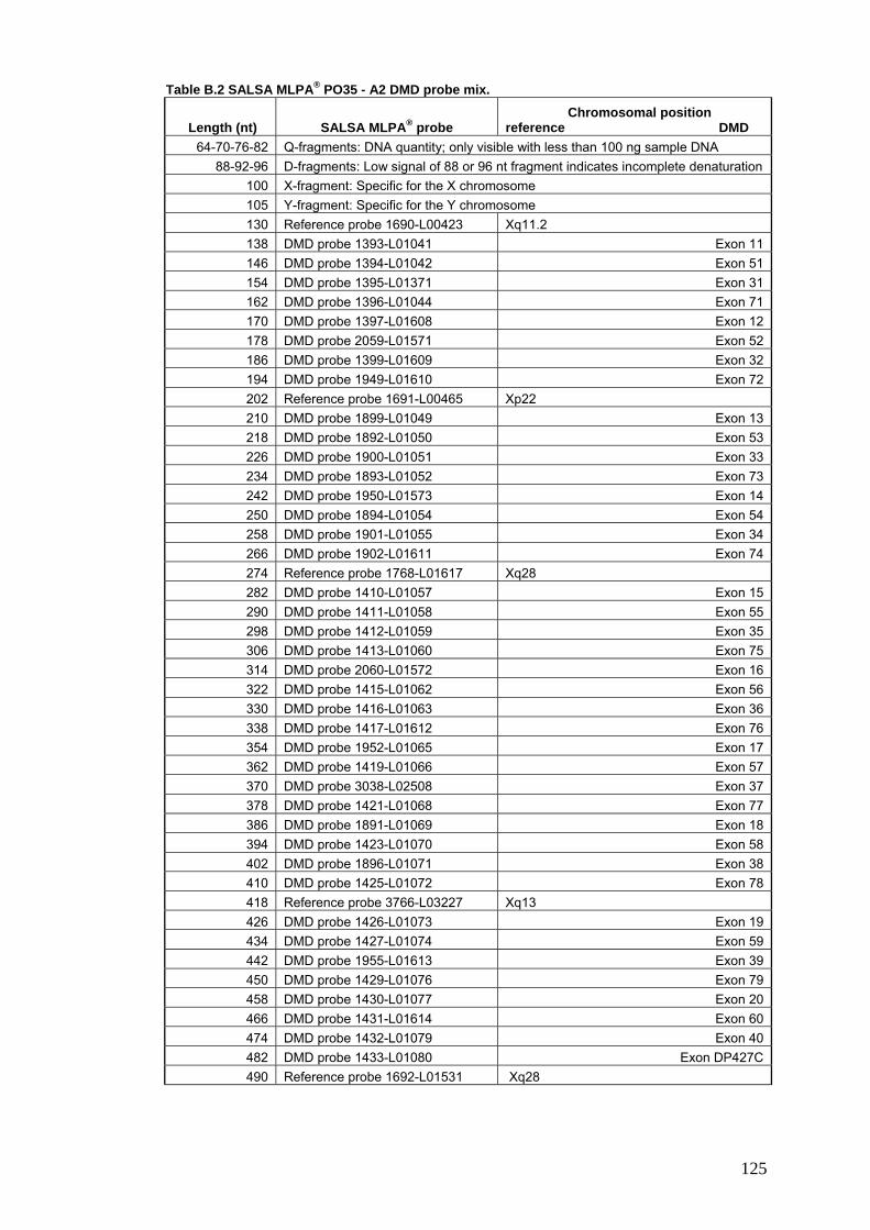
Table B.2 SALSA MLPA® PO35 - A2 DMD probe mix.
Length (nt) SALSA MLPA® probe Chromosomal position reference DMD
64-70-76-82 Q-fragments: DNA quantity; only visible with less than 100 ng sample DNA
88-92-96 D-fragments: Low signal of 88 or 96 nt fragment indicates incomplete denaturation
100 X-fragment: Specific for the X chromosome
105 Y-fragment: Specific for the Y chromosome
130 Reference probe 1690-L00423 Xq11.2
138 DMD probe 1393-L01041 Exon 11
146 DMD probe 1394-L01042 Exon 51
154 DMD probe 1395-L01371 Exon 31
162 DMD probe 1396-L01044 Exon 71
170 DMD probe 1397-L01608 Exon 12
178 DMD probe 2059-L01571 Exon 52
186 DMD probe 1399-L01609 Exon 32
194 DMD probe 1949-L01610 Exon 72
202 Reference probe 1691-L00465 Xp22
210 DMD probe 1899-L01049 Exon 13
218 DMD probe 1892-L01050 Exon 53
226 DMD probe 1900-L01051 Exon 33
234 DMD probe 1893-L01052 Exon 73
242 DMD probe 1950-L01573 Exon 14
250 DMD probe 1894-L01054 Exon 54
258 DMD probe 1901-L01055 Exon 34
266 DMD probe 1902-L01611 Exon 74
274 Reference probe 1768-L01617 Xq28
282 DMD probe 1410-L01057 Exon 15
290 DMD probe 1411-L01058 Exon 55
298 DMD probe 1412-L01059 Exon 35
306 DMD probe 1413-L01060 Exon 75
314 DMD probe 2060-L01572 Exon 16
322 DMD probe 1415-L01062 Exon 56
330 DMD probe 1416-L01063 Exon 36
338 DMD probe 1417-L01612 Exon 76
354 DMD probe 1952-L01065 Exon 17
362 DMD probe 1419-L01066 Exon 57
370 DMD probe 3038-L02508 Exon 37
378 DMD probe 1421-L01068 Exon 77
386 DMD probe 1891-L01069 Exon 18
394 DMD probe 1423-L01070 Exon 58
402 DMD probe 1896-L01071 Exon 38
410 DMD probe 1425-L01072 Exon 78
418 Reference probe 3766-L03227 Xq13
426 DMD probe 1426-L01073 Exon 19
434 DMD probe 1427-L01074 Exon 59
442 DMD probe 1955-L01613 Exon 39
450 DMD probe 1429-L01076 Exon 79
458 DMD probe 1430-L01077 Exon 20
466 DMD probe 1431-L01614 Exon 60
474 DMD probe 1432-L01079 Exon 40
482 DMD probe 1433-L01080 Exon DP427C
490 Reference probe 1692-L01531 Xq28
125

Table B.3 DMD probes arranged according to chromosomal order.
Length (nt) P034/P035
SALSA MLPA®A probe DMD exon
Ligation site M18533
Partial sequence (20 nt adjacent to ligation site)
Distance to next exon
P034/P035
482 * 1433-L01080 Exon DP427c
233-234 in NM_000109.2
CAGTAATAGA-ATGCTTTCAG 127.9 Kb
138 * 1353-L01001 Exon 1 26-27 CCCCCTACAG-GACTCAGATC 191.3 Kb
170 * 1357-L01005 Exon 2 266-267 CAAAAGAAAA-CATTCACAAA 170.4 Kb
210 1361-L01009 Exon 3 335-336 CTCTTCAGTG-ACCTACAGGA 5.0 Kb
242 1365-L01013 Exon 4 435-436 CCCTGAACAA-TGTCAACAAG 21.5 Kb
282 1954-L01574 Exon 5 517-518 AGATGGAAAT-CATAAACTGA 6.7 Kb
314 1373-L01021 Exon 6 618-619 CCAACAGTGA-AAAGATTCTC 7.0 Kb
354 1713-L01281 Exon 7 765-766 GGAATAGTGT-GGTTTGCCAG 110.4 Kb
386 1715-L01283 Exon 8 951-952 TTGAAGCCAT-CCAGGAAGTG 1.2 Kb
426 1385-L01033 Exon 9 1066-1067 ACAGGGATAT-GAGAGAACTT 52.8 Kb
458 1718-L01286 Exon 10 1195-1196 AGACAAGTCA-TTTGGCAGTT 0.8 Kb
138 1393-L01041 Exon 11 1382-1383 TTGACAGCCC-ATCAGGGCCG 29.9 Kb
170 1397-L01608 Exon 12 1647-1648 AGCCTCTTGG-ACCTGATCTT 18.5 Kb
210 1899-L01049 Exon 13 1762-1763 GGTAGTTGAT-GAATCTAGTG 22.0 Kb
242 1950-L01573 Exon 14 1836-1837 GGGCAAACAT-CTGTAGATGG 0.2 Kb
282 1410-L01057 Exon 15 1937-1938 TGGCTTTCAG-AAAAAGAAGA 7.8 Kb
314 2060-L01572 Exon 16 2059-2060 GCAATCCATG-GGCAAACTGT 20.6 Kb
354 1952-L01065 Exon 17 2292-2293 AACAGATCCT-GGTAAAGCAT 27.2 Kb
386 1891-L01069 Exon 18 2433-2434 AAGCTGTGTT-GCAGAGTCCT 16.3 Kb
426 1426-L01073 Exon 19 2526-2527 AAGCTGAGAA-GTTCAGAAAA 10.4 Kb
458 1430-L01077 Exon 20 2650-2651 GTGGATCGAA-TTCTGCCAGT 6.4 Kb
154 1355-L01615 Exon 21 2916-2917 AAGGACAAGG-ACCCATGTTC 12.7 Kb
186 1359-L01007 Exon 22 3051-3052 AGGAGACCAT-GAGTGCCATC 3.6 Kb
226 1363-L01011 Exon 23 3200-3201 GGCCTATACT-ATCTCAGCAC 4.0 Kb
258 1958-L01518 Exon 24 3441-3442 GGCCTGCCCT-TGGGGATTCA 1.1 Kb
298 1371-L01019 Exon 25 3554-3555 CAGAAGATAA-AGAATGAAGC 8.8 Kb
330 1375-L01023 Exon 26 3695-3696 GTAAGCCTCC-AGAAAGATCT 6.2 Kb
370 1379-L01616 Exon 27 3871-3872 TGAGTCTGTA-AATAGTGTCA 7.3 Kb
402 1716-L01284 Exon 28 4020-4021 GGCATGAGTT-ATTGTCATAC 3.0 Kb
442 1387-L01035 Exon 29 4198-4199 ACAGACCCTA-ACAGATGGCG 26.4 Kb
126

Table B.3 DMD probes arranged according to chromosomal order cont.
Length (nt) P034/P035
SALSA MLPA® probe DMD exon
Ligation site M18533
Partial sequence (20 nt adjacent to ligation site)
Distance to next exon
474 1391-L01039 Exon 30 4304-4305 AAGTTGCTTG-AACAGAGCAT 21.8 Kb
154 1395-L01371 Exon 31 4519-4520 GGAGGCTGCC-CAAAGAGTCC 0.6 Kb
186 1399-L01609 Exon 32 4674-4675 CTGCATTGGA-AACAAAGAGT 3.1 Kb
226 1900-L01051 Exon 33 4766-4767 TCTGAAGTGG-AAATGGTGAT 5.8 Kb
258 1901-L01055 Exon 34 4947-4948 GAAAGGAAAT-GAATGTCTTG 15.5 Kb
298 1412-L01059 Exon 35 5098-5099 CCTGAAGAGT-ATCACAGAGG 0.5 Kb
330 1416-L01063 Exon 36 5288-5289 ATCACAAAGT-GGATCATTCA 1.8 Kb
370 * 3038-L02508 Exon 37 5498-5497 Rev. TGGCTGCAAA-TCGATGGTTG 14.4 Kb
402 1896-L01071 Exon 38 5613-5614 CTGAAATTCA-GCAGGGGGTG 2.4 Kb
442 1955-L01613 Exon 39 5700-5701 TGCAAAGAGG-AGACAACTTA 2.8 Kb
474 1432-L01079 Exon 40 5828-5829 AAGGCTCTAG-AAATTTCTCA 1.0 Kb
146 1354-L01002 Exon 41 6019-6020 GGGCTTGTCT-GAGGATGGG 32.0 Kb
178 1711-L01279 Exon 42 6159-6160 CGATGATGGT-GATGACTGAA 22.6 Kb
218 1362-L01010 Exon 43 6403-6404 ATTGCAAAGT-GCAACGCCTG 70.6 Kb
250 1366-L01014 Exon 44 6589-6590 ACAGTTTCTC-AGAAAGACAC 248.6 Kb
290 1370-L01287 Exon 45 6745-6746 AGATGCCAGT-ATTCTACAGG 36.3 Kb
322 1374-L01288 Exon 46 6913-6914 CATTGCTAGT-ATCCCACTTG 2.4 Kb
362 1378-L01026 Exon 47 7017-7018 TCAAACAATT-AAATGAAACT 54.4 Kb
394 1382-L01030 Exon 48 7225-7226 GTTAAATCAT-CTGCTGCTGT 38.5 Kb
434 1717-L01285 Exon 49 7356-7357 AAGAGATTTT-GTCTAAAGGG 16.7 Kb
466 1390-L01038 Exon 50 7455-7456 TAAACCGTTT-ACTTCAAGAG 46.0 Kb
146 1394-L01042 Exon 51 7630-7631 TCTGGCAGAT-TTCAACCGGG 44.4 Kb
178 * 2059-L01571 Exon 52 7837-7836 Rev. AGCCTCTTGA-TTGCTGGTCT 50.2 Kb
218 1892-L01050 Exon 53 7994-7995 GAGCAGGTCT-TAGGACAGGC 21.3 Kb
250 1894-L01054 Exon 54 8116-8117 GCAGACAAAT-GTAGATGTGG 30.3 Kb
290 1411-L01058 Exon 55 8276-8277 ACTCATAGAT-TACTGCAACA 120.5 Kb
322 1415-L01062 Exon 56 8535-8536 TCCTGTTACA-AAGACGTTTG 10.5 Kb
362 1419-L01066 Exon 57 8686-8687 GAAAGATGAT-GAATTAAGCC 17.8 Kb
394 1423-L01070 Exon 58 8817-8818 AGACTGTACG-AATATTTCTG 0.8 Kb
434 1427-L01074 Exon 59 9005-9006 GATGAGACCC-TTGAAAGACT 33.6 Kb
127

Table B.3 DMD probes arranged according to chromosomal order cont.
Length (nt) P034/P035
SALSA MLPA® probe DMD exon
Ligation site M18533
Partial sequence (20 nt adjacent to ligation site)
Distance to next exon
466 1431-L01614 Exon 60 9175-9176 GCCTCTGAAA-GAGAACGTGA 96.0 Kb
162 1356-L01004 Exon 61 9325-9326 GCAGCTGCAT-GAAGCCCAC 25.0 Kb
194 1897-L01008 Exon 62 9394-9395 TCCCTGGGAG-AGAGCCATCT 62.6 Kb
234 * 1364-L01012 Exon 63 9453-9454 AAACAACTTG-CTGGGACCAT 37.9 Kb
266 * 1368-L01016 Exon 64 9547-9546 Rev. CTGCAGTCTT-CGGAGTTTCA 13.4 Kb
306 1372-L01020 Exon 65 9599-9600 GCTGCATGTG-ATGCCTTGGA 3.0 Kb
338 1376-L01024 Exon 66 9806-9807 CTGTCTTTTA-AAACTGGCAT 2.6 Kb
378 * 1960-L01520 Exon 67 9987-9986 Rev. ACACTTGGCT-CAATGTTACT 21.1 Kb
410 2482-L02710 Exon 68 10064-10065 GACTGGATGA-GACTGGAACC 2.4 Kb
450 1388-L01036 Exon 69 10238-10239 TTTTTTTCTG-GTCGAGTTGC 1.7 Kb
482 1392-L01040 Exon 70 10385-10386 CCCCGAATGG-GCTACCTGCC 0.8 Kb
162 * 1396-L01044 Exon 71 10450-10451 TCTGATCAAC-TTCTGGCCAG 4.4 Kb
194 1949-L01610 Exon 72 10493-10494 CCTCAGCTTT-CACACGATGA 1.2 Kb
234 * 1893-L01052 Exon 73 10571-10570 Reverse
TATCATTTAG-ATAAGATCCA 2.9 Kb
266 1902-L01611 Exon 74 10693-10694 CCAGATCTTG-ATTTCCTTAG 22.1 Kb
306 1413-L01060 Exon 75 10885-10886 TGAGCTCATT-GCTGAGGCCA 1.0 Kb
338 1417-L01612 Exon 76 11063-11064 ACCTCTCTAC-AGAGGTCCGA 12.2 Kb
378 1421-L01068 Exon 77 11164-11165 CCAGGACACA-AGCACAGGGT 7.5 Kb
410 * 1425-L01072 Exon 78 11245-11246 TGGAAAGCCA-ATGAGAGAGG 4.8 Kb
450 1429-L01076 Exon 79 11286-11287 CCACATGGCA-GATGATTTGG
*These probes detect the reverse sequence or extend into an intron and are
expected to give no signal with cDNA. The two exon 1 sequences are present in
alternative transcripts and may also give a weak or no signal on cDNA (Kesari et al.,
2008).
Tables of probe mixes were adapted from SALSA MLPA® KIT P034-A2 / P035-A2
DMD / Becker (www.mlpa.com). The identity of the genes detected by the reference
probes is available on request from [email protected].
128

APPENDIX C
1. IMPORTANT CONSIDERATIONS FOR HR- MCA ASSAY DESIGN
The HRM platform
Successful HRM can only be achieved with specialized instrumentation capable of
collecting data with exceptional real-time and thermo-optical precision. A cross-
platform comparison by Herrmann et al. (2006) effectively highlighted the limitations
of conventional systems and the unique qualities required for accurate HRM
analysis, particularly:
High intensity illumination
High sensitivity optical detection
A fast data acquisition rate
Exquisite sample temperature control
An absolute minimum of sample-to-sample thermal and optical variation
The first instrument marketed for the sole purpose of exquisitely accurate HRM
analysis, was the HR-1™ (Idaho Technology), which remains the performance
benchmark (Reed, Kent, & Wittwer, 2007). However, the HR-1™ is not capable of
thermal cycling and can only analyze a single sample per run from within a glass
capillary, making data analysis time consuming. The multi-well LightScanner® (Idaho
Technology) uses a modified block-based design but is also an HRM-only machine,
not capable of thermal cycling. Subsequent technological advancements have
resulted in introduction of machines with real-time cycling as well as HRM capability
i.e. LightCycler®480 (Roche, Germany) and the RotorGene™6000 (Corbett Life
Science, Australia) (Figure C.1). A major advantage of this is that the amplification
efficiency can be monitored in real time, with the initial PCR cycling followed by
HRM in a closed-tube system, without the need for post-PCR manipulation. In
addition, the methods used to normalize and display HRM data for intuitive manual
analysis or automated genotyping are specific and continue to improve.
The Rotor-Gene™6000, used in this study, stands apart from other analysers by
virtue of its unique centrifugal format. Every tube spins quickly in a chamber of
moving air. This is especially suitable for HRM, as there is virtually no positional
temperature variation such as the recognized “edge effect” observed in block-based
instruments. Optically, the Rotor-Gene™6000 is similarly uniform, because every
tube moves past identical excitation and detection points (Herrmann et al., 2007;
White & Potts, 2006). As a result, the Rotor-Gene™000 HRM performance closely
matches the HR-1™ benchmark.
129

The RotorGene™6000, fulfils the criteria for reliable HRM analysis, with a melting
rate which can be adjusted manually to the lowest level of the above mentioned
instruments (0.05°C/sec), and a default of 0.1°C increments with a 2 second hold at
each step. The Rotor-Gene™6000 has a wide optical range, with 6 channels
spanning UV to infra-red wavelengths (www.corbettlifescience.com). It offers the
benefits of real-time PCR along with its HRM capability, successfully tested in a
number of applications, including:
Gene expression analysis
Pathogen detection
DNA methylation analysis
Genotyping
Mutation scanning
SNP analysis
High Resolution Melting Analysis
In an evaluation report by Helen White and Gemma Potts of NGRL, the overall HRM
sensitivity and specificity of the RotorGene™6000 was found to be 100% and 95%
respectively, as compared to HR1™ (Idaho Technology) at 98% and 95%, and the
384-well LightScanner® (Idaho Technology) at 99% and 88% (White & Potts, 2006).
Other instrumentation with real-time and HRM capabilities have since been
marketed, but at this time, those mentioned above are most often mentioned in the
literature and have been subject to cross platform comparisons.
Figure C.1 The RotorGene™6000 with its centrifugal design.
130

Choice of a dsDNA intercalating dye
The advancements in intercalating dye technology since Higuchi first demonstrated
the use of ethidium bromide in qPCR (Higuchi et al., 1993), along with
instrumentation that allows highly controlled temperature transitions and acquisition,
have played a major role in the emergence of HRM methodology. Different base
substitutions produce slight differences in melting behaviour, the resolution of which
requires an appropriate intercalating dye. SYBR Green® is the most widely used
intercalating dye for monitoring real-time PCR, but is generally regarded unsuitable
for use in hrMCA due to its strong tendency to inhibit PCR and promote mispriming
at high (saturating) concentrations. According to the proposed “dye saturation
hypothesis” (Wittwer et at., 2003; Liew et al., 2004), SYBR Green dissociates from
dsDNA during melting, and dynamically re-intercalates into the neighbouring
duplexes that melt at higher temperatures (Figure C.2), potentially masking small
differences in melting behaviour, which could then go undetected. Low dye
concentrations on the other hand, (e.g. < 0.5 μM) can compromise PCR signal
strength, and the dye redistribution possibly results in an unreliable DNA melt curve.
“Saturating dyes” are thus defined as those that can be used at concentrations
sufficiently high to saturate all DNA binding sites without inhibiting the PCR, thereby
achieving a greater melting curve resolution (Wittwer et al 2003, Liew et al 2004).
New generation dyes such as LC Green®, LC Green®PLUS, SYTO 9®, BEBO,
CHROMOFY® and Eva Green™, show minimal PCR toxicity at high levels and allow
for accurate genotyping of single-nucleotide polymorphisms and sensitive mutation
scanning.
Figure C.2 The dye re-distribution theory and the 2nd and 3rd generation dsDNA intercalating dyes (Cor-Protocol-6000-1-July06).
131

The dye redistribution theory however, has been contested, as evidence for this
theory was collected by a pre-HRM era LightCycler® (Roche Molecular Systems)
using templates which included a low mass DNA size ladder (Wittwer et al., 2003;
Liew et al., 2004). According to Corbett Research, saturation levels of SYBR Green®
are not required when using the Rotor-Gene™6000. The experimental data shows
good HRM results and the authors encourage users not to dismiss SYBR Green® as
a dye for HRM, provided that access to an appropriate instrument is available.
Corbett Research do however concede that while saturating dye levels appear
unnecessary for HRM on the Rotor-Gene™6000 (when apparently required on other
instrument systems), the reason for this is not clear. They suggest that the
explanation may lie in the centrifugal rotary format of the instrument, as that is what
sets it apart from other HRM machines.
The dsDNA intercalating utalised in this project, EvaGreen™ (Biotum), is included in
the HRM Sensimix kit, optimised by Quantace specifically for use on the
RotorGene™6000. EvaGeen is a green fluorescent nucleic acid dye useful in qPCR,
hrMCA, real-time monitoring of thermophilic helicase-dependent amplification
(tHDA), routine solution DNA quantification and capillary gel electrophoresis. It is
also compatible with multiplex PCR, as no dye migration from amplicon to amplicon
takes place when used at the recommended concentration. EvaGreen™ is
essentially nonfluorescent by itself, but becomes highly fluorescent upon binding to
dsDNA. Once bound, it has excitation and emission spectra similar to fluorescein
(FAM) or SYBR Green® 1 and is therefore readily compatible with instruments
equipped with the 488 nm argon laser or any visible light excitation filter with a
wavelength in that region. It is thus compatible with all major brand qPCR
instruments and enzyme systems. The extreme thermal, hydrolytic and photo-
stability of EvaGreen™ provides convenience during routine handling, as the dye
can withstand repeated cycles of freezing and thawing, does not decompose in PCR
buffer at 95-100°C for 48 hours and is highly stable under either alkaline or acidic
condition. Compared with SYBR Green®, EvaGreen™ is generally less inhibitory
toward PCR and less likely to cause non-specific amplification. Unlike SYBR
Green®, which enters cells rapidly and is known to be a powerful mutation-enhancer,
EvaGreen™ is nonmutagenic and noncytotoxic by being completely impermeable to
cell membranes (Ohta et al., 2001; Mao et al., 2007; White et al., 2007). Regardless
of the validity of the dye redistribution theory, EvaGreen can be used at a much
higher dye concentrations than SYBR Green® I, resulting in a more robust PCR
signal.
132

PCR design and optimisation
Besides the availability of an appropriate HRM platform and choice of a dsDNA
intercalating dye, the success of any hrMCA assay is highly dependant on the
design and optimisation of the pre-HRM PCR reaction, as the presence of non-
specific products and primer dimmers significantly reduces HRM performance. Use
of a real-time thermocycler is recommended, particularly when optimizing or
troubleshooting an assay, as information on amplicon quality can be monitored in
real time.
Primer design
The PCR product should not include sequence variations other than the one
investigated e.g. polymorphic regions, as their presence will alter the melting curve.
Also, secondary structure formation of the primers and the amplicon will affect the
amplification efficiency and should be avoided. Optimal primer design and checking
for formation of secondary structures can be achieved with software such as
Primer3 and the DINAmelt Servers from the Rensselaer Polytechnic Institute,
respectively (Markham & Zuker, 2005). HR-MCA of polymorphic regions however,
has been successfully performed but involves inclusion of known, genotype-specific
controls (White & Potts, 2006). PCR reaction optimisation for HRM, may require a
significant increase in the annealing temperatures (2C – 10C), because of the
presence of dsDNA intercalating dye.
MgCl2 concentration
The amount of MgCl2 has a strong effect on the melting behaviour of dsDNA and
should be optimized carefully. Generally, a higher concentration of MgCl2 is needed
in a PCR for HR-MCA, which includes a dsDNA intercalating dye, as opposed to an
ordinary PCR, where no dye is present. The general guidelines also indicate that the
required MgCl2 concentration varies according to the specific dye chosen e.g. 1.5
mM MgCl2 with SYTO® 9, or 3 mM MgCl2 with LCGreen® or EvaGreen™
(www.corbettlifescience.com)
PCR reagents
It is recommended to use PCR reagents validated or optimized for HRM analysis,
when available, although this may be more expensive. Hot start enzymes, generally
included in HRM mixes such as the AmpliTaq Gold 360 Master Mix (Applied
Biosysems, USA) or the HRM Sensimix (Quantace), are also recommended, as they
increase primer specificity.
133

2. FACTORS AFFECTING THE MELTING CURVE AND RELIABILITY OF hrMCA
DNA and amplicon quality/quantity
The effect of DNA quality on an HRM assay was clearly demonstrated in an
experiment by White et al. (2003), where amplifiability of DNA samples of variable
quality was assessed by using a multiplexed control gene primer set which amplifies
products of 100, 200, 300, 400 and 600bp with equal intensity. Loss of the higher
molecular weight products was seen as an indicator of poor DNA quality. It was
shown that samples in which products of 200bp or less were amplified, gave false
positive results when analysed by HRM.
The use of spectral analysis for determining DNA concentration and purity is
recommended, as the amount of starting template should be similar between
samples in a run, since it affects the amount of generated PCR product. At 260 nm
one absorbance unit is equal to 50 μg/mL of DNA and pure DNA will provide a 260
nm to 280 nm ratio of 2. The amplicon concentration in turn, affects its melting
temperature(TM) and the outcome of HRM analysis. The threshold cycle (CT) of an
efficient amplification plot, should not exceed 30 cycles, as amplification log phase
starting beyond that point, is typically due to insufficient amount of starting template
or presence of degradation effects. Allowing every reaction to amplify to the plateau
phase is important, as a similar extent of amplification for all tests should be
achieved irrespective of their starting amount. However, poor reaction might result in
a plateau with a different quantity of product, due to factors such as inconsistent
assay set-up (e.g. insufficient amount of primer).
Monitoring of the amplification curve in real time is a valuable trouble-shooting tool
in hrMCA, as problems and inconsistencies involving the quality and quantity of
DNA, the amplicon and the reaction set up can be readily identified. They manifest
as late amplification, curves with log-linear phase that is not steep, is jagged, or
reaches a low signal plateau as compared to other reactions in the run. HRM data
from such samples can be inconclusive and should be excluded from analysis.
Amplicon size
Differentiation of variant melting curves is best achieved in smaller PCR fragments
(~100bp). HrMCA analysis of products larger than 300bp can be analyzed
successfully but usually with lower resolution. This is simply because a single base
variation affects the melting behavior of a 100 bp amplicon more than a 500 bp
amplicon and will therefore be easier to detect (www.corbettlifescience.com). Large
amplicons are also more likely to have complex melting curves with two or more
134

melting domains, complicating the analysis and making it less reliable. Ideally, a
PCR fragment should have a single melting domain, otherwise each domain must
be analysed separately, by manual adjustment of the normalisation regions. Optimal
amplicon design and subsequent optimisation are therefore crucial to the success of
HRM. For this reason a number of exons in the DMD gene were split into two or
more fragments i.e. exons 23, 48, 53, 61, 67, 68, 76 and 79 thus creating 96 sets of
PCR primers to amplify 79 exons of the DMD gene.
Primer dimers
Like other non-specific products, presence of primer-dimmers may affect the melting
curve characteristics and therefore primer concentration should generally not
exceed 300nM.
Effect of mutation type or the surrounding sequence context
The position of a mutation within a PCR fragment per se, does not appear to have a
significant effect on HRM-based mutation detection. However, an experiment carried
out as part of a cross HRM platform comparison by White et al. (2006) showed that
certain mutations, such as the 110A>G and 588delA mutations in exons 1 and 7
respectively, are intrinsically difficult to detect by HRM, possibly because they are
sited in regions which, when melted, produce only subtle overall changes in
fluorescent intensity. Interestingly, it was shown that differences in data handling
between the compared platforms (HR-1™, Light Scanner™ and RotorGene™6000),
also affect variant detection efficiency of some mutations. Specifically, 1459 C>T,
1451 delA and 1409+14G>A sequence variants in exon13, appear to alter the
melting temperature rather than the overall shape of the melting curve. Data from
the HR-1 ™ and Light Scanner™ is first normalised and then temperature shifted.
When the data are temperature shifted these curves become superimposed on the
wild type samples, making detection difficult. Data from the RotorGene™6000 is
normalized, but not temperature shifted, and therefore such mutations can be more
easily detected as the melt curves do not become overlaid on the wild type curve
(White & Potts, 2006).
Homo- and hemizygous changes appear to have similar effect on the melting
dynamic of the DNA fragment i.e. they appear to alter the temperature at which the
amplicon melts rather than the shape of the melt curve (Kwok et al., 2007; Yan et
al., 2010). Temperature-shifting for data analysis, causes the curves of homozygous
samples to become superimposed on the wild type masking the change. As
mentioned above, this effect does not take place during analysis with the
135

RotorGene™6000, as the data is normalized but not temperature shifted and
homozygous mutations are readily detected.
Sample-to-sample uniformity
Consistency in reaction setup and reagent use is critical to the success hrMCA.
Reaction volume and the amount of dye in each tube must be the same. Similarly,
identical reaction tubes from the same manufacturer should be used to avoid
variations in plastic thickness and autofluorescence properties. DNA melting
behavior is affected by salts in the reaction mix, so concentration of buffers, MgCl2
and other salts should be kept uniform in all samples. The various available DNA
extraction methods utalise different chemistries and elution buffers, which may affect
the DNA melting behavior. It is therefore best to compare samples extracted with the
same method, which is an important consideration when attempting analysis of
archival samples or samples from other laboratories.
Temperature range during melting
HRM data points over a window of approximately 10°C (or greater), should be
captured, centered around the observed TM of the fragment under analysis This
provides enough baseline data points for effective curve normalization and will result
in tight replicates and easier data interpretation.
Regions of normalisation
The data analysis software on the RotorGene™6000 allows for both visual and
auto-calling of genotypes. Two normalization regions are used to normalize
fluorescencece (Y axis), represented by vertical cursors placed pre-melt (region 1)
and post-melt (region 2) phases (Figure C.3).
Figure C.3 Adjustable normalisation regions.
ºC65 66 67 68 69 70 71 72 73 74 75 76 77 78 79 80 81 82 83 84 85 86 87
Flu
ores
cenc
e
55
50
45
40
35
30
25
20
15
10
5
Nor
mal
isat
ion
Reg
ion
1
Nor
mal
isat
ion
Reg
ion
2
region 1
region 2
136

The cursors are placed at default positions, which can be manually adjusted.
Shifting the cursors can alter the appearance of a normalised curve and cause
differences in both qualitative manual calling and automated mutation calling.
Therefore, in a diagnostic setting, criteria for positioning the cursors for different
amplicons should be incorporated into standard operating procedures to ensure
minimal data analysis variability between operators. Corbett Research recommend
that these default settings be changed only to avoid certain parts of the curve, such
as when analysing complicated curves with multiple melting domains. In such
instances, the pre-melt region 1 should be set at a point where all samples in a run
show uniform melting (seen as parallel lines), and the post-melt region 2 should be
set after complete melting of all samples, so that the normalisation range should
stretch across approximately 10°C.
The four classes of base changes
Detection of a single base alteration, as the smallest genetic change underlies the
sensitivity of an HRM analysis. Single Nucleotide Polymorphisms (SNPs) often used
for genotyping have been divided into 4 classes according to their rarity and difficulty
in detection. As presented in Table C.1 below, the A:T to G:C interchange, which is
the most common single base variation in the human genome also results in the
greatest difference in Tm (>0.5), which is readily detected. Other base changes give
rise to smaller differences in Tm, resulting in lower specificity that depends on the
exact base change and also on the surrounding sequence.
Table C.1 Classification of SNPs (Venter et al., 2001).
SNP Class Base Change Typical TM Melt Curve Shift Rarity
(in the human genome)
1 C/T and G/A 64%
2 C/A and G/T 30%
3 C/G 9%
4 A/T
Large (>0.5°C)
Very Small (<0.2°C) 7%
137

3. PRIMER SEQUENCES Table C.2 HRM and sequencing primers
# PRIMER SEQUENCE FRAGMENT LENGTH
ANNEALING TEMP
EX01-F TGTAAAACGACGGCCAGTGCAGGTCCTGGAATTTGA 405 61Cº EX01-R CAGGAAACAGCTATGACCCAAACTAAACGTTATGCCACA EX02-F TGTAAAACGACGGCCAGTCACTAACACATCATAATGG 269 61Cº EX02-R CAGGAAACAGCTATGACCGATACACAGGTACATAGTC EX03 A-F TGTAAAACGACGGCCAGTTCATCCGTCATCTTCGGCAGATTAA 176 61Cº EX03 A-R CAGGAAACAGCTATGACCCAGGCGGTAGAGTATGCCAAATGAAAATCA EX03B-F TCTTCAGTGACCTACAGGATGG 184 64Cº EX03B-R CGCCCGCCGtgctgtttcaatcagtacctagtca EX04-F TGTAAAACGACGGCCAGTTTGTCGGTCTCTCTGCTGGTCAGTG 233 60Cº EX04-R CAGGAAACAGCTATGACCCCAAAGCCCTCACTCAAAC EX05-F TGTAAAACGACGGCCAGTCAACTAGGCATTTGGTCTC 261 61Cº EX05-R CAGGAAACAGCTATGACCTTGTTTCACACGTCAAGGG
138

Table C.2 HRM and sequencing primers cont. EX06-F TGTAAAACGACGGCCAGTTGGTTCTTGCTCAAGGAATG 335 61Cº EX06-R CAGGAAACAGCTATGACCTGGGGAAAAATATGTCATCAG EX07-F TGTAAAACGACGGCCAGTCTATGGGCATTGGTTGTC 296 60Cº EX07-R CAGGAAACAGCTATGACCAAAAGCAGTGGTAGTCCAG EX08-F TGTAAAACGACGGCCAGTTCGTCTTCCTTTAACTTTG 343 61Cº EX08-R CAGGAAACAGCTATGACCTCTTGAATAGTAGCTGTCC EX09-F TGTAAAACGACGGCCAGTTCTATCCACTCCCCCAAACC 318 61Cº EX09-R CAGGAAACAGCTATGACCAACAAACCAGCTCTTCAC EX10-F CGACGTTGTAAAACGACGGCCAGTGGAACAATCTGCAAAGAC 350 61Cº EX10-R CAGGAAACAGCTATGACCAAAGGATGACTTGCCATTATAAC EX11-F TGTAAAACGACGGCCAGTCAAATAAAACTCAAAACCACACC 337 61Cº EX11-R CAGGAAACAGCTATGACCCTTCCAAAACTTGTTAGTCTTC EX12-F TGTAAAACGACGGCCAGTCTTTCAAAGAGGTCATAATAGG 305 61Cº EX12-R CAGGAAACAGCTATGACCCATCTGTGTTACTGTGTATAGG
139

Table C.2 HRM and sequencing primers cont. EX13-F TGTAAAACGACGGCCAGTGCAAATCATTTCAACACAC 387 60Cº EX13-R CAGGAAACAGCTATGACCTCTTTAAATCACAGCACTTC EX14-15-F TGTAAAACGACGGCCAGTTGGCAAATTATTCATGCCATT 548 63Cº EX14-15-R CAGGAAACAGCTATGACCTGATCCAAGCAAAAATAAACATT EX16-F TGTAAAACGACGGCCAGTATGCAACCCAGGCTTATTC 286 61Cº EX16-R CAGGAAACAGCTATGACCCTGTAGCATGATAATTGGTATCAC EX17-F TGTAAAACGACGGCCAGTTTTTCCTTTGCCACTCCAAG 362 61Cº DEX17-R CAGGAAACAGCTATGACCCACCACCAACAAAACTGCTG EX18-F CGACGTTGTAAAACGACGGCCAGTTGTCAGGCAGGAGTCTCAGAT 339 63Cº EX18-R CAGGAAACAGCTATGACCCGGAGTTTACAAGCAGCACA EX19-F TGTAAAACGACGGCCAGT gattcacgtgataagctgacaga 286 63Cº EX19-R CAGGAAACAGCTATGACCCGCCCGCCGCGCttcagctgataaatatgaacctatgtg EX20-F TGTAAAACGACGGCCAGTTGGCTTTCAGATCATTTCTTTC 393 61Cº EX20-R CAGGAAACAGCTATGACCAAATACCTATTGATTATGCTCC
140

Table C.2 HRM and sequencing primers cont. EX21-F TGTAAAACGACGGCCAGTGCAAAATGTAATGTATGCAAAG 355 63Cº EX21-R CAGGAAACAGCTATGACCATGTTAGTACCTTCTGGATTTC EX22-F TGTAAAACGACGGCCAGTAGGAAAACATGGCAAAGTGTG 370 63Cº EX22-R CAGGAAACAGCTATGACCTGCTCAATGGGCAAACTACC EX23 A-F TGTAAAACGACGGCCAGTACTCATCAATTATTATTCATCAATTAGGGT 126 61Cº EX23 A-R CAGGAAACAGCTATGACCCATCTCTTTCACAGTGGTGC EX23 B-F TGTAAAACGACGGCCAGTAGCAACAAAGTGGCCTATAC 135 61Cº EX23 B-F CAGGAAACAGCTATGACCGCTGGGAGGAGAGCTTC EX23 C-F TGTAAAACGACGGCCAGTTTGAAGAAATTGAGGGACGC 175 61Cº EX23C-R CAGGAAACAGCTATGACCCTTTACAGTTTACAGTGTATCGTTAGG EX24-F TGTAAAACGACGGCCAGTTTGGGCCTGTGTTTAGACATA 327 63Cº EX24-R CAGGAAACAGCTATGACCAAATCCACCCCAGCTGTAAAA EX25-F TGTAAAACGACGGCCAGTTGTGGCAGTAATTTTTTTCAG 296 61Cº EX25-R CAGGAAACAGCTATGACCAGGAAATCTTAGTTAAGTACG
141

Table C.2 HRM and sequencing primers cont. EX26-F TGTAAAACGACGGCCAGTTGAGTGTATCTGATCCCCATGA 438 61Cº EX26-R CAGGAAACAGCTATGACCTGTTGCATTTCTTTCTTTTTC EX27-F TGTAAAACGACGGCCAGTTGGGATGTTGTGAGAAAGAA 365 63Cº EX27-R CAGGAAACAGCTATGACCTGACCATGTATTGACATATCATTGA EX28-F TGTAAAACGACGGCCAGTGAAGTTTTAATAATGAAATGGCAAAA 311 61Cº EX28-R CAGGAAACAGCTATGACCTGACCTCTTTTAATACTGCATAT EX29-F TGTAAAACGACGGCCAGTCCAATGTATTTAGAAAAAAAAGGAG 279 63Cº EX29-R CAGGAAACAGCTATGACCGCAAATTAGATTAAAGAGATTTTTCAC EX30-F TGTAAAACGACGGCCAGTTACAGAAAAGCTATCAAGAG 297 61Cº EX30-R CAGGAAACAGCTATGACCAAAAACAAAAGAATGGAAGC EX31-F TGTAAAACGACGGCCAGTATGGTAGAGGTGGTTGAGGA 296 61Cº EX31-R CAGGAAACAGCTATGACCTATAATGCCCAACGAAAACA EX32-F TGTAAAACGACGGCCAGTCAGTTATTGTTTGAAAGGCAAA 322 61Cº EX32-R CAGGAAACAGCTATGACCCTTCTTAATGAGGAAAGTCAAGG
142

Table C.2 HRM and sequencing primers cont. EX33-F CGACGTTGTAAAACGACGGCCAGTTGGAATAGCAATTAAGGG 393 60Cº EX33-R CAGGAAACAGCTATGACCGCTAAGACTCTAATCATAC EX34-F TGTAAAACGACGGCCAGTCAGAAATATAAAAGTTCCAAATAAGTG 374 61Cº EX34-R CAGGAAACAGCTATGACCCATGTTAATACTTCCTTACAAAATC EX35-F TGTAAAACGACGGCCAGTCCGTTTCATAAGCATTAAATC 307 61Cº EX35-R CAGGAAACAGCTATGACCAGCTTCTAGCCTTTTCTC EX36-F CGACGTTGTAAAACGACGGCCAGTTGTCTAACCAATAATGCCATG 257 64Cº EX36-R CAGGAAACAGCTATGACCCTGGTGTACAATTTGGACA EX37-F CGACGTTGTAAAACGACGGCCAGTCTTTCTCACTCTTCTCGCTCAC 377 61Cº EX37-R CAGGAAACAGCTATGACCTTCGCAAGAGACCATTTAGCAC EX38-F TGTAAAACGACGGCCAGTTTTAGCAACAGGAGGTTGAA 267 64Cº EX38-R CAGGAAACAGCTATGACCTTCTTTCCAAATATTTATTTCCACT EX39-F TGTAAAACGACGGCCAGTCTCTGTTAACAATGTACAGCTTTTT 365 64Cº EX39-R CAGGAAACAGCTATGACCAAAAACCACAGGCAAGGTAT
143

Table C.2 HRM and sequencing primers cont. EX40-F TGTAAAACGACGGCCAGTTACAAAAAGATGAGGGAC 387 61Cº EX40-R CAGGAAACAGCTATGACCAATAGAAACAAGAACATCAAC EX41-F TGTAAAACGACGGCCAGTGTTAGCTAACTGCCCTGGGCCCTGTATTG 311 61Cº EX41-R CAGGAAACAGCTATGACCTAGAGTAGTAGTTGCAAACACATACGTGG EX42-F TGTAAAACGACGGCCAGTATGGAGGAGGTTTCACTGTT 408 61Cº EX42-R CAGGAAACAGCTATGACCCCATGTGAAAGTCAAAATGC EX43-F TGTAAAACGACGGCCAGTTTTCTATAGACAGCTAATTCATTTTT 287 63Cº EX43-R CAGGAAACAGCTATGACCACAGTTCCCTGAAAACAAATC EX44-F TGTAAAACGACGGCCAGTGTTACTTGAAACTAAACTCTGCAAATG 444 61Cº EX44-R CAGGAAACAGCTATGACCACAACAACAGTCAAAAGTAATTTCCATC EX45-F TGTAAAACGACGGCCAGTTTCTTTGCCAGTACAACTGC 357 61Cº EX45-R CAGGAAACAGCTATGACCTCTGCTAAAATGTTTTCATTCC EX46-F TGTAAAACGACGGCCAGTCCAGTTTGCATTAACAAATAGTTTGAG 409 64Cº EX46-R CAGGAAACAGCTATGACCAGGGTTAAGAAGAAATAAAGTTGTGAG
144

Table C.2 HRM and sequencing primers cont. EX47-F TGTAAAACGACGGCCAGTTGATAGACTAATCAATAGAAGCAAAGAC 399 61Cº EX47-R CAGGAAACAGCTATGACCAACAAAACAAAACAACAATCCACATACC EX48 A-F TGTAAAACGACGGCCAGTTTTGGCTTATGCCTTGAGAAT 175 61Cº EX48 A-R CAGGAAACAGCTATGACCATAACCACAGCAGCAGATG EX48 B-F TGTAAAACGACGGCCAGTGCTTGAAGACCTTGAAGAGC 185 61Cº EX48 B-R CAGGAAACAGCTATGACCAAATGAGAAAATTCAGTGATATTGCC EX49-F TGTAAAACGACGGCCAGTGTGCCCTTATGTACCAGGCAGAAATTG 475 61Cº EX49-R CAGGAAACAGCTATGACCGCAATGACTCGTTAATAGCCTTAAGATC EX50-F TGTAAAACGACGGCCAGTCACCAAATGGATTAAGATGTTCATGAAT 307 64Cº EX50-R CAGGAAACAGCTATGACCTCTCTCTCACCCAGTCATCACTTCATAG EX51-F TGTAAAACGACGGCCAGTGAAATTGGCTCTTTAGCTTGTGTTTC 424 64Cº EX51-R CAGGAAACAGCTATGACCGGAGAGTAAAGTGATTGGTGGAAAATC EX52-F TGTAAAACGACGGCCAGTGTGTTTTGGCTGGTCTCACA 298 63Cº EX52-R CAGGAAACAGCTATGACCCATGCATCTTGCTTTGTGTGT
145

Table C.2 HRM and sequencing primers cont. EX53 A-F TGTAAAACGACGGCCAGTAAGAATCCTGTTGTTCATCATCCTAGC 252 64Cº EX53 A-R CAGGAAACAGCTATGACCCCAGCCATTGTGTTGAATCCTTTAAC EX53 B-F TGTAAAACGACGGCCAGTAGTACAAGAACACCTTCAGAACCG 278 64Cº EX53 B-R CAGGAAACAGCTATGACCACTTTACATTAAACATCATTAAATTACAATCTATGG EX54-F CGACGTTGTAAAACGACGGCCAGTGTATTCTGACCTGAGGATTC 378 61Cº EX54-R CAGGAAACAGCTATGACCCATGGTCCATCCAGTTTC EX55-F TGTAAAACGACGGCCAGTAATTTAGTTCCTCCATCTTTCTCT 445 61Cº EX55-R CAGGAAACAGCTATGACCAAATACATCAGGCTGTATAAAAGC EX56-F TGTAAAACGACGGCCAGTATTCTGCACATATTCTTCTTCCTGC 353 63Cº EX56-R CAGGAAACAGCTATGACCGGATGATTTACGTAGACATGTGAG EX57-F TGTAAAACGACGGCCAGTCAATGGAATTGTTAGAATCATCA 320 63Cº EX57-R CAGGAAACAGCTATGACCCACTGGATTACTATGTGCTTAACAT EX58-F TGTAAAACGACGGCCAGTTTTTGAGAAGAATGCCACAAGCC 315 63Cº EX58-R CAGGAAACAGCTATGACCAAAATATGAGAGCTATCCAGACCC
146

Table C.2 HRM and sequencing primers cont. EX59-F TGTAAAACGACGGCCAGTAAAGAATGTGGCCTAAAACC 433 64Cº EX59-R CAGGAAACAGCTATGACCTTGTGGGAAGATAACACTGC EX60-F TGTAAAACGACGGCCAGTTAAATATTCTCATCTTCCAATTTGC 267 63Cº EX60-R CAGGAAACAGCTATGACCTTACTGTAACAAAGGACAACAATG EX61A-F TGTAAAACGACGGCCAGTCGCCCGCCGCttgctttagtgttctcagtcttgg 169 63Cº EX61A-R CAGGAAACAGCTATGACCAAAGTCCCTGTGGGCTTCAT EX61B-F TGTAAAACGACGGCCAGTCGTCGAGGACCGAGTCAG 210 63Cº EX61B-R CAGGAAACAGCTATGACCCGCCCGCCGCcaggatgatttatgcttctactgc EX62-F TGTAAAACGACGGCCAGTTAATGTTGTCTTTCCTGTTTGCG 221 63Cº EX62-R CAGGAAACAGCTATGACCATACAGGTTAGTCACAATAAATGC EX63-F TGTAAAACGACGGCCAGTTACTCATTGTAAATGCTAAAGTC 229 63Cº EX63-R CAGGAAACAGCTATGACCTAGCAAGTAACTTTCACACTGC EX64-F TGTAAAACGACGGCCAGTTTCTGATGGAATAACAAATGCT 322 61Cº EX64-R CAGGAAACAGCTATGACCCATTCTAGGCAAACTCTAGGC
147

Table C.2 HRM and sequencing primers cont. EX65-F TGTAAAACGACGGCCAGTagtgtggttcacgtttggt 386 64Cº EX65-R CAGGAAACAGCTATGACCtgtacgctaagcctcctgtg EX66-F TGTAAAACGACGGCCAGTGTCAGTAATTGTTTTCTGCTTTG 246 61Cº EX66-R CAGGAAACAGCTATGACCATAAGAACAGTCTGTCATTTCCC EX67 A-F TGTAAAACGACGGCCAGTTCAGGTTCTGCTGGCATC 172 60Cº EX67 A-R CAGGAAACAGCTATGACCTGCAACTTCACCCAACTGTC EX67 B-F TGTAAAACGACGGCCAGTGCCTCCTTCTGCATGATT 187 61Cº EX67 B-R CAGGAAACAGCTATGACCAGAAAACGAAGCTCTGTGG EX68 A-F TGTAAAACGACGGCCAGTCGCCCGCCcagcctagctttgcaaccat 249 61Cº EX68 A-R CAGGAAACAGCTATGACCACTGGGGTTCCAGTCTCATC EX68 B-F TGTAAAACGACGGCCAGTAGCGGCCCTCTTCCTAGACT 236 61Cº EX68 B-R CAGGAAACAGCTATGACCCGCCCGC taacagcaactggcacagga EX69-F TGTAAAACGACGGCCAGTGAACGTGGTAGAAGGTTTATTAAA 267 61Cº EX69-R CAGGAAACAGCTATGACCCTAACTCTCACGTCAGGCTG
148

Table C.2 HRM and sequencing primers cont. EX70-F TGTAAAACGACGGCCAGTTGGTCATTAGTTTTGAAATCATC 273 63Cº EX70-R CAGGAAACAGCTATGACCCATCAAACAAGAGTGTGTTCTG EX71-F TGTAAAACGACGGCCAGTGGCTGAGTTTGCGTGTGTCT 174 61Cº EX71-R CAGGAAACAGCTATGACCGAGCGAATGTGTTGGTGGTA EX72-F TGTAAAACGACGGCCAGTAAGCATTCTAGGCCATGTGT 261 61Cº EX72-R CAGGAAACAGCTATGACCGGTTAGCTTTCCTTGGTTAGTT EX73-F TGTAAAACGACGGCCAGTACGTCACATAAGTTTTAATGAGC 238 63Cº EX73-R CAGGAAACAGCTATGACCATGCTAATTCCTATATCCTGTGC EX74-F TGTAAAACGACGGCCAGTATAAGGGGGGGAAAAAAC 290 63Cº EX74-R CAGGAAACAGCTATGACCTGCAAGTGTATGCACTCTG EX75-F TGTAAAACGACGGCCAGTTCTTTTTTACTTTTTTGATGC 380 60Cº EX75-R CAGGAAACAGCTATGACCAGTGCTCTCTGAGGTTTAG EX76 A-F TGTAAAACGACGGCCAGTacaatctttgggagggcttc 231 63Cº EX76 A-R CAGGAAACAGCTATGACCCTGACTGCTGTCGGACCTCT
149

Table C.2 HRM and sequencing primers cont. EX76 B-F TGTAAAACGACGGCCAGTCACAACGGTGTCCTCTCCTT 216 63Cº EX76 B-R CAGGAAACAGCTATGACCttcagtggctccctgatacc EX77-F TGTAAAACGACGGCCAGTTAATCATGGCCCTTTAATATCTG 306 63Cº EX77-R CAGGAAACAGCTATGACCGATACTGCGTGTTGGCTTCC EX78-F TGTAAAACGACGGCCAGTTTCTGATATCTCTGCCTCTTCC 267 61Cº EX78-R CAGGAAACAGCTATGACCCATGAGCTGCAAGTGGAGAGG EX79 A-F TGTAAAACGACGGCCAGTAGAGTGATGCTATCTATCTGCAC 385 61Cº EX79 A-R CAGGAAACAGCTATGACCTGCATAGACGTGTAAAACCTGCC EX79B 1–F TGTAAAACGACGGCCAGTTTGTGAAGGGTAGTGGTATTATACTG 323 60Cº EX79B 1–R CAGGAAACAGCTATGACCTGCCTCAAAGTTTTGTGTGTG EX79B 2–F TGTAAAACGACGGCCAGTCGCCCGCCGAACGCATTTTGGGTTGTTT 284 60Cº EX79B 2–R CAGGAAACAGCTATGACCTCAAATGAGCAGTGTGTAGTAGTCA EX79C1-F TGTAAAACGACGGCCAGTCTTCCTCTACCACCACACCAA 242 60Cº EX79 C1-R CAGGAAACAGCTATGACCAAGCAGGTAAGCCTGGATGA
150

151
Table C.2 HRM and sequencing primers cont. EX79 C2-F TGTAAAACGACGGCCAGTTGTTCATGTCACATCCTAATAGAAA 309 60Cº EX79 C2-R CAGGAAACAGCTATGACCCGCCCGCCGTAGCAGCAGGAAGCTGAATG EX79D 1-F TGTAAAACGACGGCCAGTCGCCCGCCGCGAGTAATCGGTTGGTTGGTTGA 265 60Cº EX79D 1-R CAGGAAACAGCTATGACC TCCTTCACTTAAAGAGTGGCCTA EX79D 2 -F TGTAAAACGACGGCCAGTGCTGGAGGGCTATGGATTC 280 60Cº EX79D 2 -R CAGGAAACAGCTATGACCCGCCCGCCGTCACAAATGTGATGGGGCTA EX79E -F TGTAAAACGACGGCCAGTAATAAACTTTGGGAAAAGGTG 536 64Cº EX79E -R CAGGAAACAGCTATGACCGAAGCCGTGTTTGATGTTAAT EX79F-F TGTAAAACGACGGCCAGTGAGAGTGGGCTGACATCAA 532 61Cº EX79F-R CAGGAAACAGCTATGACCTCACTCCAGAGCTAATGTGTCT EX79G-F TGTAAAACGACGGCCAGTAGTAAGTTTCATTCTAAAATCAGAGG 531 61Cº EX79G-R CAGGAAACAGCTATGACCGTGTTTTCACTGTCTTTCTGGA M13 F
TGTAAAACGACGGCCAGT
M13 R
CAGGAAACAGCTATGACC
Copyright © 2022 FDOKUMEN