Characterization of Individuals with Muscular Dystrophy from ...
74
Georgia State University Georgia State University ScholarWorks @ Georgia State University ScholarWorks @ Georgia State University Public Health Theses School of Public Health Spring 5-11-2018 Characterization of Individuals with Muscular Dystrophy from the Characterization of Individuals with Muscular Dystrophy from the Muscular Dystrophy Surveillance, Tracking, and Research Network Muscular Dystrophy Surveillance, Tracking, and Research Network (MD STARnet) Pilot in the United States (MD STARnet) Pilot in the United States Bailey Hill Follow this and additional works at: https://scholarworks.gsu.edu/iph_theses Recommended Citation Recommended Citation Hill, Bailey, "Characterization of Individuals with Muscular Dystrophy from the Muscular Dystrophy Surveillance, Tracking, and Research Network (MD STARnet) Pilot in the United States." Thesis, Georgia State University, 2018. https://scholarworks.gsu.edu/iph_theses/599 This Thesis is brought to you for free and open access by the School of Public Health at ScholarWorks @ Georgia State University. It has been accepted for inclusion in Public Health Theses by an authorized administrator of ScholarWorks @ Georgia State University. For more information, please contact [email protected].
-
Upload
khangminh22 -
Category
Documents
-
view
1 -
download
0
Transcript of Characterization of Individuals with Muscular Dystrophy from ...

Georgia State University Georgia State University
ScholarWorks @ Georgia State University ScholarWorks @ Georgia State University
Public Health Theses School of Public Health
Spring 5-11-2018
Characterization of Individuals with Muscular Dystrophy from the Characterization of Individuals with Muscular Dystrophy from the
Muscular Dystrophy Surveillance, Tracking, and Research Network Muscular Dystrophy Surveillance, Tracking, and Research Network
(MD STARnet) Pilot in the United States (MD STARnet) Pilot in the United States
Bailey Hill
Follow this and additional works at: https://scholarworks.gsu.edu/iph_theses
Recommended Citation Recommended Citation Hill, Bailey, "Characterization of Individuals with Muscular Dystrophy from the Muscular Dystrophy Surveillance, Tracking, and Research Network (MD STARnet) Pilot in the United States." Thesis, Georgia State University, 2018. https://scholarworks.gsu.edu/iph_theses/599
This Thesis is brought to you for free and open access by the School of Public Health at ScholarWorks @ Georgia State University. It has been accepted for inclusion in Public Health Theses by an authorized administrator of ScholarWorks @ Georgia State University. For more information, please contact [email protected].

1
Abstract
Characterization of Individuals with Muscular Dystrophy from the Muscular Dystrophy
Surveillance, Tracking, and Research Network (MD STARnet) pilot in the United States
By
Bailey Melissa Hill
23 April 2018
Introduction: Because of the variability in muscular dystrophy (MD) in terms of clinical
manifestations, affected demographic, and health trajectories, it is important to study the
distribution of characteristics by MD type; however, few U.S. population-based studies have
examined the distributions of sociodemographic, socioeconomic, and clinical factors across MD
types. MD STARnet is the only U.S. population-based surveillance system for MD. To assess the
feasibility of expanding the original surveillance methodology to other forms of MD, MDS
conducted a pilot study, which was carried out in four sites (Arizona, Colorado, Iowa, and 12
counties in Western New York).
Aims: We aim to describe the demographic, sociodemographic, and clinical characteristics of
individuals within the MD STARnet pilot cohort by MD type.
Methods: Potential MD cases were identified through searches of clinical and administrative
data sources using ICD-9-CM codes, ICD-10 codes, and prior MDS surveillance data. Data
sources included medical records from inpatient and outpatient healthcare facilities, vital records,
and hospital discharge data. Medical record abstraction of eligible cases was performed by
trained abstractors. A total of 2,862 eligible MD cases who resided in an MDS site during the
study period and had a health encounter were included in the pilot study.
Results: The MD STARnet pilot cohort were primarily male, white and non-Hispanic.
Approximately half of DBMD and DM cases had public insurance, 30-35% had private
insurance and 8-15% had both public and private. Most MD cases were not in congregate care or
assisted living. 27.9% of CMD patients and 22.8% of DM patients were on NIPPV. EDMD, DM
and LGMD patients had the most frequent use of pacemakers; heart transplants were most
frequently documented in DD, EDMD and LGMD patients. The most common medications
listed in the health records of MD patients were Lisinopril, Furosemide, Albuterol, Omeprazole,
and Prednisone.

iii
Characterization of Individuals with Muscular Dystrophy from the Muscular Dystrophy
Surveillance, Research and Tracking Network (MD STARnet) pilot in the United States
By
Bailey Melissa Hill
B.S, Presbyterian College
23 April 2018
A Thesis Submitted to the Graduate Faculty
of Georgia State University in Partial Fulfillment
of the
Requirements for the Degree
MASTER OF PUBLIC HEALTH
ATLANTA, GEORGIA
30303

iii
APPROVAL PAGE
Characterization of Individuals with Muscular Dystrophy from the expanded pilot of the
Muscular Dystrophy Surveillance, Research and Tracking Network in the United States
by
Bailey Melissa Hill
Approved:
Richard Rothenberg, MD, MPH
Committee Chair
K. Tiffany Smith, MPH
Committee Member
Natalie Street, MGC
Committee Member
April 20, 2018

iv
Author’s Statement Page
In presenting this thesis as a partial fulfillment of the requirements for an advanced degree
from Georgia State University, I agree that the Library of the University shall make it available
for inspection and circulation in accordance with its regulations governing materials of this type.
I agree that permission to quote from, to copy from, or to publish this thesis may be granted by the
author or, in his/her absence, by the professor under whose direction it was written, or in his/her
absence, by the Associate Dean, School of Public Health. Such quoting, copying, or publishing
must be solely for scholarly purposes and will not involve potential financial gain. It is understood
that any copying from or publication of this dissertation which involves potential financial gain
will not be allowed without written permission of the author.
Bailey Melissa Hill
____________________________
Signature of Author

v
Acknowledgments
I would like to express my sincere gratitude to Dr. Richard Rothenberg, who provided immense
support and guidance, to Natalie Street, who gave me kind and constructive criticism on
countless occasions and at numerous points in this process, and to Tiffany Smith, who is a
phenomenal teacher, advisor, mentor, and person. Thank you all, not only for your feedback, but
also for your encouragement and motivation over the past several months. I’m indebted to you
for the opportunities and guidance you’ve given me.
I would like to thank the Centers for Disease Control and Prevention and MD STARnet for the
use of their resources and data. The members of the Rare Disorders and Health Outcomes team
were incredibly supportive, welcoming and instructive. In particular, I would like to thank
Pangaja Paramsothy. I would also like to thank the MD STARnet personnel, including the
abstractors, local reviewers and data managers, without whom the MD STARnet pilot would not
exist.
I would also like to express my thanks to ThuyQuynh Do and Shiny Thomas for their quick
responses and expert guidance on analyses. Further, this project would not have been possible
without input and direction from Kristin Conway, Jennifer Andrews, and Richard Weinert.
Without the foundation that my prior mentors, Dr. Alicia Askew and Dr. E. Alfonso Romero-
Sandoval, gave me, I would not have the drive, curiosity or knowledge to pursue my master’s
degree. I am indebted to them for heling me to foster a love for research. A sincere thank you to
my partner, Stuart Wallace, and my family for their incredible support, unfailing love and
endless comedic relief over the past several months. No one has been more important to me
during the pursuit of this project than you all.

vi
Contents
Acknowledgments ...........................................................................................................................v
List of Tables………………………………………………………………………......………...vii
List of Figures……………………………………………………………………..……..............vii
Introduction……………..................................................................................................................1
Literature Review.............................................................................................................................3
2.1 Congenital Muscular Dystrophy....................................................................................3
2.2 Duchenne and Becker Muscular Dystrophy..................................................................4
2.3 Distal Muscular Dystrophy............................................................................................5
2.4 Emery-Dreiffus Muscular Dystrophy ...........................................................................6
2.5 Facioscapulohumeral Muscular Dystrophy ..................................................................6
2.6 Limb-Girdle Muscular Dystrophy ................................................................................8
2.7 Myotonic Dystrophy .....................................................................................................8
2.8 Oculopharyngeal Muscular Dystrophy .........................................................................9
Methods ….....................................................................................................................................12
3.1 MD STARnet pilot Cohort …....................................................................................12
3.2 MD STARnet Surveillance Methodology.....................................................................12
3.3 MD STARnet pilot methods........................................................................................13
3.4 Variables .....................................................................................................................16
3.5 Statistics ......................................................................................................................18
Results…........................................................................................................................................20
4.1 Demographic ...............................................................................................................20
4.2 Sociodemographic........................................................................................................21
4.3 Clincal..........................................................................................................................23
Discussion......................................................................................................................................27
5.1 Demographics ............................................................................................................27
5.2 Sociodemographics ....................................................................................................28
5.3 Clinical Characteristics ..............................................................................................34
5.4 Limitations .................................................................................................................41
5.5 Implications ................................................................................................................43
5.6 Conclusions.................................................................................................................44
Appendices………………………………………………………………………………….......46
References………………………................................................................................................55

vii
List of Tables
Table 1: Mean Ages and Case Counts
Table 2: Demographic Variables by Muscular Dystrophy Type
Table 3: Students and Younger than School Age by Muscular Dystrophy Type
Table 4: Employment Status by Muscular Dystrophy Type
Table 5: Mobility Status by Muscular Dystrophy Type
Table 6: Clinical Interventions by Muscular Dystrophy Type
Table 7: Mean and Median Medication Count at Most Recent Health Encounter
Table 8: Distribution of the Most Frequently Documented Medications
List of Figures
Figure 1: Frequency of MD Case in the MD STARnet pilot Cohort

1
Chapter I: Introduction
Muscular dystrophy (MD) includes heterogeneous groups of genetic muscle diseases
characterized by progressive muscle weakness and wasting. There are nine (9) major types of
MD: Becker MD, Duchenne MD, Congenital MD, Distal MD, Emery-Dreiffus MD,
Facioscapulohumeral MD, Limb-Girdle MD, Myotonic Dystrophy, and Oculopharyngeal MD.
Because Duchenne MD and Becker MD represent a spectrum of severity and are caused by
mutations of the same gene, they are referred to in the aggregate in this study. The types of MD
are described in further detail in Chapter II. The types of muscular dystrophy differ widely in
affected gene, age of onset, comorbidities and disease severity, clinical interventions needed,
health trajectories, disease outcomes, geographic distribution, and survival 1-11. Estimates of the
crude prevalence of muscular dystrophies as a whole fall between 19.8 and 25.1/100,000 people
12.
Few population-based studies in the U.S. have examined the distributions of
sociodemographic, socioeconomic, and clinical factors across muscular dystrophy types. Two
studies have examined the risk of cancer and relative risks of other comorbidities for Myotonic
Dystrophy patients in Utah using population-based research 3,13. However, to my knowledge,
there are no U.S. population-based studies of individuals diagnosed with muscular dystrophies.
To determine public health practices for targeted interventions and assess health needs, it
is imperative that the number of people affected by muscular dystrophy, as well as the
demographic and clinical characteristics of these individuals with different types of MD, be
described. A description of the clinical status of persons with MD is needed to understand the
scope of these diseases. Cross-tabulating clinical characteristics with MD type will help to
identify areas of future research for these MD types.

2
The Muscular Dystrophy Surveillance, Tracking and Research Network (MD STARnet)
is the only population-based surveillance system for MD in the U.S. and is maintained by the
Centers for Disease Control and Prevention (CDC). From 2002-2011, MD STARnet conducted
surveillance on Duchenne and Becker MD, using an active, multiple-sourced approach for case
ascertainment. To assess the feasibility of expanding the methodology of Duchenne and Becker
MD to the other forms of MD, MD STARnet funded a pilot study, which was carried out in four
U.S. sites. This study will serve as an extension to a project currently in process that describes
the methodology of the pilot of MD STARnet by describing the distribution of demographic,
sociodemographic, and clinical characteristics of individuals within the MD STARnet pilot
cohort by MD type. The MD STARnet pilot surveillance data were collected to assess the
feasibility of extending the MD STARnet population-based surveillance protocol for Duchenne
and Becker MD to other forms of muscular dystrophies. The MD STARnet pilot utilized a cross-
sectional design; therefore, our analyses will provide a snapshot of the distribution of
characteristics such as employment status, mobility status, medication use, and surgeries and
procedures in a large population with muscular dystrophy. We will describe the demographics
(race, sex, ethnicity, age) and sociodemographic factors of persons with MD (insurance,
employment), and infer the clinical status of persons with MD by quantifying their mobility and
use of supportive devices (mobility, cardiac interventions, PEG (percutaneous endoscopic
gastrostomy), NIPPV (non-invasive positive pressure ventilation), Cough Assist, Tracheostomy,
and medications).

3
Chapter II: Literature Review
2.1 Congenital Muscular Dystrophy
Congenital MD is a clinically and genetically heterogeneous type of MD. The major
subtypes of Congenital MD are collagen VI-related myopathies, laminin, alpha2-related
muscular dystrophy, the α-dystroglycan-related MDs, lamin A/C-related MD, and selenoprotein
N 1-related myopathy. Graziano et al. (2015) found that the most frequent subtypes of the
disease were alpha-dystroglycan glycosylation deficiency (40.18%)14, while another study
supported collagen VI-related myopathy as the most common subtypes of Congenital MD 15,16.
These forms of Congenital MD vary in terms of survival, underlying genetic mechanism
and symptoms 17-19, and researchers have described a wide spectrum of system involvement and
prognosis for Congenital MD patients 20. Many forms of Congenital MD feature mild symptoms
during infancy or at birth, while some forms are severe at birth and may be life-threatening in the
first few years of life. In particular, Muntoni and Voit (2004) note that while some forms of the
disease are severe in infants, others are more mild and survival may extend into adulthood 19.
Hyptonia, muscle weakness, delayed walking, and mental retardation are symptoms of many
forms of Congenital MD. Congenital MD patients typically do not have facial weakness or
ophthalmoplegia. Cardiomyopathy rarely occurs at birth in Congenital MD patients, though this
symptom may develop in the second decade. Night-time respiratory failure is a concern as the
disease progresses 21. No studies have examined the health-care related cost of Congenital MD.
Because diagnostic and genetic confirmation capacities have only begun to evolve in the
past ten years for Congenital MD 21, epidemiologic estimates of the burden of the disease are
limited. Individual studies have reported the prevalence of Congenital MD as 0.563/100,000 14,

4
13/100,000 and .77/100,000 22. In a systematic review, the pooled prevalence of Congenital MD
in children was 0.82/100,000, while the pooled prevalence in adult populations was 0.99/100,000
23.
2.2 Duchenne and Becker Muscular Dystrophy
Duchenne and Becker MD, X-linked disorders, almost exclusively affect males24, though
manifesting females have been described 25. Duchenne and Becker MD are the most common
types of MD among children 24. The mean age of diagnosis for Duchenne and Becker MD
patients is between four and five years of age 26-28. Symptoms of Duchenne and Becker MD
include delay and loss of motor function as well as muscle hypotonia, pain and weakness,
cognitive delay, calf hypertrophy and cardiomyopathy and respiratory involvement 28-32. An
earlier onset of symptoms for Duchenne and Becker MD patients is associated with an
accelerated loss of ambulation 33. Studies have found that the mean age of ambulation loss is
between 7 and 13 years34 with wheel-chair use by 8-14 years of age 32. Cardiomyopathy typically
affects Duchenne MD patients between the ages of 14-15 29-31. Scoliosis and contractures may
affect DMBD patients who use wheelchairs 32,35. The median survival for Duchenne and Becker
MD patients has been estimated at 24 years36, with death occurring before or during the third
decade 32.
A prior study conducted using MD STARnet data in 2010 found that the population-
based prevalence of Duchenne and Becker MD was 1.38 /10,000 males from 5 to 24 years of age
in six U.S. sites 37. The pooled prevalence of Duchenne and Becker MD in males globally has
been estimated at 4.78/100,000, and the incidence of Duchenne and Becker MD ranges from
10.7 to 27.8/100,000 people 38. Landfeldt et al. (2017) calculated the total cost of Duchenne and

5
Becker MD for the patient and caregiver at 624,240 to 713,840 EUR. This study constructed
financial models considering the 2015 value of the Great Britain pound.
2.3 Distal Muscular Dystrophy
Distal MD is a rare and progressive disease featuring muscle weakness and deterioration
of extremities, including hands, feet and lower legs. Due to the rare and highly variable nature of
Distal MD, the prevalence of the disease is difficult to quantify, and data are limited 39,40. One
cross-sectional study conducted in a muscle clinic in England noted that 0.9% of their MD
population studied had Distal MD41. Another study conducted in Finland examined Tibial
muscular dystrophy, a form of Distal MD, and found that 41 of their 60 patients were
symptomatic 42. Though originally identified in the Finnish populations 42, TMD has since been
identified in Italian 43, Spanish 44, French45, and Belgian 46 populations. The prevalence of some
forms of Distal MD vary geographically 41. Though most forms of Distal MD are autosomal
dominant, there are a few subtypes that are autosomal recessive.
Because of the diversity of clinical characteristics, severity and age of onset for Distal
MD, survival in these patients has not been described. Literature on Distal MD and the subtypes
is limited; however, many forms of the disease have been identified. Muscle impairment and
symptoms vary among these different forms. There is no evidence of cardiac or respiratory
involvement in Laing Distal Myopathy, Miyoshi Myopathy, nor Tibial MD 40,42,47-49. However,
cardiac and mild respiratory issues affect Distal Nebulin Myopathy patients later in disease
progression 50. While both Miyoshi Myopathy and Tibial MD begin to manifest during the
second or third decade of life 48,49, Laing Distal Myopathy features an early, yet variable, onset
from 4 to 25 years of age 48,51, and Welander Distal Myopathy does not affect patients until the
fourth or sixth decade of life.

6
2.4 Emery-Dreiffus Muscular Dystrophy
Emery-Dreiffus MD is characterized by a triad of clinical characteristics: contractures
prior to significant muscle weakness; progressive, yet slow, wasting and weakness of the
muscles with early involvement of the proximal muscle which may spread to the limb girdle
areas; and defects in the cardiac conduction system 52.
Emery-Dreiffus MD is classified as (1) autosomal dominant, (2) autosomal recessive, and
(3) X-linked recessive. The underlying genetics and clinical symptoms vary among these forms
of the disease 53-56. Cardiac involvement in the X-linked form of Emery-Dreiffus MD is more
severe than in autosomal dominant 57. Additionally, there may be cognitive impairment in the X-
linked form, and the radiologic pattern of muscle involvement has not been established between
X-linked and autosomal dominant Emery-Dreiffus MD 58.
Most patients are between 3-8 years of age at the onset of symptoms, but there is a great
variability in the age at onset. Some patients may have symptoms prior to 2 years of age, while
others may not show symptoms until adulthood 59. Little information is available on the access to
care for patients with Emery-Dreiffus MD, their survival rates, or healthcare-related costs. The
pooled prevalence of Emery-Dreiffus MD in all age groups is 0.39/100,000 23. The prevalence of
Emery-Dreiffus MD among children has only been quantified in one study, which found a
prevalence of 0.22/100,000 60.
2.5 Facioscapulohumeral Muscular Dystrophy
Facioscapulohumeral MD is the third most common type of MD 61. There are two forms
of Facioscapulohumeral MD (type 1 and type 2) that are genetically, but not clinically,
7
distinguishable 62. Though symptoms of Facioscapulohumeral MD typically first appear in the
second decade of life, there is variability in when symptoms present; some may begin at infancy
or before the age of 10 61,63,64. Though Facioscapulohumeral MD is a genetic disorder and
inherited from family members, de novo mutations have been reported 63,64. The most widely
cited prevalence of Facioscapulohumeral MD is 1/20,000 65,66. Researchers indicate that
prevalence estimates for Facioscapulohumeral MD may vary geographically. More recently, the
prevalence in Utah was estimated at 1/15,000, supporting the presence of a founder’s effect,
wherein a genetic mutation in one individual is passed along to future generations, creating a
cluster of the disease. 67. Population-based estimates of Facioscapulohumeral MD in the
Netherlands are 12/100,000 68; in this same study, the incidence rate of Facioscapulohumeral
MD was .3/100,000 person-years 68. Italian studies have estimated Facioscapulohumeral MD
prevalence at 4.6/100,000 people 69,70. A systematic review found that the pooled prevalence of
Facioscapulohumeral MD in all age groups is 3.95/100,000. The same systematic review
calculated a pooled prevalence of 0.29/100,000 for children with Facioscapulohumeral MD 23,
and the authors acknowledge the variability of prevalence estimates used to calculate the pooled
measure.
Initial muscle impairment, particularly in face, back, shoulder, humeral, trunk and leg
muscles 61,64 characterize Facioscapulohumeral MD. Progression of this disease is variable and
slow 61. Facial weakness is present in approximately 60% of Facioscapulohumeral MD patients,
but the severity varies. In those Facioscapulohumeral MD patients who have facial weakness,
25% have mild facial weakness, which may obscure the diagnosis 64. There is multisystem
involvement in Facioscapulohumeral MD; pain and fatigue are common in Facioscapulohumeral
MD patients 71-73. One study documented pain in 88.6% of their sample at the time the study was

8
conducted 74, and another noted at least moderate pain in over half of Facioscapulohumeral MD
patients. Pain also appears to impact quality of life (QoL) for Facioscapulohumeral MD patients
75. There is some evidence of cardiac abnormalities for this type of MD, including incomplete
right bundle branch block; however cardiomyopathy is not typically present in
Facioscapulohumeral MD patients 64,76. Respiration may also be affected in Facioscapulohumeral
MD patients, and infrequently, hearing and vision loss may occur 61.
2.6 Limb-Girdle Muscular Dystrophy
Generally, Limb-Girdle MD affects the hip and shoulder girdle; however, there are
several forms of Limb-Girdle MD, differentiated by the pattern of musculature involvement, age
of onset, severity, and underlying genetics. In a systematic review, Mah et al. (2016) documented
the pooled prevalence of Limb-Girdle MD as 1.63/100,000 when all age groups were considered.
The pooled prevalence of Limb-Girdle MD in children was 0.48/100,000 23. As Mah et al. (2016)
noted, there was heterogeneity in estimates of Limb-Girdle MD frequency in all age groups but
not in the estimates of the frequency of Limb-Girdle MD in children 23. Limb-Girdle MD has
been documented globally, including in Denmark 77, India 78-81, Norway 82, Mexico 83, and
Taiwan 84 among others.
The recessive forms of Limb-Girdle MD (LGMD2) are more common than the dominant
forms (LGMD1)83,84; the cumulative prevalence of LGMD2 is 1/15,000 85, while dominantly
transmitted forms of Limb-Girdle MD may only account for 10% of cases 86. Autosomal
recessive forms of the disease are grouped into Calpainopathies, Dysferlinopathies, and
Sarcoglycanopathies. Both Calpainopathies and Dysferlinopathies are slowly progressing and
impact both genders in the second decade of life. Dysferlinopathies occur globally, have a distal
or proximal onset beginning with gastrocnemius muscle impairment78. Calpainopathies are

9
characterized by proximal weakness in pelvic/shoulder girdle, joint and Achilles contractures,
and hernias78. Sarcoglycanopathies have a childhood onset and often feature weakness of knee
flexor 87,88. Cardiomyopathy and severe respiratory impairment has been reported in patients
with sarcoglycanopathies 89-91.
LGMD1B is considered a LMNA-related myopathy. Though LGMD1B has a later age of
onset than other LMNA-related disease92, such as Emery-Dreiffus MD, the lamnopathies are
often considered a continuum of diseases as there is significant overlap in symptoms 93. Because
of the clinical and genetic variability of the disease, diagnosis can be challenging.
2.7 Myotonic Dystrophy
There are two forms of Myotonic Dystrophy, Myotonic Dystrophy Type 1 (DM1) and
Myotonic Dystrophy Type 2 (DM2), which vary in terms of age of onset, survival, and severity.
Literature suggests that DM2 is clinically similar to DM1 but may have more mild symptoms
and is less frequent 94. Both DM1 and DM2 involve multiple systems, including pulmonary,
cardiac, endocrine, cognitive, sleep, and gastrointestinal dysfunction. Both DM1 and DM2
appear to be more common in Caucasians and essentially absent in other ethnicities. Researchers
posit that this is due to a founder’s effect of European origin. A study on an entirely Caucasian
group in Italy found that the age-specific total prevalence for DM1 patients was 18.29/100,000
for the 41-50 years age group whereas the 61-70 years age group of DM2 patients had the
highest prevalence at 2.23/100,000, standardizing for age based on European population
standards 95. In their systematic literature review, Theadom et al. (2014) noted that Myotonic
Dystrophy is the most prevalent type of MD globally with prevalence estimates ranging from 0.5
to 18.1/100,000 people 12. Mah et al. (2016) calculated the pooled prevalence of Myotonic
Dystrophy in all age groups as 8.26 /100,000 and in children as 1.41/100,000 23.

10
The mean and median ages at death have been estimated at 54 and 55 years for DM1
patients. However, 15% of the cohort used to obtain these estimates were congenital DM1
(cDM1) patients 96. DM1 men have a higher mortality rate than DM1 women 97. QoL and Health
Related Quality of Life are often affected in patients with DM. These individuals have been
shown to have lower scores on all SF-36 physical health subscales compared with normative data
98. Studies have found that lower measures of physical health are associated with fatigue,
muscular impairment severity, psychological distress, emotional stability, lower IQ, and not
having worked within the preceding 12 months 98. Both fatigue and the inability to do activities
were reported as the most impactful symptoms for DM2 patients 99. DM1 patients suffering from
fatigue and daytime sleepiness have lower QoL levels 98,100.
2.8 Oculopharyngeal Muscular Dystrophy
The age at onset for Oculopharyngeal MD patients is in the fifth and sixth decade 101.
Oculopharyngeal MD is characterized by the late onset and slow progression of ptosis,
dysphagia, and often proximal muscle weakness. In most cases ptosis or ptosis with dysphagia is
the first symptom of Oculopharyngeal MD; however, in some cases dysphagia alone may be the
first symptom 102. Dysphagia may lead to malnutrition and aspiration pneumonia 101,103. Other
symptoms include weakness in axial and limb girdle muscles104, external ophthalmoplegia 105, as
well as impairment to pelvic girdle and proximal leg muscles 106. Dysfunction of lower
extremities influences HR-QoL for these patients 107.
The severity and age of onset of Oculopharyngeal MD do not appear to depend on the
size of the GCNn triplet, but this is not proven 108-114. Oculopharyngeal MD, though rare, has
been reported globally, including France, Germany 110,115, the U.K 112, Thailand 116,117, Italy
111,118, Bulgarian Jews 119 and a Hispanic population in New Mexico 113,120. A large cluster of

11
individuals with Oculopharyngeal MD has been documented in French-descendants in Quebec
121. Estimates of the prevalence of Oculopharyngeal MD vary. Mazanec et al. (2013) cite studies
that estimate the prevalence of Oculopharyngeal MD in Bukhara Jews in Israel, Quebec
populations and French populations at 1/600, 1/1,000, and 1/200,000, respectively 122. The
estimated prevalence of Oculopharyngeal MD in the Czech Republic 1/285,700 122, and in a
Scottish population between 60 and 80 years of age, the prevalence of Oculopharyngeal MD has
been estimated at 3.22/100 000 114. Though survival may not be impacted in Oculopharyngeal
MD patients, QoL is reduced in comparison to controls 123.

12
Chapter III - Methods
3.1 MD STARnet pilot Cohort
Individuals with MD who resided in one of the four sites for any amount of time between
January 1, 2007 until December 31, 2011 and had at least one health encounter were included in
this surveillance. Health encounters may have occurred in neuromuscular clinics, hospitals, or
emergency departments. Eligible MD types included Becker MD, Congenital MD (excluding
congenital myopathies), Duchenne MD, Distal MD, Emery-Dreiffus MD, Facioscapulohumeral
MD, Limb-Girdle MD, DM, Oculopharyngeal MD, other MD, and MD-not otherwise specified
(MD-NOS). For the purpose of this study, the MD-NOS/Other category is the aggregate of cases
defined as MD-NOS, which was selected as the case definition when the type of MD was not
specified in the medical record, and ‘other,’ which was selected when the type of MD was
specified in the record but there was not enough evidence to confirm the accuracy of the
diagnosis. There were no age or gender limitations for inclusion.
3.2 Surveillance Methodology
The United States Congress amended the Public Health Service Act in 2001 to create the
Muscular Dystrophy Community Assistance, Research, and Education Act (MD–CARE Act),,
which directed federal agencies to conduct research into the muscular dystrophies. The MD-
CARE Act directed CDC to conduct “epidemiological activities regarding Duchenne and other
forms of muscular dystrophies, including collecting and analyzing information on the number,
incidence, correlates, and symptoms of cases.” Subsequently, the CDC funded the Muscular
Dystrophy Surveillance, Tracking, and Research Network (MD STARnet) to conduct
population–based surveillance of the muscular dystrophies 124. MD STARnet utilizes active
record review with a multiple source approach.
13
From 2002-2011, MD STARnet conducted surveillance on Duchenne and Becker MD in
the U.S. The methods of this surveillance program have been previously published (Mathews et
al., 2010; Miller et al., 2006). Briefly, the DMBD surveillance can be segmented into four stages:
identification of potential cases, case abstraction, clinical review and case definition and linkage
of cases to administrative data. Trained abstractors identified individuals with ICD-9-CM code
359.1 in hospital discharge data and identified potential cases in neuromuscular clinics from
cases lists. Sources for Duchenne and Becker MD surveillance included: neuromuscular clinics,
hospitals, private physician records, birth defects surveillance systems, hospital discharge data,
vital records (birth and death), and National Death Index searches. After a potential case was
identified at a clinic source, a trained abstractor abstracted information from medical records. For
each potential case, a portion of the information abstracted, including clinical and family history,
was sent to a Clinical Review Committee (CRC), which consisted of a clinician from each MD
STARnet site. Cases were reviewed on a monthly basis. The CRC members independently
categorized potential cases by case definition: definite, probable, possible, asymptomatic,
female, and not Duchenne and Becker MD. If case assignment was unanimous after initial
review of the case, then the agreed upon case definition was used. If the CRC was not unanimous
in assigning case definition, then the case was discussed on a monthly call to come to consensus
on the case definition. If more information was needed to determine a case definition, the case
was sent back to the site to gather additional information. Quality control measures were used to
logic-check values and ensure data completeness 125,126.
3.3 MD STARnet Pilot Methods
The MD STARnet pilot was conducted in four MD STARnet sites: Arizona (AZ),
Colorado (CO), Iowa (IA) and a 12 counties in western New York (wNY). AZ acted as an agent

14
of the Arizona Department of Health Services to conduct MD surveillance. The IRB at the
University of Arizona reviewed, approved, and monitored MD STARnet activities in AZ as well
as activities at healthcare facilities where records were accessed. CO, IA, and wNY operated
through the legal authority for public health surveillance from their respective state health
department. At each of the four aforementioned MD STARnet sites, a data manager, program
manager, and one to three abstractors performed surveillance activities, with oversight and input
from a principal investigator and a neuromuscular specialist. A surveillance protocol,
considering potential analyses, was established collaboratively between the CDC and the sites
prior to conducting surveillance activities.
The case-finding methodology for the MD STARnet pilot mirrored the case-finding
methodology used by the MD STARnet for Duchenne and Becker MD surveillance. The MD
STARnet pilot relied on case review from multiple case sources. Cases were identified in four
ways: (1) ICD-9-CM codes [359.0, 359.1, 359.21] in medical records and administrative data;
(2) ICD-10 codes [G71.0, G71.1] on death certificates; (3) cases from the Duchenne and Becker
MD MD STARnet data that met criteria. The minimum criteria for case abstraction was a clinical
diagnosis in the medical record.
Cases were ascertained from healthcare facilities where MD patients received care and
administrative data sources. Clinics and health care facilities providing data included
MDA/neuromuscular clinics, hospitals, rehabilitation or physical medicine clinics, and other
specialty clinics. Data were not obtained from other outpatients facilities due to limited time and
resources. Specialty clinics were those that provide genetic services or other outpatient services.
Administrative sources included birth defects registries, healthcare administrative data (including
15
accounting records), state hospital discharge summaries, Medicaid claims (in CO), and vital
records (state birth and death certificates).
Abstractors screened lists of ICD-9-CM and ICD-10 codes from healthcare sources and
searched for ICD-9-CM and ICD-10 codes in administrative sources. If the abstractor identified
an eligible case, additional information pertaining to the type of MD method of diagnosis, and
eligibility criteria was abstracted. The method of diagnosis could be listed as clinical diagnosis,
genetic diagnosis in self or genetic diagnosis in family. Subsequently, MD STARnet clinicians
reviewed the abstracted data for cases in their own sites to evaluate MD type and method of
diagnosis. If either the method of diagnosis or MD type were not clear, further review was
executed by the CRC on a monthly basis. Using identifying and source data, abstractors
determined if the eligible case was already included in MD STARnet’s Duchenne and Becker
MD surveillance. If the case had been previously identified and had a more recent health
encounter, then more recent information was abstracted and the records were linked. For each
eligible case that had not been previously included in the MD STARnet Duchenne and Becker
MD Surveillance, a full medical record abstraction was completed, which included core variables
such as demographics.
Before surveillance field activities began, sites collaboratively decided on the protocol,
the anticipated analyses, and the data variables needed. The MD STARnet Data Coordinating
Center (DCC) developed software to manage the collection, storage, review, and pooling of data
as well as to conduct quality control checks, in which all abstractors were trained. Training
included detailed instructions, presentations on each MD, practice cases and scenarios, and
additional training for variables/conditions that were inconsistent across sites. A manual to aid in
the structured abstraction of information was created and given to each abstractor. From the

16
lexicon of language used in MD records, equivalent terminology was developed. Abstractor
reliability was, and a high agreement in abstraction results was reflected in the >90% Inter-rater
Reliability (IRR) calculated prior to data. Quarterly assessments of abstraction progress and data
quality were conducted to resolve database issues and provide targeted training.
3.4 Variables
Abstractors recorded the most recent information for time sensitive data from visits
between January 1, 2007 and December 31, 2011. Time-sensitive variables included mobility,
living situation, medication and vital status. Variables not dependent on time, such as race,
ethnicity, and family history, were abstracted regardless of when the information was recorded
between 2007 and 2011. Because of the epidemiological importance of race and ethnicity,
abstraction of this information was not restricted to prior to the December 31, 2011 endpoint.
The following demographic variables were included in this paper: type of MD (as
described below), sex (male/female), age at start of project (January 1, 2007), race
(white/Caucasian, black/African American, multiple/other, and unknown), and Hispanic
ethnicity (yes/no/missing). The following sociodemographic variables were included in this
paper: insurance status (private, public, both, uninsured/self-pay, other, not documented), living
situation (full-time in assisted living: yes/no), and employment (as described below). The
following clinical variables were included in this study: vital status (deceased/not deceased), age
at death, mobility (as described below), percutaneous endoscopic gastrostomy (PEG) (yes, not
documented), nasal intermittent positive pressure ventilation (NIPPV) (yes, not documented),
tracheostomy (yes, not documented), cough assist (yes, not documented), pacemaker (yes, not
documented), defibrillator (yes, not documented), and cardiac transplant (yes, not documented).
17
The count of medications at the most recent visit and the most frequently used medications at the
most recent health encounter are also described.
Individuals who were American Indian/Alaska Native, Asian, multiple races, Native
Hawaiian/Pacific Islander or other were included in the multiple/other category. Hispanic
ethnicity, regardless of race, referred to persons of Cuban, Mexican, South or Central American,
or other Spanish culture or origin. Age at the start of the study was calculated using date of birth
(DOB) and January 1, 2007. Participants who were born during the study had a start age of 0.
The ages used to stratify mobility and employment were calculated using DOB and the date at
which the variables were recorded in the medical records.
Vital status was listed as deceased if documentation of death was available in the form of
a death certificate, medical record, or a newspaper obituary. For deceased cases, date of death
was recorded and the age at death was calculated using the DOB. If a case died after the
conclusion of the study period (12/31/2011), they were considered living.
Individuals with an MD type of Duchenne MD or Becker MD who were female were
reclassified as DBMD manifesting females. Male Duchenne MD and male Becker MD cases
were combined into one category, Duchenne and Becker MD. For the purposes of this study, MD
NOS and ‘Other’ were combined into the category MD NOS/Other.
Employment status was included as seven dichotomous variables: younger than school
age, student, working for pay, disabled, retired, unemployed, and not documented. For each
individual case, more than one employment status was recorded where appropriate. Employment
status was stratified by the age at which employment status was recorded, with the exception of
younger than school age and not documented. Employment status was reported for the following

18
forms of MD: Duchenne and Becker MD, Facioscapulohumeral MD, Limb-Girdle MD, DM,
Oculopharyngeal MD and MD NOS/Other.
Mobility status reflected the patient’s mobility at the most recent health encounter prior
to December 31, 2011. If a patient was able to ambulate with or without devices, such as
walkers, canes, or crutches, mobility status was ‘ambulatory’. If a patient used a wheel-chair or
stroller part-time, even if only for long distance, mobility status was ‘ambulatory with device
support’. If a patient used a full-time manual or power wheel-chair, a full-time scooter, a full-
time unknown device, or was bedridden, mobility status was ‘non-ambulatory’. When the
patient’s mobility did not conform to these categories or was not documented, then mobility
status was ‘other’ or ‘not documented’.
Clinical variables were included as ‘yes’ if they were present. An affirmative response for
NIPPV included the use of CPAP or BiPAP. The most recent medications were abstracted from
the cases’ most recent health encounter. A count of the medications for each individual was
calculated from the Medications variable. Therapeutic class was assigned for the most frequently
used medications.
When information on a variable was not available in the medical record of an individual
or in an administrative data set, ‘not documented’ or ‘unknown’ was indicated in the data set.
Missing data were excluded from analyses.
3. 5 Statistical Analyses
Statistical Analyses were conducted in SAS 9.4, (SAS Institute, Cary NC). Means and
standard deviations as well as median and interquartile ranges were reported for continuous
variables. Frequencies and proportions were reported for categorical variables. Due to the rare
nature of the muscular dystrophies, frequencies under 10 were not reported for demographic

19
variables in order to protect the privacy of subjects. Consequently, denominators for estimates
for the total sample were adjusted according to the reportable groups of MDs. In Tables 1-7,
excluded forms of MD are indicated for each variable.

20
Chapter IV: Results
4.1 Demographics
A cohort of 2,862 eligible cases was included in this study. Table 1 describes the mean
age by MD type and provides the frequency and percent for each form of MD. The most frequent
types of MD were Myotonic Dystrophy, Duchenne and Becker MD, and Facioscapulohumeral
MD, which accounted for 33%, 25.5% and 9.7% of cases, respectively. The mean age at the start
of the study was 15.5 (12.3) for Duchenne and Becker cases, while the mean age at the start of
the study for manifesting females was 31.3 (19.6). The mean ages at the start of the study for
Distal MD, Facioscapulohumeral MD, Limb-Girdle MD, and Myotonic Dystrophy were 43.9
(12.6), 43.6 (20.0), 38.5 (21.8), and 38.6 (18.3). In contrast, the mean age at the start of the study
was lower for Congenital MD cases (�̅�=13.2) and Emery-Dreiffus cases (�̅�=23.2).
Oculopharyngeal MD cases were the oldest with a mean age at the beginning of the study period
of 65.7 (9.8).
Demographic and sociodemographic variables, including gender, race, Hispanic
ethnicity, most recent insurance status and living situation, are described in Table 2. All
demographic variables were not reported for Emery-Dreiffus MD or Distal MD cases due to the
small number of cases. For those cases for whom demographics were reported, 36.2% were
female and 63.7% were male as evidenced in Table 2. Table 2 further shows that 79.9% of the
MD STARnet pilot cohort, excluding Facioscapulohumeral MD, Emery-Dreiffus MD, Distal
MD cases, were Caucasian or white, 3.0% were black or African American, and 7.6% were in
the Multiple/Other category. For 9.6% of cases, race was unknown. For Myotonic Dystrophy
cases, 85.3% were white or Caucasian, and 62.6% were not Hispanic. Similarly, 78.5% of
Duchenne and Becker MD cases were white or Caucasian, and 67.6% were not Hispanic. Thirty

21
four percent of Oculopharyngeal MD cases were Hispanic while only 26.1% were not Hispanic.
Overall, 13.8% of cases were Hispanic, 59.5% were not Hispanic, and the Hispanic ethnicity was
not known for 26.7% of the sample.
4.2 Sociodemographic
Frequencies and percentages for insurance status were not reported in Table 2 for
individuals with Congenital MD, Distal MD, Emery-Dreiffus MD, Duchenne and Becker MD
manifesting females, Facioscapulohumeral MD, Limb-Girdle MD, Oculopharyngeal MD and
those classified as MD NOS/Other due to small sample sizes. 32.4% of individuals with
Myotonic Dystrophy and males with Duchenne and Becker MD were only insured privately,
48.8% had only public insurance, and 11.6% had both public and private insurance. 2.7% of this
group of cases were uninsured or paid for medical costs out-of-pocket. Insurance status was not
documented for 4.5% of Duchenne and Becker MD and Myotonic Dystrophy cases together. A
description of the living situation was reported for Congenital MD, Myotonic Dystrophy,
Oculopharyngeal MD and MD NOS/Other in Table 2; small cell sizes limited the analysis of
other types of MD. Most cases were not in congregate care or assisted living (85.6%). Though
over 80% of Myotonic Dystrophy, Oculopharyngeal MD and MD NOS/Other each were not in
congregate care or assisted living, between 8 and 13% of these groups were not living
independently.
All Congenital MD, Distal MD, Emery-Dreiffus MD cases as well as all Duchenne and
Becker MD manifesting females were excluded from analysis of employment status as there
were not enough cases to describe patterns of employment stratified by age. Of note, it was
possible for multiple employment status and student status entries to be made; therefore, the
quantification of employment status for these MD types does not represent mutually exclusive

22
classification. The percentage and frequency of those who were younger than school age as well
as the descriptive statistics for those who were students for each type of MD are provided in
Table 3. As evidenced in Table 3, there were no Facioscapulohumeral MD, Limb-Girdle MD, or
Oculopharyngeal MD cases younger than school age. For Duchenne and Becker MD and
Myotonic Dystrophy cases, 2.9% and 2.7% of were younger than school age, respectively. While
46.2% of Duchenne and Becker MD cases who were under 5 years old were students, there were
not enough Facioscapulohumeral MD, Limb Girdle MD or Myotonic Dystrophy cases in the
same age group who were students to report exact values. The majority of Duchenne and Becker
MD(98.3%), Facioscapulohumeral MD (96.9%), and Limb-Girdle (95.4%) cases between the
ages of 5 and 18 were students whereas a smaller percentage of Myotonic Dystrophy cases in the
same age group were students (89.8%). Considering those older than 18 but younger than 35,
41.4% of Duchenne and Becker MD and 19.2% of Myotonic Dystrophy cases were students.
There were no Oculopharyngeal MD cases under the age of 35 who were students.
Table 4 describes employment status for each type of MD stratified by the age at which
the employment status was recorded. While only 13.5% of Duchenne and Becker cases between
18 and 35 were working for pay, 32.3% between 35 and 65 were working for pay. In contrast a
larger percentage of Facioscapulohumeral MD (47.9%), Limb-Girdle MD (49%) and Myotonic
Dystrophy (38.3%) cases between 18 and 35 were working for pay than Facioscapulohumeral
MD (44.6%), Limb-Girdle MD (40.5%), and Myotonic Dystrophy (25.4%) cases between 35 and
65 years of age. While 17.8% of Duchenne and Becker cases between 18 and 35 years of age
were disabled or unable to work, there were a larger percentage of those from 35 to 65 years of
age who were disabled (49.2%). Duchenne and Becker cases from 35 to 65 years of age had the
highest percentage of those who were disabled or unable to work, followed by Myotonic

23
Dystrophy cases in the same age group (45.1%). A higher percentage of Oculopharyngeal MD
cases who were between 35 and 65 years of age were disabled compared to Oculopharyngeal
MD cases over the age of 65. Sixty-four percent of Oculopharyngeal MD cases over 65 years-old
were retired, and there were Duchenne and Becker cases over the age of 65 who were retired.
4.3 Clinical Variables
The frequency and proportion of MD cases who are ambulatory, ambulatory with device
support, and non-ambulatory are stratified by the age which mobility status was recorded and
reported by MD type in Table 5. The percentage of Congenital MD cases under 10 years of age
who are ambulatory and the percentage of those who are non-ambulatory were equivalent
(37.5%). For Congenital MD cases who are 18 to 35 years of age, 47.8% were ambulatory and
39.1% were non-ambulatory. There were few cases with Congenital MD who were older than
35. Most Duchenne and Becker MD under the age of 10 were ambulatory (59.4%), and 15.6%
were ambulatory with device support. Though only 20.6% of Duchenne and Becker MD cases
under the age of 10 were non-ambulatory, 67.7% from 10 to 18 years of age were non-
ambulatory, and 78.5% between 18 and 35 years-old were non-ambulatory. No Distal MD cases
were under 18 years-old. Most (64.3%) of Distal MD cases from 35 to 65 years-old were
ambulatory. For Distal MD cases in this same age group, 14.3% were ambulatory with support,
and 21.4% were non-ambulatory. The percentage of ambulatory Emery-Dreiffus cases decreased
as age increased, and there were no Emery-Dreiffus case who were ambulatory with device
support. There were 3 Duchenne and Becker manifesting females between the ages of 18 and 35
who were non-ambulatory, representing 60% of cases in this age group.
Though 94.4% of Limb-Girdle MD cases under 10 years of age in the MD STARnet pilot
were ambulatory and no cases in this age group were non-ambulatory, only 40% of case over 65

24
were ambulatory and 45% were non-ambulatory. For Facioscapulohumeral MD cases, 74.5%
between 18 and 35 years of age were ambulatory. However, only 65% of Facioscapulohumeral
MD cases between 35 and 65 were ambulatory. Comparing Facioscapulohumeral MD cases from
35 to 65 years of age and those who are 65 years and older, the percentage of ambulatory
Facioscapulohumeral MD cases did not change greatly. Most Myotonic Dystrophy cases from 10
to 18 years of age (90.2%) and from 18 to 35 years of age (88.8%) were ambulatory. However,
76.3% and 66.2% of Myotonic Dystrophy cases from 35-65 years and over 65 years were
ambulatory, respectively. Eighty-five percent of Oculopharyngeal MD cases from 35 to 65 years
of age were ambulatory, while only 68% of Oculopharyngeal MD cases over the age of 65 were
ambulatory.
The frequency and proportion of deceased cases for each type of MD are provided in
Table 6, and the descriptive values for the age at death are provided in Table 1. The highest
proportion of deceased cases were those with Oculopharyngeal MD (21.0%), and
Oculopharyngeal MD cases had the highest average age at death (�̅�= 77.0). In contrast, a small
proportion of Distal MD, Duchenne and Becker MD manifesting females, and
Facioscapulohumeral MD were deceased. For Distal MD and Duchenne and Becker MD
manifesting females, this low proportion may be due to the small number of cases ascertained
and included in the cohort. Congenital MD cases had the youngest average age of death
(�̅�=12.1). Male Duchenne and Becker MD cases, not surprisingly, had a young average age at
death as well (�̅�=25.5).
The frequency of PEG in Congenital MD, Distal MD, Duchenne and Becker MD,
Duchenne and Becker MD manifesting females, Emery-Dreiffus MD, Limb-Girdle MD and MD
NOS/Other are provided in Table 6. Approximately 21% of Congenital MD cases had a PEG
25
while no Distal MD cases received PEGs. Additionally, 9.3% of Duchenne and Becker MD
males required a PEG. NIPPV was reported for individuals with all forms of MD except for
Oculopharyngeal MD in Table 6. In total, 21.5% of these cases had been on NIPPV, and NIPPV
was not documented in the records of the other 2133 cases. 27.9% of Congenital MD cases,
32.6% of DMBD males, and 22.8% of Myotonic Dystrophy case had used some form of NIPPV.
The frequency of cases who had ever had a tracheostomy or cough assist for Duchenne
and Becker MD males and manifesting females as well as individuals in the MD NOS group is
reported in Table 6. Overall, 9.1% cases had a tracheostomy documented in their records, and
16% of cases had the use of cough assist documented in their records. Individuals in the MD
NOS/Other group had tracheostomies (7.54%) more frequently than they used cough assist
(0.75%).
The use of cardiac devices and transplants are reported for individuals with Congenital
MD, Distal MD, EDMD, Limb-Girdle MD and Myotonic Dystrophy as well as those in the MD
NOS/Other group in Table 6. Pacemakers were noted in the records of 7.1% of cases while only
2.6% required defibrillators. Heart transplants were required in only 10 MD cases. The use of
pacemakers was documented for only 1 Congenital MD patient whereas 4 of 22 Emery-Dreiffus
MD cases had pacemakers. Pacemakers were documented for 9.9% of Myotonic Dystrophy
cases. No Distal MD cases had pacemakers, and a small percentage of Limb-Girdle MD and MD
NOS/Other cases had pacemakers. Defibrillators were documented in 3.82% Myotonic
Dystrophy cases. No Congenital MD or Distal MD cases had defibrillators, and only 1 Emery-
Dreiffus MD case had documented defibrillator use. Overall, there were 10 heart transplants
documented in the 1726 cases for which this variable was reported (0.6%). Two of 17 Distal MD
cases and 2/22 Emery-Dreiffus MD cases required heart transplants. No heart transplants were
26
documented for Congenital MD cases. Table 6 provides the frequencies and percentages of cases
who had heart transplants for each MD type.
. Descriptive statistics for medication counts are provided in Table 7. As a whole, MD
cases in the MD STARnet pilot cohort had an average of 4.41 (4.47) medications listed in their
medical record at their most recent health encounter. Oculopharyngeal MD cases had the highest
average number of medications listed at their most recent health encounter, while Emery-
Dreiffus MD cases had the lowest average of medications.
Table 8 described the frequency and percentage of MD cases who were taking frequently
documented medications. Lisinopril was most frequently used by Distal MD (23.5%) and
Duchenne and Becker MD (21.7%) cases. Approximately 9% of Oculopharyngeal MD cases
were on Furosemide, and 6.9% of Limb Girdle cases were on Furosemide. The highest
percentage of cases using Albuterol were Duchenne and Becker Manifesting Females (29.4%)
and Congenital MD (14.0%) cases. Nearly 7% of Facioscapulohumeral MD cases and 7.8% of
Duchenne and Becker cases were taking Albuterol at their most recent health encounter. The
same percentage of Myotonic Dystrophy and Oculopharyngeal MD cases (9.2%) were taking
Omeprazole. Duchenne and Becker MD cases were the most frequent users of Prednisone;
19.8% of Duchenne and Becker MD cases had Prednisone documented in their record at the
most recent health encounter.

27
Chapter V: Discussion
5.1 Demographics
Race and Hispanic Ethnicity
There is some evidence of racial and ethnic differences in MD. Though Congenital MD
is present in many races and ethnicities 16,127, 76.74% of Congenital MD cases in the MD
STARnet pilot cohort were white, while only 13.95% were categorized 'Multiple /Other'. Less
than 10 cases of Congenital MD were black or African American. Unfortunately, we were unable
to report Hispanic ethnicity for Congenital MD cases due to small cell sizes. Similarly, race and
ethnicity could not be reported for cases of Distal MD. Literature does indicate that Distal MD
primarily affects individuals of western European descent 42, and Distal MD has not yet been
documented in Hispanic populations.
Prior studies using MD STARnet Duchenne and Becker MD Surveillance data have
found some ethnic differences in the prevalence of Duchenne and Becker MD. Romitti et al.
(2015) found that DMBD was more common in Hispanic individuals than in black, non-Hispanic
individuals 128. In the MD STARnet pilot surveillance data, most Duchenne and Becker MD
cases were white or Caucasian (78.53%), and the majority of Duchenne and Becker MD cases
were not Hispanic (67.59%), which reflects the low diversity in these surveillance sites.
In a U.S-based study of the symptom burden of Facioscapulohumeral MD cases, most
Facioscapulohumeral MD cases were white and non-Hispanic 129. Though we could not report
the distribution of race for Facioscapulohumeral MD cases due to small cell sizes, it was found
that only 7.91% of Facioscapulohumeral MD cases were of Hispanic ethnicity. The majority of
these cases (57.19%) were not Hispanic. In terms of the influence of ethnicity,
Facioscapulohumeral MD has also been documented in Turkish cases 130; thus further study, with
28
larger sample sizes, is needed to adequately compare the distribution of race for
Facioscapulohumeral MD cases in the MD STARnet pilot cohort comparatively with other
literature.
As evidenced in Table 1, most Limb-Girdle MD cases (77.69%), Myotonic Dystrophy
cases (85.26%) and Oculopharyngeal MD cases (63.3%) were white or Caucasian. Though
Myotonic Dystrophy and Limb-Girdle MD cases were also primarily non-Hispanic, there were a
higher percentage of Hispanic Oculopharyngeal MD cases than non-Hispanic Oculopharyngeal
MD cases. Literature indicates that both DM1 and DM2 appear to be more common in
Caucasians and essentially absent in other ethnicities, but the ethnic and racial distribution of
Limb-Girdle MD and Oculopharyngeal MD cases are more diverse in the literature. A severe,
childhood onset form of Limb-Girdle MD has been found in Hispanic populations in Puerto Rico
131, and Oculopharyngeal MD has been described in a Hispanic Population in New Mexico 113,120.
The racial distribution of Emery-Dreiffus MD is not well described and no literature has
described Emery-Dreiffus MD in Hispanic populations. As aforementioned, Distal MD is most
common in areas in Western Europe; however, as a result of the small number of cases of
Emery-Dreiffus MD and Distal MD, race and Hispanic ethnicity were not reported. As a whole
for the MDs with reportable race and ethnicities, most were white (79.88%) and non-Hispanic
(54.49%).
5.2 Sociodemographic
Insurance Status
For Duchenne and Becker MD cases and Myotonic Dystrophy cases in the MD STARnet
pilot cohort, a similar pattern of insurance status was observed. Nearly 50% of cases were
publicly insured, between 30-35% had private insurance, and 8-15% were both publicly and

29
privately insured. The percentage of Duchenne and Becker MD and Myotonic Dystrophy cases
who were uninsured or paid for medical expenses out of pocket was small (2-3%). The
percentage of individuals who had insurance in the MD STARnet cohort was larger compared to
the percent with insurance in the 2014 study conducted by Larkindale et al. 132. Using data from
commercial insurance databases, Medicare claims, and family surveys, Larkindale et al. (2014)
noted that 51% and 70% of Duchenne MD and Myotonic Dystrophy cases, respectively, either
had private insurance or coverage via Medicare 132.
Larkindale et al. (2014) also estimated a high annual cost for individual Myotonic
Dystrophy and Duchenne MD patients ($32,236.00 and $50,952, respectively) and a high annual
cost at a national level in the U.S. for Myotonic Dystrophy and Duchenne MD patients ( $448
million and $787 million, respectively) 132. Because the cost of MD related medical expenses are
high, insurance is incredibly significant to this population as costs of MD related medical
expenses is incredibly high. Further, in a study using self-reported data from 1,057 male
Duchenne and Becker MD patients, researchers determined that insurance status was a
significant predictor of longer wheelchair-free survival 133.
To my knowledge, insurance coverage for patients with Congenital MD, Distal MD,
Emery-Dreiffus MD, Limb-Girdle MD, or Oculopharyngeal MD have not been described. Due
to small sample sizes we were not able to describe the insurance status of these patients as well
as Facioscapulohumeral MD patients.
Independence and Assisted Living
This analysis of the MD STARnet cohort revealed that most Congenital MD, Myotonic
Dystrophy, Oculopharyngeal MD and MD NOS/other cases were not in congregate care or
assisted living A description of the living situation of MD patients, including whether the patient
30
is in congregate or assisted living, is absent in the literature for many forms of MD. However,
independence has been assessed through qualitative studies and by measuring Activities of Daily
Living (ADL).
Yamaguchi et al. (2013) conducted a qualitative study in which Duchenne and Becker
MD patients in Japan were interviewed to ascertain their reasons for pursuing independent living.
The researchers found that Duchenne and Becker MD patients emphasized choice, retaining
autonomy, and improving social inclusion as the primary reasons for pursuing independent living
134 . In another qualitative study, the use of invasive home mechanical ventilation facilitated
independent living in Duchenne and Becker MD patients 135. This study did not describe the
demographics of these patients. Other researchers have found that over 30% of Duchenne MD
patients are independent in self-care, but total assistance for self-care is required between 3% and
7% of Duchenne MD patients 136. Unfortunately, due to small cell sizes, the results of
independent living for Duchenne and Becker MD cases could not be reported.
In a study examining Myotonic Dystrophy patients in combination with proximal MD
and Myopathia distalis tarda hereditaria, researchers found that over half of patients relied on
others for activities of daily living 137. Further, Natterlund (2001) found that dependence on
others for Myotonic Dystrophy patients increased over a 5 year period 137. Similar to the
deterioration in Myotonic Dystrophy patients, there appears a reduction in ADL scores overtime
for Facioscapulohumeral MD and Limb-Girdle MD patients 138,139. Though other publications
have documented a reduction in ADL over time for Myotonic Dystrophy, Limb-Girdle MD and
Facioscapulohumeral MD patients overtime, most of the MD STARnet cohort were not in
congregate care or assisted living. This suggests that while daily activities and mobility may be
reduced in individuals with MD, they are still able to live independently.

31
Future iterations of MD STARnet surveillance may collect a larger cohort of
Facioscapulohumeral MD, Distal MD, Limb-Girdle MD, and Emery-Dreiffus MD patients
which would enable a description of independent living for these individuals. Additionally,
future studies with a larger number of cases may allow for an age-stratified analysis of living
situation and analyses that examine the relationship between living situation, other clinical
interventions and health outcomes for MD cases.
Education
There were no Oculopharyngeal MD, Facioscapulohumeral MD, or Limb-Girdle MD
cases who were younger than school age. It is likely that there were no Oculopharyngeal MD
cases who were younger than school age or students because the typical onset of this MD is later
than the ages for which we reported education status. It is also not surprising that a small
percentage of Duchenne and Becker cases and Myotonic Dystrophy cases are younger than
school age as Duchenne and Becker MD affects children and there is a congenital form of
Myotonic Dystrophy. A smaller percentage of Myotonic Dystrophy cases between 18-35 years
of age in the MD STARnet cohort students compared to Duchenne and Becker MD,
Facioscapulohumeral MD, and Limb-Girdle cases of the same age group. Further a lower
percentage of Myotonic cases compared to Duchenne and Becker MD cases between 18 and 35
years of age were students. Though very little is known regarding the employment/education of
the forms of MD, one study did report that 90% of Emery-Dreiffus patients who were employed
had jobs that related to their education, supporting the significance of education for MD patients
140. In the MD STARnet study cell sizes were too small to describe the employment and student
status for Congenital MD, Distal MD, Emery-Dreiffus MD, and Duchenne and Becker MD
manifesting females. Though studies have described educational attainment in patients with

32
Myotonic Dystrophy Type 1, Facioscapulohumeral MD, and Emery-Dreiffus MD, little has been
published on education in individuals with Oculopharyngeal MD, Limb-Girdle MD, Duchenne
and Becker MD, Distal MD and Congenital MD 141-143.
Employment
Almost one third of Duchenne and Becker MD cases between 35 and 65 were working
for pay, and there were Duchenne and Becker MD cases who were over the age of 65 and retired
in the MD STARnet cohort. These cases who are 35 and over are likely representative of the
Becker MD cases. Other studies have supported that 73% of Becker MD patients had an
employment history. Further, some Becker MD patients in other studies have reported that they
ceased work due to physical disability 144. The MD STARnet cohort supports that burden of
disability in these cases as nearly half of the Duchenne and Becker MD cases between 65 and 35
years of age were disabled or unable to work
The percentage of cases working for pay decreased with age for Facioscapulohumeral
MD, LGMD, and Myotonic Dystrophy cases; however, for Duchenne and Becker MD cases,
there was an increase in those working for pay between the 18 to <35 and 35 to <65 age groups.
This result may be due to a shift in the make-up of the Duchenne and Becker MD group to a
higher percentage of Becker cases.
Though Duchenne and Becker cases from 35 to 65 years of age represented the highest
percentage of cases disabled or unable to work, a higher percentage of Myotonic Dystrophy
cases were disabled or unable to work compared to Limb Girdle cases and Facioscapulohumeral
MD cases in the 35 to <65 age group, suggesting that the symptoms of Myotonic Dystrophy may
lead to more disability in third through sixth decades of life compared to Facioscapulohumeral
MD and Limb-Girdle patients. Interestingly, a higher percentage of Oculopharyngeal MD cases

33
who were between 35 and 65 years of age were disabled compared to Oculopharyngeal MD
cases over the age of 65. It may be either that severely affected Oculopharyngeal MD cases do
not survive into the sixth decade, thus making it appear as though disability may decrease with
age for Oculopharyngeal MD cases, or that employment status was more frequently recorded as
‘retired’ as opposed to disabled/unable to work for older cases.
Employment trends in Duchenne MD patients are not well described as these patients
have historically not survived past their second or third decade of life. As the life expectancy for
Duchenne MD patients grows, researchers have emphasized the importance of transition into
adulthood for Duchenne MD patients, including education and social activities, for QoL 145. In
another study, 70% of Facioscapulohumeral MD patients were employed compared to 48% of
Myotonic Dystrophy 146. Similarly, in the MD STARnet pilot cohort, a higher percentage of
Facioscapulohumeral MD patients who were over the age of 18 were employed compared to
Myotonic Dystrophy patients who were over the age of 18. A U.S.-based registry study found
that approximately half of adult Myotonic Dystrophy and Facioscapulohumeral MD patients
reported that their disease negatively impacted their employment through a variety of
mechanisms, including earlier retirement, the need for disability and job accommodations 147,
which was supported by the results of the MD STARnet pilot as the percentage of those who
were disabled or unable to work increased as age increased for Myotonic Dystrophy and
Facioscapulohumeral MD case. Future studies may compare the retirement ages of
Facioscapulohumeral MD and Myotonic Dystrophy cases in the MD STARnet pilot to normative
data. Additionally, because QoL and symptom burden are related to employment status for MD
patients 98,99,148, characterizing and describing employment patterns for these individuals may

34
help to stratify MDs by risk of reduced QoL as a result of disrupted or decreased employment,
thus identifying areas for intervention and support.
5.3 Clinical Characteristics
Respiratory Devices
NIPPV
Diminished expiratory muscle strength can lead to pulmonary impairment and ventilator
insufficiency in MD patients 149; however, MDs differ in the degree and pattern of respiratory
involvement. NIPPV may prevent hypoventilation and atelectasis which may slow the
progression of a restrictive respiratory pattern in some forms of MD 150. In the MD STARnet
pilot cohort, NIPPV was most documented for the highest percentage of Duchenne and Becker
MD cases, and nearly 28% of Congenital MD cases had NIPPV documented in their records.
Though a smaller percentage of Limb Girdle MD and Myotonic Dystrophy cases comparatively
required NIPPV, approximately 20% of these MD cases did have this respiratory intervention
documented in their medical records. Of note, 9% of Facioscapulohumeral MD cases required
NIPPV.
In the MD STARnet pilot, Congenital MD cases required NIPPV frequently. Though
Congenital MD patients appear to have impairment to non-voluntary, inspiratory muscles while
expiratory muscles are relatively unaffected 151, only case reports have described the use of
NIPPV in Congenital MD patients 152. Consequently, it is surprising that such a large percentage
of Congenital MD cases in the MD STARnet pilot required NIPV. The percentage of Myotonic
Dystrophy cases requiring NIPPV in the MD STARnet pilot cohort (22.8%) was highly
consistent with result from other studies that determined between 25% 153 and 28% of Myotonic
Dystrophy patients need NIV 154.

35
NIPPV were required infrequently for Emery Dreiffus MD and Distal MD cases in the
MD STARnet pilot cohort as only 2 cases for both MDs were confirmed to have used NIPPV.
Other studies support the infrequency of respiratory involvement in Distal and Emery Dreiffus
MD. In a German study involving patients with a different forms of Distal MD, there was
impairment to respiration in all patients, and no patient required NIPPV 149. Respiratory failure in
Emery-Dreiffus MD cases may result from either weakness in the respiratory muscles or chest
deformities 140, and some researchers have suggested that non-invasive ventilation may benefit
Emery-Dreiffus MD patients at risk for ventilator insufficiency 155. However, the use of
ventilator devices are not well described for Emery-Dreiffus MD in the literature.
Though 9% of the Facioscapulohumeral MD cases in the MD STARnet pilot coort
required NIPPV, other studies indicate that respiratory involvement is uncommon in
Facioscapulohumeral MD patients, and the need for NIPPV is infrequent 156. Other researchers
have found that only 1% of Facioscapulohumeral MD patients needed ventilator support 157.
However, Wohlgemuth et al. (20117) showed that respiratory insufficiency in
Facioscapulohumeral MD patients is related to the progression of the disease and degree of
muscle impairment. They found no abnormalities in pulmonary function for ambulatory
Facioscapulohumeral MD patients, and these researchers did support the presence of respiratory
insufficiency in 1/3 of Facioscapulohumeral MD patients who were in wheelchairs 158. Similar to
Facioscapulohumeral MD patients, the degree of respiratory involvement for Limb-Girdle MD
patients may be related to ambulatory status; Forced Vital Capacity (FVC) for non-ambulatory
Limb-Girdle MD is lower than FVC patients who are ambulatory 159. In this same study, the
percentage of Limb-Girdle MD patients who required NIPPV (3/43) was lower than the
percentage requiring NIPPV in the MD STARnet pilot cohort (17.3%) 159. Future analyses may

36
cross tabulate and examine the relationship between the use of NIPPV and ambulatory status for
the MDs.
Cough Assist
The use of assisted cough devices was described only for Duchenne and Becker MD
males and manifesting females. Approximately one fourth of Duchenne and Becker MD males
had used Cough Assist devices, and one manifesting female used Cough Assist devices in the
MD STARnet pilot cohort. Pneumonia, acute respiratory failure and upper-respiratory infections
due to the retention of fluids subsequent to ineffective coughing may lead to intubation for
Duchenne MD patients. Assisted Cough devices are recommended in order to improve airway
clearance and reduce the risk of infection for these patients 160. The use of cough assistive
devices had not been described for manifesting females; the use of this respiratory intervention
likely speaks to the severity of symptoms for the manifesting female in this case.
Tracheostomy
Non-invasive ventilation (NIV) has been identified as an alternative to tracheostomy 161.
Tracheostomies, a more traditional approach to manage pulmonary involvement in MD patients,
have risks including that it may decrease cough efficiency, provoke secretions, lead to trachea
aspiration and elevate the frequency of respiratory infections 162. There is limited contemporary
research on tracheostomy in Myotonic Dystrophy patients. 163,164. In the MD STARnet pilot
cohort, only 10% of Duchenne and Becker MD males had tracheostomies and only one
Duchenne and Becker MD manifesting female (5.88%) had a tracheostomy whereas nearly 1/3
of Duchenne and Becker MD males in the MD STARnet pilot cohort had the use of some form
of NIPPV listed in their record.
Cardiac Devices and Interventions

37
Pacemakers and defibrillators
Research indicates that Emery-Dreiffus MD, Limb-Girdle MD, and Myotonic Dystrophy
Type 1 frequently require pacemaker implantation 165. In keeping with the literature, pacemakers
were most frequently documented in Emery-Dreiffus MD (4 of 22), Myotonic Dystrophy (93 of
943), and Limb-Girdle MD (10 of 260) patients in the MD STARnet pilot. Additionally,
defibrillator use was most frequently documented in Emery-Dreiffus MD (4.55% or 1/22) and
Myotonic Dystrophy (3.82% or 36 of 943). Though there is a bevy of information on pacemaker
and defibrillator treatment in Myotonic Dystrophy patients 166-173, research has only begun to
classify cardiac issues and pacemaker and defibrillator use in Emery-Dreiffus MD and Limb-
Girdle MD patients 165,174 and most of this research is limited to case reports and series 175.
Between 3-22% of Myotonic Dystrophy patients require antiarrhythmic devices, such as
pacemaker or cardioverter defibrillator 176. As pacemakers were documented in 9.86% of
Myotonic Dystrophy patients and defibrillator use in 3.82% of Myotonic Dystrophy patients, the
estimates of antiarrhythmic devices are consistent with the literature. Research indicates that the
burden of sudden death is higher in those who have pacemakers. Due to this increased burden of
sudden death and VT/VF in Myotonic Dystrophy patients with pacemakers, Bhakta et al. (2011)
suggest that ICDs may be the preferred device for implantation 177; however, these authors also
indicate that antiarrhythmic devices may not always improve the outcome, QoL, or the course of
disease for patients, especially for those with severe impairment and high risk of non-sudden
death 177.
Permeant pacing has been used in Emery-Dreiffus MD and Limb-Girdle MD patients for
AV block, atrial paralysis bradycardia, atrial standstill and atrial fibrillation 30,178,179, and
pacemakers and ICDs have been used in Emery-Dreiffus MD patients to avoid sudden death

38
180,181. Most Emery-Dreiffus MD patients requiring cardiac devices are between 20-40 years of
age 174,182, and studies have indicated that over 50% of Emery-Dreiffus MD patients require
pacemaker implantations 183. However, in this analysis, 4 of 22 individuals with Emery-Dreiffus
MD, or 18.18%, had pacemakers documented in his or her record and only 1 patient (4.55%) had
a defibrillator documented in his or her record. Because of the small number of Emery-Dreiffus
MD patients in the MD STARnet pilot data and because of the lack of consistency with the
literature, this measurement may not capture the true picture of defibrillator and pacemaker use
in this group of MD patients.
Cardiac involvement is frequent in some forms of Limb-Girdle MD, primarily due to
changes in the LMNA gene 184. Though studies have noted that patients with laminopathies
frequently require pacemakers or ICDs after 30 years of age 184, the frequency of the patients
with Limb-Girdle MD who need pacemakers has not been described, though studies have noted
that pacemaker implantation may precede heart transplants in these patients 185.
In this analysis, a pacemaker was only documented in the record of one of 86 Congenital
MD patients (1.16%), and no individuals with Congenital MD had defibrillators. To our
knowledge, only one case report has noted the use of a pacemaker in a child with Congenital MD
186, supporting the infrequency of pacemaker and defibrillator use in this group of MD patients.
Similarly, there was no evidence of defibrillators or pacemakers in Distal MD patients in the MD
STARnet pilot data, and only one case report has documented the use of a pacemaker in a Distal
MD patient 187.
Heart Transplants
MD patients often develop cardiomyopathy, necessitating cardiac transplantation 188. In
the MD STARnet pilot data, heart transplantation was documented most frequently for
39
individuals with Distal MD (2/17), Emery-Dreiffus MD (2/22), and Limb-Girdle MD (4/260). Of
note, no researchers have documented, described, or quantified the frequency of heart transplants
in Distal MD patients.
Case reports and series have documented cardiac transplantation in these patients 189,190,
and one study notes that 6% of the individuals with Emery-Dreiffus MD that they studied
required heart transplants 183. Though 9.09% of Emery-Dreiffus MD patients in the MD
STARnet pilot cohort had heart transplants recorded in their medical records, the number of
Emery-Dreiffus MD cases ascertained for this surveillance dataset is small. Further studies
examining a larger number of Emery-Dreiffus MD patients would be needed to assess the
frequency of heart transplants in this population as transplantation is the best therapy for
cardiomyopathy in Emery-Dreiffus MD patients. However, there are very few studies on the
efficacy and outcomes of cardiac transplant in these patients. Despite the limitations in the body
of literature, evidence suggests cardiac transplant may be life-saving in these patients 191. In the
single long-term study examining outcomes of heart transplant in Emery-Dreiffus MD patients,
the researchers found that long term outcomes did not differ for Emery-Dreiffus MD and non-
Emery-Dreiffus MD patients 192.
Similarly, cardiac involvement, frequently resulting in the need for a heart transplant, is
characteristic of other LMNA-related myopathies, including forms of Limb-Girdle MD 92,193.
The mean age of heart transplantation for Limb-Girdle MD was found to be 46 years 185, and
there is not an increase of postoperative complications in Limb-Girdle MD patients who receive
HTs compared to other HT patients 185,188. The frequency of heart transplants in Limb-Girdle
MD is not described in the literature.

40
To our knowledge no publications report the frequency of heart transplants for
Congenital MD patients, though one case report did document the transplant of a heart from a
CDM patients 194; consequently, it is not surprising that no Congenital MD patients had heart
transplants. Further, in keeping with the literature, heart transplants were infrequent in the
Myotonic Dystrophy patients. Despite the involvement of the heart in DM, this group of MD
patients are infrequently eligible for heart transplants because of the perioperative risk 195.
Medications
Lisinopril, an angiotensin converting enzyme inhibitor (ACE-I), has replaced Digoxin as
the favored treatment for cardiomyopathy in Duchenne MD patients though some also use
losartan, an angiotensin II receptor blocker 196. The use of Lisinopril for other MD patients has
not been described. However, in the MD STARnet cohort, a higher percentage of Distal MD
cases used Liniopril than Duchenne and Becker MD cases. Due to the small number of Distal
MD cases (N=17), this result may be imprecise. Oculopharyngeal MD and Limb Girdle MD
cases most frequently used Furosemide. Furosemide is a diuretic used in the treatment of heart
failure, and may amplify neuromuscular blockade during anesthesia 197. Due to the cardiac
involvement of Limb-Girdle MD, it is not surprising that approximately 7% of these cases were
on Furosemide; however, there is no evidence of cardiac involvement in Oculopharyngeal MD
patients. The intended use of Furosemide in Oculopharyngeal MD patients may therefore be
different than it is for Limb Girdle MD patients. No Congenital MD, Distal MD, Duchenne and
Becker MD manifesting females, or Emery-Dreiffus MD patients used furosemide.
Albuterol is a beta2-adrenergic agonist. Nearly 30% of Duchenne and Becker
Manifesting Females in the MD STARnet pilot cohort were on Albuterol. Albuterol was also
frequently documented in the medical records of Congenital MD, Facioscapulohumeral MD, and

41
Duchenne and Becker cases at their most recent health encounters. In Facioscapulohumeral MD
patients, Albuterol, in combination with strength training, was safe, though there was not a
significant impact on muscle strength and volume 198. Albuterol does not change pain, fatigue or
functional status for Facioscapulohumeral MD patients 199. Treatment with Albuterol does not
change manual muscle testing scores for Facioscapulohumeral MD patients, the drug does
appear to increase lean body mass as well as handgrip strength in Facioscapulohumeral MD
patients, which suggests that the drug does have an anabolic effect 200,201. A similar increase in
lean body mass after short-term treatment with Albuterol has been described for Duchenne and
Becker MD patients 202. However, because of inconsistent results between a pilot trial and further
analyses, it is unclear whether Albuterol improves function in Duchenne and Becker MD patients
202,203.
Omeprazole is a proton pump inhibitor that is commonly used to treat Gastroesophageal
reflux disease (GERD) 204,205. The percentage of Myotonic Dystrophy and Oculopharyngeal MD
cases on Omeprazole were equal in the MD STARnet pilot cohort. This suggests that GERD may
be a burdensome symptom for both these forms of MD. Researchers found that 24% of DM1 and
31% of DM2 patients have GERD 206, and medications for gastroesophageal reflux are used in
22.5% of DM1 and 18.9% of DM2 patients 207. A small percentage of Duchenne and Becker MD
cases were prescribed Omeprazole. Consistent with this modest percentage, Pane et al. (2006)
found that while GERD may be a concern for some Duchenne and Becker MD patients, it affects
them infrequently 208. Further, though other studies have found GERD to be a comorbidity in
18.1% of Facioscapulohumeral MD patients studied 206, less than 6% of the Facioscapulohumeral
MD case in the MD STARner pilot cohort were on Omeprazole.

42
Almost 20% of Duchenne and Becker MD cases had Prednisone documented in their
record at the most recent health encounter. The use of prednisone in DMBD males has been
previously described in studies using MD STARnet data 209,210. As Prednisone has been shown to
improve muscle and strength, this finding is not surprising 211.
5.4 Limitations
Though the cross-sectional design of this study allows for a preliminary characterization
of the population studied, there are limitations that arise as a result of surveillance methods.
Cross-sectional study design does not enable us to study rate of progression for the MDs.
Further, those patients who have a more rapid progression of the disease are likely to require
healthcare more frequently than those with a slower progression of the disease in the same
period. Thus, this pilot dataset may have more complete data for cases more severely affected by
MD or with faster progressing forms of the disease. Additionally, less complete information may
be available for cases who resided in an MD STARnet site but received care elsewhere. For
examples, cases who lived near to a state border in one of the four MD STARnet sites may have
traveled to larger neuromuscular clinic in another state, and this information would not be
included in the pilot dataset. In comparison with patients who lived in the geographic areas for
the entire study period, those who moved out of the areas in the first year of the study or into a
geographic area in the concluding year of the study may have less complete information.
Because of delays in diagnosis are common for many forms of MD, it is likely that there are
individuals who have MD who have yet to be diagnosed in these sites.
Using medical records as a source of data means that some data is missing or incomplete.
Some demographic and sociodemographic information in the MD STARnet pilot data is
incomplete, including income level, marital status, level of education, and type of occupation. As

43
a result of the incomplete data, this study provides only a preliminary and fragmented
characterization of the MD STARnet pilot cohort.
The most recent medications a patient took at their last health encounter during the study
period were recorded as a free text variable and included vitamins, supplements, and over-the-
counter medications. The number of medications, and measures of central tendency and
dispersion, include these over-the-counter medications and may distort the assessment of
symptomatology. Further, we did not capture the history of medications. No information
regarding the patients’ compliance, dosage, or intended use for each drug was recorded.
Consequently, the characterization of medications does not capture the full scope of the
medicinal management for these diseases. Longitudinal data would be needed to assess the
progression of medications used in concordance with the progression of symptoms. Future data
collection will feature focused collection of longitudinal data about prescription drugs for
individuals with MD.
In aggregating information for the classification of mobility status, important nuances
may have been obscured. For instance, patients who were bedridden as well as patients who used
a wheelchair full-time were classified as non-ambulatory. There are likely important quality of
life and health-related differences in patients who are bedridden compared to those who use a
device full-time. However, owing to the small number of cases originally classified as ‘bed-
ridden’ it was not possible to cross-tabulate this individual mobility status with the type of MD.
Further, this variable, like employment, was stratified by the age of the patient when the mobility
status was recorded, but this may not reflect the age at which the mobility impairment began.
The small number of cases that were ascertained for Distal MD (N=17), Emery-Dreiffus
MD (N=22), and Duchenne and Becker MD manifesting females (N=17) precluded reporting

44
many sociodemographic and demographic variables for these groups. Though we have provided
a preliminary characterization of a U.S. group with muscular dystrophy, the forms of MD extend
beyond the nine forms included in this study. Further, since the catchments from which cases
were ascertained were not chosen randomly or to be nationally representative, the findings of this
study are not generalizable to the entire U.S. population.
5.5 Implications
Though these limitations are numerous, they highlight the unique challenge of conducting
surveillance in populations with rare diseases. The pilot dataset for the MD STARnet pilot is the
only U.S., population-based active surveillance that exists for MD patients, and thus represents a
cautious beginning The robust methodology for identifying and classifying cases of MD,
including active case ascertainment, case review from a group of clinicians, and utilization of
both medical records and administrative data, is a strength of the surveillance network. This
study provides an important description of the various forms of MD and enhances the
understanding of demographic and clinical characteristics of MD patients, which may help in
identifying future research.
Currently MD STARnet is collecting data at six U.S. sites and have improved data
collection through lessons learned. With more data and larger cohorts, more detailed clinical and
public health questions may be answered. Future data collection, which may include survey data,
may seek to measure age at diagnosis and diagnostic delays for each MD Type. Future research
may also examine the relationship between QoL and demographic/sociodemographic variables
for patients with some MD types. Additional areas of interest include the presence of
comorbidities and causes of death. Future studies using the MD STARnet pilot data will seek to

45
establish the minimum treated prevalence for the MDs in these six sites and compare the clinical
interventions these patients receive to current recommendations
This analysis not only describes the only population-based cohort of individuals with
many forms of MD, but also informs future iterations of the MD STARnet pilot surveillance data
collection, which would serve as the basis for studies examining the epidemiology of the MDs
and site-specific prevalence estimates, studies on the disease progression of the MD types,
analyses of healthcare utilizations and costs as well as studies focusing on disparities in care for
these patients. Further, using future data, the MD STARnet pilot cohort may facilitate an analysis
of the factors that influence health outcomes for MD patients. Population-based surveillance for
the major forms of MD is needed to establish racial and ethnic patterns in the U.S. The MD
STARnet pilot is currently conducting surveillance in six sites; analyses using current data may
enable a demographic analysis. Consequently, these results serve as the basis for future studies
that aim to identify areas for targeted interventions, lower morbidity, and describe factors that
influence health-outcomes for MD patients.
5.6 Conclusions
Characterization of the MD population is important to assessing public health needs. MD
STARnet (MDS) is the only U.S. population-based surveillance system for MD. From 2011-
2014, the MD STARnet expanded surveillance to nine MDs to test the feasibility of identifying
individuals with childhood and adult-onset MDs. Basic demographic and clinical information
was collected in the pilot which enabled us to characterize this population. Using multiple
clinical and administrative data sources, the study showed it was feasible to conduct surveillance
for other MDs. Lessons learned from the pilot have informed current methods in MD STARnet.
This study provides an important description of the various forms of MD and enhances the

46
understanding of demographic and clinical characteristics of MD patients, which may help in
identifying future research.

47
Appendix
Figure 1: Frequency of each type of MD in the MD STARnet pilot Cohort
Figure 1 provides the frequencies of individuals with each form of eligible MD who were
included in the cohort of cases used for this analysis.
Congenital
DBM
D
Distal
Emery-D
reifuss
FSHD
Female Carrier
LGMD
MD N
os + Other
Myotonic
OPM
D
MDType
0
200
400
600
800
1000
Fre
quency
Frequency of Muscular Dystrophy Form

48
Table 1: Mean Ages and Case Counts
Caption: Frequencies for the number of cases of each type of MD are included in Table 1. Percentages represent the proportion of the
total MD STARnet pilot cohort. The mean age at the start of the study and the mean age at death are provided. Cases who were born
during the study period have a study start age of 0. 2 Myotonic Dystrophy cases were excluded from the analysis of age due to
incorrect data.
Frequency N/% 2862 86 3.0 722 25.2 17 0.6 17 0.6 22 0.8 278 9.7 260 9.1 943 33.0 119 4.2 398 13.9
Age at 1/1/2007 Mean/SD 34.6 22.1 13.2 14.3 15.5 12.3 31.3 19.6 43.9 12.6 23.2 14.7 43.6 20.0 38.5 21.8 38.6 18.3 65.7 9.8 46.9 20.5
Age at Death Mean /SD 51.6 22.2 12.2 7.5 25.5 11.8 44.2 - 73.9 - 53.2 14.9 65.6 8.5 67.0 15.9 53.6 16.3 77.0 8.7 67.0 20.6
Myotonic OPMDMD NOS +
Other
Variables TotalMuscular Dystrophy Type
CongenitalDBMD
Male
Manifesting
FemaleDistal
Emery-
DreifussFSHD LGMD

49
Table 2: Demographic Variables by Muscular Dystrophy Type
Caption: Frequencies and percentages of demographic variables are provided for each type of MD, where possible. Note that the
distribution of gender, race, Hispanic ethnicity, most recent insurance status, and living situation were not reportable for Emery-
Dreiffus MD or Distal MD due to small sample sizes. Where * is indicated in the table, the variable was no reportable for the form of
MD.
n % n % n % n % n % n % n % n % n %
Female 41 47.7 0 0.0 17 0.0 117 42.1 129 49.6 470 49.8 65 54.6 187 47.0 1026 36.3
Male 45 52.3 722 0.0 0 0.0 161 57.9 131 50.4 473 50.2 54 45.4 211 53.0 1797 63.7
White/Caucasian 66 76.7 567 78.5 11 64.7 * 202 77.7 804 85.3 75 63.0 308 77.4 2033 79.9
Black/African
American<10 - 19 2.6 0 0.0 * 14 5.4 23 2.4 0 0.0 16 4.0 75 3.0
Multiple /Other 12 14.0 83 11.5 <10 - * 16 6.2 44 4.7 13 10.9 22 5.5 193 7.6
Unknown <10 - 53 7.3 <10 - * 28 10.8 72 7.6 31 26.1 52 13.1 244 9.6
Yes * 147 20.4 * 22 7.9 30 11.5 78 8.3 41 34.5 58 14.6 376 13.8
No * 488 67.6 * 159 57.2 143 55.0 590 62.6 31 26.1 207 52.0 1618 59.5
Unknown * 87 12.1 * 97 34.9 87 33.5 275 29.2 47 39.5 133 33.4 726 26.7
Private * 210 29.1 * * * 329 34.9 * * 539 32.4
Public * 351 48.6 * * * 462 49.0 * * 813 48.8
Both * 111 15.4 * * * 82 8.7 * * 193 11.6
Uninsured / Self-pay * 23 3.2 * * * 22 2.3 * * 45 2.7
Not documented * 27 3.7 * * * 48 5.1 * * 75 4.5
Yes <10 - * * * * 97 10.3 10 8.4 51 12.8 162 10.5
No 81 94.2 * * * * 813 86.2 98 82.4 332 83.4 1324 85.6
Not documented <10 - * * * * 33 3.5 11 9.3 15 3.8 60 3.9
Variables
Muscular Dystrophy Type
TotalCongenital
DBMD
Males
Manifesting
FemaleFSHD LGMD Myotonic OPMD
MD NOS +
Other
Full-time in
Assisted living
Insurance
(most recent)
Gender
Race
Hispanic Ethnicity
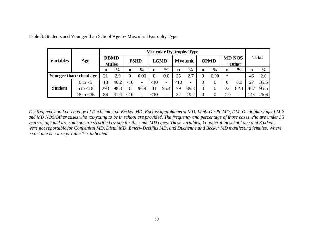
50
Table 3: Students and Younger than School Age by Muscular Dystrophy Type
The frequency and percentage of Duchenne and Becker MD, Facioscapulohumeral MD, Limb-Girdle MD, DM, Oculopharyngeal MD
and MD NOS/Other cases who too young to be in school are provided. The frequency and percentage of those cases who are under 35
years of age and are students are stratified by age for the same MD types. These variables, Younger than school age and Student,
were not reportable for Congenital MD, Distal MD, Emery-Dreiffus MD, and Duchenne and Becker MD manifesting females. Where
a variable is not reportable * is indicated.
n % n % n % n % n % n % n %
21 2.9 0 0.00 0 0.0 25 2.7 0 0.00 * 46 2.0
0 to <5 18 46.2 <10 - <10 - <10 - 0 0 0 0.0 27 35.5
5 to <18 293 98.3 31 96.9 41 95.4 79 89.8 0 0 23 82.1 467 95.5
18 to <35 86 41.4 <10 - <10 - 32 19.2 0 0 <10 - 144 26.6
TotalMD NOS
+ Other
Younger than school age
Variables Age
Muscular Dystrophy Type
DBMD
Males
Student
FSHD LGMD Myotonic OPMD

51
Table 4: Employment Status by Muscular Dystrophy Type
The employment status of Duchenne and Becker MD, Facioscapulohumeral MD, Limb-Girdle MD, DM, Oculopharyngeal MD and
MD NOS/Other cases are reported as frequencies and percentages. Employment status is stratified by the age at which the
employment status was recorded. In describing those who are working for pay, disabled or unable to work, or unemployed only cases
whose employment status was recorded when they were 18 years of age or older and younger than 65 were considered. The frequency
and percentage of retired cases is limited to those whose employment status was recorded when they were 35 years or older.
Employment status was not reportable for Congenital MD, Distal MD, Emery-Dreiffus MD, and Duchenne and Becker MD
manifesting females due to small case numbers
n % n % n % n % n % n % n %
18 to <35 28 13.46 23 47.92 25 49.02 64 38.3 0 0.0 21 31.34 161 29.8
35 to <65 21 32.3 62 44.6 49 40.5 144 25.4 12 28.6 37 18.69 325 28.7
65+ <10 - <10 - <10 - <10 - <10 - <10 - 18 5.1
18 to <35 37 17.8 <10 - <10 - 30 18.0 0 0.0 15 22.4 93 17.2
35 to <65 32 49.2 39 28.1 41 33.9 256 45.1 11 26.2 84 42.4 463 40.9
65+ <10 - <10 - <10 - 19 26.4 <10 - 10 9.7 45 12.9
18 to <35 19 9.1 <10 - <10 - 23 13.8 0 0.0 14 20.9 68 12.6
35 to <65 <10 - 16 11.5 15 12.4 76 13.4 <10 - 34 17.2 154 13.6
65+ 0 0.0 <10 - <10 - <10 - <10 - <10 - 17 4.9
35 to <65 <10 - 11 7.9 <10 - 46 8.1 <10 - 20 10.1 92 8.1
65+ <10 - 30 53.6 30 75.0 44 61.1 48 64.0 67 65.1 222 63.4
153 21.2 28 10.1 22 8.5 82 8.7 23 19.3 60 15.1 368 13.5
Total
Not documented
Age
Muscular Dystrophy Type
DBMD
MalesFSHD LGMD Myotonic OPMD
MD NOS +
Other
Working for Pay
Disabled or Unable
to Work
Unemployed
Retired
Variables

52
Table 5: Mobility Status by Muscular Dystrophy Type
Mobility status is reported as the frequency and percentage for each type of MD and is stratified
by the age at which the mobility status was recorded. Patients who do not require device
assistance to walk are classified as ‘Ambulatory’. Those who require part-time device support
are classified as ‘Ambulatory with Device Support,’ and patients who use a device, such as a
scooter or wheelchair, full-time or who are bedridden are classified as ‘Non-ambulatory’.
n % n % n % n % n %
Ambulatory 12 37.5 5 20.8 11 47.8 3 60.0 1 50.0
Ambulatory with Device Support 4 12.5 5 20.8 2 8.7 1 20.0 0 0.0
Non-Ambulatory 12 37.5 14 58.3 9 39.1 1 20.0 1 50.0
Ambulatory 107 59.4 40 17.7 43 18.5 19 31.7 1 20.0
Ambulatory with Device Support 28 15.6 32 14.2 4 1.7 5 8.3 1 20.0
Non-Ambulatory 37 20.6 153 67.7 182 78.5 35 58.3 3 60.0
Ambulatory 1 0.0 1 50.0 2 40.0 2 57.1 2 100.0
Ambulatory with Device Support 0 0.0 1 50.0 0 0.0 1 14.3 0 0.0
Non-Ambulatory 0 0.0 0 0.0 3 60.0 1 14.3 0 0.0
Ambulatory 0 0.0 0 0.0 1 100.0 9 64.3 1 50.0
Ambulatory with Device Support 0 0.0 0 0.0 0 0.0 2 14.3 1 50.0
Non-Ambulatory 0 0.0 0 0.0 0 0.0 3 21.4 0 0.0
Ambulatory 1 100.0 5 83.3 5 62.5 4 57.1 0 0.0
Ambulatory with Device Support 0 0.0 0 0.0 0 0.0 0 0.0 0 0.0
Non-Ambulatory 0 0.0 1 16.7 2 25.0 3 42.9 0 0.0
Ambulatory 6 85.7 24 92.3 35 74.5 91 65.0 37 64.9
Ambulatory with Device Support 1 14.3 0 0.0 7 14.9 11 7.9 12 21.1
Non-Ambulatory 0 0.0 1 3.9 3 6.4 33 23.6 7 12.3
Ambulatory 17 94.4 23 79.3 30 57.7 62 51.5 16 40.0
Ambulatory with Device Support 1 5.6 2 6.9 8 15.4 12 9.9 4 10.0
Non-Ambulatory 0 0.0 4 13.8 14 26.9 43 35.5 18 45.0
Ambulatory 40 70.2 55 90.2 158 88.8 425 76.3 49 66.2
Ambulatory with Device Support 1 1.8 1 1.6 5 2.8 49 8.8 10 13.5
Non-Ambulatory 3 5.3 3 4.9 6 3.4 58 10.4 12 16.2
Ambulatory 0 0.0 0 0.0 0 0.0 36 85.7 51 68.0
Ambulatory with Device Support 0 0.0 0 0.0 0 0.0 3 7.1 4 5.3
Non-Ambulatory 0 0.0 0 0.0 0 0.0 1 2.4 12 16.0
Ambulatory 6 75.0 15 71.4 46 64.8 104 53.1 46 46.5
Ambulatory with Device Support 0 0.0 1 4.8 4 5.6 15 7.7 12 12.1
Non-Ambulatory 1 12.5 5 23.8 18 25.4 56 28.6 32 32.3
Other 18 5.9 0 0.0 2 0.3 8 0.7 2 0.6
Not Documented 8 2.6 4 1.0 17 2.8 52 4.5 21 5.9
LGMD
Myotonic
OPMD
MD NOS
+Other
Congenital
DBMD Males
Distal
EDMD
FSHD
Manifesting
Females
Age
Muscular
DystrophyMobility Status
0 to <10 10 to <18 18 to <35 35 to <65 65+

53
Table 6: Clinical Interventions by Muscular Dystrophy Type
The frequency and percentage of cases who had the use of the clinical interventions considered listed in their medical record were
provided for each MD type. The percentage and frequency of those who are deceased are also provided for each type of MD. Clinical
variables are only reported for MD types where future iterations of the MD STARnet pilot will collect data. Thus, where a variable
was not reported, + is indicated.
n % n % n % n % n % n % n % n % n % n % n %
Vital Status Deceased 9 10.5 1 5.9 82 11.4 1 5.9 2 9.1 21 7.6 29 11.2 161 17.1 25 21.0 80 20.1 411 14.4
Yes 18 20.9 0 0.0 67 9.3 1 5.9 1 4.6 + 10 3.9 + + 35 8.8 132 8.7
Not doc. 68 79.1 17 100.0 653 90.7 16 94.1 21 95.5 + 250 96.2 + + 363 91.2 1388 91.3
Yes 24 27.9 2 11.8 235 32.6 2 11.8 2 9.1 25 9.0 45 17.3 215 22.8 + 58 14.6 608 21.5
Not doc. 62 72.1 15 88.2 485 67.4 15 88.2 20 90.9 253 91.0 215 82.7 728 77.2 + 340 85.4 2133 78.5
Yes + + 72 10.0 1 5.9 + + + + + 30 7.5 103 9.1
Not doc. + + 648 90.0 16 94.1 + + + + + 368 92.5 1032 90.9
Yes + + 178 24.7 1 5.9 + + + + + 3 0.8 182 16.0
Not doc. + + 542 75.3 16 94.1 + + + + + 395 99.3 953 84.0
Yes 1 1.2 0 0.0 + + 4 18.2 + 10 3.9 93 9.9 + 15 3.8 123 7.1
Not doc. 85 98.8 17 100.0 + + 18 81.8 + 250 96.2 850 90.1 + 383 96.2 1603 92.9
Yes 0 0.0 0 0.0 + + 1 4.6 + 3 1.2 36 3.8 + 5 1.3 45 2.6
Not doc. 86 100.0 17 100.0 + + 21 95.5 + 257 98.9 907 96.2 + 393 98.7 1681 97.4
Yes 0 0.0 2 11.8 + + 2 9.1 + 4 1.5 1 0.1 + 1 0.3 10 0.6
Not doc. 86 100.0 15 88.2 + + 20 90.9 + 256 98.5 942 99.9 + 397 99.8 1716 99.4
Cough Assist
Pacemaker
Defibrillator
Cardiac Transplant
Myotonic
PEG
NIPPV(CPAP/BIPAP)
Tracheostomy
Variables
Muscular Dystrophy Type
TotalCongenital Distal
DBMD
Males
Manifesting
FemalesEDMD FSHD LGMD OPMD
MD NOS +
Other

54
Table 7: Mean and Median Medication Count at Most Recent Health Encounter
Means and standard deviations are provided for the number of medications recorded in the medical records of cases at their last
health encounter for each MD type.
Mean STD Mean STD Mean STD Mean STD Mean STD Mean STD Mean STD Mean STD Mean STD Mean STD Mean STD
3.6 4.1 4.1 3.1 4.4 4.5 5.3 5.2 2.8 2.4 4.0 4.3 4.1 4.4 4.2 4.3 5.9 4.4 5.3 5.0 4.4 4.5
Medications
TotalVariableMyotonic OPMD
MD NOS +
Other
Muscular Dystrophy Type
Congenital Distal DBMDManifestin
g Female
Emery-
DreifussFSHD LGMD
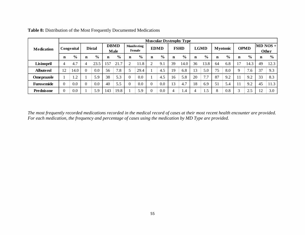
55
Table 8: Distribution of the Most Frequently Documented Medications
The most frequently recorded medications recorded in the medical record of cases at their most recent health encounter are provided.
For each medication, the frequency and percentage of cases using the medication by MD Type are provided.
n % n % n % n % n % n % n % n % n % n %
Lisinopril 4 4.7 4 23.5 157 21.7 2 11.8 2 9.1 39 14.0 36 13.8 64 6.8 17 14.3 49 12.3
Albuterol 12 14.0 0 0.0 56 7.8 5 29.4 1 4.5 19 6.8 13 5.0 75 8.0 9 7.6 37 9.3
Omeprazole 1 1.2 1 5.9 38 5.3 0 0.0 1 4.5 16 5.8 20 7.7 87 9.2 11 9.2 33 8.3
Furosemide 0 0.0 0 0.0 40 5.5 0 0.0 0 0.0 13 4.7 18 6.9 51 5.4 11 9.2 45 11.3
Prednisone 0 0.0 1 5.9 143 19.8 1 5.9 0 0.0 4 1.4 4 1.5 8 0.8 3 2.5 12 3.0
DBMD
MaleEDMDMedication
Muscular Dystrophy Type
DistalManifesting
FemaleFSHD
MD NOS +
OtherLGMD Myotonic OPMDCongenital

56
References
1. Brais B. Oculopharyngeal muscular dystrophy: a late-onset polyalanine disease. Cytogenetic and Genome Research 2003;100:252-60. 2. Mercuri E, Muntoni F. Muscular dystrophies. The Lancet;381:845-60. 3. Johnson NE, Abbott D, Cannon-Albright LA. Relative risks for comorbidities associated with myotonic dystrophy: A population-based analysis. Muscle & Nerve 2015;52:659-61. 4. Smith AE, McMullen K, Jensen MP, Carter GT, Molton IR. Symptom burden in persons with myotonic and facioscapulohumeral muscular dystrophy. Am J Phys Med Rehabil 2014;93:387-95. 5. Cheuk DK, Wong V, Wraige E, et al. Surgery for scoliosis in Duchenne muscular dystrophy. The Cochrane database of systematic reviews 2007:Cd005375. 6. Acosta-Mérida MA, Marchena-Gómez J, Afonso-Déniz JM. Utility of surgical myotomy in the dysphagia due to oculopharyngeal dystrophy. Revista Espanola de Enfermedades Digestivas 2016;108:843-4. 7. Becher MW, Morrison L, Davis LE, et al. Oculopharyngeal muscular dystrophy in hispanic new mexicans. JAMA 2001;286:2437-40. 8. Fishman FG, Goldstein EM, Peljovich AE. Surgical treatment of upper extremity contractures in Emery–Dreifuss muscular dystrophy. Journal of Pediatric Orthopaedics B 2017;26:32-5. 9. Falsaperla R, Praticò AD, Ruggieri M, et al. Congenital muscular dystrophy: from muscle to brain. Italian Journal of Pediatrics 2016;42:78. 10. Blumen SC, Nisipeanu P, Sadeh M, Asherov A, Tomé FMS, Korczyn AD. Clinical features of oculopharyngeal muscular dystrophy among Bukhara Jews. Neuromuscular Disorders 1993;3:575-7. 11. Mathieu J, De Braekeleer M, Prévost C. Genealogical reconstruction of myotonic dystrophy in the Saguenay‐Lac‐Saint‐ Jean area (Quebec, Canada). Neurology 1990;40:839-. 12. Theadom A, Rodrigues M, Roxburgh R, et al. Prevalence of muscular dystrophies: a systematic literature review. Neuroepidemiology 2014;43:259-68. 13. Abbott D, Johnson NE, Cannon-Albright LA. A population-based survey of risk for cancer in individuals diagnosed with myotonic dystrophy. Muscle and Nerve 2016;54:783-5. 14. Graziano A, Bianco F, D'Amico A, et al. Prevalence of congenital muscular dystrophy in Italy: a population study. Neurology 2015;84:904-11. 15. Okada M, Kawahara G, Noguchi S, et al. Primary collagen VI deficiency is the second most common congenital muscular dystrophy in Japan. Neurology 2007;69:1035-42. 16. Peat RA, Smith JM, Compton AG, et al. Diagnosis and etiology of congenital muscular dystrophy. Neurology 2008;71:312-21. 17. Sframeli M, Sarkozy A, Bertoli M, et al. Congenital muscular dystrophies in the UK population: Clinical and molecular spectrum of a large cohort diagnosed over a 12-year period. Neuromuscular Disorders 2017;27:793-803. 18. Lisi MT, Cohn RD. Congenital muscular dystrophies: new aspects of an expanding group of disorders. Biochimica et Biophysica Acta 2007;1772:159-72. 19. Muntoni F, Voit T. The congenital muscular dystrophies in 2004: a century of exciting progress. Neuromuscular Disorders 2004;14:635-49. 20. Mercuri E, Muntoni F. The ever-expanding spectrum of congenital muscular dystrophies. Annals of Neurology 2012;72:9-17. 21. Bertini E, D'Amico A, Gualandi F, Petrini S. Congenital muscular dystrophies: a brief review. Seminars in Pediatric Neurology 2011;18:277-88.

57
22. Norwood FLM, Harling C, Chinnery PF, Eagle M, Bushby K, Straub V. Prevalence of genetic muscle disease in Northern England: in-depth analysis of a muscle clinic population. Brain 2009;132:3175-86. 23. Mah JK, Korngut L, Fiest KM, et al. A Systematic Review and Meta-analysis on the Epidemiology of the Muscular Dystrophies. Canadian Journal of Neurological Sciences 2016;43:163-77. 24. Emery AEH. The muscular dystrophies. Bmj 1998;317:991-5. 25. Giliberto F, Radic CP, Luce L, Ferreiro V, de Brasi C, Szijan I. Symptomatic female carriers of Duchenne muscular dystrophy (DMD): genetic and clinical characterization. Journal of the Neurological Sciences 2014;336:36-41. 26. Davidson ZE, Ryan MM, Kornberg AJ, et al. Observations of body mass index in Duchenne muscular dystrophy: a longitudinal study. European Journal of Clinical Nutrition 2014;68:892-7. 27. Magri F, Govoni A, D'Angelo MG, et al. Genotype and phenotype characterization in a large dystrophinopathic cohort with extended follow-up. Journal of Neurology 2011;258:1610-23. 28. Ciafaloni E, Fox DJ, Pandya S, et al. Delayed diagnosis in duchenne muscular dystrophy: data from the Muscular Dystrophy Surveillance, Tracking, and Research Network (MD STARnet). Journal of Pediatrics 2009;155:380-5. 29. Viollet L, Thrush PT, Flanigan KM, Mendell JR, Allen HD. Effects of angiotensin-converting enzyme inhibitors and/or beta blockers on the cardiomyopathy in Duchenne muscular dystrophy. American Journal of Cardiology 2012;110:98-102. 30. Allen HD, Thrush PT, Hoffman TM, Flanigan KM, Mendell JR. Cardiac management in neuromuscular diseases. Physical Medicine & Rehabilitation Clinics of North America 2012;23:855-68. 31. Hor KN, Wansapura J, Markham LW, et al. Circumferential strain analysis identifies strata of cardiomyopathy in Duchenne muscular dystrophy: a cardiac magnetic resonance tagging study. Journal of the American College of Cardiology 2009;53:1204-10. 32. Ryder S, Leadley RM, Armstrong N, et al. The burden, epidemiology, costs and treatment for Duchenne muscular dystrophy: an evidence review. Orphanet Journal Of Rare Diseases 2017;12:79. 33. Ciafaloni E, Kumar A, Liu K, et al. Age at onset of first signs or symptoms predicts age at loss of ambulation in Duchenne and Becker Muscular Dystrophy: Data from the MD STARnet. J Pediatr Rehabil Med 2016;9:5-11. 34. Wong BL, Christopher C. Corticosteroids in Duchenne muscular dystrophy: a reappraisal. Journal of Child Neurology 2002;17:183-90. 35. Shapiro F, Zurakowski D, Bui T, Darras BT. Progression of spinal deformity in wheelchair-dependent patients with Duchenne muscular dystrophy who are not treated with steroids: coronal plane (scoliosis) and sagittal plane (kyphosis, lordosis) deformity. Bone & Joint Journal 2014;96-B:100-5. 36. Rall S, Grimm T. Survival in Duchenne muscular dystrophy. Acta Myologica 2012;31:117-20. 37. Romitti PA, Zhu Y, Puzhankara S, et al. Prevalence of Duchenne and Becker muscular dystrophies in the United States. Pediatrics 2015;135:513-21. 38. Mah JK, Korngut L, Dykeman J, Day L, Pringsheim T, Jette N. A systematic review and meta-analysis on the epidemiology of Duchenne and Becker muscular dystrophy. Neuromuscular Disorders 2014;24:482-91. 39. Udd B. Genetics and pathogenesis of distal muscular dystrophies. Advances in Experimental Medicine & Biology 2009;652:23-38. 40. Udd B. Molecular biology of distal muscular dystrophies--sarcomeric proteins on top. Biochimica et Biophysica Acta 2007;1772:145-58. 41. Norwood FLM, Harling C, Chinnery PF, Eagle M, Bushby K, Straub V. Prevalence of genetic muscle disease in Northern England: in-depth analysis of a muscle clinic population. Brain : a journal of neurology 2009;132:3175-86.

58
42. Udd B, Partanen J, Halonen P, et al. Tibial muscular dystrophy: Late adult-onset distal myopathy in 66 finnish patients. Archives of Neurology 1993;50:604-8. 43. Pollazzon M, Suominen T, Penttila S, et al. The first Italian family with tibial muscular dystrophy caused by a novel titin mutation. Journal of Neurology 2010;257:575-9. 44. Pardal-Fernandez JM, Jerez-Garcia P, Rallo-Gutierrez B, Puentes-Gil JM, Godes-Medrano B, Marco-Giner J. Tibial muscular dystrophy with late adult onset in a Spanish family. Electromyography & Clinical Neurophysiology 2005;45:285-90. 45. de Seze J, Udd B, Haravuori H, et al. The first European family with tibial muscular dystrophy outside the Finnish population. Neurology 1998;51:1746-8. 46. Van den Bergh PYK, Bouquiaux O, Verellen C, et al. Tibial muscular dystrophy in a Belgian family. Annals of Neurology 2003;54:248-51. 47. Van den Bergh PY, Martin JJ, Lecouvet F, Udd B, Schmedding E. Laing early-onset distal myopathy in a Belgian family. Acta Neurologica Belgica 2014;114:253-6. 48. Malicdan MC, Nonaka I. Distal myopathies a review: highlights on distal myopathies with rimmed vacuoles. Neurology India 2008;56:314-24. 49. Linssen WH, de Voogt WG, Krahn M, et al. Long-term follow-up study on patients with Miyoshi phenotype of distal muscular dystrophy. European Journal of Neurology 2013;20:968-74. 50. Wallgren-Pettersson C, Lehtokari VL, Kalimo H, et al. Distal myopathy caused by homozygous missense mutations in the nebulin gene. Brain 2007;130:1465-76. 51. Lamont PJ, Udd B, Mastaglia FL, et al. Laing early onset distal myopathy: slow myosin defect with variable abnormalities on muscle biopsy. Journal of Neurology, Neurosurgery & Psychiatry 2006;77:208-15. 52. Emery AE. Emery-Dreifuss muscular dystrophy - a 40 year retrospective. Neuromuscular Disorders 2000;10:228-32. 53. Felice KJ, Schwartz RC, Brown CA, Leicher CR, Grunnet ML. Autosomal dominant Emery-Dreifuss dystrophy due to mutations in rod domain of the lamin A/C gene. Neurology 2000;55:275-80. 54. Raffaele Di Barletta M, Ricci E, Galluzzi G, et al. Different mutations in the LMNA gene cause autosomal dominant and autosomal recessive Emery-Dreifuss muscular dystrophy. American Journal of Human Genetics 2000;66:1407-12. 55. Hayashi YK. X-linked form of Emery-Dreifuss muscular dystrophy. Acta Myologica 2005;24:98-103. 56. Jimenez-Escrig A, Gobernado I, Garcia-Villanueva M, Sanchez-Herranz A. Autosomal recessive Emery-Dreifuss muscular dystrophy caused by a novel mutation (R225Q) in the lamin A/C gene identified by exome sequencing. Muscle & Nerve 2012;45:605-10. 57. Goncu K, Guzel R, Guler-Uysal F. Emery-Dreifuss muscular dystrophy in the evaluation of decreased spinal mobility and joint contractures. Clinical Rheumatology 2003;22:456-60. 58. Diaz-Manera J, Alejaldre A, Gonzalez L, et al. Muscle imaging in muscle dystrophies produced by mutations in the EMD and LMNA genes. Neuromuscular Disorders 2016;26:33-40. 59. Bonne G, Mercuri E, Muchir A, et al. Clinical and molecular genetic spectrum of autosomal dominant Emery-Dreifuss muscular dystrophy due to mutations of the lamin A/C gene. Annals of Neurology 2000;48:170-80. 60. Chung B, Wong V, Ip P. Prevalence of Neuromuscular Diseases in Chinese Children: A Study in Southern China. Journal of Child Neurology 2003;18:217-9. 61. Tawil R, Kissel JT, Heatwole C, et al. Evidence-based guideline summary: Evaluation, diagnosis, and management of facioscapulohumeral muscular dystrophy: Report of the Guideline Development, Dissemination, and Implementation Subcommittee of the American Academy of Neurology and the Practice Issues Review Panel of the American Association of Neuromuscular & Electrodiagnostic Medicine. Neurology 2015;85:357-64.

59
62. de Greef JC, Lemmers RJ, Camano P, et al. Clinical features of facioscapulohumeral muscular dystrophy 2. Neurology 2010;75:1548-54. 63. Nikolic A, Ricci G, Sera F, et al. Clinical expression of facioscapulohumeral muscular dystrophy in carriers of 1-3 D4Z4 reduced alleles: experience of the FSHD Italian National Registry. BMJ Open 2016;6:e007798. 64. Mul K, Lassche S, Voermans NC, Padberg GW, Horlings CG, van Engelen BG. What's in a name? The clinical features of facioscapulohumeral muscular dystrophy. Practical Neurology 2016;16:201-7. 65. Schipper K, Bakker M, Abma T. Fatigue in facioscapulohumeral muscular dystrophy: a qualitative study of people's experiences. Disability & Rehabilitation 2017;39:1840-6. 66. Tawil R, Van Der Maarel SM. Facioscapulohumeral muscular dystrophy. Muscle & Nerve 2006;34:1-15. 67. Flanigan KM, Coffeen CM, Sexton L, Stauffer D, Brunner S, Leppert MF. Genetic characterization of a large, historically significant Utah kindred with facioscapulohumeral dystrophy. Neuromuscular Disorders 2001;11:525-9. 68. Deenen JC, Arnts H, van der Maarel SM, et al. Population-based incidence and prevalence of facioscapulohumeral dystrophy. Neurology 2014;83:1056-9. 69. Mostacciuolo ML, Pastorello E, Vazza G, et al. Facioscapulohumeral muscular dystrophy: epidemiological and molecular study in a north-east Italian population sample. Clinical Genetics 2009;75:550-5. 70. Sposito R, Pasquali L, Galluzzi F, et al. Facioscapulohumeral muscular dystrophy type 1A in northwestern Tuscany: a molecular genetics-based epidemiological and genotype-phenotype study. Genetic Testing 2005;9:30-6. 71. Dorgham M, Kodivalasa M, Vasu T. A rare case of facioscapulohumeral syndrome (FSHS) presenting with chronic wide spread pain. British Journal of Pain 2017;11 (2 Supplement 1):81-2. 72. Miro J, Gertz KJ, Carter GT, Jensen MP. Pain location and intensity impacts function in persons with myotonic dystrophy type 1 and facioscapulohumeral dystrophy with chronic pain. Muscle & Nerve 2014;49:900-5. 73. Della Marca G, Frusciante R, Vollono C, et al. Pain and the alpha-sleep anomaly: a mechanism of sleep disruption in facioscapulohumeral muscular dystrophy. Pain Medicine 2013;14:487-97. 74. Moris G, Wood L, FernaNdez-Torron R, et al. Chronic pain has a strong impact on quality of life in facioscapulohumeral muscular dystrophy. Muscle & Nerve 2017;20:20. 75. Padua L, Aprile I, Frusciante R, et al. Quality of life and pain in patients with facioscapulohumeral muscular dystrophy. Muscle & Nerve 2009;40:200-5. 76. Labombarda F, Maurice M, Simon JP, et al. Cardiac Abnormalities in Type 1 Facioscapulohumeral Muscular Dystrophy. Journal of Clinical Neuromuscular Disease 2017;18:199-206. 77. Sveen ML, Schwartz M, Vissing J. High prevalence and phenotype-genotype correlations of limb girdle muscular dystrophy type 2I in Denmark. Annals of Neurology 2006;59:808-15. 78. Khadilkar SV, Faldu HD, Patil SB, Singh R. Limb-girdle Muscular Dystrophies in India: A Review. Annals of Indian Academy of Neurology 2017;20:87-95. 79. Khadilkar SV, Chaudhari CR, Dastur RS, Gaitonde PS, Yadav JG. Limb-girdle muscular dystrophy in the Agarwals: Utility of founder mutations in CAPN3 gene. Annals of Indian Academy of Neurology 2016;19:108-11. 80. Khadilkar SV, Singh RK. Limb girdle muscular dystrophies in India. Neurology India 2008;56:281-8. 81. Khadilkar SV, Singh RK, Agarwal P, Krahn M, Levy N. Twenty-two year follow-up of an Indian family with dysferlinopathy-clinical, immunocytochemical, western blotting and genetic features. Neurology India 2008;56:388-90.

60
82. Stensland E, Lindal S, Jonsrud C, et al. Prevalence, mutation spectrum and phenotypic variability in Norwegian patients with Limb Girdle Muscular Dystrophy 2I. Neuromuscular Disorders 2011;21:41-6. 83. Pantoja-Melendez CA, Miranda-Duarte A, Roque-Ramirez B, Zenteno JC. Epidemiological and Molecular Characterization of a Mexican Population Isolate with High Prevalence of Limb-Girdle Muscular Dystrophy Type 2A Due to a Novel Calpain-3 Mutation. PLoS ONE [Electronic Resource] 2017;12:e0170280. 84. Liang WC, Chou PC, Hung CC, et al. Probable high prevalence of limb-girdle muscular dystrophy type 2D in Taiwan. Journal of the Neurological Sciences 2016;362:304-8. 85. Nigro V. Molecular bases of autosomal recessive limb-girdle muscular dystrophies. Acta Myologica 2003;22:35-42. 86. Mustafa E, Khandaker MAH, Rashid MM, Ghose SK, Chowdhury MK. Sarcoglycanopathy - A rare case report and literature review. Journal of Medicine (Bangladesh) 2014;15:77-9. 87. Kirschner J, Lochmuller H. Sarcoglycanopathies. Handbook of Clinical Neurology 2011;101:41-6. 88. Meena AK, Sreenivas D, Sundaram C, et al. Sarcoglycanopathies: a clinico-pathological study. Neurology India 2007;55:117-21. 89. Schade van Westrum SM, Dekker LR, de Voogt WG, et al. Cardiac involvement in Dutch patients with sarcoglycanopathy: a cross-sectional cohort and follow-up study. Muscle Nerve 2014;50:909-13. 90. Fanin M, Melacini P, Boito C, Pegoraro E, Angelini C. LGMD2E patients risk developing dilated cardiomyopathy. Neuromuscular disorders : NMD 2003;13:303-9. 91. Fayssoil A, Ogna A, Chaffaut C, et al. Natural History of Cardiac and Respiratory Involvement, Prognosis and Predictive Factors for Long-Term Survival in Adult Patients with Limb Girdle Muscular Dystrophies Type 2C and 2D. PLoS ONE [Electronic Resource] 2016;11:e0153095. 92. Maggi L, D'Amico A, Pini A, et al. LMNA-associated myopathies: the Italian experience in a large cohort of patients. Neurology 2014;83:1634-44. 93. Maggi L, Carboni N, Bernasconi P. Skeletal muscle laminopathies: A review of clinical and molecular features. Cells 2016;5 (3) (no pagination). 94. Turner C, Hilton-Jones D. Myotonic dystrophy: diagnosis, management and new therapies. Current Opinion in Neurology 2014;27:599-606. 95. Vanacore N, Rastelli E, Antonini G, et al. An Age-Standardized Prevalence Estimate and a Sex and Age Distribution of Myotonic Dystrophy Types 1 and 2 in the Rome Province, Italy. Neuroepidemiology 2016;46:191-7. 96. Groh WJ, Groh MR, Shen C, Monckton DG, Bodkin CL, Pascuzzi RM. Survival and CTG repeat expansion in adults with myotonic dystrophy type 1. Muscle Nerve 2011;43:648-51. 97. Dogan C, De Antonio M, Hamroun D, et al. Gender as a modifying factor influencing myotonic dystrophy type 1 phenotype severity and mortality: A nationwide multiple databases cross-sectional observational study. PLoS ONE 2016;11 (2) (no pagination). 98. Laberge L, Mathieu J, Auclair J, Gagnon E, Noreau L, Gagnon C. Clinical, psychosocial, and central correlates of quality of life in myotonic dystrophy type 1 patients. Eur Neurol 2013;70:308-15. 99. Heatwole C, Johnson N, Bode R, et al. Patient-Reported Impact of Symptoms in Myotonic Dystrophy Type 2 (PRISM-2). Neurology 2015;85:2136-46. 100. Laberge L, Dauvilliers Y, Bégin P, Richer L, Jean S, Mathieu J. Fatigue and daytime sleepiness in patients with myotonic dystrophy type 1: To lump or split? Neuromuscul Disord 2009;19:397-402. 101. Christopher K, Horkan C, Patterson RB, Yodice PC. Oculopharyngeal muscular dystrophy complicating airway management. Chest 2001;120:2101-3. 102. Tondo M, Gamez J, Gutierrez-Rivas E, Medel-Jimenez R, Martorell L. Genotype and phenotype study of 34 Spanish patients diagnosed with oculopharyngeal muscular dystrophy. Journal of Neurology 2012;259:1546-52.

61
103. Youssof S, Romero-Clark C, Warner T, Plowman E. Dysphagia-related quality of life in oculopharyngeal muscular dystrophy: Psychometric properties of the SWAL-QOL instrument. Muscle & Nerve 2017;56:28-35. 104. Van Der Sluijs BM, Hoefsloot LH, Padberg GW, Van Der Maarel SM, Van Engelen BG. Oculopharyngeal muscular dystrophy with limb girdle weakness as major complaint. Journal of Neurology 2003;250:1307-12. 105. van der Sluijs BM, Raz V, Lammens M, van den Heuvel LP, Voermans NC, van Engelen BG. Intranuclear Aggregates Precede Clinical Onset in Oculopharyngeal Muscular Dystrophy. Journal of neuromuscular diseases 2016;3:101-9. 106. van der Sluijs BM, Lassche S, Knuiman GJ, et al. Involvement of pelvic girdle and proximal leg muscles in early oculopharyngeal muscular dystrophy. Neuromuscular Disorders 2017;27:1099-105. 107. Youssof S. The relationship between physical symptoms and health-related quality of life in oculopharyngeal muscular dystrophy. Muscle & Nerve 2016;53:694-9. 108. Shan J, Chen B, Lin P, et al. Oculopharyngeal muscular dystrophy: phenotypic and genotypic studies in a Chinese population. NeuroMolecular Medicine 2014;16:782-6. 109. Robinson DO, Hammans SR, Read SP, Sillibourne J. Oculopharyngeal muscular dystrophy (OPMD): analysis of the PABPN1 gene expansion sequence in 86 patients reveals 13 different expansion types and further evidence for unequal recombination as the mutational mechanism. Human Genetics 2005;116:267-71. 110. Muller T, Deschauer M, Kolbe-Fehr F, Zierz S. Genetic heterogeneity in 30 German patients with oculopharyngeal muscular dystrophy. Journal of Neurology 2006;253:892-5. 111. Mirabella M, Silvestri G, de Rosa G, et al. GCG genetic expansions in Italian patients with oculopharyngeal muscular dystrophy. Neurology 2000;54:608-14. 112. Hill ME, Creed GA, McMullan TF, et al. Oculopharyngeal muscular dystrophy: phenotypic and genotypic studies in a UK population. Brain 2001;124:522-6. 113. Becher MW, Morrison L, Davis LE, et al. Oculopharyngeal muscular dystrophy in Hispanic New Mexicans. JAMA 2001;286:2437-40. 114. Agarwal PK, Mansfield DC, Mechan D, et al. Delayed diagnosis of oculopharyngeal muscular dystrophy in Scotland. British Journal of Ophthalmology 2012;96:281-3. 115. Bumm K, Zenker M, Bozzato A. Oculopharyngeal muscular dystrophy as a rare differential diagnosis for unexplained dysphagia: a case report. Cases journal 2009;2:94. 116. Pulkes T, Papsing C, Busabaratana M, Dejthevaporn C, Witoonpanich R. Mutation and haplotype analysis of oculopharyngeal muscular dystrophy in Thai patients. Journal of Clinical Neuroscience 2011;18:674-7. 117. Witoonpanich R, Phankhian S, Sura T, Lertrit P, Phudhichareonrat S. Oculopharyngodistal myopathy in a Thai family. Journal of the Medical Association of Thailand 2004;87:1518-21. 118. Tremolizzo L, Galbussera A, Tagliabue E, et al. An apparently sporadic case of oculopharyngeal muscular dystrophy: the first Italian report.[Erratum appears in Neurol Sci. 2008 Sep;29(4):291 Note: Curto, N [added]]. Neurological Sciences 2007;28:339-41. 119. Blumen SC, Kesler A, Dabby R, et al. Oculopharyngeal muscular dystrophy among Bulgarian Jews: a new cluster? Israel Medical Association Journal: Imaj 2013;15:748-52. 120. Tung JD, Oh SR, Gruber AB, et al. The man who could not see what he could not eat. Survey of Ophthalmology 2011;56:461-5. 121. Codere F, Brais B, Rouleau G, Lafontaine E. Oculopharyngeal muscular dystrophy: What's new? Orbit 2001;20:259-66. 122. Mazanec R, Seeman P, Seemanova E. Oculopharyngeal muscular dystrophy in the population of the Czech republic. [Czech]. Ceska a Slovenska Neurologie a Neurochirurgie 2013;76:717-22.

62
123. Palmer PM, Neel AT, Sprouls G, Morrison L. Swallow characteristics in patients with oculopharyngeal muscular dystrophy. Journal of Speech Language & Hearing Research 2010;53:1567-78. 124. H.R. 717 (107th): MD-CARE Act. 2001. (Accessed October 17, 2017, at https://www.govtrack.us/congress/bills/107/hr717 ) 125. Miller LA, Romitti PA, Cunniff C, et al. The muscular Dystrophy Surveillance Tracking and Research Network (MD STARnet): surveillance methodology. Birth Defects Research 2006;76:793-7. 126. Mathews KD, Cunniff C, Kantamneni JR, et al. Muscular Dystrophy Surveillance Tracking and Research Network (MD STARnet): case definition in surveillance for childhood-onset Duchenne/Becker muscular dystrophy. Journal of Child Neurology 2010;25:1098-102. 127. Falsaperla R, Pratico AD, Ruggieri M, et al. Congenital muscular dystrophy: from muscle to brain. Italian Journal of Pediatrics 2016;42:78. 128. Romitti PA, Zhu Y, Puzhankara S, et al. Prevalence of Duchenne and Becker muscular dystrophies in the United States.[Erratum appears in Pediatrics. 2015 May;135(5):945; PMID: 25934896]. Pediatrics 2015;135:513-21. 129. Smith AE, McMullen K, Jensen MP, Carter GT, Molton IR. Symptom Burden in Persons with Myotonie and Facioscapulohumeral Muscular Dystrophy. American Journal of Physical Medicine & Rehabilitation 2014;93:387-95. 130. Thomas NS, Wiseman K, Spurlock G, MacDonald M, Ustek D, Upadhyaya M. A large patient study confirming that facioscapulohumeral muscular dystrophy (FSHD) disease expression is almost exclusively associated with an FSHD locus located on a 4qA-defined 4qter subtelomere. Journal of Medical Genetics 2007;44:215-8. 131. Al-Zaidy SA, Malik V, Kneile K, et al. A slowly progressive form of limb-girdle muscular dystrophy type 2C associated with founder mutation in the SGCG gene in Puerto Rican Hispanics. Molecular Genetics & Genomic Medicine 2015;3:92-8. 132. Larkindale J, Yang W, Hogan PF, et al. Cost of illness for neuromuscular diseases in the United States. Muscle & Nerve 2014;49:431-8. 133. Wang RT, Silverstein Fadlon CA, Ulm JW, et al. Online self-report data for duchenne muscular dystrophy confirms natural history and can be used to assess for therapeutic benefits. PLoS currents 2014;6:17. 134. Yamaguchi M, Suzuki M. Independent living with Duchenne muscular dystrophy and home mechanical ventilation in areas of Japan with insufficient national welfare services. International Journal of Qualitative Studies on Health and Well-being 2013;8:20914. 135. Dreyer PS, Steffensen BF, Pedersen BD. Life with home mechanical ventilation for young men with Duchenne muscular dystrophy. Journal of Advanced Nursing 2010;66:753-62. 136. Nguyen TH, Thi HB, Thuy LTT, et al. Study of functional independence of patients with Duchene muscular dystrophy. Annals of Translational Medicine Conference: 12th Asia Pacific Conference on Human Genetics 2017;5. 137. Nätterlund B. Living with Muscular Dystrophy: Illness Experience, Activities of Daily Living, Coping, Quality of Life and Rehabilitation. Scandinavian Journal of Occupational Therapy 2001;8:157. 138. Stubgen JP, Stipp A. Facioscapulohumeral muscular dystrophy: a prospective study of weakness and functional impairment. Journal of Neurology 2010;257:1457-64. 139. Stubgen JP. Limb girdle muscular dystrophy: an interval study of weakness and functional impairment. Journal of Clinical Neuromuscular Disease 2008;9:333-40. 140. Madej-Pilarczyk A, Kochanski A. Emery-Dreifuss muscular dystrophy: the most recognizable laminopathy. Folia Neuropathologica 2016;54:1-8. 141. Minis M-A, Engels J, Akkermans R, et al. Employment status of patients with neuromuscular diseases in relation to personal factors, fatigue and health status. Journal of Rehabilitation Medicine 2010;42:60 - 5.

63
142. Laberge L, Mathieu J, Auclair J, Gagnon E, Noreau L, Gagnon C. Clinical, psychosocial, and central correlates of quality of life in myotonic dystrophy type 1 patients. European neurology 2013;70:308-15. 143. Madej-Pilarczyk A. Professional activity of Emery-Dreifuss muscular dystrophy patients in Poland. International Journal of Occupational Medicine and Environmental Health 2014;27:270-7. 144. Mori-Yoshimura M, Mizuno Y, Yoshida S, et al. Social involvement issues in patients with Becker muscular dystrophy: A questionnaire survey of subjects from a patient registry. Brain & Development 2017;29:29. 145. Lue YJ, Chen SS, Lu YM. Quality of life of patients with Duchenne muscular dystrophy: from adolescence to young men. Disability & Rehabilitation 2017;39:1408-13. 146. Minis MA, Kalkman JS, Akkermans RP, et al. Employment status of patients with neuromuscular diseases in relation to personal factors, fatigue and health status: a secondary analysis. Journal of Rehabilitation Medicine 2010;42:60-5. 147. Hilbert JE, Kissel JT, Luebbe EA, et al. If you build a rare disease registry, will they enroll and will they use it? Methods and data from the National Registry of Myotonic Dystrophy (DM) and Facioscapulohumeral Muscular Dystrophy (FSHD). Contemporary Clinical Trials 2012;33:302-11. 148. Rakocevic-Stojanovic V, Peric S, Madzarevic R, et al. Significant impact of behavioral and cognitive impairment on quality of life in patients with myotonic dystrophy type 1. Clinical Neurology & Neurosurgery 2014;126:76-81. 149. Kraya T, Schmidt B, Muller T, Hanisch F. Impairment of respiratory function in late-onset distal myopathy due to MATR3 Mutation. Muscle & Nerve 2015;51:916-8. 150. Annunziata A, Fiorentino G, Esquinas A. Effect on lung function of mounthpiece ventilation in Steinert disease. A case report. Acta Myol 2017;36:33-5. 151. Khirani S, Dabaj I, Amaddeo A, Ramirez A, Quijano-Roy S, Fauroux B. The value of respiratory muscle testing in a child with congenital muscular dystrophy. Respirology Case Reports 2014;2:95-8. 152. Crescimanno G, Marrone O. Successful treatment of atelectasis with Dornase alpha in a patient with congenital muscular dystrophy. Revista Portuguesa de Pneumologia 2014;20:42-5. 153. Pincherle A, Patruno V, Raimondi P, et al. Sleep breathing disorders in 40 Italian patients with Myotonic dystrophy type 1. Neuromuscul Disord 2012;22:219-24. 154. Bianchi MLE, Losurdo A, Di Blasi C, et al. Prevalence and clinical correlates of sleep disordered breathing in myotonic dystrophy types 1 and 2. Sleep and Breathing 2013:1-11. 155. Zaim S, Bach J, Michaels J. Sudden death in an Emery-Dreifuss muscular dystrophy patient with an implantable defibrillator. American Journal of Physical Medicine & Rehabilitation 2008;87:325-9. 156. Della Marca G, Frusciante R, Dittoni S, et al. Sleep disordered breathing in facioscapulohumeral muscular dystrophy. Journal of the Neurological Sciences 2009;285:54-8. 157. Wohlgemuth M, van der Kooi EL, van Kesteren RG, van der Maarel SM, Padberg GW. Ventilatory support in facioscapulohumeral muscular dystrophy. Neurology 2004;63:176-8. 158. Wohlgemuth M, Horlings CGC, van der Kooi EL, et al. Respiratory function in facioscapulohumeral muscular dystrophy 1. Neuromuscular Disorders 2017;27:526-30. 159. Mori-Yoshimura M, Segawa K, Minami N, et al. Cardiopulmonary dysfunction in patients with limb-girdle muscular dystrophy 2A. Muscle & Nerve 2017;55:465-9. 160. LoMauro A, D'Angelo MG, Aliverti A. Assessment and management of respiratory function in patients with Duchenne muscular dystrophy: current and emerging options. Therapeutics & Clinical Risk Management 2015;11:1475-88. 161. Pinto T, Chatwin M, Banfi P, Winck JC, Nicolini A. Mouthpiece ventilation and complementary techniques in patients with neuromuscular disease: A brief clinical review and update. Chronic Respiratory Disease 2017;14:187-93. 162. Nugent A, Smith IE, Shneerson JM. Domicilliary-assisted ventilation in patients with myotonic dystrophy. Chest 2002;121:459-64.

64
163. Ilce Z, Celayir S, Tekand GT, Murat NS, Erdogan E, Yeker D. Tracheostomy in childhood: 20 years experience from a pediatric surgery clinic. Pediatr Int 2002;44:306-9. 164. Ahmadian JL, Heller SL, Nishida T, Altman KW. Myotonic dystrophy type 1 (DM1) presenting with laryngeal stridor and vocal fold paresis. Muscle & Nerve 2002;25:616-8. 165. Groh WJ. Arrhythmias in the muscular dystrophies. Heart Rhythm 2012;9:1890-5. 166. Wahbi K, Meune C, Porcher R, et al. Electrophysiological study with prophylactic pacing and survival in adults with myotonic dystrophy and conduction system disease. JAMA 2012;307:1292-301. 167. Wahbi K, Babuty D, Probst V, et al. Incidence and predictors of sudden death, major conduction defects and sustained ventricular tachyarrhythmias in 1388 patients with myotonic dystrophy type 1. European heart journal 2017;38:751-8. 168. Laurent V, Pellieux S, Corcia P, et al. Mortality in myotonic dystrophy patients in the area of prophylactic pacing devices. International journal of cardiology 2011;150:54-8. 169. Bhakta D, Shen C, Kron J, Epstein AE, Pascuzzi RM, Groh WJ. Pacemaker and implantable cardioverter-defibrillator use in a US myotonic dystrophy type 1 population. Journal of cardiovascular electrophysiology 2011;22:1369-75. 170. Finsterer J, Stollberger C, Keller H. Arrhythmia-related workup in hereditary myopathies. Journal of electrocardiology 2012;45:376-84. 171. Nigro G, Papa AA, Politano L. The heart and cardiac pacing in Steinert disease. Acta myologica : myopathies and cardiomyopathies : official journal of the Mediterranean Society of Myology 2012;31:110-6. 172. Nigro G, Russo V, Politano L, et al. Does Bachmann's bundle pacing prevent atrial fibrillation in myotonic dystrophy type 1 patients? A 12 months follow-up study. Europace : European pacing, arrhythmias, and cardiac electrophysiology : journal of the working groups on cardiac pacing, arrhythmias, and cardiac cellular electrophysiology of the European Society of Cardiology 2010;12:1219-23. 173. Nigro G, Russo V, Vergara P, et al. Optimal site for atrial lead implantation in myotonic dystrophy patients: the role of Bachmann's Bundle stimulation. Pacing and clinical electrophysiology : PACE 2008;31:1463-6. 174. Steckiewicz R, Stolarz P, Swieton E, et al. Cardiac pacing in 21 patients with Emery-Dreifuss muscular dystrophy: a single-centre study with a 39-year follow-up. Kardiologia Polska 2016;74:576-83. 175. Golzio PG, Chiribiri A, Gaita F. ‘Unexpected’ sudden death avoided by implantable cardioverter–defibrillator in Emery–Dreifuss patient. EP Europace 2007;9:1158-60. 176. Laurent V, Pellieux S, Corcia P, et al. Mortality in myotonic dystrophy patients in the area of prophylactic pacing devices. Int J Cardiol 2011;150:54-8. 177. Bhakta D, Shen C, Kron J, Epstein AE, Pascuzzi RM, Groh WJ. Pacemaker and implantable cardioverter-defibrillator use in a US myotonic dystrophy type 1 population. J Cardiovasc Electrophysiol 2011;22:1369-75. 178. Hong JS, Ki CS, Kim JW, et al. Cardiac dysrhythmias,cardiomyopathy and muscular dystrophy in patients with Emery-Dreifuss muscular dystrophy and limb-girdle muscular dystrophy type 1B. Journal of Korean Medical Science 2005;20:283-90. 179. Hickey RM, Cullen JD, Sachs GM. An overview of cardiac management in neuromuscular disease. Open Cardiovascular Medicine Journal 2016;10:82-8. 180. Nigro G, Russo V, Ventriglia VM, et al. Early onset of cardiomyopathy and primary prevention of sudden death in X-linked Emery-Dreifuss muscular dystrophy. Neuromuscular Disorders 2010;20:174-7. 181. Golzio PG, Chiribiri A, Gaita F. 'Unexpected' sudden death avoided by implantable cardioverter defibrillator in Emery Dreifuss patient. Europace 2007;9:1158-60. 182. Sanna T, Dello Russo A, Toniolo D, et al. Cardiac features of Emery-Dreifuss muscular dystrophy caused by lamin A/C gene mutations. European Heart Journal 2003;24:2227-36.

65
183. Boriani G, Gallina M, Merlini L, et al. Clinical relevance of atrial fibrillation/flutter, stroke, pacemaker implant, and heart failure in Emery-Dreifuss muscular dystrophy: a long-term longitudinal study. Stroke 2003;34:901-8. 184. Nigro G, Carboni N, Marchel M, et al. Cardiac involvement in laminopathies-short invited review. Family Medicine and Primary Care Review 2015;17:327-9. 185. Ambrosi P, Mouly-Bandini A, Attarian S, Habib G. Heart transplantation in 7 patients from a single family with limb-girdle muscular dystrophy caused by lamin A/C mutation. International Journal of Cardiology 2009;137:e75-6. 186. Ellesoe SG, Reimers JI, Andersen HO. Normalisation of left ventricular systolic function after change from VVI pacing to biventricular pacing in a child with congenital complete atrioventricular block, long-QT syndrome, and congenital muscular dystrophy: a 10-year follow-up. Cardiology in the Young 2014;24:520-3. 187. Nalini A, Gayathri N, Richard P, Cobo AM, Urtizberea JA. New mutation of the desmin gene identified in an extended Indian pedigree presenting with distal myopathy and cardiac disease. Neurology India 2013;61:622-6. 188. Wu RS, Gupta S, Brown RN, et al. Clinical outcomes after cardiac transplantation in muscular dystrophy patients. Journal of Heart & Lung Transplantation 2010;29:432-8. 189. Blagova O, Nedostup A, Shumakov D, Poptsov V, Shestak A, Zaklyasminskaya E. Dilated cardiomyopathy with severe arrhythmias in Emery-Dreifuss muscular dystrophy: from ablation to heart transplantation. Journal of Atrial Fibrillation 2016;9:1468. 190. San Roman I, Navarro M, Martinez F, et al. Unclassifiable arrhythmic cardiomyopathy associated with Emery-Dreifuss caused by a mutation in FHL1. Clinical Genetics 2016;90:171-6. 191. Dell'Amore A, Botta L, Martin Suarez S, et al. Heart transplantation in patients with Emery-Dreifuss muscular dystrophy: case reports. Transplantation Proceedings 2007;39:3538-40. 192. Kracht J, Gay AJ, Nafeh-Bizet C, et al. Long-term outcomes after heart transplantation for Emery-Dreifuss muscular dystrophy. Archives of Cardiovascular Diseases Supplements 2013;5 (1):26. 193. Volpi L, Ricci G, Passino C, et al. Prevalent cardiac phenotype resulting in heart transplantation in a novel LMNA gene duplication. Neuromuscular Disorders 2010;20:512-6. 194. Plonka C, Wearden PD, Morell VO, Miller SA, Webber SA, Feingold B. Successful heart transplantation from a donor with Ullrich congenital muscular dystrophy. American Journal of Transplantation 2013;13:1915-7. 195. Conraads VM, Beckers PJ, Vorlat A, Vrints CJ. Importance of physical rehabilitation before and after cardiac transplantation in a patient with myotonic dystrophy: a case report. Archives of Physical Medicine & Rehabilitation 2002;83:724-6. 196. Allen HD, Flanigan KM, Thrush PT, et al. A randomized, double-blind trial of lisinopril and losartan for the treatment of cardiomyopathy in duchenne muscular dystrophy.[Erratum appears in PLoS Curr. 2015;7. pii: ecurrents.md.995441cf82fd58a29c6e4fdd5cd36b89. doi: 10.1371/currents.md.995441cf82fd58a29c6e4fdd5cd36b89; PMID: 25821645]. PLoS currents 2013;5:12. 197. Romero A, Joshi GP. Neuromuscular disease and anesthesia. Muscle and Nerve 2013;48:451-60. 198. van der Kooi EL, Vogels OJ, van Asseldonk RJ, et al. Strength training and albuterol in facioscapulohumeral muscular dystrophy. Neurology 2004;63:702-8. 199. van der Kooi EL, Kalkman JS, Lindeman E, et al. Effects of training and albuterol on pain and fatigue in facioscapulohumeral muscular dystrophy. Journal of Neurology 2007;254:931-40. 200. Kissel JT, McDermott MP, Mendell JR, et al. Randomized, double-blind, placebo-controlled trial of albuterol in facioscapulohumeral dystrophy. Neurology 2001;57:1434-40. 201. Rose MR, Tawil R. Drug treatment for facioscapulohumeral muscular dystrophy. Cochrane Database of Systematic Reviews 2004:CD002276.

66
202. Skura CL, Fowler EG, Wetzel GT, Graves M, Spencer MJ. Albuterol increases lean body mass in ambulatory boys with Duchenne or Becker muscular dystrophy. Neurology 2008;70:137-43. 203. Fowler EG, Graves MC, Wetzel GT, Spencer MJ. Pilot trial of albuterol in Duchenne and Becker muscular dystrophy. Neurology 2004;62:1006-8. 204. Henry SM. Discerning differences: Gastroesophageal reflux and gastroesophageal reflux disease in infants. Advances in Neonatal Care 2004;4:235-47. 205. Khatwa UA, Dy FJ. Pulmonary Manifestations of Neuromuscular Diseases. Indian Journal of Pediatrics 2015;82:841-51. 206. Fitzgerald BP, Conn KM, Smith J, et al. Medication adherence in patients with myotonic dystrophy and facioscapulohumeral muscular dystrophy. Journal of Neurology 2016;263:2528-37. 207. Hilbert JE, Barohn RJ, Clemens PR, et al. High frequency of gastrointestinal manifestations in myotonic dystrophy type 1 and type 2. Neurology 2017;89:1348-54. 208. Pane M, Vasta I, Messina S, et al. Feeding problems and weight gain in Duchenne muscular dystrophy. European Journal of Paediatric Neurology 2006;10:231-6. 209. Matthews DJ, James KA, Miller LA, et al. Use of corticosteroids in a population-based cohort of boys with duchenne and becker muscular dystrophy. Journal of Child Neurology 2010;25:1319-24. 210. Matthews E, Brassington R, Kuntzer T, Jichi F, Manzur AY. Corticosteroids for the treatment of Duchenne muscular dystrophy. Cochrane Database of Systematic Reviews 2016:CD003725. 211. Tsao CY. Muscle disease. Pediatrics in Review 2014;35:49-60.