Nitric oxide release combined with nonsteroidal antiinflammatory activity prevents muscular...
6
Nitric oxide release combined with nonsteroidal antiinflammatory activity prevents muscular dystrophy pathology and enhances stem cell therapy Silvia Brunelli* † , Clara Sciorati † , Giuseppe D’Antona ‡ , Anna Innocenzi † , Diego Covarello † , Beatriz G. Galvez † , Cristiana Perrotta †§ , Angela Monopoli ¶ , Francesca Sanvito † , Roberto Bottinelli ‡ , Ennio Ongini ¶ , Giulio Cossu † **, and Emilio Clementi †§ ** †† *Department of Experimental Medicine, University of Milano–Bicocca, 20052 Monza, Italy; † San Raffaele Scientific Institute, Stem Cell Research Institute, Via Olgettina 58, 20132 Milan, Italy; ‡ Department of Experimental Medicine, University of Pavia, 27100 Pavia, Italy; ¶ Nicox Research Institute, Via Ariosto 21, 20091 Bresso, Italy; Department of Biology, University of Milano, 20130 Milan, Italy; †† E. Medea Scientific Institute, 23842 Bosisio Parini, Italy; and § Department of Preclinical Sciences, University of Milano, 20157 Milan, Italy Edited by Salvador Moncada, University of London, London, United Kingdom, and approved November 13, 2006 (received for review September 20, 2006) Duchenne muscular dystrophy is a relatively common disease that affects skeletal muscle, leading to progressive paralysis and death. There is currently no resolutive therapy. We have developed a treatment in which we combined the effects of nitric oxide with nonsteroidal antiinflammatory activity by using HCT 1026, a nitric oxide-releasing derivative of flurbiprofen. Here, we report the results of long-term (1-year) oral treatment with HCT 1026 of two murine models for limb girdle and Duchenne muscular dystrophies (-sarcoglycan-null and mdx mice). In both models, HCT 1026 significantly ameliorated the morphological, biochemical, and functional phenotype in the absence of secondary effects, effi- ciently slowing down disease progression. HCT 1026 acted by reducing inflammation, preventing muscle damage, and preserv- ing the number and function of satellite cells. HCT 1026 was significantly more effective than the corticosteroid prednisolone, which was analyzed in parallel. As an additional beneficial effect, HCT 1026 enhanced the therapeutic efficacy of arterially delivered donor stem cells, by increasing 4-fold their ability to migrate and reconstitute muscle fibers. The therapeutic strategy we propose is not selective for a subset of mutations; it provides ground for immediate clinical experimentation with HCT 1026 alone, which is approved for use in humans; and it sets the stage for combined therapies with donor or autologous, genetically corrected stem cells. HCT 1026 satellite cells skeletal muscle -sarcoglycan-null mice mdx mice M uscular dystrophies are clinically and molecularly heteroge- neous genetic diseases causing wasting of skeletal muscle, severe local inf lammation, and at least initially muscle regeneration for which there is no resolutive therapy (1). In the most severe forms, such as Duchenne muscular dystrophy, muscle regeneration is progressively exhausted, leading the patient to complete paralysis and death, usually by respiratory and/or cardiac failure (1). The therapeutic protocols currently in use, based on corticosteroid administration, provide some delay in the progression of the disease, but they are associated with severe side effects (2). The alternative pharmacological strategies attempted so far did not yield favorable outcomes in clinical trials, and they have not entered into the clinical practice (3). Studies in mouse models have explored possible therapeutic strategies, ranging from deacetylase inhibitors (4), inhibition of myostatin (5), insulin-like growth factor 1 overexpression (6) to skipping of the mutated exon (7) and cell therapy with mesoan- gioblast stem cells (8). Although encouraging, these approaches still lack approval for use in patients and data on long-term efficacy, safety, and tolerability; in addition, they are expensive and in some cases they target only subsets of patients. The development of stem cell therapies is also hampered by problems related with isolation, in vitro expansion, and efficient engraftment of the cells used (9). To tackle these issues, we developed a treatment by combining the known beneficial effects of nitric oxide (NO) in muscle repair and regeneration (10–15) with nonsteroidal antiinflammatory activity. As a drug of choice, we selected HCT 1026, a derivative of f lurbiprofen, one of the most potent nonsteroidal antiinf lam- matory drugs, that releases NO and does not have the severe side effects of corticosteroids (16, 17). Of importance for an imme- diate testing in the clinic is the fact that HCT 1026 is safe at the gastrointestinal level, and it has been approved for use in humans; it is effective on oral administration, and it is thus suited for long-term treatment. As a second important step in the development of an efficacious therapeutic protocol for muscular dystrophy, we combined this pharmacological treatment with intraarterial delivery of mesoangioblasts. The effect of HCT 1026 was tested on two models of muscular dystrophy, the -sarcoglycan (SG)-null and mdx mice, in a long- term (1-year) treatment. HCT 1026 dramatically slowed the progress of the disease, and it maintained muscle integrity and function through mechanisms involving inhibition of inf lammation and preservation of satellite cell number and activity. HCT 1026 was significantly more potent than prednisolone, which was used as a reference corticosteroid, and it did not induce detectable side effects. Moreover, the drug treatment improved significantly the therapeutic potential of mesoangioblasts by increasing their homing to dystrophic muscles. These results open a window to an effective cure of muscular dystrophy. Results The first set of experiments was carried out in the -SG-null mice, a mouse model of limb girdle muscular dystrophy with a severe phenotype (18). Groups of mice (18 animals per group) were treated for up to 12 months with HCT 1026 (30 mg/kg of body Author contributions: S.B. and C.S. contributed equally to this work; S.B., A.M., R.B., E.O., G.C., and E.C. designed research; S.B., C.S., G.D., A.I., D.C., B.G.G., C.P., and F.S. performed research; A.M. and E.O. contributed new reagents/analytic tools; S.B., C.S., G.D., R.B., E.O., G.C., and E.C. analyzed data; and S.B., E.O., G.C., and E.C. wrote the paper. The authors declare no conflict of interest. This article is a PNAS direct submission. Abbreviations: CSA, cross-sectional area; EDL, extensor digitorum longus; ISDN, isosorbide dinitrate; MCP-1, monocyte chemoattractant protein 1; MIP-1, macrophage inflammatory protein 1; NO, nitric oxide; SDF-1, stromal cell-derived factor 1; SG, sarcoglycan. **To whom correspondence may be addressed at: Stem Cell Research Institute, H. San Raffaele Scientific Institute, Via Olgettina 58, 20132 Milan, Italy. E-mail: giulio. [email protected] or [email protected]. This article contains supporting information online at www.pnas.org/cgi/content/full/ 0608277104/DC1. © 2006 by The National Academy of Sciences of the USA 264 –269 PNAS January 2, 2007 vol. 104 no. 1 www.pnas.orgcgidoi10.1073pnas.0608277104
-
Upload
independent -
Category
Documents
-
view
3 -
download
0
Transcript of Nitric oxide release combined with nonsteroidal antiinflammatory activity prevents muscular...
Nitric oxide release combined with nonsteroidalantiinflammatory activity prevents musculardystrophy pathology and enhances stem cell therapySilvia Brunelli*†, Clara Sciorati†, Giuseppe D’Antona‡, Anna Innocenzi†, Diego Covarello†, Beatriz G. Galvez†,Cristiana Perrotta†§, Angela Monopoli¶, Francesca Sanvito†, Roberto Bottinelli‡, Ennio Ongini¶, Giulio Cossu†�**,and Emilio Clementi†§**††
*Department of Experimental Medicine, University of Milano–Bicocca, 20052 Monza, Italy; †San Raffaele Scientific Institute, Stem Cell Research Institute,Via Olgettina 58, 20132 Milan, Italy; ‡Department of Experimental Medicine, University of Pavia, 27100 Pavia, Italy; ¶Nicox Research Institute,Via Ariosto 21, 20091 Bresso, Italy; �Department of Biology, University of Milano, 20130 Milan, Italy; ††E. Medea Scientific Institute,23842 Bosisio Parini, Italy; and §Department of Preclinical Sciences, University of Milano, 20157 Milan, Italy
Edited by Salvador Moncada, University of London, London, United Kingdom, and approved November 13, 2006 (received for review September 20, 2006)
Duchenne muscular dystrophy is a relatively common disease thataffects skeletal muscle, leading to progressive paralysis and death.There is currently no resolutive therapy. We have developed atreatment in which we combined the effects of nitric oxide withnonsteroidal antiinflammatory activity by using HCT 1026, a nitricoxide-releasing derivative of flurbiprofen. Here, we report theresults of long-term (1-year) oral treatment with HCT 1026 of twomurine models for limb girdle and Duchenne muscular dystrophies(�-sarcoglycan-null and mdx mice). In both models, HCT 1026significantly ameliorated the morphological, biochemical, andfunctional phenotype in the absence of secondary effects, effi-ciently slowing down disease progression. HCT 1026 acted byreducing inflammation, preventing muscle damage, and preserv-ing the number and function of satellite cells. HCT 1026 wassignificantly more effective than the corticosteroid prednisolone,which was analyzed in parallel. As an additional beneficial effect,HCT 1026 enhanced the therapeutic efficacy of arterially delivereddonor stem cells, by increasing 4-fold their ability to migrate andreconstitute muscle fibers. The therapeutic strategy we propose isnot selective for a subset of mutations; it provides ground forimmediate clinical experimentation with HCT 1026 alone, which isapproved for use in humans; and it sets the stage for combinedtherapies with donor or autologous, genetically corrected stemcells.
HCT 1026 � satellite cells � skeletal muscle � �-sarcoglycan-null mice �mdx mice
Muscular dystrophies are clinically and molecularly heteroge-neous genetic diseases causing wasting of skeletal muscle,
severe local inflammation, and at least initially muscle regenerationfor which there is no resolutive therapy (1). In the most severeforms, such as Duchenne muscular dystrophy, muscle regenerationis progressively exhausted, leading the patient to complete paralysisand death, usually by respiratory and/or cardiac failure (1). Thetherapeutic protocols currently in use, based on corticosteroidadministration, provide some delay in the progression of thedisease, but they are associated with severe side effects (2). Thealternative pharmacological strategies attempted so far did not yieldfavorable outcomes in clinical trials, and they have not entered intothe clinical practice (3).
Studies in mouse models have explored possible therapeuticstrategies, ranging from deacetylase inhibitors (4), inhibition ofmyostatin (5), insulin-like growth factor 1 overexpression (6) toskipping of the mutated exon (7) and cell therapy with mesoan-gioblast stem cells (8). Although encouraging, these approaches stilllack approval for use in patients and data on long-term efficacy,safety, and tolerability; in addition, they are expensive and in somecases they target only subsets of patients. The development of stem
cell therapies is also hampered by problems related with isolation,in vitro expansion, and efficient engraftment of the cells used (9).
To tackle these issues, we developed a treatment by combiningthe known beneficial effects of nitric oxide (NO) in muscle repairand regeneration (10–15) with nonsteroidal antiinflammatoryactivity. As a drug of choice, we selected HCT 1026, a derivativeof flurbiprofen, one of the most potent nonsteroidal antiinflam-matory drugs, that releases NO and does not have the severe sideeffects of corticosteroids (16, 17). Of importance for an imme-diate testing in the clinic is the fact that HCT 1026 is safe at thegastrointestinal level, and it has been approved for use inhumans; it is effective on oral administration, and it is thus suitedfor long-term treatment. As a second important step in thedevelopment of an efficacious therapeutic protocol for musculardystrophy, we combined this pharmacological treatment withintraarterial delivery of mesoangioblasts.
The effect of HCT 1026 was tested on two models of musculardystrophy, the �-sarcoglycan (SG)-null and mdx mice, in a long-term (1-year) treatment. HCT 1026 dramatically slowed theprogress of the disease, and it maintained muscle integrity andfunction through mechanisms involving inhibition of inflammationand preservation of satellite cell number and activity. HCT 1026 wassignificantly more potent than prednisolone, which was used as areference corticosteroid, and it did not induce detectable sideeffects. Moreover, the drug treatment improved significantly thetherapeutic potential of mesoangioblasts by increasing their homingto dystrophic muscles. These results open a window to an effectivecure of muscular dystrophy.
ResultsThe first set of experiments was carried out in the �-SG-null mice,a mouse model of limb girdle muscular dystrophy with a severephenotype (18). Groups of mice (18 animals per group) weretreated for up to 12 months with HCT 1026 (30 mg/kg of body
Author contributions: S.B. and C.S. contributed equally to this work; S.B., A.M., R.B., E.O.,G.C., and E.C. designed research; S.B., C.S., G.D., A.I., D.C., B.G.G., C.P., and F.S. performedresearch; A.M. and E.O. contributed new reagents/analytic tools; S.B., C.S., G.D., R.B., E.O.,G.C., and E.C. analyzed data; and S.B., E.O., G.C., and E.C. wrote the paper.
The authors declare no conflict of interest.
This article is a PNAS direct submission.
Abbreviations: CSA, cross-sectional area; EDL, extensor digitorum longus; ISDN, isosorbidedinitrate; MCP-1, monocyte chemoattractant protein 1; MIP-1�, macrophage inflammatoryprotein 1�; NO, nitric oxide; SDF-1, stromal cell-derived factor 1; SG, sarcoglycan.
**To whom correspondence may be addressed at: Stem Cell Research Institute, H. SanRaffaele Scientific Institute, Via Olgettina 58, 20132 Milan, Italy. E-mail: [email protected] or [email protected].
This article contains supporting information online at www.pnas.org/cgi/content/full/0608277104/DC1.
© 2006 by The National Academy of Sciences of the USA
264–269 � PNAS � January 2, 2007 � vol. 104 � no. 1 www.pnas.org�cgi�doi�10.1073�pnas.0608277104

weight) or prednisolone (2 mg/kg), incorporated in the diet. Controlgroups receiving the same diet without any drug or the NO donorisosorbide dinitrate (ISDN; 30 mg/kg) were analyzed in parallel.Flurbiprofen was not used because of its known long-term toxicity(16, 17), whereas the daily dose of HCT 1026 we used producesantiinflammatory activity without signs of toxicity (19). Incorpo-ration of the drug into the diet led to plasma levels of flurbiprofen(the active and detectable metabolite of HCT 1026) in the range of16–20 �M after 30 days or 44 �M after 6 months (19). This plasmaconcentration is in the same order of magnitude of that found inhealthy volunteers after treatment for 7 days with HCT 1026 at 100and 200 mg orally (20). The dose of prednisolone was similar to thatused in other studies in mice (21). We started the treatment 1 monthafter weaning, roughly at the onset of the disease symptoms, thusmimicking a treatment starting in pediatric patients. To evaluateefficacy, we relied on morphological, histochemical, and functionalparameters. At the time of animal killing we carried out histologicalanalyses of bone, liver, spleen, brain, kidney, stomach, lung, andheart, to check for any major sign of HCT 1026 toxicity. No relevanthistological differences in these organs were observed between�-SG-null and wild-type (WT) mice, and no significant histologicalalterations were observed in the 12-month time window of drugtreatment [see supporting information (SI) Fig. 5] (data not shown).
HCT 1026 Reduces Creatine Kinase Serum Levels in �-SG-Null Mice.Creatine kinase, an enzyme released by damaged or necrotic fibers,is commonly used as a biomarker of the severity of musculardystrophies. The serum levels of creatine kinase in untreated�-SG-null mice were higher than those of WT animals alreadywithin the first 2 months of life, and they increased progressively upto 6 months, after which a decline was observed (Fig. 1A), consistentwith the dramatic reduction in muscular tissue known to occur atlater stages of the disease (18) (see also Fig. 2). In the groupreceiving HCT 1026, creatine kinase levels were reduced signifi-cantly with respect to those observed in untreated animals (by22.2%, 44.9%, and 75.7% at 3, 4, and 6 months, respectively; P �0.05, n � 9). A reduction in creatine kinase levels was observed alsoin prednisolone-treated animals (by 41.0%, 42.2%, and 53.6% at 3,
4, and 6 months, respectively P � 0.05; n � 9) (Fig. 1A), whereasISDN did not have significant effects (SI Table 1).
HCT 1026 Ameliorates in Vivo and ex Vivo Muscle Function in �-SG-NullMice. Muscle function was evaluated by using the running-wheeland treadmill tests, which measure animal voluntary motility andmuscle endurance, respectively. Untreated �-SG-null mice showeda significantly reduced motility in the running-wheel test (Fig. 1B)and a lower time of exhaustion in the treadmill test (Fig. 1C)compared with WT mice. The groups receiving HCT 1026 per-formed significantly better than untreated dystrophic mice in bothtests at all time points. At 6 months, the group receiving pred-nisolone performed significantly worse than the group receivingHCT 1026 on the running wheel, and they performed at compa-rable levels on the treadmill. At 12 months, prednisolone-treatedanimals also performed worse on the treadmill than those receivingHCT 1026. Prednisolone-treated mice performed significantly bet-ter than the untreated �-SG-null mice only in the treadmill test. Thegroup receiving ISDN behaved similarly to untreated �-SG-nullmice (SI Table 1).
We determined whether the reduced creatine kinase serumlevels and the increase in the in vivo performance induced by thetreatments correlated with increased muscle strength. To this end,at 12 months we measured force production by soleus and EDLmuscles. Muscles from �-SG-null mice developed less force duringtetanic contraction compared with WT mice (Fig. 1D). Treatmentwith HCT 1026 reduced this difference significantly. Prednisolonewas significantly less effective in preventing the loss of muscle forcein soleus muscle, and it did not show any effect in EDL. At the endof the experiments, the integrity of the sarcolemma was investigatedby evaluating the uptake of Evans blue dye, which stains severelydamaged and dying fibers. EDL and soleus from �-SG-null mice,untreated or treated with prednisolone, showed similarly higher dyeuptake compared with EDL from WT mice (Fig. 2C). No signif-icant differences between these groups could be detected. Bycontrast, muscles from �-SG-null mice treated with HCT 1026displayed a significantly lower dye uptake (Fig. 2C and data notshown).
HCT 1026 Enhances Regeneration and Protects Skeletal Muscle in�-SG-Null Mice from Damage. Evaluation of H&E-stained sectionswas carried out on the tibialis anterior and diaphragm muscles. Themuscle obtained from untreated �-SG-null mice revealed a pro-gressive degeneration, with the appearance of necrotic and calcifiedfibers, which was accompanied by an increase in centronucleated,regenerating fibers early on, followed by a decrease, most likelybecause of depletion of satellite cells (22) (Fig. 2 A and B). Thetreatment with prednisolone only slightly ameliorated these param-eters, whereas ISDN produced no significant effects (SI Table 1).By contrast, muscle architecture was nearly normal in the groupsreceiving HCT 1026, with a significantly lower number of necroticfibers, even at the 12-month time point. Interestingly, the numberof centronucleated fibers was higher in HCT 1026-treated animals,and it did not decline with time, indicating that exhaustion of thesatellite pool did not occur, consistent with the creatine kinaseserum level determinations and the observed reduction in musclewasting. Further support to a beneficial effect on skeletal muscle byHCT 1026 came from the analyses of frequency histograms of thecross-sectional area (CSA) of the single fibers. In the untreated�-SG-null mice, fiber CSA was very heterogeneous compared withfiber CSA of WT mice, with values distributed between 250 and3,000 �m2, the largest diameters being a sign of degeneration(pathological hypertrophy) (Fig. 2D). By contrast, the frequencyhistograms of HCT 1026-treated animals showed a significantlymore homogeneous distribution, with CSA values not significantlydifferent from those observed in the WT mice. Homogeneity inCSA values was observed also in prednisolone-treated animals, but
Fig. 1. Effects of treatment of �-SG-null mice with HCT 1026 or prednisoloneon parameters of muscle function. (A) Creatine kinase serum levels. wt,wild-type. (B and C) Locomotor performance measured on the running-wheel(B) and exhaustion treadmill (C) tests. (D) Mechanical analysis of intact soleusand extensor digitorum longus (EDL) muscle contractile activity, assessed bymeasuring tetanic force. Values shown are the results of experiments on nineanimals per group �SEM. Asterisks and crosses indicate statistical significancevs. untreated �-SG-null mice analyzed in parallel as control (NT) or prednisolo-ne-treated animals, respectively (P � 0.05).
Brunelli et al. PNAS � January 2, 2007 � vol. 104 � no. 1 � 265
MED
ICA
LSC
IEN
CES
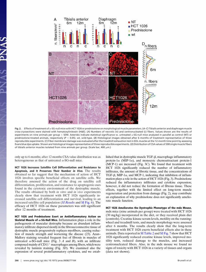
only up to 6 months; after 12 months CSA value distribution was asheterogeneous as that of untreated �-SG-null mice.
HCT 1026 Increases Satellite Cell Differentiation and Resistance toApoptosis, and It Preserves Their Number in Vivo. The resultsobtained so far suggest that the mechanism of action of HCT1026 involves specific beneficial effects on satellite cells. Wetherefore assessed the action of the drug on satellite celldifferentiation, proliferation, and resistance to apoptogenic cuesfound in the cytotoxic environment of the dystrophic muscle.The results obtained by both in vitro and in vivo experimentsclearly show that treatment with HCT 1026 significantly in-creased satellite cell differentiation and survival, leading to anincreased satellite cell population (SI Results and SI Fig. 6). Theefficacy of HCT 1026 on these parameters was still significantafter 12 months of treatment.
HCT 1026 and Prednisolone Exert an Antiinflammatory Action onSkeletal Muscle of �-SG-Null Mice. Inflammation plays a role in thepathogenesis of muscular dystrophy because an abundant inflam-matory infiltrate dispersed mostly in the fibrous/connective tissue ofdystrophic muscle progressively replaces myofibers, causing reduc-tion of muscle strength and worsening the disease (23). Azan–Mallory staining revealed frequent foci of fibrosis in muscles ofuntreated �-SG-null mice (Fig. 3 A and B), with an infiltratecomposed mainly of CD11� macrophages among fibers, which wererevealed by laminin staining (Fig. 3D). We examined also theexpression of several proinflammatory cytokines, and we estab-
lished that in dystrophic muscle TGF-�, macrophage inflammatoryprotein-1� (MIP-1�), and monocyte chemoattractant protein-1(MCP-1) are increased (Fig. 3C). We found that treatment withHCT 1026 significantly reduced the number of inflammatoryinfiltrates, the amount of fibrotic tissue, and the concentrations ofTGF-�, MIP-1�, and MCP-1, indicating that inhibition of inflam-mation plays a role in the action of HCT 1026 (Fig. 3). Prednisolonereduced the inflammatory infiltrates and cytokine expression;however, it did not reduce the formation of fibrous tissue. Theseeffects, together with the limited effect on long-term muscleregeneration and protection from damage (Fig. 2), may contributean explanation of why prednisolone does not significantly amelio-rate muscle function.
HCT 1026 Ameliorates the Dystrophic Phenotype of the mdx Mouse.mdx mice (nine animals per group) were treated with HCT 1026(30 mg/kg) incorporated in the diet, or they received plain diet(controls). Creatine kinase serum levels, motility on the running-wheel and treadmill tests, and muscle morphology were assessedafter 6 months. The results clearly show that the long-termtreatment with HCT 1026 exerts beneficial effects also in theseanimals. Data reported in SI Table 2 and SI Fig. 7 show that HCT1026 significantly reduced creatine kinase levels, improved mo-tility tests, reduced damage to the muscles, and increasedcentronucleated fibers. Also, in the mdx mouse we found nosigns of toxicity with HCT 1026 in a variety of tissues and organs(data not shown).
Fig. 2. Effects of treatment of �-SG-null mice with HCT 1026 or prednisolone on morphological muscle parameters. (A–C) Tibialis anterior and diaphragm musclecross-cryosections were stained with hematoxylin/eosin (H&E). (A) Numbers of necrotic (n) and centronucleated (c) fibers. Values shown are the results ofexperiments on nine animals per group � SEM. Asterisks indicate statistical significance vs. untreated �-SG-null mice analyzed in parallel as control (NT) orprednisolone-treated animals, respectively (P � 0.05). wt, wild-type. (B) Histological images obtained after 6 months of treatment representative of threereproducible experiments. (C) Fiber membrane damage was evaluated after the treadmill exhaustion test in EDL muscles at the 12-month time point by assessingEvans blue dye uptake. Shown are histological images representative of three reproducible experiments. (D) Distribution of CSA values of 300 single muscle fibersof tibialis anterior muscles isolated from nine animals per group. (Scale bar, 400 �m.)
266 � www.pnas.org�cgi�doi�10.1073�pnas.0608277104 Brunelli et al.

HCT 1026 Enhances the Therapeutic Efficacy of Mesoangioblasts byIncreasing Their Ability to Migrate and Engraft to Dystrophic Muscles.The treatment with HCT 1026 does not correct the genetic defectunderlying muscular dystrophy. To address this issue we combinedthe pharmacological treatment with delivery of mesoangioblasts.Mesoangioblasts were transduced with a lentiviral vector expressingGFP (8) to trace them into the various tissues, and they were usedeither untreated or after a 12-h ex vivo exposure to stromalcell-derived factor 1 (SDF-1; 50 ng/ml), a treatment that increasestheir homing to muscle (24). At variance with a previous report inwhich cells were injected in �-SG-null mice three times (8), here weperformed a single injection to evaluate best the changes intherapeutic efficacy induced by drug treatment. Migration ofmesoangioblasts was assessed by injecting them at a dose of 5 � 105
cells per animal in �-SG-null mice untreated or treated for 3 monthswith HCT 1026. Gastrocnemius and filter organs (spleen, liver,lung, and kidney, where mesoangioblasts that flow through thecirculation may be trapped) were recovered 6 h after the injection,and the percentage of the cells migrated in them was evaluated byreal-time PCR with specific primers for GFP. Mesoangioblastmigration in gastrocnemius on the side of injection (right side) wassignificantly enhanced in HCT 1026-treated animals (Fig. 4A). Asynergic effect of HCT 1026 with pretreatment of mesoangioblastswith SDF-1 before their injection was also observed. The percent-age of cells retained by the filter organs was reduced in allconditions favoring mesoangioblast migration to the muscle.
To test whether an increased migration would lead also to anincreased correction of the dystrophic phenotype, mice treated withor without HCT 1026 were killed 2 months after mesoangioblastinjection. The efficacy of the treatment was assessed by measuring
�-SG mRNA expression by RT-PCR and �-SG protein levels byWestern blotting and immunofluorescence. Mesoangioblasts en-grafted and reconstituted muscle fibers in the quadriceps, gastroc-nemius, and soleus muscles on the side of injection (Fig. 4 B–D).This effect was doubled in HCT 1026-treated animals and also bypreexposure of mesoangioblasts to SDF-1. The combination ofthese two treatments increased 4-fold the percentage of fibersexpressing a-SG protein. Of importance, both treatments increasedengrafting in the contralateral muscles also. The ability of HCT1026 to increase mesoangioblast-mediated fiber reconstitution wasaccompanied by increased single fiber-specific tension (Fig. 4E),reduced muscle damage (judged by reduced creatine kinase levels;Fig. 4F), and enhanced mouse performance on the treadmill (Fig.4G), which were significant vs. those observed in animals receivingmesoangioblasts in the absence of HCT 1026 treatment, and theywere further increased by SDF-1 pretreatment of mesoangioblasts.Thus, HCT 1026 greatly increases mesoangioblast engrafting, andit may synergize with other treatments, leading to a significantlymore effective cell therapy.
DiscussionTwo decades after the identification of the molecular defect re-sponsible for Duchenne muscular dystrophy, there are still noeffective cures for the disease. The failure of all previous pharma-cological treatments has left corticosteroids as the only availabledrug treatment. Therapy with corticosteroids, despite current at-tempts to ameliorate the protocols of administration, is still oflimited clinical benefits, and it is accompanied by severe side ef-fects (21).
The treatment we tested here meets several criteria for aneffective therapy, including the ability to block or at least signifi-cantly delay the progress of the disease, produce little or no sidetoxicity, be cost-effective, and, eventually, resolve the underlyinggenetic defect. In particular, the administration of HCT 1026 wassufficient on its own to delay significantly and persistently theprogression of muscular dystrophy in two different models relevantto muscular dystrophy in humans. The drug was effective incorrecting biochemical and morphological alterations and in lim-iting inflammation. Most importantly, the drug increased musclestrength and significantly increased the ability of animals to per-form on different motility tests. This functional amelioration waspersistent after 12 months of treatment, when disease in untreatedanimal was severe, clearly demonstrating the efficacy of the treat-ment in slowing disease progression. Long-term beneficial effectshave not been reported for any of the experimental treatments ofmuscular dystrophy investigated so far.
The beneficial effects of NO on muscle function are well known,and the mechanisms of its action are well understood. In particular,NO is generated by skeletal muscle to stimulate key actions inmuscle repair, including activation of satellite cells and release ofmyotrophic factors (10–12). In addition, NO stimulates vasodila-tion, and thus increases the supply of oxygen and glucose uptake,and it triggers mitochondrial biogenesis, all of which contribute topreserve muscle from damage during contraction (13, 14, 25, 26).Indeed, amelioration of the dystrophic phenotype had been ob-served in neuronal NO-synthase transgenic mice (15). These studiesstimulated investigations about the therapeutic potential of treat-ments based on administration of L-arginine, a metabolic precursorof NO, or molsidomine, a NO donor, to increase NO release (26,27). Although some amelioration of muscle morphology was ob-served, and in one study creatine kinase levels were reduced (26),those treatments did not yield recovery of muscle function, and theydid not improve animal motility tests. Moreover, these experimentswere short-term investigations. We found no amelioration of mor-phofunctional parameters in �-SG-null mice after a 6-month treat-ment with ISDN.
The detectable and persistent recovery of muscle function ob-served in our study is most likely because HCT 1026 combines the
Fig. 3. Effects of treatment of �-SG-null mice with HCT 1026 or prednisoloneon muscle inflammation. (A) Number of inflammatory infiltrates measured onAzan–Mallory-stained serial muscle sections of tibialis anterior (TA) and dia-phragm (DIA) muscles. (B) TA muscles were isolated after 6 months of treat-ment and rapidly homogenized. The concentrations of the indicated cytokineswere measured on muscle homogenates with appropriate antibodies. Valuesshown in A and B are the results of nine experiments �SEM. Asterisks indicatestatistical significance vs. untreated �-SG-null mice analyzed in parallel ascontrol (NT) (P � 0.05). wt, wild-type. (C) Staining of TA muscle serial sectionswith Azan–Mallory reveals an accumulation of extracellular scar tissue (blue).(D) Presence of macrophages, revealed by staining of TA sections with theanti-CD11 and anti-laminin antibodies. (C and D) Histological images repre-sentative of nine reproducible experiments. [Scale bar (D), 400 �m.]
Brunelli et al. PNAS � January 2, 2007 � vol. 104 � no. 1 � 267
MED
ICA
LSC
IEN
CES

multiple beneficial effects of NO outlined above with the potentantiinflammatory action of flurbiprofen, yielding a drug endowedwith new properties. We found that important mechanisms concurto determine the excellent therapeutic potential of HCT 1026. Thedrug inhibited significantly inflammation in the dystrophic muscleby reducing the generation of proinflammatory cytokines andreducing infiltration of proinflammatory cells. In addition, HCT1026 had a specific action in preserving satellite cell number andfunction by protecting them from the proapoptotic environment ofthe dystrophic muscle and by increasing their ability to differentiate.This action explains why in HCT 1026-treated animals, the regen-erating ability of muscle, assessed as an increased number ofcentronucleated, reduced number of necrotic fibers and normalCSA, was preserved. The beneficial effect of the treatment wepropose was tested against prednisolone, one of the widely usedcorticosteroids for treatment of Duchenne muscular dystrophy. Wefound that HCT 1026 was more effective than the steroid inameliorating morphological, biochemical, and functional parame-ters and that these actions occurred in the absence of toxic sideeffects. In particular, we did not observe gastric damage, which inHCT 1026 is minimized by the NO-donating moiety (16). More-over, the limited beneficial effects of the steroid decreased withtime, becoming insignificant at later stages of the disease.
Previous studies in the mdx mouse showing that corticosteroidseither alone or combined with other treatments exert beneficialeffects were designed only as short-term therapeutic protocols (2–3months on average) (25, 28). In humans, however, treatments haveto be delivered long-term, a situation in which the side effects ofcorticosteroid administration still represent a serious problem (21).In contrast, HCT 1026 has a profile of safety that holds promise forits future use in patients affected by Duchenne muscular dystrophy.Because of the absence of corticosteroid-related toxicity, HCT 1026appears a more promising therapy than the combination of L-arginine and deflazacort recently proposed (25). In addition, L-arginine and deflazacort were tested only up to 3 months, andL-arginine may fail to produce long-term NO release because of thedramatic decrease of NO synthase observed in dystrophic muscles(13). The second important advantage of the treatment with HCT1026 is that it significantly enhanced the engrafting to muscle ofintraarterially delivered mesoangioblasts, a type of stem cells shownto be effective in correcting the genetic defect of the muscle (8).This enhancement resulted in a significant increase in their thera-peutic efficacy as shown by morphological and biochemical param-eters and by in vivo motility assay on the treadmill. The effect wasfurther enhanced by in vitro exposure of donor cells to SDF-1, amolecule able to increase homing of mesoangioblasts (24), implying
Fig. 4. HCT 1026 increases the therapeutic efficacy of mesoangioblasts. �-SG-null mice, either untreated or exposed to a 3-month therapy with HCT 1026, wereinjected into the right femoral artery with GFP-expressing D16 mesoangioblasts treated or not with SDF-1. (A) Mesoangioblast migration to right (R) and left(L) gastrocnemius muscles (Gs) and liver (Li), lung (Lu), kidney (Ki), and spleen (Sp) evaluated by real-time PCR with specific primers for GFP on five animals pergroup. (B–E) Engrafting of mesoangioblasts to muscles. (B) Engrafting to the right and left quadriceps (Qd), Gs, and soleus (So) muscles evaluated by real-timePCR with specific primers for �-SG on three animals per group. (C) Western blot analysis of �-SG expression in Qd compared with that of GAPDH. The results shownare from one of three reproducible experiments. (D) Immunostaining of �-SG expressed in Qd fibers that were revealed in serial sections by staining with laminin;DAPI was used to identify nuclei. The results shown are from one of five reproducible experiments. (E) Specific tension of single muscle fibers (n � 101) of Gsfrom three animals per group. (F and G) Serum creatine kinase levels (F) and animal performance on the exhaustion treadmill tests (G), carried out as describedin Fig. 1 on five animals per group. (E–G) Untreated indicates the values observed in �-SG-null mice which were neither treated with HCT 1026 nor injected withmesoangioblasts. (A, B, E–G) Bars represent SEM. Asterisks indicate statistical significance vs. �-SG-null mice injected with untreated mesoangioblasts analyzedin parallel as control (P � 0.05). wt, wild-type. [Scale bar (D), 200 �M.]
268 � www.pnas.org�cgi�doi�10.1073�pnas.0608277104 Brunelli et al.
that HCT 1026 may be used in combined therapeutic strategies toyield synergic beneficial effects. Of importance, the increasedhoming of mesoangioblasts to the muscle was accompanied by aconcomitant reduction of their number in the relevant filter organs.This number is large, and it may cause damage to these organs (9).An increased efficiency of stem cell delivery will reduce either thenumber of cells needed for a single injection or the number ofinjections, leading to an optimization of cell therapy.
Compared with the current experimental therapies, the treat-ment we propose has clear advantages. Despite recent encouragingresults, therapy with stem cells alone cannot reverse the preexistingpathological changes, and it still suffers from inefficient engraft-ment (9). This inefficiency may originate from a limited homing ofthese cells to muscle and a reduced ability to resist to the cytotoxicenvironment existing in the damaged muscle (23, 29). Theseproblems are reduced by the combination of mesoangioblasts withHCT 1026.
Within the current scenario of preclinical and early clinicalstudies, HCT 1026 shows the distinct advantages of being econom-ically affordable (unlike humanized anti-myostatin antibodies),nonimmunogenic (like adeno-associated vectors), suitable for allmutations (unlike oligonucleotides for exon skipping), and with atested profile of safety. We conclude that the therapeutic strategypresented in this work represents a significant advance and sets thestage for immediate trials in patients.
Materials and MethodsAnimal Treatments. Mice were treated with HCT 1026 and pred-nisolone compounded in the chow following the European Com-munity guidelines, and with the approval of the Institutional EthicalCommittee. Mesoangioblast D16-GFP cells, pretreated with orwithout SDF-1, were delivery by intraarterial injection through theright femoral artery (8).
Determination of Creatine Kinase Serum Levels. Serum creatinekinase concentrations were measured from mouse blood samplesby using the indirect colorimetric assay (Roche Diagnostics, Basel,Switzerland), following standard procedures.
Functional Tests. For a description of the functional tests, see SIMaterials and Methods.
Mechanics of Isolated Muscles and Fibers. Mechanical analysis ofintact muscles was carried out as described in ref. 30. Tetanic
force was normalized to the estimated CSA assuming cylindricalshape of the muscle and a density of 1.06 mg/mm3 (CSAcorresponds to the wet weight divided by its length). Normalizedtetanic force is expressed as kN/m2. Absolute (Po) and specificforce (Po/CSA) of single muscle fibers were measured by usingskinned preparations (31).
Histology Immunohistochemistry and Morphometry. Muscles weredissected and frozen in liquid N2-cooled isopentane. Fiber mem-brane damage was evaluated Evans blue dye staining. Serial musclesections were stained in H&E, by using the Azan–Mallory tech-nique, or immunostained as described in ref. 8. Morphometricalanalyses to evaluate distribution of fiber CSA were carried out on300 fibers per muscle, on H&E-stained sections of tibialis anterior,by using Image 1.63 software (Scion Corporation, Frederick, MD).
Cytokine Analysis. TGF-� and MIP-1� concentrations were deter-mined on equal amounts of protein lysates from tibialis anteriormuscles, by using the Quantikine ELISA kits (R&D Systems,Minneapolis, MN). MCP-1 concentration was assessed by flowcytometry by using the cytometric bead array (BD Bioscience, SanJose, CA).
RNAs and Protein Measurements. Real-time quantitative PCR anal-ysis was carried out on cDNA from tissue samples by using anMx3000P real-time PCR detection system (Stratagene, La Jolla,CA). Each cDNA sample was amplified in duplicate by using theSYBR Green Supermix (Bio-Rad, Hercules, CA) for GFP (primersGFP: forward, AAGTTCATCTGCACCACCG; reverse, TCCT-TGAAGAAGATGGTGCG). Protein samples from homogenizedtissue were analyzed by Western blotting by using mAbs anti-�-SG(Novocastra, Newcastle upon Tyne, U.K.) and GAPDH (Biogen-esis, Kingston, NH) (8).
Statistical Analyses. Values were expressed as means �SEM. Sta-tistical significance was assessed by one-way ANOVA followed bythe Student–Newman–Keul test. A probability of �5% (P � 0.05)was considered statistically significant.
We thank K. Campbell for the �-SG-null mice, M. Sampaolesi for advice,and C. Rinaldi for technical help. This work was supported by TelethonGrant GGP05007 (to E.C.), Association Francaise contre les MyopathiesGrant 12048 (to E.C.), The European Community [Euro Stem Cell (toG.C.)], Associazione Parent Project Italia (to G.C.), the Italian Associationof Cancer Research Grant 1016 (to E.C.), and the Ministry of Health[Ricerca Corrente (to E.C.)].
1. Emery AE (2002) Lancet 359:687–695.2. Manzur AY, Kuntzer T, Pike M, Swan A (2004) Cochrane Database Syst Rev
CD003725.3. Skuk D, Vilquin JT, Tremblay JP (2002) Curr Opin Neurol 15:563–569.4. Minetti GC, Colussi C, Adami R, Serra C, Mozzetta C, Parente V, Fortuni S,
Straino S, Sampaolesi M, Di Padova M, et al. (2006) Nat Med 12:1147–1150.5. Bogdanovich S, Krag TO, Barton ER, Morris LD, Whittemore LA, Ahima RS,
Khurana TS (2002) Nature 420:418–421.6. Barton ER, Morris L, Musaro A, Rosenthal N, Sweeney HL (2002) J Cell Biol
157:137–148.7. Lu QL, Mann CJ, Lou F, Bou-Gharios G, Morris GE, Xue SA, Fletcher S,
Partridge TA, Wilton SD (2003) Nat Med 9:1009–1014.8. Sampaolesi M, Torrente Y, Innocenzi A, Tonlorenzi R, D’Antona G, Pelle-
grino MA, Barresi R, Bresolin N, De Angelis MG, Campbell KP, et al. (2003)Science 301:487–492.
9. Cossu G, Sampaolesi M (2004) Trends Mol Med 10:516–520.10. Kaliman P, Canicio J, Testar X, Palacin M, Zorzano A (1999) J Biol Chem
274:17437–17444.11. Pisconti A, Brunelli S, Di Padova M, De Palma C, Deponti D, Baesso S,
Sartorelli V, Cossu G, Clementi E (2006) J Cell Biol 172:233–244.12. Tatsumi R, Hattori A, Ikeuchi Y, Anderson JE, Allen RE (2002) Mol Biol Cell
13:2909–2918.13. Stamler JS, Meissner G (2001) Physiol Rev 81:209–237.14. Nisoli E, Falcone S, Tonello C, Cozzi V, Palomba L, Fiorani M, Pisconti A,
Brunelli S, Cardile A, Francolini M, et al. (2004) Proc Natl Acad Sci USA101:16507–16512.
15. Wehling M, Spencer MJ, Tidball JG (2001) J Cell Biol 155:123–131.16. Wallace JL, Reuter B, Cicala C, McKnight W, Grisham MB, Cirino G (1994)
Gastroenterology 107:173–179.17. Scatena R (2004) Curr Opin Investig Drugs 5:551–556.18. Duclos F, Straub V, Moore SA, Venzke DP, Hrstka RF, Crosbie RH, Durbeej
M, Lebakken CS, Ettinger AJ, van der Meulen J, et al. (1998) J Cell Biol142:1461–1471.
19. van Groen T, Kadish I (2005) Brain Res Brain Res Rev 48:370–378.20. Zacharowski P, Zacharowski K, Donnellan C, Johnston A, Vojnovic I, Forte P, Del
Soldato P, Benjamin N, O’Byrne S (2004) Clin Pharmacol Ther 76:350–358.21. Dubowitz V (2005) Lancet Neurol 4:264.22. Jejurikar SS, Kuzon WM, Jr (2003) Apoptosis 8:573–578.23. Engvall E, Wewer UM (2003) FASEB J 17:1579–1584.24. Galvez BG, Sampaolesi M, Brunelli S, Covarello D, Gavina M, Rossi B,
Costantin G, Torrente Y, Cossu G (2006) J Cell Biol 174:231–243.25. Archer JD, Vargas CC, Anderson JE (2006) FASEB J 20:738–740.26. Voisin V, Sebrie C, Matecki S, Yu H, Gillet B, Ramonatxo M, Israel M, De
la Porte S (2005) Neurobiol Dis 20:123–130.27. Barton ER, Morris L, Kawana M, Bish LT, Toursel T (2005) Muscle Nerve
32:751–760.28. Chakkalakal JV, Thompson J, Parks RJ, Jasmin BJ (2005) FASEB J 19:880–891.29. Tews DS, Goebel HH (1996) J Neuropathol Exp Neurol 55:342–347.30. Rossi R, Bottinelli R, Sorrentino V, Reggiani C (2001) Am J Physiol 281:C585–
C594.31. D’Antona G, Pellegrino MA, Adami R, Rossi R, Carlizzi CN, Canepari M,
Saltin B, Bottinelli R (2003) J Physiol (London) 552:499–511.
Brunelli et al. PNAS � January 2, 2007 � vol. 104 � no. 1 � 269
MED
ICA
LSC
IEN
CES