Molecular basis for nonspecificity of nonsteroidal anti-inflammatory drugs (NSAIDs
11
Please cite this article in press as: Dwivedi, A.K. et al. Molecular basis for nonspecificity of nonsteroidal anti-inflammatory drugs (NSAIDs), Drug Discov Today (2015), http://dx.doi.org/ 10.1016/j.drudis.2015.03.004 Drug Discovery Today Volume 00, Number 00 March 2015 REVIEWS Molecular basis for nonspecificity of nonsteroidal anti-inflammatory drugs (NSAIDs) Avaneesh K. Dwivedi 1,3 , Vaishali Gurjar 1,3 , Sanjit Kumar 2 and Nagendra Singh 1 1 School of Biotechnology, Gautam Buddha University, Greater Noida, Uttar Pradesh 201308, India 2 Center for Bioseparation Technology, VIT University, Vellore, Tamil Nadu 632014, India Inhibition of the production of inflammatory mediators by the action of nonsteroidal anti- inflammatory drugs (NSAIDs) is highly accredited to their recognition of cyclooxygenase enzymes. Along with inflammation relief, however, NSAIDs also cause adverse effects. Although NSAIDs strongly inhibit enzymes of the prostaglandin synthesis pathways, several other proteins also serve as fairly potent targets for these drugs. Based on their recognition pattern, these receptors are categorised as enzymes modifying NSAIDs, noncatalytic proteins binding to NSAIDs and enzymes with catalytic functions that are inhibited by NSAIDs. The extensive binding of NSAIDs is responsible for their limited in vivo efficacy as well as the large spectrum of their effects. The biochemical nature of drugs binding to multiple protein targets and its implications on physiology are discussed. Introduction Nonsteroidal anti-inflammatory drugs (NSAIDS) are one of the most prominent categories of compounds occupying the drug market. NSAIDs have been prescribed by physicians for over a hundred years. These drugs provide anti-inflammatory, antipy- retic and analgesic effects in various pathological conditions [1,2]. NSAIDS are so-called because they are not steroids in nature but cause the same effects as steroids by inhibiting synthesis of prostaglandins (PGs). NSAIDs are recommended for treatment of acute or chronic conditions of pain and inflammation. In the USA over 30 billion doses were sold and there were seven million prescriptions of NSAIDs in 2001 [3]. The well-known members of this category are naproxen, aspirin and ibuprofen, which are available worldwide for the treatment of pain and inflammation [4]. NSAIDs act by suppressing cyclooxygenase (COX)-1 and COX-2 enzymes or tumour necrosis factor (TNF)-a and inducible nitric oxide synthase (iNOS). Phospholipase A 2 (PLA 2 ) or phospholipase C (PLC) catalyses liberation of arachidonic acid from membrane phospholipids, which is further oxygenated into PGs by the action of lipoxygenase (LOX) and COX enzymes. By blocking the action of COX, NSAIDs can relieve inflammation by reducing vasodilata- tion and pain, which is produced mainly by prostaglandin E (PGE)2 and prostacyclin (PGI2). Most of the NSAIDs block synthe- sis of PGs by inhibiting COX enzymes nonselectively [5]. The inhibition of COX is also responsible for damage of the gastroin- testinal mucosa, because the PGs are also involved in mucosal defence and repair mechanisms [6,7]. A most commonly pre- scribed NSAID, aspirin has been found to cause gastrointestinal bleeding in half of the cases in the USA [8–10] and 12,000 cases of intestinal bleeding and 1200 deaths every year in the UK [11]. Long-term use of NSAIDs increases the risk of having acute cardiac disease and gastrointestinal-related side-effects [5]. Depending on the selectivity for either of the COX isoforms, NSAIDs have been divided into conventional and COX-2-selective categories. Con- ventional NSAIDs such as phenoprofen, ibuprofen, indomethacin, ketoprofen, keterolac, naproxen, paracetamol, piroxicam and as- pirin are nonselective COX inhibitors, whereas etodolac, melox- icam, diclofenac, celecoxib, nimesulide, valdecoxib, parecoxib, rofecoxib and etoricoxib are selective COX-2 inhibitors and are also known as coxibs. Reviews POST SCREEN Corresponding author: Singh, N. ([email protected]) 3 These authors contributed equally to this review. 1359-6446/ß 2015 Elsevier Ltd. All rights reserved. http://dx.doi.org/10.1016/j.drudis.2015.03.004 www.drugdiscoverytoday.com 1
Transcript of Molecular basis for nonspecificity of nonsteroidal anti-inflammatory drugs (NSAIDs

Reviews�POSTSCREEN
Drug Discovery Today � Volume 00, Number 00 �March 2015 REVIEWS
Molecular basis for nonspecificity ofnonsteroidal anti-inflammatory drugs(NSAIDs)Avaneesh K. Dwivedi1,3, Vaishali Gurjar1,3, Sanjit Kumar2 andNagendra Singh1
1 School of Biotechnology, Gautam Buddha University, Greater Noida, Uttar Pradesh 201308, India2Center for Bioseparation Technology, VIT University, Vellore, Tamil Nadu 632014, India
Inhibition of the production of inflammatory mediators by the action of nonsteroidal anti-
inflammatory drugs (NSAIDs) is highly accredited to their recognition of cyclooxygenase enzymes.
Along with inflammation relief, however, NSAIDs also cause adverse effects. Although NSAIDs strongly
inhibit enzymes of the prostaglandin synthesis pathways, several other proteins also serve as fairly
potent targets for these drugs. Based on their recognition pattern, these receptors are categorised as
enzymes modifying NSAIDs, noncatalytic proteins binding to NSAIDs and enzymes with catalytic
functions that are inhibited by NSAIDs. The extensive binding of NSAIDs is responsible for their limited
in vivo efficacy as well as the large spectrum of their effects. The biochemical nature of drugs binding to
multiple protein targets and its implications on physiology are discussed.
IntroductionNonsteroidal anti-inflammatory drugs (NSAIDS) are one of the
most prominent categories of compounds occupying the drug
market. NSAIDs have been prescribed by physicians for over a
hundred years. These drugs provide anti-inflammatory, antipy-
retic and analgesic effects in various pathological conditions
[1,2]. NSAIDS are so-called because they are not steroids in nature
but cause the same effects as steroids by inhibiting synthesis of
prostaglandins (PGs). NSAIDs are recommended for treatment of
acute or chronic conditions of pain and inflammation. In the USA
over 30 billion doses were sold and there were seven million
prescriptions of NSAIDs in 2001 [3]. The well-known members
of this category are naproxen, aspirin and ibuprofen, which are
available worldwide for the treatment of pain and inflammation
[4].
NSAIDs act by suppressing cyclooxygenase (COX)-1 and COX-2
enzymes or tumour necrosis factor (TNF)-a and inducible nitric
oxide synthase (iNOS). Phospholipase A2 (PLA2) or phospholipase
C (PLC) catalyses liberation of arachidonic acid from membrane
Please cite this article in press as: Dwivedi, A.K. et al. Molecular basis for nonspecificity of non10.1016/j.drudis.2015.03.004
Corresponding author: Singh, N. ([email protected])3 These authors contributed equally to this review.
1359-6446/� 2015 Elsevier Ltd. All rights reserved.
http://dx.doi.org/10.1016/j.drudis.2015.03.004
phospholipids, which is further oxygenated into PGs by the action
of lipoxygenase (LOX) and COX enzymes. By blocking the action
of COX, NSAIDs can relieve inflammation by reducing vasodilata-
tion and pain, which is produced mainly by prostaglandin E
(PGE)2 and prostacyclin (PGI2). Most of the NSAIDs block synthe-
sis of PGs by inhibiting COX enzymes nonselectively [5]. The
inhibition of COX is also responsible for damage of the gastroin-
testinal mucosa, because the PGs are also involved in mucosal
defence and repair mechanisms [6,7]. A most commonly pre-
scribed NSAID, aspirin has been found to cause gastrointestinal
bleeding in half of the cases in the USA [8–10] and 12,000 cases of
intestinal bleeding and 1200 deaths every year in the UK [11].
Long-term use of NSAIDs increases the risk of having acute cardiac
disease and gastrointestinal-related side-effects [5]. Depending on
the selectivity for either of the COX isoforms, NSAIDs have been
divided into conventional and COX-2-selective categories. Con-
ventional NSAIDs such as phenoprofen, ibuprofen, indomethacin,
ketoprofen, keterolac, naproxen, paracetamol, piroxicam and as-
pirin are nonselective COX inhibitors, whereas etodolac, melox-
icam, diclofenac, celecoxib, nimesulide, valdecoxib, parecoxib,
rofecoxib and etoricoxib are selective COX-2 inhibitors and are
also known as coxibs.
steroidal anti-inflammatory drugs (NSAIDs), Drug Discov Today (2015), http://dx.doi.org/
www.drugdiscoverytoday.com 1

REVIEWS Drug Discovery Today � Volume 00, Number 00 �March 2015
DRUDIS-1593; No of Pages 11
Review
s�P
OSTSCREEN
Most of the nonselective and selective NSAIDs have mild-to-
severe side-effects with long-term usage. NSAIDs are also associat-
ed with a broad spectrum of adverse effects on the functioning of
the liver, kidney, cardiovascular system, skin and gut. Gastroin-
testinal side-effects are the most common and constitute a wide
clinical spectrum ranging from dyspepsia, heartburn and abdomi-
nal discomfort to more serious events such as peptic ulcer with life-
threatening complications of bleeding and perforation [12–16].
Besides COX enzymes, NSAIDs have been observed recognising
various other physiological targets as well.
In this review, we report the comparative details of the atomic
interactions of NSAIDs with their target and off-target protein
receptors available in the Protein Data Bank (PDB). The atomic
coordinates of the complexes of NSAIDs and receptors were down-
loaded from the PDB website (http://www.rcsb.org/pdb/home/
home.do) and the intermolecular interactions were studied using
COOT [17], PyMOL [18] and CCP4 [19].
Protein targets binding to NSAIDsCOX-1COX enzymes catalyse the rate-limiting steps of synthesis of PGs,
prostacyclins and thromboxanes, collectively known as prosta-
noids. COX enzymes are also known as prostaglandin-endo per-
oxide synthases (PTGS/PGHS, EC: 1.14.99.1). COX enzymes
catalyse the oxidation of arachidonic acid (a v-6 polyunsaturated
fatty acid; PUFA) to PGH2, which is further converted into series 2
prostanoids.
Currently, three forms of COX are known: COX-1, COX-2 and
COX-3. COX-1 and COX-2 share 81% homology and COX-3 is a
splice variant of COX-1 [20]. COX-1 and COX-2 are also known as
PGHS1 and PGHS2, respectively. COX-1 and COX-2 function in
very similar ways but selective inhibition can make a difference to
their side effects. The expression of COX-1 is constitutive, whereas
COX-2 is induced in macrophages at the site of inflammation
[21,22]. COX-1 is also supposed to be additionally responsible for
maintaining gastrointestinal mucosa, therefore a new category of
NSAIDs specific only for COX-2 were designed. Rofecoxib and
etoricoxib are the most selective COX-2 inhibitors [23]. Recently,
rofecoxib (produced by Merck) was not found to be associated with
gastrointestinal risk but increases the chances of acute myocardial
infarction in high-risk patients, which led to the withdrawal of the
drug from the market. Other COX-2-selective drugs were also
found to be associated with myocardial infarction and thrombo-
sis-related issues [24–28].
The 3D structure of COX-1 was found to have three indepen-
dent folding units, one epidermal growth factor (EGF) domain, a
membrane-binding motif and a catalytic domain [29]. The sub-
strate- and NSAID-binding site on COX-1 is present in the form of a
long hydrophobic channel. The structure of COX-1 comprised a
homodimer. Most of the NSAIDs have been found to bind to one of
the monomers and block total activity of the dimer through
crosstalk between both monomers. However, it has also been
observed that a COX-2-selective inhibitor binding to one mono-
mer does not affect the activity of the other one [30]. The drug-
binding hydrophobic cavity is formed predominantly by hydro-
phobic amino acids Val116, Tyr348, Val349, Leu352, Leu359,
Phe381, Leu384, Tyr385, Trp387, Phe518, Ile523, Ala527 and
Leu531. Three polar amino acids Arg120, Tyr355 and Ser530 are
Please cite this article in press as: Dwivedi, A.K. et al. Molecular basis for nonspecificity of non10.1016/j.drudis.2015.03.004
2 www.drugdiscoverytoday.com
also present in the binding site. The crystal structure of COX-1 has
also been solved in complex with various NSAIDs such as flurbi-
profen, diclofenac, celecoxib, ibuprofen, alclofenac and nimesu-
lide [29–33]. The basic chemical structure of NSAIDs is generally
composed of a hydrophobic aromatic nucleus and an attached
acidic group. The aromatic portion lies in the hydrophobic chan-
nel of COX-1, whereas the acidic group is H-bonded to the side-
chain hydroxyl group of Tyr355 and guanidinium group of Arg120
(Fig. 1a). All the NSAIDs except aspirin inhibit COX-1 in a similar
way by working as competitive inhibitors, whereas aspirin and its
analogues acetylate the active-site Ser530 to act as suicide inhibi-
tors of COX enzymes.
COX-2Three dimensional structures of native and complexed states of
COX-2 have been determined [34]. The structure of COX-1 and
COX-2 is similar with a root-mean-square deviation (RMSD) of
0.9 A of their Ca atoms. The drug-binding sites in both enzymes are
identical except for the presence of the smaller Val509 instead of
Ile523 and Arg499 in place of His513 in COX-1, which causes a
slight increase in the volume of the drug-binding cavity and
creates a side-pocket in COX-2. The crystal structures of COX-2
have been reported in complex with naproxen, aspirin, flurbipro-
fen, indomethacin, diclofenac and celecoxib [31,34–37]. Various
modes of inhibitor binding to the COX active-site have been
reported including ionic interaction of carboxylic-acid-containing
NSAIDs with Arg120 of COX-1, COX-2 and insertion of arylsulfo-
namides and sulfones into the side-pocket of COX-2. Most of the
NSAIDs bind COX-2 by introducing an ionic interaction with
Arg108 and H-bond with Tyr341 in a similar way as for COX-1
(Fig. 1b), although diclofenac has been found to interact with the
COX-2 site in an inverted manner. The carboxylate group of
diclofenac makes H-bonds with Tyr385 and Ser530 instead of
Arg120 and Tyr355 [36]. COX-2-selective NSAIDs such as etodolac,
meloxicam, diclofenac, celecoxib, nimesulide, valdecoxib, pare-
coxib, rofecoxib and etoricoxib have a different mode of binding
to COX-2 than nonselective drugs. The sulfonamide group of
coxibs lies in the space created in the side pocket owing to the
presence of Val523 in COX-2. Celecoxib makes ionic interactions
with side-groups of His75 and Arg499 along with H-bonded inter-
actions with main-chain CO groups of Leu338 and Ser339 residues
of COX-2. The sulfonamide group of celecoxib is additionally H-
bonded to Gln178 as well [36,37]. In this manner, coxibs gain one
extra salt bridge and three extra H-bonded interactions compared
with nonselective COX inhibitors (Fig. 1c). The biochemical se-
lectivity of several NSAIDs was evaluated using the COX–isozyme
activity. It was observed that valdecoxib, etoricoxib, 5,5-dimethyl-
3-(3-fluorophenyl)-4-(4-methylsulfonyl)phenyl-2(5H)-furanone
(DFU) and 5,5-dimethyl-3-(2-isopropoxy)-4-(4-methanesulfonyl-
phenyl)-2(5H)-furanone (DFP) inhibited COX-1 and COX-2 with
the following COX-1:COX-2 IC50 ratios: 61.5, 344, 660 and 1918,
respectively; whereas indomethacin, celecoxib and rofecoxib had
the corresponding values of 30, 29.6 and 272 [38], indicating the
role of the sulfonamide group in providing specificity to coxibs.
PLA2Although inhibition of PG synthesis by NSAIDs has been attribut-
ed to their binding to COX enzymes, another mechanism of
steroidal anti-inflammatory drugs (NSAIDs), Drug Discov Today (2015), http://dx.doi.org/

Drug Discovery Today � Volume 00, Number 00 �March 2015 REVIEWS
DRUDIS-1593; No of Pages 11
Arg 120
(a) (b)
(c) (d)
Arg 499
His 75
Ala 513
Phe 367
Leu 370
Tyr 371
Trp 31
Phe 105Asp 49
His 48
OW Ile 9
Ile 19
Leu 17
Leu 10
Leu 3
Leu 2
Lys 69
Phe 5
Tyr 52
Tyr 22
Ser 23
Ala 18
Trp 373
Tyr 341
Ser 516
Ser 339
Leu 338
Gln 178
Val 509
Arg 108
Ala 513
Val 509
Ser 516
Phe 367
Leu 370
Ile 523
Phe 381
Leu 384
Ser 530Tyr 385
Tyr 355
Tyr 348
Tyr 341
Tyr 371
Trp 373
Leu 338Ser 339
Trp 387Val 349
Leu 352Leu 359
Drug Discovery Today
FIGURE 1
Binding of nonsteroidal anti-inflammatory drugs (NSAIDs) to prostaglandin synthesis pathway enzymes. (a) Ribbon diagram of cyclooxygenase (COX)-1 (blue) in
complex with flurbiprofen is shown. (b) Structure of COX-2 (blue) in complex with indomethacin, a nonselective COX inhibitor, and (c) COX-2 complexed withcelecoxib, a selective COX-2 inhibitor, are drawn. The figure indicates a deeper binding of coxib. (d) Diclofenac is shown in bound state to the active site of
phospholipase A2 (PLA2). The carboxyl group of the drug makes H-bonded interactions with catalytic residues His48 and Asp49 through the conserved water
molecule (OW). The drug is completely surrounded by the hydrophobic residues of the binding site of PLA2. Residues contributing to the formation of the drug-binding site and interacting with the drug are represented as ball and stick models. Drugs are shown by magenta ball and stick models. H-bonded interactions are
shown as dotted lines. The figure was drawn using PyMOL [18].
Reviews�POSTSCREEN
inhibition of PG synthesis is to stop liberation of free arachidonic
acid from phospholipids by the action of PLA2. PLA2 is a key
enzyme involved in the production of proinflammatory media-
tors. It is responsible for the liberation of arachidonic acid
from membrane phospholipids. This arachidonic acid serves as
a substrate for COX and LOX enzymes for synthesis of PGs and
Please cite this article in press as: Dwivedi, A.K. et al. Molecular basis for nonspecificity of non10.1016/j.drudis.2015.03.004
leukotrienes. The substrate binding to PLA2 occurs through a
hydrophobic channel. Many of the NSAIDs have been found to
be strong inhibitors of PLA2 enzymes. To determine the viability of
PLA2 as a target molecule for the structure-based drug design
against inflammation, arthritis and rheumatism, the crystal struc-
ture of the complex of PLA2 with various NSAIDs such as aspirin,
steroidal anti-inflammatory drugs (NSAIDs), Drug Discov Today (2015), http://dx.doi.org/
www.drugdiscoverytoday.com 3
REVIEWS Drug Discovery Today � Volume 00, Number 00 �March 2015
DRUDIS-1593; No of Pages 11
Review
s�P
OSTSCREEN
nimesulide, oxyphenbutazone, indomethacin, diclofenac and
paracetamol have been determined [39–42].
The drug-binding site in PLA2 includes mainly hydrophobic
residues such as Leu2, Leu3, Phe5, Ile9, Ala18, Ile19, Tyr22, Ser23,
Trp31, Lys69 and Phe105. The active site of PLA2 comprises His48,
Asp49 and Tyr52 residues, located at the interior end of the
substrate-binding hydrophobic channel. The stereochemical en-
vironment of the hydrophobic channel is very compatible for
binding to NSAIDs. Most of the NSAIDs have been found to block
the phospholipid-binding site and act as competitive inhibitors of
PLA2 enzymes. The hydrophobic aromatic rings of NSAIDs make
several van der Waals contacts with the substrate-binding site
residues; whereas the charged or polar functional group of the
drugs makes ionic- or H-bonds with catalytically essential residues
His48 and Asp49 directly or through a catalytically conserved
water molecule (Fig. 1d). Although most of the NSAIDs act as
competitive inhibitors of PLA2, indomethacin has been found
binding at an alternate site near to the substrate-binding hydro-
phobic channel and categorised as a noncompetitive inhibitor of
the enzyme with a Ki of 12 mM [40,43]. It has been shown that the
cumulative anti-inflammatory effects of NSAIDs are based on not
only their binding to COXs but also their strong affinity for PLA2
[39–41].
Prostaglandin keto reductase (PTGR)PTGR2 belongs to the superfamily of dehydrogenase and reduc-
tase enzymes. These enzymes catalyse NADPH-dependent reduc-
tion of unsaturated bonds in PGs and are responsible for the
eventual inactivation of PGs. Selective inhibition of PTGR2 also
enhances insulin sensitivity. Indomethacin has been shown to
interact strongly with the PTGR2, located at the site closer to the
NADP-binding site [44], carboxylic acid making H-bonds with the
sugar OH group of NADP. The hydrophobic rings of the drug
makes many van der Waals interactions with the residues Tyr51,
Cys54, Thr60, Ile65, Phe99, Tyr100, Met135, Leu288, Val289 and
Leu290.
Several NSAIDs have also been observed inhibiting other mem-
bers of the aldoketo-reductase (AKR) family. AKR1C3 acts to
suppress cell differentiation and promote proliferation in myeloid
tissues and represents a novel target for COX-independent anti-
neoplastic actions of NSAIDs. Indomethacin and flufenamic acid
have been found forming complexes with AKR1C3 [45]. Indo-
methacin binds to the active site, whereas flufenamic acid bound
additionally to a beta hairpin loop. Indomethacin and flufenamic
acid bind in a similar manner at the active site of AKR1C3. The
carboxylic acid groups of indomethacin and flufenamic acid make
two H-bonds with the hydroxyl group of Tyr55 and one salt bridge
with His117. Additionally, indomethacin makes several van der
Waals contacts with Tyr24, Leu54, Trp86, Met120, Tyr216,
Trp227, Phe306, Phe311, Pro318 and Tyr319 residues. The binding
of drugs has caused drastic readjustments of the orientation of
side-chains of several residues of the drug-binding site including
Phe306, Phe311 and Trp227, indicating an induced fit mechanism
of drug binding.
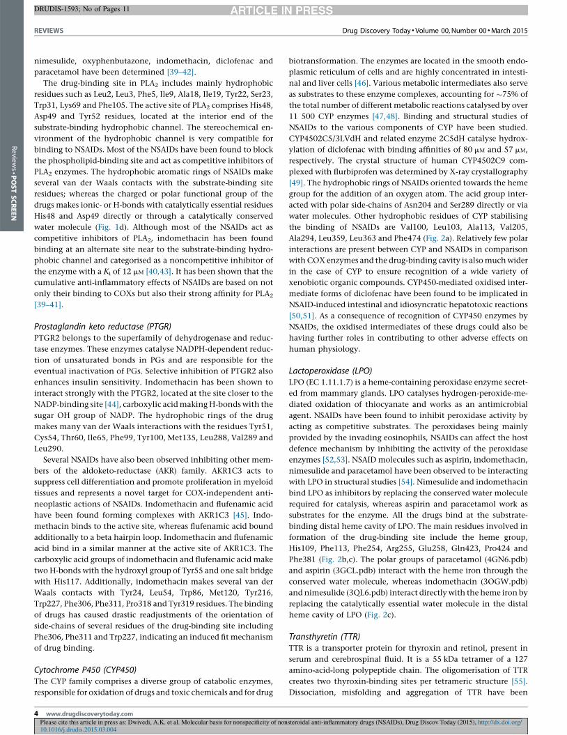
Cytochrome P450 (CYP450)The CYP family comprises a diverse group of catabolic enzymes,
responsible for oxidation of drugs and toxic chemicals and for drug
Please cite this article in press as: Dwivedi, A.K. et al. Molecular basis for nonspecificity of non10.1016/j.drudis.2015.03.004
4 www.drugdiscoverytoday.com
biotransformation. The enzymes are located in the smooth endo-
plasmic reticulum of cells and are highly concentrated in intesti-
nal and liver cells [46]. Various metabolic intermediates also serve
as substrates to these enzyme complexes, accounting for �75% of
the total number of different metabolic reactions catalysed by over
11 500 CYP enzymes [47,48]. Binding and structural studies of
NSAIDs to the various components of CYP have been studied.
CYP4502C5/3LVdH and related enzyme 2C5dH catalyse hydrox-
ylation of diclofenac with binding affinities of 80 mM and 57 mM,
respectively. The crystal structure of human CYP4502C9 com-
plexed with flurbiprofen was determined by X-ray crystallography
[49]. The hydrophobic rings of NSAIDs oriented towards the heme
group for the addition of an oxygen atom. The acid group inter-
acted with polar side-chains of Asn204 and Ser289 directly or via
water molecules. Other hydrophobic residues of CYP stabilising
the binding of NSAIDs are Val100, Leu103, Ala113, Val205,
Ala294, Leu359, Leu363 and Phe474 (Fig. 2a). Relatively few polar
interactions are present between CYP and NSAIDs in comparison
with COX enzymes and the drug-binding cavity is also much wider
in the case of CYP to ensure recognition of a wide variety of
xenobiotic organic compounds. CYP450-mediated oxidised inter-
mediate forms of diclofenac have been found to be implicated in
NSAID-induced intestinal and idiosyncratic hepatotoxic reactions
[50,51]. As a consequence of recognition of CYP450 enzymes by
NSAIDs, the oxidised intermediates of these drugs could also be
having further roles in contributing to other adverse effects on
human physiology.
Lactoperoxidase (LPO)LPO (EC 1.11.1.7) is a heme-containing peroxidase enzyme secret-
ed from mammary glands. LPO catalyses hydrogen-peroxide-me-
diated oxidation of thiocyanate and works as an antimicrobial
agent. NSAIDs have been found to inhibit peroxidase activity by
acting as competitive substrates. The peroxidases being mainly
provided by the invading eosinophils, NSAIDs can affect the host
defence mechanism by inhibiting the activity of the peroxidase
enzymes [52,53]. NSAID molecules such as aspirin, indomethacin,
nimesulide and paracetamol have been observed to be interacting
with LPO in structural studies [54]. Nimesulide and indomethacin
bind LPO as inhibitors by replacing the conserved water molecule
required for catalysis, whereas aspirin and paracetamol work as
substrates for the enzyme. All the drugs bind at the substrate-
binding distal heme cavity of LPO. The main residues involved in
formation of the drug-binding site include the heme group,
His109, Phe113, Phe254, Arg255, Glu258, Gln423, Pro424 and
Phe381 (Fig. 2b,c). The polar groups of paracetamol (4GN6.pdb)
and aspirin (3GCL.pdb) interact with the heme iron through the
conserved water molecule, whereas indomethacin (3OGW.pdb)
and nimesulide (3QL6.pdb) interact directly with the heme iron by
replacing the catalytically essential water molecule in the distal
heme cavity of LPO (Fig. 2c).
Transthyretin (TTR)TTR is a transporter protein for thyroxin and retinol, present in
serum and cerebrospinal fluid. It is a 55 kDa tetramer of a 127
amino-acid-long polypeptide chain. The oligomerisation of TTR
creates two thyroxin-binding sites per tetrameric structure [55].
Dissociation, misfolding and aggregation of TTR have been
steroidal anti-inflammatory drugs (NSAIDs), Drug Discov Today (2015), http://dx.doi.org/

Drug Discovery Today � Volume 00, Number 00 �March 2015 REVIEWS
DRUDIS-1593; No of Pages 11
Leu 103
Leu 363
Leu 359
HEME
Asn 204
His 109
Arg 255
Phe 254
Phe 381
Pro 424
Gln 423His 351
His 109Arg 255
Phe 254
Phe 381
Pro 424
Gln 423
His 351
OW 856OW
OW 857
Val 205Val 100Phe 473
(a) (b)
(c)
Phe 114
Ala 113
Ala 294
Ser 289
Drug Discovery Today
FIGURE 2
Recognition of nonsteroidal anti-inflammatory drugs (NSAIDs) by oxidising enzymes. (a) The binding of diclofenac (magenta) is shown in the active site of
cytochrome P450 (CYP450) enzyme (blue). The heme group of the enzyme is also shown as a green ball and stick model. (b) Aspirin (yellow) binding as substrate
to the distal heme cavity of lactoperoxidase (LPO), by interacting with the heme group through the catalytically conserved water molecule (OW). (c) Indomethacin
(magenta) bound to the distal heme cavity of LPO by replacing the conserved water molecule and binding in inhibitory mode to the enzyme. Heme group of LPOis also shown in ball and stick model in green.
Reviews�POSTSCREEN
reported to be associated with the amyloid diseases senile systemic
amyloidosis (SSA), familial amyloid polyneuropathy (FAP) and
familial amyloid cardiomyopathy (FAC) [56–59]. Aggregated
TTR deposited on peripheral nerves and heart tissues is responsible
for amyloidosis. Several NSAIDs including diclofenac, flufenamic
acid and flurbiprofen have been found inhibiting TTR fibril
Please cite this article in press as: Dwivedi, A.K. et al. Molecular basis for nonspecificity of non10.1016/j.drudis.2015.03.004
formation in vitro. To design drugs against amyloid diseases, crystal
structures of these NSAIDs have also been reported in complex
with TTR [60].
The structure of TTR consists of a homodimer, where each dimer
binds NSAIDs in a similar fashion. The drug-binding site present
on TTR is comparatively shallow and less-specific to NSAIDs. The
steroidal anti-inflammatory drugs (NSAIDs), Drug Discov Today (2015), http://dx.doi.org/
www.drugdiscoverytoday.com 5

REVIEWS Drug Discovery Today � Volume 00, Number 00 �March 2015
DRUDIS-1593; No of Pages 11
Review
s�P
OSTSCREEN
site is formed by contribution of two parallel and one antiparallel
b-strand. Drugs bind to the concave surface of this mixed topology
b-sheet structure in random orientations, unlike COX enzymes.
The carboxylic group of diclofenac makes two H-bonds with the
polar side-chains of Ser117 and Thr119, whereas flufenamic acid
and flurbiprofen bind differently with their carboxylate group
Please cite this article in press as: Dwivedi, A.K. et al. Molecular basis for nonspecificity of non10.1016/j.drudis.2015.03.004
Val 121Thr 106
Thr 119 Ala 108Lys 15
Leu 17Ser 117
Ile 110
Thr 663
(a) (b)
(c)
G
Tyr 155
Tyr 113
Leu 108
Val 103
Leu 110
FIGURE 3
Nonsteroidal anti-inflammatory drugs (NSAIDs) binding to nonenzymatic targets. (ab-strands of transthyretin (TTR). Chemical interactions between TTR (blue) and two Nlactoferrin in complex with diclofenac (magenta) showing a shallow binding site.
bromodomain (BRD)2 protein complex with paracetamol is shown. The drug-bind
makes a single H-bond along with several van der Waals contacts in the drug-bi
6 www.drugdiscoverytoday.com
facing towards Lys15. The hydrophobic portions of the drugs make
limited van der Waals interactions with Leu17, Thr106, Ala108,
Leu110 and Val121 located on the surface of the b-sheet (Fig. 3a).
The binding of drugs on the surface of TTR prevents intermolecu-
lar interaction of TTR molecules and inhibits aggregation, essential
for fibril formation.
steroidal anti-inflammatory drugs (NSAIDs), Drug Discov Today (2015), http://dx.doi.org/
Drug Discovery Today
Glu 664
Glu 431lu 659
Val 591
Pro 591
Pro 429Tyr 660
Asn 156 Cys 152
Ile 162
Phe 99
Gly 652
) NSAIDs in two opposite orientations on the concave surfaces of antiparallel
SAIDs, diclofenac (magenta) and flurbiprofen (cyan), are shown. (b) C-lobe of NSAIDs bind lactoferrin with relatively weaker affinities. (c) The human
ing site in BRD2 is contributed to from five a-helices. Paracetamol (magenta)
nding site.

Drug Discovery Today � Volume 00, Number 00 �March 2015 REVIEWS
DRUDIS-1593; No of Pages 11
Reviews�POSTSCREEN
Lactoferrin (LF)LF is an iron transport protein from mammary gland secretions.
Four NSAIDs: indomethacin, aspirin, diclofenac and ibuprofen,
have been shown to induce gastropathy in animal models [61].
The C-terminal half of the bovine milk LF prevented the NSAID-
induced gastropathy by binding to drugs with affinities ranging
from 2.6 to 4.8 � 10�4M. The crystal structures of the C-lobe of LF
with NSAIDs have been determined [61]. The structure of the
complexes showed that drugs bind to the C-lobe at a site formed
by two a-helices: a10 and a11. The drug-binding site on the C-lobe
represents a deep cavity that can accommodate NSAIDs in a
nonselective manner. The drug-binding site is formed by
Pro429, Glu431, Val591, Pro593, Gly652, Glu659, Thr663 and
Glu664 residues. The carboxylate group of aspirin and diclofenac
is H-bonded to Thr663. Drugs also make several van der Waals
contacts with Pro593 and Tyr660 residues (Fig. 3b). NSAIDs are
found to bind nonspecifically and in variable orientations to the C-
lobe of LF. LF and transferrin, being very similar in structure and
functions, work as transporting proteins for NSAIDs and reduce
the effective biological concentration of the drugs in body fluids.
Moreover, oral recombinant human LF supplementation during a
short-term indomethacin challenge has been observed reducing
the NSAID-mediated increase in small intestinal permeability and,
hence, provides a nutritional tool in the treatment of hyperper-
meability-associated disorders [62].
AlbuminHuman serum albumin (HSA) is abundantly present in plasma and
restricts free concentration of a wide variety of molecules in the
blood. HSA is known to have an important role in drug transport
and metabolism. At least two distinct drug-binding sites have been
observed in HSA by crystallographic studies [63]. Drug-binding
sites on HSA show flexible binding of a large variety of compounds.
Various NSAIDs such as aspirin, ibuprofen, oxyphenbutazone,
indomethacin, among others, have been shown to interact with
HSA [63,64]. Aspirin is hydrolysed into salicylic acid and the acetyl
group is transferred to Lys199 by HSA using its esterase activity.
Oxyphenbutazone and salicylic acid bind at the same site, whereas
ibuprofen and indomethacin recognise multiple and distinct sites
located deeper on HSA. All the drug-binding sites have polar and
hydrophobic amino acids to make attractive interactions with
NSAIDs. The drug-binding sites on HSA are nonspecific in nature
because other compounds also interact in similar ways to the
protein. Presence of multiple drug-binding sites on HSA contrib-
utes to its higher efficiency of drug transport and drug distribution.
Peroxisome proliferator activator receptor (PPAR)gPPARg is a member of a family of nuclear receptors. It works as a
transcription factor for genes involved in glucose homeostasis and
insulin sensitisation [65–67]. It has been implicated in the pathol-
ogy of many diseases such as obesity, diabetes, atherosclerosis and
cancer. PPARg decreases the inflammatory response of many
cardiovascular cells, particularly endothelial cells [68]. Agonists
of PPARg have been used in the treatment of hyperlipidaemia and
hyperglycaemia states [69]. PPARg is a potential therapeutic target
for metabolic syndrome and inflammatory diseases [70,71]. Indole
acetate derivatives are known to be activators of PPARg receptor,
hence indomethacin has also been reported to be an agonist of
Please cite this article in press as: Dwivedi, A.K. et al. Molecular basis for nonspecificity of non10.1016/j.drudis.2015.03.004
PPARg and beneficial in the treatment of hyperlipidaemia and
hyperglycaemia [72]. The 3D crystal structures of PPARg in com-
plexes with indomethacin and other indole derivatives have been
reported [73]. Indomethacin binds more strongly than 5-methoxy-
indole acetate (MIA) and 5-hydroxy-indole acetate (HIA) to PPARg
with a dissociation constant of 9.73 mM [72]. Indomethacin shares
the same space with other indole derivatives and potent agonists
in the ligand-binding site of PPARg [73]. Two molecules of indo-
methacin were found strongly held between a-helices of the
activation function (AF)2 site in the ligand-binding domain.
The carboxylic acid group of indomethacin makes one H-bond
with Tyr473 and two salt bridge interactions with His323 and
His449 residues. The ligand-binding site is highly hydrophobic in
chemical nature and constituted by residues Ile281, Phe282,
Cys285, Arg288, Ile326, Tyr327, Leu330, Ile341, Met348,
Leu353, Leu356, Phe360, Phe363, Met364, Leu453, Leu465 and
Leu469. Indomethacin makes extensive van der Waals interac-
tions with these hydrophobic amino acids and is strongly held in
the binding site.
Human bromodomain (BRD)2BRD is known to bind acetylated lysine. It has a four-helical-
bundle structural fold [74,75]. BRD2 is encoded by the BRD2 gene
[76]. Human BRD2 has been suggested to be involved in signal
transduction for growth control. The therapeutic benefits of mod-
ulating this target class are largely unexplored owing to the lack of
suitable chemical probes [77]. Paracetamol has been shown to
interact consistently with human BRD2. The crystal structure of
the complex of paracetamol and BRD2 has been reported [77].
Paracetamol binds in the cavity surrounded by helices. The drug
makes a single direct H-bond with Asn156 and the drug-binding
cavity is formed by Phe99, Val103, Leu108, Leu110, Tyr113,
Cys152, Tyr155 and Ile162 residues (Fig. 3c). BRD-containing
receptor proteins regulate expression of multiple genes such as
those involved in inflammatory reactions and tumour cell growth.
The inhibitors of BRD have profound antiproliferative and anti-
inflammatory effects [78]. Similarly, paracetamol can also be
implicated in antiproliferative effects along with its anti-inflam-
matory activities.
Mechanisms of NSAID-induced reactionsNSAIDs are the most widely prescribed category of drugs used
against inflammatory disorders in society. Continuing usage of
NSAIDs for longer timeframes leads to a number of lethal side-
effects such as heart attack, stroke and toxicity, as well as some
severe side-effects including gastrointestinal injury and peptic
ulceration along with a broad spectrum of adverse effects in the
liver, kidney and cardiovascular systems. The mechanisms of
adverse as well as beneficial effects of NSAIDs are COX-dependent
and COX-independent in nature. Although the precise mecha-
nism is not well understood, NSAIDs have also been found in-
creasing the survival of colon and breast cancer patients [78].
The chemo-preventive activity of NSAIDs is thought to be the
result of their binding to the aryl hydrocarbon (ArH) receptor to
modulate carcinogen metabolic enzymes [79,80]. It has also been
reported that, in several cancer cell lines, NSAIDs induced knock-
down of specificity protein transcription factors (Sp1, Sp3 and Sp4)
resulting in the decreased expression of growth-promoting and
steroidal anti-inflammatory drugs (NSAIDs), Drug Discov Today (2015), http://dx.doi.org/
www.drugdiscoverytoday.com 7

REVIEWS Drug Discovery Today � Volume 00, Number 00 �March 2015
DRUDIS-1593; No of Pages 11
Review
s�P
OSTSCREEN
proinflammatory genes [81–85], which, along with reducing the
risk of Alzheimer’s disease [86], adds to the beneficial effects of
NSAIDs. Although gastrointestinal problems are known to be
associated with inhibition of COX-1, a recent report suggested
that nonspecific simultaneous inhibition of COX-1 and COX-2 is
responsible for the damage of gastric mucosa in rats [87]. The drug-
induced liver injury (DILI) is caused by NSAID antagonism of
fernasoid-X-receptors (FXR) present on the liver cells [88]. Several
clinical studies have also reported NSAIDs inhibiting uridine 50-
diphospho-glucuronosyltransferase (UGT) enzymes, involved in
xenobiotic metabolism in hepatocytes [89], which could further
add to the COX-independent side-effects of NSAIDs. Similarly,
Please cite this article in press as: Dwivedi, A.K. et al. Molecular basis for nonspecificity of non10.1016/j.drudis.2015.03.004
TABLE 1
Interaction features of the NSAIDs and protein targets.
Target protein NSAIDs PDB code No. of polar interaction
including solvent
(2.8–3.5 A)a
COX-1 Flurbiprofen 3N8Z 3
Diclofenac 3N8Y 3
Celecoxib 3KK6 3
Ibuprofen 1EQG 3
Alclofenac 1HT8 3
Nimesulide 3N8X 7
Aspirin 1PTH 2
COX-2 Naproxen 3NT1 3
Flurbiprofen 3PGH 3
Indomethacin 4COX 3
Diclofenac 1PXX 4
Celecoxib 3LN1 4
PLA2 Aspirin 1TGM 2
Nimesulide 1ZWP 12
Oxyphenbutazone 1Q7A 1
Indomethacin 4EIX 3
Diclofenac 2B17 4
Paracetamol 2DPZ 6
CYP450 Diclofenac 1NR6 4
Flurbiprofen 1R9O 3
TTR Diclofenac 1DVX 5
Flufenamic acid 3OZL 0
Flurbiprofen 1DVT 1
Lectoferrin Indomethacin 3IB1 5
Aspirin 3IAZ 4
Diclofenac 3IB0 4
Ibuprofen 3IB2 1
HSA Aspirin 2I2Z 0
Ibuprofen 2BXG 6
Oxyphenbutazone 2BXB 6
Indomethacin 2BXM 4
PTGR Indomethacin 1S2A 6
Flufenamic acid 1S2C 6
PPARg Indomethacin 3ADS 3
BRD2 Paracetamol 4IOO 4
LPO Aspirin 2QQT 3
Indomethacin 3OGW 1
Nimesulide 3QL6 8
Paracetamol 3PY4 2
Abbreviations: BRD, bromodomain; COX, cyclooxygenase; CYP450, cytochrome P450; HSA, huma
Protein Data Bank; PLA, phospholipase A; PPAR, peroxisome proliferator-activated receptor; Pa The numbers of interactions between the drugs and receptor proteins are calculated using
8 www.drugdiscoverytoday.com
coxibs have also been found interacting with ion channels on
heart muscles in Drosophila, which could additionally be respon-
sible for their adverse effects on the heart [90]. Recently, new and
more-specific coxibs without cardiovascular deleterious effects
have been developed [91], indicating a possibility of more-specific
COX inhibitors having reduced adverse reactions.
Concluding remarksCOX enzymes catalyse the synthesis of the proinflammatory
agents from membrane-liberated arachidonic acid and are hence
used as targets for synthesis of anti-inflammatory drugs. Besides
inhibiting the activity of COX enzymes, NSAIDs have also been
steroidal anti-inflammatory drugs (NSAIDs), Drug Discov Today (2015), http://dx.doi.org/
s No. of nonpolar
interactions
(distance up to 4.0 A)a
Binding affinity
of the NSAID
References
63 Ki = 1.0 mM [33,95]
61 Ki = 0.15 mM [33,96]
387 Ki = 0.30 mM [30]35 Ki = 9.0 mM [32,97]
42 Not reported [32]
81 Ki = 0.12 mM [33,98]25 IC50 = 126 mM [31]
58 Ki > 10.0 mM [35]
50 Ki = 0.77 mM [99]
82 Ki = 0.60 mM [34]
51 Ki = 50 nM [36,98]84 Ki = 0.87 mM [37,98]
15 KD = 6.4 mM [41]
58 – To be published
57 KD = 64 nM [39]24 KD = 1.3 mM [40]
53 Ki = 48 nM [42]
45 – To be published
52 KD = 50 mM [100]56 Ki = 9.6 mM [49]
89 56.06% inhibition [60]
24 46.15% inhibition [60]
19 72.72% inhibition [60]
31 KD = 260 mM [61]28 KD = 330 mM [61]
46 Not reported [61]
22 KD = 480 mM [61]
11 KD = 70 mM [64,101]54 KA = 1.0 � 105/M [63,102]
102 KA = 2.97 � 106/M and
7.07 � 104/M
[63]
105 KA = 2.5 � 105/M [63,103]
106 Ki = 2.1 mM [45]
154 Ki = 3.1 mM [45]
127 KD = 9.73 mM [73]
46 KA = 50 mM [31,77]
60 Not reported [54]110 – To be published
111 – To be published
65 – To be published
n serum albumin; LPO, lactoperoxidase; NSAID, nonsteroidal anti-inflammatory drug; PDB,
TGR, prostaglandin keto reductase; TTR, transthyretin.
the CCP4 package [19].

Drug Discovery Today � Volume 00, Number 00 �March 2015 REVIEWS
DRUDIS-1593; No of Pages 11
Reviews�POSTSCREEN
found to interact with several other physiological receptor pro-
teins and enzymes, such as PLA2, HSA, LF, CYP450, TTR, LPO, PG
reductases, BRD2, transcription factors, FXR, UGT and PPARg,
which could additionally be responsible for their physiological
effects. Structural analysis has also revealed that, although NSAIDs
primarily inhibit catalytic functions of COX and PLA2 enzymes,
which are responsible for production of eicosanoids, the binding
affinities of drugs targeting CYP450, TTR, AKR and PTGR are also
fairly strong in nature (Table 1). On the basis of current analysis
and reported values, it can be stated that, besides inhibiting COX
enzymes, indomethacin binds strongly to PTGR and LPO whereas
diclofenac is the most potent inhibitor of PLA2 and flurbiprofen
has very high affinity for CYP450 and TTR receptors. NSAIDs
inhibit PLA2, PTGR and PPARg with micromolar affinity with a
large number of protein–ligand interactions, which are compara-
ble to their binding to COX enzymes, indicating a highly nonspe-
cific recognition of COX enzymes by these drugs (Table 1). Based
on their interactions with the drugs, the receptor protein targets
can be divided into three groups. Group I: CYP450 and LPO
enzymes, which catabolise NSAIDs. Group II: NSAIDs bind to
nonenzymatic receptors such as LF, TTR, transcription factors
and albumin, to prevent surface–surface interactions with other
Please cite this article in press as: Dwivedi, A.K. et al. Molecular basis for nonspecificity of non10.1016/j.drudis.2015.03.004
molecules or transportation. Group III: NSAIDs work as inhibi-
tors for COX, PLA2, AKR, PTGR, PPARg, FXR and UGT enzymes.
LPO can be categorised in group I and III because drugs bind as
substrates as well as in inhibitors. These nonspecific bindings of
NSAIDs to diverse groups of enzymes are additionally responsible
for their beneficial and/or adversarial physiological effects on the
human body. To develop more-specific anti-inflammatory drugs,
it is essential to avoid their binding to additional physiological
receptor molecules responsible for pathological effects that will
further increase the effective bioavailability of the drugs. In the
preview of these observations, new drugs inhibiting catalytic
activity exclusively of enzymes responsible for production of
eicosanoids might have diminished adverse effects on the hu-
man physiology. In fact, to support this hypothesis, new cate-
gories of highly selective COX-2 inhibitors are being designed to
avoid nonspecific receptors binding to reduce adverse reactions
[91–94].
AcknowledgementsThe authors acknowledge the financial assistance from the Indian
Council of Medical Research (ICMR) and Department of Science &
Technology (DST), Ministry of Science and Technology, India.
References
1 Vane, J.R. (1976) The mode of action of aspirin and similar compounds. J. Allergy
Clin. Immunol. 58, 691–712
2 Vane, J.R. (2000) The fight against rheumatism: from willow bark to COX-1
sparing drugs. J. Physiol. Pharmacol. 51, 573–586
3 Green, G.A. (2001) Understanding NSAIDs: from aspirin to COX-2. Clin.
Cornerstone 3, 50–60
4 Warden, S.J. (2010) Prophylactic use of NSAIDs by athletes: a risk/benefit
assessment. Phys. Sportsmed. 38, 132–138
5 Allaj, V. et al. (2013) Non-steroid anti-inflammatory drugs, prostaglandins, and
cancer. Cell Biosci. 3, 8
6 Wallace, J.L. (2008) Prostaglandins, NSAIDs, and gastric mucosal protection: why
doesn’t the stomach digest itself? Physiol. Rev. 88, 1547–1565
7 Wallace, J.L. (2013) Mechanisms, prevention and clinical implications of
nonsteroidal anti-inflammatory drug-enteropathy. World J. Gastroenterol. 19,
1861–1876
8 Lanas, A. et al. (1992) Objective evidence of aspirin use in both ulcer and nonulcer
upper and lower gastrointestinal bleeding. Gastroenterology 103, 862–869
9 McCarthy, D.M. (2009) GI bleeding: problems that persist. Gastrointest. Endosc. 70,
225–228
10 Stack, W.A. et al. (2002) Interactions between Helicobacter pylori and other risk
factors for peptic ulcer bleeding. Aliment. Pharmacol. Ther. 16, 497–506
11 Hawkey, C.J. (1996) Non-steroidal anti-inflammatory drug gastropathy: causes
and treatment. Scand. J. Gastroenterol. Suppl. 220, 124–127
12 Makris, U.E. et al. (2010) Adverse effects of topical nonsteroidal antiinflammatory
drugs in older adults with osteoarthritis: a systematic literature review. J.
Rheumatol. 37, 1236–1243
13 McGettigan, P. and Henry, D. (2013) Use of non-steroidal anti-inflammatory drugs
that elevate cardiovascular risk: an examination of sales and essential medicines
lists in low-, middle-, and high-income countries. PLoS Med. 10, e1001388
14 Scarpignato, C. (2013) Piroxicam-b-cyclodextrin: a GI safer piroxicam. Curr. Med.
Chem. 20, 2415–2437
15 Shau, W-Y. et al. (2012) Risk of new acute myocardial infarction hospitalization
associated with use of oral and parenteral non-steroidal anti-inflammation drugs
(NSAIDs): a case-crossover study of Taiwan’s National Health Insurance claims
database and review of current evidence. BMC Cardiovasc. Disord. 12, 4
16 Sinha, M. et al. (2013) Current perspectives in NSAID-induced gastropathy. Mediat.
Inflamm. 2013, 258209
17 Emsley, P. and Cowtan, K. (2004) Coot: model-building tools for molecular
graphics. Acta Crystallogr. D: Biol. Crystallogr. 60, 2126–2132
18 DeLano, W.L. (2005) The case for open-source software in drug discovery. Drug
Discov. Today 10, 213–217
19 Collaborative Computational Project, Number 4 (1994) The CCP4 suite: programs
for protein crystallography. Acta Crystallogr. D: Biol. Crystallogr. 50, 760–763
20 Chandrasekharan, N.V. et al. (2002) COX-3, a cyclooxygenase-1 variant inhibited
by acetaminophen and other analgesic/antipyretic drugs: cloning, structure, and
expression. Proc. Natl. Acad. Sci. U. S. A. 99, 13926–13931
21 Gudis, K. and Sakamoto, C. (2005) The role of cyclooxygenase in gastric mucosal
protection. Dig. Dis. Sci. 50 (Suppl. 1), 16–23
22 Simopoulos, A.P. (2002) Omega-3 fatty acids in inflammation and autoimmune
diseases. J. Am. Coll. Nutr. 21, 495–505
23 Boursinos, L.A. et al. (2009) Do steroids, conventional non-steroidal anti-
inflammatory drugs and selective Cox-2 inhibitors adversely affect fracture
healing? J. Musculoskelet. Neuronal Interact. 9, 44–52
24 Hennan, J.K. et al. (2001) Effects of selective cyclooxygenase-2 inhibition on
vascular responses and thrombosis in canine coronary arteries. Circulation 104,
820–825
25 Mattia, C. and Coluzzi, F. (2005) COX-2 inhibitors: pharmacological data and
adverse effects. Minerva Anestesiol. 71, 461–470
26 McGeer, P.L. et al. (2001) Cardiovascular events and COX-2 inhibitors. JAMA 286,
2810 (author reply 2811-2812)
27 Mukherjee, D. et al. (2001) Risk of cardiovascular events associated with selective
COX-2 inhibitors. JAMA 286, 954–959
28 Patrignani, P. et al. (2008) Risk management profile of etoricoxib: an example of
personalized medicine. Ther. Clin. Risk Manag. 4, 983–997
29 Picot, D. et al. (1994) The X-ray crystal structure of the membrane protein
prostaglandin H2 synthase-1. Nature 367, 243–249
30 Rimon, G. et al. (2010) Coxibs interfere with the action of aspirin by binding
tightly to one monomer of cyclooxygenase-1. Proc. Natl. Acad. Sci. U. S. A. 107, 28–
33
31 Loll, P.J. et al. (2001) O-acetylsalicylhydroxamic acid, a novel acetylating inhibitor
of prostaglandin H2 synthase: structural and functional characterization of
enzyme–inhibitor interactions. Mol. Pharmacol. 60, 1407–1413
32 Selinsky, B.S. et al. (2001) Structural analysis of NSAID binding by prostaglandin
H2 synthase: time-dependent and time-independent inhibitors elicit identical
enzyme conformations. Biochemistry 40, 5172–5180
33 Sidhu, R.S. et al. (2010) Comparison of cyclooxygenase-1 crystal structures: cross-
talk between monomers comprising cyclooxygenase-1 homodimers. Biochemistry
49, 7069–7079
34 Kurumbail, R.G. et al. (1996) Structural basis for selective inhibition of
cyclooxygenase-2 by anti-inflammatory agents. Nature 384, 644–648
35 Duggan, K.C. et al. (2010) Molecular basis for cyclooxygenase inhibition by the
non-steroidal anti-inflammatory drug naproxen. J. Biol. Chem. 285, 34950–34959
steroidal anti-inflammatory drugs (NSAIDs), Drug Discov Today (2015), http://dx.doi.org/
www.drugdiscoverytoday.com 9

REVIEWS Drug Discovery Today � Volume 00, Number 00 �March 2015
DRUDIS-1593; No of Pages 11
Review
s�P
OSTSCREEN
36 Rowlinson, S.W. et al. (2003) A novel mechanism of cyclooxygenase-2 inhibition
involving interactions with Ser-530 and Tyr-385. J. Biol. Chem. 278, 45763–45769
37 Wang, J.L. et al. (2010) The novel benzopyran class of selective cyclooxygenase-2
inhibitors. Part 2: the second clinical candidate having a shorter and favorable
human half-life. Bioorg. Med. Chem. Lett. 20, 7159–7163
38 Tacconelli, S. et al. (2002) The biochemical selectivity of novel COX-2 inhibitors in
whole blood assays of COX-isozyme activity. Curr. Med. Res. Opin. 18, 503–511
39 Singh, N. et al. (2004) Phospholipase A2 as a target protein for nonsteroidal anti-
inflammatory drugs (NSAIDS): crystal structure of the complex formed between
phospholipase A2 and oxyphenbutazone at 1.6 A resolution. Biochemistry 43,
14577–14583
40 Singh, N. et al. (2009) Simultaneous inhibition of anti-coagulation and
inflammation: crystal structure of phospholipase A2 complexed with
indomethacin at 1.4 A resolution reveals the presence of the new common ligand-
binding site. J. Mol. Recognit. 22, 437–445
41 Singh, R.K. et al. (2005) Aspirin induces its anti-inflammatory effects through its
specific binding to phospholipase A2: crystal structure of the complex formed
between phospholipase A2 and aspirin at 1.9 angstroms resolution. J. Drug Target.
13, 113–119
42 Singh, N. et al. (2006) Specific binding of non-steroidal anti-inflammatory drugs
(NSAIDs) to phospholipase A2: structure of the complex formed between
phospholipase A2 and diclofenac at 2.7 A resolution. Acta Crystallogr. D: Biol.
Crystallogr. 62, 410–416
43 Kaplan, L. et al. (1978) Low concentrations of indomethacin inhibit phospholipase
A2 of rabbit polymorphonuclear leukocytes. Proc. Natl. Acad. Sci. U. S. A. 75, 2955–
2958
44 Wu, Y-H. et al. (2008) Structural basis for catalytic and inhibitory mechanisms of
human prostaglandin reductase PTGR2. Structure 16, 1714–1723
45 Lovering, A.L. et al. (2004) Crystal structures of prostaglandin D(2) 11-
ketoreductase (AKR1C3) in complex with the nonsteroidal anti-inflammatory
drugs flufenamic acid and indomethacin. Cancer Res. 64, 1802–1810
46 Watanabe, J. et al. (1992) Relation between cytochrome P-450 increase and
endoplasmic reticulum proliferation in hepatocytes of mice treated with
phenobarbital: a microphotometric and morphometric study. J. Histochem.
Cytochem. 40, 353–357
47 Guengerich, F.P. (2008) Cytochrome p450 and chemical toxicology. Chem. Res.
Toxicol. 21, 70–83
48 Degtyarenko, K.N. (1995) Structural domains of P450-containing monooxygenase
systems. Protein Eng. 8, 737–747
49 Wester, M.R. et al. (2004) The structure of human cytochrome P450 2C9
complexed with flurbiprofen at 2.0-A resolution. J. Biol. Chem. 279, 35630–35637
50 Zhu, Y. and Zhang, Q.-Y. (2012) Role of intestinal cytochrome p450 enzymes in
diclofenac-induced toxicity in the small intestine. J. Pharmacol. Exp. Ther. 343,
362–370
51 Kishida, T. et al. (2012) Increase in covalent binding of 5-hydroxydiclofenac to
hepatic tissues in rats co-treated with lipopolysaccharide and diclofenac:
involvement in the onset of diclofenac-induced idiosyncratic hepatotoxicity. J.
Toxicol. Sci. 37, 1143–1156
52 Van Zyl, A. et al. (1979) The influence of non-steroidal anti-inflammatory and
antithyroid agents on myeloperoxidase-catalysed activities of human leucocytes.
South Afr. Med. J. (Suid-Afr. Tydskr. Vir Geneeskd.).55, 1082–1087
53 Chatterjee, R. et al. (1993) Inhibition of intestinal peroxidase activity by
nonsteroidal antiinflammatory drugs. Biochim. Biophys. Acta 1161, 168–176
54 Singh, A.K. et al. (2009) Binding modes of aromatic ligands to mammalian heme
peroxidases with associated functional implications: crystal structures of
lactoperoxidase complexes with acetylsalicylic acid, salicylhydroxamic acid, and
benzylhydroxamic acid. J. Biol. Chem. 284, 20311–20318
55 Foss, T.R. et al. (2005) The pathway by which the tetrameric protein transthyretin
dissociates. Biochemistry 44, 15525–15533
56 Andrade, C. (1952) A peculiar form of peripheral neuropathy; familiar atypical
generalized amyloidosis with special involvement of the peripheral nerves. Brain J.
Neurol. 75, 408–427
57 Coelho, T. (1996) Familial amyloid polyneuropathy: new developments in
genetics and treatment. Curr. Opin. Neurol. 9, 355–359
58 Jacobson, D.R. et al. (1997) Variant-sequence transthyretin (isoleucine 122) in late-
onset cardiac amyloidosis in black Americans. N. Engl. J. Med. 336, 466–473
59 Westermark, P. et al. (1990) Fibril in senile systemic amyloidosis is derived from
normal transthyretin. Proc. Natl. Acad. Sci. U. S. A. 87, 2843–2845
60 Klabunde, T. et al. (2000) Rational design of potent human transthyretin amyloid
disease inhibitors. Nat. Struct. Biol. 7, 312–321
61 Mir, R. et al. (2009) The structural basis for the prevention of nonsteroidal
antiinflammatory drug-induced gastrointestinal tract damage by the C-lobe of
bovine colostrum lactoferrin. Biophys. J. 97, 3178–3186
Please cite this article in press as: Dwivedi, A.K. et al. Molecular basis for nonspecificity of non10.1016/j.drudis.2015.03.004
10 www.drugdiscoverytoday.com
62 Troost, F.J. et al. (2003) Recombinant human lactoferrin ingestion attenuates
indomethacin-induced enteropathy in vivo in healthy volunteers. Eur. J. Clin. Nutr.
57, 1579–1585
63 Ghuman, J. et al. (2005) Structural basis of the drug-binding specificity of human
serum albumin. J. Mol. Biol. 353, 38–52
64 Yang, F. et al. (2007) Effect of human serum albumin on drug metabolism:
structural evidence of esterase activity of human serum albumin. J. Struct. Biol. 157,
348–355
65 Evans, R.M. et al. (2004) PPARs and the complex journey to obesity. Nat. Med. 10,
355–361
66 Lehrke, M. and Lazar, M.A. (2005) The many faces of PPARgamma. Cell 123, 993–
999
67 McKenna, N.J. et al. (2009) Minireview: evolution of NURSA, the nuclear receptor
signaling atlas. Mol. Endocrinol. 23, 740–746
68 Hamblin, M. et al. (2009) PPARs and the cardiovascular system. Antioxid. Redox
Signal. 11, 1415–1452
69 Li, Y. et al. (2008) Pomegranate flower: a unique traditional antidiabetic medicine
with dual PPAR-alpha/-gamma activator properties. Diabetes Obes. Metab. 10, 10–
17
70 Waki, H. et al. (2007) The small molecule harmine is an antidiabetic cell-type-
specific regulator of PPARgamma expression. Cell Metab. 5, 357–370
71 Walczak, R. and Tontonoz, P. (2002) PPARadigms and PPARadoxes: expanding
roles for PPARgamma in the control of lipid metabolism. J. Lipid Res. 43, 177–186
72 Lehmann, J.M. et al. (1997) Peroxisome proliferator-activated receptors alpha and
gamma are activated by indomethacin and other non-steroidal anti-inflammatory
drugs. J. Biol. Chem. 272, 3406–3410
73 Waku, T. et al. (2010) The nuclear receptor PPARg individually responds to
serotonin- and fatty acid-metabolites. EMBO J. 29, 3395–3407
74 Owen, D.J. et al. (2000) The structural basis for the recognition of acetylated
histone H4 by the bromodomain of histone acetyltransferase gcn5p. EMBO J. 19,
6141–6149
75 Zeng, L. and Zhou, M.M. (2002) Bromodomain: an acetyl-lysine binding domain.
FEBS Lett. 513, 124–128
76 Beck, S. et al. (1992) A homologue of the Drosophila female sterile homeotic (fsh)
gene in the class II region of the human MHC. J. DNA Seq. Mapp. 2, 203–210
77 Chung, C-W. et al. (2012) Fragment-based discovery of bromodomain inhibitors
part 1: inhibitor binding modes and implications for lead discovery. J. Med. Chem.
55, 576–586
78 Gosmini, R. et al. (2014) The discovery of I-BET726 (GSK1324726A), a potent
tetrahydroquinoline ApoA1 up-regulator and selective BET bromodomain
inhibitor. J. Med. Chem. 57, 8111–8131
79 Ciolino, H.P. et al. (2006) Sulindac regulates the aryl hydrocarbon receptor-
mediated expression of phase 1 metabolic enzymes in vivo and in vitro.
Carcinogenesis 27, 1586–1592
80 Hu, W. et al. (2007) Induction of cyp1a1 is a nonspecific biomarker of aryl
hydrocarbon receptor activation: results of large scale screening of
pharmaceuticals and toxicants in vivo and in vitro. Mol. Pharmacol. 71, 1475–1486
81 Abdelrahim, M. et al. (2006) Tolfenamic acid and pancreatic cancer growth,
angiogenesis, and Sp protein degradation. J. Natl. Cancer Inst. 98, 855–868
82 Abdelrahim, M. et al. (2004) Role of Sp proteins in regulation of vascular
endothelial growth factor expression and proliferation of pancreatic cancer cells.
Cancer Res. 64, 6740–6749
83 Abdelrahim, M. and Safe, S. (2005) Cyclooxygenase-2 inhibitors decrease vascular
endothelial growth factor expression in colon cancer cells by enhanced
degradation of Sp1 and Sp4 proteins. Mol. Pharmacol. 68, 317–329
84 Pathi, S. et al. (2012) Aspirin inhibits colon cancer cell and tumor growth and
downregulates specificity protein (Sp) transcription factors. PLOS ONE 7, e48208
85 Pathi, S. et al. (2014) Tolfenamic acid inhibits colon cancer cell and tumor growth
and induces degradation of specificity protein (Sp) transcription factors. Mol.
Carcinog. 53 (Suppl. 1), E53–E61
86 Imbimbo, B.P. et al. (2010) Are NSAIDs useful to treat Alzheimer’s disease or mild
cognitive impairment? Front. Aging Neurosci. 2 http://dx.doi.org/10.3389/
fnagi.2010.00019
87 Perrone, M.G. et al. (2015) Selective cyclooxygenase-1 inhibition by P6 and
gastrotoxicity: preliminary investigation. Pharmacology 95, 22–28
88 Lu, W. et al. (2015) FXR antagonism of NSAIDs contributes to drug-induced liver
injury identified by systems pharmacology approach. Sci. Rep. 5, 8114
89 Joo, J. et al. (2014) Screening of non-steroidal anti-inflammatory drugs for
inhibitory effects on the activities of six UDP-glucuronosyltransferases (UGT1A1,
1A3, 1A4, 1A6, 1A9 and 2B7) using LC–MS/MS. Biopharm. Drug Dispos. http://
dx.doi.org/10.1002/bdd.1933
90 Frolov, R.V. and Singh, S. (2012) Inhibition of ion channels and heart beat in
Drosophila by selective COX-2 inhibitor SC-791. PLOS ONE 7, e38759
steroidal anti-inflammatory drugs (NSAIDs), Drug Discov Today (2015), http://dx.doi.org/

Drug Discovery Today � Volume 00, Number 00 �March 2015 REVIEWS
DRUDIS-1593; No of Pages 11
Reviews�POSTSCREEN
91 Martelli, A. et al. (2013) The novel anti-inflammatory agent VA694, endowed with
both NO-releasing and COX2-selective inhibiting properties, exhibits NO-
mediated positive effects on blood pressure, coronary flow and endothelium in an
experimental model of hypertension and endothelial dysfunction. Pharmacol. Res.
78, 1–9
92 Eren, G. et al. (2010) Synthesis, biological evaluation, and docking studies of novel
heterocyclic diaryl compounds as selective COX-2 inhibitors. Bioorg. Med. Chem.
18, 6367–6376
93 Tietz, O. et al. (2013) Synthesis of three 18F-labelled cyclooxygenase-2 (COX-2)
inhibitors based on a pyrimidine scaffold. Org. Biomol. Chem. 11, 8052–8064
94 Consalvi, S. et al. (2015) Synthesis, biological evaluation and docking analysis of a
new series of methylsulfonyl and sulfamoyl acetamides and ethyl acetates as
potent COX-2 inhibitors. Bioorg. Med. Chem. 23, 810–820
95 Gupta, K. et al. (2004) Manipulation of kinetic profiles in 2-aryl propionic acid
cyclooxygenase inhibitors. Bioorg. Med. Chem. Lett. 14, 667–671
96 Chan, C.C. et al. (1999) Rofecoxib [Vioxx, MK-0966; 4-(40-methylsulfonylphenyl)-
3-phenyl-2-(5H)-furanone]: a potent and orally active cyclooxygenase-2 inhibitor.
Pharmacological and biochemical profiles. J. Pharmacol. Exp. Ther. 290, 551–560
Please cite this article in press as: Dwivedi, A.K. et al. Molecular basis for nonspecificity of non10.1016/j.drudis.2015.03.004
97 Viegas, A. et al. (2011) Binding of ibuprofen, ketorolac, and diclofenac to COX-1
and COX-2 studied by saturation transfer difference NMR. J. Med. Chem. 54, 8555–
8562
98 Riendeau, D. et al. (2001) Etoricoxib (MK-0663): preclinical profile and comparison
with other agents that selectively inhibit cyclooxygenase-2. J. Pharmacol. Exp. Ther.
296, 558–566
99 Warner, T.D. et al. (1999) Nonsteroid drug selectivities for cyclo-oxygenase-1
rather than cyclo-oxygenase-2 are associated with human gastrointestinal
toxicity: a full in vitro analysis. Proc. Natl. Acad. Sci. U. S. A. 96, 7563–7568
100 Wester, M.R. et al. (2003) Structure of mammalian cytochrome P450 2C5
complexed with diclofenac at 2.1 A resolution: evidence for an induced fit model
of substrate binding. Biochemistry 42, 9335–9345
101 Milojevic, J. et al. (2009) Human serum albumin inhibits Abeta fibrillization
through a monomer-competitor mechanism. Biophys. J. 97, 2585–2594
102 Desfosses, B. et al. (1991) Ligand binding at membrane mimetic interfaces. Human
serum albumin in reverse micelles. Eur. J. Biochem. FEBS 199, 79–87
103 Honore, B. et al. (1983) Interaction of indomethacin with adult human albumin
and neonatal serum. Dev. Pharmacol. Ther. 6, 347–355
steroidal anti-inflammatory drugs (NSAIDs), Drug Discov Today (2015), http://dx.doi.org/
www.drugdiscoverytoday.com 11