Delivery of antiinflammatory nutraceuticals by nanoparticles for the prevention and treatment of...
26
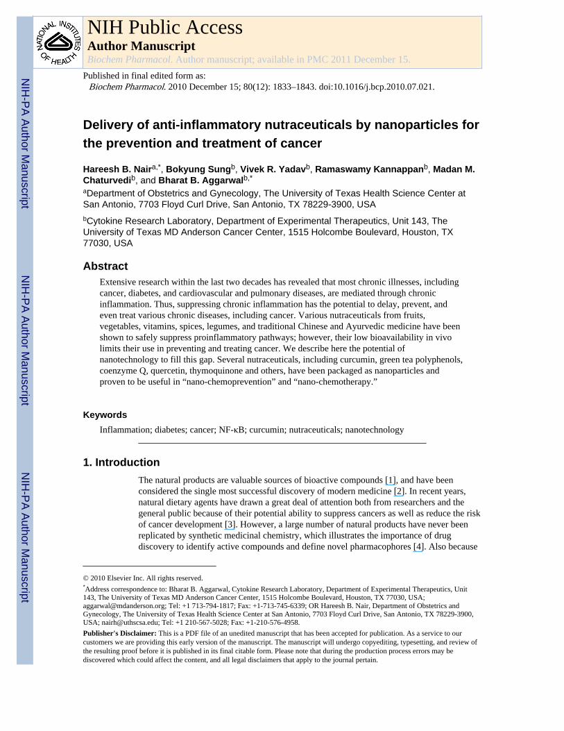
Delivery of anti-inflammatory nutraceuticals by nanoparticles for the prevention and treatment of cancer Hareesh B. Nair a,* , Bokyung Sung b , Vivek R. Yadav b , Ramaswamy Kannappan b , Madan M. Chaturvedi b , and Bharat B. Aggarwal b,* a Department of Obstetrics and Gynecology, The University of Texas Health Science Center at San Antonio, 7703 Floyd Curl Drive, San Antonio, TX 78229-3900, USA b Cytokine Research Laboratory, Department of Experimental Therapeutics, Unit 143, The University of Texas MD Anderson Cancer Center, 1515 Holcombe Boulevard, Houston, TX 77030, USA Abstract Extensive research within the last two decades has revealed that most chronic illnesses, including cancer, diabetes, and cardiovascular and pulmonary diseases, are mediated through chronic inflammation. Thus, suppressing chronic inflammation has the potential to delay, prevent, and even treat various chronic diseases, including cancer. Various nutraceuticals from fruits, vegetables, vitamins, spices, legumes, and traditional Chinese and Ayurvedic medicine have been shown to safely suppress proinflammatory pathways; however, their low bioavailability in vivo limits their use in preventing and treating cancer. We describe here the potential of nanotechnology to fill this gap. Several nutraceuticals, including curcumin, green tea polyphenols, coenzyme Q, quercetin, thymoquinone and others, have been packaged as nanoparticles and proven to be useful in “nano-chemoprevention” and “nano-chemotherapy.” Keywords Inflammation; diabetes; cancer; NF-κB; curcumin; nutraceuticals; nanotechnology 1. Introduction The natural products are valuable sources of bioactive compounds [1], and have been considered the single most successful discovery of modern medicine [2]. In recent years, natural dietary agents have drawn a great deal of attention both from researchers and the general public because of their potential ability to suppress cancers as well as reduce the risk of cancer development [3]. However, a large number of natural products have never been replicated by synthetic medicinal chemistry, which illustrates the importance of drug discovery to identify active compounds and define novel pharmacophores [4]. Also because © 2010 Elsevier Inc. All rights reserved. * Address correspondence to: Bharat B. Aggarwal, Cytokine Research Laboratory, Department of Experimental Therapeutics, Unit 143, The University of Texas MD Anderson Cancer Center, 1515 Holcombe Boulevard, Houston, TX 77030, USA; [email protected]; Tel: +1 713-794-1817; Fax: +1-713-745-6339; OR Hareesh B. Nair, Department of Obstetrics and Gynecology, The University of Texas Health Science Center at San Antonio, 7703 Floyd Curl Drive, San Antonio, TX 78229-3900, USA; [email protected]; Tel: +1 210-567-5028; Fax: +1-210-576-4958. Publisher's Disclaimer: This is a PDF file of an unedited manuscript that has been accepted for publication. As a service to our customers we are providing this early version of the manuscript. The manuscript will undergo copyediting, typesetting, and review of the resulting proof before it is published in its final citable form. Please note that during the production process errors may be discovered which could affect the content, and all legal disclaimers that apply to the journal pertain. NIH Public Access Author Manuscript Biochem Pharmacol. Author manuscript; available in PMC 2011 December 15. Published in final edited form as: Biochem Pharmacol. 2010 December 15; 80(12): 1833–1843. doi:10.1016/j.bcp.2010.07.021. NIH-PA Author Manuscript NIH-PA Author Manuscript NIH-PA Author Manuscript
Transcript of Delivery of antiinflammatory nutraceuticals by nanoparticles for the prevention and treatment of...
Delivery of anti-inflammatory nutraceuticals by nanoparticles forthe prevention and treatment of cancer
Hareesh B. Naira,*, Bokyung Sungb, Vivek R. Yadavb, Ramaswamy Kannappanb, Madan M.Chaturvedib, and Bharat B. Aggarwalb,*
aDepartment of Obstetrics and Gynecology, The University of Texas Health Science Center atSan Antonio, 7703 Floyd Curl Drive, San Antonio, TX 78229-3900, USAbCytokine Research Laboratory, Department of Experimental Therapeutics, Unit 143, TheUniversity of Texas MD Anderson Cancer Center, 1515 Holcombe Boulevard, Houston, TX77030, USA
AbstractExtensive research within the last two decades has revealed that most chronic illnesses, includingcancer, diabetes, and cardiovascular and pulmonary diseases, are mediated through chronicinflammation. Thus, suppressing chronic inflammation has the potential to delay, prevent, andeven treat various chronic diseases, including cancer. Various nutraceuticals from fruits,vegetables, vitamins, spices, legumes, and traditional Chinese and Ayurvedic medicine have beenshown to safely suppress proinflammatory pathways; however, their low bioavailability in vivolimits their use in preventing and treating cancer. We describe here the potential ofnanotechnology to fill this gap. Several nutraceuticals, including curcumin, green tea polyphenols,coenzyme Q, quercetin, thymoquinone and others, have been packaged as nanoparticles andproven to be useful in “nano-chemoprevention” and “nano-chemotherapy.”
KeywordsInflammation; diabetes; cancer; NF-κB; curcumin; nutraceuticals; nanotechnology
1. IntroductionThe natural products are valuable sources of bioactive compounds [1], and have beenconsidered the single most successful discovery of modern medicine [2]. In recent years,natural dietary agents have drawn a great deal of attention both from researchers and thegeneral public because of their potential ability to suppress cancers as well as reduce the riskof cancer development [3]. However, a large number of natural products have never beenreplicated by synthetic medicinal chemistry, which illustrates the importance of drugdiscovery to identify active compounds and define novel pharmacophores [4]. Also because
© 2010 Elsevier Inc. All rights reserved.*Address correspondence to: Bharat B. Aggarwal, Cytokine Research Laboratory, Department of Experimental Therapeutics, Unit143, The University of Texas MD Anderson Cancer Center, 1515 Holcombe Boulevard, Houston, TX 77030, USA;[email protected]; Tel: +1 713-794-1817; Fax: +1-713-745-6339; OR Hareesh B. Nair, Department of Obstetrics andGynecology, The University of Texas Health Science Center at San Antonio, 7703 Floyd Curl Drive, San Antonio, TX 78229-3900,USA; [email protected]; Tel: +1 210-567-5028; Fax: +1-210-576-4958.Publisher's Disclaimer: This is a PDF file of an unedited manuscript that has been accepted for publication. As a service to ourcustomers we are providing this early version of the manuscript. The manuscript will undergo copyediting, typesetting, and review ofthe resulting proof before it is published in its final citable form. Please note that during the production process errors may bediscovered which could affect the content, and all legal disclaimers that apply to the journal pertain.
NIH Public AccessAuthor ManuscriptBiochem Pharmacol. Author manuscript; available in PMC 2011 December 15.
Published in final edited form as:Biochem Pharmacol. 2010 December 15; 80(12): 1833–1843. doi:10.1016/j.bcp.2010.07.021.
NIH
-PA Author Manuscript
NIH
-PA Author Manuscript
NIH
-PA Author Manuscript

of their low solubility, many phytochemicals are poorly absorbed by human body, thus oneof the most important and interesting applications for encapsulation of phytochemicals is toenhance the bioavailability of phytochemicals by changing the pharmacokinetics andbiodistribution [5].
In the past decade, tremendous advancement has been made toward making nanoparticle-based therapeutic products and formulations commercially available. A 2006 EuropeanTechnological Observatory survey showed that more than 150 pharmaceutical companieswere developing nanoscale therapeutics [6] The idea of controlled drug delivery has beenshown to improve the therapeutic index of drugs by increasing their localization to specifictissues, organs, or cells [7,8]. This approach tends to decrease potential side effects byleaving the normal sensitive cells unharmed. Contemporary systemically administeredchemotherapeutic agents are extremely toxic to cancer cells, but can also harm normal cellsleading to serious side effects [9]. Controlled drug delivery systems administer the requiredamount of the drug to the target site and prevent it from circulating until its half-life finishes.The career system associated with site-specific drug delivery can be modulated for betterpharmacokinetics and drug bioavailability [5]. More than half of the chemotherapeuticagents currently being administered either are plant derived or semi-synthetic in nature. Theinevitable share of botanicals, therefore, in the development of modern chemotherapeutics isbeyond doubt. Several different classes of active natural products have been documented.
According to the National Nanotechnology Initiative (NNI; http://www.nano.gov)nanotechnologic structures should be only 1-100 nm in at least one dimension. This sizerequirement can be achieved through various rational designs, including top-down andbottom-up approaches. Nanocarriers have the potential to modify modern drugs byincreasing their efficacy, stability, and solubility; decreasing their toxicity; and sustainingtheir release [10]. Nanoparticulate drug delivery systems using liposomes and biodegradablepolymers have attracted increasing attention in recent years. In addition to liposomes andbiodegradable polymers, the most common materials for nanocarriers include dendrimers,virus nanoparticles, and magnetic nanoparticles[11] (Fig. 1). Some of the commonly usedmethods to characterize the nanoparticles are depicted in Fig. 2. The most noticeablenanotechnologic applications in medicine have been related to oncology [12,13]. In thisreview, we discuss the recent advances made in approaches for targeting anticancerbioactive natural products in both basic research and clinical trials.
2. Inflammation, chronic diseases and cancerA growing body of evidence suggests that many neoplasms are initiated by infections [14].Some recent reviews have discussed intimate connection between inflammation and cancer[15-17]. Inflammation is known to contribute to physiological and pathological processessuch as wound healing and infection by the activation and directed migration of leukocytesfrom the venous system to sites of damage[14]. Inflammation functions at all three stages oftumor development: initiation, progression, and metastasis. Tumor cells produce variouscytokines and chemokines that attract leukocytes. The inflammatory component of adeveloping neoplasm may include a diverse leukocyte population that includes neutrophils,dendritic cells, macrophages, eosinophils, and mast cells [18]. It is believed that tumor-associated macrophages are a significant component of inflammatory infiltrates in neoplastictissues and are derived from monocytes that are recruited largely by monocyte chemotacticprotein (MCP) chemokines. The proinflammatory cytokines, including tumor necrosisfactor-alpha (TNF-α) and interleukin-6 (IL-6), induce direct effects on stromal andneoplastic cells in addition to their roles in regulating leukocyte recruitment. The directevidence for the association of chronic inflammation with malignant diseases is in coloncarcinogenesis in individuals with inflammatory bowel diseases [19,20]. The emerging
Nair et al. Page 2
Biochem Pharmacol. Author manuscript; available in PMC 2011 December 15.
NIH
-PA Author Manuscript
NIH
-PA Author Manuscript
NIH
-PA Author Manuscript

concept in clinical trials reveals that inflammation is a potential therapeutic target in cancertreatment and nonsteroidal antiinflammatory drugs are thereby potentially useful as adjuvanttherapy [21].
Inflammation also plays a key role in the physiology of arthritis, diabetes, heart disease,irritable bowel syndrome, Alzheimer's disease, Parkinson's disease, and many otherillnesses. In Type I diabetes, the immune system attacks the cells that make insulin. Childrenwho have allergies are less likely to develop Type I diabetes. Several spices, vegetables, andfruits have potent antiinflammatory properties. For example, curcumin (diferuloylmethane)is a major constituent of the yellow spice turmeric, derived from the rhizomes of Curcumalonga. It is safe and nontoxic and has demonstrable antitumor, antiinflammatory, apoptotic,and antioxidant properties. We have shown previously that curcumin inhibits tumormetastasis, invasion, and angiogenesis [22]. Beside curcumin, the potential of red chili(capsaicin), cloves (eugenol), ginger (zerumbone), fennel (anethole), kokum (gambogicacid), fenugreek (diosgenin), and black cumin (thymoquinone) in cancer prevention hasbeen established [23].
3. Antiinflammatory nutraceuticalsA wide variety of nutraceuticals, the most common of which are shown in Table 1, areknown to possess antiinflammatory properties. Extensive research within the last twodecades has revealed that curcumin exhibits antioxidative, antiinflammatory, antiapoptotic,antiproliferative, antiinvasive, and antiangiogenic activity [24]. Animal studies haverevealed that curcumin can prevent carcinogen-induced tumorigenesis and inhibit the growthof implanted human tumors [25]. Such studies have led to clinical trials of curcumin inpatients with colon cancer [25-27], familial adenomatous polyposis [28], pancreaticcancer[29] , and multiple myeloma [30]. Unfortunately, these studies revealed that one ofthe major limitations of curcumin is its low oral bioavailability in vivo. Traditionally,turmeric is delivered orally as an emulsion in oil or milk, perhaps because of thehydrophobic nature of its bioactive constituents, such as curcumin and turmeric oil.Curcumin has indeed been shown to interact with phospholipids [31-34], surfactants[35],proteins [36], and cyclodextrin [37]. Various methods have been tried to enhance curcumindelivery, including its incorporation into liposomes [38,39] and into phospholipid vesicles[40]. The latter was used to deliver curcumin intravenously to bone morrow and splenicmacrophages. Another way to solve the problem of lack of water solubility and poor oralbioavailability is the use of polymer-based nanoparticles [41]. Studies from our laboratoryshowed that serum levels were almost twice as high and half-life was substantially longer incurcumin-nanoparticle than in free curcumin [42].
Many other spice-derived nutraceuticals have been found to play a role in reducinginflammation. These include ajoene, allicin, allyl isothiocyanate, anethole, apigenin,capsaicin, carnosol, caryophyllene, cinnamaldehyde, diallyl sulfide, eugenol, [6]-gingerol,humulene, kaempferol, limonene, myrcene, [6]-paradol, perillyl alcohol, phytic acid,piperine, quercetin, sulforaphane, ursolic acid, and zinger-one. Many of these nutraceuticalstarget the transcription factor NF-κB, leading to its down-regulation [43]. Other flavonoids,such as resveratrol, butein, cardamomin, chalcone, silibinin, xanthohumol, fisetin,epigallocatechin gallate (EGCG), etc. derived from fruits, vegetables, legumes, spices, andnuts also suppress the proinflammatory cell signaling pathways and thus can prevent andeven treat the cancer [44].
4. NanotechnologyTherapeutic uses of nanotechnology typically involve the delivery of small-molecule drugs,peptides, proteins, and nucleic acids. Nanoparticles have advanced pharmacological effects
Nair et al. Page 3
Biochem Pharmacol. Author manuscript; available in PMC 2011 December 15.
NIH
-PA Author Manuscript
NIH
-PA Author Manuscript
NIH
-PA Author Manuscript

compared with the therapeutic entities they contain. Active intracellular delivery andimproved pharmacokinetics and pharmacodynamics of drug nanoparticles depend on variousfactors, including their size and surface properties. Nanoparticle therapeutics is an emergingtreatment modality in cancer and other inflammatory disorders. The National CancerInstitute has recognized nanotechnology as an emerging field with the potential torevolutionize modern medicine for detection, treatment, and prevention of cancer [45]. Moreselective therapies, such as angiogenesis inhibitors, vascular disrupting agents, and estrogen-and HER-2-targeted therapies have been developed to treat cancer. These approaches haveincreased patient survival because of treatment efficacy [25]. Most solid tumors possessunique features, including enhanced angiogenesis, defective vasculature and lymphatics, andincreased vascular permeability, which stimulate their growth. Rationally designednanoparticles can take advantage of these tumor features to deliver chemotherapeuticsselectively and specifically. Some commonly used materials for preparation of nanopartclesand their advantages are shown in Table 2. The key properties of nanoparticles that can beused for delivering anticancer drugs are discussed in the following sections:
Nanoparticle size: The National Cancer Institute currently recommends that the size of the nanoparticles be10-100 nm. Even though the upper limit of nanoparticle size is not strictly defined, the lowerlimit (10 nm) is fixed based on the threshold for first-pass elimination by the kidneys [46].Size is a key factor in the biodistribution of long-circulating nanoparticles on the basis ofphysiological parameters such as hepatic filtration, tissue extravasation, tissue diffusion, andkidney excretion [47]. Tumor-targeting nanoparticles must resist hydrostatic andbiophysical/biochemical barriers, overcome cellular resistance to treatment, resistbiotransformation, and resist sudden degradation, immediate clearance, and enhanceddistribution even in poorly perused areas [48].
Surface characteristicsThe surface properties of nanoparticles determine their interaction with the localenvironment. Sterically stabilized nanoparticles exhibit minimal self and non-selfinteractions [13]. These particles keep slightly high or low negative or positive charges ontheir surfaces, which leads to an increased reticuloendothelial clearance; therefore,minimizing nonspecific interactions and controlling surface charge by steric stabilizationhelps to prevent nanoparticle loss in undesired locations to a certain extent. Nanoparticleshave high surface-to-volume ratios, which can be manipulated by rational design. Thesurface properties of the nanoparticles will determine their solubility, stability, andclearance. It has been shown that polymer drug or antibody conjugates have superior half-lives, which can improve the pharmacokinetics of the drug [49]. Increased opsonizationassociated with nanoparticles’ surface during circulation can trigger substantial hepaticagglomeration. Peggylation has been shown to reduce protein absorption. In general,peggylated nanoparticles have longer circulation times and higher levels of tumoraccumulation than non-PEGylated nanoparticles [50]
TargetingAny ideal therapy to treat a disease should destroy specifically diseased or affected cells inan organ while conserving normal cells. Ultimately, targeting will enable the cancer cells toreceive the pharmacologically required concentration of the drug molecules. Varioustargeting approaches, including passive and active targeting of drug molecules, are wellcharacterized and studied. A large body of evidence suggests that these targeting strategiescould overcome drug resistance and side effects to the vital organs and could minimizesystemic drug administration.
Nair et al. Page 4
Biochem Pharmacol. Author manuscript; available in PMC 2011 December 15.
NIH
-PA Author Manuscript
NIH
-PA Author Manuscript
NIH
-PA Author Manuscript
Passive targetingPassive targeting can be achieved by changing the physiochemical characteristics, pH, orhydrophobicity of nanoparticles. Passive targeting of nanoparticles utilizes the EPR of tumorblood vessels. Nanoparticles that escape the reticuloendothelial system (RES) can circulatefor longer times in the bloodstream and have a greater chance of reaching the targeted tumortissues. To supply oxygen and nutrients to the tumor, rapidly-growing cancer cells haveenhanced neovascularization [51]. This imbalance of angiogenic regulators such as growthfactors and matrix metalloproteinases makes tumor vessels highly disorganized and inflatedwith enlarged gap junctions between endothelial cells and improper lymphatic drainage [52].Table 3 lists several of the passively targeted drug candidates currently approved by theFood and Drug Administration (FDA) or being considered by pharmaceutical companies.
Superparamagnetic dextran-coated iron oxide nanoparticles (SDIONs) andsuperparamagnetic iron oxide nanoparticles (SPIONs) are widely used to achieve passivetargeting. SDION conjugates could protect the DNA inside the body and preventnanoparticle carrier/gene compounds from being dissociated and nonspecifically distributed.Moreover, compared with viral vectors and liposomes, tiny SDIONs could effectively evadephagocytosis by macrophages, thus prolonging the drug's circulation time in the body andincreasing its transfection efficiency, and hence playing an important role in locating andtargeting [53]. SPIONs are widely accepted magnetic resonance imaging (MRI) contrastagents for clinical use and have several important advantages over traditional gadolinium-based MRI contrast agents: lower toxicity, stronger enhancement of proton relaxation, andlower detection limits [54].
Active TargetingCancer biomarkers include a variety of molecules, such as mutant genes, RNAs, proteins,lipids, carbohydrates, and small metabolite molecules. The identification of such biomarkersis very important for individualized cancer treatment. Active targeting of cancer cells bymultifunctionalization, in which drugs and contrast agents are attached, involves a corona ofpolymeric material that improves biokinetics and biodistribution and a ligand that addsspecificity for cancer biomarker molecular recognition and attaches to cancer cells [8,55].One major advantage is the nanoparticles’ surface functionality, which allows for theselective coupling of imaging agents and targeting of ligands and/or other components toincrease tumor specificity. Adding a targeting moiety onto the surface of nanoparticles canincrease selective cellular binding and internalization through receptor-mediatedendocytosis. The ideal active-targeting nanoparticles should meet the following criteria:specificity to exclusively target cancer cells, faster uptake through receptor-mediatedendocytosis, faster delivery to tumors, stability of ligands until the drug reaches the target,stealth nature or neutral surface coating, and increased retention throughcompartmentalization. Therapeutic nanoparticles that are already in preclinical and clinicaltrials are shown in Table 4.
Structures such as antibodies, antibody fragments, proteins, small molecules, aptamers, andpeptides have all demonstrated the ability to induce nanoparticle targeting of cancer cells.Ferrari [8] showed that silicon and silica are emerging as interesting candidates forinjectable nanovectors. Porosified silicon is biodegradable with kinetics of degradation thatare much more rapid (minutes to hours) than those of the other biodegradable polymers(weeks to months), and therefore release drugs with previously unattainable time profiles.Metal-based nanovectors include nanoshells [56], which comprise a gold layer over a silicacore and are considered to be highly selective, externally activated therapeutic agents. Therelative distribution of cancer signatures and markers associated with the tumormicroenvironment have been detected by multifunctional nanoparticles such as cross-linked
Nair et al. Page 5
Biochem Pharmacol. Author manuscript; available in PMC 2011 December 15.
NIH
-PA Author Manuscript
NIH
-PA Author Manuscript
NIH
-PA Author Manuscript
iron oxide nanoparticles which were conjugated to annexin-V, which recognizes thephosphatidylserine that is present on apoptotic cells and were used for MRI identification ofcamptothecin-induced apoptosis of Jurkat T cells in vitro [57]. Apart from HER2,transferring receptor, prostate specific antigen (PSA), and prostate specific membraneantigen (PSMA), another protein that has shown potential for targeted cancer therapy is theurokinase plasminogen activator,(uPA), a natural ligand for the urokinase plasminogenactivator receptor for targeting the overexpressed receptors on colon and breast cancers [58].In a recent study, hydrophobically modified carboxymethylated chitosan nanoparticles wereused for the targeted delivery of paclitaxel. The surface of the nanoparticles was modified bythe covalent attachment of folic acid by simple carbodiimide reaction to achieve tumor cell-targeting properties [59]. The hybrid systems consisting of anticancer drugs such asmethotrexate or 5-fluorouracil and a two-dimensional inorganic delivery carrier like layereddouble hydroxide (LDH) were described by Choi et al. [60]. The advantage of the LDHnanoparticles, such as 5-fluorouracil-LDH, is that they rapidly excrete from the body and donot accumulate in the organs after administration. Therefore, the hybrid system is apromising anticancer chemotherapy agent for tumor targeting with biocompatibility.
As mentioned earlier in Section 3, even though natural products are potential candidates fortreating many dreaded diseases, the amount of the drugs needed usually impedes the furtherdevelopment of those drugs as single agents and results in the search for semisynthetic orsynthetic scaled-up strategies. Potential approaches to overcoming this scenario includetargeted delivery of these drugs. In a recent study, genistein was covalently attached toFe3O4 nanoparticles coated by cross-linked carboxymethylated chitosan (CMCH) to create anew, multifunctional, tumor-targeting drug delivery system [61]. The results from this studyindicate that the Fe3O4-CMCH-genistein nanoconjugate significantly enhanced inhibition ofSGC-7901 cancer cells compared to genistein alone. This drug delivery system may bepromising for a future multifunctional chemotherapeutic application that combines drugrelease and magnetic hyperthermia [61].
5. Formulation technologiesEfficient delivery of bioactive agents and peptides and drugs to the systemic circulation andthen to a target cell or organ has received considerable attention in medicine because ofrecent advances in biotechnology.
NanoprecipitationThis method of formulating nanopharmaceuticals involves the nanoprecipitation of apreformed polymer from an organic solution in which it is held by the diffusion of theorganic solvent in the aqueous medium in the presence of a surfactant. This method isbasically applicable to lipophilic drugs because of the miscibility of the solvent with theaqueous phase [62]. Briefly, the poly(lactic-co-glycolic acid) (PLGA) and the drug aredissolved in acetone or other organic solvents. The organic phase is then poured into watercontaining PluronicF-68 as a surfactant. The organic solvent is immediately removed fromthe colloidal suspension by rotaevaporation under reduced pressure. The resulting particlesuspension is filtered through a 1.0-μm cellulose nitrate membrane filter, adjusted in size bymechanical extrusion, and concentrated to a desired volume by the removal of water underthe same conditions. The amount of drug present in the nanoparticles is determined as thedifference between the total amount of the drug used to prepare the nanoparticles and theamount of the drug present in the aqueous medium. The drug present in the aqueous mediumanalyzed by directly injecting into a high-performance liquid chromatography system understandard conditions.
Nair et al. Page 6
Biochem Pharmacol. Author manuscript; available in PMC 2011 December 15.
NIH
-PA Author Manuscript
NIH
-PA Author Manuscript
NIH
-PA Author Manuscript
Thymoquinone (TQ), derived from the medicinal spice Nigella sativa, has been shown toexhibit antiinflammatory and anticancer activity. TQ nanoparticles prepared bynanoprecipitation have shown improved effectiveness and bioavailability [63]. Our previousstudy also showed that curcumin-loaded PLGA nanoparticles prepared by nanoprecipitationenhanced cellular uptake and increased bioactivity in vitro and superior bioavailability invivo over free curcumin [42].
Nanoemulsion techniquesAnother method for formulating nanoparticles is nanoemulsion, a heterogeneous mixture ofoil mixtures of very-small-diameter oil droplets in water (20-500 nm). They are used invarious chemical, pharmaceutical, and cosmetic applications and have been widely tested fortransdermal applications. The advantages of nanoemulsions include an opportunity tosolubilize hydrophobic compounds in the oil phase, modify the surface of the oil dropletswith polymers to extend circulation times, and passively target tumors and/or actively targetligands [64]. The oil-containing nanoemulsions are prepared by coarse homogenizationfollowed by high-energy ultrasonication as previously described [65,66]. Briefly, theaqueous phase is prepared by adding egg lecithin to the deionized water, and stirred at highspeed. The drug of interest is dissolved in a suitable organic solvent and dispersed in oil.The mixture is then heated to 70-75° C to evaporate the aqueous phase. This oil phase thatcontains entrapped drug, is then added gradually to the aqueous phase and homogenized toproduce the coarse oil-in-water emulsion [64]. The coarse emulsion is then ultrasonicated toobtain the nanosized oil droplets.
The peculiarity of nanoemulsions, making them prime techniques for nanoparticleengineering, lies in the long-term kinetic stability of their droplet suspension Nanoemulsionslast for months and withstand dilution and moderate temperature changes. Two widelyaccepted methods are used to prepare nanoemulsions: high-energy emulsification and low-energy emulsification. High-energy emulsification utilizes high (mechanical) energy; thiscommon method is particularly exploited in nanoemulsion polymerization [64]. Themechanical processes that generate nanometric emulsions include initial drop creation,followed by the deformation and disruption of these macrometric initial droplets, andsurfactant absorption at their interface to ensure steric stabilization [67]. On the other hand,low-energy emulsification uses the rapid diffusion of a water-soluble solvent, solubilizedfirst in the organic phase, and again in the aqueous phase, when the two phases are mixed.Current literature on spontaneous emulsification emphasizes the solvent displacementmethod, also called the “Ouzo effect [64,68,69], which consists of nanoemulsionformulation due to the specific and very rapid diffusion of an organic solvent (e.g., acetoneor ethanol) from the oil phase to the aqueous phase.
Nanoemulsion of caffeine (1,3,7-trimethylhanthine), found in tea leaves, coffee, cocoa,guarana, and kola nuts, has been widely studied and has tremendous potential, since caffeinecould protect the skin from ultraviolet light-induced skin cancer [70].
Reverse-phase evaporationThe reverse-phase evaporation method is widely used to prepare liposomes for various drugdelivery purposes [71-73]. Several phospholipids, either pure or mixed with other lipidssuch as cholesterol or long-chain alcohols, may be used. The lipid mixture is added to theorganic solvent and then the solvent is removed under reduced pressure by rotaevaporation.The system is then purged with nitrogen and the lipids are re-dissolved in the organic phase,in which the reverse-phase vesicles are formed. When the lipids have low solubility in ether,chloroform or methanol can be added to increase their solubility. The system is keptcontinuously under nitrogen and the aqueous phase and the resulting two-phase system is
Nair et al. Page 7
Biochem Pharmacol. Author manuscript; available in PMC 2011 December 15.
NIH
-PA Author Manuscript
NIH
-PA Author Manuscript
NIH
-PA Author Manuscript
sonicated briefly (2-5 min) until the mixture becomes either a clear one-phase dispersion ora homogeneous opalescent dispersion that does not separate for at least 30 min aftersonication. The organic solvent is then removed under reduced pressure by rotaevaporation.As the majority of the solvent is removed, the material first forms a viscous gel, thensubsequently (within 5-10 min) becomes an aqueous suspension. The preparation is theneither dialyzed or centrifuged to remove nonencapsulated material and residual organicsolvent [71].
6. Role of nanotechnology for nutraceuticalsNano formulations of nutraceuticals essentialy follow the general principles ofnanotechnology. Therefore, the nanotechnology platforms are widely being used createdelivery systems for bioactive natural products and nutraceuticals with poor water solubility.The market projections for these technologies suggest a multifold increase in theircommercial potential over the next 5 years. Table 5 summarizes a list of nutraceuticals, thematerials used for preparing nanoparticles, the size of the nanoparticles, and their possibletissue/cancer targets. Some of the extensively studied nutraceutical nanoparticles arediscussed below.
Curcumin, for example, has minimal systemic bioavailability, but its biologic activity andbioavailability have been tremendously increased via various nanoparticle formulations.Although many published studies have proposed curcumin, with its antiinflammatory andanticancer properties, as safe for cancer therapy and chemoprevention, the compound has byno means been embraced by the cancer community. A phase I study of colorectal cancerpatients found that the systemic bioavailability of postoperatively administered curcumin islow in humans [29]. In this study, patients with hepatic colorectal cancer metastases wereadministered 3,600 mg of oral curcumin daily, and levels of curcumin and its metaboliteswere measured by high-performance liquid chromatography in portal and peripheral blood.Curcumin was poorly available following oral administration, with low nanomolar levels ofthe parent compound and its glucuronide and sulphate conjugates found in the peripheral orportal circulation [112]. Bisht et al. have developed nanoparticulate curcumin using cross-linked polymeric nanoparticles comprised of N-isopropylacrylamide, N-vinyl-2-pyrrolidinone, and PEG acrylate, which has been tested in various pancreatic cancer celllines. In this study, nanoparticulate curcumin showed superior cytotoxicity anddownregulated multiple proinflammatory markers in a dose-dependent manner [83].Another study revealed that nanoparticle encapsulation improves the oral bioavailability ofcurcumin by at least 9-fold when compared to curcumin administered with piperine as anabsorption enhancer. In this study, curcumin-encapsulated nanoparticles were prepared byemulsion, and a particle size of 264 nm showed enhanced in vivo pharmacokinetics with a 9-fold increase in oral bioavailability compared to curcumin administered with piperine as anabsorption enhancer [113]. In another study, piperine has been found to enhance thebioavailability of curcumin in both preclinical and clinical studies [114]. It has beensuggested that piperine can inhibit curcumin-metabolizing enzymes and thereby circumventfirst-pass metabolism[113]. Ursolic acid (UA), another poorly soluble natural product, is atriterpenoid with a wide variety of antitumor activities [115]. To achieve a highbioavailability, targeting effect, stability, and intravenous administration, UA phospholipidnanopowders have been prepared by solvent emulsification-evaporation and ultrasonicdispersion [111]. However, limited biological testing of these particles has been reported.Lee et al. [116] have tested the ability of nanoparticles to deliver bioactive agents bypreparing amphiphilic self-assembled nanoparticles composed of chitosan and UA forprotein delivery to the skin. Topical treatment of skin diseases has the advantage that highdrug levels can be achieved at the site of disease and systemic side effects can be reducedcompared to oral or parenteral drug administration. Topical drug administration is still a
Nair et al. Page 8
Biochem Pharmacol. Author manuscript; available in PMC 2011 December 15.
NIH
-PA Author Manuscript
NIH
-PA Author Manuscript
NIH
-PA Author Manuscript

pharmaceutical challenge because of the difficulties of controlling and determining the exactamount of the drug that reaches the different skin layers [117]. Natural products likecurcumin and UA are potential candidates for treating various skin conditions, namelyatopic eczema, psoriasis, acne, skin mycosis, and inflammations. Nanoformulations willmake these candidate drugs much more valuable in the pharmaceutical market in the comingdecade. It has been shown that it is possible to enhance percutaneous absorption with lipidnanoparticles. These carriers may even allow drugs to target the skin or even itssubstructures. Thus, they might have the potential to improve the benefit-to-risk ratio oftopical drug therapy [117].
Triptolide is a purified compound of a traditional Chinese medicine with antiinflamatory,immunosuppressive, antifertility, and antineoplastic activity [118]. Mei et al. showed thatsolid lipid nanoparticles prepared for transdermal delivery increased triptolide penetrationinto the skin and its antiinflammatory activity [119]. It is assumed that )his strategyimproves the drug's bioavailability at the site of action, reduces the required dose, andreduces dose-dependent side effects like irritation and stinging. Epigallocatechin gallate(EGCG), an abundant catechin found most notably in tea, is among other plants, and is alsoa potent antioxidant that may have therapeutic properties for many disorders, includingcancer [120]. A recent study showed that EGCG nanoparticles ameliorated cyclosporine-induced nephrotoxicity in rats at a dose three times lower than the dose at which an oralsolution produced the same effect and enhanced oral bioavailability [121].
Polyphenols are another group of nutraceuticals that has proven anti-inflammatoryproperties and thus has high potentials for cancer therapy. The challenge of usingpolyphenols to treat cancer is their potentially low bioavailability and short half-life. Onealternative to using free compounds is to use polyphenol-loaded nanoparticles [122].However, additional challenges are faced while encapsulating the polyphenols due to theirvarying structures, moderate solubility, and fast oxidation under basic conditions[123,124. ]. Encapsulated EGCG nanoparticles have been limited to a PLGA nanoparticulateformulation used for in vivo evaluation of the antioxidant efficacy of EGCG in a rat model[125] and chitosan-tripolyphosphate nanoparticles for the encapsulation of green teacatechin extracts. A very interesting study reported EGCG encapsulation using gelatin-based200- to 300- nm nanoparticles consisting of a soft gel-like interior and a surrounding shell ofpolyelectrolytes (polystyrene sulfonate/polyallylamine hydrochloride [PSS/PAH]),polyglutamic acid/poly-L-lysine (PGA/PLL), dextran sulfate/protamine sulfate (DexS/ProtS),and carboxymethyl cellulose/gelatin, type A (CMC/GelA) assembled using the layer-by-layer technique [124]. In this study, two combinations of polyanion/polycation pairs wereused to form a layer-by-layer coating around 300-nm gelatin nanoparticles, PSS/PAH(strong polyanion/weak polycation), and PGA/PLL (weak polyanion/strong polycation), aswell as two combinations of polyanion/protein, DexS/ProtS (strong polyanion/stronglypositively charged polypeptide) and CMC/GelA (weak polyanion/weakly positively chargedprotein). While these particles showed promising results in vitro, no in vivo data werereported. Another research group introduced the concept of nanochemoprevention aimed atusing nanotechnology to enhance the outcome of chemoprevention [74]. In this study, PLA-PEG nanoparticles of EGCG (nano-EGCG) exhibited a >10-fold dose advantage overnonencapsulated EGCG [74].
Studies from our laboratory have shown that curcumin nanoparticles were more active thanfree curcumin for inhibiting TNFα-induced NF-κB activation and suppressing NF-κB-regulated proteins involved in cell proliferation (cyclin D1), invasion (matrixmetalloproteinase-9), and angiogenesis (vascular endothelial growth factor (VEGF)). Inmice, curcumin nanoparticles were more bioavailable and had a longer half-life than freecurcumin [42,126]. We used biodegradable nanoparticulate formulation based on PLGA and
Nair et al. Page 9
Biochem Pharmacol. Author manuscript; available in PMC 2011 December 15.
NIH
-PA Author Manuscript
NIH
-PA Author Manuscript
NIH
-PA Author Manuscript
a stabilizer, PEG-5000, for these studies. In a similar, more recent study, we showed that TQexhibits antiinflammatory and anticancer activity and encapsulation of TQ enhances itsbiological activity and bioavailability [63]. In that study, we observed that TQ nanoparticleswere also more potent for suppressing proliferation of colon cancer, breast cancer, prostatecancer, and multiple myeloma cells. Esterase staining for plasma membrane integrityrevealed that TQ nanoparticles were more potent than free TQ for sensitizing leukemic cellsto TNFα- and paclitaxel-induced apoptosis [63].
Resveratrol (trans-3,4,5-trihydroxystilbene) is a natural polyphenolic compound abundant ingrapes, peanuts, red wine, and a variety of other food sources, and has been reported to elicitmany cellular responses, including cell cycle arrest, differentiation, and apoptosis, and toinhibit the growth of several types of cancer, such as prostate and colon cancers [102,127].However, its wide array of activity has been compromised by intrinsic features that lead tolow bioavailability, low water-solubility, and instability. Active targeting of resveratrol wastested by formulating chitosan nanoparticles with amine-free surfaces for appropriate ligandconjugation was prepared by sodium chloride precipitation. The absence of amine on thesurfaces allows for the design of an active target drug delivery system.
7. In vivo pharmacokineticsSite-specific drug delivery is an important area of research that is anticipated to increase theefficacy of drugs and reduce their potential side effects. Biodegradable polymers arecurrently being used as drug carriers because of their inherent properties of controlledrelease, enhanced distribution and overall pharmacokinetic availability. The release ofloaded drugs from nanoparticles may be controlled in response to changes in environmentalconditions, such as temperature and pH. Biodistribution profiles and anticancer efficacy ofnanonutraceuticals in vivo might differ depending upon their size, surface charge,PEGylation, and other biophysical properties. Micro- and nanoparticulate systemsformulated with these polymers have shown wide applicability for oral, subcutaneous, orintravenous delivery of lipophilic and hydrophilic drugs. Wang et al. showed that t1/2 betaand area under the curve of a paclitaxel micelle formulation were 4.0- and 2.2-fold higherthan that of a Taxol injection. Their study of biodistribution in mice showed that thepaclitaxel micelle formulation not only decreased drug uptake by the liver but alsoprolonged drug retention in the blood and increased drug distribution in the kidneys, spleen,ovaries, and uterus, suggesting that this formulation could be a useful drug carrier forintravenous administration of paclitaxel [128]. In a different study of the controlled releaseof insulin to the lungs, insulin-loaded PLGA nanoparticles fabricated by a double emulsionmethod by the aid of hydroxypropyl-beta-cyclodextrin (HPbetaCD) significantly reducedblood glucose levels as a function of the administered dose in animal models [129]. In vivodata show that PLGA/HPbetaCD/insulin nanoparticles can reach alveoli and release insulin,which is absorbed in its bioactive form [129]. Another study described the development ofbiodegradable nanoparticles with a core-shell structure to formulate superparamagnetic ironoxide (CSNPSPIO) for MRI [130]. The developed nanoparticles were composed of ahydrophobic PLGA core and a positively charged glycol chitosan shell. In this study, a highlevel of radioactivity was observed in the liver shortly after the intravenous administration ofthe 99-mTc-labeled CSNPSPIO. Studies from our group and others have shown curcumin asa promising bioactive agent with antiinflammatory, antiproliferative, antiangiogenic,antihelmintic, and wound-healing properties; however, the major challenge of curcumin isits poor oral bioavailability in vivo. Shaikh et al. showed that the in vivo pharmacokinetics ofcurcumin-entrapped nanoparticles demonstrate at least a 9-fold increase in oralbioavailability when compared to curcumin administered with piperine as an absorptionenhancer [113].
Nair et al. Page 10
Biochem Pharmacol. Author manuscript; available in PMC 2011 December 15.
NIH
-PA Author Manuscript
NIH
-PA Author Manuscript
NIH
-PA Author Manuscript
The rational design of nanoparticles is very important for altering the pharmacokinetics ofthe encapsulated drug [7,131] The shape and size of the particles are key determinants ingoverning the biodistribution and bioavailability of the cargo [132] Attaining stericstabilization of particles with a methoxy-PEG surface brush layer is commonly utilized tocreate long-circulating particles. PEGylation reduces the absorption of thereticuloendothelial system. Tumor accumulation of the nanoparticles is a function of boththe rate of extravasation from the blood to the tumor space and the rate of clearance from thetumor. Hydrostatic pressure in a tumor mass typically decreases from the center to theperiphery, so particles that enter into the tumor vasculature by convection are more likely tobe cleared; this washout can be reduced by restricting the movement of particles through theextracellular tumor matrix [130].
8. ConclusionOverall, these studies indicate that nanotechnology has great potential for deliveringnutraceuticals. To fully realize this potential, more clinical trials are needed with nano-formulated nutraceuticals. Abraxan, protein-bound paclitaxel with a mean particle size ofapproximately 130 nm for injectable suspension, has been approved by the Food and DrugAdministration for patients with metastatic breast cancer, but it requires intravenousdelivery. Oral delivery of nutraceuticals, however, is preferred. The fate of the carriermaterials used for the entrapment of nutraceuticals is also unclear at present. The short-termand long-term effects of the carrier material remain to be understood. Another concern isthat properties of a nanoscale material may differ from the bulk-material, and it may alterabsorption, digestion, metabolism, or excretion of nanodrugs in the body. Furthermore,because there is little information available about the potential health risk of nanoparticles,more research on the toxicology of nanoparticles is warranted[131-133]. Chemopreventionrequires the administration of nutraceuticals to normal and healthy individuals; thus, safetyand cost are other points of concern.
AcknowledgmentsWe would like to thank Markeda Wade and Stephanie Deming for carefully editing the manuscript and providingvaluable comments. We also thank Dr. Chitra Sundaram for assisting with references. This work was supported byMD Anderson's Cancer Center Support Grant from the National Institutes of Health (NIH CA-16 672), a programproject grant from the National Institutes of Health (NIH CA-124787-01A2), and a grant from the Center forTargeted Therapy at The University of Texas MD Anderson Cancer Center, where Dr. Aggarwal is the RansomHorne, Jr., Professor of Cancer Research.
References1. Molinski TF. Developments in marine natural products. Receptor-specific bioactive compounds. J
Nat Prod. 1993; 56:1–8. [PubMed: 8450313]2. Grabley S, Thiericke R. Bioactive agents from natural sources: trends in discovery and application.
Adv Biochem Eng Biotechnol. 1999; 64:101–54. [PubMed: 9933977]3. Amin AR, Kucuk O, Khuri FR, Shin DM. Perspectives for cancer prevention with natural
compounds. J Clin Oncol. 2009; 27:2712–25. [PubMed: 19414669]4. Leach AR, Gillet VJ, Lewis RA, Taylor R. Three-dimensional pharmacophore methods in drug
discovery. J Med Chem. 53:539–58. [PubMed: 19831387]5. Huang Q, Yu H, Ru Q. Bioavailability and delivery of nutraceuticals using nanotechnology. J Food
Sci. 75:R50–7. [PubMed: 20492195]6. Wagner V, Dullaart A, Bock AK, Zweck A. The emerging nanomedicine landscape. Nat
Biotechnol. 2006; 24:1211–7. [PubMed: 17033654]7. Riehemann K, Schneider SW, Luger TA, Godin B, Ferrari M, Fuchs H. Nanomedicine--challenge
and perspectives. Angew Chem Int Ed Engl. 2009; 48:872–97. [PubMed: 19142939]
Nair et al. Page 11
Biochem Pharmacol. Author manuscript; available in PMC 2011 December 15.
NIH
-PA Author Manuscript
NIH
-PA Author Manuscript
NIH
-PA Author Manuscript
8. Ferrari M. Cancer nanotechnology: opportunities and challenges. Nat Rev Cancer. 2005; 5:161–71.[PubMed: 15738981]
9. American Cancer Society; 2010. Facts and Figures10. Merisko-Liversidge EM, Liversidge GG. Drug nanoparticles: formulating poorly water-soluble
compounds. Toxicol Pathol. 2008; 36:43–8. [PubMed: 18337220]11. Koning GA, Krijger GC. Targeted multifunctional lipid-based nanocarriers for image-guided drug
delivery. Anticancer Agents Med Chem. 2007; 7:425–40. [PubMed: 17630918]12. Mousa SA, Bharali DJ, Armstrong D. From nutraceuticals to pharmaceuticals to
nanopharmaceuticals: a case study in angiogenesis modulation during oxidative stress. MolBiotechnol. 2007; 37:72–80. [PubMed: 17914168]
13. Davis ME, Chen ZG, Shin DM. Nanoparticle therapeutics: an emerging treatment modality forcancer. Nat Rev Drug Discov. 2008; 7:771–82. [PubMed: 18758474]
14. Coussens LM, Werb Z. Inflammation and cancer. Nature. 2002; 420:860–7. [PubMed: 12490959]15. Balkwill F, Mantovani A. Cancer and inflammation: implications for pharmacology and
therapeutics. Clin Pharmacol Ther. 87:401–6. [PubMed: 20200512]16. Mantovani A, Garlanda C, Allavena P. Molecular pathways and targets in cancer-related
inflammation. Ann Med. 42:161–70. [PubMed: 20384432]17. Grivennikov SI, Karin M. Inflammation and oncogenesis: a vicious connection. Curr Opin Genet
Dev. 20:65–71. [PubMed: 20036794]18. Eming SA, Krieg T, Davidson JM. Inflammation in wound repair: molecular and cellular
mechanisms. J Invest Dermatol. 2007; 127:514–25. [PubMed: 17299434]19. Itzkowitz SH, Yio X. Inflammation and cancer IV. Colorectal cancer in inflammatory bowel
disease: the role of inflammation. Am J Physiol Gastrointest Liver Physiol. 2004; 287:G7–17.[PubMed: 15194558]
20. Danese S, Mantovani A. Inflammatory bowel disease and intestinal cancer: a paradigm of the Yin-Yang interplay between inflammation and cancer. Oncogene. 29:3313–23. [PubMed: 20400974]
21. Turini ME, DuBois RN. Cyclooxygenase-2: a therapeutic target. Annu Rev Med. 2002; 53:35–57.[PubMed: 11818462]
22. Kamat AM, Tharakan ST, Sung B, Aggarwal BB. Curcumin potentiates the antitumor effects ofBacillus Calmette-Guerin against bladder cancer through the downregulation of NF-kappaB andupregulation of TRAIL receptors. Cancer Res. 2009; 69:8958–66. [PubMed: 19903839]
23. Aggarwal BB, Kunnumakkara AB, Harikumar KB, Tharakan ST, Sung B, Anand P. Potential ofspice-derived phytochemicals for cancer prevention. Planta Med. 2008; 74:1560–9. [PubMed:18612945]
24. Sagar SM, Yance D, Wong RK. Natural health products that inhibit angiogenesis: a potentialsource for investigational new agents to treat cancer-Part 1. Curr Oncol. 2006; 13:14–26.[PubMed: 17576437]
25. Anand P, Sundaram C, Jhurani S, Kunnumakkara AB, Aggarwal BB. Curcumin and cancer: an“old-age” disease with an “age-old” solution. Cancer Lett. 2008; 267:133–64. [PubMed:18462866]
26. Sharma RA, McLelland HR, Hill KA, Ireson CR, Euden SA, Manson MM, et al.Pharmacodynamic and pharmacokinetic study of oral Curcuma extract in patients with colorectalcancer. Clin Cancer Res. 2001; 7:1894–900. [PubMed: 11448902]
27. Sharma RA, Euden SA, Platton SL, Cooke DN, Shafayat A, Hewitt HR, et al. Phase I clinical trialof oral curcumin: biomarkers of systemic activity and compliance. Clin Cancer Res. 2004;10:6847–54. [PubMed: 15501961]
28. Cruz-Correa M, Shoskes DA, Sanchez P, Zhao R, Hylind LM, Wexner SD, et al. Combinationtreatment with curcumin and quercetin of adenomas in familial adenomatous polyposis. ClinGastroenterol Hepatol. 2006; 4:1035–8. [PubMed: 16757216]
29. Dhillon N, Aggarwal BB, Newman RA, Wolff RA, Kunnumakkara AB, Abbruzzese JL, et al.Phase II trial of curcumin in patients with advanced pancreatic cancer. Clin Cancer Res. 2008;14:4491–9. [PubMed: 18628464]
Nair et al. Page 12
Biochem Pharmacol. Author manuscript; available in PMC 2011 December 15.
NIH
-PA Author Manuscript
NIH
-PA Author Manuscript
NIH
-PA Author Manuscript
30. Bhardwaj A, Sethi G, Vadhan-Raj S, Bueso-Ramos C, Takada Y, Gaur U, et al. Resveratrolinhibits proliferation, induces apoptosis, and overcomes chemoresistance through down-regulationof STAT3 and nuclear factor-kappaB-regulated antiapoptotic and cell survival gene products inhuman multiple myeloma cells. Blood. 2007; 109:2293–302. [PubMed: 17164350]
31. Began G, Sudharshan E, Sankar KU, Rao AG. Interaction of curcumin with phosphatidylcholine:A spectrofluorometric study. J Agric Food Chem. 2000; 48:576. [PubMed: 10691678]
32. Maiti K, Mukherjee K, Gantait A, Saha BP, Mukherjee PK. Curcumin-phospholipid complex:Preparation, therapeutic evaluation and pharmacokinetic study in rats. Int J Pharm. 2007; 330:155–63. [PubMed: 17112692]
33. Kunwar A, Barik A, Pandey R, Priyadarsini KI. Transport of liposomal and albumin loadedcurcumin to living cells: an absorption and fluorescence spectroscopic study. Biochim BiophysActa. 2006; 1760:1513–20. [PubMed: 16904830]
34. Marczylo TH, Verschoyle RD, Cooke DN, Morazzoni P, Steward WP, Gescher AJ. Comparison ofsystemic availability of curcumin with that of curcumin formulated with phosphatidylcholine.Cancer Chemother Pharmacol. 2007; 60:171–7. [PubMed: 17051370]
35. Tonnesen HH. Solubility, chemical and photochemical stability of curcumin in surfactantsolutions. Studies of curcumin and curcuminoids, XXVIII. Pharmazie. 2002; 57:820–4. [PubMed:12561244]
36. Kumar V, Lewis SA, Mutalik S, Shenoy DB, Venkatesh, Udupa N. Biodegradable microspheres ofcurcumin for treatment of inflammation. Indian J Physiol Pharmacol. 2002; 46:209–17. [PubMed:12500496]
37. Salmaso S, Bersani S, Semenzato A, Caliceti P. New cyclodextrin bioconjugates for active tumourtargeting. J Drug Target. 2007; 15:379–90. [PubMed: 17613656]
38. Li L, Braiteh FS, Kurzrock R. Liposome-encapsulated curcumin: in vitro and in vivo effects onproliferation, apoptosis, signaling, and angiogenesis. Cancer. 2005; 104:1322–31. [PubMed:16092118]
39. Takahashi M, Kitamoto D, Imura T, Oku H, Takara K, Wada K. Characterization andbioavailability of liposomes containing a ukon extract. Biosci Biotechnol Biochem. 2008;72:1199–205. [PubMed: 18460803]
40. Sou K, Inenaga S, Takeoka S, Tsuchida E. Loading of curcumin into macrophages using lipid-based nanoparticles. Int J Pharm. 2008; 352:287–93. [PubMed: 18063327]
41. Kim K, Kim JH, Park H, Kim YS, Park K, Nam H, et al. Tumor-homing multifunctionalnanoparticles for cancer theragnosis: Simultaneous diagnosis, drug delivery, and therapeuticmonitoring. J Control Release.
42. Anand P, Nair HB, Sung B, Kunnumakkara AB, Yadav VR, Tekmal RR, et al. Design ofcurcumin-loaded PLGA nanoparticles formulation with enhanced cellular uptake, and increasedbioactivity in vitro and superior bioavailability in vivo. Biochem Pharmacol. 79:330–8. [PubMed:19735646]
43. Aggarwal BB, Van Kuiken ME, Iyer LH, Harikumar KB, Sung B. Molecular targets ofnutraceuticals derived from dietary spices: potential role in suppression of inflammation andtumorigenesis. Exp Biol Med (Maywood). 2009; 234:825–49. [PubMed: 19491364]
44. Prasad S, Phromnoi K, Yadav VR, Chaturvedi MM, B.B. A. Targeting Inflammatory Pathways byFlavonoids for Prevention and Treatment of Cancer. Planta Medica. 2010 In press.
45. Kawasaki ES, Player A. Nanotechnology, nanomedicine, and the development of new, effectivetherapies for cancer. Nanomedicine. 2005; 1:101–9. [PubMed: 17292064]
46. Silva LF, da Boit KM. Nanominerals and nanoparticles in feed coal and bottom ash: implicationsfor human health effects. Environ Monit Assess.
47. Alexis F, Pridgen E, Molnar LK, Farokhzad OC. Factors affecting the clearance andbiodistribution of polymeric nanoparticles. Mol Pharm. 2008; 5:505–15. [PubMed: 18672949]
48. Jain RK. Transport of molecules in the tumor interstitium: a review. Cancer Res. 1987; 47:3039–51. [PubMed: 3555767]
49. Duncan R, Vicent MJ, Greco F, Nicholson RI. Polymer-drug conjugates: towards a novel approachfor the treatment of endrocine-related cancer. Endocr Relat Cancer. 2005; 12(Suppl 1):S189–99.[PubMed: 16113096]
Nair et al. Page 13
Biochem Pharmacol. Author manuscript; available in PMC 2011 December 15.
NIH
-PA Author Manuscript
NIH
-PA Author Manuscript
NIH
-PA Author Manuscript
50. Pasche S, Voros J, Griesser HJ, Spencer ND, Textor M. Effects of ionic strength and surfacecharge on protein adsorption at PEGylated surfaces. J Phys Chem B. 2005; 109:17545–52.[PubMed: 16853244]
51. Vaupel P, Kallinowski F, Okunieff P. Blood flow, oxygen and nutrient supply, and metabolicmicroenvironment of human tumors: a review. Cancer Res. 1989; 49:6449–65. [PubMed:2684393]
52. Connor KM, Krah NM, Dennison RJ, Aderman CM, Chen J, Guerin KI, et al. Quantification ofoxygen-induced retinopathy in the mouse: a model of vessel loss, vessel regrowth and pathologicalangiogenesis. Nat Protoc. 2009; 4:1565–73. [PubMed: 19816419]
53. Simberg D, Duza T, Park JH, Essler M, Pilch J, Zhang L, et al. Biomimetic amplification ofnanoparticle homing to tumors. Proc Natl Acad Sci U S A. 2007; 104:932–6. [PubMed: 17215365]
54. Caravan P. Protein-targeted gadolinium-based magnetic resonance imaging (MRI) contrast agents:design and mechanism of action. Acc Chem Res. 2009; 42:851–62. [PubMed: 19222207]
55. Peer D, Karp JM, Hong S, Farokhzad OC, Margalit R, Langer R. Nanocarriers as an emergingplatform for cancer therapy. Nat Nanotechnol. 2007; 2:751–60. [PubMed: 18654426]
56. Prestidge CA, Barnes TJ, Lau CH, Barnett C, Loni A, Canham L. Mesoporous silicon: a platformfor the delivery of therapeutics. Expert Opin Drug Deliv. 2007; 4:101–10. [PubMed: 17335408]
57. Schellenberger EA, Bogdanov A Jr. Hogemann D, Tait J, Weissleder R, Josephson L. Annexin V-CLIO: a nanoparticle for detecting apoptosis by MRI. Mol Imaging. 2002; 1:102–7. [PubMed:12920851]
58. Mazar AP. Urokinase plasminogen activator receptor choreographs multiple ligand interactions:implications for tumor progression and therapy. Clin Cancer Res. 2008; 14:5649–55. [PubMed:18794071]
59. Sahu SK, Maiti S, Maiti TK, Ghosh SK, Pramanik P. Hydrophobically modified carboxymethylchitosan nanoparticles targeted delivery of paclitaxel. J Drug Target.
60. Choi SJ, Oh JM, Choy JH. Biocompatible nanoparticles intercalated with anticancer drug for targetdelivery: pharmacokinetic and biodistribution study. J Nanosci Nanotechnol. 10:2913–6.[PubMed: 20355523]
61. Si HY, Li DP, Wang TM, Zhang HL, Ren FY, Xu ZG, et al. Improving the anti-tumor effect ofgenistein with a biocompatible superparamagnetic drug delivery system. J Nanosci Nanotechnol.10:2325–31. [PubMed: 20355429]
62. Rawat MK, Jain A, Mishra A, Muthu MS, Singh S. Development of repaglinide loaded solid lipidnanocarrier: selection of fabrication method. Curr Drug Deliv. 7:44–50. [PubMed: 20044909]
63. Ravindran J, Nair HB, Sung B, Prasad S, Tekmal RR, Aggarwal BB. Thymoquinone poly (lactide-co-glycolide) nanoparticles exhibit enhanced anti-proliferative, anti-inflammatory, andchemosensitization potential. Biochem Pharmacol. 79:1640–7. [PubMed: 20105430]
64. Anton N, Benoit JP, Saulnier P. Design and production of nanoparticles formulated from nano-emulsion templates-a review. J Control Release. 2008; 128:185–99. [PubMed: 18374443]
65. Ganta S, Amiji M. Coadministration of Paclitaxel and curcumin in nanoemulsion formulations toovercome multidrug resistance in tumor cells. Mol Pharm. 2009; 6:928–39. [PubMed: 19278222]
66. Ganta S, Sharma P, Paxton JW, Baguley BC, Garg S. Pharmacokinetics and pharmacodynamics ofchlorambucil delivered in long-circulating nanoemulsion. J Drug Target. 2009
67. Villamizar RA, Braun J, Gompf B, Dressel M, Rius FX. Morphological and electricalcharacteristics of biofunctionalized layers on carbon nanotubes. Biosens Bioelectron. 2009;25:161–6. [PubMed: 19631520]
68. Bouchemal K, Briancon S, Perrier E, Fessi H. Nano-emulsion formulation using spontaneousemulsification: solvent, oil and surfactant optimisation. Int J Pharm. 2004; 280:241–51. [PubMed:15265563]
69. Francois G, Katz JL. Nanoparticles and nanocapsules created using the Ouzo effect: spontaneousemulisification as an alternative to ultrasonic and high-shear devices. Chemphyschem. 2005;6:209–16. [PubMed: 15751338]
70. Shakeel F, Ramadan W. Transdermal delivery of anticancer drug caffeine from water-in-oilnanoemulsions. Colloids Surf B Biointerfaces. 75:356–62. [PubMed: 19783127]
Nair et al. Page 14
Biochem Pharmacol. Author manuscript; available in PMC 2011 December 15.
NIH
-PA Author Manuscript
NIH
-PA Author Manuscript
NIH
-PA Author Manuscript
71. Szoka F Jr. Papahadjopoulos D. Procedure for preparation of liposomes with large internal aqueousspace and high capture by reverse-phase evaporation. Proc Natl Acad Sci U S A. 1978; 75:4194–8.[PubMed: 279908]
72. Kim S, Martin GM. Preparation of cell-size unilamellar liposomes with high captured volume anddefined size distribution. Biochim Biophys Acta. 1981; 646:1–9. [PubMed: 7272296]
73. Meure LA, Foster NR, Dehghani F. Conventional and dense gas techniques for the production ofliposomes: a review. AAPS PharmSciTech. 2008; 9:798–809. [PubMed: 18597175]
74. Siddiqui IA, Adhami VM, Bharali DJ, Hafeez BB, Asim M, Khwaja SI, et al. Introducingnanochemoprevention as a novel approach for cancer control: proof of principle with green teapolyphenol epigallocatechin-3-gallate. Cancer Res. 2009; 69:1712–6. [PubMed: 19223530]
75. Jeong SY, Park SJ, Yoon SM, Jung J, Woo HN, Yi SL, et al. Systemic delivery and preclinicalevaluation of Au nanoparticle containing beta-lapachone for radiosensitization. J Control Release.2009; 139:239–45. [PubMed: 19619590]
76. Blanco E, Bey EA, Dong Y, Weinberg BD, Sutton DM, Boothman DA, et al. Beta-lapachone-containing PEG-PLA polymer micelles as novel nanotherapeutics against NQO1-overexpressingtumor cells. J Control Release. 2007; 122:365–74. [PubMed: 17574288]
77. Cartiera MS, Ferreira EC, Caputo C, Egan ME, Caplan MJ, Saltzman WM. Partial correction ofcystic fibrosis defects with PLGA nanoparticles encapsulating curcumin. Mol Pharm. 7:86–93.[PubMed: 19886674]
78. Mukerjee A, Vishwanatha JK. Formulation, characterization and evaluation of curcumin-loadedPLGA nanospheres for cancer therapy. Anticancer Res. 2009; 29:3867–75. [PubMed: 19846921]
79. Das RK, Kasoju N, Bora U. Encapsulation of curcumin in alginate-chitosan-pluronic compositenanoparticles for delivery to cancer cells. Nanomedicine. 6:153–60. [PubMed: 19616123]
80. Mulik RS, Monkkonen J, Juvonen RO, Mahadik KR, Paradkar AR. ApoE3 Mediated Poly(butyl)Cyanoacrylate Nanoparticles Containing Curcumin: Study of Enhanced Activity of Curcuminagainst Beta Amyloid Induced Cytotoxicity Using In Vitro Cell Culture Model. Mol Pharm.
81. Gupta V, Aseh A, Rios CN, Aggarwal BB, Mathur AB. Fabrication and characterization of silkfibroin-derived curcumin nanoparticles for cancer therapy. Int J Nanomedicine. 2009; 4:115–22.[PubMed: 19516890]
82. Sahu A, Kasoju N, Bora U. Fluorescence study of the curcumin-casein micelle complexation andits application as a drug nanocarrier to cancer cells. Biomacromolecules. 2008; 9:2905–12.[PubMed: 18785706]
83. Bisht S, Feldmann G, Soni S, Ravi R, Karikar C, Maitra A. Polymeric nanoparticle-encapsulatedcurcumin (“nanocurcumin”): a novel strategy for human cancer therapy. J Nanobiotechnology.2007; 5:3. [PubMed: 17439648]
84. Gao Y, Gu W, Chen L, Xu Z, Li Y. The role of daidzein-loaded sterically stabilized solid lipidnanoparticles in therapy for cardio-cerebrovascular diseases. Biomaterials. 2008; 29:4129–36.[PubMed: 18667234]
85. Contreras J, Xie J, Chen YJ, Pei H, Zhang G, Fraser CL, et al. Intracellular uptake and traffickingof difluoroboron dibenzoylmethane-polylactide nanoparticles in HeLa cells. ACS Nano. 4:2735–47. [PubMed: 20420413]
86. Chingunpitak J, Puttipipatkhachorn S, Chavalitshewinkoon-Petmitr P, Tozuka Y, Moribe K,Yamamoto K. Formation, physical stability and in vitro antimalarial activity of dihydroartemisininnanosuspensions obtained by co-grinding method. Drug Dev Ind Pharm. 2008; 34:314–22.[PubMed: 18363147]
87. Sonaje K, Italia JL, Sharma G, Bhardwaj V, Tikoo K, Kumar MN. Development of biodegradablenanoparticles for oral delivery of ellagic acid and evaluation of their antioxidant efficacy againstcyclosporine A-induced nephrotoxicity in rats. Pharm Res. 2007; 24:899–908. [PubMed:17377747]
88. Zu YG, Yuan S, Zhao XH, Zhang Y, Zhang XN, Jiang R. [Preparation, activity and targetingability evaluation in vitro on folate mediated epigallocatechin-3-gallate albumin nanoparticles].Yao Xue Xue Bao. 2009; 44:525–31. [PubMed: 19618731]
Nair et al. Page 15
Biochem Pharmacol. Author manuscript; available in PMC 2011 December 15.
NIH
-PA Author Manuscript
NIH
-PA Author Manuscript
NIH
-PA Author Manuscript

89. Chen F, Shi Z, Neoh KG, Kang ET. Antioxidant and antibacterial activities of eugenol andcarvacrol-grafted chitosan nanoparticles. Biotechnol Bioeng. 2009; 104:30–9. [PubMed:19408318]
90. Li FQ, Su H, Chen X, Qin XJ, Liu JY, Zhu QG, et al. Mannose 6-phosphate-modified bovineserum albumin nanoparticles for controlled and targeted delivery of sodium ferulate for treatmentof hepatic fibrosis. J Pharm Pharmacol. 2009; 61:1155–61. [PubMed: 19703364]
91. Li FQ, Su H, Wang J, Liu JY, Zhu QG, Fei YB, et al. Preparation and characterization of sodiumferulate entrapped bovine serum albumin nanoparticles for liver targeting. Int J Pharm. 2008;349:274–82. [PubMed: 17870261]
92. Liang YQ, Chen BA, Wu WW, Gao F, Xia GH, Shao ZY, et al. Effects of magnetic nanoparticleof Fe3O4 on apoptosis induced by Gambogic acid in U937 leukemia cells. Zhongguo Shi Yan XueYe Xue Za Zhi. 18:67–73. [PubMed: 20137121]
93. Silva AP, Nunes BR, De Oliveira MC, Koester LS, Mayorga P, Bassani VL, et al. Development oftopical nanoemulsions containing the isoflavone genistein. Pharmazie. 2009; 64:32–5. [PubMed:19216228]
94. Fang F, Gong C, Qian Z, Zhang X, Gou M, You C, et al. Honokiol nanoparticles inthermosensitive hydrogel: therapeutic effects on malignant pleural effusion. ACS Nano. 2009;3:4080–8. [PubMed: 19921811]
95. Yen FL, Wu TH, Lin LT, Cham TM, Lin CC. Naringenin-loaded nanoparticles improve thephysicochemical properties and the hepatoprotective effects of naringenin in orally-administeredrats with CCl(4)-induced acute liver failure. Pharm Res. 2009; 26:893–902. [PubMed: 19034626]
96. Yao J, Zhou JP, Ping QN. [Characteristics of nobiletin-loaded nanoemulsion and its in vivodistribution in mice]. Yao Xue Xue Bao. 2007; 42:663–8. [PubMed: 17702406]
97. Das S, Mandal AK, Ghosh A, Panda S, Das N, Sarkar S. Nanoparticulated quercetin in combatingage related cerebral oxidative injury. Curr Aging Sci. 2008; 1:169–74. [PubMed: 20021389]
98. Ghosh A, Mandal AK, Sarkar S, Panda S, Das N. Nanoencapsulation of quercetin enhances itsdietary efficacy in combating arsenic-induced oxidative damage in liver and brain of rats. Life Sci.2009; 84:75–80. [PubMed: 19036345]
99. Li H, Zhao X, Ma Y, Zhai G, Li L, Lou H. Enhancement of gastrointestinal absorption of quercetinby solid lipid nanoparticles. J Control Release. 2009; 133:238–44. [PubMed: 18951932]
100. Teskac K, Kristl J. The evidence for solid lipid nanoparticles mediated cell uptake of resveratrol.Int J Pharm. 390:61–9. [PubMed: 19833178]
101. Lu X, Ji C, Xu H, Li X, Ding H, Ye M, et al. Resveratrol-loaded polymeric micelles protect cellsfrom Abeta-induced oxidative stress. Int J Pharm. 2009; 375:89–96. [PubMed: 19481694]
102. Shao J, Li X, Lu X, Jiang C, Hu Y, Li Q, et al. Enhanced growth inhibition effect of resveratrolincorporated into biodegradable nanoparticles against glioma cells is mediated by the inductionof intracellular reactive oxygen species levels. Colloids Surf B Biointerfaces. 2009; 72:40–7.[PubMed: 19395246]
103. Zhang Z, Bu H, Gao Z, Huang Y, Gao F, Li Y. The characteristics and mechanism of simvastatinloaded lipid nanoparticles to increase oral bioavailability in rats. Int J Pharm.
104. Ali H, Shirode AB, Sylvester PW, Nazzal S. Preparation, characterization, and anticancer effectsof simvastatin-tocotrienol lipid nanoparticles. Int J Pharm. 389:223–31. [PubMed: 20123009]
105. Lai J, Chen J, Lu Y, Sun J, Hu F, Yin Z, et al. Glyceryl monooleate/poloxamer 407 cubicnanoparticles as oral drug delivery systems: I. In vitro evaluation and enhanced oralbioavailability of the poorly water-soluble drug simvastatin. AAPS PharmSciTech. 2009;10:960–6. [PubMed: 19636709]
106. Liu M, Dong J, Yang Y, Yang X, Xu H. Anti-inflammatory effects of triptolide loaded poly(D,L-lactic acid) nanoparticles on adjuvant-induced arthritis in rats. J Ethnopharmacol. 2005; 97:219–25. [PubMed: 15707756]
107. Xiong FL, Chen HB, Chang XL, Yang YJ, Xu HB, Yang XL. Research progress of triptolide-loaded nanoparticles delivery systems. Conf Proc IEEE Eng Med Biol Soc. 2005; 5:4966–9.[PubMed: 17281359]
108. Mei Z, Wu Q, Hu S, Li X, Yang X. Triptolide loaded solid lipid nanoparticle hydrogel for topicalapplication. Drug Dev Ind Pharm. 2005; 31:161–8. [PubMed: 15773283]
Nair et al. Page 16
Biochem Pharmacol. Author manuscript; available in PMC 2011 December 15.
NIH
-PA Author Manuscript
NIH
-PA Author Manuscript
NIH
-PA Author Manuscript
109. Mei Z, Li X, Wu Q, Hu S, Yang X. The research on the anti-inflammatory activity andhepatotoxicity of triptolide-loaded solid lipid nanoparticle. Pharmacol Res. 2005; 51:345–51.[PubMed: 15683748]
110. Shikov AN, Pozharitskaya ON, Miroshnyk I, Mirza S, Urakova IN, Hirsjarvi S, et al.Nanodispersions of taxifolin: impact of solid-state properties on dissolution behavior. Int JPharm. 2009; 377:148–52. [PubMed: 19426789]
111. Zhou XJ, Hu XM, Yi YM, Wan J. Preparation and body distribution of freeze-dried powder ofursolic acid phospholipid nanoparticles. Drug Dev Ind Pharm. 2009; 35:305–10. [PubMed:18798089]
112. Garcea G, Jones DJ, Singh R, Dennison AR, Farmer PB, Sharma RA, et al. Detection ofcurcumin and its metabolites in hepatic tissue and portal blood of patients following oraladministration. Br J Cancer. 2004; 90:1011–5. [PubMed: 14997198]
113. Shaikh J, Ankola DD, Beniwal V, Singh D, Kumar MN. Nanoparticle encapsulation improvesoral bioavailability of curcumin by at least 9-fold when compared to curcumin administered withpiperine as absorption enhancer. Eur J Pharm Sci. 2009; 37:223–30. [PubMed: 19491009]
114. Shoba G, Joy D, Joseph T, Majeed M, Rajendran R, Srinivas PS. Influence of piperine on thepharmacokinetics of curcumin in animals and human volunteers. Planta Med. 1998; 64:353–6.[PubMed: 9619120]
115. Kress CL, Konopleva M, Martinez-Garcia V, Krajewska M, Lefebvre S, Hyer ML, et al.Triterpenoids display single agent anti-tumor activity in a transgenic mouse model of chroniclymphocytic leukemia and small B cell lymphoma. PLoS One. 2007; 2:e559. [PubMed:17593960]
116. Lee SB, Cho K, Shim JK. Amphiphilic Self-assembled Nanoparticles Composed of Chitosan andUrsolic Acid for Protein Delivery on the Skin. Nanotechnology. 2006; 2 Boston, May 7-11,2006.
117. Schafer-Korting M, Mehnert W, Korting HC. Lipid nanoparticles for improved topicalapplication of drugs for skin diseases. Adv Drug Deliv Rev. 2007; 59:427–43. [PubMed:17544165]
118. Chen BJ. Triptolide, a novel immunosuppressive and anti-inflammatory agent purified from aChinese herb Tripterygium wilfordii Hook F. Leuk Lymphoma. 2001; 42:253–65. [PubMed:11699390]
119. Mei Z, Chen H, Weng T, Yang Y, Yang X. Solid lipid nanoparticle and microemulsion for topicaldelivery of triptolide. Eur J Pharm Biopharm. 2003; 56:189–96. [PubMed: 12957632]
120. Hwang JT, Ha J, Park IJ, Lee SK, Baik HW, Kim YM, et al. Apoptotic effect of EGCG in HT-29colon cancer cells via AMPK signal pathway. Cancer Lett. 2007; 247:115–21. [PubMed:16797120]
121. Siddiqui IA, Mukhtar H. Nanochemoprevention by bioactive food components: a perspective.Pharm Res. 27:1054–60. [PubMed: 20221894]
122. Barras A, Mezzetti A, Richard A, Lazzaroni S, Roux S, Melnyk P, et al. Formulation andcharacterization of polyphenol-loaded lipid nanocapsules. Int J Pharm. 2009; 379:270–7.[PubMed: 19501139]
123. Barik A, Priyadarsini KI, Mohan H. Photophysical studies on binding of curcumin to bovineserum albumins. Photochem Photobiol. 2003; 77:597–603. [PubMed: 12870844]
124. Shutava TG, Balkundi SS, Vangala P, Steffan JJ, Bigelow RL, Cardelli JA, et al. Layer-by-Layer-Coated Gelatin Nanoparticles as a Vehicle for Delivery of Natural Polyphenols. ACS Nano. 2009
125. Italia JL, Datta P, Ankola DD, Kumar MNVR. Nanoparticles Enhance Per Oral Bioavailability ofPoorly Available Molecules: Epigallocatechin Gallate Nanoparticles Ameliorates CyclosporineInduced Nephrotoxicity in Rats at Three Times Lower Dose Than Oral Solution. J BiomedNanotechnol. 2008; 4:304–12.
126. Anand P, Kunnumakkara AB, Newman RA, Aggarwal BB. Bioavailability of curcumin:problems and promises. Mol Pharm. 2007; 4:807–18. [PubMed: 17999464]
127. Jiang H, Zhang L, Kuo J, Kuo K, Gautam SC, Groc L, et al. Resveratrol-induced apoptotic deathin human U251 glioma cells. Mol Cancer Ther. 2005; 4:554–61. [PubMed: 15827328]
Nair et al. Page 17
Biochem Pharmacol. Author manuscript; available in PMC 2011 December 15.
NIH
-PA Author Manuscript
NIH
-PA Author Manuscript
NIH
-PA Author Manuscript

128. Wang Y, Li Y, Wang Q, Fang X. Pharmacokinetics and biodistribution of polymeric micelles ofpaclitaxel with pluronic P105/poly(caprolactone) copolymers. Pharmazie. 2008; 63:446–52.[PubMed: 18604988]
129. Ungaro F, d'Emmanuele di Villa Bianca R, Giovino C, Miro A, Sorrentino R, Quaglia F, et al.Insulin-loaded PLGA/cyclodextrin large porous particles with improved aerosolizationproperties: in vivo deposition and hypoglycaemic activity after delivery to rat lungs. J ControlRelease. 2009; 135:25–34. [PubMed: 19154761]
130. Lee PW, Hsu SH, Wang JJ, Tsai JS, Lin KJ, Wey SP, et al. The characteristics, biodistribution,magnetic resonance imaging and biodegradability of superparamagnetic core-shell nanoparticles.Biomaterials. 31:1316–24. [PubMed: 19959224]
131. Maynard AD, Aitken RJ, Butz T, Colvin V, Donaldson K, Oberdorster G, et al. Safe handling ofnanotechnology. Nature. 2006; 444:267–9. [PubMed: 17108940]
132. Nagahara LA, Lee JS, Molnar LK, Panaro NJ, Farrell D, Ptak K, et al. Strategic Workshops onCancer Nanotechnology. Cancer Res.
133. Srinivas PR, Philbert M, Vu TQ, Huang Q, Kokini JL, Saltos E, et al. Nanotechnology research:applications in nutritional sciences. J Nutr. 140:119–24. [PubMed: 19939997]
Nair et al. Page 18
Biochem Pharmacol. Author manuscript; available in PMC 2011 December 15.
NIH
-PA Author Manuscript
NIH
-PA Author Manuscript
NIH
-PA Author Manuscript
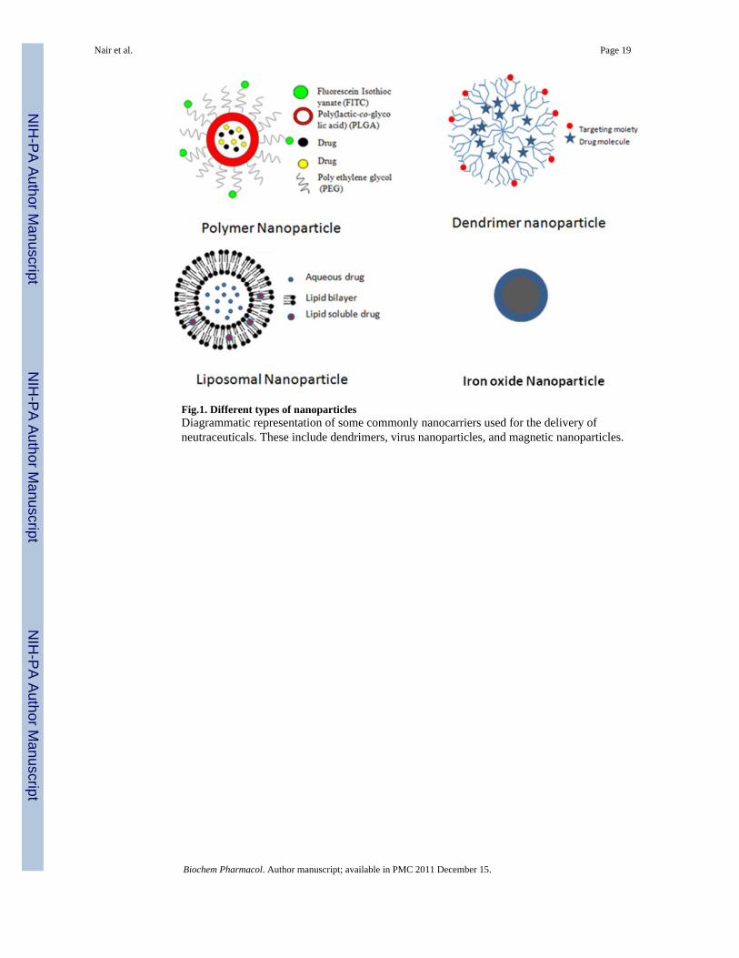
Fig.1. Different types of nanoparticlesDiagrammatic representation of some commonly nanocarriers used for the delivery ofneutraceuticals. These include dendrimers, virus nanoparticles, and magnetic nanoparticles.
Nair et al. Page 19
Biochem Pharmacol. Author manuscript; available in PMC 2011 December 15.
NIH
-PA Author Manuscript
NIH
-PA Author Manuscript
NIH
-PA Author Manuscript

Figure 2. Physical characterization of the nanoparticlesSome of the commonly used methods to characterize the nanoparticles are depicted in thefigure. A- Scanning electron microscopy (SEM) image of nanoparticles; B- Transmissionelectron microscopy (TEM) image; C- SEM image of nanoparticles after mechanicalextrusion; D – Determination of size distribution of nanoparticles by use of dynamic lightscattering (DLS).
Nair et al. Page 20
Biochem Pharmacol. Author manuscript; available in PMC 2011 December 15.
NIH
-PA Author Manuscript
NIH
-PA Author Manuscript
NIH
-PA Author Manuscript

NIH
-PA Author Manuscript
NIH
-PA Author Manuscript
NIH
-PA Author Manuscript
Nair et al. Page 21
Table 1
Common nutraceuticals that show anti-inflammatory properties
Nutraceutical Major Activities
Apigenin Antioxidative, vasoprotective, antiinflammatory, hypocholesterolemic
Naringin Vasoprotective, antiinflammatory, hypocholesterolemic
Ursolic acid Antioxidative, antimicrobial, antiinflammatory
Hesperidin Vasoprotective, antiinflammatory
Piperine Antiinflammatory, respiratory diseases, digestive, absorptive
Eucalyptol Gastroprotective, antiinflammatory, antioxidative, hepatoprotective
Curcumin Antioxidative, antiinflammatory, anticarcinogenic
Eugenol Antioxidative, antiinflammatory
Diosgenin Antioxidative, antiinflammatory, anticarcinogenic
Diallyl sulfide Antioxidative, antiinflammatory, anticarcinogenic
Gingerol Antioxidative, antiinflammatory, cardioprotective, antimicrobial
Thymoquinone Antiinflammatory, anticancer
Garcinol Antioxidative, antiinflammatory, anticarcinogenic
Capsaicin Antioxidative, antiinflammatory
Rosmarinic acid Antiinflammatory, respiratory diseases
Gossypin Antioxidant, antinociceptive, antiinflammatory, anticarcinogenic
Biochem Pharmacol. Author manuscript; available in PMC 2011 December 15.

NIH
-PA Author Manuscript
NIH
-PA Author Manuscript
NIH
-PA Author Manuscript
Nair et al. Page 22
Table 2
Commonly used biosynthetic polymers for drug delivery and their properties
Carrier Properties
Polyethylene Glycol (PEG) Increases bioavailability, biocompatibility, and circulation time
poly(lactic-co-glycolic acid (PLGA) Increases biocompatibility, decreases cytotoxicity
Ployvinyl alocohol (PVA) Reduces aggregation, increases hydrophilicity and strength
Dextran Decreases agglomeration, reduces 'stealth'nessness (increases opsonization)
Polypeptide Increases targeting
Polyacrylate Increases biostability, , biocompatibility
Chitosan Increases biocompatibility, hydrophilicity, and opsonization
Hyaluronic acid Enhances bioadhesion and targeting
Galactose Increases liver targeting
Polysaccharide modifications Improve cancer targeting
Sialic acid Decreases macrophage uptake
Poly-N-isopropylacrylamide (PNIPAm) For temperature-sensitive drug delivery
Poly(N-vinyl pyrrolidone) Increases opsonization and decreases bioavailability
Polyglycolides (PGA) Increase biocompatibility and thermostability
Polyanhydrides Increase biostability and biocompatibility
Polyorthoesters Increase biocompatibility
Biochem Pharmacol. Author manuscript; available in PMC 2011 December 15.

NIH
-PA Author Manuscript
NIH
-PA Author Manuscript
NIH
-PA Author Manuscript
Nair et al. Page 23
Table 3
List of passively targeted drug candidates currently approved by the Food and Drug Administration
Formulation Manufacturer Disease indication
Liposomal cytarabine Skyepharma Malignant lymphomatous meningitis
Liposomal morphine Skyepharma Postsurgical anesthesia
Liposomal daunorubicin Gilead Sciences HIV-related Kaposi's sarcoma
Liposomal amphotericin B Enzon Various fungal infections
Liposomal-peggylated doxorubicin Ortho Biotech/Schering-Plough Metastatic breast and ovarian cancer
Liposomal estradiol Novavax Multiple indications symptoms associated with menopause
Methoxy PEG-PLA paclitaxel Samyang Metastatic breast cancer
PEG-GCSF Amgen Chemotherapy-associated neutropenia
PEG-L-asparaginase Enzon Acute lymphoblatic leukemia
Nanoctrystalline aprepitant Elan, Merck Antiemetic
Biochem Pharmacol. Author manuscript; available in PMC 2011 December 15.
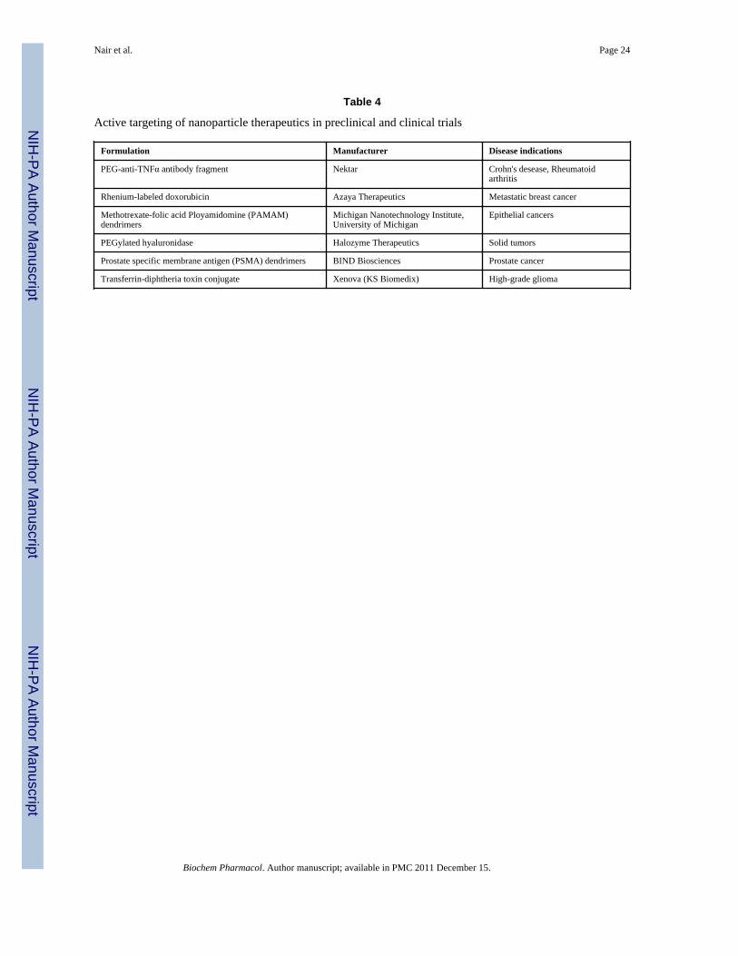
NIH
-PA Author Manuscript
NIH
-PA Author Manuscript
NIH
-PA Author Manuscript
Nair et al. Page 24
Table 4
Active targeting of nanoparticle therapeutics in preclinical and clinical trials
Formulation Manufacturer Disease indications
PEG-anti-TNFα antibody fragment Nektar Crohn's desease, Rheumatoidarthritis
Rhenium-labeled doxorubicin Azaya Therapeutics Metastatic breast cancer
Methotrexate-folic acid Ployamidomine (PAMAM)dendrimers
Michigan Nanotechnology Institute,University of Michigan
Epithelial cancers
PEGylated hyaluronidase Halozyme Therapeutics Solid tumors
Prostate specific membrane antigen (PSMA) dendrimers BIND Biosciences Prostate cancer
Transferrin-diphtheria toxin conjugate Xenova (KS Biomedix) High-grade glioma
Biochem Pharmacol. Author manuscript; available in PMC 2011 December 15.
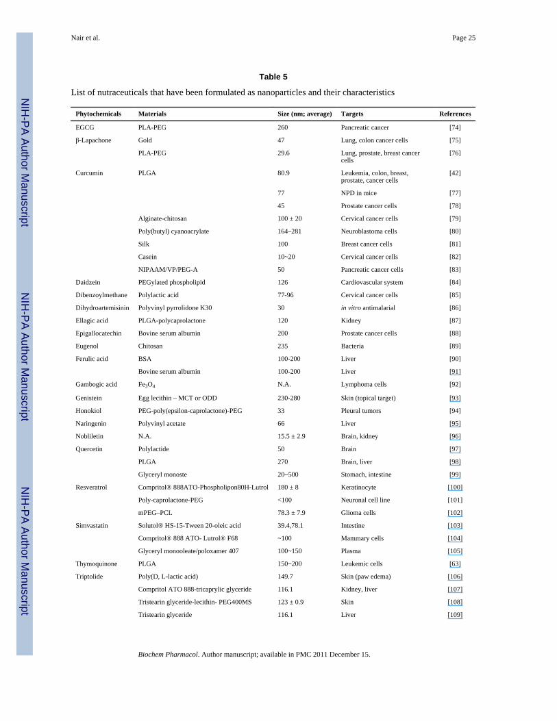
NIH
-PA Author Manuscript
NIH
-PA Author Manuscript
NIH
-PA Author Manuscript
Nair et al. Page 25
Table 5
List of nutraceuticals that have been formulated as nanoparticles and their characteristics
Phytochemicals Materials Size (nm; average) Targets References
EGCG PLA-PEG 260 Pancreatic cancer [74]
β-Lapachone Gold 47 Lung, colon cancer cells [75]
PLA-PEG 29.6 Lung, prostate, breast cancercells
[76]
Curcumin PLGA 80.9 Leukemia, colon, breast,prostate, cancer cells
[42]
77 NPD in mice [77]
45 Prostate cancer cells [78]
Alginate-chitosan 100 ± 20 Cervical cancer cells [79]
Poly(butyl) cyanoacrylate 164–281 Neuroblastoma cells [80]
Silk 100 Breast cancer cells [81]
Casein 10~20 Cervical cancer cells [82]
NIPAAM/VP/PEG-A 50 Pancreatic cancer cells [83]
Daidzein PEGylated phospholipid 126 Cardiovascular system [84]
Dibenzoylmethane Polylactic acid 77-96 Cervical cancer cells [85]
Dihydroartemisinin Polyvinyl pyrrolidone K30 30 in vitro antimalarial [86]
Ellagic acid PLGA-polycaprolactone 120 Kidney [87]
Epigallocatechin Bovine serum albumin 200 Prostate cancer cells [88]
Eugenol Chitosan 235 Bacteria [89]
Ferulic acid BSA 100-200 Liver [90]
Bovine serum albumin 100-200 Liver [91]
Gambogic acid Fe3O4 N.A. Lymphoma cells [92]
Genistein Egg lecithin – MCT or ODD 230-280 Skin (topical target) [93]
Honokiol PEG-poly(epsilon-caprolactone)-PEG 33 Pleural tumors [94]
Naringenin Polyvinyl acetate 66 Liver [95]
Nobliletin N.A. 15.5 ± 2.9 Brain, kidney [96]
Quercetin Polylactide 50 Brain [97]
PLGA 270 Brain, liver [98]
Glyceryl monoste 20~500 Stomach, intestine [99]
Resveratrol Compritol® 888ATO-Phospholipon80H-Lutrol 180 ± 8 Keratinocyte [100]
Poly-caprolactone-PEG <100 Neuronal cell line [101]
mPEG–PCL 78.3 ± 7.9 Glioma cells [102]
Simvastatin Solutol® HS-15-Tween 20-oleic acid 39.4,78.1 Intestine [103]
Compritol® 888 ATO- Lutrol® F68 ~100 Mammary cells [104]
Glyceryl monooleate/poloxamer 407 100~150 Plasma [105]
Thymoquinone PLGA 150~200 Leukemic cells [63]
Triptolide Poly(D, L-lactic acid) 149.7 Skin (paw edema) [106]
Compritol ATO 888-tricaprylic glyceride 116.1 Kidney, liver [107]
Tristearin glyceride-lecithin- PEG400MS 123 ± 0.9 Skin [108]
Tristearin glyceride 116.1 Liver [109]
Biochem Pharmacol. Author manuscript; available in PMC 2011 December 15.
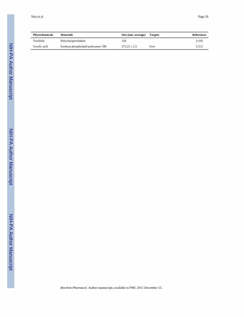
NIH
-PA Author Manuscript
NIH
-PA Author Manuscript
NIH
-PA Author Manuscript
Nair et al. Page 26
Phytochemicals Materials Size (nm; average) Targets References
Toxifolin Polyvinylpirrolidone 150 [110]
Ursolic acid Soybean phospholipid-poloxamer 188 273.23 ± 2.3 liver [111]
Biochem Pharmacol. Author manuscript; available in PMC 2011 December 15.