Shotgun proteomics implicates protease inhibition and complement activation in the antiinflammatory...
11
Research article 746 The Journal of Clinical Investigation http://www.jci.org Volume 117 Number 3 March 2007 Shotgun proteomics implicates protease inhibition and complement activation in the antiinflammatory properties of HDL Tomas Vaisar, 1 Subramaniam Pennathur, 1 Pattie S. Green, 1 Sina A. Gharib, 1 Andrew N. Hoofnagle, 2 Marian C. Cheung, 1 Jaeman Byun, 1 Simona Vuletic, 1 Sean Kassim, 1 Pragya Singh, 1 Helen Chea, 1 Robert H. Knopp, 1 John Brunzell, 1 Randolph Geary, 3 Alan Chait, 1 Xue-Qiao Zhao, 1 Keith Elkon, 1 Santica Marcovina, 1 Paul Ridker, 4 John F. Oram, 1 and Jay W. Heinecke 1 1 Department of Medicine and 2 Department of Laboratory Medicine, University of Washington School of Medicine, Seattle, Washington, USA. 3 Division of Surgical Sciences, Wake Forest University School of Medicine, Winston-Salem, North Carolina, USA. 4 Department of Medicine, Harvard Medical School, Boston, Massachusetts, USA. HDL lowers the risk for atherosclerotic cardiovascular disease by promoting cholesterol efflux from macro- phage foam cells. However, other antiatherosclerotic properties of HDL are poorly understood. To test the hypothesis that the lipoprotein carries proteins that might have novel cardioprotective activities, we used shot- gun proteomics to investigate the composition of HDL isolated from healthy subjects and subjects with coro- nary artery disease (CAD). Unexpectedly, our analytical strategy identified multiple complement-regulatory proteins and a diverse array of distinct serpins with serine-type endopeptidase inhibitor activity. Many acute- phase response proteins were also detected, supporting the proposal that HDL is of central importance in inflammation. Mass spectrometry and biochemical analyses demonstrated that HDL 3 from subjects with CAD was selectively enriched in apoE, raising the possibility that HDL carries a unique cargo of proteins in humans with clinically significant cardiovascular disease. Collectively, our observations suggest that HDL plays previ- ously unsuspected roles in regulating the complement system and protecting tissue from proteolysis and that the protein cargo of HDL contributes to its antiinflammatory and antiatherogenic properties. Introduction Mortality from cardiovascular disease has decreased significantly in the United States over the past 30 years (1, 2). Many factors have contributed to this improvement, including the identification of cardiovascular risk factors, development of interventions that reduce those risk factors, and new treatments for acute coronary syndromes. Despite these advances, however, cardiovascular disease remains the leading cause of mortality in the United States (1, 2). One important risk factor for atherosclerosis is a low level of HDL, which directly protects against atherosclerosis by removing cholesterol from artery wall macrophages (3, 4). Thus, the risk of cardiovascular disease is inversely proportional to plasma levels of HDL and its major apolipoprotein, apoA-I (5). Also, low HDL levels are particularly common in men with premature atheroscle- rosis (6), whereas high HDL levels associate with longevity (7). Antiinflammatory and antioxidant properties might also con- tribute to the cardioprotective effects of HDL (8). Moreover, both oxidative modifications and alterations in the protein cargo of HDL may alter its biological activity, creating potentially pro- atherogenic particles (8, 9). We and others recently showed that HDL isolated from plasma of patients with known coronary artery disease (CAD) is oxidatively modified and that oxidation impairs reverse cholesterol transport mediated by HDL (10–12). Circulating levels of serum amyloid A (SAA), an inflammatory pro- tein in HDL, predict the risk of heart disease in humans (13), and HDL-associated SAA has been proposed to render the lipoprotein atherogenic (14). Furthermore, alterations in the balance between pro- and antioxidative enzymes in HDL appear to play a critical role in converting the lipoprotein to a proatherogenic form (8). We hypothesized that quantifying the protein composition of HDL might provide insights into the antiatherogenic and antiin- flammatory properties of the lipoprotein. We therefore used shot- gun proteomics — direct analysis of a complex mixture of proteins (15) — to study the HDL proteome. After digesting the lipoprotein with trypsin, we analyzed the resulting peptide mixture with mass spectrometry (MS) and matched the tandem mass spectra of the peptides with spectra derived from protein sequences in protein database. We found that HDL carries protein families implicated in complement activation and regulation of proteolysis, raising the possibility that these proteins contribute to the innate immune system and the cardioprotective properties of HDL. Results Shotgun proteomics identifies distinct protein families in HDL. We first confirmed that liquid chromatography–electrospray ionization– tandem MS (LC-ESI-MS/MS) analysis reproducibly identifies the same HDL-associated proteins (Supplemental Figure 1; supple- mental material available online with this article; doi:10.1172/ JCI26206DS1). We then used this analytical strategy to investigate the protein composition of HDL isolated by ultracentrifugation in 2 independent groups of subjects (see Methods). The first used total HDL isolated from 20 apparently healthy control subjects (group 1), and the second used HDL 3 isolated from 13 subjects Nonstandard abbreviations used: CAD, coronary artery disease; CETP, cholesteryl ester transfer protein; GO, Gene Ontology; LC-ESI-MS/MS, liquid chromatogra- phy–electrospray ionization–tandem MS; MS, mass spectrometry; PON1, paraox- onase-1; PRINCE study, Pravastatin Inflammation/CRP Evaluation study; SAA, serum amyloid A. Conflict of interest: The authors have declared that no conflict of interest exists. Citation for this article: J. Clin. Invest. 117:746–756 (2007). doi:10.1172/JCI26206. Related Commentary, page 595
-
Upload
independent -
Category
Documents
-
view
0 -
download
0
Transcript of Shotgun proteomics implicates protease inhibition and complement activation in the antiinflammatory...
Research article
746 TheJournalofClinicalInvestigation http://www.jci.org Volume 117 Number 3 March 2007
Shotgun proteomics implicates protease inhibition and complement activation in the
antiinflammatory properties of HDLTomas Vaisar,1 Subramaniam Pennathur,1 Pattie S. Green,1 Sina A. Gharib,1 Andrew N. Hoofnagle,2
Marian C. Cheung,1 Jaeman Byun,1 Simona Vuletic,1 Sean Kassim,1 Pragya Singh,1 Helen Chea,1 Robert H. Knopp,1 John Brunzell,1 Randolph Geary,3 Alan Chait,1 Xue-Qiao Zhao,1 Keith Elkon,1
Santica Marcovina,1 Paul Ridker,4 John F. Oram,1 and Jay W. Heinecke1
1Department of Medicine and 2Department of Laboratory Medicine, University of Washington School of Medicine, Seattle, Washington, USA. 3Division of Surgical Sciences, Wake Forest University School of Medicine, Winston-Salem, North Carolina, USA. 4Department of Medicine,
Harvard Medical School, Boston, Massachusetts, USA.
HDLlowerstheriskforatheroscleroticcardiovasculardiseasebypromotingcholesteroleffluxfrommacro-phagefoamcells.However,otherantiatheroscleroticpropertiesofHDLarepoorlyunderstood.Totestthehypothesisthatthelipoproteincarriesproteinsthatmighthavenovelcardioprotectiveactivities,weusedshot-gunproteomicstoinvestigatethecompositionofHDLisolatedfromhealthysubjectsandsubjectswithcoro-naryarterydisease(CAD).Unexpectedly,ouranalyticalstrategyidentifiedmultiplecomplement-regulatoryproteinsandadiversearrayofdistinctserpinswithserine-typeendopeptidaseinhibitoractivity.Manyacute-phaseresponseproteinswerealsodetected,supportingtheproposalthatHDLisofcentralimportanceininflammation.MassspectrometryandbiochemicalanalysesdemonstratedthatHDL3fromsubjectswithCADwasselectivelyenrichedinapoE,raisingthepossibilitythatHDLcarriesauniquecargoofproteinsinhumanswithclinicallysignificantcardiovasculardisease.Collectively,ourobservationssuggestthatHDLplaysprevi-ouslyunsuspectedrolesinregulatingthecomplementsystemandprotectingtissuefromproteolysisandthattheproteincargoofHDLcontributestoitsantiinflammatoryandantiatherogenicproperties.
IntroductionMortality from cardiovascular disease has decreased significantly in the United States over the past 30 years (1, 2). Many factors have contributed to this improvement, including the identification of cardiovascular risk factors, development of interventions that reduce those risk factors, and new treatments for acute coronary syndromes. Despite these advances, however, cardiovascular disease remains the leading cause of mortality in the United States (1, 2).
One important risk factor for atherosclerosis is a low level of HDL, which directly protects against atherosclerosis by removing cholesterol from artery wall macrophages (3, 4). Thus, the risk of cardiovascular disease is inversely proportional to plasma levels of HDL and its major apolipoprotein, apoA-I (5). Also, low HDL levels are particularly common in men with premature atheroscle-rosis (6), whereas high HDL levels associate with longevity (7).
Antiinflammatory and antioxidant properties might also con-tribute to the cardioprotective effects of HDL (8). Moreover, both oxidative modifications and alterations in the protein cargo of HDL may alter its biological activity, creating potentially pro-atherogenic particles (8, 9). We and others recently showed that HDL isolated from plasma of patients with known coronary artery disease (CAD) is oxidatively modified and that oxidation impairs reverse cholesterol transport mediated by HDL (10–12).
Circulating levels of serum amyloid A (SAA), an inflammatory pro-tein in HDL, predict the risk of heart disease in humans (13), and HDL-associated SAA has been proposed to render the lipoprotein atherogenic (14). Furthermore, alterations in the balance between pro- and antioxidative enzymes in HDL appear to play a critical role in converting the lipoprotein to a proatherogenic form (8).
We hypothesized that quantifying the protein composition of HDL might provide insights into the antiatherogenic and antiin-flammatory properties of the lipoprotein. We therefore used shot-gun proteomics — direct analysis of a complex mixture of proteins (15) — to study the HDL proteome. After digesting the lipoprotein with trypsin, we analyzed the resulting peptide mixture with mass spectrometry (MS) and matched the tandem mass spectra of the peptides with spectra derived from protein sequences in protein database. We found that HDL carries protein families implicated in complement activation and regulation of proteolysis, raising the possibility that these proteins contribute to the innate immune system and the cardioprotective properties of HDL.
ResultsShotgun proteomics identifies distinct protein families in HDL. We first confirmed that liquid chromatography–electrospray ionization–tandem MS (LC-ESI-MS/MS) analysis reproducibly identifies the same HDL-associated proteins (Supplemental Figure 1; supple-mental material available online with this article; doi:10.1172/JCI26206DS1). We then used this analytical strategy to investigate the protein composition of HDL isolated by ultracentrifugation in 2 independent groups of subjects (see Methods). The first used total HDL isolated from 20 apparently healthy control subjects (group 1), and the second used HDL3 isolated from 13 subjects
Nonstandardabbreviationsused: CAD, coronary artery disease; CETP, cholesteryl ester transfer protein; GO, Gene Ontology; LC-ESI-MS/MS, liquid chromatogra-phy–electrospray ionization–tandem MS; MS, mass spectrometry; PON1, paraox-onase-1; PRINCE study, Pravastatin Inflammation/CRP Evaluation study; SAA, serum amyloid A.
Conflictofinterest: The authors have declared that no conflict of interest exists.
Citationforthisarticle: J. Clin. Invest. 117:746–756 (2007). doi:10.1172/JCI26206.
Related Commentary, page 595
research article
TheJournalofClinicalInvestigation http://www.jci.org Volume 117 Number 3 March 2007 747
(group 2; 6 control subjects and 7 subjects with established CAD; additional details below). The clinical characteristics of the sub-jects are shown in Table 1. The following criteria were used to iden-tify proteins: (a) a high peptide identification score (according to PeptideProphet; P > 0.90); (b) a high protein identification score (P > 0.95, ProteinProphet); and (c) at least 2 peptides unique to the protein of interest had to be detected in at least 5 subjects (total HDL) or 2 subjects (HDL3). Requiring at least 2 unique peptides with a high confidence score markedly decreases the false-positive rate of protein identification (16). Shotgun proteomics with these stringent criteria identified 48 proteins in HDL (Table 2).
This analytical approach detected every apolipoprotein already known to associate with HDL, except for apoA-V. Immunoblot analysis confirmed that apoA-I, apoA-II, apoC-II, apoE, cluster-in (apoJ), and apoM were present in the lipoprotein. Indeed, we detected more than 90% of all proteins that have previously been identified by biochemical methods in HDL, including lecithin:cholesterol acyl transferase (LCAT), phospholipid transfer protein (PLTP), cholesteryl ester transfer protein (CETP), transferrin, all 3 human serum amyloid proteins (SAA1, SAA2, SAA4), and both paraoxonase-1 (PON1) and PON3 (17–28), strongly suggesting that our overall experimental approach was valid.
We found a number of protein families not previously known to reside in HDL, including complement pathway and regulatory proteins (complement component 4 [C4A and C4B], complement component 9 [C9], vitronectin) and endopeptidase inhibitors
(α-2-antiplasmin, α-2-HS-glycoprotein, inter-α-trypsin inhibitor [α-1-microglobulin/bikunin and inter-α-trypsin inhibitor heavy chain H4], angiotensinogen, α-1-antitrypsin, serpin peptidase inhibitor [clade F, member 1], kininogen-1). We also detected plasma retinol–binding protein, which was recently implicated in the pathogenesis of type 2 diabetes (29). Interestingly, we identi-fied the lysosomal protein prenylcysteine oxidase and hemopexin, an iron-binding protein that inhibits LDL oxidation.
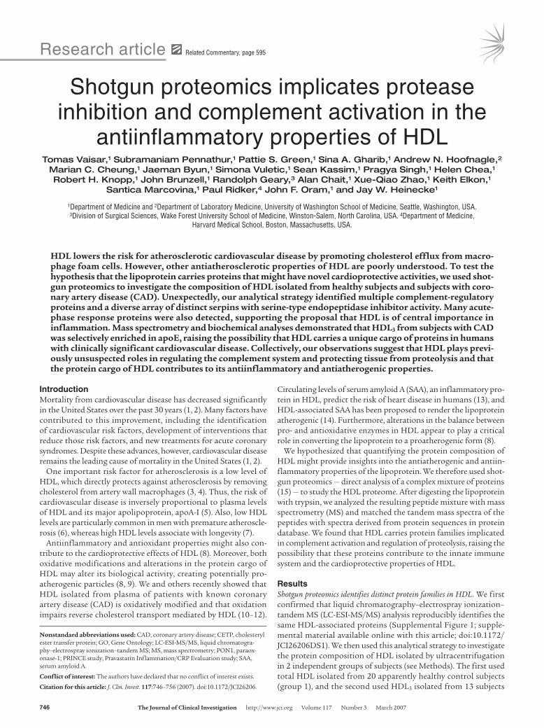
Gene Ontology (GO) analysis (Figure 1) demonstrated that 22 of the 48 HDL-associated proteins were linked to cholesterol and lipoprotein metabolism (P = 2 × 10–27). Twenty-three of the 48 were acute-phase-response proteins, whose plasma concentrations are altered markedly by acute infection and chronic inflammation (P = 1 × 10–18). Six proteins were regulators of complement acti-vation (P = 5 × 10⁻⁵). The analysis also revealed 2 major clusters of molecular function: 8 proteins were endopeptidase inhibitors (P = 2 × 10⁻⁶), and 8 were heparin-binding proteins (SAA1, SAA2, SAA4, apoE, apoH, haptoglobin-related protein, kininogen-1, vit-ronectin; P = 6 × 10⁻⁵). Collectively, these observations implicate HDL in the acute-phase response and innate immunity. They also strongly support the proposal that HDL carries proteins that may play previously unsuspected roles in regulating complement acti-vation and inhibiting proteolysis.
apoA-I particles and HDL carry members of overlapping families of proteins. To further investigate the protein composition of HDL, we used affinity chromatography to isolate lipoprotein particles
Table 1Clinical characteristics of study subjects
n HDLisolated Status Age %Male Cholesterol Triglycerides HDL-C LDL-C (yr) (mg/dl) (mg/dl) (mg/dl) (mg/dl)Group 1 20 Total Control 57 (6) 100 197 (13) 104 (29) 42 (8) 134 (14)Group 2 6 HDL3 Control 54 (14) 100 183 (29) 117 (61) 45 (12) 121 (22) 7 HDL3 CAD 54 (7) 100 231A (31) 189 (101) 40 (11) 161A (19)Group 3 32 HDL3 Control 68 (10) 19 234 (24) 106 (27) 50 (11) 148 (21) 32 HDL3 CAD 67 (10) 19 208A (36) 99 (32) 51 (11) 124A (33)
Results represent mean (SD). AP < 0.05 between subjects within a group by Student’s t test. HDL-C, HDL-cholesterol.
Figure 1Global view of biological processes and molecular functions of HDL proteins. Proteins in total HDL and HDL3 were identified using LC-ESI-MS/MS (Table 2). Proteins detected in HDL were associated with bio-logical functions using GO process annotations. This approach demonstrated significant overrepresentation of proteins involved in several categories, including lipid metabolism (P = 2 × 10–27), the acute-phase response (P = 1 × 10–18), protease inhibitor activity (P = 2 × 10⁻⁶), and complement regulation (P = 5 × 10⁻⁵). apoH, β-2-glycoprotein I; AGT, angiotensinogen; AHSG, α-2-HS-glycoprotein; AMP, bikunin; FGA, fibrinogen; HRP, haptoglobin-related protein; HPX, hemopexin; ITIH4, inter-α-trypsin inhibitor heavy chain H4; KNG1, kininogen-1; LCAT, lecithin-cholesterol acyltransferase; ORM2, α-1-acid glycoprotein 2; PLTP, phospholipid transfer protein; RBP4, retinol binding protein; SERA1, α-1-antitrypsin; SERF1, serpin peptidase inhibitor (clade F, member 1); SERF2, α-2-antiplasmin; TF, transferrin; TTR, transthyretin; VTN, vitronectin.
research article
748 TheJournalofClinicalInvestigation http://www.jci.org Volume 117 Number 3 March 2007
containing apoA-I, the major HDL protein. The particles were then analyzed by LC-ESI-MS/MS. We used this approach because it is well established that proteins can dissociate from lipoproteins during ultracentrifugation (30, 31), eliminating some proteins that HDL would carry under physiological conditions. Additionally, isolation by our orthogonal techniques and detection of the same pro-teins in both apoA-I particles and HDL would strongly support the proposal that those pro-teins actually associate with HDL and are not just abundant plasma components.
apoA-I particles prepared from plasma of healthy subjects was digested with trypsin and analyzed by LC-ESI-MS/MS. Control studies used columns containing beads cross-linked to albumin or nonspecific immunoglobulin. This approach identified 11 of the 13 proteins in the apoA-I particles that were not previ-ously known to reside in HDL and/or HDL3. Importantly, this included all of the HDL-associated complement-related proteins, the thiol protease inhibitor kininogen-1, and 5 of the 6 serine-type endopeptidase inhibitors. In contrast, we failed to detect plasma retinol–binding protein or serpin peptidase inhibitor (clade F, member 1), suggesting that apoA-I particles may not contain these proteins. We also found peptides derived from the α, β, and γ chains of fibrinogen and complement factor H–related protein, which were previously iso-lated from a minor HDL fraction (32).
apoB-100 (the major LDL protein) and albumin were detected in HDL and HDL3. These proteins are abundant in plasma and therefore might have been contaminants. The dense fraction of LDL can also be coisolated with HDL by ultracentrifugation. However, we detected both of these proteins in apoA-I isolated by affinity chromatography. Only low levels of apoB-100 and albumin were detected in material eluted from control affinity col-umns (matrix alone or cross-linked to non-specific immunoglobulin), which argues against nonspecific binding. It is noteworthy that albumin binds free fatty acids and other hydrophobic compounds with high affinity and that other investigators have proposed that HDL may interact with albumin (20). Moreover, apoA-I has recently been found associated with albumin isolated biochemi-cally or serum albumin by affinity chroma-tography, suggesting that the apolipoprotein can associate with albumin (33).
Complement component 3 complexes with HDL. GO analysis revealed a highly significant asso-ciation of HDL with complement factors (C3, C4, C9) as well as the complement-regulatory proteins vitronectin and clusterin. A major
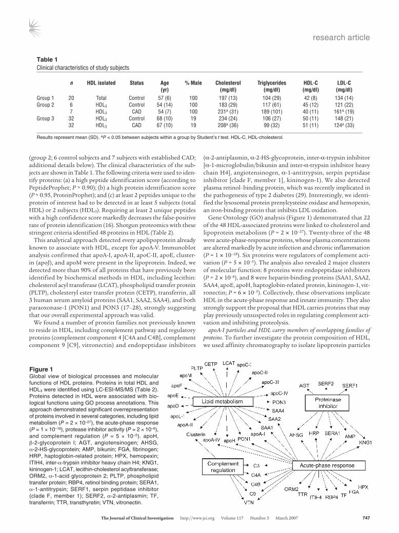
Table 2Proteins detected by LC-ESI-MS/MS in HDL (group 1) and HDL3 (group 2) isolated from control and/or CAD subjects
UniquepeptidesA
ProteinID ProteinnameB HDL HDL3
PreviouslyidentifiedHDL-associatedproteinsB
IPI00021841 apoA-I 1,013 847IPI00021854 apoA-II 342 229IPI00304273 apoA-IV 139 131IPI00021842 apoE 180 158IPI00021855 apoC-I 74 108IPI00021856 apoC-II 101 90IPI00021857 apoC-III 105 110IPI00022731 apoC-IV – 15IPI00177869 apoL-I 63 44IPI00030739 apoM 74 94IPI00299435 apoF 40 36IPI00006662 apoD 70 159IPI00298828 apoH 82 5IPI00291262 Clusterin (apoJ) 48 12IPI00022331 Lecithin-cholesterol acyltransferase 61 38IPI00006173 CETP – 4IPI00022733 Phospholipid transfer protein 28 5IPI00022368 SAA – 36IPI00452748 SAA1 60 7IPI00006146 SAA2 32 23IPI00019399 SAA4 124 130IPI00218732 PON 1 119 35IPI00299778 PON3 31 –IPI00164623 Complement component C3 18 14IPI00020091 α-1-acid glycoprotein 2 – 4IPI00022431 α-2-HS-glycoprotein 59 22IPI00305457 α-1-antitrypsin 191 180IPI00022895 α-1B-glycoprotein 17 17IPI00021885 Fibrinogen (α chain) 32 –IPI00022463 Serotransferrin 10 –IPI00296170 Haptoglobin-related protein 39 18IPI00022432 Transthyretin 71 –IPI00298853 Vitamin D–binding protein 65 –IPI00022229 apoB-100 58 9IPI00022434 Serum albumin 689 457ProteinsidentifiedinthisstudyasHDLassociatedIPI00418163 Complement component C4B – 5IPI00032258 Complement component C4A 40 13IPI00022395 Complement component C9 30 –IPI00298971 Vitronectin 42 18IPI00029863 α-2-antiplasmin – 4IPI00022426 α-1-microglobulin/bikunin 21 –IPI00218192 Inter-α-trypsin inhibitor heavy chain H4 31 –IPI00032220 Angiotensinogen – 6IPI00006114 Serpin peptidase inhibitor (clade F, member 1) 15 –IPI00032328 Kininogen-1 15 –IPI00022420 Plasma retinol–binding protein 19 –IPI00337558 Prenylcysteine oxidase 34 –IPI00022488 Hemopexin 15 –
HDL (d = 1.063–1.210 g/ml; group 1, 20 control subjects) and HDL3 (d = 1.10–1.21 g/ml; group 2, 6 control and 7 CAD subjects) were isolated from EDTA plasma by ultracentrifugation. Tryptic digests were subjected to reverse-phase separation followed by nano-ESI-MS/MS on a Finnigan LTQ ion trap (HDL) or 2-dimensional LC-ESI-MS/MS on a Finnigan LCQ ion trap instrument (HDL3). MS/MS spectra were searched against the Human International Protein Index (IPI) database, using the SEQUEST search engine. The SEQUEST results were further processed using PeptideProphet and ProteinProphet (59, 60), using an adjusted probability of greater than 0.90 and greater than 0.95 for peptides and pro-teins, respectively. For HDL, all protein identifications required detection of at least 2 unique peptides for each protein from at least 5 different individuals. For HDL3, all protein identifications required detection of at least 2 unique peptides for each protein from at least 2 different individuals. AUnique peptides identified from all samples in the analysis. BHDL-associated proteins (18–28, 42, 66). –, not detected.

research article
TheJournalofClinicalInvestigation http://www.jci.org Volume 117 Number 3 March 2007 749
effector of the complement system, C3, has been implicated in ath-erogenesis (34). All 3 unique peptides identified by MS/MS in HDL3 are between residues 954 and 1,303 of C3dg (Figure 2, A and B). This region contains a thioester bond that can cross-link C3dg to target proteins (Figure 2C), raising the possibility that C3dg forms a covalent complex with HDL proteins.
To biochemically confirm that C3 is a component of HDL, we isolated HDL3 from pooled plasma and immunoblotted it with a polyclonal antibody against C3. Three independent HDL3 prepa-rations exhibited major bands of immunoreactive material with apparent MWs of approximately 40 kDa, 45 kDa, and 65 kDa (Fig-ure 2D, lanes 5–7). We also detected immunoreactive C3 in apoA-I complexes isolated by affinity chromatography (Figure 2D, lane 4) but not in the material eluted from control columns (Figure 2D, lanes 2 and 3). The patterns of immunoreactive C3 in HDL3 and immunoisolated apoA-I differed, suggesting that the differ-ent preparations of HDL contained distinct species of C3. These observations strongly suggest that proteolytic fragments derived from C3 associate with HDL.
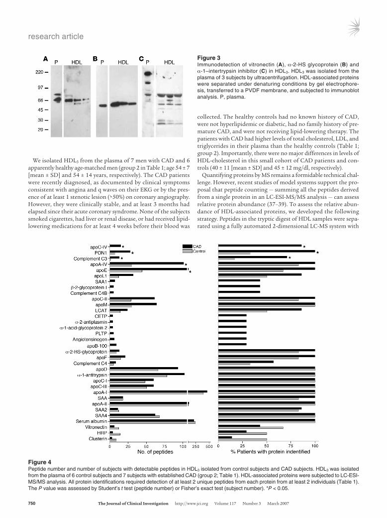
Endopeptidase inhibitor and complement-regulatory proteins associate with HDL. Immunoblot analysis of proteins separated by SDS-PAGE confirmed biochemically that vitronectin and 2 endopepti-dase inhibitors, inter-α-trypsin inhibitor and α-2-HS glycoprotein (Figure 3), were associated with HDL3.
Recent studies have demonstrated that HDL carries a wide range of low-MW peptides (35). However, the apparent MWs of vitronectin
and α-2-HS glycoprotein in plasma and HDL3 were indistinguish-able, strongly suggesting a lack of proteolytic processing of the pro-tein forms identified in HDL. The major form of immunoreactive inter-α-trypsin inhibitor detected in plasma was consistent with its reported MW of 240 kDa, which is thought to represent a complex of 1 light and 2 heavy chains (36). The material in HDL3 that immu-noreacted with the polyclonal antibody to inter-α-trypsin inhibitor differed from that in plasma. Their apparent MWs were consistent with those anticipated for the uncomplexed heavy and light chains. The light chain of this protein, also termed bikunin, contains 2 Kunitz-type protease inhibitor domains, which can complex with proteins. It is therefore of interest that an immunoreactive protein with an apparent MW somewhat greater than that of the light chain of inter-α-trypsin inhibitor was detected in HDL.
Collectively, these observations indicate that HDL3 contains immunoreactive vitronectin, α-2-HS glycoprotein, and the heavy and light chains of inter-α-trypsin inhibitor. They also raise the possibility that 2 of the proteins we detected, inter-α-trypsin inhib-itor and complement factor C3, may form complexes with other HDL-associated proteins.
HDL isolated from healthy subjects and subjects with established CAD carry different protein cargoes. To begin to investigate the protein composition of HDL from CAD subjects, we focused on HDL3, the lipoprotein’s dense subfraction. We selected HDL3 to minimize potential contamination with LDL because our analyses detected the LDL protein apoB100 in total HDL of control subjects.
Figure 2Mass spectrometric and immunological detection of C3 in HDL3. HDL3 or apoA-I complexes were isolated from plasma of control subjects. In parallel studies, plasma was passed over columns containing albumin or nonspecific immunoglobulin covalently cross-linked to beads. Isolated proteins were separated under denaturing conditions by gel electrophoresis, transferred to nitrocellulose, and analyzed through immunoblotting. (A and B) MS/MS spectra of 2 peptides unique to C3 in HDL3. (C) Sequence of the C3dg domain of human complement C3. The 3 tryptic digest peptides detected selectively in HDL3 of CAD subjects are underlined. (D) Detection of proteins reactive with antibody to C3. Lane 1: plasma. Lanes 2 and 3: eluate from control columns (albumin and IgG, respectively). Lane 4: apoA-I complexes. Lanes 5–7: 3 independent preparations of HDL3 isolated from pooled human plasma.
research article
750 TheJournalofClinicalInvestigation http://www.jci.org Volume 117 Number 3 March 2007
We isolated HDL3 from the plasma of 7 men with CAD and 6 apparently healthy age-matched men (group 2 in Table 1; age 54 ± 7 [mean ± SD] and 54 ± 14 years, respectively). The CAD patients were recently diagnosed, as documented by clinical symptoms consistent with angina and q waves on their EKG or by the pres-ence of at least 1 stenotic lesion (>50%) on coronary angiography. However, they were clinically stable, and at least 3 months had elapsed since their acute coronary syndrome. None of the subjects smoked cigarettes, had liver or renal disease, or had received lipid-lowering medications for at least 4 weeks before their blood was
collected. The healthy controls had no known history of CAD, were not hyperlipidemic or diabetic, had no family history of pre-mature CAD, and were not receiving lipid-lowering therapy. The patients with CAD had higher levels of total cholesterol, LDL, and triglycerides in their plasma than the healthy controls (Table 1; group 2). Importantly, there were no major differences in levels of HDL-cholesterol in this small cohort of CAD patients and con-trols (40 ± 11 [mean ± SD] and 45 ± 12 mg/dl, respectively).
Quantifying proteins by MS remains a formidable technical chal-lenge. However, recent studies of model systems support the pro-posal that peptide counting — summing all the peptides derived from a single protein in an LC-ESI-MS/MS analysis — can assess relative protein abundance (37–39). To assess the relative abun-dance of HDL-associated proteins, we developed the following strategy. Peptides in the tryptic digest of HDL samples were sepa-rated using a fully automated 2-dimensional LC-MS system with
Figure 3Immunodetection of vitronectin (A), α-2-HS glycoprotein (B) and α-1–intertrypsin inhibitor (C) in HDL3. HDL3 was isolated from the plasma of 3 subjects by ultracentrifugation. HDL-associated proteins were separated under denaturing conditions by gel electrophore-sis, transferred to a PVDF membrane, and subjected to immunoblot analysis. P, plasma.
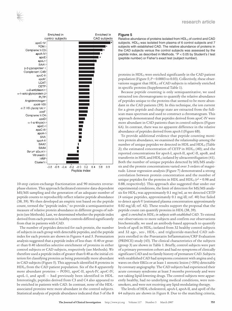
Figure 4Peptide number and number of subjects with detectable peptides in HDL3 isolated from control subjects and CAD subjects. HDL3 was isolated from the plasma of 6 control subjects and 7 subjects with established CAD (group 2; Table 1). HDL-associated proteins were subjected to LC-ESI-MS/MS analysis. All protein identifications required detection of at least 2 unique peptides from each protein from at least 2 individuals (Table 1). The P value was assessed by Student’s t test (peptide number) or Fisher’s exact test (subject number). *P < 0.05.
research article
TheJournalofClinicalInvestigation http://www.jci.org Volume 117 Number 3 March 2007 751
10-step cation-exchange fractionation and 90 minutes reverse-phase elution. This approach facilitated extensive data-dependent MS/MS sampling and the generation of an adequate number of peptide counts to reproducibly reflect relative peptide abundance (38, 39). We then developed an empiric test based on the peptide count, termed the “peptide index,” to provide a semiquantitative measure of relative protein abundance in different groups of sub-jects (see Methods). Last, we determined whether the peptide index derived from each protein in healthy controls differed significantly from that in patients with CAD.
The number of peptides detected for each protein, the number of subjects in each group with detectable peptides, and the peptide index for each protein are shown in Figures 4 and 5. Permutation analysis suggested that a peptide index of less than –0.40 or great-er than 0.40 identifies selective enrichment of proteins in either control subjects or CAD subjects, respectively (see Methods). We therefore used a peptide index of greater than 0.40 as the initial cri-terion for classifying proteins as being potentially more abundant in CAD subjects (Figure 5). This approach identified 8 proteins in HDL3 from the CAD patient population. Six of the 8 apparently more abundant proteins — PON1, apoC-II, apoA-IV, apoC-IV, apoL-1, and apoE — had previously been identified in HDL. Interestingly, peptides derived from C3 and C4 also appeared to be enriched in patients with CAD. In contrast, none of the HDL-associated proteins were more abundant in the control subjects. Statistical analysis of peptide abundance indicated that 5 of the 8
proteins in HDL3 were enriched significantly in the CAD patient population (Figure 5; P = 0.0003 to 0.03). Collectively, these obser-vations suggest that HDL3 of CAD subjects is relatively enriched in specific proteins (Supplemental Table 1).
Because peptide counting is only semiquantitative, we used extracted ion chromatograms to quantify the relative abundance of peptides unique to the proteins that seemed to be more abun-dant in the CAD patients (39). In this technique, the ion current for a given peptide and charge state are extracted from the full-scan mass spectrum and used to construct a chromatogram. This approach demonstrated that peptides derived from apoC-IV were more abundant in CAD patients than in control subjects (Figure 6A). In contrast, there was no apparent difference in the relative abundance of peptides derived from apoA-I (Figure 6B).
To provide additional evidence that peptide counting moni-tors protein abundance, we examined the relationship among the number of unique peptides we detected in HDL and HDL3 (Table 2); the estimated concentration of CETP in HDL3 (40); and the reported concentrations for apoA-I, apoA-II, apoC-II, apoB, and transferrin in HDL and HDL3 isolated by ultracentrifugation (41). Both the number of unique peptides detected by MS/MS analy-sis and the protein concentrations varied over 3 orders of magni-tude. Linear regression analysis (Figure 7) demonstrated a strong correlation between protein concentration and the number of unique peptides for the proteins in HDL and HDL3 (r2 = 0.98 and 0.88, respectively). This approach also suggested that under our experimental conditions, the limit of detection for MS/MS analy-sis for HDL3 was approximately 0.1 mg/ml, as we detected CETP (4 unique peptides, approximately 0.1 mg/dl; ref. 40) but failed to detect apoA-V (estimated plasma concentration approximately 0.02 mg/dl; ref. 42). These results support the proposal that the peptide count can quantify proteins in HDL (38, 39).
apoE is enriched in HDL3 in subjects with established CAD. To extend our observations to more subjects and confirm our observations biochemically, we used an antibody-based approach to quantify levels of apoE in HDL3 isolated from 32 healthy control subjects and 32 age-, sex-, HDL-, and triglyceride-matched CAD sub-jects enrolled in the Pravastatin Inflammation/CRP Evaluation (PRINCE) study (43). The clinical characteristics of the subjects (group 3) are shown in Table 1. Briefly, control subjects were part of a primary prevention cohort and had no symptoms of clinically significant CAD and no family history of premature CAD. Subjects with established CAD had symptoms consistent with angina and q waves on their EKGs or at least 1 stenotic lesion (>50%) detectable by coronary angiography. The CAD subjects had experienced their acute coronary syndrome at least 3 months previously and were not taking lipid-lowering drugs. The control subjects were appar-ently healthy, had no underlying medical conditions, were non-smokers, and were not receiving any lipid-modulating therapy.
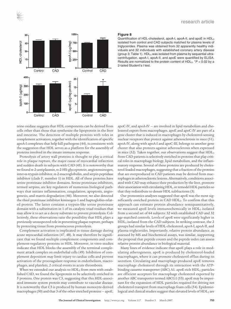
The levels of HDL-cholesterol, apoA-I, apoA-II, and apoE of the 64 subjects are shown in Figure 8. Due to the matching criteria,
Figure 5Relative abundance of proteins isolated from HDL3 of control and CAD subjects. HDL3 was isolated from plasma of 6 control subjects and 7 subjects with established CAD. The relative abundance of proteins in the CAD subjects versus the control subjects was assessed by the peptide index, as described in Methods. *P < 0.05 by Student’s t test (peptide number) or Fisher’s exact test (subject number).

research article
752 TheJournalofClinicalInvestigation http://www.jci.org Volume 117 Number 3 March 2007
the 2 groups had virtually identical HDL-cholesterol levels. In contrast, the level of apoE in HDL3 isolated from the control sub-jects and CAD subjects were 10 ± 5 (mean ± SD) and 16 ± 4 μg/mg protein, respectively (P = 0.02). It is important to note that both apoA-I and apoA-II were quantified simultaneously with apoE in HDL3 and that no significant differences were observed in either apolipoprotein. Covariant analysis indicated that sex, total choles-terol, and LDL cholesterol did not contribute significantly to the variation in apoE levels.
The subjects enrolled in the PRINCE study (group 3; Table 1) dif-fered from those used for our proteomics analysis (group 2; Table 1). The latter were male, and LDL levels were 30% higher in the CAD subjects than in the control subjects (P = 0.005). In contrast, 81% of the PRINCE subjects were female, and levels of LDL were slightly but significantly lower (16%; P = 0.01) in the CAD subjects. Collec-tively, these results indicate that HDL3 of CAD subjects is enriched in apoE and that hyperlipidemia and sex are unlikely to account for the observed differences. Our observations may be clinically relevant, given that we detected elevated levels of apoE in HDL3 iso-lated from 2 very different populations of CAD subjects.
HDL isolated from atherosclerotic lesions and HDL from CAD patients car-ries a subset of the same proteins. We and others have previously shown that HDL isolated from atherosclerotic lesions contains higher lev-els of oxidized amino acids than HDL isolated from plasma (10–12). This observation raises the possibility that proteins found in HDL from CAD patients might come in part from the artery wall.
To test this proposal, we isolated HDL by ultracentrifugation from human carotid atherosclerotic tissue recovered from 6 patients during surgery. Because arterial tissue contains relatively low levels of apoA-I, total HDL was isolated by ultracentrifuga-tion to maximize the yield. Lesion HDL analyzed by immunoblot-ting with a rabbit polyclonal antibody monospecific for human apoA-I contained a predominant protein with the predicted molecular mass of apoA-I (data not shown). Quantifying apoA-I by immunoblotting in lesions, extracted lesions, and lesion HDL showed that this procedure recovered approximately 80% of the immunoreactive protein originally present in the atherosclerotic tissue. HDL isolated from 2 individuals was combined, digested
with trypsin, and analyzed with LC-ESI-MS/MS. Using the crite-rion of 2 unique peptides with high confidence identifications, we identified more than 100 proteins in lesion HDL in 3 independent analyses. Importantly, 3 of the 5 proteins that appeared enriched in the HDL3 samples from the CAD patients were also present in lesion HDL, and apoE was the most abundant of these proteins (Supplemental Table 2).
DiscussionWe employed shotgun proteomics to test the hypothesis that HDL might carry proteins that make a previously unsuspected contri-bution to its cardioprotective and antiinflammatory activities. Forty-eight proteins were identified in HDL isolated by ultracen-trifugation from healthy controls and/or CAD subjects. Twenty-two proteins with well-characterized roles in lipid metabolism and the antioxidant properties of HDL were detected, which validates our experimental approach. Importantly, we found 13 proteins not previously known to reside in HDL.
We used annotations by the GO Consortium to connect the complex array of proteins we identified in HDL to biological processes. Twenty-two of the proteins were linked to cholesterol and lipoprotein metabolism. Remarkably, there were more acute-phase-response proteins (23 of 48), whose plasma concentrations are altered markedly by acute and chronic inflammation. A num-ber of proteins not previously known to reside in HDL, includ-ing C4A/C4B, C9, and vitronectin, were linked to the complement pathway and its regulation. Our detection of the extracellular matrix protein vitronectin and the lysosomal protein prenylcys-
Figure 6Reconstructed ion chromatograms of peptides derived from apoC-IV and apoA-I in HDL3. Tryptic digests of HDL3 isolated from CAD sub-jects and control subjects were subjected to LC-ESI-MS/MS analy-sis as described in the legend to Figure 1. Reconstructed ion chro-matograms of peptides (arrows) unique to apoC-IV and apoA-I were extracted from the full-scan mass survey spectra of each analysis. cps, counts per second.
Figure 7Relationship between unique peptides and protein concentration in HDL and HDL3. The total number of unique peptides detected by MS in HDL and HDL3 is as shown in Table 2. The protein concentrations of apoA-I (98 mg/dl), apoA-II (42 mg/dl), apoC-II (4.6 mg/dl), apoB (1.7 mg/dl), and transferrin (0.02 mg/dl) in HDL3 and HDL isolated by ultracentrifugation are those reported by McPherson et al. (where HDL equals the sum of HDL2 and HDL3; ref. 41). The concentration of CETP in HDL3 (0.1 mg/dl) was estimated as 50% that of plasma CETP (40). Linear regression analysis revealed a strong correlation (r2 = 0.98 and 0.88) between protein concentration and unique pep-tides in HDL and HDL3, respectively.
research article
TheJournalofClinicalInvestigation http://www.jci.org Volume 117 Number 3 March 2007 753
teine oxidase suggests that HDL components can be derived from cells other than those that synthesize the lipoprotein in the liver and intestine. The detection of multiple proteins with roles in complement activation, together with the identification of specific apoA-I complexes that help kill pathogens (44), is consistent with the suggestion that HDL serves as a platform for the assembly of proteins involved in the innate immune response.
Proteolysis of artery wall proteins is thought to play a critical role in plaque rupture, the major cause of myocardial infarction and sudden death in subjects with CAD (45). It is noteworthy that we found α-2-antiplasmin, α-2-HS-glycoprotein, angiotensinogen, inter-α-trypsin inhibitor, α-2-macroglobulin, and serpin peptidase inhibitor (clade F, member 1) in HDL. All of these proteins have serine proteinase inhibitor domains. Serine proteinase inhibitors, termed serpins, are key regulators of numerous biological path-ways that initiate inflammation, coagulation, apoptosis, angio-genesis, and matrix degradation (46). Moreover, we also detected the thiol proteinase inhibitor kininogen-1 and haptoglobin-relat-ed protein. The latter contains a trypsin-like serine proteinase domain with a substitution in 1 of its catalytic triad residues that may allow it to act as a decoy substrate to prevent proteolysis. Col-lectively, these observations raise the possibility that HDL plays a previously unsuspected role in preventing plaque rupture, perhaps by protecting tissue from promiscuous proteolysis.
Complement activation is implicated in tissue damage during acute myocardial infarction (47, 48). It may therefore be signifi-cant that we found multiple complement components and com-plement-regulatory proteins in HDL. Moreover, in vitro studies indicate that HDL blocks the assembly of the terminal comple-ment attack complex on endothelial cells (49). Inhibition of com-plement deposition may limit injury to cardiac cells and prevent activation of the procoagulant response in endothelium, macro-phages, and platelets, 2 critical events in acute thrombosis.
When we extended our analysis to HDL3 from men with estab-lished CAD, we found the lipoprotein to be selectively enriched in 5 proteins. One protein was C3, suggesting that this HDL-associ-ated immune system protein may contribute to vascular disease. It is noteworthy that C3 is produced by human monocyte-derived macrophages (50) and that 3 of the other enriched proteins — apoE,
apoC-IV, and apoA-IV — are involved in lipid metabolism and cho-lesterol export from macrophages. apoE and apoC-IV are part of a gene cluster that is induced in macrophages by cholesterol-sensing nuclear receptors that protect against atherosclerosis in mice (51). apoA-IV, along with apoA-I and apoC-III, belongs to another gene cluster that also protects against atherosclerosis when expressed in mice (52). Taken together, our observations suggest that HDL3 from CAD patients is selectively enriched in proteins that play criti-cal roles in macrophage biology, lipid metabolism, and the inflam-matory response. Several of these proteins are produced by choles-terol-loaded macrophages, suggesting that a fraction of the proteins that are overproduced in CAD patients may be derived from mac-rophages in atherosclerotic lesions. Alternatively, conditions associ-ated with CAD may enhance their production by the liver, promote their association with circulating HDL, or remodel HDL particles so that they redistribute to denser HDL subfractions (3).
Our proteomics analyses suggested that apoE was the most sig-nificantly enriched protein in CAD HDL3. To confirm that this approach can estimate protein abundance semiquantitatively, we measured apoE levels immunochemically in HDL3 isolated from a second set of 64 subjects: 32 with established CAD and 32 age-matched controls. Levels of apoE were significantly higher in HDL3 isolated from the CAD subjects. In striking contrast, the 2 groups had similar levels of HDL-cholesterol, apoA-I, apoA-II, and plasma triglycerides. Importantly, relative protein abundance, as assessed by MS and biochemical assays, was similar, supporting the proposal that peptide counts and the peptide index can assess relative protein abundance in biological material.
Many lines of evidence indicate that apoE plays a role in mod-ulating atherogenesis. apoE is produced by cholesterol-loaded macrophages, where it can promote cholesterol efflux during its secretion. Circulating and macrophage-produced apoE removes macrophage cholesterol through its interaction with the ATP-binding cassette transporter (ABC) A1. apoE-rich HDL2 particles are efficient acceptors for macrophage cholesterol exported by another ABC transporter termed ABCG1 (53). apoE may be impor-tant for the expansion of HDL particles required for driving net cholesterol transport from macrophage foam cells (54). Epidemio-logical and clinical studies suggest that elevated levels of HDL2 are
Figure 8Quantification of HDL-cholesterol, apoA-I, apoA-II, and apoE in HDL3 isolated from control and CAD subjects matched for plasma levels of triglycerides. Plasma was obtained from 32 apparently healthy indi-viduals and 32 individuals with established coronary artery disease (group 3; Table 1). HDL3 was isolated from plasma by sequential ultra-centrifugation. apoA-I, apoA-II, and apoE were quantified by ELISA. Results are normalized to the protein content of HDL3. *P = 0.02 by a 2-tailed Student’s t test.

research article
754 TheJournalofClinicalInvestigation http://www.jci.org Volume 117 Number 3 March 2007
cardioprotective. Moreover, apoE levels are lower in HDL2 (the less dense subfraction of HDL) isolated from subjects with CAD (55), and apoE-enriched HDL2 isolated from CETP-deficient subjects removes potentially toxic cholesterol from macrophages much more efficiently than control HDL2 (53). Our demonstration of elevated levels of apoE in HDL3 isolated from subjects with estab-lished CAD raises the possibility that redistribution of apoE from HDL2 to HDL3 impairs cholesterol efflux and promotes the forma-tion of macrophage foam cells in vivo.
In summary, our results indicate that HDL carries a population of proteins that function in lipid metabolism, proteinase inhibi-tion, complement activation, and the acute-phase response. It will be of interest to study a larger number of subjects to determine whether levels of the proteins we detected in HDL3 from CAD sub-jects might be useful indicators of cardiovascular risk and to inves-tigate the proposal that chronic inflammation alters the protein composition of HDL, making it atherogenic (3, 8, 13).
MethodsIsolation of HDL. Three groups of subjects were used to study the protein composition of HDL (Table 1). The first group (20 control male subjects from Seattle) was used for proteomics analysis of total HDL. The second group (6 control subjects and 7 CAD subjects from Seattle) was used for proteomics analysis of HDL3. The third group (64 subjects enrolled in the PRINCE study (43) was used for quantification of apoE in HDL3. All protocols involving human material were approved by the Human Studies Committees at the University of Washington, Brigham and Women’s Hos-pital, Harvard Medical School, and Wake Forest University. All subjects provided informed consent.
Blood anticoagulated with EDTA was collected from subjects recruit-ed at the University of Washington or Brigham and Women’s Hospital. The subjects had fasted overnight. Atherosclerotic tissue was harvested at carotid endarterectomy at Wake Forest University, snap-frozen, and stored at −80°C until analysis. Lesions from a single individual (approximately 0.5 g wet weight) were mixed with dry ice and pulverized with a pestle in a stainless steel mortar. HDL was extracted from the tissue powder (10). HDL (d = 1.063−1.210 g/ml) and HDL3 (d = 1.110–1.210 g/ml) were isolated by ultracentrifugation (56). apoA-I complexes were isolated from human plasma using apoA-I–specific goat polyclonal antibody covalently coupled to HiTrap NHS-activated HP columns (GE HealthCare) (57). As a control, parallel analyses were run using material eluted from washed beads cou-pled to albumin or nonspecific immunoglobulin. Protein was determined using the Bradford assay, with albumin as the standard.
Tryptic digest. HDL was precipitated with 10% trichloroacetic acid and resolubilized with 6 M urea in 25 mM ammonium bicarbonate. Reduced, alkylated proteins were suspended in 0.6 M urea in 25 mM ammonium bicarbonate, digested overnight at 37°C with trypsin (1:20, wt/wt, trypsin/HDL protein), acidified with acetic acid, dried under vacuum, and resus-pended in 0.1% formic acid. Tryptic digests were desalted with a C18 ZipTip (Millipore) prior to MS analysis.
LC-ESI-MS/MS. Tryptic digests of HDL3 were separated using 2-dimen-sional LC with strong cation exchange and reverse-phase capillary LC columns. The LC system was interfaced with a Finnigan LCQ Deca Pro-teomeX ion trap mass spectrometer (Thermo Electron) equipped with an orthogonal electrospray interface. Tryptic digests of total HDL and apoA-I particles were analyzed by reverse-phase capillary LC coupled with electro-spray ionization and a Thermo Electron linear ion trap mass spectrometer. Details are provided in the Supplemental Methods.
Peptide and protein identification. MS/MS spectra were searched against the human International Protein Index (IPI) database v.3.01 (http://www.ebi.
ac.uk/IPI/IPIhelp.html) (58), using the SEQUEST search engine (Thermo Electron) with fixed Cys alkylation and variable Met oxidation modifica-tions (58). One incomplete cleavage site was allowed in peptides for tryp-sin-restricted searches. The SEQUEST results were further validated using PeptideProphet and ProteinProphet (http://tools.proteomecenter.org/TPP.php) (59, 60), using an adjusted probability of greater than 0.90 for pep-tides and greater than 0.95 for proteins. Each charge state of a peptide was considered a unique identification. MS/MS spectra from proteins identi-fied with less than 6 peptides were confirmed by manual inspection.
Immunoblotting and apolipoprotein. Reduced proteins separated by SDS-PAGE were transferred to a PVDF membrane and probed with a rabbit polyclonal anti-C3 antibody (Quidel Corp.), a sheep polyclonal anti-α-2-HS glycoprotein antibody (BioDesign), a rabbit polyclonal anti-vitronectin antibody (BIODESIGN International), or a sheep polyclonal antibody to the inter-α-trypsin inhibitor complex (Nordic Immunological Labo-ratories). A multiplex ELISA (Linco) was used to quantify levels of apoE, apoA-I, and apoA-II (61).
Statistics. For each protein identified by MS/MS, the peptide index was calculated as: [(peptides in CAD subjects/total peptides) × (% of CAD sub-jects with ≥1 peptide)] – [(peptides in control subjects/total peptides) × (% of control subjects with ≥1 peptide)]. To validate the peptide index, we performed a permutation analysis. The cutoffs for statistical significance at 95% confidence interval (based on 1,000 random permutations) were peptide indices of –0.41 and +0.44 for relative enrichment in control sub-jects and CAD subjects, respectively. Therefore, we chose a peptide index of less than –0.4 or greater than 0.4, respectively, to identify selective enrich-ment of proteins in control subjects or CAD subjects.
For proteins that appeared enriched by the peptide index, Student’s unpaired 2-tailed t test was used to compare the number of unique pep-tides identified in CAD patients versus healthy subjects. For proteins found in only 1 group of subjects, a 1-sample t test was used to compare the number of unique peptides to a theoretical mean of 0. Fisher’s exact test was used to compare the number of CAD patients versus healthy sub-jects from which each protein was identified. The Mann-Whitney U test for nonparametric data was used to compare levels of HDL, apoA–I, apoA–II, and apoE in control and CAD subjects. For all statistical analyses, P < 0.05 was considered significant.
GO analysis. Functional annotation of proteins detected in HDL3 was obtained from the GO database (http://www.geneontology.org; ref. 62). Functional categories within HDL3 proteins that were significantly enriched relative to 3,020 human plasma proteins (63) were determined using the GOTree Machine’s algorithm (http://genereg.ornl.gov/gotm; ref. 64). For a given GO category, the relative enrichment of genes encoding the proteins detected in HDL3 relative to all reference genes in that category were calculated (http://david.abcc.ncifcrf.gov/home.jsp). A cutoff value of P < 0.0001 was used to report a functional category as significantly over-represented. Results were confirmed by repeating the analysis using the Expression Analysis Systematic Explorer algorithm (EASE; http://david.niaid.nih.gov/david/ease.htm). EASE uses a variant of the 1-tailed Fisher exact probability test to determine an EASE score, which ranks overrepre-sented GO processes (65). To address the multiple comparisons problem that arises when many processes are evaluated simultaneously, we per-formed a permutation analysis (n = 1,000 permutations) to calculate the global false discovery rate (FDR). To improve statistical confidence in our results, all enriched functional categories were required to be significant using both methods (P < 0.0001 and FDR < 0.001).
AcknowledgmentsThis research was supported by grants from the NIH (HL086798, DK017047, DK02456, HL57557, HL074223, P30ES07033,

research article
TheJournalofClinicalInvestigation http://www.jci.org Volume 117 Number 3 March 2007 755
P30DK017047, HL078527, and PO1HL030086) and the Donald W. Reynolds Foundation. T. Vaisar and S. Pennathur were support-ed by a Pilot and Feasibility Award from the Diabetes and Endo-crinology Research Center and by the Juvenile Diabetes Research Foundation, respectively. MS experiments were performed by the Mass Spectrometry Resource, Department of Medicine, and the Mass Spectrometry Core, Diabetes and Endocrinology Research Center at the University of Washington.
Received for publication July 8, 2005, and accepted in revised form December 22, 2006.
Address correspondence to: Jay W. Heinecke, Division of Metab-olism, Box 356426, University of Washington, Seattle, Wash-ington 98195, USA. Fax: (206) 685-8346; E-mail: [email protected].
Subramaniam Pennathur and Jaeman Byun’s present address is: Department of Medicine, University of Michigan, Ann Arbor, Michigan, USA.
Tomas Vaisar, Subramaniam Pennathur, and Pattie S. Green con-tributed equally to this work.
1. Braunwald, E. 1997. Cardiovascular medicine at the turn of the millennium: triumphs, concerns, and opportunities. N. Engl. J. Med. 337:1360–1369.
2. Hoyert, D.L., Kung, H.-C., and Smith, B.L. 2005. Deaths: preliminary data for 2003. Natl. Vital Stat. Rep. 53:1–48.
3. Rader, D.J. 2006. Molecular regulation of HDL metabolism and function: implications for novel therapies. J. Clin. Invest. 116:3090–3100. doi:10.1172/JCI30163.
4. Tall, A.R., Costet, P., and Wang, N. 2002. Regula-tion and mechanisms of macrophage cholesterol efflux. J. Clin. Invest. 110:899–904. doi:10.1172/JCI200216391.
5. Gordon, D.J., and Rifkind, B.M. 1989. High-density lipoprotein — the clinical implications of recent studies. N. Engl. J. Med. 321:1311–1316.
6. Wilson, P.W., Abbott, R.D., and Castelli, W.P. 1988. High density lipoprotein cholesterol and mortal-ity. The Framingham Heart Study. Arteriosclerosis. 8:737–741.
7. Barzilai, N., et al. 2003. Unique lipoprotein phe-notype and genotype associated with exceptional longevity. JAMA. 290:2030–2040.
8. Barter, P.J., et al. 2004. Antiinflammatory proper-ties of HDL. Circ. Res. 95:764–772.
9. Shao, B., Oda, M.N., Oram, J.F., and Heinecke, J.W. 2006. Myeloperoxidase: an inflammatory enzyme for generating dysfunctional high density lipopro-tein. Curr. Opin. Cardiol. 21:322–328.
10. Bergt, C., et al. 2004. The myeloperoxidase prod-uct hypochlorous acid oxidizes HDL in the human artery wall and impairs ABCA1-dependent cho-lesterol transport. Proc. Natl. Acad. Sci. U. S. A. 101:13032–13037.
11. Pennathur, S., et al. 2004. Human atherosclerotic intima and blood of patients with established coro-nary artery disease contain high density lipoprotein damaged by reactive nitrogen species. J. Biol. Chem. 279:42977–42983.
12. Zheng, L., et al. 2004. Apolipoprotein A-I is a selec-tive target for myeloperoxidase-catalyzed oxida-tion and functional impairment in subjects with cardiovascular disease. J. Clin. Invest. 114:529–541. doi:10.1172/JCI200421109.
13. Chait, A., Han, C.Y., Oram, J.F., and Heinecke, J.W. 2005. Thematic review series: The immune system and atherogenesis. Lipoprotein-associated inflam-matory proteins: markers or mediators of cardio-vascular disease? J. Lipid. Res. 46:389–403.
14. Olin, K.L., Potter-Perigo, S., Barrett, P.H., Wight, T.N., and Chait, A. 2001. Biglycan, a vascular proteo-glycan, binds differently to HDL2 and HDL3: role of apoE. Arterioscler. Thromb. Vasc. Biol. 21:129–135.
15. Link, A.J., et al. 1999. Direct analysis of protein complexes using mass spectrometry. Nat. Biotechnol. 17:676–682.
16. Resing, K.A., and Ahn, N.G. 2005. Proteomics strategies for protein identification. FEBS Lett. 579:885–889.
17. Draganov, D.I., Stetson, P.L., Watson, C.E., Billecke, S.S., and La Du, B.N. 2000. Rabbit serum paraox-onase 3 (PON3) is a high density lipoprotein-associ-
ated lactonase and protects low density lipoprotein against oxidation. J. Biol. Chem. 275:33435–33442.
18. Duchateau, P.N., et al. 1997. Apolipoprotein L, a new human high density lipoprotein apolipoprotein expressed by the pancreas. Iden-tification, cloning, characterization, and plasma distribution of apolipoprotein L. J. Biol. Chem. 272:25576–25582.
19. Getz, G.S., and Reardon, C.A. 2004. Paraoxonase, a cardioprotective enzyme: continuing issues. Curr. Opin. Lipidol. 15:261–267.
20. Heller, M., et al. 2005. Mass spectrometry-based analytical tools for the molecular protein char-acterization of human plasma lipoproteins. Pro-teomics. 5:2619–2630.
21. Karlsson, H., Leanderson, P., Tagesson, C., and Lin-dahl, M. 2005. Lipoproteomics II: mapping of pro-teins in high-density lipoprotein using two-dimen-sional gel electrophoresis and mass spectrometry. Proteomics. 5:1431–1445.
22. Kotite, L., Zhang, L.H., Yu, Z., Burlingame, A.L., and Havel, R.J. 2003. Human apoC-IV: isolation, characterization, and immunochemical quantifi-cation in plasma and plasma lipoproteins. J. Lipid Res. 44:1387–1394.
23. Kunitake, S.T., et al. 1994. Identification of pro-teins associated with apolipoprotein A-I-con-taining lipoproteins purified by selected-affinity immunosorption. Biochemistry. 33:1988–1993.
24. McVicar, J.P., Kunitake, S.T., Hamilton, R.L., and Kane, J.P. 1984. Characteristics of human lipopro-teins isolated by selected-affinity immunosorption of apolipoprotein A-I. Proc. Natl. Acad. Sci. U. S. A. 81:1356–1360.
25. Ogorzalek Loo, R.R., Yam, L., Loo, J.A., and Schu-maker, V.N. 2004. Virtual two-dimensional gel electrophoresis of high-density lipoproteins. Elec-trophoresis. 25:2384–2391.
26. Rezaee, F., Casetta, B., Levels, J.H., Speijer, D., and Meijers, J.C. 2006. Proteomic analysis of high-den-sity lipoprotein. Proteomics. 6:721–730.
27. Xu, N., and Dahlback, B. 1999. A novel human apolipoprotein (apoM). J. Biol. Chem. 274:31286–31290.
28. Zhang, L.H., Kotite, L., and Havel, R.J. 1996. Iden-tification, characterization, cloning, and expres-sion of apolipoprotein C-IV, a novel sialoglycopro-tein of rabbit plasma lipoproteins. J. Biol. Chem. 271:1776–1783.
29. Graham, T.E., et al. 2006. Retinol-binding protein 4 and insulin resistance in lean, obese, and diabetic subjects. N. Engl. J. Med. 354:2552–2563.
30. Kunitake, S.T., Jarvis, M.R., Hamilton, R.L., and Kane, J.P. 1992. Binding of transition metals by apolipoprotein A-I-containing plasma lipoproteins: inhibition of oxidation of low density lipoproteins. Proc. Natl. Acad. Sci. U. S. A. 89:6993–6997.
31. Cheung, M.C., and Wolf, A.C. 1988. Differential effect of ultracentrifugation on apolipoprotein A-I- containing lipoprotein subpopulations. J. Lipid Res. 29:15–25.
32. Park, C.T., and Wright, S.D. 2000. Fibrinogen is a component of a novel lipoprotein particle: factor
H-related protein (FHRP)-associated lipoprotein particle (FALP). Blood. 95:198–204.
33. Gundry, R.L., Fu, Q., Jelinek, C.A., VanEyk, J.E., and Cotter, R.J. 2007. Investigation of an albumin-enriched fraction of human serum and its albu-minome. J. Clin. Proteomics. In press.
34. Oksjoki, R., Kovanen, P.T., and Pentikainen, M.O. 2003. Role of complement activation in atheroscle-rosis. Curr. Opin. Lipidol. 14:477–482.
35. Hortin, G.L., Shen, R.F., Martin, B.M., and Remaley, A.T. 2006. Diverse range of small peptides associat-ed with high-density lipoprotein. Biochem. Biophys. Res. Commun. 340:909–915.
36. Zhuo, L., Hascall, V.C., and Kimata, K. 2004. Inter-alpha-trypsin inhibitor, a covalent protein-glycosaminoglycan-protein complex. J. Biol. Chem. 279:38079–38082.
37. Washburn, M.P., Ulaszek, R.R., and Yates, J.R., 3rd. 2003. Reproducibility of quantitative proteomic analyses of complex biological mixtures by mul-tidimensional protein identification technology. Anal. Chem. 75:5054–5061.
38. Liu, H., Sadygov, R.G., and Yates, J.R., 3rd. 2004. A model for random sampling and estimation of relative protein abundance in shotgun proteomics. Anal. Chem. 76:4193–4201.
39. Old, W.M., et al. 2005. Comparison of label-free methods for quantifying human proteins by shot-gun proteomics. Mol. Cell. Proteomics. 4:1487–1502.
40. Marcel, Y.L., et al. 1990. Distribution and con-centration of cholesteryl ester transfer protein in plasma of normolipemic subjects. J. Clin. Invest. 85:10–17.
41. McPherson, P.A., Young, I.S., McKibben, B., and McEneny, J. 2007. High density lipoprotein sub-fractions: isolation, composition and their duplici-tous role in oxidation. J. Lipid Res. 48:86–95.
42. O’Brien, P.J., et al. 2005. The novel apolipoprotein A5 is present in human serum, is associated with VLDL, HDL, and chylomicrons, and circulates at very low concentrations compared with other apo-lipoproteins. Clin. Chem. 51:351–359.
43. Albert, M.A., Danielson, E., Rifai, N., and Ridker, P.M. 2001. Effect of statin therapy on C-reactive protein levels: the pravastatin inflammation/CRP evaluation (PRINCE): a randomized trial and cohort study. JAMA. 286:64–70.
44. Shiflett, A.M., Bishop, J.R., Pahwa, A., and Hajduk, S.L. 2005. Human high density lipoproteins are plat-forms for the assembly of multi-component innate immune complexes. J. Biol. Chem. 280:32578–32585.
45. Davies, M.J. 1990. A macro and micro view of coro-nary vascular insult in ischemic heart disease. Cir-culation. 82(Suppl.):II38–II46.
46. Richardson, J., Viswanathan, K., and Lucas, A. 2006. Serpins, the vasculature, and viral therapeutics. Front. Biosci. 11:1042–1056.
47. Niculescu, F., and Rus, H. 2004. The role of com-plement activation in atherosclerosis. Immunol. Res. 30:73–80.
48. Pepys, M.B., et al. 2006. Targeting C-reactive pro-tein for the treatment of cardiovascular disease. Nature. 440:1217–1221.

research article
756 TheJournalofClinicalInvestigation http://www.jci.org Volume 117 Number 3 March 2007
49. Hamilton, K.K., Zhao, J., and Sims, P.J. 1993. Inter-action between apolipoproteins A-I and A-II and the membrane attack complex of complement. Affinity of the apoproteins for polymeric C9. J. Biol. Chem. 268:3632–3638.
50. Strunk, R.C., Kunke, K.S., and Giclas, P.C. 1983. Human peripheral blood monocyte-derived mac-rophages produce haemolytically active C3 in vitro. Immunology. 49:169–174.
51. Mak, P.A., et al. 2002. Regulated expression of the apolipoprotein E/C-I/C-IV/C-II gene cluster in murine and human macrophages. A critical role for nuclear liver X receptors alpha and beta. J. Biol. Chem. 277:31900–31908.
52. Vergnes, L., et al. 2000. Expression of human apolipoprotein A-I/C-III/A-IV gene cluster in mice induces hyperlipidemia but reduces atherogenesis. Arterioscler. Thromb. Vasc. Biol. 20:2267–2274.
53. Matsuura, F., Wang, N., Chen, W., Jiang, X.C., and Tall, A.R. 2006. HDL from CETP-deficient subjects shows enhanced ability to promote cholesterol efflux from macrophages in an apoE- and ABCG1-dependent pathway. J. Clin. Invest. 116:1435–1442. doi:10.1172/JCI27602.
54. Mahley, R.W., Huang, Y., and Weisgraber, K.H. 2006. Putting cholesterol in its place: apoE and reverse
cholesterol transport. J. Clin. Invest. 116:1226–1229. doi:10.1172/JCI28632.
55. Wilson, H.M., Patel, J.C., Russell, D., and Skinner, E.R. 1993. Alterations in the concentration of an apolipoprotein E-containing subfraction of plasma high density lipoprotein in coronary heart disease. Clin. Chim. Acta. 220:175–187.
56. Mendez, A.J., Oram, J.F., and Bierman, E.L. 1991. Protein kinase C as a mediator of high density lipo-protein receptor-dependent efflux of intracellular cholesterol. J. Biol. Chem. 266:10104–10111.
57. Cheung, M.C., et al. 1987. Characterization of high density lipoprotein subspecies: structural studies by single vertical spin ultracentrifugation and immunoaffinity chromatography. J. Lipid Res. 28:913–929.
58. Kersey, P.J., et al. 2004. The International Protein Index: an integrated database for proteomics exper-iments. Proteomics. 4:1985–1988.
59. Nesvizhskii, A.I., Keller, A., Kolker, E., and Aeber-sold, R. 2003. A statistical model for identifying proteins by tandem mass spectrometry. Anal. Chem. 75:4646–4658.
60. Yan, W., et al. 2004. A dataset of human liver pro-teins identified by protein profiling via isotope-coded affinity tag (ICAT) and tandem mass spec-
trometry. Mol. Cell. Proteomics. 3:1039–1041. 61. Vuletic, S., et al. 2005. Reduced CSF PLTP activity
in Alzheimer’s disease and other neurologic diseas-es; PLTP induces ApoE secretion in primary human astrocytes in vitro. J. Neurosci. Res. 80:406–413.
62. [Anonymous]. 2001. Creating the gene ontology resource: design and implementation. Gene Ontol-ogy Consortium. Genome Res. 11:1425–1433.
63. Omenn, G.S., et al. 2005. Overview of the HUPO Plasma Proteome Project: results from the pilot phase with 35 collaborating laboratories and mul-tiple analytical groups, generating a core dataset of 3020 proteins and a publicly-available database. Proteomics. 5:3226–3245.
64. Zhang, B., Schmoyer, D., Kirov, S., and Snoddy, J. 2004. GOTree Machine (GOTM): a web-based platform for interpreting sets of interesting genes using Gene Ontology hierarchies. BMC Bioinfor-matics. 5:16.
65. Hosack, D.A., Dennis, G., Jr., Sherman, B.T., Lane, H.C., and Lempicki, R.A. 2003. Identifying biologi-cal themes within lists of genes with EASE. Genome Biol. 4:R70. doi:10.1186/gb-2003-4-10-r70.
66. Navab, M., et al. 2004. The oxidation hypothesis of atherogenesis: the role of oxidized phospholipids and HDL. J. Lipid Res. 45:993–1007.