Induced circular dichroism spectra reveal binding of the antiinflammatory curcumin to human α1-acid...
7
Induced circular dichroism spectra reveal binding of the antiinflammatory curcumin to human a 1 -acid glycoprotein Ferenc Zsila, * Zsolt Bik adi and Mikl os Simonyi Department of Molecular Pharmacology, Institute of Chemistry, Chemical Research Center, PO Box 17, Budapest H-1525, Hungary Received 13 January 2004; accepted 30 March 2004 Available online 8 May 2004 Abstract—This paper reports the first experimental evidence on binding of the plant derived curcumin molecule to human a 1 -acid glycoprotein (AGP), an acute phase protein in blood. Oppositely signed induced circular dichroism (CD) bands measured in the visible spectral region in pH 7.4 phosphate buffer indicate that the protein binds this natural polyphenol molecule in a left-handed chiral conformation. Decreasing of the intrinsic fluorescence of AGP upon addition of curcumin confirmed the binding to take place. Fluorescence quenching titration curve of AGP allowed to calculate the association constant of the ligand (K a ¼ 4 10 4 M 1 ). Modification of near UV CD spectrum of the protein suggests that curcumin induces changes in the tertiary structure of AGP, which leads to the decrease of binding affinity. By using rac-warfarin and amitriptyline, selective high affinity ligands of F1–S and A genetic variants of AGP, CD displacement experiments showed that curcumin is able to bind to both variants. Molecular docking calculations performed on curcumin–AGP and warfarin–AGP complexes suggest the existence of two alternative binding sites for curcumin; either at the open end of the central hydrophobic cavity or in a surface cleft of the protein. Ó 2004 Elsevier Ltd. All rights reserved. 1. Introduction The major yellow ingredient of the powdered rhizome of Curcuma longa L. (turmeric) is a polyphenol molecule called curcumin (diferuloylmethane). 1 In Asia, it has commonly been used as a colouring and flavouring spice in food products. Curcumin has also been traditionally used to treat many inflammatory disorders and for wound healing for centuries. Besides its extensive usage in herbal medicine, it receives growing attention in the modern pharmacology as well due to its beneficial effects including strong antiinflammatory activity and chemo- preventive properties against various human malignan- cies. 2–4 Although curcumin is reported to interact directly with several cellular proteins, 2 molecular mechanisms of these interactions are poorly understood. AGP, also known as orosomucoid, is a glycoprotein of 183 residues with an extremely high carbohydrate con- tent accounting for 45% of its mass. 5 Due to sialic acid components (10–14%) the protein is negatively charged at physiological pH. On the basis of electrophoretic migration, one fast and two slow bands can be distin- guished in commercial AGP samples corresponding to the genetic variants F1, S and A, respectively. 6;7 Pro- portion of each variant are approximately F1 50%, S 23% and A 27%. There is a difference of at least 22 amino acid residues between the (F1–S) and A variants while F1 and S forms differ by only a few residues. AGP is classified as one of the positive acute phase proteins 8 of which normal plasma level (between 50 and 100 mg/100 mL) can rapidly increase up to three- or fourfold in response to acute inflammation. 9 Although its precise biological function is unclear, AGP was suggested to have both antiinflam- matory effects and a role in immunomodulation. 5;8 In the clinical practice AGP is a valuable diagnostic and prognostic tool since its structure and serum level are substantially modified in inflammatory diseases and different malignancies. As an important plasma protein beside serum albumin, AGP binds and transports a number of endogenous and exogenous compounds including various basic and neutral drugs influencing their pharmacokinetics and pharmacodynamics. 9;10 Unfortunately, three dimensional X-ray structure of AGP is unknown and despite the vast literature data Keywords: a 1 -Acid glycoprotein; Curcumin; Circular dichroism spectroscopy; Induced chirality; Warfarin. Abbreviations: AGP, a 1 -acid glycoprotein; CD, circular dichroism; CE, Cotton effect; HSA, human serum albumin; L/P , ligand/protein molar ratio * Corresponding author. Fax: +36-1-325-7750; e-mail: zsferi@ chemres.hu 0968-0896/$ - see front matter Ó 2004 Elsevier Ltd. All rights reserved. doi:10.1016/j.bmc.2004.03.074 Bioorganic & Medicinal Chemistry 12 (2004) 3239–3245
Transcript of Induced circular dichroism spectra reveal binding of the antiinflammatory curcumin to human α1-acid...

Bioorganic & Medicinal Chemistry 12 (2004) 3239–3245
Induced circular dichroism spectra reveal binding of theantiinflammatory curcumin to human a1-acid glycoprotein
Ferenc Zsila,* Zsolt Bik�adi and Mikl�os Simonyi
Department of Molecular Pharmacology, Institute of Chemistry, Chemical Research Center, PO Box 17, Budapest H-1525, Hungary
Received 13 January 2004; accepted 30 March 2004
Available online 8 May 2004
Abstract—This paper reports the first experimental evidence on binding of the plant derived curcumin molecule to human a1-acidglycoprotein (AGP), an acute phase protein in blood. Oppositely signed induced circular dichroism (CD) bands measured in thevisible spectral region in pH7.4 phosphate buffer indicate that the protein binds this natural polyphenol molecule in a left-handedchiral conformation. Decreasing of the intrinsic fluorescence of AGP upon addition of curcumin confirmed the binding to takeplace. Fluorescence quenching titration curve of AGP allowed to calculate the association constant of the ligand(Ka ¼ 4� 104 M�1). Modification of near UV CD spectrum of the protein suggests that curcumin induces changes in the tertiarystructure of AGP, which leads to the decrease of binding affinity. By using rac-warfarin and amitriptyline, selective high affinityligands of F1–S and A genetic variants of AGP, CD displacement experiments showed that curcumin is able to bind to bothvariants. Molecular docking calculations performed on curcumin–AGP and warfarin–AGP complexes suggest the existence of twoalternative binding sites for curcumin; either at the open end of the central hydrophobic cavity or in a surface cleft of the protein.� 2004 Elsevier Ltd. All rights reserved.
1. Introduction
The major yellow ingredient of the powdered rhizome ofCurcuma longa L. (turmeric) is a polyphenol moleculecalled curcumin (diferuloylmethane).1 In Asia, it hascommonly been used as a colouring and flavouring spicein food products. Curcumin has also been traditionallyused to treat many inflammatory disorders and forwound healing for centuries. Besides its extensive usagein herbal medicine, it receives growing attention in themodern pharmacology as well due to its beneficial effectsincluding strong antiinflammatory activity and chemo-preventive properties against various human malignan-cies.2–4 Although curcumin is reported to interact directlywith several cellular proteins,2 molecular mechanisms ofthese interactions are poorly understood.
AGP, also known as orosomucoid, is a glycoprotein of183 residues with an extremely high carbohydrate con-
Keywords: a1-Acid glycoprotein; Curcumin; Circular dichroism
spectroscopy; Induced chirality; Warfarin.
Abbreviations: AGP, a1-acid glycoprotein; CD, circular dichroism; CE,
Cotton effect; HSA, human serum albumin; L/P , ligand/protein molar
ratio
* Corresponding author. Fax: +36-1-325-7750; e-mail: zsferi@
chemres.hu
0968-0896/$ - see front matter � 2004 Elsevier Ltd. All rights reserved.
doi:10.1016/j.bmc.2004.03.074
tent accounting for 45% of its mass.5 Due to sialic acidcomponents (10–14%) the protein is negatively chargedat physiological pH. On the basis of electrophoreticmigration, one fast and two slow bands can be distin-guished in commercial AGP samples corresponding tothe genetic variants F1, S and A, respectively.6;7 Pro-portion of each variant are approximately F1 50%, S 23%and A 27%. There is a difference of at least 22 amino acidresidues between the (F1–S) and A variants while F1 andS forms differ by only a few residues. AGP is classified asone of the positive acute phase proteins8 of which normalplasma level (between 50 and 100mg/100mL) can rapidlyincrease up to three- or fourfold in response to acuteinflammation.9 Although its precise biological function isunclear, AGP was suggested to have both antiinflam-matory effects and a role in immunomodulation.5;8 Inthe clinical practice AGP is a valuable diagnostic andprognostic tool since its structure and serum level aresubstantially modified in inflammatory diseases anddifferent malignancies. As an important plasma proteinbeside serum albumin, AGP binds and transports anumber of endogenous and exogenous compoundsincluding various basic and neutral drugs influencingtheir pharmacokinetics and pharmacodynamics.9;10
Unfortunately, three dimensional X-ray structure ofAGP is unknown and despite the vast literature data

3240 F. Zsila et al. / Bioorg. Med. Chem. 12 (2004) 3239–3245
on small molecule–AGP interactions, the topography ofbinding sites and the binding mechanisms are not wellunderstood.7
Serum level of AGP greatly increases during inflamma-tory and immunological processes. At the same timecurcumin has pronounced antiinflammatory activity.2;3
Since AGP binding can significantly affect pharmacoki-netic/pharmacodynamic properties of drugs5;9;10 we con-sidered the possibility that curcumin might interact withAGP. Circular dichroism (CD) and electronic absorptionspectroscopy methods are proved to be sensitive tool tostudy the interaction of curcumin with proteins.11–13 Byusing these techniques, here we report that AGP is able tobind the curcumin molecule. Extrinsic CD spectrum ofthe curcumin–AGP complex was used to investigate thebinding of curcumin to the genetic variants of AGPthrough CD displacement experiments. Additionally,based on the recently published 3D molecular model ofAGP14 obtained by homology modeling, docking calcu-lations were performed to map the possible curcuminbinding sites of AGP.
2. Materials and methods
2.1. Materials
Human AGP purified from Cohn fraction VI (catalogno G-9885, lot no 081K7604), curcumin (catalog no C-7727, lot no 119H4704) and rac-warfarin were obtainedfrom Sigma and used as supplied. AmitriptylineÆHClwas a gift from EGIS Pharmaceuticals Ltd (Budapest,Hungary). Double distilled water and HPLC gradeethanol (Chemolab, Hungary) were used. All otherchemicals were of analytical grade.
2.2. Preparation of AGP solution
For spectroscopic sample preparation, AGP was dis-solved in a pH7.4 0.07M phosphate buffer solution.According to the manufacturer, purity of the AGPsample was 99% by agarose electrophoresis. Molarconcentration of AGP was calculated on the basis of amolecular mass of 40,000.
2.3. Preparation of curcumin stock solution
Curcumin was dissolved in 100% ethanol; the concen-tration was measured by determining light absorption atthe kmax (e429nm ¼ 55; 000M�1 cm�1).1
2.4. Circular dichroism and UV–vis absorption spectros-copy
CD and UV/vis spectra were recorded between 250 and580 nm on a Jasco J-715 spectropolarimeter at15� 0.2 �C under a nitrogen flow. Temperature control
was provided by a Peltier thermostat equipped withmagnetic stirring. Cuvettes of 0.2 cm path length (Hell-ma, USA) were used in the near-UV (250–330 nm) and1.0 cm path length in the visible regions (330–580 nm),respectively. Each spectrum was signal-averaged at leastthree times with a bandwidth of 1.0 nm and a resolutionof 0.2 nm at a scan speed of 100 nm/min. Spectra weresmoothed with Spectra Analysis Software, version1.53.00 (JASCO). Induced CD is defined as the CD ofcurcumin–AGP mixture minus the CD of AGP alone atthe same wavelengths and is expressed as ellipticity inmillidegrees.
2.5. CD/UV–vis titration of AGP with curcumin
The following procedure was used: 2mL of 1.3� 10�4 Mprotein solution was placed in a rectangular cuvette with1 cm optical path length and small amounts of the ligandstock solution (2.3� 10�3 M) was added with an auto-matic pipette in 10 lL aliquots to achieve L/P molarratios from 0.1 to 0.7. Ethanol added with the ligandnever exceeded 4 % (v/v).
2.6. Measurements of CD and UV/vis spectra of curcu-min–AGP solutions in the presence of specific markerligands
Stock solutions of the ligands were prepared as followsand added stepwise in lL volumes to the curcumin–AGP solutions: 3.3� 10�3 M amitriptyline in pH7.4phosphate buffer and 6.3� 10�3 M rac-warfarin in 0.1MNaOHþ pH7.4 phosphate buffer. Concentration ofAGP was 1� 10�4 M (ccurcumin ¼ 2.5� 10�3 M, L/P ¼0.26). Molar ratios of marker ligands/AGP (m/AGP)and marker ligands/curcumin (m/curcumin) were changedas follows: amitriptyline, m/AGP 0.3–1.4, m/curcumin1.3–5.3; rac-warfarin, m/AGP 0.3–1.3, m/curcumin1.3–5.0.
2.7. Fluorescence quenching of AGP with curcumin andcalculation of the association constant of the complex
AGP solution (2mL 8.6� 10�6 M) was prepared in a1 cm rectangular cell by 0.07M pH7.4 phosphate buffer.Ethanolic curcumin solution (2.3� 10�3 M) in lL vol-umes were consecutively added to the cuvette placed inthe sample chamber of the Jasco J-715 spectropolari-meter. Sample solution was excited between 250 and350 nm with 0.2 nm wavelength steps. Total fluorescenceintensity was collected at each wavelength by a Hama-matsu H5784 type photomultiplier detector mounted ona right angle to the light source.
In the sample solution, initial and final concentrations ofAGP and curcumin were 8.6� 10�6–8.0� 10�6 M and2.1� 10�7–1.5� 10�4 M, respectively. [curcumin]/[AGP]molar ratio was varied between 0.02 and 18.6. Duringthe fluorescence measurements, ethanol concentrationdid not exceed 10 v/v%. Control experiments performedwith AGP and EtOH proved the effect of the organic

F. Zsila et al. / Bioorg. Med. Chem. 12 (2004) 3239–3245 3241
solvent to be undetectable on the intrinsic fluorescenceof the protein.
The stereospecific interaction between a ligand (L) andits primary site on the protein (P ) may be quantified bythe association constant (Ka):
Lþ P � LP ; Ka � ½LP �=½L�½P � ð1Þ
It is evident that
½L� ¼ cL � ½LP� ð2Þ
and
½P � ¼ cP � ½LP� ð3Þ
where cL and cP mean the total concentrations of theligand and protein, respectively.
Assuming that the curcumin–AGP complex (1:1 stoi-chiometry) is responsible for the fluorescence quenchingof the protein, it can be written that
Fq283:5nm ð%Þ ¼ k½LP � ð4Þ
where Fq is the percent of the fluorescence quenching ofAGP measured at 283.5 nm and k is a constant.
Using Eqs. (1)–(4), we obtain
Fqð%Þ ¼ k2ðcP þ cL þ K�1
a
�ffiffiffiffiffiffiffiffiffiffiffiffiffiffiffiffiffiffiffiffiffiffiffiffiffiffiffiffiffiffiffiffiffiffiffiffiffiffiffiffiffiffiffiffiffiffiffiffiffiffiffiðcP þ cL þ K�1
a Þ2 � 4cPcL
qÞ ð5Þ
In order to calculate the optimal values of k and Ka,nonlinear regression analysis method was applied(NLREG� statistical analysis programme, version 3.4).
Figure 1. Chemical structures of keto (a) and enol (b) forms of curc-
umin (1,7-bis(4-hydroxy-3-methoxyphenyl)1,6-heptadiene-3,5-dione).
2.8. Molecular modeling calculations
Geometry optimization of curcumin and warfarin mol-ecules were carried out by GAUSSIANGAUSSIAN 98 program usingAM1 semiempirical method. AUTODOCKAUTODOCK 3.0 programpackage15 was used for mapping the energetically mostfavourable binding of neutral curcumin and warfarin toAGP. Published 3D molecular model of the F1–S ge-netic variant of AGP generated by template alignmentwas obtained from Kopecky.14 Gasteiger–Huckel partialcharges were applied both for ligands and protein. Sol-vation parameters were added to the protein coordinatefile and the ligand torsions were defined using the‘Addsol’ and ‘Autotors’ utilities, respectively. Theatomic affinity grids were prepared with 0.375�A spacingby the Autogrid programme for the whole protein tar-get. Random starting positions, orientations and tor-sions (for flexible bonds) were used for the ligands; eachdocking run consisted of 100 cycles.
3. Results and discussion
The diarylheptanoid type curcumin contains two ferulicacid residues joined by a methylene bridge (Fig. 1). Sincethe keto-enol tautomerism of the b-diketone moiety,curcumin molecules exist in solution as intramolecularlyhydrogen-bonded enols,16–18 which allows p-conjugationbetween the two feruloyl parts resulting in the lightabsorption to occur in the visible spectral region.16
Accordingly, curcumin solutions exhibit strong yellowcolour, that is, the main absorption band is centred at429 nm in ethanol. Curcumin derivatives bearing a bulkysubstituent on the central methylene carbon atom arecolourless, since they absorb light in the near-ultravio-let.18
Due to the lack of a centre of asymmetry, free curcuminmolecules show optical activity neither in water nor inorganic solutions. However, induced CD band(s) can bemeasured when the molecules bind to a chiral host, thatis, proteins.11 In our recent papers12;13 we demonstratedthat curcumin binds to a hydrophobic pocket of humanserum albumin (HSA) in bent, right-handed chiralconformation accompanied by the appearance of acharacteristic, exciton type induced CD spectrum. Usingthe CD spectroscopic method again, curcumin wasconsecutively added to the solution of native humanAGP (mixture of the three main genetic variants F1, Sand A) prepared in pH7.4 phosphate buffer. Accordingto the spectral position of the slightly broadenedabsorption band of curcumin at 428 nm, two oppositelysigned Cotton effects (CE) appear with a zero crossoverpoint at 432 nm (Fig. 2). The long-wavelength negativeband (472 nm) is three-times more intense than the po-sitive one at 414 nm. These opposite bands are typicalfor excited state interaction, called exciton coupling,between two proximal chromophores created here bymutual rotations of the two feruloyl moieties around thecentral methylene group.12;13 The exciton-coupling the-ory predicts that AGP-bound curcumin molecule has(M)-helicity (or left-handed chirality) since the CDspectrum shows longer wavelength negative and shorterwavelength positive bands. In other words there isnegative dihedral angle between the rotated parts ofcurcumin molecule. The relatively small induced CDvalues ðDeÞ might suggest that the binding site of AGPslightly prefers the left-handed chiral conformation of
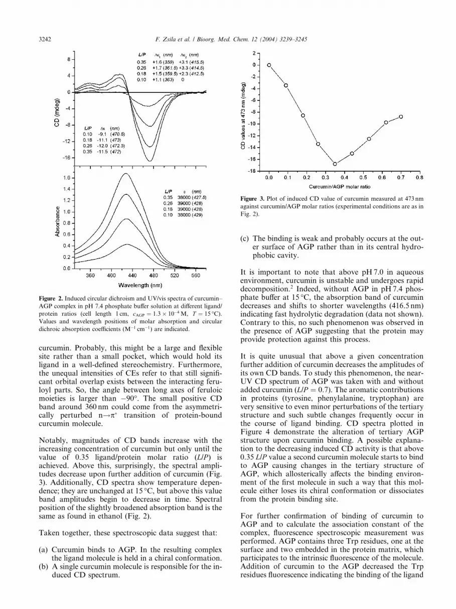
Figure 2. Induced circular dichroism and UV/vis spectra of curcumin–
AGP complex in pH 7.4 phosphate buffer solution at different ligand/
protein ratios (cell length 1 cm, cAGP ¼ 1:3� 10�4 M, T ¼ 15 �C).Values and wavelength positions of molar absorption and circular
dichroic absorption coefficients (M�1 cm�1) are indicated.
Figure 3. Plot of induced CD value of curcumin measured at 473 nm
against curcumin/AGP molar ratios (experimental conditions are as in
Fig. 2).
3242 F. Zsila et al. / Bioorg. Med. Chem. 12 (2004) 3239–3245
curcumin. Probably, this might be a large and flexiblesite rather than a small pocket, which would hold itsligand in a well-defined stereochemistry. Furthermore,the unequal intensities of CEs refer to that still signifi-cant orbital overlap exists between the interacting feru-loyl parts. So, the angle between long axes of feruloicmoieties is larger than �90�. The small positive CDband around 360 nm could come from the asymmetri-cally perturbed n!p� transition of protein-boundcurcumin molecule.
Notably, magnitudes of CD bands increase with theincreasing concentration of curcumin but only until thevalue of 0.35 ligand/protein molar ratio (L/P ) isachieved. Above this, surprisingly, the spectral ampli-tudes decrease upon further addition of curcumin (Fig.3). Additionally, CD spectra show temperature depen-dence; they are unchanged at 15 �C, but above this valueband amplitudes begin to decrease in time. Spectralposition of the slightly broadened absorption band is thesame as found in ethanol (Fig. 2).
Taken together, these spectroscopic data suggest that:
(a) Curcumin binds to AGP. In the resulting complexthe ligand molecule is held in a chiral conformation.
(b) A single curcumin molecule is responsible for the in-duced CD spectrum.
(c) The binding is weak and probably occurs at the out-er surface of AGP rather than in its central hydro-phobic cavity.
It is important to note that above pH7.0 in aqueousenvironment, curcumin is unstable and undergoes rapiddecomposition.2 Indeed, without AGP in pH7.4 phos-phate buffer at 15 �C, the absorption band of curcumindecreases and shifts to shorter wavelengths (416.5 nm)indicating fast hydrolytic degradation (data not shown).Contrary to this, no such phenomenon was observed inthe presence of AGP suggesting that the protein mayprovide protection against this process.
It is quite unusual that above a given concentrationfurther addition of curcumin decreases the amplitudes ofits own CD bands. To study this phenomenon, the near-UV CD spectrum of AGP was taken with and withoutadded curcumin (L/P ¼ 0:7). The aromatic contributionsin proteins (tyrosine, phenylalanine, tryptophan) arevery sensitive to even minor perturbations of the tertiarystructure and such subtle changes frequently occur inthe course of ligand binding. CD spectra plotted inFigure 4 demonstrate the alteration of tertiary AGPstructure upon curcumin binding. A possible explana-tion to the decreasing induced CD activity is that above0.35 L/P value a second curcumin molecule starts to bindto AGP causing changes in the tertiary structure ofAGP, which allosterically affects the binding environ-ment of the first molecule in such a way that this mol-ecule either loses its chiral conformation or dissociatesfrom the protein binding site.
For further confirmation of binding of curcumin toAGP and to calculate the association constant of thecomplex, fluorescence spectroscopic measurement wasperformed. AGP contains three Trp residues, one at thesurface and two embedded in the protein matrix, whichparticipates to the intrinsic fluorescence of the molecule.Addition of curcumin to the AGP decreased the Trpresidues fluorescence indicating the binding of the ligand

Figure 4.Near-UV CD and absorption spectra of AGP in the presence
and absence of curcumin (cell length 1 cm, cAGP ¼ 1:3� 10�4 M,
ccurcumin ¼ 8:7� 10�5 M, T ¼ 15 �C).
F. Zsila et al. / Bioorg. Med. Chem. 12 (2004) 3239–3245 3243
to the protein host (Fig. 5). Total fluorescence intensityquenching was only achieved with high excess of curc-umin due to the different spatial localizations of thearomatic residues; obviously, curcumin quenches most
0 2 4 6 8 10 12 14 16 18 20
0
20
40
60
80
100
Ka = 4 × 10⁴ M -1
Fluo
resc
ence
que
nchi
ngat
283
.5 n
m (%
)
[curcumin]/[AGP]
Figure 5. Titration of AGP with curcumin as followed by the decrease
in fluorescence intensity of the protein. The titration system consisted
of 2mL of 8.5� 10�6 M AGP in 0.07M phosphate buffer, pH7.4 (cell
length 1 cm, T ¼ 15 �C). Total fluorescence intensities measured at
283.5 nm are plotted versus the L/P ratio. (j): the experimental data.
(––): the result of the curve fitting-procedure (nonlinear regression
analysis with 1:1 stoichiometry, r2 ¼ 0:9937).
effectively the fluorescence of tryptophan located closestto the ligand binding site. By assuming 1:1 stoichiometrybinding, curve fitting procedure was performed using thefluorescence spectroscopic data (Fig. 5). The resultedcurve, which is in good coincidence with the titrationdata points allowed us to calculate the associationconstant of the curcumin–AGP complex (Ka ¼4� 10�4 M�1, see materials and methods).
To investigate whether genetic variants of AGP exhibitdifference in binding of curcumin, CD displacementexperiments were performed by adding rac-warfarin oramitriptyline to curcumin–AGP solution. The antico-agulant drug warfarin and the antidepressive agentamitriptyline are the known high affinity selective mar-ker ligands for the F1–S and A variants of AGP,respectively.7 It has been found that both drugs displacecurcumin from its AGP binding site (Fig. 6a,b), butwarfarin seemed to be more effective. At the value of 2.5warfarin/curcumin molar ratio, the induced CEs com-pletely disappeared and only a very weak, broad resid-ual band can be seen located between 410 and 520 nm.Additionally, the magnitude of absorption band ofcurcumin decreases indicating direct competition andnot allosteric effect between the molecules. Comparisonof chemical structures of curcumin and the marker li-gands used suggests that intermolecular hydrogenbonding and hydrophobic interactions (i.e., p–p) mightplay important role in the binding process of curcuminto AGP (all compounds contain hydrogen donor/acceptor functionalities and aromatic rings).
To gain deeper insight into curcumin binding mecha-nism of AGP, molecular models of curcumin–AGP andwarfarin–AGP complexes were calculated. 3D molecu-lar model of the F1–S variant of AGP constructed onthe basis of lipocaline family sequence homology wasused.14 Results of the docking procedures of curcuminand warfarin are shown in Figure 7a,b. Based on thecompetition found by the CD experiments, only com-mon protein sites were considered. Surprisingly, poordocking energy values were obtained for both molecules(especially for curcumin) docked inside the hydrophobiccavity of AGP (site 1). Conformation of the curcuminmolecule located here is dramatically altered; by rota-tion around the central methylene bridge the moleculeacquires ‘V’ shaped form (see also Fig. 8) suggestinglimited space availability within the cavity. Since the twoferuloyl parts are no longer conjugated in this confor-mation, absorption maximum of this AGP-boundcurcumin should show a large blue shift. Taking intoaccount the experimental spectra (Fig. 2) and the unfa-vourable docking energy, binding of curcumin inside thecentral cavity is unlikely.
The best docking energy result was found at the openend of the central pocket (site 2) where the Asn-75, Glu-69, Thr-67, Thr-76, Thr-77 and Tyr-78 residues lie closeenough (within 5�A) to form intermolecular H-bondswith the phenol and enol moieties of curcumin (Fig. 7a).Additionally, Phe-49 is in a suitable position for makinghydrophobic p–p interaction with the phenol ring ofcurcumin. At this site, conformation of the ligand is
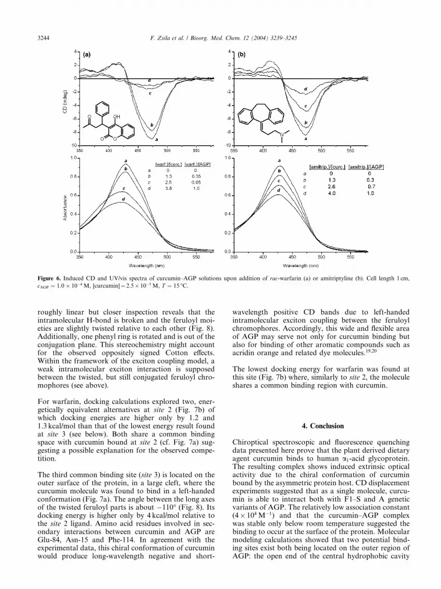
Figure 6. Induced CD and UV/vis spectra of curcumin–AGP solutions upon addition of rac-warfarin (a) or amitriptyline (b). Cell length 1 cm,
cAGP ¼ 1:0� 10�4 M, [curcumin]¼ 2.5� 10�5 M, T ¼ 15 �C.
3244 F. Zsila et al. / Bioorg. Med. Chem. 12 (2004) 3239–3245
roughly linear but closer inspection reveals that theintramolecular H-bond is broken and the feruloyl moi-eties are slightly twisted relative to each other (Fig. 8).Additionally, one phenyl ring is rotated and is out of theconjugation plane. This stereochemistry might accountfor the observed oppositely signed Cotton effects.Within the framework of the exciton coupling model, aweak intramolecular exciton interaction is supposedbetween the twisted, but still conjugated feruloyl chro-mophores (see above).
For warfarin, docking calculations explored two, ener-getically equivalent alternatives at site 2 (Fig. 7b) ofwhich docking energies are higher only by 1.2 and1.3 kcal/mol than that of the lowest energy result foundat site 3 (see below). Both share a common bindingspace with curcumin bound at site 2 (cf. Fig. 7a) sug-gesting a possible explanation for the observed compe-tition.
The third common binding site (site 3) is located on theouter surface of the protein, in a large cleft, where thecurcumin molecule was found to bind in a left-handedconformation (Fig. 7a). The angle between the long axesof the twisted feruloyl parts is about �110� (Fig. 8). Itsdocking energy is higher only by 4 kcal/mol relative tothe site 2 ligand. Amino acid residues involved in sec-ondary interactions between curcumin and AGP areGlu-84, Asn-15 and Phe-114. In agreement with theexperimental data, this chiral conformation of curcuminwould produce long-wavelength negative and short-
wavelength positive CD bands due to left-handedintramolecular exciton coupling between the feruloylchromophores. Accordingly, this wide and flexible areaof AGP may serve not only for curcumin binding butalso for binding of other aromatic compounds such asacridin orange and related dye molecules.19;20
The lowest docking energy for warfarin was found atthis site (Fig. 7b) where, similarly to site 2, the moleculeshares a common binding region with curcumin.
4. Conclusion
Chiroptical spectroscopic and fluorescence quenchingdata presented here prove that the plant derived dietaryagent curcumin binds to human a1-acid glycoprotein.The resulting complex shows induced extrinsic opticalactivity due to the chiral conformation of curcuminbound by the asymmetric protein host. CD displacementexperiments suggested that as a single molecule, curcu-min is able to interact both with F1–S and A geneticvariants of AGP. The relatively low association constant(4� 104 M�1) and that the curcumin–AGP complexwas stable only below room temperature suggested thebinding to occur at the surface of the protein. Molecularmodeling calculations showed that two potential bind-ing sites exist both being located on the outer region ofAGP: the open end of the central hydrophobic cavity

Figure 7. (a) 3D Molecular model of AGP with curcumin molecules
obtained by docking procedure (C, blue; O, red; H, white); (b) 3D
molecular model of AGP with warfarin molecules obtained by docking
procedure (C, blue; O, red; H, white).
Figure 8. Conformers of curcumin found at sites 1–3, respectively
(C, blue; O, red; H, white).
F. Zsila et al. / Bioorg. Med. Chem. 12 (2004) 3239–3245 3245
and a surface cleft. A possible significance of thissuperficial binding is that curcumin may alter theinteraction of AGP molecule with cell membranes,viruses and biopolymers involved in the acute phasereaction.5;21 Although HSA seems to be the mostimportant carrier of curcumin in physiological condi-tions, in case of several diseases with increased AGP anddecreased HSA levels9 AGP binding may also influencepharmacological effects of curcumin. These results alsohighlight that curcumin derivatives and structurally
related compounds such as vanilloids,22 stilbene deriv-atives,23 lignans etc., might also interact with AGPsupporting the need for further studies in that field.
Acknowledgements
Financial support from the National R & D Fund (1/047NKFP Medichem) and from the European Community(QLK2-CT-2002-90436) are gratefully acknowledged.
References and notes
1. Govindarajan, V. S. CRC Crit. Rev. Food. Sci. 1980, 12,199.
2. Aggarwal, B. B.; Kumar, A.; Bharti, A. C. Anticancer Res.2003, 23, 363.
3. Miquel, J.; Bernd, A.; Sempere, J. M.; Diaz-Alperi, J.;Ramirez, A. Arch. Gerontol. Geriat. 2002, 34, 37.
4. Chauhan, D. P. Curr. Pharm. Des. 2002, 8, 1695.5. Fournier, T.; Medjoubi, N. N.; Porquet, D. Biochim.
Biophys. Acta 2000, 1482, 157.6. Herv�e, F.; Gomas, E.; Duche, J. C.; Tillement, J. P. J.
Chromatogr. B 1993, 615, 47.7. Herv�e, F.; Caron, G.; Duch�e, J.-C.; Gaillard, P.; Rahman,
N. A.; Tsantili-Kakoulidou, A.; Carrupt, P.-A.; d’Athis,P.; Tillement, J.-P.; Testa, B. Mol. Pharmacol. 1998, 54,129.
8. Hochepied, T.; Berger, F. G.; Baumann, H.; Libert, C.Cytokine Growth Factor Rev. 2003, 14, 25.
9. Kremer, J. M. H.; Wilting, J.; Janssen, L. H. M.Pharmacol. Rev. 1988, 40, 1.
10. Israili, Z. H.; Dayton, P. G. Drug. Metab. Rev. 2001, 33,161.
11. Reddy, A. C. P.; Sudharshan, E.; Rao, A. G. A.; Lokesh,B. R. Lipids 1999, 34, 1025.
12. Zsila, F.; Bik�adi, Z.; Simonyi, M. Biochem. Biophys. Res.Commun. 2003, 301, 776.
13. Zsila, F.; Bik�adi, Z.; Simonyi, M. Tetrahedron: Asymmetry2003, 14, 2433.
14. Kopecky, V.; Ettrich, R.; Hofbauerova, K.; Baumruk, V.Biochem. Biophys. Res. Commun. 2003, 300, 41.
15. Morris, G. M.; Goodsell, D. S.; Halliday, R. S.; Huey, R.;Hart, W. E.; Belew, R. K.; Olson, A. J. J. Comp. Chem.1998, 19, 1639.
16. Chignell, C. F.; Bilski, P.; Reszka, K. J.; Motten, A. G.;Sik, R. H.; Dahl, T. A. Photochem. Photobiol. 1994, 59,295.
17. Roughley, P. J.; Whiting, D. A. J. Chem. Soc., PerkinTrans. 1 1973, 2379.
18. Arrieta, A.; Beyer, L.; Kleinpeter, E.; Lehmann, J.;Dargatz, M. J. Prakt. Chem. 1992, 334, 696.
19. Maruyama, T.; Otagiri, M.; Takadate, A. Chem. Pharm.Bull. 1990, 38, 1688.
20. Fitos, I.; Visy, J.; Zsila, F.; Bik�adi, Z.; M�ady, G.; Simonyi,M. Biochem. Pharmacol. 2004, 67, 679.
21. Nishi, K.; Sakai, N.; Komine, Y.; Maruyama, T.; Halsall,H. B.; Otagiri, M. Biochim. Biophys. Acta 2002, 1601, 185.
22. Szallasi, A.; Lewin, N. E.; Blumberg, P. M. J. Pharmacol.Exp. Ther. 1992, 262, 883.
23. Paterson, S. C.; Lim, C. K.; Smith, K. D. Biomed.Chromatogr. 2003, 17, 143.