A new macromolecular paramagnetic MR contrast agent binds to activated human platelets

ARTICLESPUBLISHED ONLINE: 8 DECEMBER 2014 | DOI: 10.1038/NPHYS3152
Strong magneto-chiral dichroism in aparamagnetic molecular helixobserved by hard X-raysRoberta Sessoli1*, Marie-Emmanuelle Boulon1, Andrea Caneschi1, Matteo Mannini1, Lorenzo Poggini1,FabriceWilhelm2 and Andrei Rogalev2
Magneto-chiral dichroism is a non-reciprocal—that is, directional—eect observed in magnetized chiral systems, featuring anunbalanced absorption of unpolarized light depending on the direction of the magnetization. Despite the fundamental interestin a phenomenon breaking both parity and time-reversal symmetries, magneto-chiral dichroism is one of the least investigatedaspects of light–matter interaction most likely because of the weakness of the eect in most reported experiments. Here wehave exploited the element selectivity of hard X-ray radiation to investigate the magneto-chiral properties of enantiopurecrystals of two isostructural molecular helicoidal chains comprising either cobalt(II) or manganese(II) ions. A strong magneto-chiral dichroism, with Kuhn asymmetry of the order of a few per cent, has been observed in the cobalt chains system, whereasit is practically absent for the manganese derivative. The spectral features of the X-ray magneto-chiral dichroism signaldier significantly from the natural and magnetic dichroic contributions and have been rationalized here using the multipolarexpansion of matter–radiation interaction.
Interest in the interplay between chirality and magnetism datesback to Pasteur1, but is still the subject of intense research2,being relevant for a wide class of phenomena, ranging from
exotic magnetic excitations known as skyrmions3–5 to magneto-chiral conductance6. The interaction between light and matteris a powerful tool to investigate the simultaneous breaking ofspatial symmetry—that is, the lack of inversion symmetry—andof time-reversal symmetry, as in the case of a magnetized non-centrosymmetric medium. Magnetism and chirality are indeeddirectly connected through the magneto-chiral dichroism (MχD)and birefringence7–10, as was observed for the first time by Rikkenand Raupach11 in luminescence spectra and subsequently usingX-ray radiation12. MχD is a non-reciprocal effect consisting indifferent absorption of unpolarized light by a chiral magnetizedmedium. It is a fascinating parity-violating phenomenon that hasbeen suggested to be at the origin of homochirality of life onearth13–15 as an alternative to electroweak nuclear interactions16.MχD is generally a very weak effect, with only a few examplesreported17–21 and a limited knowledge available of the factors thatlead to the phenomenon.
The conditions of simultaneous breaking of parity and time-reversal symmetry required to observe the phenomenon aresatisfied in magneto-electric media and multiferroics22,23, but theycan also be fulfilled in molecular paramagnetic and diamagneticsystems in the presence of an external magnetic field. The systemsinvestigated here, consisting of isostructural molecular helicoidalchains comprising either anisotropic cobalt(II) or isotropicmanganese(II) ions, can be considered intermediate betweenthese two classes. A thorough investigation of the magneto-chiral effect through X-ray spectroscopy at the K-edge of the3d-metals, evidencing a strong MχD in the cobalt chain system,is described.
We investigated two isostructural one-dimensional (1D) molec-ular chains of formula [M(hfac)2NITPhOMe]∞, containing bi-positive metal ions (M=MnII and CoII) shielded by ancillaryligands (hfac−= hexafluoroacetylacetonato) and bridged by sta-ble nitronyl-nitroxide organic radicals (NITPhOMe= 2-(4-meth-oxyphenyl)-4,4,5,5-tetramethylimidazoline-l-oxy1,3-oxide) carry-ing a delocalized unpaired electron24,25. The helical structure of[M(hfac)2NITPhOMe]∞, shortened to [M-NIT]∞ hereafter, is gen-erated in the crystalline phase by a three-fold screw axis (Fig. 1). De-spite the absence of chiral constituents, the compounds form enan-tiopure crystals, crystallizing either in the chiral P31 or P32 spacegroups24,25. The crystals are optically active and exhibit a significantsecond-harmonic generation efficiency26. The magnetism of bothcompounds is governed by the strong intra-chain antiferromagneticexchange interaction between the alternating paramagnetic metalions and the spin S= 1/2 of the organic radicals, with exchangeconstant, J =235K and 495K for the Co (ref. 27) and Mn (ref. 24)derivative, respectively. The exchange Hamiltonian for a chain ofN spins is defined as Hex= J6N/2
i=1 s2i−1S2i+ s2i+1S2i, where the smalls on odd sites represents the radical spin while the capital S oneven sites represents either the CoII or MnII spin. A completelydifferent behaviour is, however, observed depending on the metalion. In the case of MnII, Heisenberg 1D ferrimagnetic behaviour isobserved owing to the lack of an orbital contribution for this d5 ion,with strong long-range correlations and 3Dordering aroundT=5Kinduced by the weak inter-chain dipolar interactions24. In contrast,high-spin CoII ions in an octahedral environment have a significantorbital moment, resulting in a strong magnetic anisotropy, with theeasy axis of magnetization of each ion forming an angle of about50 with the helix axis (Fig. 1a). Interestingly, the cobalt derivativewas the first system exhibiting slowing down of the magnetizationdynamics in the paramagnetic phase, as predicted by Glauber for
1Department of Chemistry ‘Ugo Schi’ and INSTM Research Unit, University of Florence, 50019 Sesto Fiorentino, Italy. 2European Synchrotron RadiationFacility (ESRF), 38043 Grenoble, France. *e-mail: [email protected]
NATURE PHYSICS | ADVANCE ONLINE PUBLICATION | www.nature.com/naturephysics 1© 2014 Macmillan Publishers Limited. All rights reserved.

ARTICLES NATURE PHYSICS DOI: 10.1038/NPHYS3152
Table 1 | Dierent dichroic contributions to the radiation–matter interaction.
Parity Time reversal
XNCD E1M1+E1E2 − +
XMCD (E1E1+E2E2)M + −
XMχD (E1M1+E1E2)M − −
The involved mixed terms of the radiation–matter interaction and their parity behaviour arelisted for each type of dichroism. M stands for the sample magnetization. For the sake ofcompleteness we recall that other types of dichroism can be obtained with linearly polarizedlight. These are: X-ray natural linear dichroism (XNLD), X-ray magnetic linear dichroism(XMLD), which are both parity and time-reversal even, and non-reciprocal linear dichroism(NRLD), which is parity odd and time-reversal odd.
the 1D Ising model28. An activation barrier of about 170K forthe reversal of the magnetization characterizes this material, andmagnetic hysteresis in the absence of long-range order is observedbelow 5K (ref. 25). Finite-size effects with collective reversal of spinsegments29 and an unprecedentedmechanism to controlmagnetiza-tion dynamics through light-induced domain-wall kickoff have alsobeen recently observed in this fascinating material27.
To investigate the MχD of these 1D molecular crystals, hardX-rays were used to get element-specific information. The excellentstability of the ID12 beamline at ESRF was necessary to investigatesingle crystals of [Mn-NIT]∞ and [Co-NIT]∞ chains with smalldimensions (0.3×0.3×8mm). X-ray absorption near-edge spectra(XANES) were recorded at the Co and Mn K-edges—that is,transitions promoting electrons from the 1s core level—occurringat around 7.7 keV and 6.5 keV, respectively, using the total X-rayfluorescence yield detection mode (see Methods for details).
By recording the XANES spectra with opposite σ+ (circularleft) and σ− (circular right) polarizations and with a magneticfield applied either parallel (B+) or antiparallel (B−) to the X-raywavevector, we could obtain the three relevant types of dichroism:
XNCD = 1/2[µ(σ−,B+)−µ(σ+,B+)]
+[µ(σ−,B−)−µ(σ+,B−)] (1)
XMCD = 1/2[µ(σ−,B+)−µ(σ+,B+)]
−[µ(σ−,B−)−µ(σ+,B−)] (2)
XMχD = [µ(σ−,B+)+µ(σ+,B+)]
−[µ(σ−,B−)+µ(σ+,B−)] (3)
where µ(σ ,B) stands for the absorption measured for the indicatedpolarization and sign of the magnetic field. XN(M)CD indicateX-ray natural (magnetic) circular dichroism signals and aredefined as the difference in absorption spectra recorded withthe two circular polarizations. The XNCD signal is independentof the applied magnetic field, whereas XMCD changes its signwhen the direction of the applied magnetic field is reversed. Themagneto-chiral effect manifests as changes in absorption for twodirections of magnetic field, and as such does not require polarizedlight. To be able to disentangle the XMχD from other dichroiccontributions we sum up the spectra recorded with right and leftcircularly polarized X-rays. The accumulation of the spectra wasperformed changing field polarity and light helicity in a cyclic wayto minimize eventual drift effects in the evaluation of the dichroicquantities. No detectable radiation damage was observed in any ofthe investigated crystals.
First, room-temperature XNCD spectra were recorded on severalcrystals to identify two enantiomeric crystals of each [M-NIT]∞
S = 1/2
CoII
MnII
a
c
c
B, k
b
15°
+σ−σ
a b c
d
Figure 1 | Structures of the molecular magnetic helices and experimentalset-up. a,b, View of the simplified structure of the [M-NIT]∞ molecularhelices containing CoII (a) and MnII (b) ions bridged by organicnitronyl-nitroxide radicals, with a radical unit highlighted by the red circle.The metal ions are highlighted as large spheres. The ancillary hfac ligandsand the radical backbone are in grey, while the bonds constituting thepathway for the magnetic exchange interaction are highlighted in yellow,with the radical oxygen atoms in red and nitrogen atoms in blue. Somegroups of atoms (that is, CF3, CH3, and O–CH3) have been omitted for thesake of clarity. The helices develop along the crystallographic c axis of theP31/P32 space groups. The green arrows represent the orientation of themagnetic moments when the magnetic field is applied parallel to the c axis,which are not collinear to the field in the case of the anisotropic CoII ions.c, Schematic side view of the geometry of the experiment, whereneedle-like single crystals were mounted on a copper sample holder to forman angle of 15 between the chain direction, c, and the propagation vector, k,of the X-rays, which is parallel to the applied magnetic field. d, Photographof the sample mounting, viewed from the top, with a ruler as reference.
chain; then the selected crystals were transferred to an experimentalstation equippedwith a 17 T superconductingmagnet and constant-flow helium cryostat. These crystals were mounted on the coldfinger of the cryostat and orientedwith the crystallographic c (helix)axis forming an angle of 15 with the vector of propagation of thelight k, the latter being parallel to the applied magnetic field B(Fig. 1c,d). A field of 3 T was employed at T = 5K to ensure themagnetic saturation for crystals of both Mn and Co chains and tosuppress the slow magnetic relaxation for the latter.
Before describing the results, we briefly recall here somefundamental differences in the interaction between matter andlight when moving from ultraviolet–visible to hard X-ray radiation.Because atomic core states are involved in X-ray promotedtransitions, the long-wavelength approximation is still valid despitethe high energy. Thus the interaction can be expressed in the usualmultipolar expansion:
Hint=E1+M1+E2 (4)
where E1 stands for the interaction between the electric dipoleand the electric field of the electromagnetic radiation, M1 for theinteraction between the magnetic dipole and the magnetic field,and E2 for the interaction between the electric quadrupole andthe electric field gradient. Differently from the ultraviolet–visibleregion, in X-ray spectroscopy theM1 interaction is negligibly small,involving atomic states with different principal quantum number,whereas E2 becomes relevant, given its linear dependence with theenergy of the transitions.
As the absorption cross section is proportional to the squareof the transition matrix element
∣∣⟨ϕf |Hint|ϕi⟩∣∣2, with Hint from
2 NATURE PHYSICS | ADVANCE ONLINE PUBLICATION | www.nature.com/naturephysics© 2014 Macmillan Publishers Limited. All rights reserved.
NATURE PHYSICS DOI: 10.1038/NPHYS3152 ARTICLES
0.0
0.3
0.0
−0.3
0.2
0.0
−0.2
0.2
0.0
XM D
(%)
χXM
CD (%
)XN
CD (%
)XA
NES
−0.2
7,700 7,720 7,740Energy (eV)
7,760 7,780
0.5
1.0
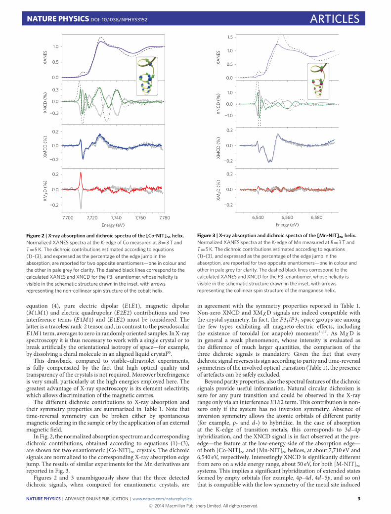
Figure 2 | X-ray absorption and dichroic spectra of the [Co-NIT]∞ helix.Normalized XANES spectra at the K-edge of Co measured at B=3 T andT=5 K. The dichroic contributions estimated according to equations(1)–(3), and expressed as the percentage of the edge jump in theabsorption, are reported for two opposite enantiomers—one in colour andthe other in pale grey for clarity. The dashed black lines correspond to thecalculated XANES and XNCD for the P31 enantiomer, whose helicity isvisible in the schematic structure drawn in the inset, with arrowsrepresenting the non-collinear spin structure of the cobalt helix.
equation (4), pure electric dipolar (E1E1), magnetic dipolar(M1M1) and electric quadrupolar (E2E2) contributions and twointerference terms (E1M1) and (E1E2) must be considered. Thelatter is a traceless rank-2 tensor and, in contrast to the pseudoscalarE1M1 term, averages to zero in randomly oriented samples. InX-rayspectroscopy it is thus necessary to work with a single crystal or tobreak artificially the orientational isotropy of space—for example,by dissolving a chiral molecule in an aligned liquid crystal30.
This drawback, compared to visible–ultraviolet experiments,is fully compensated by the fact that high optical quality andtransparency of the crystals is not required. Moreover birefringenceis very small, particularly at the high energies employed here. Thegreatest advantage of X-ray spectroscopy is its element selectivity,which allows discrimination of the magnetic centres.
The different dichroic contributions to X-ray absorption andtheir symmetry properties are summarized in Table 1. Note thattime-reversal symmetry can be broken either by spontaneousmagnetic ordering in the sample or by the application of an externalmagnetic field.
In Fig. 2, the normalized absorption spectrumand correspondingdichroic contributions, obtained according to equations (1)–(3),are shown for two enantiomeric [Co-NIT]∞ crystals. The dichroicsignals are normalized to the corresponding X-ray absorption edgejump. The results of similar experiments for the Mn derivatives arereported in Fig. 3.
Figures 2 and 3 unambiguously show that the three detecteddichroic signals, when compared for enantiomeric crystals, are
0.0
1.0
0.0
−1.0
0.2
0.0
−0.2
0.2
0.0
XMCD
(%)
XNCD
(%)
XAN
ES
−0.2
0.5
1.0
1.5
6,540 6,560 6,580Energy (eV)
XM D
(%)
χ
Figure 3 | X-ray absorption and dichroic spectra of the [Mn-NIT]∞ helix.Normalized XANES spectra at the K-edge of Mn measured at B=3 T andT=5 K. The dichroic contributions estimated according to equations(1)–(3), and expressed as the percentage of the edge jump in theabsorption, are reported for two opposite enantiomers—one in colour andother in pale grey for clarity. The dashed black lines correspond to thecalculated XANES and XNCD for the P31 enantiomer, whose helicity isvisible in the schematic structure drawn in the inset, with arrowsrepresenting the collinear spin structure of the manganese helix.
in agreement with the symmetry properties reported in Table 1.Non-zero XNCD and XMχD signals are indeed compatible withthe crystal symmetry. In fact, the P31/P32 space groups are amongthe few types exhibiting all magneto-electric effects, includingthe existence of toroidal (or anapole) moments31,32. As MχD isin general a weak phenomenon, whose intensity is evaluated asthe difference of much larger quantities, the comparison of thethree dichroic signals is mandatory. Given the fact that everydichroic signal reverses its sign according to parity and time-reversalsymmetries of the involved optical transition (Table 1), the presenceof artefacts can be safely excluded.
Beyondparity properties, also the spectral features of the dichroicsignals provide useful information. Natural circular dichroism iszero for any pure transition and could be observed in the X-rayrange only via an interference E1E2 term. This contribution is non-zero only if the system has no inversion symmetry. Absence ofinversion symmetry allows the atomic orbitals of different parity(for example, p- and d-) to hybridize. In the case of absorptionat the K-edge of transition metals, this corresponds to 3d–4phybridization, and the XNCD signal is in fact observed at the pre-edge—the feature at the low-energy side of the absorption edge—of both [Co-NIT]∞ and [Mn-NIT]∞ helices, at about 7,710 eV and6,540 eV, respectively. Interestingly XNCD is significantly differentfrom zero on a wide energy range, about 50 eV, for both [M-NIT]∞systems. This implies a significant hybridization of extended statesformed by empty orbitals (for example, 4p–4d , 4d–5p, and so on)that is compatible with the low symmetry of the metal site induced
NATURE PHYSICS | ADVANCE ONLINE PUBLICATION | www.nature.com/naturephysics 3© 2014 Macmillan Publishers Limited. All rights reserved.
ARTICLES NATURE PHYSICS DOI: 10.1038/NPHYS3152
by the ligands. Similar wide-energy XNCD features have also beenobserved in NdIII (ref. 33) and NiII (ref. 34) compounds, in whichthe chirality is induced by the structural arrangement of non-chiralmoieties, whereas the XMCD signal for a CoII complex with chiralcoordination is present only in the pre-edge region where the 3dorbitals contribute predominantly35.
Both XANES and XNCD were reproduced by calculationsperformed using the FDMNES (Finite Difference Method Near-Edge Structure) package36. The electronic structures around Coand Mn atoms were calculated using multiple scattering theorywithin the muffin-tin approximation, based on a mono-electronicapproach. Calculations were performed for clusters built fromcrystallographic data for the P31 space group including hydrogenatoms, and views of the asymmetric units generating the chainstructure are provided in Supplementary Fig. 1 together witha list of selected bond distances and angles (SupplementaryTable 1). Natural circular dichroism was calculated consideringE1E2 transitions only (see Methods for details). The spectralfeatures of both derivatives are reproduced with a reasonableagreement to assign unambiguously the P31 space group, whosechirality is shown in the inset of Figs 2 and 3, to the crystals havingtheir spectra drawn in colour in the corresponding figure. TheXNCD intensity at the pre-edge is about twice as large as for the[Mn-NIT]∞ helix, in agreement with the larger number of holes inthe 3d orbitals and the larger calculated density-of-states (DOS) ofthe d-orbitals at the Fermi level (reported as Supplementary Fig. 2).
Concerning XMCD, for which interference E1E2 contributionsare forbidden by symmetry considerations, the dichroic signal isdue to both the dipolar E1E1 (1s→ 4p) transitions at the risingedge and to the quadrupolar E2E2 (1s→3d) transitions at the pre-edge part of the spectrum, where the partially occupied 3d orbitalsare involved. Given the fact that the initial 1s state has no spin–orbit coupling, the XMCD at the K-edge is probing only the orbitalmagnetization of the final states. For a d5 ion (MnII), K-edge XMCDat the pre-edge is thus expected to be much weaker than for theCo derivative. Comparing Figs 2 and 3, it is clearly evident thatXMCD at the pre-edge is significantly reduced in moving fromCo to Mn, despite the magnetization being higher in the latter.In contrast, XMCD at the rising edge, which originates from theorbital polarization of the 4p states, is fairly similar for the Co andMn helices, as expected (see also the calculated p-type DOS inSupplementary Fig. 2).
Finally, moving on to X-ray MχD, we notice that it originatesfrom the same interference interaction terms as XNCD, althoughcombined with the orbital magnetization of the final states of theabsorbing atom. Here a significant difference is observed betweenthe two systems. [Co-NIT]∞ helices exhibit a large XMχD signalwhose intensity exceeds that of the XMCD signal. To allow abetter comparison with data extracted from ultraviolet–visiblespectroscopy, the dichroic contributions have been plotted as theircorrespondent Kuhn asymmetry—that is, g =1µ/µ, where µ isthe absorption. The results shown in the inset of Fig. 4 revealthat the magneto-chiral effect exceeds 3% of the correspondingabsorption and is, thus, a remarkable quantity compared toprevious reported values.17–21,37 For instance XMχD of [Co-NIT]∞is two orders of magnitude stronger than that obtained frompreliminary measurements at the K-edge on a CrIII–NiII molecularferromagnet34 or that measured in absorption experiments in theultraviolet–visible range on a ferromagnetically ordered CrIII–MnIImolecular compound19.
As far as the spectral shape is concerned, the XMχD signal iscompletely different from the other dichroic contributions, showinga well-defined narrow peak around 7,710 eV—that is, at the pre-edge. This is nicely in agreement with qualitative expectations—aquantitative analysis of this effect being at present beyond availabletheoretical models. In fact, the intensity of the magneto-chiral
XMCDg (%
)
XAN
ES (a
.u.)
0.00.0
0.4
0.8
0.0
−0.2
−0.4
−4
0
4
0.0 0.5 1.0 1.5Magnetic field (T)
Photon energy
7,708 7,712 7,716 7,720
2.0 2.5 3.0
0.2
0.4
0.6
XM Dχ
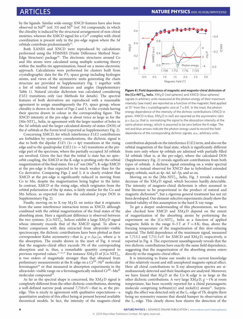
Figure 4 | Field dependence of magnetic and magneto-chiral dichroism ofthe [Co-NIT]∞ helix. XMχD (red spheres) and XMCD (blue spheres)signals in arbitrary units measured at the photon energy of their maximumintensity (see inset) are reported as a function of the magnetic field appliedat 15 from the c crystallographic axis at T=8 K. In the inset, the photonenergy dependence of the intensity of the dichroic contributions (XNCD ingreen, XMCD in blue, XMχD in red) are reported as the asymmetric ratiog=1µ/µ; that is, normalizing the signal to the absorption intensity at thesame photon energy, which is assumed to be zero before the K-edge. Thered and blue arrows indicate the photon energy used to record the fielddependence of the corresponding dichroic signals. a.u., arbitrary units.
contribution depends on the interferenceE1E2 term, and also on theorbital magnetism of the final state, which is significantly differentfrom zero only where 4p orbitals are admixed with partially filled3d orbitals (that is, at the pre-edge), where the calculated DOS(Supplementary Fig. 2) reveals significant contributions from bothtypes of orbitals. A dichroic signal extending on a wider spectralregion is instead observed for XNCD due to hybridized extendedempty orbitals, such as 4p–4d , 4d–5p, and so on.
Moving on to the [Mn-NIT]∞ helix, Fig. 3 reveals a markeddecrease of the XMχD signal, which becomes hardly detectable.The intensity of magneto-chiral dichroism is often assumed inthe literature to be proportional to the product of natural andmagnetic dichroism38, but a rigorous general treatment has not yetbeen developed.Our element-selective experiments clearly show thelimited validity of this assumption in the hard X-ray range.
To get a deeper understanding of this phenomenon, we havealso checked how XMCD and XMχD depend on the extentof magnetization of the absorbing atoms by performing theexperiment on the [Co-NIT]∞ helix as a function of appliedmagnetic fields in the range 0–3 T at T = 8K; thus, above thefreezing temperature of the magnetization of this slow-relaxingmaterial. The field dependence of the maximum signal, measuredat 7,713.2 and 7,711.5 eV for XMCD and XMχD, respectively, isreported in Fig. 4. The experiment unambiguously reveals that thetwo dichroic contributions have exactly the same field dependence,suggesting that the magnetization of the absorbing atoms entersdirectly in the magneto-chiral effect.
It is interesting to frame our results in the current knowledgeof this relatively recent and still unexplored magneto-optical effect.Here all chiral contributions to X-ray absorption spectra are si-multaneously detected and their lineshapes are analysed. Moreover,we have found that MχD at the Co K-edge is as large as theother dichroic contributions. A very large XMχD, g ∼1% at roomtemperature, has been recently reported for a chiral paramagneticmolecule comprising terbium(III) and nickel(II) atoms21. Surpris-ingly, the effect was detected only at the L3-edge of Tb, despite therebeing no symmetry reasons that should hamper its observation atthe L2-edge. This clearly shows how elusive the detection of the
4 NATURE PHYSICS | ADVANCE ONLINE PUBLICATION | www.nature.com/naturephysics© 2014 Macmillan Publishers Limited. All rights reserved.
NATURE PHYSICS DOI: 10.1038/NPHYS3152 ARTICLESmagneto-chiral effect can be, and underlines the relevance of acomplete characterization such as the one presented here.
Additional information can be extracted by the unprecedentedpossibility of comparing the magneto-chiral behaviour of twoisostructural 1D systems showing very different magneticproperties. First, it is clearly evident that the asymmetry factorof the magneto-chiral effect in this energy range is not simplythe product of the natural and magnetic effects, because in thisapproximation a large signal should be observed over a wideenergy range and much stronger XMχD should be observed for[Mn-NIT]∞. Furthermore, the XMχD signal is significant onlyfor transitions involving 3d partially filled orbitals and only inthe presence of a strong orbital contribution, as in the case of ad7 ion in an octahedral environment, which is responsible for thenon-collinear spin arrangement along the [Co-NIT]∞ helix.
It would be interesting to investigate if the three-fold screw axisgenerating the molecular [M-NIT]∞ helices plays a significant rolein the large MχD observed here. Other chains comprising the samebuilding blocks and differing only in the organic group on theradical—aliphatic instead of aromatic (Supplementary Fig. 3)—havebeen structurally and magnetically characterized, but unfortunatelyall of them crystallize in centrosymmetric space groups39. However,thanks to the element selectivity of the X-ray absorption, it has beenpossible to compute the XNCD of acentric mixed-metal structuresartificially segregating Co and Mn on sites of opposite paritycompared to the inversion centre (Supplementary Fig. 4 andNote 1).The results, obtained using the previously described computationalapproach, reveal a significant decrease, to about 1/3 of the XNCDcalculated signal, when moving from the P31/P32 to the P21 crystalspace group, as shown in Supplementary Fig. 5. As the trigonalspace group of the investigated helices is induced by the π-stackinginteractions between the aromatic substituent on the radical and thehfac ligand on the metal39, a route to enhance the magneto-chiraleffect inmolecular materials through a rational chemical design canbe envisaged.
In conclusion, the investigated magnetic molecular helices area model system to study the magneto-chiral effect in detail. Thesymmetry of the material is compatible with a large variety ofmagneto-electric effects22,23,40, still poorly investigated in molecularmaterials. According to sum rules41,42, the X-ray MχD could beassociated with the presence of an atomic anapole orbital moment,ΩL, originating from toroidal orbital currents centred on the cobaltatoms. These orbital currents, of relevance for many phenomenaranging from multiferroicity22,23 to superconductivity43, originatefrom the hybridized 3d–4p states, allowed by the absence ofinversion symmetry of the coordination environment around theatom, and therefore they are much stronger that what one wouldexpect for those induced by parity breaking due to the electroweakinteraction. It is important to underline that this atomic anapoleorbital moment should not be confused with the macroscopictoroidal moment27 that could originate from the peculiar non-collinear orientation of the magnetic moments along the three-foldhelix, which is compatible withDzyaloshinskii–Moriya interactions.However, to investigate this additional contribution, a less localprobe—namely, ultraviolet–visible light—is necessary. Last, butnot least, we recall that the [Co-NIT]∞ molecular helix presentsthe additional feature of magnetic bistability in the paramagneticphase. This system thus seems a good candidate for the detectionof the ‘inverse’ magneto-chiral effect—that is, the induction ofmagnetic polarization in a chiral system by irradiation withnon-polarized light. In the scenario of light–matter interaction,this effect, although theoretically predicted44, is the one stillnot evidenced by experiments40. The possibility to freeze, atlow temperature, the light-induced magnetization of this bistablemolecular material should facilitate the detection of a weak inversemagneto-chiral effect.
MethodsCrystals of [Mn(hfac)2NITPhOMe]∞ and [Co(hfac)2NITPhOMe]∞ were preparedas previously reported24,39. Single crystals with needle shape and approximatelargest dimensions 10×0.5×0.5mm3 were selected and checked for the absenceof twinning with an Oxford Diffraction single-crystal diffractometer.
The X-ray absorption experiments were carried out at the ID12 beamline ofthe European Synchrotron Radiation Facility (ESRF), which is dedicated topolarization-dependent X-ray spectroscopy in the photon energy range from 2 to15 keV (ref. 45). For experiments at the Mn and Co K-edge the source was thehelical undulator APPLE-II, which provides a high flux of either right or leftcircularly polarized X-rays photons with a polarization rate in excess of 0.95. Thehelicity could be changed in a time less than 5 s. X-rays were monochromatizedby the Si〈111〉 double-crystal monochromator, ensuring an energy resolutionbetter than the intrinsic broadening due to the finite core-hole lifetime. Thesamples were mounted on the cold finger of a constant-flow helium cryostatinserted in the bore of a superconducting solenoid producing a magnetic field upto 17 T. The sweep rate to reverse the direction of magnetic field rate was2 Tmin−1. All spectra were recorded in total X-ray fluorescence detection modein a backscattering geometry using Si photodiodes. The total X-ray fluorescencesignal in this energy range is expected to be isotropic. Either the helicity of theincoming X-rays or the direction of magnetic field were changed after eachconsecutive energy scan to minimize any eventual artefacts in the measurements.
The XANES and XNCD spectra were simulated using the FDMNES code36.Calculations were performed for clusters built from crystallographic data for theP31 space group including hydrogen atoms. Crystallographic data are availablefrom the Cambridge Structural Database (www.ccdc.cam.ac.uk/Community/Requestastructure/Pages/Requestastructure.aspx) using the doi codeshttp://dx.doi.org/10.1021/ic00020a029 and http://dx.doi.org/10.1039/B004244Gfor Mn and Co derivatives, respectively. Natural circular dichroism was calculatedconsidering E1E2 transitions only. Self-consistent calculations includingrelativistic effects were also performed, and similar results were obtained. Clustersof radius of 14Å were employed to reproduce the main features in the XNCD, asa further increase of the cluster size did not lead to any improvement. The sameprocedure was repeated for an aliphatic radical analogue46 crystallizing in thecentrosymmetric P21/c space group (use doi codehttp://dx.doi.org/10.1021/ic00283a018 to retrieve crystallographic data from thedatabase mentioned above) by replacing the Mn atoms on the screw axis ofopposite chirality, generated by the inversion centre, with Co atoms. Thestructural similarity between the Mn and Co derivatives and their completemiscibility to form mixed species suggest that no significant structural stress isinduced in this artificial model. The spectra were convolved with a Lorentzianwith an energy-dependent width, to take into account the core-hole lifetime, andwith a Gaussian line, to account for the energy resolution of monochromator. Thecalculated absorption cross-sections were normalized to the same edge jump tothe continuum as in the experiment.
Received 22 June 2014; accepted 9 October 2014;published online 8 December 2014
References1. Pasteur, L. La dissymétrie moléculaire. Conférence faite le 22 décembre 1883.
Rev. Sci. 7, 2–6 (1884).2. Bordacs, S. et al. Chirality of matter shows up via spin excitations. Nature Phys.
8, 734–738 (2012).3. Mühlbauer, S. et al. Skyrmion lattice in a chiral magnet. Science 323,
915–919 (2009).4. Fert, A., Cros, V. & Sampaio, J. Skyrmions on the track. Nature Nanotech. 8,
152–156 (2013).5. Romming, N. et al.Writing and deleting single magnetic skyrmions. Science
341, 636–639 (2013).6. Pop, F., Auban-Senzier, P., Canadell, E., Rikken, G. L. J. A. & Avarvari, N.
Electrical magnetochiral anisotropy in a bulk chiral molecular conductor.Nature Commun. 5, 3757 (2014).
7. Wagnière, G. & Meier, A. The influence of a static magnetic field on theabsorption coefficient of a chiral molecule. Chem. Phys. Lett. 93, 78–81 (1982).
8. Groenewege, M. P. A theory of magneto-optical rotation in diamagneticmolecules of low symmetry.Mol. Phys. 5, 541–563 (1962).
9. Barron, L. D. & Vrbancich, J. Magneto-chiral birefringence and dichroism.Mol. Phys. 51, 715–730 (1984).
10. Baranova, N. B., Bogdanov, Y. V. & Zel’Dovich, B. Y. Electrical analog of theFaraday effect and other new optical effects in liquids. Opt. Commun. 22,243–247 (1977).
11. Rikken, G. & Raupach, E. Observation of magneto-chiral dichroism. Nature390, 493–494 (1997).
12. Goulon, J. et al. X-ray magnetochiral dichroism: A new spectroscopic probe ofparity nonconserving magnetic solids. Phys. Rev. Lett. 88, 237401 (2002).
NATURE PHYSICS | ADVANCE ONLINE PUBLICATION | www.nature.com/naturephysics 5© 2014 Macmillan Publishers Limited. All rights reserved.
ARTICLES NATURE PHYSICS DOI: 10.1038/NPHYS3152
13. Wagnière, G. H. On Chiralty and the Universal Asymmetry (Verlag HelveticaChimica Acta, 2007).
14. Guijarro, A. & Yus, M. The Origin of Chirality in the Molecules of Life (RoyalSociety of Chemistry, 2009).
15. Rikken, G. L. J. A. & Raupach, E. Enantioselective magnetochiralphotochemistry. Nature 405, 932–935 (2000).
16. Zel’dovich, Y. B. Electromagnetic interaction with parity violation. Sov. Phys.JETP 6, 1184–1186 (1958).
17. Vallet, M. et al. Observation of magnetochiral birefringence. Phys. Rev. Lett. 87,183003 (2001).
18. Kubota, M. et al. X-ray directional dichroism of a polar ferrimagnet. Phys. Rev.Lett. 92, 137401 (2004).
19. Train, C. et al. Strong magneto-chiral dichroism in enantiopure chiralferromagnets. Nature Mater. 7, 729–734 (2008).
20. Kitagawa, Y., Segawa, H. & Ishii, K. Magneto-chiral dichroism of organiccompounds. Angew. Chem. Int. Ed. 50, 9133–9136 (2011).
21. Ceolín, M., Goberna-Ferrón, S. & Galán-Mascarós, J. R. Strong hard X-raymagnetochiral dichroism in paramagnetic enantiopure molecules. Adv. Mater.24, 3120–3123 (2012).
22. Cheong, S-W. & Mostovoy, M. Multiferroics: A magnetic twist forferroelectricity. Nature Mater. 6, 13–20 (2007).
23. Khomskii, D. Classifying multiferroics: Mechanisms and effects. Physics 2,20 (2009).
24. Caneschi, A., Gatteschi, D., Rey, P. & Sessoli, R. Structure andmagnetic-ordering of a ferrimagnetic helix formed by manganese(II) and anitronyl nitroxide radical. Inorg. Chem. 30, 3936–3941 (1991).
25. Caneschi, A. et al. Cobalt(II)-nitronyl nitroxide chains as molecular magneticnanowires. Angew. Chem. Int. Ed. 40, 1760–1763 (2001).
26. Cavigli, L., Sessoli, R., Gurioli, M. & Bogani, L. Second harmonic generation ina molecular magnetic chain. Phys. Status Solidi A 203, 1402–1408 (2006).
27. Heintze, E. et al. Dynamic control of magnetic nanowires by light-induceddomain-wall kickoffs. Nature Mater. 12, 202–206 (2013).
28. Glauber, R. J. Time-dependent statistic of the Ising model. J. Math. Phys. 4,294–307 (1963).
29. Bogani, L. et al. Finite-size effects in single chain magnets: An experimentaland theoretical study. Phys. Rev. Lett. 92, 207204 (2004).
30. Goulon, J. et al. X-ray optical activity: Applications of sum rules. JETP 97,402–431 (2003).
31. Spaldin, N. A., Fiebig, M. & Mostovoy, M. The toroidal moment incondensed-matter physics and its relation to the magnetoelectric effect. J. Phys.Condens. Matter 20, 434203 (2008).
32. Szaller, D., Bordács, S. & Kézsmárki, I. Symmetry conditions for nonreciprocallight propagation in magnetic crystals. Phys. Rev. B 87, 014421 (2013).
33. Alagna, L. et al. X-ray natural circular dichroism. Phys. Rev. Lett. 80,4799–4802 (1998).
34. Goulon, J., Rogalev, A. & Brouder, C. Comprehensive Chiroptical Spectroscopy457–491 (John Wiley, 2012).
35. Stewart, B. et al. Circular dichroism at the edge: Large X-ray natural CD in the1s→3d pre-edge feature of 2[Co(en)3Cl3]·NaCl·6H2O. J. Am. Chem. Soc. 121,10233–10234 (1999).
36. Bunău, O. & Joly, Y. Self-consistent aspects of x-ray absorption calculations.J. Phys. Condens. Matter 21, 345501 (2009).
37. Kitagawa, Y., Miyatake, T. & Ishii, K. Magneto-chiral dichroism of artificiallight-harvesting antenna. Chem. Commun. 48, 5091–5093 (2012).
38. Rikken, G. & Raupach, E. Pure and cascaded magnetochiral anisotropy inoptical absorption. Phys. Rev. E 58, 5081–5084 (1998).
39. Caneschi, A., Gatteschi, D., Lalioti, N., Sangregorio, C. & Sessoli, R.Supramolecular interactions and magnetism of metal-radical chains. J. Chem.Soc. Dalton Trans. 3907–3912 (2000).
40. Kibayashi, S., Takahashi, Y., Seki, S. & Tokura, Y. Magnetochiral dichroismresonant with electromagnons in a helimagnet. Nature Commun. 5,4583 (2014).
41. Carra, P., Jerez, A. & Marri, I. X-ray dichroism in noncentrosymmetric crystals.Phys. Rev. B 67, 045111 (2003).
42. Lovesey, S. W. & Balcar, E. Quantum theory of natural circular, magneto-chiraland non-reciprocal linear dichroism. Phys. Scr. 81, 065703 (2010).
43. Scagnoli, V. et al. Observation of orbital currents in CuO. Science 332,696–698 (2011).
44. Wagnière, G. Inverse magnetochiral birefringence. Phys. Rev. A 40,2437–2440 (1989).
45. Rogalev, A., Goulon, J., Goulon-Ginet, C. & Malgrange, C. inMagnetism andSynchrotron Radiation: Lecture Notes in Physics (eds Beaurepaire, E.,Scheurer, F., Krill, G. & Kappler, J. P.) 61 (Springer, 2001).
46. Caneschi, A., Gatteschi, D., Rey, P. & Sessoli, R. Structure andmagnetic-properties of ferrimagnetic chains formed by manganese(II) andnitronyl nitroxides. Inorg. Chem. 27, 1756–1761 (1988).
AcknowledgementsWe acknowledge the financial contribution of the European Research Council throughthe AdGMolNanoMaS (267746). The support of ESRF through beamtime allocation(projects HE-3896 and HC-972) is acknowledged. We are indebted to Y. Joly forassistance in spectra simulation and to R. Caciuffo, Ph. Sainctavit and J. Villain forstimulating discussions.
Author contributionsR.S. and A.R. designed the experiment. A.C. synthesized the materials and grew thecrystals. M-E.B. carried out preliminary crystallographic and magnetic analysis. M-E.B.,M.M., L.P., R.S., F.W. and A.R. participated in the synchrotron experiments and analysedthe data. F.W. simulated the XANES and XNCD spectra. R.S. and A.R. wrote themanuscript with contributions from all authors.
Additional informationSupplementary information is available in the online version of the paper. Reprints andpermissions information is available online at www.nature.com/reprints.Correspondence and requests for materials should be addressed to R.S.
Competing financial interestsThe authors declare no competing financial interests.
6 NATURE PHYSICS | ADVANCE ONLINE PUBLICATION | www.nature.com/naturephysics© 2014 Macmillan Publishers Limited. All rights reserved.
Copyright © 2022 FDOKUMEN