The many faces of the helix-turn-helix domain: Transcription regulation and beyond⋆
32
The many faces of the helix-turn-helix domain: Transcription regulation and beyond q L. Aravind * , Vivek Anantharaman, Santhanam Balaji, M. Mohan Babu, Lakshminarayan M. Iyer National Center for Biotechnology Information, National Library of Medicine, National Institutes of Health, Building 38A, Room 5N50, Bethesda, MD 20894, USA Received 20 November 2004; received in revised form 22 December 2004; accepted 23 December 2004 First published online 28 January 2005 Abstract The helix-turn-helix (HTH) domain is a common denominator in basal and specific transcription factors from the three super- kingdoms of life. At its core, the domain comprises of an open tri-helical bundle, which typically binds DNA with the 3rd helix. Drawing on the wealth of data that has accumulated over two decades since the discovery of the domain, we present an overview of the natural history of the HTH domain from the viewpoint of structural analysis and comparative genomics. In structural terms, the HTH domains have developed several elaborations on the basic 3-helical core, such as the tetra-helical bundle, the winged-helix and the ribbon-helix–helix type configurations. In functional terms, the HTH domains are present in the most prevalent transcrip- tion factors of all prokaryotic genomes and some eukaryotic genomes. They have been recruited to a wide range of functions beyond transcription regulation, which include DNA repair and replication, RNA metabolism and protein–protein interactions in diverse signaling contexts. Beyond their basic role in mediating macromolecular interactions, the HTH domains have also been incorpo- rated into the catalytic domains of diverse enzymes. We discuss the general domain architectural themes that have arisen amongst the HTH domains as a result of their recruitment to these diverse functions. We present a natural classification, higher-order rela- tionships and phyletic pattern analysis of all the major families of HTH domains. This reconstruction suggests that there were at least 6–11 different HTH domains in the last universal common ancestor of all life forms, which covered much of the structural diversity and part of the functional versatility of the extant representatives of this domain. In prokaryotes the total number of HTH domains per genome shows a strong power-equation type scaling with the gene number per genome. However, the HTH domains in two-component signaling pathways show a linear scaling with gene number, in contrast to the non-linear scaling of HTH domains in single-component systems and sigma factors. These observations point to distinct evolutionary forces in the emer- gence of different signaling systems with HTH transcription factors. The archaea and bacteria share a number of ancient families of specific HTH transcription factors. However, they do not share any orthologous HTH proteins in the basal transcription apparatus. This differential relationship of their basal and specific transcriptional machinery poses an apparent conundrum regarding the ori- gins of their transcription apparatus. Ó 2005 Federation of European Microbiological Societies. Published by Elsevier B.V. All rights reserved. Keywords: Helix-turn-helix; DNA-binding; Transcription regulation; Evolution; Two-component systems; Structure 0168-6445/$22.00 Ó 2005 Federation of European Microbiological Societies. Published by Elsevier B.V. All rights reserved. doi:10.1016/j.femsre.2004.12.008 q Edited by Mark J. Pallen. * Corresponding author. Tel.: +1 301 594 2445; fax: +1 301 435 7794. E-mail address: [email protected] (L Aravind). www.fems-microbiology.org FEMS Microbiology Reviews 29 (2005) 231–262
-
Upload
independent -
Category
Documents
-
view
3 -
download
0
Transcript of The many faces of the helix-turn-helix domain: Transcription regulation and beyond⋆

www.fems-microbiology.org
FEMS Microbiology Reviews 29 (2005) 231–262
The many faces of the helix-turn-helix domain:Transcription regulation and beyond q
L. Aravind *, Vivek Anantharaman, Santhanam Balaji,M. Mohan Babu, Lakshminarayan M. Iyer
National Center for Biotechnology Information, National Library of Medicine, National Institutes of Health, Building 38A,
Room 5N50, Bethesda, MD 20894, USA
Received 20 November 2004; received in revised form 22 December 2004; accepted 23 December 2004
First published online 28 January 2005
Abstract
The helix-turn-helix (HTH) domain is a common denominator in basal and specific transcription factors from the three super-
kingdoms of life. At its core, the domain comprises of an open tri-helical bundle, which typically binds DNA with the 3rd helix.
Drawing on the wealth of data that has accumulated over two decades since the discovery of the domain, we present an overview
of the natural history of the HTH domain from the viewpoint of structural analysis and comparative genomics. In structural terms,
the HTH domains have developed several elaborations on the basic 3-helical core, such as the tetra-helical bundle, the winged-helix
and the ribbon-helix–helix type configurations. In functional terms, the HTH domains are present in the most prevalent transcrip-
tion factors of all prokaryotic genomes and some eukaryotic genomes. They have been recruited to a wide range of functions beyond
transcription regulation, which include DNA repair and replication, RNA metabolism and protein–protein interactions in diverse
signaling contexts. Beyond their basic role in mediating macromolecular interactions, the HTH domains have also been incorpo-
rated into the catalytic domains of diverse enzymes. We discuss the general domain architectural themes that have arisen amongst
the HTH domains as a result of their recruitment to these diverse functions. We present a natural classification, higher-order rela-
tionships and phyletic pattern analysis of all the major families of HTH domains. This reconstruction suggests that there were at
least 6–11 different HTH domains in the last universal common ancestor of all life forms, which covered much of the structural
diversity and part of the functional versatility of the extant representatives of this domain. In prokaryotes the total number of
HTH domains per genome shows a strong power-equation type scaling with the gene number per genome. However, the HTH
domains in two-component signaling pathways show a linear scaling with gene number, in contrast to the non-linear scaling of
HTH domains in single-component systems and sigma factors. These observations point to distinct evolutionary forces in the emer-
gence of different signaling systems with HTH transcription factors. The archaea and bacteria share a number of ancient families of
specific HTH transcription factors. However, they do not share any orthologous HTH proteins in the basal transcription apparatus.
This differential relationship of their basal and specific transcriptional machinery poses an apparent conundrum regarding the ori-
gins of their transcription apparatus.
� 2005 Federation of European Microbiological Societies. Published by Elsevier B.V. All rights reserved.
Keywords: Helix-turn-helix; DNA-binding; Transcription regulation; Evolution; Two-component systems; Structure
0168-6445/$22.00 � 2005 Federation of European Microbiological Societies. Published by Elsevier B.V. All rights reserved.
doi:10.1016/j.femsre.2004.12.008
q Edited by Mark J. Pallen.* Corresponding author. Tel.: +1 301 594 2445; fax: +1 301 435 7794.
E-mail address: [email protected] (L Aravind).

232 L. Aravind et al. / FEMS Microbiology Reviews 29 (2005) 231–262
Contents
1. Introduction . . . . . . . . . . . . . . . . . . . . . . . . . . . . . . . . . . . . . . . . . . . . . . . . . . . . . . . . . . . . . . . . . . . . . . . . . . . . 232
2. Structural scaffold of the HTH domain and its diverse elaborations . . . . . . . . . . . . . . . . . . . . . . . . . . . . . . . . . . . . . 233
2.1. HTH domains with a simple three-helical bundle and its extensions. . . . . . . . . . . . . . . . . . . . . . . . . . . . . . . . . 234
2.2. The winged HTH domain . . . . . . . . . . . . . . . . . . . . . . . . . . . . . . . . . . . . . . . . . . . . . . . . . . . . . . . . . . . . . . 235
2.3. Other highly modified variants of the HTH domain . . . . . . . . . . . . . . . . . . . . . . . . . . . . . . . . . . . . . . . . . . . . 236
3. General and specific aspects of the domain architectures of HTH proteins . . . . . . . . . . . . . . . . . . . . . . . . . . . . . . . . 236
3.1. Simple architectures involving the HTH domain . . . . . . . . . . . . . . . . . . . . . . . . . . . . . . . . . . . . . . . . . . . . . . 237
3.2. Combinations of HTH with other nucleic acid binding domains and protein–protein interaction domains. . . . . . 241
3.3. Combinations of the HTH domain with catalytic domains . . . . . . . . . . . . . . . . . . . . . . . . . . . . . . . . . . . . . . . 241
3.4. Architectures related to two-component, PTS and serine/threonine kinase signaling . . . . . . . . . . . . . . . . . . . . . 243
3.5. Architectures related to single-component signaling . . . . . . . . . . . . . . . . . . . . . . . . . . . . . . . . . . . . . . . . . . . . 244
3.6. Unusual functional adaptations of the HTH domain . . . . . . . . . . . . . . . . . . . . . . . . . . . . . . . . . . . . . . . . . . . 245
4. The evolutionary classification of HTH domains . . . . . . . . . . . . . . . . . . . . . . . . . . . . . . . . . . . . . . . . . . . . . . . . . . 246
4.1. Lineages of basic tri-helical HTH domains . . . . . . . . . . . . . . . . . . . . . . . . . . . . . . . . . . . . . . . . . . . . . . . . . . 246
4.2. The tetra-helical HTH superclass and its derivatives . . . . . . . . . . . . . . . . . . . . . . . . . . . . . . . . . . . . . . . . . . . . 248
4.3. The wHTH superclass . . . . . . . . . . . . . . . . . . . . . . . . . . . . . . . . . . . . . . . . . . . . . . . . . . . . . . . . . . . . . . . . . 249
4.4. Other miscellaneous families of HTH domains. . . . . . . . . . . . . . . . . . . . . . . . . . . . . . . . . . . . . . . . . . . . . . . . 252
5. Proteome-wide demographic trends of HTH domains . . . . . . . . . . . . . . . . . . . . . . . . . . . . . . . . . . . . . . . . . . . . . . . 252
6. General considerations on the natural history of the HTH fold and implications for the evolution of transcription. . . . 254
7. General conclusions . . . . . . . . . . . . . . . . . . . . . . . . . . . . . . . . . . . . . . . . . . . . . . . . . . . . . . . . . . . . . . . . . . . . . . . 256
8. Supplementary material . . . . . . . . . . . . . . . . . . . . . . . . . . . . . . . . . . . . . . . . . . . . . . . . . . . . . . . . . . . . . . . . . . . . 257
References. . . . . . . . . . . . . . . . . . . . . . . . . . . . . . . . . . . . . . . . . . . . . . . . . . . . . . . . . . . . . . . . . . . . . . . . . . . . . . 257
1. Introduction
The general paradigms for the processes of transcrip-
tion initiation and regulation first emerged from the pio-
neering studies on gene expression in bacteria and
phages [1]. Transcription in bacteria was found to be
catalyzed by a single multi-subunit RNA polymerase,
which is recruited to promoters by means of a DNA-
binding protein, the sigma factor that recognizes specificsequences upstream of genes [2–4]. The sigma factor and
the RNA polymerase, together, constitute the basal
transcription apparatus that is required for the baseline
transcription of all genes. Early studies, especially in the
Bacillus subtilis sporulation model, suggested that there
may be more than one sigma factor that might recruit
the catalytic core of the RNA polymerase to alternative
sets of genes. This provided a mechanism for regulatingthe broad changes in gene expression, which correlate
with the different developmental or differentiation states
of a bacterium [5,6]. A number of early studies on met-
abolic regulation in bacteria also indicated that there are
other regulatory DNA-binding proteins that act as
switches to control the expression of specific smaller sets
of genes. These sets of genes are often collinear on the
chromosome, and encode components of a commonpathway for the utilization of a particular environmen-
tal metabolite (e.g. lactose), or constitute interacting
components of a developmental pathway (e.g. lytic or
lysogenic development of phages). These regulatory pro-
teins, termed the specific transcription factors, werefound to belong to two distinct functional types: (1)
those which negatively regulated transcription of their
target gene (repressors) and (2) those which positively
regulated transcription of their target genes (activators)
[1]. The affinities of the specific transcription factors for
their target sequences on DNA were often found to be
dependent on their interaction with low-molecular
weight compounds (effectors), which bound to them,or phosphorylation and other post-transcriptional mod-
ifications [1]. When transcription in eukaryotes was first
investigated, several differences in the subunit composi-
tion and architecture of the basal transcription machin-
ery and specific transcription factors were noted [7,8].
However, the basic regulatory mechanisms in bacterial
transcription, which were brought to light in early stud-
ies, remained applicable in a generic sense across theentire Tree of Life [1].
The pioneering investigations of Matthews, Ohlen-
dorf, Sauer, Doolittle and co-workers in 1982 provided
the first glimpse of the features unifying diverse tran-
scription regulators [9–13]. They showed that the phage
lambda transcription regulators, cro and the cI repres-
sor, and lacI, the lactose operon repressor, shared a sim-
ilar tri-helical DNA-binding domain. The 2nd and the3rd helices of this tri-helical domain constituted a
Helix-Turn-Helix motif, and this motif was shown to
be a critical determinant of their interaction with
DNA. Thus, these DNA-binding domains came to be

L. Aravind et al. / FEMS Microbiology Reviews 29 (2005) 231–262 233
referred as the helix-turn-helix (HTH) domain. Se-
quence and secondary structure analysis by these pio-
neering workers suggested that the HTH domain was
a common DNA-binding motif found in several other
bacterial repressors as well as activators, such as the
cAMP-dependent catabolite activator protein. They per-ceptively suggested that all these DNA-binding domains
have descended from a common ancestor, through
duplication and divergence, thereby generating the
diversity of transcription regulators that regulate bacte-
rial and phage genes [9,11]. Subsequent sequence analy-
sis revealed that DNA recognition by sigma factors was
also mediated by HTH domains, similar to those ob-
served in the specific transcription factors [14–16]. Inthe second half of the 1980s the conserved domains of
transcription factors regulating eukaryotic development
and differentiation, namely the homeodomains and Myb
domains, were also noted to possess the HTH fold
[17,18]. These and other investigations suggested the
general significance of this module in DNA–protein
interactions across a wide phylogenetic spectrum
[18–21].An explosion of structural studies in the 1990s, while
strengthening the basic structure–function relation be-
tween the HTH domain and DNA binding, also pro-
duced a large amount of data regarding the diversity
of DNA–protein interactions mediated by different ver-
sions of the HTH domain. A number of sequence and
structural analysis studies also uncovered HTH modules
in several specific eukaryotic transcription factors, chro-matin proteins like histone H1, and basal transcription
factors such as TFIIB and TFIIE [22–30]. These findings
lead to the idea that the HTH domain is probably one of
the most ancient conserved features of transcription
apparatus, which was already present in the last univer-
sal common ancestor of all extant life forms (LUCA).
Studies during this period also showed that although ar-
chaea and eukaryotes possess a similar basal transcrip-tion machinery, the specific transcription factors of the
former are clearly closer to those of the bacteria than
the eukaryotes [30–34]. The other major development
in the later half of the 1990s was the birth of the genomic
era, unleashing the power of comparative genomics.
Starting with the earliest comparative genomic studies
it became apparent that the HTH domain was a highly
prevalent domain in prokaryotes [35]. Comparativeanalysis also helped in identifying several major mono-
phyletic assemblages of HTH transcription factors, each
distinguished by their own distinctive sequence and
structure features (for example see Refs. [36–41]). These
classes often showed one or more distinctive domain
architectures – i.e. the fusion of the HTH domain with
additional globular domains in the same polypeptide.
These globular domains, which are linked to the HTHshow a bewildering diversity, and point to the immense
variety of functional contexts in which the HTH domain
may be deployed. The combined use of genome se-
quence data and high-throughput expression data also
cast light on the multi-level transcriptional regulatory
networks in prokaryotes in which these HTH-containing
proteins functionally interact to maintain a particular
transcriptional state in the cell [42,43].Over the years, the recruitment of the HTH domains
to biological functions beyond transcriptional regula-
tion has also become apparent. Some of these proteins,
participating in functions such as DNA repair and RNA
metabolism, exploit the nucleic acid-binding properties
of the HTH just as in the case of transcriptional regula-
tion (for example see: [44–48]). However, there are other
instances where the HTH may be adapted to very differ-ent functions, such as mediating specific protein–protein
interactions, or as a structural unit of a larger enzymatic
domain [49–51].
In this article we aim at providing a synthetic over-
view of the structural, functional and evolutionary
diversity of the HTH domain from the vantage point
of over two decades of intense investigation. We cur-
rently enjoy unprecedented advantages due to an enor-mous amount of genomic data, numerous high
resolution structures, functional studies and sensitive
computational tools to capture the highlights of the nat-
ural history of HTH domains. Our focus is principally,
but not exclusively, on versions of the domain found
in the prokaryotic super-kingdoms. We first discuss
the structural diversity seen in this fold, followed by a
discussion of the unifying themes in domain architec-tures of HTH proteins and their significance to different
biological functions. We next provide a higher-order
natural classification of these domains and discuss their
genome-wide demography in light of the general adap-
tive tendencies that can be inferred from comparative
genomics. In this context we also consider the adapta-
tion of the HTH to functional roles beyond transcrip-
tional regulation. Finally, we discuss the relevance ofthe information gleaned from these diverse areas in
reconstructing the origin and evolution of transcription
and its regulation.
2. Structural scaffold of the HTH domain and its diverse
elaborations
The basic HTH domain is a simple fold comprised of
three core helices that form a right-handed helical bun-
dle with a partly open configuration. When it is dis-
played by placing the third helix in the front and in
the horizontal orientation, the 3 helices of the domain
form an approximately triangular outline (Fig. 1). We
use this as the default orientation for all further illustra-
tions and discussions. The characteristic sharp turn,which is a defining feature of this domain, is situated be-
tween the 2nd and the 3rd helix, and typically does not

Fig. 1. Salient structural and features of the HTH domains and its
chief variants. Helices are shown in green and labeled with an �H�,strands are in blue. The top panel shows a representative of a simple
tri-helical HTH domain (PDB: 1k78). Residues that are strongly
conserved across all HTH domains are shown in the top panel in stick
representation. On the right a surface view of the same domain with
the shallow cleft, typical of the partly ‘‘open’’ configuration of the tri-
helical bundle, is shown using the surface view. In the middle panel a
representative of the tetra-helical bundle (PDB: 1a.4) is shown to the
left, while the metal-chelating HTH domain of the retroviral integrases
is show to the right (HIV integrase, PDB: 1k6y). In the bottom panel a
simple 2-stranded winged HTH (pdb: 1smt) is shown to the left, while a
4-stranded winged HTH (PDB: 1cgp) is shown to the right.
234 L. Aravind et al. / FEMS Microbiology Reviews 29 (2005) 231–262
tolerate insertions or distortions. However, the loop be-tween helix-1 and helix-2 shows far greater variability
and may accommodate several modifications in the dif-
ferent classes of HTH domains. Furthermore, diverse N-
and C-terminal extensions to the core tri-helical bundle
are also encountered in several classes of HTH domains.
The partly open configuration of the bundle results in a
shallow cleft between helix-3 and helix-1 on the side
opposite to helix-2. This cleft appears to have acted asa structural niche that favored the evolution of addi-
tional structural elements to pack into it via hydropho-
bic interactions. Thus, most of the extension to the core
HTH domain are structural elements that appear to
have evolved to generate a more closed configuration
by interacting with the cleft (Fig. 1; see below for further
discussion). The 3rd helix, known as the recognition he-
lix, typically forms the principal DNA–protein interface
by inserting itself into the major groove of the DNA
[19,24,52]. Nevertheless, the individual residues involvedin DNA contacts may widely vary across the fold. Addi-
tional secondary contacts with DNA may be mediated
by other parts of the structure or even extensions outside
of the core HTH domain, such as a basic patch at the
N-terminus of helix-1 [52].
Despite the great sequence diversity observed in this
fold, there are a few sequence elements that are widely
conserved in members of the fold. The most character-istic of these is the ‘‘shs’’ pattern (where �s� is a small
residue, most frequently glycine in the first position,
and �h� is a hydrophobic residue) that lies in the turn
between helix-2 and helix-3 of the core HTH structure.
The other well-conserved signatures is the ‘‘phs’’
(where �p� is a charged residue, most frequently gluta-
mate) that is present in helix-2 (Fig. 1). The conserved
hydrophobic residues in these motifs, together with atleast two other conserved-hydrophobic residues seen
in helix-1 and helix-3 localize to the interior and form
the characteristic hydrophobic core that stabilizes the
domain (Fig. 1). The conservation of these elements
across diverse members of this fold from the 3 super-
kingdoms of life, taken together with the conserved
structure–function associations of this fold strongly
supports the monophyletic origin of HTH domains[9,11] from a common ancestor that bore the above-
mentioned sequence features.
Based on their distinctive features, members of the
HTH fold can be divided into two major structural clas-
ses and a few other highly derived structural classes that
contain drastic alterations of the core HTH domain.
2.1. HTH domains with a simple three-helical bundle and
its extensions
The simplest version of the HTH domain, the basic
tri-helical version, is comprised entirely of the three corehelices with no additional elaborations (Figs. 1 and 2).
This configuration appears to be closest to the ancestral
state of the domain and is widely seen across the three
super-kingdoms of life. This version is represented by
the HTH domains such as the region-3 and region-4 of
the sigma factors, the RPB10 subunit of the DNA-
dependent-RNA polymerases of the archaeo-eukaryotic
lineage, bacterial transcription factors of the FIS family,the eukaryotic Homeo, POU and Myb domains, the
eukaryotic BRCA2 tower domain, and the paired mod-
ule and related HTH domains of transposases and resol-
vases (Fig. 2). A distorted version of the basic tri-helical
HTH domain is also seen in the zinc-chelating domain

Fig. 2. A pathway showing the structural elaboration of the simple HTH domain into its diverse versions. Strands are shown as yellow arrows with
the arrow heads at the C-terminus, helices are shown as blue cylinders. The orange arrows show the probable routes of transformation of the HTH
fold. The two possible origins of the L11 like HTHs are shown by dotted arrows with question marks. The topologies have been constructed using the
following PDB enteries: (1) simple trihelical bundle: 2hdd (2) FF domain: 1h40 (3) Tetrahelical bundle: 1d5y (4) Multihelical bundle: 1ais (5) MetJ/
Arc: 2cpg (6) MerR: 1jbg (7) L11: 1mms (8) CRP-like: 1cgp (9) T. vaginalis initiator: 1pp7 (10) 3 stranded wHTH: 2dtr (11) Methionine
aminopeptidase: 1xgs (12) winged HTH: 1i1g (13) wHTH with a C-terminal helix: 1jgs.
L. Aravind et al. / FEMS Microbiology Reviews 29 (2005) 231–262 235
of several retroviral integrases [53], in which helix-3 is
packed more closely against the other helices by means
of a Zn ion chelated by conserved cysteines and histi-
dines at the ends of helix-1 and helix-3 (Fig. 1). Like-wise, in RPB10, a set of zinc-chelating cysteines help
in stabilizing an N-terminal loop against helix-3 [54].
The tetra-helical version of HTH domain is an elabo-
ration of the basic tri-helical version and is characterized
by an additional C-terminal helix which packs against
the shallow cleft formed due to the open configuration
of the tri-helical core (Figs. 1 and 2). Several major fam-
ilies of prokaryotic transcription factors display this ver-sion of the domain. The multi-helical version, typified by
the archaeo-eukaryotic basal transcription factor
TFIIB, is a further elaboration of the tetra-helical
HTH, wherein two additional helices have been added
to the N-terminus of the tetra-helical core, resulting in
a larger globular helical bundle (Fig. 2). The Bright
(ARID) domain, a eukaryotic DNA-binding domain
[55], is another more divergent version of the TFIIB-like
HTH domains. This version of the fold is very infre-
quent in the bacteria, and is represented by a circularly
permuted version seen in the sporulation regulatorSpo0A from spore-forming Gram-positive bacteria
[56]. Other versions, which represent relatively infre-
quent elaborations of the basic tri-helical version, are
the KorB-like HTHs [57] and the FlhD-like HTHs
[58]. The former version is characterized by an addi-
tional N-terminal helix that packs against the basic 3-he-
lix core and the later contains a C-terminal helical
extension that packs very differently from the helicalextension seen in the above-mentioned classical tetra-
helical forms (Fig. 2).
2.2. The winged HTH domain
The winged HTH (wHTH) domains are distinguished
by presence of a C-terminal b-strand hairpin unit (the

236 L. Aravind et al. / FEMS Microbiology Reviews 29 (2005) 231–262
wing) that packs against the shallow cleft of the partially
open tri-helical core [24,25]. The simplest versions of the
wHTH domains contain a tight helical core similar to
basic tri-helical version followed by the two-strand hair-
pin (Figs. 1 and 2). However, many wHTH domains
display further serial elaborations of the b-sheet. Inthe 3-stranded version, the loop between helix-1 and he-
lix-2 assumes an extended configuration and is incorpo-
rated as the 3rd strand in the sheet, via hydrogen
bonding with the basic C-terminal hairpin (Fig. 2). In
the 4-stranded version, the linker between helix-1 and
helix-2 also forms a hairpin with two b-strands, and
along with the C-terminal wing forms an extended b-sheet (Fig. 2). In versions that bind nucleic acids, thewing often provides an additional interface for substrate
contact, typically by interacting with the minor groove
of DNA through charged residues in the hairpin
[24,25,27]. The two- and three-stranded versions of the
wHTH are encountered in DNA-binding domains of
some of the largest families of prokaryotic transcription
factors, as well as several eukaryotic DNA-binding do-
mains. The single-strand RNA-binding La domains alsohave a version of the wHTH fold with a slightly ex-
tended and variable insert between helix-1 and helix-2.
An unusual version of the 4-stranded wHTH domain,
with an additional small helical insert after helix-1,
is observed in the orphan transcription-initiator-
sequence-binding protein from the parabasalid protist,
Trichomonas vaginalis [59] (Fig. 2). The Fur family of
bacterial transcription factors, which is involved in me-tal-responsive transcriptional regulation, shows a regu-
lar 2/3-stranded wHTH domain, but the wing is
incorporated into a large sheet formed with additional
C-terminal strands. A circularly permuted version of
the basic wHTH domain, with one of the strands of
the wing moved to the N-terminus, is seen in the C-ter-
minal accessory domain of the methionine aminopepti-
dase-2 (Fig. 2) [60].
2.3. Other highly modified variants of the HTH domain
The MetJ-Arc family (also known as ribbon-helix-
helix/RHH family) of transcription factors is, to date,
known only from the prokaryotes. They are obligate di-
mers, which pair through a single N-terminal strand,
and possess a C-terminal helix-turn-helix unit (Fig. 2).The organization of the C-terminal helical unit is identi-
cal to corresponding unit in the classical versions of the
HTH domain, and it shows the characteristic conserved
sequence features of the HTH domain [61]. The sheet
formed by the N-terminal strands of the domain is in-
serted into the major groove of DNA [61]. Mutagenesis
experiments have shown that even single mutations in
the N-terminal strand convert the strand of the RHHdomain to a helix, and result in a structural packing that
is closer to the canonical HTH domain [62]. This result,
together with the notable structural and sequence simi-
larities with the HTH domains, suggest that the RHH
domain was derived from the HTH domain through
conversion of the N-terminal helix to a strand. Concom-
itant with this modification, the N-terminal strand,
which came to lie atop the recognition helix, appearsto have taken up the principal DNA-binding role of this
protein.
The DNA-binding domain of the bacterial transcrip-
tion regulator MerR defines an aberrant derivative of a
3-stranded version of the wHTH domain, in which he-
lix-1 has been lost (Fig. 2). In addition to the MerR fam-
ily of bacterial repressors this version of the fold is seen
in a wide variety of phage, bacterial and eukaryoticDNA-binding proteins and translation factors. The
topoisomerase II family has two copies of wHTH do-
mains [63]. One of these wHTH domains contains a
large insert between helix-1 and helix-2. This insert con-
tains an extended b-sheet structure that forms a brace
around double-stranded DNA. Also present in this in-
sert is a second wHTH domain which is circularly per-
muted with helix-1 occurring to the C-terminus of thewing. The two structurally related ribosomal proteins
L11 and S18 represent another distinctive derivative of
wHTH domain. While they share a b-strand hairpin be-
tween helix-1 and helix-2 with the 4-stranded wHTH,
unlike the latter they possess only a single C-terminal
strand (Fig. 2). A highly modified HTH domain seen
in the catalytic domain of the phage integrases shows
a rare insertion between helix-2 and helix-3 of the coredomain [63]; however, this insert does not distort the
structure of the core. The FF-domain [64] and the C-ter-
minal domain of the PP2C protein phosphatase [65] de-
fine another highly modified version of the HTH
domain that is currently only known from eukaryotes.
This version domain shows the insertion of a helical in-
sert between helix-1 and helix-2 resulting a different
packing of the elements and a distortion of the fold(Fig. 2).
3. General and specific aspects of the domain architectures
of HTH proteins
HTH domains are combined with other domains in
the same protein giving rise to an astonishing array ofdomain architectures (Table 1 and Fig. 3). Despite the
diversity, all the architectures can be classified into a
small number of generic architectural classes, the mem-
bers of each class being unified by certain general orga-
nizational and functional principles. These generic
architectural classes illustrate how natural selection
has convergently engineered similar functional solutions
using a relatively small repertoire of domains [66].Hence, the more highly populated architectural classes

Fig. 3. Examples of domain architectures of proteins with HTH domains. The 3 panels show the HTH domains in one component systems, those in
two component systems and those fused to enzymatic domains. Below each protein the name of the protein or its family is indicated. The domain
names and abbreviations are as shown in the Table 1. Additional abbreviations are H – any HTH domain; wH – winged HTH; Assd – archaeal
specific signaling domain; Acyclase – adenylyl cyclases; AATRS – aminoacyl tRNA synthetase; TPR – tetratricopeptide repeats; HAMP – domain
present in histidine kinases, adenylyl cyclases, methyl-accepting proteins and phosphatases, chro-chromodomain BTAD- conserved domain found in
bacterial signaling proteins, CPS-D1, the domain 1 of the large subunit of the carbamoylphosphate synthetases, MGS- the methylglyoxal synthase
domain that binds ornithine in the carbamoylphosphate synthetases, ParB- the OB-fold nuclease domain seen in ParB proteins, B-hel- a conserved
beta-barrel seen in the Lhr helicases. The transmembrane regions are indicated by yellow boxes. The grey boxes represent uncharacterized globular
domains that are unique to a particular family of proteins. Af: Archaeoglobus fulgidus, Ana: Nostoc sp., Blic: Bacillus licheniformis, Bs: Bacillus
subtilis, Bthe: Bacteroides thetaiotaomicron, Ec: Escherichia coli, Llac: Lactococcus lactis, Lmon: Listeria monocytogenes, Mjan: Methanococcus
jannaschii, Mlo: Mesorhizobium loti, Mmaz: Methanosarcina mazei, Mcap: Methylococcus capsulatus, Mthe: Methanothermobacter thermautotro-
phicus, Mtu: Mycobacterium tuberculosis, Phor: Pyrococcus horikoshii, Poke: Planomicrobium okeanokoites, Tm: Thermotoga maritima, Sent:
Salmonella enterica, Spyo: Streptococcus pyogenes, Hs: Homo sapiens, Sc: Saccharomyces cerevisiae.
L. Aravind et al. / FEMS Microbiology Reviews 29 (2005) 231–262 237
are likely to represent the more successful functional
solutions in which the HTH domain has been involved.
3.1. Simple architectures involving the HTH domain
The simplest architectures involving the HTH do-
main are seen in certain proteins related to the cI repres-
sors (e.g. the archaeal repressors typified by AF1793),
most proteins of the MetJ-Arc superfamily and the Fis
proteins. These proteins are almost entirely comprised
of just a standalone HTH, and might, at best, have some
small extensions that play a role in dimerization or inter-
actions with other components of the transcriptional
machinery. The eukaryotic ribosomal protein S10 and
the histone H1 respectively contain RNA and DNA
binding versions of the wHTH domain, which is fused
to low-complexity sequence that forms a non-globular
tail [67]. Such non-globular extensions that play a rolein non-specific nucleic acid contacts and protein con-
tacts during transcription activation are very common
in eukaryotic transcription factors with HTH domains
[41]. A family of bacterial proteins typified by the B. sub-
tilis sigma D regulator YlxL (SwrB) [68,69] contains a
HTH domain fused to a N-terminal transmembrane re-
gion (Fig. 3). These HTH proteins might regulate tran-
scription under the influence of signaling eventsassociated with the cell membrane. The next level of

Table 1
Globular domains frequently linked to the HTH domain in the same polypeptide
Domain Structure Placement of HTHa Comments
Domains in two-component, phosphorelay and S/T kinase signaling cascades
REC (receiver domain) a/b. [Flavodoxin-like topology].
PDB 1NTR
1.19N; 78.35C Is phosphorylated on an aspartate residue by the histidine kinase dependent
phospho-relay system. Typically found fused to OmpR and LuxR-family
(in NarL-like proteins) HTH domains
Histidine kinase a + b. PDB 1BXD 0.10N; 0.03C Usually occurs as a standalone subunit, but on few instances is fused to the
downstream transcription factor with receiver and HTH domains
3H a + b fold 1.00N A domain sharing a common fold with the HPr domain of the PTS system. It has 3
conserved histidine residues that are likely to be phosphorylated
FHA b fold PDB 1LGQ 0.23N; 0.16C A phosphoserine/threonine peptide-binding domain. Combinations with HTHs are
expanded mainly in Actinomyctes, which have numerous serine/threonine kinases
HPT (histidine phosphotransfer
domain)
a helical domain PDB 1QSP – Receives a phosphate on a conserved histidine in the two-component relay. Found
fused to OmpR family wHTH domains along with Receiver domains in
cyanobacteria
PRD (PTS Regulatory Domain) a helical domain PDB 1H99 3.84N Typically phosphorylated by the EIIB and HPr. It is often found fused to the HTH
in the LevR-subfamily with the NtrC-type AAA+ ATPases
PTSIIA a + b – A conserved domain of the PTS system that receives the phosphate in the
phosphor-relay and regulates the sugar permeases
PTSIIB a + b PDB 1BLE – A conserved domain of the PTS system that receives the phosphate in the
phospho-relay and phosphorylates the PRD domain
SMBDs typically associated with one-component systems
ACT (aspartokinase, chorismate
mutase, TyrA domain)
a + b sandwich [RRM-like fold].
PDB 1PSD
0.19N Binds various small molecule ligands such as amino acids and purine derivatives
Lrp-C a + b sandwich [RRM-like fold] 15.55N This domain is exclusively found at the C-termini of proteins of the Lrp family and
is closely related to the ACT fold. It is likely to be an amino-acid-sensing derivative
of the ACT fold which is unique to the Lrp proteins.
ArgR-C a + b. DcoH fold. PDB 1XXA 3.35N The amino acid-sensing domain exclusively found fused the ArgR-family of HTH
domains
DSBH (Double-stranded
b-helix domain)
A double-stranded helix of
strands. PDB: 2ARC.
6.45N; 14.68C A widespread fold with numerous enzymes that typically function as oxidases or
oxygenases. Most members of this fold contain 3 conserved residues that play a role
in ligand-recognition, as well as catalysis in the enzymatic versions of the fold. Binds
carbohydrates, oxalate and amino acids
Boa1-N All b – Domain found at the N-terminus of the Boa1-like archaeal HTHs that may be
distantly related to the DSBH domain
AlcR-N Two distinct sub-structures with
all b and all a sub-structures
1.65C This domain shows an N-terminal all b sub-structure that is likely to form a DSBH
structure and a C-terminal all-a unit (Fig. 6). A lineage specific expansion is seen in
Nostoc sp. (17 proteins). Many versions contain a conserved cysteine which may be
the site for ligand-interaction or attachment of a prosthetic group
cNMPBD (cNMP-binding
domain)
A double-stranded b-barreldomain. PDB: 1RGS
7.65 C Binds 30-50 cyclic nucleotides and typically found fused to the C-terminus of the
Crp-like HTH domains
CBS (Cystathionine
beta-synthase)
a/b; in nearly all cases, a dimer.
PDB 1ZFJ
2.61N A ligand-binding domain that probably binds a wide range of ligands which may
include purine nucleotides and cyclic nucleotides. It is found fused in different
proteins to both wHTH and cI-like tetra-helical HTH domains
UTRA (UbiC, transcription
regulator associated domain)
a + b. PDB 1FW9 13.39N Versions of this domain appear to accommodate a wide range of ligands that
include amino acids, sugars, fatty acids and alkylphosphonate. Always found fused
to a HTH domains of the HutC/FarR subfamily of the GntR family. Shares a fold
with the bacterial Chorismate Lyases
238
L.Aravin
det
al./FEMSMicro
biologyReview
s29(2005)231–262

DeoR-C a/b PDB 1LK7 0.48N This domain shares a common fold with the phosphosugar isomerases such as D-
ribose-5-phosphate isomerase. It acts as the sugar-binding domain of the DeoR
family of transcription factors
DtxR-N a-helical domain followed by
SH3-like barrel. PDB 2DTR
3.90N This domain is found exclusively in the iron sensors of the DtxR-Fur family of HTH
regulators
FadR-C a-helical. PDB 1H9G 23.58 N; 0.35C A unique seven-helical fold found fused to the subfamily of the GntR family typified
by the FadR protein
PBP-I (periplasmic binding
protein type-I domain)
a/b PDB: 1JWL 29.35N; 1.35C Members of this fold bind diverse small molecule ligands. Usually found in the
N-termini of the LacI family
PBP-II (periplasmic binding
protein type-II domain)
a/b PDB: 1AL3 87.97N; 0.61C Members of this fold bind diverse ligands like N-acetylserine, thiosulphate, amino
acids, carbohydrate. It is usually found in the C terminus of LysR family
T-OB All b PDB: 1B9M 1.23N A modified version of the OB fold that typically functions as an obligate dimer.
Found in ModE family where it binds molybdate
PAS (Per-Arnt-Sim domain) a/b. PAS-like fold PDB: 2PYP 13.61N; 3.48C A widely utilized superfamily of ligand-binding domains that bind a range of ligands
such as heme, Flavin nucleotides, cinnamate. The ligand-binding domains of the
IclR family are divergent versions of the PAS domain
GAF (cGMP phospho-
diesterase, Adenylate cyclase,
FhlA domain)
a/b. PAS-like fold 0.16N; 2.10C Another widely utilized superfamily family of ligand-binding domains sharing a
common fold with the PAS domain. Binds ligands such as cNMPs, tetrapyrroles
and formate. Often found at the N-terminus of NtrC-like proteins
TraR-N a/b. PAS-like fold. PDB: 1L3L 1.90C N-terminal domain of Agrobacterium TraR transcription factor. It is always found
at the C-terminus of several transcription factors of the LuxR family
PocR Predicted PAS-like fold 0.23C Domain found fused to the AraC family HTH domains in the 1,2-propanediol-
dependent transcription. It also occurs in other contexts fused to diverse signaling
domains. Secondary structure predictions suggest that it adopts a PAS-like fold
NTF2 a + b – A domain found in several enzymes like Steroid isomerases, Scytalone dehydratase
and the carotenoid binding protein. Likely to function as a SMBD fused to certain
sigma factors
GyrI a + b (contains a duplication of
SHS2 modules)
4.55N Frequently found associated with HTH domains of the MerR family. The
drugbinding domain of BmrR
Enzymatic domains
NadR nucleotidyl transferase
domain (HIGH)
a/b. HUP fold; PDB 1LW7 0.48N The catalytic domain functions in the adenylation of the nicotinamide
mononucleotide in NAD biosynthesis
mlr6529-C module with
metalloprotease-like and
metal-chelating domains
Contains two distinct sub-
domains An N-terminal all a-unitand a C-terminal a + b unit that
might adopt a PAS-like fold
2.16N A novel conserved module typically found at the C-terminus of HTH domains of
the cI-like family. The N-terminal sub-domain possesses the same fold of as the
Zn-dependent metalloproteases but many copies of it may be catalytically
inactive as they show disruptions of HEXXH signature. The C-terminal sub-
domain contains a conserved group of 4 cysteines, which suggests that it chelates
metals. Its predicted secondary structure suggests that it may adopt a PAS-like
fold
Biotin ligase domain BirM (Fold: Class II aaRS and
biotin synthetases) and BirC
(Fold: SH3-like barrel) PDB
1HXD
2.65N The ligase domain activates biotin to form biotinyl-5 0-adenylate which acts as an
effector for transcriptional repression. Its regular activity is the transfer of biotin
moiety to biotin-accepting proteins
PLPDE PDB 1DJU 9.48N Pyridoxal phosphate-dependent aminotransferases. They are typically found fused
to GntR family HTH domains
Sugar isomerase (SIS) a/b PDB 1M3S 2.39N Usually associated with regulators of polysaccharide metabolism regulons
Uroporphyrinogen-III synthase a + b 12.65N An enzyme in Porphyrin biosynthesis pathway. Usually found fused to a wHTH
domain of the OmpR family
(continued on next page)
L.Aravin
det
al./FEMSMicro
biologyReview
s29(2005)231–262
239

able 1 (continued)
omain Structure Placement of HTHa Comments
hosphoribosyltransferase
(PRTase)
a/b PDB 1STO 0.77N These enzymes transfer PRPP an activated form of phosphoribose to orotate and
purines in nucleotide biosynthesis. Independent fusions of HTHs to orotate and
purine phosphoribosyltransferases are seen
ugar kinase a/b. RNAse H fold 6.77N Sugar kinases involved in polysaccharide metabolism are usually found fused to a
distinct subfamily of wHTH domains of the MarR family
-D-Xylosidase a/b (TIM barrel) PDB: 1UHVD 0.58N This enzyme is found fused to the AraC-like HTH domains in regulators of
polysaccharide metabolism in low GC Gram positive bacteria
J0056 a + b 0.39N Found fused to HTH domain in well-conserved subfamily of MarR transcription
factors that are restricted to the archaea. Genes encoding these proteins are often
found in a conserved gene-neighborhood with enzymes of the riboflavin
biosynthesis pathway. The conservation pattern suggests that this domain is also
enzymatic and may be a novel diaminohydroxyphosphoribosylaminopyrimidine
deaminase, because a conventional enzyme has not been identified in most archaea
4R (Vinyl-4 reductase domain) a + b – This domain might bind intermediates of chlorophyll or porphyrin biosynthesis
pathways
hioesterase domain a + b – A thioesterase domain proto-typed by the 4-hydroxybenzoyl-CoA thioesterase. This
domain is found fused to HTH domains of the GntR family in several proteins from
low-GC Gram positive bacteria. The proteins usually also possess CBS domains
yanate lyase a + b – Always found fused to a cI-like HTH domain. The enzyme detoxifies cyanide by
combining it with bicarbonate to produce ammonia and carbon-dioxide
hiJ_PfpI a/b PDB: 1OI4 3.97C Domains of this superfamily possess a diverse range of activities, such as protease,
catalase, 50-phosphoribosylformylglycinamide:L-glutamine amido-ligase, and
redox-dependent molecular chaperone activity. Usually they are found fused to the
AraC family HTH domains
exA Protease domain All b 6.71N Serine protease domains of the signal peptidase fold. They are found fused to both
cI-like and wHTH domains
arbamoyl phosphate synthatase a/b fold. PDB 1a9x – A dyad of HTH domains are found between the carboxy and carbamate
phosphorylating units of the large subunit of enzyme
QQ biosynthesis protein C
domain
a helical. PDB 1RCW – Iron-binding redox enzyme involved in biosynthesis of Coenzyme Pyrroloquinoline
quinone. Fusions to HTH seen only in cyanobacteria
-adenosyl-L-methionine-
dependent methyltransferase
domain
a/b. Rossmann fold 1.55N; 0.26C There have been multiple independent fusions to methyltransferases with different
substrate specificities. These include fusions of the ArsR family (Pseudomonas
PA0547) and MerR (Bacillus BC2672) families with methylases of unknown
specificities, plant isoflavone O-methyltransferases and restriction methylases
ethyl-DNA protein
methyltransferase
a/b. A truncated RNAse H fold 8.52C These enzymes transfer alkyl groups from O-6-methylguanine-DNA to themselves.
The methyl acceptor is a conserved cysteine in the wing of the wHTH domain
lkA a + b, TBP-like domain followed
by helical HhH domain. PDB
1DIZ
0.48N An a-helical DNA glycosylase involved in DNA repair. The HTH domains in these
proteins belong to the AraC family
ibonuclease R (RnaseR) a + b 3.06N A multi-domain enzyme involved in the processing of tmRNA
-Loop NTPase a/b. P-loop fold 12.10N; 51.97C Diverse versions of the P-loop fold are fused to the HTH domain. These include the
AAA+ superclass members like Cdc6, MCM and DnaA, STAND NTPases in the
AfsR and MalT like regulators and GTPases in the case of the SelB and IF-12
proteins
OC Predicted a-helical 1.65C A metal dependent enzymatic domain predicted to function as a nuclease
240
L.Aravin
det
al./FEMSMicro
biologyReview
s29(2005)231–262
T
D
P
S
b
M
V
T
C
T
L
C
P
S
M
A
R
P
D

ParB
Allb.
PDB1VZ0
3.87C
Anuclease
domain
oftheOB-fold
involved
inplasm
idpartitioning.Alwaysfound
inassociationwithHTH
domainsoftheKorB
family
TOPRIM
a/b
–Catalyticdomain
foundin
topoisomerases,primasesandnucleaseswithacatalytic
DxD
motifattheactivesite
Miscellaneousdomains
Rubredoxin-likeZnR
MetalChelating
–Thesedomainsare
involved
inboth
DNA
bindingandprotein-protein
interactions.
They
are
commonin
thearchaeo-eukaryoticlineagein
HTH
proteinslikeTFIIE,
MBF1andTFIIB
TolB-N
a+b
–Domainspossessingthesamefold
astheTolB-N
term
inaldomain
are
fusedto
HTH
domainslikeIagA
from
S.typhi.Thisdomain
isinvariably
followed
byafurther
set
ofTPR
domains
S1andS1-like
Allb;
OBfold
–TworelatedRNA-bindingdomainstypified
bythebacterialribosomalS1protein
andthecold
shock
protein
(CSP)
Schalfen
N-terminalandmiddle
domains
N-terminaldomain
isa+b
–A
domain
often
foundfusedto
aP-loopATPase
intheanim
alschalfen
proteinsand
GTPasesin
somearchaeo-eukaryoticproteins.Theversionassociatedwiththe
wHTH
domain
istypicallyaccompaniedasecond‘‘middle
domain’’thatoccurs
betweentheschlafendomain
andthewHTH
domain.Operonarchitecturessuggest
thatthey
maybesubunitsofrestriction-m
odificationorDNA
repairsystem
s
aThenumber
inthiscolumnsgives
thefrequency
(per
1000HTH
proteinsin
prokaryoticgenomes)ofHTH
domainslocatedim
mediately
adjacentto
theglobulardomain
under
discussion.The
�N�indicatesthattheHTH
occurs
N-terminalto
thedomain
under
discussion,whereasCindicatesthatitoccurs
totheC-terminusofthedomain
under
discussion.A
totalsetof31100proteinsfrom
184prokaryoticgenomes
wereanalyzed.A
�–�indicatesthattheHTH
domain
iseither
notfoundim
mediately
adjacentto
themoduleunder
discussionorthatsuch
combinationsare
veryinfrequent.
L. Aravind et al. / FEMS Microbiology Reviews 29 (2005) 231–262 241
architectural diversification involves tandem duplica-
tions of HTH domains. Such versions are encountered
in the sigma factors, where regions 3 and 4 correspond
to a tandem duplication of HTH domains [70]. The
Myb/SANT family of HTH domains in eukaryotic chro-
matin proteins and transcription factors also tend toshow multiple tandem copies [71]. In certain cases, such
duplications of the HTH domain in the same protein are
accompanied by functional diversification of the copies.
For example, in the sigma factors, the HTH domain cor-
responding to the region-3 is involved in contacting the
core RNA polymerase complex, while the HTH domain
associated with region-4 binds DNA associated with the
-35 element of the promoter [70].
3.2. Combinations of HTH with other nucleic acid binding
domains and protein–protein interaction domains
HTH domains are occasionally combined with other
domains that interact with macromolecules resulting in
multivalent polypeptides. Examples of such architec-
tures are the basal transcription factors, MBF1 andTFIIE, of the archaeo-eukaryotic lineage, which com-
bine the HTH with a rubredoxin-like Zn-ribbon domain
[30]. The Zn-ribbon could either function as a second
potential nucleic acid binding domain or mediate spe-
cific protein–protein interactions with the basal tran-
scriptional machinery. Similarly, combinations of the
HTH with another four-helical DNA binding-domain
are seen in the plasmid encoded KorB proteins [57]. Inthe eukaryotes, the HTH is fused to the single-strand
DNA-binding OB-fold domains replication factor RP-
A, while in the tumor-suppressor protein, BRCA2, a basic
tri-helical HTH is inserted within one of its three OB-
folds [72]. In a class of widespread bacterial proteins,
prototyped by YitL, a wHTH domain is fused with
RNA-binding S1 domains, suggesting that this group
of proteins may perform an as yet uncharacterized rolein bacterial RNA metabolism (Fig. 3). The domain
architectures suggest that these multi-domain proteins
usually function as adaptors that may bridge two mac-
romolecular complexes, such as specific transcription
factors and the basal transcriptional machinery, by
way of the multiple interaction surfaces provided by
their distinct domains. An alternative general function
for these proteins, which is not mutually exclusive ofthe previous one, is to induce conformational changes
in nucleic acids by binding them at multiple sites.
3.3. Combinations of the HTH domain with catalytic
domains
The HTH domain is frequently combined with a
diverse set of catalytic domains, and there are several
242 L. Aravind et al. / FEMS Microbiology Reviews 29 (2005) 231–262
general functional trends associated with such combina-
tions. One common association of the HTH domain
with catalytic domains represents the utilization of the
domain as a substrate-recognition or localization do-
main. The HTH is observed to be linked to catalytic do-
mains in several proteins involved in DNA replication(e.g. the FtsK–HerA superfamily ATPase domain [73]
in bacterial chromosome pumping protein FtsK [74],
or the AAA+ ATPase domain in MCMs and Cdc6/
ORC1, which function in archaeal and eukaryotic repli-
cation initiation [75]) and repair (e.g. AlkA-type helical
DNA glycosylase domain), certain restriction endonu-
cleases (e.g. FokI [45]) and modification methylases
(e.g. ScrFIA methylase) (Fig. 3). In course of this surveywe noted that the DprA protein (Smf/Dal/CilB), which
is required for protecting DNA during transformation
in several bacteria [76], contains a wHTH domain fused
to the C-terminus of a large globular domain containing
a specialized version of the Rossmann fold (Fig. 3). This
wHTH domain might recruit the Rossmann fold do-
main to DNA, and enable it to catalyze an as yet
uncharacterized DNA-modifying activity that is re-quired for efficient transformation. Most transposases
and integrases of diverse mobile DNA elements have
at least one HTH domains that help their catalytic do-
mains associate with DNA [77]. In these instances the
HTH either serves as an additional tether that recruits
the catalytic domain to DNA or it participates in sub-
strate recognition. An extreme case of this functional
theme is seen in the topoisomerases: the HTH domainshave supplied, on at least four independent occasions,
the catalytic tyrosine of these enzymes that is covalently
linked to DNA ([63,78] and LA unpublished). Similarly,
in the methyl-DNA protein methyltransferase (O-6-
methylguanine-DNA alkyltransferase), a wHTH do-
main fused to a truncated domain of the RnaseH fold
bears the cysteine that receives the alkyl group from
damaged DNA [46]. Continuing on this theme, it hasbeen previously noted that the catalytic domain of the
lambda integrase family has itself evolved from a dupli-
cation of the HTH domains [63]. In some cases, the
HTH domain has also been recruited by enzymes in-
volved in RNA metabolism and translation for binding
RNA, especially double-stranded structures peculiar to
certain RNAs. For example, the GTPase module of
the bacterial selenocysteine-specific elongation factor(SelB) is recruited to a specific RNA hairpin of seleno-
protein encoding transcripts by a C-terminal extension
containing 4 tandem copies of the wHTH domain [44].
We also detected a wHTH domain, similar to those in
SelB at the N-terminus of the bacterial RNA-processing
enzyme Rnase R, which might bind RNA (Fig. 3). How-
ever, in proteobacteria the Rnase R is also known to
function as regulator of virulence genes [79], suggestingthat this HTH domain also additionally functions as a
conventional transcription regulator.
In related examples, the HTH domain recruits a cat-
alytic domain that may act on proteins, rather than nu-
cleic acids. A striking example of this is the wHTH
domain fused to the N-terminus of the Rio family of
protein kinases from archaea and eukaryotes [30]. The
Rio family of kinases function in 40S ribosomal subunitmaturation [80,81], and the wHTH domain recruits the
linked protein kinase domain to an rRNA processing
protein complex. The LexA protein, the repressor of sev-
eral bacterial DNA repair genes, represents another var-
iation on this general functional theme. It contains a
protease domain of the signal peptidase fold fused to a
wHTH domain. The protease domain catalyzes an auto-
catalytic cleavage in response to a DNA damage signaland triggers dissociation of its wHTH domain from tar-
get sequences, thereby allowing transcription of DNA
repair genes [82]. Architectures analogous to LexA are
also seen in the repressors typified by the heat-response
transcription factor HdiR from the Lactococcus lactis
[83], where a LexA-like protease domain is fused to a
cI-like HTH instead of the wHTH seen in LexA (Fig.
3). This implies that the mechanism of transcription reg-ulation with a proteolytic processing step was innovated
independently on at least two occasions in evolution.
The next major architectural theme involving combi-
nations of HTH and enzymatic domains appears to be
related to feedback regulation of metabolic pathways.
In such combinations, the HTH domain is fused to an
enzymatic domain catalyzing a key step in a biosynthetic
pathway, and usually regulates the transcription ofgenes in that pathway. One of the archetypal representa-
tives of this architectural theme is the biotin operon
repressor, BirA, which contains an N-terminal HTH do-
main fused to a C-terminal biotin ligase domain [23]. In
the presence of biotin the enzymatic domain synthesizes
the co-repressor, and the HTH domain represses the
transcription of the biotin biosynthesis genes. Compara-
tive genomics suggests that architectures involving fu-sions to a range of enzymes from cofactor, nucleotide,
amino acid and carbohydrate metabolism are fairly
common in archaea and bacteria [30] (Table 1). Some
notable fusions include combination of the HTH with
nicotinamide mononucleotide adenylyl transferase and
a P-loop kinase in NadR, with the pyridoxal-phosphate
dependent aminotransferase domain (in bacterial HTH
proteins of the GntR family), the orotate phosphoribo-syltransferase (in archaea), sugar kinases (Rok family in
bacteria), purine phosphoribosyltransferase (in archaea)
and the threonine synthase (restricted to the genus Pyro-
coccus) (Fig. 3). Some of these architectures, like BirA
are widely distributed in the prokaryotic genomes and
appear to be ancient. Others like the fusion of an OmpR
family wHTH with the uroporphyrinogen-III synthase
are found only in actinobacteria, while yet others likea fusion to the threonine synthase are restricted to a sin-
gle genus, and are apparently of more recent prove-

L. Aravind et al. / FEMS Microbiology Reviews 29 (2005) 231–262 243
nance. This observation suggests that the combinations
of HTHs with enzymatic domains have been repeatedly
selected for throughout the span of prokaryotic
evolution.
Two other specialized classes of domain architectures
arise through fusions of the HTH domains with either oftwo types of P-loop NTPase domains, namely the NtrC-
like AAA+ domains [84] and the STAND (signal trans-
duction ATPases with numerous domains) NTPase
domain [85]. Proteins containing the NtrC-like AAA+
domains are found only in those bacteria that contain
sigma-54, and they bind a distant enhancer element
and activate transcription of sigma-54 bound promot-
ers. The AAA+ ATPase domains of these proteins per-form an ATP-dependent chaperone-like activity that
converts the ‘‘closed’’ sigma-54-containing transcription
complexes to an ‘‘open’’ configuration, which is favor-
able for transcription initiation [84]. The NtrC-like
AAA+ domains are fused to at least two different types
of HTH domains. The classical versions like NtrC and
TyrR are fused to a C-terminal basic tri-helical HTH
domain of the Fis family [86]. The second version typi-fied by the Bacillus levanase operon regulator, LevR, in-
stead contains an N-terminal wHTH domain, suggesting
that there have been two independent fusions of the
HTH domain with NtrC-like AAA+ ATPases (Fig. 3).
The STAND P-loop NTPases are, as a rule, large mul-
ti-domain proteins that appear to catalyze the ATP-
dependent assembly of complexes in variety of signaling
contexts [85]. They typically contain repetitive super-structure-forming domains, such as the WD and TPR
domains, which may serve as surfaces for the assembly
of multi-protein complexes [85]. The archetypal mem-
bers of the architectural class combining the HTH and
STAND domains are the E. coli MalT [87], Bacillus
GutR [88] and Streptomyces AfsR proteins [89]. A re-
cent analysis of the STAND superfamily revealed that
the HTH domains have been fused to them on severalindependent occasions [85]. The fusions involving the
OmpR family of wHTH domains (e.g. in AfsR) usually
link the HTH to the N-terminus of the STAND NTPase
domain. In contrast, fusions involving the LuxR family
of HTH link it to the C-terminus of the STAND mod-
ule, with a set of a-helical repeats occurring between
these two modules (e.g. GutR and MalT) (Fig. 3). The
STAND-domain-containing transcription regulatorsare likely to integrate multiple signaling inputs via inter-
actions of their STAND and super-structure forming
domains, and are particularly prevalent in the develop-
mentally or organizationally more complex bacteria.
Another version of the HTH domain, associated with
a restriction endonuclease fold and STAND NTPases
domains (Fig. 3), is found in the PH-type ATPases that
are expanded in Pyrococci. These domains have beenpredicted to localize the endonuclease domains of these
proteins to their target sequences [30].
3.4. Architectures related to two-component, PTS and
serine/threonine kinase signaling
The two component phospho-relay system, involving
the histidine kinase and the receiver domain, which is
phosphorylated on a conserved aspartate, compriseone of the most common signaling systems in bacteria
and certain archaea [90,91]. The fusions of the receiver
domain with the HTH are typical of transcriptional reg-
ulators responding to histidine kinase-dependent signal-
ing. Two of the most common architectures, seen in the
majority of bacteria, involve combinations of a single N-
terminal receiver domain to either a LuxR-like tetra-
helical HTH domain (e.g. UhpA and NarL) or wHTHdomain (e.g. OmpR and PhoB, Fig. 4). Less frequent fu-
sions involving HTH domains of the AraC- and the
CitB families are seen in certain bacteria. Other than
these simple architectures, several more complicated
architectures involving multiple receiver domains or
even fusions to additional histidine kinase (e.g. B. cereus
protein BC3207) and NtrC-like AAA+ ATPase (e.g. E.
coli NtrC) domains are also observed (Fig. 3) [90,91].The PTS sugar-transport systems [92] use a phospho-
relay cascade to transfer a phosphate from phosphoenol
pyruvate to a histidine on the PTS regulatory domain
(PRD), which often co-occurs in the same polypeptide
with HTH domains [93,94]. The PRDs receive the phos-
phates from the HPr and EIIB proteins of the PTS sys-
tem, and depending on their phosphorylation state
regulate transcription. Architectures involving thePRD domain are analogous to those involving the recei-
ver domain of the two-component system. The simplest
versions contain an N-terminal wHTH domain fused to
a C-terminal PRD domain. The more complex forms
contain more than one PRD domains, or fusions to
NtrC-like AAA+ domains and PTS system EIIB do-
mains, which determine sugar specificity [95,96] (Fig.
3). The B. subtilis LicR protein contains an N-terminalHTH fused to two PRDs and both EIIB and EIIA com-
ponents of the PTS system [95,96], indicating that it is a
multi-functional protein that directly regulates both su-
gar uptake and transcription of sugar-utilization genes.
The 3H domain, which is related to the HPr domain
of the PTS system, is also found fused to BirA-related
wHTH domain in several bacterial proteins typified by
Tm1602 from Thermotoga maritima [97]. The 3H do-main may represent another novel domain that may be
regulated by phosphorylation on its conserved histi-
dines, perhaps via a PTS-like system.
The serine–threonine kinases are over-represented in
certain organizationally complex bacteria, like the cya-
nobacteria and the actinobacteria. In the latter group
there is class of proteins, typified by the protein EmbR,
containing a fusion of the HTH domain with the FHAdomain [98]. The FHA domain in this protein binds
phosphoserine peptides, and mediates its interaction

Fig. 4. Phyletic patterns and demography of selected families of HTH domains in selected prokaryotic proteomes. The bar graph depicts actual
counts of the number of HTHs in each family per genome. Species abbreviations are as follows: Ap: Aeropyrum pernix, Atu_w:Agrobacterium
tumefaciens C58 (U. Washington), Aae: Aquifex aeolicus, Af: Archaeoglobus fulgidus DSM 4304, Bs: Bacillus subtilis, Bth_V:Bacteroides
thetaiotaomicron VPI-5482, Blo: Bifidobacterium longum, Bbr: Bordetella bronchiseptica, Brja: Bradyrhizobium japonicum, Ccr: Caulobacter
crescentus, Ct: Chlamydia trachomatis, Ctep: Chlorobium tepidum, Ctet: Clostridium tetani E88, Cgl: Corynebacterium glutamicum, Dr: Deinococcus
radiodurans, Tth: Thermus thermophilus, Dvu: Desulfovibrio vulgaris, Ec_C:Escherichia coli CFT073, Fnu: Fusobacterium nucleatum, Gsu: Geobacter
sulfurreducens, Gvi: Gloeobacter violaceus, Hasp: Halobacterium sp. NRC-1, Lint: Leptospira interrogans serovar lai 56601, Mlo: Mesorhizobium loti,
Mj: Methanococcus jannaschii, Mac: Methanosarcina acetivorans, Mth: Methanothermobacter thermautotrophicus, Mtu_H:Mycobacterium tubercu-
losis H37Rv, Neq: Nanoarchaeum equitans, Neu: Nitrosomonas europaea, Ana: Anabaena sp. PCC 7120, Oyp: Onion yellows phytoplasma, Pto:
Picrophilus torridus, Psp: Pirellula sp., Pgi: Porphyromonas gingivalis, Pae: Pseudomonas aeruginosa, Pyae: Pyrobaculum aerophilum, Pa: Pyrococcus
abyssi, Rpa: Rhodopseudomonas palustris, Son: Shewanella oneidensis, Sme: Sinorhizobium meliloti, Sau_MR:Staphylococcus aureus subsp. aureus
MRSA252, Sav_MA:Streptomyces avermitilis MA-4680, Scoe: Streptomyces coelicolor, Sso: Sulfolobus solfataricus, Sth: Symbiobacterium
thermophilum, Ssp: Synechocystis sp. PCC 6803, Tm: Thermotoga maritima, Vch: Vibrio cholerae, Wsu: Wolinella succinogenes.
244 L. Aravind et al. / FEMS Microbiology Reviews 29 (2005) 231–262
with the upstream protein kinase in regulating the bio-
genesis of the mycobacterial cell wall [99]. Taken to-
gether, HTH domains fused to the receiver, PRD, 3Hand FHA domains represent a distinct class of architec-
tures that are typical of proteins responding to environ-
mental and physiological stimuli downstream of
signaling cascades.
3.5. Architectures related to single-component signaling
In contrast to the above-discussed signaling cascades,the single-component systems are defined as those sig-
naling systems in which the transcription regulatory do-
main and the stimulus sensor domain are combined in a
single protein. These architectures, which are function-
ally analogous to the fusions of the HTHs with the met-
abolic enzymes, are by far the most prevalent
architectural category in prokaryotes (Table 1 and Figs.
3 and 4). In their simplest form they combine a HTH do-
main with a small molecule binding domain (SMBDs)
[97]. More complex architectures may involve multipleSMBDs or even additional domains such as the NtrC-
like AAA+. The same SMBDs found in the single com-
ponent systems may also occasionally be found fused to
two-component regulators, where they may supply sec-
ondary allosteric inputs (Fig. 3).
The most common SMBDs fused to HTHs in the sin-
gle component systems are drawn from a relative small
set of ancient protein folds (Table 1): (1) The PAS-likefold, with representatives such as the PAS domain, the
GAF domain, and the ligand binding domains of the
IclR-type transcription factors [66,100]. (2) The periplas-
mic binding protein types I and II domains, which in-
clude the ligand-binding domains of the LysR family
[101–103]. (3) The ferredoxin-like fold, which includes
the ACT domain and related ligand-sensing domains
L. Aravind et al. / FEMS Microbiology Reviews 29 (2005) 231–262 245
of the Lrp-like transcription factors and the classic ferre-
doxins, which are fused to HTH domains in archaeal
and cyanobacterial proteins [104–106]. (4) The double-
stranded b-helix (cupin) fold, which contains the
AraC-type ligand-binding domains, as well as the cNMP
binding domains [97,107]. (5) The CBS domain that oc-curs as an obligate dyad [108]. (6) The GyrI domain,
which contains two copies of the SHS2 structural mod-
ule, appears to be one of the principal ligand-binding
domains of the MerR family [109].
Some other SMBDs share a common fold with enzy-
matic domains, but appear to be catalytically inactive
versions that merely bind low-molecular weight sub-
strates. Examples of these are: (1) the UTRA domain,which is found in the HutC/FarR group of GntR family
transcription factors and possesses the same fold as
chorismate lyase [110] and (2) The DeoR ligand-binding
domain, which shares a common a/b fold, which is also
present in the enzymes of the phosphosugar isomerase
family such as ribose phosphate isomerase [111]. The
enormous genomic information has resulted in the avail-
ability of proteins from numerous prokaryotes, display-ing several new domain architectures. In many of these
proteins, uncharacterized globular domains are fused
with the HTH and other signaling domains, in architec-
tures analogous to those of known sensory domains.
Thus, these analogous architectures enable the predic-
tion of novel sensory domains of one-component sys-
tems. By this procedure several new candidate sensory
domains, with somewhat lower abundance than the pre-viously described domains, were uncovered (Table 1).
An example of such a domain is suggested by the PocR
protein, from Salmonella, with an AraC-like HTH do-
main, which binds the effector 1,2-propanediol, and reg-
ulates the propanediol regulon [112]. It contains a
distinct globular N-terminal domain that is also found
fused to histidine kinases, chemotaxis receptors, and
diguanylate phosphodiesterases of the HD-GYP familyin other bacterial proteins (VA and LA unpublished).
These domain combinations suggest that it a novel evo-
lutionarily mobile, small-molecule-sensing domain,
which probably initiates responses through the domains
with which it is linked.
We also found a conserved domain in the Salmonella
typhi invasion regulator IagA [113], which occurs C-ter-
minal to the OmpR-like wHTH domain domain (Fig. 3).Additionally, it occurs independently in other bacterial
proteins fused to adenylyl cyclase and histidine kinase
domains (data not shown). Iterative sequence profile
searches suggest that this domain shares a common fold
with the TolB N-terminal domain [114,115], and is typ-
ically found at the N-termini of super-structure forming
repeats such as TPR and WD40 repeats (Fig. 3). These
architectures, and the interactions of the TolB protein[116], suggest that this domain probably acts in unison
with the super-structure forming proteins as a potential
sensor for the assembly state of certain multi-protein
complexes. Hence, transcription factors like IagA with
the TolB-N-related domains might regulate transcrip-
tion in response to the dynamics of multi-protein
complexes.
3.6. Unusual functional adaptations of the HTH domain
Beyond its usual DNA binding role the HTH domain
appears to have been exapted for a variety of functions,
where it is utilized as a molecular adaptor. For example
the permuted version of the wHTH in the N-termini of
the methionine aminopeptidases appears to represent an
ancient recruitment to a protein–protein interactionfunction [60]. Several such instances of recruitment of
the HTH domain to protein–protein interactions are
seen in the eukaryotes. One such example is the PINT
domain, which forms the structural scaffold of the pro-
teasomal lid, the signalosome and the eukaryotic initia-
tion factor eIF3 [117,118]. It appears to have been
derived from a prokaryotic wHTH precursor which sec-
ondarily lost its DNA-binding properties. The Snf8 fam-ily of proteins in eukaryotes contains two tandem copies
of a wHTH domain related to the PINT domain. This
family includes the Vps22, Vps25 and Vps36 proteins,
which are required for sorting of transmembrane pro-
teins and lipids into the multivesicular-bodies in the
eukaryotic vesicular transport system [119–121]. Just
as in the case of the PINT domain, the duplicate wHTH
domains of the Snf8 family proteins provide a scaffoldfor the formation of multi-protein ESCRT complexes
required for vesicular trafficking. Additionally, the same
complex of Snf8 family proteins are also implicated in
transcriptional elongation [119], suggesting that they
might have been secondarily recruited for a eukaryote-
specific role in vesicular transport from an original role
in transcriptional regulation. The cyclins and Retino-
blastoma are derivatives of the ancestral TFIIB proteinwhich were utilized for specific protein–protein interac-
tions, respectively involved in regulating the eukaryotic
cell-cycle controlling kinases and the E2F/DP1 proteins
[122]. Likewise, the DEP domain, which is found in sev-
eral signaling proteins [49], the cullin C-terminal domain
(found in cullins, which are the adaptors for the ana-
phase-promoting ubiquitin ligases) [50], and the C-ter-
minal domain of Esa1-like histone acetylases [123] areother notable examples in which the wHTH domain
has been recruited to mediate specific protein–protein
interactions in diverse eukaryote-specific signaling con-
texts. In most of these proteins the HTH domain sticks
out as a distinct domain in a complex modular architec-
tural context.
The plant isoflavone O-methyltransferases contain a
N-terminal wHTH domain which is related to that inbacterial transcription factors, and appears to function
as a dimerization domain [124]. The closest relatives of

246 L. Aravind et al. / FEMS Microbiology Reviews 29 (2005) 231–262
these plant methyltransferases are seen in bacteria (e.g.
McmR from Streptomyces lavendulae) suggesting that
the plant lineage probably acquired these enzymes
through lateral transfer from a bacterial source. Hence,
it is possible that the wHTH domain in the bacterial pre-
cursor originally functioned as a transcription regulatorthat was fused to the methyltransferase domain (see
above for discussion on analogous fusions) but was sub-
sequently reused in the plants in a structural role. A sim-
ilar case is presented by the carbamoylphosphate
synthetase (CPS), which contains a tandem duplication
of HTH domains between the carboxyphosphate and
carbamoyl phosphate synthetic modules of the enzyme
(Fig. 3). These HTH domains are of the simple tri-helical versions, like those encountered in bacterial tran-
scription factors such as Fis and LuxR. However, in
CPS, rather than binding DNA, they apparently func-
tion as protein–protein interaction domains that convert
the enzyme to its oligomeric form in the presence of
uridine [125].
4. The evolutionary classification of HTH domains
As the HTH is a small domain, which exhibits ex-
treme sequence divergence, reconstruction of its higher-
order natural classification is fraught with problems
arising from the erosion of evolutionary signal. Never-
theless, the availability of numerous high-resolution
structures and extensive sequence information allowsus at least to reconstruct the major evolutionary radia-
tions of this domain. This reconstruction is based on
three distinct sources of information: (1) Structural fea-
tures (see above for discussion) help in establishing the
relationships at the highest level. (2) Sequence informa-
tion can be used for clustering based on similarity
scores, conventional phylogenetic analysis and cladistic
analysis with discrete sequence characters. These se-quence-based procedures help in resolving the relation-
ships at a lower level, such as defining the principal
sequence families, the relationships within them, and
some of the higher-level groupings between sequence
families. (3) Phyletic patterns (Fig. 4) help in recon-
structing the temporal aspects of the evolutionary his-
tory of these domains and also help in constraining
the directions of derivation of particular versions fromothers. The combined scenario gleaned from these direc-
tions is presented schematically in Fig. 5 (for details of
the combined approach see [85]).
This higher-order evolutionary scheme is character-
ized by the presence of several basal lineages that retain
the primitive basic tri-helical version HTH fold, but
have no other shared-derived character that groups
them together. These basal lineages are followed bythe two great monophyletic lineages, namely the tetra-
helical superclass and its derivatives and the wHTH
and its derivatives. Beyond these, there are few other
highly derived versions whose provenance is hard to
establish on account of their extreme sequence and
structure divergence. Below, we briefly describe the ma-
jor evolutionary lineages of the HTH along with their
phyletic patterns and functional diversification.
4.1. Lineages of basic tri-helical HTH domains
Several distinct families that retain the primitive sim-
ple tri-helical HTH domain are represented in one or
more of the major divisions of life. The duplicate
HTH domains found in the carbamoyl phosphate synthe-
tase represent a distinctive lineage of simple trihelicaldomains present in all the 3 super-kingdoms of life.
Phylogenetic trees show that these proteins follow the
‘‘standard model topology’’, with a distinct archaeo-
eukaryotic branch and a bacterial branch [126]. This
topology and their phyletic pattern suggest that this line-
age most probably, goes back to the LUCA. The HTH
domains bearing the catalytic tyrosine of the topoiso-
merase I family that is found in all the 3 super-kingdomsand the archaeal topoisomerase VI are also distinctive
lineages of tri-helical HTH domains with no specific
relationship to any other HTH domains. The phyletic
pattern of the former lineage suggests that it was prob-
ably present in the common ancestor of the 3 super-
kingdoms [127].
Rbp10 family, which is defined by the eponymous
RNA polymerase core subunit [54,128], is universal inboth archaea and eukaryotes and appears to have been
part of the shared vertical inheritance of the archaeo-
eukaryotic lineage. Likewise the sigma factor family
[129] is conserved throughout the bacteria but bona fide
representatives of this group are absent in the archaea
and eukaryotes. Members of this group are character-
ized by a tandem duplication of the HTH domain. sig-
ma-70, which is the basal transcription factor of thebacteria, is usually present in single copy in all bacterial
lineages and shows a noticeable phylogenetic signal sug-
gestive of a largely vertical inheritance since the last
common ancestor of all the extant bacteria [129,130].
In contrast other sigma factor subfamilies show evidence
for lateral transfer, gene loss and lineage specific expan-
sions (Fig. 4). In particular, the ECF subfamily appears
to have been widely expanded in numerous bacterial lin-eages, especially in those with complex metabolic and
developmental capabilities [129]. The lineage specific
diversification of the ECF subfamily of sigma appears
to have been a major contributor to the evolution of
niche-specific adaptations in bacteria by allowing di-
verse patterns of differential gene expression (see below).
The sigma-54 family appears to have been derived inde-
pendently from the remaining sigma factors and is an-other distinct lineage of tri-helical HTHs that is
sporadically distributed in bacteria.
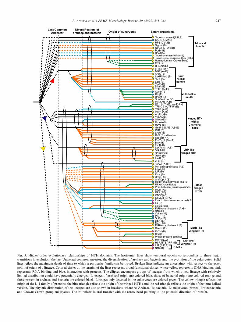
Fig. 5. Higher order evolutionary relationships of HTH domains. The horizontal lines show temporal epochs corresponding to three major
transitions in evolution, the last Universal common ancestor, the diversification of archaea and bacteria and the evolution of the eukaryotes. Solid
lines reflect the maximum depth of time to which a particular family can be traced. Broken lines indicate an uncertainty with respect to the exact
point of origin of a lineage. Colored circles at the termini of the lines represent broad functional classes: where yellow represents DNA binding, pink
represents RNA binding and blue, interaction with proteins. The ellipses encompass groups of lineages from which a new lineage with relatively
limited distribution could have potentially emerged. Lineages of archaeal origin are colored blue, those of bacterial origin are colored orange and
those present in archaea and bacteria are colored black. Lineages only detected in the eukaryotes are colored green. The yellow triangle reflects the
origin of the L11 family of proteins, the blue triangle reflects the origin of the winged HTHs and the red triangle reflects the origin of the tetra-helical
version. The phyletic distribution of the lineages are also shown in brackets, where A: Archaea; B: bacteria, E: eukaryotes, proteo: Proteobacteria
and Crown: Crown group eukaryotes. The �>� reflects lateral transfer with the arrow head pointing to the potential direction of transfer.
L. Aravind et al. / FEMS Microbiology Reviews 29 (2005) 231–262 247

248 L. Aravind et al. / FEMS Microbiology Reviews 29 (2005) 231–262
The Fis family of basic tri-helical HTH domains is
also found solely in the bacteria and typically appears
at the C-termini of the NtrC-like AAA+ domains
[131]. It appears to have emerged early in bacterial evo-
lution and spread widely via lateral gene transfer along
with the spread of sigma factors of the sigma-54 family.The Fis protein itself appears to have been secondarily
derived in the proteobacteria through the gene fission
of an NtrC-like regulator [131]. Likewise, the trihelical
HTH domains of the Rok family are restricted to the
bacteria and always found in combination with sugar ki-
nase domains of Hsp70/actin fold. The archaeal Boa1
family of tri-helical HTH domains contains a unique
all b-strand domain at the N-terminus that is likely tobind its effectors (Table 1, Fig. 4). The Myb family is a
pan-eukaryotic family of simple tri-helical domains that
appears to have diversified into multiple members prior
to the diversification of all extant eukaryotic lineages
(Fig. 5). Some versions of the Myb family, the SANT
subfamily, appear to have been recruited secondarily
for protein–protein interactions in the eukaryotic chro-
matin [132]. In bacteria, the Myb domain is only seenin the RsfA-related pre-spore transcription factors of
the low-GC Gram-positive bacteria [133], suggesting
that it was acquired relatively late through lateral trans-
fer from the eukaryotes. In addition to the Myb domain,
the homeodomain and POU domain families [41], which
are also basic tri-helical HTH domain, are respectively
widespread in the crown-group eukaryotes and metazo-
ans. The HTH domain of the eukaryotic tumor suppres-sor BRCA2 [72] is another simple trihelical version that
was derived early in eukaryotes. However, it shows no
specific relationships to any other HTH domains with
a similar structural configuration.
Beyond these classical families there are several tri-
helical HTH domains associated with diverse transpos-
ases and resolvases, such as the gamma–delta resolvase.
The exact point of origin of the HTH domains associ-ated with these mobile elements is difficult to ascertain,
but they appear to have given rise to families of HTH
domains found in cellular transcription factors on multi-
ple occasions. Particularly striking examples of these in-
clude the Paired box and Pipsqueak families involved in
metazoan developmental gene expression, and the
CENBP family (centromere binding protein) in the
crown group eukaryotes [134–137]. Likewise, the HTHdomain of the bacterial YlxL(SwrB) family (Fig. 3) is
also related to the HTH domains of the gamma–delta
resolvase [138]. It is possible that other transcription
factors families with a relatively restricted phyletic dis-
tribution, like the eukaryotic homeodomain, and the
bacterial sigma-54 family have also ultimately been de-
rived from the HTH domains of transposases. The me-
tal-binding domain of the retroviral integrase alsoappears to have been derived from the HTHs of trans-
posase/resolvase class through acquisition of metal-che-
lating residues. The KorB–ParB family also contains a
basic tri-helical version of the HTH domain, and func-
tions as the partitioning protein for diverse bacterial
plasmids. The KorB subfamily contains an additional
C-terminal 4-helical DNA-binding domain [57] fused
to the HTH domain, while the ParB subfamily containsa fusion to a nuclease domain of the OB-fold [139].
The MetJ-Arc (RHH) family of transcription factors
appears to have been derived from the basic tri-helical
bundle [61]. They are most frequently found as tran-
scriptional regulators of the mobile toxin–antitoxin
operons [140]. Hence, it is possible that they were origi-
nally derived in such toxin–antitoxin systems, through
rapid divergence from a conventional HTH. This ap-pears to have happened early in the evolution of one
of the prokaryotic lineages, after which they were widely
disseminated across the prokaryotes through horizontal
mobility.
4.2. The tetra-helical HTH superclass and its derivatives
The first major monophyletic clade of HTH domainsis defined by the unifying structural feature of the tetra-
helical bundle. Sequence similarities help in identifying
several major lineages within this group. The cI-like
family, typified by the phage lambda cI protein, contains
representatives from across the 3 super-kingdoms of life
(Fig. 4). Several distinct subfamilies can be recognized
within this family. The largest of these is the bacterial
repressor subfamily typified by the protein PbsX (Xre)from the B. subtilis prophage 168 [141]. Another notable
subfamily is the MBF1 subfamily, which is nearly uni-
versally conserved in the archaeo-eukaryotic lineage
functions, and is an adaptor that appears to bridge the
specific transcriptional regulators to the basal transcrip-
tion machinery MBF1 [142].
The next major assemblage within the tetra-helical
superclass comprises of 6 major families that are exclu-sively prokaryotic in their distribution (Figs. 4 and 5).
This assemblage includes the AraC, LuxR, LacI, DnaA,
TrpR and TetR families, which are predominantly bacte-
rial with several independent lateral transfers to archaea
(Fig. 4). The first four of these families are nearly pan-
bacterial in their distribution suggesting that these
HTH families had probably diverged from each other
even in the common ancestor of all bacteria (Fig. 5).The latter two lineages are more limited in their distribu-
tion, but are found in most proteobacteria, and low GC
Gram positive bacteria (Fig. 4). Within some of these
families several distinct lineages, often defined by spe-
cific architectural themes can be identified. However,
most of these families contain a dominant architectural
theme suggesting these might have been the earliest ver-
sions of these families. The AraC family contains aduplication of the tetra-helical version of the HTH do-
main [143], and typically occurs fused to a sugar binding

L. Aravind et al. / FEMS Microbiology Reviews 29 (2005) 231–262 249
domain of the DSBH fold [97,107] suggesting that they
predominantly function as sugar-sensing transcription
factors. However, in complex cyanobacteria, like Nos-
toc, the majority of AraC family HTH domains occur
fused to a novel sensor domain that is also found in
the siderophore alcalgin sensing transcriptional activa-tor, AlcR, from Bordetella [144] (Fig. 6 and Table 1).
The most common subfamily of the LuxR family con-
tains fusions to the receiver domain, as seen in the case
of the E. coli protein NarL. In the case of the LacI fam-
ily the dominant architecture features a fusion of the
HTH domain with a small-molecule binding domain
of the periplasmic solute-binding protein fold. DnaA is
usually found only in a single copy in all bacterial gen-omes, with the HTH occuring at the C-terminus of the
AAA+ domain [145]. The DnaA protein functions in
replication initiation, and also as a transcription factor
[146]. Additionally, sporadic versions of the tetrahelical
HTH superclass are also seen in several phage transpos-
ases related to the Mu transposase.
TFIIB, a basal transcription factor in the archaeo-
eukaryotic lineage, defines the TFIIB family, a derivativeof the tetra-helical class. In the archaea there is typically
a single version of this family (Fig. 4). However, in the
eukaryotes not only did TFIIB undergo duplication,
but it also spawned two more divergent families, namely
the cyclins and the Rb proteins [122]. Structural com-
parisons suggest that the eukaryote-specific Bright do-
main family [55,147], which includes DNA-binding
proteins involved in chromatin dynamics was also de-rived from a TFIIB-like precursor prior to the radiation
of eukaryotes from their common ancestor. The prove-
nance of the only bacterial relatives of this version,
namely the Spo0A protein from endospore-forming bac-
teria [56] remains unclear.
4.3. The wHTH superclass
The second major monophyletic clade of HTH do-
mains are unified by the presence of a striking derived
feature, the ‘‘wing’’. Several major assemblages with
varying distributions can be identified within the wHTH
superclass. The wHTH superclass includes the majority
of prokaryotic transcription factors. Thirteen major
families of prokaryotic wHTH domains, namely the
BirA, ArsR, GntR, DtxR-FurR, CitB, LysR, ModE,
MarR, PadR, YtcD, Rrf2, ScpB and HrcA-RuvB fami-
lies, are unified by the presence of a characteristic helix
after the wing, and comprise the largest monophyletic
assemblage within the wHTH superclass (Fig. 5). Of
these, representatives of the ArsR, MarR, YctD, GntR,
HrcA-RuvB are seen in both archaea and bacteria (Fig.
4) with phylogenetic trees suggesting distinct pan-archa-
eal and pan-bacterial branches within them (data notshown). This would imply that these families possibly
even go back to the LUCA (Fig. 5). The members of
these families, barring some striking exceptions (see be-
low) function as transcription factors suggesting that
they could have descended from an ancestral protein
that had some role in transcriptional regulation. The
MarR family has vastly proliferated in the archaea to
give rise to several archaeal subfamilies and includesmost of the major archaea-specific wHTH transcription
factors. In bacteria the RuvB sub-family, which is de-
rived from the ancestral HrcA lineage, appears to have
secondarily acquired a role in DNA recombination after
a fusion with the AAA+ domain in the ATPase subunit
of the Holliday junction resolvase [148]. Another sub-
family of the HrcA–RuvB family, S19AE is a small sub-
unit ribosomal protein in the archaeo-eukaryoticlineage, with a potential RNA-binding function (Fig.
5). A similar case is observed in the GntR family, which
has vastly proliferated in bacteria giving rise to many of
the major bacterial one-component transcription fac-
tors. However, in the archaeo-eukaryotic clade it is rep-
resented by a single lineage, the small subunit ribosomal
protein S25AE (Figs. 4 and 5).
In contrast to the above families, the BirA, ModE,PadR, ScpB and DtxR-Fur families appear to have
had a bacterial origin followed by sporadic lateral trans-
fers to the archaea (Fig. 4). The DtxR–Fur family
appears to have specialized early on in regulating me-
tal-dependent transcription of genes. The ScpB family
is typically represented in most bacteria by a single pro-
tein, which is encoded in the same operon as a kleisin
and a SMC-type ABC ATPase [149,150]. The ScpB pro-tein contains a tandem duplication of the wHTH with a
C-terminal helix after the wing (Fig. 3), and has been
shown to regulate the activity of the chromosome re-
organizing SMC ATPases [149,150]. The archaeal repre-
sentatives of this family appear to have been acquired
through a lateral transfer from the cyanobacteria
(LMI and LA unpublished). The pan-bacterial families,
namely CitB, LysR and Rrf2, are largely absent in thearchaea (Fig. 4). Interestingly, a single member of the
Rrf2 family (GLP_14_27362_29578) is seen in the early
branching eukaryote Giardia lamblia. A number of
wHTH domains with distinct sequence conservation
profiles, and occurring in large multi-domain proteins
also belong to this assemblage of wHTH domains with
a C-terminal helix after the wing. These include (1)
two HTH domains of the topoisomerase II family, (2)the HTH domain of the enigmatic topoisomerase V,
which is currently found only in Methanopyrus kandleri
[78], (3) The ESA1 family of eukaryotic histone acety-
ltransferases and (4) the N-terminal HTH domains of
the Rio kinases from archaea and eukaryotes. Two re-
lated wHTH domains from Giardia (Genbank Gis:
29245940, 29247865) also appear to have been derived
from within the above-discussed assemblage of wHTHdomains, but their precise affinities are obscured due
to rapid sequence divergence.

Fig. 6. A multiple alignment of the AlcR N-terminal module. Multiple sequence alignment the AlcrN domain was constructed using T-Coffee after
parsing high-scoring pairs from PSI-BLAST search results. The Jpred secondary structure is shown above the alignment with H representing an a-helix and E representing a b-strand. The 85% consensus shown below the alignment was derived using the following amino acid classes: hydrophobic
(h: ALICVMYFW, yellow shading); small (s: ACDGNPSTV, green) and polar (p: CDEHKNQRST, blue). The conserved �C� is shaded red. The
limits of the domains are indicated by the residue positions, on each end of the sequence. The sequences are denoted by their gene name followed by
the species abbreviation and GenBank Identifier (gi). The species abbreviations are: Ana: Nostoc sp., Bbro: Bordetella bronchiseptica, Bpar:
Bordetella parapertussis, Ctet: Clostridium tetani, Ecar: Erwinia carotovora, Gvio: Gloeobacter violaceus, Pae: Pseudomonas aeruginosa, Plum:
Photorhabdus luminescens, Psyr: Pseudomonas syringae, Rpal: Rhodopseudomonas palustris, Rsol: Ralstonia solanacearum, Smel: Sinorhizobium
meliloti, Spne: Streptococcus pneumoniae, Ssp: Synechocystis sp., Styp: Salmonella typhimurium, Tden: Treponema denticola, Wsuc: Wolinella
succinogenes.
250 L. Aravind et al. / FEMS Microbiology Reviews 29 (2005) 231–262

L. Aravind et al. / FEMS Microbiology Reviews 29 (2005) 231–262 251
The next major monophyletic assemblage of wHTH
superclass includes the DeoR, ArgR, LevR, YitL, Lrp-
AsnC, ZBD (Z-DNA binding domain), and RNase R
families. These families are unified by overall sequence
similarity, and a conserved pattern with a conserved glu-
tamine or arginine residue between helix-1 and helix-2 ofthe HTH domain. Of these the Lrp-AsnC family is
widely conserved in both bacteria and archaea (Figs. 4
and 5) and in phylogenetic trees displays distinct
branches separating the majority of archaeal and bacte-
rial members. Hence, it is possible that the Lrp-AsnC
family goes back to the LUCA. The ArgR and DeoR
are predominantly bacterial families, whereas the LevR
group is sporadically found, mainly in low GC Grampositive bacteria. The RNase R family is a limited group
that is represented by just a single pan-bacterial orthol-
ogous lineage in the form of the wHTH domain at the
extreme N-terminus of the Rnase R protein (Fig. 3).
Its widespread distribution in the bacteria suggests that
it emerged early in the evolution of this lineage. The
ZBD family is restricted to the crown group eukaryotes
and in animals it is fused to the deaminase domain in-volved in hyper-mutation of the immunoglobulin genes
[151,152]. The restricted phyletic pattern of the ZBD
family suggests that it may have evolved from one of
the prokaryotic families after lateral transfer to the
crown group eukaryotes (Fig. 5).
The wHTH domains found in the archaeo-eukaryotic
proteins involved in replication initiation, namely the
MCM proteins, the CDC6–Orc1 proteins (C-terminalto the AAA+ ATPase domains in both these cases)
and the RP-A protein comprise the replication initiation
family of wHTH domains. Both the MCM and the
CDC6 versions appear to have been present right from
the base of the archaeo-eukaryotic lineage [30,153,154].
The version associated with RP-A occurs as a stand-
alone protein in the archaea, while it appears to have
been fused to the single strand DNA binding OB folddomains in the eukaryotes. Also belonging to this family
are the C-terminal wHTH domains from the replicative
helicase-primase enzymes of various viruses such as P4,
plasmid Rep proteins and the eukaryotic RFX-type
DNA binding domain seen in transcription factors like
the MHC class II transcription factor HRFX1
[75,155,156]. This family might have originally evolved
to recognize specific DNA features associated with thereplication initiation sites, and recruiting the catalytic
activities involved in pre-initiation and initiation to
these sites [75]. The RFX domains are specifically re-
lated to certain phage helicase-primase wHTH domains
belonging to this family. This suggests that the crown
group eukaryotes may have acquired the RFX domains
from such a viral replication protein and reused them as
a transcription regulator (Fig. 5).The wHTH domains of the archaeal phenylalanyl
tRNA synthetase a-subunits, the eukaryotic ribosomal
protein S10 and the bacterial selenocysteine-specific
elongation factor SelB appear to comprise a family prin-
cipally associated with translation and RNA metabo-
lism proteins (Figs. 3 and 5). Their phyletic patterns
suggest that their recruitment to RNA-specific functions
appears to have occurred after the separation of the ma-jor superkingdoms of life, though it is possible that a
standalone precursor of this family was already present
in the LUCA. Another distinct family of wHTH do-
mains with an exclusive single-stranded RNA-binding
function is the La domain family [47,48,157]. The La do-
main has previously only been reported from eukary-
otes; however, using sequence profile analysis we show
that it is homologous to the N-terminal domain of theNAD-dependent RNA 2 0-phosphotransferase [158],
which removes the phosphate from the 2 0 ends of
RNA. In the RNA 2 0-phosphotransferase the La do-
main bears two of the four absolutely conserved cata-
lytic histidines (LA unpublished), suggesting that it is
another case of recruitment of the HTH domain for a
catalytic role. The RNA 2 0-phosphotransferases are
highly conserved in the archaeo-eukaryotic and sporad-ically observed in the bacteria. This suggests that the La
family of HTH domains emerged early in the archaeo-
eukaryotic lineage and were subsequently laterally trans-
ferred to bacteria. In the eukaryotes the non-catalytic
versions (the classical La domains) were recruited for
binding 3 0 poly(U)-rich elements of nascent RNA poly-
merase III transcripts and translation regulation [47,48].
The fungal protein frequency [157] contains an as yetfunctionally uncharacterized version of the La domain,
which may regulate the circadian clock via RNA
metabolism.
There are other distinct families of wHTH transcrip-
tion factors in prokaryotes with related 2- or 3- stranded
wHTH domains, but they do not appear to belong to
any of the aforementioned assemblages. These include
the LexA, OmpR, and IclR families that appear to bepan-bacterial families, with at best a rare presence in
the archaea (Figs. 4 and 5). The classical representatives
of the LexA family appear to be involved in regulating
responses to DNA damage in diverse bacteria [82].
However, a highly divergent, potential offshoot of the
LexA family is seen fused to the C-termini of large
DNA helicases (lhr) that are found sporadically in sev-
eral bacteria (Fig. 3). The eukaryotes display their ownfamilies of specific transcription factors, such as the
Forkhead-histone H1 [25,26] and E2F-DP1 families
[159], and the basal transcription factors, such as the
TFIIF(Rap30)-TFIIIC-63K family [160] that show
structurally similar wHTH domains, but lack specific se-
quence relationships with any of the prokaryotic fami-
lies. Within the TFIIF-TFIIIC-63K family, the RNA
polymerase III transcription factor, TFIIIC-63K, is con-served throughout eukaryotes, but TFIIF appears to be
restricted to the crown group and the apicomplexans.

252 L. Aravind et al. / FEMS Microbiology Reviews 29 (2005) 231–262
The wHTH domains of the TFIIF and TFIIIC-63K are
functionally similar to those of the Forkhead-histone H1
family suggesting that the latter were probably derived
from more widespread family of basal transcription fac-
tors [160]. The remaining eukaryotic families appear to
be chiefly represented in the eukaryotic crown group,implying that they arose relatively late from pre-existing
eukaryotic wHTH domains found in the basal transcrip-
tion machinery or via rapid divergence from laterally
transferred prokaryotic transcription factors. A similar
scenario appears to be applicable for the four eukaryotic
wHTH families that are not involved in DNA-binding,
namely the PINT domain, the SNF8 (with two tandem
copies of the wHTH domain), cullin and the DEP domain
families.
Beyond these multi-member families there are highly
conserved lineages of 2- or 3- stranded wHTH domains
that are typically found in single orthologous groups of
proteins, and cannot be linked to any of the larger
assemblages. Such lineages include wHTHs found in
the bacterial proteins such as FtsK, DprA and O-6-
methylguanine-DNA alkyltransferase, and the ar-chaeo-eukaryotic proteins like TFIIE (Fig. 3) [30,161].
Distinct from the 2- and the 3-stranded HTH do-
mains are the 4-stranded HTH domains that appear to
form a separate monophyletic assemblage within the
wHTH clade (Fig. 5). The main prokaryotic family in
this assemblage is the Crp family [36] that has a pan-bac-
terial distribution and sporadic presence in few archaea.
Thus, it seems to represent a bacterial innovation thatwas disseminated to the archaea via lateral transfer.
Members of this family are typically fused to a C-termi-
nal cNMP-binding domain, and appear to have special-
ized early on as cyclic nucleotide dependent regulators.
The eukaryotes contain a single major family of this
assemblage, the HSF family (heat shock transcription
factor), which is present only in the crown group
eukaryotes. In the animals this protein appears to havespawned two distinct sub-families that are prototyped
by the ETS domain and the IRF domain (interferon reg-
ulator factors) [162,163]. Given their relatively restricted
presence in eukaryotes, it is possible that they have orig-
inated through rapid divergence from laterally trans-
ferred prokaryotic versions. A similar scenario could
be envisaged for the origin of the orphan initiator bind-
ing protein from Trichomonas, which also contains a 4-stranded version of the HTH domain.
The MerR-like assemblage of truncated wHTH do-
mains are derivatives of 3-stranded wHTH domains
(Fig. 2). The MerR family proper [37] and the related
wHTH domains present in the DNA-binding region of
the bacterial phenyalanyl tRNA synthetase b-subunit[164] show a pan-bacterial distribution. In bacteria the
MerR family has vastly proliferated into several distinctsubfamilies, like the SoxR and CueR subfamilies [37].
However, most other versions show a more restricted
distribution. A potential RNA-binding version of this
domain is the N-terminal domain of translation initia-
tion factor IF2 from certain bacteria [165]. The remain-
ing versions, observed in the phage lambda excisionase
and terminase proteins, the phage Mu-repressor family
and the eukaryotic DNA repair protein XP-A and ani-mal transcription factors of the Dachshund family, ap-
pear to be DNA binding [166–168]. Eukaryotic Xp-A
family is involved in nucleotide excision repair [169],
and appears to have been derived at some point in
eukaryotic evolution from the functionally similar phage
excisionases. The principal diversification of this assem-
blage appears to have happened early in bacterial evolu-
tion resulting in the two ancient families, MerR and theFTRS b-subunit N-terminal domain. Given that regular
3-standed wHTHs are also found in association with
other translation proteins, like the archaeal FTRS-asubunit and SelB, it possible that the prototype of the
MerR-like version was derived from such a form
through loss of the initial helix.
4.4. Other miscellaneous families of HTH domains
In addition to the above-described major assem-
blages, there are a few ancient HTH families with uncer-
tain affinities. The chief amongst these are the related
ribosomal proteins L11 and S18 (Fig. 2). The former is
conserved in all the three super-kingdoms of life and
binds RNA in the L11-stalk structure, which appears
to go back to the shared ancestral core of the ribosome[170]. S18 appears to have been derived from L11 in the
bacteria. Despite its apparent ancient origins L11 ap-
pears to be a highly derived version of the HTH. Hence,
notwithstanding certain general similarities with the 4-
stranded wHTHs, it is more likely that L11 has conver-
gently acquired these features early in evolution.
5. Proteome-wide demographic trends of HTH domains
The availability of a large number and phyletic diver-
sity of complete genome sequences allows robust estima-
tion of the general trends in the proteome-wide
distribution of HTH domains. In order to detect the
occurrences of HTH domains in proteomes, position-
specific score matrices or sequence profiles were con-structed for the various distinct families of these domains
using seed alignments with diverse representatives. These
sequence profiles were then used to iteratively search the
target proteomes with the PSI-BLAST program [42].
Alternatively, the alignments were used to generate hid-
den Markov models, which were similarly used to search
the proteomes with the HMMER program [42]. Using a
combination of these procedures we determined the totalnumber of proteins containingHTHdomains encoded by
all completely sequenced organisms that were available at

L. Aravind et al. / FEMS Microbiology Reviews 29 (2005) 231–262 253
the time of the analysis. For prokaryotes, the plot of the
total number of HTH domains against gene number per
genome is best fitted by the power equation of the form
y = k · xc (where k and c are constants; r2 = 0.89; see
Fig. 7(a)). The r2 of 0.89 for this fit suggests that this ten-
dency is indeed strongly maintained across a wide diver-sity of genomes. This non-linear scaling of HTH
domain numbers with gene number is consistent with re-
cent studies that have suggested that the transcription fac-
tor counts follow a power equation with respect to gene
number [171]. Given that the HTH domains are the main
transcription factors in most of the prokaryotic genomes
it is clear that the trend observed for transcription factors
is principally a reflection of the distribution of HTH do-mains (Fig. 7). This distribution function suggests that
as gene number increases, a greater than linear number
of HTH domain regulators are required per gene.
Examination of major architectural classes of HTH
indicates that there is an interesting differential class-
wise partitioning of the trends. HTH proteins belong-
ing to two-component, serine/threonine kinase and
PTS signaling cascades are almost entirely missing inarchaea. In the bacteria, where they are abundantly
present, their numbers show a linear scaling with re-
spect to gene count per genome (r2 = 0.8) (Fig. 7(c))
[42]. This suggests that each HTH in two-component
and related phosphorylation cascades regulates a fixed
number of target genes, and as the gene numbers grow
larger, the regulators increase in direct proportion to
their increase in targets. This is consistent with a modelof evolution in which the two-component systems and
their target genes undergo duplication with approxi-
mately the same probability as the size of a bacterial
genome increases in evolution. In contrast to the
two-component systems, the one component systems,
which chiefly comprise of those HTH domains fused
to specific SMBDs or metabolic enzymes, scaled non-
linearly with increase in gene count per genome. Thebest fit for the one component systems was obtained
with the power equation of the form y = k · xc
(r2 = 0.76, Fig. 7(b)). This equation suggests that the
HTH domains belonging to the one-component system
are likely to be a significant contributor to the over-all
power equation-type distribution of the HTH domains.
This observation implies that as genome size increases
a greater than proportional increase in the numbersof one-component transcription factors is required for
controlling the newly added genes. This tendency might
correlate with the need to regulate specialized groups
of their genes, by combining the distinct inputs sensed
by the effector-binding domains of multiple sets of one-
component transcription factors in the metabolically or
organizationally complex bacteria with large genomes.
The counts of sigma factors, too on an average, arepositively correlated with gene count per genome (Fig.
7(d)), and scale non-linearly with it (the best fit being
provided by a quadratic curve of the form y = a ·x2 + b · x; where �a� and �b� are constants; r2 = 0.76).
The non-linear scaling of the sigma factors suggests that
in the more complex genomes the additional genes are
distributed amongst several functionally specialized
gene batteries, which are under the regulation of de-voted sigma factors responding to specific situations.
Interestingly, a few genomes show a significantly greater
than expected number of sigma factors (Fig. 7(d)). The
most striking example is seen in the case of Phytoplasma
asteris, which, like other Mycoplasmas, has a highly re-
duced genome with just over 700 genes [172]. Whereas,
the other Mycoplasmas have only a basal sigma-54, P.
asteris has, in addition to sigma-54, a recent lineage-spe-cific expansion of 11 sigma factors that are related to the
Bacillus sigma F. Likewise, Bacteroides thetaiotaomicron
and Nitrosomonas show recent lineage-specific expan-
sions of ECF-type sigma factors that have given rise
to at least 10 closely related paralogous members in their
proteomes. In the case of P. asteris there is evidence that
the sigma factors may constitute a novel transposon
[173]. While this possibility also exists in the case ofthe other bacteria that show a greater than expected
number of sigma factors, it is likely that some of them
might have been utilized as transcriptional regulators.
This potential link between sigma factors and transpo-
son is consistent with the repeated recruitment of
HTH domains from transposases as specific transcrip-
tional regulators.
The genomic data from eukaryotes is still not ade-quate to discuss general genome-wide trends. However,
the available data suggests that eukaryotes display dif-
ferent tendencies from the prokaryotes. The HTH do-
mains of the homeo and Myb proteins are amongst
the most prominent DNA-binding domains of transcrip-
tional regulators in the plant and animal lineages. How-
ever their numbers are relatively low in the other
eukaryotic lineages. This suggests that the rise in prom-inence of HTH transcription factors may have been a
relatively late phenomenon that occurred on multiple
occasions in the eukaryotic crown group. The parasitic
apicomplexa, including those forms that have genome
sizes comparable to some free-living fungi, have far
fewer transcription factors in general [174]. The early
branching eukaryote Giardia lamblia has at least 13
Myb domains and 2 Bright domains, but apparentlyno other representatives of the HTH domains found in
the eukaryotic specific transcription factors (LA unpub-
lished). Furthermore, the scaling of the total number of
transcription factors in eukaryotes with gene counts per
genome is not equivalent with what is observed in the
prokaryotes. This difference may arise due to two main
reasons: (1) the emergence of a complex apparatus for
chromatin-structure regulation might have changedthe nature of transcriptional control in eukaryotes. (2)
Most eukaryotic transcription factors are effectively

Fig. 7. Scaling of HTH domains with gene number per genome. All graphs show a scatter plot of number of proteins with HTH domains in a given
proteome (Y-axis) versus the number of protein-coding genes in that organism (X-axis). (a) The Y-axis is the overall number of proteins with HTH
domains. (b) The Y-axis is the number of predicted one-component system proteins with HTH domains. (c) The Y-axis is number of two-component
system and other phospho-relay system proteins with HTH domains. (d) The Y-axis shows the number of sigma factors. For each graph the best-
fitting trend line along with its r2 value is shown.
254 L. Aravind et al. / FEMS Microbiology Reviews 29 (2005) 231–262
down-stream of signaling cascades that communicate
from the cell membrane, or the cytoplasm to the nu-
cleus. Hence, there are there are few equivalents of the
genuine prokaryote-type two-component systems inthe eukaryotes. Additionally, the eukaryotes might also
extensively employ post-transcriptional control mecha-
nisms, involving regulatory RNAs, resulting in a lower
dependence on transcriptional regulators [175,176]. A
combination of these factors might account for the dif-
ference in the average number of genes controlled by
per transcription factor in eukaryotes and prokaryotes.
6. General considerations on the natural history of the
HTH fold and implications for the evolution of
transcription
With the exception of the ribosomal protein L11 no
bona fide HTH domains with an ancestral RNA-bind-
ing role can be confidently traced back to the LUCA.In the HrcA and GntR families the representatives from
the archaeo-eukaryotic clade are ribosomal proteins
(S19AE and S25AE); whereas all the known bacterial
representatives of this family are specific transcription
factors (Fig. 5). All other versions of the HTH associ-ated with translation and RNA metabolism, such as
those found in La/RNA 2 0 phosphatase, SelB and ribo-
somal protein S10E, appear to have been derived after
the separation of the archaeo-eukaryotic and bacterial
clades. The simplest interpretation of these observations
is that the majority of HTH domains associated with
RNA metabolism settled into their extant functional
niches only after the divergence of the major lineagesfrom LUCA. Hence, excluding L11, it is likely that other
HTH domains associated with RNA metabolism in the
LUCA performed more generic functions compared to
their extant counterparts. The DNA-binding property
is strongly preserved across diverse lineages and struc-
tural variants of the HTH fold, and involves helix-3, de-
spite the several variations in the details of the
interactions of individual versions of the domain. Thisobservation, taken together with the more sporadic

L. Aravind et al. / FEMS Microbiology Reviews 29 (2005) 231–262 255
distribution of the versions of the domain associated
with translation and RNA metabolism, suggests that
the ancestor of most of the extant versions of the
HTH, excluding L11, was a DNA-binding protein. Fur-
thermore, the diversification of this domain was poten-
tially associated with the emergence of DNA asgenetic material [75,153].
While there are HTH domains in the basal transcrip-
tion factors of both the bacterial (sigma factors) and ar-
chaeo-eukaryotic (TFIIB, TFIIE, and MBF1) lineages,
none of these can be considered as being truly ortholo-
gous [30,32,33]. In contrast, several families of the
HTH domains in specific transcription factors appear
to be extensively shared by the bacteria and archaea(Fig. 4). Though several of the prokaryotic families
shared by bacteria and archaea can be easily explained
as arising from relatively recent lateral transfer between
the prokaryotic super-kingdoms, some others like the
MarR, ArsR, YctD, Lrp, HrcA and GntR families ap-
pear to show distinct pan-archaeal and pan-bacterial
groups suggesting that they were present in the earliest
representatives of each of the super-kingdoms, hencepotentially go back to the LUCA (Fig. 5). Despite the
basal transcription machinery shared with the archaea,
the specific transcription factors that are traceable to
the last eukaryotic common ancestor do not belong to
the same families as the specific transcription factors
seen in the archaea [30]. These patterns raise a profound
conundrum regarding the origin of the phyletic patterns
of HTH domains in the basal and specific transcrip-tional regulators of extant organisms.
Even though several scenarios could in principle ex-
plain these patterns, there are only a few parsimonious
alternatives that account for the currently available
data. Given that both the basal transcription machinery
and the domain architecture of the RNA polymerase
catalytic subunits are very different in the bacterial and
the archaeo-eukaryotic lineages [130], one could extrap-olate that the basal transcription factors arose only after
the two great lineages had separated from the LUCA. In
this situation, the sharing of the specific transcription
factors by the archaea and the bacteria could be ex-
plained in two possible ways: (1) The LUCA had several
specific transcription factors but no basal transcription
factors. (2) Alternatively, there were neither specific
nor basal transcription factors in the LUCA and bothtypes emerged after the lineages separated. However,
multiple very early lateral transfer events resulted in pro-
karyotic lineages sharing a common set of specific tran-
scription factors. This latter scenario is the consistent
with the evidence for extensive lateral transfer between
the two prokaryotic super-kingdoms throughout their
evolution [32,177]. At least in the case of the HrcA
and GntR families, the striking difference in functionsof the extant bacterial and archaeo-eukaryotic represen-
tatives suggests that the ancestral versions of these fam-
ilies in LUCA performed a generic nucleic-acid-binding
function. They appear to have been secondarily re-
cruited as specific transcription factors only in the bacte-
ria, thereby supporting the second scenario. The basal
transcription factors and ribosomal proteins tend to
show a stronger signal of vertical inheritance as com-pared to specific transcriptions factors which are prone
to rampant gene loss and lateral transfer [32,177]. This
is not unexpected, given the functional constraints act-
ing on the basal transcription factors, and might con-
found the reconstruction of the actual evolutionary
scenario for the specific transcription factors.
Although the first scenario of basal transcription fac-
tors emerging after the specific transcription factorsmight appear counter-intuitive, detailed analysis of the
reconstructed house-keeping functions in the LUCA
suggest that it is hardly implausible. Earlier studies on
the DNA replication and chromosome partitioning sys-
tems suggest that the central enzymes of the DNA rep-
lication apparatus appear to have emerged only after
the split of the archaeo-eukaryotic and bacterial lineages
[32,127,178]. Thus, the DNA replication system resem-bles the basal transcription apparatus in its origins.
More specifically, the absence of an ancestral DNA
polymerase and associated replication enzymes in the
LUCA suggest that it probably had a system of replica-
tion involving reverse transcription [127]. However,
simpler but completely functional DNA-dependent
RNApolymerase (DdRP) subunits have been recon-
structed for the LUCA [130,179]. Hence, it is possiblethat the LUCA did not use basal transcription factors
and the DdRPs chiefly functioned as enzymes that sup-
plied the RNA template for the replication process
involving a reverse transcription step. The precursors
of the specific transcription factors might have still func-
tioned under these circumstances, primarily acting as
general repressors that regulated the synthesis of the
RNA template. The basal transcription factors probablyarose only when the genome got organized into multiple
tandem operons, each needing its own transcription ini-
tiation signal. This suggestion is consistent with the fact
that the prokaryotic specific transcription factors can
function equally well with both archaeo-eukaryotic-
and bacterial-type basal transcription machinery and
RNA polymerases, despite the numerous differences
[180].Irrespective of the scenarios, the HTH domains of
both the simple tri-helical type (e.g. carbamoylphos-
phate synthetase and topoisomerase I) and the wHTH
type (e.g. topoisomerase II family) are present in pro-
teins that can be confidently traced back to the LUCA.
Depending on the scenario, there were at least 6–11 dif-
ferent HTH domains in the cellular genomes, suggesting
that the fold had undergone structural and functionaldifferentiation even before the period of the LUCA.
Subsequently, in course of prokaryotic evolution, the

256 L. Aravind et al. / FEMS Microbiology Reviews 29 (2005) 231–262
HTH domains rapidly expanded in several prokaryotic
genomes to rank amongst the folds with highest repre-
sentation [32,35]. The currently available evidence sug-
gests that the common ancestor of extant eukaryotes
arose later, via an endosymbiotic event involving an
archaeal precursor for the nucleus, translation andsecretion apparatus and an a-proteobacterial precursorfor the mitochondrion. However, neither the mitochon-
drial, nor the nuclear genome, appears to have retained
the specific transcription factors might have been inher-
ited from the respective prokaryotic precursors. Instead,
the principal pan-eukaryotic HTH transcription factor
families, like Bright and MYB domains, are only dis-
tantly related to the prokaryotic counterparts. The ori-gin of the eukaryotes saw the emergence of a distinct
nuclear compartment, extensive RNA-dependent post-
transcriptional gene regulation, a complex chromatin
structure and the proliferation of enzymatic complexes
involved in chromatin dynamics, such as the Swi2 ATP-
ases, acetylases and deacetylases [181]. The compart-
mentalization of the cell probably rendered the
prokaryote-type one-component systems ineffective inthe eukaryotes. Furthermore, the regulators of chroma-
tin dynamics, such as the histone deacetylases and asso-
ciated Swi2 ATPases took up the role of transcriptional
repressors [181], and probably resulted in the ancestral
prokaryote-type repressors becoming superfluous.
Hence, the origin of the eukaryotes was probably
accompanied by a massive loss of the prokaryote-type
transcription factors, along with the innovation throughrapid sequence divergence of new versions that suited
the eukaryotic milieu. Some of the HTH domains inher-
ited from the ancestral prokaryotic genomes were also
reused by the eukaryotes as adaptor modules in signal-
ing systems un-related to DNA-binding or transcription
regulation. However, many versions inherited from the
archaea appear to have persisted in the basal transcrip-
tion machinery, where they were indispensable for tran-scription of the nuclear genome.
Finally, the rise of organizational complexity in
plant, animal and fungal lineages went hand-in-hand
with the emergence of new specific transcriptional regu-
lators. Some were drawn from pre-existing HTH fami-
lies, like the Myb domain, while others like the HSF,
Homeo, Pou, Pipsqueak and Paired families arose
through rapid divergence from different sources. TheHTH domains of transposons provided the source mate-
rial for some of these domains, whereas other might
have diverged rapidly from laterally transferred pro-
karyotic transcription factors. Thus, on one hand pro-
karyotes appear to share a sizeable common set of
transcription factors, whose phyletic patterns are chiefly
governed by the lateral transfers and gene losses acting
over and above a basic signal of vertical inheritance.On the other hand, the eukaryotes share only a few un-
ique ancient DNA-binding domains, and their transcrip-
tion factors have chiefly emerged through multiple
lineage-specific expansions.
7. General conclusions
The HTH domain, one of the best-studied of the
double-stranded DNA-binding domains, is one of the
key protein domains in the transcriptional apparatus
of all extant organisms. With the ‘‘hindsight’’ of over
two decades of investigations since the discovery of the
domain we attempt to provide a synthetic overview of
the natural history of the HTH domain from the view-
point of comparative genomics. Despite the HTH beinga rather simple structural scaffold, it is observed to be
capable of considerable structural variety and functional
versatility, while still preserving a core set of correlated
structure–function features. Most HTH domains, de-
spite their structural diversity, participate in a variety
of functions that depend on their DNA-binding proper-
ties. These include their central role in mediating the
substrate interactions of various enzymes that operateon DNA, and their role as both basal and specific tran-
scription regulators. Thus, the HTH domains are the
predominant transcription factors in all prokaryotic
organisms and the more complex eukaryotes, such as
the plants, animals and fungi. Beyond these DNA-
binding functions, the HTH domains have been
recruited on multiple occasions in a RNA-binding
capacity and as mediators of protein–protein interac-tions. The last universal common ancestor already had
anywhere between 6 and 11 distinct versions of the
HTH domain, which covered much of the structural
diversity, and at least some of the functional diversity
seen in the extant versions of the domain. Though sev-
eral families of specific transcription factors are shared
by the two prokaryotic kingdoms and may even go back
to their common ancestor, the HTH proteins in thebasal transcription factors do not appear to be ortholo-
gous. This presents an interesting evolutionary conun-
drum, whose solution might emerge from new data on
alternative transcription and replication systems, like
those in viruses and other selfish elements [75,156].
The HTH domain occurs frequently in modular pro-
teins, whose domain architectures are often correlated
with the general functional properties of the protein.In prokaryotes the dominant domain architecture is
the one-component system that combines the HTH with
a sensor domain. While many of the sensor domains of
commonly known one-component systems have been
characterized previously, several others remain structur-
ally and functionally unexplored, and suggest a new
direction for exploring the intricacies of biological sen-
sors in prokaryotic systems. The extensive use of diver-gent HTH domains in protein–protein interactions,
especially in eukaryotes, is another area that might
L. Aravind et al. / FEMS Microbiology Reviews 29 (2005) 231–262 257
develop further in the future as the actual mechanistic
details of such interactions become clearer. In prokary-
otic systems the wealth of sequence and structure data
might finally allow us to investigate some of the more
difficult problems such as, the overall transcriptional
regulatory network of organisms, and the details ofhow target DNA sequence and ligand specificity are
achieved by transcriptional regulators. We hope that
the overview presented by us will provide a framework
for such future investigations.
8. Supplementary material
A complete list of gis of the HTH domains detected in
183 completely sequenced prokaryotic genomes, and
alignments of major families will be available by ftp.
ftp.ncbi.nih.gov/pub/aravind/.
References
[1] Ptashne, M. (2004) Genetic Switch: Phage Lambda Revisited.
Cold Spring Harbor Laboratory Press, New York.
[2] Tahirov, T.H., Temiakov, D., Anikin, M., Patlan, V., McAll-
ister, W.T., Vassylyev, D.G. and Yokoyama, S. (2002) Structure
of a T7 RNA polymerase elongation complex at 2.9 A
resolution. Nature 420, 43–50.
[3] Zhang, G., Campbell, E.A., Minakhin, L., Richter, C., Sever-
inov, K. and Darst, S.A. (1999) Crystal structure of Thermus
aquaticus core RNA polymerase at 3.3 A resolution. Cell 98,
811–824.
[4] Darst, S.A., Polyakov, A., Richter, C. and Zhang, G. (1998)
Structural studies of Escherichia coli RNA polymerase. Cold
Spring Harb. Symp. Quant. Biol. 63, 269–276.
[5] Haldenwang, W.G. (1995) The sigma factors of Bacillus subtilis.
Microbiol. Rev. 59, 1–30.
[6] Stragier, P. and Losick, R. (1990) Cascades of sigma factors
revisited. Mol. Microbiol. 4, 1801–1806.
[7] Kornberg, R.D. (1999) Eukaryotic transcriptional control.
Trends Cell Biol. 9, 46–49.
[8] Kornberg, R.D. (1998) Mechanism and regulation of yeast RNA
polymerase II transcription. Cold Spring Harb. Symp. Quant.
Biol. 63, 229–232.
[9] Ohlendorf, D.H., Anderson, W.F. and Matthews, B.W. (1983)
Many gene-regulatory proteins appear to have a similar a-helicalfold that binds DNA and evolved from a common precursor. J.
Mol. Evol. 19, 109–114.
[10] Ohlendorf, D.H., Anderson, W.F., Fisher, R.G., Takeda, Y. and
Matthews, B.W. (1982) The molecular basis of DNA–protein
recognition inferred from the structure of cro repressor. Nature
298, 718–723.
[11] Sauer, R.T., Yocum, R.R., Doolittle, R.F., Lewis, M. and Pabo,
C.O. (1982) Homology among DNA-binding proteins suggests
use of a conserved super-secondary structure. Nature 298, 447–
451.
[12] Steitz, T.A., Ohlendorf, D.H., McKay, D.B., Anderson, W.F.
and Matthews, B.W. (1982) Structural similarity in the DNA-
binding domains of catabolite gene activator and cro repressor
proteins. Proc. Natl. Acad. Sci. USA 79, 3097–3100.
[13] Matthews, B.W., Ohlendorf, D.H., Anderson, W.F. and Takeda,
Y. (1982) Structure of the DNA-binding region of lac repressor
inferred from its homology with cro repressor. Proc. Natl. Acad.
Sci. USA 79, 1428–1432.
[14] Gribskov, M. and Burgess, R.R. (1986) Sigma factors from E.
coli, B. subtilis, phage SP01, and phage T4 are homologous
proteins. Nucl. Acids Res. 14, 6745–6763.
[15] Yura, T., Tobe, T., Ito, K. and Osawa, T. (1984) Heat shock
regulatory gene (htpR) of Escherichia coli is required for growth
at high temperature but is dispensable at low temperature. Proc.
Natl. Acad. Sci. USA 81, 6803–6807.
[16] Landick, R., Vaughn, V., Lau, E.T., VanBogelen, R.A., Erick-
son, J.W. and Neidhardt, F.C. (1984) Nucleotide sequence of the
heat shock regulatory gene of E. coli suggests its protein product
may be a transcription factor. Cell 38, 175–182.
[17] Frampton, J., Leutz, A., Gibson, T. and Graf, T. (1989) DNA-
binding domain ancestry. Nature 342, 134.
[18] Otting, G., Qian, Y.Q., Muller, M., Affolter, M., Gehring, W.
and Wuthrich, K. (1988) Secondary structure determination for
the Antennapedia homeodomain by nuclear magnetic resonance
and evidence for a helix-turn-helix motif. EMBO J. 7, 4305–
4309.
[19] Brennan, R.G. and Matthews, B.W. (1989) The helix-turn-helix
DNA binding motif. J. Biol. Chem. 264, 1903–1906.
[20] Dodd, I.B. and Egan, J.B. (1990) Improved detection of helix-
turn-helix DNA-binding motifs in protein sequences. Nucl.
Acids Res. 18, 5019–5026.
[21] Dodd, I.B. and Egan, J.B. (1987) Systematic method for the
detection of potential lambda Cro-like DNA-binding regions in
proteins. J. Mol. Biol. 194, 557–564.
[22] Schultz, S.C., Shields, G.C. and Steitz, T.A. (1991) Crystal
structure of a CAP-DNA complex: the DNA is bent by 90�.Science 253, 1001–1007.
[23] Wilson, K.P., Shewchuk, L.M., Brennan, R.G., Otsuka, A.J. and
Matthews, B.W. (1992) Escherichia coli biotin holoenzyme
synthetase/bio repressor crystal structure delineates the biotin-
and DNA-binding domains. Proc. Natl. Acad. Sci. USA 89,
9257–9261.
[24] Brennan, R.G. (1993) The winged-helix DNA-binding motif:
another helix-turn-helix takeoff. Cell 74, 773–776.
[25] Clark, K.L., Halay, E.D., Lai, E. and Burley, S.K. (1993) Co-
crystal structure of the HNF-3/fork head DNA-recognition
motif resembles histone H5. Nature 364, 412–420.
[26] Ramakrishnan, V., Finch, J.T., Graziano, V., Lee, P.L. and
Sweet, R.M. (1993) Crystal structure of globular domain of
histone H5 and its implications for nucleosome binding. Nature
362, 219–223.
[27] Swindells, M.B. (1995) Identification of a common fold in the
replication terminator protein suggests a possible mode for DNA
binding. Trends Biochem. Sci. 20, 300–302.
[28] Kodandapani, R., Pio, F., Ni, C.Z., Piccialli, G., Klemsz, M.,
McKercher, S., Maki, R.A. and Ely, K.R. (1996) A new pattern
for helix-turn-helix recognition revealed by the PU.1 ETS-
domain-DNA complex. Nature 380, 456–460.
[29] Gajiwala, K.S. and Burley, S.K. (2000) Winged helix proteins.
Curr. Opin. Struct. Biol. 10, 110–116.
[30] Aravind, L. and Koonin, E.V. (1999) DNA-binding proteins and
evolution of transcription regulation in the archaea. Nucl. Acids
Res. 27, 4658–4670.
[31] Bell, S.D. and Jackson, S.P. (2001) Mechanism and regulation of
transcription in archaea. Curr. Opin. Microbiol. 4, 208–213.
[32] Makarova, K.S., Aravind, L., Galperin, M.Y., Grishin, N.V.,
Tatusov, R.L., Wolf, Y.I. and Koonin, E.V. (1999) Comparative
genomics of the Archaea (Euryarchaeota): evolution of con-
served protein families, the stable core, and the variable shell.
Genome Res. 9, 608–628.
[33] Bell, S.D. and Jackson, S.P. (1998) Transcription and translation
in Archaea: a mosaic of eukaryal and bacterial features. Trends
Microbiol. 6, 222–228.

258 L. Aravind et al. / FEMS Microbiology Reviews 29 (2005) 231–262
[34] Rivera, M.C., Jain, R., Moore, J.E. and Lake, J.A. (1998)
Genomic evidence for two functionally distinct gene classes.
Proc. Natl. Acad. Sci. USA 95, 6239–6244.
[35] Koonin, E.V., Tatusov, R.L. and Rudd, K.E. (1995) Sequence
similarity analysis of Escherichia coli proteins: functional and
evolutionary implications. Proc. Natl. Acad. Sci. USA 92,
11921–11925.
[36] Korner, H., Sofia, H.J. and Zumft, W.G. (2003) Phylogeny of
the bacterial superfamily of Crp-Fnr transcription regulators:
exploiting the metabolic spectrum by controlling alternative gene
programs. FEMS Microbiol. Rev. 27, 559–592.
[37] Brown, N.L., Stoyanov, J.V., Kidd, S.P. and Hobman, J.L.
(2003) The MerR family of transcriptional regulators. FEMS
Microbiol. Rev. 27, 145–163.
[38] Rigali, S.b., Derouaux, A., Giannotta, F. and Dusart, J. (2002)
Subdivision of the helix-turn-helix GntR family of bacterial
regulators in the FadR, HutC, MocR, and YtrA subfamilies. J.
Biol. Chem. 277, 12507–12515.
[39] Gallegos, M.T., Schleif, R., Bairoch, A., Hofmann, K. and
Ramos, J.L. (1997) Arac/XylS family of transcriptional regula-
tors. Microbiol. Mol. Biol. Rev. 61, 393–410.
[40] Weickert, M.J. and Adhya, S. (1992) A family of bacterial
regulators homologous to Gal and Lac repressors. J. Biol. Chem.
267, 15869–15874.
[41] Gehring, W.J., Affolter, M. and Burglin, T. (1994) Homeodo-
main proteins. Annu. Rev. Biochem. 63, 487–526.
[42] Babu, M.M., Luscombe, N.M., Aravind, L., Gerstein, M. and
Teichmann, S.A. (2004) Structure and evolution of transcrip-
tional regulatory networks. Curr. Opin. Struct. Biol. 14, 283–
291.
[43] Teichmann, S.A. and Babu, M.M. (2004) Gene regulatory
network growth by duplication. Nat. Genet. 36, 492–496.
[44] Selmer, M. and Su, X.-D. (2002) Crystal structure of an mRNA-
binding fragment of Moorella thermoacetica elongation factor
SelB. EMBO J. 21, 4145–4153.
[45] Wah, D.A., Hirsch, J.A., Dorner, L.F., Schildkraut, I. and
Aggarwal, A.K. (1997) Structure of the multimodular endonu-
clease FokI bound to DNA. Nature 388, 97–100.
[46] Moore, M.H., Gulbis, J.M., Dodson, E.J., Demple, B. and
Moody, P.C. (1994) Crystal structure of a suicidal DNA repair
protein: the Ada O6-methylguanine-DNA methyltransferase
from E. coli. EMBO J. 13, 1495–1501.
[47] Alfano, C., Sanfelice, D., Babon, J., Kelly, G., Jacks, A., Curry,
S. and Conte, M.R. (2004) Structural analysis of cooperative
RNA binding by the La motif and central RRM domain of
human La protein. Nat. Struct. Mol. Biol. 11, 323–329.
[48] Dong, G., Chakshusmathi, G., Wolin, S.L. and Reinisch, K.M.
(2004) Structure of the La motif: a winged helix domain mediates
RNA binding via a conserved aromatic patch. EMBO J. 23,
1000–1007.
[49] Wong, H.C., Mao, J., Nguyen, J.T., Srinivas, S., Zhang, W., Liu,
B., Li, L., Wu, D. and Zheng, J. (2000) Structural basis of the
recognition of the dishevelled DEP domain in the Wnt signaling
pathway. Nat. Struct. Biol. 7, 1178–1184.
[50] Zheng, N., Schulman, B.A., Song, L., Miller, J.J., Jeffrey, P.D.,
Wang, P., Chu, C., Koepp, D.M., Elledge, S.J., Pagano, M.,
Conaway, R.C., Conaway, J.W., Harper, J.W. and Pavletich,
N.P. (2002) Structure of the Cul1-Rbx1-Skp1-F boxSkp2 SCF
ubiquitin ligase complex. Nature 416, 703–709.
[51] Guo, F., Gopaul, D.N. and van Duyne, G.D. (1997) Structure of
Cre recombinase complexed with DNA in a site-specific recom-
bination synapse. Nature 389, 40–46.
[52] Otting, G., Qian, Y.Q., Billeter, M., Muller, M., Affolter, M.,
Gehring, W.J. and Wuthrich, K. (1990) Protein–DNA contacts
in the structure of a homeodomain–DNA complex determined
by nuclear magnetic resonance spectroscopy in solution. EMBO
J. 9, 3085–3092.
[53] Cai, M., Zheng, R., Caffrey, M., Craigie, R., Clore, G.M. and
Gronenborn, A.M. (1997) Solution structure of the N-terminal
zinc binding domain of HIV-1 integrase. Nat. Struct. Biol. 4,
567–577.
[54] Mackereth, C.D., Arrowsmith, C.H., Edwards, A.M. and
McIntosh, L.P. (2000) Zinc-bundle structure of the essential
RNA polymerase subunit RPB10 from Methanobacterium ther-
moautotrophicum. Proc. Natl. Acad. Sci. USA 97, 6316–6321.
[55] Iwahara, J. and Clubb, R.T. (1999) Solution structure of the
DNA binding domain from Dead ringer, a sequence-specific AT-
rich interaction domain (ARID). EMBO J. 18, 6084–6094.
[56] Zhao, H., Msadek, T., Zapf, J., Madhusudan, Hoch, J.A. and
Varughese, K.I. (2002) DNA complexed structure of the key
transcription factor initiating development in sporulating bacte-
ria. Structure (Camb) 10, 1041–1050.
[57] Khare, D., Ziegelin, G.n., Lanka, E. and Heinemann, U. (2004)
Sequence-specific DNA binding determined by contacts outside
the helix-turn-helix motif of the ParB homolog KorB. Nat.
Struct. Mol. Biol. 11, 656–663.
[58] Campos, A., Zhang, R.G., Alkire, R.W., Matsumura, P. and
Westbrook, E.M. (2001) Crystal structure of the global regulator
FlhD from Escherichia coli at 1.8 A resolution. Mol. Microbiol.
39, 567–580.
[59] Schumacher, M.A., Lau, A.O.T. and Johnson, P.J. (2003)
Structural basis of core promoter recognition in a primitive
eukaryote. Cell 115, 413–424.
[60] Liu, S., Widom, J., Kemp, C.W., Crews, C.M. and Clardy, J.
(1998) Structure of human methionine aminopeptidase-2 com-
plexed with fumagillin. Science 282, 1324–1327.
[61] Gomis-Rueth, F.X., Sola¡, M., Acebo, P., Parraga, A., Guasch,
A., Eritja, R., Gonzalez, A., Espinosa, M., del Solar, G. and
Coll, M. (1998) The structure of plasmid-encoded transcriptional
repressor CopG unliganded and bound to its operator. EMBO J.
17, 7404–7415.
[62] Cordes, M.H., Walsh, N.P., McKnight, C.J. and Sauer, R.T.
(1999) Evolution of a protein fold in vitro. Science 284, 325–
328.
[63] Grishin, N.V. (2000) Two tricks in one bundle: helix-turn-helix
gains enzymatic activity. Nucl. Acids Res. 28, 2229–2233.
[64] Allen, M., Friedler, A., Schon, O. and Bycroft, M. (2002) The
structure of an FF domain from human HYPA/FBP11. J. Mol.
Biol. 323, 411–416.
[65] Das, A.K., Helps, N.R., Cohen, P.T. and Barford, D. (1996)
Crystal structure of the protein serine/threonine phosphatase 2C
at 2.0 A resolution. EMBO J. 15, 6798–6809.
[66] Aravind, L., Mazumder, R., Vasudevan, S. and Koonin, E.V.
(2002) Trends in protein evolution inferred from sequence and
structure analysis. Curr. Opin. Struct. Biol. 12, 392–399.
[67] Lewis, J.D., Saperas, N.R., Song, Y., Zamora, M.J., Chiva, M.
and Ausio, J. (2004) Histone H1 and the origin of protamines.
Proc. Natl. Acad. Sci. USA 101, 4148–4152.
[68] Werhane, H., Lopez, P., Mendel, M., Zimmer, M., Ordal, G.W.
and Marquez-Magana, L.M. (2004) The last gene of the fla/che
operon in Bacillus subtilis, ylxL, is required for maximal sigmaD
function. J. Bacteriol. 186, 4025–4029.
[69] Kearns, D.B., Chu, F., Rudner, R. and Losick, R. (2004) Genes
governing swarming in Bacillus subtilis and evidence for a phase
variation mechanism controlling surface motility. Mol. Micro-
biol. 52, 357–369.
[70] Campbell, E.A., Muzzin, O., Chlenov, M., Sun, J.L., Olson,
C.A., Weinman, O., Trester-Zedlitz, M.L. and Darst, S.A. (2002)
Structure of the bacterial RNA polymerase promoter specificity
sigma subunit. Mol. Cell. 9, 527–539.
[71] Aasland, R., Stewart, A.F. and Gibson, T. (1996) The SANT
domain: a putative DNA-binding domain in the SWI-SNF and
ADA complexes, the transcriptional co-repressor N-CoR and
TFIIIB. Trends Biochem. Sci. 21, 87–88.
L. Aravind et al. / FEMS Microbiology Reviews 29 (2005) 231–262 259
[72] Yang, H., Jeffrey, P.D., Miller, J., Kinnucan, E., Sun, Y.,
Thoma, N.H., Zheng, N., Chen, P.-L., Lee, W.-H. and Pavletich,
N.P. (2002) BRCA2 function in DNA binding and recombina-
tion from a BRCA2-DSS1-ssDNA structure. Science 297, 1837–
1848.
[73] Iyer, L.M., Makarova, K.S., Koonin, E.V. and Aravind, L.
(2004) Comparative genomics of the FtsK-HerA superfamily of
pumping ATPases: implications for the origins of chromosome
segregation, cell division and viral capsid packaging. Nucl. Acids
Res. 32, 5260–5279.
[74] Aussel, L., Barre, F.X., Aroyo, M., Stasiak, A., Stasiak, A.Z.
and Sherratt, D. (2002) FtsK Is a DNA motor protein that
activates chromosome dimer resolution by switching the cata-
lytic state of the XerC and XerD recombinases. Cell 108, 195–
205.
[75] Giraldo, R. and FernA¡ ndez-Tresguerres, M.E. (2004) Twenty
years of the pPS10 replicon: insights on the molecular mecha-
nism for the activation of DNA replication in iteron-containing
bacterial plasmids. Plasmid 52, 69–83.
[76] Berge, M., Mortier-Barriere, I., Martin, B. and Claverys, J.-P.
(2003) Transformation of Streptococcus pneumoniae relies on
DprA- and RecA-dependent protection of incoming DNA single
strands. Mol. Microbiol. 50, 527–536.
[77] Pietrokovski, S. and Henikoff, S. (1997) A helix-turn-helix DNA-
binding motif predicted for transposases of DNA transposons.
Mol. Gen. Genet. 254, 689–695.
[78] Belova, G.I., Prasad, R., Nazimov, I.V., Wilson, S.H. and
Slesarev, A.I. (2002) The domain organization and properties of
individual domains of DNA topoisomerase V, a type 1B
topoisomerase with DNA repair activities. J. Biol. Chem. 277,
4959–4965.
[79] Tobe, T., Sasakawa, C., Okada, N., Honma, Y. and Yoshikawa,
M. (1992) vacB, a novel chromosomal gene required for
expression of virulence genes on the large plasmid of Shigella
flexneri. J. Bacteriol. 174, 6359–6367.
[80] Angermayr, M., Roidl, A. and Bandlow, W. (2002) Yeast Rio1p
is the founding member of a novel subfamily of protein serine
kinases involved in the control of cell cycle progression. Mol.
Microbiol. 44, 309–324.
[81] LaRonde-LeBlanc, N. and Wlodawer, A. (2004) Crystal struc-
ture of A. fulgidus Rio2 defines a new family of serine protein
kinases. Structure (Cambridge) 12, 1585–1594.
[82] Peat, T.S., Frank, E.G., McDonald, J.P., Levine, A.S., Wood-
gate, R. and Hendrickson, W.A. (1996) Structure of the UmuD0
protein and its regulation in response to DNA damage. Nature
380, 727–730.
[83] Savijoki, K., Ingmer, H., Frees, D., Vogensen, F.K., Palva,
A. and Varmanen, P. (2003) Heat and DNA damage
induction of the LexA-like regulator HdiR from Lactococcus
lactis is mediated by RecA and ClpP. Mol. Microbiol. 50,
609–621.
[84] Zhang, X., Chaney, M., Wigneshweraraj, S.R., Schumacher, J.,
Bordes, P., Cannon, W. and Buck, M. (2002) Mechanochemical
ATPases and transcriptional activation. Mol. Microbiol. 45,
895–903.
[85] Leipe, D.D., Koonin, E.V. and Aravind, L. (2004) STAND, a
class of P-loop NTPases including animal and plant regulators of
programmed cell death: multiple, complex domain architectures,
unusual phyletic patterns, and evolution by horizontal gene
transfer. J. Mol. Biol. 343, 1–28.
[86] Wang, Y., Zhao, S., Somerville, R.L. and Jardetzky, O. (2001)
Solution structure of the DNA-binding domain of the TyrR
protein of Haemophilus influenzae. Protein Sci. 10, 592–598.
[87] Larquet, E., Schreiber, V.r., Boisset, N. and Richet, E. (2004)
Oligomeric assemblies of the Escherichia coli MalT transcrip-
tional activator revealed by cryo-electron microscopy and image
processing. J. Mol. Biol. 343, 1159–1169.
[88] Poon, K.K., Chu, J.C. and Wong, S.L. (2001) Roles of glucitol
in the GutR-mediated transcription activation process in Bacillus
subtilis: glucitol induces GutR to change its conformation and to
bind ATP. J. Biol. Chem. 276, 29819–29825.
[89] Lee, P.-C., Umeyama, T. and Horinouchi, S. (2002) afsS is a
target of AfsR, a transcriptional factor with ATPase activity that
globally controls secondary metabolism in Streptomyces coeli-
color A(32). Mol. Microbiol. 43, 1413–1430.
[90] Pao, G.M. and Saier, M.H. (1995) Response regulators of
bacterial signal transduction systems: selective domain shuffling
during evolution. J. Mol. Evol. 40, 136–154.
[91] West, A.H. and Stock, A.M. (2001) Histidine kinases and
response regulator proteins in two-component signaling systems.
Trends Biochem. Sci. 26, 369–376.
[92] Stulke, J. and Hillen, W. (1998) Coupling physiology and
gene regulation in bacteria: the phosphotransferase sugar
uptake system delivers the signals. Naturwissenschaften 85,
583–592.
[93] Stulke, J., Arnaud, M., Rapoport, G. and Martin-Verstraete, I.
(1998) PRD–a protein domain involved in PTS-dependent
induction and carbon catabolite repression of catabolic operons
in bacteria. Mol. Microbiol. 28, 865–874.
[94] Hu, K.-Y. and Saier, M.H. (2002) Phylogeny of phosphoryl
transfer proteins of the phosphoenolpyruvate-dependent sugar-
transporting phosphotransferase system. Res. Microbiol. 153,
405–415.
[95] Reizer, J. and Saier, M.H. (1997) Modular multidomain
phosphoryl transfer proteins of bacteria. Curr. Opin. Struct.
Biol. 7, 407–415.
[96] Tobisch, S., Stulke, J. and Hecker, M. (1999) Regulation of the
lic operon of Bacillus subtilis and characterization of potential
phosphorylation sites of the LicR regulator protein by site-
directed mutagenesis. J. Bacteriol. 181, 4995–5003.
[97] Anantharaman, V., Koonin, E.V. and Aravind, L. (2001)
Regulatory potential, phyletic distribution and evolution of
ancient, intracellular small-molecule-binding domains. J. Mol.
Biol. 307, 1271–1292.
[98] Hofmann, K. and Bucher, P. (1995) The FHA domain: a
putative nuclear signalling domain found in protein kinases and
transcription factors. Trends Biochem. Sci. 20, 347–349.
[99] Molle, V., Kremer, L., Girard-Blanc, C., Besra, G.S., Cozzone,
A.J. and Prost, J.-F.O. (2003) An FHA phosphoprotein recog-
nition domain mediates protein EmbR phosphorylation by
PknH, a Ser/Thr protein kinase from Mycobacterium tuberculo-
sis. Biochemistry 42, 15300–15309.
[100] Taylor, B.L., Zhulin, I.B. and Johnson, M.S. (1999) Aerotaxis
and other energy-sensing behavior in bacteria. Annu. Rev.
Microbiol. 53, 103–128.
[101] Tyrrell, R., Verschueren, K.H., Dodson, E.J., Murshudov, G.N.,
Addy, C. and Wilkinson, A.J. (1997) The structure of the
cofactor-binding fragment of the LysR family member, CysB: a
familiar fold with a surprising subunit arrangement. Structure 5,
1017–1032.
[102] Tam, R. and Saier, M.H. (1993) Structural, functional, and
evolutionary relationships among extracellular solute-binding
receptors of bacteria. Microbiol. Rev. 57, 320–346.
[103] Vartak, N.B., Reizer, J., Reizer, A., Gripp, J.T., Groisman,
E.A., Wu, L.F., Tomich, J.M. and Saier, M.H. (1991) Sequence
and evolution of the FruR protein of Salmonella typhimurium: a
pleiotropic transcriptional regulatory protein possessing both
activator and repressor functions which is homologous to the
periplasmic ribose-binding protein. Res. Microbiol. 142, 951–
963.
[104] Ettema, T.J.G., Brinkman, A.B., Tani, T.H., Rafferty, J.B. and
Van Der Oost, J. (2002) A novel ligand-binding domain involved
in regulation of amino acid metabolism in prokaryotes. J. Biol.
Chem. 277, 37464–37468.

260 L. Aravind et al. / FEMS Microbiology Reviews 29 (2005) 231–262
[105] Aravind, L. and Koonin, E.V. (1999) Gleaning non-trivial
structural, functional and evolutionary information about pro-
teins by iterative database searches. J. Mol. Biol. 287, 1023–1040.
[106] Bull, P.C. and Cox, D.W. (1994) Wilson disease and Menkes
disease: new handles on heavy-metal transport. Trends Genet.
10, 246–252.
[107] Dunwell, J.M., Culham, A., Carter, C.E., Sosa-Aguirre, C.R.
and Goodenough, P.W. (2001) Evolution of functional diversity
in the cupin superfamily. Trends Biochem. Sci. 26, 740–746.
[108] Bateman, A. (1997) The structure of a domain common to
archaebacteria and the homocystinuria disease protein. Trends
Biochem. Sci. 22, 12–13.
[109] Heldwein, E.E. and Brennan, R.G. (2001) Crystal structure of
the transcription activator BmrR bound to DNA and a drug.
Nature 409, 378–382.
[110] Aravind, L. and Anantharaman, V. (2003) HutC/FarR-like
bacterial transcription factors of the GntR family contain a
small molecule-binding domain of the chorismate lyase fold.
FEMS Microbiol. Lett. 222, 17–23.
[111] Zhang, R.G., Andersson, C.E., Savchenko, A., Skarina, T.,
Evdokimova, E., Beasley, S., Arrowsmith, C.H., Edwards,
A.M., Joachimiak, A. and Mowbray, S.L. (2003) Structure of
Escherichia coli ribose-5-phosphate isomerase: a ubiquitous
enzyme of the pentose phosphate pathway and the Calvin cycle.
Structure (Cambridge) 11, 31–42.
[112] Ailion, M., Bobik, T.A. and Roth, J.R. (1993) Two global
regulatory systems (Crp and Arc) control the cobalamin/
propanediol regulon of Salmonella typhimurium. J. Bacteriol.
175, 7200–7208.
[113] Miras, I., Hermant, D., Arricau, N. and Popoff, M.Y. (1995)
Nucleotide sequence of iagA and iagB genes involved in invasion
of HeLa cells by Salmonella enterica subsp. enterica ser. Typhi.
Res. Microbiol. 146, 17–20.
[114] Abergel, C., Bouveret, E., Claverie, J.M., Brown, K., Rigal, A.,
Lazdunski, C. and Benedetti, H. (1999) Structure of the
Escherichia coli TolB protein determined by MAD methods at
1.95 A resolution. Struct. Fold Des. 7, 1291–1300.
[115] Carr, S., Penfold, C.N., Bamford, V., James, R. and Hemmings,
A.M. (2000) The structure of TolB, an essential component of
the tol-dependent translocation system, and its protein–protein
interaction with the translocation domain of colicin E9. Struct.
Fold Des. 8, 57–66.
[116] Walburger, A., Lazdunski, C. and Corda, Y. (2002) The Tol/Pal
system function requires an interaction between the C-terminal
domain of TolA and the N-terminal domain of TolB. Mol.
Microbiol. 44, 695–708.
[117] Hofmann, K. and Bucher, P. (1998) The PCI domain: a common
theme in three multiprotein complexes. Trends Biochem. Sci. 23,
204–205.
[118] Aravind, L. and Ponting, C.P. (1998) Homologues of 26S
proteasome subunits are regulators of transcription and trans-
lation. Protein Sci. 7, 1250–1254.
[119] Wernimont, A. and Weissenhorn, W. (2004) Crystal structure of
subunit VPS25 of the endosomal trafficking complex ESCRT-II.
BMC Struct. Biol. 4, 10.
[120] Teo, H., Perisic, O., GonzA¡lez, B. and Williams, R.L. (2004)
ESCRT-II, an endosome-associated complex required for pro-
tein sorting: crystal structure and interactions with ESCRT-III
and membranes. Dev. Cell 7, 559–569.
[121] Hierro, A., Sun, J., Rusnak, A.S., Kim, J., Prag, G., Emr, S.D.
and Hurley, J.H. (2004) Structure of the ESCRT-II endosomal
trafficking complex. Nature 431, 221–225.
[122] Gibson, T.J., Thompson, J.D., Blocker, A. and Kouzarides, T.
(1994) Evidence for a protein domain superfamily shared by the
cyclins, TFIIB and RB/p107. Nucl. Acids Res. 22, 946–952.
[123] Yan, Y., Barlev, N.A., Haley, R.H., Berger, S.L. and Marmor-
stein, R. (2000) Crystal structure of yeast Esa1 suggests a unified
mechanism for catalysis and substrate binding by histone
acetyltransferases. Mol. Cell 6, 1195–1205.
[124] Zubieta, C., He, X.Z., Dixon, R.A. and Noel, J.P. (2001)
Structures of two natural product methyltransferases reveal the
basis for substrate specificity in plant O-methyltransferases. Nat.
Struct. Biol. 8, 271–279.
[125] Kim, J. and Raushel, F.M. (2001) Allosteric control of the
oligomerization of carbamoyl phosphate synthetase from Esch-
erichia coli. Biochemistry 40, 11030–11036.
[126] Lawson, F.S., Charlebois, R.L. and Dillon, J.A. (1996) Phylo-
genetic analysis of carbamoylphosphate synthetase genes: com-
plex evolutionary history includes an internal duplication within
a gene which can root the tree of life. Mol. Biol. Evol. 13, 970–
977.
[127] Leipe, D.D., Aravind, L. and Koonin, E.V. (1999) Did DNA
replication evolve twice independently. Nucl. Acids Res. 27,
3389–3401.
[128] Rubbi, L., Labarre-Mariotte, S., Cheldin, S. and Thuriaux, P.
(1999) Functional characterization of ABC10alpha, an essential
polypeptide shared by all three forms of eukaryotic DNA-
dependent RNA polymerases. J. Biol. Chem. 274, 31485–31492.
[129] Gruber, T.M. and Gross, C.A. (2003) Multiple sigma subunits
and the partitioning of bacterial transcription space. Annu. Rev.
Microbiol. 57, 441–466.
[130] Iyer, L.M., Koonin, E.V. and Aravind, L. (2004) Evolution of
bacterial RNA polymerase: implications for large-scale bacterial
phylogeny, domain accretion, and horizontal gene transfer. Gene
335, 73–88.
[131] Morett, E. and Bork, P. (1998) Evolution of new protein
function: recombinational enhancer Fis originated by horizontal
gene transfer from the transcriptional regulator NtrC. FEBS
Lett. 433, 108–112.
[132] Gruene, T., Brzeski, J., Eberharter, A., Clapier, C.R., Corona,
D.F.V., Becker, P.B. and Mueller, C.W. (2003) Crystal structure
and functional analysis of a nucleosome recognition module of
the remodeling factor ISWI. Mol. Cell. 12, 449–460.
[133] Juan Wu, L. and Errington, J. (2000) Identification and
characterization of a new prespore-specific regulatory gene,
rsfA, of Bacillus subtilis. J. Bacteriol. 182, 418–424.
[134] Izsak, Z., Khare, D., Behlke, J., Heinemann, U., Plasterk, R.H.
and Ivics, Z.n. (2002) Involvement of a bifunctional, paired-like
DNA-binding domain and a transpositional enhancer in sleeping
beauty transposition. J. Biol. Chem. 277, 34581–34588.
[135] Czerny, T., Schaffner, G. and Busslinger, M. (1993) DNA
sequence recognition by Pax proteins: bipartite structure of the
paired domain and its binding site. Genes Dev. 7, 2048–2061.
[136] Tanaka, Y., Nureki, O., Kurumizaka, H., Fukai, S., Kawaguchi,
S., Ikuta, M., Iwahara, J., Okazaki, T. and Yokoyama, S. (2001)
Crystal structure of the CENP-B protein–DNA complex: the
DNA-binding domains of CENP-B induce kinks in the CENP-B
box DNA. EMBO J. 20, 6612–6618.
[137] Smit, A.F. and Riggs, A.D. (1996) Tiggers and DNA transposon
fossils in the human genome. Proc. Natl. Acad. Sci. USA 93,
1443–1448.
[138] Subramanian, G., Koonin, E.V. and Aravind, L. (2000) Com-
parative genome analysis of the pathogenic spirochetes Borrelia
burgdorferi and Treponema pallidum. Infect. Immun. 68, 1633–
1648.
[139] Close, S.M. and Kado, C.I. (1992) A gene near the plasmid pSa
origin of replication encodes a nuclease. Mol. Microbiol. 6, 521–
527.
[140] Anantharaman, V. and Aravind, L. (2003) New connections in
the prokaryotic toxin-antitoxin network: relationship with the
eukaryotic nonsense-mediated RNA decay system. Genome
Biol., 4.
[141] Wood, H.E., Devine, K.M. and McConnell, D.J. (1990)
Characterisation of a repressor gene (xre) and a temperature-

L. Aravind et al. / FEMS Microbiology Reviews 29 (2005) 231–262 261
sensitive allele from the Bacillus subtilis prophage, PBSX. Gene
96, 83–88.
[142] Takemaru, K.i., Li, F.Q., Ueda, H. and Hirose, S. (1997)
Multiprotein bridging factor 1 (MBF1) is an evolutionarily
conserved transcriptional coactivator that connects a regulatory
factor and TATA element-binding protein. Proc. Natl. Acad.
Sci. USA 94, 7251–7256.
[143] Rhee, S., Martin, R.G., Rosner, J.L. and Davies, D.R. (1998) A
novel DNA-binding motif in MarA: the first structure for an
AraC family transcriptional activator. Proc. Natl. Acad. Sci.
USA 95, 10413–10418.
[144] Brickman, T.J., Kang, H.Y. and Armstrong, S.K. (2001)
Transcriptional activation of Bordetella alcaligin siderophore
genes requires the AlcR regulator with alcaligin as inducer. J.
Bacteriol. 183, 483–489.
[145] Fujikawa, N., Kurumizaka, H., Nureki, O., Terada, T., Shir-
ouzu, M., Katayama, T. and Yokoyama, S. (2003) Structural
basis of replication origin recognition by the DnaA protein.
Nucl. Acids Res. 31, 2077–2086.
[146] Messer, W. and Weigel, C. (2003) DnaA as a transcription
regulator. Meth. Enzymol. 370, 338–349.
[147] Wilsker, D., Patsialou, A., Dallas, P.B. and Moran, E. (2002)
ARID proteins: a diverse family of DNA binding proteins
implicated in the control of cell growth, differentiation, and
development. Cell Growth Differ. 13, 95–106.
[148] Yamada, K., Miyata, T., Tsuchiya, D., Oyama, T., Fujiwara, Y.,
Ohnishi, T., Iwasaki, H., Shinagawa, H., Ariyoshi, M., Mayan-
agi, K. and Morikawa, K. (2002) Crystal structure of the RuvA–
RuvB complex: a structural basis for the Holliday junction
migrating motor machinery. Mol. Cell 10, 671–681.
[149] Soppa, J.r., Kobayashi, K., Noirot-Gros, M.-F.o., Oesterhelt,
D., Ehrlich, S.D., Dervyn, E., Ogasawara, N. and Moriya, S.
(2002) Discovery of two novel families of proteins that are
proposed to interact with prokaryotic SMC proteins, and
characterization of the Bacillus subtilis family members ScpA
and ScpB. Mol. Microbiol. 45, 59–71.
[150] Mascarenhas, J., Soppa, J.R., Strunnikov, A.V. and Graumann,
P.L. (2002) Cell cycle-dependent localization of two novel
prokaryotic chromosome segregation and condensation proteins
in Bacillus subtilis that interact with SMC protein. EMBO J. 21,
3108–3118.
[151] Schwartz, T., Rould, M.A., Lowenhaupt, K., Herbert, A. and
Rich, A. (1999) Crystal structure of the Zalpha domain of the
human editing enzyme ADAR1 bound to left-handed Z-DNA.
Science 284, 1841–1845.
[152] Schade, M., Turner, C.J., Lowenhaupt, K., Rich, A. and
Herbert, A. (1999) Structure–function analysis of the Z-DNA-
binding domain Zalpha of dsRNA adenosine deaminase type I
reveals similarity to the (a + b) family of helix-turn-helix
proteins. EMBO J. 18, 470–479.
[153] Giraldo, R. and Diaz-Orejas, R. (2001) Similarities between the
DNA replication initiators of Gram-negative bacteria plasmids
(RepA) and eukaryotes (Orc4p)/archaea (Cdc6p). Proc. Natl.
Acad. Sci. USA 98, 4938–4943.
[154] Liu, J., Smith, C.L., DeRyckere, D., DeAngelis, K., Martin,
G.S. and Berger, J.M. (2000) Structure and function of Cdc6/
Cdc18: implications for origin recognition and checkpoint
control. Mol. Cell 6, 637–648.
[155] Emery, P., Strubin, M., Hofmann, K., Bucher, P., Mach, B. and
Reith, W. (1996) A consensus motif in the RFX DNA binding
domain and binding domain mutants with altered specificity.
Mol. Cell Biol. 16, 4486–4494.
[156] Yeo, H.-J., Ziegelin, G.N., Korolev, S., Calendar, R., Lanka, E.
and Waksman, G. (2002) Phage P4 origin-binding domain
structure reveals a mechanism for regulation of DNA-binding
activity by homo- and heterodimerization of winged helix
proteins. Mol. Microbiol. 43, 855–867.
[157] Anantharaman, V., Koonin, E.V. and Aravind, L. (2002)
Comparative genomics and evolution of proteins involved in
RNA metabolism. Nucl. Acids Res. 30, 1427–1464.
[158] Spinelli, S.L., Kierzek, R., Turner, D.H. and Phizicky, E.M.
(1999) Transient ADP-ribosylation of a 20-phosphate implicated
in its removal from ligated tRNA during splicing in yeast. J.
Biol. Chem. 274, 2637–2644.
[159] Zheng, N., Fraenkel, E., Pabo, C.O. and Pavletich, N.P. (1999)
Structural basis of DNA recognition by the heterodimeric cell
cycle transcription factor E2F-DP. Genes Dev. 13, 666–674.
[160] Groft, C.M., Uljon, S.N., Wang, R. and Werner, M.H. (1998)
Structural homology between the Rap30 DNA-binding domain
and linker histone H5: implications for preinitiation complex
assembly. Proc. Natl. Acad. Sci. USA 95, 9117–9122.
[161] Meinhart, A., Blobel, J. and Cramer, P. (2003) An extended
winged helix domain in general transcription factor E/IIE alpha.
J. Biol. Chem. 278, 48267–48274.
[162] Landsman, D. and Wolffe, A.P. (1995) Common sequence and
structural features in the heat-shock factor and Ets families of
DNA-binding domains. Trends Biochem. Sci. 20, 225–226.
[163] Mackereth, C.D., Scharpf, M., Gentile, L.N., MacIntosh, S.E.,
Slupsky, C.M. and McIntosh, L.P. (2004) Diversity in structure
and function of the Ets family PNT domains. J. Mol. Biol. 342,
1249–1264.
[164] Dou, X., Limmer, S. and Kreutzer, R. (2001) DNA-binding of
phenylalanyl-tRNA synthetase is accompanied by loop forma-
tion of the double-stranded DNA. J. Mol. Biol. 305, 451–458.
[165] Laursen, B.S.g., Mortensen, K.K., Sperling-Petersen, H.U. and
Hoffman, D.W. (2003) A conserved structural motif at the N
terminus of bacterial translation initiation factor IF2. J. Biol.
Chem. 278, 16320–16328.
[166] Sam, M.D., Cascio, D., Johnson, R.C. and Clubb, R.T. (2004)
Crystal structure of the excisionase–DNA complex from bacte-
riophage lambda. J. Mol. Biol. 338, 229–240.
[167] Wojciak, J.M., Iwahara, J. and Clubb, R.T. (2001) The Mu
repressor–DNA complex contains an immobilized �wing� withinthe minor groove. Nat. Struct. Biol. 8, 84–90.
[168] de Beer, T., Fang, J., Ortega, M., Yang, Q., Maes, L., Duffy, C.,
Berton, N., Sippy, J., Overduin, M., Feiss, M. and Catalano,
C.E. (2002) Insights into specific DNA recognition during the
assembly of a viral genome packaging machine. Mol. Cell 9,
981–991.
[169] Ikegami, T., Kuraoka, I., Saijo, M., Kodo, N., Kyogoku, Y.,
Morikawa, K., Tanaka, K. and Shirakawa, M. (1998) Solution
structure of the DNA- and RPA-binding domain of the human
repair factor XPA. Nat. Struct. Biol. 5, 701–706.
[170] Agrawal, R.K., Linde, J., Sengupta, J., Nierhaus, K.H. and
Frank, J. (2001) Localization of L11 protein on the ribosome
and elucidation of its involvement in EF-G-dependent translo-
cation. J. Mol. Biol. 311, 777–787.
[171] van Nimwegen, E. (2003) Scaling laws in the functional content
of genomes. Trends Genet. 19, 479–484.
[172] Oshima, K., Kakizawa, S., Nishigawa, H., Jung, H.-Y., Wei, W.,
Suzuki, S., Arashida, R., Nakata, D., Miyata, S.-i., Ugaki, M.
and Namba, S. (2004) Reductive evolution suggested from the
complete genome sequence of a plant-pathogenic phytoplasma.
Nat. Genet. 36, 27–29.
[173] Lee, I.-M., Zhao, Y. and Bottner, K.D. (2003) Identification of
putative insertion sequence (IS) associated with members of the
aster yellows (AY) phytoplasma group. Phytopathology, 93.
[174] Templeton, T.J., Iyer, L.M., Anantharaman, V., Enomoto, S.,
Abrahante, J.E., Subramanian, G.M., Hoffman, S.L., Abraham-
sen, M.S. and Aravind, L. (2004) Comparative analysis of
apicomplexa and genomic diversity in eukaryotes. Genome Res.
14, 1686–1695.
[175] Bartel, D.P. (2004) MicroRNAs: genomics, biogenesis, mecha-
nism, and function. Cell 116, 281–297.

262 L. Aravind et al. / FEMS Microbiology Reviews 29 (2005) 231–262
[176] Mattick, J.S. (2001) Non-coding RNAs: the architects of
eukaryotic complexity. EMBO Rep. 2, 986–991.
[177] Boucher, Y., Douady, C.J., Papke, R.T., Walsh, D.A., Boud-
reau, M.E.R., NesbA�, C.L., Case, R.J. and Doolittle, W.F.
(2003) Lateral gene transfer and the origins of prokaryotic
groups. Annu. Rev. Genet. 37, 283–328.
[178] Edgell, D.R. and Doolittle, W.F. (1997) Archaea and the
origin(s) of DNA replication proteins. Cell 89, 995–998.
[179] Iyer, L.M., Koonin, E.V. and Aravind, L. (2003) Evolutionary
connection between the catalytic subunits of DNA-dependent
RNA polymerases and eukaryotic RNA-dependent RNA poly-
merases and the origin of RNA polymerases. BMC Struct. Biol.
3, 1.
[180] Brinkman, A.B., Bell, S.D., Lebbink, R.J., de Vos, W.M. and
van der Oost, J. (2002) The Sulfolobus solfataricus Lrp-like
protein LysM regulates lysine biosynthesis in response to lysine
availability. J. Biol. Chem. 277, 29537–29549.
[181] Grewal, S.I.S. and Moazed, D. (2003) Heterochromatin
and epigenetic control of gene expression. Science 301,
798–802.