Perspectivism, Accessibility and the Failure of Conjunction ...
Published online 1 December 2007 Nucleic Acids Research, 2008, Vol. 36, No. 4 e20doi:10.1093/nar/gkm1062
Measuring the dynamic surface accessibility of RNAwith the small paramagnetic molecule TEMPOLVincenzo Venditti1, Neri Niccolai1 and Samuel E. Butcher2,3,*
1Biomolecular Structure Research Center and Dipartimento di Biologia Molecolare, Universita di Siena,via Fiorentina 1, 53100 Siena, Italy, 2Department of Biochemistry and 3Nuclear Magnetic Resonance Facilityat Madison, University of Wisconsin-Madison, 433 Babcock Dr. Madison, WI 53706, USA
Received September 11, 2007; Revised and Accepted November 8, 2007
ABSTRACT
The surface accessibility of macromolecules plays akey role in modulating molecular recognition events.RNA is a complex and dynamic molecule involvedin many aspects of gene expression. However,there are few experimental methods available tomeasure the accessible surface of RNA. Here,we investigate the accessible surface of RNAusing NMR and the small paramagnetic moleculeTEMPOL. We investigated two RNAs with knownstructures, one that is extremely stable and one thatis dynamic. For helical regions, the TEMPOL probingdata correlate well with the predicted RNA surface,and the method is able to distinguish subtle varia-tions in atom depths, such as the relative accessi-bility of pyrimidine versus purine aromatic carbonatoms. Dynamic motions are also detected byTEMPOL probing, and the method accuratelyreports a previously characterized pH-dependentconformational transition involving formation ofa protonated C–A pair and base flipping. Someloop regions are observed to exhibit anomalouslyhigh accessibility, reflective of motions that are notevident within the ensemble of NMR structures.We conclude that TEMPOL probing can providevaluable insights into the surface accessibility anddynamics of RNA, and can also be used as anindependent means of validating RNA structure anddynamics in solution.
INTRODUCTION
Surface accessibility plays a key role in delineating mole-cular interactions. However, accessible surface areas,calculated on the basis of static 3D structures, may notaccurately describe the effective atom exposure to the bulksolution. In fact, dynamic features such as local flexibility
(1,2), and/or hydration shell motions (3–5), have beenobserved to modulate surface accessibility in proteins.Experimental studies on protein dynamics are, indeed,
important in structural biology, and complement theotherwise ‘static’ view of molecular structure. In thisrespect, soluble paramagnetic probes have been shown tobe useful in obtaining insight into processes regulatingprotein surface accessibility. In multidimensional NMRspectra, soluble paramagnetic molecules cause distance-dependent paramagnetic relaxation enhancement (PRE),manifested as line broadening and attenuation of cross-peak intensity. The extent of attenuation depends uponthe local concentration of the paramagnetic probe. Thus,higher atom exposures result in higher degrees of para-magnetic attenuation (6,7). Recently, PRE studies havebeen widely applied in the investigation of protein–ligand(3), protein–protein (8–11) and protein–DNA(12) interac-tions, as well as in the localization of protein active sites(1,3,5,6,13,14). PRE studies are capable of efficientlydetecting transient intermediates and short-lived encoun-ter complexes that may otherwise go undetected by othermethods (11,12).The absence of strong interactions between biomole-
cules and paramagnetic probes is a prerequisite forprobing surface accessibility. In the past few years, fourparamagnetic agents with different sizes and chemicalcharacteristics have been selected to fulfill this require-ment: TEMPOL (3,5,6,7,15), Gd(III)DTPA-BMA (16),Gd2(L7)(H2O)2 (10) and O2 (2). Among these probes,TEMPOL has been used most extensively in proteinaccessibility studies, and a large amount of data on themechanisms modulating the TEMPOL approach onprotein surfaces is available (3,5,7,10,15,16). TEMPOLis soluble and small, with a radius of 3.7 A (Figure 1A).Thus, TEMPOL is small enough to enter the narrowmajor groove of an RNA helix, and is therefore anideal probe for investigating the surface accessibility ofRNA. The paramagnetism of TEMPOL is due to theunpaired electron on the nitroxide group (Figure 1A),which induces fast relaxation of nuclear spins throughelectron-nuclear dipole–dipole coupling. In the absence
*To whom correspondence should be addressed. Tel: +1 608 263 3890; Fax: +1 6082623453; Email: [email protected]
� 2007 The Author(s)
This is an Open Access article distributed under the terms of the Creative Commons Attribution Non-Commercial License (http://creativecommons.org/licenses/
by-nc/2.0/uk/) which permits unrestricted non-commercial use, distribution, and reproduction in any medium, provided the original work is properly cited.
at National Institutes of H
ealth Library on Septem
ber 5, 2013http://nar.oxfordjournals.org/
Dow
nloaded from

of specific interactions, TEMPOL approaches a molecularsurface randomly, and thus the paramagnetic nitroxidegroup can be considered to be at the center of a spherewith a radius of 3.7 A (the radius of TEMPOL). Theparamagnetic effect of TEMPOL is exerted in a distancedependent, through-space manner, with >90% of relaxa-tion induced through distances of 10 A or less from theparamagnetic center (7).Here, we show that TEMPOL induced PRE is a facile
method that can be used to investigate the surfaceaccessibility of RNA. For stable helical regions of RNA,there is excellent agreement between the surface accessi-bility and the position of the atom within the structure.Furthermore, we show that the method is capable ofdetecting dynamic conformational transitions. Somedynamic regions result in higher levels of PRE thanwould be predicted from the corresponding ensemble ofNMR structures, suggesting that the method detectstransient conformational dynamics that are ‘invisible’with respect to the NMR-derived structures. Therefore,TEMPOL probing provides a facile way to independentlyvalidate and complement RNA structure models anddynamic measurements.
MATERIALS AND METHODS
Sample preparation and NMRmeasurements
The investigated RNA’s, whose sequences are shownin Figure 1, were prepared by in vitro transcriptionas previously described (17,18). All NMR sampleswere �1mM in D2O. The pH was adjusted to 6.8for the HIV-1 RNA, and 8.0 and 5.7 for the U6 ISLRNA NMR samples. Paramagnetic samples contained anoptimal 20mM TEMPOL (4-hydroxy-2,2,6,6-tetramethyl-piperidine-1 oxyl, 97% purity, Sigma-Aldrich) concentra-tion, which was achieved by adding directly to the NMRtube a few microliters of a 2M TEMPOL solution. StockTEMPOL solutions were prepared by directly dissolvingTEMPOL into 99.9% D2O to a final concentration 2M.Since denaturing effects of TEMPOL on RNA struc-
tures could not be excluded a priori, after each additionof a 2M solution of TEMPOL to the �1mM RNAsolutions, 2D TOCSY spectra were recorded to check forchemical shift differences due to TEMPOL. No significant
chemical shift changes upon TEMPOL addition wereobserved.
Conventional 2D TOCSY (40ms mixing time), NOESY(150ms mixing time) and 1H-13C HSQC spectra wereacquired at the National Magnetic Resonance Facilityat Madison (NMR-FAM) on Bruker DMX 600 and750MHz spectrometers. Data processing was carriedout using XWINNMR 2.6 software (Bruker Biospin).Resonance assignments were performed on the basis ofpreviously reported NMR data (BMRB entries: 5371,5703, 5834, 6320 and 6543) (17–21). Resonance assign-ments were nonetheless accurately checked for diamag-netic and paramagnetic samples by standard proceduresbased on TOCSY and NOESY experiments.
Cross-peak volumes were measured with a greater than90% confidence level using the Sparky integration tool(http://www.cgl.ucsf.edu/home/sparky).
Cross-peak attenuations
The cross-peak volumes were autoscaled according to therelation (13):
� p,di ¼
Vp,di
ð1=nÞP
nV
p,di
where �ip,d is the autoscaled volume of peak i from the
paramagnetic or diamagnetic spectra, Vip,d is the mea-
sured peak volume from the paramagnetic or diamagneticspectra, and n is the number of peaks measured. Hence theautoscaled volume is equal to the measured volumedivided by a scaling factor corresponding to the meancross-peak volume over n molecular locations. Paramag-netic attenuations, Ai’s, were calculated from the auto-scaled diamagnetic and paramagnetic peak volumes,respectively, �d and � p, according to the relation (13):
Ai ¼ 2�� pi
� di
Depth index calculation
The 3D atom depths, reported as depth indexes Di,r’s,were calculated on the basis of the NMR solutionstructures (PDB codes: 1PJY, 1SY4, 1SY7) (17,20,21)using the SADIC software (22). For each RNA, reportedDi,r’s were averaged over the whole NMR structureensemble.
Di,r is defined according to the relation (22):
Di,r ¼2Vi,r
Vo,r,
where Vi,r is the exposed volume of a sphere of radiusr (sampling radius) centered on atom i and V0,r is the totalvolume of the same sphere. The sampling radius was setto 10 A, which corresponds to the practical limit of theparamagnetic effect (7). The probe radius, used for thecomputation of the macromolecular surface, was set to3.7 A corresponding to the TEMPOL molecular radiusas calculated by using the MOLMOL software (23). For
Figure 1. Structure of TEMPOL and secondary structures of theinvestigated RNAs. (A) TEMPOL, (B) HIV-1 RNA and (C) U6 ISL.
e20 Nucleic Acids Research, 2008, Vol. 36, No. 4 PAGE 2 OF 10
at National Institutes of H
ealth Library on Septem
ber 5, 2013http://nar.oxfordjournals.org/
Dow
nloaded from
each C–H correlation, the entire ensemble of 20 NMRstructures were analyzed and the averaged Di,10 isreported. For simplicity, the averaged Di,10 value isreferred to in the text as Di.
Molecular dynamics
Several MD simulations were performed in explicit solventstarting from the lowest energy solution structures of theinvestigated RNA’s (PDB codes: 1PJY, 1SY4) (17,21,24)by using the AMBER package (25) and the AMBER-99force field (26).
Each RNA was centered in a truncated octahedron andthe initial shortest distance between the RNA atoms andthe box boundaries was set to 10 A. Then, an optimalamount of sodium ions was added in order to generatea neutral system. The remaining box volume was filledwith TIP3P type water molecules (27). Water and ionspositions were minimized with 500 steps of steepestdescent plus 500 steps of conjugate gradient by keepingthe RNA fixed. Then, the entire system was energyminimized with 1500 steps of steepest descent followed by1000 steps of conjugate gradient minimization.
Then, each system was first equilibrated in a 20 ps runwhere temperature was gradually raised from 0 to 300K.Hence, the equilibrated systems were simulated by keepingthe temperature (300K) and pressure (1 atm) constant.Thus, a weak coupling to external pressure baths wasapplied (relaxation time 1.0 ps) (28), while Langevinthermostat was used to control the temperature (collisionfrequency 1.0 ps�1) (29). Bonds were constrained bySAHKE algorithm (30). Non-bonded interactions wereaccounted by using the PME method (31). An integrationtime step of 2 fs was used and trajectories were saved every0.2 ps.
The backbone root mean square deviation (r.m.s.d.)of investigated RNA’s over the MD runs (Figure S1,Supplementary Data) level off after equilibration periodsshorter than 500 ps in all the cases. Thus all the subsequentanalyses of the trajectories were carried out from 500 psonward.
RESULTS
The stable HIV-1 frameshift site stem-loop
The frameshift site stem-loop RNA of HIV-1 is ahighly conserved and very thermodynamically stable(TM> 908C) RNA domain (Figure 1B) (17,19,32). Dueto its high stability and regular shape, it is a good initialmodel RNA to investigate the use of TEMPOL as a probeof RNA surface accessibility.
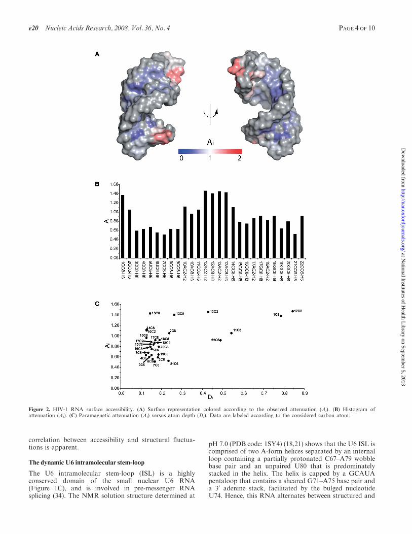
2D 1H-13C HSQC NMR spectroscopy is particularlysuited for probing the macromolecular surface withTEMPOL, since the paramagnetic attenuation affectscross-peak intensities arising from covalently bound13C and 1H nuclei that are very close in space (3).Furthermore, due to a favorable chemical shift dispersion,the aromatic CH correlations are suitable HSQC signalsfor delineating the accessibility of RNA structures. TheTEMPOL generated PRE effects were analyzed for allthe aromatic C2–H2, C6–H6 and C8–H8 correlations
of the HIV-1 frameshift site RNA. The PRE values werequantitated as attenuation index (Ai) values, as describedin the Materials and Methods section.The TEMPOL induced Ai values were mapped to
the surface of the RNA, and also plotted graphically(Figure 2A and B). The data correlate well with thestructure of the HIV-1 RNA. The tetraloop and terminalends of the RNA are more accessible than the A-formhelical region, suggesting that 3D structure is the mainfactor modulating the surface accessibility of the RNA.Intriguingly, we noticed that the purine aromatic C8–H8correlations have overall higher Ai values than the pyrimi-dine aromatic C6–H6 correlations. Quantitation of atomexposure, expressed as 3D atom depth index (Di) (22)values, confirmed that the purine C8–H8 groups are moreexposed than the pyrimidine C6–H6 groups by an averagevalue of 8% over the interior of the helix (residues 2–8 and15–21). The agreement between the average atom depth(Di) calculated from the ensemble of NMR structures andattenuation index (Ai) is shown in Figure 2C. In general,CH groups having high Di values, i.e. are close to themolecular surface, exhibit high Ai. In contrast, CH groupsmore buried in the structure have low or intermediate Ai
values. This trend holds true even for nucleotides thathave minor differences in atom exposure. For example,the first 50 nucleotide is more accessible than the last 30
nucleotide, even though they both reside at the helicalend (Figure 2B). This is because the 50 aromatic carbon(1C8, Di=0.786) is indeed more exposed than the 30
aromatic carbon (22C6, Di=0.491), as indicated by itshigher Di value (Figure 2C). This is in agreement withchemical acylation studies showing that RNA helical 50
ends are more accessible than 30 ends (33). However,anomalously high Ai’s are observed for nucleotides inthe tetraloop region (A10, A12, A13 and G14), which isa less ordered region of the structure (17). In particular,the C8 groups of A12 and A13, corresponding to the 30
side of the tetraloop, are highly attenuated, even thoughthey are well stacked and relatively buried within theensemble of NMR structures. These data suggest thatthe conformational variation within the time averagedNMR ensemble does not reflect the true conformationaldynamics of the molecule. The NMR structures of RNAloop regions are largely defined by NOE distancerestraints (�5 A), and the unstacking of loop nucleotidesmay go undetected by NOE methods, since such transi-tions may result in internucleotide distances >5 A. Theparamagnetic attenuation of the tetraloop nucleotidessuggests a strong connection between accessibility andRNA conformational flexibility.To further investigate the accessibility–flexibility corre-
lation, we performed a 10 ns MD simulation on the NMRstructure of the HIV-1 frameshift site RNA (PDB code:1PJY) (17). As expected, the root mean square fluctua-tions (r.m.s.f.) plot shows enhanced flexibility for thetetraloop and terminal nucleotides (Figure 3). In contrast,the helical residues experience limited and homogeneousfluctuations, consistent with the high stability previouslyobserved for this RNA stem (19). By comparing theattenuation and the r.m.s.f. data (Figures 2B and 3), the
PAGE 3 OF 10 Nucleic Acids Research, 2008, Vol. 36, No. 4 e20
at National Institutes of H
ealth Library on Septem
ber 5, 2013http://nar.oxfordjournals.org/
Dow
nloaded from
correlation between accessibility and structural fluctua-tions is apparent.
The dynamic U6 intramolecular stem-loop
The U6 intramolecular stem-loop (ISL) is a highlyconserved domain of the small nuclear U6 RNA(Figure 1C), and is involved in pre-messenger RNAsplicing (34). The NMR solution structure determined at
pH 7.0 (PDB code: 1SY4) (18,21) shows that the U6 ISL iscomprised of two A-form helices separated by an internalloop containing a partially protonated C67–A79 wobblebase pair and an unpaired U80 that is predominatelystacked in the helix. The helix is capped by a GCAUApentaloop that contains a sheared G71–A75 base pair anda 30 adenine stack, facilitated by the bulged nucleotideU74. Hence, this RNA alternates between structured and
Figure 2. HIV-1 RNA surface accessibility. (A) Surface representation colored according to the observed attenuation (Ai). (B) Histogram ofattenuation (Ai). (C) Paramagnetic attenuation (Ai) versus atom depth (Di). Data are labeled according to the considered carbon atom.
e20 Nucleic Acids Research, 2008, Vol. 36, No. 4 PAGE 4 OF 10
at National Institutes of H
ealth Library on Septem
ber 5, 2013http://nar.oxfordjournals.org/
Dow
nloaded from

dynamic regions, enabling us to further investigate therelationship between accessibility and structural flexibility.Analyses of NMR line shapes and relaxation ratesindicated that the U6 ISL RNA displays motions onboth the ps–ns and ms–ms timescales (20,35).Furthermore, the U6 ISL undergoes a pH-dependentconformational switch induced by protonation of A79(pKa=6.5), which results in stabilization of the C67–A79wobble pair and base flipping of the neighboring U80nucleotide (20,21,35). The C67–A79 wobble pair has alifetime of 20 ms, and the base flipping of U80 occurs on an�80 ms timescale (20,35). Since the dynamics of this RNAhave been previously characterized by NMR, it is an idealmodel system for exploring the application of theTEMPOL PRE analysis in detecting RNA conforma-tional transitions.
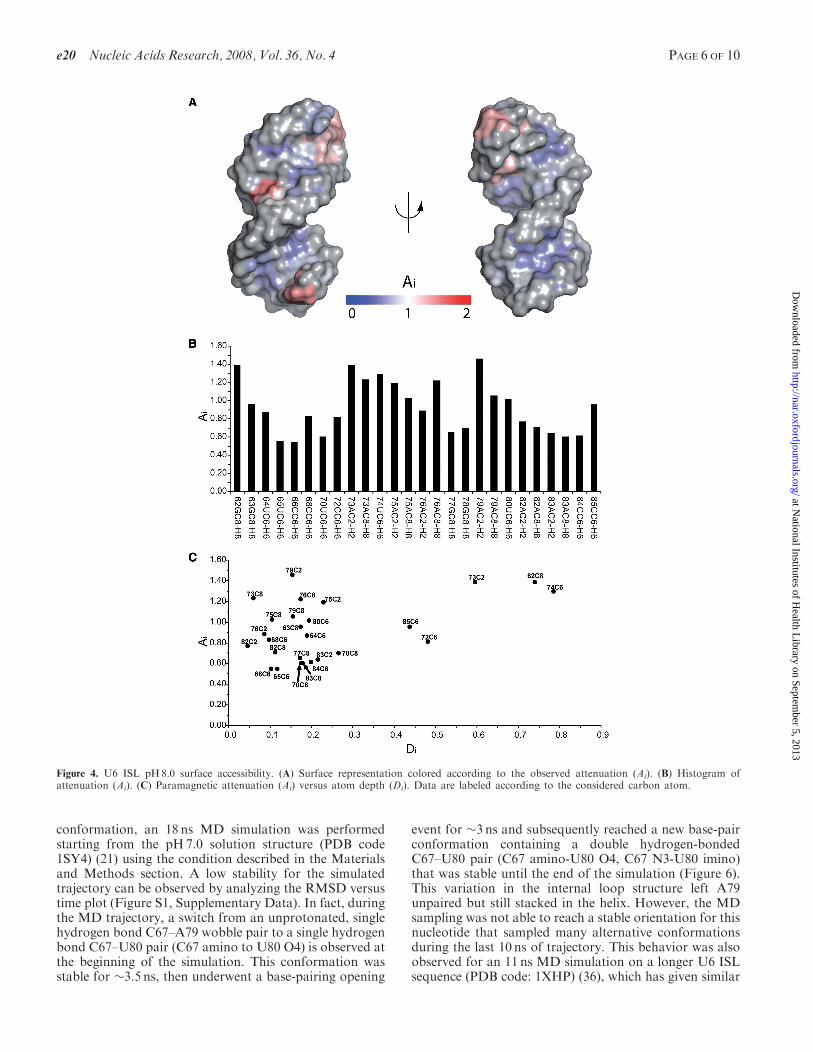
Since the pH-dependent conformational transitionwithin the U6 ISL is directly correlated to the protonationstate of A79 (35), the accessibility studies were performedat pH8.0 and at pH5.7. These experimental conditionsassure minimal interference from the other conformer(population < 10%) (20). The TEMPOL PRE analysiswas possible for 26/30 and 25/30 aromatic CH correla-tions, respectively, for the RNA at pH8.0 and pH5.7. Theremaining resonances could not be reliably analyzed dueto chemical shift overlap. The pH8.0 attenuation profile(Figure 4A and B) correlates well with the U6 RNAstructure. Higher Ai values are observed for the terminalnucleotides (62 and 85), the pentaloop and adjacentnucleotides (72–76; 71 was not measured due to spectraloverlap), and the internal loop nucleotides (79 and 80).
Inspection of Figure 4C shows the good agreementbetween accessibility and atom exposure. All of the anom-alously high Ai values are associated with nucleotideslocated in the dynamic pentaloop and internal loopregions (residues 73, 75, 76 and 79). The greatest anomalyis found for the A79 C2–H2 correlation, which experiencesthe highest TEMPOL accessibility at pH8.0, in spite ofits relatively low exposure in the pH7.0 NMR solution
structure. The A79 C2 atom is directly adjacent to thepartially protonated nitrogen (A79 N1). Interestingly,the dynamics of protonation (20), as well as the relaxationrates for the base (35) suggest that A79 is indeed dynamicand must be accessible to solvent when unprotonated,although the solvent-accessible conformation could not bedirectly observed in the time-averaged ensemble of NMRstructures calculated at pH7.0 (20,35).The conformational switch induced by lowering the pH
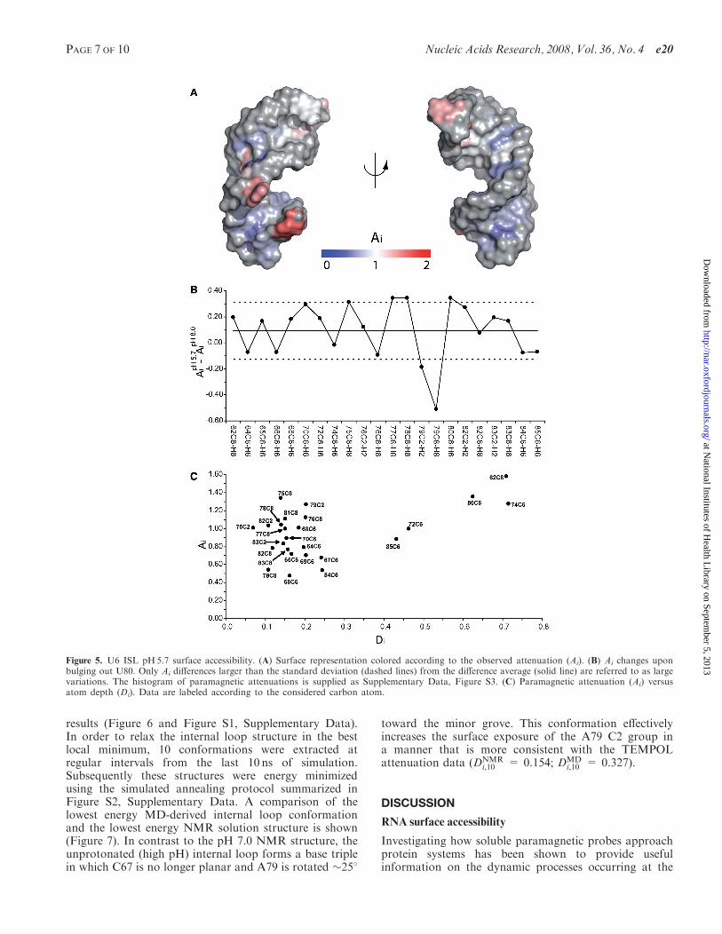
to 5.7 caused changes in the TEMPOL PRE pattern whichare mainly located in the internal loop region (Figure 5Aand B). In particular, the most significant change is thelarge decrease in Ai (1.1–0.5) detected for A79 C8. This isin agreement with the structure and thermodynamics atpH5.7 (20), which indicate that the internal loop structureis stabilized by formation of a protonated C67–A79wobble pair at low pH, and that this base pair is fullystacked within the helix. Furthermore, an increase in Ai
(1.1–1.4) is detected for U80 C6. The Ai value of 1.4 isconsistent with an increase in the population of the base-flipped conformation for U80. As a comparison, theextrahelical U74 nucleotide experiences dynamics on theps–ns timescale in a manner that is not pH-dependent (35)and has an Ai value that does not change with pH(Figure 5B). Additionally, the Ai values for the extra-helical nucleotides U74 and U80 are very similar atpH5.7, and due to their extrahelical conformations, bothnucleotides are also predicted to have high Di values(Figure 5C). Although the increase in Ai for U80 corre-lates with an increased proportion of the base-flippedconformer at low pH, as expected (20), the increase in Ai
for U80 relative to the pH8.0 sample is less than might beexpected, since the difference between the two measure-ments (0.3 Ai units) is barely above the observed standarddeviation (Figure 5B). This is likely due to the fact thatthe pH8.0 Ai value of 1.0 is relatively high, which may bereflective of the fact that a 3% population of based-flippedconformer still exists at this pH (35). Relative to the80 microsecond base-flipping timescale (35), TEMPOLprobing is very fast and results in irreversible PRE,allowing detection of even very transient populations.Therefore, the relatively small difference in Ai values(Figure 5B) for U80 is likely to be due to the detection ofthe base-flipped conformer even when it is present asa minor poplulation. On the other hand, the low pHcondition locks A79 into a protonated wobble base pairwith C67, which results in a more pronounced differencein attenuation for the A79 C2 and C8 resonances(Figure 5B). The flipped out U80 nucleotide may helpshield A79 from TEMPOL, which could further accountfor the large drop in attenuation for the A79 C8–H8correlation.The A79 C2 resonance has an anomalously high Ai
value at pH 8.0 (Figure 4C), suggesting that it is muchmore exposed than its position in the NMR structure,which was previously determined at pH7.0 (21). Further-more, interpretation of NMR relaxation rates at this siteare complicated by chemical exchange due to the rapidprotonation equilibrium of the adjacent N1 group (20,35).Therefore, in order to gain a better understanding of thedynamic motions associated with the unprotonated A79
Figure 3. Histogram of root mean square fluctuations (r.m.s.f., A)calculated from the 10 ns HIV-1 RNA MD simulation.
PAGE 5 OF 10 Nucleic Acids Research, 2008, Vol. 36, No. 4 e20
at National Institutes of H
ealth Library on Septem
ber 5, 2013http://nar.oxfordjournals.org/
Dow
nloaded from
conformation, an 18 ns MD simulation was performedstarting from the pH7.0 solution structure (PDB code1SY4) (21) using the condition described in the Materialsand Methods section. A low stability for the simulatedtrajectory can be observed by analyzing the RMSD versustime plot (Figure S1, Supplementary Data). In fact, duringthe MD trajectory, a switch from an unprotonated, singlehydrogen bond C67–A79 wobble pair to a single hydrogenbond C67–U80 pair (C67 amino to U80 O4) is observed atthe beginning of the simulation. This conformation wasstable for �3.5 ns, then underwent a base-pairing opening
event for �3 ns and subsequently reached a new base-pairconformation containing a double hydrogen-bondedC67–U80 pair (C67 amino-U80 O4, C67 N3-U80 imino)that was stable until the end of the simulation (Figure 6).This variation in the internal loop structure left A79unpaired but still stacked in the helix. However, the MDsampling was not able to reach a stable orientation for thisnucleotide that sampled many alternative conformationsduring the last 10 ns of trajectory. This behavior was alsoobserved for an 11 ns MD simulation on a longer U6 ISLsequence (PDB code: 1XHP) (36), which has given similar
Figure 4. U6 ISL pH8.0 surface accessibility. (A) Surface representation colored according to the observed attenuation (Ai). (B) Histogram ofattenuation (Ai). (C) Paramagnetic attenuation (Ai) versus atom depth (Di). Data are labeled according to the considered carbon atom.
e20 Nucleic Acids Research, 2008, Vol. 36, No. 4 PAGE 6 OF 10
at National Institutes of H
ealth Library on Septem
ber 5, 2013http://nar.oxfordjournals.org/
Dow
nloaded from
results (Figure 6 and Figure S1, Supplementary Data).In order to relax the internal loop structure in the bestlocal minimum, 10 conformations were extracted atregular intervals from the last 10 ns of simulation.Subsequently these structures were energy minimizedusing the simulated annealing protocol summarized inFigure S2, Supplementary Data. A comparison of thelowest energy MD-derived internal loop conformationand the lowest energy NMR solution structure is shown(Figure 7). In contrast to the pH 7.0 NMR structure, theunprotonated (high pH) internal loop forms a base triplein which C67 is no longer planar and A79 is rotated �258
toward the minor grove. This conformation effectivelyincreases the surface exposure of the A79 C2 group ina manner that is more consistent with the TEMPOLattenuation data (DNMR
i,10 = 0.154; DMDi,10 = 0.327).
DISCUSSION
RNA surface accessibility
Investigating how soluble paramagnetic probes approachprotein systems has been shown to provide usefulinformation on the dynamic processes occurring at the
Figure 5. U6 ISL pH5.7 surface accessibility. (A) Surface representation colored according to the observed attenuation (Ai). (B) Ai changes uponbulging out U80. Only Ai differences larger than the standard deviation (dashed lines) from the difference average (solid line) are referred to as largevariations. The histogram of paramagnetic attenuations is supplied as Supplementary Data, Figure S3. (C) Paramagnetic attenuation (Ai) versusatom depth (Di). Data are labeled according to the considered carbon atom.
PAGE 7 OF 10 Nucleic Acids Research, 2008, Vol. 36, No. 4 e20
at National Institutes of H
ealth Library on Septem
ber 5, 2013http://nar.oxfordjournals.org/
Dow
nloaded from

protein–solution interface (1–3,5–10,14). Here, the surfaceaccessibility of RNA molecules characterized by differentshape and dynamics was analyzed by evaluating thePRE generated by TEMPOL on conventional 2DNMR measurements. The present results show thatTEMPOL approaches RNA surfaces more uniformlythan proteins (1,3,6,10,13,14). In fact, the agreementbetween the paramagnetic attenuation and atom depthsfor helical RNA molecules (Figures 2C, 4C and 5C),suggests that the shape, local dynamics and compositionof RNA molecular surfaces are more homogeneousthan those of proteins. Furthermore, the high negativecharge typical of RNA surfaces may hamper formationof transient aggregates, which have been shown tosignificantly modulate the surface accessibility of proteins(5,8–10).However, we find that nucleotides residing in
flexible loop regions show higher accessibility than theones observed for helical residues of similar depth(Figures 2–5). This finding suggests that these nucleotidestransiently unstack, increasing the exposure of the baseto the bulk solvent. In many cases, such transient eventsmay not be evident within NMR structures that aretypically time-averaged and population-weighted ensem-bles. Recently, the transient unstacking of stable GNRAtetraloops has been observed using femptosecond time-scale fluorescence spectroscopy (37).
Detecting RNA conformational changes
RNA conformational transitions are essential for ribo-some and spliceosome function and play a significant rolein recognition of proteins, RNA and ligands (38–41).Thus, studying RNA conformational dynamics is of
primary relevance in understanding the correlationbetween RNA structure and function. However, investi-gating conformational changes in solution may be acomplex and time-consuming endeavor. Hence, alterna-tive methods able to map conformational transitionsin RNA may be very helpful in complementing otherinvestigation techniques. In the present report, weshow that a small paramagnetic probe can detect apH-dependent conformational switch in U6 RNA thathas implications for spliceosome function and catalysis(20,35). The detailed pathway and associated energetics ofthis conformational change are unknown, but are drivenby protonation of A79 (20,35). Here, we observe forma-tion of the protonated C67–A79 pair causes large Ai
variations in the internal loop region (Figure 5B).Comparison of the TEMPOL probing data (Figures 4and 5) support the hypothesis that the transition betweenthe protonated and unprotonated U6 ISL proceedsthrough an intermediate structure that is protonatedbut has a more exposed A79 (35). Interestingly, our MDsimulation suggests a new conformation for the unproto-nated U6 ISL internal bulge, in which the A79 base isrotated toward the minor grove, enhancing its solventexposure (Figure 7). The MD simulation suggests thatU80 is at least transiently involved in a base triple withC67 and A79. This may help explain the preferencefor this nucleotide to adopt a stacked conformation athigh pH.
Further Ai variations are observed for nucleotidesflanking the U6 ISL internal loop (G77 and G78)that show enhanced TEMPOL accessibility at low pH.These nucleotides are in stable Watson–Crick pairs, andprevious thermodynamic investigations indicate that thestructure is even more stable at low pH (21), dismissingthe possibility of increased conformational flexibility forthese nucleotides. Possibly, the modified molecular shapeas well as the proximity of the highly dynamic loopnucleotides could increase the self-diffusion properties ofthe hydration layer surrounding these nucleotides, thusenhancing their accessibility to the paramagnetic probe(3–5,42).
Recently, we have demonstrated that TEMPOL prob-ing can be used to detect the binding of a small moleculeligand to the surface of the HIV RNA (43). Therefore, theTEMPOL probing method is well suited for probing RNAstructure, dynamics and ligand interactions.
SUPPLEMENTARY DATA
Supplementary Data are available at NAR Online.
ACKNOWLEDGEMENTS
This study made use of the National Magnetic ResonanceFacility at Madison (www.nmrfam.wisc.edu), which issupported by National Institutes of Health grantsP41RR02301 (Biomedical Research TechnologyProgram, National Center for Research Resources) andP41GM66326 (National Institute of General MedicalSciences). Equipment in the facility was purchased with
Figure 6. Snapshots of the MD simulations performed on two NMRstructures (PDB codes: 1SY4 and 1XHP) of U6 ISL pH7.0 conformershowing the internal bulge conformation.
Figure 7. Comparison of the NMR and MD-derived U6 ISL internal-loop conformation. Hydrogen bonds are indicated with red lines.
e20 Nucleic Acids Research, 2008, Vol. 36, No. 4 PAGE 8 OF 10
at National Institutes of H
ealth Library on Septem
ber 5, 2013http://nar.oxfordjournals.org/
Dow
nloaded from
funds from the University of Wisconsin, the NationalInstitutes of Health (P41GM66326, P41RR02301,RR02781, RR08438), the National Science Foundation(DMB-8415048, BIR-9214394) and the US Department ofAgriculture. This work was supported by NIH grantsGM072447 and GM065166 to S.E.B. Funding to pay theOpen Access publication charges for this article wasprovided by NIH grant GM065166 to S.E.B.
Conflict of interest statement. None declared.
REFERENCES
1. Scarselli,M., Bernini,A., Segoni,C., Molinari,H., Esposito,G.,Lesk,A.M., Laschi,F., Temussi,P. and Niccolai,N. (1999)Tendamistat surface accessibility to the TEMPOL paramagneticprobe. J. Biomol. NMR, 15, 125–133.
2. Teng,C.L. and Bryant,R.G. (2004) Mapping oxygen accessibility toribonuclease a using high-resolution nmr relaxation spectroscopy.Biophys. J., 86, 1713–1725.
3. Niccolai,N., Spiga,O., Bernini,A., Scarselli,M., Ciutti,A., Fiaschi,I.,Chiellini,S., Molinari,H. and Temussi,P.A. (2003) NMR studies ofprotein hydration and TEMPOL accessibility. J. Mol. Biol., 332,437–447.
4. De Simone,A., Spadaccini,R., Temussi,P. A. and Fraternali,F.(2006) Toward the understanding of MNEI sweetness fromhydration map surfaces. Biophys. J., 90, 3052–3061.
5. Venditti,V., Bernini,A., De Simone,A., Spiga,O., Prischi,F. andNiccolai,N. (2007) MD and NMR studies of [alpha]-bungarotoxinsurface accessibility. Biochem. Biophys. Res. Commun., 356,114–117.
6. Niccolai,N., Ciutti,A., Spiga,O., Scarselli,M., Bernini,A., Bracci,L.,Di Maro,D., Dalvit,C., Molinari,H. et al. (2001) NMR studies ofprotein surface accessibility. J. Biol. Chem., 276, 42455–42461.
7. Teng,C.L. and Bryant,R.G. (2006) Spin relaxation measurements ofelectrostatic bias in intermolecular exploration. J. Magn. Reson.,179, 199–205.
8. Liepinsh,E., Baryshev,M., Sharipo,A., Ingelman-Sundberg,M.,Otting,G. and Mkrtchian,S. (2001) Thioredoxin fold as homodi-merization module in the putative chaperone ERp29: NMRstructures of the domains and experimental model of the 51 kDadimer. Structure, 9, 457–471.
9. Bernini,A., Spiga,O., Ciutti,A., Venditti,V., Prischi,F.,Governatori,M., Bracci,L., Lelli,B., Pileri,S. et al. (2006) NMRstudies of BPTI aggregation by using paramagnetic relaxationreagents. Biochim. Biophys. Acta, 1764, 856–862.
10. Bernini,A., Spiga,O., Venditti,V., Prischi,F., Bracci,L., Tong,A.P.L.,Wong,W. T. and Niccolai,N. (2006) NMR studies of lysozymesurface accessibility by using different paramagnetic relaxationprobes. J. Am. Chem. Soc., 128, 9290–9291.
11. Tang,C., Iwahara,J. and Clore,G.M. (2006) Visualization oftransient encounter complexes in protein-protein association.Nature, 444, 383–386.
12. Iwahara,J. and Clore,G.M. (2006) Detecting transient intermediatesin macromolecular binding by paramagnetic NMR. Nature, 440,1227–1230.
13. Molinari,H., Esposito,G., Ragona,L., Pegna,M., Niccolai,N.,Brunne,R.M., Lesk,A.M. and Zetta,L. (1997) Probing proteinstructure by solvent perturbation of NMR spectra: the surfaceaccessibility of bovine pancreatic trypsin inhibitor. Biophys. J., 73,382–396.
14. Niccolai,N., Spadaccini,R., Scarselli,M., Bernini,A., Crescenzi,O.,Spiga,O., Ciutti,A., Di Maro,D., Bracci,L. et al. (2001) Probing thesurface of a sweet protein: NMR study of MNEI with aparamagnetic probe. Protein Sci., 10, 1498–1507.
15. Zhou,N., Mascagni,P., Gibbons,W.A., Niccolai,N., Rossi,C. andWyssbrod,H. (1985) Confirmation of the solution structure oftyrocidine a using perturbation of proton relaxation rates bynitroxide spin labels. J. Chem. Soc., Perkin Trans., 2, 581–587.
16. Pintacuda,G. and Otting,G. (2002) Identification of ProteinSurfaces by NMR Measurements with a Paramagnetic Gd(III)Chelate. J. Am. Chem. Soc., 124, 372–373.
17. Staple,D.W. and Butcher,S.E. (2003) Solution structure of theHIV-1 frameshift inducing stem-loop RNA. Nucleic Acids Res., 31,4326–4331.
18. Huppler,A., Nikstad,L.J., Allmann,A.M., Brow,D.A. andButcher,S.E. (2002) Metal binding and base ionization in the U6RNA intramolecular stem-loop structure. Nat. Struct. Mol. Biol., 9,431–435.
19. Staple,D.W. and Butcher,S.E. (2005) Solution Structure andThermodynamic Investigation of the HIV-1 Frameshift InducingElement. J. Mol. Biol., 349, 1011–1023.
20. Reiter,N.J., Blad,H., Abildgaard,F. and Butcher,S.E. (2004)Dynamics in the U6 RNA intramolecular stem-loop: a base flippingconformational change. Biochemistry, 43, 13739–13747.
21. Reiter,N.J., Nikstad,L.J., Allmann,A.M., Johnson,R.J. andButcher,S.E. (2003) Structure of the U6 RNA intramolecular stem-loop harboring an SP-phosphorothioate modification. RNA, 9,533–542.
22. Varrazzo,D., Bernini,A., Spiga,O., Ciutti,A., Chiellini,S.,Venditti,V., Bracci,L. and Niccolai,N. (2005) Three-dimensionalcomputation of atom depth in complex molecular structures.Bioinformatics, 21, 2856–2860.
23. Koradi,R., Billeter,M. and Wuthrich,K. (1996) MOLMOL: Aprogram for display and analysis of macromolecular structure.J. Mol. Graph. Model., 14, 51–55.
24. Davis,J.H., Foster,T.R., Tonelli,M. and Butcher,S.E. (2007) Role ofmetal ions in the tetraloop-receptor complex as analyzed by NMR.RNA, 13, 76–86.
25. Case,D.A., Cheatham,T.E., Darden,T., Gohlke,H., Luo,R.,Merz,K.M., Onufriev,A., Simmerling,C., Wang,B. et al. (2005) TheAmber biomolecular simulation programs. J. Comput. Chem., 26,1668–1688.
26. Wang,J., Cieplak,P. and Kollman,P.A. (2000) How well does arestrained electrostatic potential (RESP) model perform in calcu-lating conformational energies of organic and biological molecules?J. Comput. Chem., 21, 1049–1074.
27. Mohoney,M.W. and Jorgensen,W.L. (2000) A five-site modelfor liquid water and the reproduction of the density anomaly byrigid, nonpolarizable potential functions. J. Chem. Phys., 112,8910–8922.
28. Berendsen,H.J.C., Postma,J.P.M., van Gunsteren,W.F. and DiNola,A. (1984) Molecular dynamics with coupling to an externalbath. J. Phys. Chem., 91, 6269–6271.
29. Pastor,R.W., Brooks,B.R. and Pastor,R.W. (1988) An analysis ofthe accuracy of Langevin and molecular dynamics algorithms.Mol. Phys., 65, 1409–1419.
30. Ryckaert,J.P., Ciccotti,G. and Berendsen,H.J.C. (1977) Numericalintegration of the Cartesian equations of motion of a system withconstraints: Molecular dynamics of n-alkanes. J. Comput. Chem.,23, 327–341.
31. Darden,T., Perera,L., Li,L. and Pedersen,L. (1999) New tricks formodellers from the crystallography toolkit: the particle mesh Ewaldalgorithm and its use in nucleic acid simulations. Struct. Fold Des.,7, R55–R60.
32. Hofacker,I.L., Fekete,M., Flamm,C., Huynen,M.A., Rauscher,S.,Stolorz,P.E. and Stadler,P.F. (1998) Automatic detection ofconserved RNA structure elements in complete RNA virusgenomes. Nucleic Acids Res., 26, 3825–3836.
33. Weeks,K.M. and Crothers,D.M. (1993) Major grove accessibility ofRNA. Science, 261, 1574–1577.
34. Fortner,D.M., Troy,R.G. and Brow,D.A. (1994) A stem/loop in U6RNA defines a conformational switch required for pre- mRNAsplicing. Genes Dev., 8, 221–233.
35. Blad,H., Reiter,N.J., Abildgaard,F., Markley,J.L. and Butcher,S.E.(2005) Dynamics and metal ion binding in the U6 RNAintramolecular stem-loop as analyzed by NMR. J. Mol. Biol., 353,540–555.
36. Sashital,D.G., Cornilescu,G., McManus,C.J., Brow,D.A. andButcher,S.E. (2004) U2�U6 RNA folding reveals a group II intron-like domain and a four-helix junction. Nat. Struct. Mol. Biol., 11,1237–1242.
PAGE 9 OF 10 Nucleic Acids Research, 2008, Vol. 36, No. 4 e20
at National Institutes of H
ealth Library on Septem
ber 5, 2013http://nar.oxfordjournals.org/
Dow
nloaded from
37. Zhao,L. and Xia,T. (2007) Direct revelation of multiple conforma-tions in RNA by femtosecond dynamics. J. Am. Chem. Soc., 129,4118–4119.
38. Noller,H.F. and Baucom,A. (2002) Structure of the 70 S ribosome:implications for movement. Biochem. Soc. Trans., 30, 1159–1161.
39. Rodnina,M.V., Daviter,T., Gromadski,K. and Wintermeyer,W.(2002) Structural dynamics of ribosomal RNA during decoding onthe ribosome. Biochimie, 84, 745–754.
40. Valle,M., Zavialov,A., Sengupta,J., Rawat,U., Ehrenberg,M. andFrank,J. (2003) Locking and unlocking of ribosomal motions. Cell,114, 123–134.
41. Butcher,S.E. and Brow,D.A. (2005) Towards understanding thecatalytic core structure of the spliceosome. Biochem. Soc. Trans., 33,447–449.
42. De Simone,A., Dodson,G.G., Verma,C.S., Zagari,A. andFraternali,F. (2005) Prion and water: tight and dynamical hydrationsites have a key role in structural stability. Proc. Natl Acad. Sci.U.SA, 102, 7535–7540.
43. Staple,D.W., Venditti,V., Niccolai,N., Elson-Schwab,L., Tor,Y. andButcher,S.E. (2007) Guanidinio-neomycin B recognition of anHIV-1 RNA helix. Chem. Biochem., In Press.
e20 Nucleic Acids Research, 2008, Vol. 36, No. 4 PAGE 10 OF 10
at National Institutes of H
ealth Library on Septem
ber 5, 2013http://nar.oxfordjournals.org/
Dow
nloaded from
Copyright © 2022 FDOKUMEN