Single-cell chromatin accessibility landscape in ... - Nature
12
ARTICLE Single-cell chromatin accessibility landscape in kidney identifies additional cell-of-origin in heterogenous papillary renal cell carcinoma Qi Wang 1,8 , Yang Zhang 1,2,8 , Bolei Zhang 3 , Yao Fu 4 , Xiaozhi Zhao 5 , Jing Zhang 1 , Ke Zuo 2 , Yuexian Xing 2 , Song Jiang 2 , Zhaohui Qin 6 , Erguang Li 1 , Hongqian Guo 5 ✉ , Zhihong Liu 1,2 ✉ & Jingping Yang 1,2,7 ✉ Papillary renal cell carcinoma (pRCC) is the most heterogenous renal cell carcinoma. Patient survival varies and no effective therapies for advanced pRCC exist. Histological and molecular characterization studies have highlighted the heterogeneity of pRCC tumours. Recent studies identified the proximal tubule (PT) cell as a cell-of-origin for pRCC. However, it remains elusive whether other pRCC subtypes have different cell-of-origin. Here, by obtaining genome-wide chromatin accessibility profiles of normal human kidney cells using single-cell transposase-accessible chromatin-sequencing and comparing the profiles with pRCC sam- ples, we discover that besides PT cells, pRCC can also originate from kidney collecting duct principal cells. We show pRCCs with different cell-of-origin exhibit different molecular characteristics and clinical behaviors. Further, metabolic reprogramming appears to mediate the progression of pRCC to the advanced state. Here, our results suggest that determining cell-of-origin and monitoring origin-dependent metabolism could potentially be useful for early diagnosis and treatment of pRCC. https://doi.org/10.1038/s41467-021-27660-3 OPEN 1 Medical School of Nanjing University, Nanjing, Jiangsu 210093, China. 2 National Clinical Research Center for Kidney Disease, Jinling Hospital, Medical School of Nanjing University, Nanjing, Jiangsu 210002, China. 3 School of Computer Science, Nanjing University of Posts and Telecommunications, Nanjing, Jiangsu 210023, China. 4 Department of Pathology, Affiliated Drum Tower Hospital, Medical School of Nanjing University, Nanjing, Jiangsu 210008, China. 5 Department of Urology, Affiliated Drum Tower Hospital, Medical School of Nanjing University, Nanjing, Jiangsu 210008, China. 6 Department of Biostatistics and Bioinformatics, Rollins School of Public Health, Emory University, Atlanta, GA 30322, USA. 7 Jiangsu Key Laboratory of Molecular Medicine, Medical School, Nanjing University, Nanjing 210093, China. 8 These authors contributed equally: Qi Wang, Yang Zhang. ✉ email: [email protected]; [email protected]; [email protected] NATURE COMMUNICATIONS | (2022)13:31 | https://doi.org/10.1038/s41467-021-27660-3 | www.nature.com/naturecommunications 1 1234567890():,;
-
Upload
khangminh22 -
Category
Documents
-
view
0 -
download
0
Transcript of Single-cell chromatin accessibility landscape in ... - Nature

ARTICLE
Single-cell chromatin accessibility landscape inkidney identifies additional cell-of-origin inheterogenous papillary renal cell carcinomaQi Wang1,8, Yang Zhang1,2,8, Bolei Zhang3, Yao Fu 4, Xiaozhi Zhao5, Jing Zhang1, Ke Zuo2, Yuexian Xing2,
Song Jiang 2, Zhaohui Qin 6, Erguang Li1, Hongqian Guo 5✉, Zhihong Liu 1,2✉ & Jingping Yang 1,2,7✉
Papillary renal cell carcinoma (pRCC) is the most heterogenous renal cell carcinoma. Patient
survival varies and no effective therapies for advanced pRCC exist. Histological and molecular
characterization studies have highlighted the heterogeneity of pRCC tumours. Recent studies
identified the proximal tubule (PT) cell as a cell-of-origin for pRCC. However, it remains
elusive whether other pRCC subtypes have different cell-of-origin. Here, by obtaining
genome-wide chromatin accessibility profiles of normal human kidney cells using single-cell
transposase-accessible chromatin-sequencing and comparing the profiles with pRCC sam-
ples, we discover that besides PT cells, pRCC can also originate from kidney collecting duct
principal cells. We show pRCCs with different cell-of-origin exhibit different molecular
characteristics and clinical behaviors. Further, metabolic reprogramming appears to mediate
the progression of pRCC to the advanced state. Here, our results suggest that determining
cell-of-origin and monitoring origin-dependent metabolism could potentially be useful for
early diagnosis and treatment of pRCC.
https://doi.org/10.1038/s41467-021-27660-3 OPEN
1Medical School of Nanjing University, Nanjing, Jiangsu 210093, China. 2 National Clinical Research Center for Kidney Disease, Jinling Hospital, MedicalSchool of Nanjing University, Nanjing, Jiangsu 210002, China. 3 School of Computer Science, Nanjing University of Posts and Telecommunications, Nanjing,Jiangsu 210023, China. 4 Department of Pathology, Affiliated Drum Tower Hospital, Medical School of Nanjing University, Nanjing, Jiangsu 210008, China.5 Department of Urology, Affiliated Drum Tower Hospital, Medical School of Nanjing University, Nanjing, Jiangsu 210008, China. 6Department ofBiostatistics and Bioinformatics, Rollins School of Public Health, Emory University, Atlanta, GA 30322, USA. 7 Jiangsu Key Laboratory of Molecular Medicine,Medical School, Nanjing University, Nanjing 210093, China. 8These authors contributed equally: Qi Wang, Yang Zhang. ✉email: [email protected];[email protected]; [email protected]
NATURE COMMUNICATIONS | (2022) 13:31 | https://doi.org/10.1038/s41467-021-27660-3 | www.nature.com/naturecommunications 1
1234
5678
90():,;

While pRCC patients with poor survival are known todisplay molecular features such as metabolic features,immunologic features, or CpG island methylator
phenotype (CIMP)1–3, it is unclear whether these features are thecause or outcome of tumor progression. From histological andmolecular characterization studies, we know pRCC tumors arehighly heterogenous and display various subtypes with differentmalignancies, making the disease extremely difficult to treat1–4.Studies of other cancers have shown that tumor heterogeneitymay emerge from different cells-of-origin—normal cells afflictedwith the cancer-causing mutation that go on to determine the fateand pathology of a tumor cell. In breast cancers5 andglioblastoma6,7, the same genetic mutation in a different celllineage can lead to different morphology, phenotype and malig-nancy. Hematopoietic stem cells-derived leukemias showinghigher methylation levels than granulocyte-macrophage pro-genitor cells-derived leukemias8 also suggest that the molecularfeatures of heterogenous tumors could be underpinned by cell-of-origin5,7,9,10. There is also evidence showing that cell-of-origincan affect tumor response to treatments8,11,12. Cell-of-origin,whose features are retained in evolving tumor cells13, is thereforea powerful biomarker for early diagnosis. Screening cell-of-originsignatures in pRCC could potentially uncover unknown cells-of-origin that are linked to the various pRCC subtypes and theirclinical behaviors. Understanding how the subtypes progresscould help us develop more targeted treatments and reducedeaths14.
Stable and heritable epigenetic signatures such as transcriptionfactors programs15, histone modifications16 and DNAmethylation17 have been used effectively to trace the cell-of-originand subtypes of morphological and molecular evolving tumorcells. In our study, we used another epigenetic signature, chro-matin accessibility, to trace the cell-of-origin signatures in pRCC.Because chromatin accessibility reflects transcription factorbinding, histone modifications and DNA methylation, it offersgreater insights into the gene regulatory mechanisms of the cells.Additionally, the chromatin accessibility profiles of 34 humanpRCC samples from The Cancer Genome Atlas (TCGA) haverevealed the heterogeneity of pRCC18.
In this work, we use single-cell assay for transposase-accessiblechromatin sequencing (scATAC-seq) to capture the cell type-specific chromatin accessibility landscape of normal human kid-ney at single-cell resolution. The alignment of the ATAC-seqprofiles of human pRCC samples against the landscape resolvesheterogenous cell-of-origin of pRCC. Besides proximal tubule(PT) cells, pRCC can also originate from collecting duct principalcells (CD_PC) in the distal tubules of the kidney. We find thatpRCCs with different cell-of-origin have different molecularcharacteristics, undergo different carcinogenic transformations,and display different clinical behaviors. pRCC also show cell-of-origin-dependent risks of progression, which is characterized bymetabolism reprogramming. pRCC that originated from CD_PCare enriched for advanced pRCC and activated interferon sig-naling, suggesting their potential response to checkpoint immu-notherapy. Our study demonstrates that the chromatinaccessibility landscape is a potential early diagnostic method foradvanced pRCC, allowing cell-of-origin dependent molecularcharacteristics to direct the treatment of pRCC.
ResultsscATAC captures the epigenetic landscape of normal kidney.We performed scATAC-seq on paracancerous kidney tissuesfrom two patients undergoing nephrectomy to profile chromatinaccessibility for cells in the kidney (Supplementary Fig. 1a, b). Atotal of 9460 high-quality nuclei were kept after stringent quality
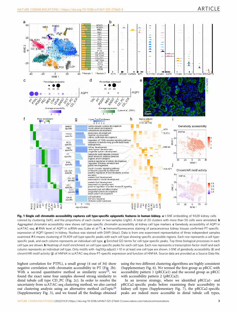
controls (Supplementary Fig. 1c, d), with a median read depth of14,158 fragments per nuclei and median transcription start site(TSS) ratio (the percentage of fragments at TSS) of 0.15. Afterbatch correction and unbiased clustering with Latent SemanticAnalysis on term frequency-inverse document frequency (LSA-log (TF-IDF)), the nuclei formed 20 clusters (SupplementaryFig. 1e) with an even distribution in the samples (SupplementaryFig. 1f). Clusters with more than 50 cells were annotated byintegrating the scATAC-seq with four human kidney single-cellRNA sequencing (scRNA-seq) datasets19–22 (SupplementaryFig. 2a). Clusters that could not be consistently annotated (e.g.,cluster 9) were re-clustered and re-annotated (SupplementaryFig. 2b, c). After integrating the scRNA-seq and examining themarker genes, a total of 20 clusters were finally annotated and allthe main cell types in the kidney were identified (Fig. 1a andSupplementary Data 1). The annotation revealed a PT-relatedrenal progenitor-like (PTPL) subgroup with chromatin accessi-bility at VCAM1 and PROM1 loci (Supplementary Fig. 2d).Further, the profiles display cell type-specific chromatin accessi-bility (Fig. 1b), notably at loci AQP1 in PT cells, which were notdetected in scRNA-seq (Fig. 1c, d). Immunofluorescence stainingof the kidney samples confirmed that high levels of AQP1 areexpressed only in PT cells (Fig. 1e). These results demonstratethat scATAC-seq captured genome-wide transcriptional regula-tion with high sensitivity.
To investigate the cell type-specificity of epigenetic regulation,we characterized the chromatin accessible regions by comparingthe scATAC-seq peaks of different cell types (SupplementaryFig. 3a). We detected 19,409 cell type-specific peaks and each celltype showed cell type-specific accessible regions (Fig. 1f andSupplementary Fig. 3b). A vast majority of these peaks were innon-coding regions (Supplementary Fig. 3c, d). Functionalanalysis of the specific peaks by GREAT23 revealed that eachgroup of cell type-specific peaks corresponds to the functions ofthe cell type (Fig. 1g). For instance, PT-specific peaks reflect theirrole in catabolism of small molecules while CD_PC-specificpeaks, though small in number, reflect their specific role inurinary excretion. The cell type-specific peaks also revealed keytranscription factors in each cell type (Fig. 1h). HNF4A, as a keytranscription factor in PT cells24, is highly accessible only inPT cells (Fig. 1i), and the genomic sites having the HNF4A motifonly show activity in PT cells as well (Fig. 1j). These resultsindicate that the scATAC-seq profile sensitively captured celltype-specific epigenetic features in the kidney.
Cell-of-origin of pRCC is heterogenous. We used the establishedcell type-specific epigenetic features in kidney cells and theATAC-seq of 34 human pRCC samples from TCGA identified byCorces et al.18 to investigate the cell-of-origin of pRCC. Toascertain if cell type-specific epigenetic features can be used toreveal the cell-of-origin of cancers, we compared the cell type-specific peaks from our normal kidney scATAC-seq profile withthe cancer type-specific peaks from pan-cancer chromatinaccessibility profiles for 410 tumor samples across 23 cancer typesidentified in Corces et al.18. Renal cell carcinoma including pRCCshowed significant overlap with kidney PT cells (Fig. 2a andSupplementary Fig. 4). This enrichment, consistent with previousreports that have identified pRCC tumor cells as originating fromPT cells19,25, confirmed that cell type-specific epigenetic featurescan be used to identify the cell-of-origin of cancers.
When the cell-of-origin for the 34 pRCC samples with ATAC-seq from TCGA18 were subsequently examined individually, twodifferent chromatin accessibility patterns were seen (Fig. 2b).While most of the pRCC samples (30 out of 34) show positivecorrelation with chromatin accessibility in the PT groups (with
ARTICLE NATURE COMMUNICATIONS | https://doi.org/10.1038/s41467-021-27660-3
2 NATURE COMMUNICATIONS | (2022) 13:31 | https://doi.org/10.1038/s41467-021-27660-3 | www.nature.com/naturecommunications
highest correlation for PTPL), a small group (4 out of 34) shownegative correlation with chromatin accessibility in PT (Fig. 2b).With a second quantitative method as similarity score19, wefound the exact same four samples showed strong similarity todistal tubule cell type CD_PC (Fig. 2c). In order to resolve theuncertainty from scATAC-seq clustering method, we also carriedout clustering analysis using an alternative method cisTopic26
(Supplementary Fig. 5), and we found all the findings obtained
using the two different clustering algorithms are highly consistent(Supplementary Fig. 6). We termed the first group as pRCC withaccessibility pattern 1 (pRCCa1) and the second group as pRCCwith accessibility pattern 2 (pRCCa2).
In an inverse strategy, where we identified pRCCa1- andpRCCa2-specific peaks before examining their accessibility inkidney cell types (Supplementary Fig. 7), the pRCCa2-specificpeaks are indeed more accessible in distal tubule cell types,
Fig. 1 Single cell chromatin accessibility captures cell type-specific epigenetic features in human kidney. a t-SNE embedding of 9428 kidney cellscolored by clustering (left), and the proportions of each cluster in two samples (right). A total of 20 clusters with more than 50 cells were annotated. bAggregated chromatin accessibility view shows cell type-specific chromatin accessibility at kidney cell type markers. c Genebody accessibility of AQP1 inscATAC-seq. d RNA level of AQP1 in scRNA-seq (Lake et al.21). e Immunofluorescence staining of paracancerous kidney tissues confirmed PT-specificexpression of AQP1 (green) in kidney. Nucleus was stained with DAPI (blue). Data is from one experiment representative of three independent samplesexamined. f K-means clustering of 19,409 cell type-specific peaks with each cell type showing specific accessible regions. Each row represents a cell type-specific peak, and each column represents an individual cell type. g Enriched GO terms for cell type-specific peaks. Top three biological processes in eachcell type are shown. h Heatmap of motif enrichment on cell type-specific peaks for each cell type. Each row represents a transcription factor motif and eachcolumn represents an individual cell type. Only motifs with -log10(p.adjust) > 10 in at least one cell type are shown. t-SNE of genebody accessibility (i) andchromVAR motif activity (j) of HNF4A in scATAC-seq show PT-specific expression and function of HNF4A. Source data are provided as a Source Data file.
NATURE COMMUNICATIONS | https://doi.org/10.1038/s41467-021-27660-3 ARTICLE
NATURE COMMUNICATIONS | (2022) 13:31 | https://doi.org/10.1038/s41467-021-27660-3 | www.nature.com/naturecommunications 3

especially CD_PC (Fig. 2d). For example, pRCCa2 do not showchromatin accessibility at the PT marker AQP1 locus, but areaccessible at the CD_PC marker GATA3 locus and at GRHL2locus which encodes a protein required for the function ofcollecting duct27 (Fig. 2e and Supplementary Fig. 8a). Addition-ally, the pRCCa1/2-specific peaks are enriched with motifs fortheir respective cell-of-origin’s key transcription factors (Supple-mentary Fig. 7). To verify this previously unknown cell-of-originfor pRCC, we recruited an independent pRCC cohort of 50patients from Drum Tower hospital (Supplementary Fig. 1b) and
performed immunofluorescence staining with the PT markerAQP1, the CD_PC marker GATA3 or GRHL2, on tumor samplesfrom these patients. Although most patients were AQP1+-GATA3− or AQP1+ GRHL2−, which indicates the tumorsoriginated from PT cells (Fig. 2f and Supplementary Fig. 8b), 2out of the 50 patients were AQP1-GATA3+ or AQP1-GRHL2+,suggesting a CD_PC origin (Fig. 2g and Supplementary Fig. 8c).Tumor cells with GATA3 or GRHL2 positive nuclei also containthe pRCC marker cytokeratin 7 (CK7) in the cytoplasm, withoutany traces of AQP1 (Fig. 2g and Supplementary Fig. 8c).
ARTICLE NATURE COMMUNICATIONS | https://doi.org/10.1038/s41467-021-27660-3
4 NATURE COMMUNICATIONS | (2022) 13:31 | https://doi.org/10.1038/s41467-021-27660-3 | www.nature.com/naturecommunications

Together, these results demonstrate that the cell-of-origin ofpRCC is heterogeneous and, in addition to the classical PT origin,we now identify distal tubule cells as an alternative cell-of-originfor pRCC.
Cell-of-origin determines pRCC subtypes and their character.Within TCGA are 255 additional pRCC samples without ATAC-seq. To examine the cell-of-origin for these pRCC samples, wetransformed the different chromatin accessibility patterns intocell-of-origin feature genes (Fig. 3a). To do this, we optimizedCicero28 to construct a regulatory network for each kidney celltype. We built the network based on genebody because itsaccessibility correlated better with transcriptional regulation thanTSS accessibility29 (Supplementary Fig. 9a). The optimized net-works significantly improve regulatory prediction (Supplemen-tary Fig. 9b) and show cell type-specificity (SupplementaryFig. 9c–f). We used our established regulatory networks for kid-ney cell types or a previously defined pRCC regulatory network18
to extract the regulatory feature genes (Fig. 3a). IntegratingpRCCa1 or pRCCa2 feature genes with their cell-of-origin featuregenes, we obtained pRCC origin-derived features (Fig. 3a andSupplementary Fig. 10a). The features show both pRCCa1- orpRCCa2-specific and cell type-specific gene activity (Fig. 3b) aswell as specific expression (Supplementary Fig. 10b, c).
Clustering the 255 TCGA pRCC samples based on expressionof origin-derived features also revealed two groups, pRCCa1_Like(214 samples) and pRCCa2_Like (41 samples) (Fig. 3c). Most ofthose in the pRCCa2_Like group were pathological Type 2 pRCC(left panel in Fig. 3d) and 8 out of 41 displayed CIMP featuresthat are present in advanced pRCC patients with worst survival(right panel in Fig. 3d). Interestingly, all CIMP samples in thepRCC cohort were unexpectedly characterized as pRCCa2 orpRCCa2_Like (p= 3.9e-11) (Fig. 3d and Supplementary Fig. 11).This suggests that the pRCCa2 cell-of-origin might be a predictorfor CIMP. Trajectory analysis of the normal and pRCC samplesshowed both pRCCa1_Like and pRCCa2_Like diverged from thenormal state and formed their own distinct paths, suggesting thatthey underwent different transformations (Fig. 3e). ComparingpRCCa1 or pRCCa2 with their corresponding cell-of-originnormal cells, we found that pRCCa1 was enriched with mTORand NOTCH signaling pathways (Fig. 3f), consistent with arecent report that showed NOTCH overexpression in renalprogenitor cells can lead to pRCC25. This finding suggests thatinhibitors for NOTCH pathway30 such as LY3039478 couldpotentially be used to reduce the incidence of pRCCa1. Incontrast, pRCCa2 was mostly enriched with interferon-gammaand immune-related signaling pathways as shown by the highexpression of IL6 (Fig. 3g). Additionally, the two subgroups showdistinct mutation patterns (Supplementary Fig. 12 and
Supplementary Data 2). Mutations in MET, PBRM1 andSMARCB1 only exist in pRCCa1_Like, while mutated genesenriched in pRCCa2_Like are enriched in NFR2 and HIPPOpathways. Together, these results indicate that cell-of-origin cansegregate the pRCC cohort, and that pRCC with different cell-of-origin carry different molecular features and possibly underwentdistinct transformation pathways.
Clinical performance of pRCC depends on cell-of-origin. Todetermine whether cell-of-origin is related to clinical performanceof pRCC, we performed a disease association analysis usingDisGeNET31 and found that pRCCa2 origin-derived features arespecifically related to tumor progression (Fig. 4a). Examining theclinical profiles confirmed that pRCCa2 or pRCCa2_Like dis-played a higher distribution of late-stage tumor, higher metastasisto regional lymph nodes and worse survival than pRCCa1 orpRCCa1_Like (Fig. 4b, c and Supplementary Fig. 13a, b). Whenthe effect of each origin-derived feature on clinical performancewas examined, we found the panel of pRCCa2 origin-derivedfeatures predicted unfavorable prognosis, while pRCCa1 origin-derived features predicted favorable prognosis (Fig. 4d and Sup-plementary Fig. 13c–f). The prediction powers of pRCCa2 origin-derived features are even higher than other existing indicators(Supplementary Fig. 13g). Notably, these predictions are renal cellcarcinoma-specific, especially for pRCC (Fig. 4e). In an alter-native strategy, we extracted the top gene features for CIMP, themore lethal variants of pRCC, with logistic regression. We foundthese CIMP-associated gene features were highly expressed inCD_PC but not PT (Supplementary Fig. 14). Thus, the cell-of-origin of pRCC is related to their clinical performance.
The predicted poorer prognosis of pRCCa2_Like was in linewith the finding that all CIMP were characterized as pRCCa2_-Like (Fig. 3d). When we inspected the trajectory of pRCC, wefound pRCCa2_Like_CIMP tended to locate at the end of thepRCCa2_Like branch while pRCCa2_Like_NotCIMP were foundbetween CIMP and normal samples (Fig. 4f). Survival analysisrevealed that pRCCa2_Like_NotCIMP performed better thanpRCCa2_Like_CIMP but remained worse than pRCCa1_Like(Supplementary Fig. 15a). The progression from the normal stateto the terminal of this branch was accompanied by a gradualincrease in death and tumor metastasis to regional lymph nodes(Supplementary Fig. 15b). These results suggest that CIMP islikely an advanced state in the pRCCa2_Like subgroup.
To understand the factors driving pRCC progression in sampleswith the same cell-of-origin, we compared pRCCa2_Like_Not-CIMP with pRCCa2_Like_CIMP and found that metabolicpathways were disrupted. The oxidative phosphorylation pathwaywas mostly disrupted between the two states (SupplementaryFig. 16a). The most up-regulated gene was lactate dehydrogenase A
Fig. 2 Integration of kidney scATAC-seq and 34 pRCC ATAC-seq from TGCA reveal the cell-of-origin of pRCC is heterogeneous. a Heatmap showingthe overlap between kidney cell type-specific peaks called from our kidney scATAC-seq and cancer type-specific peaks called from the pan-cancer ATAC-seq. Colors indicate the percentage of cancer peaks that overlap with kidney cell type-specific peaks. Pearson correlations (b) or similarity scores (c)between normalized chromatin accessibilities in 34 pRCC samples and normal kidney cell types identified with SnapATAC. Hierarchical clustering of pRCCsamples based on the correlations or similarity scores revealed two subgroups (left), and heatmap view of the correlations or similarity scores show twodifferent accessibility correlation patterns (pRCCa1 and pRCCa2) in the 34 pRCC samples. Each row represents a pRCC sample and each columnrepresents a cell type. The red box highlights the PT cell type groups. The patients’ prefixes of Stanford UUIDs are indicted for each sample. d Normalizedread counts in kidney cell types at pRCCa1- (top) or pRCCa2 differential peaks (bottom) confirm pRCCa2 peaks are accessible in CD_PC. Box plots depictthe median, quartiles and range. n= 973 peaks for pRCCa1 specific peak and n= 175 peaks for pRCCa2 specific peak. e Genome browser view in PT,CD_PC, pRCCa1, and pRCCa2 at PT marker, AQP1 (top) or at CD_PC marker, GRHL2 (bottom) loci. f–g Immunofluorescence staining of tumor tissues withAQP1 (red), GRHL2 (red), pRCC marker cytokeratin 7 (CK7, green) and DAPI (blue). Colocalization of AQP1 and CK7 and absence of GRHL2 confirmedPT-originated pRCC (f). Data are from one experiment representative of 48 independent experiments samples examined. Localization of GRHL2 in the cellnuclei with CK7 and absence of AQP1 confirmed CD_PC-originated pRCC (g). Data are from one experiment representative of two independent samplesexamined. Source data are provided as a Source Data file.
NATURE COMMUNICATIONS | https://doi.org/10.1038/s41467-021-27660-3 ARTICLE
NATURE COMMUNICATIONS | (2022) 13:31 | https://doi.org/10.1038/s41467-021-27660-3 | www.nature.com/naturecommunications 5

(LDHA), which encodes an enzyme relevant to metabolism thatconverts glucose into energy, while the most down-regulated genewas fumarate hydratase (FH), which encodes an enzyme in theKrebs cycle that allows cells to use oxygen to generate energy(Fig. 4g). Survival analysis further showed that high expression ofLDHA predicted the unfavorable prognosis for pRCCa2_Like(Fig. 4h). However, the association between LDHA and poorprognosis only existed in pRCCa2_Like, but not in pRCCa1_Like(Supplementary Fig. 16b). We then tried to develop a model topredict CIMP. With origin-derived features and LDHA, we traineda Random Forest model on the 34 pRCC samples where weidentified the origin-derived features, and tested it on theremaining 255 pRCC samples. The model performed well on theidentification of CIMP with AUROC at 0.98 (SupplementaryFig. 17). These results demonstrate that cell-of-origin dictates theprognosis of the clinical behaviors of pRCC. In the case of pRCCa2,
metabolic reprogramming appears to mediate its progression to theadvanced state as CIMP and should therefore be closely monitoredas a preventive measure (Fig. 4i).
DiscussionPrevious study indicated that some of the pRCC samples are notsimilar to the proximal nephron with transcriptional referencefrom micro-dissected nephrons3, but the limitation in the dataresolution made resolving their cell-of-origin at cell type levelalmost impossible. Using scATAC-seq technology, we con-structed the epigenetic landscape of normal kidney cell types atsingle-cell resolution and used the landscape to resolve the cell-of-origin of pRCC. We discovered that the cell-of-origin of pRCCis heterogenous in that, besides the classical PT cells, we identifiedCD_PC in the distal tubule of the kidney as an alternative origin.
Fig. 3 pRCC with different cell-of-origin show different characteristics. a Strategy used to obtain origin-derived feature genes from chromatinaccessibility. Optimized Cicero was used to construct a regulatory network from chromatin accessibility profiles. Regulatory networks are used to extractfeature genes. Overlapping feature genes in pRCC subgroups and their respective cell-of-origin produce the origin-derived features. b Normalized geneactivities of origin-derived features in pRCCa1, pRCCa2, PT and CD_PC. Gene activities were calculated based on the regulatory networks. Box plots showdistribution of gene activities of origin-derived features. Each dot represents an individual feature gene. Feature genes show the pRCC subgroup and kidneycell type-specific activities. Box plots depict the median, quartiles and range. n= 25 genes for pRCCa1 origin-derived features and n= 7 genes for pRCCa2origin-derived features. p= 3.97e-09 between pRCCa1 and pRCCa2 and p= 6.24e-16 between PT and CD_PC on differential activity of pRCCa1 origin-derived features. p= 0.009 between pRCCa1 and pRCCa2 and p= 0.0007 between PT and CD_PC on differential activity of pRCCa2 origin-derivedfeatures. Two-sided paired t tests were used. ** indicates p < 0.01. c Heatmap of gene expression of origin-derived features for 255 TCGA pRCC samplesshow two expression patterns, pRCCa1_Like and pRCCa2_Like. Colors represent Z-scores of RNA-seq gene expression across the samples. Each rowrepresents a feature gene and each column represents a pRCC sample. d Percentage of pathological Type 1/Type 2 (left) or CIMP (right) in pRCCa1_Like orpRCCa2_Like samples. e Trajectory of the 255 TGCA pRCC samples and 32 normal kidney samples show pRCC diverged into distinct branches. Samplesare colored according to their identities. f Top 5 Gene Set Enrichment Analysis (GSEA) hallmark terms enriched in pRCCa1 or pRCCa2 compared with theirrespective cell-of-origin based on gene activity. g Expression of representative genes in 255 TCGA pRCC samples in the same order as c. AQP1: featuregene for pRCCa1 with PT origins; WNT5A: representative gene in Notch pathway; GATA3: feature gene for pRCCa2 with CD_PC origins; IL6:representative gene in Interferon-gamma pathway. Colors represent Z-scores of RNA-seq gene expression across the samples. Source data are provided asa Source Data file.
ARTICLE NATURE COMMUNICATIONS | https://doi.org/10.1038/s41467-021-27660-3
6 NATURE COMMUNICATIONS | (2022) 13:31 | https://doi.org/10.1038/s41467-021-27660-3 | www.nature.com/naturecommunications

Unlike pRCC with PT origins where NOTCH and mTOR sig-naling pathways dominate, pRCC originating from CD_PC wasenriched for inflammation and immune-related pathways. Ourresults further show that pRCC with CD_PC origins have ahigher risk of progressing to advanced pRCC than those with PTorigins, and this progression appears to be driven by metabolicreprogramming. Previous studies have identified metabolic and
immunologic features for CIMP, the more lethal pRCCsvariants2,3. However, these features are dynamic during tumorprogression. For example, ccRCC aggressiveness is linked toincreases in metabolites involved in glutathione and cysteine/methionine metabolism pathways32. Although these features caninform the more lethal state of pRCC, but they are unable to serveas prognostic biomarkers for early detection of the potential
NATURE COMMUNICATIONS | https://doi.org/10.1038/s41467-021-27660-3 ARTICLE
NATURE COMMUNICATIONS | (2022) 13:31 | https://doi.org/10.1038/s41467-021-27660-3 | www.nature.com/naturecommunications 7

aggressive pRCC. In contrast, cell-of-origin is stable and retainedin the evolving tumor cells, and is therefore a much more reliableprognostic biomarker for early diagnosis.
Our study has several implications. Using ATAC-seq tech-nology to identify the cell-of-origin of pRCC at the outset enablesus to better diagnose the risk of progression and plan treatments.Further, monitoring metabolic changes could be an efficient wayto track the progression of pRCC. Capturing the etiology of thedisease early on can substantially improve treatment and patientsurvival33. For advanced pRCC where no standard-of-care sys-temic therapy exists34, identifying the cell-of-origin of pRCCprovides insights on the extent of the heterogeneity of the disease,which could be used to develop better targeted treatments. Forinstance, insights from our study suggest that metabolism-directed therapy could potentially be effective for preventing theprogression of pRCC to the advanced stage while NOTCHinhibitors are better for pRCC with PT origins.
Importantly, our results also explain why current treatmentsfor pRCC such as VEGF- and mTOR-directed therapies35 andaddition of interferon- α36 show only marginal clinical benefits.As shown in our study, most advanced pRCC originate fromCD_PC that display signatures related to immune response.Instead of NOTCH and mTOR signaling that are related toaberrant cell proliferation, advanced pRCC shows a pre-existingadaptive immunity. This suggests that using immune checkpointinhibitors are likely to offer better clinical outcomes for advancedpRCC. Indeed, recent clinical trials demonstrated that the PD-L1inhibitor Atezolizumab improved the objective response rate in agroup of advanced renal carcinoma with immune signatures37.Thus, identifying cell-of-origin can predict differential clinicaloutcomes that will require different therapies for the differentstages of pRCC.
MethodsTissue procurement. This study was approved by Jinling Hospital and DrumTower Hospital Affiliated with the Medical School of Nanjing University under anestablished Institutional Review Board protocol. We have complied with all rele-vant ethical regulations with human participants. Informed consent for the humanstudy was obtained by all participants. Paracancerous kidney tissues and tumortissues were obtained from patients diagnosed as pRCC and undergoingnephrectomy for renal tumor.
Nuclei isolation and scATAC-seq library preparation. Kidney tissues were cutinto 1 mm3 segments and underwent cell lysis using a Dounce homogenizer(885302–0002; Kimble Chase) in 1 ml chilled Lysis Buffer (10 mM Tris-HCl (pH7.4), 10 mM NaCl, 3 mM MgCl2, 0.1% Tween-20, 0.1% Nonidet P40 Substitute,0.01% digitonin and 1% BSA) on ice. 9 ml chilled Wash Buffer (10 mM Tris-HCl
(pH 7.4), 10 mM NaCl, 3 mM MgCl2, 0.1% Tween-20 and 1% BSA) was thenadded and the resulting solution was filtered through a 30-µm cell strainer (130-098-458; Miltenyi Biotec). Nuclei were collected by centrifuging (500 g for 5 min at4 °C) and resuspended in chilled Diluted Nuclei Buffer (10x Genomics; 2000207) at~3000–4000 nuclei per µl. The nuclei concentration was determined using aCountess II FL Automated Cell Counter.
scATAC-seq libraries were prepared according to manufacturer protocol ofChromium Single Cell ATAC Library Kit (10x Genomics, PN-1000087). Briefly,the required number of nuclei were combined with ATAC Buffer B and ATACEnzyme to form a Transposition Mix which was incubated at 37 °C for 60 min.Then the premix containing Barcoding Reagent B, Reducing Agent B andBarcoding Enzyme, the transposed nuclei, the Single Cell ATAC Gel Beads andpartitioning Oil was loaded onto a Chromium Chip E. Resulting single-cell GEMswere collected and linear amplification was conducted in a C1000 Touch Thermalcycler as 72 °C for 5 min, 98 °C for 30 s, cycled 12×: 98 °C for 10 s, 59 °C for 30 sand 72 °C for 1 min. Emulsions were then coalesced using the Recovery Agent andcleaned up using Dynabeads. Indexed sequencing libraries were then constructed,purified and sequenced on an Illumina NextSeq instrument at Annoroad GeneTechnology Company (Beijing, China).
Raw data processing and quality control of scATAC-seq. Barcode sequencesconsisting of 16 bp were obtained from the I2 index reads. For each read pair, weappended the barcode sequence to the read name. We checked the barcode againstthe barcode whitelist, and only kept the read pairs whose barcode perfectly mat-ched any barcodes in the whitelist. We next trimmed the read pairs passing barcodechecking for adapter sequences using trimmomatic38 (0.32) with options“TRAILING:3 SLIDINGWINDOW:4:10 MINLEN:25”. The trimmed read pairswere aligned to hg38 reference genome using Bowtie239 (2.3.0) with options “-X2000 -3 1”, and aligned fragments with a mapping quality less than 30 or that didnot map uniquely to autosomes or sex chromosomes were filtered out usingSamtools40 (1.3.1). We subsequently removed PCR duplicates (fragments withidentical start and end positions) on a cell-by-cell basis.
We identified barcodes representing genuine cells based on three criteria: (1)number of total fragments count; (2) the ratio of fragments in TSS region; (3)periodicity in the frequency of insert sizes. TSS region was defined as +/−1000 bpfrom TSS (RefSeq of hg38). Periodicity was measured as Cusanovich et al.41.Briefly, a periodogram was calculated using a fast Fourier transform of insert sizesfor each barcode using the “spec.pgram” in R (3.5.1) with parameters “pad= 0.3,tap= 0.5, span= 20” and the spectral densities for frequencies between 100 and300 bp were summed as banding score for the measure of periodicity. Thelog10(numbers of total fragments), the ratios of fragments in TSS region, andlog10(banding scores) showed bimodal distribution. We used both the mclust42
package in R and view by plot to determine the cutoff of the criteria, and selectedthe more stringent one as thresholds. The barcodes retained as cells were those withlog10 (number of total fragments) > 3.4, the ratio of fragments in TSS region > 0.15and log10 (banding score) >−1.75.
Clustering analysis for scATAC-seq. The clustering analysis were conductedusing LSA-log (TF-IDF) in SnapATAC43 (1.0.0) or cisTopic26. For SnapATAC, wesplit the genome into 5 kb windows and removed windows overlapping ENCODEblacklisted regions (http://mitra.stanford.edu/kundaje/akundaje/release/blacklists).We counted fragments for each window in each cell, converted non-zero value to 1,and generated a large, sparse, binary window-by-cell matrix. Based on this matrix,we filtered out the top 5% of windows with abnormally high coverage and windows
Fig. 4 Cell-of-origin predicts clinical behavior of pRCC. a Top 5 enriched DisGeNET terms for pRCCa2 origin-derived features suggest that pRCC withthese features have poor clinical performance. b Distribution of tumor stage (green), and percentage of metastasis to regional lymph nodes (orange) inpRCCa1_Like or pRCCa2_Like samples. N: regional lymph nodes. N0: No regional lymph node metastasis; NX: Regional lymph nodes cannot be assessed;N1/N2: Metastasis in regional lymph node(s). c Overall survival for pRCCa2_Like samples (n= 41) is poorer than pRCCa1_Like (n= 214). The Log-rank testwas used to assess the univariate survival differences. d Hazard ratio from survival analysis for pRCCa2 and pRCCa1 origin-derived features and randomselected genes. Boxplot shows distributions of hazard ratios for each group of feature genes. pRCCa2 origin features predicted unfavorable prognosis whilepRCCa1 origin features predicted favorable prognosis. Each dot represents an individual gene. Box plots depict the median, quartiles and range. n= 25genes for pRCCa1 origin-derived features, n= 7 genes for pRCCa2 origin-derived features and n= 30 genes for random selected features. e Hazard ratiofrom survival analysis for pRCCa1 (left) or pRCCa2 (right) origin feature panel. Survival analysis is performed for individual cancer types. Dash lines indicatelog2(HR)=−1 or log2(HR)= 1. Box plots depict the median, quartiles and range. n= 25 genes for pRCCa1 origin-derived features and n= 7 genes forpRCCa2 origin-derived features. * denotes the cancer type with median log2(HR) > 1 or <−1. f Same trajectory as Fig. 3e but with pRCCa2_Like_CIMP andpRCCa2_Like_NotCIMP identified. g Gene expression in the oxidative phosphorylation pathway show LDHA was upregulated while FH was down-regulated. Data is ranked by GSEA rank matrix score (pRCCa2_Like_CIMP versus pRCCa2_Like_NotCIMP). h Kaplan-Meier analysis of overall survival ofpRCCa2_Like stratified by LDHA expression. LDHA gene expression was divided into “high” and “low” representing the upper and lower quarter of LDHAgene expression, respectively. The Log-rank test was used to assess the univariate survival differences. i Schematic shows pRCC initiated from differentcell-of-origin possesses different molecular characteristics and presents different clinical performance. pRCCa2 with CD_PC origins display a higher risk ofprogression to terminal stage than pRCCa1 with PT origins. Metabolic reprogramming leads to the progression in pRCCa2. Source data are provided as aSource Data file.
ARTICLE NATURE COMMUNICATIONS | https://doi.org/10.1038/s41467-021-27660-3
8 NATURE COMMUNICATIONS | (2022) 13:31 | https://doi.org/10.1038/s41467-021-27660-3 | www.nature.com/naturecommunications
with no coverage. We then used “Log TF-IDF” transformation to reduce thedimensionality of this large binary matrix, and used harmony44 to remove batcheffect. We used the 2nd through 40th dimensions to construct the k-nearestneighbor (KNN) Graph with the option “k= 15”, which was subsequently used forLouvain45 clustering with a default resolution of 1 and for Rtsne46 visualization.Re-clustering was performed the same way as above. All clusters with more than 50cells were retained for downstream analysis. For cisTopic, the filtered binarywindow-by-cell matrix created above was used to perform Latent Dirichlet Allo-cation, and the resulted topics-by-cells probability matrix was used for downstreamanalyses with default parameters.
Identifying peaks for scATAC-seq. To identify peaks for each cell cluster, weaggregated unique aligned fragments associated with cells in a given cluster. Wethen used MACS247 (2.1.1) to call peaks on the aggregated profiles with parameters“–nomodel –keep-dup all –extsize 200 –shift 100 –nolambda -q 0.001”. Peaksoverlapping ENCODE blacklisted regions were removed to generate clean peaks foreach cluster.
A master peak list was generated by taking all clean peaks identified in anycluster and merging them with bedtools48 (2.25.0).
High quality peaks were selected as peaks from master peak list withaccessibility in more than 5% of the cells in any cluster.
To identify high variable peaks and cell type-specific peaks, we generated apeak-by-cell-type proportion matrix. We calculated the proportion of cells of eachcell type that were accessible at each high quality peak. High variable peaks wereidentified based on the peak-by-cell-type proportion matrix using“FindVariableFeatures” in Seurat49 with parameters “selection.method= “vst”,nfeatures=50,000”.
Cell type-specific peaks were identified using the R script developed byCusanovich et al.41. Briefly, we truncated peak-by-cell-type proportion matrix forthe 50,000 high variable peaks. We then multiplied the matrix for each cell typewith a scaling factor, which was the median fragment number for cells in each celltype, to arrive at cell type normalized proportion matrix. The normalizedproportion matrix was used to calculated Jensen-Shannon-based specificity scores.We then squared these scores and multiplied them again by the normalizedproportion matrix to produce final specificity scores. We selected top 20,000returns ranked by final specificity scores, which were represented by 19,409 celltype-specific peaks. K-means clustering of cell type-specific peaks were performedwith Cluster350 and visualized with Treeview51. Genomic annotations for cell type-specific peaks were performed with ChIPseeker52, and gene ontology enrichmentanalysis of cell type-specific peaks were performed with GREAT23.
Normalized bigwigs and sequencing tracks. To visualize scATAC-seq clusterdata genome-wide, we created ATAC-seq signal tracks that were normalized by thenumber of fragments in master peaks. We converted the bedGraph file for eachcluster into bigwig file using “bedGraphToBigWig” tool. We then counted thenumber of fragments in master peaks for each cluster from the bam file using“bamfile.fetch” in Python (2.7.10) and used the number as scale factor for eachcluster. The bigwig files were viewed in IGV. All track figures in this paper showgroups of tracks with matched scale factors.
Cell type annotation of scATAC-seq clusters. To annotate cell type identity foreach cluster, we integrated scATAC-seq with scRNA-seq using Seurat53. FourscRNA-seq datasets of human kidney were obtained from GSE14098920,GSE12186221, GSE13188222, or downloaded from the supplementary files ofYoung et al. 19. For the scRNA-seq datasets without author-provided annotation asGSE140989 and GSE131882, we re-analyzed the dataset by clustering usingscRNA-seq pipeline in Seurat49 (3.0.0) and annotating with markers reported inrespective manuscript. With the scRNA-seq annotation results, we followed theworking flow of ‘Integrating scRNA-seq and scATAC-seq data’ in Seurat toannotate scATAC-seq clusters. Briefly, we constructed a peak-by-cell count matrixfor master peaks and collapsed the peak-by-cell count matrix using “Create-GeneActivityMatrix” to a genebody accessibility-by-cell matrix which was sub-sequenctly used for unsupervised identification of anchors between scATAC-seqand scRNA-seq with “FindTransferAnchors”. These anchors were then used totransfer the cell type labels learned from scRNA-seq to scATAC-seq cells. Wecalculated the proportion of each transferred scRNA-seq label type in eachscATAC-seq cluster. The scRNA-seq label type with highest proportion wasassigned to the scATAC-seq cluster. The cell type assignments were accepted ifthey were consistent in at least 2 out of the 4 scRNA-seq datasets.
For clusters without successful assignment through above integration strategy,we then manually annotated them based on commonly used marker genes forkidney cell types22,54–56.
Motif analysis. To identify key regulators of chromatin accessibility, we performedmotif enrichment analysis using HOMER57 (4.11.1) on cell type-specific peaks withparameter “-size given”. In each cell type, the −log10(p.adjust) for each knownmotif was calculated as -log10(p values) multiplying 1000 and then corrected bydividing the sum of the calculated values (for p value= 0, the calculated value was
set to 200). Motifs with −log10(p.adjust) > 10 in at least one cell type were used forclustering and visualization.
In addition to HOMER, we measured global TF activity using chromVAR58
(1.4.1), based on peak-by-cell count matrix. We corrected GC bias based on“BSgenome.Hsapiens.UCSC.hg38” using “addGCBias”. The filtered collection ofhuman motifs from CIS-BP database was used and the deviation z-scores for eachTF motif in each cell were calculated using “computeDeviations” with defaultparameters.
Comparing normal kidney scATAC-seq with cancer ATAC-seqComparison by peak overlapping. The comparison of chromatin accessibilitybetween normal kidney cell type and pan-cancer types was performed based onoverlapping of peaks. The peaks for each normal kidney cell type were the cell type-specific peaks defined above. The peaks for each cancer type were downloadedfrom https://gdc.cancer.gov/about-data/publications/ATACseq-AWG18. We over-lapped peaks from each cancer type with peaks from each normal kidney cell typeusing “findOverlaps” in GenomicRanges. We then calculated the percentage ofpeaks from each cancer type that overlapped with peaks from each normal kidneycell type. The significance of overlapping was calculated with a permutation test.For each overlap between a cell type and a cancer type, we randomly select thesame number of peaks for the cell type from the pooled cell type-specific peaks,count the number of the cancer type-specific peaks that overlap with the randomlyselect cell type peaks, and calculate the percentage. We repeat these steps 1000times, and the average of the 1000 percentages is taken as the expected overlappingpercentage. p value is calculated using binomial test with the “binom.test” functionin R. All the p-values are then adjusted using the Benjamini–Hochberg procedurewith the “p.adjust” function in R.
Comparison by correlation. The comparison of chromatin accessibility betweennormal kidney cell types and 34 pRCC samples was performed based on correla-tion of accessibility matrix. Normalized panpeak-by-sample matrix for 34 pRCCsamples was downloaded from https://gdc.cancer.gov/about-data/publications/ATACseq-AWG18. As each sample had two replicates, we took the average of thetwo replicates to represent each sample and constructed a 562,709 × 34 matrix forpRCC. In order to be comparable with the 34 pRCC samples, we constructed anormalized panpeak-by-cell matrix for scATAC-seq the same way as described inCorces et al.18. We used the 562,709 peaks from pan-cancer ATAC-seq whichcovered 84% of scATAC-seq master peaks to generate the matrix. We adjusted thestart and end of the fragments (“+” stranded +4 bp, “−” stranded −5 bp) using“scanbam” in Rsamtools, obtained the number of fragments per peak for eachcluster using “countOverlaps”, and compiled a 562,709 × 20 matrix. The matrix wasthen normalized using “cpm” in edgeR (3.24.3) with parameters “log= TRUE,prior.count= 5” followed by a quantile normalization using “normalize.quantiles”in “preprocessCore”.
Based on these two normalized matrices, we next calculated the correlation ofaccessibility between normal kidney cell types and pRCC individual samples.Briefly, we used pRCC matrix to identify high variable peaks for pRCC ATAC-sequsing “FindVariableFeatures” in Seurat with parameters “selection.method= “vst”,nfeatures= 50000”. The high variable peaks for normal kidney scATAC-seq weredefined above. The overlapping high variable peaks were used to reduce the twonormalized matrices to 15,390 × 34 and 15,390 × 20 dimensions. Pearsoncorrelation coefficient was calculated for pairs of each normal kidney cell type andeach pRCC sample using “cor” in R and the average correlation coefficient fromcell subtypes was taken for each cell type group.
Comparison by similarity score. In order to predict the similarity scores for bulkpRCC ATAC-seq from the normal kidney scATAC-seq data, we generated pseudo-bulk reference panel from kidney scATAC-seq data. We randomly sample 50 cells20 times from each cell type to generate the pseudo-bulk reference panel. The full562,709 × 220 matrix of pseudo-bulk reference for the 11 cell type groups wasconverted from the panpeak-by-cell matrix by sum up the counts from the 50 cells.The pseudo-bulk reference matrix then normalized using “cpm” in edgeR (3.24.3)with parameters “log= TRUE, prior.count= 5” followed by a quantile normal-ization using “normalize.quantiles” in “preprocessCore”. Cell type-specific peaksfrom normal kidney scATAC-seq and high-quality peaks from pRCC ATAC-seq(top 80,000 ranked) were used to reduce the pseudo-bulk reference matrix andpanpeak-by-sample pRCC matrix to 15,081 × 220 and 15,081 × 34 dimensions,respectively.
We then trained binomial logistic regression models on the cellular identities inthe pseudo-bulk reference, and used these models to predict similarity scores foreach pRCC sample. This approach was implemented using the R package glmnet59
(4.1-1). In the training process, we used an offset for each model as log (f/(1− f)) (fis the fraction of cells in the cell type being trained), performed tenfold crossvalidation and selected the largest regularization coefficient, lambda. In thepredicting process, an offset of 0 was used.
Differential peaks between pRCC subgroups. The differential peaks between pRCCsubgroups (pRCCa1and pRCCa2) were identified by edgeR (3.24.3) asabs(logFC) > 2.5, FDR < 0.05, and logCPM > 1. The accessibilities of these peaks in
NATURE COMMUNICATIONS | https://doi.org/10.1038/s41467-021-27660-3 ARTICLE
NATURE COMMUNICATIONS | (2022) 13:31 | https://doi.org/10.1038/s41467-021-27660-3 | www.nature.com/naturecommunications 9
normal kidney cell types were then compiled from above normalized panpeak-by-cell matrix.
Optimization of Cicero and computing gene activity. We optimized Cicero28
(1.0.15) by using “genebody accessibility” instead of “TSS accessibility” to calcu-lated co-accessibility scores. Briefly, we computed “genebody accessibility” for eachprotein_coding gene (ensemble hg38) in each cell by summarizing the overallchromatin accessibility of all peaks within a given gene based on the binary peak-by-cell count matrix. The ‘genebody accessibility’ matrix were assigned withrownames as−200/+100 bp coordination of TSS, and was appended to the originalbinary peak-by-cell count matrix. We then used “ran_cicero” function in Cicero onthis matrix for each cell type to establish co-accessible connections using para-meters “aggregation k= 30, window size= 500 kb, distance constraint= 250 kb”.Cutoff as 0.1 were used to establish the regulatory networks.
Gene activities were calculated using “build_gene_activity_matrix” in Cicero.For normal kidney cells, the above regulatory networks were used. For pRCCsamples, the cancer regulatory networks (https://api.gdc.cancer.gov/data/dbf21e32-c335-4d86-b42b-f0d5d1808fef)18 were used.
Origin-derived features. We identified feature genes for normal kidney cell typesand pRCC subgroups based on gene activity matrices compiled above. Briefly, fornormal kidney cells, we filtered out genes that were accessible in less than ten cells,and extracted cell type feature genes with “pct.1 > 0.5, avg_logFC > 0.5” using“FindAllMarkers” in Seurat. For pRCC subgroups, we extracted feature genes with“abs (logFC) > 1, FDR < 0.05, logCPM > 5” using edgeR. We then used overlappingof PT feature genes and pRCCa1 feature genes as pRCCa1 origin-derived features,and overlapping of CD_PC feature genes and pRCCa2 feature genes as pRCCa2origin-derived features. DisGeNET31 enrichment analysis of origin-derived featureswas performed using Enrichr60.
Clustering of pRCC samples. Clustering of pRCC was performed using Cluster3,based on correlation with PT cell types for 34 TCGA pRCC samples with ATAC-seq, or based on RNA-seq gene expression of origin-derived features for 255 pRCCsamples without ATAC-seq. RNA-seq gene expression (FPKM) of pRCC weredownloaded from UCSC XENA61. Clinical information of pRCC was downloadedfrom Ricketts et al.2.
Trajectory analysis of 255 pRCC samples. Trajectory analysis of 255 TCGA pRCCsamples along with normal kidney samples was performed using Monocle 262
(2.10.1) pseudotime ordering algorithm. Briefly, we filter out genes with averageexpression < 1 or deviation of expression > 10 in the gene expression matrix con-sisting of 214 pRCCa1_Like samples, 8 pRCCa2_Like_CIMP samples, 33pRCCa2_Like_NotCIMP samples and 32 normal kidney samples. The filteredmatrix subsequently underwent t-SNE dimensionality reduction. Differentialexpression test was performed for each gene, including the sample labels as indi-cator variables to find differential expression genes across samples. The differentialexpression genes with p-value < 0.00003 were chosen as ordering genes for tra-jectory inference. We then used Monocle 2’s DDRTree dimensionality reductionalgorithm to align samples to a branched trajectory.
Top gene features associated with CIMP. The top gene features associated withCIMP were identified by feature selection function using L1-regularized logisticregression based on coefficient value. The expression profiles with all genes of the288 (33+ 255) TCGA pRCC samples (one of the 34 samples does not haveexpression data) were used. The gene expressions were normalized across thesamples using z-score for each gene feature. In the training process, L1-regularization was used for sparse coefficient values and the inverse regularizationstrength value was set as C= 1.0.
Model for predicting CIMP. Random Forest model was trained with expression ofthe origin-derived features and LDHA. The model was trained on the 33 classifiedpRCC samples (one of the 34 samples does not have expression data) using scikit-learn (V.0.4.2) package. The trained model was then used to predict CIMP fromthe other 255 pRCC samples. In the Random Forest model, we set the maximumdepth as 2 to avoid overfitting.
Gene set enrichment analysis. Gene set enrichment analysis was performed on geneactivity matrix of pRCC subgroups and their respective cell-of-origin cell typesusing GSEA63 (4.1.0) against hallmark gene sets.
Survival analysis. Survival analysis of pRCC (TCGA) was performed using “Cgdsr”and “Survival” in R. The Kaplan-Meier method was used to generate curves foroverall survival, which was defined as the time from the nephrectomy to death ofany cause. The Log-rank test was used to assess the univariate survival differences.
For Hazard plots of risk of dying from pRCC, the Cox Proportional Hazardmodel included the binary value of feature gene expression, age, gender, stage, andtumor type as covariates. Feature gene expression was divided into “high” or “low”representing the upper or lower quarter of gene expression.
Differential expression between CIMP and NotCIMP in pRCCa2_Like. We filteredout genes with average expression < 1 or deviation of expression > 10 in expressionmatrix. The differential expression genes between CIMP and NotCIMP inpRCCa2_Like were identified as “abs (logFC) > 1, FDR < 0.05, logCPM > 5” usingedgeR. Gene ontology enrichment was performed using Metascape64 or KEGGpathways analysis. The rank matrix scores of genes in oxidative phosphorylationpathway were calculated by GSEA.
Immunofluorescence staining. Tissues from patients were formalin-fixed, paraffin-embedded, and processed for sectioning. For immunofluorescence staining, sec-tions were deparaffinized, rehydrated and underwent antigen retrieval. Sectionswere then blocked with 10% bovine serum in PBS for 10 m and incubated withprimary antibodies for 4 h at RT. After wash, sections were incubated with sec-ondary antibodies for 1 h at RT and mounted with Fluoroshield Mounting Mediumwith DAPI (ab104139, Abcam). Images were obtained with fluorescence micro-scopy (DM5000B, Leica). The primary antibodies included mouse anti-AQP1(ab9566; Abcam, 1:100 diluted), rabbit anti-GATA3 (ZA-0661; ZSGB-BIO), rabbitanti-GRHL2 (HPA004820; Sigma-Aldrich,1:50 diluted), rabbit anti-CK7 (ZA-0573;ZSGB-BIO) and mouse anti-CK7 (ZM-0071; ZSGB-BIO). The secondary anti-bodies included Alexa Fluor® 647 goat anti-rabbit IgG (H&L) (ab150079, Abcam),Alexa Fluor® 594 goat anti-rabbit IgG (B40944, Invitrogen), Swine Anti-Rabbit IgG(F020502-2, Dako), Rabbit Anti-Mouse IgG (F026102-2, Dako) and Alexa Fluor®
488 Donkey Anti-Mouse IgG (H&L) (ab150105, Abcam).
Statistics. Differential analysis of gene activities for feature genes between pRCCsubtypes and their cell-of-origin was assessed using paired t test (for pRCCa1origin features: df= 24, t= 8.9647 between pRCC subtypes and t= 18.927 betweenorigin cell types; for pRCCa2 origin features: df= 6, t=−3.7814 between pRCCsubtypes and t=−6.3796 between origin cell types).
The probability of recovering all CIMP in pRCCa2_Like (41 samples) was testedby bootstrap. We randomly selected 41 samples in 255 pRCC samples and countedthe number of CIMP. The processes were repeated 10000 times to generate adistribution of the number of recovered CIMP. We then calculated the probabilityof recovering all eight CIMP using “pnorm” with parameter “lower.tail= FALSE”based on this distribution. All other statistical analyses were described in respectivesections.
Reporting summary. Further information on research design is available in the NatureResearch Reporting Summary linked to this article.
Data availabilityThe scRNA-seq publicly available data used in this study are available in the GEOdatabase under accession code GSE14098920, GSE12186221, GSE13188222 ordownloaded from the supplementary files of Young et al.19. The publicly availableATAC-seq data for 34 pRCC samples and the cancer regulatory networks weredownloaded from https://gdc.cancer.gov/about-data/publications/ATACseq-AWG. Thepublicly available gene expression (FPKM) of pRCC was downloaded from https://xenabrowser.net/datapages/?cohort=GDC%20TCGA%20Kidney%20Papillary%20Cell%20Carcinoma%20(KIRP)&removeHub=https%3A%2F%2Fxena.treehouse.gi.ucsc.edu%3A443. The publicly available clinical information of pRCC was downloaded from thesupplementary files of Ricketts et al., 20182. The raw scATAC-seq data generated in thisstudy have been deposited in the GEO database under accession GSE166547. The rawscATAC-seq data are also available at Genome Sequence Archive for Human (GSA-Human) under accession number HRA001419. The remaining data supporting thefindings of this study are available within the Article, Supplementary Information orSource Data file. Source data are provided with this paper.
Code availabilityCustomized code65 used in this study is available at GitHub (https://github.com/labYangNJU/scATAC-seq) and Zenodo and the corresponding DOI is as follows:(https://doi.org/10.5281/zenodo.5537024).
Received: 12 April 2021; Accepted: 2 December 2021;
References1. Linehan, W. M. et al. Comprehensive molecular characterization of papillary
renal-cell carcinoma. N. Engl. J. Med. 374, 135–145 (2016).2. Ricketts, C. J. et al. The Cancer Genome Atlas comprehensive molecular
characterization of renal cell carcinoma (vol 23, pg 313, 2018). Cell Rep. 23,3698–3698 (2018).
3. Chen, F. et al. Multilevel genomics-based taxonomy of renal cell carcinoma.Cell Rep. 14, 2476–2489 (2016).
ARTICLE NATURE COMMUNICATIONS | https://doi.org/10.1038/s41467-021-27660-3
10 NATURE COMMUNICATIONS | (2022) 13:31 | https://doi.org/10.1038/s41467-021-27660-3 | www.nature.com/naturecommunications

4. Moch, H., Cubilla, A. L., Humphrey, P. A., Reuter, V. E. & Ulbright, T. M. The2016 WHO classification of tumours of the urinary system and male genitalorgans—part A: renal, penile, and testicular tumours. Eur. Urol. 70, 93–105(2016).
5. Van Keymeulen, A. et al. Reactivation of multipotency by oncogenic PIK3CAinduces breast tumour heterogeneity. Nature 525, 119–123 (2015).
6. Wang, Z. et al. Cell lineage-based stratification for glioblastoma. Cancer Cell38, 366–379 e368 (2020).
7. Alcantara Llaguno, S. R. et al. Adult lineage-restricted CNS progenitors specifydistinct glioblastoma subtypes. Cancer Cell 28, 429–440 (2015).
8. Krivtsov, A. V. et al. Cell of origin determines clinically relevant subtypes ofMLL-rearranged AML. Leukemia 27, 852–860 (2013).
9. Hoadley, K. A. et al. Cell-of-origin patterns dominate the molecularclassification of 10,000 tumors from 33 types of cancer. Cell 173, 291–29(2018).
10. Pommier R. M., et al. Comprehensive characterization of claudin-low breasttumors reflects the impact of the cell-of-origin on cancer evolution. Nat.Commun. 11, 3431 (2020).
11. Hide, T. et al. Combination of a Ptgs2 inhibitor and an epidermal growthfactor receptor-signaling inhibitor prevents tumorigenesis of oligodendrocytelineage-derived glioma-initiating cells. Stem Cells 29, 590–599 (2011).
12. Cai, S. F. et al. Leukemia cell of origin influences apoptotic priming andsensitivity to LSD1 inhibition. Cancer Discov. 10, 1500–1513 (2020).
13. Levine, J. H. et al. Data-driven phenotypic dissection of AML revealsprogenitor-like cells that correlation prognosis. Cell 162, 184–197 (2015).
14. Golemis, E. A. et al. Molecular mechanisms of the preventable causes ofcancer in the United States. Genes Dev. 32, 868–902 (2018).
15. Gay, C. M. et al. Patterns of transcription factor programs and immunepathway activation define four major subtypes of SCLC with distincttherapeutic vulnerabilities. Cancer Cell 39, 346–360.e347 (2021).
16. Hawkins, R. D. et al. Distinct epigenomic landscapes of pluripotent andlineage-committed human cells. Cell Stem Cell 6, 479–491 (2010).
17. Rodríguez-Paredes, M. et al. Methylation profiling identifies two subclasses ofsquamous cell carcinoma related to distinct cells of origin. Nat. Commun. 9,577 (2018).
18. Corces, M. R. et al. The chromatin accessibility landscape of primary humancancers. Science 362, 420–42 (2018).
19. Young, M. D. et al. Single-cell transcriptomes from human kidneys reveal thecellular identity of renal tumors. Science 361, 594–59 (2018).
20. Menon R. et al. Single cell transcriptomics identifies focal segmentalglomerulosclerosis remission endothelial biomarker. JCI Insight 5, e133267(2020).
21. Lake, B. B. et al. A single-nucleus RNA-sequencing pipeline to decipher themolecular anatomy and pathophysiology of human kidneys. Nat. Commun.10, 2832 (2019).
22. Wilson, P. C. et al. The single-cell transcriptomic landscape of early humandiabetic nephropathy. Proc. Natl Acad. Sci. USA 116, 19619–19625 (2019).
23. McLean, C. Y. et al. GREAT improves functional interpretation of cis-regulatory regions. Nat. Biotechnol. 28, 495–501 (2010).
24. Marable, S. S., Chung, E. & Park, J. S. Hnf4a is required for the development ofCdh6-expressing progenitors into proximal tubules in the mouse kidney. J.Am. Soc. Nephrol. 31, 2543–2558 (2020).
25. Peired A. J. et al. Acute kidney injury promotes development of papillary renalcell adenoma and carcinoma from renal progenitor cells. Sci. Transl. Med. 12,eaaw6003 (2020).
26. Bravo Gonzalez-Blas, C. et al. cisTopic: cis-regulatory topic modeling onsingle-cell ATAC-seq data. Nat. Methods 16, 397–400 (2019).
27. Hinze, C. et al. GRHL2 is required for collecting duct epithelial barrierfunction and renal osmoregulation. J. Am. Soc. Nephrol. 29, 857–868 (2018).
28. Pliner, H. A. et al. Cicero predicts cis-regulatory DNA interactions fromsingle-cell chromatin accessibility data. Mol. Cell 71, 858–871 e858 (2018).
29. Yan L. J., Zhai X. Q., Zhao Z. L., Fan G. Q. Whole-genome landscape ofH3K4me3, H3K36me3 and H3K9ac and their association with geneexpression during Paulownia witches’ broom disease infection and recoveryprocesses. 3 Biotech 10, 336 (2020).
30. Bhagat, T. D. et al. Notch pathway is activated via genetic and epigeneticalterations and is a therapeutic target in clear cell renal cancer. J. Biol. Chem.292, 837–846 (2017).
31. Pinero, J. et al. The DisGeNET knowledge platform for disease genomics: 2019update. Nucleic Acids Res. 48, D845–D855 (2020).
32. Hakimi, A. A. et al. An integrated metabolic atlas of clear cell renal cellcarcinoma. Cancer Cell 29, 104–116 (2016).
33. Pashayan, N. & Pharoah, P. D. P. The challenge of early detection in cancer.Science 368, 589–590 (2020).
34. Shuch, B., Hahn, A. W. & Agarwal, N. Current treatment landscape ofadvanced papillary renal cancer. J. Clin. Oncol. 35, 2981–2983 (2017).
35. Choueiri, T. K. & Motzer, R. J. Systemic therapy for metastatic renal-cellcarcinoma. N. Engl. J. Med. 376, 354–366 (2017).
36. Hudes, G. et al. Temsirolimus, interferon alfa, or both for advanced renal-cellcarcinoma. N. Engl. J. Med. 356, 2271–2281 (2007).
37. Motzer, R. J. et al. Molecular subsets in renal cancer determine outcome tocheckpoint and angiogenesis blockade. Cancer Cell 38, 803–817 e804 (2020).
38. Bolger, A. M., Lohse, M. & Usadel, B. Trimmomatic: a flexible trimmer forIllumina sequence data. Bioinformatics 30, 2114–2120 (2014).
39. Langmead, B. & Salzberg, S. L. Fast gapped-read alignment with Bowtie 2. Nat.Methods 9, 357–359 (2012).
40. Li, H. et al. The sequence alignment/map format and SAMtools.Bioinformatics 25, 2078–2079 (2009).
41. Cusanovich, D. A. et al. A single-cell atlas of in vivo mammalian chromatinaccessibility. Cell 174, 1309–1324 e1318 (2018).
42. Scrucca, L., Fop, M., Murphy, T. B. & Raftery, A. E. mclust 5: clustering.Classification Density Estimation Using Gaussian Finite Mixture Models R. J. 8,289–317 (2016).
43. Fang, R. et al. Comprehensive analysis of single cell ATAC-seq data withSnapATAC. Nat. Commun. 12, 1337 (2021).
44. Korsunsky, I. et al. Fast, sensitive and accurate integration of single-cell datawith Harmony. Nat. Methods 16, 1289–128 (2019).
45. Blondel V. D., Guillaume J. L., Lambiotte R., Lefebvre E. Fast unfolding ofcommunities in large networks. J. Stat. Mech.-Theory Exp. 10008 (2008).
46. Kitazono, J., Grozavu, N., Rogovschi, N., Omori, T. & Ozawa, S. t-Distributedstochastic neighbor embedding with inhomogeneous degrees of freedom.Neural Inf. Process., Iconip 2016, Pt Iii 9949, 119–128 (2016).
47. Zhang, Y. et al. Model-based analysis of ChIP-Seq (MACS). Genome Biol. 9,R137 (2008).
48. Quinlan, A. R. & Hall, I. M. BEDTools: a flexible suite of utilities forcomparing genomic features. Bioinformatics 26, 841–842 (2010).
49. Butler, A., Hoffman, P., Smibert, P., Papalexi, E. & Satija, R. Integrating single-cell transcriptomic data across different conditions, technologies, and species.Nat. Biotechnol. 36, 411–420 (2018).
50. de Hoon, M. J., Imoto, S., Nolan, J. & Miyano, S. Open source clusteringsoftware. Bioinformatics 20, 1453–1454 (2004).
51. Saldanha, A. J. Java Treeview–extensible visualization of microarray data.Bioinformatics 20, 3246–3248 (2004).
52. Yu, G., Wang, L. G. & He, Q. Y. ChIPseeker: an R/Bioconductor package forChIP peak annotation, comparison and visualization. Bioinformatics 31,2382–2383 (2015).
53. Stuart, T. et al. Comprehensive Integration of single-cell data. Cell 177,1888–1902 e1821 (2019).
54. Angelotti, M. L. et al. Characterization of renal progenitors committed towardtubular lineage and their regenerative potential in renal tubular injury. StemCells 30, 1714–1725 (2012).
55. Wu, H. et al. Single-cell transcriptomics of a human kidney allograft biopsyspecimen defines a diverse inflammatory response. J. Am. Soc. Nephrol. 29,2069–2080 (2018).
56. Wu, H. et al. Comparative analysis and refinement of human PSC-derivedkidney organoid differentiation with single-cell transcriptomics. Cell Stem Cell23, 869–881 e868 (2018).
57. Heinz, S. et al. Simple combinations of lineage-determining transcriptionfactors prime cis-regulatory elements required for macrophage and B cellidentities. Mol. Cell 38, 576–589 (2010).
58. Schep, A. N., Wu, B., Buenrostro, J. D. & Greenleaf, W. J. chromVAR:inferring transcription-factor-associated accessibility from single-cellepigenomic data. Nat. Methods 14, 975–978 (2017).
59. Friedman, J., Hastie, T. & Tibshirani, R. Regularization paths for generalizedlinear models via coordinate descent. J. Stat. Softw. 33, 1–22 (2010).
60. Chen, E. Y. et al. Enrichr: interactive and collaborative HTML5 gene listenrichment analysis tool. BMC Bioinforma. 14, 128 (2013).
61. Goldman, M. J. et al. Visualizing and interpreting cancer genomics data viathe Xena platform. Nat. Biotechnol. 38, 675–678 (2020).
62. Qiu, X. et al. Reversed graph embedding resolves complex single-celltrajectories. Nat. Methods 14, 979–982 (2017).
63. Subramanian, A. et al. Gene set enrichment analysis: a knowledge-basedapproach for interpreting genome-wide expression profiles. Proc. Natl Acad.Sci. USA 102, 15545–15550 (2005).
64. Zhou, Y. et al. Metascape provides a biologist-oriented resource for theanalysis of systems-level datasets. Nat. Commun. 10, 1523 (2019).
65. Wang Q. et al. Single-cell chromatin accessibility landscape in kidneyidentifies additional cell-of-origin in heterogenous papillary renal cellcarcinoma. Zenodo https://doi.org/10.5281/zenodo5537024 (2021).
AcknowledgementsWe thank Dr. Si Wei for insightful discussion and thank Ai Lin Chun for criticallyreading and editing the manuscript. We are grateful to support from the NationalNatural Science Foundation of China (81500515, J.Y.), Natural Science Foundation ofJiangsu Province (BK20150591, J.Y.), Nanjing University and Emory University
NATURE COMMUNICATIONS | https://doi.org/10.1038/s41467-021-27660-3 ARTICLE
NATURE COMMUNICATIONS | (2022) 13:31 | https://doi.org/10.1038/s41467-021-27660-3 | www.nature.com/naturecommunications 11

Collaborative Research Grants (NE2019003, J.Y.), the Fundamental Research Funds forthe Central University (021114380172, J.Y.), Jiangsu Clinical Medical Center (innovationplatform, YXZXA2016003, Z.L.), The Open Project of Jiangsu Biobank of ClinicalResources (JSRB2021-01, J.Y.).
Author contributionsJ.Y. and Z.L. conceived of and designed the project. Q.W. and J.Y. wrote the manuscriptwith input from all other authors. Y.F., X.Z., K.Z., S.J., and H.G. prepared the para-cancerous and tumor samples. Y.Z. generated scATAC-seq libraries and performed theimmunofluorescence staining experiments; Q.W. designed the data analysis plan andoptimized Cicero. Q.W. and B.Z. performed data analysis with assistance from Z.Q., E.L.,Y.X., and J.Z.. All authors read and approved the final manuscript.
Competing interestsThe authors have declared that no conflict of interest exists.
Additional informationSupplementary information The online version contains supplementary materialavailable at https://doi.org/10.1038/s41467-021-27660-3.
Correspondence and requests for materials should be addressed to Hongqian Guo,Zhihong Liu or Jingping Yang.
Peer Review Information Nature Communications thanks Ari Hakimi, Thomas Mitchelland the other, anonymous, reviewer(s) for their contribution to the peer review of thiswork. Peer reviewer reports are available.
Reprints and permission information is available at http://www.nature.com/reprints
Publisher’s note Springer Nature remains neutral with regard to jurisdictional claims inpublished maps and institutional affiliations.
Open Access This article is licensed under a Creative CommonsAttribution 4.0 International License, which permits use, sharing,
adaptation, distribution and reproduction in any medium or format, as long as you giveappropriate credit to the original author(s) and the source, provide a link to the CreativeCommons license, and indicate if changes were made. The images or other third partymaterial in this article are included in the article’s Creative Commons license, unlessindicated otherwise in a credit line to the material. If material is not included in thearticle’s Creative Commons license and your intended use is not permitted by statutoryregulation or exceeds the permitted use, you will need to obtain permission directly fromthe copyright holder. To view a copy of this license, visit http://creativecommons.org/licenses/by/4.0/.
© The Author(s) 2022
ARTICLE NATURE COMMUNICATIONS | https://doi.org/10.1038/s41467-021-27660-3
12 NATURE COMMUNICATIONS | (2022) 13:31 | https://doi.org/10.1038/s41467-021-27660-3 | www.nature.com/naturecommunications