Myocyte Nuclear Factor, aNovelWinged-Helix Transcription Factor underbothDevelopmental andNeural...
11
1994, 14(7):4596. DOI: 10.1128/MCB.14.7.4596. Mol. Cell. Biol. Seldin and R S Williams R Bassel-Duby, M D Hernandez, Q Yang, J M Rochelle, M F and neural regulation in striated myocytes. transcription factor under both developmental Myocyte nuclear factor, a novel winged-helix http://mcb.asm.org/content/14/7/4596 Updated information and services can be found at: These include: CONTENT ALERTS more» cite this article), Receive: RSS Feeds, eTOCs, free email alerts (when new articles http://journals.asm.org/site/misc/reprints.xhtml Information about commercial reprint orders: http://journals.asm.org/site/subscriptions/ To subscribe to to another ASM Journal go to: on March 6, 2014 by guest http://mcb.asm.org/ Downloaded from on March 6, 2014 by guest http://mcb.asm.org/ Downloaded from
-
Upload
independent -
Category
Documents
-
view
2 -
download
0
Transcript of Myocyte Nuclear Factor, aNovelWinged-Helix Transcription Factor underbothDevelopmental andNeural...
1994, 14(7):4596. DOI: 10.1128/MCB.14.7.4596. Mol. Cell. Biol.
Seldin and R S WilliamsR Bassel-Duby, M D Hernandez, Q Yang, J M Rochelle, M F and neural regulation in striated myocytes.transcription factor under both developmental Myocyte nuclear factor, a novel winged-helix
http://mcb.asm.org/content/14/7/4596Updated information and services can be found at:
These include:
CONTENT ALERTS more»cite this article),
Receive: RSS Feeds, eTOCs, free email alerts (when new articles
http://journals.asm.org/site/misc/reprints.xhtmlInformation about commercial reprint orders: http://journals.asm.org/site/subscriptions/To subscribe to to another ASM Journal go to:
on March 6, 2014 by guest
http://mcb.asm
.org/D
ownloaded from
on M
arch 6, 2014 by guesthttp://m
cb.asm.org/
Dow
nloaded from

MOLECULAR AND CELLULAR BIOLOGY, July 1994, p. 4596-4605 Vol. 14, No. 70270-7306/94/$04.00+0Copyright X) 1994, American Society for Microbiology
Myocyte Nuclear Factor, a Novel Winged-Helix Transcription Factorunder both Developmental and Neural Regulation
in Striated MyocytesRHONDA BASSEL-DUBY,l* MARIA D. HERNANDEZ,' QUAN YANG,' JULIE M. ROCHELLE,2
MICHAEL F. SELDIN,2 AND R. SANDERS WILLIAMS"13Departments of Internal Medicine' and Biochemistry,3 University of Texas Southwestern Medical Center,
Dallas, Texas 75235, and Department of Medicine, Duke University Medical Center,Durham, North Carolina 277102
Received 20 December 1993/Returned for modification 17 February 1994/Accepted 30 March 1994
A sequence motif (CCAC box) within an upstream enhancer region of the human myoglobin gene is essentialfor transcriptional activity in both cardiac and skeletal muscle. A cDNA clone, myocyte nuclear factor (MNF),was isolated from a murine expression library on the basis of sequence-specific binding to the myoglobin CCACbox motif and was found to encode a novel member of the winged-helix or HNF-3/fork head family oftranscription factors. Probes based on this sequence identify two mRNA species that are upregulated duringmyocyte differentiation, and antibodies raised against recombinant MNF identify proteins of approximately 90,68, and 65 kDa whose expression is regulated following differentiation of myogenic cells in culture. In addition,the 90-kDa form of MNF is phosphorylated and is upregulated in intact muscles subjected to chronic motornerve stimulation, a potent stimulus to myoglobin gene regulation. Amino acid residues 280 to 389 of MNFdemonstrate 35 to 89%o sequence identity to the winged-helix domain from other known members of this family,but MNF is otherwise divergent. A proline-rich amino-terminal region (residues 1 to 206) of MNF functionsas a transcriptional activation domain. These studies provide the first evidence that members of thewinged-helix family of transcription factors have a role in myogenic differentiation and in remodeling processesof adult muscles that occur in response to physiological stimuli.
Myoglobin is a cytosolic hemoprotein that is expressedselectively in cardiac and skeletal myocytes (57, 64). Duringembryonic development, myoglobin is expressed at low levelsin fetal ventricular myocytes and in skeletal muscles but not inthe fetal atria (46). Following birth, however, myoglobinexpression is markedly and progressively upregulated in allchambers of the heart and in most skeletal muscles (46, 64),coincident with the increased respiratory demand placed uponthe heart during postnatal life and with increased respiratoryand locomotor activity. Even in adult animals, myoglobinexpression is subject to physiological regulation in response tochanging demands for mitochondrial respiration (57). Thesetemporal and spatial patterns of myoglobin expression aredistinct from those of sarcomeric proteins, suggesting thatmyoglobin transcription may be controlled, at least in part, byfactors different from those involved in expression of othermarkers of terminal differentiation in striated muscles.
In previous studies we have defined control elements withinthe 5'-flanking region of the human myoglobin gene requiredfor transcriptional activity in both skeletal and cardiac muscle(1, 2, 10). Results from gene transfer experiments with cul-tured skeletal myogenic cells, direct injection of plasmidconstructs into the myocardial wall of living animals, and germline transgenic mice are consistent and indicate that a 380-bpupstream region of the myoglobin gene (-373 to +7) issufficient to recapitulate the spatial and temporal pattern ofexpression of the endogenous myoglobin gene.An extensive mutational analysis of this region demon-
* Corresponding author. Mailing address: Department of InternalMedicine, University of Texas Southwestern Medical Center, 5323Harry Hines Blvd., Dallas, TX 75235-8573. Phone: (214) 648-1404.Fax: (214) 648-1450.
strated that a canonical TATA box and two upstream sequenceelements within this region are necessary for muscle-specifictranscriptional activity (2, 60). One of these upstream elementsis rich in adenine and thymidine bases (A/T element) andfunctions in vitro as a binding site for members of the MEF2family of transcriptional activators, as well as for other nuclearfactors that remain to be identified. A second site is charac-terized by a CCCACCC motif (CCAC box) and binds specif-ically to nuclear protein factors present in skeletal and cardiacmyocytes. Cooperative interactions between factors binding atthe CCAC box and A/T element are required for transcrip-tional activation of the myoglobin gene.
In the present study, we report the cloning of a novelprotein, termed myocyte nuclear factor (MNF), that is ex-pressed in cardiac and skeletal myocytes and exhibits se-quence-specific DNA-binding activity for the myoglobinCCAC box. The predicted amino acid sequence of MNFreveals it to be a member of the winged-helix family ofDNA-binding proteins, defined by a 110-amino-acid domainfirst identified in the fork head gene of Drosophila melanogasterand in mammalian hepatocyte nuclear factor 3at (HNF-3o),HNF-P, and HNF--y, which are involved in liver-specific ex-pression of the transthyretin and cx,-antitrypsin genes (34, 35,61, 62). fork head functions in the Drosophila embryo as aregion-specific homeotic gene that promotes terminal, in con-trast to segmental, development. Mutations in fork head resultin the replacement of the foregut and hindgut by ectopic headstructures. fork head is a nuclear protein expressed in gut, yolknuclei, salivary glands, and the nervous system of developingflies. Recently, seven additional fork head-related genes havebeen isolated and identified in Drosophila species (20).
Other members of this family have been identified inorganisms as diverse as Saccharomyces cerevisiae and humans
4596
on March 6, 2014 by guest
http://mcb.asm
.org/D
ownloaded from

WINGED-HELIX TRANSCRIPTION FACTOR IN MUSCLE 4597
(11, 18, 29, 34-37, 41, 48, 55, 56, 62, 67). HCM1 functions inyeast cells in a calmodulin-dependent signal transductioncascade (67), while human ILF was identified on the basis ofsequence-specific DNA binding to an interleukin-2 responseelement within the terminal repeat region of the humanimmunodeficiency virus (36). Rat BF-1 is expressed selectivelyin the developing brain (56), whereas avian qin functions as a
dominant oncogene (37). A chromosomal translocation thatgenerates a gene fusion between PAX3 and a fork head-relatedgene encoded on chromosome 13 in human alveolar rhab-domyosarcoma cells was recently identified (13). Thus, in a
manner analogous to the extensive basic helix-loop-helix familyof transcription factors, the family of proteins related to forkhead and HNF-3 includes many members with a multiplicity offunctions during development and in mediating responses toextracellular stimuli in a variety of cell types (6, 53).A crystal structure of the signature motif that characterizes
this family was recently determined (5). Three ct-helical re-
gions are separated by short,B-sheets, and two loops within theDNA-bound form resemble the wings of a butterfly, resultingin the descriptor "winged helix" to define this family (33). Aninteresting feature of known winged-helix proteins is thediversity of apparently dissimilar DNA sequence motifs recog-
nized by various members of this family. Our current resultsidentify a novel winged-helix protein that recognizes a se-
quence motif unlike those to which other members of thisfamily bind. In addition, our results provide the first evidencethat winged-helix proteins are involved in control of gene
expression within myogenic lineages.The cDNA we have termed MNF (to highlight the structural
relationship to HNF-3) encodes a protein with a predictedmolecular size of 66 kDa that binds specifically to an essentialtranscriptional control element from the human myoglobingene and is expressed in cardiac and skeletal myocytes in whichthe endogenous myoglobin gene is transcriptionally active. Inaddition to the winged-helix domain required for DNA bind-ing, MNF includes a transcriptional activation domain, as
assessed by fusion of an amino-terminal region to the DNA-binding domain of GAL4 and transfection into S. cerevisiae.Probes based on the MNF sequence detect two major mRNAspecies in skeletal muscle of approximately 4.3 and 2.4 kb, bothof which are upregulated during myocyte differentiation. An-tibodies raised against recombinant MNF identify three majorpolypeptides of approximately 90, 68, and 65 kDa. The abun-dance of these MNF proteins is upregulated during muscledifferentiation in culture, as well as in muscles of intact animalsin which myoglobin expression is induced by chronic motornerve stimulation. MNF is encoded by a single-copy gene andmaps to distal mouse chromosome 5.
MATERIALS AND METHODS
Cell culture. sol8 myogenic cells were derived by ChristianPinset from a primary culture of mouse soleus muscle (9) andwere obtained from Vijak Mahdavi, Boston, Mass. The sol8monolayers were grown in Dulbecco modified Eagle medium(DMEM) supplemented with 20% fetal bovine serum. Myo-tube formation was induced by growing the cells to confluenceand switching the medium to differentiation medium (DMEMsupplemented with 2% horse serum, 10 ,ug of insulin [Gibco/BRL Research, Gaithersburg, Md.] per ml, and 10 ,ug oftransferrin [Gibco/BRL] per ml).
Isolation and sequencing ofMNF cDNA clones. A modifiedversion of the Xgtll expression cloning procedure (58) was
used to isolate clones that bind to the CCAC transcriptionalcontrol element of myoglobin. A radiolabeled probe consisting
of three CCAC elements (5'-CAACCACCCCACCCCCTGTGG-3') flanked by BamHI and BglII restriction enzymesites (2) was used to screen a Xgtl1 cDNA library derived frommouse 266-6 cells (44) and obtained from Galvin Swift andRaymond MacDonald. Two unique clones survived threerounds of plaque purification, and cDNA inserts from theseclones were amplified by PCR (950C for 1 min, 520C for 0.5min, and 720C for 2 min done for 35 cycles) with forward andreverse Xgtll primers (New England Biolabs, Beverly, Mass.).PCR products were ligated into the pCRII vector by the TAcloning procedure (Invitrogen, San Diego, Calif.). One ofthese, designated pCRII-MNF, contained a unique 1,605-nucleotide sequence and was used in further screening of aXZAP (Stratagene, La Jolla, Calif.) cDNA library preparedfrom mouse 266-6 cells. Overlapping clones that extended theknown sequence of MNF to 2,394 bp were obtained.
Activation domain assay. Fusion constructs encoding theGal4 DNA-binding domain (28) linked to MNF cDNA encod-ing either amino acids 1 to 206 or 207 to 617 were constructedby ligating a 630-bp BamHI-BglII DNA fragment or an 1.8-kbBglII-Sall DNA fragment, respectively, into the pAS1 vector(12). Vector pAS1 or the fusion construct MNFcDNA encod-ing amino acids 1 to 206 or amino acids 207 to 617 wastransformed by the lithium acetate method (25) into yeaststrain Y190 (MA Ta gal4 gal80 his3 trpl-901 ade2-101 ura3-52leu2-3,112 + URA::GAL-lacZ LYS2::GAL-lacZ HIS3 cyhr)(22) that contains a chromosomally integrated reporter gene,Escherichia coli lacZ, under the control of the GAL4 promoter.Whole-cell extracts were prepared by the glass bead method,and ,B-galactosidase activities were assayed (4, 26). Resultswere plotted relative to those obtained with vector alone infour independent experiments.
Construction and purification of histidine-tagged MNFfusion protein. A 1.6-kb DNA fragment of MNF cDNA wasexcised from pCRII-MNF by cleavage with restriction enzymeNsiI and was subcloned into the PstI site of the expressionvector pQE-30 (Qiagen, Studio City, Calif.). This construct wasused to transform E. coli M15(pREP4). Induction of transfor-mants with 2 mM isopropyl-f3-D-thiogalactopyranoside (IPTG)generated recombinant MNF (rMNF) protein fused to sixhistidine residues. Protein purification was achieved by affinitychromatography involving the binding of the histidine-taggedrMNF to immobilized nickel and elution of the protein with astep gradient of imidazole (40 to 320 mM). rMNF was elutedfrom the nickel column with 160 mM imidazole and wasconcentrated and dialyzed with Dulbecco phosphate-bufferedsaline by using a Centricon unit (Amicon, Danvers, Mass.).One liter of induced cultures yielded 3 to 5 mg of purifiedprotein.
Production of polyconal antibody against rMNF protein.Polyclonal antibody was obtained by using 0.5 mg of purifiedrMNF mixed with TiterMax adjuvant (CytRx Corp., Norcross,Ga.) and injected subcutaneously into a New Zealand Whiterabbit. Blood samples were acquired by standard methods (21).
Electrophoretic gel mobility shift assay. Oligonucleotideprobes were incubated with purified rMNF protein and ana-lyzed by electrophoretic gel mobility shift assay as previouslydescribed (2) with 500 ng of rMNF. Oligonucleotides corre-sponding to both strands of the CCAC box region (-226 to-205 upstream region of myoglobin) were synthesized with anadded GATC nucleotide overhang at the 5' terminal of eacholigonucleotide. The complementary strands were annealedand were then end labeled with the Klenow fragment of DNApolymerase I (Promega) and [a-32P]dATP (3,000 Ci/mmol).Competition studies were performed with unlabeled, prean-nealed oligonucleotides corresponding to the CCAC box re-
VOL. 14, 1994
on March 6, 2014 by guest
http://mcb.asm
.org/D
ownloaded from
4598 BASSEL-DUBY ET AL.
gion or to functionally inactive CCAC box mutants, mut 3 andmut 4 (2). The sequences of these oligonucleotides were asfollows: CCAC sense, 5'-GATCACGCACAACCACCCCACCCCCTGTG-3'; CCAC antisense, 5'-GATCCACAGGGGGTGGGGTGGTTGTGCGT-3'; CCAC mut 3 sense, 5'-ACGCACAACCACCCCGGTACCTGTGGCCTGAGC-3'; CCACmut 3 antisense, 5'-GTGCTCAGGCCACAGGTACCGGGGTGGTTGTG-3'; CCAC mut 4 sense, 5'-ACGCACAACCACCCCACCGGTACCGGCCTGAGCTGTCC-3'; CCAC mut 4antisense, 5'-GTGGACAGCTCAGGCCGGTACCGGTGGGGTGGTTGTG-3'.Chromosome localization. C3H/HeJ-gld and Mus spretus
(Spain) mice and (C3H/HeJ-gld x M. spretus)Fl X C3H/HeJ-gld interspecific backcross mice were bred and maintained aspreviously described (54). M. spretus was chosen as the secondparent in this cross because of the relative ease of detection ofinformative restriction fragment length variants in comparisonwith crosses obtained by using conventional inbred laboratorystrains.DNA isolated from mouse organs by standard techniques
was digested with restriction endonucleases, and 10-jig sam-ples were electrophoresed in 0.9% agarose gels. DNA wastransferred to Nytran membranes (Schleicher & Schuell, Inc.,Keene, N.H.), hybridized at 650C, and washed under stringentconditions (50). Probes used included a 475-bp fragmentencoding the winged-helix (fork head) region of MNF, mouseplatelet-derived growth factor (Pdga), (49), the erythropoietin(Epo) clone MSEP1.2 (38), and ,B-glucuronidase (Gus) (14).Gene linkage was determined by segregation analysis (17).
Gene order was determined by analyzing all haplotypes andminimizing crossover frequency between all genes that weredetermined to be within a linkage group. This method resultedin determination of the most likely gene order (3).
Northern (RNA) blot analysis. Total RNA was obtainedfrom sol8 myoblasts and differentiated myotubes and frommouse skeletal muscle and heart by using RNA STAT-60solution as described by the supplier (Tel-Test B Inc., Friends-wood, Tex.). A 20-jig portion of RNA was loaded onto 1%agarose-formaldehyde gels, and following electrophoresis at96 V for 3 h, the RNA was blotted to nitrocellulose paper (50).Filters were prehybridized in 50% formamide-0.8 M NaCl-50mM Na2HPO4 (pH 7.5)-2.5 x Denhardt's solution-1 mMEDTA-0.1% sodium dodecyl sulfate (SDS)-250 jig of heat-denatured sonicated herring sperm DNA per ml for 4 h priorto addition of radiolabeled riboprobe (106 cpm/ml). Hybrid-ization was performed at 55°C for 18 h, and then the filterswere washed twice at room temperature and twice at 60°C inwash buffer (20 mM Na2HPO4 [pH 7.5], 30 mM NaCl, 1 mMEDTA, 0.1% SDS) prior to autoradiography.
Plasmid pGEM5-FkhMNF, consisting of a 475-bp fragmentcontaining the fork head domain inserted into pGEM5 (Pro-mega), was linearized with SacII, isolated on an agarose gel,and incubated with [a-32P]UTP (Amersham Corp., ArlingtonHeights, Ill.) in the presence of SP6 RNA polymerase (Pro-mega) to generate an antisense radiolabeled RNA probe.Immunoblot analysis. Cellular extracts from sol8 cells were
prepared at various times during differentiation by transferringthe cells to ice, removing the medium, and washing the cells inDulbecco phosphate-buffered saline prior to the addition oflysis buffer (50 mM Tris-HCl [pH 7.5], 1% Nonidet P-40, 100jig of aprotinin per ml). The cellular lysate was frozen, thawed,and cleared by centrifugation at 12,000 X g for 15 min. Theprotein concentration in the supernatant was measured by theBradford protein assay (Bio-Rad, Richmond, Calif.). A 10-jigsample of each protein extract was separated by SDS-poly-acrylamide gel electrophoresis (PAGE) and transferred to
nitrocellulose paper by using the Bio-Rad protein blotter.Immunoblotting was performed at 100 mA for 16 h at 40C.Primary antibodies used were either rabbit anti-rMNF or goatanti-myoglobin (Cappel Research Products, Durham, N.C.) atdilutions of 1:5,000 and 1:1,000, respectively. Control antibod-ies were preimmune serum from rabbits or goats. Secondaryantibodies were either goat anti-rabbit immunoglobulin G(Bio-Rad) or rabbit anti-goat immunoglobulin G (Cappel),used at a 1:10,000 dilution. Enhanced chemiluminescence(ECL) reagents were purchased from Amersham Corp. Expo-sure times were 1 and 10 min for the rMNF and myoglobinimmunoblots, respectively.
Immunoprecipitation ofMNF protein metabolically labeledwith 32P. After 1 day in differentiation medium, sol8 myotubeswere rinsed with phosphate-deficient DMEM (D-DMEM)(Sigma Chemical Co., St. Louis, Mo.) and then incubated for18 h at 370C with D-DMEM containing 2% dialyzed fetalbovine serum, 100 U of penicillin per ml, 100 jig of strepto-mycin per ml, and 1 mCi of 32p; (Amersham Corp.). Celllysates were solubilized in 0.6 ml of lysis buffer, frozen, thawed,and centrifuged at 12,000 x g for 15 min. The supernatant wascleared by incubation with a 1/100 dilution of preimmunerabbit serum and 100 jil of a 15% solution of protein A-Sepharose (Pharmacia LKB Biotechnology Inc., Piscataway,N.J.) for 30 min at 40C and centrifuged for 5 min at 12,000 Xg. The supernatant was divided into two tubes each containing0.5 ml of immunoprecipitation buffer (50 mM Tris-HCl [pH7.4], 5 mM EDTA, 150 mM NaCl, 0.25% gelatin, 0.05%Nonidet P-40), 100 jil of a 15% solution of protein A-Sepharose (Pharmacia LKB Biotechnology Inc.), and either 1jil of antiserum raised against MNF or 1 jil of preimmunerabbit serum. After incubation for 30 min at 4°C, the immu-nocomplexes were centrifuged for 5 min at 12,000 X g andwashed three times with immunoprecipitation buffer, and theprotein was eluted from the complex by adding SDS-PAGEsample buffer, boiling the sample for 5 min, and centrifugingthe sample for 5 min at 12,000 X g. The samples wereseparated by SDS-PAGE, and after electrophoresis, the gelwas dried and exposed to film.
Chronic electrical stimulation of muscle. Pulse generatorswere implanted in adult New Zealand White rabbits, andstimulating electrodes were applied to the common peronealnerve, as previously described (65). Continuous electricalstimulation at 10 Hz was applied, and tibialis anterior muscleswere harvested after periods of stimulation ranging from 45min to 3 days. Tissue samples were quickly frozen in liquidnitrogen and stored at -70°C. Soluble protein extracts wereprepared as described previously (39) and analyzed by theimmunoblotting procedure described above.
Nucleotide sequence accession number. The sequence re-ported in this paper has been deposited in the GenBank database under accession number L26507.
RESULTS
Isolation and sequence analysis of a cDNA encoding MNF.We used a probe consisting of three repeats of the myoglobinCCAC element (nucleotides -225 through -205 upstream ofthe transcriptional start site of the human myoglobin gene) toisolate Xgtl1 clones expressing proteins that bind to thissequence motif. The CCAC box was identified previously as anelement required for transcription of the myoglobin gene inboth cardiac and skeletal muscle (1, 2, 10). Here we report theisolation and characterization of a unique cDNA that encodesa CCAC box-binding protein, termed MNF. The cDNA cloneinitially isolated consisted of 1,605 bp. Further screening
MOL. CELL. BIOL.
on March 6, 2014 by guest
http://mcb.asm
.org/D
ownloaded from
VOL. 14, 1994 WINGED-HELIX TRANSCRIPTION FACTOR IN MUSCLE 4599
GGCGCGAACATGGCCGAAGTCGGCGAGGACAGCGGCGCCCGCGCCCTGCTGGCGCTGCGCTCGGCTCCCTGCAGCCCCGTGCTATGCGCCMN: A -E V G:E D S G A R A L L A L R S A P C S P V L C ®A)
GCGGCTGCGGCCGCCGCCTTCCCGGCCACTACGTCCCCGCCGCCGGCGCAACCTCCACCCGGGCCGCCCGCGCTGCCCGCCGAGCCCGGC
CCCGGCGGTGCCCTCCACCGTCGCCACTGCCACCACCACCGCGCCCGCCCTGGTGGCCGCGGCCGCTGCCTCCGTGCGCCAGAGCCCGGGt)::G-t-0= ~i ;g:- -ifR-:: -0 Cx ::H H H R A R P G G R XG fR CL R A P Z VP tG
CCTGCCCTGGCTCGCCTGGAGGGTCGGGAGTTCGAGTTCCTGATGCGACAGCCCAGCGTCACCATCGGCCGCAACTCGTCGCAGGGCTCGP A :;:- A-i'R0 -;--Z0G :--- F:Z0sE L M R Q P S V T I G R N S S Q G S
GTGGACCTAAGCATGGGCCTGTCGAGCTTCATTTCCCGGCGCCACCTGCAGCTCAGCTTCCAGGAGCCTCACTTCTATCTTCGCTGTCTCV DD -- S-3H.0 0t i S-F:I; S: R R :H L Q L S F Q E; P H F Y L R 0C L
GGCAAGAACGGCGTCTTCGTGGACGGGGCCTTCCAGAGGCGCGGAGCGCCCGCCCTGCAGCTACCCCAACAATGCACCTTCCGGTTCCCGG K NIGVi: -F Vf.DDG Am F Q R R - G A P A L Q. L P Q Q C T F R F P
9027
18057
27087
360117
450147
540177
AGCACGGCCATCAAGATCCAGTTCACATCGCTATACCATAAAGAGGAGGCCCCAGCATCGCCCCTGCGGCCACTCTACCCACAGATCTCC 630S T A KI Q F T: S`L Y H K E K A P A S P L R P L Y P Q I S 207
CCACTGAAGATCCACATTCCGGAGCCGGATCTCCGGAGCCTCGTCAGCCCCATCCCTTCCCCAACCGGCACCATCAGTGTCCCCAACTCC 720P L K I H I P E P D L R S L V S P I P S P T G T I S V P N S 237
TGTCCAGCAAGTCCTCGAGGGGCTGGGTCATCCAGTTATCGCTTTGTCCAGAATGTGACCTCCGACCTTCAGCTGGCCGCAGAGTTTGCT 810C P A S P R G A G S S S Y R F V Q N V T S D L Q L A A E F A 267
GCGAAGGCCGCCTCCGAGCAGCAAGCAGATGCGTCGGGAGACAGCCCCAAGGACGAGTCGAAGCCACCGTACTCCTATGCGCAGCTG | 900A K A A S E Q Q A D A S G D S P K D E S K P P Y S Y A Q L 297
IATCGTGCAGGCQTCATCCTCAGCCCAGGAQAGGCAGCTAACACTAAGTGGCATCTACGCCCACATCACCAAACATTACCCTTACTACAGG 990I V Q A I S S A Q D R Q L T L S G I Y A H I T K H Y P Y Y R 327
ACTGCCGACAAGGGCTGGCAGAACTCTATCCGACACAACCTCTCTCTTAACCGCTACTTCATCAAAGTCCCTCGGTCCCAGGAGGAGCCT 1080T A D R G W Q N S I R H N L S L N R Y F I R V P R S Q E E P |357
GGGAAGGGCTCTTTTTGGCGAATAGACCCTGCCTCCGAAGCCAAGCTCGTGGAACAAGCATTCCGAAAGCGGAGACAGAGAGGTGTCTCC 1170G K G S F W R I D P A S E A L V E Q A F R K R R Q R G V S 386
ITGCTTCCG CCCCCTTCGGGCCTCTGTCCTCACGGAGGTCTCCAGCTTCACCCACCCACCCAGGGCTGATGTCCCCTCGTTCCAGTGGC 1260CF R P F G P L S S R R S P A S P T H P G L M S P R S S G 417
CTGCAGACCCCAGAGTGCCTGTCTCGGGAGGGCTCCCCCATTCCACATGATCCCGACTTGGGGTCAAAGTTAGCCTCTGTTCCAGAGTAC 1350L Q T P E C L S R E G S P I P H D P D L G S K L A S V P E Y 447
CGCTATTCCCAGAGTGCCCCAGGCTCCCCTGTCAGCGCCCAGCCGGTGATCATGGCTGTCCCTCCCCGACCTTCCAACCTAGTGGCTAAG 1440R Y S Q S A P G S P V S A Q P V I M A V P P R P S N L V A K 477
CCTGTCGCCTACATGCCAGCTTCCATAGTGACCTCACAGCAGCCCTCAGGCCACGCCATCCATGTGGTCCAGCAGGCCCCTACCGTCACC 1530P V A Y M P A S I V T S Q Q P S G H A I H V V Q Q A P T V T 507
ATGGTGAGGGTGGTTACCACCTCTGCCAACTCAGCCAACGGGTACATCCTGGCTAGCCAGGGCTCGACTGGGACCTCCCACGACACAGCA 1620M V R V V T T S A N S A N G Y I L A S Q G S T G T S H D T A 536
GGCACAGCCGTGTTGGACCTGGGCAATGAGGCTGCAGGTTTGGAAGAGAAGCCCACCATAGCATTTGCCACAATCCCCGCAGCCAGCCGA 1710G T A V L D L G N E A A G L E E K P T I A F A T I P A A S R 567
GTTA- _CAGACGGTCGCCAGCCAGATGGCCCCAGGGAGTCCCCGGACACACAGTCACCATCCTACAGCCGGCTACACCAGTGACTATCGG 1800V I Q T V A S Q M A P G S P R T H S H H P T A G Y T S D Y R 597
GCAGCACCACTTCCGGTCCGGGCTGTCACTCAGAATGGAAAGCACGGCTGTACCCACGAATAGCTTGACTGGCAATGCTTATGCCCTCAG 1890A A P L P V R A V T Q N G K H G C T H E 617CAGCCCCCTGCAGCTCCTGGCAGCCCAGGCAAGTTCATCCACTCCAGTGGTCATCACCCGGGTGTGTGAGGTGGGGCCTGAGGAGCCAGC 1980AGCAGCCGTCTCAGTAGCCGCTAATGCAGCGCCAACCCCAGCCGCCTCCACTACCACATCTGCCTCTTCTAGCGGAGAGCCCGAGGTCAA 2070GAGGTCCCGGGTAGAGGAACCCGGTGGGACAGCCACCACACAGCCCACAGCTATGGCAGCCACCCGCTCAGGGCCCGGGGACCGGCGAGT 2160GAAGTGTCGCCTGCAGGAAGGTGGAGTGAAACTCACGCCACAGGAGCCCTAGTGGCCAGCAGCTACATTCAAGGACCGAGTGGAAAAGCC 2250CATCACAGCCTGCAGCCCTCATGTGGGTGGCCGCCGCACTCGGACAGTTTCCTTTTTATCTCCTGTCCTCCCATGGTTTGAAACAAACAC 2340ATAAACACGTACAGACAACTTGATTGTATGAAAAAAAAAAhAAAAAAAhAA 2394
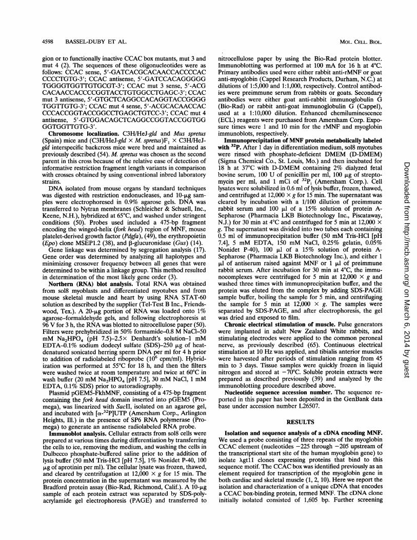
FIG. 1. Nucleotide and predicted amino acid sequence of MNF cDNA. The fork head domain is boxed. Shaded amino acids outline theactivation domain of MNF. Alanines and proline clusters within the activation domain are highlighted with circles.
yielded additional cDNA clones containing overlapping se-quences extending to 2,394 bp (Fig. 1). This cDNA includes anopen reading frame initiated from a methionine codon thatfulfills Kozak's criteria (32) and encodes a 617-amino-acidprotein with a predicted molecular mass of 66 kDa.
Searches of protein data bases revealed that a 110-amino-acid region within MNF is closely related to the winged-helixdomain (35, 62) common to members of the HNF-3lfork headfamily of transcription factors (boxed in Fig. 1). The winged-helix domain of MNF exhibits 35 to 89% sequence identity bycomparison with known members of this multigene family (Fig.2). MNF appears to be most closely related to ILF (36), aprotein identified on the basis of binding affinity to an inter-
leukin-2 response element within the long terminal repeat ofhuman immunodeficiency virus. MNF is, however, a uniqueprotein, and exhibits less than 50% identity to ILF or to anyother known protein outside of the winged-helix region.MNF includes a transcriptional activation domain within
an amino-terminal region. Nucleotide sequences encoding thefirst 206 amino acids of MNF (lacking the winged-helix region)or residues 207 to 617 were fused to the DNA-bindingdomain-encoding sequences of the yeast transcriptional acti-vator GAL4 in the expression vector pAS1 (12). Transfectionof the amino-terminal MNF-GAL4 chimera resulted in 10-foldtrans-activation of a reporter gene (13-galactosidase) controlledby the GAL] promoter (Fig. 3A). However, the GAL4 chimera
on March 6, 2014 by guest
http://mcb.asm
.org/D
ownloaded from

4600 BASSEL-DUBY ET AL.
AMNFILFBF-1FKHRHNF 3,BFKH
20 struc:
MNFILFBF-1FKHRHNF 31FKH
1 10 20 30 40 50GGDSPKDESKPPYSYAQLIVQAISSAQDRQLTLSGIYAHITKHYPYYRTADKGWQNSIRH....... .... TM-PK*-*....N** *T*.* *................
*DKKNGKYE...NA. MM * RQSPEKR*... N.* EF*M*NF***.ENKQ........KSS*SRRNAWGNL *. D *-TK *-E*SAEKR** -. Q.* EWMV * SV*FKDKG*. *.K...
TYRRSYTHA. IS. *TM-* QQSPNKM ....E*-QW*MDLF*F*RQNQQR.......TTRRSYTHA. IS*-TM* QNNPT*M**.E. * *QF*MDLF*F *-QNQQR.......
HHHHHHHHHHH SBBHHHHHHHHHH HHHHHHHHHHH
60 70 80 90 100 110NLSLNRYFIKVPRSQEEPGKGSFWRIDPASEAKLVEQAFRKRRQRGVSCFR..................... .......... - ........ p ...p ... S*-I*-----
..KC*V.... HYDD*..-NY*ML*-S*DDVFIGGTTG*L*R*STTSRA...* HSK*-R*QNEGT--*-S*W*MLN*------ EGGKSG*SPR*RAASMDS *-F*DC*L*.... PDK *- TLH*D*GNIFENGCYLR*QK*FKCEKQS *-F*DC*V*I *-TPDK *...... TLH*D*GNMFENGCY-L *...KRFKDEK
20 struc: HHHH BBBBBBB BBB
BPercent identity to MNF forkhead domain
ILF 89%BF-1 47%FKHR 35%HNF-31 42%FKH 43%
FIG. 2. Comparison of the MNF fork head domain sequence withother fork head proteins. (A) Alignment of MNF fork head domainwithfork head domains from ILF (36), BF-1 (56), HNF-3,B (35), FKHR(13), and FKH (62). Dots represent identical alignment of amino acidswith the MNF sequence. The asterisk indicates the omission of fiveamino acids (SNSSAG). A vertical line corresponds to a gap in thesequence. Gaps have been introduced to allow maximal sequence
alignment. The secondary structural elements are extrapolated fromthe known crystal structure of the HNF-3lfork head domain (5) anddenoted by H for a-helix and B for 1-strand. (B) The degree ofsequence identity between each of thefork head domains and the MNFfork head domain is given as a percentage.
that includes amino acids 207 through 617 of MNF exhibitedno trans-activation function (Fig. 3A). These results indicatethat a transcriptional activation domain resides within the first206 amino acids ofMNF (Fig. 3B, shaded region), a region richin alanine and proline residues (circled in Fig. 1).
Purified rMNF containing the winged-helix region bindsspecifically to the myoglobin CCAC box motif. A histidine tagwas placed at the amino terminus of MNF by using the pQEexpression vector system (Qiagen), to facilitate purification ofrecombinant protein, rMNF (Fig. 4D), after expression in E.coli M15 cells (Fig. 4A). Southwestern (DNA-protein) blotanalysis showed that purified rMNF binds the myoglobinCCAC box sequence (Fig. 4B), and immunoblot analysisdemonstrated that rabbit polyclonal antibodies raised againstpurified rMNF recognize the protein at a dilution of 1:40,000(Fig. 4C).
Binding of rMNF to the myoglobin CCAC box was sequencespecific, as assessed by electrophoretic mobility shift assayswith purified protein (Fig. 5, lane 1). An excess of unlabeledoligonucleotides containing the native CCAC sequence dis-rupted binding of rMNF to the labeled CCAC box probe (Fig.5, lane 2), whereas nonfunctional CCAC box mutants, mut 3and mut 4 (2), failed to inhibit binding (Fig. 5, lanes 3 and 4).Chromosomal mapping of MNF. Southern blot analysis of
genomic DNA digested with several different restriction en-
zymes indicated that MNF is encoded by a single gene in themouse genome. Chromosomal localization of the MNF genewas performed by analyzing a panel of DNA samples from an
interspecific cross that has been characterized for over 600genetic markers throughout the mouse genome. The genetic
A
o
Cu
0a)
Cu
a)
cn
Ct
*U)
oCb
14-
12.
10.
8-
6.
4.
2-
T
B
1 206 617 aa
HNF-3/Fkh
FIG. 3. An activation domain resides within the first 206 aminoacids (aa) of MNF. (A) 13-Galactosidase activity measured from aprotein extract of yeast cells transformed with fusion constructsencoding the GAL4 DNA-binding domain linked either to amino acids1 to 206 (shaded area) or 207 to 617 (hatched area) of MNF. Resultswere expressed relative to vector pAS1 (12) and represent mean values(± standard error of the mean) from at least four independentobservations. (B) Diagram of MNF protein. HNF-3/Fkh illustrates theposition of the winged-helix domain. The shaded area is the aminoterminus of the protein and contains the activation domain.
markers included in this map span between 50 and 80 centi-morgans (cM) on each mouse autosome and the X chromo-some. Initially, DNA from the parental mice was digested withvarious restriction enzymes and hybridized with MNF cDNAprobe to determine restriction fragment length variants(RFLVs) for haplotype analyses. Informative RFLVs weredetected with TaqI-restricted DNAs, and each of the 114TaqI-restricted DNAs from the interspecific backcross micedisplayed either the homozygous or heterozygous F1 patternwhen hybridized with the MNF probe.
Comparison of the haplotype distribution of MNF indicatedthat in all of the 114 meiotic events examined, the Mnf locuscosegregated with the Pdgfa locus (Fig. 6) previously mappedto the distal mouse chromosome 5 (31, 49). In the most likelygene order, the Mnf locus was located 1.8 ± 1.2 cM distal toEpo (38) and 3.5 ± 1.8 cM distal to Gus (14).MNF is expressed in tissues and cells in which the myoglo-
bin gene is transcriptionally active. Northern blot analysis wasperformed with RNA extracted from skeletal and cardiacmuscle of adult mice (Fig. 7A) and from mouse sol8 myogeniccells at various times during differentiation induced by with-drawal of growth factors (Fig. 7B). Under conditions of highstringency and using a probe complementary to nucleotidesequences 805 to 1266 of the MNF cDNA, we detected mRNAtranscripts of approximately 4.3 and 2.4 kb in skeletal muscle(Fig. 7A, lane Sk) and slightly larger molecular size in heartmuscle (Fig. 7A, lane H). Following differentiation of sol8
MOL. CELL. BIOL.
,<,, ,Alb4 'A,
lbe-
on March 6, 2014 by guest
http://mcb.asm
.org/D
ownloaded from
WINGED-HELIX TRANSCRIPTION FACTOR IN MUSCLE 4601
BM 1 2 3 4
=
Coomassie blue stain
C
1 2 3 4
noCA
32P-CCAC probe
Gus
Epo
** F
Mnf, Pdgfa * E:M# of backcross
mice
antibodyagainstrMNF
262 aa617 aa
| HiS TA''G1 aa 617 aa
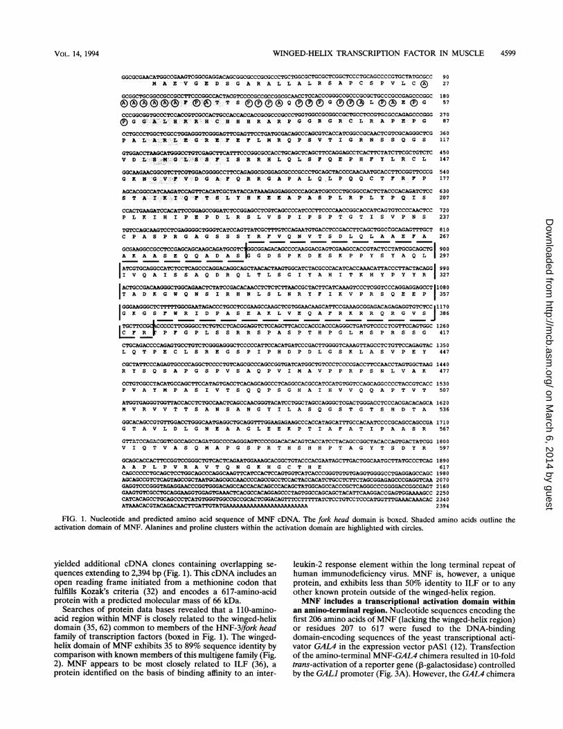
MNF[_FIG. 4. Characterization of rMNF. (A) rMNF was bound to a
Ni-agarose column and eluted with four 1-ml fractions containing 160mM imidazole. A 10-,ul sample from each fraction was analyzed bySDS-PAGE (10% polyacrylamide) (lanes 1 to 4). Protein was detectedby Coomassie blue staining. (B) Protein-DNA (Southwestern) blotanalysis of rMNF with [32P]CCAC. Lanes 1 to 4 correspond to theprotein samples in panel A. (C) Western immunoblot analysis ofrMNF with antibodies (1:40,000 dilution) generated against rMNF.(D) Illustration of rMNF linked to the histidine tag and MNF.
myoblasts to multinucleated myotubes that express the endo-genous myoglobin gene, both the 4.3- and 2.4-kb transcriptsincrease in quantity by 1.8- and 5.8-fold, respectively (Fig. 7B).MNF expression is not limited, however, to striated myo-
competitor
I l-I o
-.s....9,
1 2 3 4 5
-223 bp -204 bpCCAC: 5'-ACCACCCCACCCCCTGTGGC-3'mut 3: 5'-ACCACCCCO CCTGTGGC-3'mut 4: 5'-ACCACCCCACGt 3GC-3'
FIG. 5. rMNF binds specifically to the CCAC box element. 32p_labeled CCAC box probe was used in a gel mobility shift assay to assessspecific binding of rMNF (lane 1). Competitive-binding studies in-cluded 100 ng of unlabeled CCAC box sequence (lane 2) or 100 ng offunctionally inactive CCAC box mutants (2), mut 3 (lane 3), or mut 4(lane 4). The DNA sequences of CCAC, mut 3, and mut 4 are shown.
45 65 2 0 1 1
FIG. 6. Segregation of MNF among mouse distal chromosome 5loci in (C3H/HeJ-gld x M. spretus)Fj X C3H/HeJ-gld interspecificbackcross mice. Solid boxes represent the homologous C3H pattern,and open boxes represent the F1 pattern. The informative RFLVs forMNF are described in the text. For the other markers, the followingRFLVs were used: Gus (14), BamHI (C3H 2.0 and 1.3 kb; M. sprefus4.3 kb), Epo (38), TaqI (C3H 1.8 and 1.0 kb,M sprefus 2.0 and 1.2 kb).
cytes. RNase protection assays (results not shown) indicatethat at least one form of MNF mRNA can be detected in othermouse tissues (kidneys and brain).MNF and other closely related proteins are present in
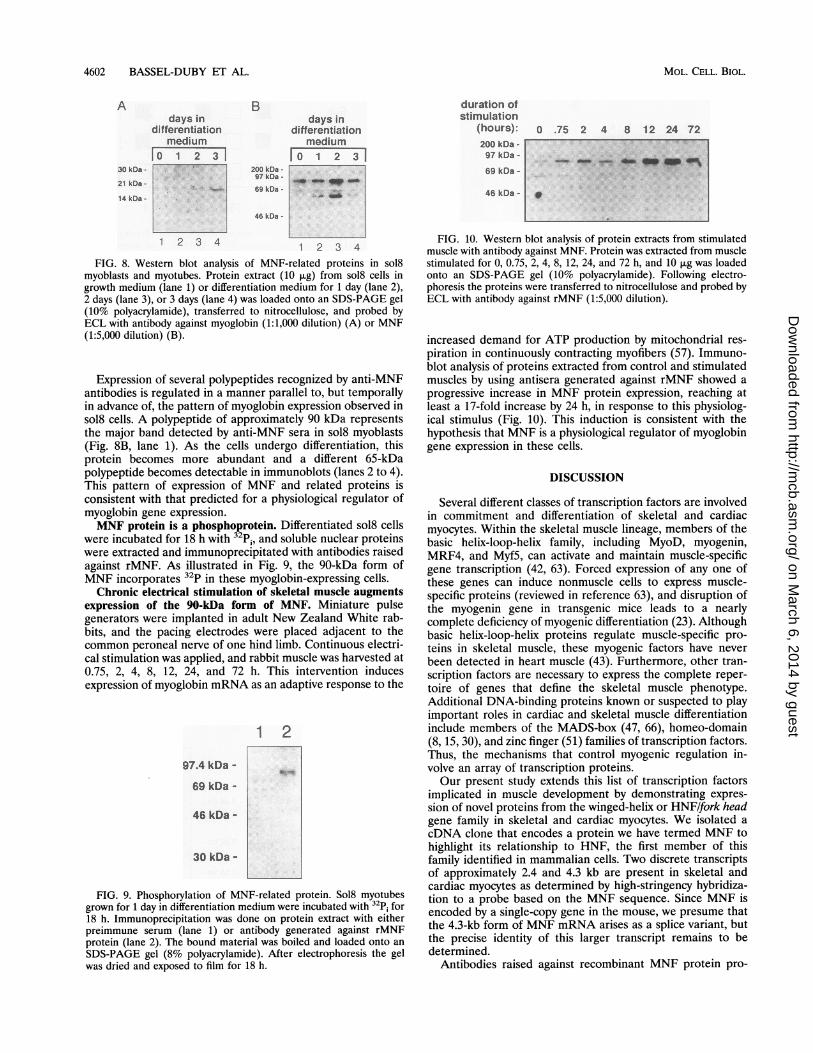
myocytes, and become more abundant during muscle differ-entiation. The relationship between expression of myoglobinand proteins recognized by antibodies raised against rMNFwas examined by using sol8 myogenic cells in the proliferativephase and at various stages of differentiation (Fig. 8). After 1day in differentiation medium, these cells can be distinguishedfrom myoblasts morphologically (rod shaped) and by expres-sion of sarcomeric proteins (data not shown). Myoglobinexpression is, however, limited or absent at this stage (Fig. 8A,lanes 1 and 2). After 2 days in differentiation medium,multinucleated myotubes are abundant and myoglobin proteinis detectable (lane 3). By day 3 in differentiation medium,myotubes are increased in size and myoglobin protein isexpressed to higher levels (lane 4). These results are corrobo-rated by immunohistochemical studies that demonstrate uni-form expression of myosin heavy-chain protein as early as day1 in differentiation medium but uniform expression of myoglo-bin only after 3 days in differentiation medium (data notshown). Other investigators also have observed this delayedexpression of myoglobin during differentiation of other myo-genic cell lines in culture (19).
A
7.46 kb -
4.40 kb -
2.37 kb -
1.35 kb -
Sk H
go
B
7.46 kb -
4.40 kb -
2.37 kb -
1.35 kb -
days indifferentiation
medium
l 2 31
.__*s
FIG. 7. Northern blot analysis of MNF. A radioactive riboprobeencoding the HNF-3/fork head domain of MNF was used to probeNorthern blots with RNA extracted from mouse skeletal muscle (lane1) or heart tissue (lane 2) (A) or RNA extracted from sol8 cells grownin differentiation medium for 0 (lane 1), 1 (lane 2), 2 (lane 3), or 3(lane 4) days (B).
A
200 kD
116 kD97 kD66 kD
45 kD
D
rMNF
VOL. 14, 1994
on March 6, 2014 by guest
http://mcb.asm
.org/D
ownloaded from
4602 BASSEL-DUBY ET AL.
days indifferentiation
medium|0 1 2 3 1
B
200 kDa -
97 kDa -
69 kDa -
46 kDa -
days indifferentiation
mediuml0 1 2 3 1
duration ofstimulation
(hours): o .75 2 4 8 12 24 72
200 kDa -
97 kDa -
69 kDa -
46 kDa -
1 2 3 4 1 2 3 4
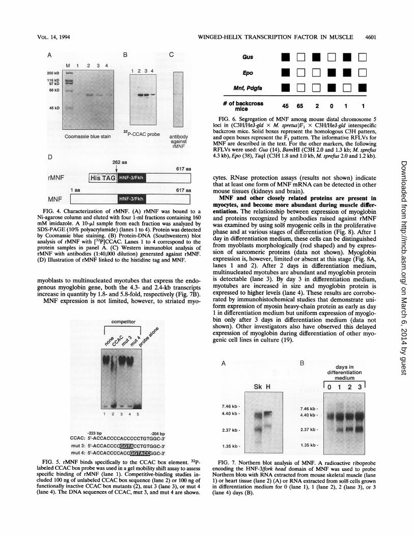
FIG. 8. Western blot analysis of MNF-related proteins in sol8myoblasts and myotubes. Protein extract (10 [ug) from sol8 cells ingrowth medium (lane 1) or differentiation medium for 1 day (lane 2),2 days (lane 3), or 3 days (lane 4) was loaded onto an SDS-PAGE gel(10% polyacrylamide), transferred to nitrocellulose, and probed byECL with antibody against myoglobin (1:1,000 dilution) (A) or MNF(1:5,000 dilution) (B).
Expression of several polypeptides recognized by anti-MNFantibodies is regulated in a manner parallel to, but temporallyin advance of, the pattern of myoglobin expression observed insol8 cells. A polypeptide of approximately 90 kDa representsthe major band detected by anti-MNF sera in sol8 myoblasts(Fig. 8B, lane 1). As the cells undergo differentiation, thisprotein becomes more abundant and a different 65-kDapolypeptide becomes detectable in immunoblots (lanes 2 to 4).This pattern of expression of MNF and related proteins isconsistent with that predicted for a physiological regulator ofmyoglobin gene expression.MNF protein is a phosphoprotein. Differentiated sol8 cells
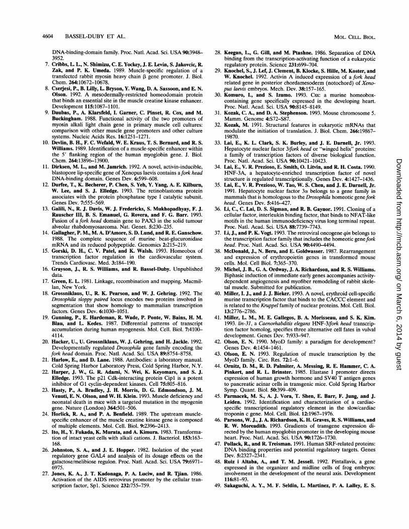
were incubated for 18 h with 32p;, and soluble nuclear proteinswere extracted and immunoprecipitated with antibodies raisedagainst rMNF. As illustrated in Fig. 9, the 90-kDa form ofMNF incorporates 32p in these myoglobin-expressing cells.Chronic electrical stimulation of skeletal muscle augments
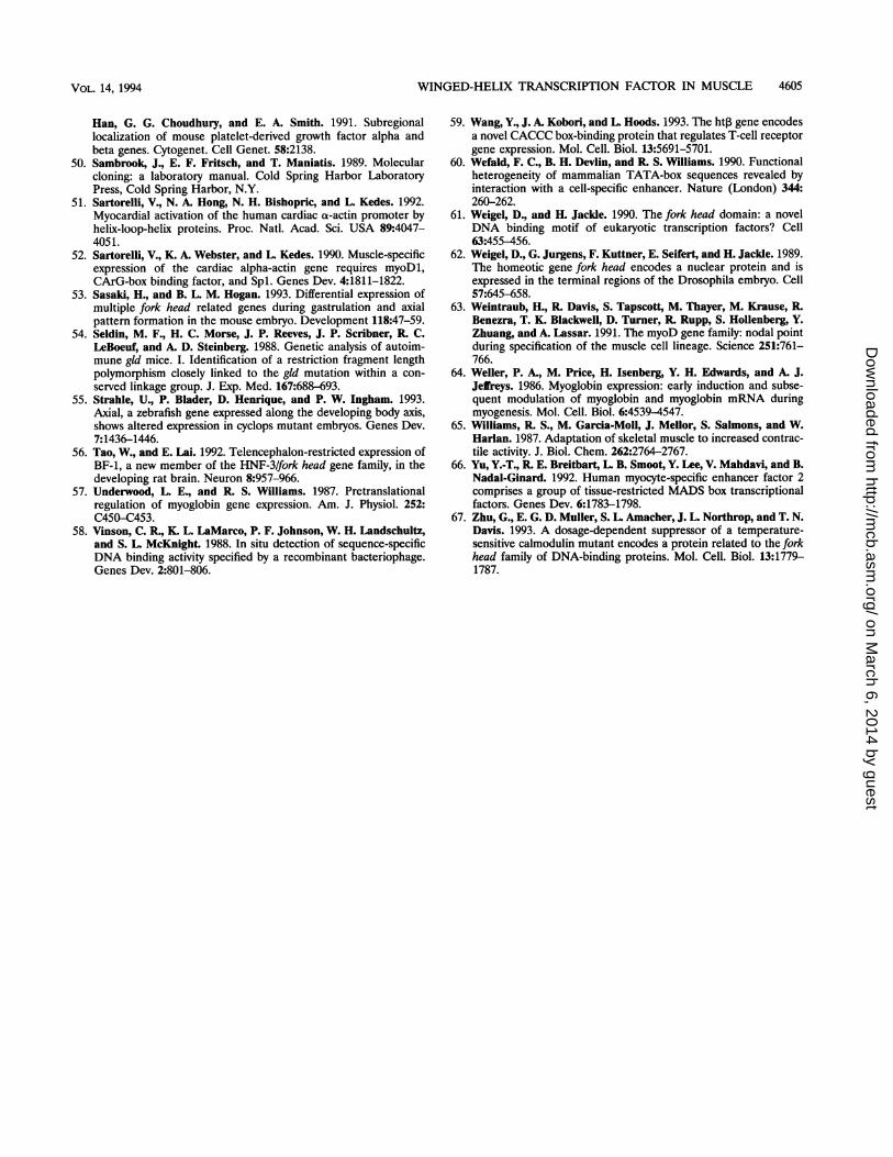
expression of the 90-kDa form of MNF. Miniature pulsegenerators were implanted in adult New Zealand White rab-bits, and the pacing electrodes were placed adjacent to thecommon peroneal nerve of one hind limb. Continuous electri-cal stimulation was applied, and rabbit muscle was harvested at0.75, 2, 4, 8, 12, 24, and 72 h. This intervention inducesexpression of myoglobin mRNA as an adaptive response to the
12
97.4 kDa -
69 kDa -
46 kDa -
30 kDa -
FIG. 9. Phosphorylation of MNF-related protein. Sol8 myotubesgrown for 1 day in differentiation medium were incubated with 32p; for18 h. Immunoprecipitation was done on protein extract with eitherpreimmune serum (lane 1) or antibody generated against rMNFprotein (lane 2). The bound material was boiled and loaded onto an
SDS-PAGE gel (8% polyacrylamide). After electrophoresis the gelwas dried and exposed to film for 18 h.
FIG. 10. Western blot analysis of protein extracts from stimulatedmuscle with antibody against MNF. Protein was extracted from musclestimulated for 0, 0.75, 2, 4, 8, 12, 24, and 72 h, and 10 pug was loadedonto an SDS-PAGE gel (10% polyacrylamide). Following electro-phoresis the proteins were transferred to nitrocellulose and probed byECL with antibody against rMNF (1:5,000 dilution).
increased demand for ATP production by mitochondrial res-
piration in continuously contracting myofibers (57). Immuno-blot analysis of proteins extracted from control and stimulatedmuscles by using antisera generated against rMNF showed a
progressive increase in MNF protein expression, reaching atleast a 17-fold increase by 24 h, in response to this physiolog-ical stimulus (Fig. 10). This induction is consistent with thehypothesis that MNF is a physiological regulator of myoglobingene expression in these cells.
DISCUSSION
Several different classes of transcription factors are involvedin commitment and differentiation of skeletal and cardiacmyocytes. Within the skeletal muscle lineage, members of thebasic helix-loop-helix family, including MyoD, myogenin,MRF4, and Myf5, can activate and maintain muscle-specificgene transcription (42, 63). Forced expression of any one ofthese genes can induce nonmuscle cells to express muscle-specific proteins (reviewed in reference 63), and disruption ofthe myogenin gene in transgenic mice leads to a nearlycomplete deficiency of myogenic differentiation (23). Althoughbasic helix-loop-helix proteins regulate muscle-specific pro-teins in skeletal muscle, these myogenic factors have never
been detected in heart muscle (43). Furthermore, other tran-scription factors are necessary to express the complete reper-toire of genes that define the skeletal muscle phenotype.Additional DNA-binding proteins known or suspected to playimportant roles in cardiac and skeletal muscle differentiationinclude members of the MADS-box (47, 66), homeo-domain(8, 15, 30), and zinc finger (51) families of transcription factors.Thus, the mechanisms that control myogenic regulation in-volve an array of transcription proteins.Our present study extends this list of transcription factors
implicated in muscle development by demonstrating expres-sion of novel proteins from the winged-helix or HNF/fork headgene family in skeletal and cardiac myocytes. We isolated a
cDNA clone that encodes a protein we have termed MNF tohighlight its relationship to HNF, the first member of thisfamily identified in mammalian cells. Two discrete transcriptsof approximately 2.4 and 4.3 kb are present in skeletal andcardiac myocytes as determined by high-stringency hybridiza-tion to a probe based on the MNF sequence. Since MNF isencoded by a single-copy gene in the mouse, we presume thatthe 4.3-kb form of MNF mRNA arises as a splice variant, butthe precise identity of this larger transcript remains to bedetermined.
Antibodies raised against recombinant MNF protein pro-
A
30 kDa -
21 kDa -
14 kDa-
- -, -..WW 40 m-t 4M 4 .# .40
MOL. CELL. BIOL.
on March 6, 2014 by guest
http://mcb.asm
.org/D
ownloaded from

WINGED-HELIX TRANSCRIPTION FACTOR IN MUSCLE 4603
duced in bacteria detect three discrete polypeptides of approx-imately 90, 68, and 65 kDa in immunoblots prepared fromdifferentiated mouse sol8 myotubes, indicative of posttransla-tional modifications and/or alternative splicing of exons withinprotein-coding sequences of the gene. Our finding that the90-kDa form is phosphorylated provides direct evidence forposttranslational modifications of MNF, and the aforemen-tioned Northern blot analysis suggests that alternative splicingalso occurs.
Several findings support the premise that MNF has aphysiological function during muscle development. MNF ex-hibits sequence-specific DNA binding to an upstream activa-tion sequence (CCAC element) that is required for transcrip-tional activity of the human myoglobin promoter in bothskeletal and cardiac myocytes. In addition, MNF is regulated inparallel to expression of myoglobin in two different experimen-tal systems in which the myoglobin gene is upregulated. Theabundance of MNF mRNA and of immunoreactive MNFpolypeptides is increased during muscle differentiation inculture. The induction of the 65-kDa polypeptide in sol8myotubes is particularly striking, since this form of MNF isvirtually undetectable in myoblasts. Also, MNF is inducedduring fiber-type transformation that occurs as a consequenceof continuous motor nerve stimulation in skeletal muscles ofadult animals.Our current data also demonstrate that MNF includes a
functional trans-activation domain, but further studies arerequired to define more completely its physiological functionsin the intact cell. Other winged-helix proteins bind DNA asmonomers, and recombinant MNF is capable of DNA bindingin vitro in the absence of accessory proteins. Our currentfindings are insufficient, however, to define the active form(s)of MNF and the potential regulatory effects of phosphoryla-tion. Likewise, we cannot exclude a requirement for otherproteins in hetero-oligomeric complexes in transcriptionalactivation mediated by MNF. To the contrary, multimerizationof the myoglobin CCAC box motif (to which MNF binds)upstream of a TATA element is insufficient to direct muscle-specific expression of a reporter gene following transfectioninto cultured cells, unless the A+T-rich upstream activationsequence from the myoglobin gene is included in the construc-tion (16). Thus, it is likely that MNF may require protein-protein interactions with other heterologous transcription fac-tors to effect efficient trans-activation in muscle cells.Although MNF was isolated on the basis of sequence-
specific binding to the myoglobin CCAC box motif, additionalstudies are necessary to determine in a definitive mannerwhether MNF is indeed a physiological trans-activator of themyoglobin gene and of other genes that require closely relatedCCAC elements for transcriptional activity. Promoters orenhancers from certain other muscle-specific genes includingmuscle creatine kinase (24), cardiac ot-actin (51), P-myosinheavy chain (7) and slow cardiac troponin C (45) also containCCAC motifs, mutations of which alter transcription.
Spl, a ubiquitously expressed transcription factor, is capableof binding to the CCAC motif (52), which differs from aconsensus Spl-binding site (27) largely by the substitution of aguanine base in the position occupied by an adenine in theCCAC box. Our current data suggest, however, that MNFbinds the myoglobin CCAC element with higher affinity thanSpl does. Several proteins within the erythroid lineage thatbind a CACCC motif from the P-globin gene have beendescribed (40), but the expression of these factors in develop-ing muscle and their relative affinity for binding the myoglobinCCAC box have not been assessed. Other data from our ownlaboratory also suggest that additional CCAC box-binding
factors, distinct from MNF and Spl, are present in developingmyotubes. When multimerized CCAC sequences are used toprobe protein blots prepared from nuclei isolated from sol8myotubes, we observe prominent binding to a 40-kDa protein(2), as well as to a larger polypeptide consistent with themolecular mass of MNF (data not shown). These data areparticularly interesting in view of the recent report by Wang etal. (59) of a 40-kDa protein termed htMP, isolated from a humanT-cell lymphoma-derived cell line on the basis of binding to aCCAC box sequence of the human T-cell receptor VP38.1promoter element from positions -72 to -92. Studies areunder way in our laboratory to determine the relationshipbetween CBF40 and htW3 and the relative importance of theseproteins and MNF for physiological trans-activation of myo-globin expression.
In summary, we have identified a novel member of thewinged-helix or HNF-3lfork head family of transcription fac-tors on the basis of sequence-specific DNA binding to anupstream activation sequence from the human myoglobingene. This protein, MNF, includes an amino-terminal trans-activation domain and is expressed in adult skeletal andcardiac myocytes in which the myoglobin gene is transcription-ally active. MNF is encoded by a single locus on chromosome5 in the mouse genome, but alternative splicing and/or post-translational modifications give rise to two forms of MNFmRNA and three forms of MNF protein. The abundance ofMNF polypeptides is developmentally regulated during differ-entiation of skeletal myotubes in culture and is responsive toneural regulation in adult skeletal muscle fibers. These dataprovide the first evidence for participation of winged-helixproteins as transcriptional regulators in mammalian striatedmyocytes and suggest a role for MNF in both muscle develop-ment and postnatal physiological adaptations to changing workdemands.
ACKNOWLEDGMENTS
We are grateful to Galvin Swift and Ray MacDonald for providingus with both the Xgtll and XZap libraries and to Jeffrey Michel andGeorge Ordway for providing protein extracts from stimulated rabbitmuscle. We thank Amanda Tostrud, Yuxia Luo, and Vaijayanti Aptefor technical assistance.
This work was supported by a grant from NIH (RO1 AR40849) andby an American Heart Association Grant-In-Aid (92G-102). M.D.H.was supported by NIH training grant HL07360.
REFERENCES1. Bassel-Duby, R., C. M. Grohe, M. E. Jessen, W. J. Parsons, J. A.
Richardson, R. Chao, J. Grayson, W. S. Ring, and R. S. Williams.1993. Sequence elements required for transcriptional activity ofthe human myoglobin promoter in intact myocardium. Circ. Res.73:360-366.
2. Bassel-Duby, R., M. D. Hernandez, M. A. Gonzalez, J. K. Krueger,and R. S. Williams. 1992. A 40-kilodalton protein binds specifi-cally to an upstream sequence element essential for muscle-specific transcription of the human myoglobin promoter. Mol.Cell. Biol. 12:5024-5032.
3. Bishop, D. T. 1985. The information content of phase-knownmatings for ordering genetic loci. Genet. Epidemiol. 2:349-361.
4. Bostian, K., J. M. Lemire, L. E. Cannon, and H. 0. Halvorson.1980. In vitro synthesis of repressible yeast acid phosphatase:identification of multiple mRNA and products. Proc. Natl. Acad.Sci. USA 77:4505-4508.
5. Clark, K. L., E. D. Halay, E. Lai, and S. K. Burley. 1993. Co-crystalstructure of the HNF-3/fork head DNA-recognition motif resem-bles histone H5. Nature (London) 364:412-420.
6. Clevidence, D. E., D. G. Overdier, W. Tao, X. Qian, L. Pani, E. Lai,and R. H. Costa. 1993. Identification of nine tissue-specifictranscription factors of the hepatocyte nuclear factor 3/fork head
VOL. 14, 1994
on March 6, 2014 by guest
http://mcb.asm
.org/D
ownloaded from
4604 BASSEL-DUBY ET AL.
DNA-binding-domain family. Proc. Natl. Acad. Sci. USA 90:3948-3952.
7. Cribbs, L. L., N. Shimizu, C. E. Yockey, J. E. Levin, S. Jakovcic, R.Zak, and P. K. Umeda. 1989. Muscle-specific regulation of atransfected rabbit myosin heavy chain P gene promoter. J. Biol.Chem. 264:10672-10678.
8. Cserjesi, P., B. Lilly, L. Bryson, Y. Wang, D. A. Sassoon, and E. N.Olson. 1992. A mesodermally-restricted homeodomain proteinthat binds an essential site in the muscle creatine kinase enhancer.Development 115:1087-1101.
9. Daubas, P., A. Klarsfeld, I. Garner, C. Pinset, R. Cox, and M.Buckingham. 1988. Functional activity of the two promoters ofmyosin alkali light chain gene in primary muscle cell cultures:comparison with other muscle gene promoters and other culturesystems. Nucleic Acids Res. 16:1251-1271.
10. Devlin, B. H., F. C. Wefald, W. E. Kraus, T. S. Bernard, and R. S.Williams. 1989. Identification of a muscle-specific enhancer withinthe 5' flanking region of the human myoglobin gene. J. Biol.Chem. 264:13896-13900.
11. Dirksen, M. L., and M. Jamrich. 1992. A novel, activin-inducible,blastopore lip-specific gene of Xenopus laevis contains afork headDNA-binding domain. Genes Dev. 6:599-608.
12. Durfee, T., K. Becherer, P. Chen, S. Yeh, Y. Yang, A. E. Kilburn,W. Lee, and S. J. Elledge. 1993. The retinoblastoma proteinassociates with the protein phosphatase type I catalytic subunit.Genes Dev. 7:555-569.
13. Galili, N., R. J. Davis, W. J. Fredericks, S. Mukhopadhyay, F. J.Rauscher III, B. S. Emanuel, G. Rovera, and F. G. Barr. 1993.Fusion of a fork head domain gene to PAX3 in the solid tumouralveolar rhabdomyosarcoma. Nat. Genet. 5:230-235.
14. Gallagher, P. M., M. A. D'Amore, S. D. Lund, and R. E. Ganschow.1988. The complete sequence of murine beat-glucuronidasemRNA and its reduced polypeptide. Genomics 2:215-219.
15. Gorski, D. H., C. V. Patel, and K. Walsh. 1993. Homeobox oftranscription factor regulation in the cardiovascular system.Trends Cardiovasc. Med. 3:184-190.
16. Grayson, J., R. S. Williams, and R. Bassel-Duby. Unpublisheddata.
17. Green, E. L. 1981. Linkage, recombination and mapping. Macmil-lan, New York.
18. Grossniklaus, U., R. K. Pearson, and W. J. Gehring. 1992. TheDrosophila sloppy paired locus encodes two proteins involved insegmentation that show homology to mammalian transcriptionfactors. Genes Dev. 6:1030-1051.
19. Gunning, P., E. Hardeman, R. Wade, P. Ponte, W. Bains, H. M.Blau, and L. Kedes. 1987. Differential patterns of transcriptaccumulation during human myogenesis. Mol. Cell. Biol. 7:4100-4114.
20. Hacker, U., U. Grossniklaus, W. J. Gehring, and H. Jackie. 1992.Developmentally regulated Drosophila gene family encoding thefork head domain. Proc. Natl. Acad. Sci. USA 89:8754-8758.
21. Harlow, E., and D. Lane. 1988. Antibodies: a laboratory manual.Cold Spring Harbor Laboratory Press, Cold Spring Harbor, N.Y.
22. Harper, J. W., G. R. Adami, N. Wei, K. Keyomars, and S. J.Elledge. 1993. The p21 Cdk-interacting protein Cipl is a potentinhibitor of G1 cyclin-dependent kinases. Cell 75:805-816.
23. Hasty, P., A. Bradley, J. H. Morris, D. G. Edmondson, J. M.Venuti, E. N. Olson, and W. H. Klein. 1993. Muscle deficiency andneonatal death in mice with a targeted mutation in the myogeningene. Nature (London) 364:501-506.
24. Horlick, R. A., and P. A. Benfield. 1989. The upstream muscle-specific enhancer of the muscle creatine kinase gene is composedof multiple elements. Mol. Cell. Biol. 9:2396-2413.
25. Ito, H., Y. Fukada, K. Murata, and A. Kimura. 1983. Transforma-tion of intact yeast cells with alkali cations. J. Bacteriol. 153:163-168.
26. Johnston, S. A., and J. E. Hopper. 1982. Isolation of the yeastregulatory gene GAL4 and analysis of its dosage effects on thegalactose/melibiose regulon. Proc. Natl. Acad. Sci. USA 79:6971-6975.
27. Jones, K. A., J. T. Kadonaga, P. A. Luciw, and R. Tjian. 1986.Activation of the AIDS retrovirus promoter by the cellular tran-scription factor, Spl. Science 232:755-759.
28. Keegan, L., G. Gill, and M. Ptashne. 1986. Separation of DNAbinding from the transcription-activating function of a eukaryoticregulatory protein. Science 231:699-704.
29. Knochel, S., J. Lef, J. Clement, B. Klocke, S. Hille, M. Koster, andW. Knochel. 1992. Activin A induced expression of a fork headrelated gene in posterior chordamesoderm (notochord) of Xeno-pus laevis embryos. Mech. Dev. 38:157-165.
30. Komuro, I., and S. Izumo. 1993. Csx: a murine homeobox-containing gene specifically expressed in the developing heart.Proc. Natl. Acad. Sci. USA 90:8145-8149.
31. Kozak, C. A., and D. A. Stephenson. 1993. Mouse chromosome 5.Mamm. Genome 4:S72-S87.
32. Kozak, M. 1991. Structural features in eukaryotic mRNAs thatmodulate the initiation of translation. J. Biol. Chem. 266:19867-19870.
33. Lai, E., K. L. Clark, S. K. Burley, and J. E. Darnell, Jr. 1993.Hepatocyte nuclear factor 3/fork head or "winged helix" proteins:a family of transcription factors of diverse biological function.Proc. Nati. Acad. Sci. USA 90:10421-10423.
34. Lai, E., V. R. Prezioso, E. Smith, 0. Litvin, and R. H. Costa. 1990.HNF-3A, a hepatocyte-enriched transcription factor of novelstructure is regulated transcriptionally. Genes Dev. 4:1427-1436.
35. Lai, E., V. R. Prezioso, W. Tao, W. S. Chen, and J. E. Darnell, Jr.1991. Hepatocyte nuclear factor 3a belongs to a gene family inmammals that is homologous to the Drosophila homeotic geneforkhead. Genes Dev. 5:416-427.
36. Li, C., C. Lai, D. S. Sigman, and R. B. Gaynor. 1991. Cloning of acellular factor, interleukin binding factor, that binds to NFAT-likemotifs in the human immunodeficiency virus long terminal repeat.Proc. Natl. Acad. Sci. USA 88:7739-7743.
37. Li, J., and P. K. Vogt. 1993. The retroviral oncogene qin belongs tothe transcription factor family that includes the homeotic geneforkhead. Proc. Natl. Acad. Sci. USA 90:4490-4494.
38. McDonald, J., N. Beru, and E. Goldwasser. 1987. Rearrangementand expression of erythropoietin genes in transformed mousecells. Mol. Cell Biol. 7:365-370.
39. Michel, J. B., G. A. Ordway, J. A. Richardson, and R. S. Williams.Biphasic induction of immediate early genes accompanies activity-dependent angiogenesis and myofiber remodeling of rabbit skele-tal muscle. Submitted for publication.
40. Miller, I. J., and J. J. Bieker. 1993. A novel, erythroid cell-specificmurine transcription factor that binds to the CACCC element andis related to the Kruppel family of nuclear proteins. Mol. Cell. Biol.13:2776-2786.
41. Miller, L. M., M. E. Gallegos, B. A. Morisseau, and S. K. Kim.1993. lin-31, a Caenorhabditis elegans HNF-3/fork head transcrip-tion factor homolog, specifies three alternative cell fates in vulvaldevelopment. Genes Dev. 7:933-947.
42. Olson, E. N. 1990. MyoD family: a paradigm for development?Genes Dev. 4:1454-1461.
43. Olson, E. N. 1993. Regulation of muscle transcription by theMyoD family. Circ. Res. 72:1-6.
44. Ornitz, D. M., R. D. Palmiter, A. Messing, R. E. Hammer, C. A.Pinkert, and R. L. Brinster. 1985. Elastase I promoter directsexpression of human growth hormone and SV40 T antigen genesto pancreatic acinar cells in transgenic mice. Cold Spring HarborSymp. Quant. Biol. 50:399-409.
45. Parmacek, M. S., A. J. Vora, T. Shen, E. Barr, F. Jung, and J.Leiden. 1992. Identification and characterization of a cardiac-specific transcriptional regulatory element in the slow/cardiactroponin c gene. Mol. Cell. Biol. 12:1967-1976.
46. Parsons, W. J., J. A. Richardson, K. H. Graves, R. S. Williams, andR. W. Moreadith. 1993. Gradients of transgene expression di-rected by the human myoglobin promoter in the developing mouseheart. Proc. Natl. Acad. Sci. USA 90:1726-1730.
47. Pollack, R., and R. Treisman. 1991. Human SRF-related proteins:DNA binding properties and potential regulatory targets. GenesDev. 5:2327-2341.
48. Ruiz i Altaba, A., and T. M. Jessell. 1992. Pintallavis, a geneexpressed in the organizer and midline cells of frog embryos:involvement in the development of the neural axis. Development116:81-93.
49. Sakaguchi, A. Y., M. F. Seldin, L. Martinez, P. A. Lalley, E. S.
MOL. CELL. BIOL.
on March 6, 2014 by guest
http://mcb.asm
.org/D
ownloaded from
WINGED-HELIX TRANSCRIPTION FACTOR IN MUSCLE 4605
Han, G. G. Choudhury, and E. A. Smith. 1991. Subregionallocalization of mouse platelet-derived growth factor alpha andbeta genes. Cytogenet. Cell Genet. 58:2138.
50. Sambrook, J., E. F. Fritsch, and T. Maniatis. 1989. Molecularcloning: a laboratory manual. Cold Spring Harbor LaboratoryPress, Cold Spring Harbor, N.Y.
51. Sartorelli, V., N. A. Hong, N. H. Bishopric, and L. Kedes. 1992.Myocardial activation of the human cardiac a-actin promoter byhelix-loop-helix proteins. Proc. Natd. Acad. Sci. USA 89:4047-4051.
52. Sartorelli, V., K. A. Webster, and L. Kedes. 1990. Muscle-specificexpression of the cardiac alpha-actin gene requires myoD1,CArG-box binding factor, and Spl. Genes Dev. 4:1811-1822.
53. Sasaki, H., and B. L. M. Hogan. 1993. Differential expression ofmultiple fork head related genes during gastrulation and axialpattern formation in the mouse embryo. Development 118:47-59.
54. Seldin, M. F., H. C. Morse, J. P. Reeves, J. P. Scribner, R. C.LeBoeuf, and A. D. Steinberg. 1988. Genetic analysis of autoim-mune gid mice. I. Identification of a restriction fragment lengthpolymorphism closely linked to the gid mutation within a con-
served linkage group. J. Exp. Med. 167:688-693.55. Strahle, U., P. Blader, D. Henrique, and P. W. Ingham. 1993.
Axial, a zebrafish gene expressed along the developing body axis,shows altered expression in cyclops mutant embryos. Genes Dev.7:1436-1446.
56. Tao, W., and E. Lai. 1992. Telencephalon-restricted expression ofBF-1, a new member of the HNF-3lfork head gene family, in thedeveloping rat brain. Neuron 8:957-966.
57. Underwood, L. E., and R S. Williams. 1987. Pretranslationalregulation of myoglobin gene expression. Am. J. Physiol. 252:C450-C453.
58. Vinson, C. R., K. L. LaMarco, P. F. Johnson, W. H. Landschultz,and S. L. McKnight. 1988. In situ detection of sequence-specificDNA binding activity specified by a recombinant bacteriophage.Genes Dev. 2:801-806.
59. Wang, Y., J. A. Kobori, and L. Hoods. 1993. The hMt gene encodesa novel CACCC box-binding protein that regulates T-cell receptorgene expression. Mol. Cell. Biol. 13:5691-5701.
60. Wefald, F. C., B. H. Devlin, and R. S. Williams. 1990. Functionalheterogeneity of mammalian TATA-box sequences revealed byinteraction with a cell-specific enhancer. Nature (London) 344:260-262.
61. Weigel, D., and H. Jackle. 1990. The fork head domain: a novelDNA binding motif of eukaryotic transcription factors? Cell63:455-456.
62. Weigel, D., G. Jurgens, F. Kuttner, E. Seifert, and H. Jackle. 1989.The homeotic gene fork head encodes a nuclear protein and isexpressed in the terminal regions of the Drosophila embryo. Cell57:645-658.
63. Weintraub, H., R. Davis, S. Tapscott, M. Thayer, M. Krause, RBenezra, T. K. Blackwell, D. Turner, R Rupp, S. Hollenberg, Y.Zhuang, and A. Lassar. 1991. The myoD gene family: nodal pointduring specification of the muscle cell lineage. Science 251:761-766.
64. Weller, P. A., M. Price, H. Isenberg, Y. H. Edwards, and A. J.Jeffreys. 1986. Myoglobin expression: early induction and subse-quent modulation of myoglobin and myoglobin mRNA duringmyogenesis. Mol. Cell. Biol. 6:4539-4547.
65. Williams, R. S., M. Garcia-Moll, J. Mellor, S. Salmons, and W.Harlan. 1987. Adaptation of skeletal muscle to increased contrac-tile activity. J. Biol. Chem. 262:2764-2767.
66. Yu, Y.-T., R E. Breitbart, L. B. Smoot, Y. Lee, V. Mahdavi, and B.Nadal-Ginard. 1992. Human myocyte-specific enhancer factor 2comprises a group of tissue-restricted MADS box transcriptionalfactors. Genes Dev. 6:1783-1798.
67. Zhu, G., E. G. D. Muller, S. L. Amacher, J. L. Northrop, and T. N.Davis. 1993. A dosage-dependent suppressor of a temperature-sensitive calmodulin mutant encodes a protein related to the forkhead family of DNA-binding proteins. Mol. Cell. Biol. 13:1779-1787.
VOL. 14, 1994
on March 6, 2014 by guest
http://mcb.asm
.org/D
ownloaded from