Role of the Na +Ca 2+ Exchanger as an Alternative Trigger of CICR in Mammalian Cardiac Myocytes
14
Role of the Na -Ca 2 Exchanger as an Alternative Trigger of CICR in Mammalian Cardiac Myocytes Chunlei Han, Pasi Tavi, and Matti Weckstro ¨m Department of Physical Sciences/Division of Biophysics, Department of Physiology, and Biocenter Oulu, University of Oulu, Finland ABSTRACT Ca 2 influx through the L-type Ca 2 channels is the primary pathway for triggering the Ca 2 release from the sarcoplasmic reticulum (SR). However, several observations have shown that Ca 2 influx via the reverse mode of the Na -Ca 2 exchanger current (I Na-Ca ) could also trigger the Ca 2 release. The aim of the present study was to quantitate the role of this alternative pathway of Ca 2 influx using a mathematical model. In our model 20% of the fast sodium channels and the Na -Ca 2 exchanger molecules are located in the restricted subspace between the sarcolemma and the SR where triggering of the calcium-induced calcium release (CICR) takes place. After determining the strengths of the alternative triggers with simulated voltage-clamps in varied membrane voltages and resting [Na] i values, we studied the CICR in simulated action potentials, where fast sodium channel current contributes [Na] i of the subspace. In low initial [Na] i the Ca 2 influx via the L-type Ca 2 channels is the major trigger for Ca 2 release from the SR, and the Ca 2 influx via the reverse mode of the Na -Ca 2 exchanger cannot trigger the CICR. However, depending on the initial [Na] i , the contribution of the Ca 2 entry via the exchanger may account for 25% (at [Na] i 10 mM) to nearly 100% ([Na] i 30 mM) of the trigger Ca 2 . The shift of the main trigger from L-type calcium channels to the exchanger reduced the delay between the action potential upstroke and the intracellular calcium transient. This may contribute to the function of the myocyte in physiological situations where [Na] i is elevated. These main results remain the same when using different estimates for the most crucial parameters in the modeling or different models for the exchanger. INTRODUCTION During an action potential in cardiac myocytes a small amount of Ca 2 enters the cells via L-type calcium chan- nels. This triggers a larger amount of Ca 2 to be released from the sarcoplasmic reticulum (SR) (also called Ca 2 - induced Ca 2 release, or CICR; Fabiato and Fabiato, 1975). This causes a rise in [Ca 2 ] i , called the calcium transient, which is sufficient to produce contraction of the myocyte (Bers, 1993; Eisner et al., 1998). This view was first pro- posed by Fabiato (1985) based on a study with skinned cardiac muscles. Since then a close association has been documented between the amplitude of the L-type Ca 2 current and the amplitude of the Ca 2 transient (Beuckel- mann and Weir, 1988; Na ¨bauer et al., 1989). Studies of the elementary calcium release events, called Ca 2 sparks, in- dicate a tight functional connection between L-type Ca 2 channels and the calcium release channels, called ryanodine receptors (RyR). Due to the close proximity, the small amount of Ca 2 entering via the L-type Ca 2 channels is able to activate small clusters of RyR receptors and trigger the CICR (Lopez-Lopez et al., 1995; Cannell et al., 1995). Structural studies support this hypothesis by showing co-localization of the L-type Ca 2 channels and the RyR receptors in peripheral couplings of chicken cardiac mus- cle (Sun et al., 1995). Therefore, the interaction between the L-type Ca 2 channels and the RyR receptors takes place in a restricted subspace, or a microdomain (cleft space, or “fuzzy space”), where the concentrations of the key ions may change much more than in the bulk cyto- plasm because of the limited possibilities for diffusion (Stern, 1992). Although the important role of L-type Ca 2 current as a trigger of CICR in cardiac excitation-contraction (E-C) cou- pling is well-established, experimental data propose that Ca 2 entry via the reverse mode of the Na -Ca 2 ex- changer could also contribute (Berlin et al., 1987; Bers et al., 1988; Leblanc and Hume, 1990; Lederer et al., 1990; Nuss and Houser, 1992; Levi et al., 1994; Lipp and Niggli, 1994). The ability of Na -Ca 2 exchange for triggering Ca 2 release from SR has been established in cardiac ven- tricular myocytes in a variety of species, such as rat (Was- serstrom and Vites, 1996), guinea pig (Sipido et al., 1997), and rabbit (Litwin et al., 1998). This is also supported by immunofluorescence labeling studies showing the existence of the Na -Ca 2 exchanger in the cardiac T-tubular system (Kieval et al., 1992; Frank et al., 1992). Controversially, some investigators have been unable to detect Ca 2 release from SR in the absence of L-type Ca 2 current (Na ¨bauer et al., 1989; Sham et al., 1992; Evans and Cannell, 1997). Recent structural studies have shown in rat ventricular myo- cytes that, even when the Na -Ca 2 exchangers and Na channels do not co-localize with the L-type Ca 2 channels and ryanodine receptors, they are present in the near vicin- ity, in the T-tubules (Scriven et al., 2000). The quantitative description of the relationship between the Na -Ca 2 exchange and L-type Ca 2 channels in trig- Submitted January 23, 2001, and accepted for publication December 3, 2001. Address reprint requests to Dr. Matti Weckstro ¨m, M.D., Dept. of Physical Sciences, Division of Biophysics, University of Oulu, P.O. Box 3000, 90014 Oulun yliopisto, Finland. Tel.: 358-8-537-5317; Fax: 358-8- 537-5320; E-mail: [email protected]. © 2002 by the Biophysical Society 0006-3495/02/03/1483/14 $2.00 1483 Biophysical Journal Volume 82 March 2002 1483–1496
Transcript of Role of the Na +Ca 2+ Exchanger as an Alternative Trigger of CICR in Mammalian Cardiac Myocytes
Role of the Na�-Ca2� Exchanger as an Alternative Trigger of CICR inMammalian Cardiac Myocytes
Chunlei Han, Pasi Tavi, and Matti WeckstromDepartment of Physical Sciences/Division of Biophysics, Department of Physiology, and Biocenter Oulu, University of Oulu, Finland
ABSTRACT Ca2� influx through the L-type Ca2� channels is the primary pathway for triggering the Ca2� release from thesarcoplasmic reticulum (SR). However, several observations have shown that Ca2� influx via the reverse mode of theNa�-Ca2� exchanger current (INa-Ca) could also trigger the Ca2� release. The aim of the present study was to quantitate therole of this alternative pathway of Ca2� influx using a mathematical model. In our model 20% of the fast sodium channels andthe Na�-Ca2� exchanger molecules are located in the restricted subspace between the sarcolemma and the SR wheretriggering of the calcium-induced calcium release (CICR) takes place. After determining the strengths of the alternativetriggers with simulated voltage-clamps in varied membrane voltages and resting [Na]i values, we studied the CICR insimulated action potentials, where fast sodium channel current contributes [Na]i of the subspace. In low initial [Na]i the Ca2�
influx via the L-type Ca2� channels is the major trigger for Ca2� release from the SR, and the Ca2� influx via the reverse modeof the Na�-Ca2� exchanger cannot trigger the CICR. However, depending on the initial [Na]i, the contribution of the Ca2�
entry via the exchanger may account for 25% (at [Na]i � 10 mM) to nearly 100% ([Na]i � 30 mM) of the trigger Ca2�. The shiftof the main trigger from L-type calcium channels to the exchanger reduced the delay between the action potential upstrokeand the intracellular calcium transient. This may contribute to the function of the myocyte in physiological situations where[Na]i is elevated. These main results remain the same when using different estimates for the most crucial parameters in themodeling or different models for the exchanger.
INTRODUCTION
During an action potential in cardiac myocytes a smallamount of Ca2� enters the cells via L-type calcium chan-nels. This triggers a larger amount of Ca2� to be releasedfrom the sarcoplasmic reticulum (SR) (also called Ca2�-induced Ca2� release, or CICR; Fabiato and Fabiato, 1975).This causes a rise in [Ca2�]i, called the calcium transient,which is sufficient to produce contraction of the myocyte(Bers, 1993; Eisner et al., 1998). This view was first pro-posed by Fabiato (1985) based on a study with skinnedcardiac muscles. Since then a close association has beendocumented between the amplitude of the L-type Ca2�
current and the amplitude of the Ca2� transient (Beuckel-mann and Weir, 1988; Nabauer et al., 1989). Studies of theelementary calcium release events, called Ca2� sparks, in-dicate a tight functional connection between L-type Ca2�
channels and the calcium release channels, called ryanodinereceptors (RyR). Due to the close proximity, the smallamount of Ca2� entering via the L-type Ca2� channels isable to activate small clusters of RyR receptors and triggerthe CICR (Lopez-Lopez et al., 1995; Cannell et al., 1995).Structural studies support this hypothesis by showingco-localization of the L-type Ca2� channels and the RyRreceptors in peripheral couplings of chicken cardiac mus-
cle (Sun et al., 1995). Therefore, the interaction betweenthe L-type Ca2� channels and the RyR receptors takesplace in a restricted subspace, or a microdomain (cleftspace, or “fuzzy space”), where the concentrations of thekey ions may change much more than in the bulk cyto-plasm because of the limited possibilities for diffusion(Stern, 1992).
Although the important role of L-type Ca2� current as atrigger of CICR in cardiac excitation-contraction (E-C) cou-pling is well-established, experimental data propose thatCa2� entry via the reverse mode of the Na�-Ca2� ex-changer could also contribute (Berlin et al., 1987; Bers etal., 1988; Leblanc and Hume, 1990; Lederer et al., 1990;Nuss and Houser, 1992; Levi et al., 1994; Lipp and Niggli,1994). The ability of Na�-Ca2� exchange for triggeringCa2� release from SR has been established in cardiac ven-tricular myocytes in a variety of species, such as rat (Was-serstrom and Vites, 1996), guinea pig (Sipido et al., 1997),and rabbit (Litwin et al., 1998). This is also supported byimmunofluorescence labeling studies showing the existenceof the Na�-Ca2� exchanger in the cardiac T-tubular system(Kieval et al., 1992; Frank et al., 1992). Controversially,some investigators have been unable to detect Ca2� releasefrom SR in the absence of L-type Ca2� current (Nabauer etal., 1989; Sham et al., 1992; Evans and Cannell, 1997).Recent structural studies have shown in rat ventricular myo-cytes that, even when the Na�-Ca2� exchangers and Na�
channels do not co-localize with the L-type Ca2� channelsand ryanodine receptors, they are present in the near vicin-ity, in the T-tubules (Scriven et al., 2000).
The quantitative description of the relationship betweenthe Na�-Ca2� exchange and L-type Ca2� channels in trig-
Submitted January 23, 2001, and accepted for publication December 3,2001.
Address reprint requests to Dr. Matti Weckstrom, M.D., Dept. of PhysicalSciences, Division of Biophysics, University of Oulu, P.O. Box 3000,90014 Oulun yliopisto, Finland. Tel.: �358-8-537-5317; Fax: �358-8-537-5320; E-mail: [email protected].
© 2002 by the Biophysical Society
0006-3495/02/03/1483/14 $2.00
1483Biophysical Journal Volume 82 March 2002 1483–1496
gering Ca2� transient during cardiac action potential re-mains unclear. Experiments solving this question wouldrequire simultaneous measurement of at least Na�-Ca2�
exchanger current (�A/�F) (INa-Ca), ICa, and [Ca2�]i, mak-ing it virtually insolvable. Another approach is to simulateexperiments with a mathematical model. In the model of thesubspace (dyadic cleft), Langer and Peskoff (1996) predictchanges in sodium and calcium concentration in the vicinityof the center of the calcium release site that are large enoughto cause a sizable exchanger current. The recently devel-oped mathematical model of the cardiac myocyte (Jafri etal., 1998), also including the local control mechanism of theCICR, raises the possibility of quantitatively assessing, atthe myocyte level, the role of the reverse mode of theNa�-Ca2� exchanger in triggering Ca2� release from theSR. In the present study answers to the following weresought: 1) how much calcium the reverse mode of Na�-Ca2� exchange contributes to the triggering of the SR Ca2�
release; 2) under which conditions the Ca2� entry throughonly the reverse exchange is able to trigger SR Ca2�; and 3)how the CICR changes with increasing participation of theexchanger current.
METHODS
The modeling approach consists of numerically simulating a large group ofdifferential equations that describe ion movements and storage, membranecurrents and voltage, and especially calcium balance of the guinea pigventricular myocyte. The model development was started from that of Luoand Rudy (1994) and Jafri et al. (1998), and extensive comparison toexperimental data is to be found in those; we restrict treatment to thecrucial parameters and results to those directly related to the problematicsof the sodium-calcium exchanger as a trigger of CICR.
Based on the ultrastructural observations, Bers (1993) estimated that theratios of the T-tubular system area/sarcolemmal area are 21% in ratventricular myocytes, 37% in mouse ventricular myocytes, and 41% inrabbit ventricular myocytes. The ratios of the cleft area/T-tubular systemarea are quite close to this, 46% by Page (1978) and 48% by Langer andPeskoff (1996). Therefore, the cleft area/sarcolemmal area in the mamma-lian ventricle is likely to be in the range of 10–20%. We have used thehigh-end ratio (20%) in this simulation study. It is known that the T-tubularsystem has an abundance of Na�-Ca2� exchangers (Kieval et al., 1992;Frank et al., 1992). The two studies did not agree entirely with thedistribution. Kieval et al. (1992) suggested that the exchanger is uniformlydistributed in sarcolemmal membrane. Frank et al. (1992) suggested thatthe density of the exchanger is higher in the T-tubular area than in otherparts. By assuming the uniform distribution of the fast sodium channels andthe Na�-Ca2� exchanger in the sarcolemma, we introduced a fraction of20% of the fast sodium channels and the Na�-Ca2� exchangers to therestricted subspace in the model.
With the modification described above we had to find the suitabletime constant for sodium diffusion from the restricted subspace to bulkcytoplasm. In the model of Jafri et al. (1998) the time constant forcalcium diffusion from the subspace to bulk cytoplasm is 3.125 ms.Generally, sodium diffusion should be much faster than calcium inwater due to its smaller diameter. However, in cytoplasm the diffusionis likely to be substantially slower. Wendt-Gallitelli et al. (1993)estimated that the sodium diffusion coefficient in the restricted sub-space is approximately equal to 1% of its value in water, and Kabakov(1998) estimated this value as �0.001% of its value in water. Much
slower diffusion time constants of up to several tens of seconds may beinferred from the experimental data by Fujioka et al. (1998). In ourmodel we introduced 2 ms as the time constant for sodium diffusionfrom restricted subspace to bulk cytoplasm, and this may be taken as alower, conservative limit for the diffusion speed. Slower diffusion(larger time constants) would exaggerate the effect of the exchanger astrigger of Ca2� release.
Fig. 1 A shows a scheme of the myocyte model, and Fig. 1 B shows theCa2� and Na� fluxes contributing to their concentrations in cleft space: 1)Iss, the Ca2� influx of two components, the L-type Ca2� channel and theNa�-Ca2� exchanger; 2) Jxfer, the Ca2� diffusing from the subspace tobulk cytoplasm; and 3) Jrel, the Ca2� flux from SR to the subspace. Withthe model it is possible to define thresholds for triggering the CICR in thesubspace: 1) the threshold of local Ca2� concentration in subspace([Ca]ss,th); and 2) the threshold of Ca2� influx through sarcolemma fortriggering the CICR process (Iss,th), which includes two components, theCa2� currents via the L-type Ca2� channel and via the reverse mode of theNa�-Ca2� exchanger. We defined these two thresholds by clamping them(i.e., keeping them at a forced value, keeping all other variables constant).After the duration-density curves were plotted, they were fitted to theequation
d �B
�y � yo�n (1)
where y is Iss,th or [Ca]ss,th, y0 is the minimum threshold of Iss,th or [Ca]ss,th,d is the duration for clamping time, and n and B are characteristic param-eters for describing this curve. This equation is usually used for studyingthe action potential thresholds of excitable cells with n � 1. In this study,however, the exponent was allowed to vary because there is no a priorireason to assume the value of unity of the exponent.
The original Luo-Rudy formulation of the Na�-Ca2� exchanger did notinclude binding reactions of the ions to the exchanger, but relies on theGoldman-Hodgkin-Katz (GHK) current equation that is not able to repro-duce, e.g., saturation. To study possibly different predictions of the Luo-Rudy formulation (non-kinetic model) as compared to a kinetic model ofthe exchanger, we used an alternative formulation of the exchanger func-tion (Matsuoka and Hilgemann, 1992). The Hilgemann model is a state-dependent model including modeling of the binding kinetics of the ions tothe exchanger. The formulation involves differential equations for theshifts of the molecule between states. We used the four-state model ofMatsuoka and Hilgemann (1992) (Fig. 1 C), and the exchanger current thenbecomes
INa,Ca � K�En � kn,n�1 � En�1 � kn�1,n� (2)
where K is a scaling factor (60.8 �A/�F) necessary to scale the ionic fluxwith unknown amounts of the exchanger in different states into experi-mentally found membrane current, En and En-1 are the fractional amountsof the exchanger in any two successive states, and the kn,n-1 and kn-1,n arethe corresponding rate constants.
All the simulations in the present study were performed with aprogram developed in MATLAB v.5.0 ODE (ordinary differential equa-tion) suite (The Math Works Inc., Natick, MA) using a PC (Pentium II233) and a Sun Workstation.
RESULTS
The trigger thresholds of CICR insimulation study
In all of the following simulations the formulation of theNa�-Ca2� exchanger has been the numerical approximationused in the Luo-Rudy model, based on the GHK current
1484 Han et al.
Biophysical Journal 82(3) 1483–1496
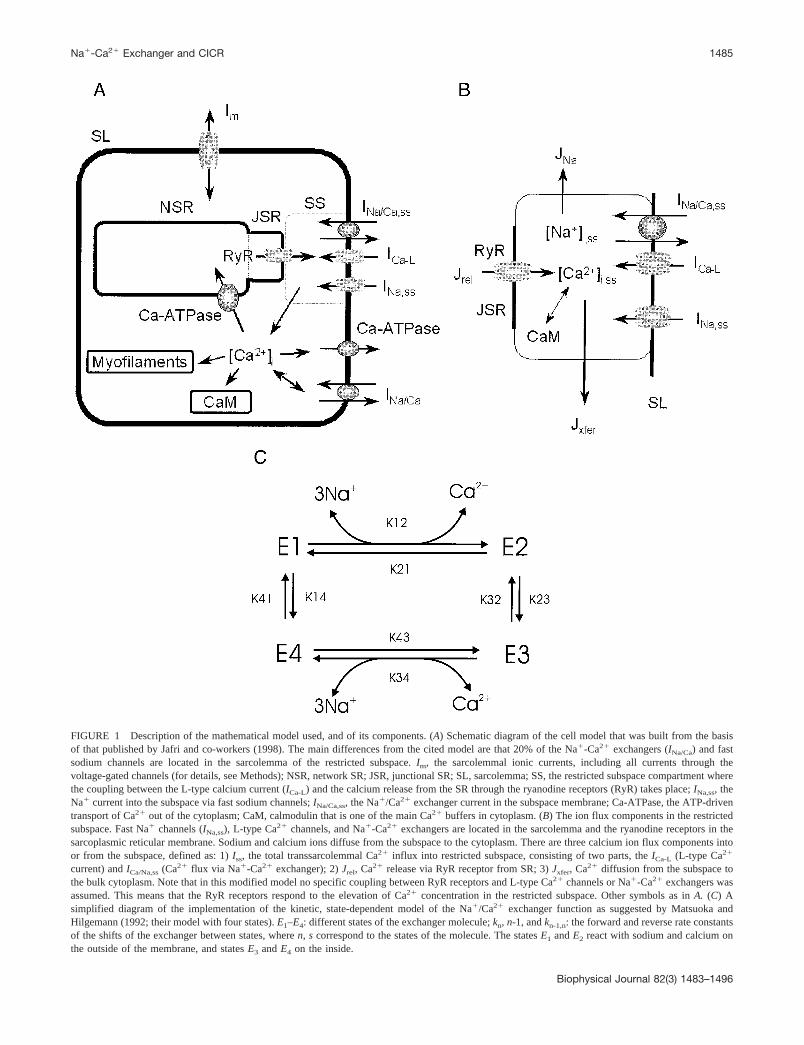
FIGURE 1 Description of the mathematical model used, and of its components. (A) Schematic diagram of the cell model that was built from the basisof that published by Jafri and co-workers (1998). The main differences from the cited model are that 20% of the Na�-Ca2� exchangers (INa/Ca) and fastsodium channels are located in the sarcolemma of the restricted subspace. Im, the sarcolemmal ionic currents, including all currents through thevoltage-gated channels (for details, see Methods); NSR, network SR; JSR, junctional SR; SL, sarcolemma; SS, the restricted subspace compartment wherethe coupling between the L-type calcium current (ICa-L) and the calcium release from the SR through the ryanodine receptors (RyR) takes place; INa,ss, theNa� current into the subspace via fast sodium channels; INa/Ca,ss, the Na�/Ca2� exchanger current in the subspace membrane; Ca-ATPase, the ATP-driventransport of Ca2� out of the cytoplasm; CaM, calmodulin that is one of the main Ca2� buffers in cytoplasm. (B) The ion flux components in the restrictedsubspace. Fast Na� channels (INa,ss), L-type Ca2� channels, and Na�-Ca2� exchangers are located in the sarcolemma and the ryanodine receptors in thesarcoplasmic reticular membrane. Sodium and calcium ions diffuse from the subspace to the cytoplasm. There are three calcium ion flux components intoor from the subspace, defined as: 1) Iss, the total transsarcolemmal Ca2� influx into restricted subspace, consisting of two parts, the ICa-L (L-type Ca2�
current) and ICa/Na,ss (Ca2� flux via Na�-Ca2� exchanger); 2) Jrel, Ca2� release via RyR receptor from SR; 3) Jxfer, Ca2� diffusion from the subspace tothe bulk cytoplasm. Note that in this modified model no specific coupling between RyR receptors and L-type Ca2� channels or Na�-Ca2� exchangers wasassumed. This means that the RyR receptors respond to the elevation of Ca2� concentration in the restricted subspace. Other symbols as in A. (C) Asimplified diagram of the implementation of the kinetic, state-dependent model of the Na�/Ca2� exchanger function as suggested by Matsuoka andHilgemann (1992; their model with four states). E1–E4: different states of the exchanger molecule; kn, n-1, and kn-1,n: the forward and reverse rate constantsof the shifts of the exchanger between states, where n, s correspond to the states of the molecule. The states E1 and E2 react with sodium and calcium onthe outside of the membrane, and states E3 and E4 on the inside.
Na�-Ca2� Exchanger and CICR 1485
Biophysical Journal 82(3) 1483–1496
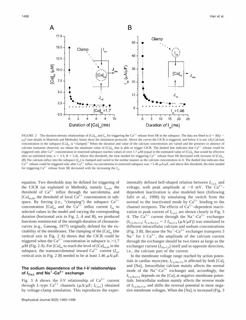
equation. Two thresholds may be defined for triggering ofthe CICR (as explained in Methods), namely Iss,th, thethreshold of Ca2� influx through the sarcolemma, and[Ca]ss,th, the threshold of local Ca2� concentration in sub-space. By forcing (i.e., “clamping”) the subspace Ca2�
concentration [Ca]ss and the Ca2� influx current Iss toselected values in the model and varying the correspondingduration (horizontal axis in Fig. 2, A and B), we producedfunctions reminiscent of the strength-duration of chronaxie-curves (e.g., Ganong, 1977) originally defined for the ex-citability of the membranes. The clamping of the [Ca]ss (thevertical axis in Fig. 2 A) shows that the CICR could betriggered when the Ca2� concentration in subspace is �1.7�M (Fig. 2 A). For [Ca]ss to reach the level of [Ca]ss,th in thesubspace, the transsarcolemmal inward Ca2� current (Iss,vertical axis in Fig. 2 B) needed to be at least 1.46 �A/�F.
The sodium dependence of the I-V relationshipsof ICa,L and Na�-Ca2� exchange
Fig. 3 A shows the I-V relationship of Ca2� currentthrough L-type Ca2� channels (�A/�F; ICa,L) obtainedby voltage-clamp simulation. This reproduces the exper-
imentally defined bell-shaped relation between ICa,L andvoltage, with peak amplitude at �0 mV. The Ca2�-dependent inactivation is also modeled here (followingJafri et al., 1998) by simulating the switch from thenormal to the inactivated mode by Ca2� binding to thechannel receptors. The effects of Ca2�-dependent inacti-vation to peak current of ICa,L are shown clearly in Fig. 3A. The Ca2� current through the Na�-Ca2� exchanger(ICa,Na-Ca; ICa,Na-Ca � 2 INa-Ca (�A/�F)) was simulated indifferent intracellular calcium and sodium concentrations(Fig. 3 B). Because the Na�-Ca2� exchanger transports 3Na� for 1 Ca2�, the amplitude of the calcium currentthrough the exchanger should be two times as large as theexchanger current (INa-Ca) itself and in opposite direction,i.e., the calcium part of the current.
In the membrane voltage range reached by action poten-tials in cardiac myocytes, ICa,Na-Ca is affected by both [Ca]i
and [Na]i. Intracellular calcium mainly affects the normalmode of the Na�-Ca2� exchanger and, accordingly, theICa,Na-Ca depends on the [Ca]i at negative membrane poten-tials. Intracellular sodium mainly affects the reverse modeof ICa,Na-Ca, and shifts the reversal potential to more nega-tive membrane voltages. When the [Na]i is increased (Fig. 3
FIGURE 2 The duration-density relationships of [Ca]ss and Iss for triggering the Ca2� release from SR in the subspace. The data are fitted to d � B/(y �y0)n (see details in Materials and Methods). Insets show the simulation protocols. Above the curves the CICR is triggered, and below it is not. (A) Calciumconcentration in the subspace [Ca]ss is “clamped.” When the duration and value of the calcium concentration are varied and the presence or absence ofcalcium transients observed, we obtain the minimum value of [Ca]ss that is able to trigger CICR. The dashed line indicates that Ca2� release could betriggered only after Ca2� concentration in restricted subspace reaches values of over 1.7 �M (equal to the estimated value of [Ca]ss that would be effectiveafter an unlimited time, n � 3.3, B � 5.4). Above this threshold, the time needed for triggering Ca2� release from SR decreased with increase of [Ca]ss.(B) The calcium influx into the subspace (Iss) is clamped and varied in the similar manner as the calcium concentration in A. The dashed line indicates thatCa2� release could be triggered only after Ca2� influx via sarcolemma to restricted subspace was �1.46 �A/�F, and above this threshold, the time neededfor triggering Ca2� release from SR decreased with the increasing the Iss.
1486 Han et al.
Biophysical Journal 82(3) 1483–1496
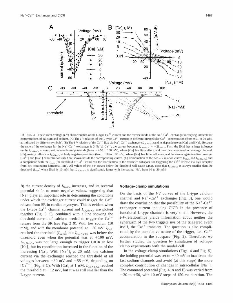
B) the current density of INa-Ca increases, and its reversalpotential shifts to more negative values, suggesting that[Na]i plays an important role in determining the conditionsunder which the exchanger current could trigger the Ca2�
release from SR in cardiac myocytes. This is evident whenthe L-type Ca2� channel current and ICa,Na-Ca are plottedtogether (Fig. 3 C), combined with a line showing thethreshold current of calcium needed to trigger the Ca2�
release from the SR (see Fig. 2 B). With low sodium (10mM), and with the membrane potential at �30 mV, ICa,L
reached the threshold (Iss,th), but ICa,Na-Ca was below thethreshold even when the potential was at �100 mV.ICa,Na-Ca was not large enough to trigger CICR in low[Na]i, but its contribution increased in the function of theincreasing [Na]i. With [Na�]i at 20 mM, the calciumcurrent via the exchanger reached the threshold at allvoltages between �30 mV and �55 mV, depending on[Ca2�]i (Fig. 3 C). With [Ca]i at 1 �M, ICa,Na-Ca reachedthe threshold at �12 mV, but it was still smaller than theL-type current.
Voltage-clamp simulations
On the basis of the I-V curves of the L-type calciumchannel and Na�-Ca2� exchanger (Fig. 3), one woulddraw the conclusion that the possibility of the Na�-Ca2�
exchanger current inducing CICR in the presence offunctional L-type channels is very small. However, theI-V-relationships yields information about neither thesynergism of the two triggers nor of the triggered eventitself, the Ca2� transient. The question is also compli-cated by the cumulative nature of the trigger, i.e., Ca2�
accumulation in the subspace (Fig. 2). Therefore, wefurther studied the question by simulation of voltage-clamp experiments with the model cell.
In the voltage-clamp simulations (Figs. 4 and Fig. 5),the holding potential was set to �40 mV to inactivate thefast sodium channels and avoid (at this stage) the morecomplex contribution of changes in intracellular [Na�]i.The command potential (Fig. 4, A and E) was varied from�30 to �50, with 10-mV steps of 150-ms duration. The
FIGURE 3 The current-voltage (I-V) characteristics of the L-type Ca2� current and the reverse mode of the Na�-Ca2� exchanger in varying intracellularconcentrations of calcium and sodium. (A) The I-V relation of the L-type Ca2� current in different intracellular Ca2� concentration (from 0.01 to 30 �M,as indicated by different symbols). (B) The I-V relation of the Ca2� flux via Na�-Ca2� exchanger (ICa,Na-Ca) and its dependence on [Ca]i and [Na]i. Becausethe ratio of the exchange for the Na�-Ca2� exchanger is 3 Na�:1 Ca2�, the current becomes ICa,Na-Ca � �2INa-Ca. First, the [Na]i has a large influenceon the ICa,Na-Ca at very positive membrane potentials (from ��50 to 100 mV), where [Ca]i has little effect, and thus the curves tend to converge. Second,[Ca]i mainly influences ICa,Na-Ca at fairly negative potentials (from �50 to �90 mV), where [Na]i has little influence, and the curves again tend to converge.[Ca2�] and [Na�] concentrations used are shown beside the corresponding curves. (C) Combination of the two I-V relation curves (ICa,L and ICa,Na-Ca) anda comparison with the Iss,th (the threshold of Ca2� influx via the sarcolemma to the restricted subspace for triggering the Ca2� release via RyR receptorfrom SR; continuous horizontal line). All values of the I-V curves below the threshold will cause CICR. Note that ICa,Na-Ca is always smaller than thethreshold (Iss,th) when [Na]i is 10 mM, but ICa,Na-Ca is significantly larger with increasing [Na]i from 10 to 20 mM.
Na�-Ca2� Exchanger and CICR 1487
Biophysical Journal 82(3) 1483–1496
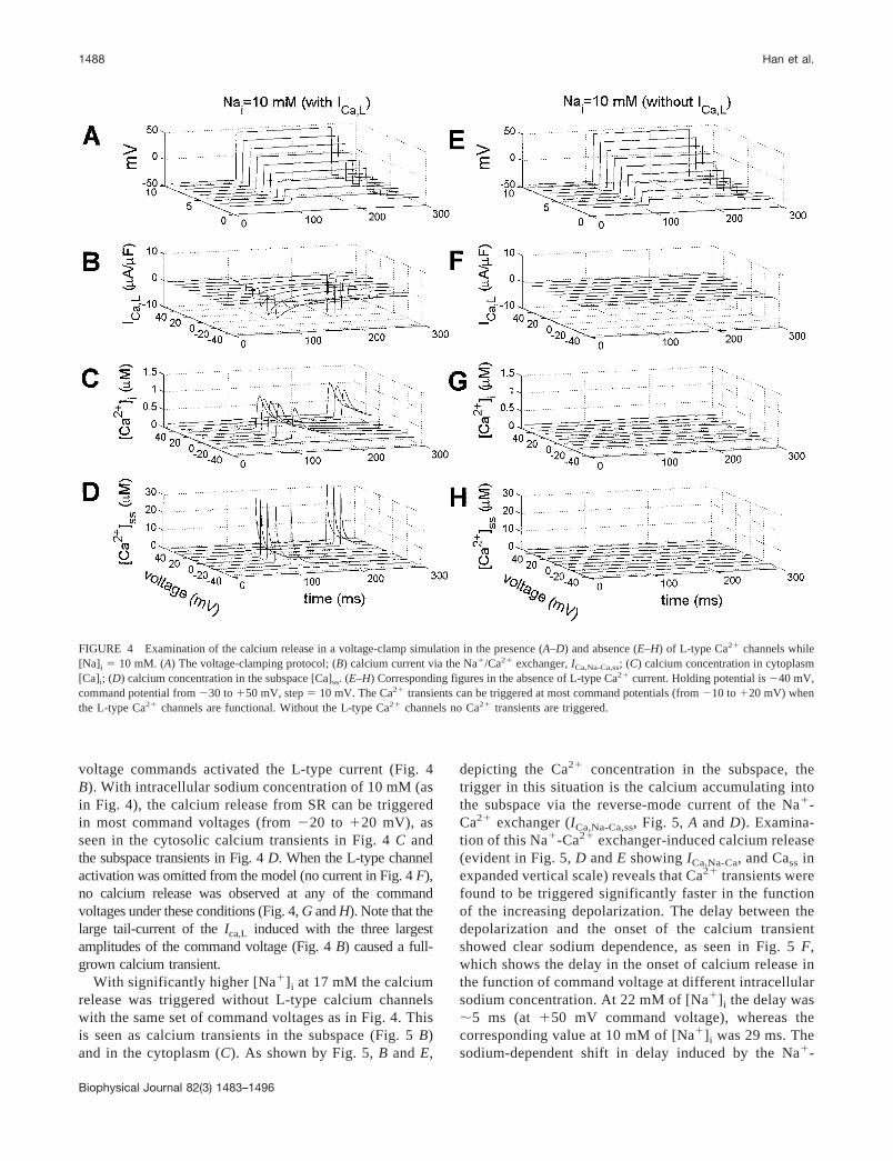
voltage commands activated the L-type current (Fig. 4B). With intracellular sodium concentration of 10 mM (asin Fig. 4), the calcium release from SR can be triggeredin most command voltages (from �20 to �20 mV), asseen in the cytosolic calcium transients in Fig. 4 C andthe subspace transients in Fig. 4 D. When the L-type channelactivation was omitted from the model (no current in Fig. 4 F),no calcium release was observed at any of the commandvoltages under these conditions (Fig. 4, G and H). Note that thelarge tail-current of the Ica,L induced with the three largestamplitudes of the command voltage (Fig. 4 B) caused a full-grown calcium transient.
With significantly higher [Na�]i at 17 mM the calciumrelease was triggered without L-type calcium channelswith the same set of command voltages as in Fig. 4. Thisis seen as calcium transients in the subspace (Fig. 5 B)and in the cytoplasm (C). As shown by Fig. 5, B and E,
depicting the Ca2� concentration in the subspace, thetrigger in this situation is the calcium accumulating intothe subspace via the reverse-mode current of the Na�-Ca2� exchanger (ICa,Na-Ca,ss, Fig. 5, A and D). Examina-tion of this Na�-Ca2� exchanger-induced calcium release(evident in Fig. 5, D and E showing ICa,Na-Ca, and Cass inexpanded vertical scale) reveals that Ca2� transients werefound to be triggered significantly faster in the functionof the increasing depolarization. The delay between thedepolarization and the onset of the calcium transientshowed clear sodium dependence, as seen in Fig. 5 F,which shows the delay in the onset of calcium release inthe function of command voltage at different intracellularsodium concentration. At 22 mM of [Na�]i the delay was�5 ms (at �50 mV command voltage), whereas thecorresponding value at 10 mM of [Na�]i was 29 ms. Thesodium-dependent shift in delay induced by the Na�-
FIGURE 4 Examination of the calcium release in a voltage-clamp simulation in the presence (A–D) and absence (E–H) of L-type Ca2� channels while[Na]i � 10 mM. (A) The voltage-clamping protocol; (B) calcium current via the Na�/Ca2� exchanger, ICa,Na-Ca,ss; (C) calcium concentration in cytoplasm[Ca]i; (D) calcium concentration in the subspace [Ca]ss. (E–H) Corresponding figures in the absence of L-type Ca2� current. Holding potential is �40 mV,command potential from �30 to �50 mV, step � 10 mV. The Ca2� transients can be triggered at most command potentials (from �10 to �20 mV) whenthe L-type Ca2� channels are functional. Without the L-type Ca2� channels no Ca2� transients are triggered.
1488 Han et al.
Biophysical Journal 82(3) 1483–1496

Ca2� exchanger current opens the possibility that theNa�-Ca2� exchanger current precedes the L-type cal-cium current at high [Na�]i. This means that the (main)trigger of CICR could be changed upon accumulation ofsodium.
Role of the Na�/Ca2� exchanger as a CICRtrigger during action potential
On the basis of the voltage-clamp simulations, the re-verse mode of the Na�/Ca2� exchanger contributes toCICR in a sodium-dependent manner. However, duringnormal APs the [Na�]i is also influenced by the fastsodium current. To investigate this, we performed modelsimulations at varying [Na]i (10, 13, 17, 20, and 22 mM).
Fig. 6 A shows a simulated action potential with corre-sponding changes in the Na�-Ca2� exchanger current(Fig. 6 B), sodium concentration [Na]ss in the subspace(Fig. 6 C), the calcium transients in subspace ([Ca]ss)(Fig. 6 D), the calcium component of the Na�/Ca2�
exchanger current in subspace membrane, INa,Na-Ca,ss
(Fig. 6 E), and the L-type calcium channel current insubspace membrane (Fig. 6 F). Insets show the earlyevents during the first 5 ms in an expanded time scale. Asexpected, the AP duration and amplitude decreased uponincrease of the initial [Na]i. This effect is due to theincreased Na�/Ca2�-exchanger outward current (Fig. 6B), reflecting the increased calcium influx during earlyphases of the upstroke of the AP (Fig. 6, E and F). Inthese simulations we observed the same phenomenon as
FIGURE 5 Voltage-clamp simulation with increased [Na]i (17 mM) and without functional L-type calcium current. The voltage-clamp protocol is thesame as in Fig. 4. (A) ICa,Na-Ca; (B) calcium concentration in subspace [Ca]ss; (C) calcium concentration in the cytoplasm [Ca]i. With increased intracellularsodium the Ca2� releases are now triggered. (D and E) A closer examination of the ICa,Na-Ca and the Cass. Note that the calcium transients (E) are triggeredat different times after the beginning of the stimulus pulse (at 100 ms), depending on the amplitude of the voltage pulse. (F) The sodium- andvoltage-dependence of the delay for triggering Ca2� release from intracellular calcium stores with the presence of L-type Ca2� channels. The delay timeis defined as the duration between depolarization (leading edge of the voltage-clamp pulse) and the onset of the calcium transient. The delay is shown forthree different levels of [Na]i: (�) 10 mM, (E) 17 mM, and (�) 23 mM.
Na�-Ca2� Exchanger and CICR 1489
Biophysical Journal 82(3) 1483–1496

seen in the voltage-clamp experiments, namely that athigh [Na�]i the dominant trigger of the CICR is shiftedfrom the L-type current toward the Na�/Ca2� exchangercurrent. This is also contributed by the sodium current,which is able to increase the [Na�]ss transiently to �20mM in normal [Na�]i (Fig. 6 C). In high [Na�]i the peak[Na�]ss reaches 30 mM, augmenting the exchanger cur-rent (Fig. 6, B and E). As a result, the delay between thedepolarization and subspace Ca2�-transient is greatlyreduced (Fig. 6 D, inset). The small voltage-dependentinactivation of the L-type channels explains the paradox-ical increase of the L-type current during lower voltage(i.e., reduced AP peak voltage) in Fig. 6 F.
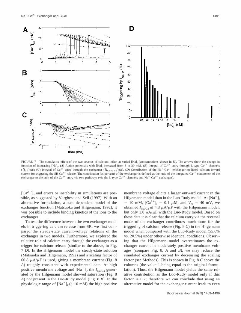
As determined earlier (Fig. 2), the triggering event ofCICR can be characterized by calcium accumulation intothe subspace, i.e., calcium flux per time. The estimationof the trigger efficiency of L-type calcium current andNa�/Ca2� exchanger current would be best evaluated bycomparing the time integral of the currents. To do so, theCa2� entering the cell through either the L-type channelsor via the exchanger current was calculated by integratingthe Ca2� currents from the start of the action potential tothe activation of SR ryanodine receptors (Fig. 7). Thecontribution of the exchanger to the SR Ca2� release(Fig. 7 D) is the ratio of the integration of Ca2� compo-
nent of the exchange with the sum of Ca2� entry via twopathways, i.e., via L-type Ca2� and the reverse mode ofthe Na�-Ca2� exchange. Upon increase of the [Na�]i (asindicated by the arrows), the subspace calcium transientis triggered faster than previously (Fig. 7 A), as found inthe previous sections (see Figs. 5 and 6). As seen fromthe Fig. 7 C, subspace exchanger current dominates thisphenomenon, being activated more rapidly than subspaceL-type current (Fig. 7 B) upon sodium increase. There-fore, the contribution of the exchange to SR Ca2� releaseis 25% in normal [Na]i, and the contribution of theexchange increases to 40%, 52%, 67%, and 73% when[Na]i increased to 13, 17, 20, and 22 mM, respectively(Fig. 7 D). In extreme situations ([Na]i � 30 mM) thecontribution of the Na�-Ca2� exchange is almost 100%.
Use of a state-dependent model ofthe exchanger
In the simulations above the function of the Na�-Ca2�
exchanger was modeled according to the Luo-Rudymodel. Because in this formulation no binding of Na�
and Ca2� is involved, the model does not predict anysaturation of the exchanger current at high [Na�]i and
FIGURE 6 Calcium concentrations and currents during action potentials (L-type current functional) at varied concentrations of [Na]i (arrows show thedirection of the change in function of the increased intracellular sodium. In all figures this is in the order 10, 13, 17, 20, 22 mM with corresponding stylesof solid, dashed, dotted, dash-dotted, and dash-double-dotted lines). (A) Action potential; (B) INa-Ca; (C) subspace concentration of sodium [Na]ss; (D)[Ca]ss; (E) ICa,Na-Ca; (F) ICa,L. Insets show the early events during the first 5 ms in an expanded time scale (horizontal scale bar � 1 ms; vertical scale remainsthe same).
1490 Han et al.
Biophysical Journal 82(3) 1483–1496
[Ca2�]i, and errors or instability in simulations are pos-sible, as suggested by Varghese and Sell (1997). With analternative formulation, a state-dependent model of theexchanger function (Matsuoka and Hilgemann, 1992), itwas possible to include binding kinetics of the ions to theexchanger.
To test the difference between the two exchanger mod-els in triggering calcium release from SR, we first com-pared the steady-state current-voltage relations of theexchanger in two models. Furthermore, we explored therelative role of calcium entry through the exchanger as atrigger for calcium release (similar to the above, in Fig.7 D). In the Hilgemann model the steady-state solution(Matsuoka and Hilgemann, 1992) and a scaling factor of60.8 �A/�F is used, giving a membrane current (Fig. 8A) roughly consistent with experimental data. At highpositive membrane voltage and [Na�]i, the INa/Ca gener-ated by the Hilgemann model showed saturation (Fig. 8A) not present in the Luo-Rudy model (Fig. 8 B). In thephysiologic range of [Na�]i (�10 mM) the high positive
membrane voltage elicits a larger outward current in theHilgemann model than in the Luo-Rudy model. At [Na�]i
� 10 mM, [Ca2�]i � 0.1 �M, and Vm � 40 mV, weobtained INa/Ca of 4.3 �A/�F with the Hilgemann model,but only 1.0 �A/�F with the Luo-Rudy model. Based onthese data it is clear that the calcium entry via the reversalmode of the exchanger contributes much more for thetriggering of calcium release (Fig. 8 C) in the Hilgemannmodel when compared with the Luo-Rudy model (55.6%vs. 20.5%) under otherwise identical conditions. Observ-ing that the Hilgemann model overestimates the ex-changer current in moderately positive membrane volt-ages (compare Fig. 8, A and B), we may reduce thesimulated exchanger current by decreasing the scalingfactor (see Methods). This is shown in Fig. 8 C above thecolumns (the value 1 being equal to the original formu-lation). Thus, the Hilgemann model yields the same rel-ative contribution as the Luo-Rudy model only if thisfactor is 0.2; therefore we can conclude that using analternative model for the exchanger current leads to even
FIGURE 7 The cumulative effect of the two sources of calcium influx at varied [Na]i (concentrations shown in D). The arrows show the change infunction of increasing [Na]i. (A) Action potentials with [Na]i increased from 8 to 30 mM. (B) Integral of Ca2� entry through L-type Ca2� channels(ICa(t)dt); (C) Integral of Ca2� entry through the exchanger (ICa,Na-Ca(t)dt). (D) Contribution of the Na�-Ca2� exchanger-mediated calcium inwardcurrent for triggering the SR Ca2� release. The contribution (as percent) of the exchanger is defined as the ratio of the integrated Ca2� component of theexchanger to the sum of the Ca2� entry via two pathways (via the L-type Ca2� channels and Na�-Ca2� exchanger).
Na�-Ca2� Exchanger and CICR 1491
Biophysical Journal 82(3) 1483–1496
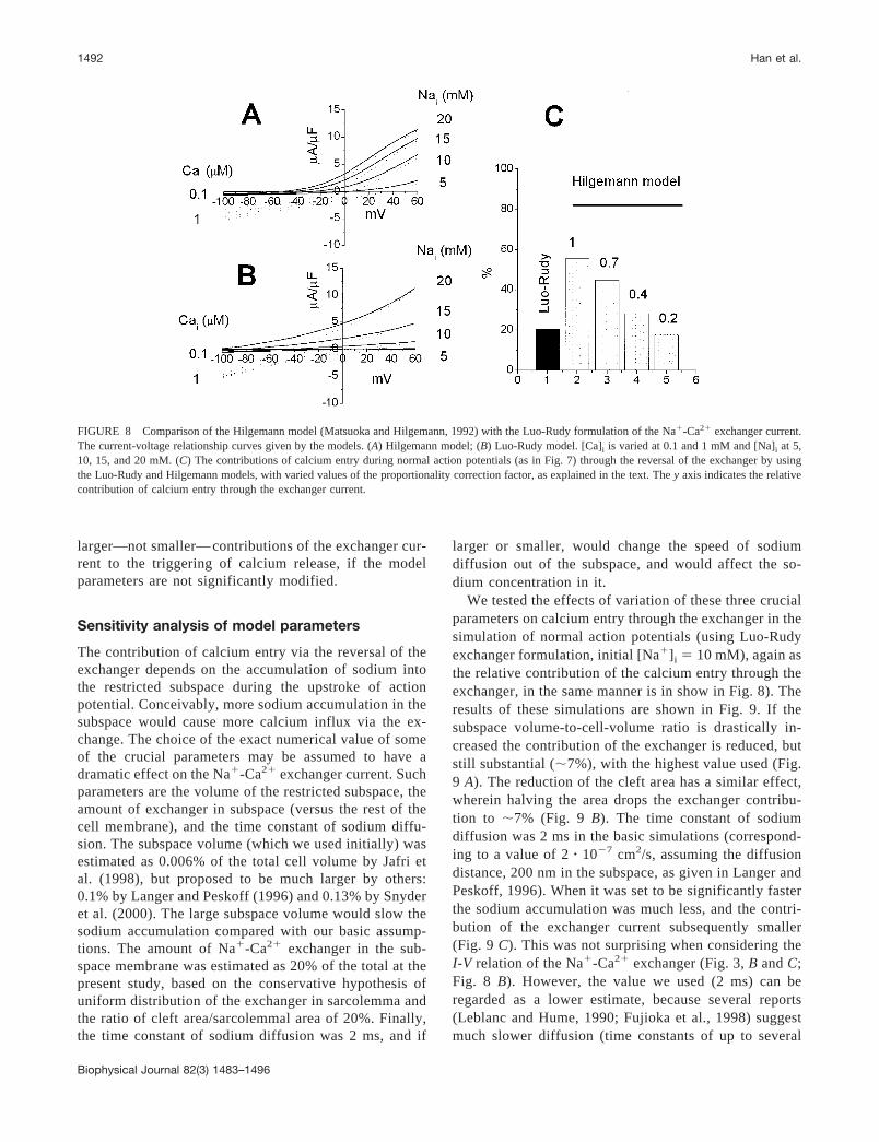
larger—not smaller— contributions of the exchanger cur-rent to the triggering of calcium release, if the modelparameters are not significantly modified.
Sensitivity analysis of model parameters
The contribution of calcium entry via the reversal of theexchanger depends on the accumulation of sodium intothe restricted subspace during the upstroke of actionpotential. Conceivably, more sodium accumulation in thesubspace would cause more calcium influx via the ex-change. The choice of the exact numerical value of someof the crucial parameters may be assumed to have adramatic effect on the Na�-Ca2� exchanger current. Suchparameters are the volume of the restricted subspace, theamount of exchanger in subspace (versus the rest of thecell membrane), and the time constant of sodium diffu-sion. The subspace volume (which we used initially) wasestimated as 0.006% of the total cell volume by Jafri etal. (1998), but proposed to be much larger by others:0.1% by Langer and Peskoff (1996) and 0.13% by Snyderet al. (2000). The large subspace volume would slow thesodium accumulation compared with our basic assump-tions. The amount of Na�-Ca2� exchanger in the sub-space membrane was estimated as 20% of the total at thepresent study, based on the conservative hypothesis ofuniform distribution of the exchanger in sarcolemma andthe ratio of cleft area/sarcolemmal area of 20%. Finally,the time constant of sodium diffusion was 2 ms, and if
larger or smaller, would change the speed of sodiumdiffusion out of the subspace, and would affect the so-dium concentration in it.
We tested the effects of variation of these three crucialparameters on calcium entry through the exchanger in thesimulation of normal action potentials (using Luo-Rudyexchanger formulation, initial [Na�]i � 10 mM), again asthe relative contribution of the calcium entry through theexchanger, in the same manner is in show in Fig. 8). Theresults of these simulations are shown in Fig. 9. If thesubspace volume-to-cell-volume ratio is drastically in-creased the contribution of the exchanger is reduced, butstill substantial (�7%), with the highest value used (Fig.9 A). The reduction of the cleft area has a similar effect,wherein halving the area drops the exchanger contribu-tion to �7% (Fig. 9 B). The time constant of sodiumdiffusion was 2 ms in the basic simulations (correspond-ing to a value of 2 � 10�7 cm2/s, assuming the diffusiondistance, 200 nm in the subspace, as given in Langer andPeskoff, 1996). When it was set to be significantly fasterthe sodium accumulation was much less, and the contri-bution of the exchanger current subsequently smaller(Fig. 9 C). This was not surprising when considering theI-V relation of the Na�-Ca2� exchanger (Fig. 3, B and C;Fig. 8 B). However, the value we used (2 ms) can beregarded as a lower estimate, because several reports(Leblanc and Hume, 1990; Fujioka et al., 1998) suggestmuch slower diffusion (time constants of up to several
FIGURE 8 Comparison of the Hilgemann model (Matsuoka and Hilgemann, 1992) with the Luo-Rudy formulation of the Na�-Ca2� exchanger current.The current-voltage relationship curves given by the models. (A) Hilgemann model; (B) Luo-Rudy model. [Ca]i is varied at 0.1 and 1 mM and [Na]i at 5,10, 15, and 20 mM. (C) The contributions of calcium entry during normal action potentials (as in Fig. 7) through the reversal of the exchanger by usingthe Luo-Rudy and Hilgemann models, with varied values of the proportionality correction factor, as explained in the text. The y axis indicates the relativecontribution of calcium entry through the exchanger current.
1492 Han et al.
Biophysical Journal 82(3) 1483–1496

tens of seconds). The slower Na� diffusion would ofcourse enhance the triggering role of the exchanger and,e.g., with a time constant of 14 ms, �30% of the Ca2�
trigger would come via the exchanger current (Fig. 9 C).Thus, even when using quite different estimates for the
most crucial parameters in the modeling, the simulationssuggest that the exchanger current has a role in thetriggering of the Ca2� release. Furthermore, the sensitiv-ity analysis of the parameters treated above shows thatthey are different in affecting the calcium entry and intriggering calcium release from SR. For example, whenthe ratio of the subspace volume to cytosolic volume(Vsubspace/Vmyocytol) increases 17-fold (from 0.1% to0.006%), the relative contribution of the exchanger cur-rent decreases 2.5-fold (from 20.5% to 8.3%), and thesensitivity is 0.14. Similar analysis gives a sensitivity of1.25 for the ratio of the area of the cleft membrane to thearea of the total sarcolemma (Acleft/Asarcolemma), and 0.006for the sodium diffusion time constant. The sensitivityanalysis shows that the order of importance of these threeparameters in determining the relative contribution of thecalcium entry through the exchanger current is 1) theamount of the exchanger molecules in the subspace; 2)the volume of the subspace; and 3) the speed of thesodium diffusion.
DISCUSSION
In this study we have quantitatively evaluated the role of thereverse mode of the Na�-Ca2� exchanger current in trig-gering the CICR based on a computer simulation of amathematical model of the cardiac myocyte. In the modelthe increase of intracellular calcium concentration causingthe CICR takes place in a diffusionally restricted subspaceof the cytoplasm, the so-called fuzzy space or dyadic cleft.The main finding of the simulations is that the Na�-Ca2�
exchanger indeed plays a role in triggering the release ofcalcium from SR. Under conditions where the [Na�]i issmall, of the order of 10 mM or lower, the reverse modecurrent is not large enough to trigger the CICR by itself,although it contributes a fraction of the total triggeringcalcium. However, the contribution of the Na�-Ca2� ex-changer increases up to nearly 100% with the increase of theintracellular sodium concentration and/or positive potentialwith very clear effects, also on the relative timing of theevents. The detailed model of the CICR in the dyadic cleftby Langer and Peskoff (1996; Peskoff and Langer, 1998)predicts larger changes of [Na�]i and [Ca2�]i in the sub-space than in our model, reaching maximally above 45 mMfor sodium and over 1 mM for calcium near the center of thecleft. In our model the concentration changes are moremodest. However, because our model covers the whole of
FIGURE 9 Sensitivity analysis of the crucial modeling parameters. The figures show comparison of contributions of calcium entry via the exchangercurrent during normal action potentials (as in Fig. 7) with varied ratios of (A) Vsubspace/Vcytosol; (B) Acleft/Asarcolemma; and (C) different sodium diffusion timeconstants, with the Luo-Rudy model. Vsubspace, volume of the restrict subspace; Vcytosol, volume of cardiac myocytol; Acleft, surface area of the cleft;Asarcolemma, surface area of cardiac sarcolemma. The numbers at the top of each column are the values of the ratios (A and B) or sodium diffusion timeconstant (C) used in the simulation. The black column in each figure shows the result in the basic simulation analyzed in depth in text.
Na�-Ca2� Exchanger and CICR 1493
Biophysical Journal 82(3) 1483–1496
the myocyte, less details are present, and the spatial dimen-sion in the cleft is omitted. The predictions of our model andthe model of Langer and Peskoff do not contradict eachother, but our model examines the role of the exchanger inmore detail in the whole myocyte.
Validation of the model and simulation methods
The restricted subspace was used in modeling the cardiacE-C coupling by Jafri et al. (1998), and the development ofthe mathematical model used in the present investigation isbased on that model. In our basic model 20% of the Na�-Ca2� exchanger activity was placed into the subspace mem-brane. This crucial parameter obviously needs justification.When assuming a uniform distribution of the exchangermolecules in the sarcolemma we arrive at such a figure forthe junctional membrane (see Methods). Also, without ef-ficient means for transporting Ca2� from the subspace(leaving the relatively slow diffusion into the bulk cyto-plasm as the only means for this) the subspace concentra-tions would be excessively high. Thus we can take 20% asa starting estimate, meaning that the ratio of L-type calciumconductance to the exchanger currents is the same every-where in the sarcolemma. The number of exchanger mole-cules could easily be still higher in the subspace membrane,but while we have no exact numerical data of the exchangerdistribution, an approximation has to be made. A smalleramount would mean that the cells are less efficient inregulating calcium and sodium concentrations in the sub-space. With this modification this model may be regarded tobe more physiological, especially in the description of theCICR. Our model thus gives a useful tool to study thequantitative relationship between the ICa,L and the INa-Ca
in causing the CICR, and allows simulation of “idealexperiments,” interventions where the pathways of cal-cium influx through sarcolemma in CICR may be iden-tified and controlled.
The determination of the kind of boundary conditions forthe CICR (Fig. 2) in terms of concentration and Ca2�
current thresholds was one application of the model. Theconcept of “clamping” either the concentration or the cur-rent also emphasizes the idea inherent in the present mod-eling, that as far as the CICR in the subspace compartmentis concerned, it is irrelevant where the calcium signal orig-inated once the calcium transient has been triggered. Theboundary conditions were compared with the I-V relation-ships of the currents (the L-type calcium current and theNa�-Ca2� exchanger current) in Fig. 3, where also thecurrent threshold for the CICR has been reproduced. In thepresent simulation of an Na�/Ca2� I-V relation, as shown inFig. 3, the INa/Ca under conditions of 20 mM [Na�]i, 0.1 �M[Ca2�]i, and �40 mV of membrane voltage is 8.5 pA/pF. Inexperiments in guinea pig ventricular myocytes under sim-ilar conditions (Shigematsu and Arita, 1999) the exchangercurrent was 6.6 pA/pF. In experimental reports the voltage
protocols used (Convery and Hancox, 1999) and calciumbuffers (Coetzee et al., 1994) may have an effect on theexchanger current. When comparing the exchanger currentduring the upstroke of action potential we found that theresult from our simulation is quite close to the measurementfrom myocytes. In guinea pig ventricular myocytes,Grantham and Cannell (1996) had INa/Ca values of 0.55pA/pF when ICa,L reached its peak, whereas in our modelINa/Ca is 0.49 pA/pF in the same time. The model I-V curvesof the currents are thus within the experimental variation,which validates this part of the modeling (see, e.g., Mc-Donald et al., 1986; Watanabe et al., 2001).
Controversies concerning theNa�/Ca2� exchanger
Although it is clear that L-type Ca2� channels have adominant role in triggering the CICR, it has been arguedthat they may not be the only triggers for the Ca2� releasefrom the SR. Since the first reports of Ca2� release from SRthat could be triggered by the Na�-Ca2� exchanger in Ca2�
overload conditions (Berlin et al., 1987; Bers et al., 1988),several investigators have shown that Ca2� entry via thereverse mode of the Na�-Ca2� exchanger could be anothertriggering mechanism. With high internal Na� solution (20mM), this mechanism has been demonstrated in cat (Nussand Houser, 1992) and guinea pig ventricular myocytes(Kohmoto et al., 1994), and in lower internal Na� concen-tration in guinea pig (Kohmoto et al., 1994; Vornanen et al.,1994) and rat ventricular myocytes (Levi et al., 1994; Was-serstrom and Vites, 1996). However, several papers report anegligible role for the exchanger, either indirectly (Nabaueret al., 1989) or directly (Sham et al., 1992; Bouchard et al.,1993; Sipido et al., 1997). One problem in interpreting theseresults is that, as suggested by, e.g., Vornanen et al. (1994),the Na�-Ca2� exchanger may be much more dependent ontemperature than the L-type current, and when experimentsare performed at different temperatures, differing resultsmay be obtained. Additionally, the exchanger is dependenton the changes in internal concentrations of sodium andcalcium, and those changes are necessarily larger in therestricted subspace than in the rest of the cytosol. Sodiumaccumulation would favor the exchanger as a trigger, assuggested by Bers et al. (1988), Leblanc and Hume (1990),Carmeliet (1992), and Evans and Cannell (1997). In oursimulation, varying the [Na]i both in voltage-clamp andaction potential simulations (Figs. 4–6), the contribution ofthe exchanger current is greater, the larger the (initial) [Na]i.The triggering efficiency of the exchanger (Fig. 7 D) was�20% when [Na]i was below 10 mM, but rose steeply in thefunction of the [Na]i, reaching values near 100% at 30 mM.A rise in the internal Na� concentration also cruciallychanges the delay (from beginning of voltage change tobeginning of the calcium transient), as shown in Fig. 5 F. InSipido et al. (1997) a delay of �60 ms was found in the
1494 Han et al.
Biophysical Journal 82(3) 1483–1496

guinea pig in the cytosolic calcium transients with 20 mM[Na�]i. Our data in Fig. 5 F depict the delays for thesubspace calcium and, in fact, delays in our model are of thesame order as those in Sipido et al. (1997) when the cyto-solic transients are observed (Fig. 5 C).
One main result of the present study is the confirmationthat in the normal, low [Na]i it is very unlikely that thereverse-mode current of the Na�-Ca2� exchanger (ICa,Na-Ca)could trigger the CICR if it is the sole external source ofcalcium. This is in agreement with those investigations withnegative findings on the exchanger’s role. Equally impor-tantly, we have demonstrated that the reverse mode of theexchanger current contributes to the increase of calcium inthe subspace under all conditions studied in the presentinvestigation (Fig. 7 D). This would not be the case if theexchanger current were slower than the L-type calciumcurrent. In fact, Ca2� influx through the exchanger into thesubspace is always beginning faster than the L-type current inthe action potential simulations (Figs. 6 and 7) because of therapid rise in the [Na]i in subspace associated with the upstrokeof the action potential and the fast sodium (inward) current.
Change of dominant triggering of CICR inelevated [Na�]i
If we presume that sodium ion distribution in the cytoplasmis uniform, the global sodium concentration increases littlefollowing an action potential. During one action potential,the cytoplasmic sodium concentration increased by 0.0137mM in our model. This is consistent with the estimation byLederer et al. (1990) that in guinea pig myocytes a 50-nANa� current with an inactivation time constant of 1 msgenerates a 0.025 mM increase in sodium concentration.However, with sodium entry into the restricted subspace, asizable change in [Na]i occurs (Fig. 6 C), as also shown byLanger and Peskoff (1996) in their detailed model of thediffusion and concentration gradients in the subspace. In ourmodel, when the resting [Na]i is 10 mM, the peak of sodiumconcentration in the subspace reaches �30 mM (Fig. 6 C),which induces a transient Ca2� entry through the reverseexchange. Under normal action potentials (Figs. 6 and 7) theL-type Ca2� current and the Na�-Ca2� exchanger currentare functioning in synergy. This means that even when thereverse mode of exchanger current is not triggering theCICR it defines the offset, on which the L-type current doesthe triggering. By this mechanism, the gain of the calciumrelease would be controlled via the changes in [Na]i in thesubspace, as suggested by Cordeiro et al. (2000). In addition,the calcium currents into the subspace do not simply sumlinearly in bringing out the CICR, because the relationshipbetween the open probability of ryanodine receptors and pCa issigmoidal (Copello et al., 1997; Litwin et al., 1998).
When we observe the relative timing of the integrals ofthe inward calcium currents via the L-type channels (Fig. 7B) and the exchanger (Fig. 7 C), we can conclude that the
loading of calcium into the subspace by the exchangercurrent precedes that of the L-type current, in line withsuggestions by Cordeiro et al. (2000). In addition, when theinitial [Na]i is increased, the difference in timing becomesever larger. When the relative contribution of the exchangercurrent increases, it causes the threshold for the CICR to bereached earlier. Even when the L-type current contributesmore than half of the calcium causing the CICR, the ex-changer current is reducing the delay between the actionpotential upstroke (time 0 ms in Fig. 7) and the calciumtransient. One important contribution of the calcium currentvia the exchanger is, therefore, the timing of the calciumtransient with reference to the action potential.
Possible physiological implications
The role of the Na�-Ca2� exchanger as a trigger for calciumrelease has numerous implications in physiology of thecardiac myocytes. Considering that [Na]i is able to regulatethe relative contributions of the two different triggers ofCICR, any physiological stimulus or medication thatchanges the [Na]i will affect the regulation of the calciumrelease. Hormonal stimuli, such as ET-1-receptor activation(Alvarez et al., 1999), and increase of action potentialfrequency (Simor et al., 1997) will increase [Na]i, and thusemphasize calcium release triggering via the Na�/Ca2�
exchanger as opposed to via the L-type calcium current. Thefact that the delay of the exchange-induced calcium releaseis radically influenced by [Na]i in the subspace (Fig. 7 A)opens interesting perspectives. An increase of the frequencycould influence the electromechanical interval, with a greatercontribution of the exchanger-induced calcium release with areduced delay, being part of adaptational mechanisms makingefficient contractions possible in high frequency.
Support from Finnish Heart Research Foundation, Wihuri Foundation, andthe Academy of Finland is gratefully acknowledged.
REFERENCES
Alvarez, B. V., N. G. Perez, I. L. Ennis, M. C. Camilion de Hurtado, andH. E. Cingolani. 1999. Mechanisms underlying the increase in force andCa2� transient that follow stretch of cardiac muscle: a possible expla-nation of the anrep effect. Circ. Res. 85:716–722.
Berlin, J. R., M. B. Cannell, and W. J. Lederer. 1987. Regulation of twitchtension in sheep cardiac Purkinje fibers during calcium overload.Am. J. Physiol. Heart Circ. Physiol. 253:H1540–H1547.
Bers, D. M. 1993. Excitation-Contraction Coupling and Cardiac Contrac-tile Force. Kluwer Academic Publishers, Dordrecht, The Netherlands.
Bers, D. M., D. M. Christensen, and T. X. Nguyen. 1988. Can Ca entry viaNa-Ca exchange directly activate cardiac muscle contraction? J. Mol.Cell. Cardiol. 20:405–414.
Beuckelmann, D. J., and W. G. Weir. 1988. Mechanisms of release ofcalcium from sarcoplasmic reticulum of guinea-pig cardiac cells.J. Physiol. 405:233–255.
Bouchard, R. A., R. B. Clark, and W. R. Giles. 1993. Role of sodium-calcium exchange in activation of contraction in rat ventricle. J. Physiol.472:391–413.
Na�-Ca2� Exchanger and CICR 1495
Biophysical Journal 82(3) 1483–1496
Cannell, M. B., H. Cheng, and W. J. Lederer. 1995. The control of calciumrelease in heart muscle. Science. 268:1045–1049.
Carmeliet, E. 1992. A fuzzy subsarcolemmal space for intracellular Na� incardiac cells. Cardiovasc. Res. 26:433–442.
Coetzee, W. A., H. Ichikawa, and D. J. Hearse. 1994. Oxidant stress inhibitsNa-Ca-exchange current in cardiac myocytes: mediation by sulfhydrylgroups? Am. J. Physiol. Heart Circ. Physiol. 266:H909–H919.
Convery, M. K., and J. C. Hancox. 1999. Comparison of Na�-Ca2� exchangecurrent elicited from isolated rabbit ventricular myocytes by voltage rampand step protocols. Pflugers Arch-Eur. J. Physiol. 437:944–954.
Copello, J. A., S. Barg, H. Onoue, and S. Fleischer. 1997. Heterogeneity ofCa2� gating in skeletal muscle and cardiac ryanodine receptors.Biophys. J. 73:141–156.
Cordeiro, J. M., S. Litwin, and J. H. B. Bridge. 2000. Can NCX rapidly setthe gain of EC-coupling without directly triggering SR calcium releasein heart. Biophys. J. 78:2202.
Eisner, D. A., A. W. Trafford, M. E. Diaz, C. L. Overend, and S. C.O’Neill. 1998. The control of Ca release from the cardiac sarcoplasmicreticulum: regulation versus autoregulation. Cardiovasc. Res 38:589–604.
Evans, A. M., and M. B. Cannell. 1997. The role of L-type Ca2� currentand Na� current-stimulated Na/Ca exchange in triggering SR calciumrelease in guinea-pig cardiac ventricular myocytes. Cardiovasc. Res.35:294–302.
Fabiato, A. 1985. Time and calcium dependence of activation and inactivationof calcium-induced release of calcium from the sarcoplasmic reticulum of askinned canine cardiac Purkinje cell. J. Gen. Physiol. 85:247–289.
Fabiato, A., and F. Fabiato. 1975. Concentrations induced by a calcium-triggered release of calcium from the sarcoplasmic reticulum of singleskinned cardiac cells. J. Physiol. 249:469–495.
Frank, J. S., G. M. D. Reid, R. S. Molday, and K. D. Philipson. 1992.Distribution of the Na�-Ca2� exchange protein in mammalian cardiacmyocytes: an immunofluorescence and immunocolloidal gold-labelingstudy. J. Cell Biol. 2:337–345.
Fujioka, Y., S. Matsuoka, T. Ban, and A. Noma. 1998. Interaction of theNa�-K� pump and Na�-Ca2� exchange via [Na�]i in a restricted spaceof guinea-pig ventricular cells. J. Physiol. 509:457–470.
Ganong, W. F. 1977. Review of Medical Physiology. Lange, Los Altos,California.
Grantham, C. J., and M. B. Cannell. 1996. Ca2� influx during the cardiacaction potential in guinea pig ventricular myocytes. Circ. Res. 79:194–200.
Jafri, M. S., J. J. Rice, and R. L. Winslow. 1998. Cardiac Ca2� dynamics:the roles of ryanodine receptor adaptation and sarcoplasmic reticulumload. Biophys. J. 74:1149–1168.
Kabakov, A. Y. 1998. Activation of KATP channels by Na/K pump inisolated cardiac myocytes and giant membrane patches. Biophys. J.75:2858–2867.
Kieval, R. S., R. J. Bloch, G. E. Lindenmayer, A. Ambesi, and W. J.Lederer. 1992. Immunofluorescence localization of the Na-Ca ex-changer in heart cells. Am. J. Physiol. Cell Physiol. 263:C545–C550.
Kohmoto, O., A. J. Levi, and H. B. Bridge. 1994. Relation between reversesodium-calcium exchange and sarcoplasmic reticulum calcium release inguinea pig ventricular cells. Circ. Res. 74:550–554.
Langer, G. A., and A. Peskoff. 1996. Calcium concentration and movementin the diadic cleft space of the cardiac ventricular cell. Biophys. J.70:1169–1182.
Leblanc, N., and J. R. Hume. 1990. Sodium current-induced release ofcalcium from cardiac sarcoplasmic reticulum. Science. 248:372–376.
Lederer, W. J., E. Niggli, and R. W. Hadley. 1990. Sodium-calciumexchange in excitable cells: fuzzy space. Science. 248:283.
Levi, A. J., K. W. Spitzer, O. Kohmoto, and J. H. B. Bridge. 1994.Depolarization-induced Ca entry via Na-Ca exchange triggers SR re-lease in guinea pig cardiac myocytes. Am. J. Physiol. Heart Circ.Physiol. 266:H1422–H1433.
Lipp, P., and E. Niggli. 1994. Sodium current-induced calcium signals inisolated guinea-pig ventricular myocytes. J. Physiol. 474:439–446.
Litwin, S. E., J. Li, and J. H. B. Bridge. 1998. Na-Ca exchanger and thetrigger for sarcoplasmic reticulum Ca release: studies in adult rabbitventricular myocytes. Biophys. J. 75:359–371.
Lopez-Lopez, J. R., P. S. Shacklock, C. W. Balke, and W. G. Wier. 1995.Local calcium transients triggered by single L-type calcium channelcurrents in cardiac cell. Science. 268:1024–1045.
Luo, C. H., and Y. Rudy. 1994. A dynamic model of the cardiac ventricularaction potential I. Simulation of ionic currents and concentrationchanges. Circ. Res. 74:1071–1096.
Matsuoka, S., and D. W. Hilgemann. 1992. Steady-state and dynamicproperties of cardiac sodium-calcium exchange. Ion and voltage depen-dencies of the transport cycle. J. Gen. Physiol. 100:963–1001.
McDonald, T. F., F. A. Cavalie, W. Trautwein, and D. Pelzer. 1986.Voltage-dependent properties of macroscopic and elementary calciumchannel currents in guinea pig ventricular myocytes. Pflugers Arch-Eur.J. Physiol. 406:437–448.
Nabauer, M., G. Callewaert, L. Cleemann, and M. Morad. 1989. Regula-tion of calcium release is gated by calcium current, not gating charges,in cardiac myocytes. Science. 244:800–803.
Nuss, H. B., and S. R. Houser. 1992. Sodium-calcium exchange-mediatedcontractions in feline ventricular myocytes. Am. J. Physiol. Heart Circ.Physiol. 263:H1161–H1169.
Page, E. 1978. Quantitative ultrastructural analysis in cardiac membranephysiology. Am. J. Physiol. Cell Physiol. 235:C147–C158.
Peskoff, A., and G. A. Langer. 1998. Calcium concentration and movementin the ventricular cardiac cell during an excitation-contraction cycle.Biophys. J. 74:153–174.
Scriven, R. L., P. Dan, and D. W. Moore. 2000. Distribution of proteinsimplicated in excitation-contraction coupling in rat ventricular myo-cytes. Biophys. J. 79:2682–2691.
Sham, J. S., L. Cleemann, and M. Morad. 1992. Gating of the cardiac Ca2�
release channel: the role of Na� current and Na�-Ca2� exchange.Science. 255:850–853.
Shigematsu, S., and M. Arita. 1999. Anoxia depresses sodium-calciumexchange currents in guinea-pig ventricular myocytes. J. Mol. Cell.Cardiol. 31:895–905.
Simor, T. S., T. Lorand, B. Gaszner, and G. A. Elgavish. 1997. Themodulation of pacing-induced changes in intracellular sodium levels byextracellular Ca2� in isolated perfused rat hearts. J. Mol. Cell. Cardiol.29:1225–1235.
Sipido, K. R., M. Maes, and F. V. Werf. 1997. Low efficiency of Ca2�
entry through the Na�-Ca2� exchanger as trigger for Ca2� release fromthe sarcoplasmic reticulum. Circ. Res. 81:1034–1044.
Snyder, S. M., B. M. Palmer, and R. L. Moore. 2000. A mathematicalmodel of cardiocyte Ca2� dynamics with a novel representation ofsarcoplasmic reticular Ca2� control. Biophys. J. 79:94–115.
Stern, M. D. 1992. Theory of excitation-contraction coupling in cardiacmuscle. Biophys. J. 63:497–517.
Sun, X. H., F. Protasi, M. Takahashi, H. Takeshima, D. G. Ferguson, andC. Franzini-Armstrong. 1995. Molecular architecture of membranesinvolved in excitation-contraction coupling of cardiac muscle. J. Cell.Biol. 291:659–671.
Varghese, A., and G. S. Sell. 1997. A conservation principle and its effecton the formulation of Na-Ca exchanger current in cardiac cells. J. Theor.Biol. 189:33–40.
Vornanen, M., N. Shepherd, and G. Isenberg. 1994. Tension-voltage rela-tions of single myocytes reflect Ca release triggered by Na/Ca exchangeat 30°C but not 23°C. Am. J. Physiol. Cell Physiol. 267:C623–C633.
Wasserstrom, J. A., and A. M. Vites. 1996. The role of Na�-Ca2� ex-change in activation of excitation-contraction coupling in rat ventricularmyocytes. J. Physiol. 493:529–542.
Watanabe, Y., T. Iwamoto, I. Matsuoka, S. Ohkubo, T. Ono, T. Watano,M. Shikegawa, and J. Kimura. 2001. Inhibitory effect of 2,3-butanedionemonoxime (BDM) on Na�/Ca2� exchange current in guinea-pig cardiacventricular myocytes. Br. J. Pharmacol. 132:1317–1325.
Wendt-Gallitelli, M. F., T. Voigt, and G. Isenberg. 1993. Microheteroge-neity of subsarcolemmal sodium gradient: electron probe microanalysisin guinea-pig ventricular myocytes. J. Physiol. 472:33–44.
1496 Han et al.
Biophysical Journal 82(3) 1483–1496