Antiviral Activities of Oleanolic Acid and Its Analogues - MDPI
Upload
independentCategory
view
2download
0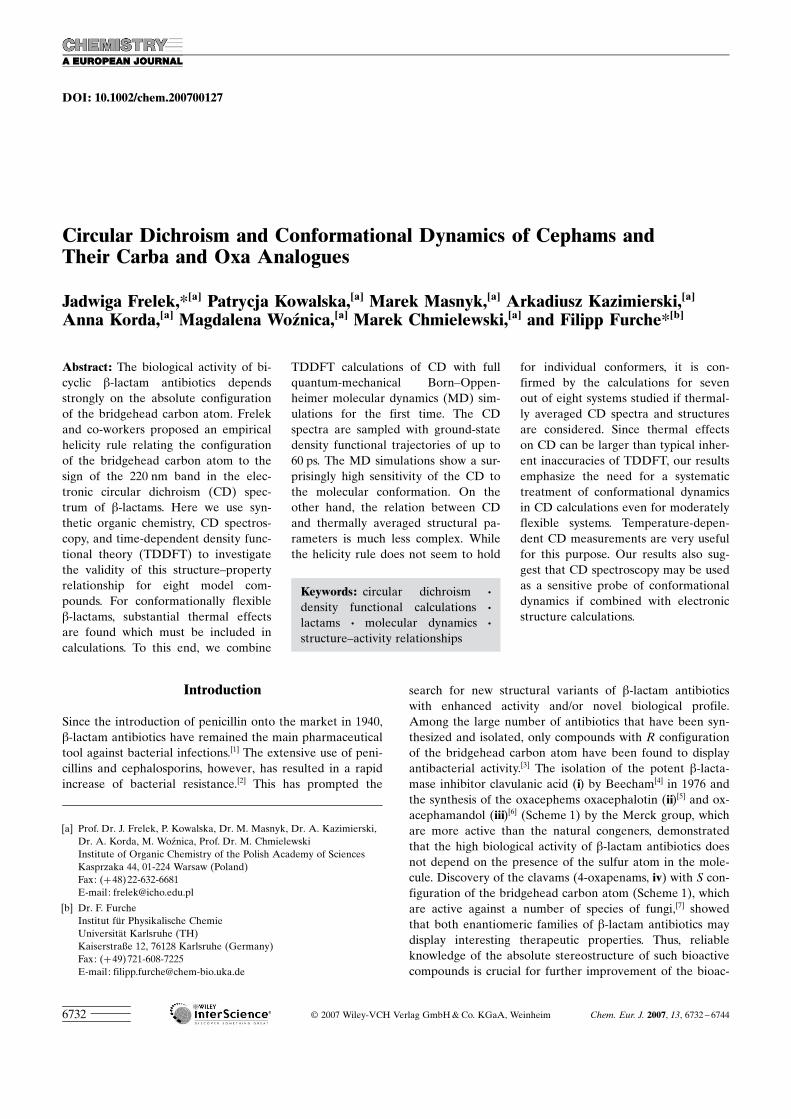
DOI: 10.1002/chem.200700127
Circular Dichroism and Conformational Dynamics of Cephams andTheir Carba and Oxa Analogues
Jadwiga Frelek,*[a] Patrycja Kowalska,[a] Marek Masnyk,[a] Arkadiusz Kazimierski,[a]
Anna Korda,[a] Magdalena Woznica,[a] Marek Chmielewski,[a] and Filipp Furche*[b]
Introduction
Since the introduction of penicillin onto the market in 1940,b-lactam antibiotics have remained the main pharmaceuticaltool against bacterial infections.[1] The extensive use of peni-cillins and cephalosporins, however, has resulted in a rapidincrease of bacterial resistance.[2] This has prompted the
search for new structural variants of b-lactam antibioticswith enhanced activity and/or novel biological profile.Among the large number of antibiotics that have been syn-thesized and isolated, only compounds with R configurationof the bridgehead carbon atom have been found to displayantibacterial activity.[3] The isolation of the potent b-lacta-mase inhibitor clavulanic acid (i) by Beecham[4] in 1976 andthe synthesis of the oxacephems oxacephalotin (ii)[5] and ox-acephamandol (iii)[6] (Scheme 1) by the Merck group, whichare more active than the natural congeners, demonstratedthat the high biological activity of b-lactam antibiotics doesnot depend on the presence of the sulfur atom in the mole-cule. Discovery of the clavams (4-oxapenams, iv) with S con-figuration of the bridgehead carbon atom (Scheme 1), whichare active against a number of species of fungi,[7] showedthat both enantiomeric families of b-lactam antibiotics maydisplay interesting therapeutic properties. Thus, reliableknowledge of the absolute stereostructure of such bioactivecompounds is crucial for further improvement of the bioac-
Abstract: The biological activity of bi-cyclic b-lactam antibiotics dependsstrongly on the absolute configurationof the bridgehead carbon atom. Frelekand co-workers proposed an empiricalhelicity rule relating the configurationof the bridgehead carbon atom to thesign of the 220 nm band in the elec-tronic circular dichroism (CD) spec-trum of b-lactams. Here we use syn-thetic organic chemistry, CD spectros-copy, and time-dependent density func-tional theory (TDDFT) to investigatethe validity of this structure–propertyrelationship for eight model com-pounds. For conformationally flexibleb-lactams, substantial thermal effectsare found which must be included incalculations. To this end, we combine
TDDFT calculations of CD with fullquantum-mechanical Born–Oppen-heimer molecular dynamics (MD) sim-ulations for the first time. The CDspectra are sampled with ground-statedensity functional trajectories of up to60 ps. The MD simulations show a sur-prisingly high sensitivity of the CD tothe molecular conformation. On theother hand, the relation between CDand thermally averaged structural pa-rameters is much less complex. Whilethe helicity rule does not seem to hold
for individual conformers, it is con-firmed by the calculations for sevenout of eight systems studied if thermal-ly averaged CD spectra and structuresare considered. Since thermal effectson CD can be larger than typical inher-ent inaccuracies of TDDFT, our resultsemphasize the need for a systematictreatment of conformational dynamicsin CD calculations even for moderatelyflexible systems. Temperature-depen-dent CD measurements are very usefulfor this purpose. Our results also sug-gest that CD spectroscopy may be usedas a sensitive probe of conformationaldynamics if combined with electronicstructure calculations.
Keywords: circular dichroism ·density functional calculations ·lactams · molecular dynamics ·structure–activity relationships
[a] Prof. Dr. J. Frelek, P. Kowalska, Dr. M. Masnyk, Dr. A. Kazimierski,Dr. A. Korda, M. Woznica, Prof. Dr. M. ChmielewskiInstitute of Organic Chemistry of the Polish Academy of SciencesKasprzaka 44, 01-224 Warsaw (Poland)Fax: (+48)22-632-6681E-mail : [email protected]
[b] Dr. F. FurcheInstitut fAr Physikalische ChemieUniversitCt Karlsruhe (TH)Kaiserstraße 12, 76128 Karlsruhe (Germany)Fax: (+49)721-608-7225E-mail : [email protected]
F 2007 Wiley-VCH Verlag GmbH&Co. KGaA, Weinheim Chem. Eur. J. 2007, 13, 6732 – 67446732
tivity by directed synthetic modifications. Moreover, b-lac-tams are useful synthetic intermediates for a number ofother biologically active heterocycles such as indolizidines,pyrrolizidines, pyrrolidines, pyrroles, taxoids, and macro-lides.[8] A practical method that would allow unequivocal,fast, and reliable determination of the absolute configura-tion of b-lactams and closely related compounds is thereforehighly desirable.
Circular dichroism spectroscopy is a convenient and sensi-tive technique to probe the stereochemistry of azetidinonesand their polycyclic derivatives.[9] To assign the absolute con-figuration by CD spectroscopy, a theoretical model isneeded that correlates the observed spectrum to a three-di-mensional molecular structure. Recently, Frelek and co-workers reported a correlation between the stereostructureof 5-dethia-5-oxacephams and their circular dichroism.[10] Asa result, a simple helicity rule was proposed which assignsthe absolute configuration at the ring junction carbon atomC(6) based on the sign of the CD band at around 220 nm.According to this rule, a positive sign of the 220 nm Cottoneffect (CE) corresponds to an R absolute configuration atthe bridgehead carbon atom, whereas a negative sign of thesame CE is related to a 6S absolute configuration. The rulewas experimentally demonstrated to be correct for a varietyof oxacephams.[10,11] Moreover, it was shown to hold forclavams as oxa analogues of penicillins.[12] However, the hel-icity rule was established empirically on the basis of X-raystructures and the tentative assignment of the electronictransition at approximately 220 nm to an n!p* amide tran-sition in the azetidinone system.[10,12] Despite its appealingsimplicity and its apparent success, the underlying assump-tions of conformational rigidity and electronic decoupling ofthe amide chromophore require further validation.
In the present work, we employ time-dependent densityfunctional calculations to investigate the relation betweenmolecular structure and CD for the set of eight b-lactammodel compounds shown in Scheme 2. These model com-
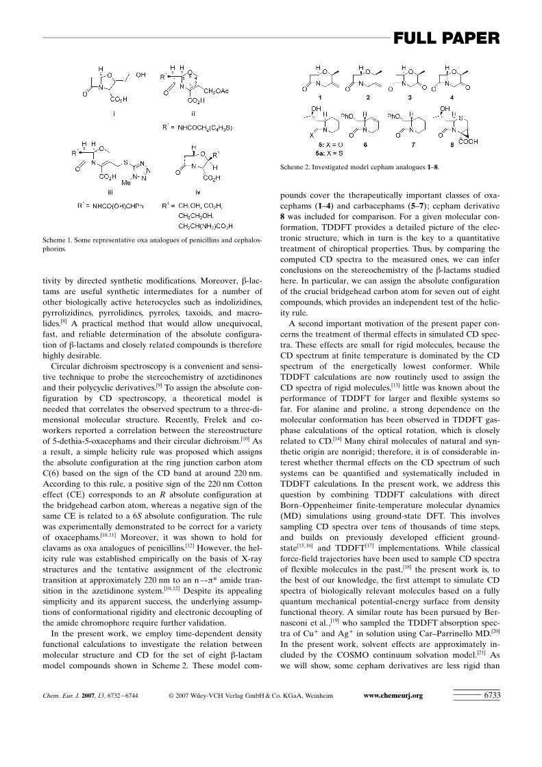
pounds cover the therapeutically important classes of oxa-cephams (1–4) and carbacephams (5–7); cepham derivative8 was included for comparison. For a given molecular con-formation, TDDFT provides a detailed picture of the elec-tronic structure, which in turn is the key to a quantitativetreatment of chiroptical properties. Thus, by comparing thecomputed CD spectra to the measured ones, we can inferconclusions on the stereochemistry of the b-lactams studiedhere. In particular, we can assign the absolute configurationof the crucial bridgehead carbon atom for seven out of eightcompounds, which provides an independent test of the helic-ity rule.
A second important motivation of the present paper con-cerns the treatment of thermal effects in simulated CD spec-tra. These effects are small for rigid molecules, because theCD spectrum at finite temperature is dominated by the CDspectrum of the energetically lowest conformer. WhileTDDFT calculations are now routinely used to assign theCD spectra of rigid molecules,[13] little was known about theperformance of TDDFT for larger and flexible systems sofar. For alanine and proline, a strong dependence on themolecular conformation has been observed in TDDFT gas-phase calculations of the optical rotation, which is closelyrelated to CD.[14] Many chiral molecules of natural and syn-thetic origin are nonrigid; therefore, it is of considerable in-terest whether thermal effects on the CD spectrum of suchsystems can be quantified and systematically included inTDDFT calculations. In the present work, we address thisquestion by combining TDDFT calculations with directBorn–Oppenheimer finite-temperature molecular dynamics(MD) simulations using ground-state DFT. This involvessampling CD spectra over tens of thousands of time steps,and builds on previously developed efficient ground-state[15,16] and TDDFT[17] implementations. While classicalforce-field trajectories have been used to sample CD spectraof flexible molecules in the past,[18] the present work is, tothe best of our knowledge, the first attempt to simulate CDspectra of biologically relevant molecules based on a fullyquantum mechanical potential-energy surface from densityfunctional theory. A similar route has been pursued by Ber-nasconi et al.,[19] who sampled the TDDFT absorption spec-tra of Cu+ and Ag+ in solution using Car–Parrinello MD.[20]
In the present work, solvent effects are approximately in-cluded by the COSMO continuum solvation model.[21] Aswe will show, some cepham derivatives are less rigid than
Scheme 1. Some representative oxa analogues of penicillins and cephalos-phorins.
Scheme 2. Investigated model cepham analogues 1–8.
Chem. Eur. J. 2007, 13, 6732 – 6744 F 2007 Wiley-VCH Verlag GmbH&Co. KGaA, Weinheim www.chemeurj.org 6733
FULL PAPER
expected, and finite temperature can change their CD spec-tra dramatically. Our simulations account for vibronic ef-fects in a classical approximation; the importance of theseeffects for CD[22] and for optical rotations[23] has recentlybeen stressed.
Results and Discussion
General aspects : The CD data of cepham analogues 1–8 arecollected in Table 1. Oxacephams 1 and 3 show a positive
Cotton effect (CE) at around 220 nm, whilst oxacephams 2and 4 have a negative CE in the same spectral region. Ac-cording to the helicity rule, we expect a 6R configuration for1 and 3, and a 6S configuration for 2 and 4.[10,12] This hasbeen confirmed by X-ray structure analysis of oxacephams 2and 3.[24] The presence of the exo-methylene group at C(3)in 1 and 2 does not constitute any extension of the b-lactamchromophoric system. Therefore, the shape of the CD spec-tra of 1 and 2 around 220 nm does not differ in comparisonto the saturated oxacephams described before.[10,12] Howev-er, the exo-methylene group contributes to the overall CDspectrum in the high-energy part of the spectrum, as dis-cussed below. The presence of an additional Cotton effect ataround 313 nm in oxacephams 3 and 4 is due to the carbonylgroup at C(3). Despite the opposite configuration of thebridgehead carbon atom, this band has the same sign inboth compounds.
Compounds 8 and 5 representing cepham and its carbaanalogue, respectively, behave similarly to oxacephams dis-cussed before. In their CD spectra a positive CE at around220 nm indicates a 6R configuration, in agreement with thehelicity rule. In contrast, 6 and 7 with an S configuration atthe bridgehead carbon atom have a negative 220 nm CDband, as suggested by the rule. Thus, it can be stated thatcompounds 5–8 fulfil the requirements of the helicity rule.Therefore, we can expect that the b-lactam unit in thesecompounds is nonplanar and that the sense of chirality ofthe chromophore is controlled by the 6R or 6S absolute con-figuration and is also the sign-determining factor for the220 nm CD band. The X-ray data for cepham 8 indicate theskewness of the b-lactam unit by negative O(9)-C(8)-N(1)-C(2) and O(9)-C(8)-N(1)-C(6) torsion angles of �22.9 and
�177.08, respectively.[25] Therefore, a positive sign of thelong-wavelength CD band is observed in its CD spectrum,in agreement with the helicity rule. The computed torsionangles a ACHTUNGTRENNUNG(9,8,1,6) for compounds 1–8 provide corroboratingevidence for the nonplanarity of the amide chromophore(Table 2). Additionally, we report a ACHTUNGTRENNUNG(8,6,2,1) dihedral anglesto quantify the pyramidalization of the amide nitrogenatom.
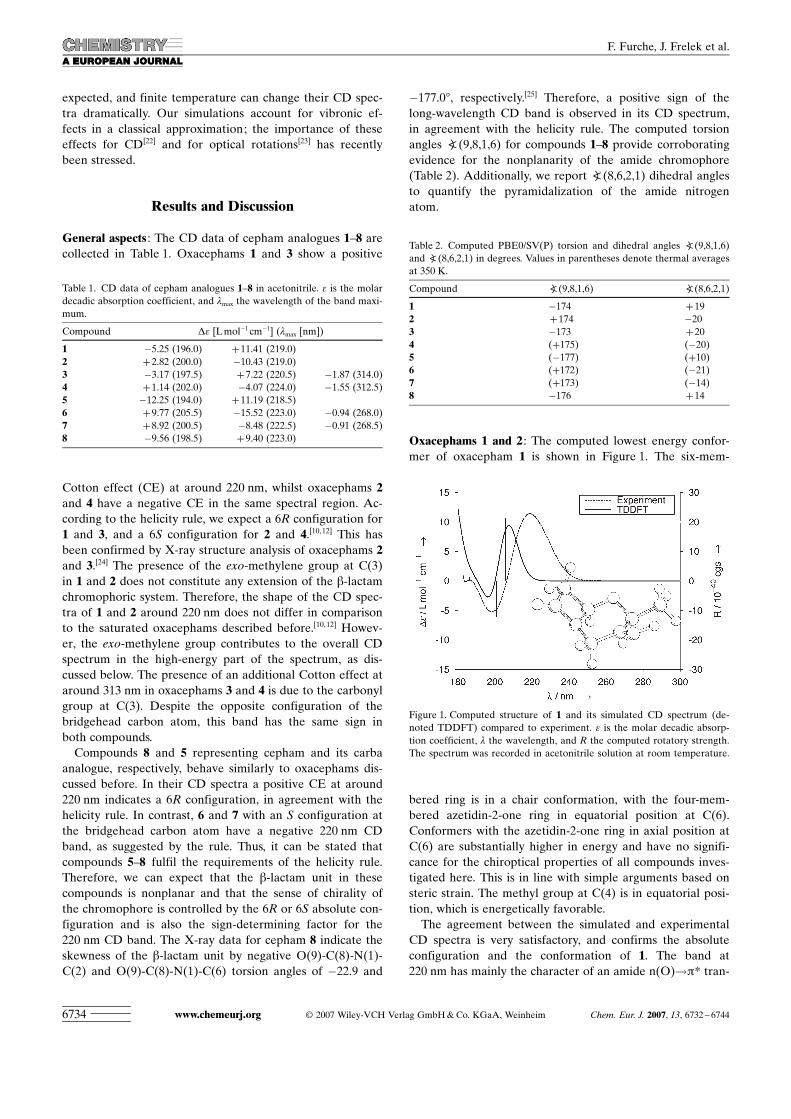
Oxacephams 1 and 2 : The computed lowest energy confor-mer of oxacepham 1 is shown in Figure 1. The six-mem-
bered ring is in a chair conformation, with the four-mem-bered azetidin-2-one ring in equatorial position at C(6).Conformers with the azetidin-2-one ring in axial position atC(6) are substantially higher in energy and have no signifi-cance for the chiroptical properties of all compounds inves-tigated here. This is in line with simple arguments based onsteric strain. The methyl group at C(4) is in equatorial posi-tion, which is energetically favorable.
The agreement between the simulated and experimentalCD spectra is very satisfactory, and confirms the absoluteconfiguration and the conformation of 1. The band at220 nm has mainly the character of an amide n(O)!p* tran-
Table 1. CD data of cepham analogues 1–8 in acetonitrile. e is the molardecadic absorption coefficient, and lmax the wavelength of the band maxi-mum.
Compound De [Lmol�1 cm�1] (lmax [nm])
1 �5.25 (196.0) +11.41 (219.0)2 +2.82 (200.0) �10.43 (219.0)3 �3.17 (197.5) +7.22 (220.5) �1.87 (314.0)4 +1.14 (202.0) �4.07 (224.0) �1.55 (312.5)5 �12.25 (194.0) +11.19 (218.5)6 +9.77 (205.5) �15.52 (223.0) �0.94 (268.0)7 +8.92 (200.5) �8.48 (222.5) �0.91 (268.5)8 �9.56 (198.5) +9.40 (223.0)
Table 2. Computed PBE0/SV(P) torsion and dihedral angles a ACHTUNGTRENNUNG(9,8,1,6)and a ACHTUNGTRENNUNG(8,6,2,1) in degrees. Values in parentheses denote thermal averagesat 350 K.
Compound a ACHTUNGTRENNUNG(9,8,1,6) aACHTUNGTRENNUNG(8,6,2,1)
1 �174 +192 +174 �203 �173 +204 ACHTUNGTRENNUNG(+175) ACHTUNGTRENNUNG(�20)5 ACHTUNGTRENNUNG(�177) ACHTUNGTRENNUNG(+10)6 ACHTUNGTRENNUNG(+172) ACHTUNGTRENNUNG(�21)7 ACHTUNGTRENNUNG(+173) ACHTUNGTRENNUNG(�14)8 �176 +14
Figure 1. Computed structure of 1 and its simulated CD spectrum (de-noted TDDFT) compared to experiment. e is the molar decadic absorp-tion coefficient, l the wavelength, and R the computed rotatory strength.The spectrum was recorded in acetonitrile solution at room temperature.
www.chemeurj.org F 2007 Wiley-VCH Verlag GmbH&Co. KGaA, Weinheim Chem. Eur. J. 2007, 13, 6732 – 67446734
F. Furche, J. Frelek et al.
sition. Its positive sign is predicted by both the helicity ruleand the TDDFT calculations. The calculated band is slightlyblue-shifted, which is not untypical of the PBE0 functionalwe use here; also, thermal effects may contribute to this dis-crepancy (see below). The weaker negative band at about200 nm originates predominantly from a short-range charge-transfer transition out of the nitrogen lone pair into themethylene C�C p* orbital, with some C=C p!p* admix-ture. The intense positive C=C p!p* and amide n(N)!p*transitions are located at the blue end of the spectrum.
The notion of an “amide n(N)!p*” transition requiressome clarification. In an unstrained planar amide, the nitro-gen atom has a planar “sp2” configuration and its lone pairis part of the delocalized p system. However, in the cephamsand oxacephams investigated here, the nitrogen atom ispartly pyramidalized due to steric constraints; compare thecomputed pyramidalization angles in Table 2. As the pyra-midalization angle increases, the nitrogen lone pair decou-ples from the C=O p system and becomes localized at thenitrogen atom. Since the transition from an amide p orbitalto a nitrogen lone pair is gradual, the “amide n(N)!p*”transition can be imagined as gradually evolving from thep!p* transition of planar amide chromophores as the ni-trogen becomes nonplanar.
The lowest energy conformer of oxacepham 2 again ex-hibits a chair conformation of the six-membered ring. Theinverted configuration of C(6) implies that the methyl sub-stituent at C(4) now occupies an axial position. This intrigu-ing result is supported by previous X-ray studies on 2.[24]
The TDDFT calculations predict a negative sign of theamide n(O)!p* transition at 220 nm (see Figure 2), whichconfirms the helicity rule in this case. The sign of the bandsat 200 and 180 nm is inverted, too. Since 1 and 2 are diaste-reomers, this is not a trivial result.
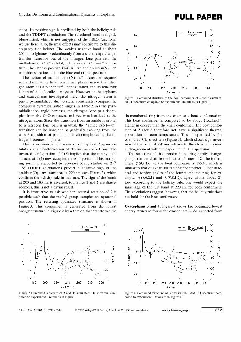
It is instructive to ask whether internal rotation of 2 ispossible such that the methyl group occupies an equatorialposition. The resulting optimized structure is shown inFigure 3. This conformer is generated from the lowestenergy structure in Figure 2 by a torsion that transforms the
six-membered ring from the chair to a boat conformation.This boat conformer is computed to be about 2 kcalmol�1
higher in energy than the chair conformer. The boat confor-mer of 2 should therefore not have a significant thermalpopulation at room temperature. This is supported by thecomputed CD spectrum (Figure 3), which shows sign inver-sion of the band at 220 nm relative to the chair conformer,in disagreement with the experimental CD spectrum.
The structure of the azetidin-2-one ring hardly changesgoing from the chair to the boat conformer of 2. The torsionangle a ACHTUNGTRENNUNG(9,8,1,6) of the boat conformer is 175.68, which issimilar to that of 173.88 for the chair conformer. Other dihe-dral and torsion angles of the four-membered ring, for ex-ample, a ACHTUNGTRENNUNG(8,6,2,1) and a ACHTUNGTRENNUNG(9,8,1,2), agree within about 28,too. According to the helicity rule, one would expect thesame sign of the CD band at 220 nm for both conformers.The calculations suggest, however, that the helicity rule doesnot hold for the boat conformer.
Oxacephams 3 and 4 : Figure 4 shows the optimized lowestenergy structure found for oxacepham 3. As expected from
Figure 2. Computed structure of 2 and its simulated CD spectrum com-pared to experiment. Details as in Figure 1.
Figure 3. Computed structure of the boat conformer of 2 and its simulat-ed CD spectrum compared to experiment. Details as in Figure 1.
Figure 4. Computed structure of 3 and its simulated CD spectrum com-pared to experiment. Details as in Figure 1.
Chem. Eur. J. 2007, 13, 6732 – 6744 F 2007 Wiley-VCH Verlag GmbH&Co. KGaA, Weinheim www.chemeurj.org 6735
FULL PAPERCircular Dichroism and Conformational Dynamics of Cephams
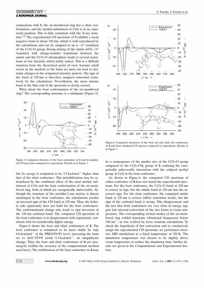
comparison with 1, the six-membered ring has a chair con-formation, and the methyl substituent at C(6) is in an equa-torial position. This is fully consistent with the X-ray struc-ture.[24] The experimental CD spectrum of 3 exhibits a weaknegative band at about 320 nm, which is well reproduced bythe calculations and can be assigned to an n!p* transitionof the C(3)=O group. Strong mixing of the amide n(O)!p*transition with charge-transfer transitions between theamide and the C(3)=O chromophore leads to several transi-tions of low intensity which partly cancel. This is a difficultsituation from the theoretical point of view, because smallerrors in the method or the basis set used can lead to dra-matic changes in the computed intensity pattern. The sign ofthe band at 220 nm is therefore assigned somewhat tenta-tively by the calculations. Nevertheless, the more intenseband at the blue end of the spectrum is clearly correct.
What about the boat conformation of the six-memberedring? The corresponding structure is a minimum (Figure 5),
but its energy is computed to be 3.3 kcalmol�1 higher thanthat of the chair conformer. This destabilization may be ra-tionalized by the combined effect of the axial methyl sub-stituent at C(4) and the boat conformation of the six-mem-bered ring, both of which are energetically unfavorable. Al-though the structure of the azetidin-2-one moiety is almostunchanged in the boat conformer, the calculations predictan inversed sign of the CD band at 220 nm. Thus, the helici-ty rule apparently does not hold for the boat conformers.The conformational change also leads to sign inversion ofthe 320 nm carbonyl band. The computed CD spectrum ofthe boat conformer is in disagreement with experiment, con-sistent with its considerably higher energy.
Figure 6 shows the boat and chair conformers of 4. Theboat conformer is computed to be more stable by only0.6 kcalmol�1 at the PBE0/SV(P) level; increasing the basisset to def2-TZVP yields 0.3 kcalmol�1, an insignificantchange. Thus, the boat and chair conformers of 4 are isoe-nergetic (within the accuracy of the computational methodused here). The stabilization of the boat conformer in 4 may
be a consequence of the smaller size of the C(3)=O groupcompared to the C(3)=CH2 group of 2, reducing the ener-getically unfavorable interaction with the eclipsed methylgroup at C(4) in the boat conformer.
As shown in Figure 6, the computed CD spectrum ofeither conformer of 4 does not match the experimental spec-trum. For the boat conformer, the C(3)=O band at 320 nmis correct in sign, but the amide band at 220 nm has the in-correct sign. For the chair conformer, the computed amideband at 220 nm is correct (albeit somewhat weak), but thesign of the carbonyl band is wrong. This disagreement andthe fact that both conformers are very close in energy sug-gest fast internal conversion of the two forms at room tem-perature. The corresponding torsion modes of the six-mem-bered ring exhibit harmonic vibrational frequencies below100 cm�1, as was verified by force constant calculations. Tocheck the hypothesis of fast conversion and to conclusivelyassign the experimental CD spectrum, we performed exten-sive MD simulations at a fixed temperature of 350 K. Thesimulation temperature was chosen to be slightly aboveroom temperature to reduce the simulation time; further de-tails are given in the Computational and Experimental Sec-tion.
Figure 5. Computed structure of the boat conformer of 3 and its simulat-ed CD spectrum compared to experiment. Details as in Figure 1.
Figure 6. Computed structures of the boat (a) and chair (b) conformersof 4 and their simulated CD spectra compared to experiment. Details asin Figure 1.
www.chemeurj.org F 2007 Wiley-VCH Verlag GmbH&Co. KGaA, Weinheim Chem. Eur. J. 2007, 13, 6732 – 67446736
F. Furche, J. Frelek et al.
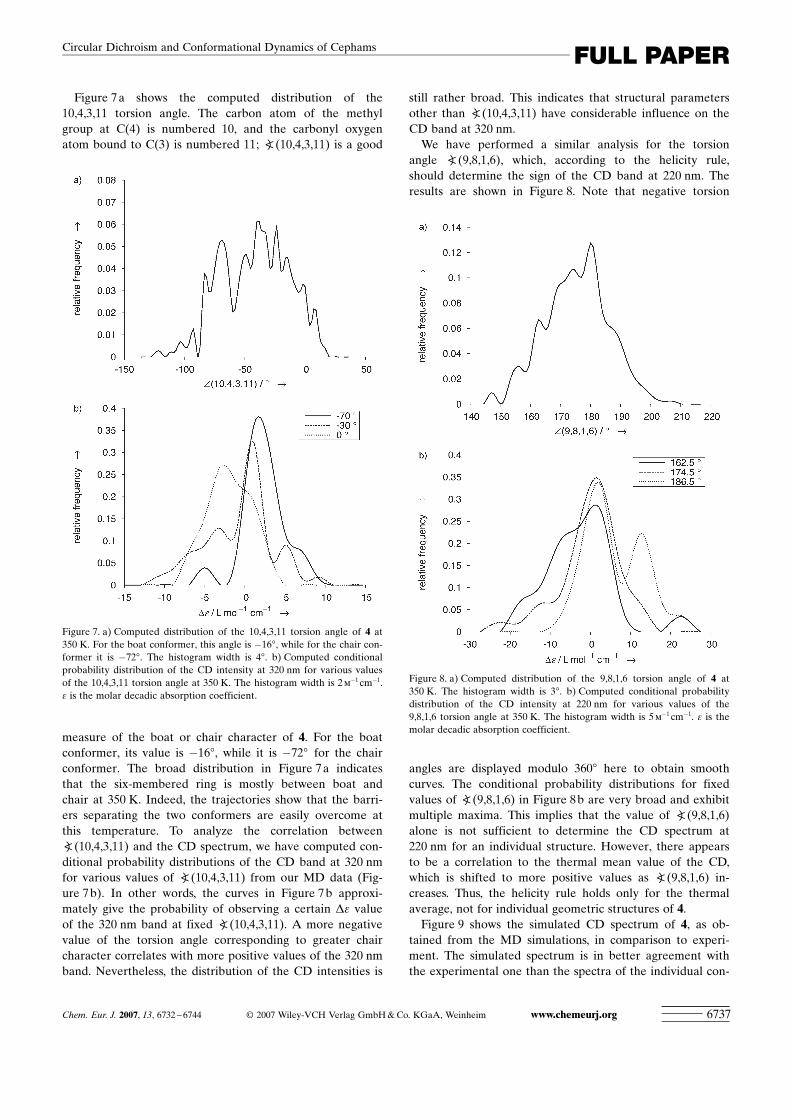
Figure 7a shows the computed distribution of the10,4,3,11 torsion angle. The carbon atom of the methylgroup at C(4) is numbered 10, and the carbonyl oxygenatom bound to C(3) is numbered 11; a(10,4,3,11) is a good
measure of the boat or chair character of 4. For the boatconformer, its value is �168, while it is �728 for the chairconformer. The broad distribution in Figure 7a indicatesthat the six-membered ring is mostly between boat andchair at 350 K. Indeed, the trajectories show that the barri-ers separating the two conformers are easily overcome atthis temperature. To analyze the correlation betweena(10,4,3,11) and the CD spectrum, we have computed con-ditional probability distributions of the CD band at 320 nmfor various values of a(10,4,3,11) from our MD data (Fig-ure 7b). In other words, the curves in Figure 7b approxi-mately give the probability of observing a certain De valueof the 320 nm band at fixed a(10,4,3,11). A more negativevalue of the torsion angle corresponding to greater chaircharacter correlates with more positive values of the 320 nmband. Nevertheless, the distribution of the CD intensities is
still rather broad. This indicates that structural parametersother than a(10,4,3,11) have considerable influence on theCD band at 320 nm.
We have performed a similar analysis for the torsionangle a ACHTUNGTRENNUNG(9,8,1,6), which, according to the helicity rule,should determine the sign of the CD band at 220 nm. Theresults are shown in Figure 8. Note that negative torsion
angles are displayed modulo 3608 here to obtain smoothcurves. The conditional probability distributions for fixedvalues of a ACHTUNGTRENNUNG(9,8,1,6) in Figure 8b are very broad and exhibitmultiple maxima. This implies that the value of a ACHTUNGTRENNUNG(9,8,1,6)alone is not sufficient to determine the CD spectrum at220 nm for an individual structure. However, there appearsto be a correlation to the thermal mean value of the CD,which is shifted to more positive values as a ACHTUNGTRENNUNG(9,8,1,6) in-creases. Thus, the helicity rule holds only for the thermalaverage, not for individual geometric structures of 4.
Figure 9 shows the simulated CD spectrum of 4, as ob-tained from the MD simulations, in comparison to experi-ment. The simulated spectrum is in better agreement withthe experimental one than the spectra of the individual con-
Figure 7. a) Computed distribution of the 10,4,3,11 torsion angle of 4 at350 K. For the boat conformer, this angle is �168, while for the chair con-former it is �728. The histogram width is 48. b) Computed conditionalprobability distribution of the CD intensity at 320 nm for various valuesof the 10,4,3,11 torsion angle at 350 K. The histogram width is 2m�1 cm�1.e is the molar decadic absorption coefficient.
Figure 8. a) Computed distribution of the 9,8,1,6 torsion angle of 4 at350 K. The histogram width is 38. b) Computed conditional probabilitydistribution of the CD intensity at 220 nm for various values of the9,8,1,6 torsion angle at 350 K. The histogram width is 5m�1 cm�1. e is themolar decadic absorption coefficient.
Chem. Eur. J. 2007, 13, 6732 – 6744 F 2007 Wiley-VCH Verlag GmbH&Co. KGaA, Weinheim www.chemeurj.org 6737
FULL PAPERCircular Dichroism and Conformational Dynamics of Cephams

formers, although the computed spectrum is red-shifted by0.45 eV. We expect a red shift compared to the spectrum at0 K, because thermal motion increases average bondlengths, which generally lowers excitation energies. The signof the band at 220 nm is correct, as is the shape of thewhole spectrum in the 180–250 nm region. The band at320 nm is less satisfactory; the sign is slightly positivearound 300 nm, in contrast to the experimental result. In-spection of the MD data shows that the band at 320 nm re-sults from cancellation of large individual contributions ofopposite sign, and its absolute value is only slightly abovethe statistical uncertainty. In addition, the sign of the 320 nmband depends sensitively on the details of the potential-energy surface. Our present methodology might not be accu-rate enough to fully account for all these effects.
Carbacepham 5 : The lowest energy conformer of carbacep-ham 5 is predicted to have a chair conformation of the six-membered ring by our calculations (Figure 10), in agree-ment with simple steric-strain arguments. The only chromo-
phore of 5 that absorbs above 180 nm is the amide group ofthe azetidin-2-one ring. It is thus surprising that the comput-ed CD spectrum of the lowest energy conformer of 5 haslittle resemblance to the measured CD spectrum, as is seenin Figure 10: the amide n(O)!p* band is computed with aconsiderable blue shift; the amide n(N)!p* band at the leftend of the spectrum is also blue-shifted and even has thewrong sign.
We performed MD simulations of 5 at 350 K to investi-gate the impact of thermal effects on the CD spectrum. Incontrast to oxacepham 3, no competing low-energy structurewas observed in the simulations. However, the saturated six-membered ring system displayed considerable flexibility.This is illustrated in Figure 11 for the torsion angle a-
ACHTUNGTRENNUNG(8,1,2,3). The distribution is broad and unsymmetrical, witha maximum at about �1208, which differs from the value of�1158 at 0 K. Again, the CD spectrum was found to dependsensitively on the conformation of both the four- and six-membered rings, with sign inversions occurring for bothbands at various points of the MD simulation. The simulat-ed CD spectrum at 350 K compares much better with ex-periment than that at 0 K (see Figure 10). Both bands arebroadened and red-shifted compared to the spectrum at 0 Kand have the correct sign.
To provide further evidence for the large thermal effectspredicted by the calculations, temperature-dependent meas-urements of the CD spectrum of 5 were attempted. Howev-er, due to the cutoff limits of EPA (diethyl ether/isopentane/ethanol 5/5/2 by vol), a solvent of choice for temperature-dependent CD measurements and solubility issues of 5, thespectrum of the thioamide of 5 (5a, see Experimental Sec-tion) had to be used instead of the parent compound. Whenthe temperature was lowered to 253 K, the recorded spectrashowed an anomalous intensity decrease for the counterpartof the 220 nm CD band (occurring at 339 nm in the spec-trum of 5a), and a strong suppression of the counterpart ofthe 200 nm CD band (occurring at 272 nm in the spectrumof 5a). Both observations support the present calculations,
Figure 9. Simulated CD spectrum of 4 at 350 K compared to experiment.The curve denoted (s) was blue-shifted by 0.45 eV. Details as in Figure 1.
Figure 10. Computed structure of 5 and its simulated CD spectrum at 0 Kand 350 K compared to experiment. The 0 K curve corresponds to theconformer shown, while the 350 K curve is the result of MD simulation.Details as in Figure 1.
Figure 11. Computed probability distribution of the 8,1,2,3 torsion angleof 5 at 350 K. The histogram width is 48.
www.chemeurj.org F 2007 Wiley-VCH Verlag GmbH&Co. KGaA, Weinheim Chem. Eur. J. 2007, 13, 6732 – 67446738
F. Furche, J. Frelek et al.
because one expects an increasing population of the lowestenergy conformer at lower temperature, and thus an increas-ing similarity of the experimental CD spectrum to the com-puted spectrum at 0 K in Figure 10. The MD simulationssuggest that considerably lower temperatures than 253 K arerequired to fully “freeze out” the lowest energy conformershown in Figure 10.
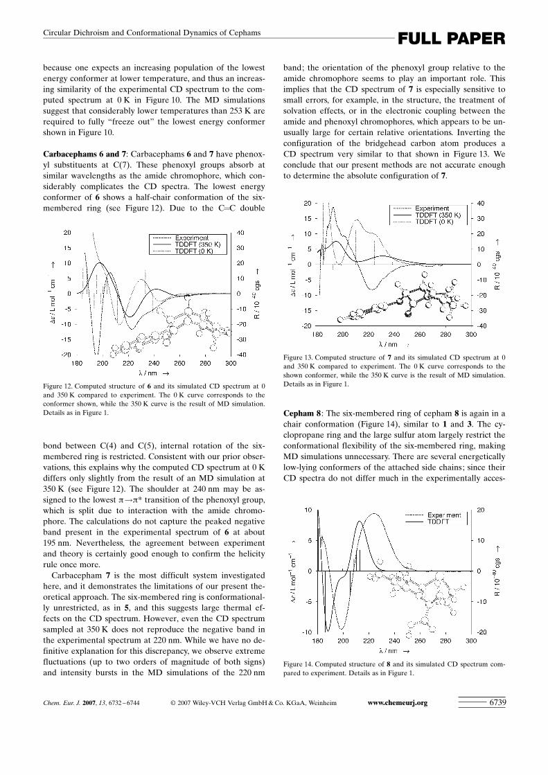
Carbacephams 6 and 7: Carbacephams 6 and 7 have phenox-yl substituents at C(7). These phenoxyl groups absorb atsimilar wavelengths as the amide chromophore, which con-siderably complicates the CD spectra. The lowest energyconformer of 6 shows a half-chair conformation of the six-membered ring (see Figure 12). Due to the C=C double
bond between C(4) and C(5), internal rotation of the six-membered ring is restricted. Consistent with our prior obser-vations, this explains why the computed CD spectrum at 0 Kdiffers only slightly from the result of an MD simulation at350 K (see Figure 12). The shoulder at 240 nm may be as-signed to the lowest p!p* transition of the phenoxyl group,which is split due to interaction with the amide chromo-phore. The calculations do not capture the peaked negativeband present in the experimental spectrum of 6 at about195 nm. Nevertheless, the agreement between experimentand theory is certainly good enough to confirm the helicityrule once more.
Carbacepham 7 is the most difficult system investigatedhere, and it demonstrates the limitations of our present the-oretical approach. The six-membered ring is conformational-ly unrestricted, as in 5, and this suggests large thermal ef-fects on the CD spectrum. However, even the CD spectrumsampled at 350 K does not reproduce the negative band inthe experimental spectrum at 220 nm. While we have no de-finitive explanation for this discrepancy, we observe extremefluctuations (up to two orders of magnitude of both signs)and intensity bursts in the MD simulations of the 220 nm
band; the orientation of the phenoxyl group relative to theamide chromophore seems to play an important role. Thisimplies that the CD spectrum of 7 is especially sensitive tosmall errors, for example, in the structure, the treatment ofsolvation effects, or in the electronic coupling between theamide and phenoxyl chromophores, which appears to be un-usually large for certain relative orientations. Inverting theconfiguration of the bridgehead carbon atom produces aCD spectrum very similar to that shown in Figure 13. Weconclude that our present methods are not accurate enoughto determine the absolute configuration of 7.
Cepham 8 : The six-membered ring of cepham 8 is again in achair conformation (Figure 14), similar to 1 and 3. The cy-clopropane ring and the large sulfur atom largely restrict theconformational flexibility of the six-membered ring, makingMD simulations unnecessary. There are several energeticallylow-lying conformers of the attached side chains; since theirCD spectra do not differ much in the experimentally acces-
Figure 12. Computed structure of 6 and its simulated CD spectrum at 0and 350 K compared to experiment. The 0 K curve corresponds to theconformer shown, while the 350 K curve is the result of MD simulation.Details as in Figure 1.
Figure 13. Computed structure of 7 and its simulated CD spectrum at 0and 350 K compared to experiment. The 0 K curve corresponds to theshown conformer, while the 350 K curve is the result of MD simulation.Details as in Figure 1.
Figure 14. Computed structure of 8 and its simulated CD spectrum com-pared to experiment. Details as in Figure 1.
Chem. Eur. J. 2007, 13, 6732 – 6744 F 2007 Wiley-VCH Verlag GmbH&Co. KGaA, Weinheim www.chemeurj.org 6739
FULL PAPERCircular Dichroism and Conformational Dynamics of Cephams
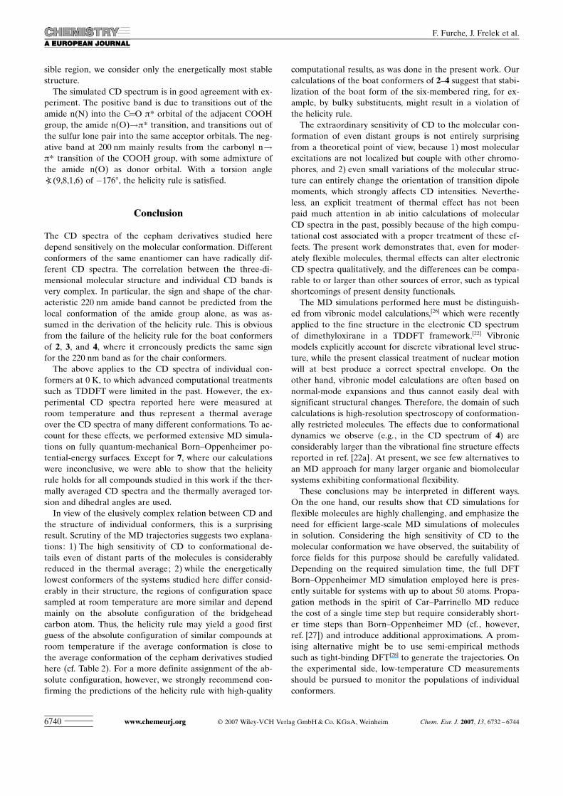
sible region, we consider only the energetically most stablestructure.
The simulated CD spectrum is in good agreement with ex-periment. The positive band is due to transitions out of theamide n(N) into the C=O p* orbital of the adjacent COOHgroup, the amide n(O)!p* transition, and transitions out ofthe sulfur lone pair into the same acceptor orbitals. The neg-ative band at 200 nm mainly results from the carbonyl n!p* transition of the COOH group, with some admixture ofthe amide n(O) as donor orbital. With a torsion anglea ACHTUNGTRENNUNG(9,8,1,6) of �1768, the helicity rule is satisfied.
Conclusion
The CD spectra of the cepham derivatives studied heredepend sensitively on the molecular conformation. Differentconformers of the same enantiomer can have radically dif-ferent CD spectra. The correlation between the three-di-mensional molecular structure and individual CD bands isvery complex. In particular, the sign and shape of the char-acteristic 220 nm amide band cannot be predicted from thelocal conformation of the amide group alone, as was as-sumed in the derivation of the helicity rule. This is obviousfrom the failure of the helicity rule for the boat conformersof 2, 3, and 4, where it erroneously predicts the same signfor the 220 nm band as for the chair conformers.
The above applies to the CD spectra of individual con-formers at 0 K, to which advanced computational treatmentssuch as TDDFT were limited in the past. However, the ex-perimental CD spectra reported here were measured atroom temperature and thus represent a thermal averageover the CD spectra of many different conformations. To ac-count for these effects, we performed extensive MD simula-tions on fully quantum-mechanical Born–Oppenheimer po-tential-energy surfaces. Except for 7, where our calculationswere inconclusive, we were able to show that the helicityrule holds for all compounds studied in this work if the ther-mally averaged CD spectra and the thermally averaged tor-sion and dihedral angles are used.
In view of the elusively complex relation between CD andthe structure of individual conformers, this is a surprisingresult. Scrutiny of the MD trajectories suggests two explana-tions: 1) The high sensitivity of CD to conformational de-tails even of distant parts of the molecules is considerablyreduced in the thermal average; 2) while the energeticallylowest conformers of the systems studied here differ consid-erably in their structure, the regions of configuration spacesampled at room temperature are more similar and dependmainly on the absolute configuration of the bridgeheadcarbon atom. Thus, the helicity rule may yield a good firstguess of the absolute configuration of similar compounds atroom temperature if the average conformation is close tothe average conformation of the cepham derivatives studiedhere (cf. Table 2). For a more definite assignment of the ab-solute configuration, however, we strongly recommend con-firming the predictions of the helicity rule with high-quality
computational results, as was done in the present work. Ourcalculations of the boat conformers of 2–4 suggest that stabi-lization of the boat form of the six-membered ring, for ex-ample, by bulky substituents, might result in a violation ofthe helicity rule.
The extraordinary sensitivity of CD to the molecular con-formation of even distant groups is not entirely surprisingfrom a theoretical point of view, because 1) most molecularexcitations are not localized but couple with other chromo-phores, and 2) even small variations of the molecular struc-ture can entirely change the orientation of transition dipolemoments, which strongly affects CD intensities. Neverthe-less, an explicit treatment of thermal effect has not beenpaid much attention in ab initio calculations of molecularCD spectra in the past, possibly because of the high compu-tational cost associated with a proper treatment of these ef-fects. The present work demonstrates that, even for moder-ately flexible molecules, thermal effects can alter electronicCD spectra qualitatively, and the differences can be compa-rable to or larger than other sources of error, such as typicalshortcomings of present density functionals.
The MD simulations performed here must be distinguish-ed from vibronic model calculations,[26] which were recentlyapplied to the fine structure in the electronic CD spectrumof dimethyloxirane in a TDDFT framework.[22] Vibronicmodels explicitly account for discrete vibrational level struc-ture, while the present classical treatment of nuclear motionwill at best produce a correct spectral envelope. On theother hand, vibronic model calculations are often based onnormal-mode expansions and thus cannot easily deal withsignificant structural changes. Therefore, the domain of suchcalculations is high-resolution spectroscopy of conformation-ally restricted molecules. The effects due to conformationaldynamics we observe (e.g., in the CD spectrum of 4) areconsiderably larger than the vibrational fine structure effectsreported in ref. [22a]. At present, we see few alternatives toan MD approach for many larger organic and biomolecularsystems exhibiting conformational flexibility.
These conclusions may be interpreted in different ways.On the one hand, our results show that CD simulations forflexible molecules are highly challenging, and emphasize theneed for efficient large-scale MD simulations of moleculesin solution. Considering the high sensitivity of CD to themolecular conformation we have observed, the suitability offorce fields for this purpose should be carefully validated.Depending on the required simulation time, the full DFTBorn–Oppenheimer MD simulation employed here is pres-ently suitable for systems with up to about 50 atoms. Propa-gation methods in the spirit of Car–Parrinello MD reducethe cost of a single time step but require considerably short-er time steps than Born–Oppenheimer MD (cf., however,ref. [27]) and introduce additional approximations. A prom-ising alternative might be to use semi-empirical methodssuch as tight-binding DFT[28] to generate the trajectories. Onthe experimental side, low-temperature CD measurementsshould be pursued to monitor the populations of individualconformers.
www.chemeurj.org F 2007 Wiley-VCH Verlag GmbH&Co. KGaA, Weinheim Chem. Eur. J. 2007, 13, 6732 – 67446740
F. Furche, J. Frelek et al.
On the other hand, with electronic structure calculationsenabling us to read and understand the complex informationcontained in electronic CD spectra, CD spectroscopy maybecome a powerful tool to investigate details of the molecu-lar structure. The carbonyl group in the oxacephams 3 and 4exemplifies how spectator chromophores may be used assensitive “chirality probes” of the molecular conformation.Such chirality probes may eventually be useful for studies ofthe interaction of b-lactam antibiotics and penicillin-bindingproteins or b-lactamases under physiological conditions.
Computational and Experimental Section
Ground-state DFT and TDDFT calculations : Ground-state structureswere optimized by using a split-valence basis set with a set of polarizationfunctions at each non-hydrogen atom [SV(P)].[29] SV(P) energetics werevalidated with larger triple-zeta valence basis sets with two sets of polari-zation functions at each non-hydrogen atom (def2-TZVP).[30] Fine quad-rature grids (size m4[15,31]) were employed. Energies were converged to1 mhartree and gradients were converged to a maximum norm of 10�3 a.u.in the geometry optimizations. All stationary points were confirmed tobe minima by force-constant calculations. In the TDDFT calculations,the number of excitations included was chosen to cover the entire rangeof the experimental spectra (180–400 nm). SV(P) basis sets were used,since exploratory calculations in larger basis sets showed a moderate in-crease in the CD intensities on diffuse augmentation, and a slight overallshift to longer wavelengths, but no qualitative change. CD spectra weresimulated by superposition of Gaussians with a uniform line width of0.16 eV. Solvent effects were taken into account by the COSMOmodel[21] using the dielectric constant of acetonitrile (37.5). The fast re-sponse of the screening potential was neglected in the TDDFT responsecalculations. The hybrid density functional of Perdew, Burke, and Ernzer-hof (PBE0)[32] was used throughout, unless stated otherwise. Containing25% of Hartree–Fock exchange, PBE0 is more robust for higher excita-tion energies[33] and less susceptible to charge-transfer error than otherfunctionals. All calculations were carried out with the TURBOMOLEprogram suite.[34]
Molecular dynamics simulations : Finite temperature effects on the com-puted CD spectra were taken into account by fully quantum-mechanicalBorn–Oppenheimer MD simulations. In these simulations, the ensembleaverage of an observable A (e.g., a bond angle or the CD intensity) is re-placed by its time average by virtue of the ergodic theorem.[35] The timeaverage is computed by propagating the system on the Born–Oppen-heimer ground-state potential-energysurface by using a classical descrip-tion of the nuclei. To this end, wesolve the classical equations ofmotion of the nuclei in finite timesteps (“MD steps”) t by means of theVerlet leapfrog algorithm,[36] as avail-able in the FROG module[37] of TUR-BOMOLE. To simulate the canonicalensemble, the leapfrog algorithm wascombined with canonical NosT–Hoover dynamics[38] for the presentwork. The thermostat relaxation timewas chosen to be four times as largeas the time step (see below).
The trajectories were generated asfollows: Initial configurations weregenerated from an 800 K trajectory.Starting from these initial configura-tions, 22 trajectories were computedat 350 K. Each trajectory comprised2200 time steps of step length t=
25 a.u. (0.6 fs), corresponding to a total simulation time of 29.3 ps. Forthe sake of efficiency, energies and forces were evaluated with the BP86functional[39] in combination with the RI-J approximation[31,40] and a smallquadrature grid (size 1[15]). Relative conformational energies are ratherinsensitive to these changes; for example, the energy difference of thechair and boat conformers of 4 changed by about 0.5 kcalmol�1. The en-semble average of A was computed according to Equation (1):
hAi ¼ 1N
XN
i¼1
Ai ð1Þ
where N is the number of sampling steps. After an initial equilibrationphase of 300 steps, every hundredth step was used to sample structuralparameters and CD spectra, corresponding to N=440 in total. The CDspectrum was calculated for each of these 440 configurations as describedabove.
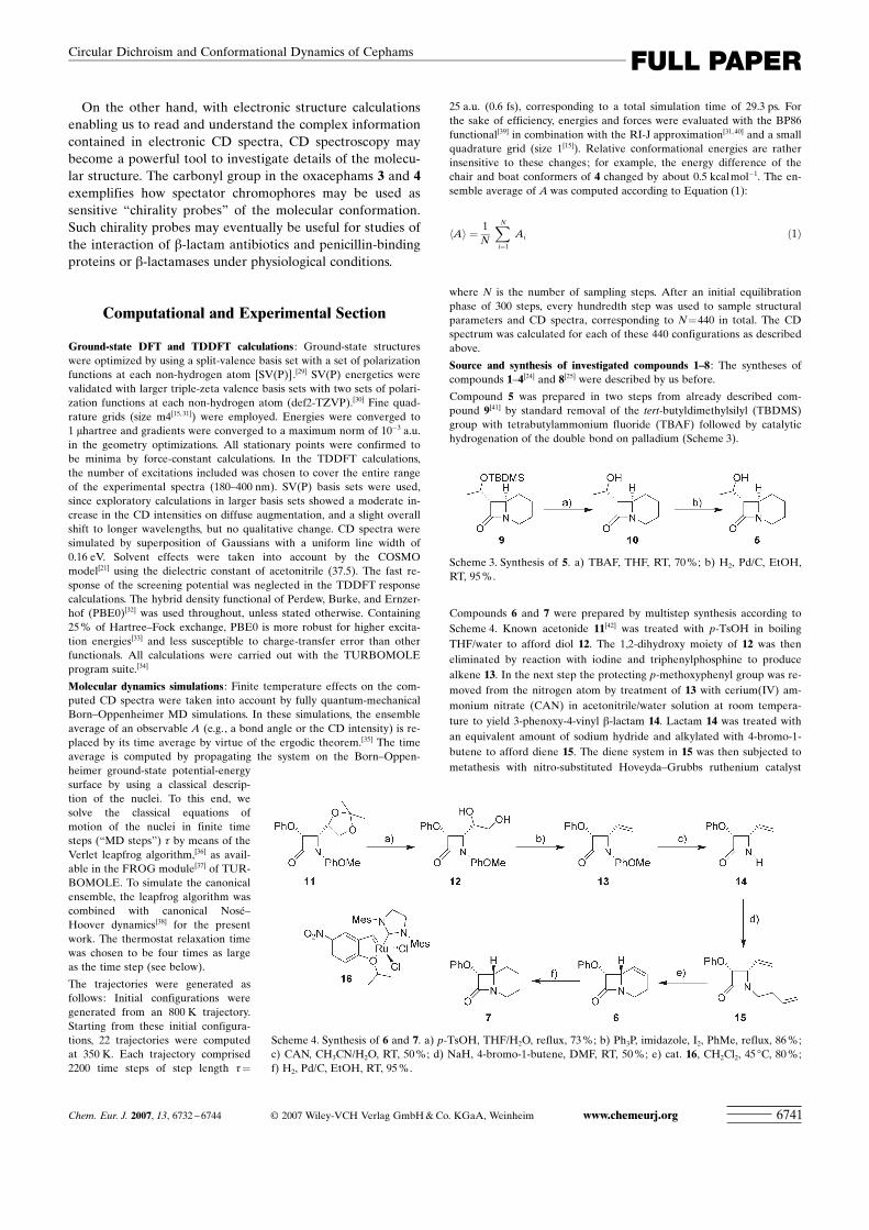
Source and synthesis of investigated compounds 1–8 : The syntheses ofcompounds 1–4[24] and 8[25] were described by us before.
Compound 5 was prepared in two steps from already described com-pound 9[41] by standard removal of the tert-butyldimethylsilyl (TBDMS)group with tetrabutylammonium fluoride (TBAF) followed by catalytichydrogenation of the double bond on palladium (Scheme 3).
Compounds 6 and 7 were prepared by multistep synthesis according toScheme 4. Known acetonide 11[42] was treated with p-TsOH in boilingTHF/water to afford diol 12. The 1,2-dihydroxy moiety of 12 was theneliminated by reaction with iodine and triphenylphosphine to producealkene 13. In the next step the protecting p-methoxyphenyl group was re-moved from the nitrogen atom by treatment of 13 with cerium(IV) am-monium nitrate (CAN) in acetonitrile/water solution at room tempera-ture to yield 3-phenoxy-4-vinyl b-lactam 14. Lactam 14 was treated withan equivalent amount of sodium hydride and alkylated with 4-bromo-1-butene to afford diene 15. The diene system in 15 was then subjected tometathesis with nitro-substituted Hoveyda–Grubbs ruthenium catalyst
Scheme 3. Synthesis of 5. a) TBAF, THF, RT, 70%; b) H2, Pd/C, EtOH,RT, 95%.
Scheme 4. Synthesis of 6 and 7. a) p-TsOH, THF/H2O, reflux, 73%; b) Ph3P, imidazole, I2, PhMe, reflux, 86%;c) CAN, CH3CN/H2O, RT, 50%; d) NaH, 4-bromo-1-butene, DMF, RT, 50%; e) cat. 16, CH2Cl2, 45 8C, 80%;f) H2, Pd/C, EtOH, RT, 95%.
Chem. Eur. J. 2007, 13, 6732 – 6744 F 2007 Wiley-VCH Verlag GmbH&Co. KGaA, Weinheim www.chemeurj.org 6741
FULL PAPERCircular Dichroism and Conformational Dynamics of Cephams
16, recently introduced to synthetic practice,[43] to produce target com-pound 6. Target compound 7 was obtained by catalytic hydrogenation of6 on palladium in ethanol solution at room temperature under standardpressure.
Compound 5a was prepared from 9 by hydrogenation of a double bondon palladium, replacement of the carbonyl oxygen atom with a sulfuratom by reaction with LawessonUs reagent, and removal of the TBDMSgroup by reaction with TBAF (Scheme 5).
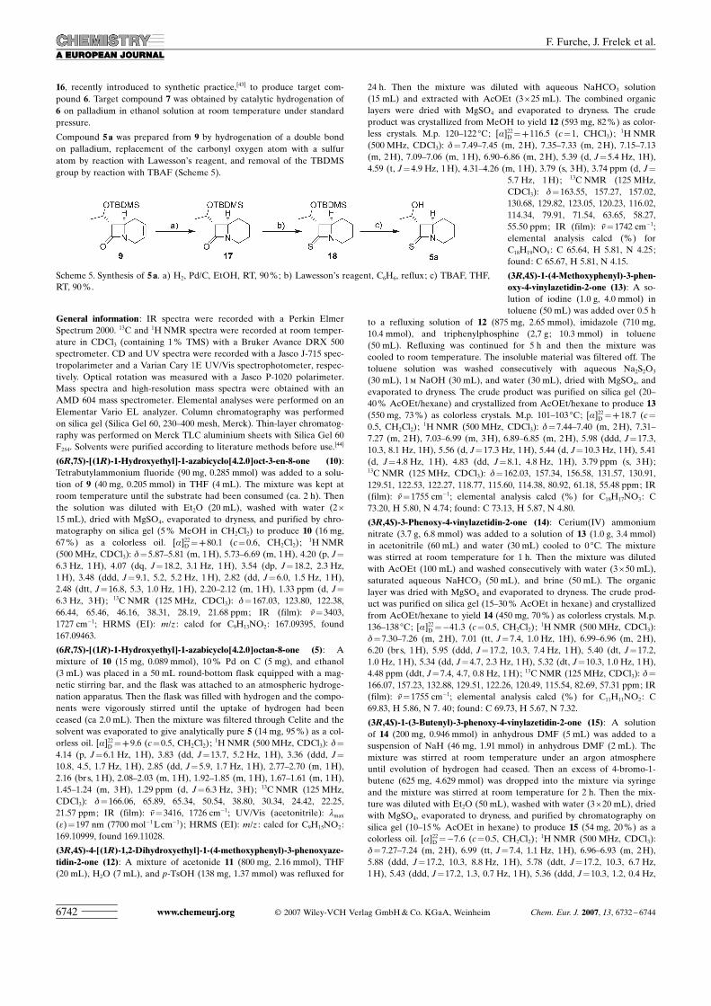
General information : IR spectra were recorded with a Perkin ElmerSpectrum 2000. 13C and 1H NMR spectra were recorded at room temper-ature in CDCl3 (containing 1% TMS) with a Bruker Avance DRX 500spectrometer. CD and UV spectra were recorded with a Jasco J-715 spec-tropolarimeter and a Varian Cary 1E UV/Vis spectrophotometer, respec-tively. Optical rotation was measured with a Jasco P-1020 polarimeter.Mass spectra and high-resolution mass spectra were obtained with anAMD 604 mass spectrometer. Elemental analyses were performed on anElementar Vario EL analyzer. Column chromatography was performedon silica gel (Silica Gel 60, 230–400 mesh, Merck). Thin-layer chromatog-raphy was performed on Merck TLC aluminium sheets with Silica Gel 60F254. Solvents were purified according to literature methods before use.[44]
ACHTUNGTRENNUNG(6R,7S)-[(1R)-1-Hydroxyethyl]-1-azabicyclo ACHTUNGTRENNUNG[4.2.0]oct-3-en-8-one (10):Tetrabutylammonium fluoride (90 mg, 0.285 mmol) was added to a solu-tion of 9 (40 mg, 0.205 mmol) in THF (4 mL). The mixture was kept atroom temperature until the substrate had been consumed (ca. 2 h). Thenthe solution was diluted with Et2O (20 mL), washed with water (2V15 mL), dried with MgSO4, evaporated to dryness, and purified by chro-matography on silica gel (5% MeOH in CH2Cl2) to produce 10 (16 mg,67%) as a colorless oil. [a]22D =++80.1 (c=0.6, CH2Cl2);
1H NMR(500 MHz, CDCl3): d=5.87–5.81 (m, 1H), 5.73–6.69 (m, 1H), 4.20 (p, J=6.3 Hz, 1H), 4.07 (dq, J=18.2, 3.1 Hz, 1H), 3.54 (dp, J=18.2, 2.3 Hz,1H), 3.48 (ddd, J=9.1, 5.2, 5.2 Hz, 1H), 2.82 (dd, J=6.0, 1.5 Hz, 1H),2.48 (dtt, J=16.8, 5.3, 1.0 Hz, 1H), 2.20–2.12 (m, 1H), 1.33 ppm (d, J=6.3 Hz, 3H); 13C NMR (125 MHz, CDCl3): d=167.03, 123.80, 122.38,66.44, 65.46, 46.16, 38.31, 28.19, 21.68 ppm; IR (film): n=3403,1727 cm�1; HRMS (EI): m/z : calcd for C9H13NO2: 167.09395, found167.09463.
ACHTUNGTRENNUNG(6R,7S)-[(1R)-1-Hydroxyethyl]-1-azabicyclo ACHTUNGTRENNUNG[4.2.0]octan-8-one (5): Amixture of 10 (15 mg, 0.089 mmol), 10% Pd on C (5 mg), and ethanol(3 mL) was placed in a 50 mL round-bottom flask equipped with a mag-netic stirring bar, and the flask was attached to an atmospheric hydroge-nation apparatus. Then the flask was filled with hydrogen and the compo-nents were vigorously stirred until the uptake of hydrogen had beenceased (ca 2.0 mL). Then the mixture was filtered through Celite and thesolvent was evaporated to give analytically pure 5 (14 mg, 95%) as a col-orless oil. [a]22D =++9.6 (c=0.5, CH2Cl2);
1H NMR (500 MHz, CDCl3): d=4.14 (p, J=6.1 Hz, 1H), 3.83 (dd, J=13.7, 5.2 Hz, 1H), 3.36 (ddd, J=10.8, 4.5, 1.7 Hz, 1H), 2.85 (dd, J=5.9, 1.7 Hz, 1H), 2.77–2.70 (m, 1H),2.16 (br s, 1H), 2.08–2.03 (m, 1H), 1.92–1.85 (m, 1H), 1.67–1.61 (m, 1H),1.45–1.24 (m, 3H), 1.29 ppm (d, J=6.3 Hz, 3H); 13C NMR (125 MHz,CDCl3): d=166.06, 65.89, 65.34, 50.54, 38.80, 30.34, 24.42, 22.25,21.57 ppm; IR (film): n=3416, 1726 cm�1; UV/Vis (acetonitrile): lmax
(e)=197 nm (7700 mol�1Lcm�1); HRMS (EI): m/z : calcd for C9H15NO2:169.10999, found 169.11028.
ACHTUNGTRENNUNG(3R,4S)-4-[(1R)-1,2-Dihydroxyethyl]-1-(4-methoxyphenyl)-3-phenoxyaze-tidin-2-one (12): A mixture of acetonide 11 (800 mg, 2.16 mmol), THF(20 mL), H2O (7 mL), and p-TsOH (138 mg, 1.37 mmol) was refluxed for
24 h. Then the mixture was diluted with aqueous NaHCO3 solution(15 mL) and extracted with AcOEt (3V25 mL). The combined organiclayers were dried with MgSO4 and evaporated to dryness. The crudeproduct was crystallized from MeOH to yield 12 (593 mg, 82%) as color-less crystals. M.p. 120–122 8C; [a]22D =++116.5 (c=1, CHCl3);
1H NMR(500 MHz, CDCl3): d=7.49–7.45 (m, 2H), 7.35–7.33 (m, 2H), 7.15–7.13(m, 2H), 7.09–7.06 (m, 1H), 6.90–6.86 (m, 2H), 5.39 (d, J=5.4 Hz, 1H),4.59 (t, J=4.9 Hz, 1H), 4.31–4.26 (m, 1H), 3.79 (s, 3H), 3.74 ppm (d, J=
5.7 Hz, 1H); 13C NMR (125 MHz,CDCl3): d=163.55, 157.27, 157.02,130.68, 129.82, 123.05, 120.23, 116.02,114.34, 79.91, 71.54, 63.65, 58.27,55.50 ppm; IR (film): n=1742 cm�1;elemental analysis calcd (%) forC18H19NO5: C 65.64, H 5.81, N 4.25;found: C 65.67, H 5.81, N 4.15.
ACHTUNGTRENNUNG(3R,4S)-1-(4-Methoxyphenyl)-3-phen-oxy-4-vinylazetidin-2-one (13): A so-lution of iodine (1.0 g, 4.0 mmol) intoluene (50 mL) was added over 0.5 h
to a refluxing solution of 12 (875 mg, 2.65 mmol), imidazole (710 mg,10.4 mmol), and triphenylphosphine (2,7 g; 10.3 mmol) in toluene(50 mL). Refluxing was continued for 5 h and then the mixture wascooled to room temperature. The insoluble material was filtered off. Thetoluene solution was washed consecutively with aqueous Na2S2O3
(30 mL), 1m NaOH (30 mL), and water (30 mL), dried with MgSO4, andevaporated to dryness. The crude product was purified on silica gel (20–40% AcOEt/hexane) and crystallized from AcOEt/hexane to produce 13(550 mg, 73%) as colorless crystals. M.p. 101–103 8C; [a]22D =++18.7 (c=0.5, CH2Cl2);
1H NMR (500 MHz, CDCl3): d=7.44–7.40 (m, 2H), 7.31–7.27 (m, 2H), 7.03–6.99 (m, 3H), 6.89–6.85 (m, 2H), 5.98 (ddd, J=17.3,10.3, 8.1 Hz, 1H), 5.56 (d, J=17.3 Hz, 1H), 5.44 (d, J=10.3 Hz, 1H), 5.41(d, J=4.8 Hz, 1H), 4.83 (dd, J=8.1, 4.8 Hz, 1H), 3.79 ppm (s, 3H);13C NMR (125 MHz, CDCl3): d=162.03, 157.34, 156.58, 131.57, 130.91,129.51, 122.53, 122.27, 118.77, 115.60, 114.38, 80.92, 61.18, 55.48 ppm; IR(film): n=1755 cm�1; elemental analysis calcd (%) for C18H17NO3: C73.20, H 5.80, N 4.74; found: C 73.13, H 5.87, N 4.80.
ACHTUNGTRENNUNG(3R,4S)-3-Phenoxy-4-vinylazetidin-2-one (14): Cerium(IV) ammoniumnitrate (3.7 g, 6.8 mmol) was added to a solution of 13 (1.0 g, 3.4 mmol)in acetonitrile (60 mL) and water (30 mL) cooled to 0 8C. The mixturewas stirred at room temperature for 1 h. Then the mixture was dilutedwith AcOEt (100 mL) and washed consecutively with water (3V50 mL),saturated aqueous NaHCO3 (50 mL), and brine (50 mL). The organiclayer was dried with MgSO4 and evaporated to dryness. The crude prod-uct was purified on silica gel (15–30% AcOEt in hexane) and crystallizedfrom AcOEt/hexane to yield 14 (450 mg, 70%) as colorless crystals. M.p.136–138 8C; [a]22D =�41.3 (c=0.5, CH2Cl2);
1H NMR (500 MHz, CDCl3):d=7.30–7.26 (m, 2H), 7.01 (tt, J=7.4, 1.0 Hz, 1H), 6.99–6.96 (m, 2H),6.20 (br s, 1H), 5.95 (ddd, J=17.2, 10.3, 7.4 Hz, 1H), 5.40 (dt, J=17.2,1.0 Hz, 1H), 5.34 (dd, J=4.7, 2.3 Hz, 1H), 5.32 (dt, J=10.3, 1.0 Hz, 1H),4.48 ppm (ddt, J=7.4, 4.7, 0.8 Hz, 1H); 13C NMR (125 MHz, CDCl3): d=166.07, 157.23, 132.88, 129.51, 122.26, 120.49, 115.54, 82.69, 57.31 ppm; IR(film): n=1755 cm�1; elemental analysis calcd (%) for C11H11NO2: C69.83, H 5.86, N 7. 40; found: C 69.73, H 5.67, N 7.32.
ACHTUNGTRENNUNG(3R,4S)-1-(3-Butenyl)-3-phenoxy-4-vinylazetidin-2-one (15): A solutionof 14 (200 mg, 0.946 mmol) in anhydrous DMF (5 mL) was added to asuspension of NaH (46 mg, 1.91 mmol) in anhydrous DMF (2 mL). Themixture was stirred at room temperature under an argon atmosphereuntil evolution of hydrogen had ceased. Then an excess of 4-bromo-1-butene (625 mg, 4.629 mmol) was dropped into the mixture via syringeand the mixture was stirred at room temperature for 2 h. Then the mix-ture was diluted with Et2O (50 mL), washed with water (3V20 mL), driedwith MgSO4, evaporated to dryness, and purified by chromatography onsilica gel (10–15% AcOEt in hexane) to produce 15 (54 mg, 20%) as acolorless oil. [a]22D =�7.6 (c=0.5, CH2Cl2);
1H NMR (500 MHz, CDCl3):d=7.27–7.24 (m, 2H), 6.99 (tt, J=7.4, 1.1 Hz, 1H), 6.96–6.93 (m, 2H),5.88 (ddd, J=17.2, 10.3, 8.8 Hz, 1H), 5.78 (ddt, J=17.2, 10.3, 6.7 Hz,1H), 5.43 (ddd, J=17.2, 1.3, 0.7 Hz, 1H), 5.36 (ddd, J=10.3, 1.2, 0.4 Hz,
Scheme 5. Synthesis of 5a. a) H2, Pd/C, EtOH, RT, 90%; b) LawessonUs reagent, C6H6, reflux; c) TBAF, THF,RT, 90%.
www.chemeurj.org F 2007 Wiley-VCH Verlag GmbH&Co. KGaA, Weinheim Chem. Eur. J. 2007, 13, 6732 – 67446742
F. Furche, J. Frelek et al.
1H), 5.26 (d, J=4.4 Hz, 1H), 5.14 (dq, J=17.2, 1.6 Hz, 1H), 5.10 (dq, J=10.3, 1.6 Hz, 1H), 4.36 (dd, J=8.8, 4.4 Hz, 1H), 3.50 (dt, J=14.0, 7.3 Hz,1H), 3.14 (ddd, J=13.8, 7.2, 6.5 Hz, 1H), 2.41–2.27 ppm (m, 2H);13C NMR (125 MHz, CDCl3): d=165.35, 157.32, 134.70, 131.93, 129.42,122.33, 122.09, 117.30, 115.53, 81.45, 61.46, 39.63, 31.96 ppm; IR (film):n=1763 cm�1; elemental analysis calcd (%) for C15H17NO2: C 74.05, H7.04, N 5.76; found: C 74.04, H 7.04, N 5.74.
ACHTUNGTRENNUNG(6R,7S)-7-Phenoxy-1-azabicycloACHTUNGTRENNUNG[4.2.0]oct-4-en-8-one (6): A mixture oflactam 15 (46 mg, 0.189 mmol), dry dichloromethane (10 mL), and nitro-substituted ruthenium catalyst 16 (2,2 mg, 0.003 mmol) was refluxedunder an argon atmosphere for 2 h. Then the solvent was removed andthe residue was purified by chromatography on silica gel (10% AcOEt inhexane) to produce 6 (34 mg, 83%) as a colorless oil. [a]22D =�223.6 (c=0.5, CH2Cl2);
1H NMR (500 MHz, CDCl3): d=7.32–7.28 (m, 2H), 7.03–6.96 (m, 3H), 6.02–5.96 (m, 1H), 5.76–5.72 (m, 1H), 5.28 (d, J=4.3 Hz,1H), 4.29–4.25 (m, 1H), 4.00 (dd, J=13.8, 7.8 Hz, 1H), 2.97 (ddd, J=13.8, 10.6, 5.6 Hz, 1H), 2.58–2.48 (m, 1H), 2.11–2.03 ppm (m, 1H);13C NMR (125 MHz, CDCl3): d=168.32, 157.48, 129.61, 129.45, 122.33,122.10, 115.16, 81.12, 51.99, 36.04, 23.39 ppm; IR (film): n=1764 cm�1;UV/Vis (acetonitrile): lmax (e)=269 (1350), 218 (12000, sh), 194 nm(37200mol�1Lcm�1); elemental analysis calcd (%) for C13H13NO2: C72.54, H 6.09, N 6.51; found: C 72.39, H 6.13, N 6.53.
ACHTUNGTRENNUNG(6S,7R)-7-Phenoxy-1-azabicyclo ACHTUNGTRENNUNG[4.2.0]octan-8-one (7): A mixture of 6(20 mg, 0.093 mmol), 10% Pd on C (5 mg), and ethanol (3 mL) wasplaced in a 50 mL round-bottom flask equipped with a magnetic stirringbar and the flask was attached to an atmospheric hydrogenation appara-tus. Then the flask was filled with hydrogen and the components werevigorously stirred until uptake of hydrogen had been ceased (ca. 2.1 mL).Then the mixture was filtered through Celite and the solvent was evapo-rated to yield analytically pure 7 (19 mg, 95%) as a colorless oil. [a]22D =
�93.0 (c=0.5, CH2Cl2);1H NMR (500 MHz, CDCl3): d=7.30–7.26 (m,
2H), 7.00 (t, J=7.4 Hz, 1H), 6.97–6.94 (m, 2H), 5.20 (dd, J=4.1, 1.4 Hz,1H), 3.90 (dd, J=13.6, 4.7 Hz, 1H), 3.75 (dt, J=10.8, 4.3 Hz, 1H), 2.79–2.72 (m, 1H), 1.93–1.87 (m, 1H), 1.72–1.55 (m, 3H), 1.53–1.38 ppm (m,2H); 13C NMR (125 MHz, CDCl3): d=164.81, 157.49, 129.53, 121.93,115.07, 81.28, 54.12, 38.59, 24.45, 23.82, 21.31 ppm; IR (film): n=
1760 cm�1; UV/Vis (acetonitrile): lmax (e)=269 (1400), 218 (10500, sh),196 nm (30500 mol�1Lcm�1); elemental analysis calcd (%) forC13H15NO2: C 71.87, H 6.96, N 6.45; found: C 71.60, H 7.21, N 6.46.
ACHTUNGTRENNUNG(6R,7S)-7-[(1R)-1-(tert-butyldimethylsilyl)oxyethyl]-1-azabicyclo-ACHTUNGTRENNUNG[4.2.0]octan-8-one (17): A mixture of 9 (45 mg, 0.159 mmol), 10% Pd onC (10 mg), and ethanol (7 mL) was placed in a 50 mL round-bottom flaskequipped with a magnetic stirring bar and the flask was attached to an at-mospheric hydrogenation apparatus. Then the flask was filled with hydro-gen and the components were vigorously stirred until uptake of hydrogenhad ceased (ca. 3.6 mL). Then the mixture was filtered through Celiteand the solvent was evaporated to yield analytically pure 17 (40 mg,90%) as a colorless oil. [a]22D =++2.9 (c=0.5, CH2Cl2);
1H NMR(500 MHz, CDCl3): d=4.12 (p, J=6.1 Hz, 1H), 3.81 (dd, J=13.7, 4.9 Hz,1H), 3.31 (ddd, J=10.8, 4.4, 1.5 Hz, 1H), 2.76 (dd, J=5.5, 1.6 Hz, 1H),2.71–2.65 (m, 1H), 2.02–1.96 (m, 1H), 1.89–1.84 (m, 2H), 1.44–1.34 (m,2H), 1.13–1.25 (m, 1H), 1.21 (d, J=6.2, 3H), 0.87 (s, 9H), 0.07 (s, 3H),0.06 ppm (s, 3H); 13C NMR (125 MHz, CDCl3): d=166.12, 66.40, 65.86,50.53, 38.55, 30.30, 25.69, 24.44, 22.78, 22.41, 17.91, �4.19, �5.00 ppm; IR(film): n=1752 cm�1; HRMS (LSIMS): m/z : calcd for C15H29NO2NaSi[M+Na]+ : 306.18598, found 306.18522.
ACHTUNGTRENNUNG(6R,7S)-7-[(1R)-1-Hydroxyethyl]-1-azabicyclo ACHTUNGTRENNUNG[4.2.0]octane-8-thione (5a):A solution of 17 (35 mg, 0.124 mmol) and LawessonUs reagent (25 mg,0.062 mmol) in benzene (7 mL) was refluxed for 3 h. Then the solventwas removed and the residue filtered through a pad of silica gel (10%AcOEt in hexane). The solvent was removed and the crude product 18(34 mg, 0.114 mmol) was dissolved in anhydrous THF. TBAF (72 mg,0.228 mmol) was added to the solution and the reaction mixture was keptat room temperature for 2 h. Then the solution was diluted with Et2O(20 mL) and washed with water (2V15 mL), dried with MgSO4, evaporat-ed to dryness, and purified by chromatography on silica gel (5% MeOHin CH2Cl2) to produce 5a (23 mg, total yield 81%) as a colorless oil.[a]22D =�1.8 (c=0.5, CH2Cl2);
1H NMR (500 MHz, CDCl3): d=4.27–4.19
(m, 2H), 3.90–3.86 (m, 1H), 2.97–2.91 (m, 1H), 2.86 (d, J=4.4 Hz, 1H),2.08–2.02 (m, 2H), 1.95–1.90 (m, 1H), 1.82–1.76 (m, 1H), 1.54–1.40 (m,3H), 1.30 ppm (d, J=6.4 Hz, 3H); 13C NMR (125 MHz, CDCl3): d=
198.26, 66.76, 65.04, 58.70, 41.79, 29.97, 24.16, 21.88, 20.30 ppm; IR (film):n=1442 cm�1; HRMS (EI): m/z : calcd for C9H15NOS: 185.08744, found185.08806.
Acknowledgements
F.F. acknowledges helpful discussions with R. Ahlrichs. The authors aredeeply indebted to Dr. K. Grela for providing a sample of nitro-substitut-ed ruthenium catalyst. This work was supported by the Polish State Com-mittee for Scientific Research (KBN), grant No. PBZ-KBN-126/T09/2004, and by the Center for Functional Nanostructures (CFN) of theDeutsche Forschungsgemeinschaft (DFG) within project C3.9.
[1] F. von Nussbaum, M. Brands, K. Hinzen, S. Weigand, D. HCbich,Angew. Chem. 2006, 118, 5194–5254; Angew. Chem. Int. Ed. 2006,45, 5072–5129.
[2] J. F. Fisher, S. O. Meroueh, S. Mobashery, Chem. Rev. 2005, 105,395–424.
[3] R. B. Morin, M. E. Gorman, Chemistry and Biology of b-LactamAntibiotics, Academic Press, New York, 1982.
[4] a) A. G. Brown, D. Butterworth, M. Cole, G. Hanscomb, J. D. Hood,C. Reading, G. N. Rolinson, J. Antibiot. 1976, 29, 668–669; b) A. G.Brown, D. F. Corbett, J. Goodacre, J. B. Harbridge, T. T. Howarth,R. J. Ponsford, I. Stirling, T. I. King, J. Chem. Soc. Perkin Trans. 11984, 635–650.
[5] L. D. Cama, B. G. Christensen, J. Am. Chem. Soc. 1974, 96, 7582–7584.
[6] R. A. Firestone, J. L. Fahey, N. S. Maciejewicz, G. S. Patel, B. G.Christensen, J. Med. Chem. 1977, 20, 551–556.
[7] a) D. B. Brown, R. J. Evans, J. Chem. Soc. Chem. Commun. 1979,282–283; b) H. U. Naegeli, H. R. Loosli, A. Nussbaumer, J. Anti-biot. 1986, 39, 516–524.
[8] B. Alcaide, P. Almendros, Curr. Med. Chem. 2004, 11, 1921–1949.[9] a) H. Ogura, H. Takayanagi, K. Kubo, K. Furuhata, J. Am. Chem.
Soc. 1973, 95, 8056–8059; b) D. B. Boyd, J. P. Riehl, F. S. Richard-son, Tetrahedron 1979, 35, 1499–1508.
[10] R. Łysek, K. Borsuk, M. Chmielewski, Z. Kałuza, Z. UrbaÇczyk-Lipkowska, A. Klimek, J. Frelek, J.Org.Chem. 2002, 67, 1472–1479.
[11] T. T. Danh, W. Bocian, L. Kozerski, P. Szczukiewicz, J. Frelek, M.Chmielewski, Eur. J. Org. Chem. 2005, 429–440.
[12] J. Frelek, R. Łysek, K. Borsuk, J. JagodziÇski, B. Furman, A.Klimek, M. Chmielewski, Enantiomer 2002, 7, 107–114.
[13] a) F. Furche, R. Ahlrichs, C. Wachmann, E. Weber, A. Sobanski, F.Vçgtle, S. Grimme, J. Am. Chem. Soc. 2000, 122, 1717–1724; b) J.Autschbach, S. Patchkovskii, T. Ziegler, S. J. A. Van Gisbergen, E. J.Baerends, J. Chem. Phys. 2002, 117, 581–592; c) C. Diedrich, S.Grimme, J. Phys. Chem. A 2003, 107, 2524–2539; d) M. Pecul, K.Ruud, T. Helgaker, Chem. Phys. Lett. 2004, 388, 110–119; e) D.Rappoport, F. Furche in Time-Dependent Density Functional Theory(Eds.: M. Marques, C. A. Ullrich, F. Nogueira, A. Rubio, K. Burke,E. K. U. Gross), Springer, Berlin, 2006, pp. 337–354.
[14] M. Pecul, K. Ruud, A. Rizzo, T. Helgaker, J. Chem. Phys. 2004, 108,4269–4272.
[15] O. Treutler, R. Ahlrichs, J. Chem. Phys. 1995, 102, 346–354.[16] F. Furche, J. P. Perdew, J. Chem. Phys. 2006, 124, 044103.[17] F. Furche, D. Rappoport in Computational Photochemistry, Theoreti-
cal and Computational Chemistry, Vol. 16 (Ed.: M. Olivucci), Elsevi-er, Amsterdam, 2005, pp. 93–128.
[18] a) I. Demachy, J. Ridard, H. Laguitton-Pasquier, E. Durnerin, G.Vallverdu, P. Archirel, B. Levy, J. Phys. Chem. B 2005, 109, 24121–24133; b) G. Bringmann, J. MAhlbacher, C. Repges, J. Fleischhauer,J. Comput. Chem. 2001, 22, 1273–1278; c) D. Seebach, J. V. Schreib-
Chem. Eur. J. 2007, 13, 6732 – 6744 F 2007 Wiley-VCH Verlag GmbH&Co. KGaA, Weinheim www.chemeurj.org 6743
FULL PAPERCircular Dichroism and Conformational Dynamics of Cephams
er, S. Ablele, X. Daura, W. F. van Gunsteren, Helv. Chim. Acta 2000,83, 34–57.
[19] L. Bernasconi, J. Blumberger, M. Sprik, R. Vuilleumier, J. Chem.Phys. 2004, 121, 11885–11899.
[20] R. Car, M. Parrinello, Phys. Rev. Lett. 1985, 55, 2471–2474.[21] A. Klamt, G. SchArmann, J. Chem. Soc. Perkin Trans. 2 1993, 5,
799–805.[22] a) J. Neugebauer, E. J. Baerends, M. Nooijen, J. Autschbach, J.
Chem. Phys. 2005, 122, 234305; b) M. Nooijen, Int. J. QuantumChem. 2006, 106, 2489–2510.
[23] B. C. Mort, J. Autschbach, J. Phys. Chem. A 2006, 110, 11381–11383.
[24] Z. Kałuza, A. Kazimierski, K. Lewandowski, K. SuwiÇska, B.SzczÞsna, M. Chmielewski, Tetrahedron 2003, 59, 5893–5903.
[25] A. Korda, J. Winiarski, Biorgan. Med. Chem. 2003, 11, 1957–1967.[26] H. Kçppel, W. Domcke, L. S. Cederbaum, Adv. Chem. Phys. 1984,
57, 59–246.[27] T. D. KAhne, M. Krack, F. R. Mohammed, M. Parrinello, Phys. Rev.
Lett. 2007, 98, 066401.[28] M. Elstner, D. Porezag, G. Jungnickel, J. Elsner, M. Haugk, T.
Frauenheim, S. Suhai, G. Seifert, Phys. Rev. B 1998, 58, 7260–7268.[29] A. SchCfer, H. Horn, R. Ahlrichs, J. Chem. Phys. 1992, 97, 2571–
2577.[30] F. Weigend, R. Ahlrichs, Phys. Chem. Chem. Phys. 2005, 7, 3297–
3305.[31] K. Eichkorn, F. Weigend, O. Treutler, R. Ahlrichs, Theor. Chem.
Acc. 1997, 97, 119–124.[32] J. P. Perdew, M. Ernzerhof, K. Burke, J. Chem. Phys. 1996, 105,
9982–9985.
[33] C. Adamo, G. E. Scuseria, V. Barone, J. Chem. Phys. 1999, 111,2889–2899.
[34] a) R. Ahlrichs, M. BCr, M. HCser, H. Horn, C. Kçlmel, Chem. Phys.Lett. 1989, 162, 165–169; b) http://www.turbomole.com.
[35] M. P. Allen, D. J. Tildesley, Computer Simulation of Liquids, OxfordUniversity Press, Oxford, 1987.
[36] L. Verlet, Phys. Rev. 1967, 159, 98–103.[37] S. D. Elliott, R. Ahlrichs, O. Hampe, M. M. Kappes, Phys. Chem.
Chem. Phys. 2000, 2, 3415–3424.[38] W. G. Hoover, Phys. Rev. A 1985, 31, 1695–1697.[39] a) A. D. Becke, Phys. Rev. A 1988, 38, 3098–3100; b) J. P. Perdew,
Phys. Rev. B 1986, 33, 8822–8824.[40] K. Eichkorn, O. Treutler, H. ^hm, M. HCser, R. Ahlrichs, Chem.
Phys. Lett. 1995, 242, 652–660.[41] A. G. M. Barett, S. P. D. Baugh, D. C. Braddock, K. Flack, V. C.
Gibson, M. R. Giles, E. L. Marshall, P. A. Procopiou, A. J. P. White,D. J. Williams, J. Org. Chem. 1998, 63, 7893–7907.
[42] D. R. Wagle, C. Garai, J. Chiang, M. G. Monteleone, B. E. Kurys,T. W. Strohmeyer, V. R. Hegde, M. S. Manhas, A. K. Bose, J. Org.Chem. 1988, 53, 4227–4236.
[43] A. Michrowska, R. Bujok, S. Harutyunyan, V. Sashuk, G. Dolgonos,K. Grela, J. Am. Chem. Soc. 2004, 126, 9318–9325.
[44] D. D. Perrin, W. L. F. Armarego, D. R. Perrin, Purification of Labo-ratory Chemicals, Pergamon Press, Oxford, 1980.
Received: January 24, 2007Published online: May 16, 2007
www.chemeurj.org F 2007 Wiley-VCH Verlag GmbH&Co. KGaA, Weinheim Chem. Eur. J. 2007, 13, 6732 – 67446744
F. Furche, J. Frelek et al.
Copyright © 2022 FDOKUMEN