Reevaluating the Trypanosoma cruzi proteomic map: The shotgun description of bloodstream...
23
Reevaluating the Trypanosoma cruzi proteomic map: the shotgun description of bloodstream trypomastigotes Giselle Villa Flor Brunoro, Marcelle Almeida Caminha, Andr´ e Teixeira da Silva Ferreira, Felipe da Veiga Leprevost, Paulo Costa Carvalho, Jonas Perales, Richard Hemmi Valente, Rubem Figueiredo Sadok Menna-Barreto PII: S1874-3919(14)00556-9 DOI: doi: 10.1016/j.jprot.2014.12.003 Reference: JPROT 2011 To appear in: Journal of Proteomics Received date: 1 October 2014 Accepted date: 11 December 2014 Please cite this article as: Brunoro Giselle Villa Flor, Caminha Marcelle Almeida, da Silva Ferreira Andr´ e Teixeira, da Veiga LeprevostFelipe, Carvalho Paulo Costa, Perales Jonas, Valente Richard Hemmi, Menna-Barreto Rubem Figueiredo Sadok, Reevaluating the Trypanosoma cruzi proteomic map: the shotgun description of bloodstream trypo- mastigotes, Journal of Proteomics (2014), doi: 10.1016/j.jprot.2014.12.003 This is a PDF file of an unedited manuscript that has been accepted for publication. As a service to our customers we are providing this early version of the manuscript. The manuscript will undergo copyediting, typesetting, and review of the resulting proof before it is published in its final form. Please note that during the production process errors may be discovered which could affect the content, and all legal disclaimers that apply to the journal pertain.
Transcript of Reevaluating the Trypanosoma cruzi proteomic map: The shotgun description of bloodstream...
�������� ����� ��
Reevaluating the Trypanosoma cruzi proteomic map: the shotgun descriptionof bloodstream trypomastigotes
Giselle Villa Flor Brunoro, Marcelle Almeida Caminha, Andre Teixeirada Silva Ferreira, Felipe da Veiga Leprevost, Paulo Costa Carvalho, JonasPerales, Richard Hemmi Valente, Rubem Figueiredo Sadok Menna-Barreto
PII: S1874-3919(14)00556-9DOI: doi: 10.1016/j.jprot.2014.12.003Reference: JPROT 2011
To appear in: Journal of Proteomics
Received date: 1 October 2014Accepted date: 11 December 2014
Please cite this article as: Brunoro Giselle Villa Flor, Caminha Marcelle Almeida, daSilva Ferreira Andre Teixeira, da Veiga Leprevost Felipe, Carvalho Paulo Costa, PeralesJonas, Valente Richard Hemmi, Menna-Barreto Rubem Figueiredo Sadok, Reevaluatingthe Trypanosoma cruzi proteomic map: the shotgun description of bloodstream trypo-mastigotes, Journal of Proteomics (2014), doi: 10.1016/j.jprot.2014.12.003
This is a PDF file of an unedited manuscript that has been accepted for publication.As a service to our customers we are providing this early version of the manuscript.The manuscript will undergo copyediting, typesetting, and review of the resulting proofbefore it is published in its final form. Please note that during the production processerrors may be discovered which could affect the content, and all legal disclaimers thatapply to the journal pertain.
ACC
EPTE
D M
ANU
SCR
IPT
ACCEPTED MANUSCRIPT
Reevaluating the Trypanosoma cruzi proteomic map: the shotgun
description of bloodstream trypomastigotes
Giselle Villa Flor Brunoroa, Marcelle Almeida Caminhaa,b, André Teixeira
da Silva Ferreiraa, Felipe da Veiga Leprevostc, Paulo Costa Carvalhoc,
Jonas Peralesa, Richard Hemmi Valentea, Rubem Figueiredo Sadok
Menna-Barretob,*
aLaboratório de Toxinologia, Instituto Oswaldo Cruz, Fundação Oswaldo
Cruz, Rio de Janeiro, Brazil
bLaboratório de Biologia Celular, Instituto Oswaldo Cruz, Fundação Oswaldo
Cruz, Rio de Janeiro, Brazil
cLaboratório de Proteômica e Engenharia de Proteínas, Instituto Carlos
Chagas, Fiocruz, Paraná, Brazil
Running title: Proteome of bloodstream trypomastigotes
*Address correspondence to: Dr. R.F.S. Menna-Barreto, Laboratório de
Biologia Celular, Instituto Oswaldo Cruz, FIOCRUZ, Av. Brasil 4365 – 21040-
360, Manguinhos, Rio de Janeiro, Brazil. Phone: 00 55 21 25621393. Fax: 00
55 21 25621432. Email: [email protected]
ACC
EPTE
D M
ANU
SCR
IPT
ACCEPTED MANUSCRIPT
ABSTRACT
Chagas disease is a neglected disease, caused by the protozoan Trypanosoma cruzi.
This kinetoplastid presents a cycle involving different forms and hosts, being
trypomastigotes the main infective form. Despite various T. cruzi proteomic studies, the
assessment of bloodstream trypomastigotes profile remains unexplored. The aim of
this work is T. cruzi bloodstream form proteomic description. Employing shotgun
approach, 17,394 peptides were identified, corresponding to 7,514 proteins of which
5,901 belong to T. cruzi. Cytoskeletal proteins, chaperones, bioenergetics-related
enzymes, trans-sialidases are among the top-scoring. GO analysis revealed that all T.
cruzi compartments were assessed; and majority of proteins are involved in metabolic
processes and/or presented catalytic activity. The comparative analysis between the
bloodstream trypomastigotes and cultured-derived or metacyclic trypomastigotes
proteomic profiles pointed to 2,202 proteins exclusively detected in the bloodstream
form. These exclusive proteins are related to: (a) surface proteins; (b) non-classical
secretion pathway; (c) cytoskeletal dynamics; (d) cell cycle and transcription; (e)
proteolysis; (f) redox metabolism; (g) biosynthetic pathways; (h) bioenergetics; (i)
protein folding; (j) cell signaling; (k) vesicular traffic; (l) DNA repair; (m) cell death. This
large-scale evaluation of bloodstream trypomastigotes, responsible for the parasite
dissemination in the patient, marks a step forward in the comprehension of Chagas
disease pathogenesis.
ACC
EPTE
D M
ANU
SCR
IPT
ACCEPTED MANUSCRIPT
Significance
The hemoflagellate protozoan Trypanosoma cruzi is the etiological agent of
Chagas disease and affects people by the millions in Latin America and other
non-endemic countries. The absence of efficient drugs, especially for treatment
during the chronic phase of the disease, stimulates the continuous search for
novel molecular targets. The identification of essential molecules, particularly
those found in clinically relevant forms of the parasite, could be crucial. Inside
the vertebrate host, trypomastigotes circulate in the bloodstream before
infecting various tissues. The exposure of bloodstream forms of the parasite to
the host immune system likely leads to differential protein expression in the
parasite. In this context, an extensive characterization of the proteomic profile of
bloodstream trypomastigotes could help to find not only promising drug targets
but also antigens for vaccines or diagnostics. This work is a large-scale
proteomic assessment of bloodstream trypomastigotes that shows a
considerable number of proteins belonging to different metabolic pathways and
functions exclusive to this parasitic form, and provides a valuable dataset for the
biological understanding of this clinically relevant form of T. cruzi.
ACC
EPTE
D M
ANU
SCR
IPT
ACCEPTED MANUSCRIPT
1. Introduction
Chagas disease, caused by the hemoflagellate protozoan Trypanosoma cruzi,
affects approximately eight million people in Latin America and is an emergent
illness in non-endemic countries due to the triatomine-independent transmission
routes and immigration globalization [1-3]. The pathogenesis of the disease
involves a high parasitemia in the acute phase and cardiac and/or digestive
injury in chronic patients [1, 4, 5]. In the chronic phase, a lower number of
parasites are observed in the affected tissues, and the clinical manifestations
are mainly associated to the host immune response [6]. Until now, benznidazole
and nifurtimox are the commercial drugs available for Chagas disease therapy;
however, their side effects together with controversial efficacy in chronic patients
makes these drugs far from ideal as trypanocidal compounds, justifying the
continuous search for novel molecular drug targets in the parasite [7, 8].
The T. cruzi biological cycle can be considered complex, involving different
cell forms and hosts. In the invertebrate midgut, epimastigote forms proliferate
and differentiate into infective metacyclic trypomastigotes in the triatomine
posterior rectum. These forms are eliminated with the insect feces and may
reach the vertebrate tissues. In the intracellular environment, two steps of
differentiation occur: the replicative amastigotes and, subsequently, the
bloodstream non-dividing trypomastigotes. The last form is responsible for cell
lysis and for the dissemination of the infection inside of the host. The cycle is
completed when a triatomine ingests trypomastigotes during foraging [9].
To find potential biomarkers related to disease progression, antigens for
vaccines or diagnosis, and/or drug targets, high-throughput proteomics can play
a pivotal role in the identification of proteins and the analysis of metabolic
pathways variance that could be involved in disease development [10, 11].
Particularly for trypanosomatids, proteomic approaches are essential due to the
special molecular characteristics of these protozoa. Trypanosomatids present
an uncommon organization of their open reading frames in large polycistronic
clusters and peculiar gene expression regulation at the transcriptional level
directly depending on mRNA stability [12, 13]. The presence of non-translated
mRNA described in T. cruzi [14] provides strong evidence for the requirement of
protein content analysis in differential gene expression studies in
ACC
EPTE
D M
ANU
SCR
IPT
ACCEPTED MANUSCRIPT
trypanosomatids models; therefore, large-scale proteomic technology could be
an attractive alternative.
The T. cruzi proteomic map was first assessed in 2004 [15], and in the
last ten years, descriptive analyses were performed in a variety of evolutive
stages and strains, and differential expression studies were also conducted to
evaluate the parasite’s susceptibility/resistance to drugs and its adaptation to
stress conditions [10-12, 15-24]. Specifically for trypomastigote samples,
despite two studies that were performed employing two-dimensional
electrophoresis (2-DE) followed by matrix-assisted laser desorption/ionization
time of flight mass spectrometry (MALDI-TOF/TOF MS) [15, 25], most of the
proteomic analysis of this parasite form involved a liquid chromatography
coupled to electrospray ionization tandem mass spectrometry (LC-ESI-MS/MS)
approach. Until now, all studies of this infective form evaluated metacyclic or
culture-derived trypomastigotes obtained as in vitro models for different
purposes [17, 26-29]. However, a detailed description of the bloodstream forms
exposed to the host immune system had not been performed yet. It is well
known that pathogenic protozoa, including T. cruzi, evade mammalian immune
defenses by expressing key molecules such as calreticulin, trans-sialidase and
mucins, among others [30-32]. In this scenario, a large-scale evaluation of the
protein content could represent an interesting strategy for the comprehension of
the pathogenesis of Chagas disease. Here, we disclose the large-scale shotgun
proteomic analysis of bloodstream trypomastigotes.
2. Materials and Methods
2.1. Parasites purification and sample preparation
T. cruzi bloodstream trypomastigotes (Y strain) were obtained by heart puncture
of infected albino Swiss mice (Mus musculus) at the peak of parasitemia. Red
and white blood cells were removed by differential centrifugation, and the yield
of parasite purification was improved by repeating the centrifugation steps twice.
This procedure reaches the yield of approximately 1.7x10⁷
trypomastigotes/mouse. Finally, the parasites were resuspended in Dulbecco’s
modified Eagle’s medium (DMES, Sigma-Aldrich, St. Louis, MO, USA)
supplemented with 10% fetal bovine serum (Cultilab, Campinas, Brazil) and
ACC
EPTE
D M
ANU
SCR
IPT
ACCEPTED MANUSCRIPT
kept on ice until use. The parasites were washed three times with phosphate
buffered saline (PBS, pH 7.4), the washing solutions were discarded, and the
washed parasites were incubated in sample lysis solution (7 M urea, 2 M
thiourea, 4% CHAPS, 40 mM Tris, 60 mM dithiothreitol, 1% ampholytes)
containing Complete Mini protease inhibitor cocktail (Roche Applied Science,
Indianapolis, USA). Subsequently, 10 freezing-thawing cycles were performed,
and the parasite homogenate was submitted to centrifugation to separate only
the soluble protein fraction as previously described for epimastigotes [11]. The
protein quantification was determined by 2D Quant kit (GE Healthcare,
Buckinghamshire, England). Four independent biological replicates were
processed.
2.2. Protein precipitation and trypsinization
To each volume of sample (200 μg of protein), 4 volumes of ice-cold ethanol
and 4 volumes of iced-cold acetone were added followed by incubation for 12 h
at -20°C. The samples were centrifuged at 20,000 x g for 30 min at 4°C, and the
supernatant was removed. Each pellet was washed 3 times with an ice-cold
solution containing 40% ethanol/40% acetone/20% water (followed by
centrifugation for supernatant removal) and dried at room temperature. Each
pellet was dissolved in 80.8 μL of 400 mM ammonium bicarbonate, 8 M urea
and 0.1% SDS, and the protein concentration was determined again. One
hundred micrograms of protein from each sample was reduced by incubating
with 10 μL of 100 mM dithiothreitol for 3 h at 37°C. After reaching room
temperature, the samples were alkylated with 10 μL of 400 mM iodoacetamide
for 15 min in the dark. Water was added to dilute the urea concentration to 1 M.
Trypsin (Promega, Madison, USA) was added at a 1:50 (m/m) enzyme to
substrate ratio. The digestion was performed for 16 h at 37°C; the reaction was
stopped by drying in a vacuum centrifuge. The samples were stored at -20°C
until use.
2.3. Isoelectric focusing of peptides (OFFGEL)
Peptides were resolubilized in Peptide OFFGEL solution [(4% (v/v) glycerol and
1% (v/v) ampholytes pH 3-10)] and separated using a 3100 OFFGEL
Fractionator with an OFFGEL High Res Kit, pH 3–10 immobilized pH gradient
ACC
EPTE
D M
ANU
SCR
IPT
ACCEPTED MANUSCRIPT
(IPG) DryStrip (Agilent Technologies, Santa Clara, USA). The peptides were
separated according to the instructions given in the manual. Briefly, twenty-four-
well fractions were focused for 50 kVh with a maximum current of 50 mA and
power of 200 mW. Fractions were desalted with C18 ZipTip micropipette tips
(Millipore, Bedford, USA), completely dried in a vacuum centrifuge and then
resuspended in 20 μL of 1% formic acid.
2.4. Reversed phase chromatography online with mass spectrometry
The desalted peptides from each fraction of the OFFGEL separation were
separately loaded on a 10-cm reversed phase (RP) column and separated on-
line in the mass spectrometer by using an EASY-nLC-System (Proxeon
Biosystems, West Palm Beach, USA). Four microliters were initially applied to a
2-cm (100 µm internal diameter) trap column packed with a 5-µm 200 A Magic
C18 AQ matrix (Michrom Bioresources, Auburn, USA) followed by separation
on a 10-cm (75 µm internal diameter) column that was packed with the same
matrix, directly on a self-pack 15-µm PicoFrit empty column (New Objective,
West Palm Beach, USA). Samples were loaded onto the trap column at 2,000
nL/min while chromatographic separation occurred at 200 nL/min. Mobile phase
A consisted of 0.1% (v/v) formic acid in water, while mobile phase B consisted
of 0.1% (v/v) formic acid in acetonitrile. Peptides were eluted with a gradient of
2 to 60% of B over 32 min followed by up to 80% B in 4 min while maintaining
this concentration for 2 min more before column re-equilibration. The HPLC
system was coupled to the LTQ-Orbitrap XL via a nanoscale LC interface
(Thermo, USA). Source voltage was set to 1.9 kV, the temperature of the
heated capillary was set to 200 ºC and tube lens voltage to 100 V. Ion trap full
and MSn AGC target values were 30,000 and 10,000, respectively, while the
FTMS full AGC target was set to 500,000. MS1 spectra were acquired on the
Orbitrap analyzer (300 to 1,700 m/z) at a 60,000 resolution (for m/z 445.1200).
For each spectra, the 10 most intense ions were submitted to CID
fragmentation (minimum signal required of 10,000; isolation width of 2.5;
normalized collision energy of 35.0; activation Q of 0.25 and activation time of
30 ms) followed by MS2 acquisition on the linear trap XL analyzer. The dynamic
exclusion option was enabled and set with the following values for each
parameter: repeat count = 1; repeat duration = 30 s; exclusion list size = 500;
ACC
EPTE
D M
ANU
SCR
IPT
ACCEPTED MANUSCRIPT
exclusion duration = 45 s and exclusion mass width = 10 ppm. Data were
acquired using the Xcalibur software (version 2.0.7).
2.5. Data analysis
The tandem mass spectra were extracted to the MS2 format using RawXtractor
[33]. Kinetoplastida protein sequences were downloaded from the NCBInr in
November 2012. M. musculus protein sequences were downloaded from
Uniprot in December 2012 and were added to the database. Subset sequences
were eliminated from the database. We then included the sequences of 127
common mass spectra contaminants (e.g., keratin), and for each sequence, a
reversed version of it (i.e., a decoy) using the PatternLab’s Search Database
Generator [34]. Peptide sequence matching was performed using the ProLuCID
search engine [35]. The search parameters were tryptic and semi-tryptic peptide
candidates, the fixed modification of cysteine carbamidomethylation, a
precursor mass tolerance of 50 ppm for MS1 and 500 ppm for MS/MS. The
validity of the peptide-spectrum matches (PSM) was assessed using the Search
Engine Processor (SEPro) [34]. Briefly, identifications were grouped by charge
state (+2 and >= +3) and then by tryptic status (i.e., tryptic or semi-tryptic),
resulting in four distinct subgroups. For each result, the ProLuCID XCorr,
DeltaCN and ZScore values were used to generate a Bayesian discriminator. A
cutoff score was established to accept a false-discovery rate (FDR) of 1%
based on the number of decoys. This procedure was independently performed
on each data subset, resulting in a false-positive rate that was independent of
tryptic status or charge state. Additionally, a minimum sequence length of 6
amino acid residues was required. Similar proteins, which represent a sequence
in another sequence (fragment), were eliminated. Then, only PSMs with less
than 5 ppm were considered to compose a final list of proteins. All results were
reported with less than 1% FDR. Table S1 lists all the identifications from all
technical replicates of the four biological replicates together. A secondary list
reporting the minimum number of proteins that explain all identified peptides
(maximum parsimony list) according to the bi-partite graph strategy is also
reported [36]. ProteinCenter software (Proxeon Bioinformatics, Odense,
Denmark) was used to categorize the identified proteins by biological process,
molecular function and cellular localization, according to GO Slim
ACC
EPTE
D M
ANU
SCR
IPT
ACCEPTED MANUSCRIPT
(http://www.geneontology.org/GO.slims.shtml). The non-exclusive classification
corresponds to the proteins localized in more than one cellular compartment or
that participate in more than one biological process or molecular function. In
order to compare the proteomic profile of bloodstream trypomastigotes, and the
proteins previously identified for culture-derived and metacyclic trypomastigotes
[17, 28], all identified proteins in each study were converted to the same gene
set (RefSeq Release 58). The comparisons were performed using the same
filtering conditions (without the maximum parsimony filter).
3. Results
Each of the four independent biological replicates were fractionated and
analyzed by a shotgun approach in technical triplicate in a high-resolution mass
spectrometer. This experimental design allowed for the identification of 17,394
peptides [where 13% (2,304 of them) were semi-tryptic in nature], which
corresponds to 7,514 proteins or 4,598 non-redundant proteins according to the
maximum parsimony criterion with false-positive rates of 0.35% for peptides and
1% for proteins (Table S1). From these 4,598 non-redundant proteins identified,
3,716 were assigned to T. cruzi and 882 to M. musculus (5,901 and 1,613
proteins without the maximum parsimony criterion, respectively). All identified
proteins are listed in Table S1, separated by organism and organized in a non-
increasing order according to their spectral counts. Since this descriptive work
generated an extensive identification list, the biological functions of the proteins
identified with more than 500 spectral counts were further discussed. For T.
cruzi, among the 62 proteins that presented more than 500 spectral counts, we
identified trans-sialidases, which are surface proteins necessary for successful
invasion of the host cell; protein disulfide isomerase, which is involved in cellular
homeostasis; pyruvate phosphate dikinase and elongation factors, which are
involved in catalysis and nucleotide binding; enolase, aconitase, NADH-
dependent fumarate reductase, fructose-bisphosphate aldolase, glyceraldehyde
3-phosphate dehydrogenase, glycosomal phosphoenolpyruvate carboxykinase
and hexokinase, which are energetic metabolism enzymes; and clathrin,
paraflagellar rod protein, cytoskeleton-associated protein CAP5.5, dynein and
microtubule-associated proteins, which provide structural functions. For M.
ACC
EPTE
D M
ANU
SCR
IPT
ACCEPTED MANUSCRIPT
musculus, 38 proteins (presenting more than 500 spectral counts) were further
discussed and all were related to blood components and grouped as band 3
anion transport protein, ankyrin, hemoglobin subunits, actin, protein spnb1,
tubulin, spectrin, and talin.
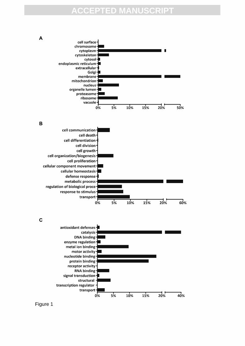
For T. cruzi, the list of identified proteins without the maximum parsimony
criterion added to 5,901 identifications. From these, 2,627 were annotated in 15
cellular component categories (Fig. 1A). The most common cellular localizations
were membranes (51.7%) and cytoplasm (23.1%) followed by nucleus (6.6%)
and ribosome (6.2%). All other cellular compartments presented less than 5% of
the whole identifications. As for biological processes, 2,723 were annotated
proteins and therefore categorized into 15 groups, with metabolic processes
being the most recurrent (61.9%) (Fig. 1B). Three additional classifications
presented percentages greater than 5%: cell transport, regulation and stimulus
response (Fig. 1B). Regarding molecular function, 4,147 annotated proteins
were grouped into 14 categories: catalysis (39.9%), nucleotide binding (18.3%),
protein binding (15.9%), and metal ion binding (9.7%) were the most prominent
classifications (Fig. 1C). Similar localization and functional analysis were also
performed for the identified proteins of M. musculus. The 1,613 proteins
identified (without the maximum parsimony criterion) were distributed among 17
cellular component categories and biological processes: cytoplasm (27.6%) and
metabolic process (20.5%) were the most represented (Figs. S1A and S1B).
Additionally, the mammalian annotated proteins identified were allocated into 14
molecular functions, and protein binding (30.9%) was the most common (Fig.
S1C). All of these data are discriminated in Tables S2 and S3.
To date, proteomic profiles of trypomastigotes were assessed only from
culture-derived or metacyclic forms. From these profiles, we have chosen to
compare our data to the ones that represent the largest trypomastigote
descriptive proteomic maps [17, 28].
4. Discussion
To reevaluate the Trypanosoma cruzi proteome map, we compared our results
with two other descriptive studies that evaluated the culture-derived [17] and
metacyclic trypomastigotes [17, 28]. The comparison correlated the identified
ACC
EPTE
D M
ANU
SCR
IPT
ACCEPTED MANUSCRIPT
proteins in (a) bloodstream trypomastigotes from this work; (b) culture-derived
trypomastigotes from Atwood et al (2005); and (c) metacyclic trypomastigotes
from Atwood et al (2005) and de Godoy et al (2012). In this context, we found
2,214 proteins exclusively present in the bloodstream form, 9 proteins common
to both bloodstream and cultured-derived forms, and 3,215 proteins common to
both bloodstream and metacyclic trypomastigotes (Table S3).
The proteins only identified in bloodstream trypomastigotes were
analyzed according to their protein description (Table S2) and function, as
discussed below. Although differences in the parasite surface proteomic profile
would be expected between the distinct trypomastigotes considering their
environment, the identification of surface proteins are among the most
interesting exclusive proteins due to their exposure to the host immune system,
leading to a better understanding of its interaction with mammalian cells [37].
Among these proteins are phospholipases (A1 and C), mucins, transporters
(amino acids, nucleobases, cations), cAMP-phosphodiesterase, GPI-anchor
transamidase, acid phosphatase, Tb-291 membrane-associated protein, TolT
and surface antigen 2. Further evaluation of the bloodstream trypomastigote
surface proteomics is imperative for a deep characterization of crucial
molecules that participate in parasite-host cell interactions.
Interestingly, some proteins of the parasite non-classical secretion
pathway were also detected in the bloodstream form of the parasite in this study.
Exosomes are extracellular small vesicles that carry a variety of secreted
molecules important for cell communication, among other functions. In T. cruzi,
exosomes (10-100 nm in diameter) and ectosomes (100-200 nm in diameter)
have already been described in metacyclic trypomastigotes and epimastigotes
[38]. Recently, Garcia-Silva and colleagues (2014) showed that exosomes
stimulated metacyclogenesis and also increased the susceptibility of host cells
to infection [39]. Here, we identified four exosome-related proteins in
bloodstream trypomastigotes, suggesting the existence of exosomal pathways
in vivo. Additionally, some structural proteins were originally described in this
work. Proteins such as ARP2/3, which regulates actin dynamics, were identified
[40]. The presence of these proteins only in bloodstream trypomastigotes
suggested special motility and vesicular traffic characteristics of this parasite
form; further analyses are under way.
ACC
EPTE
D M
ANU
SCR
IPT
ACCEPTED MANUSCRIPT
Different cell cycle and transcriptional regulation factors (especially
elongation factors) were also exclusively identified in the bloodstream forms,
including three splicing-related proteins. This suggests the presence of
peculiarities in post-transcriptional control in infective forms exposed to
constituents of mammalian blood. The presence of various proteins involved in
proteolysis and degradation processes as well as enzymes from redox
metabolism were also previously observed in trypomastigotes [17].
Nevertheless, autophagic and proteasomal pathways were only predicted in
silico in trypanosomatids. Thus, their components, such as E1-like enzyme,
were misannotated in databases.
The other exclusive proteins found in bloodstream trypomastigotes were
distributed among the following classes of biological function: (a) biosynthetic
pathways (especially fatty acids metabolism); (b) bioenergetics (polyphosphate,
carbohydrate and mitochondrial metabolisms); (c) protein folding (chaperones
such as T-complex protein); (d) cell signaling (diacylglycerol kinase,
phosphatidylinositol kinase); (e) vesicular traffic (snare and rab proteins); (f)
DNA repair and degradation; and (g) programmed cell death.
Among the 5,901 identified proteins, those likely to be the most abundant
due to a presentation of high spectral counts (>500 spectra) were cytoskeletal
proteins, chaperones, energetic metabolism enzymes, trans-sialidase and
elongation factors. Dynein, clathrin, a hypothetical protein and a 75-77-kDa
antigen were also abundant. Among the most abundant structural proteins were
tubulin, paraflagellar rod protein, and cytoskeletal associated proteins. These
proteins were highly expressed in almost all cells and were commonly present
on the most abundant lists in proteomic studies of trypanosomatids models [11,
17, 18, 28]. Another abundant cytoskeleton associated protein identified was
dynein, which is involved in active intracellular vesicular traffic and flagellar
motility, which are crucial for essential processes such as organelle movement
and beating of the flagellum [41].
Chaperones, such as heat shock proteins and calreticulin, were
commonly overexpressed in various T. cruzi forms, especially in response to
distinct chemical and physical stress conditions [11, 17, 18, 23, 28]. Our data
are in accordance with the expression of high amounts of heat shock proteins
as a consequence of the extreme temperature variation that trypomastigotes
ACC
EPTE
D M
ANU
SCR
IPT
ACCEPTED MANUSCRIPT
are submitted to in the bloodstream, more severe than that observed in the
insect midgut [42]. The presence of the host immune response may also
contribute to stress conditions in trypomastigotes, leading to the expression of
chaperones. Additionally, elongation factors are GTP-dependent enzymes
involved in prokaryotic and eukaryotic protein synthesis [43]. Therefore, their
observed high abundance suggests an intense protein synthesis demand in
bloodstream trypomastigotes.
Among top-scoring proteins, trans-sialidase is presented on the parasite
surface and, upon its secretion, catalyzes the trans-glycosylation of sialic acid to
glycoconjugates. This enzyme is highly expressed in trypomastigotes, and its
biological roles include participation in adhesion and invasion processes and in
the evasion of the host immune system [44]. Interestingly, five enzymes
involved in energetic metabolism were also observed in the top list. Pyruvate
phosphate dikinase, enolase, hexokinase and fructose-bisphosphate aldolase
are enzymes involved in highly active glycolysis/gluconeogenesis pathways in T.
brucei and T. cruzi bloodstream trypomastigotes, which corroborates previous
suggestions that the bloodstream form is more dependent on glycolysis
compared to the insect form [45, 46]. However, aconitase (citric acid cycle
enzyme) has been poorly studied in T. cruzi, and further experiments about its
function in the infective parasite form must be performed.
Clathrin is a key protein in receptor-mediated endocytosis, promoting the
entry of important molecules into eukaryotic cells, including T. cruzi
epimastigotes, that present a high endocytic capacity. Indeed, the activity of this
pathway has been extensively discussed in the mammalian forms of the
parasite [47, 48]. Although Sant'Anna et al. (2008) described at least part of the
endocytic pathway in bloodstream trypomastigotes, clathrin expression had not
been detected until now. The detection of this protein in this work raises once
more the discussion about endocytosis in trypomastigotes. However, clathrin
was also present in the Golgi apparatus, both in mammals and parasite, actively
participating in exocytosis. This correlates with the presence of exosomes and
secretion pathways in trypomastigotes, as already mentioned above [38, 39].
Moreover, the role(s) of hypothetical protein and 75-77-kDa antigen in
bloodstream trypomastigotes need to be investigated.
ACC
EPTE
D M
ANU
SCR
IPT
ACCEPTED MANUSCRIPT
GO analysis of T. cruzi-identified proteins showed that all cellular
compartments were assessed, being approximately 50% of the whole
identifications from membranes, indicating that the extraction method used also
works well to extract hydrophobic proteins, as previously described by our
group [11, 49]. Furthermore, an in silico analysis revealed that 62% of identified
proteins were involved in metabolic processes, and 40% presented catalytic
activity as a molecular function. Such information may correlate the high
metabolic activity of trypomastigotes to the previous data of Gonçalves et al.
(2011) and Clayton & Michels (1996) about the intense glycolytic pathway
activity of bloodstream forms, as previously mentioned [45, 46].
We note that many mammalian proteins from blood components such as
erythrocytes and platelets were identified together with the parasite content.
The high stringency for purifying and washing the parasites and the highly
sensitive and accurate mass spectrometry performed helped to identify a
possible interaction between the parasite and the host cell [50]. During this
interaction, the recognition of molecules is essential prior to protozoa invasion,
and the adhesion step may have contributed to the presence of the host cells
proteins in the sample. As glycoconjugates play an important role as targets in
the recognition process, even desialylated erythrocytes are able to interact with
the parasite [51]. Although red blood cells have a higher density than
trypomastigotes, we identified many proteins present in those cells, which
reinforces that a possible adsorption to the parasite surface may occur.
Following the same criteria employed for T. cruzi, thirty-eight M. musculus
proteins were considered abundant, including hemoglobin subunits, spectrin,
ankyrin, talin, tubulin and actin, as well as putative uncharacterized proteins.
Interestingly, GO analysis pointed to the presence of several mice cytosolic and
membrane proteins, and also to protein binding characteristics. A plausible
explaination for this observation is that trypomastigotes may specifically bind to
host proteins to avoid the immune response or even trigger some biochemical
pathway to use the mammalian machinery to obtain nutrients and metabolites;
however, further analysis is required to test this hypothesis. The proteomic
evaluation of the bloodstream contributes to a better understanding of Chagas
disease pathogenesis and opens new perspectives for parasite biology studies
in the near future.
ACC
EPTE
D M
ANU
SCR
IPT
ACCEPTED MANUSCRIPT
Conflict of interest
The authors declare that there are no competing interests.
Acknowledgments
We are very thankful to Marcos Meuser for his excellent technical support and
to Dr. Solange L. de Castro for the helpful discussions. This work was
supported with grants from CNPq (Universal and PAPES VI), FAPERJ (APQ1),
PDTIS/FIOCRUZ and FIOCRUZ.
Appendix A. Supplementary data
Supplementary data to this article can be found online at
http://www.journals.elsevier.com/journal-of-proteomics/
References
[1] Rocha MO, Teixeira MM, Ribeiro AL. An update on the management of Chagas
cardiomyopathy. Expert Rev Anti Infect Ther. 2007;5:727-43.
[2] Rassi A, Jr., Rassi A, Marin-Neto JA. Chagas disease. Lancet. 2010;375:1388-402.
[3] Schmunis GA, Yadon ZE. Chagas disease: a Latin American health problem becoming a
world health problem. Acta tropica. 2010;115:14-21.
[4] Rassi Jr A, Rassi A, Marin-Neto JA. Chagas heart disease: pathophysiologic mechanisms,
prognostic factors and risk stratification. Mem Inst Oswaldo Cruz. 2009;104 Suppl 1:152-8.
[5] Sosa-Estani S, Viotti R, Segura EL. Therapy, diagnosis and prognosis of chronic Chagas
disease: insight gained in Argentina. Mem Inst Oswaldo Cruz. 2009;104 Suppl 1:167-80.
[6] Higuchi Mde L, Benvenuti LA, Martins Reis M, Metzger M. Pathophysiology of the heart in
Chagas' disease: current status and new developments. Cardiovasc Res. 2003;60:96-107.
[7] Soeiro Mde N, de Castro SL. Screening of Potential anti-Trypanosoma cruzi Candidates: In
Vitro and In Vivo Studies. Open Med Chem J. 2011;5:21-30.
[8] Urbina JA. Specific chemotherapy of Chagas disease: relevance, current limitations and new
approaches. Acta tropica. 2010;115:55-68.
[9] Clayton J. Chagas disease: pushing through the pipeline. Nature. 2010;465:S12-5.
ACC
EPTE
D M
ANU
SCR
IPT
ACCEPTED MANUSCRIPT
[10] Ferella M, Nilsson D, Darban H, Rodrigues C, Bontempi EJ, Docampo R, Andersson B.
Proteomics in Trypanosoma cruzi--localization of novel proteins to various organelles.
Proteomics. 2008;8:2735-49.
[11] Menna-Barreto RF, Beghini DG, Ferreira AT, Pinto AV, De Castro SL, Perales J. A
proteomic analysis of the mechanism of action of naphthoimidazoles in Trypanosoma cruzi
epimastigotes in vitro. Journal of proteomics. 2010;73:2306-15.
[12] Parodi-Talice A, Monteiro-Goes V, Arrambide N, Avila AR, Duran R, Correa A,
Dallagiovanna B, Cayota A, Krieger M, Goldenberg S, Robello C. Proteomic analysis of
metacyclic trypomastigotes undergoing Trypanosoma cruzi metacyclogenesis. J Mass
Spectrom. 2007;42:1422-32.
[13] Clayton C, Shapira M. Post-transcriptional regulation of gene expression in trypanosomes
and leishmanias. Molecular and biochemical parasitology. 2007;156:93-101.
[14] Holetz FB, Alves LR, Probst CM, Dallagiovanna B, Marchini FK, Manque P, Buck G,
Krieger MA, Correa A, Goldenberg S. Protein and mRNA content of TcDHH1-containing
mRNPs in Trypanosoma cruzi. The FEBS journal. 2010;277:3415-26.
[15] Paba J, Santana JM, Teixeira AR, Fontes W, Sousa MV, Ricart CA. Proteomic analysis of
the human pathogen Trypanosoma cruzi. Proteomics. 2004;4:1052-9.
[16] Parodi-Talice A, Duran R, Arrambide N, Prieto V, Pineyro MD, Pritsch O, Cayota A,
Cervenansky C, Robello C. Proteome analysis of the causative agent of Chagas disease:
Trypanosoma cruzi. International journal for parasitology. 2004;34:881-6.
[17] Atwood JA, 3rd, Weatherly DB, Minning TA, Bundy B, Cavola C, Opperdoes FR, Orlando R,
Tarleton RL. The Trypanosoma cruzi proteome. Science. 2005;309:473-6.
[18] Andrade HM, Murta SM, Chapeaurouge A, Perales J, Nirde P, Romanha AJ. Proteomic
analysis of Trypanosoma cruzi resistance to Benznidazole. J Proteome Res. 2008;7:2357-67.
[19] Sant'Anna C, Nakayasu ES, Pereira MG, Lourenco D, de Souza W, Almeida IC, Cunha
ESNL. Subcellular proteomics of Trypanosoma cruzi reservosomes. Proteomics. 2009;9:1782-
94.
[20] Sodre CL, Chapeaurouge AD, Kalume DE, de Mendonca Lima L, Perales J, Fernandes O.
Proteomic map of Trypanosoma cruzi CL Brener: the reference strain of the genome project.
Arch Microbiol. 2009;191:177-84.
[21] Kikuchi SA, Sodre CL, Kalume DE, Elias CG, Santos AL, de Nazare Soeiro M, Meuser M,
Chapeaurouge A, Perales J, Fernandes O. Proteomic analysis of two Trypanosoma cruzi
zymodeme 3 strains. Experimental parasitology. 2010;126:540-51.
[22] Ulrich PN, Jimenez V, Park M, Martins VP, Atwood J, 3rd, Moles K, Collins D, Rohloff P,
Tarleton R, Moreno SN, Orlando R, Docampo R. Identification of contractile vacuole proteins in
Trypanosoma cruzi. PloS one. 2011;6:e18013.
[23] Perez-Morales D, Lanz-Mendoza H, Hurtado G, Martinez-Espinosa R, Espinoza B.
Proteomic analysis of Trypanosoma cruzi epimastigotes subjected to heat shock. J Biomed
Biotechnol. 2012;2012:902803.
ACC
EPTE
D M
ANU
SCR
IPT
ACCEPTED MANUSCRIPT
[24] Menna-Barreto RF, Perales J. The expected outcome of the Trypanosoma cruzi proteomic
map: a review of its potential biological applications for drug target discovery. Subcell Biochem.
2014;74:305-22.
[25] Magalhaes AD, Charneau S, Paba J, Guercio RA, Teixeira AR, Santana JM, Sousa MV,
Ricart CA. Trypanosoma cruzi alkaline 2-DE: Optimization and application to comparative
proteome analysis of flagellate life stages. Proteome Sci. 2008;6:24.
[26] Atwood JA, 3rd, Minning T, Ludolf F, Nuccio A, Weatherly DB, Alvarez-Manilla G, Tarleton
R, Orlando R. Glycoproteomics of Trypanosoma cruzi trypomastigotes using subcellular
fractionation, lectin affinity, and stable isotope labeling. J Proteome Res. 2006;5:3376-84.
[27] Cordero EM, Nakayasu ES, Gentil LG, Yoshida N, Almeida IC, da Silveira JF. Proteomic
analysis of detergent-solubilized membrane proteins from insect-developmental forms of
Trypanosoma cruzi. J Proteome Res. 2009;8:3642-52.
[28] de Godoy LM, Marchini FK, Pavoni DP, Rampazzo Rde C, Probst CM, Goldenberg S,
Krieger MA. Quantitative proteomics of Trypanosoma cruzi during metacyclogenesis.
Proteomics. 2012;12:2694-703.
[29] Nakayasu ES, Sobreira TJ, Torres R, Jr., Ganiko L, Oliveira PS, Marques AF, Almeida IC.
Improved proteomic approach for the discovery of potential vaccine targets in Trypanosoma
cruzi. J Proteome Res. 2012;11:237-46.
[30] Almeida IC, Gazzinelli RT. Proinflammatory activity of glycosylphosphatidylinositol anchors
derived from Trypanosoma cruzi: structural and functional analyses. J Leukoc Biol.
2001;70:467-77.
[31] Frasch AC. Functional diversity in the trans-sialidase and mucin families in Trypanosoma
cruzi. Parasitology today (Personal ed. 2000;16:282-6.
[32] Buscaglia CA, Campo VA, Frasch AC, Di Noia JM. Trypanosoma cruzi surface mucins:
host-dependent coat diversity. Nature reviews. 2006;4:229-36.
[33] McDonald WH, Tabb DL, Sadygov RG, MacCoss MJ, Venable J, Graumann J, Johnson JR,
Cociorva D, Yates JR, 3rd. MS1, MS2, and SQT-three unified, compact, and easily parsed file
formats for the storage of shotgun proteomic spectra and identifications. Rapid Commun Mass
Spectrom. 2004;18:2162-8.
[34] Carvalho PC, Fischer JS, Xu T, Cociorva D, Balbuena TS, Valente RH, Perales J, Yates JR,
3rd, Barbosa VC. Search engine processor: Filtering and organizing peptide spectrum matches.
Proteomics. 2012;12:944-9.
[35] Xu T, Venable JD, Park SK, Cociorva D, al. e. ProLuCID, a fast and sensitive tandem mass
spectra-based protein identification program. Mol Cell Proteomics. 2006;5:S:174.
[36] Zhang B, Chambers MC, Tabb DL. Proteomic parsimony through bipartite graph analysis
improves accuracy and transparency. J Proteome Res. 2007;6:3549-57.
[37] Meirelles MN, Chiari E, de Souza W. Interaction of bloodstream, tissue culture-derived and
axenic culture-derived trypomastigotes of Trypanosoma cruzi with macrophages. Acta tropica.
1982;39:195-203.
ACC
EPTE
D M
ANU
SCR
IPT
ACCEPTED MANUSCRIPT
[38] Bayer-Santos E, Aguilar-Bonavides C, Rodrigues SP, Cordero EM, Marques AF, Varela-
Ramirez A, Choi H, Yoshida N, da Silveira JF, Almeida IC. Proteomic analysis of Trypanosoma
cruzi secretome: characterization of two populations of extracellular vesicles and soluble
proteins. J Proteome Res. 2013;12:883-97.
[39] Garcia-Silva MR, das Neves RF, Cabrera-Cabrera F, Sanguinetti J, Medeiros LC, Robello
C, Naya H, Fernandez-Calero T, Souto-Padron T, de Souza W, Cayota A. Extracellular vesicles
shed by Trypanosoma cruzi are linked to small RNA pathways, life cycle regulation, and
susceptibility to infection of mammalian cells. Parasitology research. 2013;113:285-304.
[40] Abu Taha A, Schnittler HJ. Dynamics between actin and the VE-cadherin/catenin complex:
Novel aspects of the ARP2/3 complex in regulation of endothelial junctions. Cell Adh Migr.
2014;8.
[41] Springer AL, Bruhn DF, Kinzel KW, Rosenthal NF, Zukas R, Klingbeil MM. Silencing of a
putative inner arm dynein heavy chain results in flagellar immotility in Trypanosoma brucei.
Molecular and biochemical parasitology. 2010;175:68-75.
[42] Van der Ploeg LH, Giannini SH, Cantor CR. Heat shock genes: regulatory role for
differentiation in parasitic protozoa. Science. 1985;228:1443-6.
[43] Noble CG, Song H. Structural studies of elongation and release factors. Cell Mol Life Sci.
2008;65:1335-46.
[44] Dc-Rubin SS, Schenkman S. T rypanosoma cruzi trans-sialidase as a multifunctional
enzyme in Chagas' disease. Cell Microbiol. 2012;14:1522-30.
[45] Clayton CE, Michels P. Metabolic compartmentation in African trypanosomes. Parasitology
today (Personal ed. 1996;12:465-71.
[46] Gonçalves RL, Barreto RF, Polycarpo CR, Gadelha FR, Castro SL, Oliveira MF. A
comparative assessment of mitochondrial function in epimastigotes and bloodstream
trypomastigotes of Trypanosoma cruzi. J Bioenerg Biomembr. 2011;43:651-61.
[47] Correa JR, Atella GC, Menna-Barreto RS, Soares MJ. Clathrin in Trypanosoma cruzi: in
silico gene identification, isolation, and localization of protein expression sites. The Journal of
eukaryotic microbiology. 2007;54:297-302.
[48] Sant'Anna C, Parussini F, Lourenco D, de Souza W, Cazzulo JJ, Cunha-e-Silva NL. All
Trypanosoma cruzi developmental forms present lysosome-related organelles. Histochem Cell
Biol. 2008;130:1187-98.
[49] Beghini DG, Ferreira AT, Almeida VC, Caminha MA, Silva-Jr FP, Perales J, Menna-Barreto
RF. New insights in Trypanosoma cruzi proteomic map: further post-translational modifications
and potential drug targets in Y strain epimastigotes. J Integr OMICS. 2012;2:106-13.
[50] Umekita LF, Mota I. In-vitro lysis of sensitized Trypanosoma cruzi by platelets: role of C3b
receptors. Parasite immunology. 1989;11:561-6.
[51] Silber AM, Marcipar IS, Roodveldt C, Cabeza Meckert P, Laguens R, Marcipar AJ.
Trypanosoma cruzi: identification of a galactose-binding protein that binds to cell surface of
human erythrocytes and is involved in cell invasion by the parasite. Experimental parasitology.
2002;100:217-25.
ACC
EPTE
D M
ANU
SCR
IPT
ACCEPTED MANUSCRIPT
Figure 1
ACC
EPTE
D M
ANU
SCR
IPT
ACCEPTED MANUSCRIPT
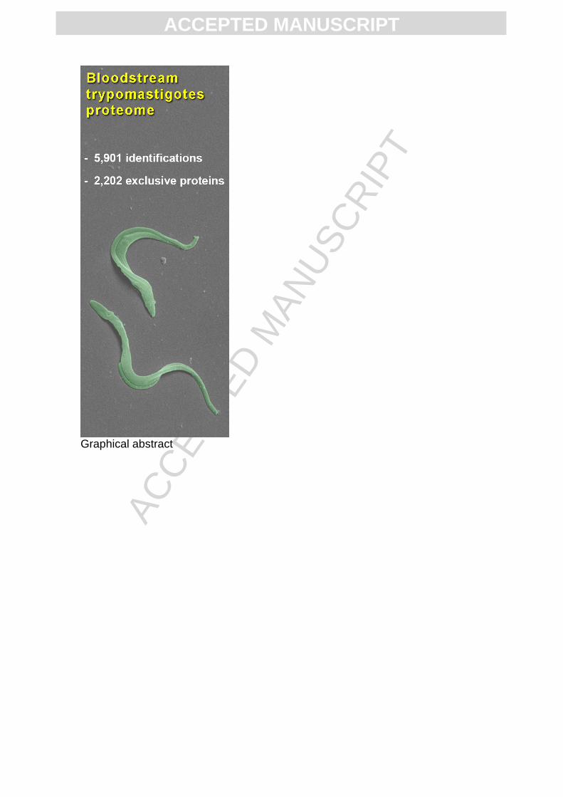
Graphical abstract
ACC
EPTE
D M
ANU
SCR
IPT
ACCEPTED MANUSCRIPT
Conflict of Interest: None to declare

ACC
EPTE
D M
ANU
SCR
IPT
ACCEPTED MANUSCRIPT
Highlights First proteome description of bloodstream trypomastigotes with 5,901
identifications.
More than 60% of the identified proteins were involved in metabolic processes.
2,202 proteins were exclusively detected in the bloodstream form.