False positives complicate ancient pathogen identifications using high-throughput shotgun sequencing
15
RESEARCH ARTICLE Open Access False positives complicate ancient pathogen identifications using high-throughput shotgun sequencing Michael G Campana 1,2* , Nelly Robles García 3 , Frank J Rühli 1,2 and Noreen Tuross 1 Abstract Background: Identification of historic pathogens is challenging since false positives and negatives are a serious risk. Environmental non-pathogenic contaminants are ubiquitous. Furthermore, public genetic databases contain limited information regarding these species. High-throughput sequencing may help reliably detect and identify historic pathogens. Results: We shotgun-sequenced 8 16th-century Mixtec individuals from the site of Teposcolula Yucundaa (Oaxaca, Mexico) who are reported to have died from the huey cocoliztli (‘Great Pestilence’ in Nahautl), an unknown disease that decimated native Mexican populations during the Spanish colonial period, in order to identify the pathogen. Comparison of these sequences with those deriving from the surrounding soil and from 4 precontact individuals from the site found a wide variety of contaminant organisms that confounded analyses. Without the comparative sequence data from the precontact individuals and soil, false positives for Yersinia pestis and rickettsiosis could have been reported. Conclusions: False positives and negatives remain problematic in ancient DNA analyses despite the application of high-throughput sequencing. Our results suggest that several studies claiming the discovery of ancient pathogens may need further verification. Additionally, true single molecule sequencing’s short read lengths, inability to sequence through DNA lesions, and limited ancient-DNA-specific technical development hinder its application to palaeopathology. Keywords: True single molecule sequencing, High-throughput sequencing, Pathogen, Ancient DNA, False positive Background Diseases have ravaged human populations throughout his- tory. The pathogens responsible for many historic epidemics remain either unknown or speculative (e.g. the Plague of Athens [1,2]). Identification of these historic pathogens is critical to understanding disease evolution, which in turn has direct impacts on the development of effective medical treatments for these conditions. The discovery that ancient pathogen-diagnostic biomolecules may survive in archaeo- logical bones and the development of high-throughput DNA sequencing technologies have opened new possibilities for determining past disease [3-14]. Most biomolecular palaeopathological studies have been based on the polymerase chain reaction (PCR) (e.g. [3-7]). Supplementary methods include lipid and immunological assays [6-12]. Due to the limitations of molecular preser- vation and detection, negative assays do not constitute evi- dence of absence of any infection [15-17]. False positives are a serious concern since bacteria, viruses and fungi are ubiquitous and the specificity of molecular tests may not be able to distinguish between target disease agents and their closely related relatives [18-20]. Many PCR-based palaeopathological reports are contentious due to insuffi- cient anti-contamination procedures or lack of experi- mental and analytical rigor [21-23]. Furthermore, PCR-based methods are limited in their ability to identify unknown pathogens since they require * Correspondence: [email protected] 1 Department of Human Evolutionary Biology, Harvard University, Peabody Museum, 11 Divinity Avenue, Cambridge, MA 02138, USA 2 Centre for Evolutionary Medicine, Anatomy Institute, University of Zurich, 190 Winterthurerstrasse, Zurich 8057, Switzerland Full list of author information is available at the end of the article © 2014 Campana et al.; licensee BioMed Central Ltd. This is an Open Access article distributed under the terms of the Creative Commons Attribution License (http://creativecommons.org/licenses/by/4.0), which permits unrestricted use, distribution, and reproduction in any medium, provided the original work is properly credited. The Creative Commons Public Domain Dedication waiver (http://creativecommons.org/publicdomain/zero/1.0/) applies to the data made available in this article, unless otherwise stated. Campana et al. BMC Research Notes 2014, 7:111 http://www.biomedcentral.com/1756-0500/7/111
-
Upload
independent -
Category
Documents
-
view
1 -
download
0
Transcript of False positives complicate ancient pathogen identifications using high-throughput shotgun sequencing

Campana et al. BMC Research Notes 2014, 7:111http://www.biomedcentral.com/1756-0500/7/111
RESEARCH ARTICLE Open Access
False positives complicate ancient pathogenidentifications using high-throughput shotgunsequencingMichael G Campana1,2*, Nelly Robles García3, Frank J Rühli1,2 and Noreen Tuross1
Abstract
Background: Identification of historic pathogens is challenging since false positives and negatives are a serious risk.Environmental non-pathogenic contaminants are ubiquitous. Furthermore, public genetic databases contain limitedinformation regarding these species. High-throughput sequencing may help reliably detect and identify historicpathogens.
Results: We shotgun-sequenced 8 16th-century Mixtec individuals from the site of Teposcolula Yucundaa (Oaxaca,Mexico) who are reported to have died from the huey cocoliztli (‘Great Pestilence’ in Nahautl), an unknown diseasethat decimated native Mexican populations during the Spanish colonial period, in order to identify the pathogen.Comparison of these sequences with those deriving from the surrounding soil and from 4 precontact individualsfrom the site found a wide variety of contaminant organisms that confounded analyses. Without the comparativesequence data from the precontact individuals and soil, false positives for Yersinia pestis and rickettsiosis could havebeen reported.
Conclusions: False positives and negatives remain problematic in ancient DNA analyses despite the application ofhigh-throughput sequencing. Our results suggest that several studies claiming the discovery of ancient pathogensmay need further verification. Additionally, true single molecule sequencing’s short read lengths, inability tosequence through DNA lesions, and limited ancient-DNA-specific technical development hinder its application topalaeopathology.
Keywords: True single molecule sequencing, High-throughput sequencing, Pathogen, Ancient DNA, False positive
BackgroundDiseases have ravaged human populations throughout his-tory. The pathogens responsible for many historic epidemicsremain either unknown or speculative (e.g. the Plague ofAthens [1,2]). Identification of these historic pathogens iscritical to understanding disease evolution, which in turnhas direct impacts on the development of effective medicaltreatments for these conditions. The discovery that ancientpathogen-diagnostic biomolecules may survive in archaeo-logical bones and the development of high-throughput
* Correspondence: [email protected] of Human Evolutionary Biology, Harvard University, PeabodyMuseum, 11 Divinity Avenue, Cambridge, MA 02138, USA2Centre for Evolutionary Medicine, Anatomy Institute, University of Zurich,190 Winterthurerstrasse, Zurich 8057, SwitzerlandFull list of author information is available at the end of the article
© 2014 Campana et al.; licensee BioMed CentCommons Attribution License (http://creativecreproduction in any medium, provided the orDedication waiver (http://creativecommons.orunless otherwise stated.
DNA sequencing technologies have opened new possibilitiesfor determining past disease [3-14].Most biomolecular palaeopathological studies have been
based on the polymerase chain reaction (PCR) (e.g. [3-7]).Supplementary methods include lipid and immunologicalassays [6-12]. Due to the limitations of molecular preser-vation and detection, negative assays do not constitute evi-dence of absence of any infection [15-17]. False positivesare a serious concern since bacteria, viruses and fungi areubiquitous and the specificity of molecular tests may notbe able to distinguish between target disease agents andtheir closely related relatives [18-20]. Many PCR-basedpalaeopathological reports are contentious due to insuffi-cient anti-contamination procedures or lack of experi-mental and analytical rigor [21-23].Furthermore, PCR-based methods are limited in their
ability to identify unknown pathogens since they require
ral Ltd. This is an Open Access article distributed under the terms of the Creativeommons.org/licenses/by/4.0), which permits unrestricted use, distribution, andiginal work is properly credited. The Creative Commons Public Domaing/publicdomain/zero/1.0/) applies to the data made available in this article,

Table 1 Teposcolula Yucundaa individuals analyzed here
Individual Graveyard Sex Age
TP02 Grand Plaza Female 25 ± 1
TP04 Grand Plaza Female 26 ± 2
TP09 Grand Plaza Male 20 ± 2
TP10 Grand Plaza Male 20-21
TP15 Grand Plaza Indeterminate Adolescent
TP18 Grand Plaza Female 35 ± 4
TP26 Grand Plaza Female 40 ± 4
TP32 Churchyard Female 19 ± 1
TP37 Grand Plaza Female Young Adult
TP42 Churchyard Indeterminate 5
TP45 Churchyard Female 36
TP48 Churchyard Female 32 ± 2
Sample details are from [27,29].
Campana et al. BMC Research Notes 2014, 7:111 Page 2 of 15http://www.biomedcentral.com/1756-0500/7/111
candidate disease agents of known sequence in advance.Although there are a few studies that attempt such de novoidentification (e.g. [5,24]), the vast majority of investiga-tions have been restricted to targeted, likely diseases. WhileDNA capture methods, including in-solution [e.g. 13] andarray-hybridization [e.g. 14] capture, have become the goldstandard for palaeopathogenomics, no such technology isyet available for palaeopathogen diagnosis. In theory, it ispossible to identify unknown diseases by high-throughputDNA sequencing of known afflicted individuals and com-paring these pools with suitable controls (such as the sur-rounding soil and uninfected archaeologically relatedindividuals). Organisms found only in the infected popula-tion would thus be candidates for the responsible patho-gens. Nevertheless, since the majority of sequences in mostancient DNA samples are derived from the environment(particularly soil bacteria), false positives could remainproblematic in high-throughput analyses [23,25]. More-over, the public genetic databases are both biased towardspathogenic cellular organisms and deficient in virus se-quences, thereby increasing the false positive rate for cellu-lar pathogens while simultaneously raising the falsenegative rate for viral infections in de novo identifications.Here we show that false positives are a significant error
source in palaeopathological analyses using high-throughput sequencing. Using two platforms (the HelicosBioSciences HeliScope and the Illumina HiSeq 2500), weattempted to identify de novo the pathogen responsible forthe huey cocoliztli (‘Great Pestilence’ in Nahautl), a diseasethat ravaged the native Mexican population during theSpanish colonial period [26].
MethodsDNA extractionIn order to identify the huey cocoliztli pathogen, we ana-lyzed the site of Teposcolula Yucundaa (Oaxaca, Mexico),a large Mixtec city at the time of Spanish colonization[27]. The site was abandoned in 1552 following a majoroutbreak of the huey cocoliztli in the 1540 s. Excavationsat Teposcolula Yucundaa conducted between 2004 and2006 revealed two Mixtec cemeteries: a colonial periodgraveyard (the Grand Plaza) and a smaller precolonial one(the Churchyard) [27]. Warinner and colleagues [27] iden-tified the Grand Plaza graveyard as a plague pit corre-sponding to the 1540 s pandemic of the huey cocoliztli.The precontact Churchyard population is assumed tohave been uninfected with the huey cocoliztli since the na-tive Mexican populations were unfamiliar with the diseaseat the time of its first appearance during the colonialperiod [28]. DNA preservation at Teposcolula Yucundaais exceptional, probably due to the high-altitude, cool en-vironment and relatively recent date [27].Twelve femoral cortical bone specimens (each represent-
ing a unique individual; Table 1) were collected in the field
and ground to a fine power (described in [29]). DNA ex-tractions were performed in a dedicated ancient DNA la-boratory in the Department of Human EvolutionaryBiology, Harvard University according to standard anti-contamination protocols [30]. Approximately 1 g of bonepowder per individual was decalcified in 0.5 M EDTA,pH 8.0. Raw DNA extract was passed through a vacuumfilter to remove residual protein and powder and then con-centrated to ~500 μl via ultrafiltration using Vivaspin® 20’s(Vivaproducts) with a 10 kDa molecular weight cut-off.Concentrated extracts were treated with proteinase K.Protein-digestion completion was verified using a Qubit®2.0 fluorometer (Life Technologies). Digested extracts werethen purified using QIAquick PCR Purification Kits(Qiagen). Bulk DNA extracts consisting of 4 individ-uals per bulk were constructed for the Grand Plaza(individuals TP02, TP10, TP15, TP26) and Churchyard(individuals TP32, TP42, TP45, TP48) populations. Bulk-ing the samples increased the likelihood of detecting thepathogen since only a fraction of the infected individualsare expected to have endogenous disease DNA preserved[e.g. 13,14].Three samples of soil from the Grand Plaza and the
Churchyard cemeteries were also collected. Soil was col-lected from the burial contexts (within a few centimeters ofthe skeletons) by trowelling samples directly into collectionbags while wearing gloves to limit DNA contamination.DNA was extracted from 5–8 g of soil per sample using thePowerMax® Soil DNA Isolation Kit (Mo-Bio Laboratories).Purified extracts from each burial were then combined intoa bulk sample.Finally, subsamples of all three bulk extracts (Churchyard,
Grand Plaza and soil) were sheared to 150 bp average lengthusing a S220 Focused-ultrasonicator (Covaris, Inc.) for sub-sequent true single molecule sequencing (tSMS). Althoughshearing DNA extracts is typically denigrated in high-

Campana et al. BMC Research Notes 2014, 7:111 Page 3 of 15http://www.biomedcentral.com/1756-0500/7/111
throughput ancient DNA analyses (e.g. [31]), the sequencesgenerated by tSMS are shorter (typically <40 bp) than theendogenous DNA molecules known to be present in theTeposcolula Yucundaa individuals by PCR (~100 bp) [27]and Bioanalyzer assays (see below). Since tSMS only gener-ates one sequence per molecule and has limited ability to se-quence through DNA lesions, shearing might make more ofthe endogenous DNA sequenceable (but at the cost of apossible increase in microbial contamination) by producingmultiple 3′-termini per original molecule. Additionally, sixextracts representing single individuals (TP04, TP09, TP10,TP18, TP37, TP48) and a subsample of the soil bulk extractwere sheared to 150–200 bp average length for subsequentIllumina library construction using the automated Apollo324 (IntegenX) platform.DNA concentrations for all individual extracts, bulks
and sheared bulks were calculated using a high-specificityDNA kit on a Qubit® fluorometer. Length distributions ofDNA molecules in these samples were calculated using ahigh-specificity DNA chip on an Agilent 2100 Bioanalyzer(Agilent Biotechnologies).
Helicos HeliScope sequencingA total of 12 channels of tSMS was performed on a HelicosHeliScope sequencer at Helicos BioSciences (Cambridge,MA). Eight μl per channel of each bulk and sheared bulk
Table 2 Sequencing statistics for the studied samples
Platform Sample Description No. Filtered Sequen
HeliScope Grand Plaza Bulk 1 31,183,210
Grand Plaza Bulk 2 30,773,387
Grand Plaza Phos. 1 26,269,497
Grand Plaza Phos. 2 23,061,503
Churchyard Bulk 1 28,960,308
Churchyard Bulk 2 27,035,110
Churchyard Phos. 1 28,423,683
Churchyard Phos. 2 29,660,255
Grand Plaza Sheared 4,088
Churchyard Sheared 5,309
Soil Bulk 840,915
Soil Sheared 123,211
HiSeq 2500 TP04 9,643,548
TP09 6,640,789
TP10 5,368,267
TP18 7,693,734
TP37 9,113,392
TP48 3,720,696
Soil Bulk 4,807,489
“Bulk” samples are the unsheared bulks. “Phos.” samples are the phosphatase-treateSequences” is the number of sequences after filtering for sequencing artifacts (e.g.of the generated reads.
were prepared for tSMS according to the standardprotocol for ancient samples [32]. Two channels of eachbulk and one channel of each sheared bulk were se-quenced. Additionally, samples of the Churchyard andGrand Plaza bulk extracts were treated with AntarcticPhosphatase (New England Biolabs), diluted to the equiva-lent concentration of their untreated counterparts andprepared for HeliScope sequencing as described in [33].Phosphatase-treatment may increase HeliScope sequenceyield [33]. Two channels of each phosphatase-treated bulkwere sequenced. The bulks and phosphatase-treated bulkswere sequenced on one chip, while the soil and shearedbulks were sequenced on another. For the unshearedChurchyard and Grand Plaza bulks (both phosphatase-treated and untreated samples), 23–31 million quality-controlled sequences were generated per channel (Table 2).There were technical issues with the sheared and soil sam-ples (see below), so only between 4,000 and 840,000 readsper channel were obtained for these four channels (Table 2).
Illumina HiSeq 2500 sequencingInitial tSMS results (see below) revealed very high micro-bial species diversity in the bone and soil samples. Thevery short sequence length hampered accurate identifica-tion of these species. Therefore, we constructed Illuminalibraries from six bone extracts representing single
ces Molecular Length (bp) Total Sequenced (bp)
34.1 ± 7.1 1,063,972,634
34.4 ± 7.2 1,058,435,915
34.4 ± 7.3 904,192,902
35.0 ± 7.4 806,304,129
33.9 ± 7.0 981,718,381
33.8 ± 7.0 914,002,286
34.5 ± 7.3 981,600,068
33.8 ± 7.0 1,003,204,069
27.1 ± 3.4 110,619
27.2 ± 3.5 144,140
33.4 ± 7.2 28,082,034
31.4 ± 6.4 3,869,834
219.5 ± 43.9 2,117,000,413
236.2 ± 39.2 1,568,449,339
223.7 ± 40.3 1,201,119,486
215.5 ± 39.1 1,658,247,428
207.2 ± 39.0 1,888,081,563
227.8 ± 40.5 847,609,750
222.3 ± 40.6 1,068,497,871
d unsheared bulks. “Sheared” samples are the sheared bulks. The “No. FilteredPCR duplicates). “Molecular Length” is the mean length (± standard deviation)

Campana et al. BMC Research Notes 2014, 7:111 Page 4 of 15http://www.biomedcentral.com/1756-0500/7/111
individuals and the bulk soil sample using the PrepXIllumina Kit (IntegenX) on the Apollo 324 according tomanufacturer’s instructions using NEXTflex™ DNA barcodes(BioO Scientific). The indexed libraries were then enrichedby 13 cycles of PCR using the NEXTflex™ kit. Library qual-ities were confirmed via fluorometric quantitation (Qubit®),analysis on a high-specificity DNA chip (Agilent 2100) andquantitative PCR using the KAPA Library QuantificationKit – Illumina/Universal (KAPA Biosystems). The librarieswere then pooled in equimolar ratios, and paired-end150 bp reads were generated on the Illumina HiSeq 2500platform. Initial quality control and demultiplexing was per-formed using CASAVA 1.8.2. Paired-end reads were mergedusing PANDAseq 2.4.0 [34]. Adapter artifacts and PCRduplicates were removed using TagDust 1.12 [35] andCD-HIT 4.6 [36]. The library sequencing qualities werechecked using FastQC 1.32 [37]. The final data setscontained 3.7–9.6 million quality-controlled reads perlibrary (Table 2).
Bioinformatic analysisSequence file formats were manipulated using SAMtools0.1.18 [38]. Sequence data sets were aligned against refer-ence genomes of interest (Additional file 1, see Resultsand discussion below) using BWA 0.6.2 [39,40] according
Table 3 Alignment statistics of reads mapped against the hum
Platform Sample Description Reads Mapped (% M
HeliScope Grand Plaza Bulk 1 254,977 (0.82%)
Grand Plaza Bulk 2 283,309 (0.77%)
Grand Plaza Phos. 1 186,016 (0.71%)
Grand Plaza Phos. 2 152,275 (0.66%)
Churchyard Bulk 1 387,992 (1.34%)
Churchyard Bulk 2 382,493 (1.41%)
Churchyard Phos. 1 250,427 (0.88%)
Churchyard Phos. 2 312,392 (1.05%)
Grand Plaza Sheared 254 (6.21%)
Churchyard Sheared 340 (6.40%)
Soil Bulk 9,441 (1.12%)
Soil Sheared 2,895 (2.35%)
HiSeq 2500 TP04 2,655 (0.03%)
TP09 27,430 (0.41%)
TP10 1,332 (0.02%)
TP18 5,521 (0.00%)
TP37 6,172 (0.00%)
TP48 300 (0.00%)
Soil Bulk 37 (0.00%)
“Bulk” samples are the unsheared bulks. “Phos.” samples are the phosphatase-treatecates the percentage of the total number of reads comprised by the reads mappeddeviation) of the mapped reads.
to the recommended settings in [41]. Sequences were alsoaligned against the GenBank® non-redundant nucleotidedatabase using megaBLAST (BLAST 2.2.25+) [42] and an-alyzed in MEGAN 4.70.4 [43]. Statistical analyses wereperformed in R 2.15.3 [44] or using Biopieces [45] andcustom scripts.
Results and discussionHuman DNA and authenticityAlignment of the samples against the human (Homo sapiens)reference genome (GRCh37.p11) revealed that 0.66%–6.40%of the HeliScope reads in each channel and 0.00%–0.42% ofIllumina sequences in each library corresponded to hu-man DNA (Table 3, Additional file 1). The HeliScope en-dogenous human DNA quantifications are probably anunderestimate of the true human DNA content sinceshort sequences with low information contents are un-likely to map against the reference genome with sufficientconfidence to be assigned (See ‘Soil Complexity’ below).Furthermore, the mean lengths (~25 bp) of the sequencesmapped to the human genome were shorter than those(~34 bp) of all the sequences (mapped and unmapped in-cluded) in each channel (one-tailed t-test, p < 0.00001,Tables 2 and 3). As in [32], analysis of the mapped reads’substitution artifacts using mapDamage2.0 [46] showed a
an genome
apped) Molecular Length (bp) Total Mapped (bp)
25.2 ± 2.7 6,414,294
25.2 ± 2.8 6,013,180
25.1 ± 2.5 4,668,531
25.2 ± 2.7 3,838,807
27.3 ± 5.3 10,579,724
27.3 ± 5.3 10,425,423
26.2 ± 4.3 6,559,094
26.5 ± 4.7 8,290,314
24.5 ± 1.0 6,226
24.5 ± 0.8 8,325
24.9 ± 2.1 234,694
24.8 ± 1.9 71,816
214.4 ± 39.6 569,144
192.1 ± 36.4 5.267,951
204.9 ± 37.6 272,903
201.7 ± 35.4 1,113,561
191.0 ± 31.4 1,179,076
208.6 ± 39.3 62,568
181.2 ± 34.0 6,706
d unsheared bulks. “Sheared” samples are the sheared bulks. “% Mapped” indi-onto the human genome. “Molecular Length” is the mean length (± standard

Campana et al. BMC Research Notes 2014, 7:111 Page 5 of 15http://www.biomedcentral.com/1756-0500/7/111
uniform distribution of C→T and G→A transitionsalong the molecules. These substitutions typically accu-mulate at the 5′- and 3′-termini of ancient DNA mole-cules respectively [47]. These results suggest that theHeliScope chemistry cannot sequence through uracillesions effectively, thereby shortening the recovered se-quences of the endogenous ancient molecules and pro-ducing a uniform transition profile.Due to the HeliScope sequences’ short read lengths, false
positives due to the presence of contamination fromclosely related species (e.g. rodents) were a concern. There-fore, we also aligned the HeliScope data sets against the rat(Rattus norvegicus) genome (Rnor_5.0) and the chimpan-zee (Pan troglodytes) genome (Pan_troglodytes-2.1.4)(Additional file 1). This ad-hoc test revealed a significantgradient of increasing numbers of hits against the ge-nomes more closely related to humans (Kruskal-Wallistest, p = 0.0004609) with a concomitant increase inmean hit length in the more closely related species(Kruskal-Wallis test, p = 0.007629), suggesting that theHeliScope human-aligned reads derived from humans(Additional file 2).Human DNA (1.12%–2.35% of all reads) was also identi-
fied in the soil HeliScope data sets, raising the possibility oflaboratory contamination affecting our data. Nevertheless,analysis of the same soil bulk sample via Illumina sequen-cing revealed only 37 sequences (0.00%) that matchedhumans in the soil at the longer lengths (150–298 bp)obtained in the Illumina data. Comparatively, the indi-vidual bones had 300–27,430 human DNA sequences(0.01–0.42%) at equivalent lengths (Table 3). This indi-cates that the human DNA in the soil has undergonemore degradation than that in the bone, a result incon-sistent with the laboratory contamination hypothesis[30]. Moreover, although mitochondrial haplotype pro-files were incomplete due to absence of a mitochondrialDNA enrichment step, the individual haplotypes (basedon the Illumina sequences) generally agreed with thosefound in [29], although a high rate of nucleotide misin-corporations was observed (Additional file 3). Analysisof these substitutions using mapDamage2.0 showed thecharacteristic ancient DNA pattern, with higher rates ofC→ T and G→A transitions at the 5′- and 3′-terminirespectively [47]. This suggests that the human DNA inthe bone samples is endogenous. It is possible that thesoil human DNA derives from archaeologists in thefield, although this is relatively unlikely since the bonesand soil were collected while wearing gloves. Since thesite was abandoned shortly after the huey cocoliztli pan-demic [27], the most likely source of human DNA inthe soil is not from later occupants of the site, but ra-ther the numerous decomposing dead (estimated to atleast 800 individuals) interred nearly simultaneously in theGrand Plaza cemetery. Detection of leached DNA is
especially likely since the soil samples were collected im-mediately adjacent to the interred skeletons. If endogen-ous human DNA leached into the soil, it is possible thatDNA from the huey cocoliztli pathogen may also have.However, since pathogen DNA is typically much rarerthan host DNA [e.g. 13], this level of DNA leaching wouldprobably be undetectable using current methodologies.
Candidate diseasesBased on historical descriptions of the huey cocoliztli,likely candidate pathogens include pneumonic plague(Yersinia pestis), typhus and other forms of rickettsiosis(Rickettsia spp.), smallpox or alastrim (Variola spp.),and viral hemorrhagic fevers (VHF), such as Dengueand Yellow Fever [26,28,48,49]. Measles (Morbillivirus Mea-sles virus), dysentery, influenza (Influenzavirus spp.), pneu-monia and pleurisy have also been suggested, althoughthese are less likely diagnoses since the historical descrip-tions of the huey cocolitzli symptoms do not match thesediseases’ extant presentations [28]. Since dysentery, pneu-monia and pleurisy can be caused by a wide range of organ-isms and are unlikely diagnoses for the huey cocoliztli, wedid not analyze them further via the candidate genome ap-proach described here. Additionally, VHFs, measles and in-fluenza are caused by RNA viruses, which are unlikely tosurvive in the archaeological record due to RNA’s instabilityand the ubiquity of RNases [50]. Nevertheless, some havereported the successful amplification and sequencing ofRNA viruses from medical archival preserved tissue datingback several decades [51,52], and Fordyce and col-leagues [50] reported high-throughput sequencing ofRNA transcripts in 700-year-old maize kernels. More-over, the HeliScope will sequence RNA directly (evenusing DNA settings for the machine as conducted herealbeit with reduced efficiency), so there remains a finitedetection possibility for RNA viruses.A substantial proportion of the unsheared HeliScope
reads (0.01%, 2198–3880 reads per channel) matched theYersinia pestis genome in both the Grand Plaza andChurchyard populations (Additional file 1). This alsomatched the proportion of putative Y. pestis reads foundin the HeliScope-sequenced soil data set (0.01%, 63 reads)(Table 4). BLAST analysis of the mapped reads inMEGAN showed that these identifications were non-specific, with the sequences matching a wide variety of or-ganisms, suggesting these mapped reads are false positives(Additional file 4). According to the BLAST identifica-tions, only 0.29% and 2.0% of the reads mapped to theYerinia pestis genome by BWA matched the Enterobacte-riaceae and Gammaproteobacteria respectively, Yersinia’sfamily and class.Alignment of the unsheared Churchyard and Grand Plaza
bulk HeliScope data against typhus (Rickettsia prowazekiiand Rickettsia typhi) genomes found that 0.00%–0.01%

Table 4 Alignment statistics of reads mapped against the Yersinia pestis genome
Platform Sample Description Reads Mapped (% Mapped) Molecular Length (bp) Total Mapped (bp)
HeliScope Grand Plaza Bulk 1 3,880 (0.01%) 28.8 ± 5.6 111,646
Grand Plaza Bulk 2 3,451 (0.01%) 30.0 ± 5.7 99,916
Grand Plaza Phos. 1 2,626 (0.01%) 28.5 ± 5.4 74,846
Grand Plaza Phos. 2 2,198 (0.01%) 28.9 ± 5.8 63,479
Churchyard Bulk 1 3,794 (0.01%) 28.8 ± 5.8 109,284
Churchyard Bulk 2 3,672 (0.01%) 28.9 ± 5.9 106,060
Churchyard Phos. 1 2,595 (0.01%) 28.1 ± 5.4 72,809
Churchyard Phos. 2 3,723 (0.01%) 28.5 ± 5.7 106,284
Grand Plaza Sheared 0 (0.00%) — 0
Churchyard Sheared 2 (0.04%) 24.0 ± 0.0 48
Soil Bulk 63 (0.01%) 26.3 ± 3.7 1,656
Soil Sheared 3 (0.00%) 25.3 ± 1.2 76
HiSeq 2500 TP04 0 (0.00%) — 0
TP09 0 (0.00%) — 0
TP10 0 (0.00%) — 0
TP18 0 (0.00%) — 0
TP37 0 (0.00%) — 0
TP48 0 (0.00%) — 0
Soil Bulk 0 (0.00%) — 0
“Bulk” samples are the unsheared bulks. “Phos.” samples are the phosphatase-treated unsheared bulks. “Sheared” samples are the sheared bulks. “% Mapped” indi-cates the percentage of the total number of reads comprised by the reads mapped onto the Yersinia pestis genome. “Molecular Length” is the mean length(± standard deviation) of the mapped reads.
Campana et al. BMC Research Notes 2014, 7:111 Page 6 of 15http://www.biomedcentral.com/1756-0500/7/111
(1,210–2,089 reads) corresponded to these species (Table 5,Additional file 1). The identification of Rickettsia in bonematches its prevalence in the HeliScope-sequenced soilsample (0.00%, 30 reads). It is possible that the putativeRickettsia DNA in the soil derives from leaching from thebone. However, this explanation is improbable since thequantity of leached pathogen DNA is expected to be toosmall to detect given the low amounts of soil human DNA[e.g. 13]. Moreover, there were no differences between theGrand Plaza and Churchyard graveyards in terms of thesespecies’ prevalences or lengths of the aligned sequences(30 ± 6 bp). Only one read corresponding to Rickettsia wasfound in the Illumina data sets. BLAST analysis of theRickettsia hits showed that these sequences derived fromvarious soil organisms rather than bona fide pathogens(Additional file 5). According to BLAST analyses, no readsmatched the Rickettsiales and only 1.3% belonged to theAlphaproteobacteria order. We thus find no evidence thatrickettsiosis is responsible for the huey cocoliztli.A negligible number (11–30 reads per channel in the
unsheared HeliScope bone data sets, none in the Illuminadata sets) of reads mapped onto the Variola genome, theone candidate DNA virus (Table 6, Additional file 1).Alignment against the RNA virus candidates, including allfour primary families of VHF (Arenaviridae, Bunyaviridae,
Filoviridae, and Flaviviridae), measles and influenza, alsoyielded negative results (<120 reads per channel per gen-ome, Tables 7,8,9,10,11 and 12, Additional file 1).In conclusion, alignment of the HeliScope and Illumina
data sets compared against the candidate disease referencegenomes produced inconclusive results. While it is pos-sible that the huey cocoliztli is not preserved in femoralcortical bone, historical documents describe a systemic in-fection with symptoms including hemorrhage and ulcer-ation in multiple organs [48]. It is therefore likely that thepathogen would be preserved in all vascularized tissues.Additionally, due to the instability of the RNA moleculeand the paucity of the virus sequence databases, negativeviral results are currently difficult to evaluate. Our resultsdemonstrate that false positives are a serious problem foranalyses identifying molecules via alignment against refer-ence genomes and for analyses that omit sequencing arch-aeological controls. Nevertheless, capture of completespecies-specific diagnostic sequences and genomes (e.g.[13,14]) may be a viable method for isolating and verifyingancient pathogen DNA in the absence of these controls.Currently, few high-throughput palaeopathogen DNA
analyses have been conducted. The majority of studieshave been PCR-based, an approach whose limitations arewell documented [e.g. 21-23]. Although it is difficult to

Table 5 Alignment statistics of reads mapped against typhus genomes
Platform Sample Description Reads Mapped (% Mapped) Molecular Length (bp) Total Mapped (bp)
HeliScope Grand Plaza Bulk 1 2,089 (0.01%) 30.2 ± 5.9 63,008
Grand Plaza Bulk 2 1,901 (0.01%) 30.4 ± 6.1 57,799
Grand Plaza Phos. 1 1,400 (0.00%) 30.3 ± 5.8 42,381
Grand Plaza Phos. 2 1,142 (0.00%) 30.7 ± 6.2 35,032
Churchyard Bulk 1 1,998 (0.01%) 29.8 ± 6.0 59,602
Churchyard Bulk 2 1,905 (0.01%) 30.1 ± 6.2 57,251
Churchyard Phos. 1 1,210 (0.00%) 29.9 ± 6.0 36,209
Churchyard Phos. 2 1,754 (0.01%) 30.2 ± 6.2 53,055
Grand Plaza Sheared 2 (0.05%) 24.5 ± 0.7 49
Churchyard Sheared 0 (0.00%) — 0
Soil Bulk 30 (0.00%) 27.0 ± 4.5 810
Soil Sheared 4 (0.00%) 24.3 ± 0.5 97
HiSeq 2500 TP04 0 (0.00%) — 0
TP09 0 (0.00%) — 0
TP10 1 (0.00%) 156 156
TP18 0 (0.00%) — 0
TP37 0 (0.00%) — 0
TP48 0 (0.00%) — 0
Soil Bulk 0 (0.00%) — 0
“Bulk” samples are the unsheared bulks. “Phos.” samples are the phosphatase-treated unsheared bulks. “Sheared” samples are the sheared bulks. “% Mapped” indicates thepercentage of the total number of reads comprised by the reads mapped onto the typhus genome. “Molecular Length” is the mean length (± standard deviation) of themapped reads.
Table 6 Alignment statistics of reads mapped against the Variola virus genome
Platform Sample Description Reads Mapped (% Mapped) Molecular Length (bp) Total Mapped (bp)
HeliScope Grand Plaza Bulk 1 19 (0.00%) 24.2 ± 0.5 459
Grand Plaza Bulk 2 28 (0.00%) 24.7 ± 0.9 692
Grand Plaza Phos. 1 11 (0.00%) 24.3 ± 0.5 267
Grand Plaza Phos. 2 14 (0.00%) 24.6 ± 1.2 344
Churchyard Bulk 1 35 (0.00%) 24.9 ± 2.0 870
Churchyard Bulk 2 30 (0.00%) 25.7 ± 5.4 772
Churchyard Phos. 1 21 (0.00%) 25.5 ± 3.9 535
Churchyard Phos. 2 28 (0.00%) 24.8 ± 2.1 693
Grand Plaza Sheared 0 (0.00%) — 0
Churchyard Sheared 0 (0.00%) — 0
Soil Bulk 1 (0.00%) 24.0 24
Soil Sheared 0 (0.00%) — 0
HiSeq 2500 TP04 0 (0.00%) — 0
TP09 0 (0.00%) — 0
TP10 0 (0.00%) — 0
TP18 0 (0.00%) — 0
TP37 0 (0.00%) — 0
TP48 0 (0.00%) — 0
Soil Bulk 0 (0.00%) — 0
“Bulk” samples are the unsheared bulks. “Phos.” samples are the phosphatase-treated unsheared bulks. “Sheared” samples are the sheared bulks. “% Mapped” indicatesthe percentage of the total number of reads comprised by the reads mapped onto the Variola genome. “Molecular Length” is the mean length (± standard deviation) ofthe mapped reads.
Campana et al. BMC Research Notes 2014, 7:111 Page 7 of 15http://www.biomedcentral.com/1756-0500/7/111

Table 7 Alignment statistics of reads mapped against Arenaviridae genomes
Platform Sample Description Reads Mapped (% Mapped) Molecular Length (bp) Total Mapped (bp)
HeliScope Grand Plaza Bulk 1 37 (0.00%) 25.9 ± 3.4 959
Grand Plaza Bulk 2 24 (0.00%) 26.9 ± 4.6 646
Grand Plaza Phos. 1 13 (0.00%) 24.5 ± 0.7 319
Grand Plaza Phos. 2 15 (0.00%) 26.5 ± 3.8 397
Churchyard Bulk 1 193 (0.00%) 26.3 ± 3.7 5,071
Churchyard Bulk 2 254 (0.00%) 26.3 ± 3.4 6,673
Churchyard Phos. 1 65 (0.00%) 26.3 ± 3.7 1,711
Churchyard Phos. 2 110 (0.00%) 26.8 ± 4.1 2,946
Grand Plaza Sheared 0 (0.00%) — 0
Churchyard Sheared 0 (0.00%) — 0
Soil Bulk 2 (0.00%) 24. 5 ± 0.7 49
Soil Sheared 0 (0.00%) — 0
HiSeq 2500 TP04 0 (0.00%) — 0
TP09 0 (0.00%) — 0
TP10 0 (0.00%) — 0
TP18 0 (0.00%) — 0
TP37 0 (0.00%) — 0
TP48 0 (0.00%) — 0
Soil Bulk 0 (0.00%) — 0
“Bulk” samples are the unsheared bulks. “Phos.” samples are the phosphatase-treated unsheared bulks. “Sheared” samples are the sheared bulks. “% Mapped” indicatesthe percentage of the total number of reads comprised by the reads mapped onto Arenaviridae genomes. “Molecular Length” is the mean length (± standard deviation)of the mapped reads.
Table 8 Alignment statistics of reads mapped against Bunyaviridae genomes
Platform Sample Description Reads Mapped (% Mapped) Molecular Length (bp) Total Mapped (bp)
HeliScope Grand Plaza Bulk 1 8 (0.00%) 24.5 ± 0.9 196
Grand Plaza Bulk 2 10 (0.00%) 24.4 ± 0.5 244
Grand Plaza Phos. 1 6 (0.00%) 24.3 ± 0.8 146
Grand Plaza Phos. 2 3 (0.00%) 25.0 ± 1.0 75
Churchyard Bulk 1 10 (0.00%) 24.3 ± 0.5 243
Churchyard Bulk 2 16 (0.00%) 24.3 ± 0.6 388
Churchyard Phos. 1 10 (0.00%) 24.6 ± 0.5 246
Churchyard Phos. 2 15 (0.00%) 24.6 ± 0.7 369
Grand Plaza Sheared 0 (0.00%) — 0
Churchyard Sheared 0 (0.00%) — 0
Soil Bulk 0 (0.00%) — 0
Soil Sheared 0 (0.00%) — 0
HiSeq 2500 TP04 0 (0.00%) — 0
TP09 0 (0.00%) — 0
TP10 0 (0.00%) — 0
TP18 0 (0.00%) — 0
TP37 0 (0.00%) — 0
TP48 0 (0.00%) — 0
Soil Bulk 0 (0.00%) — 0
“Bulk” samples are the unsheared bulks. “Phos.” samples are the phosphatase-treated unsheared bulks. “Sheared” samples are the sheared bulks. “% Mapped” indicatesthe percentage of the total number of reads comprised by the reads mapped onto Bunyaviridae genomes. “Molecular Length” is the mean length (± standard deviation)of the mapped reads.
Campana et al. BMC Research Notes 2014, 7:111 Page 8 of 15http://www.biomedcentral.com/1756-0500/7/111

Table 9 Alignment statistics of reads mapped against Filoviridae genomes
Platform Sample Description Reads Mapped (% Mapped) Molecular Length (bp) Total Mapped (bp)
HeliScope Grand Plaza Bulk 1 27 (0.00%) 24.4 ± 0.9 659
Grand Plaza Bulk 2 25 (0.00%) 24.6 ± 0.8 614
Grand Plaza Phos. 1 18 (0.00%) 24.7 ± 1.2 444
Grand Plaza Phos. 2 15 (0.00%) 24.1 ± 0.3 361
Churchyard Bulk 1 22 (0.00%) 24.2 ± 0.5 532
Churchyard Bulk 2 26 (0.00%) 24.3 ± 0.6 631
Churchyard Phos. 1 22 (0.00%) 24.4 ± 0.7 536
Churchyard Phos. 2 23 (0.00%) 24.3 ± 0.5 560
Grand Plaza Sheared 0 (0.00%) — 0
Churchyard Sheared 0 (0.00%) — 0
Soil Bulk 0 (0.00%) — 0
Soil Sheared 0 (0.00%) — 0
HiSeq 2500 TP04 0 (0.00%) — 0
TP09 0 (0.00%) — 0
TP10 0 (0.00%) — 0
TP18 0 (0.00%) — 0
TP37 0 (0.00%) — 0
TP48 0 (0.00%) — 0
Soil Bulk 0 (0.00%) — 0
“Bulk” samples are the unsheared bulks. “Phos.” samples are the phosphatase-treated unsheared bulks. “Sheared” samples are the sheared bulks. “% Mapped” indicatesthe percentage of the total number of reads comprised by the reads mapped onto Filoviridae genomes. “Molecular Length” is the mean length (± standard deviation)of the mapped reads.
Table 10 Alignment statistics of reads mapped against Flaviviridae genomes
Platform Sample Description Reads Mapped (% Mapped) Molecular Length (bp) Total Mapped (bp)
HeliScope Grand Plaza Bulk 1 93 (0.00%) 24.6 ± 0.8 2,284
Grand Plaza Bulk 2 111 (0.00%) 24.6 ± 0.9 2,726
Grand Plaza Phos. 1 112 (0.00%) 24.7 ± 0.9 2,762
Grand Plaza Phos. 2 76 (0.00%) 24.8 ± 1.0 1,881
Churchyard Bulk 1 122 (0.00%) 24.6 ± 1.3 3,004
Churchyard Bulk 2 126 (0.00%) 24.7 ± 1.1 3,116
Churchyard Phos. 1 106 (0.00%) 24.5 ± 0.8 2,602
Churchyard Phos. 2 105 (0.00%) 24.5 ± 0.8 2,573
Grand Plaza Sheared 0 (0.00%) — 0
Churchyard Sheared 0 (0.00%) — 0
Soil Bulk 3 (0.00%) 24.3 ± 0.6 73
Soil Sheared 0 (0.00%) — 0
HiSeq 2500 TP04 0 (0.00%) — 0
TP09 0 (0.00%) — 0
TP10 0 (0.00%) — 0
TP18 0 (0.00%) — 0
TP37 0 (0.00%) — 0
TP48 0 (0.00%) — 0
Soil Bulk 0 (0.00%) — 0
“Bulk” samples are the unsheared bulks. “Phos.” samples are the phosphatase-treated unsheared bulks. “Sheared” samples are the sheared bulks. “% Mapped” indicatesthe percentage of the total number of reads comprised by the reads mapped onto Flaviviridae genomes. “Molecular Length” is the mean length (± standard deviation)of the mapped reads.
Campana et al. BMC Research Notes 2014, 7:111 Page 9 of 15http://www.biomedcentral.com/1756-0500/7/111
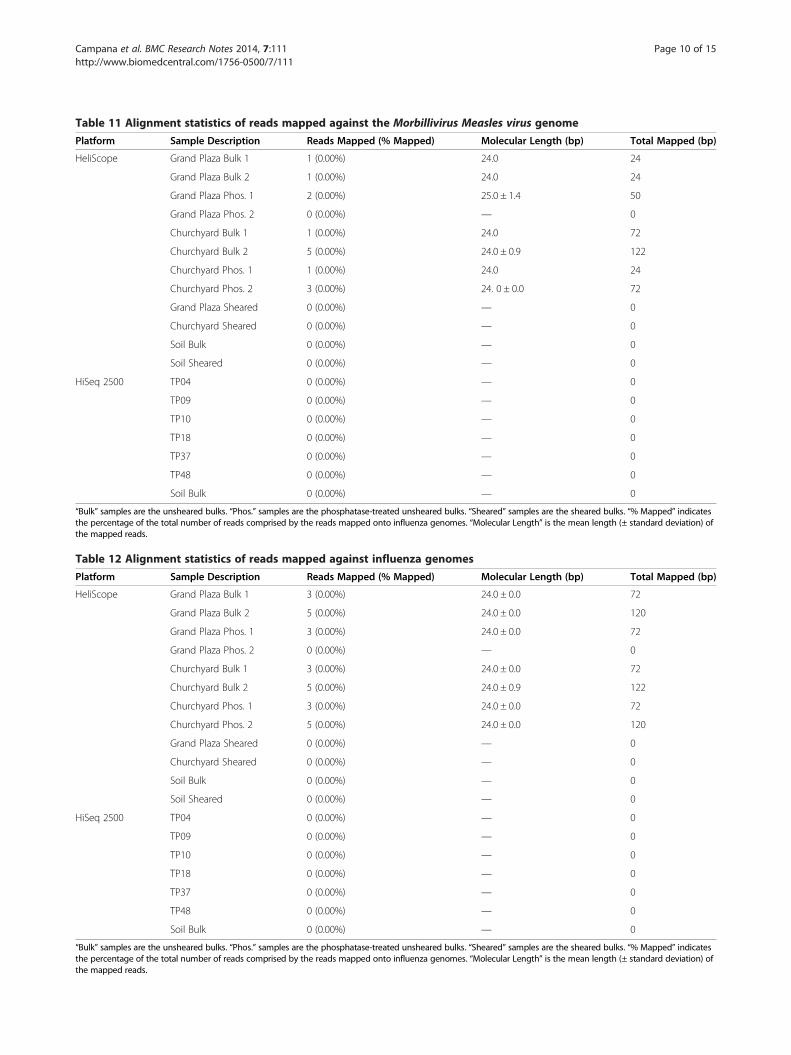
Table 11 Alignment statistics of reads mapped against the Morbillivirus Measles virus genome
Platform Sample Description Reads Mapped (% Mapped) Molecular Length (bp) Total Mapped (bp)
HeliScope Grand Plaza Bulk 1 1 (0.00%) 24.0 24
Grand Plaza Bulk 2 1 (0.00%) 24.0 24
Grand Plaza Phos. 1 2 (0.00%) 25.0 ± 1.4 50
Grand Plaza Phos. 2 0 (0.00%) — 0
Churchyard Bulk 1 1 (0.00%) 24.0 72
Churchyard Bulk 2 5 (0.00%) 24.0 ± 0.9 122
Churchyard Phos. 1 1 (0.00%) 24.0 24
Churchyard Phos. 2 3 (0.00%) 24. 0 ± 0.0 72
Grand Plaza Sheared 0 (0.00%) — 0
Churchyard Sheared 0 (0.00%) — 0
Soil Bulk 0 (0.00%) — 0
Soil Sheared 0 (0.00%) — 0
HiSeq 2500 TP04 0 (0.00%) — 0
TP09 0 (0.00%) — 0
TP10 0 (0.00%) — 0
TP18 0 (0.00%) — 0
TP37 0 (0.00%) — 0
TP48 0 (0.00%) — 0
Soil Bulk 0 (0.00%) — 0
“Bulk” samples are the unsheared bulks. “Phos.” samples are the phosphatase-treated unsheared bulks. “Sheared” samples are the sheared bulks. “% Mapped” indicatesthe percentage of the total number of reads comprised by the reads mapped onto influenza genomes. “Molecular Length” is the mean length (± standard deviation) ofthe mapped reads.
Table 12 Alignment statistics of reads mapped against influenza genomes
Platform Sample Description Reads Mapped (% Mapped) Molecular Length (bp) Total Mapped (bp)
HeliScope Grand Plaza Bulk 1 3 (0.00%) 24.0 ± 0.0 72
Grand Plaza Bulk 2 5 (0.00%) 24.0 ± 0.0 120
Grand Plaza Phos. 1 3 (0.00%) 24.0 ± 0.0 72
Grand Plaza Phos. 2 0 (0.00%) — 0
Churchyard Bulk 1 3 (0.00%) 24.0 ± 0.0 72
Churchyard Bulk 2 5 (0.00%) 24.0 ± 0.9 122
Churchyard Phos. 1 3 (0.00%) 24.0 ± 0.0 72
Churchyard Phos. 2 5 (0.00%) 24.0 ± 0.0 120
Grand Plaza Sheared 0 (0.00%) — 0
Churchyard Sheared 0 (0.00%) — 0
Soil Bulk 0 (0.00%) — 0
Soil Sheared 0 (0.00%) — 0
HiSeq 2500 TP04 0 (0.00%) — 0
TP09 0 (0.00%) — 0
TP10 0 (0.00%) — 0
TP18 0 (0.00%) — 0
TP37 0 (0.00%) — 0
TP48 0 (0.00%) — 0
Soil Bulk 0 (0.00%) — 0
“Bulk” samples are the unsheared bulks. “Phos.” samples are the phosphatase-treated unsheared bulks. “Sheared” samples are the sheared bulks. “% Mapped” indicatesthe percentage of the total number of reads comprised by the reads mapped onto influenza genomes. “Molecular Length” is the mean length (± standard deviation) ofthe mapped reads.
Campana et al. BMC Research Notes 2014, 7:111 Page 10 of 15http://www.biomedcentral.com/1756-0500/7/111

Table 13 Alignment statistics of HeliScope reads mappedagainst the Teposcolula Yucundaa soil Illumina-sequencedsample
SampleDescription
Reads Mapped(% Mapped)
MolecularLength (bp)
Total Mapped(bp)
Grand PlazaBulk 1
2,145,104(6.88%)
28.6 ± 5.8 61,255,930
Grand PlazaBulk 2
2,072,270(6.73%)
28.6 ± 5.8 59,267,370
Grand PlazaPhos. 1
1,641,432(6.25%)
28.1 ± 5.5 46,147,661
Grand PlazaPhos. 2
1,275,863(5.53%)
28.5 ± 5.7 36,333,528
Churchyard Bulk1
2,423,489(8.37%)
29.4 ± 6.1 71,205,930
Churchyard Bulk2
2,292,885(8.48%)
29.4 ± 6.1 67,327,090
ChurchyardPhos. 1
2,033,593(7.15%)
28.7 ± 5.9 58,421,595
ChurchyardPhos. 2
2,563,785(8.64%)
28.9 ± 5.9 74,145,815
Grand PlazaSheared
46 (1.13%) 24.5 ± 0.7 1,126
ChurchyardSheared
90 (1.70%) 24.6 ± 1.0 2,212
Soil Bulk 80,900 (9.62%) 29.0 ± 5.7 2,349,874
Soil Sheared 6,729 (5.46%) 27.1 ± 4.4 182,463
“Bulk” samples are the unsheared bulks. “Phos.” samples are thephosphatase-treated unsheared bulks. “Sheared” samples are the shearedbulks. “% Mapped” indicates the percentage of the total number of readscomprised by the reads mapped onto influenza genomes. “Molecular Length”is the mean length (± standard deviation) of the mapped reads.
Campana et al. BMC Research Notes 2014, 7:111 Page 11 of 15http://www.biomedcentral.com/1756-0500/7/111
compare different archaeological collections and data setsin terms of the likelihood of ancient pathogen recovery,our data are illustrative of the challenges encountered inhigh-throughput metagenomic pathogen studies. Theyemphasize the level of analytic rigor and proof required toauthenticate ancient pathogen analyses. In light of ourfindings, some previous metagenomic ancient DNA re-search claiming the discovery of pathogen-derived DNAmay need further verification. For instance, althoughThèves and colleagues [22] and Khairat and colleagues[53] report identifying Bordetella sp., Streptococcus pneu-moniae and Shigella dystenteriae [22] and Plasmodiumfalciparum and Toxoplasma gondii [53] respectively, nei-ther group sequenced archaeological controls such as soilor mummy wrappings. Thèves and colleagues amplifiedand sequenced microbial barcode 16S genes, increasingtheir results’ reliability since these genes are well charac-terized across a wide variety of species. Khairat and col-leagues, however, based their apicomplexan identificationson MEGAN analysis, with no attempt to evaluate thespecies-specificity of the sequenced molecules. Theseidentifications are therefore suspect since the genetic data-bases are skewed towards pathogenic members of thislineage. Additionally, a recent study by Chan and col-leagues [54] claiming the identification of multiple strainsof pathogenic tuberculosis (Mycobacterium tuberculosis)through non-targeted metagenomic sequencing has dem-onstrated insufficient analytical rigor to support their con-clusions. The authors aligned their sequences against asingle strain of pathogenic tuberculosis, but did not ac-count for misalignments or environmental contamin-ation with ubiquitous soil mycobacteria. Chan andcolleagues’ data merit reanalysis with appropriate envir-onmental controls. We recommend that the authors ofthese three studies demonstrate the veracity of theirfindings using a targeted capture approach and furtherbioinformatic analysis.
Soil complexityExamination of the Teposcolula soil DNA in MEGANrevealed that the microenvironment is complex, makingidentification of species-of-interest difficult. Whilethere were significant differences in the relative fre-quencies of microorganisms between the Grand Plazaand Churchyard samples (Additional file 6), these dif-ferences corresponded to variation in the distributionof environment-derived organisms across the site ratherthan pathogen-related frequency variation. For in-stance, we found significant differences in the preva-lence of Viridiplantae and the Rhizobiaceae, which arealmost certainly environmental contaminants.Moreover, the vast majority of HeliScope sequences
(~95%) are unknown due to both limitations in the data-bases and the non-specificity of the short sequences. Due
to the limitations of the genetic databases, some au-thors have amplified 16S genes (the most studiedmicrobial barcode genes) before high-throughput se-quencing in order to derive more species-informativeancient DNA metagenomic data sets (e.g. [24,55]).We have not conducted this PCR procedure heresince it negates the advantages of single-moleculesequencing. Instead, since the majority of sequencesin the soil Illumina data set are unknown, weattempted to use the Illumina data set as a pseudo-‘reference metagenome’ to identify environmentalcontaminants in our HeliScope data. However, only1.13%–9.62% of the sequences in the Helicos datasets (including the HeliScope soil sequence pools)mapped onto the Illumina soil sequences (Table 13).Given our low estimate of endogenous human DNA,we would expect >99% of the HeliScope reads (or100% in the case of the soil reads) to map onto theIllumina soil data set. This indicates that the HeliScopeis generating sequences with too low an informationcontent to align against reference genomes with suffi-cient precision to be considered likely matches.

Campana et al. BMC Research Notes 2014, 7:111 Page 12 of 15http://www.biomedcentral.com/1756-0500/7/111
DNA length, GC content and sequencing yieldsAgilent 2100 Bioanalyzer profiles of the unsheared DNA ex-tracts revealed the typical bimodal distribution of molecularlengths, with a large primary peak between 1000 and10,000 bp and a small secondary peak around 70 bp. Thesepeaks have previously been determined to correspond pri-marily to microbial contamination (large peak) and a mix-ture of fragmented contaminant and authentic ancientDNA (small peak) respectively [20,32,56]. Shearing the ex-tracts produced unimodal distributions with modal lengthsbetween 150 and 200 bp.Shearing of the DNA extracts reduced the total se-
quence yield drastically (5400–6800-fold reduction in thebone and 6.8-fold reduction in the soil), but yielded a con-comitant 8.0–10.6-fold enrichment in the proportion ofsequences mapping to the human genome in the bonesamples and a 2.3-fold enrichment in human sequences inthe soil. The cause of this yield reduction remains unclear.Variation between runs cannot completely explain it sincethe sheared and unsheared soil samples were sequencedon the same chip and samples from unrelated projectson the same chip had typical HeliScope sequencingyields [D. Jones, Pers. Comm. 2013]. Hypothetically,oxidative products (such as 8-oxoguanine and abasicsites) near the 3′-terminus of the sheared DNA moleculecould disrupt the downstream poly (A)-tailing reactionand reduce the overall sequencing yield. Costello and col-leagues [57] noted that shearing DNA samples to 150 bplengths using the Covaris instrument produced 8-oxoguanine lesions yielding C→A transversion artifactsin downstream Illumina library production. Nevertheless,8-oxoguanine lesions were rare and were only observablein libraries constructed from small initial DNA inputs. 8-oxoguanine lesions thus seem an implausible explanationfor our results, although the production of abasic sites atthe 3′-terminus cannot be ruled out.An alternative explanation is that the shearing caused a
decrease in the percentage of denaturable DNA molecules.DNA molecules that cannot be rendered single-stranded areunsequenceable on the HeliScope platform, thus reducingthe effective input DNA concentration. One mechanismcould be a relative increase in GC-rich sequences in thesheared samples, thus increasing the energy required for de-naturation of these molecules. Shearing longer GC-richDNAs into smaller and more numerous molecules wouldincrease the apparent GC content since this would raise therelative number of available 3′-termini from the GC-rich se-quences while the shorter AT-rich sequences would notshear into as many sequenceable fragments. Although theHeliScope platform is noted for its improved performancein both GC- and AT-rich regions in comparison to otherhigh-throughput technologies, it is not impervious to thesebiochemical effects. This would cause an asymmetric de-naturation step with only shorter and/or AT-rich molecules
becoming single-stranded in the pool. An alternative, similarmechanism is that the sheared DNA molecules (whichmostly derive from the high-molecular-weight peak) aremore likely to be cross-linked to proteins and themselves,thus preventing denaturation and sequencing. Since theshearing raised the average length of the molecules (in com-parison to the smaller ‘ancient DNA’ peak), this indicatesthat a far greater percentage of the sequenced molecules inthe sheared pools were derived from the high-molecular-weight ‘microbial contaminant’ peak than from the ‘ancientDNA’ peak. Many microbial metagenomes have been notedto be particularly GC-rich. Our samples have a high GCcontent at baseline (mean 62.4%, standard deviation 1.1% inthe unsheared bone and mean 62.1% in the soil). Shearingreduced the apparent GC content to 46.4% (standard devi-ation 1.0%) in the bones and a 58.1% in the soil. Moreover,there is a decrease in molecular length in comparison totheir unsheared counterparts (median lengths of 26–30 bpin the sheared samples versus 32–34 bp in the unshearedsamples). The AT enrichment, reduction in molecularlength and increased percentage of human molecules areconsistent with the denaturation hypothesis since the se-quenced sheared molecules would on average have lowerGC contents and be shorter, thus increasing the likelihoodof sequencing endogenous molecules.
Antarctic phosphatase treatmentGinolhac and colleagues [32] reported a 7.5–9.7-fold en-richment in the total sequence yield in their tSMS dataafter treatment of samples with Antarctic phosphatase.Conversely, we observed no such enrichment in ourdata; phosphatase-treated samples had 80%–104% rela-tive yield of their untreated counterparts. They also doc-umented a decrease in the proportion of endogenoussequences in the phosphatase-treated samples (34%–50%relative yield compared to untreated samples). In generalagreement with Ginolhac and colleagues’ results, we ob-served a decrease in endogenous human sequences afterphosphatase treatment (70%–90% relative yield). Thediscrepancy in overall yields may be due to differencesin the concentrations of sequenceable molecules in theDNA extracts, such as those possibly caused by varyingburial environments (Pleistocene permafrost versusMexican highlands), GC contents between the sample(~40% in their data versus ~60% in our individuals) andextraction procedures. If our samples had greater con-centrations of sequenceable molecules (which is likelysince our untreated sample yields exceeded those ofGinolhac and colleagues by ~100-fold), it is possiblethat we saturated the channel and thus limited the ef-fects of phosphatase treatment. The discrepancy be-tween Ginolhac and colleagues’ and our phosphatasetreatment results could also be a statistical artifact. Dueto the large number (typically 10–20 million) of

Campana et al. BMC Research Notes 2014, 7:111 Page 13 of 15http://www.biomedcentral.com/1756-0500/7/111
sequences generated in each HeliScope channel and thesignificant variation between replicate runs (relativestandard deviations ranged between 0.66% and 6.5%),the balance of effect sizes versus discriminatory poweris difficult to optimize.
ConclusionsEnvironmental contamination is a critical issue in an-cient DNA investigations of diseases. Both false positivesand false negatives are a serious problem. Previous investi-gations of ancient pathogens using both PCR and high-throughput sequencing may need to be re-evaluatedbecause pathogen identifications require extensive verifi-cation procedures due to the high risk of false positives.Currently, molecular enrichment to isolate moleculesof interest from the complex background (via eitherhybridization to probes or amplification of 16S regions)is the most promising route for ancient pathogenstudies.Furthermore, although the benefits of single-molecule
sequencing are promising, methodological challenges re-main in its application to ancient DNA research. Wefound that tSMS is not immune to GC-content-relatedbiases and that the benefits of phosphatase-treatment arenot universal. Analysis of these data remains complex dueto the short lengths of the sequenced molecules and limi-tations of public databases. Bioinformatic methods devel-oped for modern DNA analysis and for longer sequencesare not appropriate for HeliScope ancient DNA data.Finally, further methodological development is re-
quired for the identification of ancient pathogens to be-come routine and reliable. Until these techniquesbecome available, the burden-of-proof is on the re-searchers reporting the discovery of these disease agentsto demonstrate their results’ authenticity.
Availability of supporting dataThe data sets supporting the results of this article areavailable in the National Center for Biotechnology Infor-mation Sequence Read Archive, [SRP022977: http://www.ncbi.nlm.nih.gov/sra].
Additional files
Additional file 1: GenBank accessions for the genomes utilized inthis study.
Additional file 2: Alignment statistics of the HeliScope data setsagainst the rat and chimpanzee genomes.
Additional file 3: Concordance of the mitochondrial haplotypesbetween our Illumina data sets and those reported in Warinner.
Additional file 4: MEGAN analysis of the reads mapped to theYersinia pestis genome. The reads are non-specific for the pathogen.
Additional file 5: MEGAN analysis of the reads mapped to Rickettsiagenomes. The reads are non-specific for the pathogens. Instead, theycorrespond to common environmental organisms.
Additional file 6: MEGAN comparison of the BLAST hits betweenthe Grand Plaza (blue) and Churchyard (red) populations. Significantdifferences in relative species prevalence are highlighted in black. Thevast majority of the DNA sequences are derived from the environment,obscuring any ancient pathogen signal.
AbbreviationsADNA: Ancient deoxyribonucleic acid; BP: Base pairs; DNA: Deoxyribonucleicacid; PCR: Polymerase chain reaction; RNA: Ribonucleic acid; TSMS: Truesingle-molecule sequencing; VHF: Viral hemorrhagic fever.
Competing interestsThe authors declare that they have no competing interests.
Authors’ contributionsMGC conducted the molecular genetic analyses, performed thebioinformatic analysis and drafted the manuscript. NRG provided samplesand conceived the project. FJR participated in the coordination and designof the project. NT performed molecular experiments and conceived,coordinated and led the project. All authors read and approved the finalmanuscript.
AcknowledgementsThe Instituto Nacional de Antropología e Historia graciously suppliedspecimens for analysis. Christina Warinner collected bone and soil specimensin the field. Aurelien Ginolhac and Ludovic Orlando made an unpublishedversion of their manuscript available to us. Dan Jones (Helicos BioSciences)provided technical expertise. The Harvard Initiative for the Science of theHuman Past (Harvard University) funded the Illumina sequencing. LindaReynard helpfully commented on the manuscript. The Bauer Core (Faculty ofArts and Sciences Center for Systems Biology, Harvard University) andResearch Computing (Faculty of Arts and Sciences, Harvard University)provided facilities and technical expertise. The Mäxi Foundation and theFaculty of Arts and Sciences (Harvard University) supported MGC. NRG wasfunded by the Instituto Nacional de Antropología e Historia. The MäxiFoundation supported FJR. The Faculty of Arts and Sciences (HarvardUniversity) and the David Rockefeller Center for Latin American Studiessupported NT.
Author details1Department of Human Evolutionary Biology, Harvard University, PeabodyMuseum, 11 Divinity Avenue, Cambridge, MA 02138, USA. 2Centre forEvolutionary Medicine, Anatomy Institute, University of Zurich, 190Winterthurerstrasse, Zurich 8057, Switzerland. 3Instituto Nacional deAntropología e Historia, Mexico City, Mexico.
Received: 19 February 2014 Accepted: 20 February 2014Published: 25 February 2014
References1. Papagrigorakis MJ, Yapijakis C, Synodinos PN, Baziotopoulou-Valavani E: DNA
examination of ancient dental pulp incriminates typhoid fever as a probablecause of the Plague of Athens. Int J Infect Dis 2006, 10:206–214.
2. Shapiro B, Rambaut A, Gilbert MTP: No proof that typhoid caused the Plagueof Athens (a reply to Papagrigorakis et al.). Int J Infect Dis 2006, 10:334–335.
3. Drancourt M, Aboudharam G, Signoli M, Dutour O, Raoult D: Detection of400-year-old Yersinia pestis DNA in human dental pulp: an approach tothe diagnosis of ancient septicemia. Proc Natl Acad Sci USA 1998,95:12637–12640.
4. Zink AR, Sola C, Reischl U, Grabner W, Rastogi N, Wolf H, Nerlich AG:Characterization of Mycobacterium tuberculosis complex DNAs fromEgyptian mummies by spoligotyping. J Clin Microbiol 2003, 41:359–367.
5. Nguyen-Hieu T, Aboudharam G, Signoli M, Rigeade C, Drancourt M, Raoult D:Evidence of a louse-born outbreak involving typhus in Douai, 1710–1712during the War of Spanish Succession. PLoS One 2010, 5:e15405.
6. Gernaey AM, Minnikin DE, Copley MS, Dixon RA, Middleton JC, Roberts CA:Mycolic acids and ancient DNA confirm an osteological diagnosis oftuberculosis. Tuberculosis 2001, 81:259–265.

Campana et al. BMC Research Notes 2014, 7:111 Page 14 of 15http://www.biomedcentral.com/1756-0500/7/111
7. Kolman CJ, Centurion-Lara A, Lukehart SA, Owsley DA, Tuross N: Identification ofTreponema pallidum subspecies pallidum in a 200-year-old skeletal specimen.J Infect Dis 2060–2063, 1999:180.
8. Bianucci R, Rahalison L, Ferroglio E, Massa ER, Signoli M: Détection del’antigène F1 de Yersinia pestis dans les restes humains anciens à l’aided’un test de diagnostic rapide. C R Biol 2007, 330:747–754.
9. Bianucci R, Rahalison L, Massa ER, Peluso A, Ferroglio E, Signoli M: Technicalnote: a rapid diagnostic test detects plague in ancient human remains:an example of the interaction between archaeological and biologicalapproaches (Southeastern France, 16th‒18th centuries). Am J PhysAnthropol 2008, 136:361–367.
10. Bianucci R, Rahalison L, Peluso A, Massa ER, Ferroglio E, Signoli M, LangloisJ-Y, Gallien V: Plague immunodetection in remains of religious exhumedfrom burial sites in central France. J Arch Sci 2009, 36:616–621.
11. Haensch S, Bianucci R, Signoli M, Rajerison M, Schultz M, Kacki S, VermuntM, Weston DA, Hurst D, Achtman M, Carniel E, Bramanti B: Distinct clonesof Yersinia pestis caused the Black Death. PLoS Pathog 2010, 6:e1001134.
12. Kacki S, Rahalison L, Rajerison M, Ferroglio E, Bianucci R: Black Death in therural cemetery of Saint-Laurent-de-la-Cabrerisse Aude-Languedoc,southern France, 14th century: immunological evidence. J Arch Sci 2011,38:581–587.
13. Schuenemann VJ, Bos K, DeWitte S, Schmedes S, Jamieson J, Mittnik A,Forrest S, Coombes BK, Wood JW, Earn DJD, White W, Krause J, Poinar HN:Targeted enrichment of ancient pathogens yielding the pPCP1 plasmidof Yersinia pestis from victims of the Black Death. Proc Natl Acad Sci USA2011, 108:E746–E752.
14. Bos KI, Schuenemann VJ, Golding GB, Burbano HA, Waglechner N, CoombesBK, McPhee JB, DeWitte SN, Meyer M, Schmedes S, Wood J, Earn DJD,Herring DA, Bauer P, Poinar HN, Krause J: A draft genome of Yersinia pestisfrom victims of the Black Death. Nature 2011, 478:506–510.
15. Barnes I, Thomas MG: Evaluating bacterial pathogen DNA preservation inmuseum osteological collections. P Roy Soc B 2006, 273:645–653.
16. Bouwman AS, Brown TA: The limits of biomolecular palaeopathology:ancient DNA cannot be used to study venereal syphilis. J Arch Sci 2005,32:703–713.
17. Gilbert MTP, Cuccui J, White W, Lynnerup N, Titball RW, Cooper A, PrenticeMB: Absence of Yersinia pestis-specific DNA in human teeth from fiveEuropean excavations of putative plague victims. Microbiology 2004,150:341–354.
18. Gilbert MTP, Rudbeck L, Willerslev E, Hansen AJ, Smith C, Penkman KEH,Prangenberg K, Nielsen-Marsh CM, Jans ME, Arthur P, Lynnerup N,Turner-Walker G, Biddle M, Kjølbye-Biddle B, Collins MJ: Biochemicaland physical correlates of DNA contamination in archaeologicalhuman bones and teeth excavated at Matera, Italy. J Arch Sci 2005,32:785–793.
19. Gilbert MTP, Hansen AJ, Willerslev E, Turner-Walker G, Collins M: Insightsinto the processes behind the contamination of degraded human teethand bone samples with exogenous sources of DNA. Int J Osteoarchaeol2006, 16:156–164.
20. Pääbo S, Poinar H, Serre D, Jaenicke-Després V, Hebler J, Rohland N, Kuch M,Krause J, Vigilant L, Hofreiter M: Genetic analyses from ancient DNA. AnnuRev Genet 2004, 38:645–679.
21. Drancourt M, Raoult DR: Palaeomicrobiology: current issues andperspectives. Nat Rev Microbiol 2005, 3:23–35.
22. Roberts C, Ingham S: Using ancient DNA analysis in palaeopathology: acritical analysis of published papers and recommendations for futurework. Int J Osteoarchaeol 2008, 18:600–613.
23. Tsangaras K, Greenwood AD: Museums and disease: using tissue archiveand museum samples to study pathogens. Ann Anat 2012, 194:58–73.
24. Thèves C, Senescau A, Vanin S, Keyser C, Ricaut FX, Alekseev AN, DabernatH, Ludes B, Fabre R, Crubézy: Molecular identification of bacteria by totalsequence screening: determining the cause of death in ancient humansubjects. PLoS One 2011, 6:e21733.
25. Knapp M, Hofreiter M: Next generation sequencing of ancient DNA:requirements, strategies and perspectives. Genes 2010, 1:227–243.
26. Acuna-Soto R, Stahle DW, Therrell MD, Griffin RD, Cleaveland MK: When halfof the population died: the epidemic of hemorrhagic fevers of 1576 inMexico. FEMS Microbiol Lett 2004, 240:1–5.
27. Warinner C, Robles García N, Spores R, Tuross N: Disease, demography, anddiet in early colonial New Spain: investigation of a sixteenth-century Mix-tec cemetery at Teposcolula Yucundaa. Lat Am Antiq 2012, 23:467–489.
28. Marr JS, Kiracofe JB: Was the huey cocoliztli a haemorrhagic fever? MedHist 2000, 44:341–362.
29. Warinner CG: Life and Death at Teposcolula Yucundaa: Mortuary,Archaeogenetic, and Isotopic Investigations of the Early Colonial Period inMexico, PhD thesis. Harvard University, Human Evolutionary BiologyDepartment; 2010.
30. Cooper A, Poinar HN: Ancient DNA: do it right or not at all. Science 2000, 289:1139.31. Debruyne R, Schwarz C, Poinar H: Comment on ‘Whole-genome shotgun
sequencing of mitochondria from ancient hair shafts”. Science 2008,322:857a.
32. Orlando L, Ginolhac A, Raghavan M, Vilstrup J, Rasmussen M, Magnussen K,Steinmann KE, Kapranov P, Thompson JF, Zazula G, Froese D, Moltke I,Shapiro B, Hofreiter M, Al-Rasheid KAS, Gilbert MTP, Willerslev E: Truesingle-molecule DNA sequencing of a pleistocene horse bone. GenomeRes 2011, 21:1705–1719.
33. Ginolhac A, Vilstrup J, Stenderup J, Rasmussen M, Stiller M, Shapiro B, ZazulaG, Froese D, Steinmann KE, Thompson JF, Al-Rasheid KAS, Gilbert TMP,Willerslev E, Orlando L: Improving the performance of true singlemolecule sequencing for ancient DNA. BMC Genomics 2012, 13:177.
34. Masella AP, Bartram AK, Truszkowski JM, Brown DG, Neufeld JD: PANDAseq:PAired-eND Assembler for Illumina sequences. BMC Bioinformatics 2012,13:31.
35. Lassman T, Hayashizaki Y, Daub CO: TagDust—a program to eliminateartifacts from next generation sequencing data. Bioinformatics 2009,25:2839–2840.
36. Li W, Fu L, Niu B, Wu S, Wooley J: Ultrafast clustering algorithms formetagenomic sequence analysis. Brief Bioinform 2012, 13:656–668.
37. Andrews S: Fastqc: A Quality Control Tool For High Throughput SequenceData. www.bioinformatics.babraham.ac.uk/projects/fastqc/.
38. Li H, Handsaker B, Wysoker A, Fennell T, Ruan J, Homer N, Marth G,Abecasis G, Durbin R, 1000 Genome Project Data Processing Subgroup: Thesequence alignment/map format and SAMtools. Bioinformatics 2078–2079,2009:25.
39. Li H, Durbin R: Fast and accurate short read alignment with Burrows-Wheelertransform. Bioinformatics 2009, 25:1754–1760.
40. Li H, Durbin R: Fast and accurate long read alignment with Burrows-Wheelertransform. Bioinformatics 2010, 26:589–595.
41. Schubert M, Ginolhac A, Lindgreen S, Thompson JF, Al-Rasheid KAS,Willerslev E, Krogh A, Orlando L: Improving ancient DNA read mappingagainst modern reference genomes. BMC Genomics 2012, 13:178.
42. Zhang Z, Schwartz S, Wagner L, Miller W: A greedy algorithm for aligningDNA sequences. J Comput Biol 2000, 7:203–214.
43. Huson D, Mitra S, Ruscheweyh H-J, Weber N, Schuster SC: Integrativeanalysis of environmental sequences using MEGAN4. Genome Res 2011,2011(21):1552–1560.
44. R Core Team: R: A Language And Environment For Statistical Computing.Vienna: R Foundation for Statistical Computing; 2013.
45. Hansen MA, Oey H, Fernandez-Valverde S, Jung C-H, Mattick JS: Biopieces: ABioinformatics Toolset and Framework. www.biopieces.org.
46. Jónsson H, Ginolhac A, Schubert M, Johnson P, Orlando L: mapDamage2.0:fast approximate Bayesian estimates of ancient DNA damageparameters. Bioinformatics 2013, 29(13:1682–1684.
47. Briggs AW, Stenzel U, Johnson PLF, Green RE, Kelso J, Prüfer K, Meyer M,Krause J, Ronan MT, Lachmann M, Pääbo S: Patterns of damage ingenomic DNA sequences from a Neandertal. Proc Natl Acad Sci USA 2007,104:14616–14621.
48. Acuna-Soto R, Calderon Romero L, Maguire JH: Large epidemics ofhemorrhagic fevers in Mexico 1545–1815. Am J Trop Med Hyg 2000,62:733–739.
49. Acuna-Soto R, Stahle DW, Cleaveland MK, Therrell MD: Megadrought andmegadeath in 16th century Mexico. Emerg Infect Dis 2002, 8:360–362.
50. Gilbert MTP, Rambaut A, Wlasiuk G, Spira TJ, Pitchenik AE, Worobey M: Theemergence of HIV/AIDS in the Americas and beyond. Proc Natl Acad SciUSA 2007, 104:18566–18570.
51. Fordyce SL, Ávila-Arcos MC, Rasmussen M, Cappellini E, Romero-Navarro JA,Wales N, Alquezar-Planas DE, Penfield S, Brown TA, Vielle-Calzada J-P,Montiel R, Jørgensen T, Odegaard N, Jacobs M, Arriaza B, Higham TFG,Bronk Ramsey C, Willerslev E, Gilbert MTP: Deep sequencing of RNA fromancient maize kernels. PLoS One 2013, 8:e50961.
52. Worobey M, Gemmel M, Teuwen DE, Haselkorn T, Kunstman K, Bunce M,Muyembe J-J, Kabongo J-MM, Kalengayi RM, Van Marck E, Gilbert MTP,

Campana et al. BMC Research Notes 2014, 7:111 Page 15 of 15http://www.biomedcentral.com/1756-0500/7/111
Wolinksy SM: Direct evidence of extensive diversity of HIV-1 in Kinshasaby 1960. Nature 2008, 455:661–664.
53. Khairat R, Ball M, Chang C-CH, Bianucci R, Nerlich AG, Trautmann M, Ismail S,Shanab GML, Karim AM, Gad YZ, Pusch CM: First insights into the meta-genome of Egyptian mummies using next-generation sequencing.J Appl Genetics 2013, 54:309–325.
54. Chan JZ-M, Sergeant MJ, Lee OY-C, Minnikin DE, Besra GS, Pap I, SpigelmanM, Donoghue HD: Metagenomic analysis of tuberculosis in a mummy.N Engl J Med 2013, 369:289–290.
55. Adler CJ, Dobney K, Weyrich LS, Kaidonis J, Walker AW, Haak W, BradshawCJA, Townsend G, Sołtysiak A, Alt KW, Parkhill J, Cooper A: Sequencingancient calcified dental plaque shows changes in oral microbiota withdietary shifts of the Neolithic and Industrial revolutions. Nat Genet 2013,45:450–455.
56. Malmström H, Svensson EM, Gilbert MTP, Willerslev E, Götherström A,Holmlund G: More on contamination: the use of asymmetric molecularbehavior to identify authentic ancient human DNA. Mol Biol Evol 2007,24:998–1004.
57. Costello M, Pugh TJ, Fennell TJ, Stewart C, Lichtenstein L, Meldrim JC,Fostel JF, Friedrich DC, Perrin D, Dionne D, Kim S, Gabriel SB, Lander ES,Fisher S, Getz G: Discovery and characterization of artifactual mutationsin deep coverage targeted capture sequencing data due to oxidativeDNA damage during sample preparation. Nucleic Acids Res 2013, 41:e67.
doi:10.1186/1756-0500-7-111Cite this article as: Campana et al.: False positives complicate ancientpathogen identifications using high-throughput shotgun sequencing.BMC Research Notes 2014 7:111.
Submit your next manuscript to BioMed Centraland take full advantage of:
• Convenient online submission
• Thorough peer review
• No space constraints or color figure charges
• Immediate publication on acceptance
• Inclusion in PubMed, CAS, Scopus and Google Scholar
• Research which is freely available for redistribution
Submit your manuscript at www.biomedcentral.com/submit