False Aneurysms After Sagittal Split Ramus Osteotomies
8
J Oral Maxillofac Surg 70:e58-e65, 2012 False Aneurysms After Sagittal Split Ramus Osteotomies David S. Precious, CM, DDS, MSc,* Joel E. Powell, DDS, MD, MSc,† Aysegul M. Tuzuner, DDS, PhD,‡ Matthias Schmidt, MD,§ Jean-Charles Doucet, DMD, MD, MSc, and Robert Vandorpe, MD¶ False aneurysms of the face, also referred to as pseu- doaneurysms or traumatic aneurysms, are very un- common. 1-3 The superficial temporal artery is the most common site for a facial pseudoaneurysm, fol- lowed by the maxillary and facial arteries. 4,5 They have also been reported in the external carotid, inter- nal carotid, supraorbital, occipital, labial and lingual arteries, and in tributaries of the maxillary artery, including its sphenopalatine, meningeal, and descend- ing palatine branches. 1-10 Possible causes include fa- cial fractures, penetrating injuries, gunshot wounds, blunt trauma, and iatrogenic trauma secondary to a surgical procedure. 4,5,8,9 Although rare, iatrogenic pseudoaneurysms of the fa- cial vasculature have been reported after mandibular surgical procedures. Case reports of false aneurysms exist in the literature after augmentation mentoplasty, circumferential mandibular wiring, temporomandibular joint arthroplasty, arthroscopy, condylar osteotomy, al- veoloplasty, and mandibular vertical ramus osteoto- mies. 1,3,4,8 Significant bleeding and swelling is usually noted at the time of surgery, but delayed hemorrhage after an uncomplicated procedure has also been re- ported. 1 Although the sagittal split ramus osteotomy (SSRO) is a very popular procedure for correcting a host of mandibular deformities, few cases of false aneurysm resulting from this procedure have been reported. 11-13 Complications of facial artery pseudoaneurysms in- clude swelling, pain, and rupture leading to severe hemorrhage with the possibility of exsanguination and death. Oropharyngeal hemorrhage secondary to a facial artery false aneurysm secondary to a gunshot wound has even been reported, with a clear risk to airway patency. 7 Most false aneurysms of the face are a delayed phenomenon usually presenting 1 to 16 weeks after injury but possibly even years later. 4,10 They are thought to result from an inciting tangential tear or partial transection in an artery. 4,8,10 This leads to ex- travasation of blood under pressure into the surround- ing tissues, which eventually slows and then ceases when the pressure within the hematoma exceeds the feeding arterial pressure. Arterial flow distal to the vessel injury and adjacent hematoma is thus restored. The resultant hematoma organizes and a fibrous pseudocapsule forms. This sac may then eventually undergo liquefaction and the wall endothelialize. Be- cause the vessel wall was transected, the endothelial- ized sac is in continuity with the vessel lumen. A pulsatile, expanding mass characterizes the false an- eurysm. The distinction between a true and a false aneurysm is that a true aneurysm contains all layers of the vessel wall (externa, media, and intima), whereas a pseudoaneurysm has an endothelialized pseudocap- sule only. Reported treatment approaches include primary surgical resection alone, arterial embolization alone, or embolization followed by surgical resection. Super- selective arterial embolization alone appears to be the favored approach, particularly in deep lesions. 4,7,10 This report describes 3 cases of false aneurysm of the facial artery after SSRO procedures in the correction of dentofacial deformities, the diagnostic challenges, and subsequent management. Report of Cases CASE 1 Case 1 was a 32-year-old man referred for correction of asymmetric mandibular prognathism and bimaxillary cant. His medical history was significant for rheumatoid arthritis Received from Dalhousie University, Halifax, Nova Scotia, Canada. *Professor, Department of Oral and Maxillofacial Surgery, and Dean Emeritus. †Former Senior Resident, Department of Oral and Maxillofacial Surgery. ‡Former Fellow, Department of Oral and Maxillofacial Surgery. §Assistant Professor, Department of Diagnostic Radiology. Senior Resident, Department of Oral and Maxillofacial Surgery. ¶Assistant Professor, Department of Diagnostic Radiology. Address correspondence and reprint requests to Dr Precious: Department of Oral and Maxillofacial Surgery, Dalhousie Univer- sity, 1459 Oxford Street, Halifax, Nova Scotia, Canada B3H 4R2; e-mail: [email protected] © 2012 American Association of Oral and Maxillofacial Surgeons 0278-2391/12/7001-0$36.00/0 doi:10.1016/j.joms.2011.09.007 e58
Transcript of False Aneurysms After Sagittal Split Ramus Osteotomies

c
nap(or
0
d
J Oral Maxillofac Surg70:e58-e65, 2012
False Aneurysms After Sagittal SplitRamus Osteotomies
David S. Precious, CM, DDS, MSc,*
Joel E. Powell, DDS, MD, MSc,† Aysegul M. Tuzuner, DDS, PhD,‡
Matthias Schmidt, MD,§ Jean-Charles Doucet, DMD, MD, MSc,�
and Robert Vandorpe, MD¶
tp
False aneurysms of the face, also referred to as pseu-doaneurysms or traumatic aneurysms, are very un-ommon.1-3 The superficial temporal artery is the
most common site for a facial pseudoaneurysm, fol-lowed by the maxillary and facial arteries.4,5 Theyhave also been reported in the external carotid, inter-nal carotid, supraorbital, occipital, labial and lingualarteries, and in tributaries of the maxillary artery,including its sphenopalatine, meningeal, and descend-ing palatine branches.1-10 Possible causes include fa-cial fractures, penetrating injuries, gunshot wounds,blunt trauma, and iatrogenic trauma secondary to asurgical procedure.4,5,8,9
Although rare, iatrogenic pseudoaneurysms of the fa-cial vasculature have been reported after mandibularsurgical procedures. Case reports of false aneurysmsexist in the literature after augmentation mentoplasty,circumferential mandibular wiring, temporomandibularjoint arthroplasty, arthroscopy, condylar osteotomy, al-veoloplasty, and mandibular vertical ramus osteoto-mies.1,3,4,8 Significant bleeding and swelling is usually
oted at the time of surgery, but delayed hemorrhagefter an uncomplicated procedure has also been re-orted.1 Although the sagittal split ramus osteotomySSRO) is a very popular procedure for correcting a hostf mandibular deformities, few cases of false aneurysmesulting from this procedure have been reported.11-13
Received from Dalhousie University, Halifax, Nova Scotia, Canada.
*Professor, Department of Oral and Maxillofacial Surgery, and
Dean Emeritus.
†Former Senior Resident, Department of Oral and Maxillofacial
Surgery.
‡Former Fellow, Department of Oral and Maxillofacial Surgery.
§Assistant Professor, Department of Diagnostic Radiology.
�Senior Resident, Department of Oral and Maxillofacial Surgery.
¶Assistant Professor, Department of Diagnostic Radiology.
Address correspondence and reprint requests to Dr Precious:
Department of Oral and Maxillofacial Surgery, Dalhousie Univer-
sity, 1459 Oxford Street, Halifax, Nova Scotia, Canada B3H 4R2;
e-mail: [email protected]
© 2012 American Association of Oral and Maxillofacial Surgeons
278-2391/12/7001-0$36.00/0
oi:10.1016/j.joms.2011.09.007
e58
Complications of facial artery pseudoaneurysms in-clude swelling, pain, and rupture leading to severehemorrhage with the possibility of exsanguinationand death. Oropharyngeal hemorrhage secondary to afacial artery false aneurysm secondary to a gunshotwound has even been reported, with a clear risk toairway patency.7
Most false aneurysms of the face are a delayedphenomenon usually presenting 1 to 16 weeks afterinjury but possibly even years later.4,10 They arehought to result from an inciting tangential tear orartial transection in an artery.4,8,10 This leads to ex-
travasation of blood under pressure into the surround-ing tissues, which eventually slows and then ceaseswhen the pressure within the hematoma exceeds thefeeding arterial pressure. Arterial flow distal to thevessel injury and adjacent hematoma is thus restored.The resultant hematoma organizes and a fibrouspseudocapsule forms. This sac may then eventuallyundergo liquefaction and the wall endothelialize. Be-cause the vessel wall was transected, the endothelial-ized sac is in continuity with the vessel lumen. Apulsatile, expanding mass characterizes the false an-eurysm. The distinction between a true and a falseaneurysm is that a true aneurysm contains all layers ofthe vessel wall (externa, media, and intima), whereasa pseudoaneurysm has an endothelialized pseudocap-sule only.
Reported treatment approaches include primarysurgical resection alone, arterial embolization alone,or embolization followed by surgical resection. Super-selective arterial embolization alone appears to be thefavored approach, particularly in deep lesions.4,7,10
This report describes 3 cases of false aneurysm of thefacial artery after SSRO procedures in the correctionof dentofacial deformities, the diagnostic challenges,and subsequent management.
Report of Cases
CASE 1Case 1 was a 32-year-old man referred for correction of
asymmetric mandibular prognathism and bimaxillary cant.
His medical history was significant for rheumatoid arthritis
PRECIOUS ET AL e59
diagnosed approximately 7 years previously, asthma, gastro-esophageal reflux, and a functional heart murmur. Preoper-ative medications included indomethacin, glucosamine,chondroitin sulfate, fluticasone inhaler daily, a salbutamolinhaler as needed, and omeprazole. Previous surgeries in-cluded tonsillectomy at 8 years of age without any knowncomplications. Preoperative blood work showed a normalcomplete blood count and electrolytes.
On November 23, 2005, the patient was admitted andtaken to the operating room, where, under general anes-thetic, bilateral SSROs (BSSROs) of the mandible were per-formed in the standard fashion. During the right-sided ver-tical osteotomy in the molar region with a Lindemann-likebur on a rotary handpiece, brisk pulsatile bleeding occurredfrom the underlying soft tissue envelope at the inferior borderof the mandible. The location of the bleeding was consistentwith the facial artery. The area was packed with moist gauzeafter completion of the vertical osteotomy, providing tempo-rary hemostasis, and the sagittal splitting procedure of theright ramus was completed without incident. The gauze wasremoved and continued bleeding, although less brisk, wasnoted. Upon careful inspection, a violation of the periosteumwas apparent; however, clear visualization of a bleeding vesselwas not observed. The area was again packed tightly withmoist gauze and hemostasis was obtained. With the packs leftin place, the SSRO was completed on the left side of themandible without further incident.
A single-piece Le Fort I osteotomy with differential im-paction and miniplate fixation was performed next withoutcomplication or evidence of further bleeding from the rightSSRO site. After securing the occlusion with intermaxillaryfixation (IMF), the right mandible was reexamined forbleeding. Although there was still some active bleeding, itwas deemed to be quite minor. Fixation of the bilateralmandibular osteotomies was accomplished with miniplatefixation and monocortical screws without incident. Thearea where the bleeding occurred was tightly packed withabsorbable gelatin sponge, resulting in complete cessationof active bleeding. All wounds were closed with 3-0 plaingut sutures and the right sagittal osteotomy incision wasoversutured with 3-0 polyglactin 910. At the completion ofthe procedure, tight elastic IMF was reapplied and a pres-sure dressing was applied over the angle of the mandible.The patient was extubated and transferred to the postanes-thetic care unit and then to the ward. His postoperative stayon the ward was uneventful; he developed a normal amountof edema and no evidence of hematoma or active bleedingfrom the right mandible. The premasseteric notch area atthe inferior border was fuller on the right side than on theleft owing to the presence of the subperiosteal absorbablegelatin sponge. The pressure dressing was removed beforedischarge home on postoperative day 2.
Two weeks postoperatively, the patient was seen in theoutpatient clinic for routine follow-up, when the tight IMFwas removed, and the patient appeared to be doing verywell. Although the right side was still larger than the left,the edema was significantly decreased overall. Several dayslater, the patient reported an episode of minor oozing fromthe intraoral incision site on the right mandible. Uponevaluation in the outpatient clinic, no bleeding was noted,there was no abnormal swelling, and there were no clearsigns of infection. The patient returned home with nofurther difficulties until 2 days later, when he again reportedbleeding from the incision site. Upon evaluation, there wasevidence of slightly increased swelling and dark, clot-likematerial in the right mandibular vestibule. Again, no active
bleeding was occurring and none could be elicited by minormanipulation of the area. The patient remained pain free.Two days after that episode, he presented to a local emer-gency room complaining of swelling and pain in the rightsubmandibular region, with occasional episodes of drainageof dark material from the same area. He was diagnosed bythe emergency room physician with an infection and pre-scribed an oral antibiotic, although the authors’ service wasnot informed of this at the time.
The patient attended a scheduled follow-up appointment2 days later (3 weeks postoperatively) in the outpatientclinic. At this time, he reported a 24-hour history of increasingswelling and pain. A small amount of dark, viscous materialwith a clot-like component could be expressed through theintraoral incision site, although there was no active bleeding.Swelling of the right submandibular area and lateral mandiblewas moderate and no pulsations or bruits were noted. Thepatient was admitted with a tentative diagnosis of infection,placed on intravenous antibiotics, and angiography was ar-ranged to rule out a potential false aneurysm of the right facialartery secondary to the BSSRO.
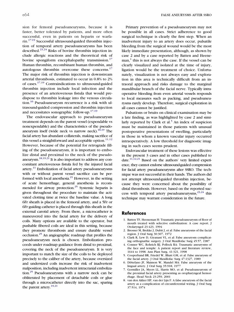
The angiogram was recorded the following day (Decem-ber 14, 2006) under conscious sedation and showed a2.7-cm pseudoaneurysm coming off the right facial arteryjust anterior to the angle of the mandible (Fig 1). Onceidentified by the interventional neuroradiologist, the lesionwas treated by proximal coil embolization during the samesession. There were no complications and an angiogramafter treatment showed no filling of the false aneurysm. Thepatient recovered uneventfully from the procedure and wasdischarged home the following day with a recommendationto repeat angiography in a few weeks to ensure resolution.The patient had significant resolution of his discomfort and
FIGURE 1. Case 1. Lateral view from a right external carotidangiogram shows a jet of contrast material (arrow) filling a largepseudoaneurysm.
Precious et al. False Aneurysms After SSRO. J Oral Maxillofac Surg
2012.
ccawalptabc
eae
t
P
e60 FALSE ANEURYSMS AFTER SSRO
swelling over the following week, with no further evidenceof bleeding. Repeat angiography was performed 3 weeksafter the embolization procedure and showed no signs ofresidual false aneurysm (Fig 2). The patient was last seenapproximately 6 weeks later and was asymptomatic andwith no evidence of any lesion.
CASE 2Case 2 involved a 26-year-old man referred for correction
of vertical maxillary excess and mandibular deficiency. Thepatient’s medical history was significant for mild asthma anddepression, and he was taking only sertraline hydrochloridepreoperatively. Routine blood work showed a normal com-plete blood count and electrolytes.
On October 12, 2005, the patient was admitted and takento the operating room, where, under general anesthetic,BSSRO of the mandible and a 1-piece Le Fort I maxillaryosteotomy with impaction were undertaken in the standardfashion. The bone was noted to be slightly more yellowthan usual and quite dense, consistent with previous tetra-ycline ingestion; however, the osteotomies and splits oc-urred without any difficulties. Fixation of the mandible wasccomplished with miniplates and monocortical screws, andire osteosynthesis of the maxilla was used. There was no
bnormal intraoperative bleeding, and the estimated bloodoss of the entire procedure was 150 mL. The patient waslaced in elastic IMF and, after a brief stay in the postanes-hetic care unit, he convalesced on the ward with the usualmount of edema and discomfort. There was no abnormalleeding after the procedure, and the patient was dis-
FIGURE 2. Case 1. Lateral view from a right common carotidangiogram 3 weeks after coil embolization shows coils (arrow) inthe thrombosed facial artery. The pseudoaneurysm and parentvessel no longer opacify.
Precious et al. False Aneurysms After SSRO. J Oral Maxillofac Surg2012.
harged on the second postoperative day. 2
The tight IMF was removed at 2 weeks and the facialdema, although still present, had decreased significantly asnticipated. When the patient returned at 5 weeks postop-ratively, he complained of tenderness in the right angle of
FIGURE 3. Case 2. Color Doppler images show A, characteristicto-and-fro blood flow in a pseudoaneurysm and B, arterial inflow tohe pseudoaneurysm (arrow).
recious et al. False Aneurysms After SSRO. J Oral Maxillofac Surg
012.
PRECIOUS ET AL e61
the mandible and reported the swelling had not subsided inthis area. Examination showed a soft, tender mass in theright submandibular region and slight tenderness over thearea of the fixation plate. A panoramic radiograph did notshow any evidence of hardware failure or osteomyelitis. Aworking diagnosis of minor infection was made, and thepatient was started on clindamycin orally.
Follow-up examination 1 week later found no change inthe size or tenderness of the right submandibular lesiondespite further decreases in facial edema, although thetenderness over the fixation plate was gone. The antibioticswere changed to amoxicillin/clavulanic acid. One weeklater (7 weeks postoperatively), the lesion had not changedin size and mild discomfort was still present, but uponbimanual palpation, the mass was consistent in texture andlocation with the submandibular gland. Furthermore, nosaliva could be expressed from the right gland, although itwas free-flowing from the left. The lesion was no longerjudged to represent an infectious process, but rather ablockage of the submandibular duct, perhaps secondary toa mucous plug. High liquid intake and sour candies wererecommended in hopes of spontaneous recovery.
Two weeks later, the patient’s symptom of mild discomforthad not changed; however, examination showed that salivawas flowing from the right gland. Palpation of the mass andinspection of the neck in the submandibular region showedvascular pulsations. A duplex ultrasound was arranged for thenext day, and it clearly showed a pseudoaneurysm of anarterial branch of the external carotid artery measuring 2.3 cmin diameter (Fig 3). Because the patient was mostly asymptom-atic and the lesion had not been expanding, the patient wasdetermined by the interventional radiologist to not be emer-gent; accordingly, he was scheduled for angiography withembolization 2 weeks later.
On January 6, 2006, the patient was admitted and underconscious sedation had a coil embolization of the right facialartery pseudoaneurysm without complication. He was dis-charged the following morning, and the interventional ra-diologist recommended a repeat ultrasound in severalweeks. This was performed 6 weeks later, and there was noblood flow to the lesion. At the most recent follow-up,several days after the ultrasound, the patient reported nosymptoms and a smaller lesion since the embolization. Ex-amination showed no swelling or visible lesion but a palpa-ble, firm, nontender lump in the corresponding locationestimated to be smaller than 1 cm. Clinical follow-up at 1year was unremarkable.
CASE 3Case 3 involved a 20-year-old man referred for correction
of bimalar deficiency, asymmetric vertical maxillary excess,and mandibular prognathism. The patient was otherwisehealthy. His surgical history included a tonsillectomy andadenoidectomy at 9 years of age without any known com-plication. Preoperative blood work showed a normal com-plete blood count and electrolytes.
The patient was first planned to undergo surgery onJanuary 5, 2011. The procedure was postponed because ofdentoalveolar-mandibular trauma from a fall on December 31,2010. On March 9, 2011, the patient was admitted and takento the operating room, where, under general anesthesia,BSSRO of the mandible was undertaken in the standard fash-ion. Venous bleeding was noted during the right-sided medialramus dissection. The area was packed with moist gauze afterthe horizontal osteotomy was performed with a Lindemann
bur on a rotary handpiece. After the vertical osteotomy, fur-ther bleeding was noted from the soft tissue envelope in theregion of the inferior border of the mandible. This area wasalso packed with moist gauze for temporary hemostasis duringthe completion of the sagittal cut. The 2 areas were thenreassessed and no further significant bleeding was noted. Thesplit was delayed and the 2 areas were repacked.
The left-sided osteotomies were then completed withoutcompleting the split. Using an intermediate acrylic stent andthe mandible as a guide, a single-piece Le Fort I osteotomyimpaction with wire osteosynthesis fixation was performednext. Bilateral porous high-density polyethylene zygomaticimplants were then adapted and fixed into place usingtitanium screws. The left SSRO was then completed. Nocomplication or further bleeding from the right SSRO oc-curred during these procedures. The gauzes from the rightSSRO wound were removed and no further bleeding wasnoted. The split was then completed without difficulty.With better visualization, a pulsatile bleeding vessel wasnoted in the region of the facial artery. Hemostasis of thearea was achieved using electrocauterization. Fixation ofthe BSSRO was then accomplished with miniplates andmonocortical screws. The right SSRO wound was re-evalu-ated and slow oozing was noted without clear visualizationof a bleeding vessel. Control was obtained after packing thearea with oxidized cellulose. All wounds were closed with3-0 plain gut sutures and the right SSRO incision was over-sutured with 3-0 polyglactin 910. Light elastic IMF wasapplied and the patient was extubated and transferred tothe postanesthetic care unit in a stable condition. The esti-mated blood loss of the entire procedure was 1,000 mL. Theconvalescence was uneventful with no abnormal bleedingor edema. The patient was discharged on the second post-operative day, with a prescription of cephalexin for 7 days.
One week postoperatively, the patient was seen in theoutpatient clinic because of increased right-sided edema.Examination showed a mass in the right submandibularregion with purulent discharge from the SSRO wound. Theocclusion was stable with no evidence of fixation failure.The patient was diagnosed with a minor infection and theantibiotic regimen was changed to oral clindamycin.
At the routine 2-week follow-up, the facial edema wasdecreased significantly, but the patient reported recurrentepisodes of minor bleeding from the right SSRO wound.
FIGURE 4. Case 3. Ultrasound shows a pseudoaneurysm as arounded, hypoechoic structure (black arrow). The angle of the jaw(white arrow) is posterior to the pseudoaneurysm, and the subman-dibular gland (SMG) is anterior to the pseudoaneurysm.
Precious et al. False Aneurysms After SSRO. J Oral Maxillofac Surg
2012.
aenpaiDis
acr
P2
e62 FALSE ANEURYSMS AFTER SSRO
The occlusion was still stable and the IMF was released toguiding elastics. Palpation of the right submandibular regionshowed vascular pulsations, but no bruits were noted onauscultation. An ultrasound, arranged on the same day,showed a 6-mm pseudoaneurysm associated with the rightfacial artery (Fig 4). Computed tomographic angiography(CTA) performed 3 days later confirmed the diagnosis. Thepseudoaneurysm measured 9 mm on CTA (Fig 5).
One week later, the pseudoaneurysm had enlarged to 1.6cm on ultrasound. Ultrasound-guided compression of thepseudoaneurysm was attempted using a high-frequency lin-ear probe with a small footprint. The technique consisted ofcompressing the pseudoaneurysm with the ultrasoundprobe for 3 consecutive periods of 20 minutes. After thethird period of compression, ultrasonogram showed thatthe pseudoaneurysm was completely thrombosed, with pa-tency of the facial artery lumen (Fig 6). Unfortunately, asecond CTA performed 4 days later showed incompletethrombosis (Fig 7). Because of the increase in size of thepseudoaneurysm, a second session of ultrasound-guidedcompression was attempted. Postoperatively, the patientwas instructed to apply daily manual compression for 2weeks to promote durable thrombosis. This technique wasagain found to be unsuccessful on a repeat CTA performed2.5 weeks later.
On May 18, 2011, the patient was admitted and emboli-zation was carried out proximal and distal to the pseudoan-eurysm of the right facial artery treated the pseudoaneu-rysm successfully (Fig 8). The patient convalesced well andwas discharged home the following day, with no change inocclusion, good mandibular range of motion, no mobility atthe level of the right SSRO, and no further bleeding. Thepatient was followed weekly for 1 month, with no signs ofresidual false aneurysm and a stable occlusion.
Discussion
False aneurysms after orthognathic surgery are rareoccurrences but can pose diagnostic and manage-ment challenges. Case 1 presented with signs andsymptoms similar to infection and was thereforetreated as such for a period before clinical suspicionled to angiography. Case 2 initially was diagnosed asan infection and then as a submandibular gland swell-ing before the final diagnosis was made by a change inclinical examination and subsequent ultrasound. Case 3was initially diagnosed as an infection, but the history ofrecurrent episodes of minor bleeding strongly raised thesuspicion of a pseudoaneurysm.
Ultrasound diagnosis of a facial artery pseudoaneu-rysm was first described in 1995.14 The portabilitynd noninvasiveness of ultrasound have made evenmergency bedside diagnosis of facial artery pseudoa-eurysms possible.15 Regardless of their location,seudoaneurysms have a characteristic sonographicppearance, with an anechoic sac on gray-scale imag-ng and conspicuous to-and-fro blood flow on coloroppler interrogation.16-19 CTA can diagnose arterial
njuries in the head and neck with high sensitivity andpecificity.20 CTA with a modern multidetector scan-
ner permits high-resolution image reconstruction in
any plane, provides a global view of the vascular andnonvascular anatomy, and allows the definition of therelation of the pseudoaneurysm to the parent vessel
FIGURE 5. Case 3. A, A 3-dimensional shaded surface displaynd B, an axial source image from an intravenously enhancedomputed tomogram show the pseudoaneurysm (black arrow) inelation to the facial artery (white arrow).
recious et al. False Aneurysms After SSRO. J Oral Maxillofac Surg012.
with high precision; this is very useful for planning

lsnb
PRECIOUS ET AL e63
endovascular interventions and is replacing ultra-sound as a first-line modality in many practices.18,19,21
However, it is more invasive than ultrasound, requiringintravenous injection of dye and exposure to ionizingradiation. Magnetic resonance angiography may substi-tute for CTA in some settings.18 However, it is lessavailable and more time consuming. Conventional an-
FIGURE 6. Case 3. Ultrasound with color Doppler after compres-sion shows no flow in the pseudoaneurysm. There is an echogenicclot in the pseudoaneurysm dome (black arrow) and normal flow inthe facial artery (white arrow).
Precious et al. False Aneurysms After SSRO. J Oral Maxillofac Surg2012.
FIGURE 7. Case 3. A 3-dimensional shaded surface display froman intravenously enhanced computed tomogram after compressionshows residual filling in the pseudoaneurysm (arrow).
Precious et al. False Aneurysms After SSRO. J Oral Maxillofac Surg
t2012.giography is still considered the gold standard for thediagnosis and characterization of pseudoaneurysms,with very high spatial resolution and the ability to studyparent vessel and potential collateral vessels selec-tively.18,19 However, conventional angiography is in-creasingly reserved for cases in which CTA has beenperformed for diagnosis and planning, and endovascularintervention is the immediate next step.19
Image-guided percutaneous and endovascular inter-vention has largely overtaken surgical repair as thefirst-line treatment for pseudoaneurysms.19 Ultra-sound-guided compression is a logical extension ofdiagnostic ultrasound for superficially situated pseu-doaneurysms, and this was considered the preferredfirst-line treatment for femoral pseudoaneurysms untilrecently.17,19 Successful ultrasound-guided compres-sion of a superficial temporal artery aneurysm hasbeen described.22 However, compression can beengthy and painful, often requiring analgesia and/oredatives.17-19 Risks described to date include skinecrosis, arterial and venous thromboses, distal em-olism, and pseudoaneurysm rupture.17,18 Contrain-
dications include peripheral or cutaneous ischemia,local infection, and vessel grafts.17 Ultrasound-guided
FIGURE 8. Case 3. Lateral view from a right common carotidangiogram obtained after deployment of the last coil and with-drawal of the microcatheter shows abrupt termination of the facialartery because of thrombosis (black arrow) and coils in the vessel(between white arrows).
Precious et al. False Aneurysms After SSRO. J Oral Maxillofac Surg2012.
hrombin injection is currently preferred to compres-
tdcb
ta
ft
wf
pccipamtott
e64 FALSE ANEURYSMS AFTER SSRO
sion for femoral pseudoaneurysms, because it isfaster, better tolerated by patients, and more oftensuccessful, even in patients on heparin or warfa-rin.17-19 Successful ultrasound-guided thrombin injec-ion of temporal artery pseudoaneurysms has beenescribed.23,24 Risks of bovine thrombin injection in-lude allergic reactions and the theoretical risk ofovine spongiform encephalopathy transmission.17
Human thrombin, recombinant human thrombin, andautologous thrombin overcome these concerns.17
The major risk of thrombin injection is downstreamarterial thrombosis, estimated to occur in 0.8% to 2%of cases.17,19 Contraindications to ultrasound-guidedthrombin injection include local infection and thepresence of an arteriovenous fistula that would pre-dispose to thrombin leakage into the venous circula-tion.16 Pseudoaneurysm recurrence is a risk with ul-rasound-guided compression and thrombin injectionnd necessitates sonographic follow-up.17,19
The endovascular approach to pseudoaneurysmtreatment depends on the parent vessel (expendable vsnonexpendable) and the characteristics of the pseudo-aneurysm itself (wide neck vs narrow neck).18,19 Theacial artery has abundant collaterals, making sacrifice ofhis vessel a straightforward and acceptable option.11,12
However, because of the potential for retrograde fill-ing of the pseudoaneurysm, it is important to embo-lize distal and proximal to the neck of the pseudo-aneurysm.18,19,21 It is also important to address any con-comitant arteriovenous fistula fed by the injured facialartery.11 Embolization of facial artery pseudoaneurysms
ith or without parent vessel sacrifice can be per-ormed with local anesthesia.21 However, in the setting
of acute hemorrhage, general anesthesia is recom-mended for airway protection.25 Systemic heparin isgiven throughout the procedure to maintain the acti-vated clotting time at twice the baseline value. A long6Fr sheath is placed in the femoral artery, and a 5Fr or6Fr guiding catheter is placed through this sheath in theexternal carotid artery. From there, a microcatheter ismaneuvered into the facial artery for the delivery ofcoils. Many options are available to the operator, butpushable fibered coils are ideal in this setting, becausethey promote thrombosis and ensure durable vesselocclusion.25 An angiographic roadmap that profiles the
seudoaneurysm neck is chosen. Embolization pro-eeds under roadmap guidance from distal to proximal,overing the neck of the pseudoaneurysm. It is verymportant to match the size of the coils to be deployedrecisely to the caliber of the artery, because oversizednd undersized coils increase the risk of inadvertentalposition, including inadvertent intracranial emboliza-
ion.25 Pseudoaneurysms with a narrow neck can bebliterated by placement of detachable coils or gluehrough a microcatheter directly into the sac, sparing
he parent artery.19,21Primary prevention of a pseudoaneurysm may notbe possible in all cases. Strict adherence to goodsurgical technique is clearly the first step. When aninadvertent injury to an artery does occur, pulsatilebleeding from the surgical wound would be the mostlikely immediate presentation, although, as shown bycase 2 and by a case reported by Batten and Heene-man,1 this is not always the case. If the vessel can beclearly visualized and isolated at the time of injury,ligation would be the treatment of choice. Unfortu-nately, visualization is not always easy and explora-tion in this area is technically difficult from an in-traoral approach and risks damage to the marginalmandibular branch of the facial nerve. Typically intra-operative bleeding from even arterial vessels respondsto local measures such as packing, and pseudoaneu-rysms rarely develop. Therefore, surgical exploration inall cases cannot be justified.
Pulsations or bruits on clinical examination may bea late finding, as was highlighted by case 2 and simi-larly reported by Clark et al.3 An index of suspicionmust be maintained in those patients with unusualpostoperative presentations of swelling, particularlyin those in whom a known vascular injury occurredintraoperatively. A low threshold for diagnostic imag-ing in such cases seems prudent.
Endovascular treatment of these lesions was effectivein the present 3 cases and in other cases published todate.11,12,21 Based on the authors’ very limited experi-ence, they cannot endorse ultrasound-guided compressionfor facial artery pseudoaneurysms after SSRO. The tech-nique was not successful in their hands. The authors didnot attempt ultrasound-guided thrombin injection, be-cause they were concerned about the possibility ofdistal thrombosis. However, based on the reported suc-cess with temporal artery pseudoaneurysms,23,24 thistechnique may warrant consideration in the future.
References1. Batten TF, Heeneman H: Traumatic pseudoaneurysm of floor of
mouth treated with selective embolization: A case report. JOtolaryngol 23:423, 1994
2. Bresner M, Brekke J, Dubit J, et al: False aneurysms of the facialregion. J Oral Surg 30:307, 1972
3. Clark R, Lew D, Giyanani VL, et al: False aneurysm complicat-ing orthognathic surgery. J Oral Maxillofac Surg 45:57, 1987
4. Conner WC, Rohrich RJ, Pollock RA: Traumatic aneurysms ofthe face and temple: A patient report and literature review,1644 to 1998. Ann Plast Surg, 41:321, 1998
5. Cooperband BR, Friedel W, Bhatt GM, et al: False aneurysm ofthe facial artery. J Oral Maxillofac Surg 47:1327, 1989
6. DiStefano JF, Maimon W, Mandel MA: False aneurysm of thelingual artery. J Oral Surg 35:918, 1977
7. Germiller JA, Myers LL, Harris MO, et al: Pseudoaneurysm ofthe proximal facial artery presenting as oropharyngeal hemor-rhage. Head Neck 23:259, 2001
8. van den Akker HP, van der Lijn F: A false aneurysm of the facial
artery as a complication of circumferential wiring. J Oral Surg37:514, 1974
PRECIOUS ET AL e65
9. Wineland PL, Topazian RG, Marble HB Jr: False aneurysm of thefacial artery. J Oral Surg 34:642, 1976
10. Zachariades N, Rallis G, Papademetriou G, et al: Embolizationfor the treatment of pseudoaneurysm and transection of facialvessels. Oral Surg Oral Med Oral Pathol Oral Radiol Endod92:491, 2001
11. Madani M, Veznedaroglu E, Pazoki A, et al: Pseudoaneurysm ofthe facial artery as a late complication of bilateral sagittal splitosteotomy and facial trauma. Oral Surg Oral Med Oral PatholOral Radiol Endod 110:579, 2010
12. Pappa H, Richardson D, Niven S: False aneurysm of the facialartery as complication of sagittal split osteotomy. J Craniomax-illofac Surg 36:180, 2008
13. Silva AC, O’Ryan F, Beckley ML, et al: Pseudoaneurysm of abranch of the maxillary artery following mandibular sagittalsplit ramus osteotomy: Case report and review of the literature.J Oral Maxillofac Surg 65:1807, 2007
14. Partridge E, Zwirewich CV, Salvian AJ: Facial artery pseudoan-eurysm: Diagnosis by colour Doppler ultrasonography. CanAssoc Radiol J 46:458, 1995
15. Jenq KY, Panebianco NL, Lee PA, et al: Diagnosis of a facialartery pseudoaneurysm using emergency bedside ultrasound.J Emerg Med 38:642, 2010
16. Hanson JM, Atri M, Power N: Ultrasound-guided thrombininjection of iatrogenic groin pseudoaneurysm: Doppler fea-tures and technical tips. Br J Radiol 81:154, 2008
17. Ahmad F, Turner SA, Torrie P, et al: Iatrogenic femoral artery
pseudoaneurysms—A review of current methods of diagnosisand treatment. Clin Radiol 63:1310, 200818. Saad NE, Saad WE, Davies MG, et al: Pseudoaneurysms and therole of minimally invasive techniques in their management.Radiographics 25(suppl 1):S173, 2005
19. Keeling AN, McGrath FP, Lee MJ: Interventional radiology inthe diagnosis, management and follow-up of pseudoaneu-rysms. Cardiovasc Interv Radiol 32:2, 2009
20. Múnera F, Soto JA, Palacio DM, et al: Penetrating neck injuries:Helical CT angiography for initial evaluation. Radiology 224:366, 2002
21. Marco de Lucas E, Gutiérrez A, González Mandly A, et al:Life-threatening pseudoaneurysm of the facial artery after den-tal extraction: Successful treatment with emergent endovascu-lar embolization. Oral Surg Oral Med Oral Pathol Oral RadiolEndod 106:129, 2008
22. Grasso RF, Quattrocchi CC, Crucitti P, et al: Superficial tempo-ral artery pseudoaneurysm: A conservative approach in a crit-ically ill patient. Cardiovasc Interv Radiol 30:286, 2007
23. Mann GS, Heran MK: Percutaneous thrombin embolization of apost-traumatic superficial temporal artery pseudoaneurysm. Pe-diatr Radiol 37:578, 2007
24. Partap VA, Cassoff J, Glikstein R: US-guided percutaneousthrombin injection: A new method of repair of superficialtemporal artery pseudoaneurysm. J Vasc Interv Radiol 11:461,2000
25. Morris PP: Endovascular treatment of dural arteriovenous mal-formations, carotid cavernous fistulas, and other injuries, in
Morris PP (ed): Practical Neuroangiography (ed 2). Philadel-phia, Lippincott Williams & Wilkins, 2007, pp 482-505


![Lucretius' Materialist Poetics: Epicurus and the 'Flawed' Consolatio of Book 3 (Ramus 15 [1986])](https://static.fdokumen.com/doc/165x107/631ff0db01d52108cc0188ae/lucretius-materialist-poetics-epicurus-and-the-flawed-consolatio-of-book-3-ramus.jpg)