the determination of safe zone for horizontal ramus cut in
12
THE DETERMINATION OF SAFE ZONE FOR HORIZONTAL RAMUS CUT IN SAGITTAL SPLIT RAMUS OSTEOTOMY WITH CONE BEAM COMPUTED TOMOGRAPHY: A RETROSPECTIVE PILOT STUDY SAGİTTAL SPLİT RAMUS OSTEOTOMİSİNDE RAMUS KESİSİ İÇİN GÜVENİLİR SINIRLARIN KONİK IŞINLI BİLGİSAYARLI TOMOGRAFİ İLE BELİRLENMESİ: RETROSPEKTİF PİLOT ÇALIŞMA İbrahim Şevki Bayrakdar, DDS, PhD 1 , Elif Bilgir, DDS, PhD 1 1 Assistant Professor, Department of Dentomaxillofacial Radiology, Faculty of Dentistry, Eskişehir Osmangazi University, Eskişehir –Turkey, Corresponding Author: Dr. Öğr. Üyesi İbrahim Şevki Bayrakdar, PhD. Eskişehir Osmangazi University, Faculty of Dentistry, Department of Dentomaxillofacial Radiology, Eskişehir –Turkey. e-mail: [email protected] Phone: +905064704248 Reprint Requests: İbrahim Şevki Bayrakdar, Eskişehir Osmangazi Üniversitesi, Dişhekimliği Fakültesi, Ağız, Diş ve Çene Radyolojisi Anabilim Dalı. 26020. Eskişehir/Türkiye. Orcid: İbrahim Şevki Bayrakdar: https://orcid.org/0000-0001-5036-9867 Elif Bilgir: https://orcid.org/0000-0001-9521-4682 RUNNING TITLE: Safe Zone for Horizontal Ramus Cut TYPE of MANUSCRIPT: Research Article CONFLICT of INTEREST: The authors declare that they have no conflict of interest. A portion of the material in this article will be presented as a oral presentation at the 16 th European Congress of Dentomaxillofacial Radiology, Lucerne/Switzerland, 13-16 June 2018.
-
Upload
khangminh22 -
Category
Documents
-
view
3 -
download
0
Transcript of the determination of safe zone for horizontal ramus cut in
THE DETERMINATION OF SAFE ZONE FOR HORIZONTAL RAMUS CUT IN
SAGITTAL SPLIT RAMUS OSTEOTOMY WITH CONE BEAM COMPUTED
TOMOGRAPHY: A RETROSPECTIVE PILOT STUDY
SAGİTTAL SPLİT RAMUS OSTEOTOMİSİNDE RAMUS KESİSİ İÇİN GÜVENİLİR
SINIRLARIN KONİK IŞINLI BİLGİSAYARLI TOMOGRAFİ İLE BELİRLENMESİ:
RETROSPEKTİF PİLOT ÇALIŞMA
İbrahim Şevki Bayrakdar, DDS, PhD1, Elif Bilgir, DDS, PhD
1
1 Assistant Professor, Department of Dentomaxillofacial Radiology, Faculty of Dentistry, Eskişehir
Osmangazi University, Eskişehir –Turkey,
Corresponding Author: Dr. Öğr. Üyesi İbrahim Şevki Bayrakdar, PhD. Eskişehir Osmangazi
University, Faculty of Dentistry, Department of Dentomaxillofacial Radiology, Eskişehir –Turkey.
e-mail: [email protected] Phone: +905064704248
Reprint Requests: İbrahim Şevki Bayrakdar, Eskişehir Osmangazi Üniversitesi, Dişhekimliği
Fakültesi, Ağız, Diş ve Çene Radyolojisi Anabilim Dalı. 26020. Eskişehir/Türkiye.
Orcid: İbrahim Şevki Bayrakdar: https://orcid.org/0000-0001-5036-9867
Elif Bilgir: https://orcid.org/0000-0001-9521-4682
RUNNING TITLE: Safe Zone for Horizontal Ramus Cut
TYPE of MANUSCRIPT: Research Article
CONFLICT of INTEREST: The authors declare that they have no conflict of interest.
A portion of the material in this article will be presented as a oral presentation at the 16 th European
Congress of Dentomaxillofacial Radiology, Lucerne/Switzerland, 13-16 June 2018.
ABSTRACT
THE DETERMINATION OF SAFE ZONE FOR HORIZONTAL RAMUS CUT IN
SAGITTAL SPLIT RAMUS OSTEOTOMY WITH CONE BEAM COMPUTED
TOMOGRAPHY: A RETROSPECTIVE PILOT STUDY
Aim
Bilateral sagittal split ramus osteotomy (BSSRO) is commonly used to correct mandibular
deformities. The first cut for this osteotomy is performed on the medial aspect above the
lingula. During this procedure; The most feared intraoperative complication, the undesired
fracture. Most of the BSSRO complications consist of condyle fracture or buccal/lingual plate
fracture. The aim of this study was to determined the distance between the lingula and the
sigmoid notch which is a surgical importance for BSSRO.
Material and Methods
This retrospective study composed of the cone beam computed tomography images that were
obtained at the our clinic. The sample compromised 70 patients (32 males and 38 females,
with an age range of 12 to 28 years). The perpendicular distance between the fixed plane and
upper limit of lingula was measured. The sample comprised 70 subjects (32 males and 38
females, with an age range of 12 to 28 years). The obtained data were statistically evaluated.
Results
The minimum distance of the between fixed plane passing from sigmoid notch with lingula
was found as a minimum of 4.80mm and a maximum of 19.20mm (mean±std. dev.=
11.99mm±2.40, n=140). The right and left side measurement ranged from 4.80mm to 16.4mm
(mean±std. dev=11.34mm±2.26, n=70) and 7.72mm to 19.20mm (mean±std.
dev.=12.64mm±2.39, n=70), respectively.
Conclusion
The findings obtained in this study provide a guideline for surgeons performing BSSRO on
young populations in Turkey. Pre-procedural three-dimensional evaluation provides valuable
information to prevent undesirable fractures.
Keywords: Horizontal Ramus Osteotomy, CBCT, Sagittal Split Osteotomy
ÖZET
SAGİTTAL SPLİT RAMUS OSTEOTOMİSİNDE RAMUS KESİSİ İÇİN GÜVENİLİR
SINIRLARIN KONİK IŞINLI BİLGİSAYARLI TOMOGRAFİ İLE BELİRLENMESİ:
RETROSPEKTİF PİLOT ÇALIŞMA
Amaç:
Bilateral sagittal split ramus osteotomisi, mandibular deformiteleri düzeltmek için sıklıkla
kullanılan cerrahi bir yöntemdir. Bu osteotomi için ilk kesi lingula üzerinden geçen bir hat
üzerinde gerçekleştirilir. Bu cerrahi sırasında en korkulan intraoperatif komplikasyon
istenmeyen kırık oluşumudur. Operasyon esnasında hem proksimal hem de distal
segmentlerde kırık oluşabilir. Bu çalışmanın amacı, güvenli bir bilateral sagittal split ramus
osteotomisi için cerrahi önemi olan lingula ve sigmoid çentik arasındaki mesafeyi
belirlemektir.
Gereç ve Yöntem
Bu retrospektif çalışma, kliniğimizde çeşitli dental sebeplerle elde edilen konik ışınlı
bilgisayarlı tomografi görüntüleri üzerinde gerçekleştirildi.
Aksiyel düzlem, sigmoid çentikten geçecek şekilde konumlandırılarak, oluşturulan sabit
düzlem ile lingula üst sınırı arasındaki dikey mesafe ölçüldü. Örneklem 70 kişinin (12-28 yaş
aralığında, 32 erkek, 38 kadın) verilerini içermekteydi. Veriler istatistiksel olarak
değerlendirildi.
Bulgular:
Minimum mesafe 4.8 mm maksimum mesafe 19 mm olarak bulundu (ort±std sapma=11,99
mm±2.40, n=140). Sağ ve sol taraf ölçümleri sırasıyla 4.80 mm’den16.4mm’ye (ort±std.
sapma=11.34mm±2.26, n=70), 7.72mm’den 19.20mm’ye (std. dev.=12.64mm±2.39, n=70)
değişmekteydi.
Sonuç:
Bu çalışmada elde edilen bulgular genç Türk popülasyonuna bilateral sagittal split ramus
osteotomisi için cerrahlara bir rehber olabilir. Prosedür öncesi üç boyutlu değerlendirme,
istenmeyen fraktürlerin önlenmesinde değerli bilgiler sağlar.
Anahtar Kelimeler: Horizontal Ramus Kesisi, KIBT, Sagittal Split Osteotomisi
THE DETERMINATION OF SAFE ZONE FOR HORIZONTAL RAMUS CUT IN
SAGITTAL SPLIT RAMUS OSTEOTOMY WITH CONE BEAM COMPUTED
TOMOGRAPHY: A RETROSPECTIVE PILOT STUDY
INTRODUCTION
Bilateral sagittal split ramus osteotomy (BSSRO) is commonly used to correct
mandibular deformities, such as mandibular retrognathism or prognathism (1-3)
. In 1957
Trauner and Obwegeser started popularizing the BSSRO. The technique has been modified by
Dal Pont (1961)(4)
, Hansuck (1968)(5)
, and Epker (1977) (6)
.
The complications of BSSRO can be subdivided into such categories as vascular,
neural, infectious, occlusal, and dental. They can also manifest as dysfunctional TMJ,
undesired fractures (bad split), fixation complications, or any combination of those. The most
feared intraoperative complication, the undesired fracture, has been reported in up to 20% of
patients (7)
, and this type fracture may develop in the proximal or distal segment (most of the
complications consist of condyle fracture or buccal/lingual plate fracture). The experienced
orthognathic surgeon has encountered at least once the horizontal ramus cut, the distal
segment vertical fracture (fracture of the lingual plate), or the fracture of the medial condyle
and neck(8)
.
The first cut for this osteotomy is performed on the medial aspect above the lingula.
During this procedure; besides the undesired fracture that may occur during this procedure,
the most feared complication is lingual nerve damage or neurosensorial disturbances.
Postoperative instability, relapse, or dysfunction of the mandible with consecutive impairment
can also lead to the development of temporomandibular joint dysfunction. With efforts to
prevent bad splits, old age, a thin osteotomy site, a high mandibular lingula, a lack of attention
from the surgeon, and incorrect inclination of the osteotomy are possible risk factors to
undesired fracture. Therefore, knowledge about the anatomical characteristics of the
mandible, undesired fracture and nerve damages can provide considerable insight into the
osteotomy sites and prevention of complications(9)
. The aim of this study was to determined
the distance between the sigmoid notch and the lingula which is a surgical importance for
BSSRO.
MATHERIALS-METHODS
This study composed of the CBCT images that were obtained at the Eskişehir
Osmangazi University Dentistry Faculty as retrospective and as low as reasonably achievable
principle (ALARA) was applied on the taking of all CBCTs on patients. The Non-
Interventional Clinical Research Ethics Committee of Eskisehir Osmangazi University,
Medicine Faculty approved this study with decision no: 112, dated: 20.04.2018.
All of the patients had been admitted to the diagnosis and treatment planning of
various troubles involving the dento-maxillofacial region. The sample comprised 70 subjects
(32 males and 38 females, with an age range of 12 to 28 years). All tomographic images were
obtained in a standard supine position by using CBCT machine (Planmeca Promax 3D mid,
Helsinki, Finland). The exclusion criteria were include; craniofacial syndromes, the poor
image quality, presence of cleft lip or palate, any osseous disease of cranio-facial region,
previous orthognathic surgery.
Determination of measurement points
CBCT data sets were reformatted to a 20-mm thick CBCT panoramic view (CBCT-
pan) and 0.4-mm cross-sections that includes condyle, coronoid and sigmoid notch. The axial
plane was positioned to pass through the sigmoid notch. The perpendicular distance between
the fixed plane and upper limit of lingula or the upper point of the mandibular foramen was
measured (Figure 1 a/b/c).
Statistical Analysis
All data were evaluated using the Statistical Package for the Social Sciences (SPSS
version 20.0 for Windows, Chicago, Illinois, USA). Shapiro-Wilk statistics were used for the
normality test. Descriptive statistics of age, gender, the perpendicular distance between the
fixed plane and upper limit of lingula or the upper point of the mandibular foramen were
performed.
The differences according to genders were analysed independent t-test. Right and left
side measurements were compared with using paired-t test. In our evaluations statistical
significance was set at 0.05, therefore, p<0.05 denotes significant difference between groups,
and p>0.05 denotes absence of significant difference between groups.
RESULTS
Our study was performed on 140 segments of 70 patients between the ages of 12 and
28 years (mean age ± std. dev. = 16.51 ± 3.06); of those, 32 were male (45.7%) and 38 were
female (54.3%). The mean age of the men were 16.32 ± 3.18, and the mean age of the women
were 16.68 ± 2.99.
The minimum distance of the between fixed plane passing from sigmoid notch with
lingula was found as a minimum of 4.80 and a maximum of 19.20 (mean ± std. dev.= 11.99 ±
2.40, n=140). The right and left side measurement ranged from 4.80 to 16.4 (mean ± std.
dev=11.34 ± 2.26, n=70) and from 7.72 to 19.20 (std. dev.=12.64 ± 2.39, n=70) respectively.
The minimum and maximum values in men/women and total population are shown in Table
1.
When the comparative statistics are performed according to side, a statistically
significant difference was detected (p <0.005). The distance of the between fixed plane
passing from sigmoid notch with lingula on the left side were statistically significantly higher.
But no statistically difference was detected according to gender.
DISCUSSION
BSSRO currently is the most commonly used procedure for orthognathic surgery to
correct jaw deformities. In this technique, the mandibular ramus is divided into both sides in
the sagittal plane, and the distal fragment is moved forward or backward to correct the bite(10)
.
In BSSRO, subperiosteal dissection was performed on the ramus superior, medial, and
lateral, the next important step is to locate the lingula. This structure can be visualized directly
when the soft tissues are adequately retracted medially with a (generally Seldin retractor)
retractor; however, at times, this is very difficult to achieve(8)
.
The initial cut on the lingual aspect of the mandible requires greater vertical or
downward orientation than does a typical oblique tangential cut into the retrolingual
recession(11)
.
When a comminuted fracture occurs, as, for example, with a large buccal cortical plate
fracture plus a separate condylar segment, the limited visual area permits just a few treatment
options. If reoperation after healing is attempted, the delay in treatment may have deleterious
consequences. But if immediate repair is attempted through large extraoral facial or cervical
incisions, the advantage of direct visualization of the segments requiring reduction has to be
weighed against the obvious inherent risks involving facial nerve damage, scarring, or both(8)
.
BSSRO is probably the most commonly used procedure for the correction of
mandibular dentofacial deformities. Although there have been many improvements in the
technique within 30 years of the procedure, various complications still occur. The most
frequent complications include undesired fractures during surgery, paresthesia, and relapse(12)
.
There are many retrospective and morphologic studies evaluating of mandibular
anatomy associated with undesired fracture in BSSRO (9, 12-16)
.
Wang et al.(9)
and Aarabi et al.(13)
reported that compared with normal group undesired
fracture group exhibited significantly shorter sigmoid notch and inferior border of mandible.
However, the researchers did not evaluate the distance between lingula and sigmoid notch.
Shaeran et al.(17)
reported that prognathic mandible has higher lingula level.
Smith et al.(12)
in their study on 50 dried intact adult mandibles of unknown gender
have found the mean value of medullary bone measurements between sigmoid notch and
lingula as 7.5mm ± 3.9mm. In our study, the mean value of measurements between sigmoid
notch and lingula was 11.99 mm ± 2.40 mm. We measured the total bone height in our study.
In addition, our study population consisted of young individuals because the orthognatic
surgery population was younger.
Due to the variable anatomy of the mandible and its relation to important anatomical
structures, the procedure still presents technical difficulties leading to both intraoperative and
postoperative complications. The rate of complications depends on the experience of the
maxillofacial surgeon, as it requires intensive learning and practice(18)
.
Generally, the orthopantomogram is considered as part of the preoperative evaluation
of mandibular structures. Previous studies based on standardized norms and two-dimensional
representation of three-dimensional (3D) changes has not been able to answer many questions
about response to treatment and factors affecting skeletal remodeling. Two-dimensional
imaging has proved to be problematic when evaluating the anatomy of the bone. In addition,

magnifications and superpositions that prevent accurate measurements can be considered a
disadvantage. The application of 3D imaging of the craniofacial complex in prospective
controlled trials can be considered as one of the important advances in the investigation of
complete diagnosis, treatment planning and outcome evaluation. Computed tomography (CT)
can be used where surgeons need 3D evaluation of the bone before the surgery. However, CT
imaging is not suitable for routine use due to its high cost and higher radiation exposure.
Research using CBCT in orthodontics and oral maxillofacial surgery has shown that this new
tool can improve the determination of mandibular anatomy position before and after
orthognathic surgery. CBCT provides the opportunity for the surgeon to find the
neurovascular bundle in three dimensions, making it possible to individual modification of the
lower boundary approach depending on the distance from the neurovascular bundle to the
lower border and the buccal plate (17, 19, 20)
.
In conclusion, 140 segments were evaluated for this study with aid of CBCT. This
study revealed that the safe distance for BSSRO may range from 4.8 mm to 19.2 mm in
Turkish population.
The measurement of the perpendicular distance between the fixed plane and upper
limit of lingula with CBCT provides useful preoperative information and must be known
before BSSRO.
REFERENCES
1. Baek SM, Kim SS, Bindiger A. The prominent mandibular angle: preoperative management, operative technique, and results in 42 patients. Plast Reconstr Surg. 1989;83(2):272-80. 2. Deguchi M, Iio Y, Kobayashi K, Shirakabe T. Angle-splitting ostectomy for reducing the width of the lower face. Plast Reconstr Surg. 1997;99(7):1831-9. 3. Ertas Ü, Saruhan N, Yalçin E. Surgical Treatment of Class III Malocclusion: Monozygotic Twin. Journal of Craniofacial Surgery. 2016;27(5):e471-e3. 4. Dal Pont G. Retromolar osteotomy for correction of prognathism. J Oral Surg. 1961;19:42-7. 5. Hunsuck E. A modified intraoral sagittal splitting technique for correction of mandibular prognathism. J Oral Surg. 1968;26:249-52. 6. Epker B. Modifications in the sagittal osteotomy of the mandible. J Oral Surg. 1977;35:157-9. 7. Mehra P, Castro V, Freitas RZ, Wolford LM. Complications of the mandibular sagittal split ramus osteotomy associated with the presence or absence of third molars. Journal of oral and maxillofacial surgery. 2001;59(8):854-8. 8. Joseph P. McCain KK. Endoscopic Oral and Maxillofacial Surgery. In: Shahrakh C. Bagheri RBB, Husain Ali Khan., editor. Current Theraphy in Oral and Maxillofacial Surgery. United Kiingdom: Saunders; 2012. p. 45-7. 9. Wang T, Han JJ, Oh H-K, Park H-J, Jung S, Park Y-J, et al. Evaluation of mandibular anatomy associated with bad splits in sagittal split ramus osteotomy of mandible. Journal of Craniofacial Surgery. 2016;27(5):e500-e4. 10. Blomqvist JE, Alberius P, Isaksson S. Sensibility following sagittal split osteotomy in the mandible: a prospective clinical study. Plast Reconstr Surg. 1998;102(2):325-33. 11. Lee. JJ. Mandibular Asymmetry: Diagnosis and Treatment Considerations. In: Shahrakh C. Bagheri RBB, Husain Ali Khan., editor. Current Theraphy in Oral and Maxillofacial Surgery. United Kingdom: Saunders; 2012. p. 617-84. 12. Smith BR, Rajchel JL, Waite DE, Read L. Mandibular ramus anatomy as it relates to the medial osteotomy of the sagittal split ramus osteotomy. Journal of oral and maxillofacial surgery. 1991;49(2):112-6. 13. Aarabi M, Tabrizi R, Hekmat M, Shahidi S, Puzesh A. Relationship between mandibular anatomy and the occurrence of a bad split upon sagittal split osteotomy. Journal of Oral and Maxillofacial Surgery. 2014;72(12):2508-13. 14. Trost O, Kazemi A, Cheynel N, Benkhadra M, Soichot P, Malka G, et al. Spatial relationships between lingual nerve and mandibular ramus: original study method, clinical and educational applications. Surgical and radiologic anatomy. 2009;31(6):447-52. 15. Fujimura K, Segami N, Kobayashi S. Anatomical study of the complications of intraoral vertico-sagittal ramus osteotomy. Journal of oral and maxillofacial surgery. 2006;64(3):384-9. 16. Yeh AY, Finn BP, Jones RH, Goss AN. The variable position of the inferior alveolar nerve (IAN) in the mandibular ramus: a computed tomography (CT) study. Surgical and Radiologic Anatomy. 2018:1-13. 17. Shaeran TAT, Shaari R, Rahman SA, Alam MK, Husin AM. Morphometric analysis of prognathic and non-prognathic mandibles in relation to BSSO sites using CBCT. Journal of oral biology and craniofacial research. 2017;7(1):7-12. 18. Sahoo N, Kaur P, Roy I, Sharma R. Complications of sagittal split ramus osteotomy. Journal of Oral and Maxillofacial Surgery, Medicine, and Pathology. 2017;29(2):100-4. 19. Agbaje JO, Sun Y, De Munter S, Schepers S, Vrielinck L, Lambrichts I, et al. CBCT-based predictability of attachment of the neurovascular bundle to the proximal segment of the mandible during sagittal split osteotomy. International journal of oral and maxillofacial surgery. 2013;42(3):308-15. 20. Motta ATSd, Carvalho FdAR, Cevidanes LHS, Almeida MAdO. Assessment of mandibular advancement surgery with 3D CBCT models superimposition. Dental press journal of orthodontics. 2010;15(1):45e1-e12.
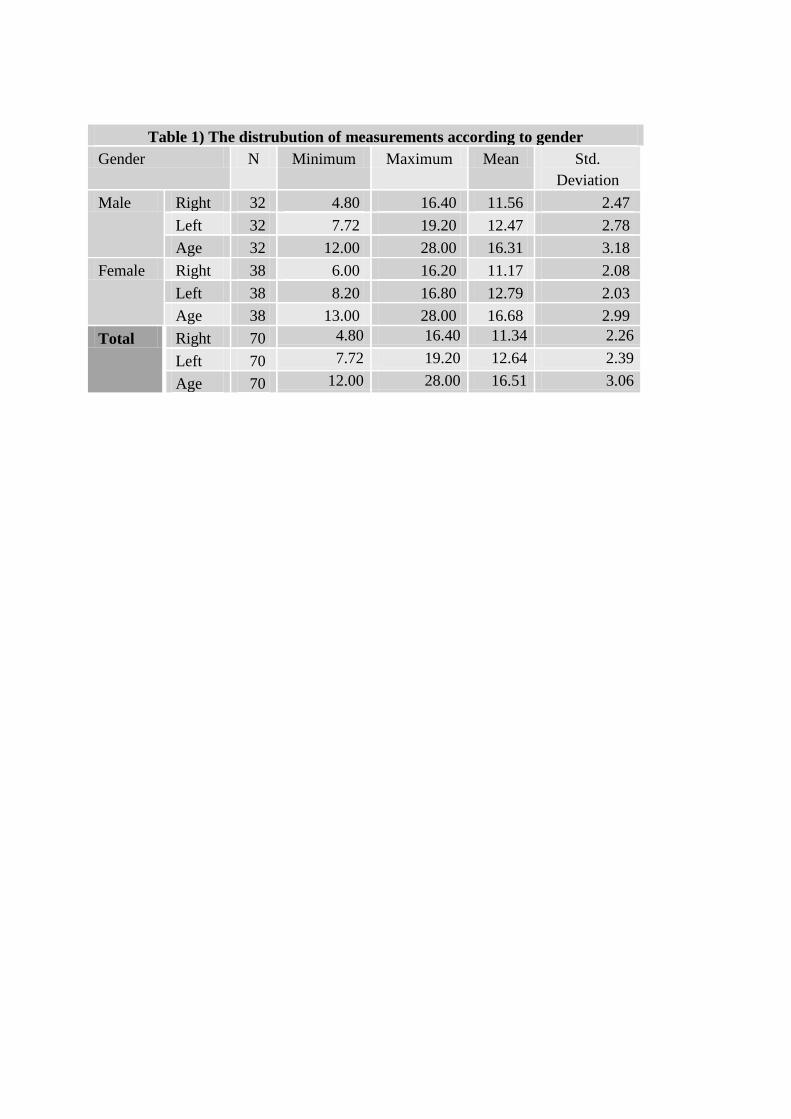
Table 1) The distrubution of measurements according to gender
Gender N Minimum Maximum Mean Std.
Deviation
Male Right 32 4.80 16.40 11.56 2.47
Left 32 7.72 19.20 12.47 2.78
Age 32 12.00 28.00 16.31 3.18
Female Right 38 6.00 16.20 11.17 2.08
Left 38 8.20 16.80 12.79 2.03
Age 38 13.00 28.00 16.68 2.99
Total Right 70 4.80 16.40 11.34 2.26
Left 70 7.72 19.20 12.64 2.39
Age 70 12.00 28.00 16.51 3.06

Figure Legends:
Figure 1
a: CBCT data sets were reformatted to a 20-mm thick CBCT panoramic view (CBCT-
pan) and 0.4-mm cross-sections that includes condyle, coronoid and sigmoid notch.
b: The axial plane was positioned to pass through the sigmoid notch.
c: The perpendicular distance measurement between the fixed plane and upper limit of
lingula or the upper point of the mandibular foramen.