DECISION MAKING ON SAGITTAL IMBALANCE CORRECTION WITH COMPUTER ASSISTANCE
8
ORJİNAL MAKALE / ORIGINAL ARTICLE DECISION MAKING ON SAGITTAL IMBALANCE CORRECTION WITH COMPUTER ASSISTANCE SAGİTTAL DENGESİZLİĞİN DÜZELTİLMESİNDE BİLGİSAYAR YARDIMLI KARAR VERME Akif ALBAYRAK 1 , Yunus ATICI 1 , Mehmet Bülent BALIOĞLU 1 , Deniz KARGIN 1 , Mehmet Nurullah ERMIŞ 1 , Evren AKPINAR 1 , Abdul Fettah BÜYÜK 1 Cilt: 25 • Sayı: 2 • Nisan 2014 ss. 97-104 97 1 Ortopedi ve Travmatoloji Kliniği, Metin Sabancı Baltalimanı Kemik Hastalıkları Eğitim ve Araştırma Hastanesi, İstanbul, Türkiye The Journal of Turkish Spinal Surgery SUMMARY Objective: To document the surgical techniques in a variety of scenarios in virtual environment in terms of their applications and consequences and to determine the usefulness of computer-assisted osteotomy planning for spinal deformity. Summary of Background Data: Computer-assisted surgical planning of sagittal plane deformity corrections allows operative maneuvers to be simulated on a computer to help for developing insight before their application in the operating room. Image-guided surgical planning of a deformity correction improves surgical accuracy and can help translating a virtual surgical plan into a real operation. Methods: We used Surgimap Spine program to determine the optimal corrective osteotomy for sagittal plane deformity for 11 patients. The program guided us through planning the osteotomy level, the osteotomy number and osteotomy type-whether Smith-Petersen Osteotomy (SPO) or Pedicle Subtraction Osteotomy (PSO) or both combined. Results: After virtual osteotomy the surgical plan was translated into the real one in the operating room and the osteotomy also including instrumentation in all cases was performed safely, resulting in anticipated results with the statistical importance (p>0.01). Conclusion: We advocate the application of virtual surgical planning for reasons of safety and efficiency improvements in complex spinal deformity corrections. Keywords: Computer-assisted osteotomy, spinal osteotomy, sagittal imbalance, ankylosing spondylitis Level of Evidence: Retrospective clinical study, Level III ÖZET Amaç: Omurga deformiteleri için bilgisayar yardımlı osteotomi planlamanın çeşitli senaryolar üzerinden sanal ortamda uygulama ve planlamasının tanımlanması. Özet: Sagittal plan deformitelerinin ameliyat öncesi bilgisayar yardımlı planlanması ameliyatta uygulanacak manevraların sanal ortamda uygulanarak sonuçların öngörülebilmesini sağlamaktadır. Görüntü destekli ameliyat planlama sayesinde cerrahinin doğruluğu arttırılırken sanal cerrahi planların gerçek ameliyatlarda uygulanırlığı sağlanmış olur. Gereç ve Yöntem: Sagittal düzlem omurga deformitesi bulunan on bir hastanın osteotomi planlaması için Surgimap Spine programını kullandık. Program bize osteotomi seviyesi, sayısı ve tipi ( Smith-Petersen Osteotomisi (SPO) veya Pedikül Substraction Osteotomisi (PSO) ) konusunda yol gösterdi. Sonuçlar: Sanal osteotomiden sonra cerrahi planla ameliyatlar uygulandı, osteotomi ve enstrümantasyon sonrası beklenenle istatistikî olarak benzer sonuçlar elde edildi (p>0.01). Sonuç: Kompleks omurga deformite düzeltme ameliyatlarında sanal cerrahi planlamayı hasta güvenliği ve ameliyat etkinliğini arttırmak adına önermekteyiz. Anahtar Sözcükler: Bilgisayar destekli osteotomi, omurga osteotomisi, sagittal balans kaybı, ankilozan spondilit Kanıt Düzeyi: Retrospektif klinik çalışma, Düzey III
Transcript of DECISION MAKING ON SAGITTAL IMBALANCE CORRECTION WITH COMPUTER ASSISTANCE

ORJİNAL MAKALE / ORIGINAL ARTICLE
DECISION MAKING ON SAGITTAL IMBALANCE CORRECTION WITH COMPUTER ASSISTANCE
SAGİTTAL DENGESİZLİĞİN DÜZELTİLMESİNDE BİLGİSAYAR YARDIMLI KARAR VERME
Akif ALBAYRAK1, Yunus ATICI1, Mehmet Bülent BALIOĞLU1, Deniz KARGIN1, Mehmet Nurullah ERMIŞ1, Evren AKPINAR1, Abdul Fettah BÜYÜK1
Cilt: 25 • Sayı: 2 • Nisan 2014ss. 97-104
97
1Ortopedi ve Travmatoloji Kliniği, Metin Sabancı Baltalimanı Kemik Hastalıkları Eğitim ve Araştırma Hastanesi, İstanbul, Türkiye
The Journal of Turkish Spinal Surgery
SUMMARY
Objective: To document the surgical techniques in a variety of scenarios in virtual environment in terms of their applications and consequences and to determine the usefulness of computer-assisted osteotomy planning for spinal deformity.
Summary of Background Data: Computer-assisted surgical planning of sagittal plane deformity corrections allows operative maneuvers to be simulated on a computer to help for developing insight before their application in the operating room. Image-guided surgical planning of a deformity correction improves surgical accuracy and can help translating a virtual surgical plan into a real operation.
Methods: We used Surgimap Spine program to determine the optimal corrective osteotomy for sagittal plane deformity for 11 patients. The program guided us through planning the osteotomy level, the osteotomy number and osteotomy type-whether Smith-Petersen Osteotomy (SPO) or Pedicle Subtraction Osteotomy (PSO) or both combined.
Results: After virtual osteotomy the surgical plan was translated into the real one in the operating room and the osteotomy also including instrumentation in all cases was performed safely, resulting in anticipated results with the statistical importance (p>0.01).
Conclusion: We advocate the application of virtual surgical planning for reasons of safety and efficiency improvements in complex spinal deformity corrections.
Keywords: Computer-assisted osteotomy, spinal osteotomy, sagittal imbalance, ankylosing spondylitis
Level of Evidence: Retrospective clinical study, Level III
ÖZET
Amaç: Omurga deformiteleri için bilgisayar yardımlı osteotomi planlamanın çeşitli senaryolar üzerinden sanal ortamda uygulama ve planlamasının tanımlanması.
Özet: Sagittal plan deformitelerinin ameliyat öncesi bilgisayar yardımlı planlanması ameliyatta uygulanacak manevraların sanal ortamda uygulanarak sonuçların öngörülebilmesini sağlamaktadır. Görüntü destekli ameliyat planlama sayesinde cerrahinin doğruluğu arttırılırken sanal cerrahi planların gerçek ameliyatlarda uygulanırlığı sağlanmış olur.
Gereç ve Yöntem: Sagittal düzlem omurga deformitesi bulunan on bir hastanın osteotomi planlaması için Surgimap Spine programını kullandık. Program bize osteotomi seviyesi, sayısı ve tipi ( Smith-Petersen Osteotomisi (SPO) veya Pedikül Substraction Osteotomisi (PSO) ) konusunda yol gösterdi.
Sonuçlar: Sanal osteotomiden sonra cerrahi planla ameliyatlar uygulandı, osteotomi ve enstrümantasyon sonrası beklenenle istatistikî olarak benzer sonuçlar elde edildi (p>0.01).
Sonuç: Kompleks omurga deformite düzeltme ameliyatlarında sanal cerrahi planlamayı hasta güvenliği ve ameliyat etkinliğini arttırmak adına önermekteyiz.
Anahtar Sözcükler: Bilgisayar destekli osteotomi, omurga osteotomisi, sagittal balans kaybı, ankilozan spondilit
Kanıt Düzeyi: Retrospektif klinik çalışma, Düzey III

98
Akif ALBAYRAK, Yunus ATICI, Mehmet Bülent BALIOĞLU, Deniz KARGIN, Mehmet Nurullah ERMIŞ, Evren AKPINAR, Abdul Fettah BÜYÜK
INTRODUCTION:
The SRS describes positive sagittal balance as
the condition of the sagittal vertical axis (SVA) going
through anterior to the L5-S1 disc on the upright long
cassette film. In principle, any deviation of the SVA
anterior to the normal position of the C7 plumb line
greater than 5 cm is considered as positive sagittal
imbalance.
Large deviations in the anterior-posterior or
lateral plane requires greater effort to preserve a
standing position.
Once a spinal deformity has reached the marked
level of loss in function that deteriorates the quality
of life, surgery is often recommended (4).
Substantial imbalance in the sagittal plane
sometimes cannot be corrected individually with
only arthrodesis with instrumentation, especially
when the deformity is very rigid. In these rare cases,
spinal osteotomies must be performed to achieve
balance in the sagittal plane.
Smith-Petersen osteotomy provides a little
amount of correction but it can be used at multiple
levels with minimal blood loss and possesses lower
operative risk. PSO’s are generally used in patients
with greater imbalances in the sagittal plane and
when a minimum of 30° of correction is needed.
The goal of a surgical procedure is to restore both
the patient’s gaze and the sagittal balance of the
spine.
Preoperative forecasting of the effect of an
osteotomy is crucial regarding the correction it can
provide on the sagittal balance of the spine. However
the effect mentioned depends on both the osteotomy
angle and the osteotomy level simultaneously. The
correction of the sagittal balance is maximal when
the intervention is performed at the lowest possible
level of the lumbar Spine (6,7).
In recent times, biomechanical methods founded
on mathematical principles has been developed
to assess deformities for sagittal plane corrective
osteotomies of the spine in Ankylosing Spondylitis
and these can be expressed as a form of mathematical
equation, but neither the crucial mathematic
equations nor the resulting monogram are easy to
use in daily practice (6).
We use Surgimap Spine a free computer program
(http://www.surgimap.com; Nemaris Inc, New York,
NY) to determine the optimal corrective osteotomy
level, number of recursion and its type.
The program analyses the input and visualizes the
planning procedure, which can be used easily in daily
practice.
MATERIAL AND METHOD:
From May 2012 to January 2013, 11 patients (7
female, 4 male, mean age 55) with sagittal imbalance,
undergoing spinal osteotomy (PSO or SPO) in the
spine for correcting a kyphotic deformity were
studied. Standard full-length anterior-posterior and
lateral radiographs covering the whole spine are
taken from PACS system and saved in JPEG, DICOM or
PNG formats on a standard USB memory-stick.
The program can open all medical image formats
in one interface. It runs directly on memory-stick,
without requiring any installation. Measurements
were made on radiographs obtained before surgery,
6 months after surgery, and at the most recent follow-
up at a minimum of 2 years after surgery.
RESULTS:
After starting up the program, first we browse and
load the image. For the calibration of the program we
mark two points on the grid of the film and define the
distance in millimeter. After the calibration we define
sagittal Cobb angles from T2-T12, T10-T12, L1-S1,
pelvic parameters and the sagittal vertical axis (SVA)
as the horizontal distance between the posterior
corner of the sacrum and the plumb line from C7,
which is performed by using spine measurement
tools. After determining these parameters, virtual
osteotomy plan should be done according to these
values.

Decision Making On Sagittal Imbalance Correction With Computer Assistance
99
The total amount of correction needed in the spine is determined by the simulation of the osteotomy at the apex and at adjacent levels of the apex of the deformity.
After the virtual osteotomy, the program makes Cobb angle, pelvic parameters and SVA changes automatically.
To achieve a certain decrease in the SVA abnormality, a smaller correction angle is needed if the osteotomy is performed at a lower spinal level.
Preoperative lumbar lordosis averaged 5° (range, from -23° to 25°) and improved to 35° (range, 26°-46°) at final follow-up. Thoracic kyphosis averaged 30° (range, from 4° to 112°) before surgery and increased to 36° (range, 9° – 55°) at the final follow-up (p<0.05). Sagittal balance improved from a preoperative 11 cm (range, 0–30 cm) to 3 cm (range, − 3 to 9 cm), resulting in 9 cm correction at the final follow-up (p<0.05). All patients demonstrated changes in preoperative to postoperative radiographic parameters including decreased Pelvic tilt (PT) (30° to 11°) and Sagittal vertical axis (SVA) (18 cm to 7 cm) and increased Sacral slope (SS) (16° to 35°) (p<0.05).
All postoperative sagittal contours and parameters of the sagittal trunk balance was statistically similar with the findings of the Surgimap program (p>0.01).
All 11 patients completed outcome questionnaires. A significant improvement in the back pain score was noted. The mean back pain score was 65 before surgery and 13 at the time of the final follow-up (p <0.05).
Case samples - one:
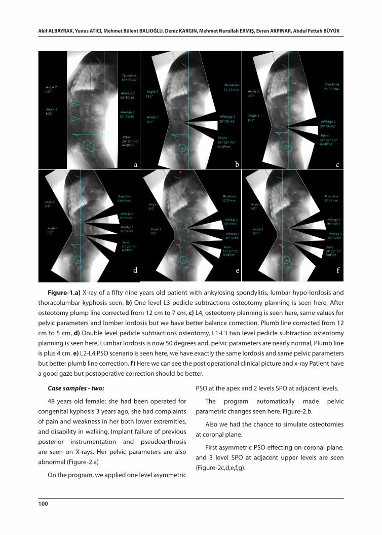
This is a 59 years old patient with ankylosing spondylitis. These patients usually present a lumbar hypo-lordosis and thoracolumbar kyphosis or cervicothoracic kyphosis. For this patient, certain initial parameters are as follows: thoracic kyphosis
39° lumbar lordosis 12°. Pelvic parameters are: Sacral slop 20°, Pelvic tilt 50°, Pelvic incidence 70° as seen in Figure-1.a. This patient does not have thoracic hyper kyphosis and because of this we tried to correct deformity of the lumbar area only.
First osteotomy planning, a one level L3 pedicle subtraction is seen in Figure-1b.
After virtual osteotomy, plumb line is corrected from 12 cm to 7 cm.
The program makes these parametric changes automatically; the user just simulates the osteotomy and program outputs the new lordosis, kyphosis and pelvic angles.
When evaluating pelvic parameters, we observe sacral slope increase which returns to the normal value but pelvic tilt and pelvic incidence still remains pathologic.
Second scenario, a L4 level osteotomy planning is seen in Figure-1.c.
Same values are observed for pelvic parameters and lumber lordosis but we have better balance correction, plumb line is corrected from 12 cm to 5 cm.
Third alternative, L1-L3 two level pedicle subtraction osteotomy planning is seen in Figure-1.d, lumbar lordosis is now 50 degrees and pelvic parameters are nearly normal, plumb line is plus 4 cm.
Last one is an L2-L4 PSO scenario seen in Figure-1.e, which returns exactly same lordosis and pelvic parameters but better plumb line correction.
So we decided to do L2-L4 osteotomy combination because of its better plumb line correction ability. After we bring the scenario to the operation room, we can see the post operational clinical picture and X-ray in Figure-1.f The patient has a good gaze but postoperative correction of the pulmp line is lower than the expected level.
100
Akif ALBAYRAK, Yunus ATICI, Mehmet Bülent BALIOĞLU, Deniz KARGIN, Mehmet Nurullah ERMIŞ, Evren AKPINAR, Abdul Fettah BÜYÜK
Case samples - two:
48 years old female; she had been operated for congenital kyphosis 3 years ago, she had complaints of pain and weakness in her both lower extremities, and disability in walking. Implant failure of previous posterior instrumentation and pseudoarthrosis are seen on X-rays. Her pelvic parameters are also abnormal (Figure-2.a)
On the program, we applied one level asymmetric
PSO at the apex and 2 levels SPO at adjacent levels.
The program automatically made pelvic parametric changes seen here. Figure-2.b.
Also we had the chance to simulate osteotomies at coronal plane.
First asymmetric PSO effecting on coronal plane, and 3 level SPO at adjacent upper levels are seen (Figure-2c,d,e,f,g).
Figure-1.a) X-ray of a fifty nine years old patient with ankylosing spondylitis, lumbar hypo-lordosis and thoracolumbar kyphosis seen, b) One level L3 pedicle subtractions osteotomy planning is seen here, After osteotomy plump line corrected from 12 cm to 7 cm, c) L4, osteotomy planning is seen here, same values for pelvic parameters and lomber lordosis but we have better balance correction. Plumb line corrected from 12 cm to 5 cm, d) Double level pedicle subtractions osteotomy, L1-L3 two level pedicle subtraction osteotomy planning is seen here, Lumbar lordosis is now 50 degrees and, pelvic parameters are nearly normal, Plumb line is plus 4 cm. e) L2-L4 PSO scenario is seen here, we have exactly the same lordosis and same pelvic parameters but better plumb line correction. f) Here we can see the post operational clinical picture and x-ray Patient have a good gaze but postoperative correction should be better.
a b c
d e f

Decision Making On Sagittal Imbalance Correction With Computer Assistance
101
We should obtain good coronal balance with these osteotomies.
After virtual osteotomy the surgical plan was translated to the operating room, the osteotomy was performed and instrumentation was done.
We had excellent sagittal balance; lordosis and kyphosis angles were ideal but our prediction of pelvic parameters did not fully meet the expectation (Figure-2.h,j).
Figure-2.a) X-ray of forty-eight years old female. Previously operated for congenital kyphosis, she had implant failure of previous posterior instrumentation and pseudoarthrosis can be seen on x-rays, pelvic parameters are also abnormal. b) One level asymmetric PSO at the apex and 2 levels Smith Petersen osteotomies at adjacent levels, the program automatically makes pelvic parametric changes. c) Simulation of osteotomies at coronal plane are seen, first asymmetric PSO’s affect on coronal plane seen. e,f) 3 SPO at adjacent upper levels, g) Scenario of good coronal balance with these osteotomies seen, h) Excellent sagittal balance, ideal lordosis and kyphosis angles but our prediction of pelvic parameters did not fully met the expectation, i) Good coronal balance seen here
a b c
d e f g
h i

102
Akif ALBAYRAK, Yunus ATICI, Mehmet Bülent BALIOĞLU, Deniz KARGIN, Mehmet Nurullah ERMIŞ, Evren AKPINAR, Abdul Fettah BÜYÜK
Case samples - three:
The last case is a 65 years old male patient, sequel of thoracolumbar fracture,
He has hypo-kyphosis and hypo-lordosis because of thoracolumbar junctional kyphosis.
Apex of the deformity was obviously visible so we plan one level PSO at apical level and 2 SPO at adjacent levels.
As seen on X-rays of virtual osteotomies, by the 3 level osteotomy, pelvis and sagittal balance seems over-corrected (Figure-3.a)
But we know that non-instrumented proximal levels of the spine are flexible so this over-correction should be compensated by the kyphosis of this part. So we performed one level PSO and one level SPO; post-op X rays and patient clinic are seen in Figure-3.b.
DISCUSSION
Balance of the body essentially depends on how far the head is to the midline.
Bridwell classified sagittal imbalance into type-1 and type-2 (2). In type-1 imbalance, the patient has a segmental problem. A part of the spine is considerably hyper-kyphotic, but the patient is able to sustain balance by hyperextending segments above and below. In type-2 imbalance the regional
hyper-kyphosis is so much that the patient cannot achieve balance by hyperextending segments above and below (5).
Preoperative planning of the osteotomy on the sagittal plane deformities is very crucial to predict the outcome of the surgery.
We can do osteotomy planning in several ways.
One way, for instance is the option of using paper templates of the long casette x-rays for the appropriate place of wedge resection.
Also simulation can be performed on the computer with a program like ASKyphoplan that was used by Van Royen for treating ankylosing spondylitis, or Surgimap Spine that was used by us. Both programs analyses and visualizes the relationship between the angle and the level of osteotomy in relation to the sagittal balance. (7)
In addition, Surgimap spine program can be used for simulating the coronal plane osteotomies (Figure-2.c, d, e, f, g, h).
When the sagittal deformity is rigid, spinal osteotomies must be performed to achieve balance of the sagittal plane (1). There are several osteotomy types to obtain correction. Considering two prominent examples, PSO’s offer nearly 3 times the per-level correction of SPO can provide but they are burdened with increased technical demands, longer
Figure-3.a) Sixty-five years old man, squeal of thoracolumbar fracture, plan of apical PSO and adjacent 1 or 2 SPO’ seen, with the 3 level osteotomy. pelvis and sagittal balance seems will be overcorrected b) After one PSO and one SPO postoperative x-rays are seen.
a b

Decision Making On Sagittal Imbalance Correction With Computer Assistance
103
operative time, and greater blood loss and associated morbidity. Cho at all reported that when comparing three or more SPOs with one PSO, the correction in kyphosis was nearly identical, but the improvement in the C7 plumb-line was significantly better in favor of the PSO group (3).
Their recommendation was to consider one or two SPOs for a C7 plumb line that is in the range of 6 to 8 cm positive, however for patients displaying 12 cm or more positive range their recommendation was PSO.
They considered this huge correction in terms of balance achieved by the PSO group because of the preoperative being too much deformity. We favor the PSO over the SPO for the reason that deformities did not recover in the fulcrum radiographs.
In both virtual and real osteotomy, we observed that the lower the level of osteotomy performed in the lumbar region, the greater the impact of it on the sagittal vertical axis. As shown in sample case, moving osteotomies to a lower level in turn resulted to increasing improvement in global balance (Figure-1.d,e).
Van Royen at al reported that the effect of a spinal osteotomy on the correction of the horizontal gaze was directly proportional to the osteotomy angle, irrespective of the osteotomy level. But this rule is valid only for ankylosing spondilitis patients (6).
If all segments of the spine was rigid, this rule would be valid for all patients, however for most patients the deformity is flexible above and below the apex so the effect of the vertebrae osteotomy is compensated by moving segments that hinders the realization of expected impact on the patient’s horizon
Looking at the sample cases in which patients are flexible at the regions outside of the apex, if the spinal regions outside the deformity were not instrumented, each of these segments were compensating post-osteotomy condition proportional to their individual capability of movement; a situation effectively shown
at the figures of 2nd sample case, when 5° thoracic kyphosis improves to 25°, and at 3rd sample case, when 2° thoracic kyphosis improves spontaneously to 27°.
In patients with ankylosing spondylitis, all segments excluded from spine fusion related osteotomy had carried on the same pre-operative kyphosis condition observed in the radiograph post-operatively, for this reason computer-assisted planning became easier.
For the patients flexible at the regions outside of the apex, preoperative flexibility of the spine should be evaluated by using the lateral bending at the AP radiograph and the fulcrum at the LAT radiograph and the effects over the post-op results should be anticipated accordingly.
We deduced that a program of this kind is useful in creating a healthy simulation of correction in an overall rigid spine but cannot foresee the post-op secondary flexibilities in regions outside of the deformity.
For spines with flexible segments, we saw that making an osteotomy simulation on bending and traction radiographs allows us to predict the movements at the adjacent segments that will occur in post-osteotomy period.
In conclusion, Surgimap-spine program is a useful tool for deformity correction planning and osteotomy simulation in a virtual environment.
104
Akif ALBAYRAK, Yunus ATICI, Mehmet Bülent BALIOĞLU, Deniz KARGIN, Mehmet Nurullah ERMIŞ, Evren AKPINAR, Abdul Fettah BÜYÜK
1. Atici Y, Sokucu S, Uzumcugil O, Albayrak A, Erdoğan S, Kaygusuz A. The results of closing wedge osteotomy with posterior instrumented fusion for the surgical treatment of congenital kyphosis. Eur Spine J 2013; 22(6): 1368–1374.
2. Bridwell KH. Decision making regarding Smith-Petersen vs. pedicle subtraction osteotomy vs. vertebral column resection for spinal deformity. Spine 2006; 31(19): 171–178.
3. Cho K J, Bridwell K H, Lenke G L, Berra B A, Baldus C. Comparison of Smith-Petersen versus pedicle subtraction osteotomy for the correction of fixed sagittal imbalance. Spine 2005; 30(18): 2030–2037.
4. Schwab F, Patel A, Ungar B, Farcy J P, Lafage F. Adult spinal deformity postoperative standing imbalance assessing alignment and planning corrective surgery Spine 2010; 35(25): 2224–2231.
5. Toyone T, Shiboi R, Ozawa T, Inada K, Shirahata T, Kamikawa K, Watanabe A, Matsuki K, Ochiai S, Kaiho T. Asymmetrical pedicle subtraction osteotomy for rigid degenerative lumbar kyphoscoliosis. Spine 2012; 37(21): 1847–1852.
6. Van Royen BJ, De Gast A, Smith TH. Deformity planning for sagittal plane corrective osteotomies of the spine in ankylosing spondylitis Eur Spine J 2000; 9: 492–498.
7. Van Royen BJ, Scheerder FJ, Jansen E, Smith TH. ASKyphoplan. A program for deformity planning in ankylosing spondylitis. Eur Spine J 2007; 16(9): 1445–1449.
Adres: Dr. Yunus Atıcı, S.B. Metin Sabancı Baltalimanı Kemik Hastalıkları Eğitim ve Araştırma Hastanesi, Ortopedi ve Travmatoloji Kliniği, Omurga Hastalıkları Cerrahisi ve Protez Cerrahisi Grubu, Baltalimanı, Sarıyer, İSTANBULTel.: 0505 492 19 45E-mail: [email protected]ş Tarihi: 1 Ocak 2014Kabul Tarihi: 30 Ocak 2014
REFERENCES