Redox Imbalance in Idiopathic Pulmonary Fibrosis
25
Redox Imbalance in Idiopathic Pulmonary Fibrosis Citation for published version (APA): Veith, C., Boots, A. W., Idris, M., van Schooten, F-J., & van der Vliet, A. (2019). Redox Imbalance in Idiopathic Pulmonary Fibrosis: A Role for Oxidant Cross-Talk Between NADPH Oxidase Enzymes and Mitochondria. Antioxidants & Redox Signaling, 31(14), 1092-1115. https://doi.org/10.1089/ars.2019.7742 Document status and date: Published: 10/11/2019 DOI: 10.1089/ars.2019.7742 Document Version: Publisher's PDF, also known as Version of record Document license: Taverne Please check the document version of this publication: • A submitted manuscript is the version of the article upon submission and before peer-review. There can be important differences between the submitted version and the official published version of record. People interested in the research are advised to contact the author for the final version of the publication, or visit the DOI to the publisher's website. • The final author version and the galley proof are versions of the publication after peer review. • The final published version features the final layout of the paper including the volume, issue and page numbers. Link to publication General rights Copyright and moral rights for the publications made accessible in the public portal are retained by the authors and/or other copyright owners and it is a condition of accessing publications that users recognise and abide by the legal requirements associated with these rights. • Users may download and print one copy of any publication from the public portal for the purpose of private study or research. • You may not further distribute the material or use it for any profit-making activity or commercial gain • You may freely distribute the URL identifying the publication in the public portal. If the publication is distributed under the terms of Article 25fa of the Dutch Copyright Act, indicated by the “Taverne” license above, please follow below link for the End User Agreement: www.umlib.nl/taverne-license Take down policy If you believe that this document breaches copyright please contact us at: [email protected] providing details and we will investigate your claim. Download date: 10 Jan. 2022
-
Upload
khangminh22 -
Category
Documents
-
view
0 -
download
0
Transcript of Redox Imbalance in Idiopathic Pulmonary Fibrosis

Redox Imbalance in Idiopathic Pulmonary Fibrosis
Citation for published version (APA):
Veith, C., Boots, A. W., Idris, M., van Schooten, F-J., & van der Vliet, A. (2019). Redox Imbalance inIdiopathic Pulmonary Fibrosis: A Role for Oxidant Cross-Talk Between NADPH Oxidase Enzymes andMitochondria. Antioxidants & Redox Signaling, 31(14), 1092-1115. https://doi.org/10.1089/ars.2019.7742
Document status and date:Published: 10/11/2019
DOI:10.1089/ars.2019.7742
Document Version:Publisher's PDF, also known as Version of record
Document license:Taverne
Please check the document version of this publication:
• A submitted manuscript is the version of the article upon submission and before peer-review. There canbe important differences between the submitted version and the official published version of record.People interested in the research are advised to contact the author for the final version of the publication,or visit the DOI to the publisher's website.• The final author version and the galley proof are versions of the publication after peer review.• The final published version features the final layout of the paper including the volume, issue and pagenumbers.Link to publication
General rightsCopyright and moral rights for the publications made accessible in the public portal are retained by the authors and/or other copyrightowners and it is a condition of accessing publications that users recognise and abide by the legal requirements associated with theserights.
• Users may download and print one copy of any publication from the public portal for the purpose of private study or research.• You may not further distribute the material or use it for any profit-making activity or commercial gain• You may freely distribute the URL identifying the publication in the public portal.
If the publication is distributed under the terms of Article 25fa of the Dutch Copyright Act, indicated by the “Taverne” license above,please follow below link for the End User Agreement:
www.umlib.nl/taverne-license
Take down policyIf you believe that this document breaches copyright please contact us at:
providing details and we will investigate your claim.
Download date: 10 Jan. 2022

FORUM REVIEW ARTICLE
Redox Imbalance in Idiopathic Pulmonary Fibrosis:A Role for Oxidant Cross-Talk Between NADPHOxidase Enzymes and Mitochondria
Carmen Veith,1 Agnes W. Boots,1 Musa Idris,1 Frederik-Jan van Schooten,1 and Albert van der Vliet2
Abstract
Significance: Idiopathic pulmonary fibrosis (IPF) is a progressive age-related lung disease with a mediansurvival of only 3 years after diagnosis. The pathogenic mechanisms behind IPF are not clearly understood, andcurrent therapeutic approaches have not been successful in improving disease outcomes.Recent Advances: IPF is characterized by increased production of reactive oxygen species (ROS), primarily byNADPH oxidases (NOXes) and mitochondria, as well as altered antioxidant defenses. Recent studies haveidentified the NOX isoform NOX4 as a key player in various important aspects of IPF pathology. In addition,mitochondrial dysfunction is thought to enhance pathological features of IPF, in part by increasing mito-chondrial ROS (mtROS) production and altering cellular metabolism. Recent findings indicate reciprocalinteractions between NOX enzymes and mitochondria, which affect regulation of NOX activity as well asmitochondrial function and mtROS production, and collectively promote epithelial injury and profibroticsignaling.Critical Issues and Future Directions: The precise molecular mechanisms by which ROS from NOX ormitochondria contribute to IPF pathology are not known. This review summarizes the current knowledgewith respect to the various aspects of ROS imbalance in the context of IPF and its proposed roles in diseasedevelopment, with specific emphasis on the importance of inappropriate NOX activation, mitochondrialdysfunction, and the emerging evidence of NOX–mitochondria cross-talk as important drivers in IPFpathobiology. Antioxid. Redox Signal. 31, 1092–1115.
Keywords: IPF, reactive oxygen species, aging, lung, NOX4, mitochondria
Introduction
Idiopathic pulmonary fibrosis (IPF) is a progressive andirreversible lung disease of unknown etiology, and repre-
sents a specific form of chronic fibrosing interstitial pneu-monia that occurs primarily in the lungs. The pathogenicmechanisms of IPF are still largely unclear, but over the lastdecade the paradigm of IPF pathogenesis has shifted froma generally inflammation-driven disease to an epithelial-fibroblastic one, resulting from a disrupted homeostasis ofepithelial cells upon damage by various triggers (50, 75).Continuous epithelial injury results in aberrant wound heal-
ing responses, and eventually causes excessive collagendeposition in the alveolar epithelium and remodeling of thelung structure (50). IPF is generally diagnosed in the sixthdecade of life, and aging is now recognized as one of thestrongest risk factors for IPF (169). As the average lifeexpectancy continues to increase worldwide, the incidenceof age-related lung diseases such as IPF will also increase ata rapid pace. It is estimated that the number of people >60years of age will increase by *50% between 2015 and2050, and account for 22% of the total world population(240). As the elderly population is growing, there is agrowing need to understand the underlying mechanisms of
1Department of Pharmacology and Toxicology, Faculty of Health, Medicine and Life Sciences, NUTRIM School of Nutrition, Trans-lational Research and Metabolism, University of Maastricht, Maastricht, the Netherlands.
2Department of Pathology and Laboratory Medicine, Larner College of Medicine, University of Vermont, Burlington, Vermont.
ANTIOXIDANTS & REDOX SIGNALINGVolume 31, Number 14, 2019ª Mary Ann Liebert, Inc.DOI: 10.1089/ars.2019.7742
1092
Dow
nloa
ded
by U
NIV
OF
MA
AST
RIC
HT
fro
m w
ww
.lieb
ertp
ub.c
om a
t 03/
31/2
1. F
or p
erso
nal u
se o
nly.

aging and age-associated phenomena and their contributionto disease pathogenesis.
Aging results in decreased resistance to multiple forms ofstress and progressive loss of regenerative capacity, andthereby increases susceptibility to chronic lung diseases, in-cluding IPF, chronic obstructive pulmonary disease, and lungcancer (144). Indeed, common hallmarks of aging, includinggenomic instability, telomere shortening, epigenetic alter-ations, loss of proteostasis, dysregulated nutrient sensing,mitochondrial dysfunction, cellular senescence/apoptosis,stem cell exhaustion, and distorted intercellular communi-cation (132), are also seen in IPF and frequently occur pre-maturely (4, 5, 10, 144, 147, 150, 154, 249).
Both aging and fibrotic diseases are associated with anincreased oxidant burden (109, 117, 149), and lung tissues aswell as breath condensates from IPF patients show increasedlevels of oxidative damage markers such as 8-isoprostane andcarbonylated proteins (13, 126, 178). Studies in mice indicatethat aging increases susceptibility to pulmonary fibrosis,likely due to decreased resistance to oxidative stress (76). Themain cellular sources of reactive oxygen species (ROS) areNADPH oxidases (NOXes) and mitochondria (35), and whileROS production from both sources serves various biologicalroles in, for example, host defense, cell differentiation, orcellular responses to injury (207, 229), both have also beenimplicated in IPF pathology. For example, various lines ofevidence indicate that mitochondria are dysfunctional in IPF(27), resulting in increased production of mitochondrial ROS(mtROS) (150), and that approaches to attenuate mtROS maybe beneficial (254). In addition, recent studies have impli-cated NOX4 as an important player in the development of IPF(77), based on its ability to induce alveolar epithelial cell(AEC) death, (myo)fibroblast differentiation, and collagendeposition (88). More recently, NOX4 has also been impli-cated as a mediator of mitochondrial dysfunction (19), sug-gesting cross-talk between these two ROS-generating systems.
This review summarizes our current knowledge with re-spect to the presence of a redox imbalance in IPF, and theimportance of ROS in the development and progression ofIPF. Specifically, we highlight the importance of NOX4 andthe emerging cross-talk between NOX4 and mitochondria.Finally, we discuss important gaps of knowledge and po-tential future approaches for development of more effectivetreatment strategies.
IPF—Pathology and Affected Cell Types
General pathology
IPF is the most common form of idiopathic interstitiallung diseases and is characterized by scarring of the lungtissue causing symptoms such as nonproductive cough andbreathlessness. As a chronic progressive lung disease, IPFhas a median survival of 2–4 years after diagnosis due torespiratory failure and hypoxemia (1). Two-thirds of IPFpatients are >60 years at time of diagnosis (181), and aging isthe most well-known risk factor for developing this debili-tating disease. Although it is a relatively rare disease thatcurrently affects *5 million people worldwide, its overallincidence increases every year probably due to the increasingnumber of elderly individuals (156, 158). In the UnitedStates, the incidence of IPF varies between 1.1 new cases per100,000 person-years in 18–34 old individuals up to 19.3 in
individuals >55 of age (179). Besides aging, IPF has severalother risk factors, including exposure to environmental fac-tors, for example, cigarette smoke and asbestos (151). Inaddition, the pathogenesis of the disease is associated withgenomic instability, mitochondrial dysfunction, and alteredintracellular signaling (132), all hallmarks of normal agingbut also all processes compromised by environmental factorssuch as tobacco smoking.
Histologically, the most prominent morphological hall-mark of IPF is usual interstitial pneumonia (UIP). One his-tologic feature of UIP is honeycombing characterized byclusters of cysts, often filled with mucus, in a subpleurallocation (54). An additional feature of UIP is the presence offibroblastic foci. Fibroblastic foci contain granuloma of ac-tivated myofibroblasts producing extracellular matrix (ECM)proteins and are mainly found in dense fibrotic areas of thelungs (43). UIP is further characterized by hyperplastic typeII AECs and reduced type I AECs. During IPF development,a disturbed lung homeostasis results in a profibrotic milieu,thereby affecting the survival and death of both fibroblastsand AECs. AECs become more prone to undergo apoptosis(212), while fibroblasts and myofibroblasts become moreresistant against this process (203). Also, distinct alterationsin immune cells are reported in IPF as the lungs of thesepatients exhibit chronic inflammation associated with im-munosenescence (206).
Cell-specific aspects of IPF
IPF is a complex disease that involves the contribution ofmany different cell types. The following paragraphs sum-marize the specific involvement of distinct lung cell types,such as epithelial cells, fibroblasts, immune cells, and endo-thelial cells.
Epithelial cells. The alveolar lining of the lungs is cov-ered by 95% type I AECs, important in facilitating gas ex-change, and 5% type II AECs that are involved in surfactantproduction. In IPF pathogenesis, repetitive injury to the al-veolar epithelium leads to abnormal re-epithelialization andrepair of AECs (205). Lung tissue from IPF patients typicallyshows loss of integrity of the alveolar epithelium with dis-ruption of basement membrane integrity and collapse of thealveolar structure. While type II AECs normally proliferateand differentiate into type I AECs in response to injury topromote re-epithelialization, in the context of fibrosis, AECsare more prone to undergo apoptosis, which is a prominentfeature during IPF manifestation. Indeed, AEC type II cellsstain positive for apoptotic markers in patients with IPFcompared with controls (175). Moreover, experimental in-duction of epithelial cell apoptosis is sufficient to pro-mote features of fibrosis (212), and inhibition of apoptosisattenuates bleomycin-induced pulmonary fibrosis (118, 227).
In addition to evidence for AEC type II apoptosis in IPF,numbers of type I AECs in the lung of IPF patients arereduced causing AEC type II to undergo hyperplasia,thereby replacing type I AECs and inducing ineffective re-epithelialization (95). In addition, AECs secrete a variety ofprofibrotic mediators, such as platelet-derived growth fac-tor, endothelin-1, angiotensin II, connective tissue growthfactor, and transforming growth factor (TGF)-b, whichcan induce fibroblasts to differentiate into myofibroblasts
REDOX IMBALANCE IN IDIOPATHIC PULMONARY FIBROSIS 1093
Dow
nloa
ded
by U
NIV
OF
MA
AST
RIC
HT
fro
m w
ww
.lieb
ertp
ub.c
om a
t 03/
31/2
1. F
or p
erso
nal u
se o
nly.

(147). Furthermore, increased epithelial senescence resultsin enhanced secretion of growth factors, cytokines, chemo-kines, and matrix metalloproteins that stimulate continuousfibroblast activity (2). In addition, these factors promote asenescence-associated secretory phenotype (SASP), which is akey event in IPF possibly leading to diminished capacity ofAEC regeneration (147).
Finally, AECs have also suggested to contribute to fibro-blast development in IPF due to epithelial-to-mesenchymaltransition (EMT), a process in which epithelial cells lose theirepithelial phenotype and acquire more mesenchymal/fibro-blastic characteristics such as a-smooth muscle actin (a-SMA) and fibronectin expression. Indeed, epithelial cellsisolated from IPF patients have been found to express notonly epithelial but also mesenchymal markers (91). However,the overall significance of EMT to IPF development/mani-festation is still not completely clear (89). Lineage tracingstrategies have suggested a contribution for EMT to increasedpresence of fibroblasts in IPF lungs (91, 100, 238), but otherstudies have also disputed that epithelial cells are indeed thesource of fibroblasts (192). In summary, repetitive epithelialinjury and dysregulated repair are critical features in IPFdevelopment and progression as they lead to increased apo-ptosis, abnormal regeneration, and EMT-like features.Moreover, TGF-b, an important growth factor in IPF, canpromote epithelial apoptosis as well as EMT (28), furtherindicating the important role of the lung epithelium in IPF.
Fibroblasts. Epithelial cells within the airways and al-veoli are in proximity to and communication with mesen-chymal cells such as pulmonary fibroblasts, referred to as theepithelial mesenchymal trophic unit. Consequently, epithe-lial damage impacts fibroblasts by promoting their differen-tiation into myofibroblasts, which is mediated by TGF-b1(225). Myofibroblasts actively participate in remodeling ofthe lung by secreting ECM proteins into the lung alveolarstructure also upon induction by TGF-b1. Various sources ofmyofibroblasts have been suggested to be recruited to the siteof injury, including proliferation and differentiation of resi-dent lung fibroblasts, epithelial and endothelial cells thattransform into fibroblasts and transition of fibrocytes or othercirculating progenitor cells (79). During normal wound re-pair, myofibroblasts undergo apoptosis, and clearance of theECM occurs upon closing the wound, thus avoiding theformation of scar tissue. In IPF, however, overactivation ofmyofibroblasts due to persistent epithelial injury leads toaberrant wound repair (177) and increased scarring, which ischaracterized by the formation of fibroblastic foci. This is amajor hallmark of IPF (168) and is responsible for decreasedpulmonary function (107, 159). In addition, fibroblasts andmyofibroblasts from IPF patients have an apoptosis-resistantphenotype, which is also associated with higher levels ofsenescence markers, such as p16, compared with fibroblastsfrom age-matched controls (5, 248). In summary, thesestudies suggest that fibroblasts from patients with IPF arecharacterized by a senescent apoptosis-resistant phenotypeand are thereby actively involved in the progression of thedisease by remodeling of the lung structure.
Inflammatory cells. During the last few decades, a com-mon view has been that chronic inflammation underlies thepathogenic sequence in IPF as a main cause of lung injury and
fibrogenesis. Although this ‘‘inflammatory fibrosis’’ hypothesishas been somewhat invalidated (61, 205), chronic inflammationis still considered a common feature of IPF, yet most likely as asecondary event instead (205). Damaged epithelial cells releasea variety of chemokines and cytokines that recruit inflamma-tory monocytes and neutrophils to the injured site. Recruitmentand activation of neutrophils to the bronchoalveolar space havebeen shown to correlate with disease progression (90), and arealso a predictor of early mortality in IPF (106). Normally,monocytes differentiate into phagocytic macrophages thatphagocytose dead cells and neutrophils. However, because ofthe continuous injury in IPF, neutrophils and macrophages arenot eliminated quickly enough. Consequently, they can developresistance to apoptosis, which will further exacerbate the on-going inflammation and fibrotic cycle (28).
Macrophages can mediate antifibrotic as well as profibroticeffects depending on their phenotype. Classically activatedmacrophages (M1) are activated by the T-helper (TH) cell 1cytokine interferon-c and mainly involved in the initial in-flammatory response, whereas alternatively activated mac-rophages (M2) are activated by TH2 cytokines, includinginterleukin (IL)-4 and IL-13, and involved in tissue re-modeling and resolution of inflammation (243). Furthermore,studies have demonstrated that the TH1/TH2 balance is dis-turbed in favor of TH2 cytokines (60, 245), further contrib-uting to a M2 phenotype. M2 polarized macrophagespromote profibrotic effects through the section of profibroticmediators, including TGF-b (14). In addition, alveolar neu-trophils and macrophages undergo a respiratory burst uponphagocytosis, which in turn promotes AEC injury, therebyinducing a vicious circle (13). Taken together, studies haveproven that inflammatory cells participate in the profibroticcircle by secreting profibrotic and proinflammatory cyto-kines, although they are not the prominent cell type involvedin the development and initial manifestation of IPF.
Endothelial cells. Endothelial cells are important inmaintaining the alveolar–capillary barrier for facilitating gasexchange (196). Pulmonary endothelial cells are close to theinterstitium, the primary site of injury during IPF, and coverthe intravascular lumen, making them vulnerable to injuries.Endothelial cells from IPF patients (134, 221), and pulmonaryendothelial cells from mice with bleomycin-induced pulmo-nary fibrosis, display increased markers of endothelial cellinjury (96) and endothelial cell apoptosis (56), which result inloss of integrity of the alveolar–capillary barrier. Moreover,endothelial progenitor cells are reduced in IPF patients (136),which impairs effective re-endothelialization and vascularrepair upon injury. In addition, the damaged endothelium se-cretes a variety of factors that activate circulating platelets andvon Willebrand factor, which are involved in angiogenesis andrecruitment of inflammatory cells (122). Finally, endothelialcells have also been implicated as a source of fibroblaststhrough EMT during fibrogenesis (71, 174). Endothelial dys-function in patients with IPF has also been correlated withpulmonary hypertension, a common comorbidity in IPF (127).
Taken together, AEC apoptosis and (myo)fibroblast acti-vation are now considered the primary responsible eventscontributing to excessive lung remodeling in IPF, whereasthe specific role of endothelial cells and inflammation is lesswell established and still needs to be further elucidated.
1094 VEITH ET AL.
Dow
nloa
ded
by U
NIV
OF
MA
AST
RIC
HT
fro
m w
ww
.lieb
ertp
ub.c
om a
t 03/
31/2
1. F
or p
erso
nal u
se o
nly.
Current treatment strategies for IPF
Currently, there are only two Food and Drug Administra-tion (FDA)-approved drugs available for patients with IPF.Both pirfenidone and nintedanib can slow down the pro-gression of the disease (112), although they cannot reversethe existing pulmonary fibrotic injury. Pirfenidone (Esbriet�;Roche/Genentech) is thought to have antioxidative, anti-inflammatory, and antifibrotic effects, although the exactworking mechanisms are not clear as confirmatory data fromIPF patients are lacking (112). In vitro studies and studies onbleomycin-induced murine pulmonary fibrosis indicate that itreduces markers of oxidative stress (65, 148), decreases thesecretion of proinflammatory cytokines (e.g., tumor necrosisfactor-a, IL-1b, IL-6) (163), and inhibits fibroblast proliferation,myofibroblast differentiation, and TGF-b-induced collagenproduction (42). Nintedanib (Ofev�; Boehringer Ingelheim) isan intracellular tyrosine kinase inhibitor, which mainly inhibitsthe platelet-derived growth factor receptor, the fibroblast growthfactor receptor, and vascular endothelial growth factor recep-tor as well as nonreceptor tyrosine kinases of the SRC family(112), which mediate its antifibrotic effects through its inhib-itory action on fibroblasts (83).
The Disturbed Redox Balance in IPF
ROS in biology and disease
All cells in the body consume and metabolize oxygen andas a result produce ROS, which include superoxide anion(O2�-), hydrogen peroxide (H2O2), and hydroxyl radical
(OH�), as well as other secondary ROS. ROS are produced asa result of aerobic metabolism within the mitochondrialelectron transport chain (ETC), but can also be generated byvarious enzyme systems, including xanthine oxidase, lipidperoxidases, uncoupled endothelial NO synthase, cytochromeP450 enzymes, and NOXes (39). The latter group of enzymesare the only enzymes that are considered to generate ROS astheir primary product, and serve essential functions as medi-ators of host defense against diverse pathogens, and in variousother aspects of cell biology through redox-based cell signal-ing (15, 229). Inappropriate or uncontrolled ROS production isgenerally thought to contribute to disease pathology, due tooverproduction of ROS by, for example, increased NOX ac-tivation or by mitochondrial dysfunction, or due to compro-mised metabolism of ROS by antioxidant systems (41). Suchan increased oxidant burden can lead to extensive moleculardamage to macromolecules, including DNA, lipids, and pro-teins (228), thereby causing protein dysfunction and alteredproteostasis, genomic instability, or production of secondaryreactive lipid-derived species (e.g., electrophiles such asmalondialdehyde or 4-hydroxynonenal).
Specific to IPF, several studies reported that IPF patientshave a higher oxidant burden compared with healthy controls.Indeed, ROS production is dysregulated due to enhanced NOXexpression and activation, as well as mitochondrial dysfunc-tion and increased mtROS generation (13, 67). Pulmonaryinflammatory cells obtained from epithelial lining fluid (ELF)of IPF patients generate higher levels of ROS compared withthose of healthy controls (31), and IPF patients have increasedlevels of H2O2 within their exhaled breath condensates(EBCs) (178). IPF patients also demonstrate increased cir-culating markers of lipid peroxidation compared with healthy
nonsmokers (182), and increased levels of the lipid oxidationproduct 8-isoprostane within bronchoalveolar lavage fluid(BALF) and EBC (149). BALF or lung tissue from IPF pa-tients also contains higher amounts of irreversibly oxidizedproteins (e.g., carbonylation, nitration) (126, 195, 199), andepithelial cells of patients with UIP show increased levels ofDNA oxidation, illustrated by 8-hydroxy-deoxyguanosine(119). In summary, IPF patients show increased markers ofoxidative damage (59). However, evidence for their causalcontribution to the IPF disease is less prominent, and will bediscussed in the following sections.
Pulmonary antioxidant defense systems in IPF
All organs, including the lung, contain a variety of anti-oxidant systems to prevent inappropriate ROS production orunwanted actions of cellular ROS. These include enzymesthat metabolize ROS (superoxide dismutases [SODs], cat-alase, peroxiredoxins [PRXs], and glutathione peroxidases),thiol reductases that reverse oxidative cysteine modifications(thioredoxin [TRX], glutaredoxin [GRX]), phase 2 detoxifyingenzymes (e.g., glutathione-S-transferases [GSTs]), metal-binding proteins (transferrin, lactoferrin), and small mo-lecular weight antioxidants (vitamins and glutathione) (41).A number of studies have demonstrated that several of theseantioxidant systems are altered or impaired in IPF (Table 1).
Among the earliest lines of evidence of altered antioxidantstatus in IPF are findings of reduced levels of the cellularantioxidant thiol-containing tripeptide glutathione (GSH)within the ELF of the lower respiratory tract (30, 146) as wellas in blood (152, 231) and sputum (16). In some cases, thesedecreases were accompanied by increased levels of the oxi-dized form of GSH, glutathione disulfide, thus illustrating analtered redox balance in the alveolar lumen of IPF patients.Of the three known isoforms of SOD, which catalyzes thedismutation of O2
�- into H2O2 and O2 (108), some studiessuggest that lung tissue expression of extracellular SOD(SOD3) is reduced in IPF (Table 1) (110). The importance ofIPF pathology is supported by studies using SOD3 knockoutmice, which were found to have greater pulmonary fibrosis inresponse to bleomycin compared with wild-type littermates(57) and, conversely, overexpression of SOD3 protects micefrom developing pulmonary fibrosis (24). Most likely, SOD3prevents ROS-induced ECM degradation (111). The role ofSOD1 and SOD2 in the progression of IPF is less well un-derstood. In fact, SOD1 has been reported to be increased inIPF patients (22), and SOD1 knockout mice developed lessoxidative stress and were protected from asbestos-inducedpulmonary fibrosis compared with wild-type littermates (73).
The expression of catalase, an important scavenger ofH2O2 that is widely expressed within the alveolar epitheliumas well as the inflammatory cells in the lung, was found to beattenuated by TGF-b1 (78), and is also decreased in lungs ofIPF patients (161). Since catalase is capable of inhibitingH2O2-mediated activation of fibroblasts (111), such a de-crease in catalase may contribute to H2O2-mediated fibro-blast activation in IPF. Additional studies also indicatealterations in other redox proteins in IPF, such as TRXs,PRXs, and GRX. For example, TRX is decreased in the al-veolar epithelium of patients with UIP compared with con-trols but is increased in the metaplastic alveolar epithelium(226). Also, PRXII is increased in the hyperplastic epithelium
REDOX IMBALANCE IN IDIOPATHIC PULMONARY FIBROSIS 1095
Dow
nloa
ded
by U
NIV
OF
MA
AST
RIC
HT
fro
m w
ww
.lieb
ertp
ub.c
om a
t 03/
31/2
1. F
or p
erso
nal u
se o
nly.

of IPF patients but is decreased in IPF lung tissue comparedwith controls (233). Moreover, it has been shown that GRX1mRNA expression as well as enzymatic activity is decreasedin patients with IPF compared with non-IPF individuals,and recent studies indicate that GRX1 activity is lost in pa-tients with IPF primarily by oxidative inactivation (7, 172).Moreover, consistent with the role of GRX1 in reversingprotein S-glutathionylation, observed decreases in GRX areaccompanied with an increase in protein S-glutathionylation,and were found to correlate significantly with reduced lungfunction in IPF patients (7). Taken together, while severalstudies highlighted reduced levels of activity of antioxidantsystems in IPF, such changes appear to be highly variable dueto the heterogeneous nature of IPF pathology. Hence, thespecific contributions of such alterations to IPF developmentare not always clear.
Many antioxidant defense systems are under transcrip-tional control by the redox-sensitive transcription factornuclear factor erythroid 2-related factor 2 (Nrf2) (35). Thisfactor binds to antioxidant response element in the nucleusto induce expression of, for example, heme-oxygenase 1,GST, NADP(H) quinone oxidoreductase 1, TRX, and c-
glutamylcysteine synthetase, indicating that these systemsare often induced in response to oxidative stress (37). Twoclinical studies have shown increased Nrf2 expression inlungs of IPF patients (138, 143) with one of them displayingincreased levels in the hyperplastic alveolar epithelium,yet not fibroblastic foci, of IPF patients compared with theirnormal epithelium as well as that of healthy controls (143).Animal studies support the importance of Nrf2 in IPF devel-opment, since AECs isolated from Nrf2-deficient mice aremore prone to oxidant-induced cell death (187) and Nrf2-deficient mice develop more fibrosis in response to bleomycin(99). It is important to recognize, however, that Nrf2 upregu-lation does not necessarily lead to Nrf2 activation and inductionof antioxidant gene expression. Indeed, activation of Nrf2-induced antioxidant responses is declined with increasing age(220, 256), which is associated with simultaneous upregulationof negative Nrf2 regulators such as c-MYC and BACH-1 (258).Hence, upregulation of Nrf2 in the context of IPF may notnecessarily lead to enhanced activation, and therefore enhancesusceptibility to oxidative stress.
Taken together, these results suggest that the activation ofNrf2 is involved in fibrosis, but its upregulation alone is not
Table 1. Alterations in Antioxidant Status in Idiopathic Pulmonary Fibrosis Patients
Antioxidant Sample Groups (n) Change References
Nonenzymatic antioxidantsGSH ELF IPF (15) vs. Ctrl (19) Decreased (30)
BALF IPF (23) vs. Ctrl (17) (smokers excluded) Decreased (182)Blood IPF (11) vs. Ctrl (9) Decreased (231)Blood IPF (22) vs. Ctrl (29) Decreased (152)Sputum/plasma IPF (16) vs. Ctrl (15) Decreased (16)ELF IPF (17) vs. Ctrl (14) Decreased (146)BALF IPF (17) vs. Ctrl (14) No change (146)Plasma/BALF IPF (16) vs. Ctrl (20) No change (138)
GSSG BALF IPF (23) vs. Ctrl (17) (smokers excluded) No change (182)Blood IPF (11) vs. Ctrl (9) No change (231)Blood IPF (22) vs. Ctrl (29) Increased (152)BALF IPF (16) vs. Ctrl (20) Increased (138)Plasma IPF (16) vs. Ctrl (20) No change (138)
TEAC Plasma/BALF IPF (23) vs. Ctrl (17) (smokers excluded) Decreased (182)Plasma IPF (11) vs. Ctrl (9) No change (231)
Uric acid Plasma IPF (11) vs. Ctrl (9) No change (231)Plasma/BALF IPF (16) vs. Ctrl (20) Increased (138)
Vitamin A Plasma/BALF IPF (16) vs. Ctrl (20) Increased (138)Vitamin C Blood IPF (11) vs. Ctrl (9) No change (231)
Plasma/BALF IPF (16) vs. Ctrl (20) Increased (138)Vitamin E Plasma/BALF IPF (16) vs. Ctrl (20) Increased (138)
Enzymatic antioxidantsCAT Lung tissue IPF (12) vs. Ctrl (10) Decreased (161)SOD1 Serum IPF (25) vs. Ctrl (40) Increased (22)SOD3 Lung tissue IPF (10) (fibrotic vs. nonfibrotic tissue) Decreased (110)GRX1 Lung tissue IPF (160) vs. Ctrl (132) Decreased (7)
Lung tissue IPF (5) vs. Ctrl (5) Decreased (172)PRXII Lung tissue IPF (10) vs. Ctrl (10) Decreased (233)TRX Lung biopsies UIP (15) vs. Ctrl (6) Decreased (226)
Redox-sensitive transcription factorNrf2 Lung tissue IPF (16) vs. Ctrl (20) Increased (138)
Lung tissue IPF (7) vs. Ctrl (7) Increased (143)Lung tissue (epithelium) IPF (7) vs. Ctrl (7) Increased (143)
BALF, bronchoalveolar lavage fluid; CAT, catalase; ELF, epithelial lining fluid; IPF, idiopathic pulmonary fibrosis; GRX, glutaredoxins;GSH, glutathione; GSSG, glutathione disulfide; Nrf2, nuclear factor erythroid 2-related factor 2; PRX, peroxiredoxin; SOD, superoxidedismutase; TEAC, trolox equivalent antioxidant capacity; TRX, thioredoxin; UIP, usual interstitial pneumonia.
1096 VEITH ET AL.
Dow
nloa
ded
by U
NIV
OF
MA
AST
RIC
HT
fro
m w
ww
.lieb
ertp
ub.c
om a
t 03/
31/2
1. F
or p
erso
nal u
se o
nly.

enough to counteract the increased ROS associated with thepathophysiology of this disease.
Contributions of disturbed redoxhomeostasis to IPF pathology
The combination of increased ROS production and com-promised antioxidant mechanisms in IPF would suggest thatdysregulated redox processes may contribute to the pathol-ogy of IPF. However, the molecular mechanisms by whichsuch dysregulated redox processes contribute to IPF pathol-ogy are not fully elucidated. Over the past decade, it hasbecome widely appreciated that biological ROS productionserves a broad range of physiological functions through redox-dependent signaling processes that control proliferation, mi-gration, differentiation, or survival, by inducing specific andreversible redox-mediated post-translational modifications onredox-sensitive proteins (49, 186). Consequently, IPF pathol-ogy may be mediated by dysregulated redox processes ratherthan ‘‘oxidative stress’’ per se (230).
One commonly accepted mechanism by which increasedROS production in IPF contributes to disease pathology is bypromoting AEC death (119) and the highly aberrant woundhealing response after chronic repetitive injury to the lungepithelium (224). Extracellular generation of H2O2 by lungmyofibroblasts may mediate additional fibrogenic effects byinducing apoptosis of adjacent lung epithelial cells (234).One mechanism of apoptotic cell death involves activation ofthe death receptor Fas by Fas ligand (FasL), which contrib-utes to cell death by caspase activation (236). Myofibroblastsderived from IPF patients are capable of inducing apoptosisof AECs through Fas-dependent mechanisms, which is en-hanced by oxidative modification of the Fas receptor throughS-glutathionylation on cysteine residue 294 (8, 9). The in-creased oxidant burden also results in myofibroblast accu-mulation with an apoptosis-resistant phenotype (248), whichis linked with impaired induction of Nrf2 and increased H2O2
production (76).
ROS play a critical role in the activation of the profibroticcytokine TGF-b. TGF-b1 is involved in epithelial cell apo-ptosis, EMT, epithelial cell migration, fibroblast prolifera-tion, and differentiation as well as myofibroblast activation(58). TGF-b is synthesized as an inactive precursor bound tolatency-binding peptide (LAP) and secreted as a latent form,which can be activated by ROS through disruption of itsinteraction with LAP (223). In vitro studies have shown thatROS can increase the release of TGF-b from AECs (17). Inturn, TGF-b increases ROS production through mitochondriaand NOXes in addition to suppressing antioxidant systems(130), introducing a vicious cycle between ROS and TGF-b,further contributing to a fibrotic milieu. ROS production alsocontributes to chronic inflammation through the activation ofnuclear factor kappa B, which in turn induces expression ofvarious proinflammatory cytokines (111).
In summary, the dysregulated redox balance in IPF maycontribute to disease progression by diverse and inter-relatedmechanisms, including apoptotic AECII cell death, myofi-broblast activation, and inflammation (Fig. 1). To appreciatethe specific roles of ROS-based mechanisms in IPF, it isimportant to consider the major sources of dysregulated ROSproduction, which may include inappropriate activation ofNOX enzymes or mtROS production (39). The remainder ofthis review will discuss current knowledge with respect totheir specific roles in IPF pathobiology.
NOX Enzymes in the Pathophysiology of IPF
The NOX family
The NOX family consists of seven NOX homologs, NOX1–5 and the dual oxidases DUOX1 and 2 (Fig. 2). All NOXisoforms have six transmembrane spanning alpha helices withcytosolic N and C termini. The C-terminal flavoprotein do-main contains an NADPH-binding region and a flavin ade-nine dinucleotide-binding region, whereas the N-terminalhydrophobic domain consists of six transmembrane alphahelices that contain two heme-binding sites (120). Through
FIG. 1. Altered lung redox ho-meostasis in IPF. In a healthy lung,there is a redox homeostasis, for ex-ample, ROS produced by exogenousor endogenous sources (mitochon-dria, NOXes, inflammatory cells) areappropriately countered by AOX. InIPF, there is a redox imbalance asROS-generating processes are en-hanced (increased NOX, mito-chondrial dysfunction) and someantioxidant systems are compro-mised. This redox imbalance isthought to contribute to epithelialcell death, excessive collagen depo-sition, and persistent inflammation,resulting in pulmonary fibrosis andtissue scarring. AEC, alveolar epi-thelial cell; AOX, antioxidants; IPF,idiopathic pulmonary fibrosis;NOXes, NADPH oxidases; ROS,reactive oxygen species. Color ima-ges are available online.
REDOX IMBALANCE IN IDIOPATHIC PULMONARY FIBROSIS 1097
Dow
nloa
ded
by U
NIV
OF
MA
AST
RIC
HT
fro
m w
ww
.lieb
ertp
ub.c
om a
t 03/
31/2
1. F
or p
erso
nal u
se o
nly.

the membrane-associated flavocytochrome b558 (gp91phox)and p22phox as well as various cytosolic cofactors, active NOXenzymes promote transmembrane electron transfer fromNADPH to O2, thereby reducing it to superoxide (O2
�-) andH2O2 (15). Activation of NOX1–3 requires association withp22phox as well as assembly with Rac-GTPase subunits andcytosolic activation proteins, for instance p47phox and p67phox
or their homologs NOX organizer 1 and NOX activator 1 (15).NOX4 also needs association with p22phox but does not
require any other cofactors for its activation, and is thought tobe constitutively active (141). In contrast, NOX5, DUOX1,and DUOX2 are activated by calcium signaling and bindingto their calcium-binding EF-hand domains, and do not needp22phox or other cofactors for their activation (121). DUOX1and DUOX2 also contain an extracellular peroxidase domain,but its exact function in mammalian enzymes is still unclear(145). The NOX homologs are differentially expressed andregulated in various tissues, and have different subcellularlocalizations and even produce distinct ROS. NOX1–3 andNOX5 primarily produce O2
�-, whereas NOX4, DUOX1, andDUOX2 mainly produce H2O2 (98), and through their pro-duction of ROS, the various NOX enzymes regulate not onlyhost defense but also cell proliferation, differentiation, andmigration by redox-dependent signaling pathways (18).
Specific roles of NOX enzymes in IPF
Recent studies indicate that expression or activation ofseveral NOX enzymes is altered in the lungs of IPF patients,
and may contribute to disease pathogenesis (75) (Fig. 3).Current evidence indicating alterations in NOX expression/activation in IPF and their functional contribution to pul-monary fibrosis in animal models are summarized in Table 2,and will be discussed in the following paragraphs.
NOX1. NOX1 is expressed in epithelial and endothelialcells and in smooth muscle cells (SMCs), with various de-scribed functions, but the specific contribution of NOX1 toIPF pathology is still largely unclear. Human pulmonary ar-tery endothelial cells transfected with NOX1-targetedshRNA show decreased levels of intracellular ROS as wellas reduced fibrotic markers such as a-SMA, vimentin, andCD31, and NOX inhibition using VAS2870 was found toreduce collagen deposition in mouse lungs during radiation-induced fibrosis (38), even though the latter does not neces-sarily implicate NOX1 since VAS2870 is a nonselectiveNOX inhibitor. NOX1-mediated ROS by endothelial andepithelial cells have also been implicated in the induction ofcell death in response to acute lung injury (33).
NOX2. NOX2 has been studied primarily in the contextof phagocytic cells as a critical component of the innateimmune response (157). Although the exact role of inflam-mation in the development in IPF is not completely clear,studies on patients with IPF have shown that ROS productionfrom alveolar macrophages and neutrophils contributes toAEC death (31, 216), which could be regulated through
FIG. 2. Structural overview of NOX family enzymes. NOX enzymes consist of six transmembrane domains with aNADPH-binding cytoplasmic C-terminal. NOX1, NOX2, NOX3, and NOX4 share the same structure and require associationwith p22phox as well as other cytosolic factors. NOX5 has an N-terminal, which contains EF hand Ca2+-binding sites. DUOX1and DUOX2 share the same structure as NOX5 but also contain an extracellular peroxidase domain and require the cofactorDUOXA1/2. DUOX, dual oxidase. Color images are available online.
1098 VEITH ET AL.
Dow
nloa
ded
by U
NIV
OF
MA
AST
RIC
HT
fro
m w
ww
.lieb
ertp
ub.c
om a
t 03/
31/2
1. F
or p
erso
nal u
se o
nly.

NOX2 activity. Neutrophils isolated from BALF of IPF pa-tients have higher p47phox and p67phox expression comparedwith healthy controls, suggesting a specific role for NOX2 inalveolar neutrophils (235). Studies have shown that NOX2-deficient mice are protected from bleomycin (137) or carbonnanotube (210)-induced pulmonary fibrosis, but this may alsoinvolve nonimmune cells that express NOX2. However,translation to IPF patients is rather difficult since initiation offibrosis is driven through inflammation in these animalmodels, which differs from the initiation process in patientswhere inflammation is seen as a secondary event instead
(205). Conversely, it has been shown that NOX2 is importantin the resolution of inflammation (211). NOX2 is also ex-pressed in endothelial cells promoting endothelial proliferation(173); however, the specific role of NOX2 in the developmentof IPF still has to be elucidated.
NOX4. Among the seven members of the NOX family,NOX4 has been most commonly implicated in a variety offibrotic diseases, including the liver, skin, kidney, heart, andlung. NOX4 is the only isoform that is highly upregulated inthe lungs of IPF patients, mainly within epithelial cells (32)
FIG. 3. Proposed involve-ment of NOX enzymes infibrotic responses. NOX-derived ROS facilitate pul-monary fibrosis by inducingapoptosis of AECII, EMT,proliferation and differentia-tion of fibroblasts, activationof myofibroblasts as well asproliferation of endothelialcells in response to injury tothe lung epithelium. AEC,alveolar epithelial cell; EMT,epithelial-to-mesenchymaltransition. Color images areavailable online.
Table 2. Involvement of NADPH Oxidase Enzymes in Profibrotic Processes In Vitro, In Vivo,
and in Idiopathic Pulmonary Fibrosis Patients
NADPHoxidase Cell/tissue type Model
DUOX/NOXactivity Key finding Reference
NOX1 Human pulmonaryartery endothelialcells
Radiation Increased NOX1 inhibition by shRNA reduces intracellularROS and reduced phenotypic changes
(38)
C57BL/6 mice Radiation Increased NOX1 is associated with profibrotic gene expression (38)
NOX2 BALF mice BLM Increased NOX2-deficient mice show a moderate protectionfrom bleomycin-induced lung fibrosis
(137)
C57BL/6 gp91phox-/-
miceCarbon
nanotubesIncreased NOX2 deficiency is associated with the suppression
of the profibrotic response, with decreased TGF-band lower levels of collagen deposition
(210)
NOX4 Human lungfibroblasts
TGF Increased NOX4 regulates myofibroblast differentiation (77)
Human alveolarepithelial cells
BLM Increased NOX4 is a key player in epithelial cell death (32)
Mouse fibroblasts BLM Increased NOX4 mediates senescence and apoptosis resistance (76)Human pulmonary
smooth muscle cellsIPF patients Increased NOX4 is expressed in thickened pulmonary arteries (165)
Mouse fibroblasts andhuman fibroblasts
BLM Increased NOX4 is increased in senescent fibroblasts andcontributes to apoptosis resistance
(76)IPF patients
Human lungfibroblasts
IPF patients Increased NOX4 mediates differentiation into myofibroblasts (6)
BLM, bleomycin; DUOX, dual oxidase; NOX, NADPH oxidase; ROS, reactive oxygen species; TGF-b, transforming growth factor b.
REDOX IMBALANCE IN IDIOPATHIC PULMONARY FIBROSIS 1099
Dow
nloa
ded
by U
NIV
OF
MA
AST
RIC
HT
fro
m w
ww
.lieb
ertp
ub.c
om a
t 03/
31/2
1. F
or p
erso
nal u
se o
nly.

and (myo)fibroblasts (6), and is involved in several profi-brotic processes.
NOX4 has been shown to contribute to cell death in hy-perplastic type II AECs (32), and NOX4-deficient micedemonstrate significant less bleomycin-induced pulmonaryfibrosis and associated AEC death (32), indicating that NOX4contributes at least partially by inducing epithelial cell death(32, 77). As discussed above, EMT has been implicated as afeature of IPF, and the process of EMT may involve ROS-mediated mechanisms (29, 89, 92, 139). Studies on cancercells indicate that EMT is largely driven by NOX4 (23, 80).NOX4 is highly expressed in pulmonary fibroblasts isolatedfrom patients with IPF, and mediates fibroblast differentia-tion into myofibroblasts (6) and is also involved in the TGF-b1-induced activation of myofibroblasts and collagen depo-sition (77). IPF lung fibroblasts display a senescent pheno-type, and recent studies indicate that bleomycin-inducedpulmonary fibrosis in aged mice (18 months) is characterizedby the accumulation of senescent myofibroblasts, which in-volves a role for NOX4-induced ROS (76). Furthermore,NOX4 expression can be regulated through histone modifi-cations (200), which may be linked to the upregulation ofNOX4 in senescent fibroblasts.
Patients with IPF often acquire pulmonary hypertension,which could also be mediated by increased activity of NOX4,and NOX4 is expressed in thickened arteries of IPF patients(165). Indeed, SMCs that are important in the regulation ofpulmonary perfusion are activated by TGF-b1 to induceNOX4 via a SMAD2/3-dependent pathway, leading to in-creased SMC proliferation (217). Endothelial cells liningpulmonary vessels also express more NOX4 at sites of an-giogenesis within fibrotic regions and adjacent to fibrotic foci(87). TGF-b has been implicated in endothelial cell death andlinked to generation of NOX4-dependent ROS, althoughoverexpression of NOX4 was protective against TGF-induced endothelial cell apoptosis (247), indicating that theprecise role of NOX4 in endothelial cells still remains un-clear. In spite of the widespread evidence for a role of NOX4in IPF pathology, with specific actions in different cell types,the precise downstream mechanisms by which NOX4-derived H2O2 mediates these responses are not known.
Other NOXes. Almost nothing is known with respect toother NOX enzymes in IPF. Interestingly, the process ofEMT might also involve DUOX2, based on studies in humancolon cancer cells (93). Conversely, our group recently ob-served that a loss of lung epithelial DUOX1, commonly ob-served in various cancers, can promote EMT (129). However,the relevance for DUOX in IPF or other fibrotic diseases hasto date not been addressed.
Mitochondrial Dysfunction as Driver in IPF
Mitochondrial dysfunction is a hallmark of age-relatedlung diseases (132), and has been associated with chroniclung diseases, including IPF (183). Mitochondrial dysfunc-tion is characterized by a loss of efficiency in the ETC, re-sulting in an increased ROS generation in addition to a reducedmembrane potential and altered mitochondrial function (160).IPF is also associated with changes in mitochondrial homeo-stasis, which is important for the maintenance of the redoxbalance, mitochondrial DNA (mtDNA) protection as well as
AEC apoptosis and senescence, making cells more vulnerableto cellular stress (254).
Mitochondria in cell homeostasis
Mitochondria are double membrane-containing organellesthat are typically between 0.75 and 3 lm in diameter, al-though they are approximately three times larger in type IIAECs compared with other lung cells such as type I AECs(40, 131). Their main function is to produce cellular energy inthe form of ATP through oxidative phosphorylation (OX-PHOS), thereby generating >90% of the required metabolicenergy in most cells (194). In addition, mitochondria playimportant roles in other cell signaling processes involved in,for example, differentiation or apoptosis, and appropriatemitochondrial function is tightly controlled by mitochondrialbiogenesis, fission/fusion dynamics, and mitophagy (254).In response to cellular stress or injury, mitochondria canrapidly adapt their behavior by changing mitochondrial fu-sion (merging) and fission (division) dynamics, which willalter the ATP production in line with cellular demand (254).
During differentiation from type II to type I AECs, type IIAECs reduce the number and size of mitochondria to adapt tocellular stress, which results in lower energy demanding typeI AECs (40). Increased fusion promotes the formation ofelongated mitochondria, and is a stress-resolving mechanismin which the fused mitochondria are protected from degra-dation to respond to the cells’ higher energy demand to repaircellular damage (66). Conversely, increased fission or re-duced fusion increases mitochondrial fragmentation, whichunder severe stress promotes mitochondrial autophagy, alsocalled mitophagy, a process in which damaged mitochondriaare removed to maintain mitochondrial homeostasis (166).Similarly, mitophagy also regulates mtROS production in-directly by removing dysfunctional mitochondria with highmtROS production (219). The intrinsic mitochondria-regulatedcell apoptosis pathway is activated by various fibrotic stimuli,including hypoxia, oxidative stress, and DNA damage, whichin turn stimulate the proapoptotic Bcl-2 family members inthe mitochondria increasing mitochondrial membrane per-meability, which causes the release of cytotoxic proteins intothe cytosol such as cytochrome C, thereby activating caspase3 and 9 (53, 115). These regulatory mechanisms are impor-tant for mitochondrial homeostasis, and if dysregulated mi-tochondrial dysfunction can occur predisposing to disease.
Mitochondrial dysfunction in IPF
Mitochondrial dysregulation of mitochondrial dynamicsdisrupts adaption to cellular stress, making lung cells morevulnerable to (oxidative) injury, thereby promoting pulmo-nary fibrosis. Recently, it has been identified that epithelialcells as well as fibroblasts of IPF patients show signatures ofdysfunctional mitochondria and disturbed mitochondrial dy-namics (150, 254).
Dysregulated fission and fusion dynamics. Mitochondrialbiogenesis is regulated by the peroxisome proliferator-activatedreceptor c coactivator (PGC)-1a and PGC-1b. PGC-1 is capableof activating nuclear respiratory factors (NRF-1 and NRF-2),which are essential for mitochondrial biogenesis (242). Of note,these nuclear respiratory factors are distinct from nuclear factorerythroid 2-related factors, which are commonly denoted as
1100 VEITH ET AL.
Dow
nloa
ded
by U
NIV
OF
MA
AST
RIC
HT
fro
m w
ww
.lieb
ertp
ub.c
om a
t 03/
31/2
1. F
or p
erso
nal u
se o
nly.

Nrf1 and Nrf2, a confusion made even worse by observationsthat PGC-1a can also upregulate Nrf2 (12). In addition, mito-chondrial biogenesis requires the replication and synthesis ofmtDNA, which is regulated by the mitochondrial transcrip-tion factor A (TFAM) (26). With aging, the capacity of mi-tochondrial biogenesis declines through reduction in upstreamactivators of PGC-1 (190), thereby slowing down mitochon-drial turnover. In addition, expression of PGC-1a is reduced inIPF patients and in murine models of IPF (251, 254). AEC typeII isolated from aged mice show an increase of mitochondrialfusion markers but a decrease in mitochondrial fission (27),resulting in an increased mitochondrial area. Increased fusioncan suggest a need for more energy under stress conditions,thereby promoting cell survival (125).
Alterations in mitophagy. Mitophagy is essential tomaintain normal cellular function by regulating the numberof mitochondria and to prevent apoptosis through the removalof dysfunctional mitochondria by autophagy (11). Deficiencyin mitophagy has been associated with the development ofpulmonary fibrosis in response to injury (214). Selectivemitophagy of damaged mitochondria occurs through thePTEN-induced putative kinase 1 (PINK1) that accumulateson defective mitochondria acting as a marker for mitochon-drial damage (51). Subsequently, PINK1 activates the E3-ubiquitin ligase Parkin in the cytosol by phosphorylation,which labels the outer membrane of dysfunctional mitochon-dria for trafficking to the autophagosome (27). With aging andalso in lungs of IPF patients, a reduction in autophagic activity,indicated by a reduced number of autophagosomes and ex-pression of LC3 and p62, has been described (170). Thisdecrease in mitophagy has been associated with a decrease inPINK1 expression in IPF lungs, thereby promoting the ac-cumulation of damaged mitochondria in IPF (27). However,short-term stimulation with TGF-b1 induces PINK1 expres-sion in BEAS-2B cells, thereby protecting epithelial cells byremoval of damaged mitochondria and evading apoptosis,whereas long-term TGF-b1 stimulation (24 h) reduces PINK1in lung fibroblasts (214). Interestingly, type II AECs fromlungs of IPF patients express less PINK1, suggesting thatPINK1 deficiency impairs mitochondrial function of AECs(27). Furthermore, PINK1 deficiency results in the accumula-tion of dysfunctional mitochondria and exacerbates bleomycin-induced pulmonary fibrosis, which is also associated withincreased apoptosis (171). In fibroblasts, TGF-b1 inhibits au-tophagy leading to an increase in profibrotic gene expressionsuch as a-SMA and fibronectin contributing to myofibroblastdifferentiation (170). Taken together, short-term exposure ofTGF-b1 leads to the initial stabilization of PINK1, therebyinducing mitophagy, whereas long-term exposure results inimpaired mitophagy through downregulation of various pro-teins involved in PINK1 activation promoting AEC apoptosis.
Mitochondria-mediated AEC death. Mitochondrial dys-function is suggested to represent a key mechanism for epi-thelial cell apoptosis in pulmonary fibrosis (167, 183) as typeII AECs are more susceptible to apoptosis when mitochon-drial function is impaired (27). In addition, mtDNA damageis linked to oxidative injury and cellular stress driving apo-ptotic and senescence pathways, thereby contributing topulmonary fibrosis (203). As mtDNA is more susceptible tooxidative damage compared with nuclear DNA due to its lack
of histones and its proximity to the ETC (131), mtDNA repairis essential for mitochondrial dynamics. Experimental mod-els of pulmonary fibrosis contain a higher amount of damagedmtDNA (85, 101), which is linked to insufficient mtDNArepair (254). Eight-oxoguanine DNA glycosylase 1 (OGG1)is an important base excision repair enzyme in mtDNA repairand deficiency promotes pulmonary fibrosis (102). Knockoutof OGG1 has been shown to augment (36), whereas over-expression of mitochondria-targeted OGG1 prevents oxidant-induced AEC apoptosis (103) in asbestos-induced models ofpulmonary fibrosis.
Mitochondrial damage-associated molecular patterns(mtDAMPs) have also been linked to mitochondria-mediatedapoptosis (52). mtDAMPs are mitochondria-derived mole-cules that are important for proper cell signaling but can alsobehave as damage signal in response to tissue injury, therebyactivating pathogen recognition receptors (40). One of themost important mtDAMPs is the release of oxidized and/orfragmented mtDNA from damaged mitochondria, which cantrigger apoptosis (40) and inflammation (257). Indeed, mtDNAis increased in the lungs and blood of patients with IPF (197). Inaddition, it has been shown that mtDNA release can triggerTGF-b1 release from AECs in paraquat-induced pulmonaryfibrosis (128) and inflammasome activation in macrophages(257). Excessive ATP release by damaged and apoptotic AECscan also act as mtDAMP, activating the purinergic receptorP2X7 (40), and is increased in bleomycin-induced pulmonaryfibrosis (191).
Dysregulated mtROS production. During OXPHOS, O2
is reduced to H2O, which leads to the formation of O2�- and/
or H2O2 due to electron leakage in the mitochondrial ETC,and these mtROS are thought to promote pulmonary fibrosis(86). The mitochondrial ETC comprises five large proteincomplexes: complex 1 (NADH-coenzyme Q oxidoreductase),complex II (succinate/coenzyme Q oxidoreductase), complexIII (Q-cytochrome c oxidoreductase), complex IV (cytochromec oxidase), and complex V (ATP synthase) (213). mtROS aregenerated at 11 distinct mitochondrial sites within complexes I,II, and III (239), and are the main sites of electron leakage tooxygen to produce O2
�- and H2O2 (25). Complex III and mi-tochondrial glycerol 3-phosphate dehydrogenase generate O2
�-
on the cytosolic side of the mitochondrial inner membrane aswell as the matrix, whereas the other sites generate O2
�- and/orH2O2 exclusively in the matrix (25). Effective regulation ofmtROS as well as the capacity to scavenge them by antioxi-dants declines with age, making the lungs more susceptibletoward oxidative damage (123). Normally, mtROS productionis tightly regulated, however when dysregulated they promotemitochondrial dysfunction, apoptosis, and cellular (mitochon-drial) DNA damage (254).
TGF-b stimulation of fibroblasts from IPF patients resultsin increased mtROS compared with similar stimulation offibroblasts from control subjects (86). Similarly, mtROSgeneration is also increased in bleomycin-induced pulmonaryfibrosis (101). The profibrotic cytokine TGF-b1 enhancesmtROS through the inhibition of mitochondrial complex IVin lung epithelial cells (250). Genetic disruption of mito-chondrial complex III-generated ROS attenuates TGF-b1-induced profibrotic gene expression during myofibroblastdifferentiation (86), further indicating the important role ofmtROS in the development of pulmonary fibrosis. Moreover,
REDOX IMBALANCE IN IDIOPATHIC PULMONARY FIBROSIS 1101
Dow
nloa
ded
by U
NIV
OF
MA
AST
RIC
HT
fro
m w
ww
.lieb
ertp
ub.c
om a
t 03/
31/2
1. F
or p
erso
nal u
se o
nly.

studies have suggested a key role for TGF-induced mtROSproduction in the induction of senescence in epithelial cells(250).
Dysregulated mtROS generation is not only a key player inprofibrotic signaling but also plays a role in mtDNA damageleading to apoptosis. Persistent mtDNA damage can result inthe activation of apoptosis (201). Furthermore, oxidativedamage to mtDNA can lead to mutations resulting in thesynthesis of defective ETC components, thereby further en-hancing the production of mtROS (102). In agreement withthese observations, mice that overexpress mitochondrialcatalase show lower level of mtROS-induced mtDNA dam-age than their wild-type littermates and are protected frombleomycin-induced pulmonary fibrosis (101). Interestingly, ithas been shown that mtROS can also damage nuclear DNA,which in turn causes mitochondrial dysfunction by the acti-vation of nucleus-to-mitochondria signaling pathways (55).However, more in vivo studies are needed to elucidate theexact role of mtROS in the development of IPF. In addition tointerference with normal cellular function, mitochondrialdysfunction has been implicated as a driver for EMT incancer cells (69).
In summary, redox alterations induced by mitochondrialdysfunction contribute to IPF pathophysiology, which mayalso be associated with their interactions with NOX activa-tion (86, 202), as will be discussed in the next section.
Cross-Talk Between NOX Enzymes and Mitochondria
Since most cell types express multiple NOX isoforms andall mammalian cells contain various amounts of mitochon-dria, it is often difficult to determine the precise cellularsource of ROS, and this may in fact involve combined inputfrom various sources. Indeed, over the past several years,various lines of evidence support the existence of cross-talkmechanisms between different NOX isoforms, and also be-tween NOX enzymes and mitochondria, thereby potentiallyaltering mitochondrial function or NOX activity (253). Suchinteractions between NOX and mitochondria are also re-ciprocal; that is, activation of NOX can increase mtROSproduction but mitochondria (via mtROS) can also contributeto activation of NOX. The significance of such NOX–mitochondria interactions and their potential relevance forIPF will be discussed in the following sections.
Stimulation of mtROS production by NOX enzymes
The first studies indicating a role for NOXes in mito-chondrial dysfunction have been in the context of angiotensinII (AngII) signaling in vascular biology (241). AngII is animportant hormone in the renin–angiotensin system, and in-duces vascular dysfunction in part by activation of vascularNOX1 and NOX2. AngII also leads to increased vascularendothelial mtROS production, which was decreased by de-letion of the NOX cofactor p22phox or by preincubation withthe NOX inhibitor apocynin (48, 105). In addition, AngII-induced NOX activation was associated with opening ofredox-sensitive mitoKATP channels, leading to depolarizationof mitochondrial membrane potential and subsequent mtROSgeneration (46, 48). More recent studies of vascular endo-thelial growth factor (VEGF)-induced angiogenesis indicateda sequential mechanism involving successive activation ofNOX2, NOX4, and mtROS production (105), and VEGF-
induced endothelial cell migration and proliferation could beinhibited by silencing either NOX2 or NOX4 or by mito-chondrial targeting of catalase (105). Cross-talk betweenmitochondria and NOX has also been implicated in the de-velopment of nitrate tolerance and associated endothelialdysfunction (21, 237), which could be prevented by selectiveinhibition of mtROS production using rotenone or the mito-chondrial pore blocker cyclosporine A, or by inhibition ofNOX activity by chimeric peptide that interferes with as-sembly of p47phox and NOX2 (189). Taken together, thesestudies indicate that NOX activity increases mtROS pro-duction, although the exact mechanisms still remain to beelucidated.
Activation of NOX enzymes by mtROS
ROS production by pulmonary artery smooth muscleduring acute hypoxia was found to originate from mitochon-dria, and subsequent NOX activation, based on inhibitionof increase in NOX activity, measured by O2
�- -dependentcytochrome c reduction, by the complex I inhibitor rotenone(185). A study using human leukocytes indicated that NOX2activation depends on mtROS formation, as it was enhancedby deletion of mitochondrial SOD2 (114). Similarly, AngII-induced endothelial dysfunction, which involves NOX2,was more pronounced in SOD2 knockout mice (114). NOXactivation by AngII-induced mtROS production was alsodemonstrated by inhibitory effects of mitochondrial-targetedantioxidant mitoTEMPO (47). Studies with the human em-bryonic kidney cell line 293T indicated that mtROS can alsoactivate NOX1, under conditions of serum starvation, bypromoting phosphoinositide 3-kinase and Rac1 activation,events that were inhibited by rotenone (124). Separate studiesindicate that mitochondrial dysfunction (perhaps throughmtROS) can also regulate NOX1 expression (241). Hence,emerging lines of evidence indicate a contribution of mtROSin activation of various NOX enzymes, including NOX1 andNOX2. More relevant to IPF, mitochondria also display cross-talk with NOX4, as will be discussed in the next section.
Cross-talk between NOX4 and mitochondria—potential role in IPF?
As mentioned earlier, NOX4 contributes to critical fea-tures of pulmonary fibrosis. Various lines of evidence indi-cate that NOX4 can be localized to mitochondria since NOX4contains a 73-amino-acid long mitochondrial targeting signalin its N-terminus, allowing it to localize to the inner mito-chondrial membrane (18, 68). As such, NOX4 has been re-ported to be expressed in the mitochondria of rat kidneycortex (20), in cardiac myocytes (116), and in cancer cells(68), and it has been suggested that mitochondrial NOX4-produced ROS are implicated in several disease pathologiesthrough modulation of senescence, apoptosis, and carci-nogenesis. Indeed, NOX4-induced mtROS production incardiac myocytes has been implicated in cell apoptosis andcardiac hypertrophy (142). Furthermore, cardiac NOX4contributes to cardiac failure through the generation of ROS(116), and NOX4-deficient mice have attenuated mtROSgeneration and mitochondrial dysfunction indicating in-terplay between NOX4 and mtROS in the failing heart(116). It is worth noting that inhibition of NOX4 may alsolead to a more basal reduced state [based on NAD(P)+/
1102 VEITH ET AL.
Dow
nloa
ded
by U
NIV
OF
MA
AST
RIC
HT
fro
m w
ww
.lieb
ertp
ub.c
om a
t 03/
31/2
1. F
or p
erso
nal u
se o
nly.
NAD(P)H ratio], and can paradoxically enhance mtROSproduction under conditions of, for example, ischemia inthe heart (252).
Mitochondrial activity is regulated by various extracellularand intracellular signaling pathways, including SRC kinases(162), which can be subject to NOX-dependent redox mod-ulation (255). Conversely, the SRC family kinase memberFYN was identified as a negative regulator of NOX4, bypromoting NOX4 phosphorylation on tyrosine 566 (142).Studies on human endothelial cells indicate that NOX4 canalso directly interact with mtROS production through inhibitionof mitochondrial complex I, thereby reducing the mitochondrialrespiratory capacity and contributing to mitochondrial dys-function (113), although the specific subcellular localizationof NOX4 was not determined in this study. In fact, NOX4-depleted endothelial cells showed a reduction in H2O2 pro-duction in the mitochondria as well as in the cytosol. Anotherstudy identified mitochondrial NOX4-dependent ROS pro-duction as a key mediator in apoptosis of kidney tubular cellsin response to AngII (104). Most examples of connectionsbetween NOX4 and mitochondria stem from cardiovascularstudies, but recent studies also highlight the presence ofNOX4–mitochondria cross-talk in IPF, and suggest a role forNOX4 in mitochondrial dysfunction, associated with alteredbiogenesis, increased glycolysis, and increased ATP degra-dation (94, 254).
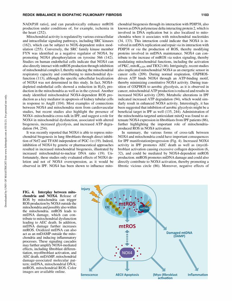
It was recently reported that NOX4 is able to repress mito-chondrial biogenesis in lung fibroblasts through direct inhibi-tion of Nrf2 and TFAM independent of PGC-1a (19). Indeed,inhibition of NOX4 by genetic or pharmaceutical approachesresulted in increased mitochondrial biogenesis, illustrated byincreased mitochondrial-to-nuclear DNA ratio (19). Un-fortunately, these studies only evaluated effects of NOX4 de-letion and not of NOX4 overexpression, as it would beobserved in IPF. NOX4 has been shown to influence mito-
chondrial biogenesis through its interaction with PDIP38, alsoknown as DNA polymerase delta interacting protein 2, which isinvolved in DNA replication but is also localized to mito-chondria where it associates with mitochondrial nucleotides(34, 133). This interaction could indicate that NOX4 is in-volved in mtDNA replication and repair via its interaction withPDIP38 or via the production of ROS, thereby modifyingproteins involved in mtDNA maintenance. NOX4 can con-tribute to the increase of mtROS via redox signaling, therebymodulating mitochondrial functions, including the activationof PKC, mitoKATP, and TRX2 (46). Intriguingly, recent studiesalso implicated mitochondrial NOX4 as an energetic sensor incancer cells (209). During normal respiration, OXPHOS-driven ATP binds NOX4 through an ATP-binding motif,thereby minimizing constitutive NOX4 activity. During tran-sition of OXPHOS to aerobic glycolysis, as it is observed incancer, mitochondrial ATP production is reduced and results inincreased NOX4 activity (209). Metabolic alterations in IPFindicated increased ATP degradation (94), which would sim-ilarly result in enhanced NOX4 activity. Interestingly, it hasbeen suggested that inhibition of aerobic glycolysis might be abeneficial target in IPF as well (135, 244). Administration ofthe mitochondria-targeted antioxidant mitoQ was found to at-tenuate NOX4 expression in fibroblasts from IPF patients (86),further highlighting the important role of mitochondria-produced ROS in NOX4 activation.
In summary, the various forms of cross-talk betweenNOX4 and mitochondria could have important consequencesfor IPF manifestation/progression (Fig. 4). Increased NOX4activity in IPF promotes AEC death as well as (myo)fi-broblast activation causing excessive collagen deposition (6,32), and could be mediated by NOX4-dependent mtROSproduction. mtROS promotes mtDNA damage and could alsodirectly contribute to NOX4 activation, thereby promoting afibrotic vicious circle (86). Moreover, negative effects of
FIG. 4. Interplay between mito-chondria and NOX4. Release ofROS by mitochondria can triggerROS production by NOX4 outside themitochondria and possibly also withinthe mitochondria. mtROS leads tomtDNA damage, which can con-tribute to mitochondrial dysfunctionleading to AEC death. In addition,mtDNA damage further increasesmtROS. Oxidized mtDNA can alsoact as an mtDAMP outside the mito-chondria and inducing inflammatoryprocesses. These signaling cascadesmay further amplify NOX4-mediatedeffects, including fibroblast differen-tiation, myofibroblast activation, andAEC death. mtDAMP, mitochondrialdamage-associated molecular pat-tern; mtDNA, mitochondrial DNA;mtROS, mitochondrial ROS. Colorimages are available online.
REDOX IMBALANCE IN IDIOPATHIC PULMONARY FIBROSIS 1103
Dow
nloa
ded
by U
NIV
OF
MA
AST
RIC
HT
fro
m w
ww
.lieb
ertp
ub.c
om a
t 03/
31/2
1. F
or p
erso
nal u
se o
nly.

NOX4 on mitochondrial biogenesis could further drive mi-tochondrial dysfunction. While these NOX4–mitochondriainteractions may be related to localization of NOX4 in mi-tochondria, evidence for such localization rest primarily ondetection with antibodies with unknown specificity, and it istypically not associated with costainings of p22phox, which isimportant for its activity. Hence, the exact role of NOX4 inmitochondria still remains unclear, and further research isneeded to more firmly establish NOX4 as a mitochondrialprotein, and its reciprocal interactions with mitochondrialprocesses.
Redox-Modulatory Therapeutic Strategies
Based on the various lines of evidence implicating a redoximbalance in IPF, several redox-based therapeutic strategieshave been proposed focusing at quenching ROS or restoringthe disturbed redox balance. For example, several studieshave addressed the ability of N-acetyl-cysteine (NAC), aprecursor of GSH, to mitigate IPF. However, in spite ofdemonstrated encouraging results in various in vitro studies(218) as well as experimental fibrosis models (44, 70), NACsupplementation has failed to be fully effective in the clinic(140, 153, 193) because of variable effects in patients withIPF. These variable effects may be due to differences in doseof drug administration, delivery method as well as the patientpopulation.
Several studies have suggested that inhaled NAC(352.4 mg, two times daily) improves lung function in earlystage IPF (82, 152, 164), indicating that local antioxidanttherapy might be more beneficial in an early stage of IPF tocounteract ROS-induced damage and the underlying inflam-mation. Oral administration of NAC (600 mg, three timesdaily), on the contrary, had variable outcomes. In the PAN-THER trial, no significant improvement of lung function couldbe observed in mild-to-moderate IPF compared with the pla-cebo (84), whereas it was reported before in the IFIGENIAtrial that the same dose of NAC in combination with predni-sone and azathioprine reduced decline of lung function (45). Inadvanced IPF, NAC monotherapy showed no beneficial effect;however, combination therapy has been suggested to improvelung function (198). Consequently, it is important to investi-gate specific subgroups in IPF who could benefit from NAC asmonotherapy or in combination therapy that has been sug-gested to be more beneficial in an advanced stage.
Other studies have explored the ability of antioxidant foodsupplements, such as quercetin and resveratrol, to reduceoxidative stress in profibrotic responses in vitro (62, 64, 155,231) and in animal models of pulmonary fibrosis (3, 74, 208,232). While these compounds have reported antioxidantactivity in vitro, they might act by alternative mechanismsand are also capable of activating Nrf2 (204). Intriguingly,a recent study described that quercetin reverses bleomycin-induced pulmonary fibrosis in aged mice through the reductionof various senescence markers and SASP (81). Furthermore,transcriptome analysis of pathways involved in senescenceand drugs that interfere with these pathways yielded dasatinib(a SRC/ABL protein kinase inhibitor) and quercetin (a putativeantioxidant but also an inhibitor of various kinases) as potentsenolytic drugs (259). Indeed, the combination of dasatinibwith quercetin (D + Q) was found to kill senescent fibroblastsin a mouse model of bleomycin-induced pulmonary fibrosis,
and thereby improve lung function (203). While these findingsdo not directly indicate antioxidant-based mechanisms, such amechanism could contribute to the observed inhibitory effects,especially in combination with other antifibrotic drugs. Thecombination of D + Q is currently investigated in a clinical trial(NCT02874989) to determine the effects on proinflammatorycells in IPF.
In addition to approaches using general scavengers ofROS, alternative strategies have focused on specificallymodulating the activity of redox systems. For example, basedon observed loss of GRX1 activity in IPF, and correspondingincreases in S-glutathionylation of Fas and caspase activation,administration of recombinant GRX1 has been explored as atherapeutic strategy, and preclinical studies on mice indicatethe ability of GRX1 to inhibit and even promote reversal ofpulmonary fibrosis in experimental models in mice (7).
Based on the diverse and specific actions of ROS producedby specific NOX enzymes or by mitochondria, as discussed inthis review, it would seem more fruitful to specifically target(enzymatic) sources of ROS that are dysregulated in IPF,rather than more generic approaches to neutralize ROS oraffect redox systems, as these latter could also interfere withnormal physiological functions of ROS, and thus exert un-wanted effects (63, 97). Indeed, recent approaches have fo-cused primarily on the specific roles of NOX enzymes andmitochondria in ROS production (39). Pharmacological ap-proaches to inhibit NOX4 are being developed as potentialtherapeutic strategies in IPF treatment. Indeed, a small mo-lecular inhibitor that selectively targets NOX4 as well asNOX1 (GKT137831) was found to minimize bleomycin-induced fibrosis in a mouse model, by reducing fibroblastactivation and collagen deposition as well as epithelial celldeath (75, 87). Moreover, GKT137831 treatment in IPF lungfibroblasts results in reduced markers of senescence and in-creased the susceptibility to apoptosis, suggesting that NOX4contributes to senescence (76) but further studies with spe-cific inhibitors are needed to establish this hypothesis.
Based on the accumulating evidence that associates mtROSwith IPF development, various mitochondria-targeted therapieshave been developed to improve mitochondrial biogenesis,activate mitophagy, inhibit mtROS-regulated apoptosis, andscavenge mtROS in general. The mitochondrial antioxidantmitoQ, a ubiquinone conjugated to a lipophilic triphenyl-phosphonium cation that accumulates within mitochondriaand is reduced to the antioxidant ubiquinol, has been shownto attenuate liver fibrosis (188) and reduce TGF-inducedprofibrotic gene expression in human fibroblasts (86). An-other mitochondrial-targeted small molecule, XBJ-5-131,was shown to improve the survival of neurons in a mousemodel of Huntington’s disease through inhibition of oxi-dative (mitochondrial) DNA damage (176, 246). Anotherexample is the antidiabetic drug, metformin, which inhibitsthe activity of complex I, thereby reducing mtROS. Metforminhas been suggested as antifibrotic drug as it interferes withTGF-b1 signaling, inhibits myofibroblast differentiation (222),and reduces collagen deposition in bleomycin-induced pul-monary fibrosis (184). A recent review, however, indicated nobeneficial effect on lung function in patients with IPF (215).
Overall, these various findings suggest that selective tar-geting of specific NOX isoforms or mtROS production may bebeneficial in management of IPF, especially in light of studies,indicating that redox alterations induced by mitochondrial
1104 VEITH ET AL.
Dow
nloa
ded
by U
NIV
OF
MA
AST
RIC
HT
fro
m w
ww
.lieb
ertp
ub.c
om a
t 03/
31/2
1. F
or p
erso
nal u
se o
nly.

dysfunction may also be associated with their interactions withNOX activation (86, 202).
Conclusion and Future Directions
Although IPF is well recognized for its devastating natureand poor prognosis, only minimal advances have been madein the last few years to effectively treat patients with IPF.Recent FDA-approved drugs pirfenidone and nintedanibhave only elicited modest improvements in disease progres-sion and cannot ameliorate the poor survival associated withIPF (180). Moreover, they do not work for all IPF patients,indicating the need to investigate more treatment strategies(72). As discussed in this review, IPF is characterized by adysregulated redox homeostasis and mitochondrial dysfunc-tion. Yet, attempts to reduce ROS with general antioxidantssuch as N-acetyl cysteine have largely been unsuccessful inthe clinic, which is likely due to our incomplete under-standing of specific redox-based mechanisms that contributeto IPF development, and the fact that such general antioxidantstrategies would also interfere with beneficial homeostaticfunctions of ROS. However, we would caution against neg-ative conclusions based on unsuccessful studies and dismissthe potential importance of ROS-based mechanisms in IPFpathology, because of accumulating evidence implicatingspecific redox-based mechanisms (increased NOX activa-tion, mitochondrial dysfunction) that ought to be targetedmore directly. In this regard, it would be prudent to explorethe efficacy of selective targeting of NOX4, based on com-pelling preclinical studies, or approaches to specifically inhibitmtROS such as MitoQ and MitoTEMPO, which have shownsome success in experimental mouse models of pulmonaryfibrosis. Another common challenge with such pharma-ceutical approaches is successful targeting of fibroblast foci,which may be poorly accessible to systemically adminis-tered drugs.
Another reason for our relatively limited understandingof ROS in IPF is the fact that detection of specific ROSpathways in clinical specimens is exceedingly difficult(especially reversible modifications that may be more im-portant in dysregulated cell signaling than stable proteinoxidation products). In this regard, recent studies of proteinS-glutathionylation in IPF in association with clinical out-comes (7) are particularly enlightening. Moreover, addres-sing the causality of specific ROS pathways would requireappropriate animal models, and currently used models ofpulmonary fibrosis do not adequately model the patho-physiology of human IPF and its progression. Also, whileIPF is well known to be age associated, most animal studiesare still using young animals and thus do not recapitulate thecontribution of age-associated alterations on specific redox-based pathways. Although costly, future studies should ad-dress pharmacological strategies in age-appropriate models.Another problem with studies of ROS in disease models suchas IPF is that they typically address the importance of aspecific NOX enzyme or antioxidant system throughout thedisease model, that is, during the initiation and developmentof the disease, which may not necessarily translate intoclinical practice if pre-existing fibrosis is not reversed. In thislight, some recent studies suggesting that antioxidant-basedapproaches can actually reverse fibrosis, at least in animalmodels (7, 184), are particularly encouraging.
Although evidence is growing for a role of specific ROSpathways in IPF (e.g., NOX4, mtROS), we still know rela-tively little with respect to the precise molecular mechanismsinvolved (i.e., what are the critical cellular targets for e.g.,NOX4-derived ROS that facilitate AEC apoptosis or fibro-blast activation). In light of emerging evidence that NOX4may be localized to mitochondria and that mitochondria andNOX4 exert reciprocal interactions, future studies should aimat unraveling the molecular redox mechanisms underlyingsuch interactions, for example by using approaches to in-terfere with mitochondrial NOX4 signaling. Successfulidentification of specific cellular targets for NOX4-derivedROS or for mtROS in IPF may reveal alternative and po-tentially more attractive therapeutic targets for treatment ofIPF. Given the rapid expansion of redox-based research inrecent years, and development of novel research tools toassess site-specific redox events in subcellular compart-ments or in specific proteins, it is anticipated that our un-derstanding of such NOX4–mitochondria interactions willsoon increase and may indeed lead to novel redox-basedtherapeutic strategies that may prove to be beneficial inimproving the survival and the quality of life in patientssuffering from IPF.
Acknowledgments
C.V. is supported by the Nutrim Graduate Programme(Maastricht University). A.v.d.V. gratefully acknowledgesresearch support from the National Heart Lung and BloodInstitute (R01 grants HL085646 and HL138708) and theNational Institute on Aging (R21 AG055325). In addition,the authors thank Birgit Hettler for her assistance in design-ing the figures.
References
1. American Thoracic Society. Idiopathic pulmonary fibrosis:diagnosis and treatment. International consensus statement.American Thoracic Society (ATS), and the EuropeanRespiratory Society (ERS). Am J Respir Crit Care Med161: 646–664, 2000.
2. Abbadie C, Pluquet O, and Pourtier A. Epithelial cellsenescence: an adaptive response to pre-carcinogenicstresses? Cell Mol Life Sci 74: 4471–4509, 2017.
3. Akgedik R, Akgedik S, Karamanli H, Uysal S, Bozkurt B,Ozol D, Armutcu F, and Yildirim Z. Effect of resveratrolon treatment of bleomycin-induced pulmonary fibrosis inrats. Inflammation 35: 1732–1741, 2012.
4. Alder JK, Chen JJ, Lancaster L, Danoff S, Su SC, Co-gan JD, Vulto I, Xie M, Qi X, Tuder RM, Phillips JA,3rd, Lansdorp PM, Loyd JE, and Armanios MY. Shorttelomeres are a risk factor for idiopathic pulmonary fi-brosis. Proc Natl Acad Sci U S A 105: 13051–13056,2008.
5. Alvarez D, Cardenes N, Sellares J, Bueno M, Corey C,Hanumanthu VS, Peng Y, D’Cunha H, Sembrat J, NouraieM, Shanker S, Caufield C, Shiva S, Armanios M, MoraAL, and Rojas M. IPF lung fibroblasts have a senescentphenotype. Am J Physiol Lung Cell Mol Physiol 313:L1164–L1173, 2017.
6. Amara N, Goven D, Prost F, Muloway R, Crestani B, andBoczkowski J. NOX4/NADPH oxidase expression is in-creased in pulmonary fibroblasts from patients with idio-pathic pulmonary fibrosis and mediates TGFbeta1-induced
REDOX IMBALANCE IN IDIOPATHIC PULMONARY FIBROSIS 1105
Dow
nloa
ded
by U
NIV
OF
MA
AST
RIC
HT
fro
m w
ww
.lieb
ertp
ub.c
om a
t 03/
31/2
1. F
or p
erso
nal u
se o
nly.

fibroblast differentiation into myofibroblasts. Thorax 65:733–738, 2010.
7. Anathy V, Lahue KG, Chapman DG, Chia SB, CaseyDT, Aboushousha R, van der Velden JLJ, Elko E,Hoffman SM, McMillan DH, Jones JT, Nolin JD, Ab-dalla S, Schneider R, Seward DJ, Roberson EC, LiptakMD, Cousins ME, Butnor KJ, Taatjes DJ, Budd RC,Irvin CG, Ho YS, Hakem R, Brown KK, Matsui R,Bachschmid MM, Gomez JL, Kaminski N, van der VlietA, and Janssen-Heininger YMW. Reducing proteinoxidation reverses lung fibrosis. Nat Med 24: 1128–1135, 2018.
8. Anathy V, Roberson E, Cunniff B, Nolin JD, HoffmanS, Spiess P, Guala AS, Lahue KG, Goldman D, FlemerS, van der Vliet A, Heintz NH, Budd RC, Tew KD,and Janssen-Heininger YM. Oxidative processing oflatent Fas in the endoplasmic reticulum controls thestrength of apoptosis. Mol Cell Biol 32: 3464–3478,2012.
9. Anathy V, Roberson EC, Guala AS, Godburn KE, BuddRC, and Janssen-Heininger YM. Redox-based regulationof apoptosis: S-glutathionylation as a regulatory mecha-nism to control cell death. Antioxid Redox Signal 16: 496–505, 2012.
10. Armanios M. Telomerase and idiopathic pulmonary fi-brosis. Mutat Res 730: 52–58, 2012.
11. Ashrafi G and Schwarz TL. The pathways of mitophagyfor quality control and clearance of mitochondria. CellDeath Differ 20: 31–42, 2013.
12. Baldelli S, Aquilano K, and Ciriolo MR. Punctum on twodifferent transcription factors regulated by PGC-1alpha:nuclear factor erythroid-derived 2-like 2 and nuclear re-spiratory factor 2. Biochim Biophys Acta 1830: 4137–4146, 2013.
13. Bargagli E, Olivieri C, Bennett D, Prasse A, Muller-Quernheim J, and Rottoli P. Oxidative stress in the path-ogenesis of diffuse lung diseases: a review. Respir Med103: 1245–1256, 2009.
14. Barron L and Wynn TA. Fibrosis is regulated by Th2 andTh17 responses and by dynamic interactions between fi-broblasts and macrophages. Am J Physiol GastrointestLiver Physiol 300: G723–G728, 2011.
15. Bedard K and Krause KH. The NOX family of ROS-generating NADPH oxidases: physiology and pathophys-iology. Physiol Rev 87: 245–313, 2007.
16. Beeh KM, Beier J, Haas IC, Kornmann O, Micke P, andBuhl R. Glutathione deficiency of the lower respiratorytract in patients with idiopathic pulmonary fibrosis. EurRespir J 19: 1119–1123, 2002.
17. Bellocq A, Azoulay E, Marullo S, Flahault A, FouquerayB, Philippe C, Cadranel J, and Baud L. Reactive oxygenand nitrogen intermediates increase transforming growthfactor-beta1 release from human epithelial alveolar cellsthrough two different mechanisms. Am J Respir Cell MolBiol 21: 128–136, 1999.
18. Bernard K, Hecker L, Luckhardt TR, Cheng G, andThannickal VJ. NADPH oxidases in lung health and dis-ease. Antioxid Redox Signal 20: 2838–2853, 2014.
19. Bernard K, Logsdon NJ, Miguel V, Benavides GA,Zhang J, Carter AB, Darley-Usmar VM, and ThannickalVJ. NADPH oxidase 4 (Nox4) suppresses mitochondrialbiogenesis and bioenergetics in lung fibroblasts viaa nuclear factor erythroid-derived 2-like 2 (Nrf2)-
dependent pathway. J Biol Chem 292: 3029–3038,2017.
20. Block K, Gorin Y, and Abboud HE. Subcellular locali-zation of Nox4 and regulation in diabetes. Proc Natl AcadSci U S A 106: 14385–14390, 2009.
21. Boden WE, Padala SK, Cabral KP, Buschmann IR, andSidhu MS. Role of short-acting nitroglycerin in the man-agement of ischemic heart disease. Drug Des Devel Ther9: 4793–4805, 2015.
22. Borzi RM, Grigolo B, Meliconi R, Fasano L, Sturani C,Fabbri M, Porstmann T, and Facchini A. Elevated serumsuperoxide dismutase levels correlate with disease sever-ity and neutrophil degranulation in idiopathic pulmonaryfibrosis. Clin Sci 85: 353–359, 1993.
23. Boudreau HE, Casterline BW, Rada B, Korzeniowska A,and Leto TL. Nox4 involvement in TGF-beta and SMAD3-driven induction of the epithelial-to-mesenchymal transi-tion and migration of breast epithelial cells. Free RadicBiol Med 53: 1489–1499, 2012.
24. Bowler RP, Nicks M, Warnick K, and Crapo JD. Role ofextracellular superoxide dismutase in bleomycin-inducedpulmonary fibrosis. Am J Physiol Lung Cell Mol Physiol282: L719–L726, 2002.
25. Brand MD. Mitochondrial generation of superoxide andhydrogen peroxide as the source of mitochondrial redoxsignaling. Free Radic Biol Med 100: 14–31, 2016.
26. Brenmoehl J and Hoeflich A. Dual control of mitochon-drial biogenesis by sirtuin 1 and sirtuin 3. Mitochondrion13: 755–761, 2013.
27. Bueno M, Lai YC, Romero Y, Brands J, St. Croix CM,Kamga C, Corey C, Herazo-Maya JD, Sembrat J, Lee JS,Duncan SR, Rojas M, Shiva S, Chu CT, and Mora AL.PINK1 deficiency impairs mitochondrial homeostasisand promotes lung fibrosis. J Clin Invest 125: 521–538,2015.
28. Camelo A, Dunmore R, Sleeman MA, and Clarke DL. Theepithelium in idiopathic pulmonary fibrosis: breaking thebarrier. Front Pharmacol 4: 173, 2014.
29. Cannito S, Novo E, di Bonzo LV, Busletta C, ColombattoS, and Parola M. Epithelial-mesenchymal transition: frommolecular mechanisms, redox regulation to implicationsin human health and disease. Antioxid Redox Signal 12:1383–1430, 2010.
30. Cantin AM, Hubbard RC, and Crystal RG. Glutathionedeficiency in the epithelial lining fluid of the lower re-spiratory tract in idiopathic pulmonary fibrosis. Am RevRespir Dis 139: 370–372, 1989.
31. Cantin AM, North SL, Fells GA, Hubbard RC, andCrystal RG. Oxidant-mediated epithelial cell injury inidiopathic pulmonary fibrosis. J Clin Invest 79: 1665–1673, 1987.
32. Carnesecchi S, Deffert C, Donati Y, Basset O, Hinz B,Preynat-Seauve O, Guichard C, Arbiser JL, Banfi B,Pache JC, Barazzone-Argiroffo C, and Krause KH. Akey role for NOX4 in epithelial cell death during de-velopment of lung fibrosis. Antioxid Redox Signal 15:607–619, 2011.
33. Carnesecchi S, Deffert C, Pagano A, Garrido-Urbani S,Metrailler-Ruchonnet I, Schappi M, Donati Y, MatthayMA, Krause KH, and Barazzone Argiroffo C. NADPHoxidase-1 plays a crucial role in hyperoxia-induced acutelung injury in mice. Am J Respir Crit Care Med 180: 972–981, 2009.
1106 VEITH ET AL.
Dow
nloa
ded
by U
NIV
OF
MA
AST
RIC
HT
fro
m w
ww
.lieb
ertp
ub.c
om a
t 03/
31/2
1. F
or p
erso
nal u
se o
nly.
34. Cheng X, Kanki T, Fukuoh A, Ohgaki K, Takeya R, AokiY, Hamasaki N, and Kang D. PDIP38 associates withproteins constituting the mitochondrial DNA nucleoid. JBiochem 138: 673–678, 2005.
35. Cheresh P, Kim SJ, Tulasiram S, and Kamp DW. Oxi-dative stress and pulmonary fibrosis. Biochim BiophysActa 1832: 1028–1040, 2013.
36. Cheresh P, Morales-Nebreda L, Kim SJ, Yeldandi A,Williams DB, Cheng Y, Mutlu GM, Budinger GR, RidgeK, Schumacker PT, Bohr VA, and Kamp DW. Asbestos-induced pulmonary fibrosis is augmented in 8-oxoguanineDNA glycosylase knockout mice. Am J Respir Cell MolBiol 52: 25–36, 2015.
37. Cho HY, Reddy SP, and Kleeberger SR. Nrf2 defends thelung from oxidative stress. Antioxid Redox Signal 8: 76–87, 2006.
38. Choi SH, Kim M, Lee HJ, Kim EH, Kim CH, and Lee YJ.Effects of NOX1 on fibroblastic changes of endothelialcells in radiation induced pulmonary fibrosis. Mol MedRep 13: 4135–4142, 2016.
39. Ciencewicki J, Trivedi S, and Kleeberger SR. Oxidantsand the pathogenesis of lung diseases. J Allergy ClinImmunol 122: 456–468; quiz 469–470, 2008.
40. Cloonan SM and Choi AM. Mitochondria in lung disease.J Clin Invest 126: 809–820, 2016.
41. Comhair SA and Erzurum SC. Antioxidant responses tooxidant-mediated lung diseases. Am J Physiol Lung CellMol Physiol 283: L246–L255, 2002.
42. Conte E, Gili E, Fagone E, Fruciano M, Iemmolo M, andVancheri C. Effect of pirfenidone on proliferation, TGF-beta-induced myofibroblast differentiation and fibrogenicactivity of primary human lung fibroblasts. Eur J Phar-maceut Sci 58: 13–19, 2014.
43. Cool CD, Groshong SD, Rai PR, Henson PM, Stewart JS,and Brown KK. Fibroblast foci are not discrete sites oflung injury or repair: the fibroblast reticulum. Am J RespirCrit Care Med 174: 654–658, 2006.
44. Cortijo J, Cerda-Nicolas M, Serrano A, Bioque G, EstrelaJM, Santangelo F, Esteras A, Llombart-Bosch A, andMorcillo EJ. Attenuation by oral N-acetylcysteine ofbleomycin-induced lung injury in rats. Eur Respir J 17:1228–1235, 2001.
45. Demedts M, Behr J, Buhl R, Costabel U, Dekhuijzen R,Jansen HM, MacNee W, Thomeer M, Wallaert B, LaurentF, Nicholson AG, Verbeken EK, Verschakelen J, FlowerCD, Capron F, Petruzzelli S, De Vuyst P, van den BoschJM, Rodriguez-Becerra E, Corvasce G, Lankhorst I, Sar-dina M, Montanari M, and IFEGENIA Group. High-doseacetylcysteine in idiopathic pulmonary fibrosis. N Engl JMed 353: 2229–2242, 2005.
46. Dikalov S. Cross talk between mitochondria and NADPHoxidases. Free Radic Biol Med 51: 1289–1301, 2011.
47. Dikalova AE, Bikineyeva AT, Budzyn K, Nazarewicz RR,McCann L, Lewis W, Harrison DG, and Dikalov SI.Therapeutic targeting of mitochondrial superoxide in hy-pertension. Circ Res 107: 106–116, 2010.
48. Doughan AK, Harrison DG, and Dikalov SI. Molecularmechanisms of angiotensin II-mediated mitochondrialdysfunction: linking mitochondrial oxidative damage andvascular endothelial dysfunction. Circ Res 102: 488–496,2008.
49. Droge W. Free radicals in the physiological control of cellfunction. Physiol Rev 82: 47–95, 2002.
50. du Bois RM. Strategies for treating idiopathic pulmonaryfibrosis. Nat Rev Drug Discov 9: 129–140, 2010.
51. Durcan TM and Fon EA. The three ‘P’s of mitophagy:PARKIN, PINK1, and post-translational modifications.Genes Dev 29: 989–999, 2015.
52. Ellson CD, Dunmore R, Hogaboam CM, Sleeman MA,and Murray LA. Danger-associated molecular patternsand danger signals in idiopathic pulmonary fibrosis. Am JRespir Cell Mol Biol 51: 163–168, 2014.
53. Elmore S. Apoptosis: a review of programmed cell death.Toxicol Pathol 35: 495–516, 2007.
54. Evans CM, Fingerlin TE, Schwarz MI, Lynch D, KurcheJ, Warg L, Yang IV, and Schwartz DA. Idiopathic pul-monary fibrosis: a genetic disease that involves muco-ciliary dysfunction of the peripheral airways. Physiol Rev96: 1567–1591, 2016.
55. Fang EF, Scheibye-Knudsen M, Chua KF, Mattson MP,Croteau DL, and Bohr VA. Nuclear DNA damage sig-nalling to mitochondria in ageing. Nat Rev Mol Cell Biol17: 308–321, 2016.
56. Farkas L, Farkas D, Ask K, Moller A, Gauldie J, MargettsP, Inman M, and Kolb M. VEGF ameliorates pulmonaryhypertension through inhibition of endothelial apoptosis inexperimental lung fibrosis in rats. J Clin Invest 119: 1298–1311, 2009.
57. Fattman CL, Chang LY, Termin TA, Petersen L, En-ghild JJ, and Oury TD. Enhanced bleomycin-inducedpulmonary damage in mice lacking extracellular su-peroxide dismutase. Free Radic Biol Med 35: 763–771,2003.
58. Fernandez IE and Eickelberg O. The impact of TGF-betaon lung fibrosis: from targeting to biomarkers. Proc AmThorac Soc 9: 111–116, 2012.
59. Fois AG, Paliogiannis P, Sotgia S, Mangoni AA, ZinelluE, Pirina P, Carru C, and Zinellu A. Evaluation of oxi-dative stress biomarkers in idiopathic pulmonary fibrosisand therapeutic applications: a systematic review. RespirRes 19: 51, 2018.
60. Furuie H, Yamasaki H, Suga M, and Ando M. Alteredaccessory cell function of alveolar macrophages: apossible mechanism for induction of Th2 secretoryprofile in idiopathic pulmonary fibrosis. Eur Respir J10: 787–794, 1997.
61. Gauldie J, Kolb M, and Sime PJ. A new direction in thepathogenesis of idiopathic pulmonary fibrosis? Respir Res3: 1, 2002.
62. Gharaee-Kermani M, Moore BB, and Macoska JA.Resveratrol-mediated repression and reversion of pros-tatic myofibroblast phenoconversion. PLoS One 11:e0158357, 2016.
63. Ghezzi P, Jaquet V, Marcucci F, and Schmidt H. Theoxidative stress theory of disease: levels of evidenceand epistemological aspects. Br J Pharmacol 174:1784–1796, 2017.
64. Giovannelli L, Pitozzi V, Jacomelli M, Mulinacci N,Laurenzana A, Dolara P, and Mocali A. Protective effectsof resveratrol against senescence-associated changes incultured human fibroblasts. J Gerontol A Biol Sci Med Sci66: 9–18, 2011.
65. Giri SN, Leonard S, Shi X, Margolin SB, and VallyathanV. Effects of pirfenidone on the generation of reactiveoxygen species in vitro. J Environ Pathol Toxicol Oncol18: 169–177, 1999.
REDOX IMBALANCE IN IDIOPATHIC PULMONARY FIBROSIS 1107
Dow
nloa
ded
by U
NIV
OF
MA
AST
RIC
HT
fro
m w
ww
.lieb
ertp
ub.c
om a
t 03/
31/2
1. F
or p
erso
nal u
se o
nly.

66. Gomes LC, Di Benedetto G, and Scorrano L. Duringautophagy mitochondria elongate, are spared from deg-radation and sustain cell viability. Nat Cell Biol 13: 589–598, 2011.
67. Gonzalez-Gonzalez FJ, Chandel NS, Jain M, and Bu-dinger GRS. Reactive oxygen species as signaling mole-cules in the development of lung fibrosis. Transl Res 190:61–68, 2017.
68. Graham KA, Kulawiec M, Owens KM, Li X, DesoukiMM, Chandra D, and Singh KK. NADPH oxidase 4 is anoncoprotein localized to mitochondria. Cancer Biol Ther10: 223–231, 2010.
69. Guo Q. Changes in mitochondrial function during EMTinduced by TGFbeta-1 in pancreatic cancer. Oncol Lett13: 1575–1580, 2017.
70. Hagiwara SI, Ishii Y, and Kitamura S. Aerosolized ad-ministration of N-acetylcysteine attenuates lung fibrosisinduced by bleomycin in mice. Am J Respir Crit CareMed 162: 225–231, 2000.
71. Hashimoto N, Phan SH, Imaizumi K, Matsuo M, Naka-shima H, Kawabe T, Shimokata K, and Hasegawa Y.Endothelial-mesenchymal transition in bleomycin-induced pulmonary fibrosis. Am J Respir Cell Mol Biol 43:161–172, 2010.
72. Hayton C and Chaudhuri N. Managing idiopathic pul-monary fibrosis: which drug for which patient? DrugsAging 34: 647–653, 2017.
73. He C, Murthy S, McCormick ML, Spitz DR, Ryan AJ, andCarter AB. Mitochondrial Cu,Zn-superoxide dismutasemediates pulmonary fibrosis by augmenting H2O2 gener-ation. J Biol Chem 286: 15597–15607, 2011.
74. He X, Wang L, Szklarz G, Bi Y, and Ma Q. Resveratrolinhibits paraquat-induced oxidative stress and fibrogenicresponse by activating the nuclear factor erythroid 2-related factor 2 pathway. J Pharmacol Exp Ther 342: 81–90, 2012.
75. Hecker L, Cheng J, and Thannickal VJ. Targeting NOXenzymes in pulmonary fibrosis. Cell Mol Life Sci 69:2365–2371, 2012.
76. Hecker L, Logsdon NJ, Kurundkar D, Kurundkar A,Bernard K, Hock T, Meldrum E, Sanders YY, andThannickal VJ. Reversal of persistent fibrosis in aging bytargeting Nox4-Nrf2 redox imbalance. Sci Transl Med 6:231ra47, 2014.
77. Hecker L, Vittal R, Jones T, Jagirdar R, Luckhardt TR,Horowitz JC, Pennathur S, Martinez FJ, and ThannickalVJ. NADPH oxidase-4 mediates myofibroblast activationand fibrogenic responses to lung injury. Nat Med 15:1077–1081, 2009.
78. Herrera B, Murillo MM, Alvarez-Barrientos A, Beltran J,Fernandez M, and Fabregat I. Source of early reactiveoxygen species in the apoptosis induced by transforminggrowth factor-beta in fetal rat hepatocytes. Free RadicBiol Med 36: 16–26, 2004.
79. Hinz B, Phan SH, Thannickal VJ, Galli A, Bochaton-Piallat ML, and Gabbiani G. The myofibroblast: onefunction, multiple origins. Am J Pathol 170: 1807–1816,2007.
80. Hiraga R, Kato M, Miyagawa S, and Kamata T. Nox4-derived ROS signaling contributes to TGF-beta-inducedepithelial-mesenchymal transition in pancreatic cancercells. Anticancer Res 33: 4431–4438, 2013.
81. Hohmann MS, Habiel DM, Coelho AL, Verri WA, Jr.,and Hogaboam CM. Quercetin enhances ligand-induced
apoptosis in senescent IPF fibroblasts and reduces lungfibrosis in vivo. Am J Respir Cell Mol Biol 60: 28–40,2019.
82. Homma S, Azuma A, Taniguchi H, Ogura T, Mochiduki Y,Sugiyama Y, Nakata K, Yoshimura K, Takeuchi M, KudohS, and Japan NAC Couplinical Study Group. Efficacy ofinhaled N-acetylcysteine monotherapy in patients withearly stage idiopathic pulmonary fibrosis. Respirology17: 467–477, 2012.
83. Hostettler KE, Zhong J, Papakonstantinou E, Kar-akiulakis G, Tamm M, Seidel P, Sun Q, Mandal J,Lardinois D, Lambers C, and Roth M. Anti-fibrotic ef-fects of nintedanib in lung fibroblasts derived frompatients with idiopathic pulmonary fibrosis. Respir Res15: 157, 2014.
84. Idiopathic Pulmonary Fibrosis Clinical Research Network,Martinez FJ, de Andrade JA, Anstrom KJ, King TE, Jr.,and Raghu G. Randomized trial of acetylcysteine in idi-opathic pulmonary fibrosis. N Engl J Med 370: 2093–2101, 2014.
85. Jablonski RP, Kim SJ, Cheresh P, Williams DB,Morales-Nebreda L, Cheng Y, Yeldandi A, Bhorade S,Pardo A, Selman M, Ridge K, Gius D, Budinger GRS,and Kamp DW. SIRT3 deficiency promotes lung fi-brosis by augmenting alveolar epithelial cell mito-chondrial DNA damage and apoptosis. FASEB J 31:2520–2532, 2017.
86. Jain M, Rivera S, Monclus EA, Synenki L, Zirk A, Ei-senbart J, Feghali-Bostwick C, Mutlu GM, Budinger GR,and Chandel NS. Mitochondrial reactive oxygen speciesregulate transforming growth factor-beta signaling. J BiolChem 288: 770–777, 2013.
87. Jarman ER, Khambata VS, Cope C, Jones P, Roger J,Ye LY, Duggan N, Head D, Pearce A, Press NJ, Bel-lenie B, Sohal B, and Jarai G. An inhibitor of NADPHoxidase-4 attenuates established pulmonary fibrosis in arodent disease model. Am J Respir Cell Mol Biol 50:158–169, 2014.
88. Jiang F, Liu GS, Dusting GJ, and Chan EC. NADPHoxidase-dependent redox signaling in TGF-beta-mediatedfibrotic responses. Redox Biol 2: 267–272, 2014.
89. Jolly MK, Ward C, Eapen MS, Myers S, Hallgren O,Levine H, and Sohal SS. Epithelial-mesenchymaltransition, a spectrum of states: role in lung develop-ment, homeostasis, and disease. Dev Dyn 247: 346–358, 2018.
90. Jones HA, Schofield JB, Krausz T, Boobis AR, and Ha-slett C. Pulmonary fibrosis correlates with duration oftissue neutrophil activation. Am J Respir Crit Care Med158: 620–628, 1998.
91. Kage H and Borok Z. EMT and interstitial lung disease: amysterious relationship. Curr Opin Pulm Med 18: 517–523, 2012.
92. Kalluri R and Neilson EG. Epithelial-mesenchymal tran-sition and its implications for fibrosis. J Clin Invest 112:1776–1784, 2003.
93. Kang KA, Ryu YS, Piao MJ, Shilnikova K, Kang HK, YiJM, Boulanger M, Paolillo R, Bossis G, Yoon SY, KimSB, and Hyun JW. DUOX2-mediated production of re-active oxygen species induces epithelial mesenchymaltransition in 5-fluorouracil resistant human colon cancercells. Redox Biol 17: 224–235, 2018.
94. Kang YP, Lee SB, Lee JM, Kim HM, Hong JY, Lee WJ,Choi CW, Shin HK, Kim DJ, Koh ES, Park CS, Kwon
1108 VEITH ET AL.
Dow
nloa
ded
by U
NIV
OF
MA
AST
RIC
HT
fro
m w
ww
.lieb
ertp
ub.c
om a
t 03/
31/2
1. F
or p
erso
nal u
se o
nly.

SW, and Park SW. Metabolic profiling regarding patho-genesis of idiopathic pulmonary fibrosis. J Proteome Res15: 1717–1724, 2016.
95. Kasper M and Barth K. Potential contribution of alveolarepithelial type I cells to pulmonary fibrosis. Biosci Rep 37:pii: BSR20171301, 2017.
96. Kato S, Inui N, Hakamata A, Suzuki Y, Enomoto N,Fujisawa T, Nakamura Y, Watanabe H, and Suda T.Changes in pulmonary endothelial cell properties duringbleomycin-induced pulmonary fibrosis. Respir Res 19:127, 2018.
97. Kawagishi H and Finkel T. Unraveling the truth aboutantioxidants: ROS and disease: finding the right balance.Nat Med 20: 711–713, 2014.
98. Kawahara T, Quinn MT, and Lambeth JD. Molecularevolution of the reactive oxygen-generating NADPH ox-idase (Nox/Duox) family of enzymes. BMC Evol Biol 7:109, 2007.
99. Kikuchi N, Ishii Y, Morishima Y, Yageta Y, Haraguchi N,Itoh K, Yamamoto M, and Hizawa N. Nrf2 protectsagainst pulmonary fibrosis by regulating the lung oxidantlevel and Th1/Th2 balance. Respir Res 11: 31, 2010.
100. Kim KK, Kugler MC, Wolters PJ, Robillard L, GalvezMG, Brumwell AN, Sheppard D, and Chapman HA. Al-veolar epithelial cell mesenchymal transition developsin vivo during pulmonary fibrosis and is regulated by theextracellular matrix. Proc Natl Acad Sci U S A 103:13180–13185, 2006.
101. Kim SJ, Cheresh P, Jablonski RP, Morales-Nebreda L,Cheng Y, Hogan E, Yeldandi A, Chi M, Piseaux R,Ridge K, Michael Hart C, Chandel N, Scott BudingerGR, and Kamp DW. Mitochondrial catalase over-expressed transgenic mice are protected against lungfibrosis in part via preventing alveolar epithelial cellmitochondrial DNA damage. Free Radic Biol Med 101:482–490, 2016.
102. Kim SJ, Cheresh P, Jablonski RP, Williams DB, andKamp DW. The role of mitochondrial DNA in mediatingalveolar epithelial cell apoptosis and pulmonary fibrosis.Int J Mol Sci 16: 21486–21519, 2015.
103. Kim SJ, Cheresh P, Williams D, Cheng Y, Ridge K,Schumacker PT, Weitzman S, Bohr VA, and Kamp DW.Mitochondria-targeted Ogg1 and aconitase-2 preventoxidant-induced mitochondrial DNA damage in alveolarepithelial cells. J Biol Chem 289: 6165–6176, 2014.
104. Kim SM, Kim YG, Jeong KH, Lee SH, Lee TW, Ihm CG,and Moon JY. Angiotensin II-induced mitochondrialNox4 is a major endogenous source of oxidative stress inkidney tubular cells. PLoS One 7: e39739, 2012.
105. Kim YM, Kim SJ, Tatsunami R, Yamamura H, Fukai T,and Ushio-Fukai M. ROS-induced ROS release orches-trated by Nox4, Nox2, and mitochondria in VEGF sig-naling and angiogenesis. Am J Physiol Cell Physiol 312:C749–C764, 2017.
106. Kinder BW, Brown KK, Schwarz MI, Ix JH, Kervitsky A,and King TE, Jr. Baseline BAL neutrophilia predicts earlymortality in idiopathic pulmonary fibrosis. Chest 133:226–232, 2008.
107. King TE, Jr., Schwarz MI, Brown K, Tooze JA, ColbyTV, Waldron JA, Jr., Flint A, Thurlbeck W, and Cher-niack RM. Idiopathic pulmonary fibrosis: relationshipbetween histopathologic features and mortality. Am JRespir Crit Care Med 164: 1025–1032, 2001.
108. Kinnula VL and Crapo JD. Superoxide dismutases in thelung and human lung diseases. Am J Respir Crit Care Med167: 1600–1619, 2003.
109. Kinnula VL, Fattman CL, Tan RJ, and Oury TD. Oxida-tive stress in pulmonary fibrosis: a possible role for redoxmodulatory therapy. Am J Respir Crit Care Med 172:417–422, 2005.
110. Kinnula VL, Hodgson UA, Lakari EK, Tan RJ, SormunenRT, Soini YM, Kakko SJ, Laitinen TH, Oury TD, andPaakko PK. Extracellular superoxide dismutase has ahighly specific localization in idiopathic pulmonary fi-brosis/usual interstitial pneumonia. Histopathology 49:66–74, 2006.
111. Kliment CR and Oury TD. Oxidative stress, extracellularmatrix targets, and idiopathic pulmonary fibrosis. FreeRadic Biol Med 49: 707–717, 2010.
112. Kolb M, Bonella F, and Wollin L. Therapeutic targets inidiopathic pulmonary fibrosis. Respir Med 131: 49–57,2017.
113. Koziel R, Pircher H, Kratochwil M, Lener B, Hermann M,Dencher NA, and Jansen-Durr P. Mitochondrial respira-tory chain complex I is inactivated by NADPH oxidaseNox4. Biochem J 452: 231–239, 2013.
114. Kroller-Schon S, Steven S, Kossmann S, Scholz A, DaubS, Oelze M, Xia N, Hausding M, Mikhed Y, Zinssius E,Mader M, Stamm P, Treiber N, Scharffetter-Kochanek K,Li H, Schulz E, Wenzel P, Munzel T, and Daiber A.Molecular mechanisms of the crosstalk between mito-chondria and NADPH oxidase through reactive oxygenspecies-studies in white blood cells and in animal models.Antioxid Redox Signal 20: 247–266, 2014.
115. Kubli DA and Gustafsson AB. Mitochondria and mito-phagy: the yin and yang of cell death control. Circ Res111: 1208–1221, 2012.
116. Kuroda J, Ago T, Matsushima S, Zhai P, Schneider MD,and Sadoshima J. NADPH oxidase 4 (Nox4) is a majorsource of oxidative stress in the failing heart. Proc NatlAcad Sci U S A 107: 15565–15570, 2010.
117. Kurundkar A and Thannickal VJ. Redox mechanisms inage-related lung fibrosis. Redox Biol 9: 67–76, 2016.
118. Kuwano K, Kunitake R, Maeyama T, Hagimoto N, Ka-wasaki M, Matsuba T, Yoshimi M, Inoshima I, YoshidaK, and Hara N. Attenuation of bleomycin-induced pneu-mopathy in mice by a caspase inhibitor. Am J PhysiolLung Cell Mol Physiol 280: L316–L325, 2001.
119. Kuwano K, Nakashima N, Inoshima I, Hagimoto N, FujitaM, Yoshimi M, Maeyama T, Hamada N, Watanabe K, andHara N. Oxidative stress in lung epithelial cells frompatients with idiopathic interstitial pneumonias. Eur Re-spir J 21: 232–240, 2003.
120. Lambeth JD. NOX enzymes and the biology of reactiveoxygen. Nat Rev Immunol 4: 181–189, 2004.
121. Lambeth JD, Kawahara T, and Diebold B. Regulation ofNox and Duox enzymatic activity and expression. FreeRadic Biol Med 43: 319–331, 2007.
122. Leach HG, Chrobak I, Han R, and Trojanowska M. En-dothelial cells recruit macrophages and contribute to afibrotic milieu in bleomycin lung injury. Am J Respir CellMol Biol 49: 1093–1101, 2013.
123. Lee HC, Yin PH, Chi CW, and Wei YH. Increase inmitochondrial mass in human fibroblasts under oxidativestress and during replicative cell senescence. J Biomed Sci9: 517–526, 2002.
REDOX IMBALANCE IN IDIOPATHIC PULMONARY FIBROSIS 1109
Dow
nloa
ded
by U
NIV
OF
MA
AST
RIC
HT
fro
m w
ww
.lieb
ertp
ub.c
om a
t 03/
31/2
1. F
or p
erso
nal u
se o
nly.

124. Lee SB, Bae IH, Bae YS, and Um HD. Link betweenmitochondria and NADPH oxidase 1 isozyme for thesustained production of reactive oxygen species and celldeath. J Biol Chem 281: 36228–36235, 2006.
125. Lennon FE and Salgia R. Mitochondrial dynamics: bi-ology and therapy in lung cancer. Expert Opin InvestDrugs 23: 675–692, 2014.
126. Lenz AG, Costabel U, and Maier KL. Oxidized BAL fluidproteins in patients with interstitial lung diseases. EurRespir J 9: 307–312, 1996.
127. Lettieri CJ, Nathan SD, Barnett SD, Ahmad S, and ShorrAF. Prevalence and outcomes of pulmonary arterial hy-pertension in advanced idiopathic pulmonary fibrosis.Chest 129: 746–752, 2006.
128. Li G, Yuzhen L, Yi C, Xiaoxiang C, Wei Z, ChangqingZ, and Shuang Y. DNaseI protects against Paraquat-induced acute lung injury and pulmonary fibrosis me-diated by mitochondrial DNA. BioMed Res Int 2015:386952, 2015.
129. Little AC, Sham D, Hristova M, Danyal K, Heppner DE,Bauer RA, Sipsey LM, Habibovic A, and van der Vliet A.DUOX1 silencing in lung cancer promotes EMT, cancerstem cell characteristics and invasive properties. Onco-genesis 5: e261, 2016.
130. Liu RM and Desai LP. Reciprocal regulation of TGF-betaand reactive oxygen species: a perverse cycle for fibrosis.Redox Biol 6: 565–577, 2015.
131. Liu X and Chen Z. The pathophysiological role of mito-chondrial oxidative stress in lung diseases. J Transl Med15: 207, 2017.
132. Lopez-Otin C, Blasco MA, Partridge L, Serrano M, andKroemer G. The hallmarks of aging. Cell 153: 1194–1217,2013.
133. Lyle AN, Deshpande NN, Taniyama Y, Seidel-Rogol B,Pounkova L, Du P, Papaharalambus C, Lassegue B, andGriendling KK. Poldip2, a novel regulator of Nox4 andcytoskeletal integrity in vascular smooth muscle cells.Circ Res 105: 249–259, 2009.
134. Magro CM, Waldman WJ, Knight DA, Allen JN, NadasdyT, Frambach GE, Ross P, and Marsh CB. Idiopathic pul-monary fibrosis related to endothelial injury and anti-endothelial cell antibodies. Hum Immunol 67: 284–297,2006.
135. Maher TM. Aerobic glycolysis and the Warburg effect.An unexplored realm in the search for fibrosis therapies?Am J Respir Crit Care Med 192: 1407–1409, 2015.
136. Malli F, Koutsokera A, Paraskeva E, Zakynthinos E, Pa-pagianni M, Makris D, Tsilioni I, Molyvdas PA, Gour-goulianis KI, and Daniil Z. Endothelial progenitor cells inthe pathogenesis of idiopathic pulmonary fibrosis: anevolving concept. PLoS One 8: e53658, 2013.
137. Manoury B, Nenan S, Leclerc O, Guenon I, Boichot E,Planquois JM, Bertrand CP, and Lagente V. The absenceof reactive oxygen species production protects miceagainst bleomycin-induced pulmonary fibrosis. Respir Res6: 11, 2005.
138. Markart P, Luboeinski T, Korfei M, Schmidt R, WygreckaM, Mahavadi P, Mayer K, Wilhelm J, Seeger W, GuentherA, and Ruppert C. Alveolar oxidative stress is associatedwith elevated levels of nonenzymatic low-molecular-weight antioxidants in patients with different forms ofchronic fibrosing interstitial lung diseases. Antioxid RedoxSignal 11: 227–240, 2009.
139. Marmai C, Sutherland RE, Kim KK, Dolganov GM, FangX, Kim SS, Jiang S, Golden JA, Hoopes CW, MatthayMA, Chapman HA, and Wolters PJ. Alveolar epithelialcells express mesenchymal proteins in patients with idi-opathic pulmonary fibrosis. Am J Physiol Lung Cell MolPhysiol 301: L71–L78, 2011.
140. Martinez FJ, de Andrade JA, Anstrom KJ, King TE, Jr.,and Raghu G. Randomized trial of acetylcysteine in idi-opathic pulmonary fibrosis. N Engl J Med 370: 2093–2101, 2014.
141. Martyn KD, Frederick LM, von Loehneysen K, DinauerMC, and Knaus UG. Functional analysis of Nox4 revealsunique characteristics compared to other NADPH oxi-dases. Cell Signal 18: 69–82, 2006.
142. Matsushima S, Kuroda J, Zhai P, Liu T, Ikeda S, Nagar-ajan N, Oka S, Yokota T, Kinugawa S, Hsu CP, Li H,Tsutsui H, and Sadoshima J. Tyrosine kinase FYN nega-tively regulates NOX4 in cardiac remodeling. J Clin In-vest 126: 3403–3416, 2016.
143. Mazur W, Lindholm P, Vuorinen K, Myllarniemi M,Salmenkivi K, and Kinnula VL. Cell-specific elevationof NRF2 and sulfiredoxin-1 as markers of oxidativestress in the lungs of idiopathic pulmonary fibrosis andnon-specific interstitial pneumonia. APMIS 118: 703–712, 2010.
144. Meiners S, Eickelberg O, and Konigshoff M. Hallmarks ofthe ageing lung. Eur Respir J 45: 807–827, 2015.
145. Meitzler JL and Ortiz de Montellano PR. Caenorhabditiselegans and human dual oxidase 1 (DUOX1) ‘‘peroxi-dase’’ domains: insights into heme binding and catalyticactivity. J Biol Chem 284: 18634–18643, 2009.
146. Meyer A, Buhl R, and Magnussen H. The effect of oral N-acetylcysteine on lung glutathione levels in idiopathicpulmonary fibrosis. Eur Respir J 7: 431–436, 1994.
147. Minagawa S, Araya J, Numata T, Nojiri S, Hara H, Yu-mino Y, Kawaishi M, Odaka M, Morikawa T, NishimuraSL, Nakayama K, and Kuwano K. Accelerated epithelialcell senescence in IPF and the inhibitory role of SIRT6 inTGF-beta-induced senescence of human bronchial epi-thelial cells. Am J Physiol Lung Cell Mol Physiol 300:L391–L401, 2011.
148. Misra HP and Rabideau C. Pirfenidone inhibits NADPH-dependent microsomal lipid peroxidation and scavengeshydroxyl radicals. Mol Cell Biochem 204: 119–126, 2000.
149. Montuschi P, Ciabattoni G, Paredi P, Pantelidis P, du BoisRM, Kharitonov SA, and Barnes PJ. 8-Isoprostane as abiomarker of oxidative stress in interstitial lung diseases.Am J Respir Crit Care Med 158: 1524–1527, 1998.
150. Mora AL, Bueno M, and Rojas M. Mitochondria in thespotlight of aging and idiopathic pulmonary fibrosis. JClin Invest 127: 405–414, 2017.
151. Mora AL, Rojas M, Pardo A, and Selman M. Emergingtherapies for idiopathic pulmonary fibrosis, a progres-sive age-related disease. Nat Rev Drug Discov 16: 810,2017.
152. Muramatsu Y, Sugino K, Ishida F, Tatebe J, Morita T, andHomma S. Effect of inhaled N-acetylcysteine mono-therapy on lung function and redox balance in idiopathicpulmonary fibrosis. Respir Invest 54: 170–178, 2016.
153. Myllarniemi M and Kaarteenaho R. Pharmacologicaltreatment of idiopathic pulmonary fibrosis—preclinicaland clinical studies of pirfenidone, nintedanib, and N-acetylcysteine. Eur Clin Respir J 2, 2015.
1110 VEITH ET AL.
Dow
nloa
ded
by U
NIV
OF
MA
AST
RIC
HT
fro
m w
ww
.lieb
ertp
ub.c
om a
t 03/
31/2
1. F
or p
erso
nal u
se o
nly.

154. Naikawadi RP, Disayabutr S, Mallavia B, Donne ML,Green G, La JL, Rock JR, Looney MR, and Wolters PJ.Telomere dysfunction in alveolar epithelial cells causeslung remodeling and fibrosis. JCI Insight 1: e86704,2016.
155. Nakamura T, Matsushima M, Hayashi Y, Shibasaki M,Imaizumi K, Hashimoto N, Shimokata K, Hasegawa Y,and Kawabe T. Attenuation of transforming growthfactor-beta-stimulated collagen production in fibroblastsby quercetin-induced heme oxygenase-1. Am J Respir CellMol Biol 44: 614–620, 2011.
156. Nalysnyk L, Cid-Ruzafa J, Rotella P, and Esser D.Incidence and prevalence of idiopathic pulmonary fi-brosis: review of the literature. Eur Respir Rev 21:355–361, 2012.
157. Nauseef WM. How human neutrophils kill and degrademicrobes: an integrated view. Immunol Rev 219: 88–102,2007.
158. Navaratnam V, Fleming KM, West J, Smith CJ, JenkinsRG, Fogarty A, and Hubbard RB. The rising incidence ofidiopathic pulmonary fibrosis in the U.K. Thorax 66: 462–467, 2011.
159. Nicholson AG, Fulford LG, Colby TV, du Bois RM,Hansell DM, and Wells AU. The relationship betweenindividual histologic features and disease progression inidiopathic pulmonary fibrosis. Am J Respir Crit Care Med166: 173–177, 2002.
160. Nicolson GL. Mitochondrial dysfunction and chronicdisease: treatment with natural supplements. Integr Med(Encinitas) 13: 35–43, 2014.
161. Odajima N, Betsuyaku T, Nagai K, Moriyama C, WangDH, Takigawa T, Ogino K, and Nishimura M. The roleof catalase in pulmonary fibrosis. Respir Res 11: 183,2010.
162. Ogura M, Yamaki J, Homma MK, and Homma Y. Mi-tochondrial c-Src regulates cell survival through phos-phorylation of respiratory chain components. Biochem J447: 281–289, 2012.
163. Oku H, Shimizu T, Kawabata T, Nagira M, Hikita I,Ueyama A, Matsushima S, Torii M, and Arimura A.Antifibrotic action of pirfenidone and prednisolone: dif-ferent effects on pulmonary cytokines and growth factorsin bleomycin-induced murine pulmonary fibrosis. Eur JPharmacol 590: 400–408, 2008.
164. Okuda R, Matsushima H, Oba T, Kawabe R, Matsu-bayashi M, Amano M, Nishizawa T, and Honda K. Effi-cacy and safety of inhaled N-acetylcysteine in idiopathicpulmonary fibrosis: a prospective, single-arm study. Re-spir Investig 54: 156–161, 2016.
165. Pache JC, Carnesecchi S, Deffert C, Donati Y, HerrmannFR, Barazzone-Argiroffo C, and Krause KH. NOX-4 isexpressed in thickened pulmonary arteries in idiopathicpulmonary fibrosis. Nat Med 17: 31–32; author reply 32–33, 2011.
166. Palikaras K and Tavernarakis N. Mitochondrial homeo-stasis: the interplay between mitophagy and mitochondrialbiogenesis. Exp Gerontol 56: 182–188, 2014.
167. Panduri V, Liu G, Surapureddi S, Kondapalli J, SoberanesS, de Souza-Pinto NC, Bohr VA, Budinger GR, Schu-macker PT, Weitzman SA, and Kamp DW. Role of mi-tochondrial hOGG1 and aconitase in oxidant-induced lungepithelial cell apoptosis. Free Radic Biol Med 47: 750–759, 2009.
168. Pardo A and Selman M. Idiopathic pulmonary fibrosis:new insights in its pathogenesis. Int J Biochem Cell Biol34: 1534–1538, 2002.
169. Pardo A and Selman M. Lung fibroblasts, aging, and id-iopathic pulmonary fibrosis. Ann Am Thorac Soc 13:S417–S421, 2016.
170. Patel AS, Lin L, Geyer A, Haspel JA, An CH, Cao J,Rosas IO, and Morse D. Autophagy in idiopathic pulmo-nary fibrosis. PLoS One 7: e41394, 2012.
171. Patel AS, Song JW, Chu SG, Mizumura K, Osorio JC,Shi Y, El-Chemaly S, Lee CG, Rosas IO, Elias JA, ChoiAM, and Morse D. Epithelial cell mitochondrial dys-function and PINK1 are induced by transforming growthfactor-beta1 in pulmonary fibrosis. PLoS One 10: e0121246,2015.
172. Peltoniemi M, Kaarteenaho-Wiik R, Saily M, SormunenR, Paakko P, Holmgren A, Soini Y, and Kinnula VL.Expression of glutaredoxin is highly cell specific in hu-man lung and is decreased by transforming growth factor-beta in vitro and in interstitial lung diseases in vivo. HumPathol 35: 1000–1007, 2004.
173. Petry A, Djordjevic T, Weitnauer M, Kietzmann T, HessJ, and Gorlach A. NOX2 and NOX4 mediate proliferativeresponse in endothelial cells. Antioxid Redox Signal 8:1473–1484, 2006.
174. Piera-Velazquez S, Li Z, and Jimenez SA. Role ofendothelial-mesenchymal transition (EndoMT) in thepathogenesis of fibrotic disorders. Am J Pathol 179: 1074–1080, 2011.
175. Plataki M, Koutsopoulos AV, Darivianaki K, Delides G,Siafakas NM, and Bouros D. Expression of apoptotic andantiapoptotic markers in epithelial cells in idiopathicpulmonary fibrosis. Chest 127: 266–274, 2005.
176. Polyzos A, Holt A, Brown C, Cosme C, Wipf P, Gomez-Marin A, Castro MR, Ayala-Pena S, and McMurray CT.Mitochondrial targeting of XJB-5-131 attenuates or im-proves pathophysiology in HdhQ150 animals with well-developed disease phenotypes. Hum Mol Genet 25: 1792–1802, 2016.
177. Prasad S, Hogaboam CM, and Jarai G. Deficient repairresponse of IPF fibroblasts in a co-culture model of epi-thelial injury and repair. Fibrogenesis Tissue Repair 7: 7,2014.
178. Psathakis K, Mermigkis D, Papatheodorou G, Loukides S,Panagou P, Polychronopoulos V, Siafakas NM, andBouros D. Exhaled markers of oxidative stress in idio-pathic pulmonary fibrosis. Eur J Clin Invest 36: 362–367,2006.
179. Raghu G, Chen SY, Hou Q, Yeh WS, and Collard HR.Incidence and prevalence of idiopathic pulmonary fibrosisin US adults 18–64 years old. Eur Respir J 48: 179–186,2016.
180. Raghu G, Rochwerg B, Zhang Y, Garcia CA, Azuma A,Behr J, Brozek JL, Collard HR, Cunningham W, HommaS, Johkoh T, Martinez FJ, Myers J, Protzko SL, RicheldiL, Rind D, Selman M, Theodore A, Wells AU, Hoog-steden H, Schunemann HJ, American Thoracic Society,European Respiratory Society, Japanese Respiratory So-ciety, and Latin American Thoracic Association. An Of-ficial ATS/ERS/JRS/ALAT clinical practice guideline:treatment of idiopathic pulmonary fibrosis. An update ofthe 2011 clinical practice guideline. Am J Respir CritCare Med 192: e3–e19, 2015.
REDOX IMBALANCE IN IDIOPATHIC PULMONARY FIBROSIS 1111
Dow
nloa
ded
by U
NIV
OF
MA
AST
RIC
HT
fro
m w
ww
.lieb
ertp
ub.c
om a
t 03/
31/2
1. F
or p
erso
nal u
se o
nly.

181. Raghu G, Weycker D, Edelsberg J, Bradford WZ, andOster G. Incidence and prevalence of idiopathic pulmo-nary fibrosis. Am J Respir Crit Care Med 174: 810–816,2006.
182. Rahman I, Skwarska E, Henry M, Davis M, O’ConnorCM, FitzGerald MX, Greening A, and MacNee W. Sys-temic and pulmonary oxidative stress in idiopathic pul-monary fibrosis. Free Radic Biol Med 27: 60–68, 1999.
183. Rangarajan S, Bernard K, and Thannickal VJ. Mitochon-drial dysfunction in pulmonary fibrosis. Ann Am ThoracSoc 14: S383–S388, 2017.
184. Rangarajan S, Bone NB, Zmijewska AA, Jiang S, ParkDW, Bernard K, Locy ML, Ravi S, Deshane J, MannonRB, Abraham E, Darley-Usmar V, Thannickal VJ, andZmijewski JW. Metformin reverses established lung fi-brosis in a bleomycin model. Nat Med 24: 1121–1127,2018.
185. Rathore R, Zheng YM, Niu CF, Liu QH, Korde A, Ho YS,and Wang YX. Hypoxia activates NADPH oxidase toincrease [ROS]i and [Ca2+]i through the mitochondrialROS-PKCepsilon signaling axis in pulmonary arterysmooth muscle cells. Free Radic Biol Med 45: 1223–1231, 2008.
186. Ray PD, Huang BW, and Tsuji Y. Reactive oxygen spe-cies (ROS) homeostasis and redox regulation in cellularsignaling. Cell Signal 24: 981–990, 2012.
187. Reddy NM, Kleeberger SR, Cho HY, Yamamoto M,Kensler TW, Biswal S, and Reddy SP. Deficiency in Nrf2-GSH signaling impairs type II cell growth and enhancessensitivity to oxidants. Am J Respir Cell Mol Biol 37: 3–8,2007.
188. Rehman H, Liu Q, Krishnasamy Y, Shi Z, Ramshesh VK,Haque K, Schnellmann RG, Murphy MP, Lemasters JJ,Rockey DC, and Zhong Z. The mitochondria-targetedantioxidant MitoQ attenuates liver fibrosis in mice. Int JPhysiol Pathophysiol Pharmacol 8: 14–27, 2016.
189. Rey FE, Cifuentes ME, Kiarash A, Quinn MT, and PaganoPJ. Novel competitive inhibitor of NAD(P)H oxidase as-sembly attenuates vascular O(2)(-) and systolic bloodpressure in mice. Circ Res 89: 408–414, 2001.
190. Reznick RM, Zong H, Li J, Morino K, Moore IK, Yu HJ,Liu ZX, Dong J, Mustard KJ, Hawley SA, Befroy D,Pypaert M, Hardie DG, Young LH, and Shulman GI.Aging-associated reductions in AMP-activated proteinkinase activity and mitochondrial biogenesis. Cell Metab5: 151–156, 2007.
191. Riteau N, Gasse P, Fauconnier L, Gombault A, CouegnatM, Fick L, Kanellopoulos J, Quesniaux VF, Marchand-Adam S, Crestani B, Ryffel B, and Couillin I. Extra-cellular ATP is a danger signal activating P2X7 receptorin lung inflammation and fibrosis. Am J Respir Crit CareMed 182: 774–783, 2010.
192. Rock JR, Barkauskas CE, Cronce MJ, Xue Y, HarrisJR, Liang J, Noble PW, and Hogan BL. Multiplestromal populations contribute to pulmonary fibrosiswithout evidence for epithelial to mesenchymal tran-sition. Proc Natl Acad Sci U S A 108: E1475–E1483,2011.
193. Rogliani P, Calzetta L, Cavalli F, Matera MG, and Caz-zola M. Pirfenidone, nintedanib and N-acetylcysteine forthe treatment of idiopathic pulmonary fibrosis: a system-atic review and meta-analysis. Pulm Pharmacol Ther 40:95–103, 2016.
194. Romano AD, Greco E, Vendemiale G, and Serviddio G.Bioenergetics and mitochondrial dysfunction in aging:recent insights for a therapeutical approach. Curr PharmDes 20: 2978–2992, 2014.
195. Rottoli P, Magi B, Cianti R, Bargagli E, Vagaggini C,Nikiforakis N, Pallini V, and Bini L. Carbonylated pro-teins in bronchoalveolar lavage of patients with sarcoid-osis, pulmonary fibrosis associated with systemic sclerosisand idiopathic pulmonary fibrosis. Proteomics 5: 2612–2618, 2005.
196. Ryan US and Ryan JW. Cell biology of pulmonary en-dothelium. Circulation 70: III46–III62, 1984.
197. Ryu C, Sun H, Gulati M, Herazo-Maya JD, Chen Y,Osafo-Addo A, Brandsdorfer C, Winkler J, Blaul C,Faunce J, Pan H, Woolard T, Tzouvelekis A, Antin-Ozerkis DE, Puchalski JT, Slade M, Gonzalez AL, Bo-genhagen DF, Kirillov V, Feghali-Bostwick C, Gibson K,Lindell K, Herzog RI, Dela Cruz CS, Mehal W, KaminskiN, Herzog EL, and Trujillo G. Extracellular mitochondrialDNA is generated by fibroblasts and predicts death inidiopathic pulmonary fibrosis. Am J Respir Crit Care Med196: 1571–1581, 2017.
198. Sakamoto S, Itoh T, Muramatsu Y, Satoh K, Ishida F,Sugino K, Isobe K, and Homma S. Efficacy of pirfenidonein patients with advanced-stage idiopathic pulmonary fi-brosis. Intern Med 52: 2495–2501, 2013.
199. Saleh D, Barnes PJ, and Giaid A. Increased production ofthe potent oxidant peroxynitrite in the lungs of patientswith idiopathic pulmonary fibrosis. Am J Respir Crit CareMed 155: 1763–1769, 1997.
200. Sanders YY, Liu H, Liu G, and Thannickal VJ. Epige-netic mechanisms regulate NADPH oxidase-4 expressionin cellular senescence. Free Radic Biol Med 79: 197–205, 2015.
201. Santos JH, Hunakova L, Chen Y, Bortner C, and VanHouten B. Cell sorting experiments link persistent mito-chondrial DNA damage with loss of mitochondrialmembrane potential and apoptotic cell death. J Biol Chem278: 1728–1734, 2003.
202. Sato N, Takasaka N, Yoshida M, Tsubouchi K, MinagawaS, Araya J, Saito N, Fujita Y, Kurita Y, Kobayashi K, ItoS, Hara H, Kadota T, Yanagisawa H, Hashimoto M, Ut-sumi H, Wakui H, Kojima J, Numata T, Kaneko Y, OdakaM, Morikawa T, Nakayama K, Kohrogi H, and KuwanoK. Metformin attenuates lung fibrosis development viaNOX4 suppression. Respir Res 17: 107, 2016.
203. Schafer MJ, White TA, Iijima K, Haak AJ, Ligresti G,Atkinson EJ, Oberg AL, Birch J, Salmonowicz H, Zhu Y,Mazula DL, Brooks RW, Fuhrmann-Stroissnigg H, Pirts-khalava T, Prakash YS, Tchkonia T, Robbins PD, AubryMC, Passos JF, Kirkland JL, Tschumperlin DJ, Kita H,and LeBrasseur NK. Cellular senescence mediates fibroticpulmonary disease. Nat Commun 8: 14532, 2017.
204. Schmidt HH, Stocker R, Vollbracht C, Paulsen G, RileyD, Daiber A, and Cuadrado A. Antioxidants in transla-tional medicine. Antioxid Redox Signal 23: 1130–1143,2015.
205. Selman M, King TE, Pardo A, American Thoracic S,European Respiratory Society, and American College ofChest Physicians. Idiopathic pulmonary fibrosis: pre-vailing and evolving hypotheses about its pathogenesisand implications for therapy. Ann Intern Med 134: 136–151, 2001.
1112 VEITH ET AL.
Dow
nloa
ded
by U
NIV
OF
MA
AST
RIC
HT
fro
m w
ww
.lieb
ertp
ub.c
om a
t 03/
31/2
1. F
or p
erso
nal u
se o
nly.

206. Selman M and Pardo A. Revealing the pathogenic andaging-related mechanisms of the enigmatic idiopathicpulmonary fibrosis. An integral model. Am J Respir CritCare Med 189: 1161–1172, 2014.
207. Sena LA and Chandel NS. Physiological roles of mito-chondrial reactive oxygen species. Mol Cell 48: 158–167,2012.
208. Sener G, Topaloglu N, Sehirli AO, Ercan F, and Gedik N.Resveratrol alleviates bleomycin-induced lung injury inrats. Pulm Pharmacol Ther 20: 642–649, 2007.
209. Shanmugasundaram K, Nayak BK, Friedrichs WE,Kaushik D, Rodriguez R, and Block K. NOX4 functionsas a mitochondrial energetic sensor coupling cancer met-abolic reprogramming to drug resistance. Nat Commun 8:997, 2017.
210. Shvedova AA, Kisin ER, Murray AR, Kommineni C,Castranova V, Fadeel B, and Kagan VE. Increased accu-mulation of neutrophils and decreased fibrosis in the lungof NADPH oxidase-deficient C57BL/6 mice exposed tocarbon nanotubes. Toxicol Appl Pharmacol 231: 235–240,2008.
211. Singel KL and Segal BH. NOX2-dependent regulation ofinflammation. Clin Sci 130: 479–490, 2016.
212. Sisson TH, Mendez M, Choi K, Subbotina N, Courey A,Cunningham A, Dave A, Engelhardt JF, Liu X, White ES,Thannickal VJ, Moore BB, Christensen PJ, and SimonRH. Targeted injury of type II alveolar epithelial cellsinduces pulmonary fibrosis. Am J Respir Crit Care Med181: 254–263, 2010.
213. Smeitink J, van den Heuvel L, and DiMauro S. The ge-netics and pathology of oxidative phosphorylation. NatRev Genet 2: 342–352, 2001.
214. Sosulski ML, Gongora R, Danchuk S, Dong C, Luo F, andSanchez CG. Deregulation of selective autophagy duringaging and pulmonary fibrosis: the role of TGFbeta1. AgingCell 14: 774–783, 2015.
215. Spagnolo P, Kreuter M, Maher TM, Wuyts W, Bonella F,Corte TJ, Kopf S, Weycker D, Kirchgaessler KU, andRyerson CJ. Metformin does not affect clinically relevantoutcomes in patients with idiopathic pulmonary fibrosis.Respiration 96: 314–322, 2018.
216. Strausz J, Muller-Quernheim J, Steppling H, and FerlinzR. Oxygen radical production by alveolar inflammatorycells in idiopathic pulmonary fibrosis. Am Rev Respir Dis141: 124–128, 1990.
217. Sturrock A, Cahill B, Norman K, Huecksteadt TP, Hill K,Sanders K, Karwande SV, Stringham JC, Bull DA, GleichM, Kennedy TP, and Hoidal JR. Transforming growthfactor-beta1 induces Nox4 NAD(P)H oxidase and reactiveoxygen species-dependent proliferation in human pulmo-nary artery smooth muscle cells. Am J Physiol Lung CellMol Physiol 290: L661–L673, 2006.
218. Sugiura H, Ichikawa T, Liu X, Kobayashi T, Wang XQ,Kawasaki S, Togo S, Kamio K, Mao L, Ann Y, IchinoseM, and Rennard SI. N-acetyl-l-cysteine inhibits TGF-beta1-induced profibrotic responses in fibroblasts. PulmPharmacol Ther 22: 487–491, 2009.
219. Sureshbabu A and Bhandari V. Targeting mitochondrialdysfunction in lung diseases: emphasis on mitophagy.Front Physiol 4: 384, 2013.
220. Swamy SM, Rajasekaran NS, and Thannickal VJ. Nuclearfactor-erythroid-2-related factor 2 in aging and lung fi-brosis. Am J Pathol 186: 1712–1723, 2016.
221. Takabatake N, Arao T, Sata M, Abe S, Inoue S, Shibata Y,Takeishi Y, and Kubota I. Involvement of pulmonaryendothelial cell injury in the pathogenesis of pulmonaryfibrosis: clinical assessment by 123I-MIBG lung scintig-raphy. Eur J Nucl Med Mol Imaging 32: 221–228, 2005.
222. Thakur S, Viswanadhapalli S, Kopp JB, Shi Q, Barnes JL,Block K, Gorin Y, and Abboud HE. Activation of AMP-activated protein kinase prevents TGF-beta1-inducedepithelial-mesenchymal transition and myofibroblast ac-tivation. Am J Pathol 185: 2168–2180, 2015.
223. Thannickal VJ and Fanburg BL. Activation of an H2O2-generating NADH oxidase in human lung fibroblasts bytransforming growth factor beta 1. J Biol Chem 270:30334–30338, 1995.
224. Thannickal VJ and Horowitz JC. Evolving concepts ofapoptosis in idiopathic pulmonary fibrosis. Proc AmThorac Soc 3: 350–356, 2006.
225. Thannickal VJ, Lee DY, White ES, Cui Z, Larios JM,Chacon R, Horowitz JC, Day RM, and Thomas PE.Myofibroblast differentiation by transforming growthfactor-beta1 is dependent on cell adhesion and integrinsignaling via focal adhesion kinase. J Biol Chem 278:12384–12389, 2003.
226. Tiitto L, Kaarteenaho-Wiik R, Sormunen R, Holmgren A,Paakko P, Soini Y, and Kinnula VL. Expression of thethioredoxin system in interstitial lung disease. J Pathol201: 363–370, 2003.
227. Uhal BD, Joshi I, Hughes WF, Ramos C, Pardo A, andSelman M. Alveolar epithelial cell death adjacent to un-derlying myofibroblasts in advanced fibrotic human lung.Am J Physiol 275: L1192–L1199, 1998.
228. Valko M, Leibfritz D, Moncol J, Cronin MT, Mazur M,and Telser J. Free radicals and antioxidants in normalphysiological functions and human disease. Int J BiochemCell Biol 39: 44–84, 2007.
229. van der Vliet A. NADPH oxidases in lung biology andpathology: host defense enzymes, and more. Free RadicBiol Med 44: 938–955, 2008.
230. van der Vliet A, Janssen-Heininger YMW, and Anathy V.Oxidative stress in chronic lung disease: from mitochon-drial dysfunction to dysregulated redox signaling. MolAspects Med 63: 59–69, 2018.
231. Veith C, Drent M, Bast A, van Schooten FJ, and BootsAW. The disturbed redox-balance in pulmonary fibrosis ismodulated by the plant flavonoid quercetin. Toxicol ApplPharmacol 336: 40–48, 2017.
232. Verma R, Kushwah L, Gohel D, Patel M, Marvania T,and Balakrishnan S. Evaluating the ameliorative po-tential of quercetin against the bleomycin-inducedpulmonary fibrosis in wistar rats. Pulm Med 2013:921724, 2013.
233. Vuorinen K, Ohlmeier S, Lepparanta O, Salmenkivi K,Myllarniemi M, and Kinnula VL. Peroxiredoxin II ex-pression and its association with oxidative stress and cellproliferation in human idiopathic pulmonary fibrosis. JHistochem Cytochem 56: 951–959, 2008.
234. Waghray M, Cui Z, Horowitz JC, Subramanian IM,Martinez FJ, Toews GB, and Thannickal VJ. Hydrogenperoxide is a diffusible paracrine signal for the inductionof epithelial cell death by activated myofibroblasts. FAS-EB J 19: 854–856, 2005.
235. Wang CL, Kang J, and Li ZH. [Increased expression ofNADPH oxidase p47-PHOX and p67-PHOX factor in
REDOX IMBALANCE IN IDIOPATHIC PULMONARY FIBROSIS 1113
Dow
nloa
ded
by U
NIV
OF
MA
AST
RIC
HT
fro
m w
ww
.lieb
ertp
ub.c
om a
t 03/
31/2
1. F
or p
erso
nal u
se o
nly.

idiopathic pulmonary fibrosis]. Zhonghua Jie He He Hu XiZa Zhi 30: 265–268, 2007 (Article in Chinese).
236. Waring P and Mullbacher A. Cell death induced by theFas/Fas ligand pathway and its role in pathology. ImmunolCell Biol 77: 312–317, 1999.
237. Wenzel P, Mollnau H, Oelze M, Schulz E, Wickrama-nayake JM, Muller J, Schuhmacher S, Hortmann M,Baldus S, Gori T, Brandes RP, Munzel T, and Daiber A.First evidence for a crosstalk between mitochondrial andNADPH oxidase-derived reactive oxygen species innitroglycerin-triggered vascular dysfunction. AntioxidRedox Signal 10: 1435–1447, 2008.
238. Willis BC, Liebler JM, Luby-Phelps K, Nicholson AG,Crandall ED, du Bois RM, and Borok Z. Induction ofepithelial-mesenchymal transition in alveolar epithelialcells by transforming growth factor-beta1: potential rolein idiopathic pulmonary fibrosis. Am J Pathol 166: 1321–1332, 2005.
239. Wong HS, Dighe PA, Mezera V, Monternier PA, andBrand MD. Production of superoxide and hydrogenperoxide from specific mitochondrial sites under dif-ferent bioenergetic conditions. J Biol Chem 292:16804–16809, 2017.
240. World health organization. Ageing and health. 2018.http://who.int/news-room/fact-sheets/detail/ageing-and-health (accessed November 26, 2018).
241. Wosniak J, Jr., Santos CX, Kowaltowski AJ, and LaurindoFR. Cross-talk between mitochondria and NADPH oxi-dase: effects of mild mitochondrial dysfunction on an-giotensin II-mediated increase in Nox isoform expressionand activity in vascular smooth muscle cells. AntioxidRedox Signal 11: 1265–1278, 2009.
242. Wu Z, Puigserver P, Andersson U, Zhang C, Adelmant G,Mootha V, Troy A, Cinti S, Lowell B, Scarpulla RC, andSpiegelman BM. Mechanisms controlling mitochondrialbiogenesis and respiration through the thermogeniccoactivator PGC-1. Cell 98: 115–124, 1999.
243. Wynn TA and Vannella KM. Macrophages in TissueRepair, Regeneration, and Fibrosis. Immunity 44: 450–462, 2016.
244. Xie N, Tan Z, Banerjee S, Cui H, Ge J, Liu RM, BernardK, Thannickal VJ, and Liu G. Glycolytic reprogrammingin myofibroblast differentiation and lung fibrosis. Am JRespir Crit Care Med 192: 1462–1474, 2015.
245. Xu X, Dai H, and Wang C. Epithelium-dependent profi-brotic milieu in the pathogenesis of idiopathic pulmonaryfibrosis: current status and future directions. Clin Respir J10: 133–141, 2016.
246. Xun Z, Rivera-Sanchez S, Ayala-Pena S, Lim J, BudworthH, Skoda EM, Robbins PD, Niedernhofer LJ, Wipf P, andMcMurray CT. Targeting of XJB-5-131 to mitochondriasuppresses oxidative DNA damage and motor decline in amouse model of Huntington’s disease. Cell Rep 2: 1137–1142, 2012.
247. Yan F, Wang Y, Wu X, Peshavariya HM, Dusting GJ,Zhang M, and Jiang F. Nox4 and redox signaling mediateTGF-beta-induced endothelial cell apoptosis and pheno-typic switch. Cell Death Dis 5: e1010, 2014.
248. Yanai H, Shteinberg A, Porat Z, Budovsky A, BraimanA, Ziesche R, and Fraifeld VE. Cellular senescence-likefeatures of lung fibroblasts derived from idiopathic pul-monary fibrosis patients. Aging (Albany NY) 7: 664–672,2015.
249. Yang IV and Schwartz DA. Epigenetics of idiopathicpulmonary fibrosis. Transl Res 165: 48–60, 2015.
250. Yoon YS, Lee JH, Hwang SC, Choi KS, and Yoon G. TGFbeta1 induces prolonged mitochondrial ROS generationthrough decreased complex IV activity with senescent ar-rest in Mv1Lu cells. Oncogene 24: 1895–1903, 2005.
251. Yu G, Tzouvelekis A, Wang R, Herazo-Maya JD, IbarraGH, Srivastava A, de Castro JPW, DeIuliis G, Ahangari F,Woolard T, Aurelien N, Arrojo EDR, Gan Y, Graham M,Liu X, Homer RJ, Scanlan TS, Mannam P, Lee PJ, HerzogEL, Bianco AC, and Kaminski N. Thyroid hormone in-hibits lung fibrosis in mice by improving epithelial mito-chondrial function. Nat Med 24: 39–49, 2018.
252. Yu Q, Lee CF, Wang W, Karamanlidis G, Kuroda J, Mat-sushima S, Sadoshima J, and Tian R. Elimination of NADPHoxidase activity promotes reductive stress and sensitizes theheart to ischemic injury. J Am Heart Assoc 3: e000555, 2014.
253. Zandalinas SI and Mittler R. ROS-induced ROS release inplant and animal cells. Free Radic Biol Med 122: 21–27,2018.
254. Zank DC, Bueno M, Mora AL, and Rojas M. IdiopathicPulmonary Fibrosis: aging, Mitochondrial Dysfunction, andCellular Bioenergetics. Front Med (Lausanne) 5: 10, 2018.
255. Zhang H, Davies KJ, and Forman HJ. TGFbeta1 rapidlyactivates Src through a non-canonical redox signalingmechanism. Arch Biochem Biophys 568: 1–7, 2015.
256. Zhang H, Davies KJA, and Forman HJ. Oxidative stressresponse and Nrf2 signaling in aging. Free Radic BiolMed 88: 314–336, 2015.
257. Zhong Z, Liang S, Sanchez-Lopez E, He F, Shalapour S,Lin XJ, Wong J, Ding S, Seki E, Schnabl B, Hevener AL,Greenberg HB, Kisseleva T, and Karin M. New mito-chondrial DNA synthesis enables NLRP3 inflammasomeactivation. Nature 560: 198–203, 2018.
258. Zhou L, Zhang H, Davies KJA, and Forman HJ. Aging-related decline in the induction of Nrf2-regulated antiox-idant genes in human bronchial epithelial cells. RedoxBiol 14: 35–40, 2018.
259. Zhu Y, Tchkonia T, Pirtskhalava T, Gower AC, Ding H,Giorgadze N, Palmer AK, Ikeno Y, Hubbard GB, LenburgM, O’Hara SP, LaRusso NF, Miller JD, Roos CM, Ver-zosa GC, LeBrasseur NK, Wren JD, Farr JN, Khosla S,Stout MB, McGowan SJ, Fuhrmann-Stroissnigg H, GurkarAU, Zhao J, Colangelo D, Dorronsoro A, Ling YY, Bar-ghouthy AS, Navarro DC, Sano T, Robbins PD, Nie-dernhofer LJ, and Kirkland JL. The Achilles’ heel ofsenescent cells: from transcriptome to senolytic drugs.Aging Cell 14: 644–658, 2015.
Address correspondence to:Dr. Albert van der Vliet
Department of Pathology and Laboratory MedicineLarner College of Medicine
University of VermontHSRF 216
149 Beaumont AvenueBurlington, VT 05405
E-mail: [email protected]
Date of first submission to ARS Central, January 28, 2019;date of acceptance, February 11, 2019.
1114 VEITH ET AL.
Dow
nloa
ded
by U
NIV
OF
MA
AST
RIC
HT
fro
m w
ww
.lieb
ertp
ub.c
om a
t 03/
31/2
1. F
or p
erso
nal u
se o
nly.

Abbreviations Used
a-SMA¼ a-smooth muscle actinAEC¼ alveolar epithelial cell
AngII¼ angiotensin IIBALF¼ bronchoalveolar lavage fluid
DUOX¼ dual oxidaseEBC¼ exhaled breath condensatesECM¼ extracellular matrixELF¼ epithelial lining fluid
EMT¼ epithelial-to-mesenchymal transitionETC¼ electron transport chainFDA¼Food and Drug AdministrationGRX¼ glutaredoxinGSH¼ glutathioneGST¼ glutathione-S-transferase
H2O2¼ hydrogen peroxideIL¼ interleukin
IPF¼ idiopathic pulmonary fibrosisLAP¼ latency-binding peptide
M2¼ alternatively activated macrophagesmtDAMP¼mitochondrial damage-associated
molecular patternmtDNA¼mitochondrial DNA
mtROS¼mitochondrial ROSNAC¼ N-acetyl cysteine
NOXes¼NADPH oxidasesNRF¼ nuclear respiratory factorNrf2¼ nuclear factor erythroid 2-related factor 2O2�-¼ superoxide anion
OGG1¼ oxoguanine DNA glycosylase 1OXPHOS¼ oxidative phosphorylation
PGC-1a¼ peroxisome proliferator-activatedreceptor c coactivator 1a
PINK1¼ PTEN-induced putative kinase 1PRX¼ peroxiredoxinROS¼ reactive oxygen species
SASP¼ senescence-associated secretoryphenotype
SMC¼ smooth muscle cellSOD¼ superoxide dismutase
TFAM¼mitochondrial transcription factor ATGF-b¼ transforming growth factor b
TH¼T-helperTRX¼ thioredoxinUIP¼ usual interstitial pneumonia
VEGF¼ vascular endothelial growth factor
REDOX IMBALANCE IN IDIOPATHIC PULMONARY FIBROSIS 1115
Dow
nloa
ded
by U
NIV
OF
MA
AST
RIC
HT
fro
m w
ww
.lieb
ertp
ub.c
om a
t 03/
31/2
1. F
or p
erso
nal u
se o
nly.