PREVALENCE OF ORAL SUBMUCOUS FIBROSIS AMONG ...
9
646 PREVALENCE OF ORAL SUBMUCOUS FIBROSIS AMONG 50,915 INDIAN VILLAGERS J. J. PINDBORG*, F. S. MEHTA, P. C. GUPTA AND D. K. DAFTARY From the Basic Dental Research Unit, Tata Institute of Fundamental Research, Homi Bhabha Road, Bombay 5, India Received for publication May 30, 1968 ORAL submucous fibrosis is a condition which has been studied in only the last 15 years, though it probably has existed for a long period of time. With our present knowledge we may define submucous fibrosis as an insidious, chronic disease affecting any part of the oral cavity and sometimes pharynx and oesophagus. Occasionally preceded by and/or associated with vesicle formation, fibrous bands are always present, preferably in the buccal mucosa, pterygo- mandibular raphe and the labial mucosa. In later stages, the oral mucosa becomes stiff causing trismus and thereby inability to eat. Pigment changes-either as loss of pigment or as hyperpigmentation are seen in most cases affecting the oral mucosa. Thus, submucous fibrosis is a clinical entity and defined as such. The condition is, however, associated with characteristic histological changes (Pindborg and Sirsat, 1966). The condition has mainly been reported among Indians living in India, but isolated cases have been reported in Taiwan (Su, 1954), in Nepal, Thailand, South Viet-Nam, Ceylon (Pindborg and Sirsat, 1966). Among Indians living outside of India submucous fibrosis has been found in Kenya (Schwartz, 1952), Malaysia (Pindborg and Sirsat, 1966; Krishnappa, 1967), Uganda (Millard, 1966), South Africa (Shear et al., 1967) and Fiji Islands (Pindborg, 1967). Isolated cases among Pakistanis and Indians living in the United Kingdom have been reported (Rowell, 1967; Kennedy and MacDonald, 1968; and Moos and Madan, 1968). Further- more, submucous fibrosis has been diagnosed among domiciled Europeans living in Hyderabad (Rao, 1962) and in a British female living in England and married to a Pakistani (Simpson, 1968, personal communication). It has been suggested that submucous fibrosis is a precancerous condition (Paymaster, 1956; Pindborg, 1965; Dockrat, 1967) due to its frequent association with leukoplakia and oral cancer (Pindborg et al., 1967). Epidemiological studies on the prevalence of submucous fibrosis have been done by Pindborg and co-workers and Shear et al. (1967). Pindborg et al. (1965a, b) and Zachariah et al. (1966) examined 35,000 urban Indians seeking the admission clinics at dental colleges in Lucknow, Bombay, Bangalore, and Trivandrum and found the following prevalence figures: 055%, 0.5%, 0.2% and 1.2%. Shear et al. (1967) who examined 1000 Indians in South Africa found a prevalence of 05%0. In order to compare the findings in urbanized Indians with those in rural Inidians it was decided to make an epidemiological survey among villagers in * Present address: Department of Oral Pathology, Royal Dental College, and Dental Depart- menit, University Hospital, Copenhagen, Denmark. brought to you by CORE View metadata, citation and similar papers at core.ac.uk provided by PubMed Central
-
Upload
khangminh22 -
Category
Documents
-
view
6 -
download
0
Transcript of PREVALENCE OF ORAL SUBMUCOUS FIBROSIS AMONG ...

646
PREVALENCE OF ORAL SUBMUCOUS FIBROSIS AMONG50,915 INDIAN VILLAGERS
J. J. PINDBORG*, F. S. MEHTA, P. C. GUPTA AND D. K. DAFTARY
From the Basic Dental Research Unit, Tata Institute of Fundamental Research,Homi Bhabha Road, Bombay 5, India
Received for publication May 30, 1968
ORAL submucous fibrosis is a condition which has been studied in only thelast 15 years, though it probably has existed for a long period of time.
With our present knowledge we may define submucous fibrosis as an insidious,chronic disease affecting any part of the oral cavity and sometimes pharynx andoesophagus. Occasionally preceded by and/or associated with vesicle formation,fibrous bands are always present, preferably in the buccal mucosa, pterygo-mandibular raphe and the labial mucosa. In later stages, the oral mucosa becomesstiff causing trismus and thereby inability to eat. Pigment changes-either asloss of pigment or as hyperpigmentation are seen in most cases affecting the oralmucosa. Thus, submucous fibrosis is a clinical entity and defined as such. Thecondition is, however, associated with characteristic histological changes (Pindborgand Sirsat, 1966).
The condition has mainly been reported among Indians living in India, butisolated cases have been reported in Taiwan (Su, 1954), in Nepal, Thailand, SouthViet-Nam, Ceylon (Pindborg and Sirsat, 1966). Among Indians living outsideof India submucous fibrosis has been found in Kenya (Schwartz, 1952), Malaysia(Pindborg and Sirsat, 1966; Krishnappa, 1967), Uganda (Millard, 1966), SouthAfrica (Shear et al., 1967) and Fiji Islands (Pindborg, 1967). Isolated cases amongPakistanis and Indians living in the United Kingdom have been reported (Rowell,1967; Kennedy and MacDonald, 1968; and Moos and Madan, 1968). Further-more, submucous fibrosis has been diagnosed among domiciled Europeans livingin Hyderabad (Rao, 1962) and in a British female living in England and marriedto a Pakistani (Simpson, 1968, personal communication).
It has been suggested that submucous fibrosis is a precancerous condition(Paymaster, 1956; Pindborg, 1965; Dockrat, 1967) due to its frequent associationwith leukoplakia and oral cancer (Pindborg et al., 1967).
Epidemiological studies on the prevalence of submucous fibrosis have beendone by Pindborg and co-workers and Shear et al. (1967). Pindborg et al. (1965a,b) and Zachariah et al. (1966) examined 35,000 urban Indians seeking the admissionclinics at dental colleges in Lucknow, Bombay, Bangalore, and Trivandrum andfound the following prevalence figures: 055%, 0.5%, 0.2% and 1.2%. Shearet al. (1967) who examined 1000 Indians in South Africa found a prevalence of05%0.
In order to compare the findings in urbanized Indians with those in ruralInidians it was decided to make an epidemiological survey among villagers in
* Present address: Department of Oral Pathology, Royal Dental College, and Dental Depart-menit, University Hospital, Copenhagen, Denmark.
brought to you by COREView metadata, citation and similar papers at core.ac.uk
provided by PubMed Central

ORAL SUBMUCOUS FIBROSIS
India. The survey also comprised a study of oral cancer and several oral precan-cerous conditions.
MATERIAL AND METHODSiStudy population
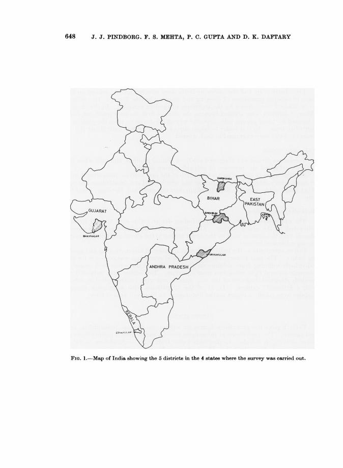
Five districts in 4 of the states in India were selected for the survey on thebasis of existing prevalence of chewing and smoking habits, Fig. 1. The villagesto be studied were chosen by the technique of random sampling. In the state ofBihar, 2 districts were studied because the district first chosen turned out to beinhabited by tribal groups with a specific way of life deviating from the pattern innontribal areas. In this house-to-house survey about 10,000 individuals (all 15years or older) were examined in each district.
Diagnostic criteriaSubmucous fibrosis was diagnosed solely on clinical grounds, and only when the
patients exhibited the presence of palpable fibrous bands.Leukoplakia was defined as a white patch of the oral mucosa, measuring 5 mm.
or more, which could not be scraped off and which could not be attributed to anyother diagnosable disease. The definition does not carry any histologic connota-tion.
Methods of examinationThe examinations were done by 9 Indian dentists who were trained by and
calibrated to the senior author. The criteria for leukoplakia and submucousfibrosis were the same as used by the senior author in the above-mentioned surveysamong urbanized Indians.
Before examination, the individuals were questioned about chewing and smok-ing habits. The past history with regard to oral symptoms was collected for theindividuals suffering from submucous fibrosis. The examination took place inadequate natural light using two mouth mirrors. The lesions were indicated onspecially designed diagrams of the oral mucosa and were photographed in colourwith a Polaroid® camera. In 54 of the 63 patients with submucous fibrosisbiopsies were taken; a report on the histological findings will appear later.
OBSERVATIONS
Table I gives the prevalence figures for submucous fibrosis, leukoplakia, andoral cancer. The prevalence of submucous fibrosis varies from 0 in Singhbum inBihar to 0.44% in Kerala. Leukoplakia varies from 0.2% in Singhbum in Biharto 5 10% in Andhra Pradesh. The highest number of oral cancer cases wasfound in Kerala (10 cases) and Andhra Pradesh (7 cases). The distribution of the63 cases according to sex and age are seen in Table II. The ratio female: maleis 3: 1. No case was found below the age of 20 years.
T'he oral symptoms were registered for 61 patients. From Table III it is seenthat a burning sensation to spicy food was experienced in 54 patients. Next infrequency were pain, dryness of the mouth, and stomatitis. It is interesting tonote that 16 patients complained of increased salivation. Twenty patients hadnoticed the presence of vesicles during the course of the disease.
647
648 J. J. PINDBORG. F. S. MEHTA, P. C. GUPTA AND D. K. DAFTARY
FIG.*I.-MapYI.5 5
FIG. 1.-Map of India showing the 5 districts in the 4 states where the survey was carried out.

ORAL SUBMUCOUS FIBROSIS
°
a 0 0O Xco
c) 0
ocro
0 Co
0~~~~~
EO O 4)
;4 e O O n O
A} Pe
i
O Mb
~~ ~ ~ ~
649

650 J. J. PINDBORG, F. S. MEHTA, P. C. GUPTA AND D. K. DAFTARY
TABLE II. Distribution of the 63 Patients with Submucous FibrosisAccording to Age and Sex
Age Group Male Female Total20-29 3 330-39 . 1 12 . 1340-49 . 6 . 11 . 1750-59 . 3 12 1560-69 5 . 7 1270-79 . 1 1 280-89 . . 1 . 1
Total . 16 . 47 63
TABLE II1.-Oral Symptoms Reported for 61 Patients with Submucous FibrosisSymptom Total
Burning sensation on spicy food . . 54Pain 41Dryness of the mouth 34Stomatitis . . . 29Burning sensation on ordinary food . 27Ulceration . . 25Burning sensation, intermittent . . 23Vesicles . 20Burning sensation, continuous 19Increased salivation . 16Referred pain . 15Numbness . . . 6
TABLE IV.-Location of Fibrous Bands in 63 Patients with Submucous FibrosisLocation Number
Buccal mucosaRight 60Left 59
Soft palate 31Tongue 23Labial mucosaUpper 18Lower . 22
Floor of the mouth . . 18Uvula 11
In Table IV the location of fibrous bands is given for the 63 cases of submucousfibrosis. The buccal mucosa is the site most frequently affected. Next infrequency are the soft palate, tongue, labial mucosa, and floor of the mouth.
Often the tongue is the seat of marked atrophy of the papillae (Fig. 2). Amongthe 63 cases, 24 presented a total atrophy of the tongue papillae, and 14 a partialatrophy. It means that 600% of the patients with submucous fibrosis exhibitedchanges in the papillary pattern of the tongue. Sixteen patients (25.4%) couldnot protrude the tongue beyond the muco-cutaneous junction of the lips and twobeyond the incisal edges of the lower anterior teeth. Deviations from the normaloral pigmentation were observed in 23 cases. The presence of vesicles at the timeof examination was noted in 6 patients.
In 8 patients (or 12-7 %0) the submucous fibrosis was associated with leukoplakia.Of the 10 cases with oral cancer in Kerala two also suffered from submucous fibrosis.

ORAL SUBMUCOUS FIBROSIS
2
FiG. 2.-Tongue changes in a 30-year-old man, with submucous fibrosis, from Kerala. Thetongue exhibits total loss of papillae and an area of retraction due to the presence of fibrousbands. The tongue cannot be stretched very much beyond the incisal edge of the lowerincisor teeth. Note also the fibrotic pterygomandibular raphe in both sides and the patchyloss of pigment on the vermilion border.
DISCUSSION
It is interesting to compare the prevalence figures for submucous fibrosisfound in the present study with those reported by Pindborg and co-workersamong urbanized Indians (Table V). That material was to a certain extentsalected as it only comprised individuals seeking the dental colleges. They werenot, however, coming because of symptoms from their submucous fibrosis, butbecause they wanted to get their teeth extracted or cavities filled. As in thepresent study, Zachariah et al. (1967) found the highest prevalence of submucousfibrosis in Kerala. The lowest prevalence found among urbanized Indians wasin Bangalore, which is located about 1000 metres above sea level. At the presenttime it cannot be said whether altitude plays any role in the prevalence ofsubmucous fibrosis.
The sex distribution in the present survey with a female: male ratio of 3: 1is surprising in the light of previous findings. In the largest materials publishedso far (Pindborg and Sirsat; 1966; Wahi et al., 1966) males have dominated overfemales. As the present study is a house-to-house survey any selection shouldbe excluded. Also Shear et al. (1967) found a predominance of females amongunselected Indians in South Africa.
651

652 J. J. PINDBORG, F. S. MEHTA, P. C. GUPTA AND D. K. DAFTARY
C)
o C)
0qBE
. g
° .^t 6ce
a
o
4 *.
ORAL SUBMUCOUS FIBROSIS
The present study has not provided new information with regard to the aetiologyof submucous fibrosis. The use of tobacco (Wahi et al., 1966) and chillies (Pind-borg and Sirsat, 1966) has been incriminated and so have vitamin deficiencies(Wahi et al., 1966). It is a fact that the disease is predominantly observed amongEast Indians, though it has also been found in other countries of South East Asia.It is also well known that the Indians living outside Africa to a large extent keeptheir Indian dietary habits and that chillies are an important ingredient of thefood. In the present study intolerance to spicy food was observed in 88.5%of the submucous fibrosis cases. Of the 63 cases of submucous fibrosis 31-8%did not have any chewing or smoking habit speaking against the theory thattobacco plays an important role.
It seems beyond any doubt that submucous fibrosis is most prevalent in Keralain South India, where the oral cancer prevalence is very high. In the presentstudy 12.7% of the submucous fibrosis cases were associated with leukoplakia,which is significantly higher than the 2.0% found in the entire survey.
This figure is lower than the 26-6% reported by Pindborg (1965) in submucousfibrosis patients from Bombay and Lucknow. The lower prevalence of leuko-plakia in the present material may be explained by the fact that the 16 cases ofsubmucous fibrosis in Gujarat were found among women, who had no chewingor smoking habits. Therefore, they lacked the agents probably responsible forinducing leukoplakia.
Of the 10 cases of oral cancer in Kerala, 3 had a simultaneous occurrence ofsubmucous fibrosis which is in good agreement with the findings of Pindborget al. (1967), viz., 40% with submucous fibrosis among 100 cases of oral cancer.The results indicate a positive relationship between the two conditions. Thehistologic findings from the present study show a considerable number of pre-malignant features in the patients with submucous fibrosis thus emphasizingthe precancerous nature ofsubmucous fibrosis.
SUMMARY
The prevalence of submucous fibrosis has been studied in 5 groups, approxi-mately 10,000 in each, of Indian villagers in 4 states of India. The prevalencerate varied from 0 to 0.4%. Clinical data are given on the 63 cases found in thesurvey. A conspicuous feature is the 60% prevalence of atrophy of the tonguepapillae. The etiology of submucous fibrosis is still unknown though the use ofchillies seems to be associated with the development of the disease. The presentfindings support the hypothesis that submucous fibrosis is a precancerous condition.
The research conducted for this paper was supported in whole by funds fromthe National Institutes of Health, U.S. Public Health Service, under P.L. 480research agreement No. 644,322.
The authors wish to express their profound appreciation to the dentists in theexamining teams: Dr. R. B. Bhonsle, S. K. Choksi, V. V. Dandekar, Y. Mehta,V. K. Pitkar, P. N. Sihor, N. C. Shah, B. C. Shroff, P. S. Turner and S.Upadhyay.
The project is greatly indebted to Polaroid Land Corporation for aninvaluable supply of cameras and films.
653

654 J. J. PINDBORG, F. S. MEHTA, P. C. GUPTA AND D. K. DAFTARY
REFERENCES
DOCKRAT, I. S.-(1967) S. Afr. Cancer Bull., 11, 103.KENNEDY, T. F. AND MACDONALD, D. G.-(1968) Br. dent. J., 124, 121.KRISHNAPPA, A.-(1967) Dent. J. Malaysia & Singapore, 7, 32.MILLARD, P. R.-(1966) Br. J. Derm., 78, 305.MOOS, K. F. AND MADAN, D. K.-(1968) Br. dent. J., 124, 313.PAYMASTER, J. C.-(1956) Cancer, N.Y., 9, 431.PINDBORG, J. J.-(1965) Bull. Wld Hlth Org., 32, 748. (1967) Report on 'Studies on
Oral Leukoplakias in New Guinea and Fiji'. Submitted to the World HealthOrganization.
PINDBORG, J. J., CHAWLA, T. N., MISRA, R. K., NAGPAUL, R. K. AND GUPTA, V. K.--(1965a) J. dent. Res., 44, 615.
PINDBORG, J. J., KALAPESSI, H. K., KALE, S., SINGH, B. AND TALYERKHAN, B. N.-(1965b) J. all-India dent. A88., 37, 228.
PINDBORG, J. J., POULSEN, H. E. AND ZACHARIAH, J. (1967) Cancer N.Y., 20, 1141.PINDBORG, J. J. AND SIRSAT, M. S.-(1966) Oral Surg., 22, 764.RAO, A. B. N.-(1962) Br. J. Surg., 50, 23.ROWELL, N. R.-(1967) Br. J. Derm., 79, 64.SCHWARTZ, J.-(1952) Cited by Sirsat and Khanolkar, 1962.SHEAR, M., LEMMER, J. AND DOCKRAT, I. S.-(1967) S. Afr. J. med. Sci., 32, 41.Su, I. P.-(1954) Arch. Otolar., 59, 330.WAHI, P. N., KAPUR, V. L., LUTHRA, U. K. AND SRIVASTAVA, M. C. (1966) Bull. Wld
Hlth Org., 35, 793.ZACHARIAH, J., MATHEW, B., VARMA, N. A. R., IQBAL, A. M. AND PINDBORG, J. J.-
(1966) J. all-India dent. A8s., 38, 290.